S1 Available online http://ccforum.com/supplements/11/S2 Critical Care Volume 11 Suppl 2, 2007 27th International Symposium on Intensive Care and Emergency Medicine Brussels, Belgium, 27–30 March 2007 Published online: 22 March 2007 These abstracts are available online at http://ccforum.com/supplements/11/S2 © 2007 BioMed Central Ltd P1 Infusion of sodium sulfide improves myocardial and endothelial function in a canine model of cardiopulmonary bypass C Szabó 1 , G Veres 2 , T Radovits 2 , M Karck 2 , G Szabó 2 1 Ikaria Inc., Seattle, WA, USA; 2 University of Heidelberg, Germany Critical Care 2007, 11(Suppl 2):P1 (doi: 10.1186/cc5161) Hydrogen sulfide is produced endogenously by a variety of enzymes involved in cysteine metabolism. Clinical data indicate that endogenous levels of hydrogen sulfide are diminished in various forms of cardiovascular diseases. The aim of the current study was to investigate the effects of hydrogen sulfide supple- mentation on cardiac function during reperfusion in a clinically relevant experimental model of cardiopulmonary bypass. Twelve anesthetized dogs underwent hypothermic cardiopulmonary bypass. After 60 minutes of hypothermic cardiac arrest, reper- fusion was started after application of either saline vehicle (control, n = 6), or the sodium sulfide infusion (1 mg/kg/hour, n = 6). Biventricular hemodynamic variables were measured by combined pressure–volume–conductance catheters. Coronary and pulmonary blood flow, vasodilator responses to acetylcholine and sodium- nitroprusside and pulmonary function were also determined. Administration of sodium sulfide led to a significantly better recovery of left and right ventricular systolic function (P < 0.05) after 60 minutes of reperfusion. Coronary blood flow was also significantly higher in the sodium sulfide-treated group (P < 0.05). Sodium sulfide treatment improved coronary blood flow, and preserved the acetylcholine-induced increases in coronary and pulmonary blood (P < 0.05). Myocardial ATP levels were markedly improved in the sulfide-treated group. Thus, supplementation of sulfide improves the recovery of myocardial and endothelial function and energetic status after hypothermic cardiac arrest during cardiopulmonary bypass. These beneficial effects occurred without any detectable adverse hemodynamic or cardiovascular effects of sulfide at the dose used in the current study. P2 Cytoprotective and anti-inflammatory effects of hydrogen sulfide in macrophages and mice C Szabo, L Kiss, E Pankotai University of Medicine and Dentistry of New Jersey, Newark, NJ, USA Critical Care 2007, 11(Suppl 2):P2 (doi: 10.1186/cc5162) The aim of the current study was to test potential cytoprotective and anti-inflammatory effects of the novel biological mediator hydrogen sulfide in murine models. Murine J774 macrophages were grown in culture and exposed to cytotoxic concentrations of nitrosoglutathione, or peroxynitrite (a reactive species formed from the reaction of nitric oxide and superoxide). Pretreatment of the cells with sodium sulfide (60–300 µM) reduced the loss of cell viability elicited by the nitric oxide donor compound (3 mM) or by peroxynitrite (3 mM), as measured by the MTT method. Sodium sulfide did not affect cell viability in the concentration range tested. In mice subjected to bacterial lipopolysaccharide (LPS, 5 mg/kg i.p.), treatment of the animals with sodium sulfide (0.2 mg/kg/hour for 4 hours, administered in Alzet minipumps) reduced the LPS- induced increase in plasma IL-1β and TNFα levels. These responses were attenuated when animals were pretreated with the heme oxygenase inhibitor tin-protoporphyrin IX (6 mg/kg). The current results point to the cytoprotective and anti-inflammatory effects of hydrogen sulfide, in cells exposed to nitrosative stress, and in animals subjected to endotoxemia. P3 Epithelial cell apoptosis is similar but hypoxic-inducible factor expression is weaker in acute acalculous cholecystitis than in calculous cholecystitis M Vakkala 1 , J Laurila 1 , J Saarnio 2 , V Koivukangas 2 , H Syrjälä 3 , T Karttunen 4 , Y Soini 4 , T Ala-Kokko 1 1 Department of Anesthesiology, 2 Department of Surgery, 3 Department of Infection Control and 4 Department of Pathology, Oulu University Hospital, Oulu, Finland Critical Care 2007, 11(Suppl 2):P3 (doi: 10.1186/cc5163) Introduction It has been previously shown that the two forms of acute cholecystitis, acute acalculous cholecystitis (AAC) and acute calculous cholecystitis (ACC), have significantly different histopathological features suggesting that AAC is a manifestation of systemic critical illness whereas ACC is a local disease of the gallbladder. A balance between cell proliferation and cell death is essential for cell homeostasis. The purpose of this study was to compare the markers of apoptosis, cell proliferation, and expression of hypoxic-inducible factor alpha (HIF-1α) in AAC, ACC and normal gallbladders. Methods The AAC group consisted of 30 patients who underwent open cholecystectomy due to acute acalculous cholecystitis during their ICU stay. The ACC group consisted of 21 hospitalized patients who underwent cholecystectomy due to acute calculous cholecystitis. The control group consisted of nine samples taken from normal gallbladders extirpated during pancreatic tumor surgery. The immunohistochemical analysis was done according to the manufacturer’s recommendations and they consisted of Ki-67 (proliferation), M30 (apoptosis) and HIF-1α antibodies. Cell proliferation and degree of apoptosis were expressed as the percentage of positive cells. HIF-1α expression was expressed as absent or weak (Score 1) or strong (Score 2). Results Apoptosis (median, 25th, 75th percentiles) was significantly increased in AAC 1.3% (1.0%, 3.3%), P = 0.001 and ACC 0.93% (0.40%, 3.25%), P = 0.011 compared with controls 0.32% (0.20%, 0.40%). Proliferation rate was also significantly increased in AAC

Welcome message from author
This document is posted to help you gain knowledge. Please leave a comment to let me know what you think about it! Share it to your friends and learn new things together.
Transcript
S1
Available online http://ccforum.com/supplements/11/S2
Critical Care Volume 11 Suppl 2, 200727th International Symposium on Intensive Care and EmergencyMedicineBrussels, Belgium, 27–30 March 2007
Published online: 22 March 2007These abstracts are available online at http://ccforum.com/supplements/11/S2© 2007 BioMed Central Ltd
P1Infusion of sodium sulfide improves myocardial andendothelial function in a canine model of cardiopulmonarybypass
C Szabó1, G Veres2, T Radovits2, M Karck2, G Szabó2
1Ikaria Inc., Seattle, WA, USA; 2University of Heidelberg, GermanyCritical Care 2007, 11(Suppl 2):P1 (doi: 10.1186/cc5161)
Hydrogen sulfide is produced endogenously by a variety ofenzymes involved in cysteine metabolism. Clinical data indicatethat endogenous levels of hydrogen sulfide are diminished invarious forms of cardiovascular diseases. The aim of the currentstudy was to investigate the effects of hydrogen sulfide supple-mentation on cardiac function during reperfusion in a clinicallyrelevant experimental model of cardiopulmonary bypass. Twelveanesthetized dogs underwent hypothermic cardiopulmonarybypass. After 60 minutes of hypothermic cardiac arrest, reper-fusion was started after application of either saline vehicle (control,n = 6), or the sodium sulfide infusion (1 mg/kg/hour, n = 6).Biventricular hemodynamic variables were measured by combinedpressure–volume–conductance catheters. Coronary and pulmonaryblood flow, vasodilator responses to acetylcholine and sodium-nitroprusside and pulmonary function were also determined.Administration of sodium sulfide led to a significantly betterrecovery of left and right ventricular systolic function (P < 0.05)after 60 minutes of reperfusion. Coronary blood flow was alsosignificantly higher in the sodium sulfide-treated group (P < 0.05).Sodium sulfide treatment improved coronary blood flow, andpreserved the acetylcholine-induced increases in coronary andpulmonary blood (P < 0.05). Myocardial ATP levels were markedlyimproved in the sulfide-treated group. Thus, supplementation ofsulfide improves the recovery of myocardial and endothelialfunction and energetic status after hypothermic cardiac arrestduring cardiopulmonary bypass. These beneficial effects occurredwithout any detectable adverse hemodynamic or cardiovasculareffects of sulfide at the dose used in the current study.
P2Cytoprotective and anti-inflammatory effects of hydrogensulfide in macrophages and mice
C Szabo, L Kiss, E PankotaiUniversity of Medicine and Dentistry of New Jersey, Newark, NJ, USACritical Care 2007, 11(Suppl 2):P2 (doi: 10.1186/cc5162)
The aim of the current study was to test potential cytoprotectiveand anti-inflammatory effects of the novel biological mediatorhydrogen sulfide in murine models. Murine J774 macrophageswere grown in culture and exposed to cytotoxic concentrations ofnitrosoglutathione, or peroxynitrite (a reactive species formed fromthe reaction of nitric oxide and superoxide). Pretreatment of the
cells with sodium sulfide (60–300 µM) reduced the loss of cellviability elicited by the nitric oxide donor compound (3 mM) or byperoxynitrite (3 mM), as measured by the MTT method. Sodiumsulfide did not affect cell viability in the concentration range tested.In mice subjected to bacterial lipopolysaccharide (LPS, 5 mg/kgi.p.), treatment of the animals with sodium sulfide (0.2 mg/kg/hourfor 4 hours, administered in Alzet minipumps) reduced the LPS-induced increase in plasma IL-1β and TNFα levels. Theseresponses were attenuated when animals were pretreated with theheme oxygenase inhibitor tin-protoporphyrin IX (6 mg/kg). Thecurrent results point to the cytoprotective and anti-inflammatoryeffects of hydrogen sulfide, in cells exposed to nitrosative stress,and in animals subjected to endotoxemia.
P3Epithelial cell apoptosis is similar but hypoxic-induciblefactor expression is weaker in acute acalculouscholecystitis than in calculous cholecystitis
M Vakkala1, J Laurila1, J Saarnio2, V Koivukangas2, H Syrjälä3, T Karttunen4, Y Soini4, T Ala-Kokko1
1Department of Anesthesiology, 2Department of Surgery,3Department of Infection Control and 4Department of Pathology,Oulu University Hospital, Oulu, FinlandCritical Care 2007, 11(Suppl 2):P3 (doi: 10.1186/cc5163)
Introduction It has been previously shown that the two forms ofacute cholecystitis, acute acalculous cholecystitis (AAC) and acutecalculous cholecystitis (ACC), have significantly differenthistopathological features suggesting that AAC is a manifestationof systemic critical illness whereas ACC is a local disease of thegallbladder. A balance between cell proliferation and cell death isessential for cell homeostasis. The purpose of this study was tocompare the markers of apoptosis, cell proliferation, andexpression of hypoxic-inducible factor alpha (HIF-1α) in AAC, ACCand normal gallbladders.Methods The AAC group consisted of 30 patients who underwentopen cholecystectomy due to acute acalculous cholecystitis duringtheir ICU stay. The ACC group consisted of 21 hospitalizedpatients who underwent cholecystectomy due to acute calculouscholecystitis. The control group consisted of nine samples takenfrom normal gallbladders extirpated during pancreatic tumorsurgery. The immunohistochemical analysis was done according tothe manufacturer’s recommendations and they consisted of Ki-67(proliferation), M30 (apoptosis) and HIF-1α antibodies. Cellproliferation and degree of apoptosis were expressed as thepercentage of positive cells. HIF-1α expression was expressed asabsent or weak (Score 1) or strong (Score 2).Results Apoptosis (median, 25th, 75th percentiles) was significantlyincreased in AAC 1.3% (1.0%, 3.3%), P = 0.001 and ACC 0.93%(0.40%, 3.25%), P = 0.011 compared with controls 0.32% (0.20%,0.40%). Proliferation rate was also significantly increased in AAC
S2
Critical Care March 2007 Vol 11 Suppl 2 27th International Symposium on Intensive Care and Emergency Medicine
8.0% (4.0%, 17.0%), P < 0.001 and ACC 14% (7.5%, 26.5%),P = 0.001 compared with controls 1.0% (1.0%, 3.0%). Strong HIF-1α staining was observed in 100% of ACC, in 57% of AAC and in44% of control specimens (P < 0.001). Strong HIF-1α expressionwas associated with increased cell proliferation (P = 0.002).Conclusions Cell proliferation and apoptosis were increased inAAC and ACC. The expression of hypoxic-inducible factor was,however, stronger in ACC compared with AAC.
P4Effect of prostaglandin E2 on ATP-induced Ca2+ responsesin human THP-1 monocytic cells
M Goto1, M Murakawa1, J Kimura1, I Matsuoka2
1Fukushima Medical University, Fukusima, Japan; 2Takasaki University of Health and Welfare, Gunma, JapanCritical Care 2007, 11(Suppl 2):P4 (doi: 10.1186/cc5164)
Introduction To clarify the relation between ATP and prostaglandinE2 (PGE2) in the immunologic system, we investigated the acuteand chronic effects of PGE2 on activation of purinergic signaling inmonocytes by measuring the ATP-induced elevation of intracellularCa2+ ([Ca]i) in fura-2-loaded THP-1 monocytes.Method THP-1 monocytes were grown for about 2 days. Toexamine the chronic effects, PGE2 and dibutyryl cAMP (dbcAMP)were added and incubated for another day. The cell suspensionswere washed, loaded with fura-2-AM, and transferred into a quartzcuvette and placed in the thermostat-regulated sample chamber ofa dual excitation beam spectrophotometer. To examine the acuteeffects, ATP was added immediately after PGE2 and dbcAMP intothe cuvette. In the chronic experiment, ATP alone was added intothe cuvette. Fura-2 fluorescence emission was measured at510 nm. The [Ca]i was calculated from the ratio of thefluorescence at the two excitation wavelengths.Results ATP induced a transient increase in [Ca]i followed by asustained elevation of [Ca]i. Acutely, PGE2 inhibited both thetransient and sustained ATP-induced elevations of [Ca]i. However,this acute inhibitory effect diminished gradually with time andchronic PGE2 accelerated the transient and sustained ATP-induced [Ca]i elevations for 24 hours. Both the acute and chroniceffects of PGE2 were mimicked by dbcAMP. In Ca2+-free solution,ATP did not induce the sustained elevation of [Ca]i in control cellsor cells pretreated for 24 hours with dbcAMP. This indicates thatthe ATP-induced sustained elevation of [Ca]i was due to Ca2+
entry. In addition, receptor-operated Ca2+ channel blockersinhibited the sustained ATP-induced elevation of [Ca]i in controlcells and cells pretreated with for 24 hours dbcAMP.Conclusion Acute PGE2 inhibited the ATP-induced activation ofmonocytes. On the other hand, chronic PGE2 accelerated monocyteactivation by upregulation of receptor-operated Ca2+ channels(ROCs). If this mechanism exhibits a physiological role, ROCinhibitors should be developed as new anti-inflammatory agents.
P5Interferon gamma levels are reduced by adenosine 5′′-triphosphate in lipopolysaccharide-stimulated wholehuman blood
M Nalos1, S Huang1, A Khan2, A McLean1
1Nepean Hospital, Penrith, Australia; 2Macquarie University, NorthRyde, AustraliaCritical Care 2007, 11(Suppl 2):P5 (doi: 10.1186/cc5165)
Introduction Extracellular release of ATP is an important modulatorof immune response. ATP plasma concentration is increased in
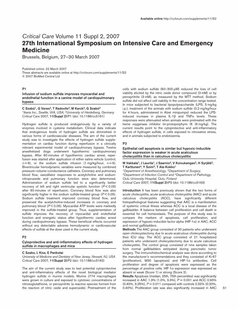
sepsis [1]. IFNγ plays a critical role in host defense by promotingTh1 phenotype and bacterial clearance. Low IFNγ levels areassociated with the Th2 phenotype consistent with critical illnessanergy [2]. It has been reported that 100 and 300 mM ATPincreased LPS/PHA-stimulated IL-10 secretion in human blood [3].Higher IL-10/IFNγ ratio shifts the immune phenotype from Th1 toTh2 response. We studied the effect of ATP on LPS-stimulatedIL-10 and IFNγ secretion in a standardized ex-vivo whole humanblood culture.Methods Venous blood from 10 healthy volunteers was drawn intotubes containing 10 ng LPS/ml (ILCSÒ; EDI GmBH, Reutlingen,Germany) and incubated with or without 100 mM ATP, respectively,at 37°C for 24 hours. The supernates were separated and frozenat –20°C. Cytokine levels were analysed on a robotic workstation(epMotion 5075; Eppendorf AG, Hamburg, Germany) in duplicateusing the ELISA Cytokine kit (Luminex; Biosource Int., Camarillo,CA, USA).Results Added ATP reduced the mean concentration of IFNγ in LPS-stimulated blood from 1,206 ± 1,667 pg/ml to 140 ± 128 pg/ml;P = 0.006. There was no consistent effect of ATP on IL-10secretion in our study (21.6 ± 16.9 pg/ml to 17.2 ± 18.8 pg/ml).Interestingly, three subjects of Indian/Indonesian origin had IL-10levels below the assay detection limit. The mean IL-10/IFNγ ratiowas increased from 0.05 ± 0.04 to 0.16 ± 0.09 in the remainingCaucasian subjects (P = 0.015). See Figure 1.Conclusions Our results suggest an immunosuppressive effect ofextracellular ATP that is evident by the decrease of IFNγ andtherefore the relative shift of the immune response towards Th2phenotype. Although this may represent a self-protectivemechanism, it may contribute to critical illness anergy.References1. Bours MJ, Swennen EL, Di Virgilio F, et al.: Adenosine 5′′-
triphosphate and adenosine as endogenous signalingmolecules in immunity and inflammation. Pharmacol Ther2006, 112:358-404.
2. Ertel W, Keel M, Neidhardt R, et al.: Inhibition of the defencesystem stimulating interleukin-12 interferon-gammapathway during critical illness. Blood 1997, 89:1612-1620.
3. Swennen EL, Bast A, Dagnelie PC: Immunoregulatory effectsof adenosine 5′′-triphosphate on cytokine release fromstimulated whole blood. Eur J Immunol 2005, 35:852-858.
Figure 1 (abstract P5)
S3
Available online http://ccforum.com/supplements/11/S2
P6Tyrosine phosphorylation modulates rat vascular responseto experimental endotoxemia in vivo and in vitro
C Lehmann, T Hammann, O Adamek, H Erber, M Manthey, T Wenzel, A Stier, M Wendt, D PavlovicErnst-Moritz-Arndt-Universität Greifswald, GermanyCritical Care 2007, 11(Suppl 2):P6 (doi: 10.1186/cc5166)
Introduction Endotoxemia is characterized by vascular hypo-reactivity, hypotension and microcirculatory changes that arepartially linked to the excess of nitric oxide production. The agentsthat can influence Ca2+ transport (affect Ca-ATPase) or modulateCa2+ sensitivity of the smooth muscle contraction (modulate phos-phorylation) may theoretically influence some of the above-mentioned effects.Methods We evaluated the effects of tyrosine phosphatase orkinase inhibitors, sodium orthovanadate (SOV) or genistein (GEN).The effects of these agents were examined in vitro, in a model ofvascular hyporeactivity of sepsis, in rings of rat aorta (RA), with orwithout endothelium (±ENDO), or in human mesenteric artery(HMA). In vivo, the intestinal microcirculation (terminal ileum) ofendotoxemic rats (LEW.1A) that received i.v. lipopolysaccharide(LPS), 15 mg/kg BW, was examined using intravital microscopy.Results In vitro. The nitric oxide production inhibitor L-NAME(5 × 10⁻⁴) and cGMP inhibitor ODQ (5 x 10⁻⁵) abolished LPS-induced hyporeactivity. GEN attenuated maximal tension (Tmax)while SOV increased the response to PE; Tmax (kg/g, dry muscle):controls vs SOV, RA (–ENDO): 0.87 ± 0.19 vs 1.42 ± 0.23(10–7); 1.56 ± 0.28 (10–6) and 2.33 ± 0.69 (10–5); RA (+ENDO):0.88 ± 0.21 vs 1.53 ± 0.35 (10–7); 1.35 ± 0.30 (10–6) and2.55 ± 0.68 (10–5); and HMA (+ENDO): 1.12 ± 0.23 vs0.37 ± 0.14 (10–7); 2.06 ± 0.21 (10–6) and 3.00 ± 0.07 (10–5).In vivo. In the LPS group GEN increased mucosal functionalcapillary density (FCD, cm/cm2; mean ± SD; LPS vs GEN,105.5 ± 44.6 vs 174.7 ± 39.1; P = 0.018). SOV (7.5 mg/kg)increased FCD not only in mucosa (163.7 ± 40.0; P = 0.024) butalso in the longitudinal muscular layer (LPS vs SOV, 111.9 ± 24.0vs 172.2 ± 19.5; P < 0.001). Surprisingly, the SOV (15 mg/kg)alone (without LPS) increased leukocyte sticking in the venules V1(LPS vs SOV, number of stickers/mm2, 403.3 ± 113.9 vs669.8 ± 150.8; P = 0.027).Conclusions The tyrosine phosphorylation pathway may play animportant role in modulation of the LPS-induced vascularhyporeactivity and could enhance terminal ileum microcirculation.This might be a result of both modulation of tyrosinephosphorylation by genistein and sodium orthovanadate, and/orplasma membrane Ca-ATPase inhibition by SOV.
P7Glibenclamide dose response in patients with septic shock
A Morelli1, C Ertmer2, M Lange2, K Broeking2, H Van Aken2, A Orecchioni1, M Rocco1, P Pietropaoli1, M Westphal21University of Rome ‘La Sapienza’, Rome, Italy; 2University Hospital of Muenster, GermanyCritical Care 2007, 11(Suppl 2):P7 (doi: 10.1186/cc5167)
Introduction (K+ATP) channels are implicated in thepathophysiology of catecholamine tachyphylaxis in septic shock.This prospective, randomized, double-blinded, clinical study wasdesigned to determine whether different doses of glibenclamidehave any effects on norepinephrine requirements and cardio-pulmonary hemodynamics in patients with septic shock.
Methods We enrolled 30 patients with septic shock requiringinvasive hemodynamic monitoring and norepinephrine infusion ≥0.5 µg/kg/min to maintain MAP between 65 and 75 mmHg.Patients were randomized to receive either 10, 20, or 30 mgenteral glibenclamide. Systemic hemodynamics, global oxygentransport, arterial lactate concentrations, gas exchange, andplasma glucose concentrations were determined at baseline, andfollowing 3, 6 and 12 hours after administration of the study drug.Results Glibenclamide decreased plasma glucose concentrationsin a dose-dependent manner, but failed to reduce norepinephrinerequirements. None of the doses had any effects oncardiopulmonary hemodynamics. See Table 1.
Table 1 (abstract P7)
Plasma glucose concentration (mg/dl)
Time
Glibenclamide 0 hours 3 hours 6 hours 12 hours
10 mg 118 ± 13 110 ± 9 109 ± 10 107 ± 10
20 mg 117 ± 5 106 ± 4 93 ± 7* 98 ± 9*
30 mg 113 ± 6 86 ± 3* 89 ± 4* 98 ± 3*
Data presented as mean ± SEM. *P < 0.05 vs baseline (0 hours) withingroups.
Conclusion Oral glibenclamide is an ineffective adjunct in thetreatment of catecholamine-dependent human septic shock.
P8Molecular mechanism of glutamine induction of HSP70involves activation of the O-linked-N-acetylglucosaminepathway in murine embryonic fibroblast cells
C Hamiel1, S Pinto2, K Singleton1, P Wischmeyer1
1University of Colorado, Denver, CO, USA; 2Valparaiso University,IN, USACritical Care 2007, 11(Suppl 2):P8 (doi: 10.1186/cc5168)
Introduction The purpose of this study was to determine whetherglutamine (GLN)-mediated cellular protection is dependent on theO-linked-N-acetylglucosamine (O-glcNAc) pathway. GLN canprotect against critical illness via induction of HSP70. The molecularmechanism by which GLN enhances HSP70 is unknown. GLN canincrease flux through the hexosamine biosynthetic pathway andactivate transcription factors by O-glcNAc. We investigated GLN’seffect on O-glcNAc levels and nuclear translocation of SP1 andHSF-1, which are vital to HSP70 expression. To determine theimportance of O-glcNAc, we used silencing RNA (siRNA) against O-linked-N-acetylglucosamine transferase (OGT), the enzyme thatcatalyzes addition of O-glcNAc to proteins.Methods Mouse embryonic fibroblast cells were treated with0 mM GLN (CT) or 10 mM GLN (GLN), heat stressed (HS) andallowed to recover for 20 minutes. Cells were stained and meanfluorescent intensities (MFIs) measured for total O-glcNAc andnuclear HSF-1 and SP1. For OGT silencing, cells were transfectedwith either no siRNA, siRNA to OGT, or negative control oligos (ncsiRNA) and then treated as above (but with 4 hours recovery).HSP70 and OGT were evaluated by western blot.Results Microscopy showed GLN treatment increased nuclearMFI for HSF-1 by 40% (HS-CT: 1,005 ± 146 vs HS-GLN:1403 ± 102, P < 0.05) and SP1 by 54% (HS-CT: 214 ± 14vs HS-GLN: 330 ± 13, P < 0.05). Total O-glcNAc levels showed44% MFI increase in HS-GLN compared with HS-CT (HS-CT:360 ± 24 vs HS-GLN: 518 ± 51, P < 0.05). Following OGTsilencing, HS-GLN showed a threefold increase in HSP70
S4
(P = 0.04). These increases were completely blocked by OGTsilencing (P = 0.02 vs non-siRNA GLN groups). GLN-nc siRNAgroups did not decrease in HSP70 production. OGT was knockeddown 86% compared with controls (siRNA: 0.999 ± 0.19 vs CT:0.131 ± 0.05). N = 3.Conclusions These results show GLN can activate the O-glcNAcpathway and enhance nuclear translocation of HSF-1 and SP1.Inhibition of OGT blocked GLN-mediated induction of HSP70.Thus, it appears the mechanism of GLN-mediated HSP70 expres-sion is dependent on enhanced O-glcNAc pathway activation.
P9The effects of N-acetylcysteine on the levels of glutathione,serum TNFαα, and tissue malondialdehyde in sepsis
M Gul, M Ayan, A Seydanoglu, B Cander, S Girisgin, I EraymanSelcuk University Meram Medical School, Konya, TurkeyCritical Care 2007, 11(Suppl 2):P9 (doi: 10.1186/cc5169)
Objectives This study was designed to determine the effects of N-acetylcysteine (NAC) as an antioxidant agent on the free oxygenradicals and their plasma levels.Methods In this study, 40 Sprague–Dawley rats were randomlydivided into three groups as sham (n = 10), sepsis (n = 10), andsepsis + NAC (20 mg/kg/24 hours) (n = 10). An experimentalsepsis model was performed by a cecal ligation and perforation(CLP). NAC was administered at 0, 8 and 16 hours after CLP. Theblood samples were taken at 24 hours to determine the levels ofserum TNFα and erythrocyte glutathione (GSH), and renal and livertissue malondialdehyde (MDA).Results The serum TNFα levels were significantly decreased ingroup 3 compared with group 2 (P < 0.05). The erythrocyte GSHlevels significantly increased in group 3 compared with group 2(P < 0.05). In group 3, the liver MDA levels were decreasedcompared with group 2, but not statistically significant (P > 0.05)In group 3, the renal MDA levels were significantly decreasedcompared with group 2 (P < 0.05). The lung tissue PMNL levelssignificantly decreased in group 3 compared with group 2(P < 0.05).Conclusion In an experimental sepsis model, with the administra-tion of NAC as an antioxidant agent at lower doses, many meaning-ful positive effects were detected on the levels of erythrocyte GSH,serum TNFα, respiration function, and renal tissue MDA. In spite ofthe low dose, NAC therapies decrease the organ function abnor-malities; these effects were not reflected in the histopathologicalinvestigations. These findings suggest that NAC could be apossible therapeutic agent for sepsis and its mortality. However,further studies are needed to elucidate the effects of these drugsat higher doses.
P10Exogenous adrenomedullin reduces the arterial lactateconcentration and mean pulmonary arterial pressure inovine endotoxemia
C Ertmer1, M Lange1, H Van Aken1, K Bröking1, S Vocke1, F Daudel1, M Booke2, M Westphal31University of Muenster, Germany; 2Hospital of the Main-Taunus-Kreis, Hofheim, Germany; 3UTMB, Galveston, USACritical Care 2007, 11(Suppl 2):P10 (doi: 10.1186/cc5170)
Introduction Sepsis-associated arterial hypotension may becomplicated by inadequate systemic and regional oxygen deliveryresulting in lactic acidosis and multiple organ failure. We hypothe-sized that exogenous administration of adrenomedullin (AM), a
vasodilatory peptide hormone with anti-inflammatory properties,may improve the oxygen delivery–demand relationship, therebylimiting the increase in arterial lactate concentrations in ovine endo-toxemia.Methods Fourteen adult ewes were instrumented for chronic hemo-dynamic monitoring. Following 16 hours of endotoxemia (Salmonellatyphosa endotoxin, 10 ng/kg/min) the animals received either acontinuous infusion of AM at incremental doses (10, 50,100 ng/kg/min; each for 30 min) or the vehicle (normal saline; n = 7each).Results Endotoxin infusion contributed to a hypotensive–hyperdynamic circulation characterized by decreases in meanarterial pressure (MAP) and systemic vascular resistance index aswell as increases in heart rate (HR), cardiac index (CI) and arteriallactate concentrations. AM infusion at 100 ng/kg/min increasedthe CI (12.2 ± 0.8 vs 7.8 ± 0.5 l/min) and oxygen delivery index(1,734 ± 121 vs 1,075 ± 63 ml/min/m2), thereby decreasing thearterial lactate concentration (0.7 ± 0.2 vs 1.7 ± 0.3 mg/dl) andmean pulmonary arterial pressure (18 ± 1 vs 24 ± 1 mmHg; eachP < 0.001 vs control) noticed in the control group. However, AMinfusion at 100 ng/kg/min was linked to a decrease in MAP(64 ± 2 vs 80 ± 4 mmHg, P < 0.001 vs control).Conclusions Despite decreasing MAP, infusion of AM reversedpulmonary hypertension and improved the oxygen supply–demandrelationship in a dose-dependent manner, as indicated by areduced arterial lactate concentration. However, due to thevasodilatory properties of AM, it may be rationale to combine AMwith a vasopressor agent.
P11Angiopoietin-2 correlates with pulmonary capillarypermeability and disease severity in critically ill patients
M van der Heijden1, V van Hinsbergh2, G van NieuwAmerongen2, P Koolwijk2, R Musters2, J Groeneveld1
1VU University Medical Center, Amsterdam, The Netherlands;2Institute for Cardiovascular Research, VU University MedicalCenter, Amsterdam, The NetherlandsCritical Care 2007, 11(Suppl 2):P11 (doi: 10.1186/cc5171)
Introduction It has previously been shown that angiopoietin-1(Ang1) protects the adult vasculature against plasma leakage,whereas Ang2 and VEGF destabilize the vascular endotheliumresulting in vascular leakage. Consequently they might be involvedin the pathophysiology of acute lung injury (ALI) and acuterespiratory distress syndrome (ARDS) in sepsis patients. Wehypothesized that plasma Ang2 levels are associated withpulmonary capillary protein permeability, the lung injury score (LIS),length of stay on the ICU, the APACHE II score and survival inseptic patients with ALI or ARDS.Methods A prospective observational study was performed in anICU of an university hospital on 112 patients: 38 after electivecardiac surgery, 26 after major vascular surgery, 24 with sepsisand 24 with trauma. Plasma levels of Ang1, Ang2 and VEGF weremeasured and a mobile probe system was used to measure thepulmonary leak index (PLI) (that is, the transvascular transport rateof gallium-67-radiolabeled transferrin).Results Plasma levels of Ang2 and the PLI were significantlyhigher in patients with sepsis compared with other patient groups.In the sepsis group, a positive linear correlation was observedbetween plasma levels of Ang2 and length of stay on the ICU(rs = 0.509, P < 0.05) as index for disease severity. For all patientstogether, Ang2 had a positive linear correlation with PLI (rs = 0.374,P < 0.01), LIS (rs = 0.489, P < 0.01) and APACHE II score(rs = 0.287, P < 0.01). Furthermore, Ang2 was significantly increased
Critical Care March 2007 Vol 11 Suppl 2 27th International Symposium on Intensive Care and Emergency Medicine
S5
in nonsurvivors. Plasma Ang1 levels did not differ between groups.VEGF levels were undetectable in the plasma of the majority ofpatients.Conclusions Our results suggest that Ang2 is a mediator ofpulmonary capillary permeability and a marker of disease severity incritically ill patients. Furthermore, the plasma levels of Ang2 andthe ratio between Ang1 and Ang2 are more important in pulmonarycapillary permeability and disease severity than absolute levels ofAng1 and VEGF.
P12Dose-dependent effects of octreotide on plasma activitiesof IL-6 and lung tissue levels of malondialdehyde in sepsis
M Gul, A Seydanoglu, M Ayan, B Cander, I Erayman, S GirisginSelcuk University Meram Medical School, Konya, TurkeyCritical Care 2007, 11(Suppl 2):P12 (doi: 10.1186/cc5172)
Background and aim Sepsis, a complex and rapidly progressinginfectious disease with high levels of mortality, is widely regardedas the most challenging problem in intensive care. The lung isfrequently the first failing organ during septic conditions. Althoughthe etiology of sepsis is multifactorial, early release of proinflam-matory cytokines and oxidative damage are probably most impor-tant factors that lead to cell damage, organ dysfunction, and death.This study aimed to determine the effects of treatment with octreo-tide (OCT), on plasma activities of IL-6 and tissue levels of malon-dialdehyde (MDA) in an experimental model of sepsis.Methods Sepsis was induced in female Sprague–Dawley rats bycecal ligation and puncture (CLP) as previously described. Group 1(n = 10), sham operated animals; Group 2 (n = 10), sepsis servedas control; Group 3 (n = 10) and Group 4 (n = 10), respectively,OCT 50 µg/kg twice a day and OCT 100 µg/kg twice a dayadministered subcutaneously immediately after the induction ofsepsis and at 12 hours. Rats were sacrificed 24 hours after thesurgical procedure. Blood and lung tissue samples were taken24 hours after sepsis induction. Plasma activities of IL-6 and lungtissue levels of MDA were measured.Results The results showed that the plasma levels of IL-6, aninflammatory indicator, and tissue levels of MDA, an oxidativeindicator, are significantly increased during experimental model ofsepsis (P < 0.05). Increase in MDA levels and IL-6 activities afterCLP-induced sepsis was significantly prevented by OCT(100 µg/kg, s.c.) administration (P < 0.05).Conclusion Octreotide seems to have a dose-dependentantioxidative and immunomodulator effect in CLP-induced sepsis inrats. Further trials are necessary to reveal the therapeutic effect ofOCT in sepsis. On the other hand, further studies should beperformed aiming to reveal the optimal OCT doses. As a drug with awide margin of safety and less adverse reaction profile, OCT meritsconsideration as a choice of treatment in sepsis and septic shock.
P13Escherichia coli porcine peritonitis induces histologicaland transcriptome evidence of cardiac injury
R Goldfarb1, I Cinel1, S Gandhi1, L Cinel2, M Levine1, Q Wang3,A Brooks3, J Parrillo1
1Cooper University Hospital and UMDNJ, Camden, NJ, USA;2Thomas Jefferson University Hospital, Philadelphia, PA, USA;3EOHSI, UMDNJ, Piscataway, NJ, USACritical Care 2007, 11(Suppl 2):P13 (doi: 10.1186/cc5173)
Introduction Cardiac dysfunction is a feature of sepsis. In order togain insight into the fundamental mechanisms of this phenotype,
gene expression analysis (Affymetrix) was applied to serial cardiacbiopsies of sham (n = 2) and E. coli infected pigs (n = 3).Methods Cardiac samples were taken basal and hourly afterinfection for gene analysis and at the end of the experiment forhistopathological examination. Genes were determined to bedifferentially regulated at a greater than or less than twofoldchange and P < 0.05.Results Sham pigs had stable heart rate, cardiac output (CO) andcore temperature for the 5-hour period; infected pigs demon-strated an early elevation in CO and ventricular shortening and/orejection (assessed by echocardiography) followed by developmentof hypodynamics. In infected animals, increasing numbers of geneswere upregulated or downregulated (36, 278, 514, 842 and 1,238at 1, 2, 3, 4 and 5 hours) (Figure 1) whereas sham infection alteredfewer (247, 67 and 384 genes at 2, 3 and 4 hours). Comparingsham vs infected animals at the same time, numbers of significantlyaltered genes increased with time (32 at basal, to 74, 189 and601 at 2, 3 and 4 hours post infection). In hematoxylin–eosin-stained sections, histopathological assessment revealed acuteinflammation in pericardium and myocardium in infected pigs.Conclusions These results will provide biomarker and mechanisticinsights to pathogenesis of cardiac dysfunction of septic peritonitisand may also help identify some altered novel gene transcriptionpathways that can serve as new targets for diagnostic tools andtherapeutic strategies. All candidate genes will be validated byquantitative PCR.
P14Alkaline phosphatase treatment improves renal function inpatients with severe sepsis or septic shock
S Heemskerk1, R Masereeuw1, O Moesker2, M Bouw2, J vander Hoeven2,3, W Peters4, M Velders5, F Russel1, P Pickkers2
1Department of Pharmacology and Toxicology, Nijmegen Centre forMedical Life Sciences, 2Department of Intensive Care Medicine,3Nijmegen University Centre for Infectious Diseases and 4Departmentof Gastroenterology, Radboud University Nijmegen Medical Centre,Nijmegen, The Netherlands; 5AM-Pharma, Bunnik, The NetherlandsCritical Care 2007, 11(Suppl 2):P14 (doi: 10.1186/cc5174)
We previously demonstrated that upregulation of renal induciblenitric oxide synthase (iNOS) during systemic inflammation is
Available online http://ccforum.com/supplements/11/S2
Figure 1 (abstract P13)
S6
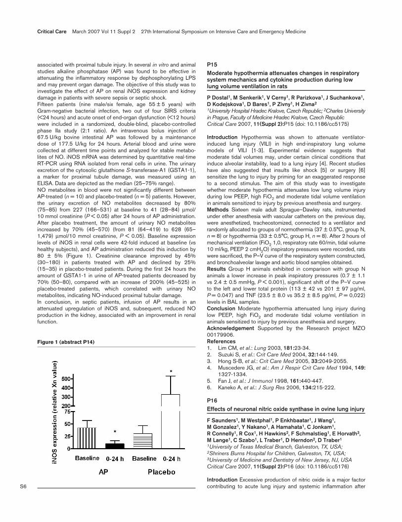
associated with proximal tubule injury. In several in vitro and animalstudies alkaline phosphatase (AP) was found to be effective inattenuating the inflammatory response by dephosphorylating LPSand may prevent organ damage. The objective of this study was toinvestigate the effect of AP on renal iNOS expression and kidneydamage in patients with severe sepsis or septic shock.Fifteen patients (nine male/six female, age 55 ± 5 years) withGram-negative bacterial infection, two out of four SIRS criteria(<24 hours) and acute onset of end-organ dysfunction (<12 hours)were included in a randomized, double-blind, placebo-controlledphase IIa study (2:1 ratio). An intravenous bolus injection of67.5 U/kg bovine intestinal AP was followed by a maintenancedose of 177.5 U/kg for 24 hours. Arterial blood and urine werecollected at different time points and analyzed for stable metabo-lites of NO. iNOS mRNA was determined by quantitative real-timeRT-PCR using RNA isolated from renal cells in urine. The urinaryexcretion of the cytosolic glutathione S-transferase-A1 (GSTA1-1),a marker for proximal tubule damage, was measured using anELISA. Data are depicted as the median (25–75% range).NO metabolites in blood were not significantly different betweenAP-treated (n = 10) and placebo-treated (n = 5) patients. However,the urinary excretion of NO metabolites decreased by 80%(75–85) from 227 (166–531) at baseline to 41 (28–84) µmol/10 mmol creatinine (P < 0.05) after 24 hours of AP administration.After placebo treatment, the amount of urinary NO metabolitesincreased by 70% (45–570) (from 81 (64–419) to 628 (65–1,479) µmol/10 mmol creatinine, P < 0.05). Baseline expressionlevels of iNOS in renal cells were 42-fold induced at baseline (vshealthy subjects), and AP administration reduced this induction by80 ± 5% (Figure 1). Creatinine clearance improved by 45%(30–180) in patients treated with AP and declined by 25%(15–35) in placebo-treated patients. During the first 24 hours theamount of GSTA1-1 in urine of AP-treated patients decreased by70% (50–80), compared with an increase of 200% (45–525) inplacebo-treated patients, which correlated with urinary NOmetabolites, indicating NO-induced proximal tubular damage.In conclusion, in septic patients, infusion of AP results in anattenuated upregulation of iNOS and, subsequent, reduced NOproduction in the kidney, associated with an improvement in renalfunction.
P15Moderate hypothermia attenuates changes in respiratorysystem mechanics and cytokine production during lowlung volume ventilation in rats
P Dostal1, M Senkerik1, V Cerny1, R Parizkova1, J Suchankova1,D Kodejskova1, D Bares1, P Zivny1, H Zivna2
1University Hospital Hradec Kralove, Czech Republic; 2Charles Universityin Prague, Faculty of Medicine Hradec Kralove, Czech RepublicCritical Care 2007, 11(Suppl 2):P15 (doi: 10.1186/cc5175)
Introduction Hypothermia was shown to attenuate ventilator-induced lung injury (VILI) in high end-inspiratory lung volumemodels of VILI [1-3]. Experimental evidence suggests thatmoderate tidal volumes may, under certain clinical conditions thatinduce alveolar instability, lead to a lung injury [4]. Recent studieshave also suggested that insults like shock [5] or surgery [6]sensitize the lung to injury by priming for an exaggerated responseto a second stimulus. The aim of this study was to investigatewhether moderate hypothermia attenuates low lung volume injuryduring low PEEP, high FiO2 and moderate tidal volume ventilationin animals sensitized to injury by previous anesthesia and surgery.Methods Sixteen male adult Sprague–Dawley rats, instrumentedunder ether anesthesia with vascular catheters on the previous day,were anesthetized, tracheostomized, connected to a ventilator andrandomly allocated to groups of normothermia (37 ± 0.5°C, group N,n = 8) or hypothermia (33 ± 0.5°C, group H, n = 8). After 2 hours ofmechanical ventilation (FiO2 1,0, respiratory rate 60/min, tidal volume10 ml/kg, PEEP 2 cmH2O) inspiratory pressures were recorded, ratswere sacrificed, the P–V curve of the respiratory system constructed,and bronchoalveolar lavage and aortic blood samples obtained.Results Group H animals exhibited in comparison with group Nanimals a lower increase in peak inspiratory pressures (0.7 ± 1.1vs 2.4 ± 0.5 mmHg, P < 0.001), significant shift of the P–V curveto the left and lower total protein (113 ± 42 vs 201 ± 97 µg/ml,P = 0.047) and TNF (23.5 ± 8.0 vs 35.2 ± 8.5 pg/ml, P = 0,022)levels in BAL samples.Conclusion Moderate hypothermia attenuated lung injury duringlow PEEP, high FiO2 and moderate tidal volume ventilation inanimals sensitized to injury by previous anesthesia and surgery.Acknowledgement Supported by the Research project MZO00179906.References1. Lim CM, et al.: Lung 2003, 181:23-34.2. Suzuki S, et al.: Crit Care Med 2004, 32:144-149.3. Hong S-B, et al.: Crit Care Med 2005, 33:2049-2055.4. Muscedere JG, et al.: Am J Respir Crit Care Med 1994, 149:
1327-1334.5. Fan J, et al.: J Immunol 1998, 161:440-447.6. Kaneko A, et al.: J Surg Res 2006, 134:215-222.
P16Effects of neuronal nitric oxide synthase in ovine lung injury
F Saunders1, M Westphal1, P Enkhbaatar1, J Wang1, M Gonzalez1, Y Nakano1, A Hamahata1, C Jonkam1, R Connelly1, R Cox1, H Hawkins2, F Schmalstieg1, E Horvath3,M Lange1, C Szabo1, L Traber1, D Herndon2, D Traber1
1University of Texas Medical Branch, Galveston, TX, USA;2Shriners Burns Hospital for Children, Galveston, TX, USA;3University of Medicine and Dentistry of New Jersey, NJ, USACritical Care 2007, 11(Suppl 2):P16 (doi: 10.1186/cc5176)
Introduction Excessive production of nitric oxide is a major factorcontributing to acute lung injury and systemic inflammation after
Critical Care March 2007 Vol 11 Suppl 2 27th International Symposium on Intensive Care and Emergency Medicine
Figure 1 (abstract P14)
S7
burn and smoke inhalation injury. We hypothesized that the use of7-nitroindazole (7-NI), a selective nNOS inhibitor, blocks molecularmechanisms in this pathogenesis.Methods Eleven ewes were surgically instrumented and randomlyallocated to either an injured untreated control group (40% totalbody surface area flame burn and 48 breaths of cotton smoke,n = 6), or an injury group treated with 7-NI (1 mg/kg/hour, n = 5).Results This insult was associated with systemic inflammation andoxidative stress, as evidenced by a 2.5-fold increase in plasmanitrite/nitrate (NOx) levels, as well as sixfold, twofold, threefold andtwofold increases in IL-8, myeloperoxidase (MPO), malondialde-hyde (MDA) and poly-ADP-ribose-polymerase (PARP) lung tissueconcentrations, respectively. These molecular changes were linkedto severe pulmonary derangements. Compared with untreatedcontrols, 7-NI significantly reduced NOx plasma levels (8.4 ± 1 vs26 ± 10 µmol/l) and decreased IL-8, MPO (3.9 ± 0.2 vs5.8 ± 0.7 U/g tissue), MDA (2.7 ± 0.3 vs 6.6 ± 1.1 nmol/mgprotein) and PARP lung tissue content (3.4 ± 0.7 vs 6.7 ± 0.7),thereby decreasing pulmonary obstruction (12.4 ± 2.2 vs28.7 ± 5.2 obstruction score) and increasing the PaO2/FiO2 ratio(456 ± 40 vs 313 ± 56, each P < 0.05).Conclusions These data suggest that nNOS-derived NO plays apivotal role in the pathophysiology of this double-hit injury and thatselective nNOS inhibition may represent a useful approach toattenuate the degree of pulmonary damage.
P17nNOS and Nox4 go nuclear: nNOS-derived and NADPHoxidase-derived reactive oxygen/nitrogen speciespromote oxidative nuclear damage in alveolar epithelialcells
R Connelly, F Schmalstieg, D TraberUniversity of Texas Medical Branch, Galveston, TX, USACritical Care 2007, 11(Suppl 2):P17 (doi: 10.1186/cc5177)
Emerging evidence implicates a role for angiotensin II (Ang II)-stimulated reactive oxygen and nitrogen species (ROS/RNS)formation in acute lung injury (ALI). However, details of themechanism are lacking. We hypothesized that compartmentalizedgeneration of superoxide (O2
–) and nitric oxide (•NO) may be keyevents in the Ang II-stimulated progression of ALI. In the presentstudy, we found that Ang II markedly enhanced ROS/RNSproduction 7.4-fold, an effect blocked by the specific nNOSinhibitor N(G)-propyl-L-arginine, the NADPH oxidase inhibitorapocynin, or small interfering RNA (siRNA)-specific gene silencingtargeted against nNOS or Nox4. nNOS/Nox4 transiently co-immunoprecipitates, and co-localizes at the peri-nuclear region 15minutes post Ang II stimulation. Subsequently, confocal andwestern blot analyses show that nNOS/Nox4 translocates to thenucleus, suggesting that nNOS/Nox4 may directly regulate nuclearsignaling. Furthermore, PAR polymers, which are undetectable inresting conditions, were generated following Ang II stimulation, aneffect blocked with apocynin or N(G)-propyl-L-arginine. Inconclusion, these data suggest Ang II causes nNOS/Nox4 to co-localize at the peri-nuclear region of A549 cells, where superoxideproduced by Nox4, and •NO produced by nNOS immediately reactto form peroxynitrite, which leads to subsequent nuclear oxidativedamage as evidenced by increased PAR polymer formation.Furthermore, these experiments demonstrate inflammatory-stimulatednuclear translocalization of nNOS/Nox4, which has importantimplications for direct ROS/RNS-mediated nuclear activities.Therefore, inhibition of nNOS/Nox4 may be an effective thera-peutic target in patients with ALI.
P18Dose effects of recombinant human IL-11 on the systemichemodynamic function in hemorrhagic shock
K Honma1, N Koles2, H Alam2, P Rhee2, J Keith, Jr3, M Pollack2
1Shin-Koga Hospital, Kurume, Fukuoka, Japan; 2UniformedServices University of the Health Sciences, Bethesda, MD, USA;3Wyeth Research, Andover, MA, USACritical Care 2007, 11(Suppl 2):P18 (doi: 10.1186/cc5178)
Introduction We have previously demonstrated that administrationof recombinant human IL-11 (rhIL-11) during resuscitation improvesthe cardiovascular functions in a rodent model of hemorrhagicshock. The purpose of this study was to elucidate: (1) whetherthese beneficial effects were dose related, and (2) whether theeffects of rhIL-11 could be reproduced in a large animal model.Methods Swine (n = 56, weight = 25–35 kg) underwent 40%blood volume hemorrhage, and a 1-hour shock period, followed byresuscitation with 0.9% sodium chloride (three times the shedblood volume). The animals were randomized to receive: (1) groupI, 5 µg/kg rhIL-11 (n = 6); group II, 20 µg/kg rhIL-11 (n = 5); groupIII, 50 µg/kg rhIL-11 (n = 6) – and then, (2) group IV, shamhemorrhage (sham, n = 10); group V, sham hemorrhage and50 µg/kg rhIL-11 (sham + IL-11, n = 6); group VI, no drug (saline,n = 15); group VII, 50 µg/kg rhIL-11 (IL-11, n = 14). Bloodsamples and urine were obtained and analyzed at baseline, the endof hemorrhage, and at every hour.Results (1) The mean arterial pressure was higher post-resuscitation (PR) in group III (62.9 ± 8.2 mmHg) than in groups Iand II (54.9 ± 1.7, 53.9 ± 4.3 mmHg; P < 0.01). The urine output(I: 999 ± 428, II: 1,249 ± 180, III: 1,434 ± 325 ml) and the cardiacoutput (CO) (I: 3.01 ± 0.66, II: 3.30 ± 0.49, III: 3.43 ± 0.57 l/min)increased dose dependently. The volume of third space fluid lossof group III decreased significantly (I: 157 ± 32, II: 138 ± 32, III:82 ± 21 ml; P < 0.05). (2) Mean arterial pressure was higher PRamong groups IV, V and VII (71.4 ± 7.5, 71.0 ± 8.9,72.9 ± 12.3 mmHg) compared with group VI (59.9 ± 10.9) andCO of PR was higher in group VII (3.46 ± 0.56 l/min) than groupIV (2.99 ± 0.62; P < 0.01). Following resuscitation, the urineoutput was higher, and the urine specific gravity and third spacefluid loss were lower in group VII (1,434 ± 325 ml, 1.0035,82 ± 21 ml) compared with group VI (958 ± 390 ml, 1.0053,125 ± 32 ml; P < 0.05).Conclusion The effects of rhIL-11 on the cardiovascular functionswere influenced by the dose of rhIL-11, although the relationshipdid not follow simple linearity. A 50µg/kg dose rhIL-11 significantlyimproves cardiovascular functions in a porcine model of hemor-rhagic shock.
P19Degradation of endothelial glycocalyx provides new insightsin the pathogenesis of septic shock microvascular failure
R Nevière1, R Favory2, X Marechal11School of Medicine, Lille, France; 2Calmette Hospital, Lille, FranceCritical Care 2007, 11(Suppl 2):P19 (doi: 10.1186/cc5179)
Introduction Glycocalyx (GLX) is implicated in mechanotrans-duction of shear stress and microvascular blood flow. We testedwhether GLX loss accounts for the microvascular dysfunction insepsis and whether activated protein C (APC) preservesendothelial GLX integrity.Methods Endotoxin LPS (10 mg/kg) was infused in rats treated ornot with APC (240 µg/kg/hour). Changes in GLX were assessedby circulating levels of hyaluronan (a GLX constituent) and by GLX
Available online http://ccforum.com/supplements/11/S2
S8
apparent thickness evaluated using intravital microscopy bycomparing 4 and 150 kDa dextran distribution as markers of GLXpermeable and impermeable tracers, respectively. Intravital micro-scopy was used to characterize mesentery functional capillarydensity. Because glycocalyx is extremely sensitive to free radical,oxidative stress was evaluated by oxidation of dihydrorhodamine(DHR) in microvascular beds and by concentrations of heartmalondialdehyde (MDA) and plasma carbonyl proteins (CP).Results LPS elicited a 4 hours later profound reduction in GLXlayer thickness and increase in plasma hyaluronan levels. LPS ratshad decreases in capillary continuous flow, and significant increasesin intermittent and stopped flow capillaries compared with controls.The pressor responses to norepinephrine were greatly reduced,indicative of vascular hyporeactivity. In vivo oxidation of DHR andlevels of heart MDA and plasma CP were all increased in LPS-treated rats. Interestingly, in LPS rats, APC reduced plasmahyaluronan levels and GLX destruction, which was accompaniedwith major improvements in vasopressor response and functionalcapillary density. APC treatment also prevented increases inbiochemical and in vivo microvascular oxidative stress markers.Conclusion In our model of septic shock, increased plasmahyluronan levels and reduction in endothelial layer thicknessindicated GLX degradation. APC prevented vascular oxidativestress and limited GLX loss. GLX degradation plays a critical rolein the septic vasculature and generation of free radicals duringseptic shock is potentially toxic to GLX function.
P20Exhaled breath condensate mediators in mechanicallyventilated brain-injured patients with no acute lung injuryare mostly related to markers of systemic inflammation
I Korovesi1, E Papadomichelakis2, O Livaditi1, E Giamarellos-Bourboulis3, C Sotiropoulou1, A Koutsoukou4, I Dimopoulou2,A Armaganidis2, C Roussos4, N Marczin5, A Kotanidou4, S Orfanos2
1University of Athens, Greece; 2Attikon Hospital, 2nd CriticalCare Department, Haidari (Athens), Greece; 3Attikon Hospital,4th Department of Medicine, Haidari (Athens), Greece;4Evangelismos Hospital, Athens, Greece; 5Imperial CollegeLondon, UKCritical Care 2007, 11(Suppl 2):P20 (doi: 10.1186/cc5180)
Introduction Mechanical ventilation may induce lung injury inpatients with normal lungs. Application of PEEP appears protective.Lung injury is associated with the production and release ofinflammatory mediators. Such mediators have been identified inpatients’ exhaled breath condensate (EBC) in various lung
pathologies. In this study we identified EBC inflammatory markersin 27 mechanically ventilated brain-injured subjects with neitheracute lung injury (ALI) nor sepsis.Methods Patients were ventilated with 8 ml/kg tidal volume andwere put either on PEEP = 0 (ZEEP, n = 12) or 8 cmH2O (PEEP,n = 15). EBC was collected using the RTube device (RespiratoryResearch Inc., Charlottesville, VA, US) on the first, third, and fifthday of mechanical ventilation, and pH, IL-10, IL-1β, IL-6, IL-8,IL-12p70 and TNFα were measured. Applying mixed effectsmodels, we further investigated potential relationships of the aboveEBC markers with indices of: i, lung injury (LIS score, PaO2/FiO2,detected pathologies on lung CT); ii, brain injury (ICP, CPP, GCS,serum (s) S100 protein, pentothal and mannitol administration); iii,endothelial injury (sICAM-1, sVCAM-1, von Willebrand factorantigen); iv, systemic inflammation (temperature, leukocyte countsand neutrophil counts in blood, albumin, soluble triggering receptorexpressed on myeloid cells (sTREM), CRP, procalcitonin (PCT)and all above-mentioned cytokines in serum or plasma); and v,disease severity (APACHE II score, 24 hour ICU trauma score,presence of SIRS, mean arterial pressure).Results No significant differences in EBC measurements wereobserved between the two groups except a time-dependentdecrease in IL-10 (P < 0.05, by ANOVA) in the PEEP group. EBC pHand IL-10 showed no significant relationships (mixed effects models)with any parameter measured. All other EBC cytokines were inverselyrelated to sTREM levels. Additional significant relationships wereobtained between individual EBC cytokines and sIL-8 (IL-8, IL-12p70,TNFα), sIL-6 (IL-1β), PCT (IL-1β, IL-12p70), the existence of SIRS (IL-6, IL-8), sVCAM-1 (IL-6), and pentothal administration (IL-1β).Conclusion In our population of mechanically ventilated, brain-injured patients with no ALI, ZEEP or applied PEEP did not inducedetectable changes in most lung inflammatory mediators in EBC;the latter appear mostly related to markers of systemicinflammation (especially sTREM-1) rather than to indices of brainand endothelial injury.
P21Reduced local inflammatory reactivity in septic patientscompared with healthy controls
D Ikeoka1, C Pachler1, S Korsatko1, M Bodenlenz2, J Mader1, H Weinhandl1, A Plasnik1, M Suppan1, K Smolle1, J Plank1, T Pieber1, M Ellmerer1
1Medical University Graz, Austria; 2Joanneum Research, Graz, AustriaCritical Care 2007, 11(Suppl 2):P21 (doi: 10.1186/cc5181)
Introduction The aim of this study was to access the localinflammatory reactivity by measurement of the cytokine response
Critical Care March 2007 Vol 11 Suppl 2 27th International Symposium on Intensive Care and Emergency Medicine
Figure 1 (abstract P21)
S9
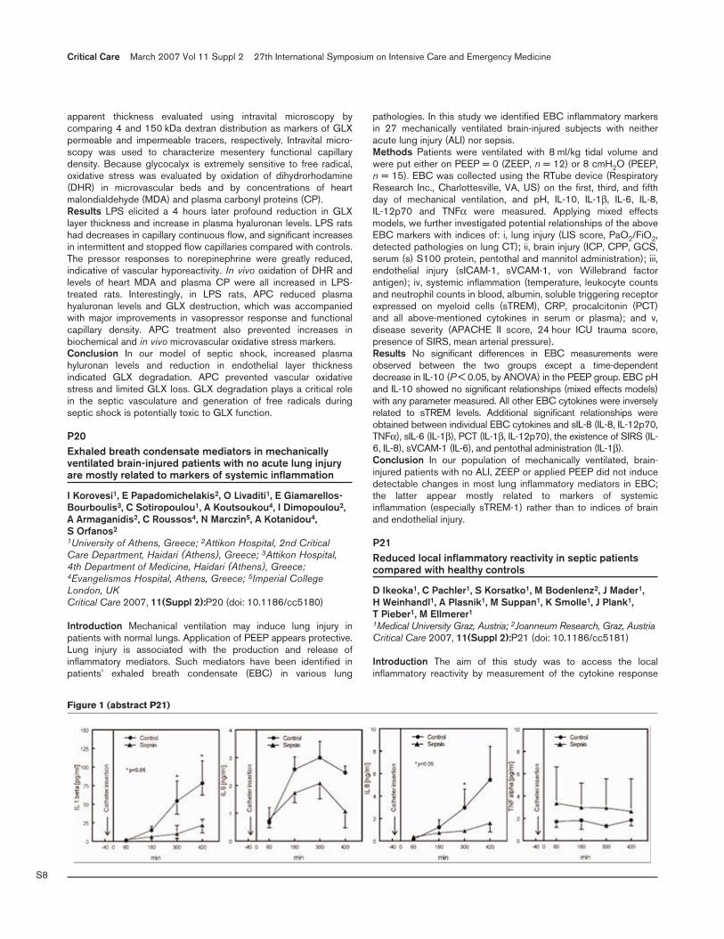
after catheter insertion into subcutaneous adipose tissue (SAT) ofpatients with severe sepsis compared with healthy volunteers.Methods Eight healthy volunteers and 10 patients with severesepsis were included. One 18-gauge open-flow microperfusiondouble-lumen catheter was inserted into SAT of the abdominal walland perfused with an isotonic solution at a flow rate of 1 µl/min.Blood samples and probe effluent samples from interstitial fluid ofSAT were withdrawn in two hourly intervals for a period of 8 hoursand retrospectively analysed using a Multiplex ELISA system forIL-1β, IL-6, IL-8 and TNFα.Results Concentrations of IL-1β, IL-6 and IL-8 were substantiallyhigher in SAT (13.3 (11.2; 31.0); 1,934 (1,650; 2,730); 917 (656;2,672) pg/ml; median (25th; 75th percentile)) than in serum (0.8(0.6; 1.3); 49.2 (3.8; 67.6); 36.1 (6.3; 89.1) pg/ml) for bothgroups, whereas TNFα concentrations were similar in serum andSAT (Figure 1). Serum concentrations of all cytokines remainedstable over time. However, a significant increase was observed forIL-1β and IL-8 in SAT in both groups. This increase wassignificantly in septic patients vs healthy controls.Conclusion Insertion of a catheter into subcutaneous adiposetissue promotes a local inflammatory response in both healthyindividuals and critically ill patients. The attenuated response inpatients with severe sepsis might be caused by reducedinflammatory reactivity in this group.
P22The evaluation of sivelestat sodium hydrate in acute lunginjury/acute respiratory distress syndrome patients in theintensive care unit
T Ikeda, K Ikeda, T Ueno, Y Kuroki, T Yokoyama, K YoshikawaHachiouji Medical Center, Tokyo Medical University, Tokyo, JapanCritical Care 2007, 11(Suppl 2):P22 (doi: 10.1186/cc5182)
The onset mechanism of ALI/ARDS and subsequent tissue injuryare considered to be associated with neutrophil elastase, and themain causes of ALI/ARDS are considered to be sepsis or aspirationpneumonia. In Japan, sivelestat sodium hydrate (Elaspol), aselective elastase inhibitor, was approved in 2002 for ALI/ARDSaccompanied by SIRS, and this medicine has been evaluated in aclinical situation. In this study, we performed a retrospectivecomparison of the sivelestat sodium administration between twogroups of patients: Group Elaspol, consisting of 308 patients(209males and 99 females, aged 66 ± 15 years) with ALI/ARDSaccompanied by SIRS who were treated with sivelestat sodium ata dose of 0.2 mg/kg/hour for 72 hours or more, after approval ofthis drug; and Group Control, consisting of 41 patients (28 malesand 13 females, aged 66 ± 14 years) with ALI/ARDS accompaniedby SIRS who were treated in the ICU under similar conditions, butusing traditional methods for respiratory control, prior to approvalsivelestat sodium. The APACHE II scores of Group Elaspol andGroup Control were 23 ± 9 and 23 ± 8, SOFA scores were8.7 ± 3.8 and 8.9 ± 4.1, and the lung injury scores were 2.1 ± 0.7and 2.1 ± 0.6, respectively, with no significant differences betweenthe groups. The initial PEEP value of Group Elaspol was 5.9 ± 3.3,which was significantly higher than that of Group Control(3.4 ± 2.7 cmH2O). The PaO2/FIO2 ratios under mechanicalventilation management 24, 48 and 72 hours after the beginning ofdrug administration were 209 ± 87, 222 ± 92, and 222 ± 82 mmHgin Group Elaspol, and were 191 ± 91, 207 ± 91, and211 ± 100 mmHg in Group Control. The ventilator-free days ofGroup Elaspol and Group Control were 18 ± 9 and 10 ± 12 days,respectively, and these values showed a significant difference(P < 0.001). Furthermore, the survival rate after 28 days wassignificantly higher in Group Elaspol than in Group Control (Group
Elaspol: 75%, Group Control: 52%; P < 0.001). These resultssuggest that sivelestat sodium hydrate is a good option as atreatment strategy for neutrophil elastase-associated septicALI/ARDS accompanied by SIRS.
P23Pharmacological modulation with prolongedadministration of moderate doses of steroid in a murinemodel of septic acute lung injury after burn insult
J Sasaki1, S Fujishima2, K Takuma1, Y Shinozawa1, N Aikawa2
1Tohoku University Hospital, Sendai, Japan; 2Keio University, Tokyo, JapanCritical Care 2007, 11(Suppl 2):P23 (doi: 10.1186/cc5183)
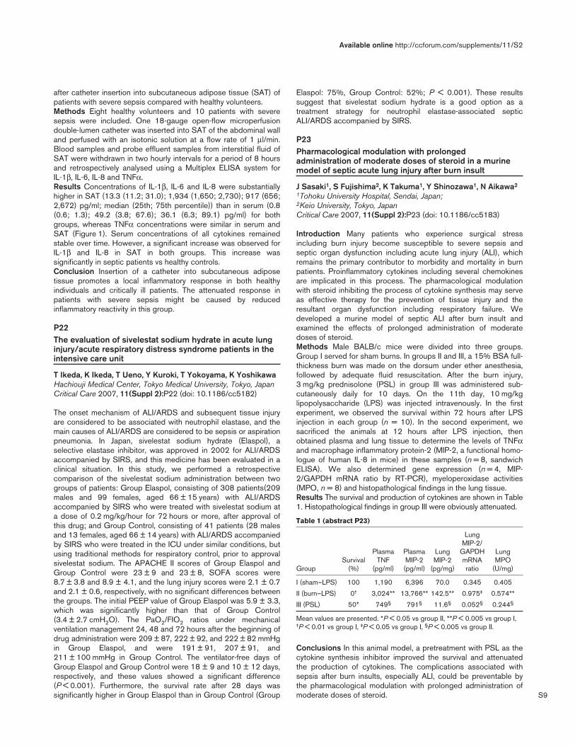
Introduction Many patients who experience surgical stressincluding burn injury become susceptible to severe sepsis andseptic organ dysfunction including acute lung injury (ALI), whichremains the primary contributor to morbidity and mortality in burnpatients. Proinflammatory cytokines including several chemokinesare implicated in this process. The pharmacological modulationwith steroid inhibiting the process of cytokine synthesis may serveas effective therapy for the prevention of tissue injury and theresultant organ dysfunction including respiratory failure. Wedeveloped a murine model of septic ALI after burn insult andexamined the effects of prolonged administration of moderatedoses of steroid.Methods Male BALB/c mice were divided into three groups.Group I served for sham burns. In groups II and III, a 15% BSA full-thickness burn was made on the dorsum under ether anesthesia,followed by adequate fluid resuscitation. After the burn injury,3 mg/kg prednisolone (PSL) in group III was administered sub-cutaneously daily for 10 days. On the 11th day, 10 mg/kglipopolysaccharide (LPS) was injected intravenously. In the firstexperiment, we observed the survival within 72 hours after LPSinjection in each group (n = 10). In the second experiment, wesacrificed the animals at 12 hours after LPS injection, thenobtained plasma and lung tissue to determine the levels of TNFαand macrophage inflammatory protein-2 (MIP-2, a functional homo-logue of human IL-8 in mice) in these samples (n = 8, sandwichELISA). We also determined gene expression (n = 4, MIP-2/GAPDH mRNA ratio by RT-PCR), myeloperoxidase activities(MPO, n = 8) and histopathological findings in the lung tissue.Results The survival and production of cytokines are shown in Table1. Histopathological findings in group III were obviously attenuated.
Table 1 (abstract P23)
Lung MIP-2/
Plasma Plasma Lung GAPDH Lung Survival TNF MIP-2 MIP-2 mRNA MPO
Group (%) (pg/ml) (pg/ml) (pg/mg) ratio (U/mg)
I (sham–LPS) 100 1,190 6,396 70.0 0.345 0.405
II (burn–LPS) 0† 3,024** 13,766** 142.5** 0.975‡ 0.574**
III (PSL) 50* 749§ 791§ 11.6§ 0.052§ 0.244§
Mean values are presented. *P < 0.05 vs group II, **P < 0.005 vs group I,†P < 0.01 vs group I, ‡P < 0.05 vs group I, §P < 0.005 vs group II.
Conclusions In this animal model, a pretreatment with PSL as thecytokine synthesis inhibitor improved the survival and attenuatedthe production of cytokines. The complications associated withsepsis after burn insults, especially ALI, could be preventable bythe pharmacological modulation with prolonged administration ofmoderate doses of steroid.
Available online http://ccforum.com/supplements/11/S2
S10
P24Glucosamine enhances heat shock protein 70 expressionin vitro and in vivo following injury
K Singleton, C Hamiel, P WischmeyerUniversity of Colorado Health Sciences Center, Denver, CO, USACritical Care 2007, 11(Suppl 2):P24 (doi: 10.1186/cc5184)
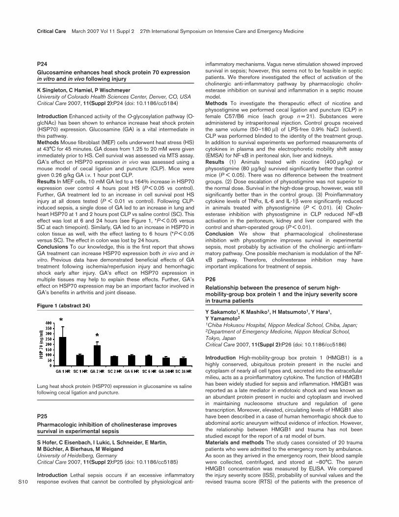
Introduction Enhanced activity of the O-glycosylation pathway (O-glcNAc) has been shown to enhance increase heat shock protein(HSP70) expression. Glucosamine (GA) is a vital intermediate inthis pathway.Methods Mouse fibroblast (MEF) cells underwent heat stress (HS)at 43°C for 45 minutes. GA doses from 1.25 to 20 mM were givenimmediately prior to HS. Cell survival was assessed via MTS assay.GA’s effect on HSP70 expression in vivo was assessed using amouse model of cecal ligation and puncture (CLP). Mice weregiven 0.26 g/kg GA i.v. 1 hour post CLP.Results In MEF cells, 10 mM GA led to a 164% increase in HSP70expression over control 4 hours post HS (P < 0.05 vs control).Further, GA treatment led to an increase in cell survival post HSinjury at all doses tested (P < 0.01 vs control). Following CLP-induced sepsis, a single dose of GA led to an increase in lung andheart HSP70 at 1 and 2 hours post CLP vs saline control (SC). Thiseffect was lost at 6 and 24 hours (see Figure 1, *P < 0.05 versusSC at each timepoint). Similarly, GA led to an increase in HSP70 incolon tissue as well, with the effect lasting to 6 hours (*P < 0.05versus SC). The effect in colon was lost by 24 hours.Conclusions To our knowledge, this is the first report that showsGA treatment can increase HSP70 expression both in vivo and invitro. Previous data have demonstrated beneficial effects of GAtreatment following ischemia/reperfusion injury and hemorrhagicshock early after injury. GA’s effect on HSP70 expression inmultiple tissues may help to explain these effects. Further, GA’seffect on HSP70 expression may be an important factor involved inGA’s benefits in arthritis and joint disease.
P25Pharmacologic inhibition of cholinesterase improvessurvival in experimental sepsis
S Hofer, C Eisenbach, I Lukic, L Schneider, E Martin, M Büchler, A Bierhaus, M WeigandUniversity of Heidelberg, GermanyCritical Care 2007, 11(Suppl 2):P25 (doi: 10.1186/cc5185)
Introduction Lethal sepsis occurs if an excessive inflammatoryresponse evolves that cannot be controlled by physiological anti-
inflammatory mechanisms. Vagus nerve stimulation showed improvedsurvival in sepsis; however, this seems not to be feasible in septicpatients. We therefore investigated the effect of activation of thecholinergic anti-inflammatory pathway by pharmacologic cholin-esterase inhibition on survival and inflammation in a septic mousemodel.Methods To investigate the therapeutic effect of nicotine andphysostigmine we performed cecal ligation and puncture (CLP) infemale C57/B6 mice (each group n = 21). Substances wereadministered by intraperitoneal injection. Control groups receivedthe same volume (50–180 µl) of LPS-free 0.9% NaCl (solvent).CLP was performed blinded to the identity of the treatment group.In addition to survival experiments we performed measurements ofcytokines in plasma and the electrophoretic mobility shift assay(EMSA) for NF-κB in peritoneal skin, liver and kidneys.Results (1) Animals treated with nicotine (400 µg/kg) orphysostigmine (80 µg/kg) survived significantly better than controlmice (P < 0.05). There was no difference between the treatmentgroups. (2) Dose escalation of physostigmine was not superior tothe normal dose. Survival in the high-dose group, however, was stillsignificantly better than in the control group. (3) Proinflammatorycytokine levels of TNFα, IL-6 and IL-1β were significantly reducedin animals treated with physostigmine (P < 0.01). (4) Cholin-esterase inhibition with physostigmine in CLP reduced NF-κBactivation in the peritoneum, kidney and liver compared with thecontrol and sham-operated group (P < 0.01).Conclusion We show that pharmacological cholinesteraseinhibition with physostigmine improves survival in experimentalsepsis, most probably by activation of the cholinergic anti-inflam-matory pathway. One possible mechanism is modulation of the NF-κB pathway. Therefore, cholinesterase inhibition may haveimportant implications for treatment of sepsis.
P26Relationship between the presence of serum high-mobility-group box protein 1 and the injury severity scorein trauma patients
Y Sakamoto1, K Mashiko1, H Matsumoto1, Y Hara1, Y Yamamoto2
1Chiba Hokusou Hospital, Nippon Medical School, Chiba, Japan;2Department of Emergency Medicine, Nippon Medical School,Tokyo, JapanCritical Care 2007, 11(Suppl 2):P26 (doi: 10.1186/cc5186)
Introduction High-mobility-group box protein 1 (HMGB1) is ahighly conserved, ubiquitous protein present in the nuclei andcytoplasm of nearly all cell types and, secreted into the extracellularmilieu, acts as a proinflammatory cytokine. The function of HMGB1has been widely studied for sepsis and inflammation. HMGB1 wasreported as a late mediator in endotoxic shock and was known asan abundant protein present in nuclei and cytoplasm and involvedin maintaining nucleosome structure and regulation of genetranscription. Moreover, elevated, circulating levels of HMGB1 alsohave been described in a case of human hemorrhagic shock due toabdominal aortic aneurysm without evidence of infection. However,the relationship between HMGB1 and trauma has not beenstudied except for the report of a rat model of burn.Materials and methods The study cases consisted of 20 traumapatients who were admitted to the emergency room by ambulance.As soon as they arrived in the emergency room, their blood samplewere collected, centrifuged, and stored at –80°C. The serumHMGB1 concentration was measured by ELISA. We comparedthe injury severity score (ISS), probability of survival values and therevised trauma score (RTS) of the patients with the presence of
Critical Care March 2007 Vol 11 Suppl 2 27th International Symposium on Intensive Care and Emergency Medicine
Figure 1 (abstract 24)
Lung heat shock protein (HSP70) expression in glucosamine vs salinefollowing cecal ligation and puncture.
S11
serum HMGB1 (group A) and without the presence (group B). Wetherefore divided into two groups, high ISS group (≤25) and lowISS group (>25), and examined the relation with the serumHMGB1 level.Results Our data showed that the number in group A was ninecases and group B was 11 cases. The ISS of group A wassignificantly higher than that in group B (P = 0.0013). The P valueof group A was significantly lower than in group B (P = 0.0131).The serum HMGB1 level of the >25 ISS group was significantlyhigher than in the ≤25 ISS group.Discussion These data suggest that HMGB1 seems to be aprimary mediator of trauma-induced pathology. Because the ISSwas significantly correlated with the presence of serum HMGB1,HMGB1 may be expressed in severe injuries and it may be aimportant parameter that indicates the severity of injury.
P27Beneficial effects of antiplatelet drugs in patients withcommunity-acquired pneumonia and in endotoxin shock inmice
J Winning1, J Baranyai1, R Claus1, I Eisenhut2, J Hamacher2, K Reinhart1, M Bauer1, W Lösche1
1University Hospital Jena, Germany; 2University Hospital Homburg,GermanyCritical Care 2007, 11(Suppl 2):P27 (doi: 10.1186/cc5187)
Aims Systemic inflammation and sepsis are associated with bloodplatelet activation, which may contribute to the development oforgan failure. In this study we proved whether antiplatelet drugshave a benefit in patients who may develop sepsis as well as in amouse model of endotoxin shock.Methods Data obtained from 224 patients with community-acquired pneumonia (CAP) were retrospectively analysed for anassociation between prehospital treatment with long-actingantiplatelet drugs such as acetyl salicylic acid (n = 36) orthienopyridine ADP-receptor antagonists (clopidogrel or ticlopidin,n = 8) and clinical outcome. Use of statins was an exclusioncriterion. BALB/c mice were pretreated with clopidogrel for 4 daysprior to an intraperitoneal injection of LPS (Escherichia coli0111:B4). For platelet counts and blood gas analysis, standardprocedures were used. Lung tissues were stained with HE or aFITC-labelled anti-fibrin(ogen) antibody.Results CAP patients with antiplatelet drugs (n = 44) were olderthan control patients (n = 180; 69 ± 7 vs 58 ± 13 years,P < 0.00001). At the day of hospital admission there were nodifferences in platelet or leukocyte counts, CRP and SOFA scoresbetween both groups. However, patients on antiplatelet drugsdeveloped organ failure less frequently than control patients (ICUadmission: 9.1% vs 26.1%; P < 0.02). In the mouse model ofendotoxin shock, clopidogrel reduced the drop in platelet countand the degree of lung injury. Compared with controls we found20 hours after LPS injection in the clopidogrel-treated animals alower number of thrombi in the lung vasculature (6.1 ± 2.3 vs11.5 ± 4.4 thrombi per screen, P < 0.025) as well as higher bloodpH and bicarbonate levels (7.01 ± 0.01 vs 6.93 ± 0.04, P < 0.04and 10.2 ± 0.14 vs 7.3 ± 0.14 mmol/l, P < 0.03, respectively).Conclusions Antiplatelet drugs may have a beneficial effect insystemic inflammation and sepsis, and could be a novel therapyoption, at least in patients of low bleeding risk. One mechanism oftheir effects could be a reduction in the microvascular thrombusformation.
P28Aggressive and moderate fluid resuscitation in septic pigs:consequences on morbidity
S Brandt, A Elftheriadis, T Regueira, H Bracht, J Gorrasi, J Takala, S JakobUniversity Hospital Inselspital, Bern, SwitzerlandCritical Care 2007, 11(Suppl 2):P28 (doi: 10.1186/cc5188)
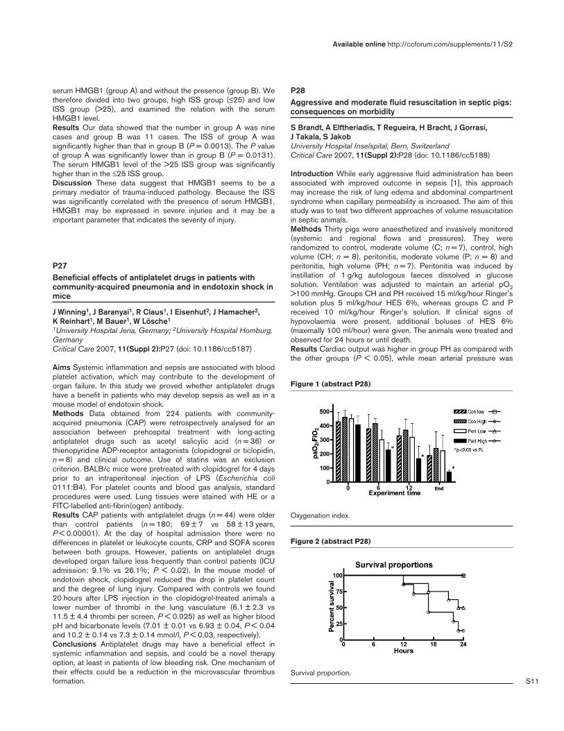
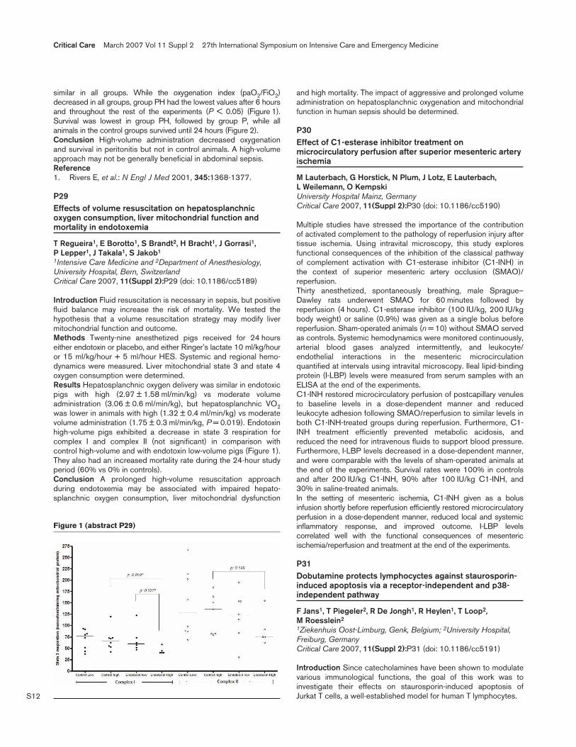
Introduction While early aggressive fluid administration has beenassociated with improved outcome in sepsis [1], this approachmay increase the risk of lung edema and abdominal compartmentsyndrome when capillary permeability is increased. The aim of thisstudy was to test two different approaches of volume resuscitationin septic animals.Methods Thirty pigs were anaesthetized and invasively monitored(systemic and regional flows and pressures). They wererandomized to control, moderate volume (C; n = 7), control, highvolume (CH; n = 8), peritonitis, moderate volume (P; n = 8) andperitonitis, high volume (PH; n = 7). Peritonitis was induced byinstillation of 1 g/kg autologous faeces dissolved in glucosesolution. Ventilation was adjusted to maintain an arterial pO2>100 mmHg. Groups CH and PH received 15 ml/kg/hour Ringer’ssolution plus 5 ml/kg/hour HES 6%, whereas groups C and Preceived 10 ml/kg/hour Ringer’s solution. If clinical signs ofhypovolaemia were present, additional boluses of HES 6%(maximally 100 ml/hour) were given. The animals were treated andobserved for 24 hours or until death.Results Cardiac output was higher in group PH as compared withthe other groups (P < 0.05), while mean arterial pressure was
Available online http://ccforum.com/supplements/11/S2
Figure 1 (abstract P28)
Oxygenation index.
Figure 2 (abstract P28)
Survival proportion.
S12
similar in all groups. While the oxygenation index (paO2/FiO2)decreased in all groups, group PH had the lowest values after 6 hoursand throughout the rest of the experiments (P < 0.05) (Figure 1).Survival was lowest in group PH, followed by group P, while allanimals in the control groups survived until 24 hours (Figure 2).Conclusion High-volume administration decreased oxygenationand survival in peritonitis but not in control animals. A high-volumeapproach may not be generally beneficial in abdominal sepsis.Reference1. Rivers E, et al.: N Engl J Med 2001, 345:1368-1377.
P29Effects of volume resuscitation on hepatosplanchnicoxygen consumption, liver mitochondrial function andmortality in endotoxemia
T Regueira1, E Borotto1, S Brandt2, H Bracht1, J Gorrasi1, P Lepper1, J Takala1, S Jakob1
1Intensive Care Medicine and 2Department of Anesthesiology,University Hospital, Bern, SwitzerlandCritical Care 2007, 11(Suppl 2):P29 (doi: 10.1186/cc5189)
Introduction Fluid resuscitation is necessary in sepsis, but positivefluid balance may increase the risk of mortality. We tested thehypothesis that a volume resuscitation strategy may modify livermitochondrial function and outcome.Methods Twenty-nine anesthetized pigs received for 24 hourseither endotoxin or placebo, and either Ringer’s lactate 10 ml/kg/houror 15 ml/kg/hour + 5 ml/hour HES. Systemic and regional hemo-dynamics were measured. Liver mitochondrial state 3 and state 4oxygen consumption were determined.Results Hepatosplanchnic oxygen delivery was similar in endotoxicpigs with high (2.97 ± 1.58 ml/min/kg) vs moderate volumeadministration (3.06 ± 0.6 ml/min/kg), but hepatosplanchnic VO2was lower in animals with high (1.32 ± 0.4 ml/min/kg) vs moderatevolume administration (1.75 ± 0.3 ml/min/kg, P = 0.019). Endotoxinhigh-volume pigs exhibited a decrease in state 3 respiration forcomplex I and complex II (not significant) in comparison withcontrol high-volume and with endotoxin low-volume pigs (Figure 1).They also had an increased mortality rate during the 24-hour studyperiod (60% vs 0% in controls).Conclusion A prolonged high-volume resuscitation approachduring endotoxemia may be associated with impaired hepato-splanchnic oxygen consumption, liver mitochondrial dysfunction
and high mortality. The impact of aggressive and prolonged volumeadministration on hepatosplanchnic oxygenation and mitochondrialfunction in human sepsis should be determined.
P30Effect of C1-esterase inhibitor treatment onmicrocirculatory perfusion after superior mesenteric arteryischemia
M Lauterbach, G Horstick, N Plum, J Lotz, E Lauterbach, L Weilemann, O KempskiUniversity Hospital Mainz, GermanyCritical Care 2007, 11(Suppl 2):P30 (doi: 10.1186/cc5190)
Multiple studies have stressed the importance of the contributionof activated complement to the pathology of reperfusion injury aftertissue ischemia. Using intravital microscopy, this study exploresfunctional consequences of the inhibition of the classical pathwayof complement activation with C1-esterase inhibitor (C1-INH) inthe context of superior mesenteric artery occlusion (SMAO)/reperfusion.Thirty anesthetized, spontaneously breathing, male Sprague–Dawley rats underwent SMAO for 60 minutes followed byreperfusion (4 hours). C1-esterase inhibitor (100 IU/kg, 200 IU/kgbody weight) or saline (0.9%) was given as a single bolus beforereperfusion. Sham-operated animals (n = 10) without SMAO servedas controls. Systemic hemodynamics were monitored continuously,arterial blood gases analyzed intermittently, and leukocyte/endothelial interactions in the mesenteric microcirculationquantified at intervals using intravital microscopy. Ileal lipid-bindingprotein (I-LBP) levels were measured from serum samples with anELISA at the end of the experiments.C1-INH restored microcirculatory perfusion of postcapillary venulesto baseline levels in a dose-dependent manner and reducedleukocyte adhesion following SMAO/reperfusion to similar levels inboth C1-INH-treated groups during reperfusion. Furthermore, C1-INH treatment efficiently prevented metabolic acidosis, andreduced the need for intravenous fluids to support blood pressure.Furthermore, I-LBP levels decreased in a dose-dependent manner,and were comparable with the levels of sham-operated animals atthe end of the experiments. Survival rates were 100% in controlsand after 200 IU/kg C1-INH, 90% after 100 IU/kg C1-INH, and30% in saline-treated animals.In the setting of mesenteric ischemia, C1-INH given as a bolusinfusion shortly before reperfusion efficiently restored microcirculatoryperfusion in a dose-dependent manner, reduced local and systemicinflammatory response, and improved outcome. I-LBP levelscorrelated well with the functional consequences of mesentericischemia/reperfusion and treatment at the end of the experiments.
P31Dobutamine protects lymphocyctes against staurosporin-induced apoptosis via a receptor-independent and p38-independent pathway
F Jans1, T Piegeler2, R De Jongh1, R Heylen1, T Loop2, M Roesslein2
1Ziekenhuis Oost-Limburg, Genk, Belgium; 2University Hospital,Freiburg, GermanyCritical Care 2007, 11(Suppl 2):P31 (doi: 10.1186/cc5191)
Introduction Since catecholamines have been shown to modulatevarious immunological functions, the goal of this work was toinvestigate their effects on staurosporin-induced apoptosis ofJurkat T cells, a well-established model for human T lymphocytes.
Critical Care March 2007 Vol 11 Suppl 2 27th International Symposium on Intensive Care and Emergency Medicine
Figure 1 (abstract P29)
S13
Methods Jurkat T cells passages 1–12 were used. Apoptosis wasmeasured with a caspase-activity assay and with FACS analysis ofannexin–propidium iodide double-stained cells.Results Exposure of Jurkat T cells for 2 hours to staurosporin(2 µM) induced apoptosis: the number of apoptotic cells increasedto 14.0 ± 0.8% versus 2.3 ± 0.4% in the control group. Pre-treatment (4 hours) with dobutamine 100 and 500 µM decreasedthe staurosporin-induced apoptosis to 11.6 ± 0.6% and 8.7 ± 0.7%,respectively (P < 0.01, mean ± SEM, n = 44). Other catechol-amines like epinephrine and norepinephrine (both up to 500 µM)had no effect on staurosporin-induced apoptosis. To investigatewhether this protective effect of dobutamine was mediated via β-receptors, specific β-blockers were used: neither atenolol (β1)(100 mM), nor ICI 118,551 (β2) (10 mM) blocked the protectiveeffect of dobutamine. Furthermore, dobutamine (1–500 µM) didnot increase cAMP production in these cells. Therefore, theprotective effect of dobutamine is not β-receptor-mediated. Since itwas previously demonstrated that MAPKs p38 and JNK, but notERK, are activated by dobutamine in Jurkat T cells, we investigatedwhether the activation of these MAPKs are involved in theprotection by dobutamine: inhibition of JNK activation with SP600125 (1 µM) did not influence the protective effect of dobuta-mine. Inhibition of p38 activation with SBI 202190 (5 µM) evenseemed to reinforce the protection afforded by dobutamine.Conclusions These experiments demonstrate that dobutaminepretreatment protects T cells from staurosporin-induced apoptosis.This protective effect is not β-receptor-mediated. Also, activation ofMAPKs p38 or JNK by dobutamine is not responsible for theprotective effect. The molecular mechanisms by which dobutamineexerts this protective effect remain to be elucidated.
P32Serum vasopressin concentrations in critically ill patientsin the intensive care unit
A Melissaki, A Efthymiou, T Kyriakopoulou, G Kribeni, E Evaggelaki, A Tsikali, D Andreopoulos, A Zaglis, N BaziotisSaint Savvas General Hospital, Athens, GreeceCritical Care 2007, 11(Suppl 2):P32 (doi: 10.1186/cc5192)
Introduction The aim of the study is the measurement of serumvasopressin concentrations in the mixed critically ill patients,24 hours after admission to the ICU and just before the discharge.Methods In this study there were included patients admitted to theICU from June until November 2006 (n = 22; 12 males, 10females), mean age 46.45 ± 22.03, APACHE II score 8.59 ± 4.76,length of stay 9.68 ± 6.52. Patients with central nervous systemfailure, neurosurgical patients and patients remaining in the ICU forno longer than 72 hours were excluded. Serum vasopressinconcentrations were measured 24 hours after their admission tothe ICU and just before their discharge. The control group was 20healthy volunteers (blood donors). Vasopressin was measured bythe radioimmunoassay method in pmol/l. The sensitivity of themethod is 0.5 pmol/l and the specificity is 100%. The statisticalanalysis was done with the t test.Results Vasopressin serum concentrations at 24 hours afteradmission were 32,618 ± 20,570 pmol/l. Vasopressin serumconcentrations in critically ill patients were significantly higher thanin the healthy control group (11,302 ± 31,002, P < 0.001). Serumvasopressin concentrations on admission compared with vaso-pressin concentrations at discharge were statistically significantlyincreased (P < 0.001).Conclusions Serum vasopressin concentrations in critically illpatients in a mixed ICU are increased 24 hours after admissioncompared with the control group. The value at discharge is lower
than the value on admission and it approaches the value ofvasopressin in healthy volunteers. To confirm these results, morestudies will be needed.
P33Effects of simultaneously infused terlipressin anddobutamine in septic shock
A Morelli1, C Ertmer2, M Lange2, K Broeking2, A Orecchioni1, M Rocco1, H Van Aken2, P Pietropaoli1, M Westphal21University of Rome ‘La Sapienza’, Rome, Italy; 2University Hospitalof Muenster, GermanyCritical Care 2007, 11(Suppl 2):P33 (doi: 10.1186/cc5193)
Introduction Terlipressin is increasingly used in the treatment ofsepsis-associated hypotension. However, terlipressin may reducecardiac output and global oxygen supply.Methods We performed a prospective, randomized, controlledclinical study to determine whether dobutamine may counter-balance the depressions in cardiac index and mixed-venous oxygensaturation resulting from sole terlipressin infusion. We enrolled 60septic shock patients requiring high doses of norepinephrine(0.9 µg/kg/min) to maintain mean arterial pressure at 70 ± 5 mmHg.Patients were randomly allocated to be treated either with (a) 1 mgterlipressin, (b) 1 mg terlipressin followed by incremental dobuta-mine doses to reverse the anticipated reductions in mixed-venousoxygen saturation, or (c) sole norepinephrine infusion (control;each n = 20).Results Data from right heart catheterization, thermo-dye dilutioncatheter, gastric tonometry, as well as organ function andcoagulation were obtained at baseline and after 2 and 4 hours.Terlipressin (with and without dobutamine) infusion preserved themean arterial pressure at threshold values of 70 ± 5 mmHg, whileallowing one to reduce norepinephrine doses to 0.18 ± 0.04 and0.2 ± 0.05 µg/kg/min, respectively (vs 1.4 ± 0.07 µg/kg/min incontrols at 4 hours; each P < 0.01). The terlipressin-linkeddecrease in mixed-venous oxygen saturation was reversed bydobutamine (at 4 hours: 59 ± 2 vs 69 ± 3%, P = 0.023). Nostatistically significant differences were found intra-group andbetween groups in terms of differences between gastric mucosaland arterial carbon dioxide partial pressure, blood clearance ofindocyanine green, as well as the plasma disappearance rate ofindocyanine greenConclusions In catecholamine-dependent human septic shock,terlipressin (with and without concomitant dobutamine) stabilizeshemodynamics and reduces norepinephrine requirements.Dobutamine is a useful inotropic agent to reverse the depression inglobal oxygen transport resulting from sole terlipressin infusionwithout obvious side effects.
P34Vasopressin substitution causes microcirculatory changesin patients with septic shock
S Klinzing, C Reinhard, T Simon, T Schürholz, Y Sakr, K Reinhart, G MarxFriedrich Schiller Universität, Jena, GermanyCritical Care 2007, 11(Suppl 2):P34 (doi: 10.1186/cc5194)
Introduction We tested the effects of arginine vasopressin ontissue oxygenation, microvascular reactivity and oral mucosamicrocirculation in patients with septic shock.Methods In 20 patients with septic shock, tissue microcirculationwas determined before treatment with AVP (2 IU/hour), after2 hours of treatment and 2 hours after treatment.
Available online http://ccforum.com/supplements/11/S2
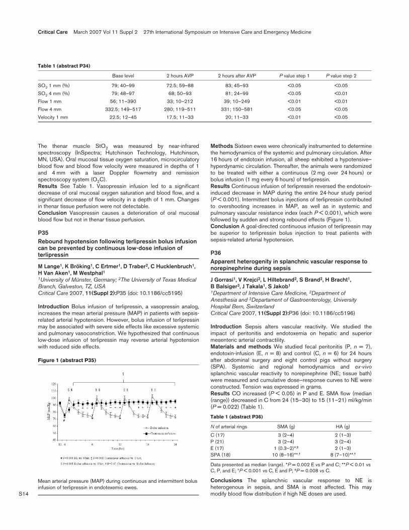
S14
The thenar muscle StO2 was measured by near-infraredspectroscopy (InSpectra; Hutchinson Technology, Hutchinson,MN, USA). Oral mucosal tissue oxygen saturation, microcirculatoryblood flow and blood flow velocity were measured in depths of 1and 4 mm with a laser Doppler flowmetry and remissionspectroscopy system (O2C).Results See Table 1. Vasopressin infusion led to a significantdecrease of oral mucosal oxygen saturation and blood flow, and asignificant decrease of flow velocity in a depth of 1 mm. Changesin thenar tissue perfusion were not detectable.Conclusion Vasopressin causes a deterioration of oral mucosalblood flow but not in thenar tissue perfusion.
P35Rebound hypotension following terlipressin bolus infusioncan be prevented by continuous low-dose infusion ofterlipressin
M Lange1, K Bröking1, C Ertmer1, D Traber2, C Hucklenbruch1,H Van Aken1, M Westphal11University of Münster, Germany; 2The University of Texas MedicalBranch, Galveston, TZ, USACritical Care 2007, 11(Suppl 2):P35 (doi: 10.1186/cc5195)
Introduction Bolus infusion of terlipressin, a vasopressin analog,increases the mean arterial pressure (MAP) in patients with sepsis-related arterial hypotension. However, bolus infusion of terlipressinmay be associated with severe side effects like excessive systemicand pulmonary vasoconstriction. We hypothesized that continuouslow-dose infusion of terlipressin may reverse arterial hypotensionwith reduced side effects.
Methods Sixteen ewes were chronically instrumented to determinethe hemodynamics of the systemic and pulmonary circulation. After16 hours of endotoxin infusion, all sheep exhibited a hypotensive–hyperdynamic circulation. Thereafter, the animals were randomizedto be treated with either a continuous (2 mg over 24 hours) orbolus infusion (1 mg every 6 hours) of terlipressin.Results Continuous infusion of terlipressin reversed the endotoxin-induced decrease in MAP during the entire 24-hour study period(P < 0.001). Intermittent bolus injections of terlipressin contributedto overshooting increases in MAP, as well as in systemic andpulmonary vascular resistance index (each P < 0.001), which werefollowed by sudden and strong rebound effects (Figure 1).Conclusion A goal-directed continuous infusion of terlipressin maybe superior to terlipressin bolus injection to treat patients withsepsis-related arterial hypotension.
P36Apparent heterogenity in splanchnic vascular response tonorepinephrine during sepsis
J Gorrasi1, V Krejci2, L Hiltebrand2, S Brand2, H Bracht1, B Balsiger3, J Takala1, S Jakob1
1Department of Intensive Care Medicine, 2Department ofAnesthesia and 3Departament of Gastroenterology, UniversityHospital Bern, SwitzerlandCritical Care 2007, 11(Suppl 2):P36 (doi: 10.1186/cc5196)
Introduction Sepsis alters vascular reactivity. We studied theimpact of peritonitis and endotoxemia on hepatic and superiormesenteric arterial contractility.Materials and methods We studied fecal peritonitis (P, n = 7),endotoxin-infusion (E, n = 8) and control (C, n = 6) for 24 hoursafter abdominal surgery and eight control pigs without surgery(SPA). Systemic and regional hemodynamics and ex-vivosplanchnic vascular reactivity to norepinephrine (NE; tissue bath)were measured and cumulative dose–response curves to NE wereconstructed. Tension was expressed in grams.Results CO increased (P < 0.05) in P and E. SMA flow (median(range)) decreased in C from 24 (15–30) to 15 (11–21) ml/kg/min(P = 0.022) (Table 1).
Table 1 (abstract P36)
N of arterial rings SMA (g) HA (g)
C (17) 3 (2–4) 2 (1–3)P (21) 3 (2–4) 3 (2–4)E (17) 1 (0.3–2)*;‡ 2 (1–3)SPA (18) 10 (8–16)**,† 8 (7–10)**,†
Data presented as median (range). *P = 0.002 E vs P and C; **P < 0.01 vsC, P, and E; †P < 0.001 vs C, E and P; ‡P = 0.008 vs C.
Conclusions The splanchnic vascular response to NE isheterogenous in sepsis, and SMA is most affected. This maymodify blood flow distribution if high NE doses are used.
Critical Care March 2007 Vol 11 Suppl 2 27th International Symposium on Intensive Care and Emergency Medicine
Table 1 (abstract P34)
Base level 2 hours AVP 2 hours after AVP P value step 1 P value step 2
SO2 1 mm (%) 79; 40–99 72.5; 59–88 83; 45–93 <0.05 <0.05
SO2 4 mm (%) 79; 48–97 68; 50–93 81; 24–99 <0.05 <0.01
Flow 1 mm 56; 11–390 33; 10–212 39; 10–249 <0.01 <0.01
Flow 4 mm 332.5; 149–517 280; 119–511 331; 150–581 <0.05 <0.05
Velocity 1 mm 22.5; 12–45 17.5; 11–33 20; 11–33 <0.01 <0.05
Figure 1 (abstract P35)
Mean arterial pressure (MAP) during continuous and intermittent bolusinfusion of terlipressin in endotexemic ewes.
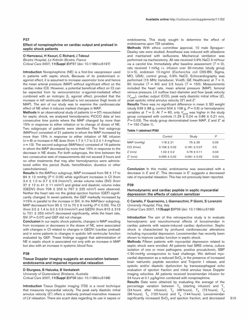
S15
P37Effect of norepinephrine on cardiac output and preload inseptic shock patients
O Hamzaoui, H Ksouri, C Richard, J TeboulBicetre Hospital, Le Krêmlin Bicetre, FranceCritical Care 2007, 11(Suppl 2):P37 (doi: 10.1186/cc5197)
Introduction Norepinephrine (NE) is a first-line vasopressor usedin patients with septic shock. Because of its predominant α-agonist effect, it is assumed to increase vasomotor tone and hencethe mean arterial pressure (MAP) without significant effect on thecardiac index (CI). However, a potential beneficial effect on CI canbe expected from its venoconstrictor α-agonist-mediated effectcombined with an inotropic β1 agonist effect, provided that theincrease in left ventricular afterload is not excessive (high levels ofMAP). The aim of our study was to examine the cardiovasculareffect of NE when it induces marked changes in MAP.Methods In an observational study of patients (n = 37) resuscitatedfor septic shock, we analysed hemodynamic PiCCO data at twoconsecutive time points where the MAP changed by more than15% in response to either initiation or to change of doses of NE.Two subgroups of patients were identified. The first subgroup(MAPincr) consisted of 21 patients in whom the MAP increased bymore than 15% in response to either initiation of NE infusion(n = 8) or increase in NE dose (from 1.7 ± 1.7 to 2.2 ± 1.4 mg/hour;n = 13). The second subgroup (MAPdecr) consisted of 16 patientsin whom the MAP decreased by more than 15% in response to thedecrease in NE doses. For both subgroups, the time between thetwo consecutive sets of measurements did not exceed 2 hours andno other treatments that may alter hemodynamics were adminis-tered within this period (fluids, hemofiltration, diuretics or othercatecholamines).Results In the MAPincr subgroup, MAP increased from 56 ± 17 to84 ± 12 mmHg (P < 0.05) while significant increases in CI (from3.4 ± 1.0 to 3.7 ± 0.9 l/min/m2), stroke volume index (SVi) (from37 ± 12 to 41 ± 11 ml/m2) and global end diastolic volume index(GEDVi) (from 706 ± 203 to 767 ± 225 ml/m2) were observed.Neither the heart rate nor the global ejection fraction (GEF) signifi-cantly changed. In seven patients, the GEF markedly increased by>15% in parallel to the increase in SVi. In the MAPdecr subgroup,MAP decreased from 95 ± 12 to 70 ± 9 mmHg (P < 0.05). The CI(from 3.5 ± 1.4 to 3.0 ± 0.9 l/min/m2) and GEDVi (from 815 ± 319to 721 ± 253 ml/m2) decreased significantly, while the heart rate,SVi (P = 0.07) and GEF did not change.Conclusion In our septic shock patients, changes in MAP resultingfrom increases or decreases in the doses of NE, were associatedwith changes in CI related to changes in GEDVi (cardiac preload)and in some patients to changes in systolic left ventricular functionevaluated by GEF. These findings suggest that administration ofNE in septic shock is associated not only with an increase in MAPbut also with an increase in systemic blood flow.
P38Tissue Doppler imaging suggests an association betweenendotoxemia and impaired myocardial relaxation
D Sturgess, B Haluska, B VenkateshUniversity of Queensland, Brisbane, AustraliaCritical Care 2007, 11(Suppl 2):P38 (doi: 10.1186/cc5198)
Introduction Tissue Doppler imaging (TDI) is a novel techniquethat measures myocardial velocity. The peak early diastolic mitralannulus velocity (E′) offers a relatively preload-insensitive measureof LV relaxation. There are scant data regarding its use in sepsis or
endotoxemia. This study sought to determine the effect ofendotoxemia upon TDI variables.Methods With ethics committee approval, 10 male Sprague–Dawley rats were studied. Anesthesia was induced with alfaxaloneand maintained with isofluorane. Mechanical ventilation wasperformed via tracheostomy. All rats received 0.9% NaCl 3 ml/hourvia a carotid line. Immediately after baseline assessment (T = 0),rats received 1 ml/kg i.v. infusion over 30 minutes (study group(n = 5), endotoxin 10 mg/ml (Escherichia coli O55:B5; Sigma,MO, USA); control group, 0.9% NaCl). Echocardiography wasperformed (15 MHz transducer, Vivid5; GE Healthcare) at T = 0,60 minutes (T = 60) and 2.5 hours (T = 150). Measurementsincluded the heart rate, mean arterial pressure (MAP), femoralvenous pressure, LV outflow tract diameter and flow (peak velocity(Vpeak), cardiac output (CO)), peak early diastolic mitral inflow (E),peak systolic mitral annulus velocity (S′) and E′.Results There was no significant difference in mean ± SD weight(study 539 ± 88 g, control 504 ± 108 g, P = 0.6) or hemodynamicvariables at T = 0. At T = 60, only Vpeak was higher in the studygroup compared with controls (1.29 ± 0.24 vs 0.86 ± 0.21 m/s,P = 0.03). The study group demonstrated lower MAP, E and E′ atT = 150 (Table 1).
Table 1 (abstract P38)
Control Study P
MAP (mmHg) 118 ± 21 75 ± 35 0.05
CO (l/min) 0.156 ± 0.02 0.181 ± 0.07 0.5
E (m/s) 1.02 ± 0.2 0.76 ± 0.11 0.04
E′ (m/s) 0.095 ± 0.02 0.061 ± 0.02 0.03
Conclusion In this model, endotoxemia was associated with adecrease in E and E′. This decrease in E′ suggests a decreasedrate of myocardial relaxation. This has not previously been reported.
P39Hemodynamic and cardiac peptide in septic myocardialdepression: the effects of calcium sensitizer
C Cariello, F Guarracino, L Giannecchini, P Giomi, S LorenziniUniversity Hospital, Pisa, ItalyCritical Care 2007, 11(Suppl 2):P39 (doi: 10.1186/cc5199)
Introduction The aim of this retrospective study is to evaluatehemodynamic and neurohormonal effects of levosimendan incardiac patients with sepsis-induced cardiac dysfunction. Septicshock is characterized by profound cardiovascular alterationsincluding myocardial depression. Levosimendan has recently beenshown to improve cardiac function in septic shock.Methods Fifteen patients with myocardial depression related toseptic shock were enrolled. All patients had SIRS criteria, cultureisolation of one or more pathogens, positive procalcitonin, SBP< 90 mmHg unresponsive to load challenge. We defined myo-cardial depression as a reduced SvO2 in the presence of increasedbrain natriuretic peptide secretion and Troponin I release, andsystolic and/or diastolic dysfunction by transoesophageal echoevaluation of ejection fraction and mitral annulus tissue Dopplerimaging velocities. All patients received levosimendan infusion for24 hours at 0.1 µg/kg/min combined with norepinephrine.Results Data were obtained by evaluating the average of thepercentage variation between T0 (starting infusion) and T1(24 hours after infusion), T2 (48 hours), T3 (72 hours), T4(96 hours), T5 (120 hours) and T6 (144 hours). Levosimendansignificantly increased SvO2 and ejection fraction, and decreased
Available online http://ccforum.com/supplements/11/S2
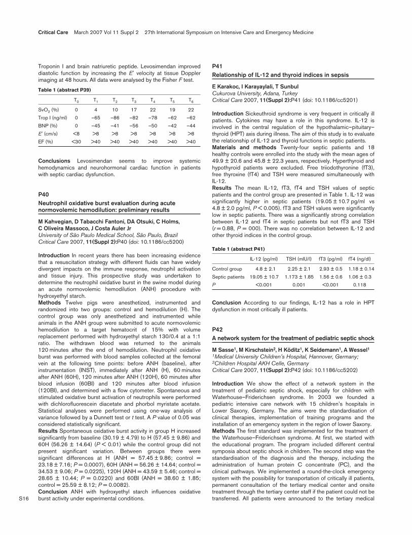
S16
Troponin I and brain natriuretic peptide. Levosimendan improveddiastolic function by increasing the E′ velocity at tissue Dopplerimaging at 48 hours. All data were analysed by the Fisher F test.
Table 1 (abstract P39)
T0 T1 T2 T3 T4 T5 T6
SvO2 (%) 0 4 10 17 22 19 22
Trop I (ng/ml) 0 –65 –86 –82 –78 –62 –62
BNP (%) 0 –45 –41 –56 –50 –42 –44
E′ (cm/s) <8 >8 >8 >8 >8 >8 >8
EF (%) <30 >40 >40 >40 >40 >40 >40
Conclusions Levosimendan seems to improve systemichemodynamics and neurohormonal cardiac function in patientswith septic cardiac dysfunction.
P40Neutrophil oxidative burst evaluation during acutenormovolemic hemodilution: preliminary results
M Kahvegian, D Tabacchi Fantoni, DA Otsuki, C Holms, C Oliveira Massoco, J Costa Auler JrUniversity of São Paulo Medical School, São Paulo, BrazilCritical Care 2007, 11(Suppl 2):P40 (doi: 10.1186/cc5200)
Introduction In recent years there has been increasing evidencethat a resuscitation strategy with different fluids can have widelydivergent impacts on the immune response, neutrophil activationand tissue injury. This prospective study was undertaken todetermine the neutrophil oxidative burst in the swine model duringan acute normovolemic hemodilution (ANH) procedure withhydroxyethyl starch.Methods Twelve pigs were anesthetized, instrumented andrandomized into two groups: control and hemodilution (H). Thecontrol group was only anesthetized and instrumented whileanimals in the ANH group were submitted to acute normovolemichemodilution to a target hematocrit of 15% with volumereplacement performed with hydroxyethyl starch 130/0.4 at a 1:1ratio. The withdrawn blood was returned to the animals120 minutes after the end of hemodilution. Neutrophil oxidativeburst was performed with blood samples collected at the femoralvein at the following time points: before ANH (baseline), afterinstrumentation (INST), immediately after ANH (H), 60 minutesafter ANH (60H), 120 minutes after ANH (120H), 60 minutes afterblood infusion (60BI) and 120 minutes after blood infusion(120BI), and determined with a flow cytometer. Spontaneous andstimulated oxidative burst activation of neutrophils were performedwith dichlorofluorescein diacetate and phorbol myristate acetate.Statistical analyses were performed using one-way analysis ofvariance followed by a Dunnett test or t test. A P value of 0.05 wasconsidered statistically significant.Results Spontaneous oxidative burst activity in group H increasedsignificantly from baseline (30.19 ± 4.79) to H (57.45 ± 9.86) and60H (56.26 ± 14.64) (P < 0.01) while the control group did notpresent significant variation. Between groups there weresignificant differences at H (ANH = 57.45 ± 9.86; control =23.18 ± 7.16; P = 0.0007), 60H (ANH = 56.26 ± 14.64; control =34.53 ± 9.06; P = 0.0225), 120H (ANH = 43.59 ± 5.46; control =28.65 ± 10.44; P = 0.0220) and 60BI (ANH = 38.60 ± 1.85;control = 25.59 ± 8.12; P = 0.0082).Conclusion ANH with hydroxyethyl starch influences oxidativeburst activity under experimental conditions.
P41Relationship of IL-12 and thyroid indices in sepsis
E Karakoc, I Karayaylali, T SunbulCukurova University, Adana, TurkeyCritical Care 2007, 11(Suppl 2):P41 (doi: 10.1186/cc5201)
Introduction Sickeuthroid syndrome is very frequent in critically illpatients. Cytokines may have a role in this syndrome. IL-12 isinvolved in the central regulation of the hypothalamic–pituitary–thyroid (HPT) axis during illness. The aim of this study is to evaluatethe relationship of IL-12 and thyroid functions in septic patients.Materials and methods Twenty-four septic patients and 18healthy controls were enrolled into the study with the mean ages of49.9 ± 20.6 and 45.8 ± 22.3 years, respectively. Hyperthyroid andhypothyroid patients were excluded. Free triiodothyronine (fT3),free thyroxine (fT4) and TSH were measured simultaneously withIL-12.Results The mean IL-12, fT3, fT4 and TSH values of septicpatients and the control group are presented in Table 1. IL-12 wassignificantly higher in septic patients (19.05 ± 10.7 pg/ml vs4.8 ± 2.0 pg/ml, P < 0.005). fT3 and TSH values were significantlylow in septic patients. There was a significantly strong correlationbetween IL-12 and fT4 in septic patients but not fT3 and TSH(r = 0.88, P = 000). There was no correlation between IL-12 andother thyroid indices in the control group.
Table 1 (abstract P41)
IL-12 (pg/ml) TSH (mIU/l) fT3 (pg/ml) fT4 (ng/dl)
Control group 4.8 ± 2.1 2.25 ± 2.1 2.93 ± 0.5 1.18 ± 0.14
Septic patients 19.05 ± 10.7 1.173 ± 1.85 1.56 ± 0.6 1.06 ± 0.3
P <0.001 0.001 <0.001 0.118
Conclusion According to our findings, IL-12 has a role in HPTdysfunction in most critically ill patients.
P42A network system for the treatment of pediatric septic shock
M Sasse1, M Kirschstein2, H Köditz1, K Seidemann1, A Wessel11Medical University Children’s Hospital, Hannover, Germany;2Children Hospital AKH Celle, GermanyCritical Care 2007, 11(Suppl 2):P42 (doi: 10.1186/cc5202)
Introduction We show the effect of a network system in thetreatment of pediatric septic shock, especially for children withWaterhouse–Friderichsen syndrome. In 2003 we founded apediatric intensive care network with 15 children’s hospitals inLower Saxony, Germany. The aims were the standardisation ofclinical therapies, implementation of training programs and theinstallation of an emergency system in the region of lower Saxony.Methods The first standard was implemented for the treatment ofthe Waterhouse–Friderichsen syndrome. At first, we started withthe educational program. The program included different centralsymposia about septic shock in children. The second step was thestandardisation of the diagnosis and the therapy, including theadministration of human protein C concentrate (PC), and theclinical pathways. We implemented a round-the-clock emergencysystem with the possibility for transportation of critically ill patients,permanent consultation of the tertiary medical center and onsitetreatment through the tertiary center staff if the patient could not betransferred. All patients were announced to the tertiary medical
Critical Care March 2007 Vol 11 Suppl 2 27th International Symposium on Intensive Care and Emergency Medicine
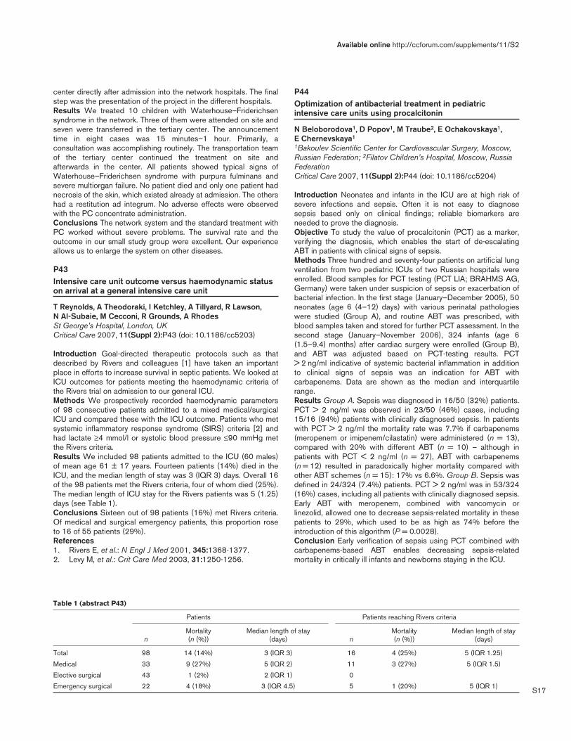
S17
center directly after admission into the network hospitals. The finalstep was the presentation of the project in the different hospitals.Results We treated 10 children with Waterhouse–Friderichsensyndrome in the network. Three of them were attended on site andseven were transferred in the tertiary center. The announcementtime in eight cases was 15 minutes–1 hour. Primarily, aconsultation was accomplishing routinely. The transportation teamof the tertiary center continued the treatment on site andafterwards in the center. All patients showed typical signs ofWaterhouse–Friderichsen syndrome with purpura fulminans andsevere multiorgan failure. No patient died and only one patient hadnecrosis of the skin, which existed already at admission. The othershad a restitution ad integrum. No adverse effects were observedwith the PC concentrate administration.Conclusions The network system and the standard treatment withPC worked without severe problems. The survival rate and theoutcome in our small study group were excellent. Our experienceallows us to enlarge the system on other diseases.
P43Intensive care unit outcome versus haemodynamic statuson arrival at a general intensive care unit
T Reynolds, A Theodoraki, I Ketchley, A Tillyard, R Lawson, N Al-Subaie, M Cecconi, R Grounds, A RhodesSt George’s Hospital, London, UKCritical Care 2007, 11(Suppl 2):P43 (doi: 10.1186/cc5203)
Introduction Goal-directed therapeutic protocols such as thatdescribed by Rivers and colleagues [1] have taken an importantplace in efforts to increase survival in septic patients. We looked atICU outcomes for patients meeting the haemodynamic criteria ofthe Rivers trial on admission to our general ICU.Methods We prospectively recorded haemodynamic parametersof 98 consecutive patients admitted to a mixed medical/surgicalICU and compared these with the ICU outcome. Patients who metsystemic inflammatory response syndrome (SIRS) criteria [2] andhad lactate ≥4 mmol/l or systolic blood pressure ≤90 mmHg metthe Rivers criteria.Results We included 98 patients admitted to the ICU (60 males)of mean age 61 ± 17 years. Fourteen patients (14%) died in theICU, and the median length of stay was 3 (IQR 3) days. Overall 16of the 98 patients met the Rivers criteria, four of whom died (25%).The median length of ICU stay for the Rivers patients was 5 (1.25)days (see Table 1).Conclusions Sixteen out of 98 patients (16%) met Rivers criteria.Of medical and surgical emergency patients, this proportion roseto 16 of 55 patients (29%).References1. Rivers E, et al.: N Engl J Med 2001, 345:1368-1377.2. Levy M, et al.: Crit Care Med 2003, 31:1250-1256.
P44Optimization of antibacterial treatment in pediatricintensive care units using procalcitonin
N Beloborodova1, D Popov1, M Traube2, E Ochakovskaya1, E Chernevskaya1
1Bakoulev Scientific Center for Cardiovascular Surgery, Moscow,Russian Federation; 2Filatov Children’s Hospital, Moscow, RussiaFederationCritical Care 2007, 11(Suppl 2):P44 (doi: 10.1186/cc5204)
Introduction Neonates and infants in the ICU are at high risk ofsevere infections and sepsis. Often it is not easy to diagnosesepsis based only on clinical findings; reliable biomarkers areneeded to prove the diagnosis.Objective To study the value of procalcitonin (PCT) as a marker,verifying the diagnosis, which enables the start of de-escalatingABT in patients with clinical signs of sepsis.Methods Three hundred and seventy-four patients on artificial lungventilation from two pediatric ICUs of two Russian hospitals wereenrolled. Blood samples for PCT testing (PCT LIA; BRAHMS AG,Germany) were taken under suspicion of sepsis or exacerbation ofbacterial infection. In the first stage (January–December 2005), 50neonates (age 6 (4–12) days) with various perinatal pathologieswere studied (Group A), and routine ABT was prescribed, withblood samples taken and stored for further PCT assessment. In thesecond stage (January–November 2006), 324 infants (age 6(1.5–9.4) months) after cardiac surgery were enrolled (Group B),and ABT was adjusted based on PCT-testing results. PCT> 2 ng/ml indicative of systemic bacterial inflammation in additionto clinical signs of sepsis was an indication for ABT withcarbapenems. Data are shown as the median and interquartilerange.Results Group A. Sepsis was diagnosed in 16/50 (32%) patients.PCT > 2 ng/ml was observed in 23/50 (46%) cases, including15/16 (94%) patients with clinically diagnosed sepsis. In patientswith PCT > 2 ng/ml the mortality rate was 7.7% if carbapenems(meropenem or imipenem/cilastatin) were administered (n = 13),compared with 20% with different ABT (n = 10) – although inpatients with PCT < 2 ng/ml (n = 27), ABT with carbapenems(n = 12) resulted in paradoxically higher mortality compared withother ABT schemes (n = 15): 17% vs 6.6%. Group B. Sepsis wasdefined in 24/324 (7.4%) patients. PCT > 2 ng/ml was in 53/324(16%) cases, including all patients with clinically diagnosed sepsis.Early ABT with meropenem, combined with vancomycin orlinezolid, allowed one to decrease sepsis-related mortality in thesepatients to 29%, which used to be as high as 74% before theintroduction of this algorithm (P = 0.0028).Conclusion Early verification of sepsis using PCT combined withcarbapenems-based ABT enables decreasing sepsis-relatedmortality in critically ill infants and newborns staying in the ICU.
Available online http://ccforum.com/supplements/11/S2
Table 1 (abstract P43)
Patients Patients reaching Rivers criteria
Mortality Median length of stay Mortality Median length of stay n (n (%)) (days) n (n (%)) (days)
Total 98 14 (14%) 3 (IQR 3) 16 4 (25%) 5 (IQR 1.25)
Medical 33 9 (27%) 5 (IQR 2) 11 3 (27%) 5 (IQR 1.5)
Elective surgical 43 1 (2%) 2 (IQR 1) 0
Emergency surgical 22 4 (18%) 3 (IQR 4.5) 5 1 (20%) 5 (IQR 1)
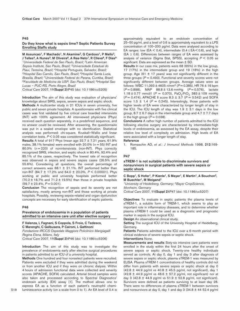
S18
P45Do they know what is sepsis time? Septic Patients SurveyEnrolling Staffs study
M Assuncao1, F Machado1, N Akamine2, G Cardoso1, P Mello3,J Telles4, A Nunes5, M Oliveira6, A Rea-Neto7, R Clleva8, F Dias9
1Universidade Federal de Sao Paulo, Brazil; 2Latin AmericanSepsis Institute, Sao Paulo, Brazil; 3Universidade Estadual doPiaui, Teresina, Brazil; 4Hospital Portugues, Salvador, Brazil;5Hospital Sao Camilo, Sao Paulo, Brazil; 6Hospital Santa Luzia,Brasilia, Brazil; 7Universidade Federal do Parana, Curitiba, Brazil;8Faculdade de Medicina da USP, Sao Paulo, Brazil; 9Hospital SaoLucas – PUC-RS, Porto Alegre, BrazilCritical Care 2007, 11(Suppl 2):P45 (doi: 10.1186/cc5205)
Introduction The aim of this study was evaluation of physicians’knowledge about SIRS, sepsis, severe sepsis and septic shock.Methods A multicenter study in 21 ICUs in seven university, fivepublic and seven private hospitals. A questionnaire with five clinicalcases was first validated by five critical care boarded intensivists(INT) with 100% agreement. All interviewed physicians (Phys)received each question separately, in a predefined sequence, andno answer could be reviewed. After answering, the questionnairewas put in a sealed envelope with no identification. Statisticalanalysis was performed: chi-square, Kruskall–Wallis and linearcorrelation tests. P < 0.05 was considered statistically significant.Results A total of 917 Phys (mean age 32.7 ± 7.21 years, 61.9%males, 38.1% females) were enrolled with 20.0% (n = 55) INT and80.0% (n = 220) of nonintensivists (non-INT). Phys correctlyrecognized SIRS, infection, and septic shock in 80.4%, 92.4% and85.1% of the cases, respectively. The lowest rate of recognitionwas observed in sepsis and severe sepsis cases (26.5% and55.6%). Considering all questions, the overall percentage ofcorrect answers was 68.1 ± 21.1%. INT performed better thannon-INT (84.7 ± 17.2% and 64.0 ± 20.0%, P < 0.00001). Physworking at public and university hospitals performed better(70.2 ± 18.7% and 71.2 ± 19.5%) than those in private hospitals(59.7 ± 23.4%, P = 0.001).Conclusion The recognition of sepsis and its severity are notsatisfactory, mostly among non-INT and those working at privatehospitals. Possibly, reviewing sepsis-related and organ dysfunctionconcepts are necessary for early identification of septic patients.
P46Prevalence of endotoxemia in a population of patientsadmitted to an intensive care unit after elective surgery
F Valenza, L Fagnani, S Coppola, S Froio, C Tedesco, C Marenghi, C Galbusera, P Caironi, L GattinoniFondazione IRCCS Ospedale Maggiore Policlinico MangiagalliRegina Elena, Milano, ItalyCritical Care 2007, 11(Suppl 2):P46 (doi: 10.1186/cc5206)
Introduction The aim of this study was to investigate theprevalence of endotoxemia early after elective surgical proceduresin patients admitted to an ICU of a university hospital.Methods One hundred and four nonselect patients were recruited.Patients were excluded if they were admitted during the weekendor from another ICU and if they were on chronic dialysis. Within4 hours of admission functional data were collected and severityscores (APACHE, SOFA) calculated. Arterial blood samples werealso taken and processed according to Spectral Diagnostics’endotoxin activity (EA) assay [1]. The method allows one toexpress EA as a function of each patient’s neutrophil chemi-luminescence activity (on a scale from 0 to 1). An EA level of 0.4 is
approximately equivalent to an endotoxin concentration of25–50 pg/ml, and a level of 0.6 is approximately equivalent to a LPSconcentration of 100–200 pg/ml. Data were analysed according toEA ranges: low (EA < 0.4), intermediate (0.4 ≤ EA < 0.6), and high(EA ≥ 0.6). Differences between ranges of EA were assessed byanalysis of variance (Sigma Stat, SPSS), accepting P < 0.05 assignificant. Data are expressed as the mean ± SD.Results In our case mix, patients were 68 (65%) in the low group,17 (17%) in the intermediate group and 19 (18%) in the highgroup. Age (61 ± 17 years) was not significantly different in thethree groups (P = 0.493). Functional and severity scores were notsignificantly different between groups. Average values were asfollows: WBC 11,093 ± 4605 n/mm3 (P = 0.385), HR 76 ± 16 bpm(P = 0.898), MAP 88.8 ± 13.6 mmHg (P = 0.576), lactate1.18 ± 0.77 mmol/l (P = 0.370), PaO2/FiO2 383 ± 109 mmHg(P = 0.474), APACHE II score 8.3 ± 3.7 (P = 0.542) and SOFAscore 1.5 ± 1.4 (P = 0.245). Interestingly, those patients withhigher levels of EA were characterized by longer length of stay inthe ICU. The ICU length of stay was 1.9 ± 3.1 days in the lowgroup, 8.7 ± 6.7 days in the intermediate group and 4.7 ± 7.7 daysin the high group (P = 0.038).Conclusions A rather high number of patients admitted to the ICUfollowing elective surgery are characterized by intermediate-highlevels of endotoxemia, as assessed by the EA assay, despite theirrelative low level of complexity on admission. High levels of EAwere associated with a longer length of stay.Reference1. Romaschin AD, et al.: J Immunol Methods 1998, 212:169-
185.
P47sTREM-1 is not suitable to discriminate survivors andnonsurvivors in surgical patients with severe sepsis orseptic shock
C Bopp1, S Hofer1, P Kienle1, S Meyer1, E Martin1, A Bouchon2,M Buechler1, M Weigand1
1University of Heidelberg, Germany; 2Bayer CropScience,Monheim, GermanyCritical Care 2007, 11(Suppl 2):P47 (doi: 10.1186/cc5207)
Objectives To evaluate in septic patients the plasma levels ofsTREM-1, a soluble form of TREM-1, which seems to play animportant role in inflammatory diseases, and to determine whetherplasma sTREM-1 could be used as a diagnostic and prognosticmarker in sepsis in the surgical ICU.Design An observational clinical study.Setting The surgical ICU of the University Hospital of Heidelberg,Germany.Patients Patients admitted to the ICU over a 6-month period withclinical evidence of severe sepsis or septic shock.Interventions None.Measurements and results Sixty-six intensive care patients wereenrolled in the study within the first 24 hours after the onset ofsevere sepsis or septic shock. Twenty-one healthy individualsserved as controls. At day 0, day 1 and day 3 after diagnosis ofsevere sepsis or septic shock, plasma sTREM-1 was measured byELISA. Plasma sTREM-1 concentrations of healthy controls did notdiffer from patients with severe sepsis or septic shock at day 0(42.8 ± 44.9 pg/ml vs 40.8 ± 45.5 pg/ml, not significant), day 1(42.8 ± 44.9 pg/ml vs 48.6 ± 57.2 pg/ml, not significant) nor atday 3 (42.8 ± 44.9 pg/ml vs 51.9 ± 52.8 pg/ml, not significant).Survivors were defined as patients surviving to at least day 28.There were no differences of plasma sTREM-1 between survivorsand nonsurvivors at day 0, day 1 and day 3 (34.8 ± 44 52.4 pg/ml
Critical Care March 2007 Vol 11 Suppl 2 27th International Symposium on Intensive Care and Emergency Medicine
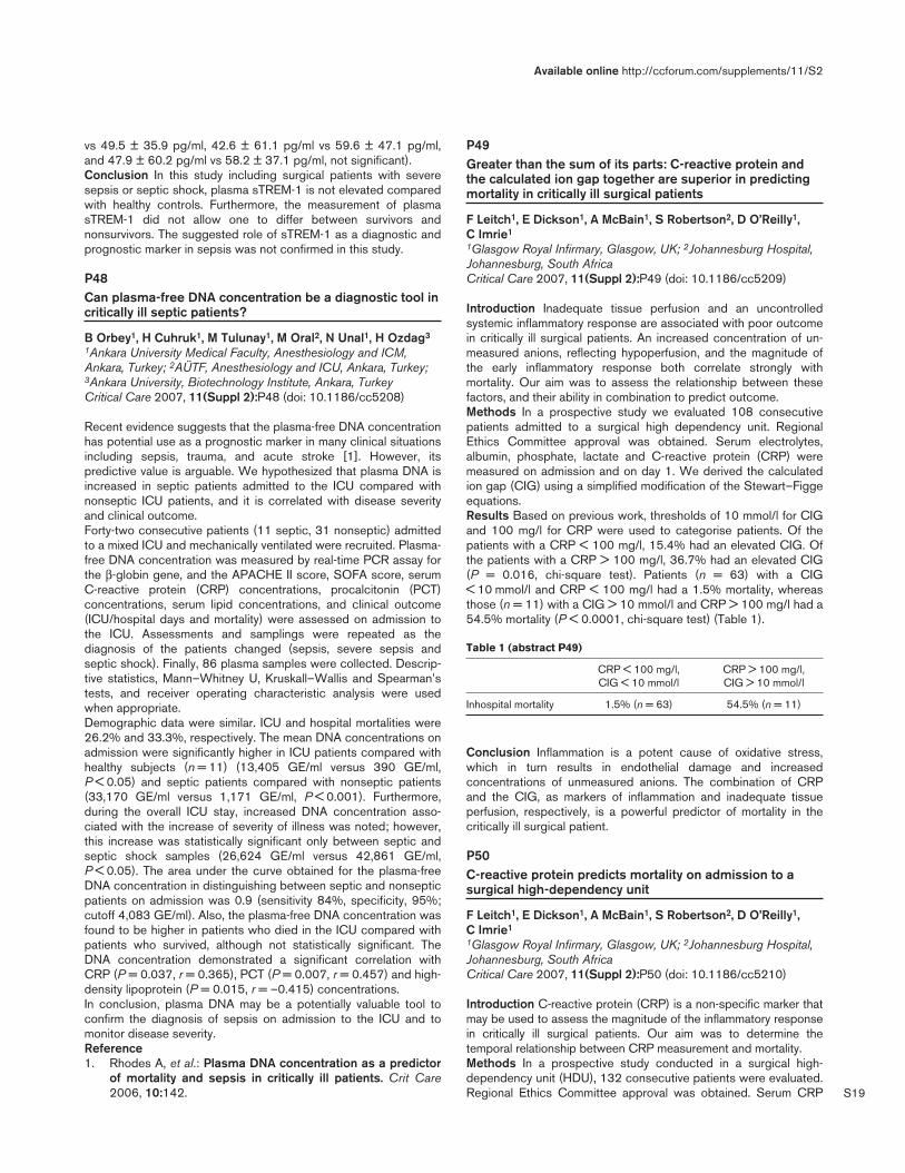
S19
vs 49.5 ± 35.9 pg/ml, 42.6 ± 61.1 pg/ml vs 59.6 ± 47.1 pg/ml,and 47.9 ± 60.2 pg/ml vs 58.2 ± 37.1 pg/ml, not significant).Conclusion In this study including surgical patients with severesepsis or septic shock, plasma sTREM-1 is not elevated comparedwith healthy controls. Furthermore, the measurement of plasmasTREM-1 did not allow one to differ between survivors andnonsurvivors. The suggested role of sTREM-1 as a diagnostic andprognostic marker in sepsis was not confirmed in this study.
P48Can plasma-free DNA concentration be a diagnostic tool incritically ill septic patients?
B Orbey1, H Cuhruk1, M Tulunay1, M Oral2, N Unal1, H Ozdag3
1Ankara University Medical Faculty, Anesthesiology and ICM,Ankara, Turkey; 2AÜTF, Anesthesiology and ICU, Ankara, Turkey;3Ankara University, Biotechnology Institute, Ankara, TurkeyCritical Care 2007, 11(Suppl 2):P48 (doi: 10.1186/cc5208)
Recent evidence suggests that the plasma-free DNA concentrationhas potential use as a prognostic marker in many clinical situationsincluding sepsis, trauma, and acute stroke [1]. However, itspredictive value is arguable. We hypothesized that plasma DNA isincreased in septic patients admitted to the ICU compared withnonseptic ICU patients, and it is correlated with disease severityand clinical outcome.Forty-two consecutive patients (11 septic, 31 nonseptic) admittedto a mixed ICU and mechanically ventilated were recruited. Plasma-free DNA concentration was measured by real-time PCR assay forthe β-globin gene, and the APACHE II score, SOFA score, serumC-reactive protein (CRP) concentrations, procalcitonin (PCT)concentrations, serum lipid concentrations, and clinical outcome(ICU/hospital days and mortality) were assessed on admission tothe ICU. Assessments and samplings were repeated as thediagnosis of the patients changed (sepsis, severe sepsis andseptic shock). Finally, 86 plasma samples were collected. Descrip-tive statistics, Mann–Whitney U, Kruskall–Wallis and Spearman’stests, and receiver operating characteristic analysis were usedwhen appropriate.Demographic data were similar. ICU and hospital mortalities were26.2% and 33.3%, respectively. The mean DNA concentrations onadmission were significantly higher in ICU patients compared withhealthy subjects (n = 11) (13,405 GE/ml versus 390 GE/ml,P < 0.05) and septic patients compared with nonseptic patients(33,170 GE/ml versus 1,171 GE/ml, P < 0.001). Furthermore,during the overall ICU stay, increased DNA concentration asso-ciated with the increase of severity of illness was noted; however,this increase was statistically significant only between septic andseptic shock samples (26,624 GE/ml versus 42,861 GE/ml,P < 0.05). The area under the curve obtained for the plasma-freeDNA concentration in distinguishing between septic and nonsepticpatients on admission was 0.9 (sensitivity 84%, specificity, 95%;cutoff 4,083 GE/ml). Also, the plasma-free DNA concentration wasfound to be higher in patients who died in the ICU compared withpatients who survived, although not statistically significant. TheDNA concentration demonstrated a significant correlation withCRP (P = 0.037, r = 0.365), PCT (P = 0.007, r = 0.457) and high-density lipoprotein (P = 0.015, r = –0.415) concentrations.In conclusion, plasma DNA may be a potentially valuable tool toconfirm the diagnosis of sepsis on admission to the ICU and tomonitor disease severity.Reference1. Rhodes A, et al.: Plasma DNA concentration as a predictor
of mortality and sepsis in critically ill patients. Crit Care2006, 10:142.
P49Greater than the sum of its parts: C-reactive protein andthe calculated ion gap together are superior in predictingmortality in critically ill surgical patients
F Leitch1, E Dickson1, A McBain1, S Robertson2, D O’Reilly1, C Imrie1
1Glasgow Royal Infirmary, Glasgow, UK; 2Johannesburg Hospital,Johannesburg, South AfricaCritical Care 2007, 11(Suppl 2):P49 (doi: 10.1186/cc5209)
Introduction Inadequate tissue perfusion and an uncontrolledsystemic inflammatory response are associated with poor outcomein critically ill surgical patients. An increased concentration of un-measured anions, reflecting hypoperfusion, and the magnitude ofthe early inflammatory response both correlate strongly withmortality. Our aim was to assess the relationship between thesefactors, and their ability in combination to predict outcome.Methods In a prospective study we evaluated 108 consecutivepatients admitted to a surgical high dependency unit. RegionalEthics Committee approval was obtained. Serum electrolytes,albumin, phosphate, lactate and C-reactive protein (CRP) weremeasured on admission and on day 1. We derived the calculatedion gap (CIG) using a simplified modification of the Stewart–Figgeequations.Results Based on previous work, thresholds of 10 mmol/l for CIGand 100 mg/l for CRP were used to categorise patients. Of thepatients with a CRP < 100 mg/l, 15.4% had an elevated CIG. Ofthe patients with a CRP > 100 mg/l, 36.7% had an elevated CIG(P = 0.016, chi-square test). Patients (n = 63) with a CIG< 10 mmol/l and CRP < 100 mg/l had a 1.5% mortality, whereasthose (n = 11) with a CIG > 10 mmol/l and CRP > 100 mg/l had a54.5% mortality (P < 0.0001, chi-square test) (Table 1).
Table 1 (abstract P49)
CRP < 100 mg/l, CRP > 100 mg/l, CIG < 10 mmol/l CIG > 10 mmol/l
Inhospital mortality 1.5% (n = 63) 54.5% (n = 11)
Conclusion Inflammation is a potent cause of oxidative stress,which in turn results in endothelial damage and increasedconcentrations of unmeasured anions. The combination of CRPand the CIG, as markers of inflammation and inadequate tissueperfusion, respectively, is a powerful predictor of mortality in thecritically ill surgical patient.
P50C-reactive protein predicts mortality on admission to asurgical high-dependency unit
F Leitch1, E Dickson1, A McBain1, S Robertson2, D O’Reilly1, C Imrie1
1Glasgow Royal Infirmary, Glasgow, UK; 2Johannesburg Hospital,Johannesburg, South AfricaCritical Care 2007, 11(Suppl 2):P50 (doi: 10.1186/cc5210)
Introduction C-reactive protein (CRP) is a non-specific marker thatmay be used to assess the magnitude of the inflammatory responsein critically ill surgical patients. Our aim was to determine thetemporal relationship between CRP measurement and mortality.Methods In a prospective study conducted in a surgical high-dependency unit (HDU), 132 consecutive patients were evaluated.Regional Ethics Committee approval was obtained. Serum CRP
Available online http://ccforum.com/supplements/11/S2
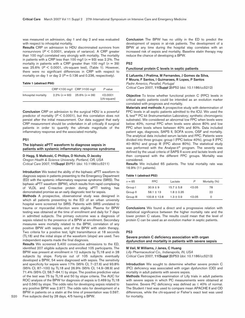
S20
was measured on admission, day 1 and day 2 and was evaluatedwith respect to inhospital mortality.Results CRP on admission to HDU discriminated survivors fromnonsurvivors (P < 0.0001, analysis of variance). A CRP greaterthan 100 mg/l correlated very strongly with mortality. The mortalityin patients with a CRP less than 100 mg/l (n = 93) was 2.2%. Themortality in patients with a CRP greater than 100 mg/l (n = 39)was 25.6% (P < 0.0001, chi-square test), (Table 1). However,there were no significant differences in CRP with respect tomortality on day 1 or day 2 (P = 0.136 and 0.236, respectively).
Table 1 (abstract P50)
CRP <100 mg/l CRP >100 mg/l P value
Inhospital mortality 2.2% (n = 93) 25.6% (n = 39) <0.0001 (chi-square)
Conclusion CRP on admission to the surgical HDU is a powerfulpredictor of mortality (P < 0.0001), but this correlation does notpersist after the initial measurement. Our data suggest that earlyCRP measurement should be undertaken in all critically ill surgicalpatients in order to quantify the ultimate magnitude of theinflammatory response and the associated mortality.
P51The biphasic aPTT waveform to diagnose sepsis inpatients with systemic inflammatory response syndrome
D Hagg, S Malkoski, C Phillips, D Nichols, D JacobyOregon Health & Science University, Portland, OR, USACritical Care 2007, 11(Suppl 2):P51 (doi: 10.1186/cc5211)
Introduction We tested the ability of the biphasic aPTT waveform todiagnosis sepsis in patients presenting to the Emergency Department(ED) with the systemic inflammatory response syndrome (SIRS). Thebiphasic aPTT waveform (BPW), which results from rapid complexingof VLDL and C-reactive protein during aPTT testing, hasdemonstrated promise as an early diagnostic test for sepsis.Methods A prospective, observational study was designed inwhich all patients presenting to the ED of an urban universityhospital were screened for SIRS. Patients with SIRS unrelated totrauma or myocardial infarction were eligible. Plasma for BPWtesting was obtained at the time of enrollment and daily for 7 daysin admitted subjects. The primary outcome was a diagnosis ofsepsis related to the presence of a BPW at enrollment. Secondarymeasures were mortality related to the BPW, correlation of anypositive BPW with sepsis, and of the BPW with statin therapy.Two criteria for a positive test, light transmittance at 18 seconds(TL18) and the initial slope of the waveform (slope) are used. Twoindependent experts made the final diagnosis.Results We screened 5,400 consecutive admissions to the ED,identified 207 eligible subjects and enrolled 105 participants. TheBPW was present at enrollment in 12 subjects by TL18 and in 28subjects by slope. Forty-six out of 105 subjects eventuallydeveloped a BPW, 54 were diagnosed with sepsis. The sensitivityand specificity for sepsis were 17% (95% CI, 7–27.6) and 93.8%(95% CI, 87–100) by TL18 and 26.9% (95% CI, 14.9–38.9) and71.4% (95% CI, 58.7–84.1) by slope. The positive predictive valueof the test was 75 by TL18 and 50 by slope criteria. The AUC forROC analysis of the BPW for diagnosis of sepsis is 0.469 by TL18and 0.560 by slope. The odds ratio for developing sepsis related toany positive BPW was 2.977. The odds ratio for development of aBPW in patients on a statin at the time of presentation was 0.597.Five subjects died by 28 days, 4/5 having a BPW.
Conclusion The BPW has no utility in the ED to predict thedevelopment of sepsis in at-risk patients. The development of aBPW at any time during the hospital stay correlates with anincreased risk of sepsis and mortality. Baseline statin therapy mayreduce the chance of developing a BPW.
P52Functional protein C levels in septic patients
E Lafuente, I Pratima, M Fernandes, J Gomes da Silva, F Moura, F Santos, I Guimaraes, R Lopes, P SantosPadre Americo, Penafiel, PortugalCritical Care 2007, 11(Suppl 2):P52 (doi: 10.1186/cc5212)
Objective To know whether functional protein C (FPC) levels incritical septic patients could be intended as an evolution markercorrelated with prognosis and mortality.Materials and methods A prospective study with determination ofFPC levels in all septic patients admitted to the ICU. We used theIL test™ PC kit (Instrumentation Laboratory; synthetic chromogenicsubstrate). We considered an abnormal low FPC when levels werebelow 40%, normal FPC when levels were above 80% and lowFPC when levels where between 40% and 80%. Data includedpatient age, diagnosis, SAPS II, SOFA score, OSF and mortality.The analytical data included serum lactate and FPC. Patients weredivided into three groups: group I (FPC below 40%), group II (FPC40–80%) and group III (FPC above 80%). The statistical studywas performed with the Analyse-it® program. The severity wasdefined by the usual criteria of SAPS II score and lactate levels andthen compared with the different FPC groups. Mortality wasconsidered.Results We included 65 patients. The total mortality rate was16.9% (11 patients).
Table 1 (abstract P52)
n = 65 FPC Lactate P Mortality (%)
Group I 30.9 ± 9 15.7 ± 5.8 <0.05 78
Group II 58.1 ± 13 1.8 ± 0.35 22
Group III 100.8 ± 12.8 1.3 ± 0.9 <0.05 0
Conclusions We found a direct and a progressive relation withstatistical significance between the higher mortality rate and thelower protein C values. The results could mean that the level ofprotein C can be used as an evolution marker in septic patients.
P53Severe protein C deficiency association with organdysfunction and mortality in patients with severe sepsis
M Vail, M Williams, J Janes, C HuangLilly Pharmaceutical Co., Indianapolis, IN, USACritical Care 2007, 11(Suppl 2):P53 (doi: 10.1186/cc5213)
Introduction We sought to determine whether severe protein C(PC) deficiency was associated with organ dysfunction (OD) andmortality in adult patients with severe sepsis.Methods Retrospective examination of Lilly trials in adult patientswith severe sepsis in which PC measurements were obtained atbaseline. Severe PC deficiency was defined as ≤ 40% of normal.The Student t test was used to compare mean APACHE II and ODdifferences, while the chi-squared or Fisher’s exact test was usedfor mortality.
Critical Care March 2007 Vol 11 Suppl 2 27th International Symposium on Intensive Care and Emergency Medicine
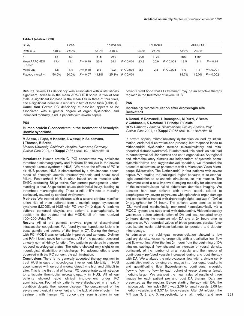
S21
Results Severe PC deficiency was associated with a statisticallysignificant increase in the mean APACHE II score in two of fourtrials, a significant increase in the mean OD in three of four trials,and a significant increase in mortality in two of three trials (Table 1).Conclusion Severe PC deficiency at baseline appears to beassociated with a greater degree of organ dysfunction, andincreased mortality in adult patients with severe sepsis.
P54Human protein C concentrate in the treatment of hemolyticuremic syndrome
M Sasse, L Pape, H Koeditz, A Wessel, K Seidemann, J Thomas, B BrentMedical University Children’s Hospital, Hannover, GermanyCritical Care 2007, 11(Suppl 2):P54 (doi: 10.1186/cc5214)
Introduction Human protein C (PC) concentrate may anticipatethrombotic microangiopathy and facilitate fibrinolysis in the severehemolytic uremic syndrome (HUS). We report the effects of PC insix HUS patients. HUS is characterized by a simultaneous occur-rence of hemolytic anemia, thrombocytopenia and acute renalfailure. Postdiarrheal HUS is often based on an infection withEHEC producing Shiga toxins. Our current pathogenetic under-standing is that Shiga toxins cause endothelial injury, leading tothrombotic microangiopathy. There is still a 5% rate of mortalityparticularly caused by cerebral involvement.Methods We treated six children with a severe cerebral manifes-tation, five of them suffered from a multiple organ dysfunctionsyndrome (MODS), of HUS with PC over 7–10 days. All patientssuffered peritoneal dialysis, one patient a plasmapheresis. Inaddition to the treatment of the MODS, all of them received100–200 U/day PC.Results All of the patients showed signs of disseminatedintravascular coagulation. We found typical hypodense lesions inbasal ganglia and edema of the brain in CT. During the therapywith PC, MODS was remarkable improved and abnormal D-dimerand PAI-1 levels could be normalised. All of the patients recovereda nearly normal kidney function. Two patients persisted in a severereduced neurological status. The others showed only slight or noneurological disabilities on discharge. No adverse effects wereobserved with the PC concentrate administration.Conclusions There is no generally accepted therapy regimen totreat HUS in case of neurological involvement. Mortality in HUSaccompanied with cerebral microangiopathy is high and difficult toalter. This is the first trial of human PC concentrate administrationto anticipate thrombotic microangiopathy in HUS. All of ourpatients showed rapid clinical improvement under PCadministration. Four of six patients were discharged in a healthycondition despite their severe disease. The containment of thesevere neurological involvement and the lack of side effects in thetreatment with human PC concentrate administration in our
patients yield hope that PC treatment may be an effective therapyregimen in the treatment of severe HUS.
P55Increasing microcirculation after drotrecogin alfa(activated)
A Donati, M Romanelli, L Romagnoli, M Ruzzi, V Beato, V Gabbanelli, S Nataloni, T Principi, P PelaiaAOU Umberto I Ancona, Rianimazione Clinica, Ancona, ItalyCritical Care 2007, 11(Suppl 2):P55 (doi: 10.1186/cc5215)
In severe sepsis, microcirculatory dysfunction caused by inflam-mation, endothelial activation and procoagulant response leads tomithocondrial dysfunction (termed microcirculatory and mito-chondrial distress syndrome). If undetected, this condition can leadto parenchymal cellular distress and so to organ failure. As regionaland microcirculatory distress are independent of systemic hemo-dynamic-derived and oxygen-derived variables, we recorded thecourse of microvascular parameters with a Microscan Video Micro-scope (Microvision, The Netherlands) in four patients with severesepsis. We studied the sublingual region because of its embryo-logic correlation to splanchnic circulation, its thin mucosa. Theinstrument used a new improved imaging modality for observationof the microcirculation called sidestream dark-field imaging. Weconsider here four patients with severe sepsis related toesophagectomy, severe polytrauma with splanchnic organ damageand mediastinitis treated with drotrecogin alpha (activated) (DA) at24 µg/kg/hour for 96 hours. The patients were admitted to theICU, ventilated mechanically, monitored hemodynamically via aPICCO system and supported with dobutamine. Videomicroscopywas made before administration of DA and was repeated every24 hours during the treatment with DA and at 24 hours after itssuspension. We recorded values of blood pressure, cardiac func-tion, lactate levels, acid–base balance, temperature and dobuta-mine dosage.At admission the sublingual microcirculation showed a lowcapillary density, vessel heterogeneity with a qualitative low flowand flow–no flow. After the first 24 hours from the beginning of DAinfusion, sublingual flow showed an increase of vessel density,particularly of the number of small vessels, and the number ofcontinuously perfused vessels increased during and post therapywith DA. We analyzed the microvascular flow with a simple semi-quantitative method dividing the images into four equal quadrantsand quantificating flow (hyperdynamic, continuous, sluggish,flow–no flow, no flow) for each cohort of vessel diameter (small,medium, large). We analyzed the mean value of results of threeimages for each patient pre and post DA therapy. Data arepresented as the median. Before starting therapy with DA, themicrovascular flow index (MFI) was 2.06 for small vessels, 2.09 formedium vessels, and 2.37 for large vessels. After DA infusion, theMFI was 3, 3, and 3, respectively, for small, medium and large
Available online http://ccforum.com/supplements/11/S2
Table 1 (abstract P53)
Study EVAA PROWESS ENHANCE ADDRESS
Protein C ≤40% >40% ≤40% >40% ≤40% >40% ≤40% >40%
n 65 60 615 959 795 1127 593 1154
Mean APACHE II 17.4 17.1 P = 0.79 25.9 24.1 P < 0.001 23.2 20.9 P < 0.001 18.5 18.1 P = 0.14score
Mean OD 1.5 1.4 P = 0.42 2.8 2.2 P < 0.001 3.1 2.4 P < 0.001 1.6 1.4 P < 0.001
Placebo mortality 50.0% 20.0% P = 0.07 41.8% 25.3% P < 0.001 19.7% 12.0% P = 0.002
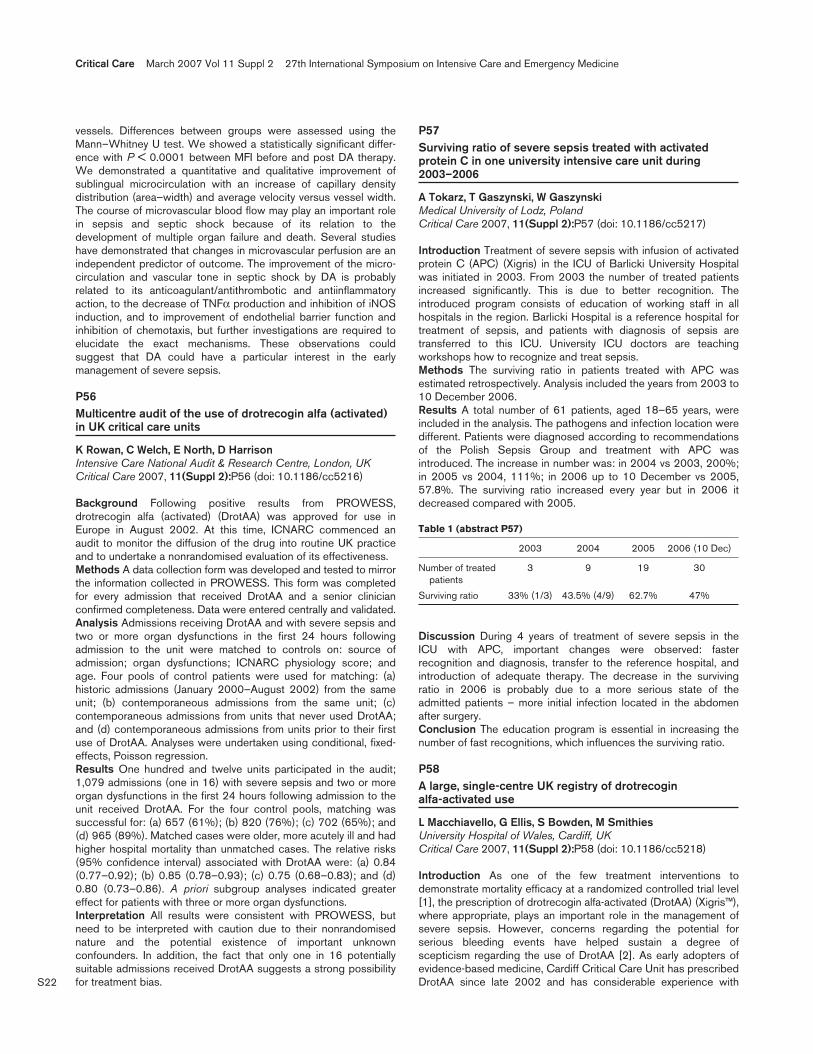
S22
vessels. Differences between groups were assessed using theMann–Whitney U test. We showed a statistically significant differ-ence with P < 0.0001 between MFI before and post DA therapy.We demonstrated a quantitative and qualitative improvement ofsublingual microcirculation with an increase of capillary densitydistribution (area–width) and average velocity versus vessel width.The course of microvascular blood flow may play an important rolein sepsis and septic shock because of its relation to thedevelopment of multiple organ failure and death. Several studieshave demonstrated that changes in microvascular perfusion are anindependent predictor of outcome. The improvement of the micro-circulation and vascular tone in septic shock by DA is probablyrelated to its anticoagulant/antithrombotic and antiinflammatoryaction, to the decrease of TNFα production and inhibition of iNOSinduction, and to improvement of endothelial barrier function andinhibition of chemotaxis, but further investigations are required toelucidate the exact mechanisms. These observations couldsuggest that DA could have a particular interest in the earlymanagement of severe sepsis.
P56Multicentre audit of the use of drotrecogin alfa (activated)in UK critical care units
K Rowan, C Welch, E North, D HarrisonIntensive Care National Audit & Research Centre, London, UKCritical Care 2007, 11(Suppl 2):P56 (doi: 10.1186/cc5216)
Background Following positive results from PROWESS,drotrecogin alfa (activated) (DrotAA) was approved for use inEurope in August 2002. At this time, ICNARC commenced anaudit to monitor the diffusion of the drug into routine UK practiceand to undertake a nonrandomised evaluation of its effectiveness.Methods A data collection form was developed and tested to mirrorthe information collected in PROWESS. This form was completedfor every admission that received DrotAA and a senior clinicianconfirmed completeness. Data were entered centrally and validated.Analysis Admissions receiving DrotAA and with severe sepsis andtwo or more organ dysfunctions in the first 24 hours followingadmission to the unit were matched to controls on: source ofadmission; organ dysfunctions; ICNARC physiology score; andage. Four pools of control patients were used for matching: (a)historic admissions (January 2000–August 2002) from the sameunit; (b) contemporaneous admissions from the same unit; (c)contemporaneous admissions from units that never used DrotAA;and (d) contemporaneous admissions from units prior to their firstuse of DrotAA. Analyses were undertaken using conditional, fixed-effects, Poisson regression.Results One hundred and twelve units participated in the audit;1,079 admissions (one in 16) with severe sepsis and two or moreorgan dysfunctions in the first 24 hours following admission to theunit received DrotAA. For the four control pools, matching wassuccessful for: (a) 657 (61%); (b) 820 (76%); (c) 702 (65%); and(d) 965 (89%). Matched cases were older, more acutely ill and hadhigher hospital mortality than unmatched cases. The relative risks(95% confidence interval) associated with DrotAA were: (a) 0.84(0.77–0.92); (b) 0.85 (0.78–0.93); (c) 0.75 (0.68–0.83); and (d)0.80 (0.73–0.86). A priori subgroup analyses indicated greatereffect for patients with three or more organ dysfunctions.Interpretation All results were consistent with PROWESS, butneed to be interpreted with caution due to their nonrandomisednature and the potential existence of important unknownconfounders. In addition, the fact that only one in 16 potentiallysuitable admissions received DrotAA suggests a strong possibilityfor treatment bias.
P57Surviving ratio of severe sepsis treated with activatedprotein C in one university intensive care unit during2003–2006
A Tokarz, T Gaszynski, W GaszynskiMedical University of Lodz, PolandCritical Care 2007, 11(Suppl 2):P57 (doi: 10.1186/cc5217)
Introduction Treatment of severe sepsis with infusion of activatedprotein C (APC) (Xigris) in the ICU of Barlicki University Hospitalwas initiated in 2003. From 2003 the number of treated patientsincreased significantly. This is due to better recognition. Theintroduced program consists of education of working staff in allhospitals in the region. Barlicki Hospital is a reference hospital fortreatment of sepsis, and patients with diagnosis of sepsis aretransferred to this ICU. University ICU doctors are teachingworkshops how to recognize and treat sepsis.Methods The surviving ratio in patients treated with APC wasestimated retrospectively. Analysis included the years from 2003 to10 December 2006.Results A total number of 61 patients, aged 18–65 years, wereincluded in the analysis. The pathogens and infection location weredifferent. Patients were diagnosed according to recommendationsof the Polish Sepsis Group and treatment with APC wasintroduced. The increase in number was: in 2004 vs 2003, 200%;in 2005 vs 2004, 111%; in 2006 up to 10 December vs 2005,57.8%. The surviving ratio increased every year but in 2006 itdecreased compared with 2005.
Table 1 (abstract P57)
2003 2004 2005 2006 (10 Dec)
Number of treated 3 9 19 30patients
Surviving ratio 33% (1/3) 43.5% (4/9) 62.7% 47%
Discussion During 4 years of treatment of severe sepsis in theICU with APC, important changes were observed: fasterrecognition and diagnosis, transfer to the reference hospital, andintroduction of adequate therapy. The decrease in the survivingratio in 2006 is probably due to a more serious state of theadmitted patients – more initial infection located in the abdomenafter surgery.Conclusion The education program is essential in increasing thenumber of fast recognitions, which influences the surviving ratio.
P58A large, single-centre UK registry of drotrecogin alfa-activated use
L Macchiavello, G Ellis, S Bowden, M SmithiesUniversity Hospital of Wales, Cardiff, UKCritical Care 2007, 11(Suppl 2):P58 (doi: 10.1186/cc5218)
Introduction As one of the few treatment interventions todemonstrate mortality efficacy at a randomized controlled trial level[1], the prescription of drotrecogin alfa-activated (DrotAA) (Xigris™),where appropriate, plays an important role in the management ofsevere sepsis. However, concerns regarding the potential forserious bleeding events have helped sustain a degree ofscepticism regarding the use of DrotAA [2]. As early adopters ofevidence-based medicine, Cardiff Critical Care Unit has prescribedDrotAA since late 2002 and has considerable experience with
Critical Care March 2007 Vol 11 Suppl 2 27th International Symposium on Intensive Care and Emergency Medicine
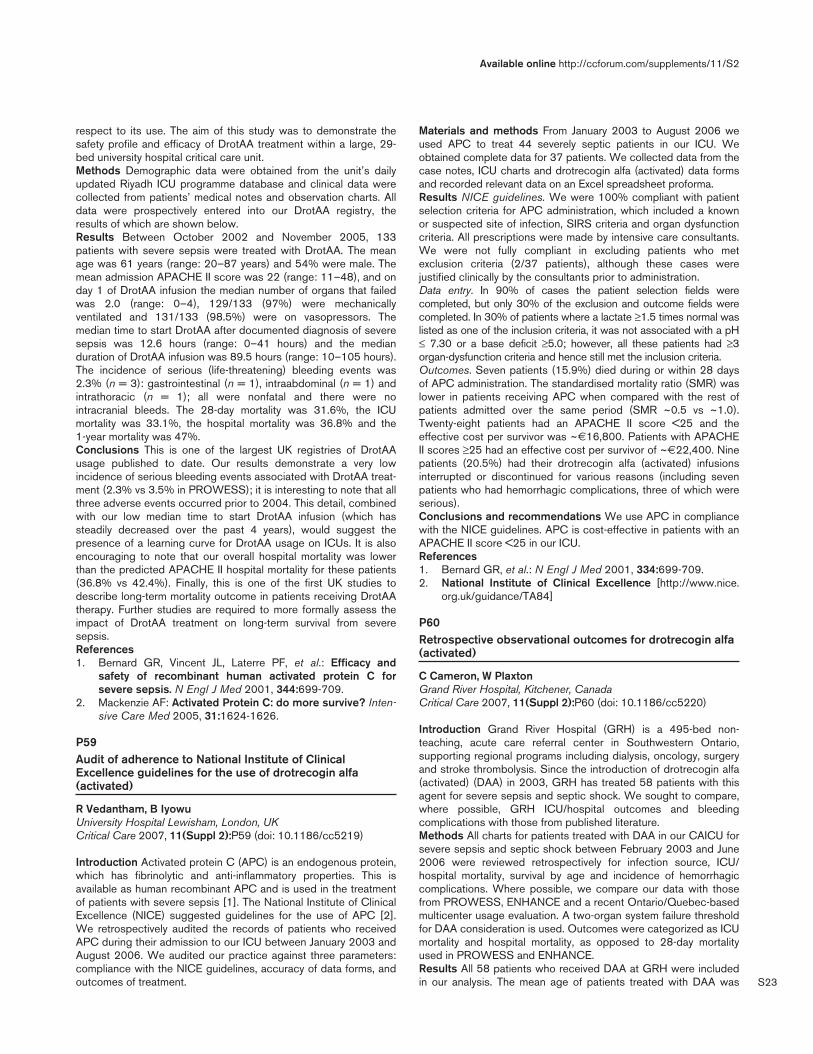
S23
respect to its use. The aim of this study was to demonstrate thesafety profile and efficacy of DrotAA treatment within a large, 29-bed university hospital critical care unit.Methods Demographic data were obtained from the unit’s dailyupdated Riyadh ICU programme database and clinical data werecollected from patients’ medical notes and observation charts. Alldata were prospectively entered into our DrotAA registry, theresults of which are shown below.Results Between October 2002 and November 2005, 133patients with severe sepsis were treated with DrotAA. The meanage was 61 years (range: 20–87 years) and 54% were male. Themean admission APACHE II score was 22 (range: 11–48), and onday 1 of DrotAA infusion the median number of organs that failedwas 2.0 (range: 0–4), 129/133 (97%) were mechanicallyventilated and 131/133 (98.5%) were on vasopressors. Themedian time to start DrotAA after documented diagnosis of severesepsis was 12.6 hours (range: 0–41 hours) and the medianduration of DrotAA infusion was 89.5 hours (range: 10–105 hours).The incidence of serious (life-threatening) bleeding events was2.3% (n = 3): gastrointestinal (n = 1), intraabdominal (n = 1) andintrathoracic (n = 1); all were nonfatal and there were nointracranial bleeds. The 28-day mortality was 31.6%, the ICUmortality was 33.1%, the hospital mortality was 36.8% and the1-year mortality was 47%.Conclusions This is one of the largest UK registries of DrotAAusage published to date. Our results demonstrate a very lowincidence of serious bleeding events associated with DrotAA treat-ment (2.3% vs 3.5% in PROWESS); it is interesting to note that allthree adverse events occurred prior to 2004. This detail, combinedwith our low median time to start DrotAA infusion (which hassteadily decreased over the past 4 years), would suggest thepresence of a learning curve for DrotAA usage on ICUs. It is alsoencouraging to note that our overall hospital mortality was lowerthan the predicted APACHE II hospital mortality for these patients(36.8% vs 42.4%). Finally, this is one of the first UK studies todescribe long-term mortality outcome in patients receiving DrotAAtherapy. Further studies are required to more formally assess theimpact of DrotAA treatment on long-term survival from severesepsis.References1. Bernard GR, Vincent JL, Laterre PF, et al.: Efficacy and
safety of recombinant human activated protein C forsevere sepsis. N Engl J Med 2001, 344:699-709.
2. Mackenzie AF: Activated Protein C: do more survive? Inten-sive Care Med 2005, 31:1624-1626.
P59Audit of adherence to National Institute of ClinicalExcellence guidelines for the use of drotrecogin alfa(activated)
R Vedantham, B IyowuUniversity Hospital Lewisham, London, UKCritical Care 2007, 11(Suppl 2):P59 (doi: 10.1186/cc5219)
Introduction Activated protein C (APC) is an endogenous protein,which has fibrinolytic and anti-inflammatory properties. This isavailable as human recombinant APC and is used in the treatmentof patients with severe sepsis [1]. The National Institute of ClinicalExcellence (NICE) suggested guidelines for the use of APC [2].We retrospectively audited the records of patients who receivedAPC during their admission to our ICU between January 2003 andAugust 2006. We audited our practice against three parameters:compliance with the NICE guidelines, accuracy of data forms, andoutcomes of treatment.
Materials and methods From January 2003 to August 2006 weused APC to treat 44 severely septic patients in our ICU. Weobtained complete data for 37 patients. We collected data from thecase notes, ICU charts and drotrecogin alfa (activated) data formsand recorded relevant data on an Excel spreadsheet proforma.Results NICE guidelines. We were 100% compliant with patientselection criteria for APC administration, which included a knownor suspected site of infection, SIRS criteria and organ dysfunctioncriteria. All prescriptions were made by intensive care consultants.We were not fully compliant in excluding patients who metexclusion criteria (2/37 patients), although these cases werejustified clinically by the consultants prior to administration.Data entry. In 90% of cases the patient selection fields werecompleted, but only 30% of the exclusion and outcome fields werecompleted. In 30% of patients where a lactate ≥1.5 times normal waslisted as one of the inclusion criteria, it was not associated with a pH≤ 7.30 or a base deficit ≥5.0; however, all these patients had ≥3organ-dysfunction criteria and hence still met the inclusion criteria.Outcomes. Seven patients (15.9%) died during or within 28 daysof APC administration. The standardised mortality ratio (SMR) waslower in patients receiving APC when compared with the rest ofpatients admitted over the same period (SMR ~0.5 vs ~1.0).Twenty-eight patients had an APACHE II score <25 and theeffective cost per survivor was ~€16,800. Patients with APACHEII scores ≥25 had an effective cost per survivor of ~€22,400. Ninepatients (20.5%) had their drotrecogin alfa (activated) infusionsinterrupted or discontinued for various reasons (including sevenpatients who had hemorrhagic complications, three of which wereserious).Conclusions and recommendations We use APC in compliancewith the NICE guidelines. APC is cost-effective in patients with anAPACHE II score <25 in our ICU.References1. Bernard GR, et al.: N Engl J Med 2001, 334:699-709.2. National Institute of Clinical Excellence [http://www.nice.
org.uk/guidance/TA84]
P60Retrospective observational outcomes for drotrecogin alfa(activated)
C Cameron, W PlaxtonGrand River Hospital, Kitchener, CanadaCritical Care 2007, 11(Suppl 2):P60 (doi: 10.1186/cc5220)
Introduction Grand River Hospital (GRH) is a 495-bed non-teaching, acute care referral center in Southwestern Ontario,supporting regional programs including dialysis, oncology, surgeryand stroke thrombolysis. Since the introduction of drotrecogin alfa(activated) (DAA) in 2003, GRH has treated 58 patients with thisagent for severe sepsis and septic shock. We sought to compare,where possible, GRH ICU/hospital outcomes and bleedingcomplications with those from published literature.Methods All charts for patients treated with DAA in our CAICU forsevere sepsis and septic shock between February 2003 and June2006 were reviewed retrospectively for infection source, ICU/hospital mortality, survival by age and incidence of hemorrhagiccomplications. Where possible, we compare our data with thosefrom PROWESS, ENHANCE and a recent Ontario/Quebec-basedmulticenter usage evaluation. A two-organ system failure thresholdfor DAA consideration is used. Outcomes were categorized as ICUmortality and hospital mortality, as opposed to 28-day mortalityused in PROWESS and ENHANCE.Results All 58 patients who received DAA at GRH were includedin our analysis. The mean age of patients treated with DAA was
Available online http://ccforum.com/supplements/11/S2
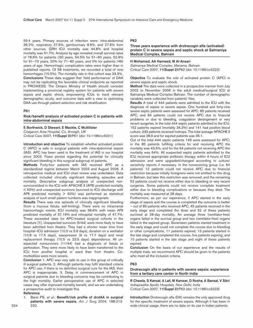
S24
59.4 years. Primary sources of infection were: intra-abdominal36.2%, respiratory 27.6%, genitourinary 8.6%, and 27.6% fromother sources. GRH ICU mortality was 44.8% and hospitalmortality was 51.7%. Analysis by age revealed overall survival ratesof 78.6% for patients ≤50 years, 54.5% for 51–60 years, 52.9%for 61–70 years, 20% for 71–80 years, and 0% for patients >80years of age. Hemorrhagic complication rates were higher than inpublished reports. Of 58 treatments, we recorded a total of ninehemorrhages (15.5%). The mortality rate in this cohort was 33.3%.Conclusions These data suggest that ‘field performance’ of DAAmay not be replicating the favorable clinical endpoints as reportedin PROWESS. The Ontario Ministry of Health should considerimplementing a provincial registry system for patients with severesepsis and septic shock, empowering ICUs to track relevantdemographic, acuity, and outcome data with a view to optimizingDAA use through patient selection and risk stratification.
P61Risk/benefit analysis of activated protein C in patients withintra-abdominal sepsis
E Borthwick, D Stewart, E Mackle, C McAllisterCraigavon Area Hospital, Co. Armagh, UKCritical Care 2007, 11(Suppl 2):P61 (doi: 10.1186/cc5221)
Introduction and objective To establish whether activated proteinC (APC) is safe in surgical patients with intra-abdominal sepsis(IAS). APC has been used in the treatment of IAS in our hospitalsince 2003. Fears persist regarding the potential for clinicallysignificant bleeding in this surgical subgroup of patients.Methods Forty-four patients with IAS received APC as astandardized regime between March 2003 and August 2006. Aretrospective medical and ICU chart review was undertaken. Datacollected included clinically significant bleeding episodes andmortality. Descriptive subgroup analysis of unexpected non-survivors(died in the ICU with APACHE II (APII) predicted mortality< 50%) and unexpected survivors (survived to ICU discharge withAPII predicted mortality > 50%) was performed as statisticalanalysis of such small patient numbers was inappropriate.Results There was one episode of clinically significant bleeding(from a mucous fistula: self-limiting). There were no intracranialhaemorrhagic events. ICU mortality was 38.6% with mean APIIpredicted mortality of 37.16% and inhospital mortality of 47.7%.These exceeded rates for APC-treated surgical cohorts in theliterature [1]. Unexpected survivors (5/44) were more likely to havebeen admitted from theatre. They had a shorter mean time fromhospital–ICU admission (10.5 vs 5.6 days), duration on a ventilator(10.8 vs 17.5 days), vasopressor (9 vs 17.7 days) and renalreplacement therapy (10.5 vs 23.5 days) dependence. All un-expected nonsurvivors (11/44) had a diagnosis of fistula orperforation. They were more likely to have been transferred to theICU from another hospital or ward than from theatre. Co-morbidities were more severe.Conclusion 1. APC was very safe to use in this group of criticallyill surgical patients. 2. Although patients may fulfil standard criteriafor APC use, if there is no definitive surgical cure for the IAS, thenAPC is inappropriate. 3. Delay in commencement of APC insurgical patients due to bleeding concerns may be contributing tothe high mortality. Earlier perioperative use of APC in selectedcases may offer improved mortality benefit, and we are undertakinga prospective audit to investigate this.Reference1. Barie PS, et al.: Benefit/risk profile of drotAA in surgical
patients with severe sepsis. Am J Surg 2004, 188:212-220.
P62Three years experience with drotrecogin alfa (activated)protein C in severe sepsis and septic shock at SalmaniyaMedical Complex, Bahrain
H Mohamed, AA Hameed, M Al-AnsariSalmaniya Medical Complex, Manama, BahrainCritical Care 2007, 11(Suppl 2):P62 (doi: 10.1186/cc5222)
Objective To evaluate the role of activated protein C (APC) insevere sepsis and septic shock.Method The data were collected in a prospective manner from July2002 to November 2006 in the adult medical/surgical ICU atSalmaniya Medical Complex Bahrain. The number of demographicvariables were collected from patients’ files.Results A total of 444 patients were admitted to the ICU with thediagnosis of sepsis or severe sepsis. One hundred and forty-ninesevere septic patients were assessed for APC: 85 patients receivedAPC, and 64 patients could not receive APC due to financialproblems or due to bleeding, coagulation derangement or veryrecent surgeries. In the total 444 septic patients admitted to the ICU,152 patients expired (mortality 34.2%) and 141 had positive bloodculture; 233 patients received inotrops. The total average APACHE IIscore was 28.9 and for expired patients was 35.1.Out of the total 444 septic patients 149 were assessed for APC;in the 85 patients fulfilling criteria for and receiving APC themortality was 43.5%, and for the 64 patients not receiving APC themortality was 64%. All suspected septic patients admitted to theICU received appropriate antibiotic therapy within 4 hours of ICUadmission and were upgraded/changed according to culture/sensitivity reports if necessary. In the nonreceiving group (i.e. 64patients) 12 patients could not receive APC due to financialrestriction because initially foreigners were not entitled to this drugin Bahrain, but later this restriction was removed, and the remaining52 patients could not receive either due to bleeding or very recentsurgeries. Some patients could not receive complete treatmenteither due to bleeding complications or because they died. Themortality was measured at 28 days.Furthermore, as per our experience, if APC started in the earlystage of sepsis and the course is completed the outcome is better– out of 85 patients who received APC, 45 patients received in theearly stage and completed the dose and 32 of these patientssurvived at 28-day mortality. An average three (ventilator-free)organs failed in the survival group and two (ventilator-free) organsfailed in the expired group. Seventeen patients started treatment inthe early stage and could not complete the course due to bleedingor other complications, 11 patients expired; 13 patients started inthe late stage and completed the course, five patients expiring; and10 patients started in the late stage and eight of these patientsexpired.Conclusion On the basis of our experience and the results ofmultiple trials, we recommend APC should be given to the patientswho meet all the inclusion criteria.
P63Drotrecogin alfa in patients with severe sepsis: experiencefrom a tertiary care center in North India
R Chawla, S Kansal, A Lall, M Kanwar, D Rosha, A Bansal, V SikriIndraprastha Apollo Hospitals, New Delhi, IndiaCritical Care 2007, 11(Suppl 2):P63 (doi: 10.1186/cc5223)
Introduction Drotrecogin alfa (DA) remains the only approved drugfor the specific treatment of severe sepsis. Although it has been inwide clinical usage, there are no data on its use in Indian patients.
Critical Care March 2007 Vol 11 Suppl 2 27th International Symposium on Intensive Care and Emergency Medicine
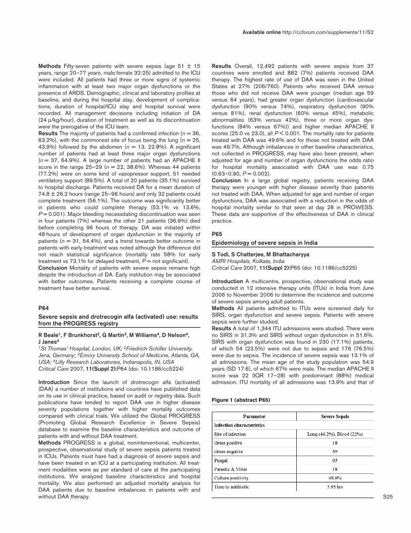
S25
Methods Fifty-seven patients with severe sepsis (age 51 ± 15years, range 20–77 years, male:female 32:25) admitted to the ICUwere included. All patients had three or more signs of systemicinflammation with at least two major organ dysfunctions or thepresence of ARDS. Demographic, clinical and laboratory profiles atbaseline, and during the hospital stay, development of complica-tions, duration of hospital/ICU stay and hospital survival wererecorded. All management decisions including initiation of DA(24 µ/kg/hour), duration of treatment as well as its discontinuationwere the prerogative of the ICU team.Results The majority of patients had a confirmed infection (n = 36,63.2%), with the commonest site of focus being the lung (n = 25,43.9%) followed by the abdomen (n = 13, 22.8%). A significantnumber of patients had at least three major organ dysfunctions(n = 37, 64.9%). A large number of patients had an APACHE IIscore in the range 25–29 (n = 22, 38.6%). Whereas 44 patients(77.2%) were on some kind of vasopressor support, 51 neededventilatory support (89.5%). A total of 20 patients (35.1%) survivedto hospital discharge. Patients received DA for a mean duration of74.8 ± 26.2 hours (range 25–96 hours) and only 32 patients couldcomplete treatment (56.1%). The outcome was significantly betterin patients who could complete therapy (53.1% vs 13.6%,P = 0.001). Major bleeding necessitating discontinuation was seenin four patients (7%) whereas the other 21 patients (36.9%) diedbefore completing 96 hours of therapy. DA was initiated within48 hours of development of organ dysfunction in the majority ofpatients (n = 31, 54.4%), and a trend towards better outcome inpatients with early treatment was noted although the difference didnot reach statistical significance (mortality rate 58% for earlytreatment vs 73.1% for delayed treatment, P = not significant).Conclusion Mortality of patients with severe sepsis remains highdespite the introduction of DA. Early institution may be associatedwith better outcomes. Patients receiving a complete course oftreatment have better survival.
P64Severe sepsis and drotrecogin alfa (activated) use: resultsfrom the PROGRESS registry
R Beale1, F Brunkhorst2, G Martin3, M Williams4, D Nelson4, J Janes4
1St Thomas’ Hospital, London, UK; 2Friedrich-Schiller University,Jena, Germany; 3Emory University School of Medicine, Atlanta, GA,USA; 4Lilly Research Laboratories, Indianapolis, IN, USACritical Care 2007, 11(Suppl 2):P64 (doi: 10.1186/cc5224)
Introduction Since the launch of drotrecogin alfa (activated)(DAA) a number of institutions and countries have published dataon its use in clinical practice, based on audit or registry data. Suchpublications have tended to report DAA use in higher diseaseseverity populations together with higher mortality outcomescompared with clinical trials. We utilized the Global PROGRESS(Promoting Global Research Excellence in Severe Sepsis)database to examine the baseline characteristics and outcome ofpatients with and without DAA treatment.Methods PROGRESS is a global, noninterventional, multicenter,prospective, observational study of severe sepsis patients treatedin ICUs. Patients must have had a diagnosis of severe sepsis andhave been treated in an ICU at a participating institution. All treat-ment modalities were as per standard of care at the participatinginstitutions. We analyzed baseline characteristics and hospitalmortality. We also performed an adjusted mortality analysis forDAA patients due to baseline imbalances in patients with andwithout DAA therapy.
Results Overall, 12,492 patients with severe sepsis from 37countries were enrolled and 882 (7%) patients received DAAtherapy. The highest rate of use of DAA was seen in the UnitedStates at 27% (206/760). Patients who received DAA versusthose who did not receive DAA were younger (median age 59versus 64 years), had greater organ dysfunction (cardiovasculardysfunction (90% versus 74%), respiratory dysfunction (90%versus 81%), renal dysfunction (60% versus 45%), metabolicabnormalities (63% versus 42%), three or more organ dys-functions (84% versus 67%)) and higher median APACHE IIscores (25.0 vs 23.0), all P < 0.001. The mortality rate for patientstreated with DAA was 49.6% and for those not treated with DAAwas 49.7%. Although imbalances in other baseline characteristics,not collected in PROGRESS, may have also been present, whenadjusted for age and number of organ dysfunctions the odds ratiofor hospital mortality associated with DAA use was 0.75(0.63–0.90, P = 0.002).Conclusion In a large global registry, patients receiving DAAtherapy were younger with higher disease severity than patientsnot treated with DAA. When adjusted for age and number of organdysfunctions, DAA was associated with a reduction in the odds ofhospital mortality similar to that seen at day 28 in PROWESS.These data are supportive of the effectiveness of DAA in clinicalpractice.
P65Epidemiology of severe sepsis in India
S Todi, S Chatterjee, M BhattacharyyaAMRI Hospitals, Kolkata, IndiaCritical Care 2007, 11(Suppl 2):P65 (doi: 10.1186/cc5225)
Introduction A multicentre, prospective, observational study wasconducted in 12 intensive therapy units (ITUs) in India from June2006 to November 2006 to determine the incidence and outcomeof severe sepsis among adult patients.Methods All patients admitted to ITUs were screened daily forSIRS, organ dysfunction and severe sepsis. Patients with severesepsis were further studied.Results A total of 1,344 ITU admissions were studied. There wereno SIRS in 31.3% and SIRS without organ dysfunction in 51.6%.SIRS with organ dysfunction was found in 230 (17.1%) patients,of which 54 (23.5%) were not due to sepsis and 176 (76.5%)were due to sepsis. The incidence of severe sepsis was 13.1% ofall admissions. The mean age of the study population was 54.9years (SD 17.6), of which 67% were male. The median APACHE IIscore was 22 (IQR 17–28) with predominant (88%) medicaladmission. ITU mortality of all admissions was 13.9% and that of
Available online http://ccforum.com/supplements/11/S2
Figure 1 (abstract P65)
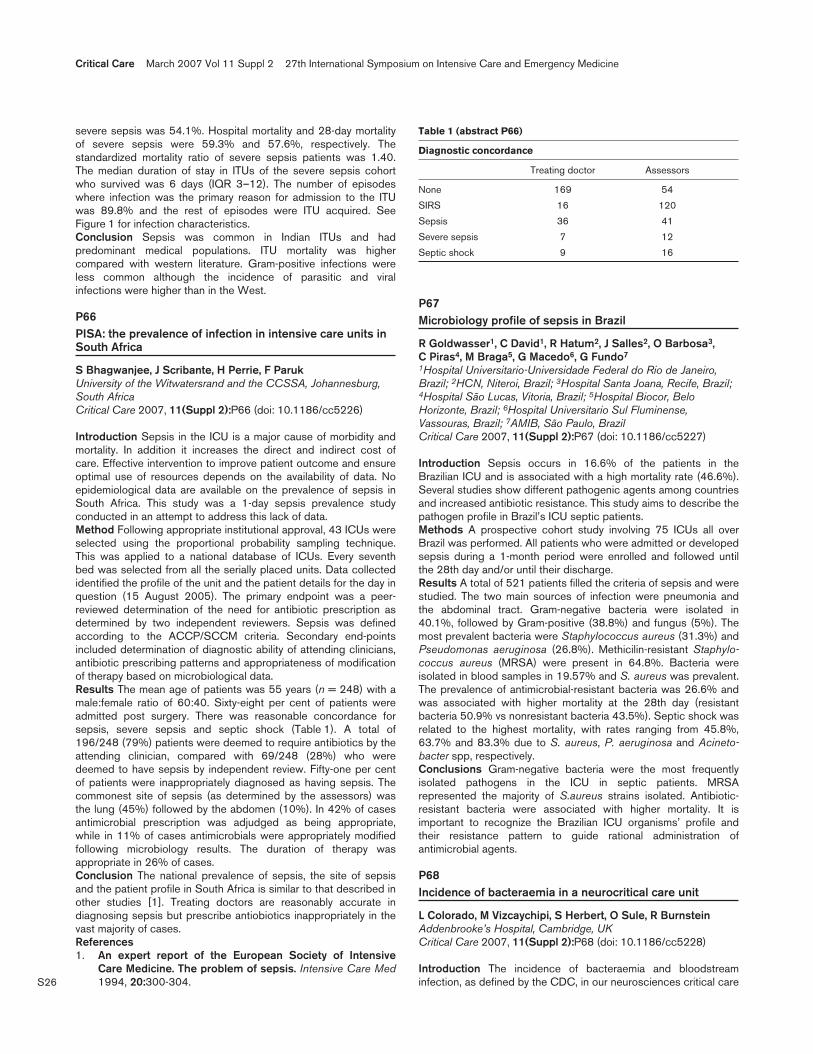
S26
severe sepsis was 54.1%. Hospital mortality and 28-day mortalityof severe sepsis were 59.3% and 57.6%, respectively. Thestandardized mortality ratio of severe sepsis patients was 1.40.The median duration of stay in ITUs of the severe sepsis cohortwho survived was 6 days (IQR 3–12). The number of episodeswhere infection was the primary reason for admission to the ITUwas 89.8% and the rest of episodes were ITU acquired. SeeFigure 1 for infection characteristics.Conclusion Sepsis was common in Indian ITUs and hadpredominant medical populations. ITU mortality was highercompared with western literature. Gram-positive infections wereless common although the incidence of parasitic and viralinfections were higher than in the West.
P66PISA: the prevalence of infection in intensive care units inSouth Africa
S Bhagwanjee, J Scribante, H Perrie, F ParukUniversity of the Witwatersrand and the CCSSA, Johannesburg,South AfricaCritical Care 2007, 11(Suppl 2):P66 (doi: 10.1186/cc5226)
Introduction Sepsis in the ICU is a major cause of morbidity andmortality. In addition it increases the direct and indirect cost ofcare. Effective intervention to improve patient outcome and ensureoptimal use of resources depends on the availability of data. Noepidemiological data are available on the prevalence of sepsis inSouth Africa. This study was a 1-day sepsis prevalence studyconducted in an attempt to address this lack of data.Method Following appropriate institutional approval, 43 ICUs wereselected using the proportional probability sampling technique.This was applied to a national database of ICUs. Every seventhbed was selected from all the serially placed units. Data collectedidentified the profile of the unit and the patient details for the day inquestion (15 August 2005). The primary endpoint was a peer-reviewed determination of the need for antibiotic prescription asdetermined by two independent reviewers. Sepsis was definedaccording to the ACCP/SCCM criteria. Secondary end-pointsincluded determination of diagnostic ability of attending clinicians,antibiotic prescribing patterns and appropriateness of modificationof therapy based on microbiological data.Results The mean age of patients was 55 years (n = 248) with amale:female ratio of 60:40. Sixty-eight per cent of patients wereadmitted post surgery. There was reasonable concordance forsepsis, severe sepsis and septic shock (Table 1). A total of196/248 (79%) patients were deemed to require antibiotics by theattending clinician, compared with 69/248 (28%) who weredeemed to have sepsis by independent review. Fifty-one per centof patients were inappropriately diagnosed as having sepsis. Thecommonest site of sepsis (as determined by the assessors) wasthe lung (45%) followed by the abdomen (10%). In 42% of casesantimicrobial prescription was adjudged as being appropriate,while in 11% of cases antimicrobials were appropriately modifiedfollowing microbiology results. The duration of therapy wasappropriate in 26% of cases.Conclusion The national prevalence of sepsis, the site of sepsisand the patient profile in South Africa is similar to that described inother studies [1]. Treating doctors are reasonably accurate indiagnosing sepsis but prescribe antiobiotics inappropriately in thevast majority of cases.References1. An expert report of the European Society of Intensive
Care Medicine. The problem of sepsis. Intensive Care Med1994, 20:300-304.
P67Microbiology profile of sepsis in Brazil
R Goldwasser1, C David1, R Hatum2, J Salles2, O Barbosa3, C Piras4, M Braga5, G Macedo6, G Fundo7
1Hospital Universitario-Universidade Federal do Rio de Janeiro,Brazil; 2HCN, Niteroi, Brazil; 3Hospital Santa Joana, Recife, Brazil;4Hospital São Lucas, Vitoria, Brazil; 5Hospital Biocor, BeloHorizonte, Brazil; 6Hospital Universitario Sul Fluminense,Vassouras, Brazil; 7AMIB, São Paulo, BrazilCritical Care 2007, 11(Suppl 2):P67 (doi: 10.1186/cc5227)
Introduction Sepsis occurs in 16.6% of the patients in theBrazilian ICU and is associated with a high mortality rate (46.6%).Several studies show different pathogenic agents among countriesand increased antibiotic resistance. This study aims to describe thepathogen profile in Brazil’s ICU septic patients.Methods A prospective cohort study involving 75 ICUs all overBrazil was performed. All patients who were admitted or developedsepsis during a 1-month period were enrolled and followed untilthe 28th day and/or until their discharge.Results A total of 521 patients filled the criteria of sepsis and werestudied. The two main sources of infection were pneumonia andthe abdominal tract. Gram-negative bacteria were isolated in40.1%, followed by Gram-positive (38.8%) and fungus (5%). Themost prevalent bacteria were Staphylococcus aureus (31.3%) andPseudomonas aeruginosa (26.8%). Methicilin-resistant Staphylo-coccus aureus (MRSA) were present in 64.8%. Bacteria wereisolated in blood samples in 19.57% and S. aureus was prevalent.The prevalence of antimicrobial-resistant bacteria was 26.6% andwas associated with higher mortality at the 28th day (resistantbacteria 50.9% vs nonresistant bacteria 43.5%). Septic shock wasrelated to the highest mortality, with rates ranging from 45.8%,63.7% and 83.3% due to S. aureus, P. aeruginosa and Acineto-bacter spp, respectively.Conclusions Gram-negative bacteria were the most frequentlyisolated pathogens in the ICU in septic patients. MRSArepresented the majority of S.aureus strains isolated. Antibiotic-resistant bacteria were associated with higher mortality. It isimportant to recognize the Brazilian ICU organisms’ profile andtheir resistance pattern to guide rational administration ofantimicrobial agents.
P68Incidence of bacteraemia in a neurocritical care unit
L Colorado, M Vizcaychipi, S Herbert, O Sule, R BurnsteinAddenbrooke’s Hospital, Cambridge, UKCritical Care 2007, 11(Suppl 2):P68 (doi: 10.1186/cc5228)
Introduction The incidence of bacteraemia and bloodstreaminfection, as defined by the CDC, in our neurosciences critical care
Critical Care March 2007 Vol 11 Suppl 2 27th International Symposium on Intensive Care and Emergency Medicine
Table 1 (abstract P66)
Diagnostic concordance
Treating doctor Assessors
None 169 54
SIRS 16 120
Sepsis 36 41
Severe sepsis 7 12
Septic shock 9 16
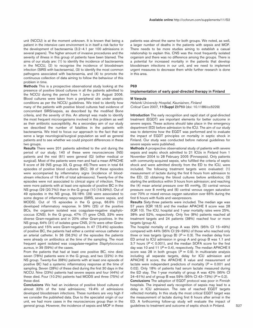
S27
unit (NCCU) is at the moment unknown. It is known that being apatient in the intensive care environment is in itself a risk factor forthe development of bacteraemia (3.2–4.1 per 100 admissions inseveral papers). The higher amount of invasive procedures and theseverity of illness in this group of patients have been blamed. Theaims of our study are: (1) to identify the incidence of bacteraemiain the NCCU, (2) to recognise the incidence of bloodstreaminfection (SIRS with bacteraemia), (3) to identify the most commonpathogens associated with bacteraemia, and (4) to promote thecontinuous collection of data aiming to follow the behaviour of thisproblem in time.Methods This is a prospective observational study looking at thepresence of positive blood cultures in all the patients admitted tothe NCCU during the period from 1 June to 31 August 2006.Blood cultures were taken from a peripheral site under asepticconditions as per the NCCU guidelines. We tried to identify howmany of the patients with positive blood cultures had evidence ofconcomitant SIRS/sepsis, as described by the modified Bonecriteria, and the severity of this. An attempt was made to identifythe most frequent microorganisms involved in this problem as wellas their antibiotic susceptibility. As a secondary aim of our studywe described the number of fatalities in the patients withbacteraemia. We tried to focus our approach to the fact that weserve a large neurological/surgical population as well as generalpatients and to see whether we could pinpoint differences in thesetwo groups.Results There were 201 patients admitted to the unit during theperiod of our study; 140 of these were neurosciences (NS)patients and the rest (61) were general (G) (either medical orsurgical). Most of the patients were men and had a mean APACHEII score of 39 (NS group 33, G group 45).There were in total 64episodes of positive blood cultures (BC); 39 of these episodeswere accompanied by inflammatory signs (incidence of blood-stream infections of 19.4% of total admissions). Twenty-five of theepisodes were not associated to clinical signs of infection. Therewere more patients with at least one episode of positive BC in theNS group (29 (20.7%)) than in the G group (10 (16.39%)). Out of49 episodes in the NS group, 59.18% (29) were associated tosome degree of inflammatory response (SIRS, severe sepsis, andMODS). Out of 15 episodes in the G group, 66.6% (10)developed inflammatory response. In 59% (25) of the positiveBCs, the organism isolated was coagulase-negative staphylo-coccus (CNS). In the G group, 47% (7) grew CNS, 33% werediverse Gram-negatives and in 20% other Gram-positives. In theNS group, 64% (31) of isolates grew CNS, 21% were other Gram-positives and 15% were Gram-negatives. In 47 (73.4%) episodesof positive BC, the patients had either a central venous catheter oran arterial catheter. In 36 (56.2%) of the episodes the patientswere already on antibiotics at the time of the sampling. The mostfrequent agent isolated was coagulase-negative Staphylococcusaureus, in 39 (59%) of the cases.From the patients that had at least one positive BC, nine died;seven (78%) patients were in the G group, and two (22%) in theNS group. Twenty-five (68%) patients with at least one episode ofpositive BC had a systemic inflammatory response at the time ofsampling. Seven (28%) of these died during the first 30 days in theNCCU. Nine (23%) patients had severe sepsis and four (44%) ofthese died. Four (10.2%) patients had MODS and three (75%) ofthese died.Conclusions We had an incidence of positive blood cultures ofalmost 32% of the total admissions; 19.4% of admissionsdeveloped bloodstream infections. These numbers are very high ifwe consider the published data. Due to the specialist origin of ourunit, we had more cases in the neurosciences group than in thegeneral group. However, the incidence of sepsis and MOF in these
patients was almost the same for both groups. We noted, as well,a larger number of deaths in the patients with sepsis and MOF.There needs to be more studies aiming to establish a casualrelationship to explain this. CNS was the most frequently isolatedorganism and there was no difference among the groups. There isa potential for increased mortality in the patients that developbloodstream infections in our unit, and we need to implementurgent measures to decrease them while further research is donein this area.
P69Implementation of early goal-directed therapy in Finland
M VarpulaHelsinki University Hospital, Kauniainen, FinlandCritical Care 2007, 11(Suppl 2):P69 (doi: 10.1186/cc5229)
Introduction The early recognition and rapid start of goal-directedtreatment (EGDT) are important elements for better outcome insevere sepsis. These actions should take place in the emergencydepartment (ED) before admission to the ICU. The aim of our studywas to determine how the EGDT was performed and to evaluatethe impact of EGDT principles on mortality in septic shock inFinland. Our study was conducted before national guidelines forsevere sepsis were published.Methods A prospective observational study of patients with severesepsis and septic shock admitted to 21 ICUs in Finland from 1November 2004 to 28 February 2005 (Finnsepsis). Only patientswith community-acquired sepsis, who fulfilled the criteria of septicshock and were admitted directly from the ED to the ICU, wereincluded. The following treatment targets were evaluated: (1)measurement of lactate during the first 6 hours from admission tothe ED; (2) obtaining the blood cultures before antibiotics; (3)starting the antibiotics within 3 hours from admission; and reachingthe (4) mean arterial pressure over 65 mmHg, (5) central venouspressure over 8 mmHg and (6) central venous oxygen saturationover 70% or mixed venous oxygen saturation over 65% during thefirst 6 hours with fluids and vasopressors.Results Sixty-three patients were included. The median age was57 years (IQR 18.5) and the median APACHE II score was 28(IQR 10). The ICU, hospital and 1-year mortality rates were 25%,38% and 52%, respectively. Only five (8%) patients reached alltreatment targets and 24 patients (38%) reached four or moretargets (group A).The hospital mortality of group A was 29% (95% CI 15–49%)compared with 44% (95% CI 29–59%) of those who reached onlythree or less targets (group B) (P = 0.3). The median delay fromED arrival to ICU admission in group A and group B was 1.1 and3.7 hours (P < 0.001), and the median SOFA score for the firstday was 10 and 11 (P = 0.4), respectively. The median APACHE IIscore was 28 in both groups (P = 0.9). In multivariate analysisincluding all separate targets, delay for ICU admission andAPACHE II score, the APACHE II value and measurement oflactate were independent predictors of mortality (P = 0.001 and0.02). Only 18% of patients had serum lactate measured duringthe ED stay. The 1-year mortality of group A was 42% (95% CI24–61%) and of group B was 59% (95% CI 43–73%) (P = 0.2).Conclusions The adoption of EGDT protocol was poor in Finnishhospitals. The impaired early recognition of sepsis may lead to adelay in ICU admission. The rate of reached EGDT targetsreflected mortality. In this study the most critical EGDT target wasthe measurement of lactate during first 6 hours after arrival in theED. A forthcoming follow-up study will evaluate the impact ofguidelines to treatment and outcome of septic shock in Finland.
Available online http://ccforum.com/supplements/11/S2
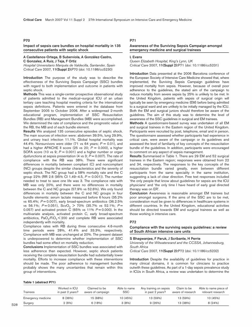
S28
P70Impact of sepsis care bundles on hospital mortality in 135consecutive patients with septic shock
A Castellanos-Ortega, B Suberviola, A González-Castro, C Gonzalez, A Ruiz, J Teja, F OrtizHospital Universitario Marqués de Valdecilla, Santander, SpainCritical Care 2007, 11(Suppl 2):P70 (doi: 10.1186/cc5230)
Introduction The purpose of the study was to describe theeffectiveness of the Surviving Sepsis Campaign (SSC) bundleswith regard to both implementation and outcome in patients withseptic shock.Methods This was a single-center prospective observational studyof patients admitted to the medical–surgical ICU of an urbantertiary care teaching hospital meeting criteria for the internationalsepsis definitions. Patients were entered in the database fromSeptember 2005 to October 2006. After a widespread 2-montheducational program, implementation of SSC ResuscitationBundles (RB) and Management Bundles (MB) were accomplished.We determined the rate of compliance and the prognostic value ofthe RB, the MB and of each bundle element.Results We analyzed 135 consecutive episodes of septic shock.The main sources of infection were: abdomen 39.5%, lung 29.9%,and urinary tract infection 11.1%. Global hospital mortality was44.4%. Nonsurvivors were older (71 vs 64 years; P = 0.01), andhad a higher APACHE II score (25 vs 20; P = 0.000), a higherSOFA score (10 vs 9; P = 0.001) and a higher number or organdysfunctions at sepsis presentation (4 vs 3; P = 0.007). The rate ofcompliance with the RB was 38%. There were significantdifferences in mortality between compliant (C) and noncompliant(NC) groups despite the similar characteristics and the severity ofseptic shock. The NC group had a 58% mortality rate and the Cgroup 22% (RR 2.6 (95% CI 1.49–4.5, P = 0.001)). The numberneeded to treat to save one life was 3. The compliance rate withMB was only 20%, and there were no differences in mortalitybetween the C and NC groups (57.9% vs 52.6%). We only founddifferences in mortality between the C and NC groups in fourbundle elements: serum lactate measured before 6 hours (35.2%vs 65.4%; P = 0.007), early broad-spectrum antibiotics (36.2.5%vs 56.1%; P = 0.051), ScvO2 > 70% (35.7% vs 52.1%; P =0.057) and activated protein C (65% vs 11% P = 0.000). In themultivariate analysis, activated protein C, early broad-spectrumantibiotics, PaO2/FiO2 < 200 and complete RB were associatedindependently with mortality.Compliance rates with RB during three consecutive 4.6-monthtime periods were 28%, 41.4% and 33.3%, respectively.Compliance with MB was unchanged at 20%. The present datasetis underpowered to determine whether implementation of SSCbundles had some effect on mortality reduction.Conclusions Implementation of SSC bundles was associated withless adherence than expected. However, septic shock patientsreceiving the complete resuscitation bundle had substantially lowermortality. Efforts to increase compliance with these interventionsshould be made. The poor adherence to management bundlesprobably shows the many uncertainties that remain within thisgroup of interventions.
P71Awareness of the Surviving Sepsis Campaign amongstemergency medicine and surgical trainees
L EvansQueen Elizabeth Hospital, King’s Lynn, UKCritical Care 2007, 11(Suppl 2):P71 (doi: 10.1186/cc5231)
Introduction Data presented at the 2006 Barcelona conference ofthe European Society of Intensive Care Medicine showed that, whereimplemented, the Surviving Sepsis Campaign guidelines haveimproved mortality from sepsis. However, because of overall pooradherence to the guidelines, the stated aim of the campaign toreduce mortality from severe sepsis by 25% is unlikely to be met. Inthe United Kingdom, patients with sepsis of surgical origin willtypically be seen by emergency medicine (EM) before being admittedto a surgical ward and are unlikely to be initially managed by the ICU.Both the EM and surgical juniors should therefore be aware of theguidelines. The aim of this study was to determine the level ofawareness of the SSC guidelines in surgical and EM trainees.Methods A questionnaire-based survey was undertaken of all EMand surgical trainees in the Eastern region of the United Kingdom.Participants were recruited by post, telephone, email and in person.The questionnaire assessed whether participants had experience incritical care, were aware of the campaign or its guidelines andassessed the level of familiarity of key concepts of the resuscitationbundle of the guidelines. In addition, participants were encouragedto comment on any aspect of sepsis management.Results Summarised in Table 1. There are 29 EM and 52 surgicaltrainees in the Eastern region; responses were obtained from 22and 34, respectively. The responses to the key concepts of theresuscitation bundle varied greatly, even between differentparticipants from the same speciality in the same institution,suggesting a lack of clear direction. Free text responses included‘the only people that know about guidelines for sepsis are the ICUphysicians’ and ‘the only time I have heard of early goal directedtherapy was on ER’.Conclusion Awareness is reasonable amongst EM trainees butpoor amongst surgeons. If the aims of the SSC are to be met,consideration must be given to differences in healthcare systems indifferent countries. In the United Kingdom, educational activitiesshould be directed towards EM and surgical trainees as well asthose working in intensive care.
P72Compliance with the surviving sepsis guidelines: a reviewof South African intensive care units
S Bhagwanjee, F Paruk, J Scribante, H PerrieUniversity of the Witwatersrand and the CCSSA, Johannesburg,South AfricaCritical Care 2007, 11(Suppl 2):P72 (doi: 10.1186/cc5232)
Introduction Despite the availability of guidelines for practice inmany clinical domains, it is common for clinicians to practiceoutwith these guidelines. As part of a 1-day sepsis prevalence studyin ICUs in South Africa, a review was undertaken to determine the
Critical Care March 2007 Vol 11 Suppl 2 27th International Symposium on Intensive Care and Emergency Medicine
Table 1 (abstract P71)
Worked in ICU Claimed to be Able to name Any training on sepsis Claim to be Able to name piece of Trainees in past 2 years? aware of campaign SSC in past 2 years? aware of research relevant research
Emergency medicine 8 (36%) 15 (68%) 10 (45%) 13 (59%) 13 (59%) 10 (45%)
Surgery 2 (6%) 6 (18%) 3 (8%) 9 (26%) 13 (38%) 8 (24%)
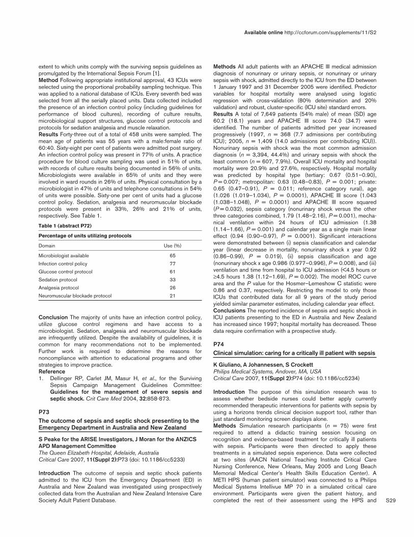
S29
extent to which units comply with the surviving sepsis guidelines aspromulgated by the International Sepsis Forum [1].Method Following appropriate institutional approval, 43 ICUs wereselected using the proportional probability sampling technique. Thiswas applied to a national database of ICUs. Every seventh bed wasselected from all the serially placed units. Data collected includedthe presence of an infection control policy (including guidelines forperformance of blood cultures), recording of culture results,microbiological support structures, glucose control protocols andprotocols for sedation analgesia and muscle relaxation.Results Forty-three out of a total of 458 units were sampled. Themean age of patients was 55 years with a male:female ratio of60:40. Sixty-eight per cent of patients were admitted post surgery.An infection control policy was present in 77% of units. A practiceprocedure for blood culture sampling was used in 51% of units,with records of culture results being documented in 56% of units.Microbiologists were available in 65% of units and they wereinvolved in ward rounds in 26% of units. Physical consultation by amicrobiologist in 47% of units and telephone consultations in 54%of units were possible. Sixty-one per cent of units had a glucosecontrol policy. Sedation, analgesia and neuromuscular blockadeprotocols were present in 33%, 26% and 21% of units,respectively. See Table 1.
Table 1 (abstract P72)
Percentage of units utilizing protocols
Domain Use (%)
Microbiologist available 65
Infection control policy 77
Glucose control protocol 61
Sedation protocol 33
Analgesia protocol 26
Neuromuscular blockade protocol 21
Conclusion The majority of units have an infection control policy,utilize glucose control regimens and have access to amicrobiologist. Sedation, analgesia and neuromuscular blockadeare infrequently utilized. Despite the availability of guidelines, it iscommon for many recommendations not to be implemented.Further work is required to determine the reasons fornoncompliance with attention to educational programs and otherstrategies to improve practice.Reference1. Dellinger RP, Carlet JM, Masur H, et al., for the Surviving
Sepsis Campaign Management Guidelines Committee:Guidelines for the management of severe sepsis andseptic shock. Crit Care Med 2004, 32:858-873.
P73The outcome of sepsis and septic shock presenting to theEmergency Department in Australia and New Zealand
S Peake for the ARISE Investigators, J Moran for the ANZICSAPD Management CommitteeThe Queen Elizabeth Hospital, Adelaide, AustraliaCritical Care 2007, 11(Suppl 2):P73 (doi: 10.1186/cc5233)
Introduction The outcome of sepsis and septic shock patientsadmitted to the ICU from the Emergency Department (ED) inAustralia and New Zealand was investigated using prospectivelycollected data from the Australian and New Zealand Intensive CareSociety Adult Patient Database.
Methods All adult patients with an APACHE III medical admissiondiagnosis of nonurinary or urinary sepsis, or nonurinary or urinarysepsis with shock, admitted directly to the ICU from the ED between1 January 1997 and 31 December 2005 were identified. Predictorvariables for hospital mortality were analysed using logisticregression with cross-validation (80% determination and 20%validation) and robust, cluster-specific (ICU site) standard errors.Results A total of 7,649 patients (54% male) of mean (SD) age60.2 (18.1) years and APACHE III score 74.0 (34.7) wereidentified. The number of patients admitted per year increasedprogressively (1997, n = 368 (7.7 admissions per contributingICU); 2005, n = 1,409 (14.0 admissions per contributing ICU)).Nonurinary sepsis with shock was the most common admissiondiagnosis (n = 3,394, 44.4%) and urinary sepsis with shock theleast common (n = 607, 7.9%). Overall ICU mortality and hospitalmortality were 20.9% and 27.6%, respectively. Hospital mortalitywas predicted by hospital type (tertiary: 0.67 (0.51–0.90),P = 0.007; metropolitan: 0.63 (0.48–0.83), P = 0.001; private:0.65 (0.47–0.91), P = 0.011; reference category rural), age(1.026 (1.019–1.034), P = 0.0001), APACHE III score (1.043(1.038–1.048), P = 0.0001) and APACHE III score squared(P = 0.032), sepsis category (nonurinary shock versus the otherthree categories combined, 1.79 (1.48–2.16), P = 0.001), mecha-nical ventilation within 24 hours of ICU admission (1.38(1.14–1.66), P = 0.001) and calendar year as a single main lineareffect (0.94 (0.90–0.97), P = 0.0001). Significant interactionswere demonstrated between (i) sepsis classification and calendaryear (linear decrease in mortality, nonurinary shock x year 0.92(0.86–0.99), P = 0.019), (ii) sepsis classification and age(nonurinary shock x age 0.986 (0.977–0.996), P = 0.008), and (iii)ventilation and time from hospital to ICU admission (<4.5 hours or≥4.5 hours 1.38 (1.12–1.69), P = 0.002). The model ROC curvearea and the P value for the Hosmer–Lemeshow C statistic were0.86 and 0.37, respectively. Restricting the model to only thoseICUs that contributed data for all 9 years of the study periodyielded similar parameter estimates, including calendar year effect.Conclusions The reported incidence of sepsis and septic shock inICU patients presenting to the ED in Australia and New Zealandhas increased since 1997; hospital mortality has decreased. Thesedata require confirmation with a prospective study.
P74Clinical simulation: caring for a critically ill patient with sepsis
K Giuliano, A Johannessen, S CrockettPhilips Medical Systems, Andover, MA, USACritical Care 2007, 11(Suppl 2):P74 (doi: 10.1186/cc5234)
Introduction The purpose of this simulation research was toassess whether bedside nurses could better apply currentlyrecommended therapeutic interventions for patients with sepsis byusing a horizons trends clinical decision support tool, rather thanjust standard monitoring screen displays alone.Methods Simulation research participants (n = 75) were firstrequired to attend a didactic training session focusing onrecognition and evidence-based treatment for critically ill patientswith sepsis. Participants were then directed to apply thesetreatments in a simulated sepsis experience. Data were collectedat two sites (AACN National Teaching Institute Critical CareNursing Conference, New Orleans, May 2005 and Long BeachMemorial Medical Center’s Health Skills Education Center). AMETI HPS (human patient simulator) was connected to a PhilipsMedical Systems Intellivue MP 70 in a simulated critical careenvironment. Participants were given the patient history, andcompleted the rest of their assessment using the HPS and
Available online http://ccforum.com/supplements/11/S2
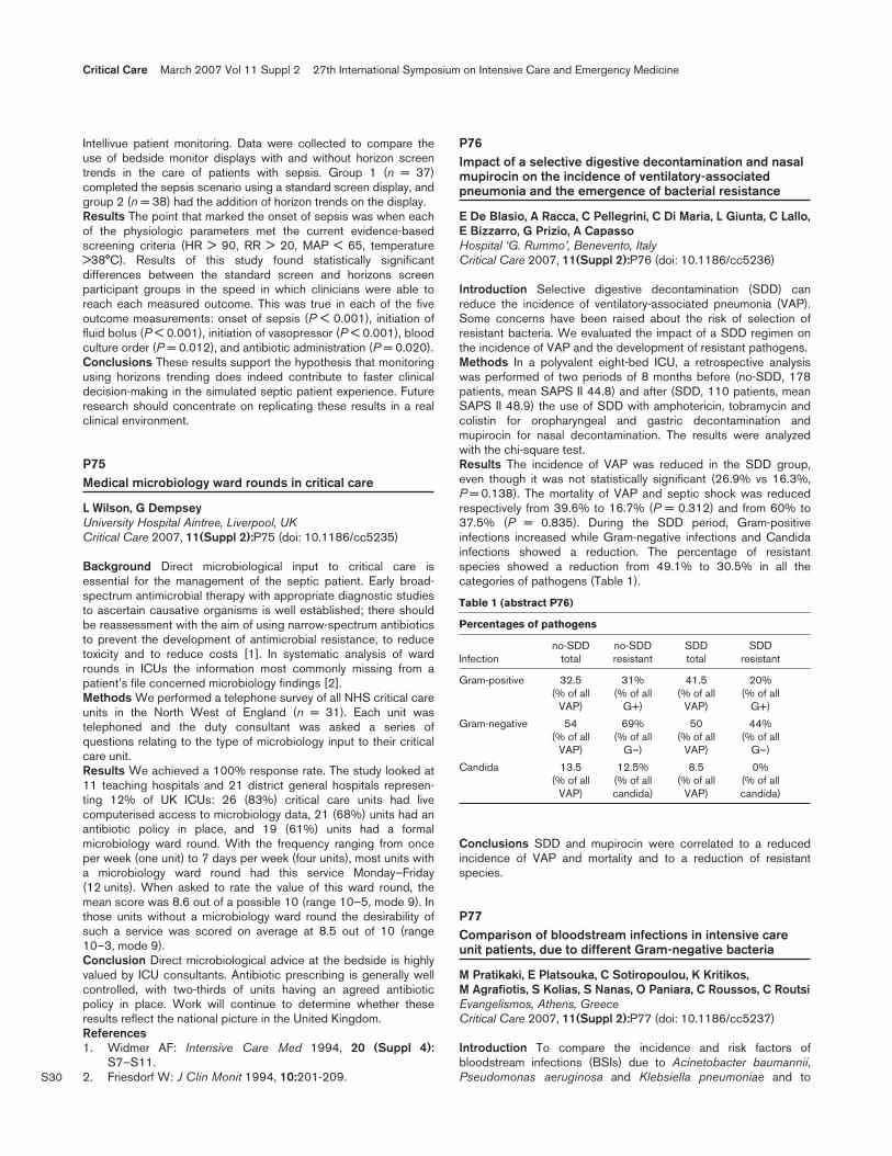
S30
Intellivue patient monitoring. Data were collected to compare theuse of bedside monitor displays with and without horizon screentrends in the care of patients with sepsis. Group 1 (n = 37)completed the sepsis scenario using a standard screen display, andgroup 2 (n = 38) had the addition of horizon trends on the display.Results The point that marked the onset of sepsis was when eachof the physiologic parameters met the current evidence-basedscreening criteria (HR > 90, RR > 20, MAP < 65, temperature>38°C). Results of this study found statistically significantdifferences between the standard screen and horizons screenparticipant groups in the speed in which clinicians were able toreach each measured outcome. This was true in each of the fiveoutcome measurements: onset of sepsis (P < 0.001), initiation offluid bolus (P < 0.001), initiation of vasopressor (P < 0.001), bloodculture order (P = 0.012), and antibiotic administration (P = 0.020).Conclusions These results support the hypothesis that monitoringusing horizons trending does indeed contribute to faster clinicaldecision-making in the simulated septic patient experience. Futureresearch should concentrate on replicating these results in a realclinical environment.
P75Medical microbiology ward rounds in critical care
L Wilson, G DempseyUniversity Hospital Aintree, Liverpool, UKCritical Care 2007, 11(Suppl 2):P75 (doi: 10.1186/cc5235)
Background Direct microbiological input to critical care isessential for the management of the septic patient. Early broad-spectrum antimicrobial therapy with appropriate diagnostic studiesto ascertain causative organisms is well established; there shouldbe reassessment with the aim of using narrow-spectrum antibioticsto prevent the development of antimicrobial resistance, to reducetoxicity and to reduce costs [1]. In systematic analysis of wardrounds in ICUs the information most commonly missing from apatient’s file concerned microbiology findings [2].Methods We performed a telephone survey of all NHS critical careunits in the North West of England (n = 31). Each unit wastelephoned and the duty consultant was asked a series ofquestions relating to the type of microbiology input to their criticalcare unit.Results We achieved a 100% response rate. The study looked at11 teaching hospitals and 21 district general hospitals represen-ting 12% of UK ICUs: 26 (83%) critical care units had livecomputerised access to microbiology data, 21 (68%) units had anantibiotic policy in place, and 19 (61%) units had a formalmicrobiology ward round. With the frequency ranging from onceper week (one unit) to 7 days per week (four units), most units witha microbiology ward round had this service Monday–Friday(12 units). When asked to rate the value of this ward round, themean score was 8.6 out of a possible 10 (range 10–5, mode 9). Inthose units without a microbiology ward round the desirability ofsuch a service was scored on average at 8.5 out of 10 (range10–3, mode 9).Conclusion Direct microbiological advice at the bedside is highlyvalued by ICU consultants. Antibiotic prescribing is generally wellcontrolled, with two-thirds of units having an agreed antibioticpolicy in place. Work will continue to determine whether theseresults reflect the national picture in the United Kingdom.References1. Widmer AF: Intensive Care Med 1994, 20 (Suppl 4):
S7–S11.2. Friesdorf W: J Clin Monit 1994, 10:201-209.
P76Impact of a selective digestive decontamination and nasalmupirocin on the incidence of ventilatory-associatedpneumonia and the emergence of bacterial resistance
E De Blasio, A Racca, C Pellegrini, C Di Maria, L Giunta, C Lallo,E Bizzarro, G Prizio, A CapassoHospital ‘G. Rummo’, Benevento, ItalyCritical Care 2007, 11(Suppl 2):P76 (doi: 10.1186/cc5236)
Introduction Selective digestive decontamination (SDD) canreduce the incidence of ventilatory-associated pneumonia (VAP).Some concerns have been raised about the risk of selection ofresistant bacteria. We evaluated the impact of a SDD regimen onthe incidence of VAP and the development of resistant pathogens.Methods In a polyvalent eight-bed ICU, a retrospective analysiswas performed of two periods of 8 months before (no-SDD, 178patients, mean SAPS II 44.8) and after (SDD, 110 patients, meanSAPS II 48.9) the use of SDD with amphotericin, tobramycin andcolistin for oropharyngeal and gastric decontamination andmupirocin for nasal decontamination. The results were analyzedwith the chi-square test.Results The incidence of VAP was reduced in the SDD group,even though it was not statistically significant (26.9% vs 16.3%,P = 0.138). The mortality of VAP and septic shock was reducedrespectively from 39.6% to 16.7% (P = 0.312) and from 60% to37.5% (P = 0.835). During the SDD period, Gram-positiveinfections increased while Gram-negative infections and Candidainfections showed a reduction. The percentage of resistantspecies showed a reduction from 49.1% to 30.5% in all thecategories of pathogens (Table 1).
Table 1 (abstract P76)
Percentages of pathogens
no-SDD no-SDD SDD SDDInfection total resistant total resistant
Gram-positive 32.5 31% 41.5 20%(% of all (% of all (% of all (% of all
VAP) G+) VAP) G+)
Gram-negative 54 69% 50 44%(% of all (% of all (% of all (% of all
VAP) G–) VAP) G–)
Candida 13.5 12.5% 8.5 0%(% of all (% of all (% of all (% of all
VAP) candida) VAP) candida)
Conclusions SDD and mupirocin were correlated to a reducedincidence of VAP and mortality and to a reduction of resistantspecies.
P77Comparison of bloodstream infections in intensive careunit patients, due to different Gram-negative bacteria
M Pratikaki, E Platsouka, C Sotiropoulou, K Kritikos, M Agrafiotis, S Kolias, S Nanas, O Paniara, C Roussos, C RoutsiEvangelismos, Athens, GreeceCritical Care 2007, 11(Suppl 2):P77 (doi: 10.1186/cc5237)
Introduction To compare the incidence and risk factors ofbloodstream infections (BSIs) due to Acinetobacter baumannii,Pseudomonas aeruginosa and Klebsiella pneumoniae and to
Critical Care March 2007 Vol 11 Suppl 2 27th International Symposium on Intensive Care and Emergency Medicine
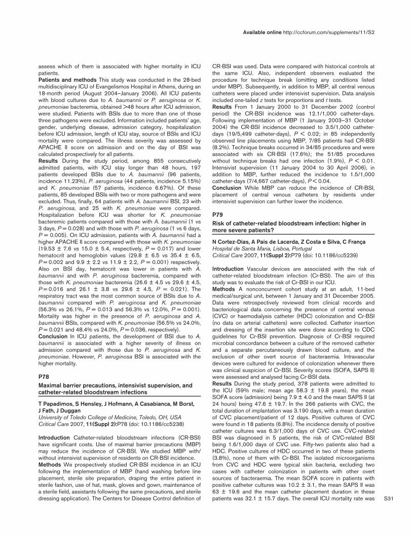
S31
assess which of them is associated with higher mortality in ICUpatients.Patients and methods This study was conducted in the 28-bedmultidisciplinary ICU of Evangelismos Hospital in Athens, during an18-month period (August 2004–January 2006). All ICU patientswith blood cultures due to A. baumannii or P. aeruginosa or K.pneumoniae bacteremia, obtained >48 hours after ICU admission,were studied. Patients with BSIs due to more than one of thosethree pathogens were excluded. Information included patients’ age,gender, underlying disease, admission category, hospitalizationbefore ICU admission, length of ICU stay, source of BSIs and ICUmortality were compared. The illness severity was assessed byAPACHE II score on admission and on the day of BSI wascalculated prospectively for all patients.Results During the study period, among 855 consecutivelyadmitted patients, with ICU stay longer than 48 hours, 197patients developed BSIs due to A. baumannii (96 patients,incidence 11.23%), P. aeruginosa (44 patients, incidence 5.15%)and K. pneumoniae (57 patients, incidence 6.67%). Of thesepatients, 85 developed BSIs with two or more pathogens and wereexcluded. Thus, finally, 64 patients with A. baumannii BSI, 23 withP. aeruginosa, and 25 with K. pneumoniae were compared.Hospitalization before ICU was shorter for K. pneumoniaebacteremic patients compared with those with A. baumannii (1 vs3 days, P = 0.028) and with those with P. aeruginosa (1 vs 6 days,P = 0.005). On ICU admission, patients with A. baumannii had ahigher APACHE II score compared with those with K. pneumoniae(19.53 ± 7.6 vs 15.0 ± 5.4, respectively, P = 0.017) and lowerhematocrit and hemoglobin values (29.8 ± 6.5 vs 35.4 ± 6.5,P = 0.002 and 9.9 ± 2.2 vs 11.9 ± 2.2, P = 0.001) respectively.Also on BSI day, hematocrit was lower in patients with A.baumannii and with P. aeruginosa bacteremia, compared withthose with K. pneumoniae bacteremia (26.6 ± 4.5 vs 29.6 ± 4.5,P = 0.016 and 26.1 ± 3.8 vs 29.6 ± 4.5, P = 0.021). Therespiratory tract was the most common source of BSIs due to A.baumannii compared with P. aeruginosa and K. pneumoniae(56.3% vs 26.1%, P = 0.013 and 56.3% vs 12.0%, P = 0.001).Mortality was higher in the presence of P. aeruginosa and A.baumannii BSIs, compared with K. pneumoniae (56.5% vs 24.0%,P = 0.021 and 48.4% vs 24.0%, P = 0.036, respectively).Conclusion In ICU patients, the development of BSI due to A.baumannii is associated with a higher severity of illness onadmission compared with those due to P. aeruginosa and K.pneumoniae. However, P. aeruginosa BSI is associated with thehigher mortality.
P78Maximal barrier precautions, intensivist supervision, andcatheter-related bloodstream infections
T Papadimos, S Hensley, J Hofmann, A Casabianca, M Borst, J Fath, J DugganUniversity of Toledo College of Medicine, Toledo, OH, USACritical Care 2007, 11(Suppl 2):P78 (doi: 10.1186/cc5238)
Introduction Catheter-related bloodstream infections (CR-BSI)have significant costs. Use of maximal barrier precautions (MBP)may reduce the incidence of CR-BSI. We studied MBP with/without intensivist supervision of residents on CR-BSI incidence.Methods We prospectively studied CR-BSI incidence in an ICUfollowing the implementation of MBP (hand washing before lineplacement, sterile site preparation, draping the entire patient insterile fashion, use of hat, mask, gloves and gown, maintenance ofa sterile field, assistants following the same precautions, and steriledressing application). The Centers for Disease Control definition of
CR-BSI was used. Data were compared with historical controls atthe same ICU. Also, independent observers evaluated theprocedure for technique break (omitting any conditions listedunder MBP). Subsequently, in addition to MBP, all central venouscatheters were placed under intensivist supervision. Data analysisincluded one-tailed z tests for proportions and t tests.Results From 1 January 2000 to 31 December 2002 (controlperiod) the CR-BSI incidence was 12.1/1,000 catheter-days.Following implementation of MBP (1 January 2003–31 October2004) the CR-BSI incidence decreased to 3.5/1,000 catheter-days (19/5,499 catheter-days), P < 0.02; in 85 independentlyobserved line placements using MBP, 7/85 patients had CR-BSI(8.2%). Technique breaks occurred in 34/85 procedures and wereassociated with six CR-BSI (17.6%); the 51/85 procedureswithout technique breaks had one infection (1.9%), P < 0.01.Intensivist supervision (11 January 2004 to 30 April 2006), inaddition to MBP, further reduced the incidence to 1.5/1,000catheter-days (7/4,667 catheter-days), P < 0.04.Conclusion While MBP can reduce the incidence of CR-BSI,placement of central venous catheters by residents underintensivist supervision can further lower the incidence.
P79Risk of catheter-related bloodstream infection: higher inmore severe patients?
N Cortez-Dias, A Pais de Lacerda, Z Costa e Silva, C FrançaHospital de Santa Maria, Lisboa, PortugalCritical Care 2007, 11(Suppl 2):P79 (doi: 10.1186/cc5239)
Introduction Vascular devices are associated with the risk ofcatheter-related bloodstream infection (Cr-BSI). The aim of thisstudy was to evaluate the risk of Cr-BSI in our ICU.Methods A nonconcurrent cohort study at an adult, 11-bedmedical/surgical unit, between 1 January and 31 December 2005.Data were retrospectively reviewed from clinical records andbacteriological data concerning the presence of central venous(CVC) or haemodialysis catheter (HDC) colonization and Cr-BSI(no data on arterial catheters) were collected. Catheter insertionand dressing of the insertion site were done according to CDCguidelines for Cr-BSI prevention. Diagnosis of Cr-BSI requiredmicrobial concordance between a culture of the removed catheterand a separate percutaneously drawn blood culture, and theexclusion of other overt source of bacteraemia. Intravasculardevices were cultured for evidence of colonization whenever therewas clinical suspicion of Cr-BSI. Severity scores (SOFA, SAPS II)were assessed and analysed facing Cr-BSI data.Results During the study period, 378 patients were admitted tothe ICU (59% male; mean age 58.3 ± 19.8 years), the meanSOFA score (admission) being 7.9 ± 4.0 and the mean SAPS II (at24 hours) being 47.6 ± 19.7. In the 266 patients with CVC, thetotal duration of implantation was 3.190 days, with a mean durationof CVC placement/patient of 12 days. Positive cultures of CVCwere found in 18 patients (6.8%). The incidence density of positivecatheter cultures was 6.3/1,000 days of CVC use. CVC-relatedBSI was diagnosed in 5 patients, the risk of CVC-related BSIbeing 1.6/1,000 days of CVC use. Fifty-two patients also had aHDC. Positive cultures of HDC occurred in two of these patients(3.8%), none of them with Cr-BSI. The isolated microorganismsfrom CVC and HDC were typical skin bacteria, excluding twocases with catheter colonization in patients with other overtsources of bacteraemia. The mean SOFA score in patients withpositive catheter cultures was 10.2 ± 3.1, the mean SAPS II was63 ± 19.6 and the mean catheter placement duration in thesepatients was 32.1 ± 15.7 days. The overall ICU mortality rate was
Available online http://ccforum.com/supplements/11/S2
S32
20.1%, being 40% in the subgroup of patients in whom Cr-BSIwas diagnosed.Conclusions Preventing Cr-BSI is important, but special care isparticularly relevant in patients with higher SAPS II scores and alonger duration of catheter placement. More studies are needed toconfirm this possible higher risk of Cr-BSI in this more severepatient subgroup.
P80Tunnelled central venous catheter-related infection incardiothoracic critical care
J Mitchell, P Shetty, F Cox, P VuddamalayRoyal Brompton & Harefield NHS Trust, Harefield, UKCritical Care 2007, 11(Suppl 2):P80 (doi: 10.1186/cc5240)
Introduction Tunnelled central venous cannulae (CVC) are used incardiothoracic (CT) critical care for long-term inotrope, antibioticand renal replacement therapy (RRT). The incidence of blood-stream-related infection (BSI) related to all types of CVC isbetween 2.9 and 11.3 per 1,000 catheter-days [1]. In CT orcardiology practice the incidence for all CVC-related infection is2.9–4.5 per 1,000 catheter-days. The incidence of BSI is reducedusing tunnelled CVC, although there are little published data onthe incidence of BSI in tunnelled CVC in CT critical care. CVC-related infection has been recognised as a priority in the UKinitiative ‘Saving Lives’ [2]. We reviewed tunnelled CVC-relatedinfection in a tertiary UK CT centre with a significant transplantpopulation.Methods A retrospective analysis from November 2001 to 2006of culture and sensitivity results of tunnelled CVC tips (BardGroshong® cuffed catheter and HemoGlide®) and blood culturesfrom the same patients.Results Ninety-three CT critical care patients received a tunnelledsubclavian CVC. The indications were inotropes (n = 40 (43%)),antibiotic administration (n = 27 (29%)), RRT (n = 14 (15.1%))and unknown (n = 10 (10.8%)). The mean duration of the catheterremaining in situ was 36 days (SD 44.0, range 1–164). Cultureresults are presented in Table 1. Twelve patients had anestablished CVC-related BSI. The mean infection rate/1,000catheter-days was 3.6.
Table 1 (abstract P80)
Positive CVC Positive Positive tip culture blood culture from both
Positive culture results (%) 36.6 18.3 12.9
Mean infection rate/ 10.2 5.1 3.61,000 catheter-days
Conclusion The incidence of tunnelled CVC colonisation andpositive blood cultures in this group of CT critical care patients isin line with previously published data for all types of CVC.Coagulase-negative staphylococcus was the predominant isolatein both this audit and previously published data [1].References1. O’Grady NP, Alexander M, Dellinger EP, et al: Guidelines for
the prevention of intravascular catheter-related infections.Centers for Disease Control and Prevention, Atlanta, USA.MMWR 2002, 51(RR-10):1-29.
2. Saving Lives: The Delivery Programme to Reduce HealthcareAssociated Infections (HCAI) including MRSA. London:Department of Health; 2005.
P81ANCCADI – Antibiotic Coated Catheter to DecreaseInfection: a pilot trial
P Kurtz, M Kalichsztein, G Nobre, G Almeida, J Kezen, F Braga,P Rosa, G Penna, L Drumond, R Vegni, M Freitas, M PintoCasa de Saude Sao Jose, Rio de Janeiro, BrazilCritical Care 2007, 11(Suppl 2):P81 (doi: 10.1186/cc5241)
Background Nosocomial catheter-related bloodstream infections(CR-BSI) have been associated with increased morbidity andpossibly increased mortality in critically ill patients. Central venouscatheters impregnated with rifampin and minocycline (RM) havebeen shown to decrease rates of colonization and CR-BSI whencompared with controls and with the chlorhexidine/silversulfadiazine catheter. However, recent randomized trials challengedthe clinical impact of such catheters, showing decreased rates incolonization but not in CR-BSI. We designed this pilot trial tocompare the rates of colonization and CR-BSI in RM catheters andcontrols in a Brazilian population of critically ill patients.Methods A prospective, nonrandomized, controlled clinical trialwas conducted in one medico-surgical 19-bed ICU. Adult patientsneeding a double or triple central venous catheter weresequentially assigned in permuted blocks of five to undergoinsertion of a control or RM-impregnated catheter. After removal, alltips were cultured by the roll-plate method in association with oneor two peripheral blood cultures. Rates of colonization and CR-BSIwere recorded and compared.Results Of 120 catheters inserted, 100 could be evaluated forcolonization and CR-BSI. Forty-nine in the uncoated group and 51in the coated group. Clinical characteristics of patients and risk forinfection were similar in the two groups, use of propofol was morefrequent in the uncoated group and the presence of a vasculardevice, other than the study catheter, was more frequent in theantibiotic-coated group. Three RM-coated catheters (5.9%) werecolonized compared with nine (18.4%) control catheters (relativerisk, 0,28; 95% confidence interval, 0.07–1.096; P = 0.05). Threecases of CR-BSI (5.9%) occurred in patients who received RMcatheters compared with five in the control group (10.2%). Therewas no significant differences in the incidence of CR-BSI betweenRM-coated and uncoated catheters. Uncoated catheters weremore frequently colonized but this difference just failed to showstatistical significance. When the duration of catheter placementwere taken into consideration, Kaplan–Meier analysis showed nosignificant differences in the risk of colonization or CR-BSIbetween RM-coated and uncoated catheters. Rates of CR-BSIwere seven per 1,000 catheter-days in the RM-coated groupcompared with 11.4 per 1,000 catheter-days in the uncoatedgroup (P = 0.7). Gram-positive and Gram-negative organisms weresimilarly responsible for colonizing catheters in our study; therewas no difference in rates of colonization by Candida species.Conclusion In this pilot study, we showed a trend toward lowerrates of colonization in RM-coated catheters when compared withuncoated control catheters. The incidence and rates of CR-BSIwere similar in the two groups, probably because of a smallnumber of catheters studied. Development of a prospectiverandomized trial with a larger number of patients is underway toconfirm or refute these results.
Critical Care March 2007 Vol 11 Suppl 2 27th International Symposium on Intensive Care and Emergency Medicine
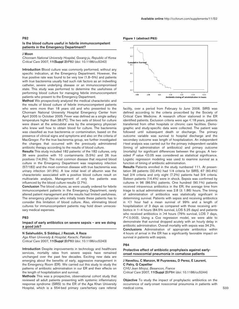
S33
P82Is the blood culture useful in febrile immunocompetentpatients in the Emergency Department?
J MoonChonnam National University Hospital, Gwang-ju, Republic of KoreaCritical Care 2007, 11(Suppl 2):P82 (doi: 10.1186/cc5242)
Introduction Blood culture was commonly performed, without anyspecific indication, at the Emergency Department. However, thetrue positive rate was found to be very low (1.8–5%) and patientswith true bacteremia usually had such risk factors as an indwellingcatheter, severe underlying disease or an immunocompromisedstate. This study was performed to determine the usefulness ofperforming blood culture for managing febrile immunocompetentpatients who present to the Emergency Department.Method We prospectively analyzed the medical characteristic andthe results of blood culture of febrile immunocompetent patientswho were more than 18 years old and who presented to theChonnam National University Hospital Emergency Center fromApril 2005 to October 2005. Fever was defined as a single axillarytemperature higher than 38.0°C. The two sets of blood for culturewere drawn at the antecubital area by the emergency physicianwho knew well how to obtain blood for culture. The bacteremiawas classified as true bacteremia or contamination, based on thepresence of clinical signs and symptoms and also on the criteria ofMacGregor. For the true bacteremia group, we further investigatedthe changes that occurred with the previously administeredantibiotic therapy according to the results of blood culture.Results This study included 182 patients: of the 182 cultures, only36 were positive with 10 contaminants (5.5%) and 26 truepositives (14.3%). The most common disease that required bloodculture in the Emergency Department was respiratory infection(57/182) and the most common disease with true bacteremia wasurinary infection (41.9%). A low initial level of albumin was thecharacteristic associated with a positive blood culture result onmultivariate analysis. Management of only five patients wasinfluenced by the blood culture results (2.7%).Conclusion The blood cultures, as were usually ordered for febrileimmunocompetent patients in the Emergency Department, rarelyaltered patient management and the results had limited usefulness.The emergency physician who initially treats these patients has toconsider this limitation of blood culture. Also, eliminating bloodcultures for immunocompetent patients may hold down unneces-sary medical expenses.
P83Impact of early antibiotics on severe sepsis – are we doinga good job?
N Salahuddin, S Siddiqui, J Razzak, A RazaAga Khan University & Hospital, Karachi, PakistanCritical Care 2007, 11(Suppl 2):P83 (doi: 10.1186/cc5243)
Introduction Despite improvements in technology and healthcareservices, mortality rates from severe sepsis have remainedunchanged over the past few decades. Exciting new data areemerging about the benefits of early, aggressive management inthe Emergency Room (ER). We carried out this study to study thepatterns of antibiotic administration in our ER and their effects onthe length of hospitalization and survival.Methods This was a prospective, observational cohort study thatreviewed all adult patients presenting with systemic inflammatoryresponse syndrome (SIRS) to the ER of the Aga Khan UniversityHospital, which is a 554-bed primary care/tertiary care referral
facility, over a period from February to June 2006. SIRS wasdefined according to the criteria proscribed by the Society ofCritical Care Medicine. A research officer stationed in the ERidentified patients. Exclusion criteria were age < 18 years, patientstransferred from other hospitals or chronic care facilities. Demo-graphic and study-specific data were collected. The patient wasfollowed until subsequent death or discharge. The primaryoutcome variable was survival to hospital discharge and thesecondary outcome was length of hospitalization. An independentt-test analysis was carried out for the primary independent variable(timing of administration of antibiotics) and primary outcome(mortality) for significant differences between the groups. A two-sided P value <0.05 was considered as statistical significance.Logistic regression modeling was used to examine survival as afunction of timing of antibiotic administration.Results Patients enrolled in the study numbered 111. At presen-tation 36 patients (32.4%) had 1/4 criteria for SIRS, 67 (60.4%)had 2/4 criteria and only eight (7.2%) patients had 3/4 criteria.Sixteen patients (14.4%) were in shock. Sepsis was confirmed bycultures in 96 (86.5%) patients. One hundred (90.1%) patientsreceived intravenous antibiotics in the ER; the average time fromtriage to actual administration was 2.8 (± 1.86) hours. The timingof administration of antibiotics was statistically significant indetermining survival. Patients with sepsis and receiving antibioticsin <1 hour had a mean survival of 99% and a length ofhospitalization of 3 days as compared with those receiving anti-biotics in 1–4 hours (84.5% survival, LOS 5.25 days) and patientswho received antibiotics in >4 hours (76% survival, LOS 7 days,P < 0.003). Using a Cox regression model, we were able todemonstrate that survival dropped acutely with an hourly delay inantibiotic administration. Overall mortality with sepsis was 34.2%.Conclusions Administration of appropriate antibiotics within4 hours of arrival in the ER has a significantly favorable impact onsurvival in patients with sepsis.
P84Protective effect of antibiotic prophylaxis against early-onset nosocomial pneumonia in comatose patients
J Navellou, C Manzon, M Puyraveau, D Perez, E Laurent, C Patry, G CapellierCHU Jean Minjoz, Besancon, FranceCritical Care 2007, 11(Suppl 2):P84 (doi: 10.1186/cc5244)
Objective To study the impact of prophylactic antibiotics on theoccurrence of early-onset nosocomial pneumonia in patients withmedical coma.
Available online http://ccforum.com/supplements/11/S2
Figure 1 (abstract P83)
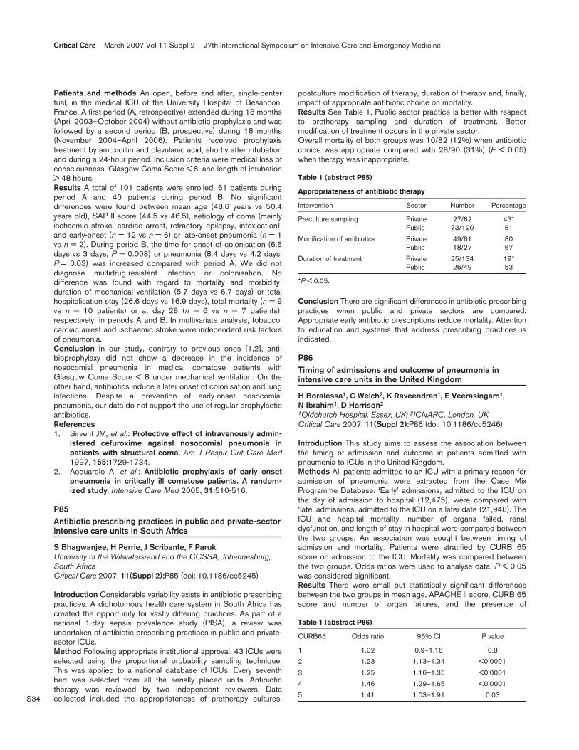
S34
Patients and methods An open, before and after, single-centertrial, in the medical ICU of the University Hospital of Besancon,France. A first period (A, retrospective) extended during 18 months(April 2003–October 2004) without antibiotic prophylaxis and wasfollowed by a second period (B, prospective) during 18 months(November 2004–April 2006). Patients received prophylaxistreatment by amoxicillin and clavulanic acid, shortly after intubationand during a 24-hour period. Inclusion criteria were medical loss ofconsciousness, Glasgow Coma Score < 8, and length of intubation> 48 hours.Results A total of 101 patients were enrolled, 61 patients duringperiod A and 40 patients during period B. No significantdifferences were found between mean age (48.6 years vs 50.4years old), SAP II score (44.5 vs 46.5), aetiology of coma (mainlyischaemic stroke, cardiac arrest, refractory epilepsy, intoxication),and early-onset (n = 12 vs n = 6) or late-onset pneumonia (n = 1vs n = 2). During period B, the time for onset of colonisation (6.6days vs 3 days, P = 0.008) or pneumonia (8.4 days vs 4.2 days,P = 0.03) was increased compared with period A. We did notdiagnose multidrug-resistant infection or colonisation. Nodifference was found with regard to mortality and morbidity:duration of mechanical ventilation (5.7 days vs 6.7 days) or totalhospitalisation stay (26.6 days vs 16.9 days), total mortality (n = 9vs n = 10 patients) or at day 28 (n = 6 vs n = 7 patients),respectively, in periods A and B. In multivariate analysis, tobacco,cardiac arrest and ischaemic stroke were independent risk factorsof pneumonia.Conclusion In our study, contrary to previous ones [1,2], anti-bioprophylaxy did not show a decrease in the incidence ofnosocomial pneumonia in medical comatose patients withGlasgow Coma Score < 8 under mechanical ventilation. On theother hand, antibiotics induce a later onset of colonisation and lunginfections. Despite a prevention of early-onset nosocomialpneumonia, our data do not support the use of regular prophylacticantibiotics.References1. Sirvent JM, et al.: Protective effect of intravenously admin-
istered cefuroxime against nosocomial pneumonia inpatients with structural coma. Am J Respir Crit Care Med1997, 155:1729-1734.
2. Acquarolo A, et al.: Antibiotic prophylaxis of early onsetpneumonia in critically ill comatose patients. A random-ized study. Intensive Care Med 2005, 31:510-516.
P85Antibiotic prescribing practices in public and private-sectorintensive care units in South Africa
S Bhagwanjee, H Perrie, J Scribante, F ParukUniversity of the Witwatersrand and the CCSSA, Johannesburg,South AfricaCritical Care 2007, 11(Suppl 2):P85 (doi: 10.1186/cc5245)
Introduction Considerable variability exists in antibiotic prescribingpractices. A dichotomous health care system in South Africa hascreated the opportunity for vastly differing practices. As part of anational 1-day sepsis prevalence study (PISA), a review wasundertaken of antibiotic prescribing practices in public and private-sector ICUs.Method Following appropriate institutional approval, 43 ICUs wereselected using the proportional probability sampling technique.This was applied to a national database of ICUs. Every seventhbed was selected from all the serially placed units. Antibiotictherapy was reviewed by two independent reviewers. Datacollected included the appropriateness of pretherapy cultures,
postculture modification of therapy, duration of therapy and, finally,impact of appropriate antibiotic choice on mortality.Results See Table 1. Public-sector practice is better with respectto pretherapy sampling and duration of treatment. Bettermodification of treatment occurs in the private sector.Overall mortality of both groups was 10/82 (12%) when antibioticchoice was appropriate compared with 28/90 (31%) (P < 0.05)when therapy was inappropriate.
Table 1 (abstract P85)
Appropriateness of antibiotic therapy
Intervention Sector Number Percentage
Preculture sampling Private 27/62 43*Public 73/120 61
Modification of antibiotics Private 49/61 80Public 18/27 67
Duration of treatment Private 25/134 19*Public 26/49 53
*P < 0.05.
Conclusion There are significant differences in antibiotic prescribingpractices when public and private sectors are compared.Appropriate early antibiotic prescriptions reduce mortality. Attentionto education and systems that address prescribing practices isindicated.
P86Timing of admissions and outcome of pneumonia inintensive care units in the United Kingdom
H Boralessa1, C Welch2, K Raveendran1, E Veerasingam1, N Ibrahim1, D Harrison2
1Oldchurch Hospital, Essex, UK; 2ICNARC, London, UKCritical Care 2007, 11(Suppl 2):P86 (doi: 10.1186/cc5246)
Introduction This study aims to assess the association betweenthe timing of admission and outcome in patients admitted withpneumonia to ICUs in the United Kingdom.Methods All patients admitted to an ICU with a primary reason foradmission of pneumonia were extracted from the Case MixProgramme Database. ‘Early’ admissions, admitted to the ICU onthe day of admission to hospital (12,475), were compared with‘late’ admissions, admitted to the ICU on a later date (21,948). TheICU and hospital mortality, number of organs failed, renaldysfunction, and length of stay in hospital were compared betweenthe two groups. An association was sought between timing ofadmission and mortality. Patients were stratified by CURB 65score on admission to the ICU. Mortality was compared betweenthe two groups. Odds ratios were used to analyse data. P < 0.05was considered significant.Results There were small but statistically significant differencesbetween the two groups in mean age, APACHE II score, CURB 65score and number of organ failures, and the presence of
Critical Care March 2007 Vol 11 Suppl 2 27th International Symposium on Intensive Care and Emergency Medicine
Table 1 (abstract P86)
CURB65 Odds ratio 95% CI P value
1 1.02 0.9–1.16 0.8
2 1.23 1.13–1.34 <0.0001
3 1.25 1.16–1.35 <0.0001
4 1.46 1.29–1.65 <0.0001
5 1.41 1.03–1.91 0.03
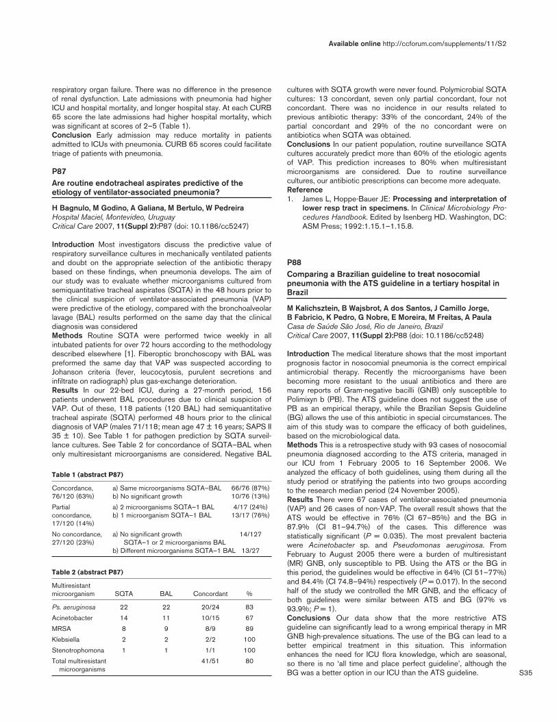
S35
respiratory organ failure. There was no difference in the presenceof renal dysfunction. Late admissions with pneumonia had higherICU and hospital mortality, and longer hospital stay. At each CURB65 score the late admissions had higher hospital mortality, whichwas significant at scores of 2–5 (Table 1).Conclusion Early admission may reduce mortality in patientsadmitted to ICUs with pneumonia. CURB 65 scores could facilitatetriage of patients with pneumonia.
P87Are routine endotracheal aspirates predictive of theetiology of ventilator-associated pneumonia?
H Bagnulo, M Godino, A Galiana, M Bertulo, W PedreiraHospital Maciel, Montevideo, UruguayCritical Care 2007, 11(Suppl 2):P87 (doi: 10.1186/cc5247)
Introduction Most investigators discuss the predictive value ofrespiratory surveillance cultures in mechanically ventilated patientsand doubt on the appropriate selection of the antibiotic therapybased on these findings, when pneumonia develops. The aim ofour study was to evaluate whether microorganisms cultured fromsemiquantitative tracheal aspirates (SQTA) in the 48 hours prior tothe clinical suspicion of ventilator-associated pneumonia (VAP)were predictive of the etiology, compared with the bronchoalveolarlavage (BAL) results performed on the same day that the clinicaldiagnosis was consideredMethods Routine SQTA were performed twice weekly in allintubated patients for over 72 hours according to the methodologydescribed elsewhere [1]. Fiberoptic bronchoscopy with BAL waspreformed the same day that VAP was suspected according toJohanson criteria (fever, leucocytosis, purulent secretions andinfiltrate on radiograph) plus gas-exchange deterioration.Results In our 22-bed ICU, during a 27-month period, 156patients underwent BAL procedures due to clinical suspicion ofVAP. Out of these, 118 patients (120 BAL) had semiquantitativetracheal aspirate (SQTA) performed 48 hours prior to the clinicaldiagnosis of VAP (males 71/118; mean age 47 ± 16 years; SAPS II35 ± 10). See Table 1 for pathogen prediction by SQTA surveil-lance cultures. See Table 2 for concordance of SQTA–BAL whenonly multiresistant microorganisms are considered. Negative BAL
cultures with SQTA growth were never found. Polymicrobial SQTAcultures: 13 concordant, seven only partial concordant, four notconcordant. There was no incidence in our results related toprevious antibiotic therapy: 33% of the concordant, 24% of thepartial concordant and 29% of the no concordant were onantibiotics when SQTA was obtained.Conclusions In our patient population, routine surveillance SQTAcultures accurately predict more than 60% of the etiologic agentsof VAP. This prediction increases to 80% when multiresistantmicroorganisms are considered. Due to routine surveillancecultures, our antibiotic prescriptions can become more adequate.Reference1. James L, Hoppe-Bauer JE: Processing and interpretation of
lower resp tract in specimens. In Clinical Microbiology Pro-cedures Handbook. Edited by Isenberg HD. Washington, DC:ASM Press; 1992:1.15.1–1.15.8.
P88Comparing a Brazilian guideline to treat nosocomialpneumonia with the ATS guideline in a tertiary hospital inBrazil
M Kalichsztein, B Wajsbrot, A dos Santos, J Camillo Jorge, B Fabricio, K Pedro, G Nobre, E Moreira, M Freitas, A PaulaCasa de Saúde São José, Rio de Janeiro, BrazilCritical Care 2007, 11(Suppl 2):P88 (doi: 10.1186/cc5248)
Introduction The medical literature shows that the most importantprognosis factor in nosocomial pneumonia is the correct empiricalantimicrobial therapy. Recently the microorganisms have beenbecoming more resistant to the usual antibiotics and there aremany reports of Gram-negative bacilli (GNB) only susceptible toPolimixyn b (PB). The ATS guideline does not suggest the use ofPB as an empirical therapy, while the Brazilian Sepsis Guideline(BG) allows the use of this antibiotic in special circumstances. Theaim of this study was to compare the efficacy of both guidelines,based on the microbiological data.Methods This is a retrospective study with 93 cases of nosocomialpneumonia diagnosed according to the ATS criteria, managed inour ICU from 1 February 2005 to 16 September 2006. Weanalyzed the efficacy of both guidelines, using them during all thestudy period or stratifying the patients into two groups accordingto the research median period (24 November 2005).Results There were 67 cases of ventilator-associated pneumonia(VAP) and 26 cases of non-VAP. The overall result shows that theATS would be effective in 76% (CI 67–85%) and the BG in87.9% (CI 81–94.7%) of the cases. This difference wasstatistically significant (P = 0.035). The most prevalent bacteriawere Acinetobacter sp. and Pseudomonas aeruginosa. FromFebruary to August 2005 there were a burden of multiresistant(MR) GNB, only susceptible to PB. Using the ATS or the BG inthis period, the guidelines would be effective in 64% (CI 51–77%)and 84.4% (CI 74.8–94%) respectively (P = 0.017). In the secondhalf of the study we controlled the MR GNB, and the efficacy ofboth guidelines were similar between ATS and BG (97% vs93.9%; P = 1).Conclusions Our data show that the more restrictive ATSguideline can significantly lead to a wrong empirical therapy in MRGNB high-prevalence situations. The use of the BG can lead to abetter empirical treatment in this situation. This informationenhances the need for ICU flora knowledge, which are seasonal,so there is no ‘all time and place perfect guideline’, although theBG was a better option in our ICU than the ATS guideline.
Available online http://ccforum.com/supplements/11/S2
Table 1 (abstract P87)
Concordance, a) Same microorganisms SQTA–BAL 66/76 (87%)76/120 (63%) b) No significant growth 10/76 (13%)
Partial a) 2 microorganisms SQTA–1 BAL 4/17 (24%)concordance, b) 1 microorganism SQTA–1 BAL 13/17 (76%)17/120 (14%)
No concordance, a) No significant growth 14/12727/120 (23%) SQTA–1 or 2 microorganisms BAL
b) Different microorganisms SQTA–1 BAL 13/27
Table 2 (abstract P87)
Multiresistant microorganism SQTA BAL Concordant %
Ps. aeruginosa 22 22 20/24 83
Acinetobacter 14 11 10/15 67
MRSA 8 9 8/9 89
Klebsiella 2 2 2/2 100
Stenotrophomona 1 1 1/1 100
Total multiresistant 41/51 80microorganisms
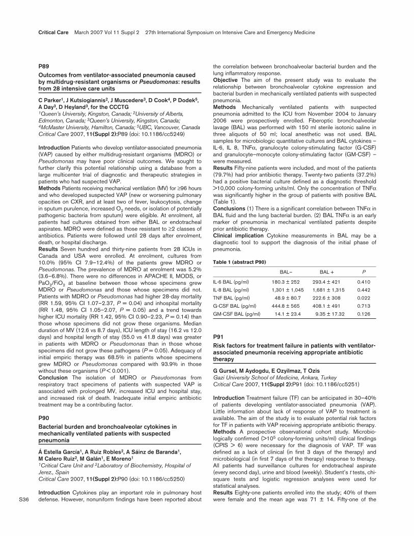
S36
P89Outcomes from ventilator-associated pneumonia causedby multidrug-resistant organisms or Pseudomonas: resultsfrom 28 intensive care units
C Parker1, J Kutsiogiannis2, J Muscedere3, D Cook4, P Dodek5,A Day3, D Heyland3, for the CCCTG1Queen’s University, Kingston, Canada; 2University of Alberta,Edmonton, Canada; 3Queen’s University, Kingston, Canada;4McMaster University, Hamilton, Canada; 5UBC, Vancouver, CanadaCritical Care 2007, 11(Suppl 2):P89 (doi: 10.1186/cc5249)
Introduction Patients who develop ventilator-associated pneumonia(VAP) caused by either multidrug-resistant organisms (MDRO) orPseudomonas may have poor clinical outcomes. We sought tofurther clarify this potential relationship using a database from alarge multicenter trial of diagnostic and therapeutic strategies inpatients who had suspected VAP.Methods Patients receiving mechanical ventilation (MV) for ≥96 hoursand who developed suspected VAP (new or worsening pulmonaryopacities on CXR, and at least two of fever, leukocytosis, changein sputum purulence, increased O2 needs, or isolation of potentiallypathogenic bacteria from sputum) were eligible. At enrolment, allpatients had cultures obtained from either BAL or endotrachealaspirates. MDRO were defined as those resistant to ≥2 classes ofantibiotics. Patients were followed until 28 days after enrolment,death, or hospital discharge.Results Seven hundred and thirty-nine patients from 28 ICUs inCanada and USA were enrolled. At enrolment, cultures from10.0% (95% CI 7.9–12.4%) of the patients grew MDRO orPseudomonas. The prevalence of MDRO at enrolment was 5.2%(3.6–6.8%). There were no differences in APACHE II, MODS, orPaO2/FiO2 at baseline between those whose specimens grewMDRO or Pseudomonas and those whose specimens did not.Patients with MDRO or Pseudomonas had higher 28-day mortality(RR 1.59, 95% CI 1.07–2.37, P = 0.04) and inhospital mortality(RR 1.48, 95% CI 1.05–2.07, P = 0.05) and a trend towardshigher ICU mortality (RR 1.42, 95% CI 0.90–2.23, P = 0.14) thanthose whose specimens did not grow these organisms. Medianduration of MV (12.6 vs 8.7 days), ICU length of stay (16.2 vs 12.0days) and hospital length of stay (55.0 vs 41.8 days) was greaterin patients with MDRO or Pseudomonas than in those whosespecimens did not grow these pathogens (P = 0.05). Adequacy ofinitial empiric therapy was 68.5% in patients whose specimensgrew MDRO or Pseudomonas compared with 93.9% in thosewithout these organisms (P < 0.001).Conclusion The isolation of MDRO or Pseudomonas fromrespiratory tract specimens of patients with suspected VAP isassociated with prolonged MV, increased ICU and hospital stay,and increased risk of death. Inadequate initial empiric antibiotictreatment may be a contributing factor.
P90Bacterial burden and bronchoalveolar cytokines inmechanically ventilated patients with suspectedpneumonia
Á Estella García1, A Ruiz Robles2, A Sáinz de Baranda1, M Calero Ruiz2, M Galán1, E Moreno1
1Critical Care Unit and 2Laboratory of Biochemistry, Hospital ofJerez., SpainCritical Care 2007, 11(Suppl 2):P90 (doi: 10.1186/cc5250)
Introduction Cytokines play an important role in pulmonary hostdefense. However, nonuniform findings have been reported about
the correlation between bronchoalveolar bacterial burden and thelung inflammatory response.Objective The aim of the present study was to evaluate therelationship between bronchoalveolar cytokine expression andbacterial burden in mechanically ventilated patients with suspectedpneumonia.Methods Mechanically ventilated patients with suspectedpneumonia admitted to the ICU from November 2004 to January2006 were prospectively enrolled. Fiberoptic bronchoalveolarlavage (BAL) was performed with 150 ml sterile isotonic saline inthree aliquots of 50 ml; local anesthetic was not used. BALsamples for microbiologic quantitative cultures and BAL cytokines –IL-6, IL 8, TNFα, granulocyte colony-stimulating factor (G-CSF)and granulocyte–monocyte colony-stimulating factor (GM-CSF) –were measured.Results Fifty-nine patients were included, and most of the patients(79.7%) had prior antibiotic therapy. Twenty-two patients (37.2%)had a positive bacterial culture defined as a diagnostic threshold>10,000 colony-forming units/ml. Only the concentration of TNFαwas significantly higher in the group of patients with positive BAL(Table 1).Conclusions (1) There is a significant correlation between TNFα inBAL fluid and the lung bacterial burden. (2) BAL TNFα is an earlymarker of pneumonia in mechanical ventilated patients despiteprior antibiotic therapy.Clinical implication Cytokine measurements in BAL may be adiagnostic tool to support the diagnosis of the initial phase ofpneumonia.
Table 1 (abstract P90)
BAL– BAL + P
IL-6 BAL (pg/ml) 180.3 ± 252 293.4 ± 421 0.410
IL-8 BAL (pg/ml) 1,301 ± 1,045 1,681 ± 1,315 0.442
TNF BAL (pg/ml) 48.9 ± 80.7 222.6 ± 308 0.022
G-CSF BAL (pg/ml) 444.8 ± 565 408.1 ± 491 0.713
GM-CSF BAL (pg/ml) 14.1 ± 23.4 9.35 ± 17.32 0.126
P91Risk factors for treatment failure in patients with ventilator-associated pneumonia receiving appropriate antibiotictherapy
G Gursel, M Aydogdu, E Ozyilmaz, T OzisGazi University School of Medicine, Ankara, TurkeyCritical Care 2007, 11(Suppl 2):P91 (doi: 10.1186/cc5251)
Introduction Treatment failure (TF) can be anticipated in 30–40%of patients developing ventilator-associated pneumonia (VAP).Little information about lack of response of VAP to treatment isavailable. The aim of the study is to evaluate potential risk factorsfor TF in patients with VAP receiving appropriate antibiotic therapy.Methods A prospective observational cohort study. Microbio-logically confirmed (>105 colony-forming units/ml) clinical findings(CPIS > 6) were necessary for the diagnosis of VAP. TF wasdefined as a lack of clinical (in first 3 days of the therapy) andmicrobiological (in first 7 days of the therapy) response to therapy.All patients had surveillance cultures for endotracheal aspirate(every second day), urine and blood (weekly). Student’s t tests, chi-square tests and logistic regression analyses were used forstatistical analyses.Results Eighty-one patients enrolled into the study; 40% of themwere female and the mean age was 71 ± 14. Fifty-one of the
Critical Care March 2007 Vol 11 Suppl 2 27th International Symposium on Intensive Care and Emergency Medicine
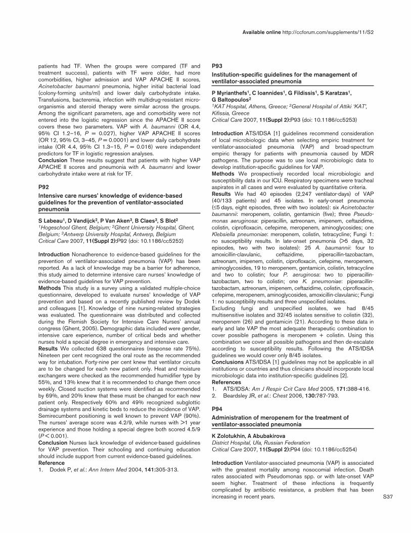
S37
patients had TF. When the groups were compared (TF andtreatment success), patients with TF were older, had morecomorbidities, higher admission and VAP APACHE II scores,Acinetobacter baumanni pneumonia, higher initial bacterial load(colony-forming units/ml) and lower daily carbohydrate intake.Transfusions, bacteremia, infection with multidrug-resistant micro-organismis and steroid therapy were similar across the groups.Among the significant parameters, age and comorbidity were notentered into the logistic regression since the APACHE II scorecovers these two parameters. VAP with A. baumanni (OR 4.4,95% CI 1.2–16, P = 0.027), higher VAP APACHE II scores(OR 12, 95% CI, 3–45, P = 0.0001) and lower daily carbohydrateintake (OR 4.4, 95% CI 1.3–15, P = 0.016) were independentpredictors for TF in logistic regression analyses.Conclusion These results suggest that patients with higher VAPAPACHE II scores and pneumonia with A. baumanni and lowercarbohydrate intake were at risk for TF.
P92Intensive care nurses’ knowledge of evidence-basedguidelines for the prevention of ventilator-associatedpneumonia
S Labeau1, D Vandijck2, P Van Aken3, B Claes3, S Blot2
1Hogeschool Ghent, Belgium; 2Ghent University Hospital, Ghent,Belgium; 3Antwerp University Hospital, Antwerp, BelgiumCritical Care 2007, 11(Suppl 2):P92 (doi: 10.1186/cc5252)
Introduction Nonadherence to evidence-based guidelines for theprevention of ventilator-associated pneumonia (VAP) has beenreported. As a lack of knowledge may be a barrier for adherence,this study aimed to determine intensive care nurses’ knowledge ofevidence-based guidelines for VAP prevention.Methods This study is a survey using a validated multiple-choicequestionnaire, developed to evaluate nurses’ knowledge of VAPprevention and based on a recently published review by Dodekand colleagues [1]. Knowledge of nine nursing-related strategieswas evaluated. The questionnaire was distributed and collectedduring the Flemish Society for Intensive Care Nurses’ annualcongress (Ghent, 2005). Demographic data included were gender,intensive care experience, number of critical beds and whethernurses hold a special degree in emergency and intensive care.Results We collected 638 questionnaires (response rate 75%).Nineteen per cent recognized the oral route as the recommendedway for intubation. Forty-nine per cent knew that ventilator circuitsare to be changed for each new patient only. Heat and moistureexchangers were checked as the recommended humidifier type by55%, and 13% knew that it is recommended to change them onceweekly. Closed suction systems were identified as recommendedby 69%, and 20% knew that these must be changed for each newpatient only. Respectively 60% and 49% recognized subglotticdrainage systems and kinetic beds to reduce the incidence of VAP.Semirecumbent positioning is well known to prevent VAP (90%).The nurses’ average score was 4.2/9, while nurses with >1 yearexperience and those holding a special degree both scored 4.5/9(P < 0.001).Conclusion Nurses lack knowledge of evidence-based guidelinesfor VAP prevention. Their schooling and continuing educationshould include support from current evidence-based guidelines.Reference1. Dodek P, et al.: Ann Intern Med 2004, 141:305-313.
P93Institution-specific guidelines for the management ofventilator-associated pneumonia
P Myrianthefs1, C Ioannides1, G Fildissis1, S Karatzas1, G Baltopoulos2
1KAT Hospital, Athens, Greece; 2General Hospital of Attiki ‘KAT’,Kifissia, GreeceCritical Care 2007, 11(Suppl 2):P93 (doi: 10.1186/cc5253)
Introduction ATS/IDSA [1] guidelines recommend considerationof local microbiologic data when selecting empiric treatment forventilator-associated pneumonia (VAP) and broad-spectrumempiric therapy for patients with pneumonia caused by MDRpathogens. The purpose was to use local microbiologic data todevelop institution-specific guidelines for VAP.Methods We prospectively recorded local microbiologic andsusceptibility data in our ICU. Respiratory specimens were trachealaspirates in all cases and were evaluated by quantitative criteria.Results We had 40 episodes (2,247 ventilator-days) of VAP(40/133 patients) and 45 isolates. In early-onset pneumonia(≤5 days, eight episodes, three with two isolates): six Acinetobacterbaumannii: meropenem, colistin, gentamicin (five); three Pseudo-monas aeruginosa: piperacillin, aztreonam, imipenem, ceftazidime,colistin, ciprofloxacin, cefepime, meropenem, aminoglycosides; oneKlebsiella pneumoniae: meropenem, colistin, tetracycline; Fungi 1:no susceptibility results. In late-onset pneumonia (>5 days, 32episodes, two with two isolates): 25 A. baumannii: four toamoxicillin-clavulanic, ceftazidime, piperacillin-tazobactam,aztreonam, imipenem, colistin, ciprofloxacin, cefepime, meropenem,aminoglycosides, 19 to meropenem, gentamicin, colistin, tetracyclineand two to colistin; four P. aeruginosa: two to piperacillin-tazobactam, two to colistin; one K. pneumoniae: piperacillin-tazobactam, aztreonam, imipenem, ceftazidime, colistin, ciprofloxacin,cefepime, meropenem, aminoglycosides, amoxicillin-clavulanic; Fungi1: no susceptibility results and three unspecified isolates.Excluding fungi and unspecified isolates, we had 8/45multisensitive isolates and 32/45 isolates sensitive to colistin (32),meropenem (26) and gentamicin (21). According to these data inearly and late VAP the most adequate therapeutic combination tocover possible pathogens is meropenem + colistin. Using thiscombination we cover all possible pathogens and then de-escalateaccording to susceptibility results. Following the ATS/IDSAguidelines we would cover only 8/45 isolates.Conclusions ATS/IDSA [1] guidelines may not be applicable in allinstitutions or countries and thus clinicians should incorporate localmicrobiologic data into institution-specific guidelines [2].References1. ATS/IDSA: Am J Respir Crit Care Med 2005, 171:388-416.2. Beardsley JR, et al.: Chest 2006, 130:787-793.
P94Administration of meropenem for the treatment ofventilator-associated pneumonia
K Zolotukhin, A AbubakirovaDistrict Hospital, Ufa, Russian FederationCritical Care 2007, 11(Suppl 2):P94 (doi: 10.1186/cc5254)
Introduction Ventilator-associated pneumonia (VAP) is associatedwith the greatest mortality among nosocomial infection. Deathrates associated with Pseudomonas spp. or with late-onset VAPseem higher. Treatment of these infections is frequentlycomplicated by antibiotic resistance, a problem that has beenincreasing in recent years.
Available online http://ccforum.com/supplements/11/S2
S38
Objective and methods The goal of the study was to evaluate theclinical efficacy of meropenem by continuous infusion administration(CIA) or by bolus intermittent infusion (BII) for the treatment of VAPcaused by Pseudomonas aeruginosa. An historic control group withVAP caused by P. aeruginosa who received initial empiric antibiotictherapy with meropenem by BII (n = 32) was compared with aprospective cohort treated with meropenem by CIA (n = 20) in a12-bed surgical ICU, at a 400-bed surgical complex of a districthospital. We looked for demography, APACHE II score, mortality,attributable mortality for VAP, days on mechanical ventilation (MV),and ICU length of stay. VAP was treated during 14 days withmeropenem (1 g/6 hours intravenously). The antibiotic clinical effectwas categorized as cure or failure. Difference between groups weretested by means of Student’s t test end exact chi-square test, usingthe MedCalc program. We consider values of P < 0.05 as asignificant difference.Results Significant differences were not found between bothgroups of patients in sex, age, APACHE II score, and diagnosis.The CIA group showed significantly greater clinical cure than theBII group (CIA 18/20 (90%) vs BII 21/32 965.6%), P = 0.041)and smaller but not significant attributable mortality to VAP (2 of20 (10%) vs 10 of 32 (31.3%), P = 0.288).Conclusion Our results suggest that administration of meropenemby CIA may have more clinical efficacy than administration by BII forthe treatment of VAP, but more studies are required to confirm this.
P95A randomized trial of combination therapy versusmonotherapy for the empiric treatment of suspectedventilator-associated pneumonia
D Heyland1, P Dodek2, J Muscedere3, A Day3, D Cook4
1Canadian Critical Care Trials Group, Kingston General Hospital,Kingston, Canada; 2St Paul’s Hospital, Vancouver, Canada;3Kingston General Hospital, Kingston, Canada; 4St Joseph’sHealthcare, Hamilton, CanadaCritical Care 2007, 11(Suppl 2):P95 (doi: 10.1186/cc5255)
Introduction Delays in adequate antibiotic therapy for ventilator-associated pneumonia (VAP) are associated with poor outcomes,and early use of broad-spectrum antibiotics may improve clinicaloutcomes. However, indiscriminant use of broad-spectrumantibiotics is associated with the emergence of antibiotic-resistantbacteria, fungal infections, and increased healthcare costs. Thepurpose of this study was to determine optimal empiric treatmentof VAP by comparing a strategy of combination therapy tomonotherapy with broad-spectrum antibiotics.Methods In a multicenter trial, we randomized mechanicallyventilated adult patients with suspected VAP that developed after96 hours in the ICU to receive either meropenem and ciprofloxacinor meropenem alone, as initial therapy. In addition, before startingantibiotics, diagnostic specimens were obtained using eitherbronchoalveolar lavage with quantitative cultures or standardendotracheal aspirates.Results We randomized 740 patients in 28 ICUs in Canada andthe United States. The baseline characteristics and etiologies ofVAP were similar between groups. There was no difference in 28-day mortality between the combination and monotherapy groups(RR = 1.05, 95% confidence interval 0.78–1.42; P = 0.74). Theduration of ICU and hospital stay, clinical and microbiologicalresponse to treatment, emergence of antibiotic-resistant bacteria,isolation of Clostridium difficile, and fungal colonization weresimilar between groups. Combination therapy resulted in a higherrate of adequate empiric therapy compared with monotherapy(93.1% vs 85.3%, P = 0.01). In a subgroup of patients with
infection due to pseudomonas species, acinetobacter species andmultidrug-resistant Gram-negative bacilli at enrollment (n = 56),the adequacy of initial antibiotics was 82.4% in the combinationgroup versus 18.8% in the monotherapy group (P < 0.001); thisdifference was associated with an increase in the microbiologicaleradication of the infecting organisms (64.1% vs 29.4%, P = 0.05)but no differences in clinical outcomes.Conclusion In patients who have suspected VAP, empirictreatment with combination therapy, as compared with mono-therapy, is safe and is associated with a higher rate of adequateantimicrobial coverage but has no effect on clinical outcomes.Acknowledgements This study was supported by grants from theCanadian Institutes of Health Research and Physicians Services Inc. ofOntario, and unrestricted grants from AztraZeneca Inc., and Bayer Inc.
P96A canine model of Pseudomonas aeruginosa ventilator-associated pneumonia using a defined bacterial inoculum
A Fahy1, M Gale1, N Chow2, S Webb2
1Royal Perth Hospital, Perth, Australia; 2University of WesternAustralia, Perth, AustraliaCritical Care 2007, 11(Suppl 2):P96 (doi: 10.1186/cc5256)
Introduction This prospective pilot study set out to develop ananimal model of Pseudomonas aeruginosa that would be suitablefor the application of molecular techniques to evaluate virulence inwhich instillation of a reference strain of P. aeruginosa results in amonoculture ventilator-associated pneumonia. For this purpose,male adult greyhounds were used in an animal research laboratory.Methods The animals were anaesthetised, orally intubated andmechanically ventilated. An inoculum of P. aeruginosa (strain PA01)was instilled into the oropharynx at 1 hour and 8 hours post-intubation. The animals were terminated at 78 hours.Results Pneumonia was evaluated based on macroscopic gradingand microbiological (bacterial count) findings. We were able tomaintain anaesthetic, haemodynamic and respiratory support forthe study duration of 78 hours. A monobacterial pulmonaryinfection was established in four out of five animals. Administrationof ceftriaxone 1 g daily effectively suppressed all other bacteria.This allowed proliferation of the single strain P. aeruginosa (PA01)we had inoculated with no culture of other organisms.Conclusions Over a short period of time we were able toreproduce a monoculture ventilator-associated pneumonia in asignificant percentage of animals. We successfully developed ananimal ICU model that we were able to sustain for 78 hours. Thiscanine model of P. aeruginosa (PA01) ventilator-associatedpneumonia is suitable for the application of molecular techniquessuch as signature-tagged mutagenesis, differential fluorescenceinduction, and in vivo expression technology.
P97Decrease in intravenous antibiotic use with adjunctiveaerosolized amikacin treatment in intubated mechanicallyventilated patients with Gram-negative pneumonia
M Niederman1, J Chastre2, K Corkery3, R Fishman3, J Fink3, C Luyt2, M Sanchez4
1Winthrop-University Hospital, Mineola, NY, USA; 25Hôpital Pitié-Salpêtrière, Paris, France; 3Nektar Therapeutics, San Carlos, CA, USA;4Hospital Principe de Asturias, Alcala de Henares, Madrid, SpainCritical Care 2007, 11(Suppl 2):P97 (doi: 10.1186/cc5257)
Introduction Aerosolized antibiotics may increase lungconcentration, reducing the need for i.v. antibiotics. We evaluated
Critical Care March 2007 Vol 11 Suppl 2 27th International Symposium on Intensive Care and Emergency Medicine
S39
safety and i.v. antibiotic use with inhaled amikacin (AMK) duringadjunctive treatment of intubated patients with Gram-negativepneumonia.Methods A double-blind, placebo-controlled, study of aerosolAMK delivered via the Pulmonary Drug Delivery System (PDDS®;Nektar Therapeutics) in ventilated patients with Gram-negativepneumonia as an adjunctive to i.v. therapy per ATS guidelines.Patients were randomized to receive aerosol containing 400 mgAMK daily with placebo (normal saline) 12 hours later, 400 mgAMK twice daily or placebo twice daily. The i.v. antibiotics (agentand duration) were determined by the attending physician. TheAMK peak serum concentration, trough concentrations andtracheal aspirates were drawn.Results The mean number of i.v. antibiotics at the end of the study(mean 7 days) were two times greater with placebo than withtwice-daily AMK (P < 0.02) (Figure 1). For daily and twice-dailyAMK, the serum Cmax were 1.3 and 1.8 µg/ml (respectively) onday 1, and 2.3 and 3.2 µg/ml on day 3. Mean trough levels were0.87 and 1.49 µg/ml. Tracheal aspirate levels (mean) on day 3were 6.9 mg/ml (daily) and 16.2 mg/ml (twice daily). Aerosol AMKwas well tolerated with no difference in adverse events acrosstreatment groups.Conclusion Repeated doses of adjunctive inhaled AMK tomechanically ventilated patients with Gram-negative pneumoniawas safe, well tolerated, and associated with less i.v. antibiotic usethan placebo.
P98Management of an outbreak of multiresistantAcinetobacter baumanii infection in a surgical intensivecare unit
J Lewejohann, M Prang, F Seyfried, A Henning, C Zimmermann, M Hansen, E Muhl, H BruchUniversity Medical Center Schleswig-Holstein – Campus Lübeck,GermanyCritical Care 2007, 11(Suppl 2):P98 (doi: 10.1186/cc5258)
The first report of multiresistant Acinetobacter baumanii (MRAB)was published in 1994. We report about an outbreak sensitive toPolimyxin only. In June 2006 a German holidaymaker (male, 70years old; patient 1) in Greece felt dyspnea, thoracic pain andfever. He went to a hospital in Crete. CT indicated left-sidedpleural empyema, mediastinal emphysema, pericardial effusion andpneumonia. Rapid deterioration lead to septic shock with need formechanical ventilation. He came to our ICU (15 beds and six IMCbeds) via air transport. Endoscopy showed esophagus perforationwith need for operation and endoscopic stenting. Several BALsand a central venous catheter from the beginning showed MRABwith intermediate susceptibility to meropenem/aminoglycosidesonly. The patient received meropenem and gentamycin at first.
Despite isolation, MRAB spread over and infected eight morepatients in separate rooms and different sections of the ICU32 days later. Further transmission occurred within a few days:three male patients with multiple trauma (42, 20, and 62 years old;patients 2, 3, and 4), cardia carcinoma (female, 66 years old;patient 5), necrotizing pancreatitis (female, 78 years old; patient 6),splenomegaly owing to polycythaemia vera (male, 74 years old;patient 7 – MRAB diagnosis postmortem), rectal carcinoma(female, 76 years old; patient 8 – isolation because of MRSAinfection even before) and respiratory failure after gastric banding(female, 41 years; patient 9). All patients suffered from septicshock with high fever, needed high volume replacement andcatecholamines several times and prolonged mechanicalventilation. MRAB was isolated in the tracheal secretion or BAL inall patients, in abdominal drainage (patient 6), and in centralvenous catheter (patient 5). Environmental investigations showedno problematic circumstances. Colistin i.v. is not available inGermany so it had to be procured from the USA, which caused adelay of treatment for a few days. Another delay occurred becauseof the rapid growing number of patients who needed Colistin.Patients were treated with an adjusted dosage for 16 days.All patients of the ICU were isolated to avoid new infections as aprecaution. After convalescence of two patients, all MRAB patientswere moved to the IMC, which was converted to an ICU for thisperiod, to isolate infected patients from uninfected. Three out ofnine patients died.All these laborious measures with a great expenditure of logisticsworked well; no further transmissions were observed.
P99Multidrug-resistant Acinetobacter baumannii susceptible onlyto colistin outbreak in a cardiac surgical intensive care unit
K Papadopoulos, A Tasouli, E Douka, E Manoli, G Saroglou, S GeroulanosOnassis Cardiac Surgery Center, Athens, GreeceCritical Care 2007, 11(Suppl 2):P99 (doi: 10.1186/cc5259)
Objectives Gram-negative bacilli including multidrug-resistantAcinetobacter baumannii (MDR-AB) are responsible for severeICU-acquired infections, mainly pneumonia and bacteraemia. Theaim of this study was to determine the incidence and mortality ofthis multiresistant strain of Acinetobacter in patients undergoingcardiac surgery, to elucidate the effectiveness of treatment withcolistin and to identify whether additional measures were able toprevent and control the dissemination of MDR-AB isolates in ourinstitution.Methods A total of 1,451 patients attended the surgical ICU(SICU) after cardiovascular surgery from 1 September 2005 to 31August 2006. We reviewed the prophylactic measures of theSICU and tried to identify epidemiological links between MDR-AB-infected patients. We implemented a two-scale multiple program.Scale 1 included classical infection control measures (that is, strictcontact and droplet isolation, surveillance of throat, nasal and analflora for MDR pathogens on all patients transferred from otherhospitals, separate nursing staff for each infected or colonizedcase and strict antibiotic policy), while Scale 2 referred togeographic isolation of MDR-AB cases with exclusive medical andnursing personnel, use of separate supplies and facilities andintense environmental surveillance.Results Fifteen patients were infected by MDR-AB, of which 13presented respiratory tract infection, one suffered deep surgicalsite infection and bacteraemia and one from catheter-relatedinfection. They were all treated with intravenous and aerolizedcolistin in combination with rifampicin or ampicillin and sulbactam.
Available online http://ccforum.com/supplements/11/S2
Figure 1 (abstract P97)
S40
Despite significant ‘in vitro’ activity of colistin against this virulentorganism and its acceptable safety profile, results werediscouraging as only 13% survived. In fact, cure or clinicalimprovement was observed only in four patients (27%) while 11patients (73%) developed sepsis and multiple organ failure.Scale 1 measures were implemented for the whole 12-monthperiod while Scale 2 for two separate 3-week periods. Followingthis infection control strategy we achieved intermittent eradicationof the pathogen during a 12-month period with continuous functionof the SICU.Conclusions Increasing prevalence of MDR-AB in ICU patientsdemands installation of strict screening and contact precautions.Due to significant mortality of MDR-AB-infected patients, additionalmeasurements like geographic isolation of all positive cases,exclusive medical and nursing personnel, use of separate suppliesand facilities and intense environmental surveillance is highlyrecommended.
P100Hypercalcaemia resulting from the use of tigecycline in thetreatment of multidrug-resistant Acinetobacter in patientswith multiorgan failure
M Duffy, M Thomas, G Auzinger, W Bernal, E Sizer, J WendonInstitute of Liver Studies, London, UKCritical Care 2007, 11(Suppl 2):P100 (doi: 10.1186/cc5260)
Introduction Tigecycline (Wyeth) is a new glycylcycline anti-microbial that has been used in the treatment of deep-seatedmultidrug-resistant Acinetobacter (MDRA) infections. Unexpectedchanges in routine hematology or serum chemistry have not beenreported.Methods All patients were managed within the liver ICU andreceived standard care. Laboratory data were collected daily andentered onto a specialist database. MDRA-positive cultures fromblood, bronchoalveolar lavage, drain fluid or samples taken atlaparotomy in the context of systemic inflammatory responsesyndrome resulted in the initiation of tigecycline 100 mg i.v.followed by 50 mg i.v. 12 hourly.Results Eleven patients received tigecycline treatment for MDRAinfections (seven male). Ten patients had a single course whilstone patient had three courses. Underlying disease states werenecrotising pancreatitis (one), polytrauma (one), post hepatectomy(one), acute and acute on chronic liver failure (four), and post-orthotopic liver transplant (four). The median duration of treatmentwas 9 days (range 4–23 days); courses <7 days were because ofpatient death (2/11). The mean APACHE II score at initiation oftherapy was 18 (range 13–26). Four out of 11 survived to ICUdischarge and 3/11 to hospital discharge. Tigecycline was welltolerated but increases in corrected calcium were observed in9/11 patients. The patient that received three courses of treatmenthad elevations in corrected calcium after each course. For the 11patients, the mean corrected calcium before treatment withtigecycline was 2.41 mmol/l. The mean corrected calcium onfinishing the course increased to 2.59 mmol/l (P = 0.012). Therewas no correlation between duration of treatment with tigecyclineand degree of change in the corrected calcium level (r = 0.08).Hypercalcaemia resolved on discontinuation of the drug; 7/11survived >7 days after treatment and had a mean correctedcalcium of 2.46 mmol/l, which was not significantly different frompretreatment levels (P = 0.94).Conclusion Tigecycline is well tolerated but appears to beassociated with an elevated corrected calcium in critically illpatients. This returns to baseline values on discontinuation of thedrug.
P101A pharmacokinetic basis for improving therapeuticoutcomes of aminoglycoside therapy during continuousvenovenous haemodiafiltration
A Spooner1, O Corrigan1, M Donnelly2
1Trinity College Dublin, Dublin, Ireland; 2Tallaght Hospital, Dublin,IrelandCritical Care 2007, 11(Suppl 2):P101 (doi: 10.1186/cc5261)
Introduction The objective of this study was to quantify the impactof continuous venovenous haemodiafiltration (CVVHDF) on amino-glycoside pharmacokinetics and to suggest dosing strategies toimprove therapeutic outcomes for these drugs in critically illpatients treated with CVVHDF. There has been limited publisheddata on aminoglycoside pharmacokinetics during CRRT. This datadeficit had led to subtherapeutic dosing, identified by a retro-spective evaluation of amikacin and gentamicin serum concentra-tions, in patients treated with CVVHDF, undertaken as part of thisresearch.Methods A prospective pharmacokinetic evaluation of amino-glycoside pharmacokinetics during CVVHDF was undertaken.Pharmacokinetic profiles of once-daily doses of intravenousamikacin and gentamicin were obtained from blood and dialysate/ultrafiltrate samples for 12 critically ill patients treated withCVVHDF using varying flow rates (1 l/hour dialysate plus 2 l/hourfiltration fluid or 2 l/hour dialysate plus 2 l/hour filtration fluid,extracorporeal blood flow 200 ml/min). Drug concentrations weremeasured using an immunoassay.Results The mean clearance of gentamicin due to CVVHDF was2.3 ± 0.3 l/hour (82.1 ± 11.3% of total body clearance (TBC)).The sieving coefficient (SC) was 0.85 ± 0.05. The CVVHDFclearance of amikacin was 2.8 ± 0.5 l/hour (93.0 ± 7.8% TBC).The SC for amikacin was 0.88 ± 0.06. The difference ingentamicin clearance versus amikacin clearance reflects differ-ences in CVVHDF conditions. The mean effluent flow rate amongthe patient sample treated with gentamicin was 2.7 l/hourcompared with 3.5 l/hour for amikacin. There was a strongcorrelation between creatinine clearance by the filter andmeasured drug clearance (P < 0.001). Individual patient estimatesof aminoglycoside pharmacokinetic parameters (k, Vd) obtainedduring CVVHDF were used to allow appropriate dosageadjustment. Individualized pharmacokinetic–pharmacodynamicgoals (e.g. Cpmax/MIC ratio) were used as indicators of adequateaminoglycoside dosing. The mean gentamicin and amikacin half-lives (approximately 8 hours) during CVVHDF therapy were farshorter than those previously reported in the literature for lessefficient forms of renal replacement therapy. Failure to adjust forincreased aminoglycoside clearance capacity due to CVVHDFcarries a risk of subtherapeutic dosing and therapy failure.Conclusion Dosing strategies on the basis of pharmacokineticanalysis of serum drug concentrations, effluent fluid drugconcentrations and CVVHDF conditions improved therapeuticoutcomes for aminoglycoside drug therapy.
P102An in vitro study of elimination of oseltamivir carboxylateby haemofiltration
P Gruber, C Gomersall, Q Tian, G JoyntThe Chinese University of Hong Kong, New Territories, Hong KongCritical Care 2007, 11(Suppl 2):P102 (doi: 10.1186/cc5262)
Introduction Oseltamivir is the drug of choice for treatment ofavian influenza A/H5N1 infection. One-quarter of patients with
Critical Care March 2007 Vol 11 Suppl 2 27th International Symposium on Intensive Care and Emergency Medicine
S41
influenza A/H5N1 develop acute renal failure. A proportion willrequire haemofiltration. There are no data to determine theelimination of oseltamivir carboxylate (the active metabolite) byhaemofiltration. An in vitro study to determine elimination bymeasuring the adsorption and sieving coefficient of oseltamivircarboxylate using two haemofilter types was undertaken.Methods An in vitro one-compartment model of continuous veno-venous haemofiltration was used. In phase 1 oseltamivir carboxy-late adsorption to the haemofilter and circuit was studied bycirculating a blood–crystalloid mixture containing clinically relevantconcentrations of oseltamivir carboxylate through a haemofiltercircuit and returning the ultrafiltrate to the mixing chamber. Inphase 2 the ultrafiltrate was removed and replaced with abicarbonate-based fluid to enable calculation of the sievingcoefficient. The study was repeated 10 times with two haemofiltertypes: polyamide and polyacrylonitrile (PAN). Finally, oseltamivircarboxylate was added to the blood–crystalloid mixture withoutcirculation through the circuit to determine its stability in solution.Blood samples collected were assayed by HPLC-MS/MS.Results Oseltamivir carboxylate remained stable in solution (meanpercentage change from baseline at 30 min: +3.97%, at 60 min:+1.91%, at 90 min: +2.36%). The mean ± SD initial oseltamivircarboxylate concentrations for the PAN (346 ± 85 µg/l) andpolyamide (453 ± 185 µg/l) showed no significant difference. Themean ± SD adsorption at 90 min was 58.18 ± 17.84 µg for PANand 75.22 ± 36.88 µg for polyamide haemofilters. There was nostatistical difference in adsorption between the haemofilters. Theinitial drug concentration was a significant predictor of adsorption(r2 = 0.734). The mean ± SD sieving coefficient of oseltamivircarboxylate for PAN (1.06 ± 0.04) and polyamide (1.03 ± 0.06)haemofilters showed no statistical difference between thehaemofilters.Conclusions Total adsorption is low and unlikely to be of clinicalsignificance. Adsorption and the sieving coefficient areindependent of the type of haemofilter membrane. The sievingcoefficient of oseltamivir carboxylate is 1, therefore clearanceduring haemofiltration can be estimated from the ultrafiltration rate.
P103A post-authorization survey to evaluate plasmaconcentrations of teicoplanin in adult hospitalized patientstreated for sepsis in Gauteng, South Africa
A Brink1, G Richards2, and the G Uts Study Group1Du Buisson, Bruinette and Partners, Johannesburg, South Africa;2Johannesburg Hospital, Johannesburg, South AfricaCritical Care 2007, 11(Suppl 2):P103 (doi: 10.1186/cc5263)
Objective This study measured and analyzed plasma concen-trations of teicoplanin in patients >18 years in the first 4 days ofadministration.Methods This was an open-label, multicentre, observational studyin patients receiving teicoplanin for suspected or diagnosed Gram-positive infection. Data collection included demographics, methodof administration, loading and maintenance doses, creatinine andadverse events. Trough and peak concentrations were determined15 minutes prior to drug administration and 60 minutes after.Serum was separated and stored at –20°C until analysis. Levelswere determined with an Abbott TDx®/FLx® analyzer and SeradynTeicoplanin Innofluor assay kits. Seradyn internal teicoplanincontrols were run within and between each batch. Mean troughand peak plasma levels were calculated for 4 days of therapy.Results Seventy-four patients with complete records wereanalyzed and whilst all patients received an 800 mg loading doseon day 1, 40 received 400 mg twice daily thereafter (BD group)
and 34 once daily (OD group), for nosocomial pneumonia (n = 14),skin and soft tissue infection (burn and nonburn including diabeticfoot) (n = 13), bacteraemia (n = 10), intra-abdominal infection(n = 8), bone and joint infection (n = 6) and as pre-emptive therapyfor severe trauma (n = 13). In the OD group, mean trough levelsremained at 9.64 µg/ml from days 2 to 4 and peak levels remainedat a mean of 24.84 µg/ml. In the BD group, mean trough levelsincreased by 5.65 µg/ml/24 hours to 21.8 µg/ml by day 4; themean peak level increased by 5.06 µg/ml/24 hours to 43.89 µg/mlby day 4.Conclusion Higher trough levels of glycopeptides (15–20 µg/ml)are targeted to improve efficacy and reduce resistancedevelopment. In the OD group the conventional target of 10 µg/mlwas achieved, whilst in the BD arm 20 µg/ml was exceeded for60% of the time by day 2 and 100% by day 4. BD dosing isrecommended for most patients with severe infections, particularlythose that are critically ill. No premature discontinuations oradverse events were reported during the study.
P104Linezolid in the treatment of HIV-infected patients withcomplicated skin and soft tissue infections
N Khachatryan1, I Dizengof2, G Smirnov2
1Moscow State University of Medicine and Dentistry, Moscow,Russian Federation; 2Hospital of Infectious Diseases N3, Moscow,Russian FederationCritical Care 2007, 11(Suppl 2):P104 (doi: 10.1186/cc5264)
Introduction The incidence of HIV-infected patients withcomplicated skin and soft tissue infections has risen. Because ofadvanced immune suppression, slower responses to antibacterialtreatment, and increased risk of bacteraemia relative tononinfected patients, the choice of initial appropriate empiricantibacterial therapy is an important aspect of care for HIV-infectedpatients. However, in recent years a dramatic increase of theresistance among Staphylococci to all classes of antimicrobialagents, including glycopeptides, has been reported.Patients and methods We studied 146 patients with skin andsoft tissue infections co-infected by HIV and 72 noninfectedpatients with soft tissue infections aged 18–45 years. All of thepatients underwent operations aimed at surgical removal of thedead tissues and pus and received different combinations ofantibacterial agents. Twenty-three patients after adequate surgeryreceived Linezolid in doses of 600 mg twice a day intravenouslyduring 3–4 days with oral follow-up of 600 mg twice a day.Results The most frequent pathogens are Staphylococci in bothgroups of patients with soft tissue infections: 56% was notedamong the noninfected patients and 61% among the HIV-infectedpatients. MRSA was identified in 30% of Staphylococci in HIV-infected patients. Among the patients receiving Linezolid, MRSAwas identified in nine cases; in two cases vancomycin-intermediateS. aureus strains, and in one case vancomycin-resistant S. aureusstrain. In three cases we revealed Staphylococcus bacteraemia, inone case MRSA bacteraemia in patient with retroperitonealphlegmon.A statistical difference was identified in duration of hightemperature, purulence and wound healing in comparison withpatients receiving different combinations of antibacterial agents. Allpatients receiving Linezolid were discharged from the hospital. Thelength of stay was 17 ± 1.67 days in comparison with patientsreceiving other antibacterial agents (from 19.52 ± 1.37 to 20.3 ±1.46 days). The length of stay in hospital among the noninfectedpatients with soft tissue infection was 9.5 days. Modification ofantibacterial treatment was not required in the group of patients
Available online http://ccforum.com/supplements/11/S2
S42
receiving Linezolid. No significant laboratory abnormalities and sideeffects were noted. We did not reveal statistical differences in theplatelet count in group of patients receiving Linezolid (5 days afteroperation 213 ± 26.0/mm3) in comparison with the groupreceiving other antibacterial agents (256 ± 32/mm3).Thrompocytopenia is characterized to HIV-infected patients, butdid not deteriorate in patients receiving LinezolidConclusion Linezolid in the complex treatment of HIV-infectedpatients with complicated skin and soft tissue infections mayimprove the results of therapy and may be used for initial empiricalintravenous-to-oral antibacterial therapy.
P105A clinico-microbiological study of extended spectrum ββ-lactamases in the intensive care unit
Y Mehta, K Bomb, S Jalota, A Arora, N TrehanEscorts Heart Institute and Research Centre, New Delhi, IndiaCritical Care 2007, 11(Suppl 2):P105 (doi: 10.1186/cc5265)
Introduction Extended spectrum β-lactamase (ESBL) producingorganisms are emerging as common nosocomial pathogens in theICU worldwide. Early detection and prevention of spread is theprimary measure to overcome the challenge posed by thesedifficult to treat ESBL infections. The aim of this study was to findthe incidence, risk factors and microbiological and clinical outcomeof patients infected with ESBL producing Escherichia coli andKlebsiellae in the ICU of a tertiary care cardiac center in India.Methods A prospective, observational, case–control study of 150patients was conducted from August 2004 to July 2005. ESBLtesting was performed by the phenotypic confirmatory discdiffusion method. Clinical data and risk factors for ESBLacquisition were analysed as well as the antimicrobial therapy, andclinical and microbiological outcomes were studied.Results A high incidence of ESBL producing E. coli andKlebsiellae was observed (85.8%). Meropenem (9.3%) andimipenem (2.8%) resistance in the ESBL producers was seen. Onmultivariate analysis with logistic regression, a central venouscatheter was an independent risk factor for ESBL acquisition(P = 0.01, OR 3.55, 95% CI 1.4–9.02). The median ICU length ofstay was 3.5 days and 3 days in the ESBL and non-ESBL groups,respectively. The overall mortality was 13.28% and 13.6% in thetwo groups, respectively. Microbiological outcomes were similar toclinical outcome, with 83.6% microbiologic success rate amongESBL producers.Conclusion ESBL producing E. coli and Klebsiellae areproblematic pathogens in our ICUs. Emergence of carbapenemresistance is of serious concern. Stringent infection controlpractices such as aseptic insertion and proper handling of centrallines within the ICU should be followed by all.
P106Impact of antibiotic utilization measures on acquisitionrate of extended spectrum ββ-lactamase enzymesproducing bacteria
A Gurnani1, A Jain1, S Sengupta1, G Rambhad2
1Kailash Hospital, Noida, India; 2Wyeth, Bombay, IndiaCritical Care 2007, 11(Suppl 2):P106 (doi: 10.1186/cc5266)
Introduction Antibiotic resistance patterns are continually changing;a new problem has been the emergence of Gram-negative bacteria,primarily Escherichia coli and Klebsiellae pneumoniae, producingextended spectrum β-lactamase enzymes (ESBL). Antibiotic usemeasures are presumably the most important intervention in
preventing their clonal outbreak, and the risk factors for ESBLinclude intensive antibiotic exposure (especially third-generationcephalosporin monotherapy). The present study was performed todetermine the impact of using piperacillin/tazobactum in reducingthe acquisition rate of ESBL producing Gram-negative bacteria inthe ICU.Methods This open-label, prospective study was carried out in140 adult patients admitted to the ICU over a period of 9 months,and was divided into two phases. Phase I (pre-intervention phase,0–3 months): upon admission to the ICU, besides standardinvestigations, additional rectal swab cultures were taken fordetection of ESBL within and after 48 hours of admission, andwere repeated every 7 days of the stay in the ICU. Routinelyprescribed antibiotics were allowed. Phase II (intervention phase,4–9 months): this was subdivided into (a) first 3 months(4–6 months): piperacillin/tazobactum was the primary antibioticused (more than 50% replacement of cephalosporins), and (b) last3 months (7–9 months): here again, rectal swab cultures weretaken and piperacillin/tazobactum was the primary antibiotic used.McNemar’s test and Fisher’s exact test were used for statisticalanalysis.Results Eighty-five patients in phase I and 55 patients in phase IIwere enrolled. Third-generation cephalosporins were the primaryantibiotic in 75.2% of cases in phase I and in 1.8% of cases inphase II (P < 0.001). The incidence of ESBL was 62.3% in phase Iand it came down to 34.5% in intervention phase II (P < 0.01).Conclusion Data from this intervention study support the conceptthat third-generation cephalosporins are of substantial importancein the emergence of ESBL; by decreasing the level of third-generation cephalosporin use and increasing the piperacillin/tazobactum use, their was a notable reduction in the acquisitionrate of ESBL producing E. coli.
P107The contamination by Staphylococcus epidermidis in theintensive care unit
K Bruno1, D Albanese1, E Fabbri1, F Petrini1, M Scesi1, F Bruno2
1SS Annunziata, Chieti, Italy; 2Villa Serena Clinic, Pescara, ItalyCritical Care 2007, 11(Suppl 2):P107 (doi: 10.1186/cc5267)
Introduction The most important way to prevent infections in theICU is to respect asepsis during the numerous invasive proceduresto which patients are exposed (central venous catheter, urinarycatheter, orotracheal tube (OTT), fibrobronchoscopy (FOB),surgical drainages, patients nursing, surgical medications).Methods The contaminations from Staphylococcus epidermidishave been valued in main infection centres on 951 patientsadmitted to our ICU for more than 72 hours from 1996 to 2005.From 2000, rigid asepsis protocols have been introduced for thecleansing of staff hands with the use of disinfectants such asClorexidina and alcoholic gel.Results From 1996 to 2005 the percentage of contamination fromS. epidermidis has been 24.7% (22% in respiratory tracts, 8% inthe urinary system, 41% in central venous catheter, 19% in theblood, 10% in other places). From 2000 to 2005 there has been asensible reduction of 3%.Conclusion The introduction in the last 5 years of strict protocolsin order to control asepsis in our ICU, combined with the use ofClorexidina and alcoholic gel, have drastically reduced thecontamination from S. epidermidis.
Critical Care March 2007 Vol 11 Suppl 2 27th International Symposium on Intensive Care and Emergency Medicine
S43
P108Colonization and infection by MRSA in critically ill patients
L Lorente, R Santacreu, J Iribarren, J Jimenez, M Martin, M MoraHospital Universitario de Canarias, Santa Cruz de Tenerife, SpainCritical Care 2007, 11(Suppl 2):P108 (doi: 10.1186/cc5268)
Objective To determine the incidence of colonization and infectionby MRSA in critically ill patients.Methods A prospective study during 30 months of the patientsadmitted to the ICU for 24 hours or more. Throat swab, trachealaspirate and urine were taken on admission and twice weekly. Thecolonization and infection by MRSA were registered. The infectionswere diagnosed according to CDC criteria. The infections wereclassified based on throat flora as: primary endogenous (PE) whenthey were caused by germs that were already colonizing the throaton the ICU admission; secondary endogenous (SE) when theywere caused by germs that were not colonizing the throat on theICU admission but were acquired during the stay in ICU; orexogenous (EX) when they were caused by germs that were notcolonizing the throat. The infections were classified based on theonset moment: early onset (EO) were those developed during thefirst 4 days of the ICU stay; and late onset (LO) were thosedeveloped 5 days after ICU admission.Results Were admitted 1,582 patients, 953 males (60.24%). Themean age was 57.91 ± 18.83 years. The mean APACHE II scorewas 13.95 ± 8.93. Admission diagnoses were: 737 (46.59%)heart surgery, 189 cardiological (11.95%), 196 neurologic(12.29%), 185 trauma (11.69%), 120 respiratory (7.59%), 104digestive (6.57%) and 51 intoxication (3.22%). Mortality was14.79% (234 patients). A total of 36 patients had colonization byMRSA, two patients at ICU admission and 34 patients during theICU stay. We documented 24 infections caused by MRSA (fourEO and 20 LO; zero PE, 21 SE and three EX): 18 pneumonias(three EO and 15 LO; zero PE, 15 SE and three EX), three primarybacteremias (one EO and two LO; three SE), two surgical woundinfections (two LO and SE) and one pressure sore infection (oneLO and SE). Death occurred in 7/24 patients (29.17%) withinfection caused by MRSA: 6/18 (33.33%) pneumonias, 1/3(33.33%) primary bacteremias and 0/3 other infections.Conclusions In our series, most of the infections caused by MRSAwere pneumonias, had a late onset and were secondaryendogenous.
P109Impact of an MRSA search and destroy policy in a tertiarycare emergency department
E Dhondt, R Duerinckx, I Laes, A SchuermansUZ Leuven, Leuven, BelgiumCritical Care 2007, 11(Suppl 2):P109 (doi: 10.1186/cc5269)
Introduction An emergency department (ED) is a major hospitalentrance and its case mix consists of patients at high risk of bothintroducing and acquiring infections. Alerted by the rise of hospital-acquired MRSA infections, the ED of a teaching hospital set up anED infection control (IC) programme. The programme and itsimpact are discussed.Methods The campaign consisted of the appliance of a proactiveMRSA admission screening protocol, selective contact isolation(quarantine) and improving hand hygiene (HH). The MRSAadmission screening strategy took into account past medicalhistory or actual suspicion of MRSA carriage, transfers from otherhospitals and long-term care facilities and admission ofhospitalised patients to the ED for upgrading of care. According to
their critical illness status, some patients were subject toquarantine. Improving HH was achieved by promoting alcohol-based hand disinfection, refraining all health care workers (HCW)from wearing hand jewellery or artificial fingernails, supplying HCWwith clip watches and by developing promotional material.Education of HCW regarding principles and techniques of HH wasprovided by the IC department, supervised by link personsselected among medical, nursing and domestic staff. The numberof new hospital-acquired MRSA infections per 1,000 admissionswas recorded. Compliance to HH was measured by observation,microbiological analysis of total counts of colony-forming units onfingerprints, and by monitoring the consumption of hand-rubsolutions (HH moments per patient-care day).Results A selective MRSA admission screening policy increasedthe carrier detection rate up to 15%, compared with 1–2% in ourpreoperative outpatient clinic. The observed compliance to HHincreased from 49% to 79% and consumption of hand-rub solutionfrom 6 to 33 l per 1,000 patient-days. The number of HH momentsincreased from 19 to 47. Total counts of colony-forming units lessthan 50 improved from 39% of the analyses to 55%.Concomitantly, a decrease in MRSA attack rate from six to onenew case per 1,000 patient-days was seen.Conclusion An ED tailored selective MRSA screening and contactisolation protocol and a change in HH behaviour in the ED havemainly contributed to a decrease of the MRSA attack rates in ourhospital far below the national rate.
P110Candida airway colonization is associated with worseoutcomes
M Delisle1, D Williamson2, M Perreault3, M Albert2, X Jiang4, D Heyland4
1Hopital de l’Enfant-Jésus, Québec, Canada; 2Hopital du Sacré-Coeur de Montréal, Canada; 3Hopital Général de Montréal, CentreUniversitaire de Santé McGill, Montréal, Canada; 4QueensUniversity, Kingston, CanadaCritical Care 2007, 11(Suppl 2):P110 (doi: 10.1186/cc5270)
Introduction Candida airway colonization is common inmechanically ventilated ICU patients. Its significance and impacton outcomes are not well defined. We aimed to describe Candidaairway colonization and assess clinical outcomes of patients with aclinical suspicion of ventilator-associated pneumonia (VAP)colonized with Candida.Methods A retrospective post-hoc analysis of the prospective,multicentre VAP study, which enrolled patients with a clinicalsuspicion of VAP, admitted to an ICU for > 96 hours and onmechanical ventilation (MV) for > 48 hours. Airway cultures weredone on randomization. Patients with positive Candida culturesfrom other sites were excluded. The remaining patients weredivided into two groups according to their Candida airway culturestatus. Demographics, admission diagnosis, comorbidities, PaO2/FiO2 ratio and APACHE II score were recorded at randomization.The length of MV, ICU and hospital stay were compared, as well ashospital, ICU and 28-day mortality. Appropriate parametricstatistical tests were applied according to data.Results Of the 739 patients enrolled in the VAP study, 639 wereincluded for analysis: 114 had Candida airway colonization (C) and525 did not (NC). No significant differences were noted indemographics and APACHE II score (20 ± 6 vs 20 ± 6, P = 0.37)except more frequent admission for sepsis (7.0% vs 2.1%,P = 0.005) and respiratory conditions (21.9% vs 14.3%, P = 0.04)in group C. More colonized patients were on antibiotics atrandomization (81.6% vs 56.7%, P < 0.001). A trend for increased
Available online http://ccforum.com/supplements/11/S2
S44
ICU (21.1% vs 13.9%, P = 0.06) and 28-day mortality (23.7% vs16.4%, P = 0.08) and a significant difference in hospital mortality(34.2% vs 21.1%, P = 0.003) was observed in group C. A trendwas found for increased median length of ICU stay (14.1 vs 11.6days, P = 0.07) and duration of MV (10.9 vs 8.1, P = 0.06).Hospital stay was significantly longer (59.9 vs 38.6 days,P = 0.006) in group C.Conclusion Respiratory tract Candida colonization in patients withclinical suspicion of VAP is associated with an increased burden ofillness. Whether Candida colonization is responsible for worseoutcomes remains to be established.
P111Fungal infections in the intensive care unit? Anotherapproach for defining a target group of patients whobenefit from implementing preemptive antimycotictreatment
M Geube, S Milanov, G GeorgievPirogov Emergency Institute, Sofia, BulgariaCritical Care 2007, 11(Suppl 2):P111 (doi: 10.1186/cc5271)
Introduction Candida spp. is the third most common reason forsepsis in the ICU, not differentiating our results from the classicpattern of ICU-acquired infection. Prevention of sepsis develop-ment and identification of potentially modifiable risk factors areimportant goals in intensive care patents. Preemptive treatment ofCandida sepsis accepted by some authors is defined as an earlyantifungal treatment given to patients with evidence of substantialcolonization in the presence of multiple risk factors for Candidainfection prior to establishing the diagnosis by cultures. Our aimwas to form a focused group of patients with significant risk forCandida sepsis; to prove the feasibility and efficacy of our pre-emptive scheme for antimycotic treatment in order to reduce therisk of development of proved Candida sepsis.Methods During a 2-year period (2005–2006), a study wasperformed in a 17-bed general ICU, divided into two phases: acase–control retrospective study in which controls comprising arepresentative subpopulation with severe bacterial sepsis werecompared with cases (patients with Candida sepsis) with respectto multiple demographic and clinical factors in a univariate analysis;and a prospective phase creating a preemptive scheme based onresults from the retrospective part followed by progressivelyimplementing it among targeted patients.Results Identified were 28 cases with Candida sepsis and 50controls with severe bacterial sepsis with an all-cause mortality rateof 40.2%. The mortality rate for Candida sepsis was 46.4% withan attributable risk of 10/100 and was associated with a worsescore of systemic injury (SAPS II = 51.7 ± 15.0), comparing with amortality rate of 35.7% and SAPS II = 38.8 ± 13.3 for bacterialsepsis. Candida sepsis was always accompanied by concurrentbacterial sepsis (2.8 ± 1.1 microorganisms/patient isolated fromblood cultures). Identified were risk factors with great significancein addition to already known ones: Candida colonization (OR =3.4), diabetes (OR = 3.2), number of antibiotics used (OR = 2.9),a nothing per os regimen (OR = 2.63), ICU length of stay (OR =1.97), length of antibiotic use (OR = 1.74), pancreatitis (OR =1.7), shock at admission (OR = 1.54), ventilator days/ICU stayratio (days)(OR = 1.4), multiple resistant bacterial strains (OR =1.5). Patients with gastrointestinal surgery were at risk fordevelopment of early fungal sepsis – by the 10th day of admission –compared with the other clinical cases – by the 21st day ofadmission. The incidence rate of positive blood cultures forCandida in the group exposed to our scheme was calculated as6.7% vs 18.5% in the control group.
Conclusions Based on our results, we accepted an algorithm forperforming a preemptive therapy for which we observed clinicalefficacy and which we considered indicated the following targetgroups of patients: with presence of clinical features of unresolvingsepsis plus three defined risk factors (PPV > 70%) in a patientwith length of ICU stay >20 days; lack of clinical improvement withcombined antibiotic treatment against established bacterial strains;evidence of sepsis accompanied with multifocal Candida coloniza-tion of sterile body spaces. Candida colonization without riskfactors requires continuous monitoring. The most important pre-sumption to accept the preemptive strategy for a certain patient isto have a serious clinical conviction that there is an invasive fungalinfection but it is still pending to be proved.
P112Candida colonization and risk of candidemia in a cardiacsurgical intensive care unit
E Douka, A Mastoraki, G Stravopodis, G Saroglou, S GeroulanosOnassis Cardiac Surgery Center, Athens, GreeceCritical Care 2007, 11(Suppl 2):P112 (doi: 10.1186/cc5272)
Introduction The aim of the study was to evaluate the incidence ofCandida colonization in a cardiac surgical ICU, the predisposingrisk factors and the impact of candidemia on outcome.Methods In an effort to answer this question a prospective studywas conducted among patients admitted to our 16-bed cardiacsurgical intensive care unit ICU during 1 December 2004–30October 2005. Candida colonization and candidemia wereidentified. Fungal colonization was defined as colonization indexexceeding 0.20 (3 g, at least two samples of seven growingCandida spp.). Candidemia was defined as the isolation Candidaspp. in at least one blood culture in a patient with temporallyrelated clinical signs. The demographic characteristics of patientswho developed candidemia, as well as the underlying disease andcomorbidities, were recorded.Results Over a 22-month period, 2,509 critically ill patients wereevaluated. Candida spp. was isolated from any site in 141 patients(5.6%), while 10 patients (0.4%) presented ICU-acquiredcandidemia. They were all hospitalized for more than 7 days (range7–34 days) in the ICU and had been exposed to broad-spectrumantibiotics (>3 agents). The mean age was 68 years (range 50–82years) and the mean ICU stay 28 days. Candidemia appeared at amean of 15.8 days after ICU admission. Candida albicans was themost common isolated pathogen. Candiduria in any count wasdetected in 12 patients but none of them experienced candidemia,while in seven patients Candida was isolated from urine and therespiratory tract. Six patients had major postoperative complica-tions. Mortality due to candidemia was 60%. All patients receivedappropriate antifungal treatment. Prophylactic antifungal treatmentwas used in patients with multifocality colonization and in patientsspending more than 7 days in the ICU after cardiac surgery.Conclusion C. albicans is the most common fungal pathogen inour ICU. Seven percent of colonized patients developedcandidemia. Major postoperative complications, excessiveantibiotic exposure and acute renal failure seem to predispose tothe development of candidemia. Patients with candidemia havehigh inhospital mortality, perhaps as a reflection of illness severity.
Critical Care March 2007 Vol 11 Suppl 2 27th International Symposium on Intensive Care and Emergency Medicine
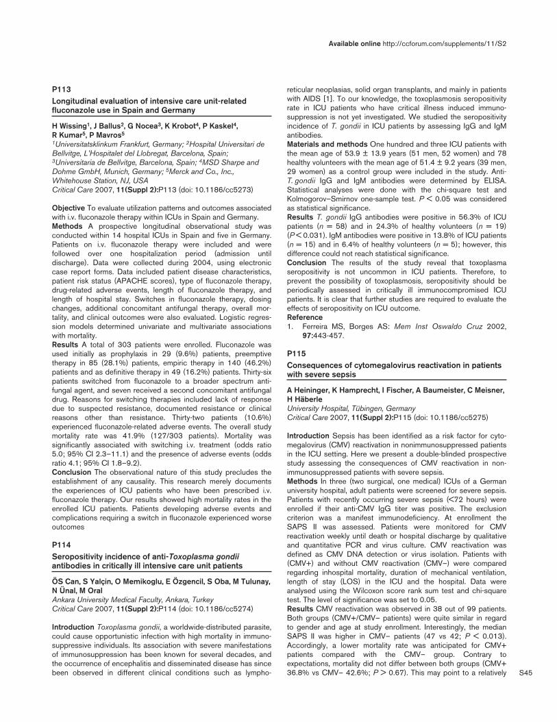
S45
P113Longitudinal evaluation of intensive care unit-relatedfluconazole use in Spain and Germany
H Wissing1, J Ballus2, G Nocea3, K Krobot4, P Kaskel4, R Kumar5, P Mavros5
1Universitatsklinkum Frankfurt, Germany; 2Hospital Universitari deBellvitge, L’Hospitalet del Llobregat, Barcelona, Spain;3Universitaria de Bellvitge, Barcelona, Spain; 4MSD Sharpe andDohme GmbH, Munich, Germany; 5Merck and Co., Inc.,Whitehouse Station, NJ, USACritical Care 2007, 11(Suppl 2):P113 (doi: 10.1186/cc5273)
Objective To evaluate utilization patterns and outcomes associatedwith i.v. fluconazole therapy within ICUs in Spain and Germany.Methods A prospective longitudinal observational study wasconducted within 14 hospital ICUs in Spain and five in Germany.Patients on i.v. fluconazole therapy were included and werefollowed over one hospitalization period (admission untildischarge). Data were collected during 2004, using electroniccase report forms. Data included patient disease characteristics,patient risk status (APACHE scores), type of fluconazole therapy,drug-related adverse events, length of fluconazole therapy, andlength of hospital stay. Switches in fluconazole therapy, dosingchanges, additional concomitant antifungal therapy, overall mor-tality, and clinical outcomes were also evaluated. Logistic regres-sion models determined univariate and multivariate associationswith mortality.Results A total of 303 patients were enrolled. Fluconazole wasused initially as prophylaxis in 29 (9.6%) patients, preemptivetherapy in 85 (28.1%) patients, empiric therapy in 140 (46.2%)patients and as definitive therapy in 49 (16.2%) patients. Thirty-sixpatients switched from fluconazole to a broader spectrum anti-fungal agent, and seven received a second concomitant antifungaldrug. Reasons for switching therapies included lack of responsedue to suspected resistance, documented resistance or clinicalreasons other than resistance. Thirty-two patients (10.6%)experienced fluconazole-related adverse events. The overall studymortality rate was 41.9% (127/303 patients). Mortality wassignificantly associated with switching i.v. treatment (odds ratio5.0; 95% CI 2.3–11.1) and the presence of adverse events (oddsratio 4.1; 95% CI 1.8–9.2).Conclusion The observational nature of this study precludes theestablishment of any causality. This research merely documentsthe experiences of ICU patients who have been prescribed i.v.fluconazole therapy. Our results showed high mortality rates in theenrolled ICU patients. Patients developing adverse events andcomplications requiring a switch in fluconazole experienced worseoutcomes
P114Seropositivity incidence of anti-Toxoplasma gondiiantibodies in critically ill intensive care unit patients
ÖS Can, S Yalçin, O Memikoglu, E Özgencil, S Oba, M Tulunay,N Ünal, M OralAnkara University Medical Faculty, Ankara, TurkeyCritical Care 2007, 11(Suppl 2):P114 (doi: 10.1186/cc5274)
Introduction Toxoplasma gondii, a worldwide-distributed parasite,could cause opportunistic infection with high mortality in immuno-suppressive individuals. Its association with severe manifestationsof immunosuppression has been known for several decades, andthe occurrence of encephalitis and disseminated disease has sincebeen observed in different clinical conditions such as lympho-
reticular neoplasias, solid organ transplants, and mainly in patientswith AIDS [1]. To our knowledge, the toxoplasmosis seropositivityrate in ICU patients who have critical illness induced immuno-suppression is not yet investigated. We studied the seropositivityincidence of T. gondii in ICU patients by assessing IgG and IgMantibodies.Materials and methods One hundred and three ICU patients withthe mean age of 53.9 ± 13.9 years (51 men, 52 women) and 78healthy volunteers with the mean age of 51.4 ± 9.2 years (39 men,29 women) as a control group were included in the study. Anti-T. gondii IgG and IgM antibodies were determined by ELISA.Statistical analyses were done with the chi-square test andKolmogorov–Smirnov one-sample test. P < 0.05 was consideredas statistical significance.Results T. gondii IgG antibodies were positive in 56.3% of ICUpatients (n = 58) and in 24.3% of healthy volunteers (n = 19)(P < 0.031). IgM antibodies were positive in 13.8% of ICU patients(n = 15) and in 6.4% of healthy volunteers (n = 5); however, thisdifference could not reach statistical significance.Conclusion The results of the study reveal that toxoplasmaseropositivity is not uncommon in ICU patients. Therefore, toprevent the possibility of toxoplasmosis, seropositivity should beperiodically assessed in critically ill immunocompromised ICUpatients. It is clear that further studies are required to evaluate theeffects of seropositivity on ICU outcome.Reference1. Ferreira MS, Borges AS: Mem Inst Oswaldo Cruz 2002,
97:443-457.
P115Consequences of cytomegalovirus reactivation in patientswith severe sepsis
A Heininger, K Hamprecht, I Fischer, A Baumeister, C Meisner,H HäberleUniversity Hospital, Tübingen, GermanyCritical Care 2007, 11(Suppl 2):P115 (doi: 10.1186/cc5275)
Introduction Sepsis has been identified as a risk factor for cyto-megalovirus (CMV) reactivation in nonimmunosuppressed patientsin the ICU setting. Here we present a double-blinded prospectivestudy assessing the consequences of CMV reactivation in non-immunosuppressed patients with severe sepsis.Methods In three (two surgical, one medical) ICUs of a Germanuniversity hospital, adult patients were screened for severe sepsis.Patients with recently occurring severe sepsis (<72 hours) wereenrolled if their anti-CMV IgG titer was positive. The exclusioncriterion was a manifest immunodeficiency. At enrollment theSAPS II was assessed. Patients were monitored for CMVreactivation weekly until death or hospital discharge by qualitativeand quantitative PCR and virus culture. CMV reactivation wasdefined as CMV DNA detection or virus isolation. Patients with(CMV+) and without CMV reactivation (CMV–) were comparedregarding inhospital mortality, duration of mechanical ventilation,length of stay (LOS) in the ICU and the hospital. Data wereanalysed using the Wilcoxon score rank sum test and chi-squaretest. The level of significance was set to 0.05.Results CMV reactivation was observed in 38 out of 99 patients.Both groups (CMV+/CMV– patients) were quite similar in regardto gender and age at study enrollment. Interestingly, the medianSAPS II was higher in CMV– patients (47 vs 42; P < 0.013).Accordingly, a lower mortality rate was anticipated for CMV+patients compared with the CMV– group. Contrary toexpectations, mortality did not differ between both groups (CMV+36.8% vs CMV– 42.6%; P > 0.67). This may point to a relatively
Available online http://ccforum.com/supplements/11/S2
S46
increased mortality in CMV+ patients, although CMV disease didnot occur. There was a striking difference between the groups inrespect to the period on ventilator: 21.5 days vs 8.0 days (median)in CMV+ and CMV– patients, respectively (P < 0.005). Similarly,CMV+ patients had a longer median LOS after enrollment either inthe ICU (29.5 days vs 10 days, P < 0.001) and in the hospital(49 days vs 23 days, P < 0.001). This difference was assuredwhen the analysis was restricted to survivors.Conclusion Our data suggest that CMV reactivation leads toincreased morbidity and treatment expenditure independently fromCMV disease. Further analysis points at a crucial role of lungpathology due to CMV reactivation.
P116The role of plasminogen activator inhibitor 1 measurementwith endotoxin adsorption therapy (PMX-DHP) forpostoperative septic shock patients
T Ikeda, K Ikeda, Y Kuroki, T Yokoyama, K YoshikawaTokyo Medical University, Hachioji Medical Center, Tokyo, JapanCritical Care 2007, 11(Suppl 2):P116 (doi: 10.1186/cc5276)
Introduction A polymyxin B immobilized fiber column (PMX; TorayIndustries Inc., Tokyo, Japan) was developed in Japan in 1994 and ithas been used for treatment of endotoxemia or septic shock patients.Materials and methods All patients received an urgent operationdue to intra-abdominal infection. In 88 cases treated with a poly-myxin B immobilized column through direct hemoperfusion (PMX-DHP), changes in hemodynamics, pulmonary oxygenation (PaO2/FIO2) and various mediators (IL-6, IL-8, IL-ra, plasminogen activatorinhibitor 1 (PAI-1)) were examined before and after PMX-DHP,stratifying with the outcome (64 survivors and 24 who died). PMX-DHP was performed through a double lumen catheter (11.5 Fr),placed in the femoral vein or internal jugular vein, at a blood flowrate of 80 ml/min using nafamostat mesilate as an anticoagulant for2 hours.Results PMX-DHP significantly increased systemic arterial pressureand mean arterial pressure, with a greater increase in the survivalgroup. Also, there appeared to be a trend for PaO2/FIO2 improve-ment as blood pressure increased. As the mechanism for improve-ment of pulmonary oxygenation by PMX-DHP has not been shownclearly, it remained to be examined further. PAI-1 valuessignificantly decreased in the survivor group (from 436 ± 549 to251 ± 283 ng/ml) immediately after PMX-DHP; also intracellularadhesion molecule-1 and endothelial leukocyte adhesion molecule-1 tended to decrease in both groups.Discussion PAI-1 is elevated by endotoxin, thrombin andcytokines, and is an indicator of vascular endothelial cell activation.In septic dissminated intravascular coagulation from Gram-nega-tive bacilli, a massive amount of PAI-1 is produced on vascularendothelial cells along with elevation of cytokine production andcoagulation activity. In addition, PAI-1, one of the fibrinolysisinhibitory factors, plays an important role in regulating fibrinolysisby inhibiting tissue plasminogen activator, which convertsplasminogen to active plasmin on fibrin, to block unnecessaryfibrinolysis.Conclusion The determination of PAI-1 may be a useful clinicalparameter for predicting PMX-DHP efficacy.
P117Mechanism and effectiveness of polymyxin B-immobilizedfiber columns for removing mediators (HMBG-1, 2-arachidonoyl glycerol, anandamide, PAI-1, protein C andIL-6) in septic shock patients
Y Sakamoto1, K Mashiko1, T Obata2, Y Yamamoto3
1Chiba Hokusou Hospital, Nippon Medical School, Chiba, Japan;2Institute of DNA Medicine, Jikei University School of Medicine,Tokyo, Japan; 3Department of Emergency Medicine, NipponMedical School, Tokyo, JapanCritical Care 2007, 11(Suppl 2):P117 (doi: 10.1186/cc5277)
Introduction Septic shock remains a major cause of multiple organfailure with a high mortality rate. To remove an endotoxin in patientplasma, direct hemoperfusion (DHP) using a polymyxin B-immobilized fiber column (PMX; Toray Industries Inc., Tokyo Japan)was developed in Japan in 1994 and has since been used for thetreatment of septic shock. The precise role of PMX is not clear.Patients and methods We treated 27 septic shock patients usingDHP-PMX. The patients were separated into two groups foranalysis: those whose systolic blood pressure (SBP) increased bymore than 30 mmHg immediately after DHP-PMX (15 cases), andthose whose SBP did not increase by more than 30 mmHg afterDHP-PMX (12 cases). Furthermore, the patients were separatedinto two other groups for analysis: those whose P/F ratio increasedby more than 20% immediately after DHP-PMX (15 cases), andthose whose P/F ratio did not increase by more 20% after DHP-PMX (12 cases). Mediators were measured at four points: beforeand after DHP-PMX, and 1 day and 3 days afterward.Results The patient group consisted of 17 males and 10 females,59.6 ± 12.7 years old. The average APACHE II score was27.2 ± 9.1, and the average SOFA score was 11.7 ± 5.2 beforeDHP-PMX. Nineteen patients survived and eight died. When thechanges in PAI-1, protein C, ATIII, IL-6 and high mobility group boxprotein 1 (HMGB-1) were compared between the groups, only theHMGB-1 levels had improved significantly in the SBP increasedgroup (P = 0.0125). The SBP increased significantly after DHP-PMX in the HMGB-1-improved group (P < 0.0001). An improve-ment in the P/F ratio and a reduction in 2-arachidonoyl glycerolduring DHP-PMX were significantly correlated (P = 0.0184).Conclusion We showed that the circulation dynamics of septicshock patients can be improved by reducing HMGB-1 levels andthat respiratory function can be improved by reducing 2-arachidonoyl glycerol levels using DHP-PMX.
P118Characterization of the coupled plasmafiltration–adsorption resin cartridge adsorptive capacity forcytokines and inflammatory mediators: adsorption profilesfrom septic patient plasma and in vitro endotoxin-stimulated whole blood
S Livigni1, D Silengo1, M Maio1, V Perlo1, M Pozzato1, P Selvaggi1, L Sereni1,2, M Wratten2
1San Giovanni Bosco, Turin, Italy; 2Bellco, Mirandola, ItalyCritical Care 2007, 11(Suppl 2):P118 (doi: 10.1186/cc5278)
Introduction Coupled plasma filtration–adsorption (CPFA) is anextracorporeal therapy that uses plasma filtration associated withan adsorbent cartridge and hemofiltration in postdilution to removecytokines and inflammatory mediators associated with septic shockand severe sepsis. We evaluated the adsorptive capacity of theresin cartridge to remove various inflammatory mediators and cyto-kines in vitro and ex vivo.
Critical Care March 2007 Vol 11 Suppl 2 27th International Symposium on Intensive Care and Emergency Medicine
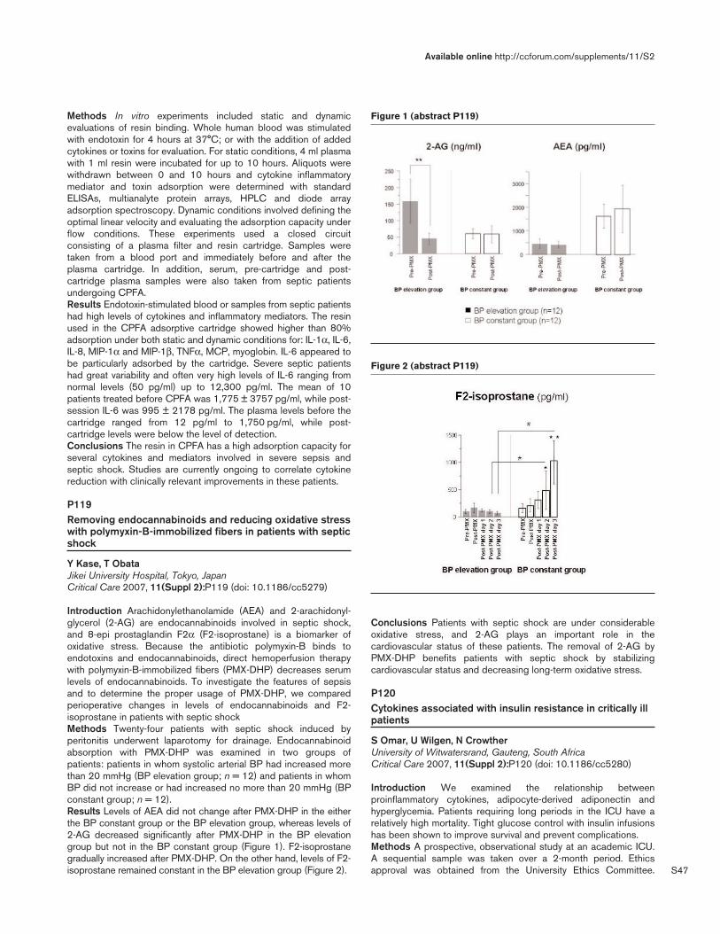
S47
Methods In vitro experiments included static and dynamicevaluations of resin binding. Whole human blood was stimulatedwith endotoxin for 4 hours at 37°C; or with the addition of addedcytokines or toxins for evaluation. For static conditions, 4 ml plasmawith 1 ml resin were incubated for up to 10 hours. Aliquots werewithdrawn between 0 and 10 hours and cytokine inflammatorymediator and toxin adsorption were determined with standardELISAs, multianalyte protein arrays, HPLC and diode arrayadsorption spectroscopy. Dynamic conditions involved defining theoptimal linear velocity and evaluating the adsorption capacity underflow conditions. These experiments used a closed circuitconsisting of a plasma filter and resin cartridge. Samples weretaken from a blood port and immediately before and after theplasma cartridge. In addition, serum, pre-cartridge and post-cartridge plasma samples were also taken from septic patientsundergoing CPFA.Results Endotoxin-stimulated blood or samples from septic patientshad high levels of cytokines and inflammatory mediators. The resinused in the CPFA adsorptive cartridge showed higher than 80%adsorption under both static and dynamic conditions for: IL-1α, IL-6,IL-8, MIP-1α and MIP-1β, TNFα, MCP, myoglobin. IL-6 appeared tobe particularly adsorbed by the cartridge. Severe septic patientshad great variability and often very high levels of IL-6 ranging fromnormal levels (50 pg/ml) up to 12,300 pg/ml. The mean of 10patients treated before CPFA was 1,775 ± 3757 pg/ml, while post-session IL-6 was 995 ± 2178 pg/ml. The plasma levels before thecartridge ranged from 12 pg/ml to 1,750 pg/ml, while post-cartridge levels were below the level of detection.Conclusions The resin in CPFA has a high adsorption capacity forseveral cytokines and mediators involved in severe sepsis andseptic shock. Studies are currently ongoing to correlate cytokinereduction with clinically relevant improvements in these patients.
P119Removing endocannabinoids and reducing oxidative stresswith polymyxin-B-immobilized fibers in patients with septicshock
Y Kase, T ObataJikei University Hospital, Tokyo, JapanCritical Care 2007, 11(Suppl 2):P119 (doi: 10.1186/cc5279)
Introduction Arachidonylethanolamide (AEA) and 2-arachidonyl-glycerol (2-AG) are endocannabinoids involved in septic shock,and 8-epi prostaglandin F2α (F2-isoprostane) is a biomarker ofoxidative stress. Because the antibiotic polymyxin-B binds toendotoxins and endocannabinoids, direct hemoperfusion therapywith polymyxin-B-immobilized fibers (PMX-DHP) decreases serumlevels of endocannabinoids. To investigate the features of sepsisand to determine the proper usage of PMX-DHP, we comparedperioperative changes in levels of endocannabinoids and F2-isoprostane in patients with septic shockMethods Twenty-four patients with septic shock induced byperitonitis underwent laparotomy for drainage. Endocannabinoidabsorption with PMX-DHP was examined in two groups ofpatients: patients in whom systolic arterial BP had increased morethan 20 mmHg (BP elevation group; n = 12) and patients in whomBP did not increase or had increased no more than 20 mmHg (BPconstant group; n = 12).Results Levels of AEA did not change after PMX-DHP in the eitherthe BP constant group or the BP elevation group, whereas levels of2-AG decreased significantly after PMX-DHP in the BP elevationgroup but not in the BP constant group (Figure 1). F2-isoprostanegradually increased after PMX-DHP. On the other hand, levels of F2-isoprostane remained constant in the BP elevation group (Figure 2).
Conclusions Patients with septic shock are under considerableoxidative stress, and 2-AG plays an important role in thecardiovascular status of these patients. The removal of 2-AG byPMX-DHP benefits patients with septic shock by stabilizingcardiovascular status and decreasing long-term oxidative stress.
P120Cytokines associated with insulin resistance in critically illpatients
S Omar, U Wilgen, N CrowtherUniversity of Witwatersrand, Gauteng, South AfricaCritical Care 2007, 11(Suppl 2):P120 (doi: 10.1186/cc5280)
Introduction We examined the relationship betweenproinflammatory cytokines, adipocyte-derived adiponectin andhyperglycemia. Patients requiring long periods in the ICU have arelatively high mortality. Tight glucose control with insulin infusionshas been shown to improve survival and prevent complications.Methods A prospective, observational study at an academic ICU.A sequential sample was taken over a 2-month period. Ethicsapproval was obtained from the University Ethics Committee.
Available online http://ccforum.com/supplements/11/S2
Figure 1 (abstract P119)
Figure 2 (abstract P119)
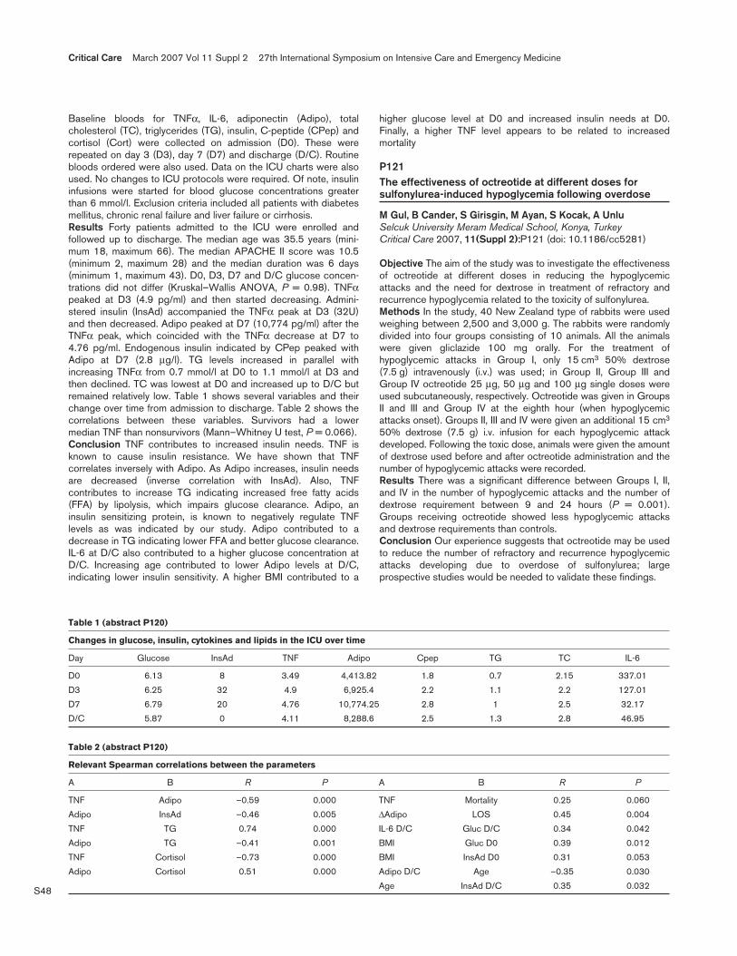
S48
Baseline bloods for TNFα, IL-6, adiponectin (Adipo), totalcholesterol (TC), triglycerides (TG), insulin, C-peptide (CPep) andcortisol (Cort) were collected on admission (D0). These wererepeated on day 3 (D3), day 7 (D7) and discharge (D/C). Routinebloods ordered were also used. Data on the ICU charts were alsoused. No changes to ICU protocols were required. Of note, insulininfusions were started for blood glucose concentrations greaterthan 6 mmol/l. Exclusion criteria included all patients with diabetesmellitus, chronic renal failure and liver failure or cirrhosis.Results Forty patients admitted to the ICU were enrolled andfollowed up to discharge. The median age was 35.5 years (mini-mum 18, maximum 66). The median APACHE II score was 10.5(minimum 2, maximum 28) and the median duration was 6 days(minimum 1, maximum 43). D0, D3, D7 and D/C glucose concen-trations did not differ (Kruskal–Wallis ANOVA, P = 0.98). TNFαpeaked at D3 (4.9 pg/ml) and then started decreasing. Admini-stered insulin (InsAd) accompanied the TNFα peak at D3 (32U)and then decreased. Adipo peaked at D7 (10,774 pg/ml) after theTNFα peak, which coincided with the TNFα decrease at D7 to4.76 pg/ml. Endogenous insulin indicated by CPep peaked withAdipo at D7 (2.8 µg/l). TG levels increased in parallel withincreasing TNFα from 0.7 mmol/l at D0 to 1.1 mmol/l at D3 andthen declined. TC was lowest at D0 and increased up to D/C butremained relatively low. Table 1 shows several variables and theirchange over time from admission to discharge. Table 2 shows thecorrelations between these variables. Survivors had a lowermedian TNF than nonsurvivors (Mann–Whitney U test, P = 0.066).Conclusion TNF contributes to increased insulin needs. TNF isknown to cause insulin resistance. We have shown that TNFcorrelates inversely with Adipo. As Adipo increases, insulin needsare decreased (inverse correlation with InsAd). Also, TNFcontributes to increase TG indicating increased free fatty acids(FFA) by lipolysis, which impairs glucose clearance. Adipo, aninsulin sensitizing protein, is known to negatively regulate TNFlevels as was indicated by our study. Adipo contributed to adecrease in TG indicating lower FFA and better glucose clearance.IL-6 at D/C also contributed to a higher glucose concentration atD/C. Increasing age contributed to lower Adipo levels at D/C,indicating lower insulin sensitivity. A higher BMI contributed to a
higher glucose level at D0 and increased insulin needs at D0.Finally, a higher TNF level appears to be related to increasedmortality
P121The effectiveness of octreotide at different doses forsulfonylurea-induced hypoglycemia following overdose
M Gul, B Cander, S Girisgin, M Ayan, S Kocak, A UnluSelcuk University Meram Medical School, Konya, TurkeyCritical Care 2007, 11(Suppl 2):P121 (doi: 10.1186/cc5281)
Objective The aim of the study was to investigate the effectivenessof octreotide at different doses in reducing the hypoglycemicattacks and the need for dextrose in treatment of refractory andrecurrence hypoglycemia related to the toxicity of sulfonylurea.Methods In the study, 40 New Zealand type of rabbits were usedweighing between 2,500 and 3,000 g. The rabbits were randomlydivided into four groups consisting of 10 animals. All the animalswere given gliclazide 100 mg orally. For the treatment ofhypoglycemic attacks in Group I, only 15 cm3 50% dextrose(7.5 g) intravenously (i.v.) was used; in Group II, Group III andGroup IV octreotide 25 µg, 50 µg and 100 µg single doses wereused subcutaneously, respectively. Octreotide was given in GroupsII and III and Group IV at the eighth hour (when hypoglycemicattacks onset). Groups II, III and IV were given an additional 15 cm3
50% dextrose (7.5 g) i.v. infusion for each hypoglycemic attackdeveloped. Following the toxic dose, animals were given the amountof dextrose used before and after octreotide administration and thenumber of hypoglycemic attacks were recorded.Results There was a significant difference between Groups I, II,and IV in the number of hypoglycemic attacks and the number ofdextrose requirement between 9 and 24 hours (P = 0.001).Groups receiving octreotide showed less hypoglycemic attacksand dextrose requirements than controls.Conclusion Our experience suggests that octreotide may be usedto reduce the number of refractory and recurrence hypoglycemicattacks developing due to overdose of sulfonylurea; largeprospective studies would be needed to validate these findings.
Critical Care March 2007 Vol 11 Suppl 2 27th International Symposium on Intensive Care and Emergency Medicine
Table 1 (abstract P120)
Changes in glucose, insulin, cytokines and lipids in the ICU over time
Day Glucose InsAd TNF Adipo Cpep TG TC IL-6
D0 6.13 8 3.49 4,413.82 1.8 0.7 2.15 337.01
D3 6.25 32 4.9 6,925.4 2.2 1.1 2.2 127.01
D7 6.79 20 4.76 10,774.25 2.8 1 2.5 32.17
D/C 5.87 0 4.11 8,288.6 2.5 1.3 2.8 46.95
Table 2 (abstract P120)
Relevant Spearman correlations between the parameters
A B R P A B R P
TNF Adipo –0.59 0.000 TNF Mortality 0.25 0.060
Adipo InsAd –0.46 0.005 ∆Adipo LOS 0.45 0.004
TNF TG 0.74 0.000 IL-6 D/C Gluc D/C 0.34 0.042
Adipo TG –0.41 0.001 BMI Gluc D0 0.39 0.012
TNF Cortisol –0.73 0.000 BMI InsAd D0 0.31 0.053
Adipo Cortisol 0.51 0.000 Adipo D/C Age –0.35 0.030
Age InsAd D/C 0.35 0.032
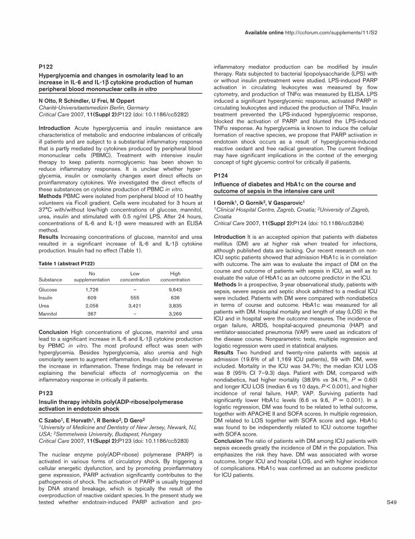
S49
P122Hyperglycemia and changes in osmolarity lead to anincrease in IL-6 and IL-1ββ cytokine production of humanperipheral blood mononuclear cells in vitro
N Otto, R Schindler, U Frei, M OppertCharité-Universitaetsmedizin Berlin, GermanyCritical Care 2007, 11(Suppl 2):P122 (doi: 10.1186/cc5282)
Introduction Acute hyperglycemia and insulin resistance arecharacteristics of metabolic and endocrine imbalances of criticallyill patients and are subject to a substantial inflammatory responsethat is partly mediated by cytokines produced by peripheral bloodmononuclear cells (PBMC). Treatment with intensive insulintherapy to keep patients normoglycemic has been shown toreduce inflammatory responses. It is unclear whether hyper-glycemia, insulin or osmolarity changes exert direct effects onproinflammatory cytokines. We investigated the direct effects ofthese substances on cytokine production of PBMC in vitro.Methods PBMC were isolated from peripheral blood of 10 healthyvolunteers via Ficoll gradient. Cells were incubated for 3 hours at37°C with/without low/high concentrations of glucose, mannitol,urea, insulin and stimulated with 0.5 ng/ml LPS. After 24 hours,concentrations of IL-6 and IL-1β were measured with an ELISAmethod.Results Increasing concentrations of glucose, mannitol and urearesulted in a significant increase of IL-6 and IL-1β cytokineproduction. Insulin had no effect (Table 1).
Table 1 (abstract P122)
No Low High Substance supplementation concentration concentration
Glucose 1,726 – 9,643
Insulin 609 555 636
Urea 2,056 3,421 3,835
Mannitol 367 – 3,269
Conclusion High concentrations of glucose, mannitol and urealead to a significant increase in IL-6 and IL-1β cytokine productionby PBMC in vitro. The most profound effect was seen withhyperglycemia. Besides hyperglycemia, also uremia and highosmolarity seem to augment inflammation. Insulin could not reversethe increase in inflammation. These findings may be relevant inexplaining the beneficial effects of normoglycemia on theinflammatory response in critically ill patients.
P123Insulin therapy inhibits poly(ADP-ribose)polymeraseactivation in endotoxin shock
C Szabo1, E Horvath1, R Benko2, D Gero2
1University of Medicine and Dentistry of New Jersey, Newark, NJ,USA; 2Semmelweis University, Budapest, HungaryCritical Care 2007, 11(Suppl 2):P123 (doi: 10.1186/cc5283)
The nuclear enzyme poly(ADP-ribose) polymerase (PARP) isactivated in various forms of circulatory shock. By triggering acellular energetic dysfunction, and by promoting proinflammatorygene expression, PARP activation significantly contributes to thepathogenesis of shock. The activation of PARP is usually triggeredby DNA strand breakage, which is typically the result of theoverproduction of reactive oxidant species. In the present study wetested whether endotoxin-induced PARP activation and pro-
inflammatory mediator production can be modified by insulintherapy. Rats subjected to bacterial lipopolysaccharide (LPS) withor without insulin pretreatment were studied. LPS-induced PARPactivation in circulating leukocytes was measured by flowcytometry, and production of TNFα was measured by ELISA. LPSinduced a significant hyperglycemic response, activated PARP incirculating leukocytes and induced the production of TNFα. Insulintreatment prevented the LPS-induced hyperglycemic response,blocked the activation of PARP and blunted the LPS-inducedTNFα response. As hyperglycemia is known to induce the cellularformation of reactive species, we propose that PARP activation inendotoxin shock occurs as a result of hyperglycemia-inducedreactive oxidant and free radical generation. The current findingsmay have significant implications in the context of the emergingconcept of tight glycemic control for critically ill patients.
P124Influence of diabetes and HbA1c on the course andoutcome of sepsis in the intensive care unit
I Gornik1, O Gornik2, V Gasparovic1
1Clinical Hospital Centre, Zagreb, Croatia; 2University of Zagreb,CroatiaCritical Care 2007, 11(Suppl 2):P124 (doi: 10.1186/cc5284)
Introduction It is an accepted opinion that patients with diabetesmellitus (DM) are at higher risk when treated for infections,although published data are lacking. Our recent research on non-ICU septic patients showed that admission HbA1c is in correlationwith outcome. The aim was to evaluate the impact of DM on thecourse and outcome of patients with sepsis in ICU, as well as toevaluate the value of HbA1c as an outcome predictor in the ICU.Methods In a prospective, 3-year observational study, patients withsepsis, severe sepsis and septic shock admitted to a medical ICUwere included. Patients with DM were compared with nondiabeticsin terms of course and outcome. HbA1c was measured for allpatients with DM. Hospital mortality and length of stay (LOS) in theICU and in hospital were the outcome measures. The incidence oforgan failure, ARDS, hospital-acquired pneumonia (HAP) andventilator-associated pneumonia (VAP) were used as indicators ofthe disease course. Nonparametric tests, multiple regression andlogistic regression were used in statistical analyses.Results Two hundred and twenty-nine patients with sepsis atadmission (19.6% of all 1,169 ICU patients), 59 with DM, wereincluded. Mortality in the ICU was 34.7%; the median ICU LOSwas 8 (95% CI 7–9.3) days. Patient with DM, compared withnondiabetics, had higher mortality (38.9% vs 34.1%, P = 0.60)and longer ICU LOS (median 6 vs 10 days, P < 0.001), and higherincidence of renal failure, HAP, VAP. Surviving patients hadsignificantly lower HbA1c levels (6.6 vs 9.6, P = 0.001). In alogistic regression, DM was found to be related to lethal outcome,together with APACHE II and SOFA scores. In multiple regression,DM related to LOS together with SOFA score and age. HbA1cwas found to be independently related to ICU outcome togetherwith SOFA score.Conclusion The ratio of patients with DM among ICU patients withsepsis exceeds greatly the incidence of DM in the population. Thisemphasizes the risk they have. DM was associated with worseoutcome, longer ICU and hospital LOS, and with higher incidenceof complications. HbA1c was confirmed as an outcome predictorfor ICU patients.
Available online http://ccforum.com/supplements/11/S2
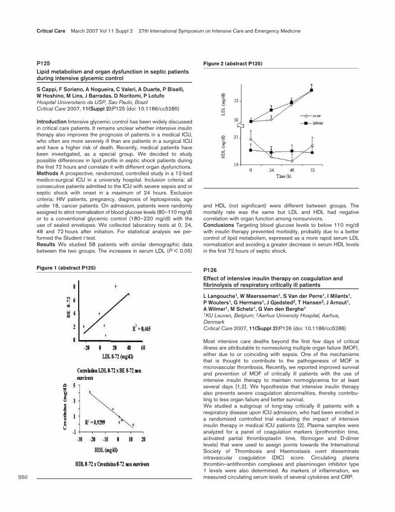
S50
P125Lipid metabolism and organ dysfunction in septic patientsduring intensive glycemic control
S Cappi, F Soriano, A Nogueira, C Valeri, A Duarte, P Biselli, W Hoshino, M Lins, J Barradas, D Noritomi, P LotufoHospital Universitario da USP, Sao Paulo, BrazilCritical Care 2007, 11(Suppl 2):P125 (doi: 10.1186/cc5285)
Introduction Intensive glycemic control has been widely discussedin critical care patients. It remains unclear whether intensive insulintherapy also improves the prognosis of patients in a medical ICU,who often are more severely ill than are patients in a surgical ICUand have a higher risk of death. Recently, medical patients havebeen investigated, as a special group. We decided to studypossible differences in lipid profile in septic shock patients duringthe first 72 hours and correlate it with different organ dysfunctions.Methods A prospective, randomized, controlled study in a 12-bedmedico-surgical ICU in a university hospital. Inclusion criteria: allconsecutive patients admitted to the ICU with severe sepsis and orseptic shock with onset in a maximum of 24 hours. Exclusioncriteria: HIV patients, pregnancy, diagnosis of leptospirosis, ageunder 18, cancer patients. On admission, patients were randomlyassigned to strict normalization of blood glucose levels (80–110 mg/dl)or to a conventional glycemic control (180–220 mg/dl) with theuse of sealed envelopes. We collected laboratory tests at 0, 24,48 and 72 hours after initiation. For statistical analysis we per-formed the Student t test.Results We studied 58 patients with similar demographic databetween the two groups. The increases in serum LDL (P < 0.05)
and HDL (not significant) were different between groups. Themortality rate was the same but LDL and HDL had negativecorrelation with organ function among nonsurvivors.Conclusions Targeting blood glucose levels to below 110 mg/dlwith insulin therapy prevented morbidity, probably due to a bettercontrol of lipid metabolism, expressed as a more rapid serum LDLnormalization and avoiding a greater decrease in serum HDL levelsin the first 72 hours of septic shock.
P126Effect of intensive insulin therapy on coagulation andfibrinolysis of respiratory critically ill patients
L Langouche1, W Meersseman1, S Van der Perre1, I Milants1, P Wouters1, G Hermans1, J Gjedsted2, T Hansen2, J Arnout1, A Wilmer1, M Schetz1, G Van den Berghe1
1KU Leuven, Belgium; 2Aarhus University Hospital, Aarhus,DenmarkCritical Care 2007, 11(Suppl 2):P126 (doi: 10.1186/cc5286)
Most intensive care deaths beyond the first few days of criticalillness are attributable to nonresolving multiple organ failure (MOF),either due to or coinciding with sepsis. One of the mechanismsthat is thought to contribute to the pathogenesis of MOF ismicrovascular thrombosis. Recently, we reported improved survivaland prevention of MOF of critically ill patients with the use ofintensive insulin therapy to maintain normoglycemia for at leastseveral days [1,2]. We hypothesize that intensive insulin therapyalso prevents severe coagulation abnormalities, thereby contribu-ting to less organ failure and better survival.We studied a subgroup of long-stay critically ill patients with arespiratory disease upon ICU admission, who had been enrolled ina randomized controlled trial evaluating the impact of intensiveinsulin therapy in medical ICU patients [2]. Plasma samples wereanalyzed for a panel of coagulation markers (prothrombin time,activated partial thromboplastin time, fibrinogen and D-dimerlevels) that were used to assign points towards the InternationalSociety of Thrombosis and Haemostasis overt disseminateintravascular coagulation (DIC) score. Circulating plasmathrombin–antithrombin complexes and plasminogen inhibitor type1 levels were also determined. As markers of inflammation, wemeasured circulating serum levels of several cytokines and CRP.
Critical Care March 2007 Vol 11 Suppl 2 27th International Symposium on Intensive Care and Emergency Medicine
Figure 1 (abstract P125)
Figure 2 (abstract P125)
S51
Mortality of intensive insulin-treated patients was lower than ofconventionally treated patients for all classes of upon-admissionDIC score, except for those patients with a DIC score of 6 orhigher. There was no effect of insulin therapy on any of thefibrinolytic, coagulation or inflammatory parameters tested. Theaccuracy of the DIC score to predict mortality in this patientsample was only moderate and comparable with that of CRP andthe SOFA score. Also, circulating plasminogen inhibitor type 1 orthrombin–antithrombin complexes levels did not correlate well withmortality or DIC score.These findings indicate that the coagulation system did not play akey role in mediating the survival benefit of intensive insulin therapy.References1. Van den Berghe G, et al.: N Engl J Med 2001, 345:1359-
1367.2. Van den Berghe G, et al.: N Engl J Med 2006, 354:449-461.
P127Variable adsorption of insulin at catheter materials used inintensive care units: polyethylene vs polyurethane –possible cause for hypoglycemia during intensive insulintreatment?
S Ley, J Ammann, C Herder, M Hartmann, D Kindgen-MillesUniversity Hospital, Duesseldorf, GermanyCritical Care 2007, 11(Suppl 2):P127 (doi: 10.1186/cc5287)
Introduction Intensive insulin therapy reduces morbidity andmortality in postoperative critical care; however, this treatment alsoincreases the risk of hypoglycemia. A possible cause for unstableblood glucose (BG) levels could be a variable adsorption of insulinat plastic material of infusion tubings. We evaluated in vitro and invivo the adsorption of insulin at standard tubing materials(polyethylene (PE) and polyurethane (PU)) and the effects of thisadsorption process on blood glucose levels.Methods (1) In vitro, a standard perfusor syringe (Perfusor®;BBraun, Germany) was filled with 50 IE normal insulin (Actrapid®;NovoNordisk, Germany) dissolved in 50 ml saline 0.9%. Thesyringe was connected to PE or PU tubings (BBraun) and, at aninfusion rate of 1 ml/hour, the insulin concentration in the syringeand at the end of the tubings was measured at hourly intervals for5 hours and again after 24 hours by the Bradford protein assay.Insulin concentrations were compared using the Student t test. (2) In a prospective, double-blinded, cross-over study, approved bythe ethics committee, 10 patients on the surgical ICU receivedinsulin via PE or PU tubing each for 24 hours in random sequence.All blood BG values, total infused insulin solution volume, andcritical care scores were documented and statistically analysed bythe Wilcoxon test.Results (1) The insulin concentration in all syringes was always>97% of the estimated value. The initial concentrations of insulin atthe end of PE and PU tubings were lower than expected (23 ± 4%of anticipated concentration in the first 6 min). In PE, theconcentration rose to 37 ± 2% and in PU to 78 ± 4% after24 hours (P < 0.0001). (2) In vivo the mean BG values did notdiffer between PE and PU (PE 141 ± 17 mg/dl; PU132 ± 23 mg/dl (not significant)). Severity of illness was notdifferent between the groups: TISS 37 ± 5 (PE) vs 39 ± 5 (PU),SAPS 43 ± 13 (PE) vs 41 ± 15 (PU) on both days; neither werecatecholamine doses and 24-hour fluid balance. However,significantly more insulin solution was infused in PE (66 ± 18 ml/24 hours) compared with PU (44 ± 15 ml/24 hours) (P = 0.0015).Conclusion Infusion of insulin using PE and PU tubings leads to arelevant adsorption of the drug in both materials. Adsorption to PEis significantly higher compared with PU. Thus, a large variation of
insulin application to the patient is possible if different tubingmaterials are used. Furthermore, variability of adsorption, acompetitive adsorption with other drugs if insulin is not infused viaa single line as well as changes of effective insulin applicationfollowing routine change of tubings, may be one cause ofunexpected hypoglycemia that can be deleterious to the patient.
P128Hyperglycemia upon onset of nosocomial bloodstreaminfection adversely affects outcome in a mixed intensivecare unit population
D Vandijck, S Oeyen, F Buyle, B Claus, S Blot, J DecruyenaereGhent University Hospital, Ghent, BelgiumCritical Care 2007, 11(Suppl 2):P128 (doi: 10.1186/cc5288)
Introduction Hyperglycemia in critically ill patients is frequentlyrelated to a hypermetabolic stress-response and has beenassociated with increased morbidity and mortality. The aim of thisstudy was to assess the relationship between blood glucose levelsand clinical outcome in a mixed cohort of critically ill patients with anosocomial bloodstream infection (BSI).Methods A retrospective observational cohort study was conduc-ted including 130 adult patients with a microbiologicallydocumented BSI admitted over a 2-year period (2003–2004) tothe ICU. Blood glucose levels were evaluated from 1 day prior toonset of BSI (d–1) until 5 days after onset of BSI (d+5). Thecontribution of hyperglycemia, divided into three subgroups(≥150 mg/dl, ≥175 mg/dl, and ≥200 mg/dl, respectively), to in-hospital mortality was estimated by logistic regression.Results The mean age of the study population was 54.7 ± 19.0years. Inhospital mortality was 36.2%. Hyperglycemia (≥175 mg/dland ≥200 mg/dl) was observed more often among the non-survivors. Over the seven study days, no differences were found indaily morning blood glucose levels between survivors (n = 83) andnonsurvivors (n = 47) (all P > 0.05). Although in the nonsurvivorsthe evolution of glycemia tended to be higher, this trend was notstatistically significant compared with the survivors. Multivariatelogistic regression revealed that age (P = 0.022), APACHE II score(P = 0.003), antibiotic resistance (P = 0.001), and hyperglycemia(≥200 mg/dl) upon onset of BSI (P = 0.001) were independentlyassociated with inhospital mortality, whereas appropriate anti-microbial therapy ≤24 hours (P = 0.016) and previous history ofdiabetes (P = 0.022) were associated with better outcome.Conclusion Trends of blood glucose levels were higher amongnonsurvivors. Hyperglycemia (≥200 mg/dl) upon onset ofnosocomial BSI adversely affects outcome in a heterogeneous ICUpopulation.
P129System for automated discontinuous venous bloodwithdrawal for glucose determination of patients in theintensive care unit
R Schaller1, F Feichtner1, A Fercher1, L Schaupp1, M Bodenlenz1, H Köhler1, J Plank2, A Wutte2, M Ellmerer3, R Hainisch4, T Pieber2
1Joanneum Research, Graz, Austria; 2Medical University Graz,Austria; 3Medical University Hospital (MUG), Graz, Austria; 4FHOÖ Forschungs & Entwicklungs GmbH, Linz, AustriaCritical Care 2007, 11(Suppl 2):P129 (doi: 10.1186/cc5289)
Introduction Intensive insulin therapy to establish normoglycaemiareduces mortality and morbidity in critically ill patients. Frequentglucose monitoring is restricted in critically ill patients due to the
Available online http://ccforum.com/supplements/11/S2
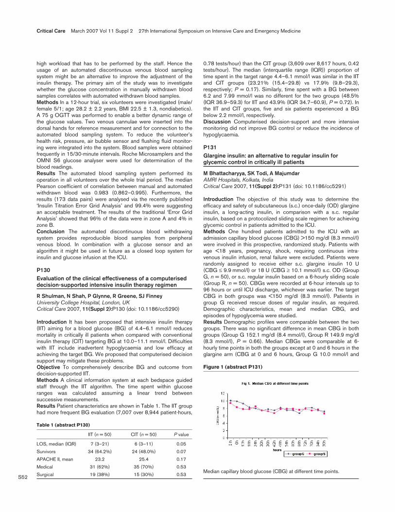
S52
high workload that has to be performed by the staff. Hence theusage of an automated discontinuous venous blood samplingsystem might be an alternative to improve the adjustment of theinsulin therapy. The primary aim of the study was to investigatewhether the glucose concentration in manually withdrawn bloodsamples correlates with automated withdrawn blood samples.Methods In a 12-hour trial, six volunteers were investigated (male/female 5/1; age 28.2 ± 2.2 years, BMI 22.5 ± 1.3, nondiabetics).A 75 g OGTT was performed to enable a better dynamic range ofthe glucose values. Two venous cannulae were inserted into thedorsal hands for reference measurement and for connection to theautomated blood sampling system. To reduce the volunteer’shealth risk, pressure, air bubble sensor and flushing fluid monitor-ing were integrated into the system. Blood samples were obtainedfrequently in 15/30-minute intervals. Roche Microsamplers and theOMNI S6 glucose analyser were used for determination of theblood readings.Results The automated blood sampling system performed itsoperation in all volunteers over the whole trial period. The medianPearson coefficient of correlation between manual and automatedwithdrawn blood was 0.983 (0.862–0.995). Furthermore, theresults (173 data pairs) were analysed via the recently published‘Insulin Titration Error Grid Analysis’ and 99.4% were suggestingan acceptable treatment. The results of the traditional ‘Error GridAnalysis’ showed that 96% of the data were in zone A and 4% inzone B.Conclusion The automated discontinuous blood withdrawingsystem provides reproducible blood samples from peripheralvenous blood. In combination with a glucose sensor and analgorithm it might be used in future as a closed loop system forinsulin and glucose infusion at the ICU.
P130Evaluation of the clinical effectiveness of a computeriseddecision-supported intensive insulin therapy regimen
R Shulman, N Shah, P Glynne, R Greene, SJ FinneyUniversity College Hospital, London, UKCritical Care 2007, 11(Suppl 2):P130 (doi: 10.1186/cc5290)
Introduction It has been proposed that intensive insulin therapy(IIT) aiming for a blood glucose (BG) of 4.4–6.1 mmol/l reducesmortality in critically ill patients when compared with conventionalinsulin therapy (CIT) targeting BG at 10.0–11.1 mmol/l. Difficultieswith IIT include inadvertent hypoglycaemia and low efficacy atachieving the target BG. We proposed that computerised decisionsupport may mitigate these problems.Objective To comprehensively describe BG and outcome fromdecision-supported IIT.Methods A clinical information system at each bedspace guidedstaff through the IIT algorithm. The time spent within glucoseranges was calculated assuming a linear trend betweensuccessive measurements.Results Patient characteristics are shown in Table 1. The IIT grouphad more frequent BG evaluation (7,007 over 8,944 patient-hours,
0.78 tests/hour) than the CIT group (3,609 over 8,617 hours, 0.42tests/hour). The median (interquartile range (IQR)) proportion oftime spent in the target range 4.4–6.1 mmol/l was similar in the IITand CIT groups (23.21% (15.4–29.8) vs 17.9% (9.8–29.3),respectively; P = 0.17). Similarly, time spent with a BG between6.2 and 7.99 mmol/l was no different for the two groups (48.5%(IQR 36.9–59.3) for IIT and 43.9% (IQR 34.7–60.9), P = 0.72). Inthe IIT and CIT groups, five and six patients experienced a BGbelow 2.2 mmol/l, respectively.Discussion Computerised decision-support and more intensivemonitoring did not improve BG control or reduce the incidence ofhypoglycaemia.
P131Glargine insulin: an alternative to regular insulin forglycemic control in critically ill patients
M Bhattacharyya, SK Todi, A MajumdarAMRI Hospitals, Kolkata, IndiaCritical Care 2007, 11(Suppl 2):P131 (doi: 10.1186/cc5291)
Introduction The objective of this study was to determine theefficacy and safety of subcutaneous (s.c.) once-daily (OD) glargineinsulin, a long-acting insulin, in comparison with a s.c. regularinsulin, based on a protocolized sliding scale regimen for achievingglycemic control in patients admitted to the ICU.Methods One hundred patients admitted to the ICU with anadmission capillary blood glucose (CBG) >150 mg/dl (8.3 mmol/l)were involved in this prospective, randomized study. Patients withage <18 years, pregnancy, shock, requiring continuous intra-venous insulin infusion, renal failure were excluded. Patients wererandomly assigned to receive either s.c. glargine insulin 10 U(CBG ≤ 9.9 mmol/l) or 18 U (CBG ≥ 10.1 mmol/l) s.c. OD (GroupG, n = 50), or s.c. regular insulin based on a 6-hourly sliding scale(Group R, n = 50). CBGs were recorded at 6-hour intervals up to96 hours or until ICU discharge, whichever was earlier. The targetCBG in both groups was <150 mg/dl (8.3 mmol/l). Patients ingroup G received rescue doses of regular insulin, as required.Demographic characteristics, mean and median CBG, andepisodes of hypoglycemia were studied.Results Demographic profiles were comparable between the twogroups. There was no significant difference in mean CBG in bothgroups (Group G 152.1 mg/dl (8.4 mmol/l), Group R 149.9 mg/dl(8.3 mmol/l), P = 0.66). Median CBGs were comparable at 6-hourly time points in both the groups except at 0 and 6 hours in theglargine arm (CBG at 0 and 6 hours, Group G 10.0 mmol/l and
Critical Care March 2007 Vol 11 Suppl 2 27th International Symposium on Intensive Care and Emergency Medicine
Table 1 (abstract P130)
IIT (n = 50) CIT (n = 50) P value
LOS, median (IQR) 7 (3–21) 6 (3–11) 0.05
Survivors 34 (64.2%) 24 (48.0%) 0.07
APACHE II, mean 23.2 25.4 0.17
Medical 31 (62%) 35 (70%) 0.53
Surgical 19 (38%) 15 (30%) 0.53
Figure 1 (abstract P131)
Median capillary blood glucose (CBG) at different time points.
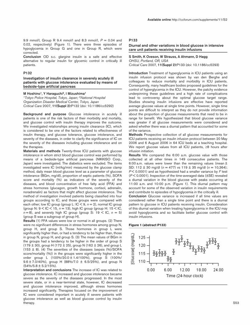
S53
9.9 mmol/l, Group R 9.4 mmol/l and 8.3 mmol/l, P = 0.04 and0.02, respectively) (Figure 1). There were three episodes ofhypoglycemia in Group G and one in Group R, which werecorrected.Conclusion OD s.c. glargine insulin is a safe and effectivealternative to regular insulin for glycemic control in critically illpatients.
P132Investigation of insulin clearance in severely acutely illpatients with glucose intolerance evaluated by means ofbedside-type artificial pancreas
M Hoshino1, Y Haraguchi2, I Mizushima1
1Tokyo Police Hospital, Tokyo, Japan; 2National HospitalOrganization Disaster Medical Center, Tokyo, JapanCritical Care 2007, 11(Suppl 2):P132 (doi: 10.1186/cc5292)
Background and purpose Glucose intolerance in acutely illpatients is one of the risk factors of their morbidity and mortality,and glucose control with insulin therapy improves the outcome.We investigated relationships among insulin clearance (IC), whichis considered to be one of the factors related to effectiveness ofinsulin therapy, and glucose tolerance, glucose intolerance, andseverity of the diseases, in order to clarify the significance of IC onthe severity of the diseases including glucose intolerance and onthe therapies.Materials and methods Twenty-three ICU patients with glucoseintolerance in whom strict blood glucose control was performed bymeans of a bedside-type artificial pancreas (NIKKISO Corp.,Japan) were investigated. The diabetics were excluded. The itemsinvestigated were IC (ml/kg/min) measured by the glucose clampmethod, daily mean blood glucose level as a parameter of glucoseintolerance (BGm, mg/dl), proportion of septic patients (%), SOFAscore and mortality (%) as indicators of the severity of thediseases, and blood concentration of free fatty acid (FFA) andstress hormones (glucagon, growth hormone, cortisol, adrenalin,noradrenalin) as factors that might affect glucose intolerance. Themethod of investigation involved patients being classified into fourgroups according to IC, and those groups were compared witheach other; low IC group (group L: IC < 9, n = 2), normal IC group(group N: 9 < IC < 15, n = 13), high IC group (group H: 15 < IC,n = 8), and severely high IC group (group S: 19 < IC, n = 5)(group S was a subgroup of group H).Results (1) FFA values were low or normal in all groups. (2) Therewere no significant differences in stress hormones among group N,group H, and group S. Those hormones in group L weresignificantly higher than, or had a tendency to be higher than, thosein group N, group H, and group S. (3) The mean values of BGm inthe groups had a tendency to be higher in the order of group S(179 ± 30), group H (172 ± 25), group N (162 ± 26), and group L(153 ± 8). (4) The severities of the diseases (sepsis (%)/SOFAscore/mortality (%)) in the groups were significantly higher in theorder group L (100%/20.0 ± 1.4/100%), group S (100%/9.6 ± 7.0/40%), group H (88%/7.0 ± 6.5/25%), and group N(54%/5.8 ± 5.2/15%).Interpretation and conclusions The increase of IC was related toglucose intolerance. IC increased and glucose intolerance becamesevere as the severity of the diseases progressed. In the mostsevere state, or in a near-terminal state, however, IC decreasedand glucose intolerance improved, although stress hormonesincreased significantly. Therapies focused on the improvement ofIC were considered important in acutely ill severe patients withglucose intolerance as well as blood glucose control by insulintherapy.
P133Diurnal and other variations in blood glucose in intensivecare unit patients receiving insulin infusions
S Smith, K Oveson, W Strauss, A Ahmann, D HaggOHSU, Portland, OR, USACritical Care 2007, 11(Suppl 2):P133 (doi: 10.1186/cc5293)
Introduction Treatment of hyperglycemia in ICU patients using aninsulin infusion protocol was shown by van den Berghe andcolleagues to reduce mortality and morbidity in ICU patients.Consequently, many healthcare bodies proposed guidelines for thecontrol of hyperglycemia in the ICU. However, the patchy evidenceunderpinning these guidelines and a high rate of complicationslead to controversy about the optimal glucose target range.Studies showing insulin infusions are effective have reportedaverage glucose values at single time points. However, single timepoints are difficult to interpret as they do not provide informationabout the proportion of glucose measurements that need to be inrange for benefit. We hypothesized that blood glucose variancewas greater if all glucose measurements were considered andasked whether there was a diurnal pattern that accounted for someof the variance.Methods Prospective collection of all glucose measurements forICU patients receiving an insulin infusion protocol between 20 May2006 and 6 August 2006 in 64 ICU beds at a teaching hospital.We report glucose values from all ICU patients, ≥8 hours afterinfusion initiation.Results We compared the 6:00 a.m. glucose value with thosecollected at all other times in 149 consecutive patients. The6:00 a.m. values were lower than the remaining values (mean ±SD: 112 ± 30 mg/dl (n = 477) vs 119 ± 35 mg/dl (n = 10,364);P < 0.0001) and as hypothesized had a smaller variance by F test(P < 0.0001). Inspection of the time-averaged data (±SE) revealeda diurnal variation in the blood glucose with peaks occurring at11:00 a.m. and 10:00 p.m. (Figure 1). This diurnal pattern mayaccount for some of the observed variation in insulin requirementsand contribute to episodes of hypoglycemia in the critically ill.Conclusion Glucose variance is increased if all time values areconsidered rather than a single time point and there is a diurnalpattern to glucose in ICU patients receiving insulin. Considerationof this diurnal variation when treating hyperglycemia in the ICU mayavoid hypoglycemia and so facilitate better glucose control withinsulin infusions.
Available online http://ccforum.com/supplements/11/S2
Figure 1 (abstract P133)
S54
P134Severe hypoglycaemia during intensive insulin therapy: arare event in critically ill patients
M Kaukonen, M Rantala, V Pettilä, M HynninenHelsinki University Hospital, Helsinki, FinlandCritical Care 2007, 11(Suppl 2):P134 (doi: 10.1186/cc5294)
Introduction Tight glycemic control reduces mortality in surgicalintensive care patients and in long-term medical intensive carepatients. The incidence of severe hypoglycaemia (glucose≤2.2 mmol/l) in the intensive treatment group has been 3.1–5.1%.Recently, a large study on intensive insulin therapy wasprematurely discontinued due to safety issues. The incidence ofhypoglycaemia was 9.8% in intensive treatment group and themortality among patients experiencing hypoglycaemia was 18.6%.As the safety of intensive insulin therapy has been questioned, wescreened all patients during a 17-month period to see theincidence of hypoglycaemia and its effects on the prognosis of thepatients.Methods A retrospective study was performed in two ICUs, oneeight-bed general ICU and one 10-bed surgical ICU. All patientstreated between 7 February 2005 and 30 June 2006 wereincluded in the study. A nurse-driven intensive insulin protocol witha target blood glucose level of 4–6.1 mmol/l had been introducedin 2004. All blood glucose measurements performed during theICU treatment were analysed. The patients were divided into twogroups according to the lowest detected blood glucose value(≤2.2 or ≥2.3 mmol/l).Results A total of 1,024 patients (1,124 treatment periods) wereincluded in the study. Thirty patients were excluded due toincompleteness of the data. During the study period 61,203 bloodglucose measurements were performed, 1,578 (2.6%) of whichwere below the target value of 4 mmol/l. Severe hypoglycaemia(≤2.2 mmol/l) occurred in 25 patients (36 measurements). Theincidence was 0.059% of the measurements and 2.3% of thepatients. The median age, sex, APACHE II score, SAPS II ordiagnosis category did not differ between the groups. The median(IQR) ICU and hospital length of stay was 4.3 (1.8–10.6) and 18(8.5–39.5) days in patients with lowest blood glucose ≤2.2, and2.7 (1.2–5.7) and 13 (7–23) days in patients with lowest bloodglucose ≥2.3 (P = 0.058 and P = 0.077, respectively). Thehospital mortalities were 25% and 15%, respectively; thedifference was not statistically significant.Conclusions Severe hypoglycaemia during intensive insulintherapy is rare in protocol-driven ICU treatment compared withprevious clinical trials. When present, hypoglycaemia may have animpact on the outcome of the patients
P135Intensive insulin therapy and indications for intensive careadmission
A Sanusi1, I Welters2, A Shenkin2, P Turner3, B Perry2
1University of Ibadan, Ibadan, Nigeria; 2University of Liverpool, UK;3The Royal Liverpool Hospital, Liverpool, UKCritical Care 2007, 11(Suppl 2):P135 (doi: 10.1186/cc5295)
Introduction Insulin resistance and hyperglycemia are common incritically ill patients, and are associated with higher morbidity andmortality in these patients if not controlled. Intensive insulin therapyhas been shown to reduce morbidity and mortality. It is not clear,however, whether the patients’ indication for admission into theICU is related to the time to achieve glycaemic control or the totaldose of insulin required. This study was designed to audit the
efficacy of an intensive insulin therapy protocol in achievingglycaemic control in patients presenting with different conditions.Methods A prospective observational study was performed over8 weeks on patients admitted to an adult ICU who receivednutrition support for up to 48 hours. Intensive insulin therapy wasadministered to those patients who developed hyperglycemia. Thedemographics, blood glucose and insulin doses were documented.Haemoglobin, white cell count, neutrophil count, antioxidants, CRPand prealbumin were measured. Outcome measures were themean and total insulin dose and the time to achieve glycaemiccontrol.Results Forty patients, 22 (55%) males and 18 (45%) females,who received nutritional support for 48 hours or more werestudied. The mean (SD) age was 59.4 (14.7) years. Enteral feedingwas given in 32 (80%) and parenteral feeding in 14 (35%)patients, while six (15%) patients received both enteral andparenteral feeding. The mean (SD) energy in 48 hours was 3,307.4(527.0) kcal, mean (SD) insulin was 1.37 (1.23) IU, mean (SD)blood glucose was 7.76 (0.9) mmol/dl and total insulin to achieveglycaemic control was 65.51 (58.6) IU. The time taken (SD) toachieve glycaemic control was 15.16 (12.65) hours. As expected,there was a relationship between the total insulin dose and thetime to achieve three consecutive glycaemic controls (r = –0.43,P = 0.023). Also, between the total insulin dose and mean bloodglucose r = 0.508, P = 0.001. There was no significant relation-ship between the total insulin dose and indication for ICUadmission, and the total insulin dose and body mass index.Conclusion Findings from this study showed that the indication foradmission did not affect either the total dose of insulin required toachieve glycaemic control or the time it takes to achieve threeconsecutive glycaemic controls.
P136Implementation of glycemic control – problems andsolutions
E Halbeck, U Jaschinski, A Scherer, A Aulmann, M Lichtwarck-Aschoff, H ForstKlinikum Augsburg, GermanyCritical Care 2007, 11(Suppl 2):P136 (doi: 10.1186/cc5296)
Introduction Glycaemic control is another example of protocol-driven therapy in intensive care medicine to improve outcome incritically ill patients. While the advantage of this approach seems tobe obvious, little is known about the problems of implementing sucha protocol. The intention of this study was to evaluate problems ofimplementation and to develop strategies to overcome them.Setting A 16-bed surgical ICU of a university teaching hospitalwith 50 critical care nurses, about 30% in part-time employment.Method Over a 7-month period all patients staying longer than 48hours in the ICU with hyperglycaemia (>150 mg%) on threeconsecutive measurements were included in the study. Thesepatients were treated according to a protocol at the discretion ofthe attending nurse. On daily rounds and every 4–5 weekssupervision was performed, and the protocol was modified threetimes during this period according to staff comments. Further on,medical as well as nonmedical problems of implementation wereanalysed and discussed. Attitudes and perceived impedingaspects of the implementation process were recorded by means ofa questionnaire.Results Since insulin sensitivity showed enormous variability,glycaemic control required a high nursing effort. Impeding aspectsto titrate blood glucose into the target range were the absence of anutritional protocol (high carbohydrate intake, despite inflam-mation/infection leading to hyperglycaemia that was difficult to
Critical Care March 2007 Vol 11 Suppl 2 27th International Symposium on Intensive Care and Emergency Medicine
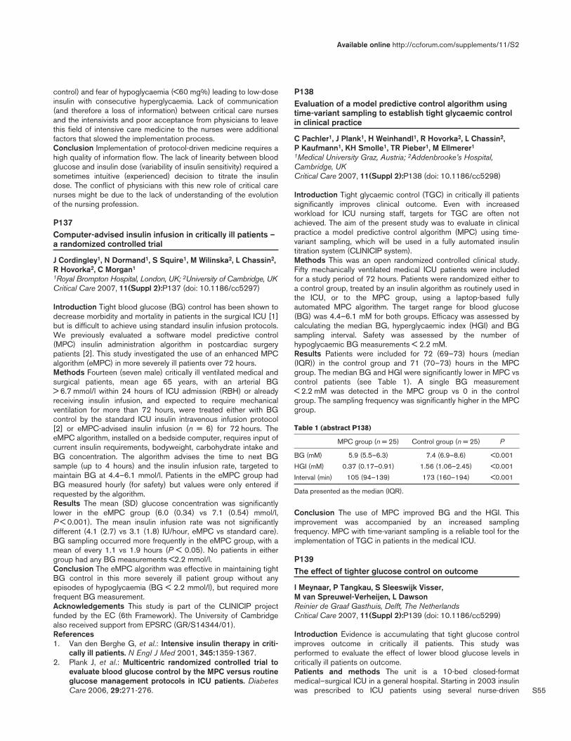
S55
control) and fear of hypoglycaemia (<60 mg%) leading to low-doseinsulin with consecutive hyperglycaemia. Lack of communication(and therefore a loss of information) between critical care nursesand the intensivists and poor acceptance from physicians to leavethis field of intensive care medicine to the nurses were additionalfactors that slowed the implementation process.Conclusion Implementation of protocol-driven medicine requires ahigh quality of information flow. The lack of linearity between bloodglucose and insulin dose (variability of insulin sensitivity) required asometimes intuitive (experienced) decision to titrate the insulindose. The conflict of physicians with this new role of critical carenurses might be due to the lack of understanding of the evolutionof the nursing profession.
P137Computer-advised insulin infusion in critically ill patients –a randomized controlled trial
J Cordingley1, N Dormand1, S Squire1, M Wilinska2, L Chassin2,R Hovorka2, C Morgan1
1Royal Brompton Hospital, London, UK; 2University of Cambridge, UKCritical Care 2007, 11(Suppl 2):P137 (doi: 10.1186/cc5297)
Introduction Tight blood glucose (BG) control has been shown todecrease morbidity and mortality in patients in the surgical ICU [1]but is difficult to achieve using standard insulin infusion protocols.We previously evaluated a software model predictive control(MPC) insulin administration algorithm in postcardiac surgerypatients [2]. This study investigated the use of an enhanced MPCalgorithm (eMPC) in more severely ill patients over 72 hours.Methods Fourteen (seven male) critically ill ventilated medical andsurgical patients, mean age 65 years, with an arterial BG> 6.7 mmol/l within 24 hours of ICU admission (RBH) or alreadyreceiving insulin infusion, and expected to require mechanicalventilation for more than 72 hours, were treated either with BGcontrol by the standard ICU insulin intravenous infusion protocol[2] or eMPC-advised insulin infusion (n = 6) for 72 hours. TheeMPC algorithm, installed on a bedside computer, requires input ofcurrent insulin requirements, bodyweight, carbohydrate intake andBG concentration. The algorithm advises the time to next BGsample (up to 4 hours) and the insulin infusion rate, targeted tomaintain BG at 4.4–6.1 mmol/l. Patients in the eMPC group hadBG measured hourly (for safety) but values were only entered ifrequested by the algorithm.Results The mean (SD) glucose concentration was significantlylower in the eMPC group (6.0 (0.34) vs 7.1 (0.54) mmol/l,P < 0.001). The mean insulin infusion rate was not significantlydifferent (4.1 (2.7) vs 3.1 (1.8) IU/hour, eMPC vs standard care).BG sampling occurred more frequently in the eMPC group, with amean of every 1.1 vs 1.9 hours (P < 0.05). No patients in eithergroup had any BG measurements <2.2 mmol/l.Conclusion The eMPC algorithm was effective in maintaining tightBG control in this more severely ill patient group without anyepisodes of hypoglycaemia (BG < 2.2 mmol/l), but required morefrequent BG measurement.Acknowledgements This study is part of the CLINICIP projectfunded by the EC (6th Framework). The University of Cambridgealso received support from EPSRC (GR/S14344/01).References1. Van den Berghe G, et al.: Intensive insulin therapy in criti-
cally ill patients. N Engl J Med 2001, 345:1359-1367.2. Plank J, et al.: Multicentric randomized controlled trial to
evaluate blood glucose control by the MPC versus routineglucose management protocols in ICU patients. DiabetesCare 2006, 29:271-276.
P138Evaluation of a model predictive control algorithm usingtime-variant sampling to establish tight glycaemic controlin clinical practice
C Pachler1, J Plank1, H Weinhandl1, R Hovorka2, L Chassin2, P Kaufmann1, KH Smolle1, TR Pieber1, M Ellmerer1
1Medical University Graz, Austria; 2Addenbrooke’s Hospital,Cambridge, UKCritical Care 2007, 11(Suppl 2):P138 (doi: 10.1186/cc5298)
Introduction Tight glycaemic control (TGC) in critically ill patientssignificantly improves clinical outcome. Even with increasedworkload for ICU nursing staff, targets for TGC are often notachieved. The aim of the present study was to evaluate in clinicalpractice a model predictive control algorithm (MPC) using time-variant sampling, which will be used in a fully automated insulintitration system (CLINICIP system).Methods This was an open randomized controlled clinical study.Fifty mechanically ventilated medical ICU patients were includedfor a study period of 72 hours. Patients were randomized either toa control group, treated by an insulin algorithm as routinely used inthe ICU, or to the MPC group, using a laptop-based fullyautomated MPC algorithm. The target range for blood glucose(BG) was 4.4–6.1 mM for both groups. Efficacy was assessed bycalculating the median BG, hyperglycaemic index (HGI) and BGsampling interval. Safety was assessed by the number ofhypoglycaemic BG measurements < 2.2 mM.Results Patients were included for 72 (69–73) hours (median(IQR)) in the control group and 71 (70–73) hours in the MPCgroup. The median BG and HGI were significantly lower in MPC vscontrol patients (see Table 1). A single BG measurement< 2.2 mM was detected in the MPC group vs 0 in the controlgroup. The sampling frequency was significantly higher in the MPCgroup.
Table 1 (abstract P138)
MPC group (n = 25) Control group (n = 25) P
BG (mM) 5.9 (5.5–6.3) 7.4 (6.9–8.6) <0.001
HGI (mM) 0.37 (0.17–0.91) 1.56 (1.06–2.45) <0.001
Interval (min) 105 (94–139) 173 (160–194) <0.001
Data presented as the median (IQR).
Conclusion The use of MPC improved BG and the HGI. Thisimprovement was accompanied by an increased samplingfrequency. MPC with time-variant sampling is a reliable tool for theimplementation of TGC in patients in the medical ICU.
P139The effect of tighter glucose control on outcome
I Meynaar, P Tangkau, S Sleeswijk Visser, M van Spreuwel-Verheijen, L DawsonReinier de Graaf Gasthuis, Delft, The NetherlandsCritical Care 2007, 11(Suppl 2):P139 (doi: 10.1186/cc5299)
Introduction Evidence is accumulating that tight glucose controlimproves outcome in critically ill patients. This study wasperformed to evaluate the effect of lower blood glucose levels incritically ill patients on outcome.Patients and methods The unit is a 10-bed closed-formatmedical–surgical ICU in a general hospital. Starting in 2003 insulinwas prescribed to ICU patients using several nurse-driven
Available online http://ccforum.com/supplements/11/S2
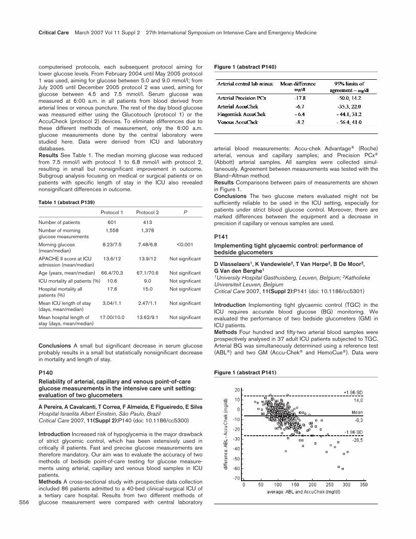
S56
computerised protocols, each subsequent protocol aiming forlower glucose levels. From February 2004 until May 2005 protocol1 was used, aiming for glucose between 5.0 and 9.0 mmol/l; fromJuly 2005 until December 2005 protocol 2 was used, aiming forglucose between 4.5 and 7.5 mmol/l. Serum glucose wasmeasured at 6:00 a.m. in all patients from blood derived fromarterial lines or venous puncture. The rest of the day blood glucosewas measured either using the Glucotouch (protocol 1) or theAccuCheck (protocol 2) devices. To eliminate differences due tothese different methods of measurement, only the 6:00 a.m.glucose measurements done by the central laboratory werestudied here. Data were derived from ICU and laboratorydatabases.Results See Table 1. The median morning glucose was reducedfrom 7.5 mmol/l with protocol 1 to 6.8 mmol/l with protocol 2,resulting in small but nonsignificant improvement in outcome.Subgroup analysis focusing on medical or surgical patients or onpatients with specific length of stay in the ICU also revealednonsignificant differences in outcome.
Table 1 (abstract P139)
Protocol 1 Protocol 2 P
Number of patients 601 413
Number of morning 1,558 1,378glucose measurements
Morning glucose 8.23/7.5 7.48/6.8 <0.001(mean/median)
APACHE II score at ICU 13.6/12 13.9/12 Not significantadmission (mean/median)
Age (years, mean/median) 66.4/70.3 67.1/70.6 Not significant
ICU mortality all patients (%) 10.6 9.0 Not significant
Hospital mortality all 17.6 15.0 Not significantpatients (%)
Mean ICU length of stay 3.04/1.1 2.47/1.1 Not significant(days, mean/median)
Mean hospital length of 17.00/10.0 13.62/9.1 Not significantstay (days, mean/median)
Conclusions A small but significant decrease in serum glucoseprobably results in a small but statistically nonsignificant decreasein mortality and length of stay.
P140Reliability of arterial, capillary and venous point-of-careglucose measurements in the intensive care unit setting:evaluation of two glucometers
A Pereira, A Cavalcanti, T Correa, F Almeida, E Figueiredo, E SilvaHospital Israelita Albert Einstein, São Paulo, BrazilCritical Care 2007, 11(Suppl 2):P140 (doi: 10.1186/cc5300)
Introduction Increased risk of hypoglycemia is the major drawbackof strict glycemic control, which has been extensively used incritically ill patients. Fast and precise glucose measurements aretherefore mandatory. Our aim was to evaluate the accuracy of twomethods of bedside point-of-care testing for glucose measure-ments using arterial, capillary and venous blood samples in ICUpatients.Methods A cross-sectional study with prospective data collectionincluded 86 patients admitted to a 40-bed clinical-surgical ICU ofa tertiary care hospital. Results from two different methods ofglucose measurement were compared with central laboratory
arterial blood measurements: Accu-chek Advantage® (Roche)arterial, venous and capillary samples; and Precision PCx®
(Abbott) arterial samples. All samples were collected simul-taneously. Agreement between measurements was tested with theBland–Altman method.Results Comparisons between pairs of measurements are shownin Figure 1.Conclusions The two glucose meters evaluated might not besufficiently reliable to be used in the ICU setting, especially forpatients under strict blood glucose control. Moreover, there aremarked differences between the equipment and a decrease inprecision if capillary or venous samples are used.
P141Implementing tight glycaemic control: performance ofbedside glucometers
D Vlasselaers1, K Vandewiele2, T Van Herpe2, B De Moor2, G Van den Berghe1
1University Hospital Gasthuisberg, Leuven, Belgium; 2KatholiekeUniversiteit Leuven, BelgiumCritical Care 2007, 11(Suppl 2):P141 (doi: 10.1186/cc5301)
Introduction Implementing tight glycaemic control (TGC) in theICU requires accurate blood glucose (BG) monitoring. Weevaluated the performance of two bedside glucometers (GM) inICU patients.Methods Four hundred and fifty-two arterial blood samples wereprospectively analysed in 37 adult ICU patients subjected to TGC.Arterial BG was simultaneously determined using a reference test(ABL®) and two GM (Accu-Chek® and HemoCue®). Data were
Critical Care March 2007 Vol 11 Suppl 2 27th International Symposium on Intensive Care and Emergency Medicine
Figure 1 (abstract P140)
Figure 1 (abstract P141)
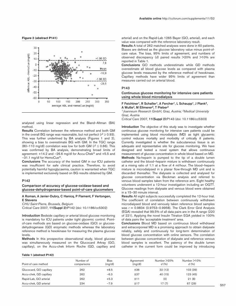
S57
analysed using linear regression and the Bland–Altman (BA)method.Results Correlation between the reference method and both GMin the overall BG range was reasonable, but not perfect (r2 ≥ 0.93).This was further underlined by BA analysis (Figures 1 and 2),showing a bias to overestimate BG with GM. In the TGC range(80–110 mg/dl) correlation was low for both GM (r2 ≤ 0.66). Thiswas confirmed by BA analysis, demonstrating broad limits ofagreement: +14.2 and –26.6 mg/dl for Accu-Chek® and +5.5 and–31.1 mg/dl for HemoCue®.Conclusions The accuracy of the tested GM in our ICU patientswas insufficient for safe clinical practice. Therefore, to avoidpotentially harmful hypoglycaemia, caution is warranted when TGCis implemented exclusively based on BG results obtained by GM.
P142Comparison of accuracy of glucose-oxidase-based andglucose-dehydrogenase-based point-of-care glucometers
A Roman, A Janier-Dubry, C Hanicq, P Flament, F Vertongen, E StevensCHU Saint-Pierre, Brussels, BelgiumCritical Care 2007, 11(Suppl 2):P142 (doi: 10.1186/cc5302)
Introduction Bedside capillary or arterial blood glucose monitoringis mandatory for ICU patients under tight glycemic control. Point-of-care methods are based on glucose-oxidase (GO) or glucose-dehydrogenase (GD) enzymatic methods whereas the laboratoryreference method is hexokinase for measuring the plasma glucoselevels.Methods In this prospective observational study, blood glucosewas simultaneously measured on the Glucocard Arkray (GO,capillary), on the Accu-chek Inform Roche (GD, capillary and
arterial) and on the Rapid-Lab 1265 Bayer (GO, arterial), and eachvalue was compared with the reference laboratory result.Results A total of 262 matched analyses were done in 60 patients.Biases are defined as the glucose laboratory value minus point-of-care value. The bias, 95% limits of agreement, and numbers ofobserved discrepancy (d) paired results >20% and >10% arereported in Table 1.Conclusions GO methods underestimate while GD methodsoverestimate all blood glucose levels as compared with plasmaglucose levels measured by the reference method of hexokinase.Capillary methods have wider 95% limits of agreement thanmeasures carried out on arterial blood.
P143Continuous glucose monitoring for intensive care patientsusing whole blood microdialysis
F Feichtner1, R Schaller1, A Fercher1, L Schaupp1, J Plank2, A Wutte2, M Ellmerer2, T Pieber2
1Joanneum Research GmbH, Graz, Austria; 2Medical UniversityGraz, AustriaCritical Care 2007, 11(Suppl 2):P143 (doi: 10.1186/cc5303)
Introduction The objective of this study was to investigate whethercontinuous glucose monitoring for intensive care patients could beimplemented using blood microdialysis (MD) as tight glycaemiccontrol reduces mortality and morbidity of critically ill patients.Currently investigated is whether the subcutaneous tissue is anadequate and representative site for glucose monitoring. We havedesigned and tested a novel system that allows continuousmeasurement of glucose concentration in whole blood based on MD.Methods Na-heparin is pumped to the tip of a double lumencatheter and the blood–heparin mixture is withdrawn continuouslyat a mixing ratio of 1:1 at a flow of 4 ml/hour. The blood–heparinmixture is microdialysed in a planar flow-through MD unit and isdiscarded thereafter. The dialysate is collected and analysed forglucose concentration via Beckman analysis and referred tovenous blood samples taken from the reference arm. Eight healthyvolunteers underwent a 12-hour investigation including an OGTT.Glucose readings from dialysate and venous blood were obtainedin a 15–30 minute interval.Results All eight subjects successfully completed the 12-hour trial.The coefficient of correlation between continuously withdrawnmicrodialysed blood and venously taken reference blood sampleswas r = 0.9834 (0.9753–0.9958). The Clark Error Grid Analysis(EGA) revealed that 99.5% of all data pairs are in the A range (220of 221). Applying the novel Insulin Titration EGA yielded in 100%of data pairs the ‘acceptable treatment’ area.Conclusions Blood MD based on continuous blood withdrawaland extracorporeal MD is a promising approach to obtain dialysatereliably, safely and continuously for long-term determination ofblood glucose concentration with online sensors. The correlationbetween glucose concentration of dialysate and reference venousblood samples is excellent. The patency of the double lumencatheter in the current form could be improved by introducing
Available online http://ccforum.com/supplements/11/S2
Figure 2 (abstract P141)
Table 1 (abstract P142)
Number of Bias Agreement Number >20% Number >10% Point-of care method comparisons (mg/dl) (mg/dl) d (%) d (%)
Glucocard, GO capillary 262 +8.5 ±36 32 (12) 103 (39)
Accu-chek, GD capillary 262 –6.3 ±37 40 (15) 123 (45)
Rapid-Lab, GO arterial 262 +5.3 ±11 (0) 21 (8)
Accu-chek, GD arterial 234 –7.9 ±17 17 (7) 67 (29)
S58
adequate flushing sequences or by using it with central lines.Further long-term studies are necessary to test the system togetherwith online sensors.
P144Subcutaneous glucose monitoring in patients with severesepsis
JK Mader1, S Korsatko1, D Ikeoka1, J Plank1, M Bodenlenz2, M Suppan2, F Sinner2, KH Smolle1, TR Pieber1, M Ellmerer1
1Medical University Graz, Austria; 2Joanneum Research GmbH,Graz, AustriaCritical Care 2007, 11(Suppl 2):P144 (doi: 10.1186/cc5304)
Introduction Tight glycemic control (TGC) to improve mortality andmorbidity in ICU patients requires frequent blood glucosemeasurements and thus increases the workload for medical staff.TGC could be simplified by subcutaneous glucose monitoring assuggested for diabetes care. Due to altered tissue perfusion asoften seen in critically ill patients, it remains unclear whethersubcutaneous adipose tissue (SAT) is a reliable measurement site.In this study we evaluated clinically whether SAT can be used assafe, alternative site to establish TGC in patients with severe sepsis.Methods For 26 hours, arterial blood and SAT microdialysissamples were taken from 10 patients with severe sepsis. HourlySAT glucose concentrations were calibrated to arterial bloodglucose (Bg) by one-point calibration either 1 hour (BgSAT1h) or6 hours (BgSAT6h) after catheter insertion. The relation betweenBg and calibrated SAT glucose readings was clinically evaluatedapplying a well-established insulin titration error grid analysis.Results Arterial and SAT glucose readings were comparable (Bg:143 (122–167) mg/dl; BgSAT1h: 147 (130–177) mg/dl; BgSAT6h:146 (117–181) mg/dl; median (IQR)). Relative differencesbetween Bg vs BgSAT1h and BgSAT6h indicated –2 (–193 to30)% and –4 (–42 to 25)%; median (5th and 95th percentiles)),respectively. Clinical evaluation of the data indicated that 86%(BgSAT1h) and 95% (BgSAT6h) of the glucose readings from SATwould allow correct treatment according to an insulin-titrationguideline. Fourteen percent of the data for BgSAT1h and 5% of thedata for BgSAT6h would cause a violation of the guideline and thusunwanted glucose excursions and a possible risk for the patient.Conclusions Clinical evaluation of subcutaneous glucosemonitoring to establish TGC indicated that only 86% of thereadings would allow acceptable treatment according to a titrationguideline. Although this result could be substantially improved byintroducing a 6-hour stabilisation period for the trauma caused bycatheter insertion, the clinical applicability of subcutaneousglucose monitoring for patients with sepsis has to be consideredwith care.Acknowledgement Funded by the European Commission as partof CLINICIP FP6 IST 506965.
P145Nutritional support in critically ill patients
S Thanthulage1, Y Yoganathan2, S Tharmalingam1, M Kumara1
1Oldchurch Hospital, Romford, UK; 2BHR NHS Trust, Brentwood,UKCritical Care 2007, 11(Suppl 2):P145 (doi: 10.1186/cc5305)
Introduction Early adequate nutritional support (NS) in critically illpatients can improve clinical outcome [1]. Although enteralnutrition is considered the best method, it carries a risk fordeveloping ventilator-associated pneumonia, particularly if patientsare nursed horizontally [1]. The aim of this prospective audit is to
assess the compliance of nutritional practise in our ICU with someaspects of recommendations of Canadian Clinical Guidelines.Method Demographic data, head elevation (HE) from thehorizontal position, daily nitrogen and calorie intake were recorded.Daily recommended calorie requirements were calculatedaccording to body weights on admission. The nasogastric tubesize and the gastric residual volume threshold (GRVT) beforeabandoning enteral feeds were also recorded.Results During 2 months 55 patients (male 47%, female 53%)were admitted, including 47 (85%) emergency admissions. Thirty-three (60%) patients stayed in the unit for >48 hours with anaverage stay of 7.1 days. Thirty-two (58%) patients received NS,and 26 (81%) of these were within 48 hours of admission. Enteraland parenteral routes were used in 26 (81%) and six (18%)patients, respectively. In five (15%) patients both methods wereused during the change of route of administration. The daily calorieintake expressed as a percentage of the recommended intake ispresented in Table 1. HE was more than 300 in 70% of the 570measurements. Blood sugar was between 6.3 and 6.9 mmol/l.Gastro Prokinetics was used in 80%. There was no feedingprotocol in the unit and low GRVTs were used beforeabandonment of the enteral regime.
Table 1 (abstract P145)
Total days (%) Calories (%) Nitrogen (%)
Nasogastric feeds 155 (71.5) 80.21 63.5
Total parenteral nutrition 48 (22.22) 151.52 118.1
Nasogastric feeds + 13 (6.06) 158.01 126.9total parenteral nutrition
Conclusion We found that there was overfeeding in the parenteraland combined routes. HE was satisfactory in 70% and morepatients could receive enteral feeds if a high GRVT was used.Small-bore tubes are easy to implement and were not practised.Reference1. Dhaliwal R, Heyland DK: Nutrition and infection in the inten-
sive care unit: what does the evidence show? Curr OpinCrit Care 2005, 11:461-467.
P146Prokinetics effect on gastric emptying in critically illventilated patients measured by the C13 breath test with anovel device
M Hersch, V Krasilnikov, S Einav, D Braverman, S Zevin, P RissemanShaare Zedek Med. Centre, Jerusalem, IsraelCritical Care 2007, 11(Suppl 2):P146 (doi: 10.1186/cc5306)
Introduction Gastroparesis in critically ill ventilated patients isrelieved by prokinetics. The best prokinetic combination is notknown and may be identified by BreathID measurement of gastricemptying (GE).Methods A prospective crossover study in stable ventilated ICUpatients without upper gastrointestinal pathology. GE measure-ment: 4-hours expiratory 13CO2 recording following intragastricadministration of C13 sodium acetate in 100 ml Osmolite. Baselinemeasurement (BM) and following 24 hours i.v. therapy with:metoclopramide (10 mg every 6 hours), metoclopramide withcontinuous erythromycin (10 mg/hour), continuous erythromycinand bolus erythromycin (200 mg every 12 hours) were done ineach patient. The BM and drug administration order was altered ina subgroup of patients. GE was assessed by calculating the
Critical Care March 2007 Vol 11 Suppl 2 27th International Symposium on Intensive Care and Emergency Medicine
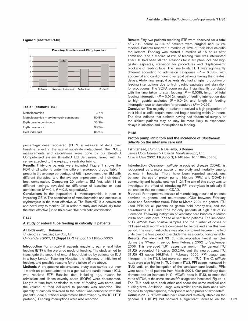
S59
percentage dose recovered (PDR), a measure of delta overbaseline reflecting the rate of substrate metabolized. The 13CO2measurements and calculations were done by our BreathIDComputerized system (BreathID Ltd, Jerusalem, Israel) with itssensor attached to the expiratory ventilator tubing.Results Thirty-one patients were included. Figure 1 shows thePDR of all patients under the different prokinetic drugs. Table 1presents the average percentage of GE improvement over BM withdifferent therapies, and the average improvement of individuals’best combination. Comparing 20 patients, BM first, with 11 atdifferent timings, revealed no difference of baseline or bestcombination (P = 0.1, P = 0.2, respectively).Conclusions In this population: 1. Metoclopramide is poor inimproving GE. 2. The combination of metoclopramide and continuouserythromycin is the most effective. 3. The BreathID is a convenientand novel way to monitor GE in order to study and individually tailorthe most effective (up to 85% over BM) prokinetic combination.
P147A study of enteral tube feeding in critically ill patients
A Holdsworth, T RahmanSt George’s Hospital, London, UKCritical Care 2007, 11(Suppl 2):P147 (doi: 10.1186/cc5307)
Introduction For critically ill patients unable to eat, enteral tubefeeding (ETF) is the preferred mode of feeding. The study aimed toinvestigate the amount of enteral feed obtained by patients on ICUin a busy London Teaching Hospital, the efficiency of initiation offeeding, and possible reasons for the failure of the above.Methods A prospective observational study was carried out over1 month on patients admitted to a general and cardiothoracic ICU,who received ETF. Baseline data including age, reason foradmission and illness severity score (SOFA) were documented.Length of time from admission to start of feeding was noted, andthe volume of feed delivered to patients was recorded. Thequantity of calories delivered to the patient was compared with thepatient’s ideal nutritional requirement (determined by the ICU ETFprotocol). Feeding interruptions were also recorded.
Results Fifty-two patients receiving ETF were observed for a totalof 7,349 hours: 67.3% of patients were surgical and 32.7%medical. Patients received a median of 75% of their ideal calorificrequirement. Feeding was started a median of 15 hours afteradmission, and a median of 5% of feeding time was interruptedafter ETF had been started. Reasons for interruption included highgastric aspirates, starvation for procedures and displacement/blockage of feeding tube. The time to start ETF was significantlydifferent according to admission categories (P = 0.033), withabdominal and cardiothoracic surgical patients having the greatestdelays. Abdominal surgical patients also had a higher proportion offeeding interruptions due to high gastric aspirates and starvationfor procedures. The SOFA score on day 1 significantly correlatedwith the time taken to start feeding (P = 0.008), length of totalfeeding interruption (P = 0.012), length of feeding interruption dueto high gastric aspirates (P = 0.043), and length of feedinginterruption due to starvation for procedures (P = 0.026).Conclusion The majority of patients received a high proportion oftheir ideal calorific requirement and began feeding within 24 hours.The data indicate that patients having had abdominal surgery orthe sickest patients may be may be more likely to experiencedelays in initiation and interruptions to feeding.
P148Proton pump inhibitors and the incidence of Clostridiumdifficile on the intensive care unit
I Whitehead, J Smith, R Bellamy, S BonnerJames Cook University Hospital, Middlesbrough, UKCritical Care 2007, 11(Suppl 2):P148 (doi: 10.1186/cc5308)
Introduction Clostridium difficile associated disease (CDAD) isrecognized as a major cause of morbidity and mortality amongpatients in hospital. There have been reported associationsbetween the use of proton pump inhibitors (PPIs) and CDAD incommunity and hospital settings [1,2]. The aim of this study was toinvestigate the effect of introducing PPI prophylaxis in critically illpatients on the incidence of CDAD.Methods Retrospective analysis of microbiology results of patientsadmitted to general and neurotrauma ICUs between February2002 and September 2006. Prior to March 2004 the general ITUused PPIs for all patients as gastric acid prophylaxis, and theneurotrauma ITU used PPIs for only patients at high risk of GIulceration. Following instigation of ventilator care bundles in March2004 both units gave PPIs to all ventilated patients. The incidenceof C. difficile toxin-positive samples and the number of doses ofPPI used each month were compared for before and after this timeperiod. The use of antibiotics was also compared between the twounits over the time period to exclude this as a confounding variable.Results We identified 92 C. difficile-positive faecal samplesduring the 57-month period from February 2002 to September2006. This averaged 1.61 cases per month. The general ITU(ITU2) presented 49 cases (53.2%), and the neurotrauma ITU(ITU3) 43 cases (46.8%). In February 2002, PPI usage wasinfrequent in the ITU3, but more common in ITU2. The C. difficilerates were also higher in ITU2 than in ITU3. PPI usage increased inITU3 until, on the instigation of the ventilator care bundle, PPIswere used for all patients from March 2004. Our preliminary datademonstrate an increase in C. difficile rates in ITU3, to meet therates of ITU2, at the same time as PPI usage was increased (Figure 1).The ITUs back onto each other and share the same medical andnursing staff. Antibiotic usage was similar across both units withregards to cephalosporins, meropenem and pipracillin/tazobactam.Conclusion C. difficile rates have remained relatively stable on thegeneral ITU (ITU2) but showed a significant increase on the
Available online http://ccforum.com/supplements/11/S2
Figure 1 (abstract P146)
Table 1 (abstract P146)
Metoclopramide 13.7%
Metoclopramide + erythromycin continuous 50.5%
Erythromycin continuous 33.3%
Erythromycin x 2 38.7%
Best individual 85.2%
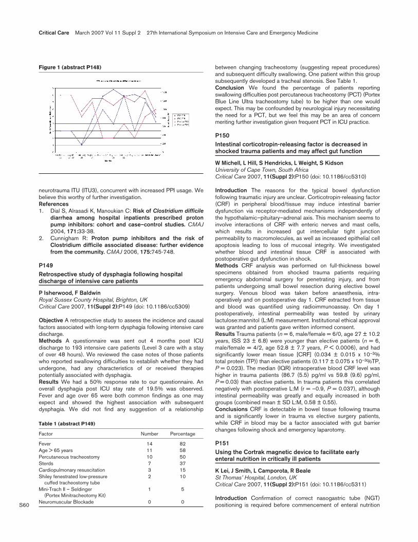
S60
neurotrauma ITU (ITU3), concurrent with increased PPI usage. Webelieve this worthy of further investigation.References1. Dial S, Alrasadi K, Manoukian C: Risk of Clostridium difficile
diarrhea among hospital inpatients prescribed protonpump inhibitors: cohort and case–control studies. CMAJ2004, 171:33-38.
2. Cunnigham R: Proton pump inhibitors and the risk ofClostridium difficile associated disease: further evidencefrom the community. CMAJ 2006, 175:745-748.
P149Retrospective study of dysphagia following hospitaldischarge of intensive care patients
P Isherwood, F BaldwinRoyal Sussex County Hospital, Brighton, UKCritical Care 2007, 11(Suppl 2):P149 (doi: 10.1186/cc5309)
Objective A retrospective study to assess the incidence and causalfactors associated with long-term dysphagia following intensive caredischarge.Methods A questionnaire was sent out 4 months post ICUdischarge to 193 intensive care patients (Level 3 care with a stayof over 48 hours). We reviewed the case notes of those patientswho reported swallowing difficulties to establish whether they hadundergone, had any characteristics of or received therapiespotentially associated with dysphagia.Results We had a 50% response rate to our questionnaire. Anoverall dysphagia post ICU stay rate of 19.5% was observed.Fever and age over 65 were both common findings as one mayexpect and showed the highest association with subsequentdysphagia. We did not find any suggestion of a relationship
between changing tracheostomy (suggesting repeat procedures)and subsequent difficulty swallowing. One patient within this groupsubsequently developed a tracheal stenosis. See Table 1.Conclusion We found the percentage of patients reportingswallowing difficulties post percutaneous tracheostomy (PCT) (PortexBlue Line Ultra tracheostomy tube) to be higher than one wouldexpect. This may be confounded by neurological injury necessitatingthe need for a PCT, but we feel this may be an area of concernmeriting further investigation given frequent PCT in ICU practice.
P150Intestinal corticotropin-releasing factor is decreased inshocked trauma patients and may affect gut function
W Michell, L Hill, S Hendricks, L Weight, S KidsonUniversity of Cape Town, South AfricaCritical Care 2007, 11(Suppl 2):P150 (doi: 10.1186/cc5310)
Introduction The reasons for the typical bowel dysfunctionfollowing traumatic injury are unclear. Corticotropin-releasing factor(CRF) in peripheral blood/tissue may induce intestinal barrierdysfunction via receptor-mediated mechanisms independently ofthe hypothalamic–pituitary–adrenal axis. This mechanism seems toinvolve interactions of CRF with enteric nerves and mast cells,which results in increased gut intercellular tight junctionpermeability to macromolecules, as well as increased epithelial cellapoptosis leading to loss of mucosal integrity. We investigatedwhether blood and intestinal tissue CRF is associated withpostoperative gut dysfunction in shock.Methods CRF analysis was performed on full-thickness bowelspecimens obtained from shocked trauma patients requiringemergency abdominal surgery for penetrating injury, and frompatients undergoing small bowel resection during elective bowelsurgery. Venous blood was taken before anaesthesia, intra-operatively and on postoperative day 1. CRF extracted from tissueand blood was quantified using radioimmunoassay. On day 1postoperatively, intestinal permeability was tested by urinarylactulose:mannitol (L:M) measurement. Institutional ethical approvalwas granted and patients gave written informed consent.Results Trauma patients (n = 6, male/female = 6/0, age 27 ± 10.2years, ISS 23 ± 6.8) were younger than elective patients (n = 6,male/female = 4/2, age 52.8 ± 7.7 years, P < 0.0006), and hadsignificantly lower mean tissue [CRF] (0.034 ± 0.015 x 10⁻³%total protein (TP)) than elective patients (0.117 ± 0.075 x 10⁻³%TP,P = 0.023). The median (IQR) intraoperative blood CRF level washigher in trauma patients (86.7 (5.5) pg/ml vs 59.8 (9.6) pg/ml,P = 0.03) than elective patients. In trauma patients this correlatednegatively with postoperative L:M (r = –0.9, P = 0.037), althoughintestinal permeability was greatly and equally increased in bothgroups (combined mean ± SD L:M, 0.58 ± 0.55).Conclusions CRF is detectable in bowel tissue following traumaand is significantly lower in trauma vs elective surgery patients,while CRF in blood may be a factor associated with gut barrierchanges following shock and emergency laparotomy.
P151Using the Cortrak magnetic device to facilitate earlyenteral nutrition in critically ill patients
K Lei, J Smith, L Camporota, R BealeSt Thomas’ Hospital, London, UKCritical Care 2007, 11(Suppl 2):P151 (doi: 10.1186/cc5311)
Introduction Confirmation of correct nasogastric tube (NGT)positioning is required before commencement of enteral nutrition
Critical Care March 2007 Vol 11 Suppl 2 27th International Symposium on Intensive Care and Emergency Medicine
Figure 1 (abstract P148)
Table 1 (abstract P149)
Factor Number Percentage
Fever 14 82Age > 65 years 11 58Percutaneous tracheostomy 10 50Sterds 7 37Cardiopulmonary resuscitation 3 15Shiley fenestrated low-pressure 2 10
cuffed tracheostomy tubeMini-Trach II – Seldinger 1 5
(Portex Minitracheotomy Kit)Neuromuscular Blockade 0 0
S61
(EN). In the ICU, however, the use of sedation, 24-hour feedingand proton pump inhibitors can make the standard confirmatorymethods recommended by the UK National Patient Safety Agency(UKNPSA) [1] unreliable, and result in the need for multiple chestX-rays (CXR), increased cost and feeding delay. We studied therole of the Cortrak® (Viasys MedSystems, USA) against standardpractice, and assessed the following outcomes: time required toconfirm correct NGT position, time to starting feeding, andpotential cost savings.Methods We enrolled patients admitted to our ICU who requiredNGT insertion for EN. A Corflow NGT with Cortrak stylet wasinserted, and the position monitored using the Cortrak magneticsensor. The Cortrak system tracks the trail of the stylet tip as itprogresses towards the stomach and provides a visual represen-tation of the NGT position on a video screen, allowing rapiddetermination of insertion success. The position of the NGT wasalso assessed using pH paper and/or CXR, as appropriate,according to the standard UKNPSA guidelines. Data wereanalysed using a paired t test and time-to-event analysis.Results Fifty-two patients were recruited and 57 insertions wereanalysed: 32 first insertions, and 25 reinsertions. Gastric contentwas successfully aspirated in 40% (23/57) of insertions, and 14%(8/57) had pH 6 or above. Forty-six CXRs were requested, withthree patients requiring multiple CXRs. The Cortrak correctlyconfirmed the position of NGT in all 57 insertions. The timerequired to confirm the NGT position was significantly less with theCortrak than with conventional methods (mean ± SE Cortrak:9.6 ± 1.7 min; pH paper: 11.6 ± 1.7 min; CXR: 122 ± 23 min;P < 0.0001). There was a 1.5-hour delay in starting EN in thestandard practice group compared with the Cortrak group(mean ± SE 3.98 ± 0.5 hours vs 2.58 ± 0.4 hours, P = 0.049). Ifthe Cortrak results had been acted upon, 46 CXRs could havebeen avoided, which equates to a saving of £2,300 (€3,220).Conclusions The Cortrak demonstrated 100% accuracy inconfirming the position of NGT in this patient series. It exposespatients to less radiation, facilitates earlier EN and is cost-effective.Consideration should therefore be given to including it into thestandard UKNPSA guideline.Reference1. UK National Patient Safety Agency [http://www.npsa.nhs.uk]
P152Propranolol attenuates factors affecting hypermetabolismin pediatric burn patients
W NorburyShriner’s Hospital for Children, Galveston, TX, USACritical Care 2007, 11(Suppl 2):P152 (doi: 10.1186/cc5312)
Background The aim of this study was to determine the effect ofpropranolol on infections and clinical parameters during the acutephase postburn. Severe thermal injury is followed by a period ofhypermetabolism that is directly proportional to the size of insultsustained. Infection and multiorgan failure are now the leadingcause of death from severe thermal injuries. Propranolol, ananticatabolic agent, improves hypermetabolism postburn. However,there is evidence that propranolol worsens immune function andincreases the incidence of infection in critically ill patients.Methods Sixty-six patients with burns >40% total body surfacearea were enrolled into the study and randomized to receivestandard burn care (controls, n = 33) or standard burn pluspropranolol for more than 21 days (propranolol, 0.5–1.5 mg/kgevery 6 hours, n = 33). Biopsies were taken three times a week formicrobiological determination. Clinical parameters were collectedand blood was drawn at regular intervals throughout the hospital
course and analyzed for IGF-1, IGFBP-3, and HGH. Patientsunderwent weekly resting energy-expenditure measurements.Statistical analysis was performed using analysis of variance withBonferoni’s correction and Student’s t test where applicable.Results Propranolol treatment reduced heart rates by 10% andsignificantly improved stroke volume throughout the acute hospitalstay compared with controls (P < 0.05). Resting energy expendi-ture was significantly decreased in the propranolol group whencompared with controls at discharge (P < 0.05). Infection rates onadmission were the same for both groups (17% propranolol vs22% control). The incidence of infection throughout hospitalcourse was significantly lower in the propranolol group (60%)compared with controls (87%) (P < 0.05). Propranolol significantlyincreased IGF-I, IGFBP-3, GH, and prealbumin, while itsignificantly decreased CRP and fatty acids (P < 0.05).Conclusions Following a severe burn, propranolol attenuatesinfections, inflammatory markers and fatty acid levels whileimproving cardiac work and endogenous anabolic hormone levels.We suggest that propranolol is a safe and efficacious modulator ofthe postburn response.
P153Early enteral immunonutrition following gastric andoesophageal surgery
S Vukosavljevic, T Randjelovic, D Pavlovic, J DanojlicKBC ‘Bezanijska Kosa’ Beograd, Belgrade, SerbiaCritical Care 2007, 11(Suppl 2):P153 (doi: 10.1186/cc5313)
Introduction The study investigates the effect of early enteralimmunonutrition on patient recovery after extensive elective surgeryin the upper abdomen [1,2]. It investigates the speed of patientrecovery administering early enteral immunonutrition combinedwith total parenteral nutrition [3].Materials and methods The total of 40 patients who hadundergone this type of surgery were involved in this study. Near theend of the surgery procedure a percutaneous jejunostomy wasperformed in 20 patients (G1), and enteral nutrition started on thefirst postoperative day with small doses of immunonutrient(Reconvan) 10 ml/hour. After every 12 hours the tolerance wasestimated (abdominal distension, diarrhoea, vomiting). After every24 hours the immunonutrient dose was increased by 20 ml/houruntil we reached the maximum of 80 ml/hour. In the first threepostoperative days the patients were also administered totalparenteral nutrition, and after that only enteral nutrition. The othergroup of 20 patients (G2) was administered only parenteralnutrition from the first postoperative day. Preoperatively, everypatient was measured for body weight, body height and body massindex, and using laboratory tests we established the levels ofalbumin, transferine, blood urea nitrogen and creatinine. On thethird and ninth postoperative days we repeated the samelaboratory tests, and measured the daily loss of nitrogen byexcretion of urea in urine.Results and discussion Patient recovery was faster in G1. Theaverage patient stay in ICU was 5 ± 1 days (G1) vs 10 ± 2 days(G2). The average hospital stay was 22 ± 3 days (G1) vs 29 ± 5days. Peristalsis was detected on the third day as an average (G1)vs 4.5 days (G2). A decrease in pulmonary complications wasachieved in G1 (one pleural effusion) vs G2 (eight pleuraleffusions). Laboratory tests show that patients in G1 are in lowercatabolism compared with G2 patients.Conclusion Early enteral immunonutrition through jejunostomy isan efficient and safe method of patient nutrition with fewerpostoperative complications, and also accounts for a hospital costdecrease of 50%.
Available online http://ccforum.com/supplements/11/S2
S62
References1. Delaney HM, Carenevale NH, Garvey JW: Jejunostomy by a
needle catheter technique. Surgery 1973, 73:786-945.2. Braga M, Giannoti L, Radaelli G, et al.: Perioperative
immunonutrition in patients undergoing cancer surgery:results of a randomized double-blinded phase 3 trial. ArchSurg 1999, 134:428-433.
3. Moore EE, Jones TN: Benefits of immediate jejunostomyfeeding after major abdominal trauma: a prospective ran-domized study. J Trauma 1986, 26:874-881.
P154Feasibility of the REDOXS study – reducing deaths due tooxidative stress: a randomized pilot trial of glutamine andantioxidant supplementation in critically ill patients
D Heyland, R Dhaliwal, J Muscedere, J Drover, for the CanadianCritical Care Trials GroupKingston General Hospital, Kingston, CanadaCritical Care 2007, 11(Suppl 2):P154 (doi: 10.1186/cc5314)
Introduction A large randomized trial is needed to evaluate thesafety and efficacy of glutamine (GLN) and antioxidant (AOX)supplements. However, high doses of such nutrients via enteraland parenteral routes early in the course of critical illness is ofteninterrupted by high illness acuity and other treatment priorities. Thepurpose of this pilot trial was to evaluate the feasibility of deliveringhigh-dose GLN and AOX supplements early on in the course ofcritical illness, and to estimate recruitment for the larger REDOXSstudy.Methods In six Canadian centers, using a 2 x 2 factorial design,we randomized mechanically ventilated adults who had two ormore organ failures within 24 hours of ICU admission to one of fourgroups: (1) GLN (0.35 g/kg/day i.v. plus 30 g enterally), (2) AOX(500 µg selenium i.v. and 300 µg selenium, 20 mg zinc, 10 mgβ-carotene, 500 mg vitamin E, and 1,500 mg vitamin C enterally),(3) both AOX + GLN, and (4) placebo. Supplementation wascontinued for a minimum of 5 days up to 28 days and wasprovided independent of nutrition support. We recorded the timefrom ICU admission to randomization, the time to start ofsupplements and nutrition support parameters.Results From April 2005 to April 2006, 80 patients wererandomized (average 2.1/site/month). The median time from ICUadmission to randomization was 18.2 hours (range 11.6–21.1 hours).All patients received parenteral supplements, the median (range)time to start was 2.7 hours (2.0–3.8 hours) and 78/80 (98%)received enteral supplements with a median (range) of 2.6 hours(1.9–4.5 hours) from randomization. The mean duration ofsupplements was 11.1 days (enteral) and 12.2 days (parenteral).The mean volumes of enteral and parenteral supplements receivedwere 84% (range 45–102%) and 93% (range 54–100%)prescribed volumes, respectively. The average prescribed energyand protein intakes were 1,802 kcal/day and 86 g protein/day butthe average daily percentage energy and protein received fromnutrition support was only 65% (range 4–95%) and 62% (range2–97%) of that prescribed, respectively.Conclusion In critically ill patients with organ failure we providedadequate amounts of study supplements via both enteral andparenteral routes in the early phases of acute illness, independentof nutrition support. We estimated recruitment of at least twopatients/site/month for our future trial.Acknowledgements This study was supported by grants from theCanadian Institutes of Health Research and Fresenius-Kabi,Germany.
P155Effect of high-dose selenium substitution on selectedlaboratory parameters and prognosis in critically illpatients
J Valenta1, H Brodska1, A Kazda2, Z Stach3, J Hendl4, K Pelinkova1
1General Teaching Hospital, Charles University, Prague, CzechRepublic; 2University Hospital, Prague, Czech Republic; 3GeneralTeaching Hospital 1st Medical Faculty of Charles University,Prague, Czech Republic; 3School of Physical Education and Sport,Charles University, Prague, Czech RepublicCritical Care 2007, 11(Suppl 2):P155 (doi: 10.1186/cc5315)
Introduction Standard selenium (Se) substitution (30–75 µg/day;0.4–0.9 µmol/l) in the critically ill is not sufficient for a sustainedplasma level (0.58–1.82 µmol/l; 46–143 µg/l). Standard Sesubstitution keeps the plasma level in the range 0.28–0.42 µmol/l.High-dose Se substitution correlated with a decrease in mortalityof patients with SIRS. The influence of high-dose substitution onselected parameters, MAP and mortality in the critically ill wereevaluated in a prospective randomized clinical trial.Methods One hundred and twenty-three patients (78 males, 45females, median age 62.7 and 60 years, respectively) wererandomized into group A (SOFA 19.27) and group B (SOFA10.23). Group A received standard Se substitution: 30–75 µgNaSelenite i.v./day. Group B received high-dose Se substitutionaccording to a protocol: 1,000 µg at day 1, followed with 500 µgat days 2–14 of NaSelenite i.v. The plasma levels of Se,prealbumin, albumin, CRP, PCT, cholesterol, gluthathionperoxidaseGSHPx, D-dimer, creatinine clearance and leucocytes wereexamined daily. MAP trends and 28-day mortality were evaluatedas clinical markers.Results The Se plasma level was significantly higher in high-doseSe-substituted patients (0.56 µmol/l vs 0.88 µmol/l, P < 0.001).GSHPx was significantly higher in high-dose Se-substitutedpatients (4,864 U/l vs 6,097 U/l, P < 0.001). No significantdifferences were found in the level of albumin, prealbumin, CRP,PCT, leucocytes, fibrinogen, cholesterol, D-dimer and creatinineclearance and MAP. The 28-day mortality was lower in a high-doseSe-substituted patients (33% vs 37%), but not significantly.Conclusion The critically ill have an increased demand for Se,which is essential for synthesis of Se enzymes and Se proteins.The increased demand for Se is not covered by standardsubstitution. High-dose Se substitution (500–1,000 µg/day)normalizes its plasma level and increases the GSHPx plasma level.High-dose Se substitution has no adverse reactions. The decreaseof 28-day mortality in high-dose Se-substituted patients is notsignificant. The trial on high-dose Se substitution further continues.
P156Magnesium deficiency in the surgical intensive care unit
W SalemNational Cancer Institute, Cairo, EgyptCritical Care 2007, 11(Suppl 2):P156 (doi: 10.1186/cc5316)
Introduction Magnesium (Mg) deficiency is a common and yetunderdiagnosed problem. Mg deficiency has been demonstrated in50% of all ICU patients. These patients have significantly highermorbidity and mortality rate. The aim of this work was to detect thepresence of Mg deficiency using a Mg loading test and to evaluatethe safety and efficacy of Mg replacement therapy in cancerpatients after major gut surgery.
Critical Care March 2007 Vol 11 Suppl 2 27th International Symposium on Intensive Care and Emergency Medicine
S63
Methods Sixty adult patients admitted to the ICU after major GITsurgery were enrolled in the study, on the basis of a documentedMg serum level < 0.8 mmol/l on arrival at the ICU. Exclusion criteriawere cardiac disease, liver dysfunction or serum creatinine > 1.3.Patients were randomly allocated to one of two groups: the Mggroup received 20 mmol (5 g) Mg sulfate, infused daily over6 hours for 3 days; and a control group received an equivalentamount of 5% dextrose. In the Mg group the next scheduled doseof MgSO4 was held if a serum magnesium level > 1.1 mmol/l,hypotension or bradycardia was recorded. Baseline and dailymeasurements of serum Mg, potassium, sodium, calcium andcreatinine were done. Twenty-four-hour urine collection was usedto determine the total urinary excretion of Mg. The net Mg balance(total Mg given – total urine Mg) was calculated. In the Mg group,Mg-deficient patients (retainers) who excreted < 70% of the Mggiven ((urine Mg in mmol / daily Mg given) x 100) and Mgnondeficient patients (nonretainers) who excreted > 70% of thetotal Mg given were recorded.Results Patients in the Mg group showed a statistically significantincrease in mean serum Mg at days 1, 2, and 3 compared with thecontrol group and with day 0 (0.73 ± 0.1, 0.81 ± 0.17, 0.8 ± 0.1,and 0.85 ± 0.11 mmol/l at days 0, 1, 2, and 3, respectively). Themean total amount of Mg given, the mean total urine Mg excretionand the net Mg balance were significantly higher in the Mg groupcompared with the control group (58 ± 0.17 vs 11.3 ± 2.8 mmol,P > 0.001; 34.0 ± 2.7 vs 15.5 ± 3.8 mmol, P > 0.001; 25.6 ± 1.65vs 5.2 ± 0.93 mmol, P > 0.001). In the Mg group, the numbers ofMg retainer patients were 24 patients on day 1, 21 on day 2 andnine patients on day 3. Mg nonretainer patients were six patientson day 1, nine on day 2 and 21 patients on day 3. Patients in theMg group showed better haemodynamic stability and fewerventricular arrhythmias.Conclusion Mg deficiency is common in ICU patients. Mg sulfateadministered according to the above regimen is safe. Earlytreatment of Mg deficiency significantly increased the serum Mglevel and provided a better magnesium, potassium and calciumbalance, resulting in a shorter ICU stay.
P157Parenteral nutrition in the intensive care unit: can wedeliver better care to our patients? Preliminary resultsfrom a multicenter, prospective, cohort study
A Pontes-Arruda1, J Teles2, E Silva3, F Machado4, M BaptistaFilho5, E Rocha6, C Silva7
1Hospital Fernandes Távora, Fortaleza, Brazil; 2Hospital Português,Salvador, Brazil; 3Hospital Albert Einstein, São Paulo, Brazil;4Hospital São Paulo – UNIFESP, São Paulo, Brazil; 5HospitalBandeirantes, São Paulo, Brazil; 6Hospital Copa D’Or, Rio deJaneiro, Brazil; 7Latin American Sepsis Institute, São Paulo, BrazilCritical Care 2007, 11(Suppl 2):P157 (doi: 10.1186/cc5317)
Introduction Nutrition therapy is an integrant aspect of ICUsupport and can influence outcomes. A delay to starting nutritionsupport after 24 hours of ICU admission is associated withincreased morbidity and mortality [1], and certain lipid emulsionscan exacerbate the inflammatory cascade. For an appropriateevaluation of the impact of these and other recent researchfindings, information regarding the use of parenteral nutrition (PN)in the ICU is needed.Methods This is the interim analysis of a multicenter, prospective,cohort study aimed to obtain information regarding the use of PN.Data were collected during 3 months from ICU patients over18 years of age on the use of PN in 20 adult ICUs in Brazil using aweb-based clinical research form.
Results One hundred and sixty-six patients were included in thisanalysis. Among the main results, 63.69% were males and77.78% were considered malnourished. The mean SOFA scorewas 6.21, with a mean APACHE II score of 19.39. In total, 97.23%of the PN used in Brazil were manufactured by third-partycompanies and this was associated with a significant delay in thebeginning of the infusion (median time 29.76 hours), and elevatedin-ICU (50%) and inhospital (55.17%) mortality rates. A total24.29% of the patients were immunosuppressed. The most usedlipid source was long-chain triglycerides/medium-chain triglycerides(80.69%).Conclusions The use of PN in Brazil is associated with asignificant delay in the start of infusion and high mortality rates. Themost used lipid emulsion (long-chain triglycerides/medium-chaintriglycerides) has been associated with more apoptosis [2] andcompromised lymphocyte proliferation [3]. The overall findings ofthese study indicate that strategies to reduce the delay in start ofPN and the use of better lipid sources must be adopted to providebetter assistance for patients in need of PN in Brazil.Acknowledgement Supported by a research grant from BaxterHospitalar Ltda.References1. Simpson F, Doig GS: Parenteral vs. enteral nutrition in the
critically ill patient: a meta-analysis of trials using theintention to treat principle. Intensive Care Med 2005, 31:12-23.
2. Buenestado A, Cortijo J, Sanz M-J, et al.: Olive oil-basedlipid emulsion’ s neutral effect on neutrophil functions andleucocyte–endothelial cell interactions. J Parenter EnteralNutr 2006, 30:286-296.
3. Cury-Boaventura M, Gorjão R, Martins de Lima T, et al.: Toxic-ity of two lipid emulsions on human lymphocytes and neu-trophils. Crit Care, in press.
P158Ultrasound-guided vs ultrasound-assisted central venouscatheterization
M Pittiruti, A LaGreca, G Scoppettuolo, D SermonetaCatholic University Hospital, Roma, ItalyCritical Care 2007, 11(Suppl 2):P158 (doi: 10.1186/cc5318)
Introduction Ultrasound (US) significantly facilitates centralvenous catheterization, reducing the percentage of failure, thepercentage of accidental arterial puncture, and the percentage ofcomplications (haematoma, haemothorax, pneumothorax).Nonetheless, it is not clear whether US guidance (USG) (so-called‘dynamic’ or ‘real-time’ US techniques: that is, venipuncture underdirect US control) may be better than US assistance (USA) (so-called ‘static’ or ‘indirect’ US techniques: that is, US imaging of thevein, with or without skin marking, and then blind venipuncture).Methods From February 2005 to September 2006, our CVC Teamadopted the following protocol for internal jugular vein (IJV)catheterization: (a) both IJVs were evaluated to assess position,dimensions, and other features known to affect the risk ofcatheterization; (b) then, a decision was made whether to continuewith USA or USG; (c) the IJV was accessed via the low lateralJernigan approach; (d) after two failed USA attempts, USGvenipuncture was adopted; (d) when IJVs were not available, USGvenipuncture of other central veins was the second choice; and (e)fluoroscopy was used only in paediatric patients, but all patients had apostoperative chest X-ray to rule out pneumothorax and malposition.Results In 20 months, 821 central venous catheters (CVCs) wereinserted in adults (181 short-term CVC + 218 tunnelled + 316ports) and in paediatric patients (age range 20 days–13 years,
Available online http://ccforum.com/supplements/11/S2
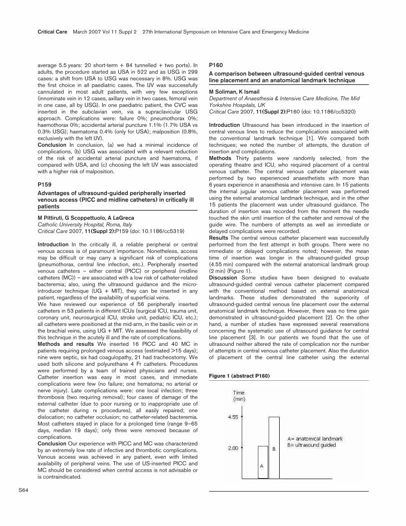
S64
average 5.5 years: 20 short-term + 84 tunnelled + two ports). Inadults, the procedure started as USA in 522 and as USG in 299cases: a shift from USA to USG was necessary in 8%. USG wasthe first choice in all paediatric cases. The IJV was successfullycannulated in most adult patients, with very few exceptions(innominate vein in 12 cases, axillary vein in two cases, femoral veinin one case, all by USG). In one paediatric patient, the CVC wasinserted in the subclavian vein, via a supraclavicular USGapproach. Complications were: failure 0%; pneumothorax 0%;haemothorax 0%; accidental arterial puncture 1.1% (1.7% USA vs0.3% USG); haematoma 0.4% (only for USA); malposition (0.8%,exclusively with the left IJV).Conclusion In conclusion, (a) we had a minimal incidence ofcomplications, (b) USG was associated with a relevant reductionof the risk of accidental arterial puncture and haematoma, ifcompared with USA, and (c) choosing the left IJV was associatedwith a higher risk of malposition.
P159Advantages of ultrasound-guided peripherally insertedvenous access (PICC and midline catheters) in critically illpatients
M Pittiruti, G Scoppettuolo, A LaGrecaCatholic University Hospital, Roma, ItalyCritical Care 2007, 11(Suppl 2):P159 (doi: 10.1186/cc5319)
Introduction In the critically ill, a reliable peripheral or centralvenous access is of paramount importance. Nonetheless, accessmay be difficult or may carry a significant risk of complications(pneumothorax, central line infection, etc.). Peripherally insertedvenous catheters – either central (PICC) or peripheral (midlinecatheters (MC)) – are associated with a low risk of catheter-relatedbacteremia; also, using the ultrasound guidance and the micro-introducer technique (UG + MIT), they can be inserted in anypatient, regardless of the availability of superficial veins.We have reviewed our experience of 56 peripherally insertedcatheters in 53 patients in different ICUs (surgical ICU, trauma unit,coronary unit, neurosurgical ICU, stroke unit, pediatric ICU, etc.);all catheters were positioned at the mid-arm, in the basilic vein or inthe brachial veins, using UG + MIT. We assessed the feasibility ofthis technique in the acutely ill and the rate of complications.Methods and results We inserted 16 PICC and 40 MC inpatients requiring prolonged venous access (estimated >15 days);nine were septic, six had coagulopathy, 21 had tracheostomy. Weused both silicone and polyurethane 4 Fr catheters. Procedureswere performed by a team of trained physicians and nurses.Catheter insertion was easy in most cases, and immediatecomplications were few (no failure; one hematoma; no arterial ornerve injury). Late complications were: one local infection; threethrombosis (two requiring removal); four cases of damage of theexternal catheter (due to poor nursing or to inappropriate use ofthe catheter during rx procedures), all easily repaired; onedislocation; no catheter occlusion; no catheter-related bacteremia.Most catheters stayed in place for a prolonged time (range 9–65days, median 19 days); only three were removed because ofcomplications.Conclusion Our experience with PICC and MC was characterizedby an extremely low rate of infective and thrombotic complications.Venous access was achieved in any patient, even with limitedavailability of peripheral veins. The use of US-inserted PICC andMC should be considered when central access is not advisable oris contraindicated.
P160A comparison between ultrasound-guided central venousline placement and an anatomical landmark technique
M Soliman, K IsmailDepartment of Anaesthesia & Intensive Care Medicine, The MidYorkshire Hospitals, UKCritical Care 2007, 11(Suppl 2):P160 (doi: 10.1186/cc5320)
Introduction Ultrasound has been introduced in the insertion ofcentral venous lines to reduce the complications associated withthe conventional landmark technique [1]. We compared bothtechniques; we noted the number of attempts, the duration ofinsertion and complications.Methods Thirty patients were randomly selected, from theoperating theatre and ICU, who required placement of a centralvenous catheter. The central venous catheter placement wasperformed by two experienced anaesthetists with more than6 years experience in anaesthesia and intensive care. In 15 patientsthe internal jugular venous catheter placement was performedusing the external anatomical landmark technique, and in the other15 patients the placement was under ultrasound guidance. Theduration of insertion was recorded from the moment the needletouched the skin until insertion of the catheter and removal of theguide wire. The numbers of attempts as well as immediate ordelayed complications were recorded.Results The central venous catheter placement was successfullyperformed from the first attempt in both groups. There were noimmediate or delayed complications noted; however, the meantime of insertion was longer in the ultrasound-guided group(4.55 min) compared with the external anatomical landmark group(2 min) (Figure 1).Discussion Some studies have been designed to evaluateultrasound-guided central venous catheter placement comparedwith the conventional method based on external anatomicallandmarks. These studies demonstrated the superiority ofultrasound-guided central venous line placement over the externalanatomical landmark technique. However, there was no time gaindemonstrated in ultrasound-guided placement [2]. On the otherhand, a number of studies have expressed several reservationsconcerning the systematic use of ultrasound guidance for centralline placement [3]. In our patients we found that the use ofultrasound neither altered the rate of complication nor the numberof attempts in central venous catheter placement. Also the durationof placement of the central line catheter using the external
Critical Care March 2007 Vol 11 Suppl 2 27th International Symposium on Intensive Care and Emergency Medicine
Figure 1 (abstract P160)
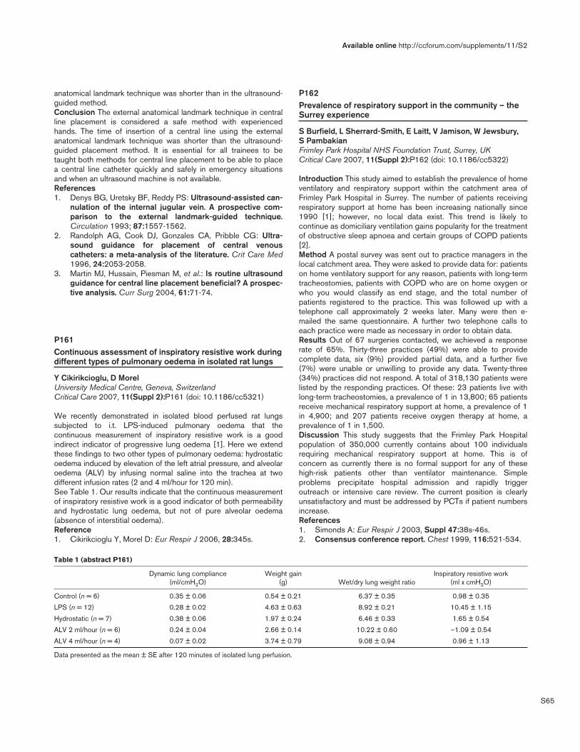
S65
anatomical landmark technique was shorter than in the ultrasound-guided method.Conclusion The external anatomical landmark technique in centralline placement is considered a safe method with experiencedhands. The time of insertion of a central line using the externalanatomical landmark technique was shorter than the ultrasound-guided placement method. It is essential for all trainees to betaught both methods for central line placement to be able to placea central line catheter quickly and safely in emergency situationsand when an ultrasound machine is not available.References1. Denys BG, Uretsky BF, Reddy PS: Ultrasound-assisted can-
nulation of the internal jugular vein. A prospective com-parison to the external landmark-guided technique.Circulation 1993; 87:1557-1562.
2. Randolph AG, Cook DJ, Gonzales CA, Pribble CG: Ultra-sound guidance for placement of central venouscatheters: a meta-analysis of the literature. Crit Care Med1996, 24:2053-2058.
3. Martin MJ, Hussain, Piesman M, et al.: Is routine ultrasoundguidance for central line placement beneficial? A prospec-tive analysis. Curr Surg 2004, 61:71-74.
P161Continuous assessment of inspiratory resistive work duringdifferent types of pulmonary oedema in isolated rat lungs
Y Cikirikcioglu, D MorelUniversity Medical Centre, Geneva, SwitzerlandCritical Care 2007, 11(Suppl 2):P161 (doi: 10.1186/cc5321)
We recently demonstrated in isolated blood perfused rat lungssubjected to i.t. LPS-induced pulmonary oedema that thecontinuous measurement of inspiratory resistive work is a goodindirect indicator of progressive lung oedema [1]. Here we extendthese findings to two other types of pulmonary oedema: hydrostaticoedema induced by elevation of the left atrial pressure, and alveolaroedema (ALV) by infusing normal saline into the trachea at twodifferent infusion rates (2 and 4 ml/hour for 120 min).See Table 1. Our results indicate that the continuous measurementof inspiratory resistive work is a good indicator of both permeabilityand hydrostatic lung oedema, but not of pure alveolar oedema(absence of interstitial oedema).Reference1. Cikirikcioglu Y, Morel D: Eur Respir J 2006, 28:345s.
P162Prevalence of respiratory support in the community – theSurrey experience
S Burfield, L Sherrard-Smith, E Laitt, V Jamison, W Jewsbury, S PambakianFrimley Park Hospital NHS Foundation Trust, Surrey, UKCritical Care 2007, 11(Suppl 2):P162 (doi: 10.1186/cc5322)
Introduction This study aimed to establish the prevalence of homeventilatory and respiratory support within the catchment area ofFrimley Park Hospital in Surrey. The number of patients receivingrespiratory support at home has been increasing nationally since1990 [1]; however, no local data exist. This trend is likely tocontinue as domiciliary ventilation gains popularity for the treatmentof obstructive sleep apnoea and certain groups of COPD patients[2].Method A postal survey was sent out to practice managers in thelocal catchment area. They were asked to provide data for: patientson home ventilatory support for any reason, patients with long-termtracheostomies, patients with COPD who are on home oxygen orwho you would classify as end stage, and the total number ofpatients registered to the practice. This was followed up with atelephone call approximately 2 weeks later. Many were then e-mailed the same questionnaire. A further two telephone calls toeach practice were made as necessary in order to obtain data.Results Out of 67 surgeries contacted, we achieved a responserate of 65%. Thirty-three practices (49%) were able to providecomplete data, six (9%) provided partial data, and a further five(7%) were unable or unwilling to provide any data. Twenty-three(34%) practices did not respond. A total of 318,130 patients werelisted by the responding practices. Of these: 23 patients live withlong-term tracheostomies, a prevalence of 1 in 13,800; 65 patientsreceive mechanical respiratory support at home, a prevalence of 1in 4,900; and 207 patients receive oxygen therapy at home, aprevalence of 1 in 1,500.Discussion This study suggests that the Frimley Park Hospitalpopulation of 350,000 currently contains about 100 individualsrequiring mechanical respiratory support at home. This is ofconcern as currently there is no formal support for any of thesehigh-risk patients other than ventilator maintenance. Simpleproblems precipitate hospital admission and rapidly triggeroutreach or intensive care review. The current position is clearlyunsatisfactory and must be addressed by PCTs if patient numbersincrease.References1. Simonds A: Eur Respir J 2003, Suppl 47:38s-46s.2. Consensus conference report. Chest 1999, 116:521-534.
Available online http://ccforum.com/supplements/11/S2
Table 1 (abstract P161)
Dynamic lung compliance Weight gain Inspiratory resistive work (ml/cmH2O) (g) Wet/dry lung weight ratio (ml x cmH2O)
Control (n = 6) 0.35 ± 0.06 0.54 ± 0.21 6.37 ± 0.35 0.98 ± 0.35
LPS (n = 12) 0.28 ± 0.02 4.63 ± 0.63 8.92 ± 0.21 10.45 ± 1.15
Hydrostatic (n = 7) 0.38 ± 0.06 1.97 ± 0.24 6.46 ± 0.33 1.65 ± 0.54
ALV 2 ml/hour (n = 6) 0.24 ± 0.04 2.66 ± 0.14 10.22 ± 0.60 –1.09 ± 0.54
ALV 4 ml/hour (n = 4) 0.07 ± 0.02 3.74 ± 0.79 9.08 ± 0.94 0.96 ± 1.13
Data presented as the mean ± SE after 120 minutes of isolated lung perfusion.
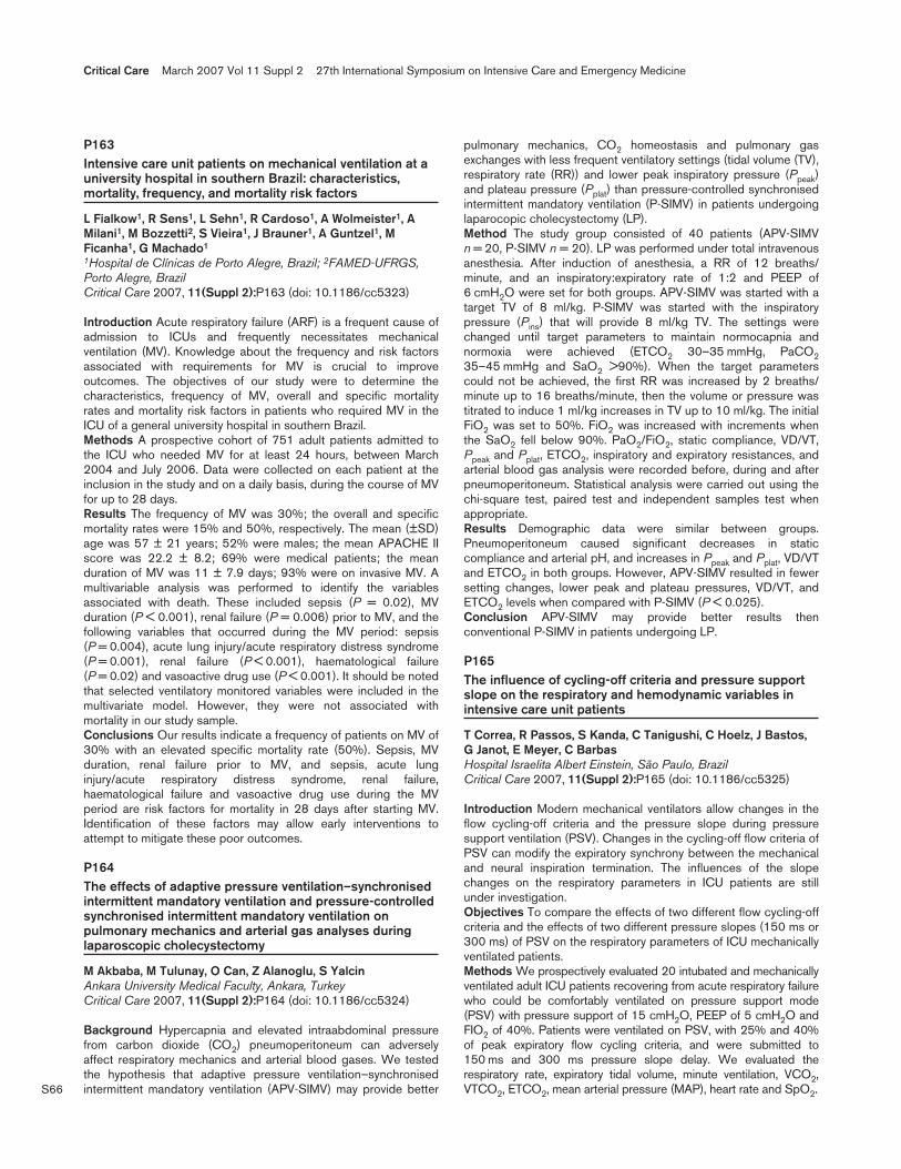
S66
P163Intensive care unit patients on mechanical ventilation at auniversity hospital in southern Brazil: characteristics,mortality, frequency, and mortality risk factors
L Fialkow1, R Sens1, L Sehn1, R Cardoso1, A Wolmeister1, AMilani1, M Bozzetti2, S Vieira1, J Brauner1, A Guntzel1, MFicanha1, G Machado1
1Hospital de Clínicas de Porto Alegre, Brazil; 2FAMED-UFRGS,Porto Alegre, BrazilCritical Care 2007, 11(Suppl 2):P163 (doi: 10.1186/cc5323)
Introduction Acute respiratory failure (ARF) is a frequent cause ofadmission to ICUs and frequently necessitates mechanicalventilation (MV). Knowledge about the frequency and risk factorsassociated with requirements for MV is crucial to improveoutcomes. The objectives of our study were to determine thecharacteristics, frequency of MV, overall and specific mortalityrates and mortality risk factors in patients who required MV in theICU of a general university hospital in southern Brazil.Methods A prospective cohort of 751 adult patients admitted tothe ICU who needed MV for at least 24 hours, between March2004 and July 2006. Data were collected on each patient at theinclusion in the study and on a daily basis, during the course of MVfor up to 28 days.Results The frequency of MV was 30%; the overall and specificmortality rates were 15% and 50%, respectively. The mean (±SD)age was 57 ± 21 years; 52% were males; the mean APACHE IIscore was 22.2 ± 8.2; 69% were medical patients; the meanduration of MV was 11 ± 7.9 days; 93% were on invasive MV. Amultivariable analysis was performed to identify the variablesassociated with death. These included sepsis (P = 0.02), MVduration (P < 0.001), renal failure (P = 0.006) prior to MV, and thefollowing variables that occurred during the MV period: sepsis(P = 0.004), acute lung injury/acute respiratory distress syndrome(P = 0.001), renal failure (P < 0.001), haematological failure(P = 0.02) and vasoactive drug use (P < 0.001). It should be notedthat selected ventilatory monitored variables were included in themultivariate model. However, they were not associated withmortality in our study sample.Conclusions Our results indicate a frequency of patients on MV of30% with an elevated specific mortality rate (50%). Sepsis, MVduration, renal failure prior to MV, and sepsis, acute lunginjury/acute respiratory distress syndrome, renal failure,haematological failure and vasoactive drug use during the MVperiod are risk factors for mortality in 28 days after starting MV.Identification of these factors may allow early interventions toattempt to mitigate these poor outcomes.
P164The effects of adaptive pressure ventilation–synchronisedintermittent mandatory ventilation and pressure-controlledsynchronised intermittent mandatory ventilation onpulmonary mechanics and arterial gas analyses duringlaparoscopic cholecystectomy
M Akbaba, M Tulunay, O Can, Z Alanoglu, S YalcinAnkara University Medical Faculty, Ankara, TurkeyCritical Care 2007, 11(Suppl 2):P164 (doi: 10.1186/cc5324)
Background Hypercapnia and elevated intraabdominal pressurefrom carbon dioxide (CO2) pneumoperitoneum can adverselyaffect respiratory mechanics and arterial blood gases. We testedthe hypothesis that adaptive pressure ventilation–synchronisedintermittent mandatory ventilation (APV-SIMV) may provide better
pulmonary mechanics, CO2 homeostasis and pulmonary gasexchanges with less frequent ventilatory settings (tidal volume (TV),respiratory rate (RR)) and lower peak inspiratory pressure (Ppeak)and plateau pressure (Pplat) than pressure-controlled synchronisedintermittent mandatory ventilation (P-SIMV) in patients undergoinglaparocopic cholecystectomy (LP).Method The study group consisted of 40 patients (APV-SIMVn = 20, P-SIMV n = 20). LP was performed under total intravenousanesthesia. After induction of anesthesia, a RR of 12 breaths/minute, and an inspiratory:expiratory rate of 1:2 and PEEP of6 cmH2O were set for both groups. APV-SIMV was started with atarget TV of 8 ml/kg. P-SIMV was started with the inspiratorypressure (Pins) that will provide 8 ml/kg TV. The settings werechanged until target parameters to maintain normocapnia andnormoxia were achieved (ETCO2 30–35 mmHg, PaCO235–45 mmHg and SaO2 >90%). When the target parameterscould not be achieved, the first RR was increased by 2 breaths/minute up to 16 breaths/minute, then the volume or pressure wastitrated to induce 1 ml/kg increases in TV up to 10 ml/kg. The initialFiO2 was set to 50%. FiO2 was increased with increments whenthe SaO2 fell below 90%. PaO2/FiO2, static compliance, VD/VT,Ppeak and Pplat, ETCO2, inspiratory and expiratory resistances, andarterial blood gas analysis were recorded before, during and afterpneumoperitoneum. Statistical analysis were carried out using thechi-square test, paired test and independent samples test whenappropriate.Results Demographic data were similar between groups.Pneumoperitoneum caused significant decreases in staticcompliance and arterial pH, and increases in Ppeak and Pplat, VD/VTand ETCO2 in both groups. However, APV-SIMV resulted in fewersetting changes, lower peak and plateau pressures, VD/VT, andETCO2 levels when compared with P-SIMV (P < 0.025).Conclusion APV-SIMV may provide better results thenconventional P-SIMV in patients undergoing LP.
P165The influence of cycling-off criteria and pressure supportslope on the respiratory and hemodynamic variables inintensive care unit patients
T Correa, R Passos, S Kanda, C Tanigushi, C Hoelz, J Bastos,G Janot, E Meyer, C BarbasHospital Israelita Albert Einstein, São Paulo, BrazilCritical Care 2007, 11(Suppl 2):P165 (doi: 10.1186/cc5325)
Introduction Modern mechanical ventilators allow changes in theflow cycling-off criteria and the pressure slope during pressuresupport ventilation (PSV). Changes in the cycling-off flow criteria ofPSV can modify the expiratory synchrony between the mechanicaland neural inspiration termination. The influences of the slopechanges on the respiratory parameters in ICU patients are stillunder investigation.Objectives To compare the effects of two different flow cycling-offcriteria and the effects of two different pressure slopes (150 ms or300 ms) of PSV on the respiratory parameters of ICU mechanicallyventilated patients.Methods We prospectively evaluated 20 intubated and mechanicallyventilated adult ICU patients recovering from acute respiratory failurewho could be comfortably ventilated on pressure support mode(PSV) with pressure support of 15 cmH2O, PEEP of 5 cmH2O andFIO2 of 40%. Patients were ventilated on PSV, with 25% and 40%of peak expiratory flow cycling criteria, and were submitted to150 ms and 300 ms pressure slope delay. We evaluated therespiratory rate, expiratory tidal volume, minute ventilation, VCO2,VTCO2, ETCO2, mean arterial pressure (MAP), heart rate and SpO2.
Critical Care March 2007 Vol 11 Suppl 2 27th International Symposium on Intensive Care and Emergency Medicine
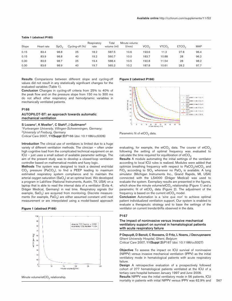
S67
Results Comparisons between different slope and cycling-offvalues did not result in any statistically significant changes for theevaluated variables (Table 1).Conclusion Changes in cycling-off criteria from 25% to 40% ofthe peak flow and on the pressure slope from 150 ms to 300 msdo not affect other respiratory and hemodynamic variables inmechanically ventilated patients.
P166AUTOPILOT-BT: an approach towards automaticmechanical ventilation
S Lozano1, K Moeller1, C Stahl2, J Guttmann2
1Furtwangen University, Villingen-Schwenningen, Germany;2University of Freiburg, GermanyCritical Care 2007, 11(Suppl 2):P166 (doi: 10.1186/cc5326)
Introduction The clinical use of ventilators is limited due to a hugevariety of different ventilation methods. The clinician – often underhigh cognitive load from the complicated technical equipment on anICU – just uses a small subset of available parameter settings. Theaim of the present study was to develop a closed-loop ventilationcontroller based on mathematical models and fuzzy logic.Methods The system was designed to track a desired end-tidalCO2 pressure (PaCO2), to find a PEEP leading to maximumestimated respiratory system compliance and to maintain thearterial oxygen saturation (SaO2) at an optimal level. We developeda program in LabView (National Instruments, Austin, TX, USA) on alaptop that is able to read the internal data of a ventilator (Evita 4;Dräger Medical, Germany) in real time. Respiratory signals (forexample, SaO2) are acquired from monitoring. Discrete measure-ments (for example, PaO2) are either assumed constant until nextmeasurement or are interpolated using a model-based approach
evaluating, for example, the etCO2 data. The course of etCO2following the setting of optimal frequency was evaluated tocalculate the time required for equilibration of etCO2.Results A module automating the initial settings of the ventilatoraccording to local ICU rules is realized. Modules were added thatoptimize breathing frequency with respect to PaCO2/etCO2 andFiO2 according to SO2 whenever no PaO2 is available. A lungsimulator (Michigan Instruments Inc., Grand Rapids, MI, USA)connected with the LS4000 (Dräger Medical) was used toevaluate the system. Exemplary results are presented in the figures,which show the minute volume/etCO2 relationship (Figure 1) and aparametric fit of etCO2 data (Figure 2). The adjustment of thefrequency is based on the current etCO2 model.Conclusion Automation is a ‘sine qua non’ to achieve optimalpatient individualized ventilation support. Our system is enabled toevaluate a therapeutic strategy and to base the settings of theventilator on current trends/drifts observed in the data.
P167The impact of noninvasive versus invasive mechanicalventilatory support on survival in hematological patientswith acute respiratory failure
P Depuydt, D Benoit, C Roosens, O Fritz, L Noens, J DecruyenaereGhent University Hospital, Ghent, BelgiumCritical Care 2007, 11(Suppl 2):P167 (doi: 10.1186/cc5327)
Objective To assess the impact on ICU survival of noninvasive(NIPPV) versus invasive mechanical ventilation (IPPV) as the initialventilatory mode in hematological patients with acute respiratoryfailure.Design A retrospective evaluation of a prospectively followedcohort of 277 hematological patients ventilated at the ICU of atertiary care hospital between January 1997 and June 2006.Results NIPPV was the initial ventilatory mode in 56 patients. ICUmortality in patients with initial NIPPV versus IPPV was 62.9% and
Available online http://ccforum.com/supplements/11/S2
Table 1 (abstract P165)
Respiratory Tidal Minute volumeSlope Heart rate SpO2 Cycling-off (%) rate volume (ml) (l/min) VCO2 VTCO2 ETCO2 MAP
0.15 83.4 98.8 25 18.2 587.5 10.6 193.6 11.2 27.8 96.4
0.15 83.9 98.8 40 19.2 560.7 10.0 183.7 10.88 28 96.2
0.30 83.6 98.7 25 19.4 588.4 10.5 192.8 11.54 28 98.2
0.30 83.6 98.9 40 19.7 565.2 10.2 187.8 10.61 28.2 97.7
Figure 1 (abstract P166)
Minute volume/etCO2 relationship.
Figure 2 (abstract P166)
Parametric fit of etCO2 data.
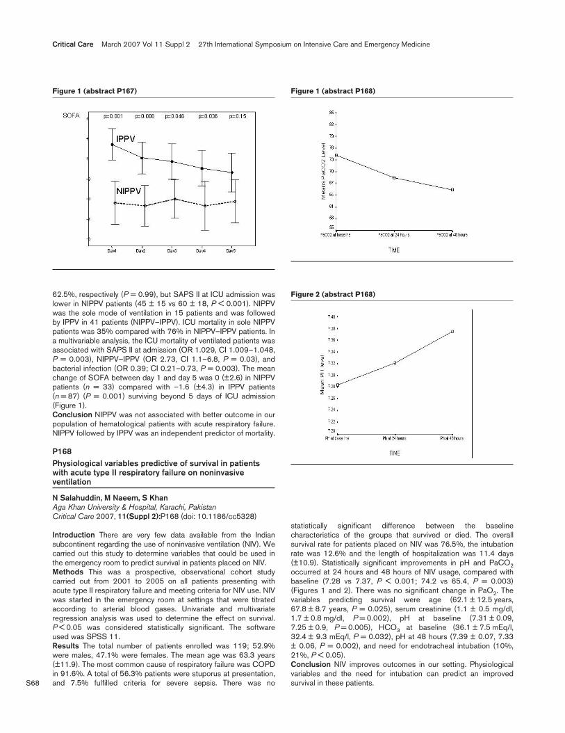
S68
62.5%, respectively (P = 0.99), but SAPS II at ICU admission waslower in NIPPV patients (45 ± 15 vs 60 ± 18, P < 0.001). NIPPVwas the sole mode of ventilation in 15 patients and was followedby IPPV in 41 patients (NIPPV–IPPV). ICU mortality in sole NIPPVpatients was 35% compared with 76% in NIPPV–IPPV patients. Ina multivariable analysis, the ICU mortality of ventilated patients wasassociated with SAPS II at admission (OR 1.029, CI 1.009–1.048,P = 0.003), NIPPV–IPPV (OR 2.73, CI 1.1–6.8, P = 0.03), andbacterial infection (OR 0.39; CI 0.21–0.73, P = 0.003). The meanchange of SOFA between day 1 and day 5 was 0 (±2.6) in NIPPVpatients (n = 33) compared with –1.6 (±4.3) in IPPV patients(n = 87) (P = 0.001) surviving beyond 5 days of ICU admission(Figure 1).Conclusion NIPPV was not associated with better outcome in ourpopulation of hematological patients with acute respiratory failure.NIPPV followed by IPPV was an independent predictor of mortality.
P168Physiological variables predictive of survival in patientswith acute type II respiratory failure on noninvasiveventilation
N Salahuddin, M Naeem, S KhanAga Khan University & Hospital, Karachi, PakistanCritical Care 2007, 11(Suppl 2):P168 (doi: 10.1186/cc5328)
Introduction There are very few data available from the Indiansubcontinent regarding the use of noninvasive ventilation (NIV). Wecarried out this study to determine variables that could be used inthe emergency room to predict survival in patients placed on NIV.Methods This was a prospective, observational cohort studycarried out from 2001 to 2005 on all patients presenting withacute type II respiratory failure and meeting criteria for NIV use. NIVwas started in the emergency room at settings that were titratedaccording to arterial blood gases. Univariate and multivariateregression analysis was used to determine the effect on survival.P < 0.05 was considered statistically significant. The softwareused was SPSS 11.Results The total number of patients enrolled was 119; 52.9%were males, 47.1% were females. The mean age was 63.3 years(±11.9). The most common cause of respiratory failure was COPDin 91.6%. A total of 56.3% patients were stuporus at presentation,and 7.5% fulfilled criteria for severe sepsis. There was no
statistically significant difference between the baselinecharacteristics of the groups that survived or died. The overallsurvival rate for patients placed on NIV was 76.5%, the intubationrate was 12.6% and the length of hospitalization was 11.4 days(±10.9). Statistically significant improvements in pH and PaCO2occurred at 24 hours and 48 hours of NIV usage, compared withbaseline (7.28 vs 7.37, P < 0.001; 74.2 vs 65.4, P = 0.003)(Figures 1 and 2). There was no significant change in PaO2. Thevariables predicting survival were age (62.1 ± 12.5 years,67.8 ± 8.7 years, P = 0.025), serum creatinine (1.1 ± 0.5 mg/dl,1.7 ± 0.8 mg/dl, P = 0.002), pH at baseline (7.31 ± 0.09,7.25 ± 0.9, P = 0.005), HCO3 at baseline (36.1 ± 7.5 mEq/l,32.4 ± 9.3 mEq/l, P = 0.032), pH at 48 hours (7.39 ± 0.07, 7.33± 0.06, P = 0.002), and need for endotracheal intubation (10%,21%, P < 0.05).Conclusion NIV improves outcomes in our setting. Physiologicalvariables and the need for intubation can predict an improvedsurvival in these patients.
Critical Care March 2007 Vol 11 Suppl 2 27th International Symposium on Intensive Care and Emergency Medicine
Figure 1 (abstract P167) Figure 1 (abstract P168)
Figure 2 (abstract P168)
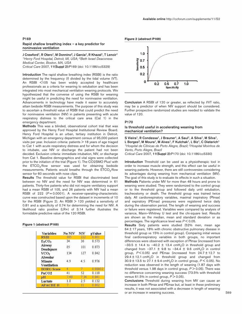
S69
P169Rapid shallow breathing index – a key predictor fornoninvasive ventilation
J Crawford1, R Otero1, M Donnino2, J Garcia1, R Khazal1, T Lenoir1
1Henry Ford Hospital, Detroit, MI, USA; 2Beth Israel DeaconessMedical Center, Boston, MA, USACritical Care 2007, 11(Suppl 2):P169 (doi: 10.1186/cc5329)
Introduction The rapid shallow breathing index (RSBI) is the ratiodetermined by the frequency (f) divided by the tidal volume (VT).An RSBI <105 has been widely accepted by healthcareprofessionals as a criteria for weaning to extubation and has beenintegrated into most mechanical ventilation weaning protocols. Wehypothesized that the converse of using the RSBI for weaningmight be useful in predicting the need for noninvasive ventilation.Advancements in technology have made it easier to accuratelyattain bedside RSBI measurements. The purpose of this study wasto ascertain a threshold value of RSBI that could predict the needfor noninvasive ventilation (NIV) in patients presenting with acuterespiratory distress to the critical care area (Cat 1) in theemergency department.Methods This was a blinded, observational cohort trial that wasapproved by the Henry Ford Hospital Institutional Review Board.Henry Ford Hospital is an urban, tertiary institution in Detroit,Michigan with an emergency department census of 95,000 patientvisits per year. Inclusion criteria: patients > 18 years of age triagedto Cat 1 with acute respiratory distress and for whom the decisionto intubate, use NIV or discharge the patient had not beendecided. Exclusion criteria: immediate intubation, NIV, or dischargefrom Cat 1. Baseline demographics and vital signs were collectedprior to the initiation of the trial (Figure 1). The CO2SMO Plus! withthe ETCO2/flow sensor was used for obtaining bedsidemeasurements. Patients would breathe through the ETCO2/flowsensor for 60 seconds with nose clips.Results The threshold value for RSBI that discriminated bestbetween no NIV and the need for NIV was determined in 61patients. Thirty-five patients who did not require ventilatory supporthad a mean RSBI of 105, and 26 patients with NIV had a meanRSBI of 222 (P = 0.0001). A receiver-operating-characteristiccurve was constructed based upon the dataset in increments of 10for the RSBI (Figure 2). An RSBI > 120 yielded a sensitivity of0.81 and a specificity of 0.74 for determining the need for NIV. Alikelihood ratio positive (LR+) of 3.14 further illustrates theformidable predictive value of the 120 RSBI.
Conclusion A RSBI of 120 or greater, as reflected by f/VT ratio,may be a predictor of when NIV support should be considered.Further prospective randomized studies are needed to validate thevalue of 120.
P170Is threshold useful in accelerating weaning frommechanical ventilation?
S Vieira1, R Condessa1, J Brauner1, A Saul1, A Silva1, M Silva1,L Borges2, M Moura1, M Alves1, F Kutchak1, L Biz1, C Dieterich1
1Hospital de Clínicas de Porto Alegre, Brazil; 2Hospital Moinhos deVento, Porto Alegre, BrazilCritical Care 2007, 11(Suppl 2):P170 (doi: 10.1186/cc5330)
Introduction Threshold can be used as a physiotherapic tool inorder to increase muscle strength, and this effect can be useful inweaning patients. However, there are still controversies consideringits advantages during weaning from mechanical ventilation (MV).The goal of this study is to evaluate its effects in such a situation.Methods Patients under MV for more than 48 hours and prone toweaning were studied. They were randomized to the control groupor to the threshold group and followed daily until extubation,tracheostomy or death. The threshold group was trained twicedaily. All cardiorespiratory variables, maximal inspiratory (PImax)and expiratory (PEmax) pressures were registered twice dailyduring the observation period. The length of weaning and successor failure were registered. Variables were compared by analysis ofvariance, Mann–Whitney U test and the chi-square test. Resultsare shown as the median, mean and standard deviation or aspercentages. The significance level was P < 0.05.Results Sixty patients were studied (52% men, mean age64 ± 17 years, 18% with chronic obstructive pulmonary disease inthreshold group vs 15% in control group). Comparing initial versusfinal cardiorespiratory variables in both groups, no importantdifferences were observed with exception of PImax (increased from–33.5 ± 14.4 to –40.2 ± 13.4 cmH2O in threshold group andchanged from –37.1 ± 9.8 to –34.4 ± 9.6 cmH2O in controlgroup, P < 0.05) and PEmax (increased from 24.7 ± 12.7 to29.4 ± 12.1 cmH2O in threshold group and changed from30.9 ± 13.5 to 27.1 ± 9.4 cmH2O in control group, P < 0.05). Noreduction was observed in the length of weaning (1.87 days withthreshold versus 1.98 days in control group, P > 0.05). There wasno difference concerning weaning success (73.5% with thresholdversus 61.5% in control group, P > 0.05).Conclusions Threshold during weaning from MV can cause anincrease in both PImax and PEmax but, at least in these preliminaryresults, it was not associated with a decrease in length of weaningor an increase in weaning success.
Available online http://ccforum.com/supplements/11/S2
Figure 1 (abstract P169)
Figure 2 (abstract P169)
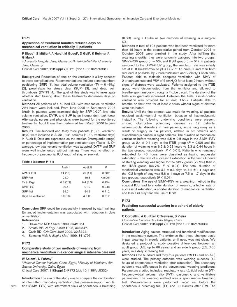
S70
P171Application of treatment bundles reduces days onmechanical ventilation in critically ill patients
F Bloos1, S Müller1, A Harz1, M Gugel1, D Geil1, K Reinhart2, G Marx2
1University Hospital Jena, Germany; 2Friedrich-Schiller UniversityJena, GermanyCritical Care 2007, 11(Suppl 2):P171 (doi: 10.1186/cc5331)
Background Reduction of time on the ventilator is a key conceptto avoid complications. Recommendations include semirecumbentpositioning (SRP) [1], low tidal volume ventilation (TV = 6 ml/kg)[2], prophylaxis for stress ulcer (SUP) [3], and deep veinthrombosis (DVTP) [4]. The goal of this study was to investigatewhether staff training about these treatments decreases days onventilation.Methods All patients of a 50-bed ICU with mechanical ventilation>24 hours were included. From June 2005 to September 2005(Audit I), patients were examined daily for SRP >30°, low tidalvolume ventilation, DVTP, and SUP by an independent task force.Afterwards, nurses and physicians were trained for the monitoredtreatments. Audit II was then performed from March 2006 to June2006.Results One hundred and thirty-three patients (1,389 ventilator-days) were included in Audit I, 141 patients (1,002 ventilator-days)in Audit II. Data are expressed as the median (interquartile range)or percentage of implementation per ventilator-days (Table 1). Onaverage, low tidal volume ventilation was adopted. DVTP and SUPwere well implemented without training. There was no effect onfrequency of pneumonia, ICU length of stay, or survival.
Table 1 (abstract P171)
Audit I Audit II P
APACHE II 24 (10) 25 (11) 0.387
SRP (%) 24.9 49.6 <0.001
TV (ml/kg) 6.3 (2.2) 6.4 (2.3) 0.154
DVTP (%) 89.5 91.9 0.048
SUP (%) 94.5 94.9 0.712
Days on ventilation 6.0 (13) 4.0 (7) 0.017
Conclusion SRP could be successfully improved by staff training.Enhanced implementation was associated with reduction in dayson ventilation.References1. Drakulovic MB: Lancet 1999, 354:1851.2. Amato MB: N Engl J Med 1998, 338:347.3. Cash BD: Crit Care Med 2002, 30:S373.4. Samama MM: N Engl J Med 1999, 341:793.
P172Comparative study of two methods of weaning frommechanical ventilation in a cancer surgical intensive care unit
W Salem1, N Fahmy2
1National Cancer Institute, Cairo, Egypt; 2Faculty of Medicine, AinShams University, Cairo, EgyptCritical Care 2007, 11(Suppl 2):P172 (doi: 10.1186/cc5332)
Introduction The aim of the study was to compare the combinationof intermittent mandatory ventilation plus pressure-support ventila-tion (SIMV+PSV) with intermittent trials of spontaneous breathing
(ITSB) using a T-tube as two methods of weaning in a surgicalICU.Methods A total of 104 patients who had been ventilated for morethan 48 hours in the postoperative period from October 2005 toOctober 2006 were enrolled in the study. After fulfilling theweaning checklist they were randomly assigned into two groups:SIMV+PSV group (n = 53), and ITSB group (n = 51). In patientsassigned to the SIMV+PSV group, the ventilator rate was initiallyset at 6–8 breaths/minute plus PSV of 15 cmH2O and then bothreduced, if possible, by 2 breaths/minute and 2 cmH2O each time.Patients able to maintain adequate ventilation with SIMV of2 breaths/minute and PSV of 5 cmH2O for at least 2 hours withoutsigns of distress were extubated. Patients assigned to the ITSBgroup were disconnected from the ventilator and allowed tobreathe spontaneously through a T-tube circuit. The duration of thetrials was gradually increased. Between the trials, assist–controlventilation was provided for at least 1 hour. Patients able tobreathe on their own for at least 2 hours without signs of distresswere extubated.Results Until the first attempt was made for weaning, all patientsreceived assist–control ventilation because of haemodynamicinstability. The following underlying conditions were present:chronic obstructive pulmonary disease in 67 patients,neuromuscular disorders in nine patients, acute lung injury as aresult of surgery in 14 patients, asthma in six patients andmiscellaneous causes in eight patients. The duration of mechanicalventilation before weaning was 2.5 ± 0.5 days in the SIMV+PSVgroup vs 2.4 ± 0.4 days in the ITSB group (P = 0.02) and theduration of weaning was 6.2 ± 0.23 hours vs 8.3 ± 0.44 hours inthe two groups, respectively (P < 0.01). Patients who remainedextubated for 48 hours were classified as having successfulextubation – the rate of successful extubation in the first 24 hoursof starting weaning was higher for the SIMV group (79.2%) than inthe ITSB group (64.7%, P < 0.01). The total duration ofmechanical ventilation was 3.3 ± 0.3 days vs 5.2 ± 1.1 days andthe ICU length of stay was 5.6 ± 1 days vs 7.5 ± 1.7 days in thetwo groups, respectively (P < 0.01).Conclusions The use of SIMV+PSV as a weaning method in thesurgical ICU lead to shorter duration of weaning, a higher rate ofsuccessful extubation, a shorter duration of mechanical ventilationand less ICU stay than the use of ITSB.
P173Predicting successful weaning in a cohort of elderlypatients
C Corbellini, A Guntzel, C Trevisan, S VieiraHospital de Clinicas de Porto Alegre, BrazilCritical Care 2007, 11(Suppl 2):P173 (doi: 10.1186/cc5333)
Introduction Aging causes structural and functional modificationsin the respiratory system. The evidence that these changes couldimpair weaning in elderly patients, until now, was not clear. Wedesigned a protocol to study possible differences between anadult group (AG, up to 60 years) and an elderly group (EG, >60years) in a daily screening trial.Methods One hundred and forty-four patients (79 EG and 65 AG)were studied. The primary outcome was weaning success (48hours of spontaneous ventilation after extubation). The secondaryoutcome was differences in the conventional weaning predictors.Parameters studied included: respiratory rate (f), tidal volume (VT),frequency–tidal volume ratio (f/VT), gasometric and ventilatoryparameters. The weaning method was a spontaneous breathingtrial. Measurements were performed twice: just before thespontaneous breathing trial (T1) and 30 minutes after (T2). The
Critical Care March 2007 Vol 11 Suppl 2 27th International Symposium on Intensive Care and Emergency Medicine
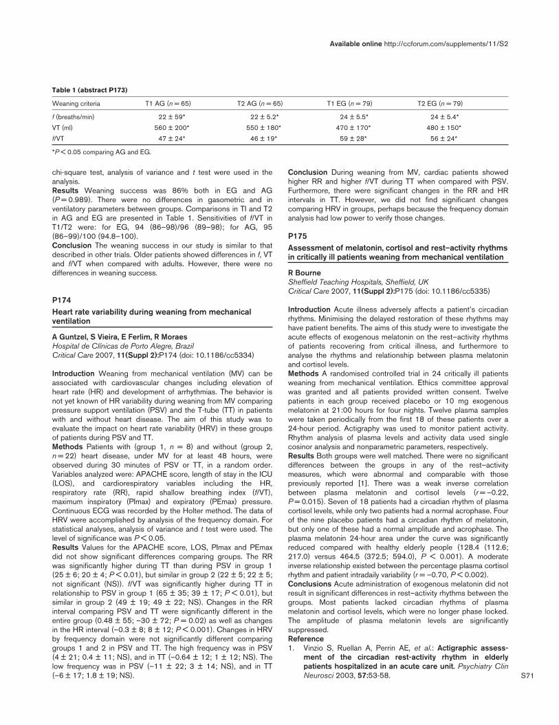
S71
chi-square test, analysis of variance and t test were used in theanalysis.Results Weaning success was 86% both in EG and AG(P = 0.989). There were no differences in gasometric and inventilatory parameters between groups. Comparisons in TI and T2in AG and EG are presented in Table 1. Sensitivities of f/VT inT1/T2 were: for EG, 94 (86–98)/96 (89–98); for AG, 95(86–99)/100 (94.8–100).Conclusion The weaning success in our study is similar to thatdescribed in other trials. Older patients showed differences in f, VTand f/VT when compared with adults. However, there were nodifferences in weaning success.
P174Heart rate variability during weaning from mechanicalventilation
A Guntzel, S Vieira, E Ferlim, R MoraesHospital de Clínicas de Porto Alegre, BrazilCritical Care 2007, 11(Suppl 2):P174 (doi: 10.1186/cc5334)
Introduction Weaning from mechanical ventilation (MV) can beassociated with cardiovascular changes including elevation ofheart rate (HR) and development of arrhythmias. The behavior isnot yet known of HR variability during weaning from MV comparingpressure support ventilation (PSV) and the T-tube (TT) in patientswith and without heart disease. The aim of this study was toevaluate the impact on heart rate variability (HRV) in these groupsof patients during PSV and TT.Methods Patients with (group 1, n = 8) and without (group 2,n = 22) heart disease, under MV for at least 48 hours, wereobserved during 30 minutes of PSV or TT, in a random order.Variables analyzed were: APACHE score, length of stay in the ICU(LOS), and cardiorespiratory variables including the HR,respiratory rate (RR), rapid shallow breathing index (f/VT),maximum inspiratory (PImax) and expiratory (PEmax) pressure.Continuous ECG was recorded by the Holter method. The data ofHRV were accomplished by analysis of the frequency domain. Forstatistical analyses, analysis of variance and t test were used. Thelevel of significance was P < 0.05.Results Values for the APACHE score, LOS, PImax and PEmaxdid not show significant differences comparing groups. The RRwas significantly higher during TT than during PSV in group 1(25 ± 6; 20 ± 4; P < 0.01), but similar in group 2 (22 ± 5; 22 ± 5;not significant (NS)). f/VT was significantly higher during TT inrelationship to PSV in group 1 (65 ± 35; 39 ± 17; P < 0.01), butsimilar in group 2 (49 ± 19; 49 ± 22; NS). Changes in the RRinterval comparing PSV and TT were significantly different in theentire group (0.48 ± 55; –30 ± 72; P = 0.02) as well as changesin the HR interval (–0.3 ± 8; 8 ± 12; P < 0.001). Changes in HRVby frequency domain were not significantly different comparinggroups 1 and 2 in PSV and TT. The high frequency was in PSV(4 ± 21; 0.4 ± 11; NS), and in TT (–0.64 ± 12; 1 ± 12; NS). Thelow frequency was in PSV (–11 ± 22; 3 ± 14; NS), and in TT(–6 ± 17; 1.8 ± 19; NS).
Conclusion During weaning from MV, cardiac patients showedhigher RR and higher f/VT during TT when compared with PSV.Furthermore, there were significant changes in the RR and HRintervals in TT. However, we did not find significant changescomparing HRV in groups, perhaps because the frequency domainanalysis had low power to verify those changes.
P175Assessment of melatonin, cortisol and rest–activity rhythmsin critically ill patients weaning from mechanical ventilation
R BourneSheffield Teaching Hospitals, Sheffield, UKCritical Care 2007, 11(Suppl 2):P175 (doi: 10.1186/cc5335)
Introduction Acute illness adversely affects a patient’s circadianrhythms. Minimising the delayed restoration of these rhythms mayhave patient benefits. The aims of this study were to investigate theacute effects of exogenous melatonin on the rest–activity rhythmsof patients recovering from critical illness, and furthermore toanalyse the rhythms and relationship between plasma melatoninand cortisol levels.Methods A randomised controlled trial in 24 critically ill patientsweaning from mechanical ventilation. Ethics committee approvalwas granted and all patients provided written consent. Twelvepatients in each group received placebo or 10 mg exogenousmelatonin at 21:00 hours for four nights. Twelve plasma sampleswere taken periodically from the first 18 of these patients over a24-hour period. Actigraphy was used to monitor patient activity.Rhythm analysis of plasma levels and activity data used singlecosinor analysis and nonparametric parameters, respectively.Results Both groups were well matched. There were no significantdifferences between the groups in any of the rest–activitymeasures, which were abnormal and comparable with thosepreviously reported [1]. There was a weak inverse correlationbetween plasma melatonin and cortisol levels (r = –0.22,P = 0.015). Seven of 18 patients had a circadian rhythm of plasmacortisol levels, while only two patients had a normal acrophase. Fourof the nine placebo patients had a circadian rhythm of melatonin,but only one of these had a normal amplitude and acrophase. Theplasma melatonin 24-hour area under the curve was significantlyreduced compared with healthy elderly people (128.4 (112.6;217.0) versus 464.5 (372.5; 594.0), P < 0.001). A moderateinverse relationship existed between the percentage plasma cortisolrhythm and patient intradaily variability (r = –0.70, P < 0.002).Conclusions Acute administration of exogenous melatonin did notresult in significant differences in rest–activity rhythms between thegroups. Most patients lacked circadian rhythms of plasmamelatonin and cortisol levels, which were no longer phase locked.The amplitude of plasma melatonin levels are significantlysuppressed.Reference1. Vinzio S, Ruellan A, Perrin AE, et al.: Actigraphic assess-
ment of the circadian rest-activity rhythm in elderlypatients hospitalized in an acute care unit. Psychiatry ClinNeurosci 2003, 57:53-58.
Available online http://ccforum.com/supplements/11/S2
Table 1 (abstract P173)
Weaning criteria T1 AG (n = 65) T2 AG (n = 65) T1 EG (n = 79) T2 EG (n = 79)
f (breaths/min) 22 ± 59* 22 ± 5.2* 24 ± 5.5* 24 ± 5.4*
VT (ml) 560 ± 200* 550 ± 180* 470 ± 170* 480 ± 150*
f/VT 47 ± 24* 46 ± 19* 59 ± 28* 56 ± 24*
*P < 0.05 comparing AG and EG.
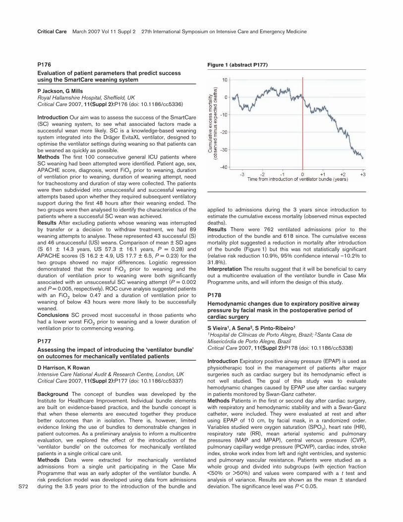
S72
P176Evaluation of patient parameters that predict successusing the SmartCare weaning system
P Jackson, G MillsRoyal Hallamshire Hospital, Sheffield, UKCritical Care 2007, 11(Suppl 2):P176 (doi: 10.1186/cc5336)
Introduction Our aim was to assess the success of the SmartCare(SC) weaning system, to see what associated factors made asuccessful wean more likely. SC is a knowledge-based weaningsystem integrated into the Dräger EvitaXL ventilator, designed tooptimise the ventilator settings during weaning so that patients canbe weaned as quickly as possible.Methods The first 100 consecutive general ICU patients whereSC weaning had been attempted were identified. Patient age, sex,APACHE score, diagnosis, worst FiO2 prior to weaning, durationof ventilation prior to weaning, duration of weaning attempt, needfor tracheostomy and duration of stay were collected. The patientswere then subdivided into unsuccessful and successful weaningattempts based upon whether they required subsequent ventilatorysupport during the first 48 hours after their weaning ended. Thetwo groups were then analysed to identify the characteristics of thepatients where a successful SC wean was achieved.Results After excluding patients whose weaning was interruptedby transfer or a decision to withdraw treatment, we had 89weaning attempts to analyse. These represented 43 successful (S)and 46 unsuccessful (US) weans. Comparison of mean ± SD ages(S 61 ± 14.3 years, US 57.3 ± 16.1 years, P = 0.28) andAPACHE scores (S 16.2 ± 4.9, US 17.7 ± 6.5, P = 0.23) for thetwo groups showed no major differences. Logistic regressiondemonstrated that the worst FiO2 prior to weaning and theduration of ventilation prior to weaning were both significantlyassociated with an unsuccessful SC weaning attempt (P = 0.002and P = 0.005, respectively). ROC curve analysis suggested patientswith an FiO2 below 0.47 and a duration of ventilation prior toweaning of below 43 hours were more likely to be successfullyweaned.Conclusions SC proved most successful in those patients whohad a lower worst FiO2 prior to weaning and a lower duration ofventilation prior to commencing weaning.
P177Assessing the impact of introducing the ‘ventilator bundle’on outcomes for mechanically ventilated patients
D Harrison, K RowanIntensive Care National Audit & Research Centre, London, UKCritical Care 2007, 11(Suppl 2):P177 (doi: 10.1186/cc5337)
Background The concept of bundles was developed by theInstitute for Healthcare Improvement. Individual bundle elementsare built on evidence-based practice, and the bundle concept isthat when these elements are executed together they producebetter outcomes than in isolation. There is, however, limitedevidence linking the use of bundles to demonstrable changes inpatient outcomes. As a preliminary analysis to inform a multicentreevaluation, we explored the effect of the introduction of the‘ventilator bundle’ on the outcomes for mechanically ventilatedpatients in a single critical care unit.Methods Data were extracted for mechanically ventilatedadmissions from a single unit participating in the Case MixProgramme that was an early adopter of the ventilator bundle. Arisk prediction model was developed using data from admissionsduring the 3.5 years prior to the introduction of the bundle and
applied to admissions during the 3 years since introduction toestimate the cumulative excess mortality (observed minus expecteddeaths).Results There were 762 ventilated admissions prior to theintroduction of the bundle and 618 since. The cumulative excessmortality plot suggested a reduction in mortality after introductionof the bundle (Figure 1) but this was not statistically significant(relative risk reduction 10.9%, 95% confidence interval –10.2% to31.8%).Interpretation The results suggest that it will be beneficial to carryout a multicentre evaluation of the ventilator bundle in Case MixProgramme units, and will inform the design of this study.
P178Hemodynamic changes due to expiratory positive airwaypressure by facial mask in the postoperative period ofcardiac surgery
S Vieira1, A Sena2, S Pinto-Ribeiro1
1Hospital de Clínicas de Porto Alegre, Brazil; 2Santa Casa deMisericórdia de Porto Alegre, BrazilCritical Care 2007, 11(Suppl 2):P178 (doi: 10.1186/cc5338)
Introduction Expiratory positive airway pressure (EPAP) is used asphysiotherapic tool in the management of patients after majorsurgeries such as cardiac surgery but its hemodynamic effect isnot well studied. The goal of this study was to evaluatehemodynamic changes caused by EPAP use after cardiac surgeryin patients monitored by Swan-Ganz catheter.Methods Patients in the first or second day after cardiac surgery,with respiratory and hemodynamic stability and with a Swan-Ganzcatheter, were included. They were evaluated at rest and afterusing EPAP of 10 cm, by facial mask, in a randomized order.Variables studied were oxygen saturation (SPO2), heart rate (HR),respiratory rate (RR), mean arterial systemic and pulmonarypressures (MAP and MPAP), central venous pressure (CVP),pulmonary capillary wedge pressure (PCWP), cardiac index, strokeindex, stroke work index from left and right ventricles, and systemicand pulmonary vascular resistance. Patients were studied as awhole group and divided into subgroups (with ejection fraction<50% or >50%) and values were compared with a t test andanalysis of variance. Results are shown as the mean ± standarddeviation. The significance level was P < 0.05.
Critical Care March 2007 Vol 11 Suppl 2 27th International Symposium on Intensive Care and Emergency Medicine
Figure 1 (abstract P177)
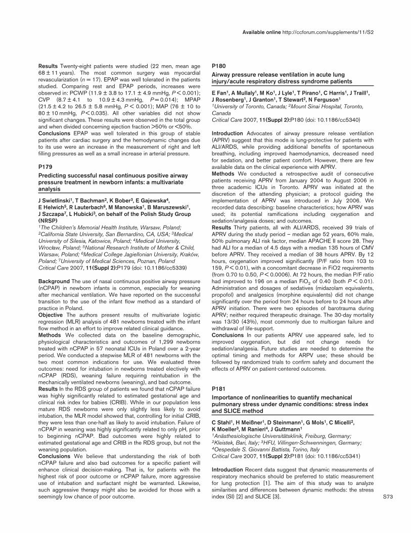
S73
Results Twenty-eight patients were studied (22 men, mean age68 ± 11 years). The most common surgery was myocardialrevascularization (n = 17). EPAP was well tolerated in the patientsstudied. Comparing rest and EPAP periods, increases wereobserved in: PCWP (11.9 ± 3.8 to 17.1 ± 4.9 mmHg, P < 0.001);CVP (8.7 ± 4.1 to 10.9 ± 4.3 mmHg, P = 0.014); MPAP(21.5 ± 4.2 to 26.5 ± 5.8 mmHg, P < 0.001); MAP (76 ± 10 to80 ± 10 mmHg, P < 0.035). All other variables did not showsignificant changes. These results were observed in the total groupand when divided concerning ejection fraction >50% or <50%.Conclusions EPAP was well tolerated in this group of stablepatients after cardiac surgery and the hemodynamic changes dueto its use were an increase in the measurement of right and leftfilling pressures as well as a small increase in arterial pressure.
P179Predicting successful nasal continuous positive airwaypressure treatment in newborn infants: a multivariateanalysis
J Swietlinski1, T Bachman2, K Bober3, E Gajewska4, E Helwich5, R Lauterbach6, M Manowska1, B Maruszewski1, J Szczapa7, L Hubicki3, on behalf of the Polish Study Group(NRSP)1The Children’s Memorial Health Institute, Warsaw, Poland;2California State University, San Bernardino, CA, USA; 3MedicalUniversity of Silesia, Katowice, Poland; 4Medical University,Wrocùaw, Poland; 5National Research Institute of Mother & Child,Warsaw, Poland; 6Medical College Jagiellonian University, Kraków,Poland; 7University of Medical Sciences, Poznan, PolandCritical Care 2007, 11(Suppl 2):P179 (doi: 10.1186/cc5339)
Background The use of nasal continuous positive airway pressure(nCPAP) in newborn infants is common, especially for weaningafter mechanical ventilation. We have reported on the successfultransition to the use of the infant flow method as a standard ofpractice in Poland.Objective The authors present results of multivariate logisticregression (MLR) analysis of 481 newborns treated with the infantflow method in an effort to improve related clinical guidance.Methods We collected data on the baseline demographic,physiological characteristics and outcomes of 1,299 newbornstreated with nCPAP in 57 neonatal ICUs in Poland over a 2-yearperiod. We conducted a stepwise MLR of 481 newborns with thetwo most common indications for use. We evaluated threeoutcomes: need for intubation in newborns treated electively withnCPAP (RDS), weaning failure requiring reintubation in themechanically ventilated newborns (weaning), and bad outcome.Results In the RDS group of patients we found that nCPAP failurewas highly significantly related to estimated gestational age andclinical risk index for babies (CRIB). While in our population lessmature RDS newborns were only slightly less likely to avoidintubation, the MLR model showed that, controlling for initial CRIB,they were less than one-half as likely to avoid intubation. Failure ofnCPAP in weaning was highly significantly related to only pH, priorto beginning nCPAP. Bad outcomes were highly related toestimated gestational age and CRIB in the RDS group, but not theweaning population.Conclusions We believe that understanding the risk of bothnCPAP failure and also bad outcomes for a specific patient willenhance clinical decision-making. That is, for patients with thehighest risk of poor outcome or nCPAP failure, more aggressiveuse of intubation and surfactant might be warranted. Likewise,such aggressive therapy might also be avoided for those with aseemingly low chance of poor outcome.
P180Airway pressure release ventilation in acute lunginjury/acute respiratory distress syndrome patients
E Fan1, A Mullaly1, M Ko1, J Lyle1, T Pirano1, C Harris1, J Traill1, J Rosenberg1, J Granton1, T Stewart2, N Ferguson1
1University of Toronto, Canada; 2Mount Sinai Hospital, Toronto,CanadaCritical Care 2007, 11(Suppl 2):P180 (doi: 10.1186/cc5340)
Introduction Advocates of airway pressure release ventilation(APRV) suggest that this mode is lung-protective for patients withALI/ARDS, while providing additional benefits of spontaneousbreathing, including improved haemodynamics, decreased needfor sedation, and better patient comfort. However, there are fewavailable data on the clinical experience with APRV.Methods We conducted a retrospective audit of consecutivepatients receiving APRV from January 2004 to August 2006 inthree academic ICUs in Toronto. APRV was initiated at thediscretion of the attending physician; a protocol guiding theimplementation of APRV was introduced in July 2006. Werecorded data describing: baseline characteristics; how APRV wasused; its potential ramifications including oxygenation andsedation/analgesia doses; and outcomes.Results Thirty patients, all with ALI/ARDS, received 39 trials ofAPRV during the study period – median age 52 years, 60% male,50% pulmonary ALI risk factor, median APACHE II score 28. Theyhad ALI for a median of 4.5 days with a median 135 hours of CMVbefore APRV. They received a median of 38 hours APRV. By 12hours, oxygenation improved significantly (P/F ratio from 103 to159, P < 0.01), with a concomitant decrease in FiO2 requirements(from 0.70 to 0.50, P < 0.0006). At 72 hours, the median P/F ratiohad improved to 196 on a median FiO2 of 0.40 (both P < 0.01).Administration and dosages of sedatives (midazolam equivalents,propofol) and analgesics (morphine equivalents) did not changesignificantly over the period from 24 hours before to 24 hours afterAPRV initiation. There were two episodes of barotrauma duringAPRV; neither required therapeutic drainage. The 30-day mortalitywas 13/30 (43%), most commonly due to multiorgan failure andwithdrawal of life-support.Conclusions In our patients APRV use appeared safe, led toimproved oxygenation, but did not change needs forsedation/analgesia. Future studies are needed to determine theoptimal timing and methods for ARPV use; these should befollowed by randomized trials to confirm safety and document theeffects of APRV on patient-centered outcomes.
P181Importance of nonlinearities to quantify mechanicalpulmonary stress under dynamic conditions: stress indexand SLICE method
C Stahl1, H Meißner1, D Steinmann1, G Mols1, C Micelli2, K Moeller3, M Ranieri4, J Guttmann1
1Anästhesiologische Universitätsklinik, Freiburg, Germany;2Kleistek, Bari, Italy; 3HFU, Villingen-Schwenningen, Germany;4Oespedale S. Giovanni Battista, Torino, ItalyCritical Care 2007, 11(Suppl 2):P181 (doi: 10.1186/cc5341)
Introduction Recent data suggest that dynamic measurements ofrespiratory mechanics should be preferred to static measurementfor lung protection [1]. The aim of this study was to analyzesimilarities and differences between dynamic methods: the stressindex (SI) [2] and SLICE [3].
Available online http://ccforum.com/supplements/11/S2
S74
Methods One hundred and two respiratory datasets from 70patients (28 ARDS, 24 postanesthesia care, 18 other) wereanalyzed. The SI and SLICE were performed using exactly thesame database (SLICE_SI) in addition to the conventional SLICEthat includes inspiratory and expiratory data (SLICE_CONV). Acompliance-based index (CSI) directly comparable with the SI wasgenerated from the compliance data.Results The SI and CSI highly correlated when calculation of theCSI was based on the same database (Figure 1). According to theresulting regression formula (Figure 1), the SI can be reliablypredicted from SLICE_SI (Figure 2). However, if SLICE_CONVwas used for calculation of the SI (Figure 3), noticeable differenceswere found. Analysis of individual datasets showed three majorreasons for the observed differences: differences in excluded dataat low volumes respective to high volumes, nonlinearity ofresistance, and differences in mechanics between inspiration andexpiration.Conclusion The SI and SLICE similarly measure the nonlinearity ofcompliance. The SI can be predicted from SLICE. However,nonlinearities of the respiratory system are not restricted tocompliance alone; it might therefore be necessary to includenonlinearities of resistance and asymmetries between inspirationand expiration in the analysis of dynamic respiratory mechanics.References1. Stahl CA, et al.: Dynamic versus static respiratory mechan-
ics in acute lung injury and acute respiratory distress syn-drome. Crit Care Med 2006, 34:2090-2098.
2. Ranieri VM, et al.: Pressure–time curve predicts minimallyinjurious ventilatory strategy in an isolated rat lung model.Anesthesiology 2000, 93:1320-1328.
3. Guttmann J, et al.: Determination of volume-dependent res-piratory system mechanics in mechanically ventilatedpatients using the new SLICE method. Technol HealthCare 1994, 2:175-191.
P182Contribution of HMSE-1 to surfactant conversion underacute inflammatory conditions
C Ruppert, P Markart, S Händel, W Seeger, A GüntherUniversity of Giessen Lung Center, Giessen, GermanyCritical Care 2007, 11(Suppl 2):P182 (doi: 10.1186/cc5342)
Introduction A reduced content of biophysically active largesurfactant aggregates is a common finding in acute inflammatorylung disease. Cyclic surface area changes and a carboxylesteraseactivity (surfactant convertase) are thought to mediate this subtypeconversion. However, data concerning regulation of surfactant
convertase are scarce. We therefore investigated the expressionand activity of lung surfactant convertase and HMSE-1, a potentialmacrophage-derived human convertase, under normal and acuteinflammatory conditions.Methods Convertase activity in lavage fluid (BALF) was assessedusing the in vitro cycling assay. The relative large surfactantaggregate content was determined by phospholipid quantificationin the pellet following centrifugation at 48,000 x g. Esterase activitywas assessed by means of a chromogenic substrate assay.Expression of both convertase and HMSE upon LPS challengewas assessed by real-time (TaqMan) PCR in murine alveolarmacrophages, murine primary type II cells, and the humanmonocytic cell line U937, respectively.Results Lavage fluid from ARDS patients displayed an increasedesterase activity when compared with BALF from healthy controls.In addition, a pronounced large to small aggregate conversion wasobserved for BALF from LPS-challenged mice or BALF from ARDSpatients. Incubation with LPS resulted in a significant increase inconvertase gene expression in primary mouse type II cells as wellas in HMSE-1 gene expression in U937 cells and monocytes fromperipheral blood. No convertase expression was found in culturedmurine alveolar macrophages.Conclusions An increased convertase activity was found underacute inflammatory conditions of the alveolar compartment, andtype II cells seem to be a relevant source of this increasedconvertase activity. However, leakage of esterase activity from thevascular space and other inflammatory cells cannot be ruled out.
P183Transgenic mice expressing a surfactant proteinB–urokinase fusion protein in the distal respiratoryepithelium are protected against acute lung injury andpostinflammatory fibrosis
P Markart1, C Ruppert1, M Wygrecka1, K Petri1, V Magdolen2, T Weaver3, W Seeger1, A Günther1
1University of Giessen Lung Center, Giessen, Germany; 2Klinikumrechts der Isar, Technical University of Munich, Germany;3Children’s Hospital Medical Center, Cincinnati, OH, USACritical Care 2007, 11(Suppl 2):P183 (doi: 10.1186/cc5343)
Introduction Persistent deposition of fibrin in the distal lung isthought to play a significant role in the pathogenesis of acute lunginjury (ALI) and postinflammatory lung fibrosis. The therapeuticconcept of the correction of the alveolar hemostatic balance,although effective in most models, needs further improvement inview of specific targeting of surfactant-containing alveolar fibrinclots.
Critical Care March 2007 Vol 11 Suppl 2 27th International Symposium on Intensive Care and Emergency Medicine
Figures 1-3 (abstract P181)
S75
Methods In the present study we generated transgenic mice thatexpress a surfactant protein B–urokinase fusion protein (SPUC) inthe distal respiratory epithelium under the control of the 3.7 kbhuman SP-C promotor. Survival was determined in a lethal ALImodel (inhalative LPS administration) in SPUC mice comparedwith wild-type mice of the same genetic background. Furthermore,the outcome, lung function, collagen content and histology wereassessed in the model of bleomycin-induced pulmonary fibrosis.Results Transgenic mice showed an improved survival afterinhalative LPS or bleomycin administration as compared with wild-type mice. The fibrotic response to inhalative bleomycin challengewas markedly attenuated in transgenic mice, as evident by reducedhistological appearance of fibrosis, improved pulmonarycompliance and reduced lung hydroxyproline content. As potentialunderlying mechanisms for the attenuated fibrotic response weobserved an improvement in alveolar surface activity, a decrease inpulmonary fibrin deposition, increased hepatocyte growth factorlevels and decreased gelatinase activity in the BAL fluids oftransgenic mice as compared with control animals.Conclusions Lung-specific expression of a surfactant proteinB–urokinase fusion protein protects against ALI after inhalativeLPS challenge and prevents fibrosis associated with bleomycin-induced lung injury.
P184Mini-bronchoalveolar lavage with and without surfactant inthe treatment of recurrent atelectasis in pediatric intensivecare patients
M Karaman Iliãã, I Škariãã, I KerovecChildren’s Hospital, Zagreb, CroatiaCritical Care 2007, 11(Suppl 2):P184 (doi: 10.1186/cc5344)
Introduction Since traditional treatment of atelectasis is ofteninsufficient to reopen the collapsed airways, mini-bronchoalveolarlavage (mini-BAL) is performed. We retrospectively compared thetreatment effects of mini-BAL only and mini-BAL combined withsurfactant in the treatment of pediatric ICU patients with recurrentatelectasis.Methods A retrospective analysis included a heterogeneous groupof 18 mechanically ventilated pediatric ICU patients with recurrentatelectasis. Nine patients (mean age, 4.4 ± 3.4 years) who receivedsurfactant after standard mini-BAL were compared with ninepatients (mean age, 4.7 ± 3.0 years) who underwent only standardmini-BAL. Gas exchange and pulmonary mechanic parameters inthe two groups were compared. The peak inspiratory pressure(PIP), positive end-expiratory pressure (PEEP), paO2/FiO2, andpartial arterial carbon dioxide pressure (paCO2) were analyzed with3 x 2 multivariate analysis of variance, with the time of measurement(before treatment, 6 and 12 hours after treatment) as a within-subject factor and the type of treatment (mini-BAL only vs mini-BALwith surfactant) as a between-subject factor.Results The groups did not differ in age (independent samplet test = 0.698). The parameters significantly changed with timeafter treatment (Wilks’ λ = 0.027, F = 25.277, P < 0.001), and thetreatment procedures had significantly different effects (time xtreatment, Wilks’ λ = 0.103, F = 6.070, P = 0.013). A significantunivariate time–treatment interaction was not present only forSpO2 (F(2,32) = 2.167, P = 0.629). Subsequent analyses showeddifferent effect of surfactant administration on PEEP comparedwith mini-BAL alone. In the mini-BAL only group, PEEP changedfrom 6.44 ± 1.13 cmH2O before treatment to 5.22 ± 0.83 cmH2O6 hours after the treatment (P = 0.019), and remained the same12 hours after the treatment. The group that received surfactant
had significantly lower PEEP values compared with the mini-BALonly group 12 hours after the treatment (3.44 ± 0.72 before vs5.22 ± 0.44 mmH2O after treatment, P = 0.025).Conclusion Mini-BAL is efficient in the treatment of recurrentatelectasis in pediatric ICU patients. Beneficial effects of surfactantadministration after mini-BAL should be confirmed prospectively ina larger number of patients.
P185Automated mechanical ventilation based on the ARDSNetwork protocol in porcine acute lung injury
T Meier1, H Luepschen2, M Großherr1, J Karsten1, S Leonhardt2
1Medical University of Schleswig Holstein, Campus Lübeck,Germany; 2Medical Information Technology, RWTH Aachen, GermanyCritical Care 2007, 11(Suppl 2):P185 (doi: 10.1186/cc5345)
Introduction The results of the ARDS Network trial [1] demon-strated a significant reduction of mortality by using a mechanicalventilation protocol with tidal volumes (VT) of 6 ml/kg predicatedbody weight. Additionally, a computer-driven weaning protocol wassuccessfully performed and a reduction of mechanical ventilationduration could be demonstrated [2]. The implementation of theARDS Network protocol in routine ICU practice remains modest[3]. A possible reason is the increased organisational and temporalburden. An automated execution of the protocol would help topropagate its day-to-day use. To test the ability to automate such acomplex protocol, we designed a pilot study in porcine acute lunginjury using an experimental medical expert system capable ofcontinuously controlling respiratory parameters and global as wellas regional ventilation with electrical impedance tomography (EIT).Methods After induction of saline lavage-induced lung injury inpigs (n = 3), automated mechanical ventilation was initiated. Themedical expert system used a closed-loop fuzzy controller with arule base of if/then rules based on the ARDS Network protocolreference card. The protocol’s algorithmic rules and therapeuticgoals (oxygenation, pH, I:E, VT) were continuously controlled andventilatory settings electronically adjusted accordingly. The medicalattendant personnel was constantly informed with status messagesabout the decisions made. During the trial, all measurements weremade using an online blood gas monitor (TrendCare Satellite;Diametrics Medical Inc., UK), a monitor for hemodynamicparameters (Sirecust 1281; Siemens, Germany), a capnograph(CO2SMO+; Respironics, Inc., USA), and an EIT prototype system(EIT Evaluation Kit; Draeger Medical, Germany). Subjects wereventilated for between 40 and 90 minutes.Results The computer-driven ventilator settings could stabilise theventilation of the lung-injured subject in the predefined thresholds.Compared with the beginning of the study, a reduction inventilation pressure and PaCO2 could be observed. Despite theinitial low PaO2/FiO2 ratio (<200 mmHg) of the subjects, FiO2could be decreased by the system in the given time withoutpenetrating the thresholds for oxygenation.Conclusion Robust execution of an automated ARDS Networkprotocol with an electronically controlled ventilator is possible andleads to pulmonary stabilisation. Further trials have to beundertaken before this successful approach can be realised inARDS patients.References1. ARDS Network: N Engl J Med 2000, 342:1301-1308.2. Lellouche et al.: Am J Respir Crit Care Med 2006, 174:894-
900.3. Young et al.: Crit Care Med 2004, 32:1260-1265.
Available online http://ccforum.com/supplements/11/S2
S76
P186Efficacy of prone ventilation in adult patients with acuterespiratory failure: a meta-analysis
R Tiruvoipati1, M Bangash1, B Manktelow2, G Peek1
1Glenfield Hospital, Leicester, UK; 2University of Leicester, UKCritical Care 2007, 11(Suppl 2):P186 (doi: 10.1186/cc5346)
Background The use of prone ventilation in acute respiratoryfailure has been investigated by several randomised controlledtrials in the recent past. To date there has been no systematicreview or meta-analysis of these trials.Objectives The primary objective was to assess the efficacy ofprone ventilation in reducing mortality of adult patients with acuterespiratory failure. The secondary objective was to evaluate changesin oxygenation, incidence of pneumonia, duration of mechanicalventilation, ICU and hospital stay, and adverse effects includingpressure sores, endotracheal tube or intravascular cathetercomplications and cost-effectiveness of using prone ventilation.Methods A systematic literature search was performed between1966 and July 2006 to identify randomised controlled trialsevaluating prone ventilation.Measurements and results Of 229 studies evaluating proneventilation, five were suitable for inclusion. Prone ventilation wasnot associated with a reduction in mortality (OR = 0.99; 95% CI =0.74–1.30), but improvement in oxygenation was significant (meandifference 21.2; P < 0.001). There was no significant difference inthe incidence of pneumonia, ICU stay and endotracheal tubecomplications. There was a trend towards an increased incidenceof pressure sores in prone-ventilated patients. The data on durationof mechanical ventilation, intravascular catheter complications orhospital stay were not suitable for meta-analysis. No study reportedcost-effectiveness.Conclusions The use of prone ventilation is associated withimproved oxygenation. It is not associated with a reduction inmortality, pneumonia or ICU stay and may be associated with anincreased incidence of pressure sores.
P187Continuous long-term prone position ventilation effects inpulmonary acute respiratory distress syndrome patients
J Gorrasi, F Pracca, A Iturralde, L Moraes, D Fischer, M CancelaDepartment of Intensive Care Medicine, University Hospital, Schoolof Medicine, Montevideo, UruguayCritical Care 2007, 11(Suppl 2):P187 (doi: 10.1186/cc5347)
Introduction The optimal duration of prone position ventilation(PPV) in acute respiratory distress syndrome (ARDS) is uncertain.It has been pointed out that pulmonary ARDS patients respondless than extrapulmonary ARDS patients.Objective To study effects of continuous long-term PPV on gasexchange, PEEP, lung injury score and multiorgan failure inpulmonary ARDS patients.Materials and methods The design was a prospective (cohort).We studied 42 PPV periods in 33 pulmonary ARDS patients.Measures were taken in the supine position before PPV and at1 hour after PPV, and then every 6 hours until the end of PPV.Statistical values are expressed as the median and interquartilerange. Wilcoxon and Kruskal–Wallis tests were used. P < 0.05was considered significant.Results The mean age was 44 (25–57) years, the initial lung injuryscore (LIS) was 3.1 (2.75–3.6), and PPV was maintained for 91(51–117) hours. The PaO2/FIO2 ratio was 125 (99–181) mmHg
before PPV and 256 (170–298) mmHg after 1 hour of PPV(P = 0.001). This difference with the supine PaO2/FIO2 ratio wassustained until the end of PPV. Initial values of PEEP were set at15 (12–18) cmH2O by constructing a PEEP-compliance curve;there were no differences in PEEP values along the study. Initialvalues of PaCO2 were 47 (41–69) mmHg and there were nosignificant differences along the study period. After 24 hours ofPPV, the LIS was significantly decreased in comparison with thesupine value before PPV: 3 (2.25–2.7) vs 2.5 (2.25–2.75),P = 0.001. There were no significant complications.Conclusions PPV had a positive effect on gas exchange even after6 hours. This effect lasts through the PPV period. Because of itseffect on the LIS, a duration of 24 hours for continuous PPV couldbe useful in this patient setting.
P188Maximal recruitment strategy guided by thoracic CT scanin severe acute respiratory distress syndrome patients: acase series report
G De Matos1, J Borges2, E Meyer1, C Hoelz1, R Passos1, M Rodrigues1, C Carvalho2, M Amato2, C Barbas1
1Hospital Israelita Albert Einstein, São Paulo, Brazil; 2Hospital dasClínicas Faculdade de Medicina Universidade de São Paulo, BrazilCritical Care 2007, 11(Suppl 2):P188 (doi: 10.1186/cc5348)
Introduction There is great controversy concerning protectiveventilation in ARDS. Recruitment maneuvers and PEEP titrationsufficient to avoid collapse and tidal recruitment are the majorgoals of the maximal recruitment strategy (MRS).Objectives To describe clinical and demographic data. To evaluatethe incidence of complications related to transportation and to theMRS.Methods Forty-three patients with ARDS were transported to CTand submitted to the MRS, which consisted of 2-minute steps ofventilation with a fixed PCV = 15 cmH2O and progressive PEEPlevels (10–45–25–10 cmH2O), RR = 10, I:E = 1:1, and FiO2 =1.0. Opening (recruitment) and closing (PEEP titration) pressureswere determined according to the least amount of collapseobserved at the CT, and were used to ventilate the patientsafterwards.Results Clinical data are presented in Table 1. There were nocomplications due to transportation and one patient developedpneumomediatinum after the protocol.
Table 1 (abstract P188)
Mortality (%) 28
Age (years) 49 ± 17
APACHE II score 20 ± 6
SOFA D1 score 9.4 ± 3
SOFA D7 score 5.2 ± 4
Maximal recruitment pressure (cmH2O) 60 ± 5
Maximal PEEP day 1 (cmH2O) 25 ± 3
Maximal plateau pressure day 1 (cmH2O) 40 ± 5
PaO2/FiO2 ratio before recruitment protocol 130 ± 43
PaO2/FiO2 ratio after recruitment protocol 317 ± 99
Conclusions MRS was well tolerated in this series of patients,rendered the gas distribution through the lung more homogeneous,improved gas exchange and was related to low mortality. A RCT totest the MRS is necessary.
Critical Care March 2007 Vol 11 Suppl 2 27th International Symposium on Intensive Care and Emergency Medicine
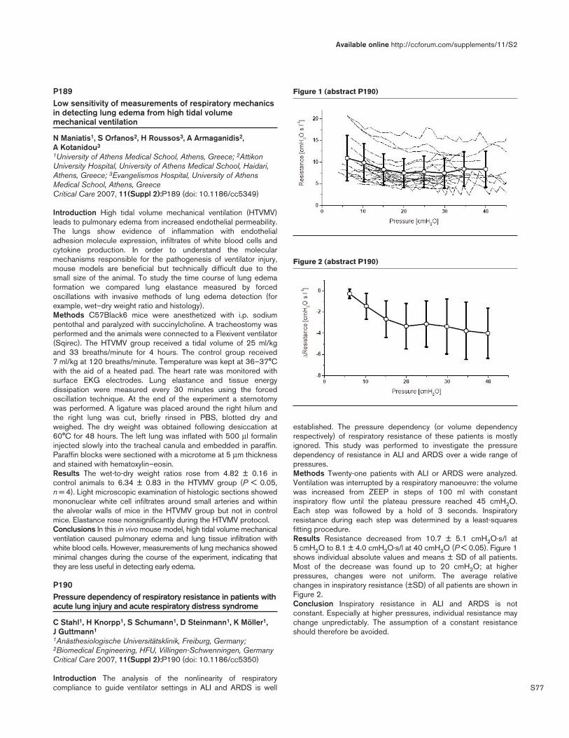
S77
P189Low sensitivity of measurements of respiratory mechanicsin detecting lung edema from high tidal volumemechanical ventilation
N Maniatis1, S Orfanos2, H Roussos3, A Armaganidis2, A Kotanidou3
1University of Athens Medical School, Athens, Greece; 2AttikonUniversity Hospital, University of Athens Medical School, Haidari,Athens, Greece; 3Evangelismos Hospital, University of AthensMedical School, Athens, GreeceCritical Care 2007, 11(Suppl 2):P189 (doi: 10.1186/cc5349)
Introduction High tidal volume mechanical ventilation (HTVMV)leads to pulmonary edema from increased endothelial permeability.The lungs show evidence of inflammation with endothelialadhesion molecule expression, infiltrates of white blood cells andcytokine production. In order to understand the molecularmechanisms responsible for the pathogenesis of ventilator injury,mouse models are beneficial but technically difficult due to thesmall size of the animal. To study the time course of lung edemaformation we compared lung elastance measured by forcedoscillations with invasive methods of lung edema detection (forexample, wet–dry weight ratio and histology).Methods C57Black6 mice were anesthetized with i.p. sodiumpentothal and paralyzed with succinylcholine. A tracheostomy wasperformed and the animals were connected to a Flexivent ventilator(Sqirec). The HTVMV group received a tidal volume of 25 ml/kgand 33 breaths/minute for 4 hours. The control group received7 ml/kg at 120 breaths/minute. Temperature was kept at 36–37°Cwith the aid of a heated pad. The heart rate was monitored withsurface EKG electrodes. Lung elastance and tissue energydissipation were measured every 30 minutes using the forcedoscillation technique. At the end of the experiment a sternotomywas performed. A ligature was placed around the right hilum andthe right lung was cut, briefly rinsed in PBS, blotted dry andweighed. The dry weight was obtained following desiccation at60°C for 48 hours. The left lung was inflated with 500 µl formalininjected slowly into the tracheal canula and embedded in paraffin.Paraffin blocks were sectioned with a microtome at 5 µm thicknessand stained with hematoxylin–eosin.Results The wet-to-dry weight ratios rose from 4.82 ± 0.16 incontrol animals to 6.34 ± 0.83 in the HTVMV group (P < 0.05,n = 4). Light microscopic examination of histologic sections showedmononuclear white cell infiltrates around small arteries and withinthe alveolar walls of mice in the HTVMV group but not in controlmice. Elastance rose nonsignificantly during the HTVMV protocol.Conclusions In this in vivo mouse model, high tidal volume mechanicalventilation caused pulmonary edema and lung tissue infiltration withwhite blood cells. However, measurements of lung mechanics showedminimal changes during the course of the experiment, indicating thatthey are less useful in detecting early edema.
P190Pressure dependency of respiratory resistance in patients withacute lung injury and acute respiratory distress syndrome
C Stahl1, H Knorpp1, S Schumann1, D Steinmann1, K Möller1, J Guttmann1
1Anästhesiologische Universitätsklinik, Freiburg, Germany;2Biomedical Engineering, HFU, Villingen-Schwenningen, GermanyCritical Care 2007, 11(Suppl 2):P190 (doi: 10.1186/cc5350)
Introduction The analysis of the nonlinearity of respiratorycompliance to guide ventilator settings in ALI and ARDS is well
established. The pressure dependency (or volume dependencyrespectively) of respiratory resistance of these patients is mostlyignored. This study was performed to investigate the pressuredependency of resistance in ALI and ARDS over a wide range ofpressures.Methods Twenty-one patients with ALI or ARDS were analyzed.Ventilation was interrupted by a respiratory manoeuvre: the volumewas increased from ZEEP in steps of 100 ml with constantinspiratory flow until the plateau pressure reached 45 cmH2O.Each step was followed by a hold of 3 seconds. Inspiratoryresistance during each step was determined by a least-squaresfitting procedure.Results Resistance decreased from 10.7 ± 5.1 cmH2O·s/l at5 cmH2O to 8.1 ± 4.0 cmH2O·s/l at 40 cmH2O (P < 0.05). Figure 1shows individual absolute values and means ± SD of all patients.Most of the decrease was found up to 20 cmH2O; at higherpressures, changes were not uniform. The average relativechanges in inspiratory resistance (±SD) of all patients are shown inFigure 2.Conclusion Inspiratory resistance in ALI and ARDS is notconstant. Especially at higher pressures, individual resistance maychange unpredictably. The assumption of a constant resistanceshould therefore be avoided.
Available online http://ccforum.com/supplements/11/S2
Figure 1 (abstract P190)
Figure 2 (abstract P190)
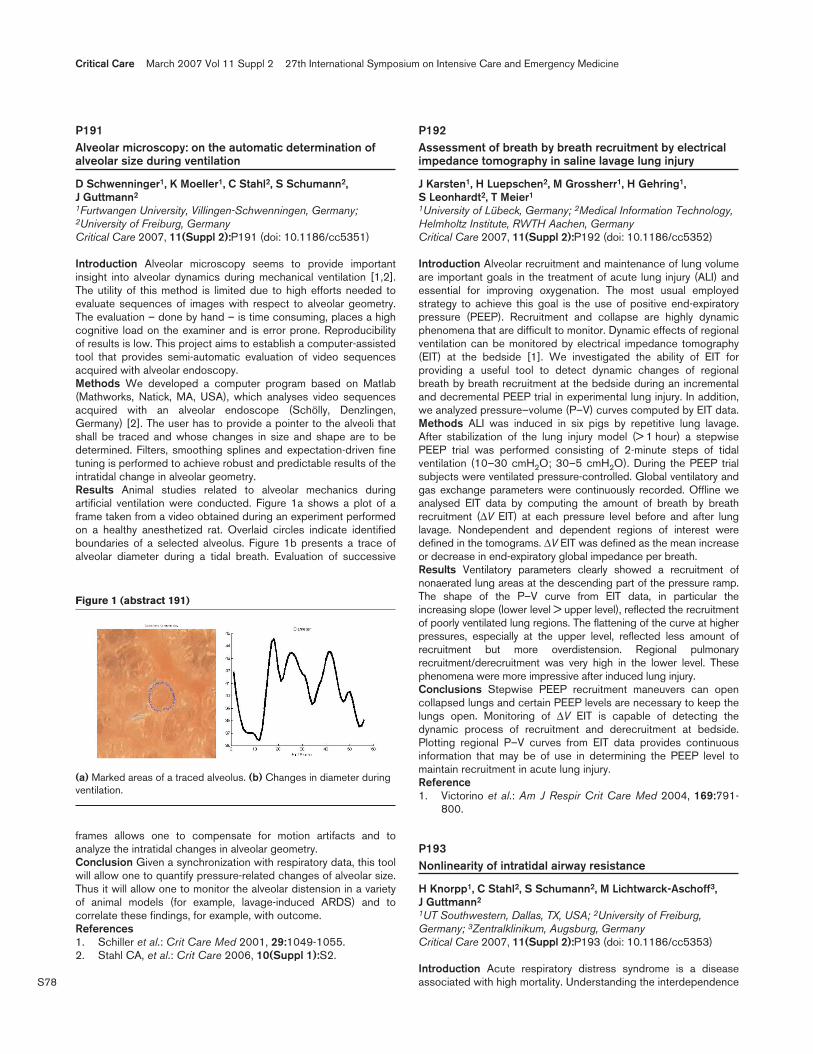
S78
P191Alveolar microscopy: on the automatic determination ofalveolar size during ventilation
D Schwenninger1, K Moeller1, C Stahl2, S Schumann2, J Guttmann2
1Furtwangen University, Villingen-Schwenningen, Germany;2University of Freiburg, GermanyCritical Care 2007, 11(Suppl 2):P191 (doi: 10.1186/cc5351)
Introduction Alveolar microscopy seems to provide importantinsight into alveolar dynamics during mechanical ventilation [1,2].The utility of this method is limited due to high efforts needed toevaluate sequences of images with respect to alveolar geometry.The evaluation – done by hand – is time consuming, places a highcognitive load on the examiner and is error prone. Reproducibilityof results is low. This project aims to establish a computer-assistedtool that provides semi-automatic evaluation of video sequencesacquired with alveolar endoscopy.Methods We developed a computer program based on Matlab(Mathworks, Natick, MA, USA), which analyses video sequencesacquired with an alveolar endoscope (Schölly, Denzlingen,Germany) [2]. The user has to provide a pointer to the alveoli thatshall be traced and whose changes in size and shape are to bedetermined. Filters, smoothing splines and expectation-driven finetuning is performed to achieve robust and predictable results of theintratidal change in alveolar geometry.Results Animal studies related to alveolar mechanics duringartificial ventilation were conducted. Figure 1a shows a plot of aframe taken from a video obtained during an experiment performedon a healthy anesthetized rat. Overlaid circles indicate identifiedboundaries of a selected alveolus. Figure 1b presents a trace ofalveolar diameter during a tidal breath. Evaluation of successive
frames allows one to compensate for motion artifacts and toanalyze the intratidal changes in alveolar geometry.Conclusion Given a synchronization with respiratory data, this toolwill allow one to quantify pressure-related changes of alveolar size.Thus it will allow one to monitor the alveolar distension in a varietyof animal models (for example, lavage-induced ARDS) and tocorrelate these findings, for example, with outcome.References1. Schiller et al.: Crit Care Med 2001, 29:1049-1055.2. Stahl CA, et al.: Crit Care 2006, 10(Suppl 1):S2.
P192Assessment of breath by breath recruitment by electricalimpedance tomography in saline lavage lung injury
J Karsten1, H Luepschen2, M Grossherr1, H Gehring1, S Leonhardt2, T Meier1
1University of Lübeck, Germany; 2Medical Information Technology,Helmholtz Institute, RWTH Aachen, GermanyCritical Care 2007, 11(Suppl 2):P192 (doi: 10.1186/cc5352)
Introduction Alveolar recruitment and maintenance of lung volumeare important goals in the treatment of acute lung injury (ALI) andessential for improving oxygenation. The most usual employedstrategy to achieve this goal is the use of positive end-expiratorypressure (PEEP). Recruitment and collapse are highly dynamicphenomena that are difficult to monitor. Dynamic effects of regionalventilation can be monitored by electrical impedance tomography(EIT) at the bedside [1]. We investigated the ability of EIT forproviding a useful tool to detect dynamic changes of regionalbreath by breath recruitment at the bedside during an incrementaland decremental PEEP trial in experimental lung injury. In addition,we analyzed pressure–volume (P–V) curves computed by EIT data.Methods ALI was induced in six pigs by repetitive lung lavage.After stabilization of the lung injury model (> 1 hour) a stepwisePEEP trial was performed consisting of 2-minute steps of tidalventilation (10–30 cmH2O; 30–5 cmH2O). During the PEEP trialsubjects were ventilated pressure-controlled. Global ventilatory andgas exchange parameters were continuously recorded. Offline weanalysed EIT data by computing the amount of breath by breathrecruitment (∆V EIT) at each pressure level before and after lunglavage. Nondependent and dependent regions of interest weredefined in the tomograms. ∆V EIT was defined as the mean increaseor decrease in end-expiratory global impedance per breath.Results Ventilatory parameters clearly showed a recruitment ofnonaerated lung areas at the descending part of the pressure ramp.The shape of the P–V curve from EIT data, in particular theincreasing slope (lower level > upper level), reflected the recruitmentof poorly ventilated lung regions. The flattening of the curve at higherpressures, especially at the upper level, reflected less amount ofrecruitment but more overdistension. Regional pulmonaryrecruitment/derecruitment was very high in the lower level. Thesephenomena were more impressive after induced lung injury.Conclusions Stepwise PEEP recruitment maneuvers can opencollapsed lungs and certain PEEP levels are necessary to keep thelungs open. Monitoring of ∆V EIT is capable of detecting thedynamic process of recruitment and derecruitment at bedside.Plotting regional P–V curves from EIT data provides continuousinformation that may be of use in determining the PEEP level tomaintain recruitment in acute lung injury.Reference1. Victorino et al.: Am J Respir Crit Care Med 2004, 169:791-
800.
P193Nonlinearity of intratidal airway resistance
H Knorpp1, C Stahl2, S Schumann2, M Lichtwarck-Aschoff3, J Guttmann2
1UT Southwestern, Dallas, TX, USA; 2University of Freiburg,Germany; 3Zentralklinikum, Augsburg, GermanyCritical Care 2007, 11(Suppl 2):P193 (doi: 10.1186/cc5353)
Introduction Acute respiratory distress syndrome is a diseaseassociated with high mortality. Understanding the interdependence
Critical Care March 2007 Vol 11 Suppl 2 27th International Symposium on Intensive Care and Emergency Medicine
Figure 1 (abstract 191)
(a) Marked areas of a traced alveolus. (b) Changes in diameter duringventilation.
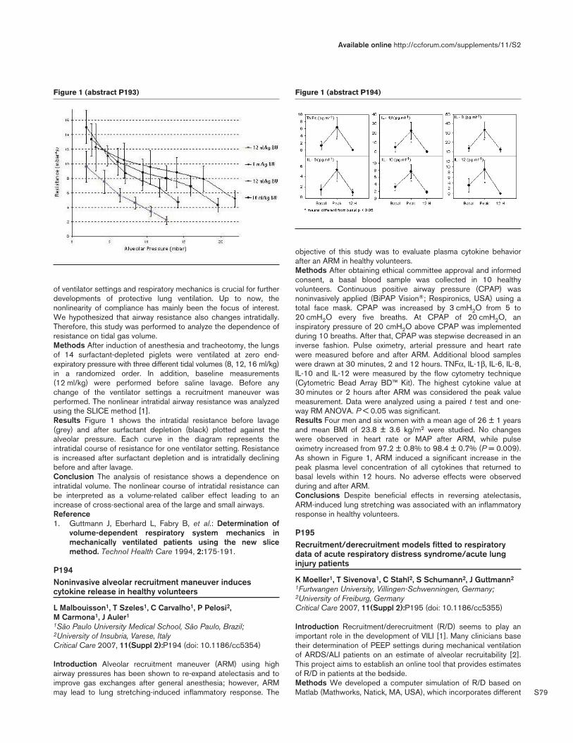
S79
of ventilator settings and respiratory mechanics is crucial for furtherdevelopments of protective lung ventilation. Up to now, thenonlinearity of compliance has mainly been the focus of interest.We hypothesized that airway resistance also changes intratidally.Therefore, this study was performed to analyze the dependence ofresistance on tidal gas volume.Methods After induction of anesthesia and tracheotomy, the lungsof 14 surfactant-depleted piglets were ventilated at zero end-expiratory pressure with three different tidal volumes (8, 12, 16 ml/kg)in a randomized order. In addition, baseline measurements(12 ml/kg) were performed before saline lavage. Before anychange of the ventilator settings a recruitment maneuver wasperformed. The nonlinear intratidal airway resistance was analyzedusing the SLICE method [1].Results Figure 1 shows the intratidal resistance before lavage(grey) and after surfactant depletion (black) plotted against thealveolar pressure. Each curve in the diagram represents theintratidal course of resistance for one ventilator setting. Resistanceis increased after surfactant depletion and is intratidally decliningbefore and after lavage.Conclusion The analysis of resistance shows a dependence onintratidal volume. The nonlinear course of intratidal resistance canbe interpreted as a volume-related caliber effect leading to anincrease of cross-sectional area of the large and small airways.Reference1. Guttmann J, Eberhard L, Fabry B, et al.: Determination of
volume-dependent respiratory system mechanics inmechanically ventilated patients using the new slicemethod. Technol Health Care 1994, 2:175-191.
P194Noninvasive alveolar recruitment maneuver inducescytokine release in healthy volunteers
L Malbouisson1, T Szeles1, C Carvalho1, P Pelosi2, M Carmona1, J Auler1
1São Paulo University Medical School, São Paulo, Brazil;2University of Insubria, Varese, ItalyCritical Care 2007, 11(Suppl 2):P194 (doi: 10.1186/cc5354)
Introduction Alveolar recruitment maneuver (ARM) using highairway pressures has been shown to re-expand atelectasis and toimprove gas exchanges after general anesthesia; however, ARMmay lead to lung stretching-induced inflammatory response. The
objective of this study was to evaluate plasma cytokine behaviorafter an ARM in healthy volunteers.Methods After obtaining ethical committee approval and informedconsent, a basal blood sample was collected in 10 healthyvolunteers. Continuous positive airway pressure (CPAP) wasnoninvasively applied (BiPAP Vision®; Respironics, USA) using atotal face mask. CPAP was increased by 3 cmH2O from 5 to20 cmH2O every five breaths. At CPAP of 20 cmH2O, aninspiratory pressure of 20 cmH2O above CPAP was implementedduring 10 breaths. After that, CPAP was stepwise decreased in aninverse fashion. Pulse oximetry, arterial pressure and heart ratewere measured before and after ARM. Additional blood sampleswere drawn at 30 minutes, 2 and 12 hours. TNFα, IL-1β, IL-6, IL-8,IL-10 and IL-12 were measured by the flow cytometry technique(Cytometric Bead Array BD™ Kit). The highest cytokine value at30 minutes or 2 hours after ARM was considered the peak valuemeasurement. Data were analyzed using a paired t test and one-way RM ANOVA. P < 0.05 was significant.Results Four men and six women with a mean age of 26 ± 1 yearsand mean BMI of 23.8 ± 3.6 kg/m2 were studied. No changeswere observed in heart rate or MAP after ARM, while pulseoximetry increased from 97.2 ± 0.8% to 98.4 ± 0.7% (P = 0.009).As shown in Figure 1, ARM induced a significant increase in thepeak plasma level concentration of all cytokines that returned tobasal levels within 12 hours. No adverse effects were observedduring and after ARM.Conclusions Despite beneficial effects in reversing atelectasis,ARM-induced lung stretching was associated with an inflammatoryresponse in healthy volunteers.
P195Recruitment/derecruitment models fitted to respiratorydata of acute respiratory distress syndrome/acute lunginjury patients
K Moeller1, T Sivenova1, C Stahl2, S Schumann2, J Guttmann2
1Furtwangen University, Villingen-Schwenningen, Germany;2University of Freiburg, GermanyCritical Care 2007, 11(Suppl 2):P195 (doi: 10.1186/cc5355)
Introduction Recruitment/derecruitment (R/D) seems to play animportant role in the development of VILI [1]. Many clinicians basetheir determination of PEEP settings during mechanical ventilationof ARDS/ALI patients on an estimate of alveolar recruitability [2].This project aims to establish an online tool that provides estimatesof R/D in patients at the bedside.Methods We developed a computer simulation of R/D based onMatlab (Mathworks, Natick, MA, USA), which incorporates different
Available online http://ccforum.com/supplements/11/S2
Figure 1 (abstract P193) Figure 1 (abstract P194)
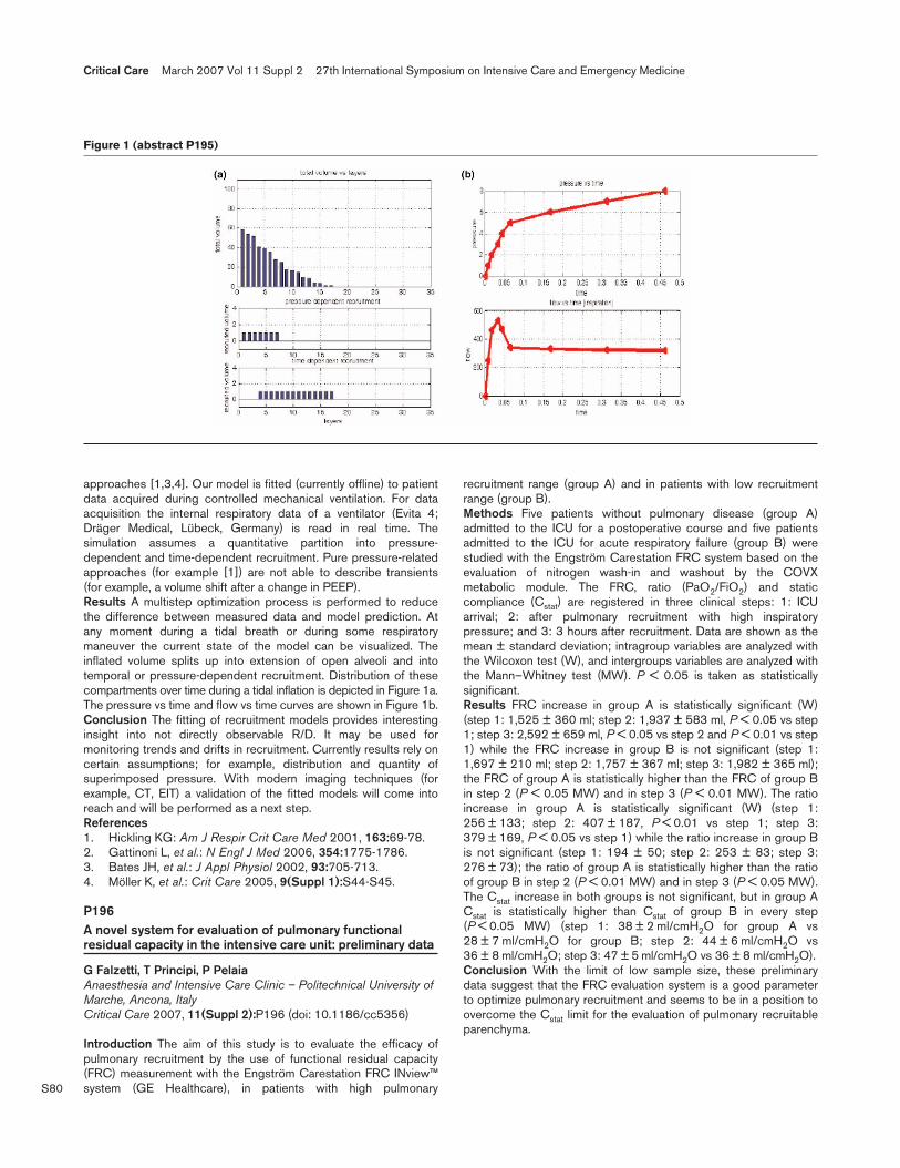
S80
approaches [1,3,4]. Our model is fitted (currently offline) to patientdata acquired during controlled mechanical ventilation. For dataacquisition the internal respiratory data of a ventilator (Evita 4;Dräger Medical, Lübeck, Germany) is read in real time. Thesimulation assumes a quantitative partition into pressure-dependent and time-dependent recruitment. Pure pressure-relatedapproaches (for example [1]) are not able to describe transients(for example, a volume shift after a change in PEEP).Results A multistep optimization process is performed to reducethe difference between measured data and model prediction. Atany moment during a tidal breath or during some respiratorymaneuver the current state of the model can be visualized. Theinflated volume splits up into extension of open alveoli and intotemporal or pressure-dependent recruitment. Distribution of thesecompartments over time during a tidal inflation is depicted in Figure 1a.The pressure vs time and flow vs time curves are shown in Figure 1b.Conclusion The fitting of recruitment models provides interestinginsight into not directly observable R/D. It may be used formonitoring trends and drifts in recruitment. Currently results rely oncertain assumptions; for example, distribution and quantity ofsuperimposed pressure. With modern imaging techniques (forexample, CT, EIT) a validation of the fitted models will come intoreach and will be performed as a next step.References1. Hickling KG: Am J Respir Crit Care Med 2001, 163:69-78.2. Gattinoni L, et al.: N Engl J Med 2006, 354:1775-1786.3. Bates JH, et al.: J Appl Physiol 2002, 93:705-713.4. Möller K, et al.: Crit Care 2005, 9(Suppl 1):S44-S45.
P196A novel system for evaluation of pulmonary functionalresidual capacity in the intensive care unit: preliminary data
G Falzetti, T Principi, P PelaiaAnaesthesia and Intensive Care Clinic – Politechnical University ofMarche, Ancona, ItalyCritical Care 2007, 11(Suppl 2):P196 (doi: 10.1186/cc5356)
Introduction The aim of this study is to evaluate the efficacy ofpulmonary recruitment by the use of functional residual capacity(FRC) measurement with the Engström Carestation FRC INview™system (GE Healthcare), in patients with high pulmonary
recruitment range (group A) and in patients with low recruitmentrange (group B).Methods Five patients without pulmonary disease (group A)admitted to the ICU for a postoperative course and five patientsadmitted to the ICU for acute respiratory failure (group B) werestudied with the Engström Carestation FRC system based on theevaluation of nitrogen wash-in and washout by the COVXmetabolic module. The FRC, ratio (PaO2/FiO2) and staticcompliance (Cstat) are registered in three clinical steps: 1: ICUarrival; 2: after pulmonary recruitment with high inspiratorypressure; and 3: 3 hours after recruitment. Data are shown as themean ± standard deviation; intragroup variables are analyzed withthe Wilcoxon test (W), and intergroups variables are analyzed withthe Mann–Whitney test (MW). P < 0.05 is taken as statisticallysignificant.Results FRC increase in group A is statistically significant (W)(step 1: 1,525 ± 360 ml; step 2: 1,937 ± 583 ml, P < 0.05 vs step1; step 3: 2,592 ± 659 ml, P < 0.05 vs step 2 and P < 0.01 vs step1) while the FRC increase in group B is not significant (step 1:1,697 ± 210 ml; step 2: 1,757 ± 367 ml; step 3: 1,982 ± 365 ml);the FRC of group A is statistically higher than the FRC of group Bin step 2 (P < 0.05 MW) and in step 3 (P < 0.01 MW). The ratioincrease in group A is statistically significant (W) (step 1:256 ± 133; step 2: 407 ± 187, P < 0.01 vs step 1; step 3:379 ± 169, P < 0.05 vs step 1) while the ratio increase in group Bis not significant (step 1: 194 ± 50; step 2: 253 ± 83; step 3:276 ± 73); the ratio of group A is statistically higher than the ratioof group B in step 2 (P < 0.01 MW) and in step 3 (P < 0.05 MW).The Cstat increase in both groups is not significant, but in group ACstat is statistically higher than Cstat of group B in every step(P < 0.05 MW) (step 1: 38 ± 2 ml/cmH2O for group A vs28 ± 7 ml/cmH2O for group B; step 2: 44 ± 6 ml/cmH2O vs36 ± 8 ml/cmH2O; step 3: 47 ± 5 ml/cmH2O vs 36 ± 8 ml/cmH2O).Conclusion With the limit of low sample size, these preliminarydata suggest that the FRC evaluation system is a good parameterto optimize pulmonary recruitment and seems to be in a position toovercome the Cstat limit for the evaluation of pulmonary recruitableparenchyma.
Critical Care March 2007 Vol 11 Suppl 2 27th International Symposium on Intensive Care and Emergency Medicine
Figure 1 (abstract P195)
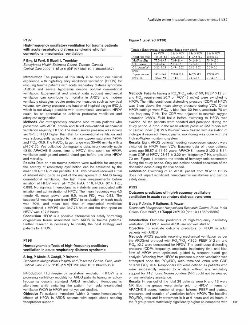
S81
P197High-frequency oscillatory ventilation for trauma patientswith acute respiratory distress syndrome who failconventional mechanical ventilation
F Eng, M Ferri, S Rizoli, L TremblaySunnybrook Health Sciences Centre, Toronto, CanadaCritical Care 2007, 11(Suppl 2):P197 (doi: 10.1186/cc5357)
Introduction The purpose of this study is to report our clinicalexperience with high-frequency oscillatory ventilation (HFOV) forrescuing trauma patients with acute respiratory distress syndrome(ARDS) and severe hypoxemia despite optimal conventionalventilation. Experimental and clinical data suggest mechanicalventilation can contribute to mortality in ARDS, and modernventilatory strategies require protective measures such as low tidalvolume, low airway pressure and fraction of inspired oxygen (FIO2),which is not always possible with conventional ventilation. HFOVcould be an alternative to achieve protective ventilation andadequate oxygenation.Methods We retrospectively analyzed nine trauma patients whopresented with ARDS criteria and failed conventional mechanicalventilation requiring HFOV. The mean airway pressure was initiallyset 3–5 cmH2O higher than that for conventional ventilation andwas subsequently adjusted to maintain oxygen saturation >90%and FiO2 <0.6. The PaCO2 target range was 35–60 mmHg with apH >7.25. We collected demographic data, injury severity scale(ISS), APACHE II score, time to HFOV, time spent on HFOV,ventilation settings and arterial blood gas before and after HFOVand mortality.Results Data on nine trauma patients were available for analysis;the severity of respiratory dysfunction can be estimated by themean PaO2/FiO2 of our patients, 131. Two patients received a trialof inhaled nitric oxide as part of the management of ARDS failingconventional ventilation. The last mean measurements beforeinitiation of HFOV were: pH 7.24, PaO2 116, PCO2 67.4, FiO20.899. No significant hemodynamic instability was associated withinitiation and administration of HFOV. The mean frequency was 4.3(mode 4), mean power was 8.5, mean FiO2 was 0.83. Thesuccessful weaning rate from HFOV to extubation or trach maskwas 70%, and mean total time of mechanical ventilation(conventional + HFOV) was 347.76 hours and the time spent onHFOV was 107.5 hours.Conclusion HFOV is a possible alternative for safely correctingoxygenation failure associated with ARDS in trauma patients.Further research is necessary to identify the best strategy andpatients for HFOV.
P198Hemodynamic effects of high-frequency oscillatoryventilation in acute respiratory distress syndrome
S Jog, P Akole, S Gadgil, P RajhansDeenanath Mangeshkar Hospital and Research Centre, Pune, IndiaCritical Care 2007, 11(Suppl 2):P198 (doi: 10.1186/cc5358)
Introduction High-frequency oscillatory ventilation (HFOV) is apromising ventilatory modality for ARDS patients having refractoryhypoxemia despite standard ARDS ventilation. Hemodynamicalterations while switching the patient from volume-controlledventilation (VCV) to HFOV are not yet well studied.Objective To evaluate immediate (within 3 hours) hemodynamiceffects of HFOV in ARDS patients with septic shock needingvasopressor support.
Methods Patients having a PO2/FiO2 ratio ≤150, PEEP >12 cmand FiO2 requirement ≥0.7 on VCV (6 ml/kg) were switched toHFOV. The initial continuous distending pressure (CDP) of HFOVwas 5 cm above the mean airway pressure during VCV. OtherHFOV settings were FiO2 1, bias flow 30 l/min, amplitude 70 cmand frequency 7 Hz. The CDP was adjusted to maintain oxygensaturation >88%. Fluid bolus before switching to HFOV wasavoided. All the patients were sedated and paralysed during thestudy period. A drop in the mean arterial pressure (MAP) ≤65 mmor cardiac index (CI) ≤2.5 l/min/m2 were treated with escalation ofinotrope if required. Hemodynamic monitoring was done with theFlotrac-Vigileo monitoring system.Results Eight ARDS patients needing vasopressor support wereswitched to HFOV from VCV. Baseline data of these patientswere: age 58.87 ± 11.69 years, APACHE II score 21.02 ± 8.14,mean CDP of HFOV 26.67 ± 3.22 cm, frequency 7 Hz, amplitude70 cm. Figure 1 presents the trends of hemodynamic parametersduring the study period. Only one patient needed escalation of thedopamine dose during the trial period.Conclusion Switching of an ARDS patient from VCV to HFOVdoes not impart significant hemodynamic instabilities and can besafely done.
P199Outcome predictors of high-frequency oscillatoryventilation in acute respiratory distress syndrome
S Jog, P Akole, P Rajhans, B PawarDeenanath Mangeshkar Hospital and Research Centre, Pune, IndiaCritical Care 2007, 11(Suppl 2):P199 (doi: 10.1186/cc5359)
Introduction Outcome predictors of high-frequency oscillatoryventilation (HFOV) in severe ARDS are not well studied.Objective To evaluate outcome predictors of HFOV in adultpatients with ARDS.Methods ARDS patients receiving mechanical ventilation as perthe ARDSnet protocol with PO2/FiO2 <150, PEEP ≥12 cm andFiO2 ≥0.7 were considered for HFOV. The continuous distendingpressure (CDP), frequency, amplitude, inspiratory time and biasflow of HFOV were optimised, guided by frequent blood gasanalysis. Weaning from HFOV to pressure support ventilation wasattempted once the PO2/FiO2 ratio remained ≥200 with CDP≤18 cm FiO2 ≤0.5. Responders (R) were defined as patients whowere successfully weaned to a state without any ventilatorysupport for >12 hours. Nonresponders (NR) could not be weanedoff any ventilatory assistance.Results Fifteen out of the total 28 patients were R and 13 wereNR. Both the groups were similar prior to HFOV in terms ofAPACHE II score, number of organ failures, PEEP and plateaupressures, and duration of ventilation before HFOV. The baselinePO2/FiO2 ratio and improvement in it at 6 hours and 24 hours inthe R group were statistically significantly higher as compared with
Available online http://ccforum.com/supplements/11/S2
Figure 1 (abstract P198)
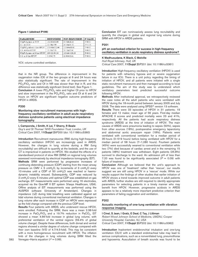
S82
that in the NR group. The difference in improvement in theoxygenation index (OI) of the two groups at 6 and 24 hours wasalso statistically significant. The rate of improvement in thePO2/FiO2 ratio and OI in NR was slower than that in R, and thisdifference was statistically significant (trend test). See Figure 1.Conclusion A lower PO2/FiO2 ratio and higher OI prior to HFOVand slow improvement in the PO2/FiO2 ratio and OI at 6 and 24hours on HFOV are significant negative outcome predictors ofHFOV in ARDS.
P200Monitoring slow recruitment manoeuvres with high-frequency oscillatory ventilation in adult acute respiratorydistress syndrome patients using electrical impedancetomography
L Camporota, J Smith, K Lei, T Sherry, R BealeGuy’s and St Thomas’ NHS Foundation Trust, London, UKCritical Care 2007, 11(Suppl 2):P200 (doi: 10.1186/cc5360)
Introduction Recruitment manoeuvres (RM) during high-frequencyoscillatory ventilation (HFOV) are increasingly used in ARDS.However, the changes in lung volume during a RM (lungrecruitability) are difficult to quantify at the bedside, and the use ofCT is impractical in patients on HFOV. We studied the effects of astandardised protocol of slow RM (SRM) on regional lung volumesassessed noninvasively by electrical impedance tomography (EIT).Methods SRM were performed by progressive increases ofcontinuing distending pressure (CDP) starting from the mean airwaypressure on CMV + 5 cmH2O, by increments of 3 cmH2O every10 minutes until a CDP of 50 cmH2O was reached or haemo-dynamic instability ensued. Subsequently, CDP was reduced by2 cmH2O every 5 minutes until optimal CDP was established on gasexchange. EIT measurements were performed using 16 electrodes,acquired via the Goe-MF II EIT system (Viasys Healthcare, USA).Offline analysis of EIT measurements was performed using theAUSPEX software (University of Amsterdam). Changes inimpedance (∆Z) during tidal breathing were calibrated against settidal volumes during conventional mechanical ventilation. Changes inlung volume after each increase in CDP on HFOV were expressedas the fold change compared with the previous CDP level.Results Four patients with ARDS, who underwent rescue HFOV,were enrolled. Following the SRM, there was a mean 2.38-foldincrease in PaO2/FiO2 and a 19.7% reduction in PaCO2. EITshowed a mean 4.66-fold increase in global lung volume, withpreferential ventilation of the ventral regions (59.4% of globalvolume change). Despite these differences, both dorsal and ventralregions showed a similar degree of volume change compared withtheir own baseline (V/D of 4.7/4.5-fold). This may be consistentwith a more homogeneous recruitment with HFOV. The inflationlimb of the changes in lung volumes during SRM fitted theVenegas–Harris equation (r2 = 0.99).
Conclusion EIT can noninvasively assess lung recruitability andquantify the changes in global and regional lung volume duringSRM with HFOV in ARDS patients.
P201pH: an overlooked criterion for success in high-frequencyoscillatory ventilation in acute respiratory distress syndrome?
K Madhusudana, K Black, C MelvilleHull Royal Infirmary, Hull, UKCritical Care 2007, 11(Suppl 2):P201 (doi: 10.1186/cc5361)
Introduction High-frequency oscillatory ventilation (HFOV) is usedfor patients with refractory hypoxia and or severe oxygenationfailure in our ICU. There is a unit policy regarding the timing ofinitiation of HFOV, and all patients were initiated with a singlestatic recruitment manoeuvre and then managed according to localguidelines. The aim of this study was to understand whichventilatory parameters best predicted successful outcomefollowing HFOV.Methods After institutional approval, we retrospectively reviewedthe case notes all the adult patients who were ventilated withHFOV during the 18-month period between January 2005 and July2006. The data were analysed using SPSS® version 13 software.Results There were 33 episodes of HFOV in 31 patients; 19females and 12 males; mean age of 56 years. First-day medianAPACHE II scores and predicted mortality were 23 and 41%,respectively. All the patients had acute respiratory distresssyndrome (ARDS) at the time of initiation of HFOV. The maincauses of ARDS were pneumonia leading to sepsis (50%), sepsisfrom other sources (18%), postoperative emergency laparotomyand abdominal aortic aneurysm repair (18%). Patients wereventilated with conventional ventilation for a median period of35 hours (0–519 hours) before being ventilated with HFOV for amedian period of 58 hours (7–1,080 hours). Fourteen patients(45%) were successfully weaned to conventional ventilation whiletwo (7%) died because of cardiac arrest and in the remaining 15patients (48%) treatment was withdrawn. Eight patients (25.8%)survived to discharge to the ward. An admission pH of less than7.20 was found to be significantly associated (P = 0.09) withfailure of treatment.Conclusion Although we believed that the unit’s approach toHFOV was one of ‘treatment’ rather than ‘rescue’, our resultssuggest we are still using HFOV in a ‘rescue’ mode. While ourresults support the findings of other studies that earlier initiation ofHFOV shows a trend towards improved outcome in adult patientswith ARDS, further studies are still required to identify appropriateparameters for selecting patients in a timely manner who maybenefit from HFOV. However, progressive acidosis in ARDSappears to be a relatively more important predictive criterion thanparameters of failing oxygenation and ventilation.
P202Acoustic monitoring of one-lung ventilation with vibrationresponse imaging
I Cinel, S Jean, I Gratz, E Deal, C Tay, J LittmanRobert Wood Johnson School of Medicine, UMDNJ, CooperUniversity Hospital, Camden, NJ, USACritical Care 2007, 11(Suppl 2):P202 (doi: 10.1186/cc5362)
Introduction Inadvertent endobronchial intubation and one-lungventilation (OLV) with a standard endotracheal tube may lead toserious complications, such as a nonventilated lung, pneumothoraxand hypoxemia. Auscultation of breath sounds was found to be
Critical Care March 2007 Vol 11 Suppl 2 27th International Symposium on Intensive Care and Emergency Medicine
Figure 1 (abstract P199)
VCV, volume controlled ventilation.
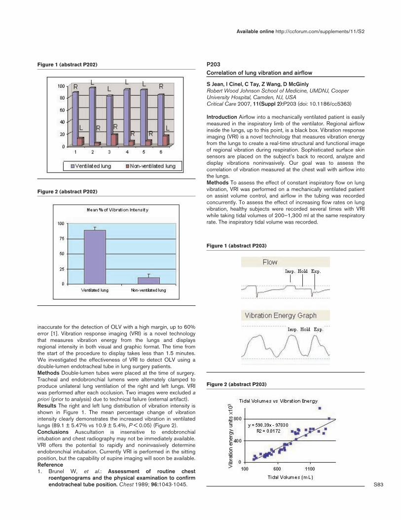
S83
inaccurate for the detection of OLV with a high margin, up to 60%error [1]. Vibration response imaging (VRI) is a novel technologythat measures vibration energy from the lungs and displaysregional intensity in both visual and graphic format. The time fromthe start of the procedure to display takes less than 1.5 minutes.We investigated the effectiveness of VRI to detect OLV using adouble-lumen endotracheal tube in lung surgery patients.Methods Double-lumen tubes were placed at the time of surgery.Tracheal and endobronchial lumens were alternately clamped toproduce unilateral lung ventilation of the right and left lungs. VRIwas performed after each occlusion. Two images were excluded apriori (prior to analysis) due to technical failure (external artifact).Results The right and left lung distribution of vibration intensity isshown in Figure 1. The mean percentage change of vibrationintensity clearly demonstrates the increased vibration in ventilatedlungs (89.1 ± 5.47% vs 10.9 ± 5.4%, P < 0.05) (Figure 2).Conclusions Auscultation is insensitive to endobronchialintubation and chest radiography may not be immediately available.VRI offers the potential to rapidly and noninvasively determineendobronchial intubation. Currently VRI is performed in the sittingposition, but the capability of supine imaging will soon be available.Reference1. Brunel W, et al.: Assessment of routine chest
roentgenograms and the physical examination to confirmendotracheal tube position. Chest 1989; 96:1043-1045.
P203Correlation of lung vibration and airflow
S Jean, I Cinel, C Tay, Z Wang, D McGinlyRobert Wood Johnson School of Medicine, UMDNJ, CooperUniversity Hospital, Camden, NJ, USACritical Care 2007, 11(Suppl 2):P203 (doi: 10.1186/cc5363)
Introduction Airflow into a mechanically ventilated patient is easilymeasured in the inspiratory limb of the ventilator. Regional airflowinside the lungs, up to this point, is a black box. Vibration responseimaging (VRI) is a novel technology that measures vibration energyfrom the lungs to create a real-time structural and functional imageof regional vibration during respiration. Sophisticated surface skinsensors are placed on the subject’s back to record, analyze anddisplay vibrations noninvasively. Our goal was to assess thecorrelation of vibration measured at the chest wall with airflow intothe lungs.Methods To assess the effect of constant inspiratory flow on lungvibration, VRI was performed on a mechanically ventilated patienton assist volume control, and airflow in the tubing was recordedconcurrently. To assess the effect of increasing flow rates on lungvibration, healthy subjects were recorded several times with VRIwhile taking tidal volumes of 200–1,300 ml at the same respiratoryrate. The inspiratory tidal volume was recorded.
Available online http://ccforum.com/supplements/11/S2
Figure 1 (abstract P202)
Figure 2 (abstract P202)
Figure 1 (abstract P203)
Figure 2 (abstract P203)
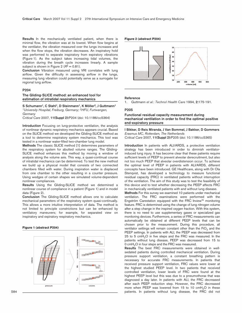
S84
Results In the mechanically ventilated patient, when there isminimal flow, the vibration was at its lowest. When flow begins atthe ventilator, the vibration measured over the lungs increases andwhen the flow stops, the vibration decreases. An inspiratory holdwas performed to separate inspiratory from expiratory vibrations(Figure 1). As the subject takes increasing tidal volumes, thevibration during the breath cycle increases linearly. A samplesubject is shown in Figure 2 (R2 = 0.81).Conclusion Vibration measured using VRI correlates with lungairflow. Given the difficulty in assessing airflow in the lungs,measuring lung vibration could potentially serve as a surrogate forregional lung airflow.
P204The Gliding-SLICE method: an enhanced tool forestimation of intratidal respiratory mechanics
S Schumann1, C Stahl1, D Steinmann1, K Möller2, J Guttmann1
1University Hospital, Freiburg, Germany; 2HFU, Furtwangen,GermanyCritical Care 2007, 11(Suppl 2):P204 (doi: 10.1186/cc5364)
Introduction Focusing on lung-protective ventilation, the analysisof nonlinear dynamic respiratory mechanics appears crucial. Basedon the SLICE method we developed the Gliding-SLICE method asa tool to determine respiratory system mechanics. This tool wastested in a nonlinear water-filled two-chamber lung model.Methods The classic SLICE method [1] determines parameters ofthe respiratory system for abutted volume ranges. The Gliding-SLICE method enhances this method by moving a window ofanalysis along the volume axis. This way, a quasi-continual courseof intratidal mechanics can be determined. To test the new methodwe build up a physical model that consists of two connectedchambers filled with water. During inspiration water is displacedfrom one chamber to the other resulting in a counter pressure.Using wedges of certain shapes we simulated volume-dependentnonlinear compliances.Results Using the Gliding-SLICE method we determined anonlinear course of compliance in a patient (Figure 1) and in modeldata (Figure 2).Conclusion The Gliding-SLICE method allows one to calculatemechanical parameters of the respiratory system quasi-continually.This allows a more intuitive interpretation of data. The method isnot limited to principle constrictions but can be enhanced byventilatory maneuvers; for example, for separated view oninspiratory and expiratory respiratory mechanics.
Reference1. Guttmann et al.: Technol Health Care 1994, 2:175-191.
P205Functional residual capacity measurement duringmechanical ventilation in order to find the optimal positiveend-expiratory pressure
I Bikker, D Reis Miranda, J Van Bommel, J Bakker, D GommersErasmus MC, Rotterdam, The NetherlandsCritical Care 2007, 11(Suppl 2):P205 (doi: 10.1186/cc5365)
Introduction In patients with ALI/ARDS, a protective ventilationstrategy has been introduced in order to diminish ventilator-induced lung injury. It has become clear that these patients requiresufficient levels of PEEP to prevent alveolar derecruitment, but alsonot too much PEEP that alveolar overdistension occur. To achievethe optimal level of PEEP in patients with ALI/ARDS, differentconcepts have been introduced. GE Healthcare, along with Dr OlaStenqvist, has developed a technology to measure functionalresidual capacity (FRC) in ventilated patients without interruptionof the ventilation. The aim of this study was to test the feasibility ofthis device and to test whether decreasing the PEEP affects FRCin mechanically ventilated patients with and without lung disease.Methods For this survey we examined 10 patients under mechanicalventilation. The FRC examinations were performed with theEngström Carestation equipped with the FRC Inview™ monitoringfeature. FRC is determined using the change of lung nitrogen volumeafter a step change in the inspired oxygen fraction. With this system,there is no need to use supplementary gases or specialized gasmonitoring devices. Furthermore, a series of FRC measurements canautomatically be obtained at different PEEP levels that can bechosen prior to the measurement. During this procedure, allventilator settings will remain constant other than the FiO2 and thePEEP settings. In patients with ALI, the PEEP was decreased from25 to 5 cmH2O in five steps and the FRC was measured. In thepatients without lung disease, PEEP was decreased from 15 to0 cmH2O in four steps and the FRC was measured.Results The best FRC measurements were obtained in well-sedated patients during controlled mechanical ventilation. Duringpressure support ventilation, a constant breathing pattern isnecessary for accurate FRC measurements. In patients thatreceived pressure support ventilation, FRC values were lower atthe highest studied PEEP level. In two patients that receivedcontrolled ventilation, lower levels of FRC were found at thehighest PEEP level but this was due to a pneumothorax that wasdiagnosed a day later. In patients with ALI, the FRC decreasedafter each PEEP reduction step. However, the FRC decreasedmore when PEEP was lowered from 15 to 10 cmH2O in thesepatients. In patients without lung disease, the FRC did not
Critical Care March 2007 Vol 11 Suppl 2 27th International Symposium on Intensive Care and Emergency Medicine
Figure 1 (abstract P204)
Figure 2 (abstract P204)
S85
decrease after PEEP was reduced from 15 to 5 cmH2O butdecreased after PEEP was reduced from 5 to 0 cmH2O.Conclusion Accurate measurements of FRC are obtained during aconstant breathing pattern that is easier to obtain during controlledventilation in comparison with pressure-support ventilation. Inpatients with ALI/ARDS, the FRC decreased during each PEEPreduction, but whether the largest change in FRC indicates theoptimal PEEP needs further research.
P206Analysis of the nonaerated lung volume in combinations ofsingle computed tomography slices – is extrapolation tothe entire lung feasible?
AW Reske, P Hepp, C Heine, K Schmidt, M Seiwerts, U Gottschaldt, AP ReskeUniversitaetsklinikum Leipzig, GermanyCritical Care 2007, 11(Suppl 2):P206 (doi: 10.1186/cc5366)
Introduction The nonaerated lung volume (Vnon) can be quantifiedfrom computed tomography (CT) images. Analysis of the CT slicescovering the entire lung is time-consuming and thus limits potentialclinical and experimental applications. This could be improved byanalyzing only a few representative CT slices. The number and ana-tomical location of CT images required for analyses that are represen-tative for the entire lung, however, is discussed controversially.Methods The percentage of Vnon (%Vnon) relative to the total lungvolume was quantified in CT-image series (n = 21) of sheep withgross anesthesia-induced atelectasis. This was performed fordifferent combinations of number and anatomical location of CTslices and the results were compared with the %Vnon of the entirelung (lung). The combinations were: one juxtadiaphragmatic slice(juxta), three apical, hiliar and juxtadiaphragmatic slices (3old), andthree consecutive juxtadiaphragmatic slices (3new). The correla-tion between %Vnon and the arterial oxygen partial pressure (PaO2)was examined for all combinations. The PaO2 was measured at thetime of the CT and transformed logarithmically (lnPaO2) to linearizethe relation between PaO2 and %Vnon. Linear regression andBland–Altman plots were used for statistical analysis.Results The R-squared (R2) values for the correlation betweenlnPaO2 and %Vnon of lung and the slice combinations juxta, 3newand 3old were 0.61, 0.60, 0.57 and 0.55, respectively. The %Vnonof lung correlated best with the %Vnon of slice combinations juxtaand 3new (R2 = 0.96 and 0.95, respectively). Comparison of theseslice combinations with lung also resulted in the least bias in theBland–Altman analyses (6.3 and 5.9%, respectively). R2 for thecorrelation between lung and 3old was 0.93, and the bias for lungvs 3old in the Bland–Altman analysis was 6.8%.Conclusion Depending on the precision required, the use of singlejuxtadiaphragmatic CT slices can help to speed up the analysisprocess and thereby propel the clinical implementation of CT-derived information. Our data suggest that juxtadiaphragmaticslices may be better suited than the ‘traditional’ combination ofapical, hiliar and juxtadiaphragmatic slices.
P207Pulmonary expansion and disobstruction maneuver with aclosed system in patients with acute lung injury and acutedistress syndrome, and its effect on gas exchange
J Belato, S Barreto, C Santos, S VieiraHospital de Clínicas de Porto Alegre, BrazilCritical Care 2007, 11(Suppl 2):P207 (doi: 10.1186/cc5367)
Introduction Respiratory physiotherapy is ever more utilized for thetreatment of critical patients. However, it is known that there are
few studies on the effect on gas exchange in patients with acutelung injury (ALI) and acute respiratory distress syndrome (ARDS)and physiotherapy techniques. The purpose of this study was toassess the effect of a pulmonary expansion and disobstructionmaneuver with a closed system on the gas exchange of patientswith ALI and ARDS.Methods The patients with the diagnosis of ALI and ARDS who metthe inclusion criteria were randomized to one of the two groups:those of the intervention group were subjected to a pulmonaryexpansion and bronchial disobstruction maneuvers, for approximately10 minutes by the association of the following physiotherapytechniques: sighs, side-lying position, expiratory rib-cagecompression and endotracheal suctioning with a closed system andafter observed for 10 minutes; the patients of the control group didnot receive any treatment, they were only observed for 20 minutes.Ventilatory parameters and arterial blood gases were measuredbefore (Time 1) and 10 minutes after the procedures (Time 2). Theanalysis of variance test for repeated measurements was used forcomparing variables at different times. Results are shown as themean and standard deviation. The significant level was P < 0.05.Results At Time 1, the control group (n = 21) had ratio of arterialpartial pressure of oxygen to fraction of inspired oxygen (PaO2/FiO2) and partial arterial carbon dioxide pressure (PaCO2) of167.7 ± 56.2 and 40.3 ± 10.1, respectively, and the interventiongroup (n = 19) had PaO2/FiO2 of 180.5 ± 67.0 and PaCO2 of38.6 ± 10.5. At Time 2, the control group had, respectively, PaO2/FiO2 and PaCO2 of 165.9 ± 63.8 and 38.9 ± 10.3, and the inter-vention group of 177.2 ± 4.5 and 39.0 ± 10.8. No variable was signifi-cantly different between the groups at Time 1 and Time 2 (P > 0.05).Conclusion The proposed maneuver was not beneficial for gasexchange in the sample studied.
P208Dry powder nebulization of a recombinant surfactantprotein C-based surfactant for treatment of acuterespiratory distress syndrome
C Ruppert, T Kuchenbuch, S Schmidt, P Markart, T Gessler, T Schmehl, W Seeger, A GuentherUniversity of Giessen Lung Center, Giessen, GermanyCritical Care 2007, 11(Suppl 2):P208 (doi: 10.1186/cc5368)
Introduction Nebulization of pulmonary surfactant for treatment ofARDS represents a desirable therapeutic approach but washitherto impossible under clinical conditions due to the technicallimitations of currently available devices. In the present study weinvestigated a new dry powder nebulizer for administration of arecombinant surfactant protein C (rSP-C)-based surfactant.Methods The nebulizer device consists of a cylindrical glasshousing that, at the bottom, ends up in a spherical lower housingpart that serves as the dry powder reservoir. A gas inlet portionwith a nozzle at its end is coaxially aligned with the housing, almostreaches the bottom of the dry powder reservoir, and inducesaerosol generation when gas pressures between 1 and 2 bar areapplied. The upper portion of the housing contains a cap with anaerosol exit port. Several nozzles ensure a discharge of unsuitablylarge aerosol particles. Aerosol characteristics were determined bylaser diffractometry. The efficacy of an inhalative rSP-C surfactantapplication was assessed in three animal models of acute lunginjury, including rabbits with acute lung injury due to eitherrepetitive lavage with prolonged and injurious ventilation, or due toinhalative application of bleomycin at day 4, and bleomycin-challenged, spontaneously breathing mice.Results The generated aerosol had a mass median aerodynamicdiameter of 1.6 µm, with 85% of all particles being smaller than
Available online http://ccforum.com/supplements/11/S2
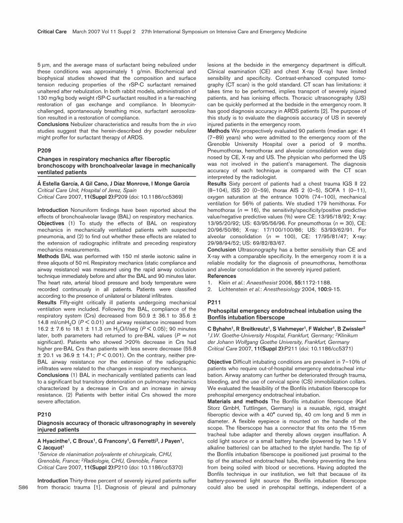
S86
5 µm, and the average mass of surfactant being nebulized underthese conditions was approximately 1 g/min. Biochemical andbiophysical studies showed that the composition and surfacetension reducing properties of the rSP-C surfactant remainedunaltered after nebulization. In both rabbit models, administration of130 mg/kg body weight rSP-C surfactant resulted in a far-reachingrestoration of gas exchange and compliance. In bleomycin-challenged, spontaneously breathing mice, surfactant aerosoliza-tion resulted in a restoration of compliance.Conclusions Nebulizer characteristics and results from the in vivostudies suggest that the herein-described dry powder nebulizermight proffer for surfactant therapy of ARDS.
P209Changes in respiratory mechanics after fiberopticbronchoscopy with bronchoalveolar lavage in mechanicallyventilated patients
Á Estella García, A Gil Cano, J Díaz Monrove, I Monge GarcíaCritical Care Unit, Hospital of Jerez, SpainCritical Care 2007, 11(Suppl 2):P209 (doi: 10.1186/cc5369)
Introduction Nonuniform findings have been reported about theeffects of bronchoalveolar lavage (BAL) on respiratory mechanics.Objectives (1) To study the effects of BAL on respiratorymechanics in mechanically ventilated patients with suspectedpneumonia, and (2) to find out whether these effects are related tothe extension of radiographic infiltrate and preceding respiratorymechanics measurements.Methods BAL was performed with 150 ml sterile isotonic saline inthree aliquots of 50 ml. Respiratory mechanics (static compliance andairway resistance) was measured using the rapid airway occlusiontechnique immediately before and after the BAL and 90 minutes later.The heart rate, arterial blood pressure and body temperature wererecorded continuously in all patients. Patients were classifiedaccording to the presence of unilateral or bilateral infiltrates.Results Fifty-eight critically ill patients undergoing mechanicalventilation were included. Following the BAL, compliance of therespiratory system (Crs) decreased from 50.9 ± 36.1 to 35.6 ±14.8 ml/cmH2O (P < 0.01) and airway resistance increased from16.2 ± 7.6 to 18.1 ± 11.3 cm H2O/l/seg (P < 0.05); 90 minuteslater, both parameters had returned to pre-BAL values (P = notsignificant). Patients who showed >20% decrease in Crs hadhigher pre-BAL Crs than patients with less severe decrease (55.8± 20.1 vs 36.9 ± 14.1; P < 0.001). On the contrary, neither pre-BAL airway resistance nor the extension of the radiographicinfiltrates were related to the changes in respiratory mechanics.Conclusions (1) BAL in mechanically ventilated patients can leadto a significant but transitory deterioration on pulmonary mechanicscharacterized by a decrease in Crs and an increase in airwayresistance. (2) Patients with better initial Crs showed the moresevere affectation.
P210Diagnosis accuracy of thoracic ultrasonography in severelyinjured patients
A Hyacinthe1, C Broux1, G Francony1, G Ferretti2, J Payen1, C Jacquot1
1Service de réanimation polyvalente et chirurgicale, CHU,Grenoble, France; 2Radiologie, CHU, Grenoble, FranceCritical Care 2007, 11(Suppl 2):P210 (doi: 10.1186/cc5370)
Introduction Thirty-three percent of severely injured patients sufferfrom thoracic trauma [1]. Diagnosis of pleural and pulmonary
lesions at the bedside in the emergency department is difficult.Clinical examination (CE) and chest X-ray (X-ray) have limitedsensibility and specificity. Contrast-enhanced computed tomo-graphy (CT scan) is the gold standard. CT scan has limitations: ittakes time to be performed, implies transport of severely injuredpatients, and has ionising effects. Thoracic ultrasonography (US)can be quickly performed at the bedside in the emergency room. Ithas good diagnosis accuracy in ARDS patients [2]. The purpose ofthis study is to evaluate the diagnosis accuracy of US in severelyinjured patients in the emergency room.Methods We prospectively evaluated 90 patients (median age: 41(7–89) years) who were admitted to the emergency room of theGrenoble University Hospital over a period of 9 months.Pneumothorax, hemothorax and alveolar consolidation were diag-nosed by CE, X-ray and US. The physician who performed the USwas not involved in the patient’s management. The diagnosisaccuracy of each technique is compared with the CT scaninterpreted by the radiologist.Results Sixty percent of patients had a chest trauma IGS II 22(8–104), ISS 20 (0–59), thorax AIS 2 (0–5), SOFA 1 (0–11),oxygen saturation at the entrance 100% (74–100), mechanicalventilation for 56% of patients. We studied 179 hemithorax. Forhemothorax (n = 16), the sensitivity/specificity/positive predictivevalue/negative predictive values (%) were CE: 13/95/18/92; X-ray:13/95/20/92; US: 63/95/56/96. For pneumothorax (n = 30), CE:20/96/50/86; X-ray: 17/100/100/86; US: 53/93/62/91. Foralveolar consolidation (n = 100), CE: 17/95/81/47; X-ray:29/98/94/52; US: 69/82/83/67.Conclusion Ultrasonography has a better sensitivity than CE andX-ray with a comparable specificity. In the emergency room it is areliable modality for the diagnosis of pneumothorax, hemothoraxand alveolar consolidation in the severely injured patient.References1. Klein et al.: Anaesthesist 2006, 55:1172-1188.2. Lichtenstein et al.: Anesthesiology 2004, 100:9-15.
P211Prehospital emergency endotracheal intubation using theBonfils intubation fiberscope
C Byhahn1, R Breitkreutz1, S Viehmeyer1, F Walcher1, B Zwissler2
1J.W. Goethe-University Hospital, Frankfurt, Germany; 2Klinikumder Johann Wolfgang Goethe University, Frankfurt, GermanyCritical Care 2007, 11(Suppl 2):P211 (doi: 10.1186/cc5371)
Objective Difficult intubating conditions are prevalent in 7–10% ofpatients who require out-of-hospital emergency endotracheal intu-bation. Airway anatomy can further be deteriorated through trauma,bleeding, and the use of cervical spine (CS) immobilization collars.We evaluated the feasibility of the Bonfils intubation fiberscope forprehospital emergency endotracheal intubation.Materials and methods The Bonfils intubation fiberscope (KarlStorz GmbH, Tuttlingen, Germany) is a reusable, rigid, straightfiberoptic device with a 40° curved tip, 40 cm long and 5 mm indiameter. A flexible eyepiece is mounted on the handle of thescope. The fiberscope has a connector that fits onto the 15-mmtracheal tube adapter and thereby allows oxygen insufflation. Acold light source or a small battery handle (powered by two 1.5 Valkaline batteries) can be attached to the stylet handle. The tip ofthe Bonfils intubation fiberscope is positioned just proximal to thetip of the attached endotracheal tube, thereby preventing the lensfrom being soiled with blood or secretions. Having adopted theBonfils technique in our institution, we felt that because of itsbattery-powered light source the Bonfils intubation fiberscopecould also be used in prehospital settings, independent of a
Critical Care March 2007 Vol 11 Suppl 2 27th International Symposium on Intensive Care and Emergency Medicine
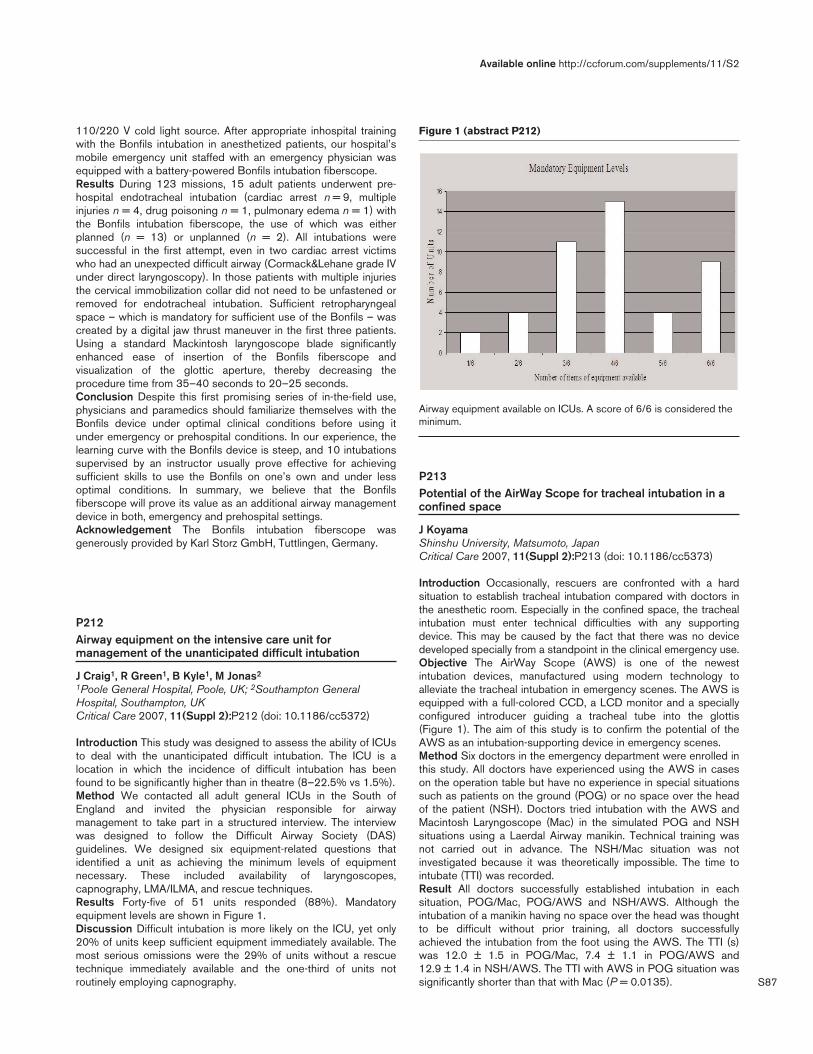
S87
110/220 V cold light source. After appropriate inhospital trainingwith the Bonfils intubation in anesthetized patients, our hospital’smobile emergency unit staffed with an emergency physician wasequipped with a battery-powered Bonfils intubation fiberscope.Results During 123 missions, 15 adult patients underwent pre-hospital endotracheal intubation (cardiac arrest n = 9, multipleinjuries n = 4, drug poisoning n = 1, pulmonary edema n = 1) withthe Bonfils intubation fiberscope, the use of which was eitherplanned (n = 13) or unplanned (n = 2). All intubations weresuccessful in the first attempt, even in two cardiac arrest victimswho had an unexpected difficult airway (Cormack&Lehane grade IVunder direct laryngoscopy). In those patients with multiple injuriesthe cervical immobilization collar did not need to be unfastened orremoved for endotracheal intubation. Sufficient retropharyngealspace – which is mandatory for sufficient use of the Bonfils – wascreated by a digital jaw thrust maneuver in the first three patients.Using a standard Mackintosh laryngoscope blade significantlyenhanced ease of insertion of the Bonfils fiberscope andvisualization of the glottic aperture, thereby decreasing theprocedure time from 35–40 seconds to 20–25 seconds.Conclusion Despite this first promising series of in-the-field use,physicians and paramedics should familiarize themselves with theBonfils device under optimal clinical conditions before using itunder emergency or prehospital conditions. In our experience, thelearning curve with the Bonfils device is steep, and 10 intubationssupervised by an instructor usually prove effective for achievingsufficient skills to use the Bonfils on one’s own and under lessoptimal conditions. In summary, we believe that the Bonfilsfiberscope will prove its value as an additional airway managementdevice in both, emergency and prehospital settings.Acknowledgement The Bonfils intubation fiberscope wasgenerously provided by Karl Storz GmbH, Tuttlingen, Germany.
P212Airway equipment on the intensive care unit formanagement of the unanticipated difficult intubation
J Craig1, R Green1, B Kyle1, M Jonas2
1Poole General Hospital, Poole, UK; 2Southampton GeneralHospital, Southampton, UKCritical Care 2007, 11(Suppl 2):P212 (doi: 10.1186/cc5372)
Introduction This study was designed to assess the ability of ICUsto deal with the unanticipated difficult intubation. The ICU is alocation in which the incidence of difficult intubation has beenfound to be significantly higher than in theatre (8–22.5% vs 1.5%).Method We contacted all adult general ICUs in the South ofEngland and invited the physician responsible for airwaymanagement to take part in a structured interview. The interviewwas designed to follow the Difficult Airway Society (DAS)guidelines. We designed six equipment-related questions thatidentified a unit as achieving the minimum levels of equipmentnecessary. These included availability of laryngoscopes,capnography, LMA/ILMA, and rescue techniques.Results Forty-five of 51 units responded (88%). Mandatoryequipment levels are shown in Figure 1.Discussion Difficult intubation is more likely on the ICU, yet only20% of units keep sufficient equipment immediately available. Themost serious omissions were the 29% of units without a rescuetechnique immediately available and the one-third of units notroutinely employing capnography.
P213Potential of the AirWay Scope for tracheal intubation in aconfined space
J KoyamaShinshu University, Matsumoto, JapanCritical Care 2007, 11(Suppl 2):P213 (doi: 10.1186/cc5373)
Introduction Occasionally, rescuers are confronted with a hardsituation to establish tracheal intubation compared with doctors inthe anesthetic room. Especially in the confined space, the trachealintubation must enter technical difficulties with any supportingdevice. This may be caused by the fact that there was no devicedeveloped specially from a standpoint in the clinical emergency use.Objective The AirWay Scope (AWS) is one of the newestintubation devices, manufactured using modern technology toalleviate the tracheal intubation in emergency scenes. The AWS isequipped with a full-colored CCD, a LCD monitor and a speciallyconfigured introducer guiding a tracheal tube into the glottis(Figure 1). The aim of this study is to confirm the potential of theAWS as an intubation-supporting device in emergency scenes.Method Six doctors in the emergency department were enrolled inthis study. All doctors have experienced using the AWS in caseson the operation table but have no experience in special situationssuch as patients on the ground (POG) or no space over the headof the patient (NSH). Doctors tried intubation with the AWS andMacintosh Laryngoscope (Mac) in the simulated POG and NSHsituations using a Laerdal Airway manikin. Technical training wasnot carried out in advance. The NSH/Mac situation was notinvestigated because it was theoretically impossible. The time tointubate (TTI) was recorded.Result All doctors successfully established intubation in eachsituation, POG/Mac, POG/AWS and NSH/AWS. Although theintubation of a manikin having no space over the head was thoughtto be difficult without prior training, all doctors successfullyachieved the intubation from the foot using the AWS. The TTI (s)was 12.0 ± 1.5 in POG/Mac, 7.4 ± 1.1 in POG/AWS and12.9 ± 1.4 in NSH/AWS. The TTI with AWS in POG situation wassignificantly shorter than that with Mac (P = 0.0135).
Available online http://ccforum.com/supplements/11/S2
Figure 1 (abstract P212)
Airway equipment available on ICUs. A score of 6/6 is considered theminimum.
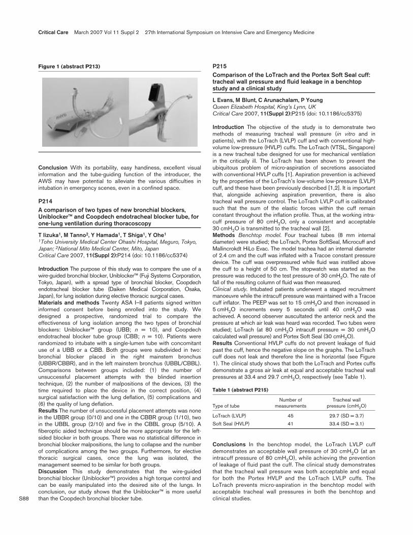
S88
Conclusion With its portability, easy handiness, excellent visualinformation and the tube-guiding function of the introducer, theAWS may have potential to alleviate the various difficulties inintubation in emergency scenes, even in a confined space.
P214A comparison of two types of new bronchial blockers,Uniblocker™ and Coopdech endotracheal blocker tube, forone-lung ventilation during thoracoscopy
T Iizuka1, M Tanno2, Y Hamada1, T Shiga1, Y Ohe1
1Toho University Medical Center Ohashi Hospital, Meguro, Tokyo,Japan; 2National Mito Medical Center, Mito, JapanCritical Care 2007, 11(Suppl 2):P214 (doi: 10.1186/cc5374)
Introduction The purpose of this study was to compare the use of awire-guided bronchial blocker, Uniblocker™ (Fuji Systems Corporation,Tokyo, Japan), with a spread type of bronchial blocker, Coopdechendotracheal blocker tube (Daiken Medical Corporation, Osaka,Japan), for lung isolation during elective thoracic surgical cases.Materials and methods Twenty ASA I–II patients signed writteninformed consent before being enrolled into the study. Wedesigned a prospective, randomized trial to compare theeffectiveness of lung isolation among the two types of bronchialblockers: Uniblocker™ group (UBB; n = 10), and Coopdechendotracheal blocker tube group (CBB; n = 10). Patients wererandomized to intubate with a single-lumen tube with concomitantuse of a UBB or a CBB. Both groups were subdivided in two:bronchial blocker placed in the right mainstem bronchus(UBBR/CBBR), and in the left mainstem bronchus (UBBL/CBBL).Comparisons between groups included: (1) the number ofunsuccessful placement attempts with the blinded insertiontechnique, (2) the number of malpositions of the devices, (3) thetime required to place the device in the correct position, (4)surgical satisfaction with the lung deflation, (5) complications and(6) the quality of lung deflation.Results The number of unsuccessful placement attempts was nonein the UBBR group (0/10) and one in the CBBR group (1/10), twoin the UBBL group (2/10) and five in the CBBL group (5/10). Afiberoptic aided technique should be more appropriate for the left-sided blocker in both groups. There was no statistical difference inbronchial blocker malpositions, the lung to collapse and the numberof complications among the two groups. Furthermore, for electivethoracic surgical cases, once the lung was isolated, themanagement seemed to be similar for both groups.Discussion This study demonstrates that the wire-guidedbronchial blocker (Uniblocker™) provides a high torque control andcan be easily manipulated into the desired site of the lungs. Inconclusion, our study shows that the Uniblocker™ is more usefulthan the Coopdech bronchial blocker tube.
P215Comparison of the LoTrach and the Portex Soft Seal cuff:tracheal wall pressure and fluid leakage in a benchtopstudy and a clinical study
L Evans, M Blunt, C Arunachalam, P YoungQueen Elizabeth Hospital, King’s Lynn, UKCritical Care 2007, 11(Suppl 2):P215 (doi: 10.1186/cc5375)
Introduction The objective of the study is to demonstrate twomethods of measuring tracheal wall pressure (in vitro and inpatients), with the LoTrach (LVLP) cuff and with conventional high-volume low-pressure (HVLP) cuffs. The LoTrach (VTSL, Singapore)is a new tracheal tube designed for use for mechanical ventilationin the critically ill. The LoTrach has been shown to prevent theubiquitous problem of micro-aspiration of secretions associatedwith conventional HVLP cuffs [1]. Aspiration prevention is achievedby the properties of the LoTrach’s low-volume low-pressure (LVLP)cuff, and these have been previously described [1,2]. It is importantthat, alongside achieving aspiration prevention, there is alsotracheal wall pressure control. The LoTrach LVLP cuff is calibratedsuch that the sum of the elastic forces within the cuff remainconstant throughout the inflation profile. Thus, at the working intra-cuff pressure of 80 cmH2O, only a consistent and acceptable30 cmH2O is transmitted to the tracheal wall [2].Methods Benchtop model. Four tracheal tubes (8 mm internaldiameter) were studied; the LoTrach, Portex SoftSeal, Microcuff andMallincrokdt HiLo Evac. The model trachea had an internal diameterof 2.4 cm and the cuff was inflated with a Tracoe constant pressuredevice. The cuff was overpressured while fluid was instilled abovethe cuff to a height of 50 cm. The stopwatch was started as thepressure was reduced to the test pressure of 30 cmH2O. The rate offall of the resulting column of fluid was then measured.Clinical study. Intubated patients underwent a staged recruitmentmanoeuvre while the intracuff pressure was maintained with a Tracoecuff inflator. The PEEP was set to 15 cmH2O and then increased in5 cmH2O increments every 5 seconds until 40 cmH2O wasachieved. A second observer auscultated the anterior neck and thepressure at which air leak was heard was recorded. Two tubes werestudied; LoTrach (at 80 cmH2O intracuff pressure = 30 cmH2Ocalculated wall pressure) and Portex Soft Seal (30 cmH2O).Results Conventional HVLP cuffs do not prevent leakage of fluidpast the cuff, hence the negative slope on the graphs. The LoTrachcuff does not leak and therefore the line is horizontal (see Figure1). The clinical study shows that both the LoTrach and Portex cuffsdemonstrate a gross air leak at equal and acceptable tracheal wallpressures at 33.4 and 29.7 cmH2O, respectively (see Table 1).
Table 1 (abstract P215)
Number of Tracheal wall Type of tube measurements pressure (cmH2O)
LoTrach (LVLP) 45 29.7 (SD = 3.7)
Soft Seal (HVLP) 41 33.4 (SD = 3.1)
Conclusions In the benchtop model, the LoTrach LVLP cuffdemonstrates an acceptable wall pressure of 30 cmH2O (at anintracuff pressure of 80 cmH2O), while achieving the preventionof leakage of fluid past the cuff. The clinical study demonstratesthat the tracheal wall pressure was both acceptable and equalfor both the Portex HVLP and the LoTrach LVLP cuffs. TheLoTrach prevents micro-aspiration in the benchtop model withacceptable tracheal wall pressures in both the benchtop andclinical studies.
Critical Care March 2007 Vol 11 Suppl 2 27th International Symposium on Intensive Care and Emergency Medicine
Figure 1 (abstract P213)
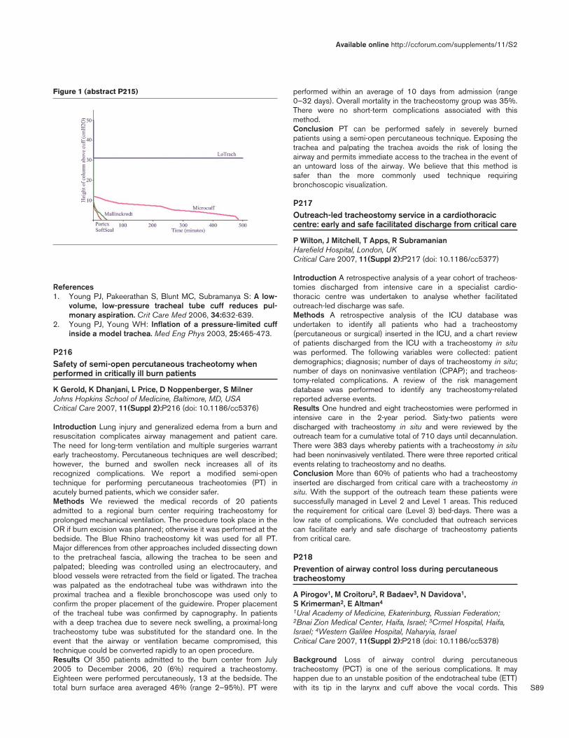
S89
References1. Young PJ, Pakeerathan S, Blunt MC, Subramanya S: A low-
volume, low-pressure tracheal tube cuff reduces pul-monary aspiration. Crit Care Med 2006, 34:632-639.
2. Young PJ, Young WH: Inflation of a pressure-limited cuffinside a model trachea. Med Eng Phys 2003, 25:465-473.
P216Safety of semi-open percutaneous tracheotomy whenperformed in critically ill burn patients
K Gerold, K Dhanjani, L Price, D Noppenberger, S MilnerJohns Hopkins School of Medicine, Baltimore, MD, USACritical Care 2007, 11(Suppl 2):P216 (doi: 10.1186/cc5376)
Introduction Lung injury and generalized edema from a burn andresuscitation complicates airway management and patient care.The need for long-term ventilation and multiple surgeries warrantearly tracheostomy. Percutaneous techniques are well described;however, the burned and swollen neck increases all of itsrecognized complications. We report a modified semi-opentechnique for performing percutaneous tracheotomies (PT) inacutely burned patients, which we consider safer.Methods We reviewed the medical records of 20 patientsadmitted to a regional burn center requiring tracheostomy forprolonged mechanical ventilation. The procedure took place in theOR if burn excision was planned; otherwise it was performed at thebedside. The Blue Rhino tracheostomy kit was used for all PT.Major differences from other approaches included dissecting downto the pretracheal fascia, allowing the trachea to be seen andpalpated; bleeding was controlled using an electrocautery, andblood vessels were retracted from the field or ligated. The tracheawas palpated as the endotracheal tube was withdrawn into theproximal trachea and a flexible bronchoscope was used only toconfirm the proper placement of the guidewire. Proper placementof the tracheal tube was confirmed by capnography. In patientswith a deep trachea due to severe neck swelling, a proximal-longtracheostomy tube was substituted for the standard one. In theevent that the airway or ventilation became compromised, thistechnique could be converted rapidly to an open procedure.Results Of 350 patients admitted to the burn center from July2005 to December 2006, 20 (6%) required a tracheostomy.Eighteen were performed percutaneously, 13 at the bedside. Thetotal burn surface area averaged 46% (range 2–95%). PT were
performed within an average of 10 days from admission (range0–32 days). Overall mortality in the tracheostomy group was 35%.There were no short-term complications associated with thismethod.Conclusion PT can be performed safely in severely burnedpatients using a semi-open percutaneous technique. Exposing thetrachea and palpating the trachea avoids the risk of losing theairway and permits immediate access to the trachea in the event ofan untoward loss of the airway. We believe that this method issafer than the more commonly used technique requiringbronchoscopic visualization.
P217Outreach-led tracheostomy service in a cardiothoraciccentre: early and safe facilitated discharge from critical care
P Wilton, J Mitchell, T Apps, R SubramanianHarefield Hospital, London, UKCritical Care 2007, 11(Suppl 2):P217 (doi: 10.1186/cc5377)
Introduction A retrospective analysis of a year cohort of tracheos-tomies discharged from intensive care in a specialist cardio-thoracic centre was undertaken to analyse whether facilitatedoutreach-led discharge was safe.Methods A retrospective analysis of the ICU database wasundertaken to identify all patients who had a tracheostomy(percutaneous or surgical) inserted in the ICU, and a chart reviewof patients discharged from the ICU with a tracheostomy in situwas performed. The following variables were collected: patientdemographics; diagnosis; number of days of tracheostomy in situ;number of days on noninvasive ventilation (CPAP); and tracheos-tomy-related complications. A review of the risk managementdatabase was performed to identify any tracheostomy-relatedreported adverse events.Results One hundred and eight tracheostomies were performed inintensive care in the 2-year period. Sixty-two patients weredischarged with tracheostomy in situ and were reviewed by theoutreach team for a cumulative total of 710 days until decannulation.There were 383 days whereby patients with a tracheostomy in situhad been noninvasively ventilated. There were three reported criticalevents relating to tracheostomy and no deaths.Conclusion More than 60% of patients who had a tracheostomyinserted are discharged from critical care with a tracheostomy insitu. With the support of the outreach team these patients weresuccessfully managed in Level 2 and Level 1 areas. This reducedthe requirement for critical care (Level 3) bed-days. There was alow rate of complications. We concluded that outreach servicescan facilitate early and safe discharge of tracheostomy patientsfrom critical care.
P218Prevention of airway control loss during percutaneoustracheostomy
A Pirogov1, M Croitoru2, R Badaev3, N Davidova1, S Krimerman2, E Altman4
1Ural Academy of Medicine, Ekaterinburg, Russian Federation;2Bnai Zion Medical Center, Haifa, Israel; 3Crmel Hospital, Haifa,Israel; 4Western Galilee Hospital, Naharyia, IsraelCritical Care 2007, 11(Suppl 2):P218 (doi: 10.1186/cc5378)
Background Loss of airway control during percutaneoustracheostomy (PCT) is one of the serious complications. It mayhappen due to an unstable position of the endotracheal tube (ETT)with its tip in the larynx and cuff above the vocal cords. This
Available online http://ccforum.com/supplements/11/S2
Figure 1 (abstract P215)
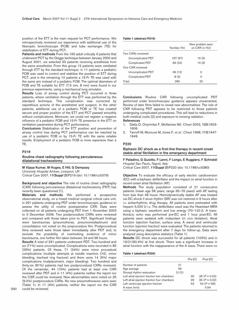
S90
position of the ETT is the main request for PCT performance. Weretrospectively reviewed our experience with additional use of thefiberoptic bronchoscope (FOB) and tube exchanger (TE) forstabilization of ETT during PCT.Patients and methods From the 160 adult critically ill patients thatunderwent PCT by the Griggs technique between January 2000 andAugust 2001, we selected 33 patients receiving anesthesia fromthe same anesthetist. From this group 12 patients were ventilatedthrough ETT by the standard technique: in 11 patients a pediatricFOB was used to control and stabilize the position of ETT duringPCT, and in the remaining 10 patients a 15-Fr TE was used withthe same aim instead of a pediatric FOB. The optimal diameters ofFOB and TE suitable for ETT (7.5 mm, 8 mm) were found in ourprevious experiments, using a mechanical lung simulator.Results Loss of airway control during PCT occurred in threepatients, where ventilation through the ETT was performed by thestandard technique. This complication was corrected byexpeditious actions of the anesthetist and surgeon. In the otherpatients, additional use of a pediatric FOB or TE has createdsecure and proper position of the ETT and PCT passed smoothlywithout complications. Moreover, we could not register a negativeinfluence of a pediatric FOB and 15-Fr TE presence in the ETT onventilation parameters during PCT performance.Conclusions Stabilization of the ETT position and prevention ofairway control loss during PCT performance can be reached byuse of a pediatric FOB or by 15-Fr TE with the same reliableresults. Employment of a pediatric FOB is more expensive than aTE.
P219Routine chest radiography following percutaneousdilatational tracheostomy
M Vijaya Kumar, M Huges, E Hill, G DempseyUniversity Hospital Aintree, Liverpool, UKCritical Care 2007, 11(Suppl 2):P219 (doi: 10.1186/cc5379)
Background and objective The role of routine chest radiography(CXR) following percutaneous dilatational tracheostomy (PDT) hasrecently been questioned [1].Materials and methods We performed a prospectiveobservational study, on a mixed medical–surgical critical care unit,in 291 patients undergoing PDT under bronchoscopic guidance toassess the utility of routine postoperative CXR. Data werecollected on all patients undergoing PDT from 1 November 2003to 5 December 2006. Two postprocedure CXRs were reviewedand compared with those taken prior to PDT. Significant findingswere barotraumas (pneumothorax, pneumomediastinum) andconsolidation not noted on the preprocedure film. Postproceduralfilms reviewed were those taken immediately after PDT and, toexclude the possibility of overlooking evidence of minorbarotrauma, one further film taken between 24 and 96 hours.Results A total of 291 patients underwent PDT. Two hundred andsix (71%) were uncomplicated. Complications were recorded in 85(29%) patients. Of these, 71 (24%) were minor proceduralcomplications (multiple attempts at needle insertion (>2), minorbleeding, tracheal ring fracture) and there were 14 (5%) majorcomplications (malplacement, major bleeding). Two hundred andthirty-six (81%) patients had two postprocedural CXRs reviewed.Of the remainder, 44 (15%) patients had at least one CXRreviewed after PDT and in 11 (4%) patients neither the report northe CXR could be reviewed. New abnormalities were noted on 25(9.0%) postprocedure CXRs. No new pneumothoraces were seen(Table 1). In 11 (4%) patients, neither the report nor the CXRcould be reviewed.
Conclusions Routine CXR following uncomplicated PDTperformed under bronchoscopic guidance appears unwarranted.Review of later films failed to reveal new abnormalities. The role ofCXR following PDT appears to be restricted to those patientsundergoing complicated procedures. This will lead to reductions inboth medical costs [2] and exposure to ionising radiation.References1. Datta D, Onyirimba F, McNamee MJ: Chest 2003, 123:1603-
1606.2. Tarnoff M, Moncure M, Jones F, et al.: Chest 1998, 113:1647-
1649.
P220Biphasic DC shock as a first-line therapy in recent-onsetstable atrial fibrillation in the emergency department
F Paladino, G Guiotto, F Lanni, F Longo, E Ruggiero, F SchiraldiHospital Sao Paulo, Napoli, ItalyCritical Care 2007, 11(Suppl 2):P220 (doi: 10.1186/cc5380)
Objective To evaluate the efficacy of early electric cardioversion(EC) with a biphasic defibrillator and the impact on atrial function inrecent-onset atrial fibrillation (AF).Methods The study population consisted of 21 consecutivepatients (mean age 58 years; range 35–76 years) with AF lastingfrom less than 48 hours. Hemodynamically stable AF was treatedvia DC shock if sinus rhythm (SR) was not restored in 6 hours afteri.v. antiarrhythmic drug therapy. All patients were pretreated withheparin 5,000 U i.v. The defibrillator used was the Heartstart MRXusing a biphasic waveform and low energy (70–120 J). A trans-thoracic echo was performed pre-EC and 1 hour post-EC. Allpatients were sedated with midazolam (in vivo titolation). Atrialfunction (ejection fraction, surface area, A wave) and ventricularfunction (ejection fraction) were evaluated. The patients returned tothe emergency department after 7 days for follow-up. Data wereanalyzed using descriptive statistics (Table 1).Results DC shock was successful for all patients (100%) and in19/21(90.4%) at first shock. There was a significant increase inatrial function with the reappearance of the A wave. There were no
Critical Care March 2007 Vol 11 Suppl 2 27th International Symposium on Intensive Care and Emergency Medicine
Table 1 (abstract P219)
New problem seen Number (%) on CXR (n (%))
Two CXRs reviewed
Uncomplicated PDT 167 (57) 15 (5)
Complicated PDT 69 (24) 10 (3)
One CXR reviewed
Uncomplicated PDT 36 (12) 0
Complicated PDT 8 (3) 0
Total 280 25
Table 1 (abstract P220)
Pre-EC Post-EC
Number of patients 21Age average 59Sinusal rhythm restoration 21/21Left atrial ejection fraction two chambers 30 35 (P = 0.02)Left atrial ejection fraction four chambers 29 35 (P = 0.02)Left ventricular ejection fraction 52 54 (P = NS)A wave (m/s) 0.54
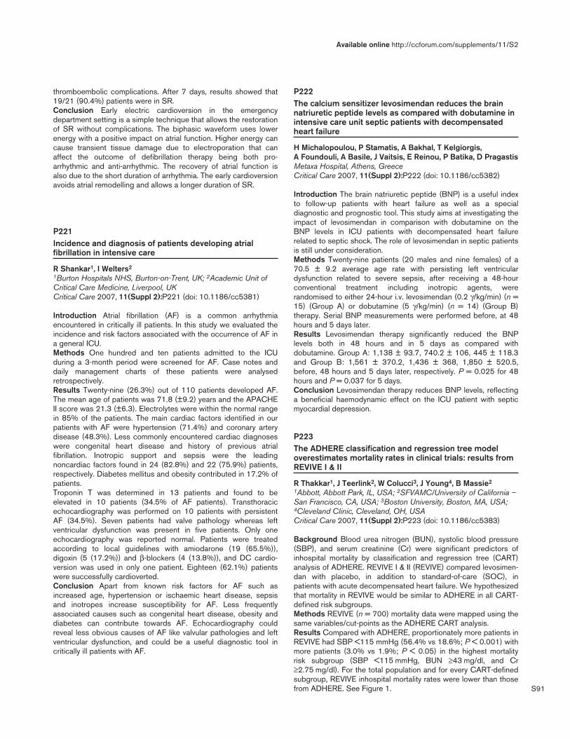
S91
thromboembolic complications. After 7 days, results showed that19/21 (90.4%) patients were in SR.Conclusion Early electric cardioversion in the emergencydepartment setting is a simple technique that allows the restorationof SR without complications. The biphasic waveform uses lowerenergy with a positive impact on atrial function. Higher energy cancause transient tissue damage due to electroporation that canaffect the outcome of defibrillation therapy being both pro-arrhythmic and anti-arrhythmic. The recovery of atrial function isalso due to the short duration of arrhythmia. The early cardioversionavoids atrial remodelling and allows a longer duration of SR.
P221Incidence and diagnosis of patients developing atrialfibrillation in intensive care
R Shankar1, I Welters2
1Burton Hospitals NHS, Burton-on-Trent, UK; 2Academic Unit ofCritical Care Medicine, Liverpool, UKCritical Care 2007, 11(Suppl 2):P221 (doi: 10.1186/cc5381)
Introduction Atrial fibrillation (AF) is a common arrhythmiaencountered in critically ill patients. In this study we evaluated theincidence and risk factors associated with the occurrence of AF ina general ICU.Methods One hundred and ten patients admitted to the ICUduring a 3-month period were screened for AF. Case notes anddaily management charts of these patients were analysedretrospectively.Results Twenty-nine (26.3%) out of 110 patients developed AF.The mean age of patients was 71.8 (±9.2) years and the APACHEII score was 21.3 (±6.3). Electrolytes were within the normal rangein 85% of the patients. The main cardiac factors identified in ourpatients with AF were hypertension (71.4%) and coronary arterydisease (48.3%). Less commonly encountered cardiac diagnoseswere congenital heart disease and history of previous atrialfibrillation. Inotropic support and sepsis were the leadingnoncardiac factors found in 24 (82.8%) and 22 (75.9%) patients,respectively. Diabetes mellitus and obesity contributed in 17.2% ofpatients.Troponin T was determined in 13 patients and found to beelevated in 10 patients (34.5% of AF patients). Transthoracicechocardiography was performed on 10 patients with persistentAF (34.5%). Seven patients had valve pathology whereas leftventricular dysfunction was present in five patients. Only oneechocardiography was reported normal. Patients were treatedaccording to local guidelines with amiodarone (19 (65.5%)),digoxin (5 (17.2%)) and β-blockers (4 (13.8%)), and DC cardio-version was used in only one patient. Eighteen (62.1%) patientswere successfully cardioverted.Conclusion Apart from known risk factors for AF such asincreased age, hypertension or ischaemic heart disease, sepsisand inotropes increase susceptibility for AF. Less frequentlyassociated causes such as congenital heart disease, obesity anddiabetes can contribute towards AF. Echocardiography couldreveal less obvious causes of AF like valvular pathologies and leftventricular dysfunction, and could be a useful diagnostic tool incritically ill patients with AF.
P222The calcium sensitizer levosimendan reduces the brainnatriuretic peptide levels as compared with dobutamine inintensive care unit septic patients with decompensatedheart failure
H Michalopoulou, P Stamatis, A Bakhal, T Kelgiorgis, A Foundouli, A Basile, J Vaitsis, E Reinou, P Batika, D PragastisMetaxa Hospital, Athens, GreeceCritical Care 2007, 11(Suppl 2):P222 (doi: 10.1186/cc5382)
Introduction The brain natriuretic peptide (BNP) is a useful indexto follow-up patients with heart failure as well as a specialdiagnostic and prognostic tool. This study aims at investigating theimpact of levosimendan in comparison with dobutamine on theBNP levels in ICU patients with decompensated heart failurerelated to septic shock. The role of levosimendan in septic patientsis still under consideration.Methods Twenty-nine patients (20 males and nine females) of a70.5 ± 9.2 average age rate with persisting left ventriculardysfunction related to severe sepsis, after receiving a 48-hourconventional treatment including inotropic agents, wererandomised to either 24-hour i.v. levosimendan (0.2 γ/kg/min) (n =15) (Group A) or dobutamine (5 γ/kg/min) (n = 14) (Group B)therapy. Serial BNP measurements were performed before, at 48hours and 5 days later.Results Levosimendan therapy significantly reduced the BNPlevels both in 48 hours and in 5 days as compared withdobutamine. Group A: 1,138 ± 93.7, 740.2 ± 106, 445 ± 118.3and Group B: 1,561 ± 370.2, 1,436 ± 368, 1,850 ± 520.5,before, 48 hours and 5 days later, respectively. P = 0.025 for 48hours and P = 0.037 for 5 days.Conclusion Levosimendan therapy reduces BNP levels, reflectinga beneficial haemodynamic effect on the ICU patient with septicmyocardial depression.
P223The ADHERE classification and regression tree modeloverestimates mortality rates in clinical trials: results fromREVIVE I & II
R Thakkar1, J Teerlink2, W Colucci3, J Young4, B Massie2
1Abbott, Abbott Park, IL, USA; 2SFVAMC/University of California –San Francisco, CA, USA; 3Boston University, Boston, MA, USA;4Cleveland Clinic, Cleveland, OH, USACritical Care 2007, 11(Suppl 2):P223 (doi: 10.1186/cc5383)
Background Blood urea nitrogen (BUN), systolic blood pressure(SBP), and serum creatinine (Cr) were significant predictors ofinhospital mortality by classification and regression tree (CART)analysis of ADHERE. REVIVE I & II (REVIVE) compared levosimen-dan with placebo, in addition to standard-of-care (SOC), inpatients with acute decompensated heart failure. We hypothesizedthat mortality in REVIVE would be similar to ADHERE in all CART-defined risk subgroups.Methods REVIVE (n = 700) mortality data were mapped using thesame variables/cut-points as the ADHERE CART analysis.Results Compared with ADHERE, proportionately more patients inREVIVE had SBP <115 mmHg (56.4% vs 18.6%; P < 0.001) withmore patients (3.0% vs 1.9%; P < 0.05) in the highest mortalityrisk subgroup (SBP <115 mmHg, BUN ≥43 mg/dl, and Cr≥2.75 mg/dl). For the total population and for every CART-definedsubgroup, REVIVE inhospital mortality rates were lower than thosefrom ADHERE. See Figure 1.
Available online http://ccforum.com/supplements/11/S2
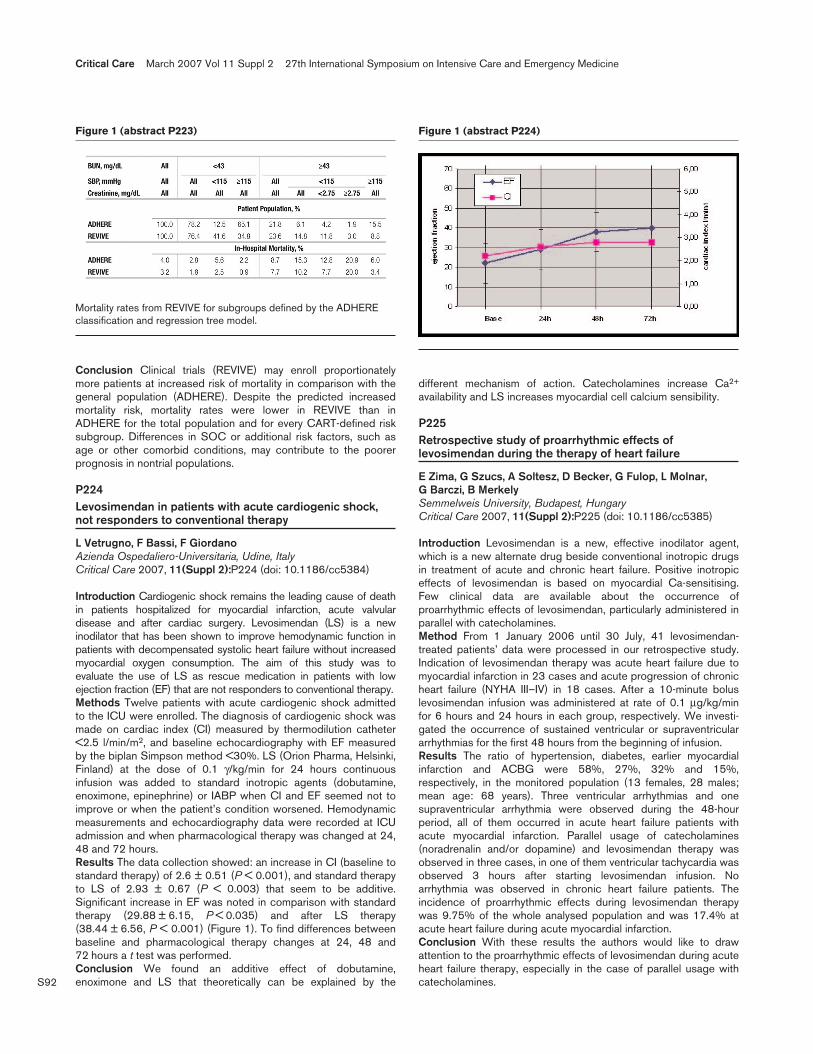
S92
Conclusion Clinical trials (REVIVE) may enroll proportionatelymore patients at increased risk of mortality in comparison with thegeneral population (ADHERE). Despite the predicted increasedmortality risk, mortality rates were lower in REVIVE than inADHERE for the total population and for every CART-defined risksubgroup. Differences in SOC or additional risk factors, such asage or other comorbid conditions, may contribute to the poorerprognosis in nontrial populations.
P224Levosimendan in patients with acute cardiogenic shock,not responders to conventional therapy
L Vetrugno, F Bassi, F GiordanoAzienda Ospedaliero-Universitaria, Udine, ItalyCritical Care 2007, 11(Suppl 2):P224 (doi: 10.1186/cc5384)
Introduction Cardiogenic shock remains the leading cause of deathin patients hospitalized for myocardial infarction, acute valvulardisease and after cardiac surgery. Levosimendan (LS) is a newinodilator that has been shown to improve hemodynamic function inpatients with decompensated systolic heart failure without increasedmyocardial oxygen consumption. The aim of this study was toevaluate the use of LS as rescue medication in patients with lowejection fraction (EF) that are not responders to conventional therapy.Methods Twelve patients with acute cardiogenic shock admittedto the ICU were enrolled. The diagnosis of cardiogenic shock wasmade on cardiac index (CI) measured by thermodilution catheter<2.5 l/min/m2, and baseline echocardiography with EF measuredby the biplan Simpson method <30%. LS (Orion Pharma, Helsinki,Finland) at the dose of 0.1 γ/kg/min for 24 hours continuousinfusion was added to standard inotropic agents (dobutamine,enoximone, epinephrine) or IABP when CI and EF seemed not toimprove or when the patient’s condition worsened. Hemodynamicmeasurements and echocardiography data were recorded at ICUadmission and when pharmacological therapy was changed at 24,48 and 72 hours.Results The data collection showed: an increase in CI (baseline tostandard therapy) of 2.6 ± 0.51 (P < 0.001), and standard therapyto LS of 2.93 ± 0.67 (P < 0.003) that seem to be additive. Significant increase in EF was noted in comparison with standardtherapy (29.88 ± 6.15, P < 0.035) and after LS therapy(38.44 ± 6.56, P < 0.001) (Figure 1). To find differences betweenbaseline and pharmacological therapy changes at 24, 48 and72 hours a t test was performed.Conclusion We found an additive effect of dobutamine,enoximone and LS that theoretically can be explained by the
different mechanism of action. Catecholamines increase Ca2+
availability and LS increases myocardial cell calcium sensibility.
P225Retrospective study of proarrhythmic effects oflevosimendan during the therapy of heart failure
E Zima, G Szucs, A Soltesz, D Becker, G Fulop, L Molnar, G Barczi, B MerkelySemmelweis University, Budapest, HungaryCritical Care 2007, 11(Suppl 2):P225 (doi: 10.1186/cc5385)
Introduction Levosimendan is a new, effective inodilator agent,which is a new alternate drug beside conventional inotropic drugsin treatment of acute and chronic heart failure. Positive inotropiceffects of levosimendan is based on myocardial Ca-sensitising.Few clinical data are available about the occurrence ofproarrhythmic effects of levosimendan, particularly administered inparallel with catecholamines.Method From 1 January 2006 until 30 July, 41 levosimendan-treated patients’ data were processed in our retrospective study.Indication of levosimendan therapy was acute heart failure due tomyocardial infarction in 23 cases and acute progression of chronicheart failure (NYHA III–IV) in 18 cases. After a 10-minute boluslevosimendan infusion was administered at rate of 0.1 µg/kg/minfor 6 hours and 24 hours in each group, respectively. We investi-gated the occurrence of sustained ventricular or supraventriculararrhythmias for the first 48 hours from the beginning of infusion.Results The ratio of hypertension, diabetes, earlier myocardialinfarction and ACBG were 58%, 27%, 32% and 15%,respectively, in the monitored population (13 females, 28 males;mean age: 68 years). Three ventricular arrhythmias and onesupraventricular arrhythmia were observed during the 48-hourperiod, all of them occurred in acute heart failure patients withacute myocardial infarction. Parallel usage of catecholamines(noradrenalin and/or dopamine) and levosimendan therapy wasobserved in three cases, in one of them ventricular tachycardia wasobserved 3 hours after starting levosimendan infusion. Noarrhythmia was observed in chronic heart failure patients. Theincidence of proarrhythmic effects during levosimendan therapywas 9.75% of the whole analysed population and was 17.4% atacute heart failure during acute myocardial infarction.Conclusion With these results the authors would like to drawattention to the proarrhythmic effects of levosimendan during acuteheart failure therapy, especially in the case of parallel usage withcatecholamines.
Critical Care March 2007 Vol 11 Suppl 2 27th International Symposium on Intensive Care and Emergency Medicine
Figure 1 (abstract P223)
Mortality rates from REVIVE for subgroups defined by the ADHEREclassification and regression tree model.
Figure 1 (abstract P224)
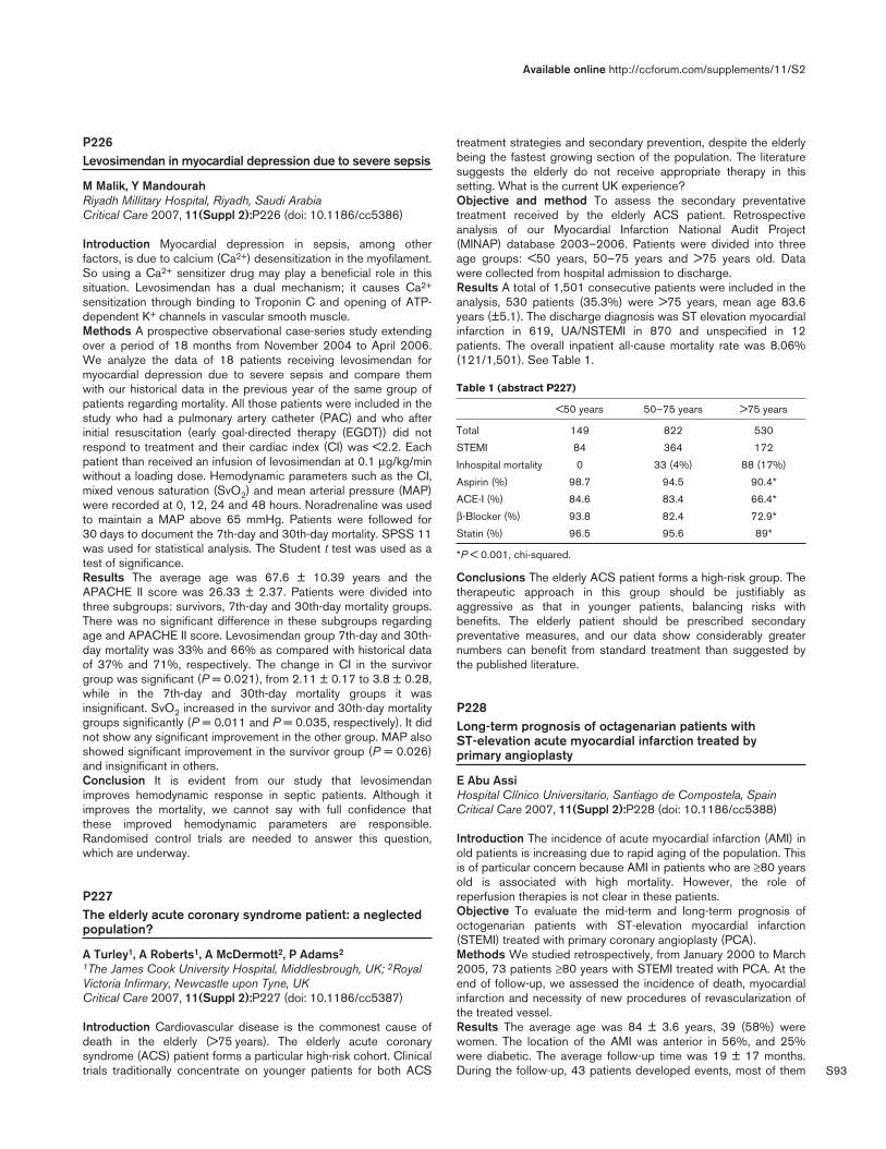
S93
P226Levosimendan in myocardial depression due to severe sepsis
M Malik, Y MandourahRiyadh Millitary Hospital, Riyadh, Saudi ArabiaCritical Care 2007, 11(Suppl 2):P226 (doi: 10.1186/cc5386)
Introduction Myocardial depression in sepsis, among otherfactors, is due to calcium (Ca2+) desensitization in the myofilament.So using a Ca2+ sensitizer drug may play a beneficial role in thissituation. Levosimendan has a dual mechanism; it causes Ca2+
sensitization through binding to Troponin C and opening of ATP-dependent K+ channels in vascular smooth muscle.Methods A prospective observational case-series study extendingover a period of 18 months from November 2004 to April 2006.We analyze the data of 18 patients receiving levosimendan formyocardial depression due to severe sepsis and compare themwith our historical data in the previous year of the same group ofpatients regarding mortality. All those patients were included in thestudy who had a pulmonary artery catheter (PAC) and who afterinitial resuscitation (early goal-directed therapy (EGDT)) did notrespond to treatment and their cardiac index (CI) was <2.2. Eachpatient than received an infusion of levosimendan at 0.1 µg/kg/minwithout a loading dose. Hemodynamic parameters such as the CI,mixed venous saturation (SvO2) and mean arterial pressure (MAP)were recorded at 0, 12, 24 and 48 hours. Noradrenaline was usedto maintain a MAP above 65 mmHg. Patients were followed for30 days to document the 7th-day and 30th-day mortality. SPSS 11was used for statistical analysis. The Student t test was used as atest of significance.Results The average age was 67.6 ± 10.39 years and theAPACHE II score was 26.33 ± 2.37. Patients were divided intothree subgroups: survivors, 7th-day and 30th-day mortality groups.There was no significant difference in these subgroups regardingage and APACHE II score. Levosimendan group 7th-day and 30th-day mortality was 33% and 66% as compared with historical dataof 37% and 71%, respectively. The change in CI in the survivorgroup was significant (P = 0.021), from 2.11 ± 0.17 to 3.8 ± 0.28,while in the 7th-day and 30th-day mortality groups it wasinsignificant. SvO2 increased in the survivor and 30th-day mortalitygroups significantly (P = 0.011 and P = 0.035, respectively). It didnot show any significant improvement in the other group. MAP alsoshowed significant improvement in the survivor group (P = 0.026)and insignificant in others.Conclusion It is evident from our study that levosimendanimproves hemodynamic response in septic patients. Although itimproves the mortality, we cannot say with full confidence thatthese improved hemodynamic parameters are responsible.Randomised control trials are needed to answer this question,which are underway.
P227The elderly acute coronary syndrome patient: a neglectedpopulation?
A Turley1, A Roberts1, A McDermott2, P Adams2
1The James Cook University Hospital, Middlesbrough, UK; 2RoyalVictoria Infirmary, Newcastle upon Tyne, UKCritical Care 2007, 11(Suppl 2):P227 (doi: 10.1186/cc5387)
Introduction Cardiovascular disease is the commonest cause ofdeath in the elderly (>75 years). The elderly acute coronarysyndrome (ACS) patient forms a particular high-risk cohort. Clinicaltrials traditionally concentrate on younger patients for both ACS
treatment strategies and secondary prevention, despite the elderlybeing the fastest growing section of the population. The literaturesuggests the elderly do not receive appropriate therapy in thissetting. What is the current UK experience?Objective and method To assess the secondary preventativetreatment received by the elderly ACS patient. Retrospectiveanalysis of our Myocardial Infarction National Audit Project(MINAP) database 2003–2006. Patients were divided into threeage groups: <50 years, 50–75 years and >75 years old. Datawere collected from hospital admission to discharge.Results A total of 1,501 consecutive patients were included in theanalysis, 530 patients (35.3%) were >75 years, mean age 83.6years (±5.1). The discharge diagnosis was ST elevation myocardialinfarction in 619, UA/NSTEMI in 870 and unspecified in 12patients. The overall inpatient all-cause mortality rate was 8.06%(121/1,501). See Table 1.
Table 1 (abstract P227)
<50 years 50–75 years >75 years
Total 149 822 530
STEMI 84 364 172
Inhospital mortality 0 33 (4%) 88 (17%)
Aspirin (%) 98.7 94.5 90.4*
ACE-I (%) 84.6 83.4 66.4*
β-Blocker (%) 93.8 82.4 72.9*
Statin (%) 96.5 95.6 89*
*P < 0.001, chi-squared.
Conclusions The elderly ACS patient forms a high-risk group. Thetherapeutic approach in this group should be justifiably asaggressive as that in younger patients, balancing risks withbenefits. The elderly patient should be prescribed secondarypreventative measures, and our data show considerably greaternumbers can benefit from standard treatment than suggested bythe published literature.
P228Long-term prognosis of octagenarian patients withST-elevation acute myocardial infarction treated by primary angioplasty
E Abu AssiHospital Clínico Universitario, Santiago de Compostela, SpainCritical Care 2007, 11(Suppl 2):P228 (doi: 10.1186/cc5388)
Introduction The incidence of acute myocardial infarction (AMI) inold patients is increasing due to rapid aging of the population. Thisis of particular concern because AMI in patients who are ≥80 yearsold is associated with high mortality. However, the role ofreperfusion therapies is not clear in these patients.Objective To evaluate the mid-term and long-term prognosis ofoctogenarian patients with ST-elevation myocardial infarction(STEMI) treated with primary coronary angioplasty (PCA).Methods We studied retrospectively, from January 2000 to March2005, 73 patients ≥80 years with STEMI treated with PCA. At theend of follow-up, we assessed the incidence of death, myocardialinfarction and necessity of new procedures of revascularization ofthe treated vessel.Results The average age was 84 ± 3.6 years, 39 (58%) werewomen. The location of the AMI was anterior in 56%, and 25%were diabetic. The average follow-up time was 19 ± 17 months.During the follow-up, 43 patients developed events, most of them
Available online http://ccforum.com/supplements/11/S2
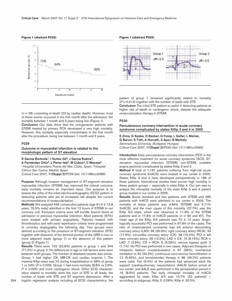
S94
(n = 28) consisting of death (23 by cardiac death). However, mostof these events occurred in the first month after the admission, themortality between 1 month and 3 years being low (Figure 1).Conclusion Our data show that the octogenarian patients withSTEMI treated by primary PCA developed a very high mortality.However, this mortality especially concentrates in the first monthafter the procedure, being low between 1 month and 3 years.
P229Outcome in myocardial infarction is related to themorphologic pattern of ST elevation
R García-Borbolla1, I Nuñez Gil2, J García Rubira2, A Fernández Ortiz2, L Perez Isla2, M Cobos2, C Macaya2
1Hospital Universitario Puerta del Mar, Cádiz, Spain; 2HospitalClínico San Carlos, Madrid, SpainCritical Care 2007, 11(Suppl 2):P229 (doi: 10.1186/cc5389)
Purpose Although invasive management of ST-segment elevationmyocardial infarction (STEMI) has improved the clinical outcome,early mortality remains an important issue. Our purpose is toassess the utility of the initial electrocardiographic (ECG) pattern indetecting patients who are at increased risk despite the currentrecommendations of revascularization.Methods We analyzed 446 consecutive patients (age 61.9 ± 13.8years, 76.5% male) admitted in the first 12 hours of STEMI to ourcoronary unit. Exclusion criteria were left bundle branch block atadmission or previous myocardial infarction. Most patients (87%)were treated with primary angioplasty. Patients treated withthrombolytics and with early reperfusion criteria were programmedto coronary angiography the following day. Two groups weredefined according to the presence of ST-segment elevation (STE)together with distortion of the terminal portion of the QRS in two ormore adjacent leads (group 1) or the absence of this pattern(group 2) (Figure 1).Results There were 102 (22.8%) patients in group 1 and 344(77.2%) in group 2. No differences in age or risk factors were seenbetween both groups. The number of diseased vessels was similar.Group 1 had higher CK, MB-CK and cardiac troponin I. Themaximal Killip class was >2 during hospitalisation in 38% of group1 vs 24% (P = 0.009). Group 1 had more mortality (8.8% vs 2.6%,P = 0.005) and more cardiogenic shock. Other ECG character-istics related to mortality were the sum of STE in all leads, thenumber of leads with STE and ST segment depression. After alogistic regression analysis including all ECG characteristics, the
pattern of group 1 remained significantly related to mortality(P = 0.013) together with the number of leads with STE.Conclusion The initial STE pattern is useful in detecting patients athigher risk of death or cardiogenic shock, despite the adequaterevascularization therapy in STEMI.
P230Percutaneous coronary intervention in acute coronarysyndrome complicated by states Killip 3 and 4 in 2005
E Zima, G Szabo, D Becker, G Fulop, L Geller, L Molnar, G Barczi, S Toth, A Horvath, A Apor, B MerkelySemmelweis University, Budapest, HungaryCritical Care 2007, 11(Suppl 2):P230 (doi: 10.1186/cc5390)
Introduction Early percutaneous coronary intervention (PCI) is themost effective treatment for acute coronary syndrome (ACS, ST-elevation myocardial infarction (STEMI), non-STEMI, unstableangina pectoris) complicated by states Killip 3 and 4.Method A total of 1,187 patients suffering from high-risk acutecoronary syndrome (hrACS) were treated in our center in 2005.States Killip 3 and 4 have developed perioperatively in 186 ofthese patients. International studies have proven high mortality inthese patient groups – especially in state Killip 4. Our aim was toanalyze the inhospital mortality of the state Killip 3 and 4 patientgroup treated in our center in 2005.Results Seven hundred and two patients with STEMI and 485patients with hrACS were admitted to our center in 2005. Themortality of these patients was 4.84% (STEMI) and 3.71%(hrACS), and the main cause of this mortality (37.7%) was theKillip 3/4 state, which was observed in 11.9% of the STEMIpatients and in 17.9% of hrACS patients (n = 84 and 87). Themean age of the Killip 3/4 patients was 70 ± 10 years. Angio-logically successful PCI was performed in 97.9% of the cases. Theratio of revascularized coronaries was left anterior descendingcoronary artery (LAD): 66 (35.9%), right coronary artery (RCA): 33(17.9%), circumflex coronary artery (CX): 28 (15.2%), PCI in leftmain coronary artery: 28 (12.5%), LAD + CX: 15 (8.15%), RCA +LAD: 7 (3.8%), CX + RCA: 6 (3.26%), venous bypass graft: 2(1.1%). No PCI was performed in two cases. Adjuvant therapies ofintraaortic balloon counterpulsation in 67 (36%), mechanicalventilation in 62 (33.3%), continuous veno-venous hemofiltration in12 (6.45%), and levosimendan therapy in 86 (46.2%) patientswere used. Ten (5.4%) of the patients had advanced adult lifesupport (cardiopulmonary resuscitation) (AALS) before arrival atour center, and AALS was performed in the perioperative period in16 (8.6%) patients. The early inhospital mortality of hrACSaggravated by state Killip 3/4 was 10.7% (20 patients) –according to subgroup: Killip 3: 0.06%; Killip 4: 30.5%.
Critical Care March 2007 Vol 11 Suppl 2 27th International Symposium on Intensive Care and Emergency Medicine
Figure 1 (abstract P228) Figure 1 (abstract P229)
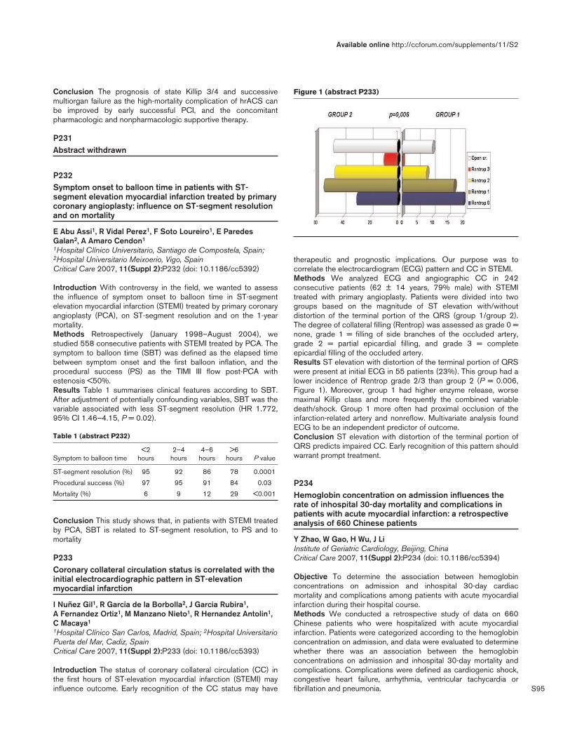
S95
Conclusion The prognosis of state Killip 3/4 and successivemultiorgan failure as the high-mortality complication of hrACS canbe improved by early successful PCI, and the concomitantpharmacologic and nonpharmacologic supportive therapy.
P231Abstract withdrawn
P232Symptom onset to balloon time in patients with ST-segment elevation myocardial infarction treated by primarycoronary angioplasty: influence on ST-segment resolutionand on mortality
E Abu Assi1, R Vidal Perez1, F Soto Loureiro1, E ParedesGalan2, A Amaro Cendon1
1Hospital Clínico Universitario, Santiago de Compostela, Spain;2Hospital Universitario Meixoerio, Vigo, SpainCritical Care 2007, 11(Suppl 2):P232 (doi: 10.1186/cc5392)
Introduction With controversy in the field, we wanted to assessthe influence of symptom onset to balloon time in ST-segmentelevation myocardial infarction (STEMI) treated by primary coronaryangioplasty (PCA), on ST-segment resolution and on the 1-yearmortality.Methods Retrospectively (January 1998–August 2004), westudied 558 consecutive patients with STEMI treated by PCA. Thesymptom to balloon time (SBT) was defined as the elapsed timebetween symptom onset and the first balloon inflation, and theprocedural success (PS) as the TIMI III flow post-PCA withestenosis <50%.Results Table 1 summarises clinical features according to SBT.After adjustment of potentially confounding variables, SBT was thevariable associated with less ST-segment resolution (HR 1.772,95% CI 1.46–4.15, P = 0.02).
Table 1 (abstract P232)
<2 2–4 4–6 >6 Symptom to balloon time hours hours hours hours P value
ST-segment resolution (%) 95 92 86 78 0.0001
Procedural success (%) 97 95 91 84 0.03
Mortality (%) 6 9 12 29 <0.001
Conclusion This study shows that, in patients with STEMI treatedby PCA, SBT is related to ST-segment resolution, to PS and tomortality
P233Coronary collateral circulation status is correlated with theinitial electrocardiographic pattern in ST-elevationmyocardial infarction
I Nuñez Gil1, R Garcia de la Borbolla2, J Garcia Rubira1, A Fernandez Ortiz1, M Manzano Nieto1, R Hernandez Antolin1,C Macaya1
1Hospital Clínico San Carlos, Madrid, Spain; 2Hospital UniversitarioPuerta del Mar, Cadiz, SpainCritical Care 2007, 11(Suppl 2):P233 (doi: 10.1186/cc5393)
Introduction The status of coronary collateral circulation (CC) inthe first hours of ST-elevation myocardial infarction (STEMI) mayinfluence outcome. Early recognition of the CC status may have
therapeutic and prognostic implications. Our purpose was tocorrelate the electrocardiogram (ECG) pattern and CC in STEMI.Methods We analyzed ECG and angiographic CC in 242consecutive patients (62 ± 14 years, 79% male) with STEMItreated with primary angioplasty. Patients were divided into twogroups based on the magnitude of ST elevation with/withoutdistortion of the terminal portion of the QRS (group 1/group 2).The degree of collateral filling (Rentrop) was assessed as grade 0 =none, grade 1 = filling of side branches of the occluded artery,grade 2 = partial epicardial filling, and grade 3 = completeepicardial filling of the occluded artery.Results ST elevation with distortion of the terminal portion of QRSwere present at initial ECG in 55 patients (23%). This group had alower incidence of Rentrop grade 2/3 than group 2 (P = 0.006,Figure 1). Moreover, group 1 had higher enzyme release, worsemaximal Killip class and more frequently the combined variabledeath/shock. Group 1 more often had proximal occlusion of theinfarction-related artery and nonreflow. Multivariate analysis foundECG to be an independent predictor of outcome.Conclusion ST elevation with distortion of the terminal portion ofQRS predicts impaired CC. Early recognition of this pattern shouldwarrant prompt treatment.
P234Hemoglobin concentration on admission influences therate of inhospital 30-day mortality and complications inpatients with acute myocardial infarction: a retrospectiveanalysis of 660 Chinese patients
Y Zhao, W Gao, H Wu, J LiInstitute of Geriatric Cardiology, Beijing, ChinaCritical Care 2007, 11(Suppl 2):P234 (doi: 10.1186/cc5394)
Objective To determine the association between hemoglobinconcentrations on admission and inhospital 30-day cardiacmortality and complications among patients with acute myocardialinfarction during their hospital course.Methods We conducted a retrospective study of data on 660Chinese patients who were hospitalized with acute myocardialinfarction. Patients were categorized according to the hemoglobinconcentration on admission, and data were evaluated to determinewhether there was an association between the hemoglobinconcentrations on admission and inhospital 30-day mortality andcomplications. Complications were defined as cardiogenic shock,congestive heart failure, arrhythmia, ventricular tachycardia orfibrillation and pneumonia.
Available online http://ccforum.com/supplements/11/S2
Figure 1 (abstract P233)
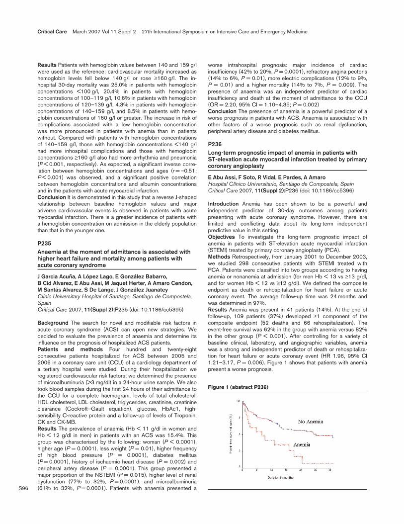
S96
Results Patients with hemoglobin values between 140 and 159 g/lwere used as the reference; cardiovascular mortality increased ashemoglobin levels fell below 140 g/l or rose ≥160 g/l. The in-hospital 30-day mortality was 25.0% in patients with hemoglobinconcentrations <100 g/l, 20.4% in patients with hemoglobinconcentrations of 100–119 g/l, 10.6% in patients with hemoglobinconcentrations of 120–139 g/l, 4.3% in patients with hemoglobinconcentrations of 140–159 g/l, and 8.5% in patients with hemo-globin concentrations of 160 g/l or greater. The increase in risk ofcomplications associated with a low hemoglobin concentrationwas more pronounced in patients with anemia than in patientswithout. Compared with patients with hemoglobin concentrationsof 140–159 g/l, those with hemoglobin concentrations <140 g/lhad more inhospital complications and those with hemoglobinconcentrations ≥160 g/l also had more arrhythmia and pneumonia(P < 0.001, respectively). As expected, a significant inverse corre-lation between hemoglobin concentrations and ages (r = –0.51;P < 0.001) was observed, and a significant positive correlationbetween hemoglobin concentrations and albumin concentrationsand in the patients with acute myocardial infarction.Conclusion It is demonstrated in this study that a reverse J-shapedrelationship between baseline hemoglobin values and majoradverse cardiovascular events is observed in patients with acutemyocardial infarction. There is a greater incidence of patients witha hemoglobin concentration on admission in the elderly populationthan that in the younger one.
P235Anaemia at the moment of admittance is associated withhigher heart failure and mortality among patients withacute coronary syndrome
J Garcia Acuña, A López Lago, E González Babarro, B Cid Alvarez, E Abu Assi, M Jaquet Herter, A Amaro Cendon,M Santás Alvarez, S De Lange, J González JuanateyClinic Universitary Hospital of Santiago, Santiago de Compostela,SpainCritical Care 2007, 11(Suppl 2):P235 (doi: 10.1186/cc5395)
Background The search for novel and modifiable risk factors inacute coronary syndrome (ACS) can open new strategies. Wedecided to evaluate the prevalence of anaemia and determine itsinfluence on the prognosis of hospitalized ACS patients.Patients and methods Four hundred and twenty-eightconsecutive patients hospitalized for ACS between 2005 and2006 in a coronary care unit (CCU) of a cardiology department ofa tertiary hospital were studied. During their hospitalization weregistered cardiovascular risk factors; we determined the presenceof microalbuminuria (>3 mg/dl) in a 24-hour urine sample. We alsotook blood samples during the first 24 hours of their admittance tothe CCU for a complete haemogram, levels of total cholesterol,HDL cholesterol, LDL cholesterol, triglycerides, creatinine, creatinineclearance (Cockroft–Gault equation), glucose, HbAc1, high-sensibility C-reactive protein and a follow-up of levels of Troponin,CK and CK-MB.Results The prevalence of anaemia (Hb < 11 g/dl in women andHb < 12 g/dl in men) in patients with an ACS was 15.4%. Thisgroup was characterised by the following: woman (P < 0.0001),higher age (P = 0.0001), less weight (P = 0.01), higher frequencyof high blood pressure (P = 0.0001), diabetes mellitus(P = 0.0001), history of ischaemic heart disease (P = 0.002) andperipheral artery disease (P = 0.0001). This group presented amajor proportion of the NSTEMI (P = 0.015), higher level of renaldysfunction (77% to 32%, P = 0.0001), and microalbuminuria(61% to 32%, P = 0.0001). Patients with anaemia presented a
worse intrahospital prognosis: major incidence of cardiacinsufficiency (42% to 20%, P = 0.0001), refractory angina pectoris(14% to 6%, P = 0.01), more electric complications (12% to 9%,P = 0.01) and a higher mortality (14% to 7%, P = 0.009). Thepresence of anaemia was an independent predictor of cardiacinsufficiency and death at the moment of admittance to the CCU(OR = 2.20, 95% CI = 1.10–4.35; P = 0.002)Conclusion The presence of anaemia is a powerful predictor of aworse prognosis in patients with ACS. Anaemia is associated withother factors of a worse prognosis such as renal dysfunction,peripheral artery disease and diabetes mellitus.
P236Long-term prognostic impact of anemia in patients withST-elevation acute myocardial infarction treated by primarycoronary angioplasty
E Abu Assi, F Soto, R Vidal, E Pardes, A AmaroHospital Clínico Universitario, Santiago de Compostela, SpainCritical Care 2007, 11(Suppl 2):P236 (doi: 10.1186/cc5396)
Introduction Anemia has been shown to be a powerful andindependent predictor of 30-day outcomes among patientspresenting with acute coronary syndrome. However, there arelimited and conflicting data about its long-term independentpredictive value in this setting.Objectives To investigate the long-term prognostic impact ofanemia in patients with ST-elevation acute myocardial infarction(STEMI) treated by primary coronary angioplasty (PCA).Methods Retrospectively, from January 2001 to December 2003,we studied 298 consecutive patients with STEMI treated withPCA. Patients were classified into two groups according to havinganemia or nonanemia at admission (for men Hb < 13 vs ≥13 g/dl,and for women Hb < 12 vs ≥12 g/dl). We defined the compositeendpoint as death or rehospitalization for heart failure or acutecoronary event. The average follow-up time was 24 months andwas determined in 97%.Results Anemia was present in 41 patients (14%). At the end offollow-up, 109 patients (37%) developed ≥1 component of thecomposite endpoint (52 deaths and 66 rehospitalization). Theevent-free survival was 62% in the group with anemia versus 82%in the other group (P < 0.001). After controlling for a variety ofbaseline clinical, laboratory, and angiographic variables, anemiawas a strong and independent predictor of death or rehospitaliza-tion for heart failure or acute coronary event (HR 1.96, 95% CI1.21–3.17, P = 0.006). Figure 1 shows that patients with anemiapresent a worse prognosis.
Critical Care March 2007 Vol 11 Suppl 2 27th International Symposium on Intensive Care and Emergency Medicine
Figure 1 (abstract P236)
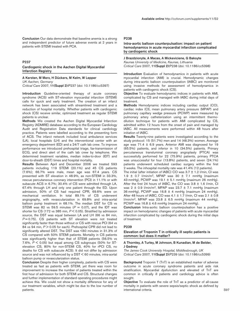
S97
Conclusion Our data demonstrate that baseline anemia is a strongand independent predictor of future adverse events at 2 years inpatients with STEMI treated with PCA.
P237Cardiogenic shock in the Aachen Digital MyocardialInfarction Registry
A Kersten, M Merx, H Dückers, M Kelm, W LepperUK Aachen, GermanyCritical Care 2007, 11(Suppl 2):P237 (doi: 10.1186/cc5397)
Introduction Guideline-oriented therapy of acute coronarysyndrome (ACS) with ST-elevation myocardial infarction (STEMI)calls for quick and early treatment. The creation of an infarctnetwork has been associated with streamlined treatment and areduction of hospital mortality. Whether patients with cardiogenicshock (CS) receive similar, optimized treatment as regular STEMIpatients is unclear.Methods We created the Aachen Digital Myocardial InfarctionRegistry (ADMIRE) database according to the European CardiologyAudit and Registration Data standards for clinical cardiologypractice. Patients were labelled according to the presenting formof ACS. The infarct network included local ambulance services(LA), local hospitals (LH) and the interventional center with anemergency department (ED) and a 24/7 cath lab crew. To improveperformance we introduced prehospital triage, fax-transmission ofECG, and direct alert of the cath lab crew by telephone. Wedetermined treatment variables, median index-to-door (IDT) anddoor-to-sheath (DST) times and hospital mortality.Results Between April and December 2006 we treated 593patients including 119 STEMI (20.1%) and 45 CS patients(7.6%); 66.7% were male, mean age was 67.4 years. CSpresented with ST elevation in 48.9%, as non-STEMI in 33.3%,rescue percutaneous coronary intervention (PCI) in 11.1% or withsubacute ACS in 6.7%; 30.4% of CS were admitted through LA,67.4% through LH and only one patient through the ED. Uponadmission, 50% of CS had required CPR, 69.6% were onmechanical ventilation. In total 89.1% of CS underwentangiography, with revascularization in 69.8% and intra-aortalballoon pump treatment in 68.1%. The median DST for CS vsSTEMI was 82 vs 59.5 minutes (P = 0.07), and the IDT wasshorter for CS (172 vs 385 min, P < 0.05). Stratified by admissionsource, the DST was equal between LA and LH (66 vs 84 min,P = 0.75). CS patients with ST elevation were not treatedsignificantly faster than those without or CS with rescue PCI (64 vs84 vs 94 min, P > 0.05 for each). Prehospital CPR did not lead tosignificantly altered DST. The DST was <60 minutes in 31.3% ofCS compared with 50% STEMI patients. Mortality in CS patientswas significantly higher than that of STEMI patients (56.5% vs7.6%, P < 0.05) but equal among CS subgroups (50% for ST-elevation CS, 80% for non-STEMI CS, 40% for rPCI CS, nodeaths for CS with subacute ACS). It did not differ by admissionsource and was not influenced by a DST < 60 minutes, intra-aortalballoon pump or revascularization status.Conclusion Despite their higher complexity, patients with CS weretreated as fast as patients with STEMI, yet there was room forimprovement to increase the number of patients treated within thefirst hour of admission for both STEMI and CS. Structural changesand further implementation of standard operating procedures mightachieve this. We could not show a mortality difference for any ofour treatment variables, which might be due to the low number ofpatients.
P238Intra-aortic balloon counterpulsation: impact on patienthemodynamics in acute myocardial infarction complicatedby cardiogenic shock
J Brazdzionyte, A Macas, A Mickeviciene, G BaksyteKaunas University of Medicine, Kaunas, LithuaniaCritical Care 2007, 11(Suppl 2):P238 (doi: 10.1186/cc5398)
Introduction Evaluation of hemodynamics in patients with acutemyocardial infarction (AMI) is crucial. Hemodynamic changesduring intra-aortic balloon counterpulsation (IABC) are monitoredusing invasive methods for assessment of hemodynamics inpatients with cardiogenic shock (CS).Objective To evaluate hemodynamic indices in patients with AMI,complicated by CS and managed with IABC during initial days oftreatment.Methods Hemodynamic indices including cardiac output (CO),cardiac index (CI), mean pulmonary artery pressure (MPAP) andpulmonary capillary wedge pressure (PCWP) were measured bypulmonary artery catheterization using an intermittent thermo-dilution technique for patients with AMI complicated by CS,admitted within 12 hours from the onset of pain and managed byIABC. All measurements were performed within 48 hours afterinitiation of IABC.Results Twenty-nine patients were investigated according to thestudy protocol: 15 (51.7%) men and 14 (48.3%) women. Averageage was 71.4 ± 6.9 years. Anterior AMI was diagnosed for 19(65.5%) patients, and inferior in 10 (34.5%) patients. Primarypercutaneous transluminal coronary angioplasty (PTCA) wassuccessfully performed for 22 (75.9%) patients, primary PTCAwas unsuccessful for four (13.8%) patients, and seven (24.1%)patients underwent scheduled cardiac surgery within the first2 weeks. The inhospital mortality rate was 41.4% (12 patients).The initial (after initiation of IABC) CO was 3.7 ± 1.2 l/min, CI was1.9 ± 0.7 l/min/m2, MPAP was 30 ± 7.1 mmHg (maximum43 mmHg), PCWP was 19.1 ± 5.1 mmHg (maximum 26 mmHg).After the first 24 hours of IABC, the CO was 3.8 ± 1.6 l/min, CIwas 2 ± 0.9 l/min/m2, MPAP was 23.7 ± 7.1 mmHg (maximum36 mmHg), PCWP was 16.8 ± 4 mmHg (maximum 24 mmHg).After 48 hours of IABC, CO was 4.1 ± 1.7 l/min, CI was 2.1 ± 0.8l/min/m2, MPAP was 23.8 ± 6.5 mmHg (maximum 44 mmHg),PCWP was 16.8 ± 4.6 mmHg (maximum 24 mmHg).Conclusion Intra-aortic balloon counterpulsation has a positiveimpact on hemodynamic changes of patients with acute myocardialinfarction complicated by cardiogenic shock during the initial daysof treatment.
P239Elevation of Troponin T in critically ill septic patients iscommon: but does it matter?
A Thornley, A Turley, M Johnson, B Kunadian, M de Belder, J GedneyThe James Cook University Hospital, Middlesbrough, UKCritical Care 2007, 11(Suppl 2):P239 (doi: 10.1186/cc5399)
Background Troponin T (TnT) is an established marker of adverseoutcome in acute coronary syndrome patients and aids riskstratification. Myocardial dysfunction and elevated of TnT arecommon in critically ill patients and cardiology advice is oftensought.Objective To evaluate the role of TnT as a predictor of all-causemortality in patients with severe sepsis/septic shock as defined byinternational criteria.
Available online http://ccforum.com/supplements/11/S2
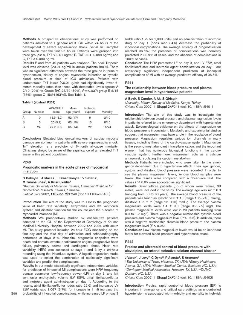
S98
Methods A prospective observational study was performed onpatients admitted to a general adult ICU within 24 hours of thedevelopment of severe sepsis/septic shock. Serial TnT sampleswere taken over the first 96 hours. Patients were grouped intothree groups: A, TnT < 0.01 ng/ml; B, TnT 0.01–0.099 ng/ml; andC, TnT > 0.099 ng/ml.Results Blood from 49 patients was analysed. The peak Troponinlevel was elevated (>0.01 ng/ml) in 39/49 patients (80%). Therewas no significant difference between the three groups in terms ofhypertension, history of angina, myocardial infarction or systolicblood pressure at time of ICU admission. Patients withundetectable TnT levels (<0.01 g/ml) had significantly lower 6-month mortality rates than those with detectable levels (group A2/10 (20%) vs Group B/C 23/39 (59%), P = 0.037; group B 8/15(53%), group C 15/24 (63%)). See Table 1.
Table 1 (abstract P239)
APACHE II Mean Inotropic Group Number score age (years) support Mortality
A 10 18.5 (8.2) 52 (17) 8 2/10
B 15 20 (5.7) 63 (15) 15 8/15
C 24 22.2 (6.8) 65 (14) 22 15/24
Conclusions Elevated biochemical markers of cardiac myocytedamage are common in patients with severe sepsis/septic shock.TnT elevation is a predictor of 6-month all-cause mortality.Clinicians should be aware of the significance of an elevated TnTassay in this patient population.
P240Prognostic markers in the acute phase of myocardialinfarction
G Baksyte1, A Macas1, J Brazdzionyte1, V Saferis1, M Tamosiunas2, A Krisciukaitis1
1Kaunas University of Medicine, Kaunas, Lithuania; 2Institute forBiomedical Research, Kaunas, LithuaniaCritical Care 2007, 11(Suppl 2):P240 (doi: 10.1186/cc5400)
Introduction The aim of the study was to assess the prognosticvalue of heart rate variability, arrhythmias and left ventricularsystolic and diastolic function for the course and the outcome ofmyocardial infarction (MI).Methods We prospectively studied 57 consecutive patientsadmitted to the ICU of the Department of Cardiology of KaunasMedical University Hospital between 2002 and 2004 with acuteMI. The study protocol included 24-hour ECG monitoring on thefirst day and the third day of admission and echocardiographyperformed at days 2–4. Inhospital prognostic endpoints weredeath and nonfatal events: postinfarction angina, progressive heartfailure, pulmonary edema and cardiogenic shock. Heart ratevariability (HRV) was assessed at days 1 and 3 by a 24-hourrecording using the ‘HeartLab’ system. A logistic regression modelwas used to select the combination of statistically significantvariables and predict the complications.Results In our model statistically significant independent variablesfor prediction of inhospital MI complications were HRV frequencydomain parameter low-frequency power (LF) on day 3, and leftventricular end-systolic volume (LV ESV), atrial fibrillation/flutterand inotropic agent administration on day 1. According to theresults, atrial fibrillation/flutter (odds ratio 25.6) and increased LVESV (odds ratio 1.067 (6.7%) for increase in 1 ml) increase theprobability of inhospital complications, while increased LF on day 3
(odds ratio 1.29 for 1,000 units) and no administration of inotropicdrug on day 1 (odds ratio 34.5) decrease the probability ofinhospital complications. The average efficacy of prognosticationreached 96.5%; the presence of complications was correctlypredicted in 88.9% of cases, and the absence of complications in100% of cases.Conclusion The HRV parameter LF on day 3, and LV ESV, atrialfibrillation/flutter and inotropic agent administration on day 1 arestatistically significant independent predictors of inhospitalcomplications of MI with an average predictive efficacy of 96.5%.
P241The relationship between blood pressure and plasmamagnesium level in hypertensive patients
A Bayir, B Cander, A Ak, S GirisginUniversity, Meram Faculty of Medicine, Konya, TurkeyCritical Care 2007, 11(Suppl 2):P241 (doi: 10.1186/cc5401)
Introduction The aim of this study was to investigate therelationship between blood pressure and plasma magnesium levelsin patients referred to the emergency department with hypertensiveattack. Epidemiological evidence on the effects of magnesium onblood pressure is inconsistent. Metabolic and experimental studiessuggest that magnesium may have a role in the regulation of bloodpressure. Magnesium regulates various ion channels in manytissues, including those of the cardiovascular system. Magnesiumis the second most abundant intracellular cation, and the importantelement that has numerous biological functions in the cardio-vascular system. Furthermore, magnesium acts as a calciumantagonist, regulating the calcium metabolism.Methods Patients were included who were taken to the emer-gency department due to hypertensive attack. Their age, gender,systolic and diastolic blood pressure were recorded. In order tosee the plasma magnesium levels, venous blood samples weretaken. The results were compared with a chi-square test. Thevalues P < 0.05 were accepted as significant.Results Seventy-three patients (35 of whom were female, 38males) were included in the study. The average age was 47 ± 6.3(ranging from 33 to 68 years). The average blood pressure of thepatients was found as systolic 200 ± 10 (range 185–240) mmHg,diastolic 105 ± 7 (range 95–110) mmHg. The average plasmamagnesium levels were 1.4 ± 0.3 (range 0.9–2.2) mg/l. Theplasma magnesium levels were low in 29 patients (ranging from0.9 to 1.7 mg/l). There was a negative relationship systolic bloodpressure and plasma magnesium level (P < 0.05). In addition, therewas a negative relationship diastolic blood pressure and plasmamagnesium level (P < 0.05).Conclusion Low plasma magnesium levels would be an importantfactor for elevated blood pressure and hypertensive attack.
P242Precise and ultrarapid control of blood pressure withclevidipine, an arterial selective calcium channel blocker
J Varon1, J Levy2, C Dyke3, P Acosta4, S Aronson5
1The University of Texas, Houston, TX, USA; 2Emory Healthcare,Atlanta, GA, USA; 3Gaston Medical Center, Gastonia, NC, USA;4Dorrington Medical Associates, Houston, TX, USA; 5DUMC,Durham, NC, USACritical Care 2007, 11(Suppl 2):P242 (doi: 10.1186/cc5402)
Introduction Precise, rapid control of blood pressure (BP) isimportant in emergency and critical care settings as uncontrolledhypertension is associated with morbidity and mortality in high-risk
Critical Care March 2007 Vol 11 Suppl 2 27th International Symposium on Intensive Care and Emergency Medicine
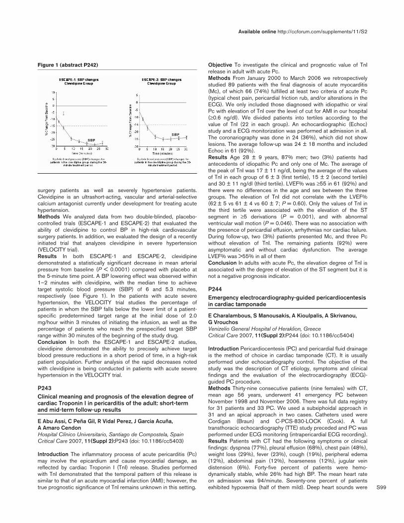
S99
surgery patients as well as severely hypertensive patients.Clevidipine is an ultrashort-acting, vascular and arterial-selectivecalcium antagonist currently under development for treating acutehypertension.Methods We analyzed data from two double-blinded, placebo-controlled trials (ESCAPE-1 and ESCAPE-2) that evaluated theability of clevidipine to control BP in high-risk cardiovascularsurgery patients. In addition, we evaluated the design of a recentlyinitiated trial that analyzes clevidipine in severe hypertension(VELOCITY trial).Results In both ESCAPE-1 and ESCAPE-2, clevidipinedemonstrated a statistically significant decrease in mean arterialpressure from baseline (P < 0.0001) compared with placebo atthe 5-minute time point. A BP lowering effect was observed within1–2 minutes with clevidipine, with the median time to achievetarget systolic blood pressure (SBP) of 6 and 5.3 minutes,respectively (see Figure 1). In the patients with acute severehypertension, the VELOCITY trial studies the percentage ofpatients in whom the SBP falls below the lower limit of a patient-specific predetermined target range at the initial dose of 2.0mg/hour within 3 minutes of initiating the infusion, as well as thepercentage of patients who reach the prespecified target SBPrange within 30 minutes of the beginning of the study drug.Conclusion In both the ESCAPE-1 and ESCAPE-2 studies,clevidipine demonstrated the ability to precisely achieve targetblood pressure reductions in a short period of time, in a high-riskpatient population. Further analysis of the rapid decreases notedwith clevidipine is being conducted in patients with acute severehypertension in the VELOCITY trial.
P243Clinical meaning and prognosis of the elevation degree ofcardiac Troponin I in pericarditis of the adult: short-termand mid-term follow-up results
E Abu Assi, C Peña Gil, R Vidal Perez, J Garcia Acuña, A Amaro CendonHospital Clínico Universitario, Santiago de Compostela, SpainCritical Care 2007, 11(Suppl 2):P243 (doi: 10.1186/cc5403)
Introduction The inflammatory process of acute pericarditis (Pc)may involve the epicardium and cause myocardial damage, asreflected by cardiac Troponin I (TnI) release. Studies performedwith TnI demonstrated that the temporal pattern of this release issimilar to that of an acute myocardial infarction (AMI); however, thetrue prognostic significance of TnI remains unknown in this setting.
Objective To investigate the clinical and prognostic value of TnIrelease in adult with acute Pc.Methods From January 2000 to March 2006 we retrospectivelystudied 89 patients with the final diagnosis of acute myocarditis(Mc), of which 66 (74%) fulfilled at least two criteria of acute Pc(typical chest pain, pericardial friction rub, and/or alterations in theECG). We only included those diagnosed with idiopathic or viralPc with elevation of TnI over the level of cut for AMI in our hospital(≥0.6 ng/dl). We divided patients into tertiles according to thevalue of TnI (22 in each group). An echocardiographic (Echoc)study and a ECG monitorization was performed at admission in all.The coronariography was done in 24 (36%), which did not showlesions. The average follow-up was 24 ± 18 months and includedEchoc in 61 (92%).Results Age 28 ± 9 years, 87% men; two (3%) patients hadantecedents of idiopathic Pc and only one of Mc. The average ofthe peak of TnI was 17 ± 11 ng/dl, being the average of the valuesof TnI in each group of 6 ± 3 (first tertile), 15 ± 2 (second tertile)and 30 ± 11 ng/dl (third tertile). LVEF% was ≥55 in 61 (92%) andthere were no differences in the age and sex between the threegroups. The elevation of TnI did not correlate with the LVEF%(62 ± 5 vs 61 ± 4 vs 60 ± 7; P = 0.60). Only the values of TnI inthe third tertile were associated with the elevation of the STsegment in ≥5 derivations (P = 0.001), and with abnormalventricular wall motion (P = 0.046). There was no association withthe presence of pericardial effusion, arrhythmias nor cardiac failure.During follow-up, two (3%) patients presented Mc, and three Pcwithout elevation of TnI. The remaining patients (92%) wereasymptomatic and without cardiac dysfunction. The averageLVEF% was >55% in all of themConclusion In adults with acute Pc, the elevation degree of TnI isassociated with the degree of elevation of the ST segment but it isnot a negative prognosis indicator.
P244Emergency electrocardiography-guided pericardiocentesisin cardiac tamponade
E Charalambous, S Manousakis, A Kioulpalis, A Skrivanou, G VrouchosVenizelio General Hospital of Heraklion, GreeceCritical Care 2007, 11(Suppl 2):P244 (doi: 10.1186/cc5404)
Introduction Pericardiocentesis (PC) and pericardial fluid drainageis the method of choice in cardiac tamponade (CT). It is usuallyperformed under echocardiography control. The objective of thestudy was the description of CT etiology, symptoms and clinicalfindings and the evaluation of the electrocardiography (ECG)-guided PC procedure.Methods Thirty-nine consecutive patients (nine females) with CT,mean age 56 years, underwent 41 emergency PC betweenNovember 1998 and November 2006. There was full data registryfor 31 patients and 33 PC. We used a subxiphoidal approach in31 and an apical approach in two cases. Catheters used wereCordigan (Braun) and C-PCS-830-LOCK (Cook). A fulltransthoracic echocardiography (TTE) study preceded and PC wasperformed under ECG monitoring (intrapericardial ECG recording).Results Patients with CT had the following symptoms or clinicalfindings: dyspnea (77%), pleural effusion (68%), chest pain (48%),weight loss (29%), fever (23%), cough (19%), peripheral edema(12%), abdominal pain (12%), hoarseness (12%), jugular veindistension (6%). Forty-five percent of patients were hemo-dynamically stable, while 26% had high BP. The mean heart rateon admission was 94/minute. Seventy-one percent of patientsexhibited hypoxemia (half of them mild). Deep heart sounds were
Available online http://ccforum.com/supplements/11/S2
Figure 1 (abstract P242)
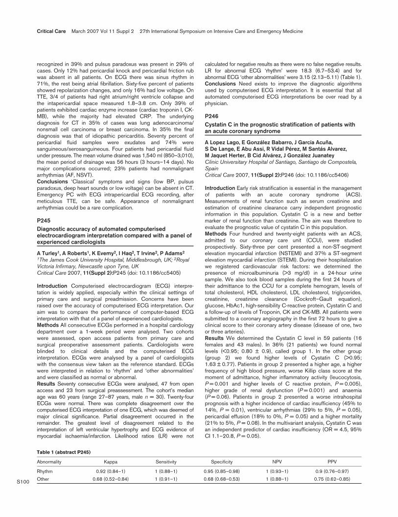
S100
recognized in 39% and pulsus paradoxus was present in 29% ofcases. Only 12% had pericardial knock and pericardial friction rubwas absent in all patients. On ECG there was sinus rhythm in71%, the rest being atrial fibrillation. Sixty-five percent of patientsshowed repolarization changes, and only 16% had low voltage. OnTTE, 3/4 of patients had right atrium/right ventricle collapse andthe intapericardial space measured 1.8–3.8 cm. Only 39% ofpatients exhibited cardiac enzyme increase (cardiac troponin I, CK-MB), while the majority had elevated CRP. The underlyingdiagnosis for CT in 35% of cases was lung adenocarcinoma/nonsmall cell carcinoma or breast carcinoma. In 35% the finaldiagnosis was that of idiopathic pericarditis. Seventy percent ofpericardial fluid samples were exudates and 74% weresanguineous/serosanguineous. Four patients had pericardial fluidunder pressure. The mean volume drained was 1,540 ml (850–3,010),the mean period of drainage was 56 hours (3 hours–14 days). Nomajor complications occurred; 23% patients had nonmalignantarrhythmias (AF, NSVT).Conclusions ‘Classical’ symptoms and signs (low BP, pulsusparadoxus, deep heart sounds or low voltage) can be absent in CT.Emergency PC with ECG intrapericardial ECG recording, aftermeticulous TTE, can be safe. Appearance of nonmalignantarrhythmias could be a rare complication.
P245Diagnostic accuracy of automated computerisedelectrocardiogram interpretation compared with a panel ofexperienced cardiologists
A Turley1, A Roberts1, K Evemy2, I Haq2, T Irvine2, P Adams2
1The James Cook University Hospital, Middlesbrough, UK; 2RoyalVictoria Infirmary, Newcastle upon Tyne, UKCritical Care 2007, 11(Suppl 2):P245 (doi: 10.1186/cc5405)
Introduction Computerised electrocardiogram (ECG) interpre-tation is widely applied, especially within the clinical settings ofprimary care and surgical preadmission. Concerns have beenraised over the accuracy of computerised ECG interpretation. Ouraim was to compare the performance of computer-based ECGinterpretation with that of a panel of experienced cardiologists.Methods All consecutive ECGs performed in a hospital cardiologydepartment over a 1-week period were analysed. Two cohortswere assessed, open access patients from primary care andsurgical preoperative assessment patients. Cardiologists wereblinded to clinical details and the computerised ECGinterpretation. ECGs were analysed by a panel of cardiologistswith the consensus view taken as the reference standard. ECGswere interpreted in relation to ‘rhythm’ and ‘other abnormalities’and were classified as normal or abnormal.Results Seventy consecutive ECGs were analysed, 47 from openaccess and 23 from surgical preassessment. The cohort’s medianage was 60 years (range 27–87 years, male n = 30). Twenty-fourECGs were normal. There was complete disagreement over thecomputerised ECG interpretation of one ECG, which was deemed ofmajor clinical significance. Partial disagreement occurred in theremainder. The greatest level of disagreement related to theinterpretation of left ventricular hypertrophy and ECG evidence ofmyocardial ischaemia/infarction. Likelihood ratios (LR) were not
calculated for negative results as there were no false negative results.LR for abnormal ECG ‘rhythm’ were 18.3 (6.7–53.4) and forabnormal ECG ‘other abnormalities’ were 3.15 (2.13–5.11) (Table 1).Conclusions Need exists to improve the diagnostic algorithmsused by computerised ECG interpretation. It is essential that allautomated computerised ECG interpretations be over read by aphysician.
P246Cystatin C in the prognostic stratification of patients withan acute coronary syndrome
A Lopez Lago, E González Babarro, J García Acuña, S De Lange, E Abu Assi, R Vidal Pérez, M Santás Alvarez, M Jaquet Herter, B Cid Alvárez, J González JuanateyClinic Universitary Hospital of Santiago, Santiago de Compostela,SpainCritical Care 2007, 11(Suppl 2):P246 (doi: 10.1186/cc5406)
Introduction Early risk stratification is essential in the managementof patients with an acute coronary syndrome (ACS).Measurements of renal function such as serum creatinine andestimation of creatinine clearance carry independent prognosticinformation in this population. Cystatin C is a new and bettermarker of renal function than creatinine. The aim was therefore toevaluate the prognostic value of cystatin C in this population.Methods Four hundred and twenty-eight patients with an ACS,admitted to our coronary care unit (CCU), were studiedprospectively. Sixty-three per cent presented a non-ST-segmentelevation myocardial infarction (NSTEMI) and 37% a ST-segmentelevation myocardial infarction (STEMI). During their hospitalizationwe registered cardiovascular risk factors: we determined thepresence of microalbuminuria (>3 mg/dl) in a 24-hour urinesample. We also took blood samples during the first 24 hours oftheir admittance to the CCU for a complete hemogram, levels oftotal cholesterol, HDL cholesterol, LDL cholesterol, triglycerides,creatinine, creatinine clearance (Cockroft–Gault equation),glucose, HbAc1, high-sensibility C-reactive protein, Cystatin C anda follow-up of levels of Troponin, CK and CK-MB. All patients weresubmitted to a coronary angiography in the first 72 hours to give aclinical score to their coronary artery disease (disease of one, twoor three arteries).Results We determined the Cystatin C level in 59 patients (16females and 43 males). In 36% (21 patients) we found normallevels (<0.95; 0.80 ± 0.9), called group 1. In the other group(group 2) we found higher levels of Cystatin C (>0.95;1.63 ± 0.77). Patients in group 2 presented a higher age, a higherfrequency of high blood pressure, worse Killip class score at themoment of admittance, higher inflammatory activity (leucocytosis,P = 0.001 and higher levels of C reactive protein, P = 0.005),higher grade of renal dysfunction (P = 0.001) and anaemia(P = 0.06). Patients in group 2 presented a worse intrahospitalprognosis with a higher incidence of cardiac insufficiency (45% to14%, P = 0.01), ventricular arrhythmias (29% to 5%, P = 0.05),pericardial effusion (18% to 0%, P = 0.05) and a higher mortality(21% to 5%, P = 0.08). In the multivariant analysis, Cystatin C wasan independent predictor of cardiac insufficiency (OR = 4.5, 95%CI 1.1–20.8, P = 0.05).
Critical Care March 2007 Vol 11 Suppl 2 27th International Symposium on Intensive Care and Emergency Medicine
Table 1 (abstract P245)
Abnormality Kappa Sensitivity Specificity NPV PPV
Rhythm 0.92 (0.84–1) 1 (0.88–1) 0.95 (0.85–0.98) 1 (0.93–1) 0.9 (0.76–0.97)
Other 0.68 (0.52–0.84) 1 (0.91–1) 0.68 (0.68–0.53) 1 (0.88–1) 0.75 (0.62–0.85)
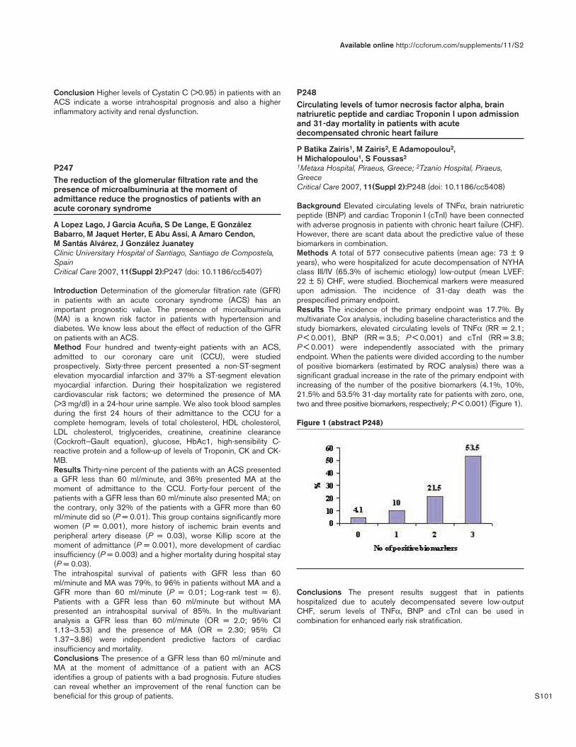
S101
Conclusion Higher levels of Cystatin C (>0.95) in patients with anACS indicate a worse intrahospital prognosis and also a higherinflammatory activity and renal dysfunction.
P247The reduction of the glomerular filtration rate and thepresence of microalbuminuria at the moment ofadmittance reduce the prognostics of patients with anacute coronary syndrome
A Lopez Lago, J Garcia Acuña, S De Lange, E GonzálezBabarro, M Jaquet Herter, E Abu Assi, A Amaro Cendon, M Santás Alvárez, J González JuanateyClinic Universitary Hospital of Santiago, Santiago de Compostela,SpainCritical Care 2007, 11(Suppl 2):P247 (doi: 10.1186/cc5407)
Introduction Determination of the glomerular filtration rate (GFR)in patients with an acute coronary syndrome (ACS) has animportant prognostic value. The presence of microalbuminuria(MA) is a known risk factor in patients with hypertension anddiabetes. We know less about the effect of reduction of the GFRon patients with an ACS.Method Four hundred and twenty-eight patients with an ACS,admitted to our coronary care unit (CCU), were studiedprospectively. Sixty-three percent presented a non-ST-segmentelevation myocardial infarction and 37% a ST-segment elevationmyocardial infarction. During their hospitalization we registeredcardiovascular risk factors; we determined the presence of MA(>3 mg/dl) in a 24-hour urine sample. We also took blood samplesduring the first 24 hours of their admittance to the CCU for acomplete hemogram, levels of total cholesterol, HDL cholesterol,LDL cholesterol, triglycerides, creatinine, creatinine clearance(Cockroft–Gault equation), glucose, HbAc1, high-sensibility C-reactive protein and a follow-up of levels of Troponin, CK and CK-MB.Results Thirty-nine percent of the patients with an ACS presenteda GFR less than 60 ml/minute, and 36% presented MA at themoment of admittance to the CCU. Forty-four percent of thepatients with a GFR less than 60 ml/minute also presented MA; onthe contrary, only 32% of the patients with a GFR more than 60ml/minute did so (P = 0.01). This group contains significantly morewomen (P = 0.001), more history of ischemic brain events andperipheral artery disease (P = 0.03), worse Killip score at themoment of admittance (P = 0.001), more development of cardiacinsufficiency (P = 0.003) and a higher mortality during hospital stay(P = 0.03).The intrahospital survival of patients with GFR less than 60ml/minute and MA was 79%, to 96% in patients without MA and aGFR more than 60 ml/minute (P = 0.01; Log-rank test = 6).Patients with a GFR less than 60 ml/minute but without MApresented an intrahospital survival of 85%. In the multivariantanalysis a GFR less than 60 ml/minute (OR = 2.0; 95% CI1.13–3.53) and the presence of MA (OR = 2.30; 95% CI1.37–3.86) were independent predictive factors of cardiacinsufficiency and mortality.Conclusions The presence of a GFR less than 60 ml/minute andMA at the moment of admittance of a patient with an ACSidentifies a group of patients with a bad prognosis. Future studiescan reveal whether an improvement of the renal function can bebeneficial for this group of patients.
P248Circulating levels of tumor necrosis factor alpha, brainnatriuretic peptide and cardiac Troponin I upon admissionand 31-day mortality in patients with acutedecompensated chronic heart failure
P Batika Zairis1, M Zairis2, E Adamopoulou2, H Michalopoulou1, S Foussas2
1Metaxa Hospital, Piraeus, Greece; 2Tzanio Hospital, Piraeus,GreeceCritical Care 2007, 11(Suppl 2):P248 (doi: 10.1186/cc5408)
Background Elevated circulating levels of TNFα, brain natriureticpeptide (BNP) and cardiac Troponin I (cTnI) have been connectedwith adverse prognosis in patients with chronic heart failure (CHF).However, there are scant data about the predictive value of thesebiomarkers in combination.Methods A total of 577 consecutive patients (mean age: 73 ± 9years), who were hospitalized for acute decompensation of NYHAclass III/IV (65.3% of ischemic etiology) low-output (mean LVEF:22 ± 5) CHF, were studied. Biochemical markers were measuredupon admission. The incidence of 31-day death was theprespecified primary endpoint.Results The incidence of the primary endpoint was 17.7%. Bymultivariate Cox analysis, including baseline characteristics and thestudy biomarkers, elevated circulating levels of TNFα (RR = 2.1;P < 0.001), BNP (RR = 3.5; P < 0.001) and cTnI (RR = 3.8;P < 0.001) were independently associated with the primaryendpoint. When the patients were divided according to the numberof positive biomarkers (estimated by ROC analysis) there was asignificant gradual increase in the rate of the primary endpoint withincreasing of the number of the positive biomarkers (4.1%, 10%,21.5% and 53.5% 31-day mortality rate for patients with zero, one,two and three positive biomarkers, respectively; P < 0.001) (Figure 1).
Conclusions The present results suggest that in patientshospitalized due to acutely decompensated severe low-outputCHF, serum levels of TNFα, BNP and cTnI can be used incombination for enhanced early risk stratification.
Available online http://ccforum.com/supplements/11/S2
Figure 1 (abstract P248)
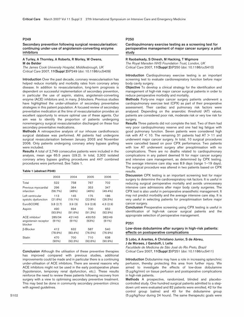
S102
P249Secondary prevention following surgical revascularisation:continuing under-use of angiotensin-converting enzymeinhibitors
A Turley, A Thornley, A Roberts, R Morley, W Owens, M de BelderThe James Cook University Hospital, Middlesbrough, UKCritical Care 2007, 11(Suppl 2):P249 (doi: 10.1186/cc5409)
Introduction Over the past decade, coronary revascularisation hashelped reduce mortality and morbidity rates from coronary arterydisease. In addition to revascularisation, long-term prognosis isdependent on successful implementation of secondary prevention,in particular the use of aspirin, statins, angiotensin-convertingenzyme (ACE) inhibitors and, in many, β-blockers. Previous studieshave highlighted the under-utilisation of secondary preventativestrategies in this patient population. A focused review of secondarypreventative medication at the time of revascularisation provides anexcellent opportunity to ensure optimal use of these agents. Ouraim was to identify the proportion of patients undergoingnonemergency surgical revascularisation discharged on these foursecondary preventative medications.Methods A retrospective analysis of our inhouse cardiothoracicsurgical database was performed. All patients had undergonesurgical revascularisation between January 2003 and November2006. Only patients undergoing coronary artery bypass graftingwere included.Results A total of 2,749 consecutive patients were included in theanalysis, mean age 65.5 years (±9.2). In total, 2,302 isolatedcoronary artery bypass grafting procedures and 447 combinedprocedures were performed. See Table 1.
Table 1 (abstract P249)
2003 2004 2005 2006
Total 522 758 767 702
Previous myocardial 296 364 353 347 infarction (56.7%) (48%) (46%) (49.4%)
Left ventricular 113 145 175 186 systolic dysfunction (21.6%) (19.1%) (22.8%) (26.5%)
EuroSCORE 3.8 (2.7) 3.9 (3) 3.9 (2.8) 4.3 (2.9)
Aspirin 490 694 700 652 (93.9%) (91.6%) (91.3%) (92.9%)
ACE inhibitor/ 285/34 421/43 430/53 382/49 angiotensin receptor (61%) (61%) (63%) (61%)blocker
β-Blocker 412 632 587 540 (78.9%) (83.4%) (76.5%) (76.9%)
Statin 470 700 710 638 (90%) (92.3%) (92.6%) (90.9%)
Conclusion Although the utilisation of these preventive therapieshas improved compared with previous studies, additionalimprovements could be made and in particular there is a continuingunder-utilisation of ACE inhibitors. There are several reasons whyACE inhibitors might not be used in the early postoperative phase(hypotension, temporary renal dysfunction, etc.). These resultsreinforce the need to review these patients following recovery fromsurgery with a view to optimising secondary preventive treatment.This may best be done in community secondary prevention clinicswith agreed guidelines.
P250Cardiopulmonary exercise testing as a screening test forperioperative management of major cancer surgery: a pilotstudy
R Raobaikady, S Dinesh, M Hacking, T WigmoreThe Royal Marsden NHS Foundation Trust, London, UKCritical Care 2007, 11(Suppl 2):P250 (doi: 10.1186/cc5410)
Introduction Cardiopulmonary exercise testing is an importantscreening test to evaluate cardiorespiratory function before majorbody cavity surgery.Objective To develop a clinical strategy for the identification andmanagement of high-risk major cancer surgical patients in order toreduce perioperative morbidity and mortality.Methods Forty-one major cancer surgery patients underwent acardiopulmonary exercise test (CPX) as part of their preoperativeassessment. Their cardiac and pulmonary risk factors wereanalysed. Depending on the anaerobic threshold (AT) values,patients are considered poor risk, moderate risk or very low risk forsurgery.Results Three patients did not complete the test. Two of them hadvery poor cardiopulmonary reserve and one had leg fatigue withgood pulmonary function. Seven patients were considered highrisk with AT < 10. The remaining 31 patients had AT > 11 andunderwent major cancer surgery. In total, 10 surgical procedureswere cancelled based on poor CPX performance. Two patientswith low AT underwent surgery after preoptimisation with nocomplications. There are no deaths related to cardiopulmonarycomplications in any patient deemed fit for major cancer surgeryand intensive care management, as determined by CPX testing.The average intensive care stay was 8.9 days (range 1–19 days).The surgical procedure was altered in two patients based on CPXresults.Discussion CPX testing is an important screening test for majorsurgery to determine the cardiorespiratory risk factors. It is useful inreducing surgical perioperative mortality and avoids unnecessaryintensive care admissions after major body cavity surgeries. TheCPX test is also useful in perioperative anaesthetic management. Itmay not predict morbidity and the average intensive care stay. It isvery useful in selecting patients for preoptimisation before majorcancer surgery.Conclusion Preoperative screening using CPX testing is useful inidentification of high-risk cancer surgical patients and theappropriate selection of perioperative management.
P251Low-dose dobutamine after surgery in high-risk patients:effects on postoperative complications
S Lobo, A Arantes, A Christiano Junior, S de Abreu, J de Moraes, J Gandolfi, L LeiteFaculdade de Medicina de São José do Rio Preto, BrazilCritical Care 2007, 11(Suppl 2):P251 (doi: 10.1186/cc5411)
Introduction Dobutamine may have a role in increasing splanchnicperfusion, thereby protecting this area from further injury. Weaimed to investigate the effects of low-dose dobutamine(5 µg/kg/min) on tissue perfusion and postoperative complicationsin high-risk patients.Methods A prospective, randomized, blinded and placebo-controlled study. One hundred surgical patients admitted to a step-down unit were evaluated and 82 patients were enrolled, 42 for thecontrol group (saline) and 40 for the dobutamine group(5 µg/kg/hour during 24 hours). The same therapeutic goals were
Critical Care March 2007 Vol 11 Suppl 2 27th International Symposium on Intensive Care and Emergency Medicine
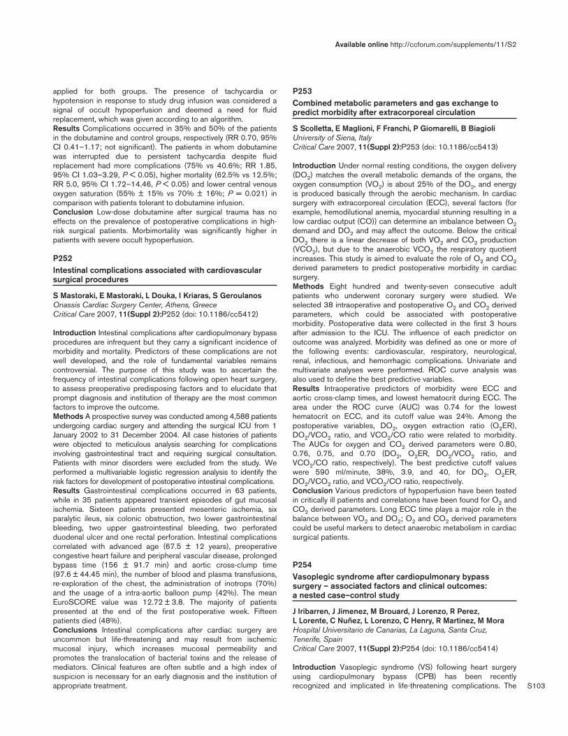
S103
applied for both groups. The presence of tachycardia orhypotension in response to study drug infusion was considered asignal of occult hypoperfusion and deemed a need for fluidreplacement, which was given according to an algorithm.Results Complications occurred in 35% and 50% of the patientsin the dobutamine and control groups, respectively (RR 0.70, 95%CI 0.41–1.17; not significant). The patients in whom dobutaminewas interrupted due to persistent tachycardia despite fluidreplacement had more complications (75% vs 40.6%; RR 1.85,95% CI 1.03–3.29, P < 0.05), higher mortality (62.5% vs 12.5%;RR 5.0, 95% CI 1.72–14.46, P < 0.05) and lower central venousoxygen saturation (55% ± 15% vs 70% ± 16%; P = 0.021) incomparison with patients tolerant to dobutamine infusion.Conclusion Low-dose dobutamine after surgical trauma has noeffects on the prevalence of postoperative complications in high-risk surgical patients. Morbimortality was significantly higher inpatients with severe occult hypoperfusion.
P252Intestinal complications associated with cardiovascularsurgical procedures
S Mastoraki, E Mastoraki, L Douka, I Kriaras, S GeroulanosOnassis Cardiac Surgery Center, Athens, GreeceCritical Care 2007, 11(Suppl 2):P252 (doi: 10.1186/cc5412)
Introduction Intestinal complications after cardiopulmonary bypassprocedures are infrequent but they carry a significant incidence ofmorbidity and mortality. Predictors of these complications are notwell developed, and the role of fundamental variables remainscontroversial. The purpose of this study was to ascertain thefrequency of intestinal complications following open heart surgery,to assess preoperative predisposing factors and to elucidate thatprompt diagnosis and institution of therapy are the most commonfactors to improve the outcome.Methods A prospective survey was conducted among 4,588 patientsundergoing cardiac surgery and attending the surgical ICU from 1January 2002 to 31 December 2004. All case histories of patientswere objected to meticulous analysis searching for complicationsinvolving gastrointestinal tract and requiring surgical consultation.Patients with minor disorders were excluded from the study. Weperformed a multivariable logistic regression analysis to identify therisk factors for development of postoperative intestinal complications.Results Gastrointestinal complications occurred in 63 patients,while in 35 patients appeared transient episodes of gut mucosalischemia. Sixteen patients presented mesenteric ischemia, sixparalytic ileus, six colonic obstruction, two lower gastrointestinalbleeding, two upper gastrointestinal bleeding, two perforatedduodenal ulcer and one rectal perforation. Intestinal complicationscorrelated with advanced age (67.5 ± 12 years), preoperativecongestive heart failure and peripheral vascular disease, prolongedbypass time (156 ± 91.7 min) and aortic cross-clump time(97.6 ± 44.45 min), the number of blood and plasma transfusions,re-exploration of the chest, the administration of inotrops (70%)and the usage of a intra-aortic balloon pump (42%). The meanEuroSCORE value was 12.72 ± 3.8. The majority of patientspresented at the end of the first postoperative week. Fifteenpatients died (48%).Conclusions Intestinal complications after cardiac surgery areuncommon but life-threatening and may result from ischemicmucosal injury, which increases mucosal permeability andpromotes the translocation of bacterial toxins and the release ofmediators. Clinical features are often subtle and a high index ofsuspicion is necessary for an early diagnosis and the institution ofappropriate treatment.
P253Combined metabolic parameters and gas exchange topredict morbidity after extracorporeal circulation
S Scolletta, E Maglioni, F Franchi, P Giomarelli, B BiagioliUniversity of Siena, ItalyCritical Care 2007, 11(Suppl 2):P253 (doi: 10.1186/cc5413)
Introduction Under normal resting conditions, the oxygen delivery(DO2) matches the overall metabolic demands of the organs, theoxygen consumption (VO2) is about 25% of the DO2, and energyis produced basically through the aerobic mechanism. In cardiacsurgery with extracorporeal circulation (ECC), several factors (forexample, hemodilutional anemia, myocardial stunning resulting in alow cardiac output (CO)) can determine an imbalance between O2demand and DO2 and may affect the outcome. Below the criticalDO2 there is a linear decrease of both VO2 and CO2 production(VCO2), but due to the anaerobic VCO2 the respiratory quotientincreases. This study is aimed to evaluate the role of O2 and CO2derived parameters to predict postoperative morbidity in cardiacsurgery.Methods Eight hundred and twenty-seven consecutive adultpatients who underwent coronary surgery were studied. Weselected 38 intraoperative and postoperative O2 and CO2 derivedparameters, which could be associated with postoperativemorbidity. Postoperative data were collected in the first 3 hoursafter admission to the ICU. The influence of each predictor onoutcome was analyzed. Morbidity was defined as one or more ofthe following events: cardiovascular, respiratory, neurological,renal, infectious, and hemorrhagic complications. Univariate andmultivariate analyses were performed. ROC curve analysis wasalso used to define the best predictive variables.Results Intraoperative predictors of morbidity were ECC andaortic cross-clamp times, and lowest hematocrit during ECC. Thearea under the ROC curve (AUC) was 0.74 for the lowesthematocrit on ECC, and its cutoff value was 24%. Among thepostoperative variables, DO2, oxygen extraction ratio (O2ER),DO2/VCO2 ratio, and VCO2/CO ratio were related to morbidity.The AUCs for oxygen and CO2 derived parameters were 0.80,0.76, 0.75, and 0.70 (DO2, O2ER, DO2/VCO2 ratio, andVCO2/CO ratio, respectively). The best predictive cutoff valueswere 590 ml/minute, 38%, 3.9, and 40, for DO2, O2ER,DO2/VCO2 ratio, and VCO2/CO ratio, respectively.Conclusion Various predictors of hypoperfusion have been testedin critically ill patients and correlations have been found for O2 andCO2 derived parameters. Long ECC time plays a major role in thebalance between VO2 and DO2; O2 and CO2 derived parameterscould be useful markers to detect anaerobic metabolism in cardiacsurgical patients.
P254Vasoplegic syndrome after cardiopulmonary bypasssurgery – associated factors and clinical outcomes: a nested case–control study
J Iribarren, J Jimenez, M Brouard, J Lorenzo, R Perez, L Lorente, C Nuñez, L Lorenzo, C Henry, R Martinez, M MoraHospital Universitario de Canarias, La Laguna, Santa Cruz,Tenerife, SpainCritical Care 2007, 11(Suppl 2):P254 (doi: 10.1186/cc5414)
Introduction Vasoplegic syndrome (VS) following heart surgeryusing cardiopulmonary bypass (CPB) has been recentlyrecognized and implicated in life-threatening complications. The
Available online http://ccforum.com/supplements/11/S2
S104
aim of this study was to identify associated factors for thedevelopment of VS after CPB.Methods We performed a nested case–control study of 50patients undergoing CPB, 27 (54%) men and 23 (46) women,mean age 66.5 (SD 9.6) years. VS was defined as systemicvascular resistance index <1,600 dyn•seg/cm5/m2 and cardiacindex >2.5 l/min/m2 within the first postoperative 4 hours.Vasoplegic shock was defined as vasoplegic patients that needednorepinephrine for at least 4 hours, after failure to respond toappropriate volume expansion. Excessive bleeding was defined asblood loss >1 l/24 hours, while total bleeding was considered asblood loss until chest tube withdrawal. Demographic variables,surgical procedures and postoperative variables were collected.We recorded data related to coagulation, fibrinolysis, complement,inflammation, blood loss at different time points, preoperative, at 0,4 and 24 hours after surgery, and hemoderivative requirements.We used the Pearson chi-squared test, the Fisher exact test, theStudent t test and the Mann–Whitney U test for nonparametricvariables. SPSS version 12.1 was used.Results Seventeen (34%) patients had VS, 11 (65%) men and six(35%) women. Longer aortic clamping time (P = 0.007) and CPBtime (P = 0.013) were associated with VS. These patients showeda higher cardiac index at 4 hours (P < 0.001) and lactic acid withinthe first 24 hours. Seven of these patients (41%) fulfilledvasoplegic shock criteria (P < 0.001). We found higher levels ofIL-6 at 0 hours (P = 0.02) and 4 hours (P = 0.001), and solubleTNF receptor at 0 hours (P = 0.044). At ICU admission (0 hours)there was a higher coagulation activation: INR (P = 0.005),fibrinogen (P = 0.001), antithrombin (P = 0.007); lower levels ofplasminogen activator inhibitor-1 (P = 0.023) as well as lowerplasminogen activator inhibitor-1/tissue-plasminogen activator ratio(P = 0.021), and higher levels of D-dimer (P = 0.041); lower levelsof C3 (P = 0.023), B factor (P = 0.013), C4 (P = 0.015) as well asa significantly higher decrease between preoperative and 0-hourlevels of C1-inhibitor, C4, C3 and B factor. Lower levels of leptinsat 0, 4 and 24 hours were found. Vasoplegic patients showedhigher blood losses along all time points (Figure 1), higherincidence of excessive bleeding (60% vs 40%; P = 0.011) andrequired more hemoderivatives during the ICU stay, plasma(P = 0.016) and platelets (P = 0.002).
Conclusions VS post-CPB was associated with activation of serinprotease systems, which leads to higher blood loss and excessivebleeding.
P255Role of plasminogen activator inhibitor-1 polymorphism onthe development of vasoplegic syndrome associated withcardiopulmonary bypass
J Jimenez, J Iribarren, M Brouard, Y Barrios, J Raya, L Lorente,R Perez, C Garcia, J Martinez, R Martinez, M MoraHospital Universitario de Canarias, La Laguna, Santa Cruz,Tenerife, SpainCritical Care 2007, 11(Suppl 2):P255 (doi: 10.1186/cc5415)
Introduction Vasoplegic syndrome (VS) after cardiac surgery withcardiopulmonary bypass (CPB) can vary from mild to severecomplication and it appears with an incidence ranging between5% and 15%. The etiology is not completely elucidated but riskfactors such as temperature and duration of cardiopulmonarybypass and preoperative treatment with angiotensin-convertingenzyme (ACE) inhibitors have been associated [1]. We wanted toinvestigate the possible role of several genetic polymorphisms inpatients with VS after elective CPB.Methods We performed a nested case–control study of 50patients undergoing CPB, 27 (54%) men and 23 (46) women,mean age 66.5 (SD 9.6) years. VS was defined as systemicvascular resistance index lower than 1,600 dyn•seg/cm5/m2 and acardiac index greater than 2.5 l/min/m2 within the first 4 hours aftersurgery. We recorded data related to hemodynamic parameters atdifferent postoperative time points, at ICU admission (0 hours), 4and 24 hours after surgery, and the polymorphism of the followinggenes: plasminogen activator inhibitor-1 (PAI-1) and β-TNF + 250.In addition, 23 neutral markers were genotyped to follow genomiccontrol strategies that would detect spurious associations due topopulation substructure. We used the Pearson chi-squared testand binary logistic regression. SPSS version 12.1 was used.Results We observed 17 (34%) patients with vasoplegia criteria,11 (65%) men and six (35%) women, age 67 (61–72) years. Theonly one associated with VS was the PAI-1 polymorphism, and itsdistribution in the study population was: 4G/G genotype in 10(20%) patients, 4G/5G in 26 (52%) patients, and 5G/G in 14
Critical Care March 2007 Vol 11 Suppl 2 27th International Symposium on Intensive Care and Emergency Medicine
Figure 1 (abstract P254)Figure 1 (abstract P255)
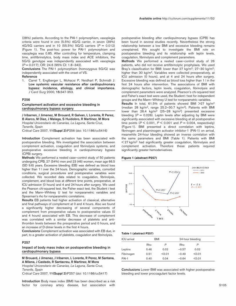
S105
(28%) patients. According to the PAI-1 polymorphism, vasoplegiacriteria were found in one (5.5%) 4G/G carrier, in seven (39%)4G/5G carriers and in 10 (55.5%) 5G/G carriers (P = 0.012)(Figure 1). The post-hoc power for PAI-1 polymorphism andvasoplegia was 0.85. After controlling for temperature, clampingtime, antifibrinolytics, body mass index and ACE inhibitors, the5G/G genotype was independently associated with vasoplegia(P = 0.017); OR: 24.6 (95% CI: 1.8–342).Conclusions The PAI-1 polymorphism (homozygous 5G/G) wasindependently associated with the onset of VS.Reference1. Carrel T, Englberger L, Mohacsi P, Neidhart P, Schmidli J:
Low systemic vascular resistance after cardiopulmonarybypass: incidence, etiology, and clinical importance.J Card Surg 2000, 15:347-353.
P256Complement activation and excessive bleeding incardiopulmonary bypass surgery
J Iribarren, J Jimenez, M Brouard, R Galvan, L Lorente, R Perez,B Alarco, M Diaz, J Malaga, S Huidobro, R Martinez, M MoraHospital Universitario de Canarias, La Laguna, Santa Cruz,Tenerife, SpainCritical Care 2007, 11(Suppl 2):P256 (doi: 10.1186/cc5416)
Introduction Complement activation has been associated withpostoperative bleeding. We investigated the association betweencomplement activation, coagulation and fibrinolysis systems, andpostoperative excessive bleeding in cardiopulmonary bypass(CPB) surgery.Methods We performed a nested case–control study of 50 patientsundergoing CPB, 27 (54%) men and 23 (46) women, mean age 66.5(SD 9.6) years. Excessive bleeding (EB) was defined as blood losshigher than 1 l over the 24 hours. Demographic variables, comorbidconditions, surgical procedures and postoperative variables werecollected. We recorded data related to coagulation, fibrinolysis,complement, and blood loss at different time points, preoperative, atICU admission (0 hours) and 4 and 24 hours after surgery. We usedthe Pearson chi-squared test, the Fisher exact test, the Student t testand the Mann–Whitney U test for nonparametric variables andSpearman’s rho for nonparametric correlations.Results EB patients had higher activation of classical, alternativeand final pathways of complement at 0 and 4 hours. Also we founda significantly higher decreasing of several components ofcomplement from preoperative values to postoperative values (0and 4 hours) associated with EB. This decrease of complementwas correlated with a similar decrease of platelets and anti-thrombin levels between the preoperative period and 0 hours, andan increase of D-dimer levels in the first 4 hours.Conclusions Complement activation was associated with EB due, inpart, to a greater activation of platelets, coagulation and fibrinolysis.
P257Impact of body mass index on postoperative bleeding incardiopulmonary bypass
M Brouard, J Jimenez, J Iribarren, L Lorente, R Perez, M Santana,A Milena, J Castedo, R Santacreu, R Martinez, M MoraHospital Universitario de Canarias, La Laguna, Santa Cruz,Tenerife, SpainCritical Care 2007, 11(Suppl 2):P257 (doi: 10.1186/cc5417)
Introduction Body mass index (BMI) has been described as a riskfactor for coronary artery disease, but association with
postoperative bleeding after cardiopulmonary bypass (CPB) hasbeen found in several studies recently. Nevertheless the strongrelationship between a low BMI and excessive bleeding remainsunexplained. We sought to investigate the BMI role onpostoperative bleeding and its relationship with leptin levels,coagulation, fibrinolysis and complement parameters.Methods We performed a nested case–control study of 26patients, who did not receive antifibrinolytic prophylaxis. We usedBray’s classification for BMI: lower than 27 kg/m2; 27–30 kg/m2;higher than 30 kg/m2. Variables were collected preoperatively, atICU admission (0 hours), and at 4 and 24 hours after surgery.Excessive bleeding was defined as blood loss higher than 1 l in thefirst 24 hours after intervention. The associations of BMI withdemographic factors, leptin levels, coagulation, fibrinolysis andcomplement parameters were analyzed. Pearson’s chi-squared testand Fisher’s exact test were used, the Student t test for independentgroups and the Mann–Whitney U test for nonparametric variables.Results In total, 61.5% of patients showed BMI >27 kg/m2
(median 28 kg/m2, range 25.2–30.7 kg/m2). Patients with BMIlower than 26.4 kg/m2 (25–28 kg/m2) presented excessivebleeding (P = 0.026). Leptin levels after adjusting by BMI weresignificantly associated with excessive bleeding at all postoperativetime points (P < 0.001, P < 0.001 and P = 0.004, respectively)(Figure 1). BMI presented a direct correlation with leptins,fibrinogen and plasminogen activator inhibitor-1 (PAI-1) on arrival,meanwhile 24-hour bleeding showed an inverse correlation withthe same parameters and BMI (Table 1). Patients with BMI< 27 kg/m2 had significantly greater coagulation, fibrinolysis andcomplement activation. Therefore these patients requiredsignificantly greater hemoderivatives.
Table 1 (abstract P257)
ICU arrival BMI 24-hour bleeding
Rho P Rho P
Leptins 0.46 0.02 –0.57 0.02
Fibrinogen 0.51 <0.01 –0.49 <0.01
PAI-1 0.40 0.04 –0.64 <0.01
Conclusions Lower BMI was associated with higher postoperativebleeding and lower procoagulant factor levels.
Available online http://ccforum.com/supplements/11/S2
Figure 1 (abstract P257)
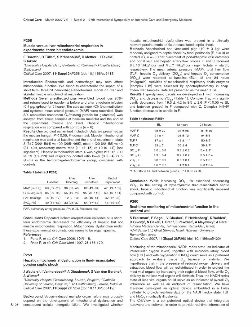
S106
P258Muscle versus liver mitochondrial respiration inexperimental three-hit endotoxemia
E Borotto1, D Tüller1, S Krähenbühl2, D Mettler1, J Takala1, S Jakob1
1University Hospital Bern, Switzerland; 2University Hospital Basel,SwitzerlandCritical Care 2007, 11(Suppl 2):P258 (doi: 10.1186/cc5418)
Introduction Endotoxemia and hemorrhage may both affectmitochondrial function. We aimed to characterize the impact of ashort-term, three-hit hemorrhage/endotoxemia model on liver andskeletal muscle mitochondrial respiration.Methods Seven anesthetized pigs were bled (blood loss 20%)and retransfused to euvolemia before and after endotoxin infusion(0.4 µg/kg/hour for 2 hours). The cardiac index (CI) (thermodilution)and systemic mean arterial pressure (MAP) were recorded. State3/4 respiration (nanoatom O2/min/mg protein for glutamate) wasassayed from tissue samples at baseline (muscle) and the end ofthe experiment (muscle and liver). Hepatic mitochondrialrespiration was compared with controls (n = 6).Results One pig died earlier (not included). Data are presented asthe median (range). P < 0.05, Friedman test. Muscle mitochondrialrespiration was similar at baseline and the end of experiment; state3 (317 (222–594) vs 409 (295–468)), state 4 (29 (22–58) vs 40(31–49)), respiratory control ratio (11 (7–15) vs 10 (9–11)) (notsignificant). Hepatic mitochondrial state 4 was higher (27 (16–31)vs 19 (13–22)) and respiratory control ratio lower (3 (3–4) vs 5(4–6)) in the hemorrhage/endotoxemia group, compared withcontrols.
Table 1 (abstract P258)
After After End of Baseline bleeding endotoxin experiment
MAP (mmHg) 69 (52–70) 36 (30–46) 67 (49–84) 47 (19–109)
CI (ml/kg/min) 85 (62–95) 55 (42–76) 95 (78–113) 69 (16–151)
PAP (mmHg) 14 (10–17) 12 (9–13) 45 (40–51) 32 (17–38)
SvO2 (%) 49 (41–56) 33 (25–37) 54 (47–68) 46 (14–66)
PAP, pulmonary artery pressure. P < 0.05, Friedman test.
Conclusions Repeated ischemia/reperfusion episodes plus short-term endotoxemia decreased the efficiency of hepatic but notmuscle mitochondrial respiration. Mitochondrial dysfunction underthese experimental circumstances seems to be organ specific.References1. Porta F, et al.: Crit Care 2006, 10:R118.2. Rhee P, et al.: Crit Care Med 1997, 25:166-170.
P259Hepatic mitochondrial dysfunction in fluid-resuscitatedporcine septic shock
J Wauters1, I Vanhorebeek2, A Dieudonne1, G Van den Berghe1,A Wilmer3
1University Hospital Gasthuisberg, Leuven, Belgium; 2CatholicUniversity of Leuven, Belgium; 3UZ Gasthuisberg, Leuven, BelgiumCritical Care 2007, 11(Suppl 2):P259 (doi: 10.1186/cc5419)
Background Sepsis-induced multiple organ failure may cruciallydepend on the development of mitochondrial dysfunction andconsequent cellular energetic failure. We investigated whether
hepatic mitochondrial dysfunction was present in a clinicallyrelevant porcine model of fluid-resuscitated septic shock.Methods Anesthetized and ventilated pigs (40 ± 3 kg) wererandomly assigned to septic shock by fecal peritonitis (F, n = 3) orcontrol (C, n = 3) after placement of portal/hepatic vein cathetersand portal vein and hepatic artery flow probes. F and C received8 ± 13 ml/kg/hour and 5 ± 7 ml/kg/hour ringer lactate + starch,respectively. The mean arterial pressure (MAP), total liver flow(TLF), hepatic O2 delivery (DO2,h) and hepatic O2 consumption(VO2,h) were recorded at baseline (BL), 12 and 24 hours(ml/kg/min). Activities of mitochondrial respiratory chain enzymes(complex I–IV) were assessed by spectrophotometry in snap-frozen liver samples. Data are presented as the mean ± SD.Results Hyperdynamic circulation developed in F with increasingDO2,h and decreasing VO2,h (Table 1). Complex II activity signifi-cantly decreased from 19.3 ± 4.2 to 9.5 ± 2.6 (P < 0.05 vs BLand between groups) in F compared with C. Complex I–III–IVfunction decreased in parallel in F.
Table 1 (abstract P259)
BL 12 hours 24 hours
MAP F 78 ± 22 98 ± 25 81 ± 19
MAP C 91 ± 4 101 ± 12 94 ± 6
TLF F 17 ± 1 46 ± 11* 50 ± 5*
TLF C 23 ± 7 35 ± 4 39 ± 7†
DO2,h F 2.0 ± 0.6 3.8 ± 0.2 5.4 ± 1†
DO2,h C 1.9 ± 0.4 3.2 ± 0.4 3.5 ± 0.4
VO2,h F 0.8 ± 0.2 0.6 ± 0.1 0.5 ± 0.1
VO2,h C 1.5 ± 0.7 1.1 ± 0.1 0.9 ± 0.4
*P < 0.05 vs BL and between groups. †P < 0.05 vs BL.
Conclusion While increasing DO2,h far exceeded decreasingVO2,h in the setting of hyperdynamic fluid-resuscitated septicshock, hepatic mitochondrial function was significantly impairedcompared with control.
P260Real-time monitoring of mitochondrial function in theurethral wall
S Preisman1, E Segal1, V Glauber1, E Heldenberg1, R Walden1,D Givony2, N Dekel2, L Oren2, E Pewzner2, A Mayevsky3, A Perel11Sheba Medical Center, Tel-Hashomer, Rama-Gan, Israel;2CritiSense Ltd, Givat Shmuel, Israel; 3Bar-Ilan University,Ramat-Gan, IsraelCritical Care 2007, 11(Suppl 2):P260 (doi: 10.1186/cc5420)
Monitoring of the mitochondrial NADH redox state (an indicator ofintracellular oxygen levels) together with microcirculatory bloodflow (TBF) and with oxygenation (HbO2) could serve as a preferredapproach to evaluate tissue O2 balance or viability. Wehypothesize that in the presence of reduced oxygen delivery andextraction, blood flow will be redistributed in order to protect themost vital organs by increasing their regional blood flow, while O2delivery to the less vital organs will diminish. Thus, the NADH redoxstate of less vital organs could serve as an indicator of overall O2imbalance as well as an endpoint of resuscitation. We havetherefore developed an optical device embedded in a Foleycatheter to provide real-time data on the NADH redox state, TBFand HbO2 in critically ill patients.The CritiView is a computerized optical device that integrateshardware and software in order to provide real-time information of
Critical Care March 2007 Vol 11 Suppl 2 27th International Symposium on Intensive Care and Emergency Medicine
S107
tissue viability [1]. A modified three-way Foley catheter thatcontains a fiberoptic probe connects the CritiView to the mucosalside of the urethral wall. We have used this device in five femalepigs that underwent graded hemorrhage, and in four patients whowere monitored during aortic abdominal aneurysm operations.These preliminary swine model and human studies confirm thefeasibility of collecting information about mitochondrial functionfrom the urethral wall. The main effects of graded hemorrhagestarted when the blood volume decreased by 30%. At 40% bloodloss, minimal levels of TBF and HbO2 were correlated to themaximal NADH levels. The values of the three parameters returnedto baseline after retransfusion of the shed blood. Aortic clamping inpatients led to a significant decrease in TBF and HbO2 whileNADH levels increased. After aortic declamping, the parametersrecovered to normal values.Our preliminary results show that the CritiView may be a useful toolfor the detection of O2 imbalance and the development of anemergency metabolic state in nonvital tissues.Reference1. Mayevsky et al.: SPIE Proc 2006, 6083:OZ1-OZ10.
P261Cytokines monitored by microdialysis detect rejectionearlier than current methods in liver transplantation
L Wælgaard, EB Thorgersen, P Line, T Mollnes, T TonnessenRikshospitalet, Oslo, NorwayCritical Care 2007, 11(Suppl 2):P261 (doi: 10.1186/cc5421)
Introduction The outcome of liver transplantation is steadilyimproving. There is still need for earlier detection of complicationssuch as hepatic artery thrombosis and rejection. In an earlier invitro study we showed that the CMA microdialysis system with a100 kDa pore size membrane can be used to measure theselected cytokines and complement. We monitored patientsundergoing liver transplantation with microdialysis continuously fora week postoperatively, and analyzed both parameters to detectischemia, and cytokines and anaphylatoxins to explore whetherrejection was detected earlier than with the standard methods.Methods Twenty patients undergoing 22 liver transplantationswere included. Two microdialysis catheter were introduced in theliver and one in subcutaneous tissue. We analyzed metabolicparameters (glucose, pyruvate, glycerol and lactate), and IL-6, IL-8,MCP-1, IP-10, and C5a.Results Fourteen patients had an uneventful coursepostoperatively, judged clinically and by routine biochemicalmarkers and ultrasound Doppler. These patients had a medianlactate starting at 3.5 mM (2 hours after reperfusion) falling tobelow 2 mM during the first 24 hours, and thereafter staying low.The L/P ratio (a specific measure of ischemia) dropped from about20 to below 10. These patients had a steady rise in IP-10 from 200to 3,000 pg/ml, and also a slight raise in IL-6 initially. Case 1. Themale patient had a steadily increasing L/P ratio during the 7 daysof microdialysis measurements, indicating an insufficient bloodsupply. He underwent surgery 5 days later and a hepatic arterythrombosis was found. A biopsy was done during the operationshowing an acute rejection. There was a significant rise in IP-10 to13,000 pg/ml 7 days before the diagnosis of rejection. Case 2.The female patient had an acute rejection verified by biopsies onday 10 postoperatively. Her IL-8, IP-10 and C5a increased 10-foldto 100-fold in the liver 3 days earlier than an increase in liverenzymes and 5 days before the rejection was verified by biopsy.Conclusion We have described the normal course of the fourcytokines IL-6, IL-8, MCP-1 and IP-10 and complement C5a afterliver transplantation, as well as metabolic parameters to detect
ischemia. In two patients with rejection we found a large increasein IP-10, IL-8 and complement split-product C5a in the liver but notin the subcutis 3–5 days before any other parameter of liver injury.
P262Microcirculatory hemodynamic alterations during cardiacluxation in off-pump coronary artery bypass grafting surgery
B Atasever1, R Speekenbrink2, J Seyffert2, C Ince3
1Erasmus Medical Center Rotterdam, The Netherlands; 2OnzeLieve Vrouwe Gasthuis, Amsterdam, The Netherlands; 3AcademicMedical Center, Amsterdam, The NetherlandsCritical Care 2007, 11(Suppl 2):P262 (doi: 10.1186/cc5422)
Introduction During luxation of the beating heart in off-pumpcoronary artery bypass grafting surgery the cardiac output dropsand causes hypotension (<60 mmHg). It is expected that this stateof obstructive shock is detrimental for adequate perfusion andoxygenation of organ tissue. However, it is unknown whether theseluxations cause microcirculatory dysfunction. In this study we haveexplored the hemodynamics of the sublingual microcirculationduring mechanical manipulations of the beating heart.Methods During cardiac luxations, in 12 patients reflectancespectrophotometry (O2C®; Lea Medizintechnik, Germany) wasused to measure oxygen availability and in 12 other patientssidestream dark field imaging (MicroScan®; MicroVision Medical,The Netherlands) was used to directly visualize the sublingualmicrocirculatory hemodynamics in a single network of micro-vessels. Microvascular analysis software (MAS®; MicroVisionMedical) was used to analyze the vessel density and blood flow.Synchronously, systemic hemodynamics were recorded and thecardiac output was calculated by pulse contour analysis of arterialpressure (PulseCO®; LiDCO, UK) in all patients.Results During cardiac luxations, the oxygen availability in thesublingual microcirculation decreased (µHbO2 64.2 ± 9.1 to48.6 ± 8.7%; P < 0.01) while the functional capillary density didnot change (15.9 ± 1.1 to 15.6 ± 1.3 mm/mm2; P = 0.65).Although the small vessels (0–20 µm) did not fall out they didshow hypoperfusion (Vmax 895 ± 209 to 396 ± 178 µm/s;P < 0.01), whereas in the medium vessels (20–50 µm) there wasno significant change in blood velocity (Vmax 751 ± 239 to596 ± 192 µm/s; P = 0.18) as observed with sidestream dark fieldimaging and calculated with microvascular analysis software.Conclusion Alterations in sublingual microcirculation hemo-dynamics reflect the direct effects of obstructive shock andelucidate the microcirculatory autoregulation.
P263Muscle microcirculation alterations increase with diseaseseverity in chronic heart failure patients
V Gerovasili, C Pierakos, S Dimopoulos, E Kaldara, S Kourtidou, S Sarafoglou, M Kravari, J Venetsanakos, G Tzanis, S NanasNational and Kapodestrian University of Athens, GreeceCritical Care 2007, 11(Suppl 2):P263 (doi: 10.1186/cc5423)
Objective To evaluate skeletal muscle microcirculation by near-infrared spectroscopy (NIRS) in patients with chronic heart failure(CHF).Background Skeletal muscle microcirculation is impaired inpatients with CHF, and this impairment seems to correlate withdisease severity.Methods We evaluated 49 patients with CHF (mean age: 58 ± 12years) and 12 healthy volunteers. Of the CHF patients, 14 had
Available online http://ccforum.com/supplements/11/S2
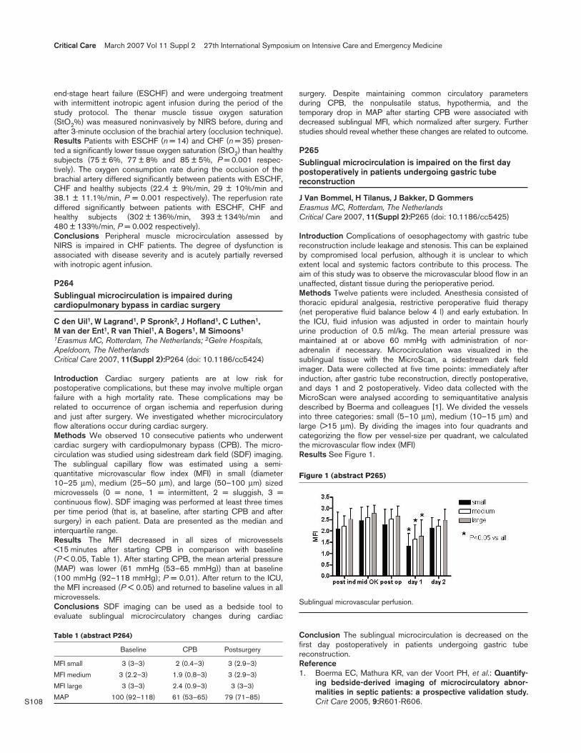
S108
end-stage heart failure (ESCHF) and were undergoing treatmentwith intermittent inotropic agent infusion during the period of thestudy protocol. The thenar muscle tissue oxygen saturation(StO2%) was measured noninvasively by NIRS before, during andafter 3-minute occlusion of the brachial artery (occlusion technique).Results Patients with ESCHF (n = 14) and CHF (n = 35) presen-ted a significantly lower tissue oxygen saturation (StO2) than healthysubjects (75 ± 6%, 77 ± 8% and 85 ± 5%, P = 0.001 respec-tively). The oxygen consumption rate during the occlusion of thebrachial artery differed significantly between patients with ESCHF,CHF and healthy subjects (22.4 ± 9%/min, 29 ± 10%/min and38.1 ± 11.1%/min, P = 0.001 respectively). The reperfusion ratediffered significantly between patients with ESCHF, CHF andhealthy subjects (302 ± 136%/min, 393 ± 134%/min and480 ± 133%/min, P = 0.002 respectively).Conclusions Peripheral muscle microcirculation assessed byNIRS is impaired in CHF patients. The degree of dysfunction isassociated with disease severity and is acutely partially reversedwith inotropic agent infusion.
P264Sublingual microcirculation is impaired duringcardiopulmonary bypass in cardiac surgery
C den Uil1, W Lagrand1, P Spronk2, J Hofland1, C Luthen1, M van der Ent1, R van Thiel1, A Bogers1, M Simoons1
1Erasmus MC, Rotterdam, The Netherlands; 2Gelre Hospitals,Apeldoorn, The NetherlandsCritical Care 2007, 11(Suppl 2):P264 (doi: 10.1186/cc5424)
Introduction Cardiac surgery patients are at low risk forpostoperative complications, but these may involve multiple organfailure with a high mortality rate. These complications may berelated to occurrence of organ ischemia and reperfusion duringand just after surgery. We investigated whether microcirculatoryflow alterations occur during cardiac surgery.Methods We observed 10 consecutive patients who underwentcardiac surgery with cardiopulmonary bypass (CPB). The micro-circulation was studied using sidestream dark field (SDF) imaging.The sublingual capillary flow was estimated using a semi-quantitative microvascular flow index (MFI) in small (diameter10–25 µm), medium (25–50 µm), and large (50–100 µm) sizedmicrovessels (0 = none, 1 = intermittent, 2 = sluggish, 3 =continuous flow). SDF imaging was performed at least three timesper time period (that is, at baseline, after starting CPB and aftersurgery) in each patient. Data are presented as the median andinterquartile range.Results The MFI decreased in all sizes of microvessels<15 minutes after starting CPB in comparison with baseline(P < 0.05, Table 1). After starting CPB, the mean arterial pressure(MAP) was lower (61 mmHg (53–65 mmHg)) than at baseline(100 mmHg (92–118 mmHg); P = 0.01). After return to the ICU,the MFI increased (P < 0.05) and returned to baseline values in allmicrovessels.Conclusions SDF imaging can be used as a bedside tool toevaluate sublingual microcirculatory changes during cardiac
surgery. Despite maintaining common circulatory parametersduring CPB, the nonpulsatile status, hypothermia, and thetemporary drop in MAP after starting CPB were associated withdecreased sublingual MFI, which normalized after surgery. Furtherstudies should reveal whether these changes are related to outcome.
P265Sublingual microcirculation is impaired on the first daypostoperatively in patients undergoing gastric tubereconstruction
J Van Bommel, H Tilanus, J Bakker, D GommersErasmus MC, Rotterdam, The NetherlandsCritical Care 2007, 11(Suppl 2):P265 (doi: 10.1186/cc5425)
Introduction Complications of oesophagectomy with gastric tubereconstruction include leakage and stenosis. This can be explainedby compromised local perfusion, although it is unclear to whichextent local and systemic factors contribute to this process. Theaim of this study was to observe the microvascular blood flow in anunaffected, distant tissue during the perioperative period.Methods Twelve patients were included. Anesthesia consisted ofthoracic epidural analgesia, restrictive peroperative fluid therapy(net peroperative fluid balance below 4 l) and early extubation. Inthe ICU, fluid infusion was adjusted in order to maintain hourlyurine production of 0.5 ml/kg. The mean arterial pressure wasmaintained at or above 60 mmHg with administration of nor-adrenalin if necessary. Microcirculation was visualized in thesublingual tissue with the MicroScan, a sidestream dark fieldimager. Data were collected at five time points: immediately afterinduction, after gastric tube reconstruction, directly postoperative,and days 1 and 2 postoperatively. Video data collected with theMicroScan were analysed according to semiquantitative analysisdescribed by Boerma and colleagues [1]. We divided the vesselsinto three categories: small (5–10 µm), medium (10–15 µm) andlarge (>15 µm). By dividing the images into four quadrants andcategorizing the flow per vessel-size per quadrant, we calculatedthe microvascular flow index (MFI)Results See Figure 1.
Conclusion The sublingual microcirculation is decreased on thefirst day postoperatively in patients undergoing gastric tubereconstruction.Reference1. Boerma EC, Mathura KR, van der Voort PH, et al.: Quantify-
ing bedside-derived imaging of microcirculatory abnor-malities in septic patients: a prospective validation study.Crit Care 2005, 9:R601-R606.
Critical Care March 2007 Vol 11 Suppl 2 27th International Symposium on Intensive Care and Emergency Medicine
Table 1 (abstract P264)
Baseline CPB Postsurgery
MFI small 3 (3–3) 2 (0.4–3) 3 (2.9–3)
MFI medium 3 (2.2–3) 1.9 (0.8–3) 3 (2.9–3)
MFI large 3 (3–3) 2.4 (0.9–3) 3 (3–3)
MAP 100 (92–118) 61 (53–65) 79 (71–85)
Figure 1 (abstract P265)
Sublingual microvascular perfusion.
S109
P266Oscillation frequency of skin microvascular blood flow isassociated with mortality in critically ill patients
H Knotzer1, S Maier1, W Pajk1, M Dünser2, W Hasibeder3
1Innsbruck Medical University, Innsbruck, Austria; 2UniversityHospital Bern, Switzerland; 3Krankenhaus der BarmherzigenSchwestern Ried, Ried, AustriaCritical Care 2007, 11(Suppl 2):P266 (doi: 10.1186/cc5426)
Introduction Microcirculatory dysfunction has been hypothesizedto play a key role in the pathophysiology of multiple organ failure,and consequently to patient outcome. The objective of the presentstudy was to investigate differences in reactive hyperemiaresponse and oscillation frequencies in survivors and nonsurvivorsof patients with multiple organ dysfunction syndrome (MODS).Methods Twenty-nine patients (15 survivors; 14 nonsurvivors) withtwo or more organ failures were eligible for study entry. All patientswere hemodynamically stabilized, and demographic and clinicaldata were recorded. A laser Doppler flowmeter was used tomeasure the cutaneous microcirculatory response. Reactivehyperemia and oscillatory changes in the Doppler signal weremeasured during 3 minutes before and after a 5-minute period offorearm ischemia during hyperemia.Results Nonsurvivors demonstrated a significantly higher MODSscore when compared with survivors (P = 0.004). Norepinephrinerequirements were higher in nonsurvivors (P = 0.018). Non-survivors had higher arterial lactate levels (P = 0.046), decreasedarterial pH levels (P = 0.001), and decreased arterial PO2 values(P = 0.013) when compared with survivors. A higher oscillationfrequency of skin microvasculature at rest (P = 0.033) and after anischemic stimulus (P = 0.009) was observed in nonsurvivors. Nodifferences were observed in reactive hyperemia responsebetween groups. The flowmotion frequency observed in reactivehyperemia was associated with the severity of the MODS(P = 0.009), and – although not statistically significant – arteriallactate concentration (P = 0.052).Conclusion An increased skin microvascular oscillation frequencyduring rest and after an ischemic stimulus is associated withincreased mortality in patients suffering from MODS. We suggestthat the underlying mechanism of the increased flowmotion couldbe a response of the skin microvasculature to hypoxia or to animpaired oxygen utilization of the skin tissue.
P267Effect of intermittent positive pressure ventilation on theskeletal muscle and small intestine microcirculation in rats
V Cerny, Z Turek, R Parizkova, P DostalUniversity Hospital Hradec Kralove, Czech RepublicCritical Care 2007, 11(Suppl 2):P267 (doi: 10.1186/cc5427)
Introduction Intermittent positive pressure ventilation (IPPV) maybe accompanied by alteration of microcirculation [1,2]; however,the effect of IPPV is not mentioned in the interpretation of theresults of studies evaluating microcirculation using orthogonalpolarization spectral or sidestream dark-field imaging. This studyaimed to evaluate the effect of IPPV on microcirculation in theskeleton muscles and in the serosa of the small intestine in rats.Methods Ten animals were tracheostomized and prepared formicrocirculation study; after tissue preparation, five rats wereallowed to breath spontaneously (Group SB = spontaneousbreathing), and five rats (Group IPPV) were connected to a smallanimal ventilator (IPPV: FiO2 0.21, respiratory rate 60/min, tidalvolume 10 ml/kg, inspiratory time 50% of respiratory cycle, and
2 cmH2O PEEP). Sidestream dark-field images were obtained fromthe quadriceps femoris muscle (QFM) and serosa surface of theileum. The arterial blood pressure and rectal temperature were alsorecorded. The functional capillary density (FCD) and small andmedium vessels rate were analysed offline using AVA V1.0 soft-ware (AMC, University of Amsterdam, The Netherlands), P ≤ 0.05.Results The FCD was decreased significantly in QFM in rats withIPPV with respect to Group SB (184 ± 27 resp. 197 ± 61 cm/cm2),but the FCD of the intestinal serosa was not affected by IPPV(265 ± 46 resp. 267 ± 25 cm/cm2). There were no differences inmean blood pressure and temperature between groups (128 ± 7Torr and 36.6 ± 0.1°C in Group SB, or 128 ± 10 Torr and36.5 ± 0.1°C in Group IPPV).Conclusion The use of IPPV should be taken into account in theinterpretation of the studies examining the changes inmicrocirculation in rats.Acknowledgement Research project MZO 00179906.References1. Saner FH, et al.: Eur J Anaesth 2006, 23:766-771.2. De Backer D, et al.: Am Heart J 2004, 147:91-99.
P268Changes in sublingual microvascular flow duringexperimental human endotoxemia
R Bemelmans1, A Draisma2, J van der Hoeven2, P Pickkers2, P Spronk1
1Gelre Ziekenhuizen, Apeldoorn, The Netherlands; 2UMCN,Nijmegen, The NetherlandsCritical Care 2007, 11(Suppl 2):P268 (doi: 10.1186/cc5428)
Introduction We examined sublingual microvascular changes inexperimental human endotoxemia. Changes in microcirculation andmitochondrial dysfunction appear to be key mechanisms in sepsis,since they can lead to regional mismatch of oxygen supply anddemand. Lipopolysaccharide (LPS) can be used to induce endo-toxemia as a model of sepsis, but the effects on microcirculatoryperfusion have not been tested before, particularly after toleranceinduction during repeated challenges of LPS.Methods Six healthy volunteers received an intravenous injectionof 2 ng/kg Escherichia coli LPS to induce endotoxemia on fiveconsecutive days. Microvascular perfusion was sublinguallymeasured using sidestream darkfield imaging just before, and 2and 4 hours after LPS injection on day 1. All measurements wererepeated on day 5 of LPS administration. Sublingual capillary flowwas estimated using a semiquantative microvascular flow index(MFI) in small (10–25 µm), medium (25–50 µm) and large-sized(50–100 µm) microvessels (no flow, 0; intermittent flow, 1;sluggish flow, 2; and continuous flow, 3). Changes were evaluatedwith the paired Wilcoxon test and sign test. P < 0.05 was judgedto indicate a significant difference. Values are expressed as themedian (P25–P75).Results Two hours after the induction of endotoxemia (n = 6),sublingual flow in small (2 (1.7–2.3)), medium-sized (1.5(1.2–1.9)), and large microvessels (2.5 (1.2–2.7)) did not differfrom baseline values (2.3 (1.5–2.8), 2.3 (1.4–2.5), and 2.3(1.3–2.5), respectively, all P = not significant). Microvascular flowdid not change in the subsequent 2 hours. In addition, nodifference in microvascular flow could be demonstrated betweentimepoints on day 1 and day 5 of intermittent endotoxemia.Conclusion In this small pilot study in experimental humanendotoxemia, no significant effect of LPS administration onmicrocirculatory perfusion could be observed, nor any sign oftolerance. Further studies should reveal whether microvascularimpairment does not occur in early human experimental
Available online http://ccforum.com/supplements/11/S2
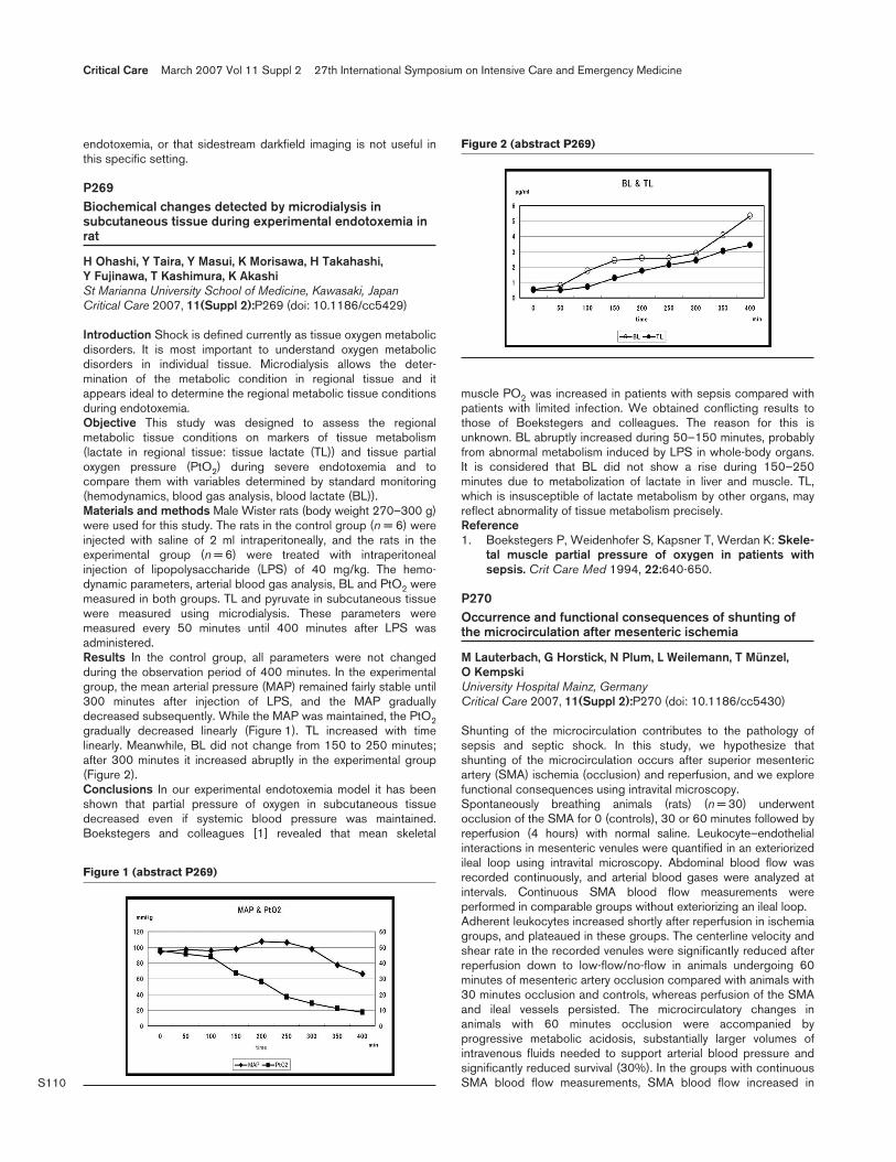
S110
endotoxemia, or that sidestream darkfield imaging is not useful inthis specific setting.
P269Biochemical changes detected by microdialysis insubcutaneous tissue during experimental endotoxemia inrat
H Ohashi, Y Taira, Y Masui, K Morisawa, H Takahashi, Y Fujinawa, T Kashimura, K AkashiSt Marianna University School of Medicine, Kawasaki, JapanCritical Care 2007, 11(Suppl 2):P269 (doi: 10.1186/cc5429)
Introduction Shock is defined currently as tissue oxygen metabolicdisorders. It is most important to understand oxygen metabolicdisorders in individual tissue. Microdialysis allows the deter-mination of the metabolic condition in regional tissue and itappears ideal to determine the regional metabolic tissue conditionsduring endotoxemia.Objective This study was designed to assess the regionalmetabolic tissue conditions on markers of tissue metabolism(lactate in regional tissue: tissue lactate (TL)) and tissue partialoxygen pressure (PtO2) during severe endotoxemia and tocompare them with variables determined by standard monitoring(hemodynamics, blood gas analysis, blood lactate (BL)).Materials and methods Male Wister rats (body weight 270–300 g)were used for this study. The rats in the control group (n = 6) wereinjected with saline of 2 ml intraperitoneally, and the rats in theexperimental group (n = 6) were treated with intraperitonealinjection of lipopolysaccharide (LPS) of 40 mg/kg. The hemo-dynamic parameters, arterial blood gas analysis, BL and PtO2 weremeasured in both groups. TL and pyruvate in subcutaneous tissuewere measured using microdialysis. These parameters weremeasured every 50 minutes until 400 minutes after LPS wasadministered.Results In the control group, all parameters were not changedduring the observation period of 400 minutes. In the experimentalgroup, the mean arterial pressure (MAP) remained fairly stable until300 minutes after injection of LPS, and the MAP graduallydecreased subsequently. While the MAP was maintained, the PtO2gradually decreased linearly (Figure 1). TL increased with timelinearly. Meanwhile, BL did not change from 150 to 250 minutes;after 300 minutes it increased abruptly in the experimental group(Figure 2).Conclusions In our experimental endotoxemia model it has beenshown that partial pressure of oxygen in subcutaneous tissuedecreased even if systemic blood pressure was maintained.Boekstegers and colleagues [1] revealed that mean skeletal
muscle PO2 was increased in patients with sepsis compared withpatients with limited infection. We obtained conflicting results tothose of Boekstegers and colleagues. The reason for this isunknown. BL abruptly increased during 50–150 minutes, probablyfrom abnormal metabolism induced by LPS in whole-body organs.It is considered that BL did not show a rise during 150–250minutes due to metabolization of lactate in liver and muscle. TL,which is insusceptible of lactate metabolism by other organs, mayreflect abnormality of tissue metabolism precisely.Reference1. Boekstegers P, Weidenhofer S, Kapsner T, Werdan K: Skele-
tal muscle partial pressure of oxygen in patients withsepsis. Crit Care Med 1994, 22:640-650.
P270Occurrence and functional consequences of shunting ofthe microcirculation after mesenteric ischemia
M Lauterbach, G Horstick, N Plum, L Weilemann, T Münzel, O KempskiUniversity Hospital Mainz, GermanyCritical Care 2007, 11(Suppl 2):P270 (doi: 10.1186/cc5430)
Shunting of the microcirculation contributes to the pathology ofsepsis and septic shock. In this study, we hypothesize thatshunting of the microcirculation occurs after superior mesentericartery (SMA) ischemia (occlusion) and reperfusion, and we explorefunctional consequences using intravital microscopy.Spontaneously breathing animals (rats) (n = 30) underwentocclusion of the SMA for 0 (controls), 30 or 60 minutes followed byreperfusion (4 hours) with normal saline. Leukocyte–endothelialinteractions in mesenteric venules were quantified in an exteriorizedileal loop using intravital microscopy. Abdominal blood flow wasrecorded continuously, and arterial blood gases were analyzed atintervals. Continuous SMA blood flow measurements wereperformed in comparable groups without exteriorizing an ileal loop.Adherent leukocytes increased shortly after reperfusion in ischemiagroups, and plateaued in these groups. The centerline velocity andshear rate in the recorded venules were significantly reduced afterreperfusion down to low-flow/no-flow in animals undergoing 60minutes of mesenteric artery occlusion compared with animals with30 minutes occlusion and controls, whereas perfusion of the SMAand ileal vessels persisted. The microcirculatory changes inanimals with 60 minutes occlusion were accompanied byprogressive metabolic acidosis, substantially larger volumes ofintravenous fluids needed to support arterial blood pressure andsignificantly reduced survival (30%). In the groups with continuousSMA blood flow measurements, SMA blood flow increased in
Critical Care March 2007 Vol 11 Suppl 2 27th International Symposium on Intensive Care and Emergency Medicine
Figure 1 (abstract P269)
Figure 2 (abstract P269)
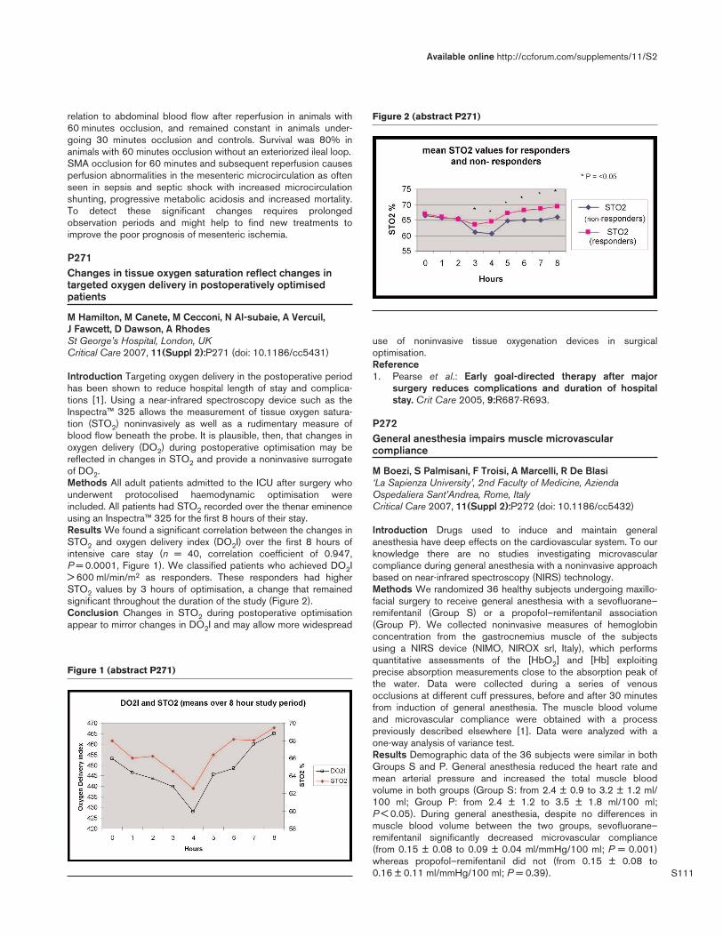
S111
relation to abdominal blood flow after reperfusion in animals with60 minutes occlusion, and remained constant in animals under-going 30 minutes occlusion and controls. Survival was 80% inanimals with 60 minutes occlusion without an exteriorized ileal loop.SMA occlusion for 60 minutes and subsequent reperfusion causesperfusion abnormalities in the mesenteric microcirculation as oftenseen in sepsis and septic shock with increased microcirculationshunting, progressive metabolic acidosis and increased mortality.To detect these significant changes requires prolongedobservation periods and might help to find new treatments toimprove the poor prognosis of mesenteric ischemia.
P271Changes in tissue oxygen saturation reflect changes intargeted oxygen delivery in postoperatively optimisedpatients
M Hamilton, M Canete, M Cecconi, N Al-subaie, A Vercuil, J Fawcett, D Dawson, A RhodesSt George’s Hospital, London, UKCritical Care 2007, 11(Suppl 2):P271 (doi: 10.1186/cc5431)
Introduction Targeting oxygen delivery in the postoperative periodhas been shown to reduce hospital length of stay and complica-tions [1]. Using a near-infrared spectroscopy device such as theInspectra™ 325 allows the measurement of tissue oxygen satura-tion (STO2) noninvasively as well as a rudimentary measure ofblood flow beneath the probe. It is plausible, then, that changes inoxygen delivery (DO2) during postoperative optimisation may bereflected in changes in STO2 and provide a noninvasive surrogateof DO2.Methods All adult patients admitted to the ICU after surgery whounderwent protocolised haemodynamic optimisation wereincluded. All patients had STO2 recorded over the thenar eminenceusing an Inspectra™ 325 for the first 8 hours of their stay.Results We found a significant correlation between the changes inSTO2 and oxygen delivery index (DO2I) over the first 8 hours ofintensive care stay (n = 40, correlation coefficient of 0.947,P = 0.0001, Figure 1). We classified patients who achieved DO2I> 600 ml/min/m2 as responders. These responders had higherSTO2 values by 3 hours of optimisation, a change that remainedsignificant throughout the duration of the study (Figure 2).Conclusion Changes in STO2 during postoperative optimisationappear to mirror changes in DO2I and may allow more widespread
use of noninvasive tissue oxygenation devices in surgicaloptimisation.Reference1. Pearse et al.: Early goal-directed therapy after major
surgery reduces complications and duration of hospitalstay. Crit Care 2005, 9:R687-R693.
P272General anesthesia impairs muscle microvascularcompliance
M Boezi, S Palmisani, F Troisi, A Marcelli, R De Blasi‘La Sapienza University’, 2nd Faculty of Medicine, AziendaOspedaliera Sant’Andrea, Rome, ItalyCritical Care 2007, 11(Suppl 2):P272 (doi: 10.1186/cc5432)
Introduction Drugs used to induce and maintain generalanesthesia have deep effects on the cardiovascular system. To ourknowledge there are no studies investigating microvascularcompliance during general anesthesia with a noninvasive approachbased on near-infrared spectroscopy (NIRS) technology.Methods We randomized 36 healthy subjects undergoing maxillo-facial surgery to receive general anesthesia with a sevofluorane–remifentanil (Group S) or a propofol–remifentanil association(Group P). We collected noninvasive measures of hemoglobinconcentration from the gastrocnemius muscle of the subjectsusing a NIRS device (NIMO, NIROX srl, Italy), which performsquantitative assessments of the [HbO2] and [Hb] exploitingprecise absorption measurements close to the absorption peak ofthe water. Data were collected during a series of venousocclusions at different cuff pressures, before and after 30 minutesfrom induction of general anesthesia. The muscle blood volumeand microvascular compliance were obtained with a processpreviously described elsewhere [1]. Data were analyzed with aone-way analysis of variance test.Results Demographic data of the 36 subjects were similar in bothGroups S and P. General anesthesia reduced the heart rate andmean arterial pressure and increased the total muscle bloodvolume in both groups (Group S: from 2.4 ± 0.9 to 3.2 ± 1.2 ml/100 ml; Group P: from 2.4 ± 1.2 to 3.5 ± 1.8 ml/100 ml;P < 0.05). During general anesthesia, despite no differences inmuscle blood volume between the two groups, sevofluorane–remifentanil significantly decreased microvascular compliance(from 0.15 ± 0.08 to 0.09 ± 0.04 ml/mmHg/100 ml; P = 0.001)whereas propofol–remifentanil did not (from 0.15 ± 0.08 to0.16 ± 0.11 ml/mmHg/100 ml; P = 0.39).
Available online http://ccforum.com/supplements/11/S2
Figure 1 (abstract P271)
Figure 2 (abstract P271)
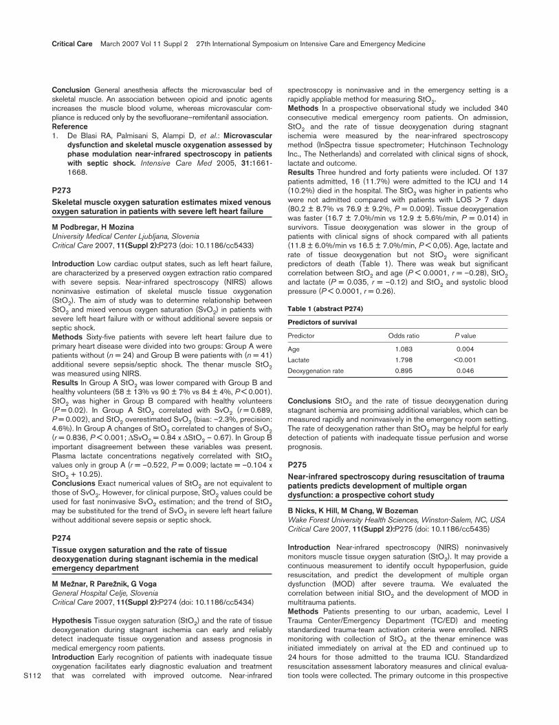
S112
Conclusion General anesthesia affects the microvascular bed ofskeletal muscle. An association between opioid and ipnotic agentsincreases the muscle blood volume, whereas microvascular com-pliance is reduced only by the sevofluorane–remifentanil association.Reference1. De Blasi RA, Palmisani S, Alampi D, et al.: Microvascular
dysfunction and skeletal muscle oxygenation assessed byphase modulation near-infrared spectroscopy in patientswith septic shock. Intensive Care Med 2005, 31:1661-1668.
P273Skeletal muscle oxygen saturation estimates mixed venousoxygen saturation in patients with severe left heart failure
M Podbregar, H MozinaUniversity Medical Center Ljubljana, SloveniaCritical Care 2007, 11(Suppl 2):P273 (doi: 10.1186/cc5433)
Introduction Low cardiac output states, such as left heart failure,are characterized by a preserved oxygen extraction ratio comparedwith severe sepsis. Near-infrared spectroscopy (NIRS) allowsnoninvasive estimation of skeletal muscle tissue oxygenation(StO2). The aim of study was to determine relationship betweenStO2 and mixed venous oxygen saturation (SvO2) in patients withsevere left heart failure with or without additional severe sepsis orseptic shock.Methods Sixty-five patients with severe left heart failure due toprimary heart disease were divided into two groups: Group A werepatients without (n = 24) and Group B were patients with (n = 41)additional severe sepsis/septic shock. The thenar muscle StO2was measured using NIRS.Results In Group A StO2 was lower compared with Group B andhealthy volunteers (58 ± 13% vs 90 ± 7% vs 84 ± 4%, P < 0.001).StO2 was higher in Group B compared with healthy volunteers(P = 0.02). In Group A StO2 correlated with SvO2 (r = 0.689,P = 0.002), and StO2 overestimated SvO2 (bias: –2.3%, precision:4.6%). In Group A changes of StO2 correlated to changes of SvO2(r = 0.836, P < 0.001; ∆SvO2 = 0.84 x ∆StO2 – 0.67). In Group Bimportant disagreement between these variables was present.Plasma lactate concentrations negatively correlated with StO2values only in group A (r = –0.522, P = 0.009; lactate = –0.104 xStO2 + 10.25).Conclusions Exact numerical values of StO2 are not equivalent tothose of SvO2. However, for clinical purpose, StO2 values could beused for fast noninvasive SvO2 estimation; and the trend of StO2may be substituted for the trend of SvO2 in severe left heart failurewithout additional severe sepsis or septic shock.
P274Tissue oxygen saturation and the rate of tissuedeoxygenation during stagnant ischemia in the medicalemergency department
M Meþþnar, R Pareþþnik, G VogaGeneral Hospital Celje, SloveniaCritical Care 2007, 11(Suppl 2):P274 (doi: 10.1186/cc5434)
Hypothesis Tissue oxygen saturation (StO2) and the rate of tissuedeoxygenation during stagnant ischemia can early and reliablydetect inadequate tissue oxygenation and assess prognosis inmedical emergency room patients.Introduction Early recognition of patients with inadequate tissueoxygenation facilitates early diagnostic evaluation and treatmentthat was correlated with improved outcome. Near-infrared
spectroscopy is noninvasive and in the emergency setting is arapidly appliable method for measuring StO2.Methods In a prospective observational study we included 340consecutive medical emergency room patients. On admission,StO2 and the rate of tissue deoxygenation during stagnantischemia were measured by the near-infrared spectroscopymethod (InSpectra tissue spectrometer; Hutchinson TechnologyInc., The Netherlands) and correlated with clinical signs of shock,lactate and outcome.Results Three hundred and forty patients were included. Of 137patients admitted, 16 (11.7%) were admitted to the ICU and 14(10.2%) died in the hospital. The StO2 was higher in patients whowere not admitted compared with patients with LOS > 7 days(80.2 ± 8.7% vs 76.9 ± 9.2%, P = 0.009). Tissue deoxygenationwas faster (16.7 ± 7.0%/min vs 12.9 ± 5.6%/min, P = 0.014) insurvivors. Tissue deoxygenation was slower in the group ofpatients with clinical signs of shock compared with all patients(11.8 ± 6.0%/min vs 16.5 ± 7.0%/min, P < 0,05). Age, lactate andrate of tissue deoxygenation but not StO2 were significantpredictors of death (Table 1). There was weak but significantcorrelation between StO2 and age (P < 0.0001, r = –0.28), StO2and lactate (P = 0.035, r = –0.12) and StO2 and systolic bloodpressure (P < 0.0001, r = 0.26).
Table 1 (abstract P274)
Predictors of survival
Predictor Odds ratio P value
Age 1.083 0.004
Lactate 1.798 <0.001
Deoxygenation rate 0.895 0.046
Conclusions StO2 and the rate of tissue deoxygenation duringstagnant ischemia are promising additional variables, which can bemeasured rapidly and noninvasively in the emergency room setting.The rate of deoxygenation rather than StO2 may be helpful for earlydetection of patients with inadequate tissue perfusion and worseprognosis.
P275Near-infrared spectroscopy during resuscitation of traumapatients predicts development of multiple organdysfunction: a prospective cohort study
B Nicks, K Hill, M Chang, W BozemanWake Forest University Health Sciences, Winston-Salem, NC, USACritical Care 2007, 11(Suppl 2):P275 (doi: 10.1186/cc5435)
Introduction Near-infrared spectroscopy (NIRS) noninvasivelymonitors muscle tissue oxygen saturation (StO2). It may provide acontinuous measurement to identify occult hypoperfusion, guideresuscitation, and predict the development of multiple organdysfunction (MOD) after severe trauma. We evaluated thecorrelation between initial StO2 and the development of MOD inmultitrauma patients.Methods Patients presenting to our urban, academic, Level ITrauma Center/Emergency Department (TC/ED) and meetingstandardized trauma-team activation criteria were enrolled. NIRSmonitoring with collection of StO2 at the thenar eminence wasinitiated immediately on arrival at the ED and continued up to24 hours for those admitted to the trauma ICU. Standardizedresuscitation assessment laboratory measures and clinical evalua-tion tools were collected. The primary outcome in this prospective
Critical Care March 2007 Vol 11 Suppl 2 27th International Symposium on Intensive Care and Emergency Medicine
S113
study was the association between StO2 and the development ofMOD within the first 24 hours based on a MOD score of 6 orgreater. Clinicians were blinded from the StO2 values.Results Over a 14-month period, 78 patients were enrolled. Of the78 patients, 26 (33.3%) developed MOD within the first 24 hours.The MOD patients had mean (SD) initial StO2 values of 53.3(±10.3), significantly lower than those of non-MOD patients (61.1(±10.0); P = 0.002). The MOD patient mean shock index of 0.92(±0.28) was also significantly higher than those of non-MODSpatients (0.73 (±0.19); P = 0.0007). Lactate values were notsignificantly different.Conclusions Noninvasive, continuous StO2 NIRS on initial arrivalin the TC/ED correlates with the shock index and with thedevelopment of MOD.
P276Buccal visible light spectroscopy and laser Dopplerflowmetry: reliability analysis
V Gath, Y Sakr, S Klinzing, T Simon, K Reinhart, G MarxFriedrich-Schiller-University Hospital, Jena, GermanyCritical Care 2007, 11(Suppl 2):P76 (doi: 10.1186/cc5436)
Background There are insatiable demands for new technologiesto advance basic biological investigation at the microcirculatorylevel. Oxygen to see (O2C)® is a newly developed system thatcombines laser Doppler flow (LDF) and visible light spectroscopy(VLS) technology. The aim of our study was to assess the reliabilityof O2C® measurements applied to the buccal mucosa and to thethenar eminence in healthy volunteers.Methods Microcirculatory hemoglobin oxygen saturation (µHbO2,%) and blood flow (flow, AU) were measured using an O2C® (LeaMedizintechnik GmbH, Giessen, Germany) probe applied to thebuccal mucosa and to the thenar eminence. Measurements wereobtained simultaneously at two depths, superficial (2 mm) anddeep (6 mm), every 2 seconds for 5 minutes and were recorded forlater analysis. The procedure was repeated on another occasion atleast 1 week apart.Results We studied 20 healthy subjects; 10 males and 10 females(mean age = 38 ± 18 years, range 21–74 years). Both µHbO2 andflow measurements were consistently higher when measured fromthe deep tissue layers (6 mm) than those measured from thesuperficial layers, regardless of the site of measurement. Buccalmucosal µHbO2 ranged from 78% to 96% and varied onlyminimally (CV: 4–7.5%), whereas there was a marked variability inflow measurements (CV: 29–63.9%). The reproducibility of buccalmucosal µHbO2 and flow measurements were moderate to good(that is, intra-individual reliability, ICC: range 0.7–0.87, P < 0.05).However, only measurements from the superficial mucosal layersshowed a moderate to good degree of inter-individual agreement(that is, inter-individual reliability, ICC: range 0.68–85, P < 0.001).LDF and VLS values measured on the thenar eminence were highlyvariable, were not reproducible, and the inter-individual agreementwas poor.Conclusion O2C® provides reliable measurement of buccalµHbO2 and microvascular flow. Skin measurements on the thenareminence are highly variable and unreliable.
P277Central venous pressure in a femoral access: a trueevaluation?
N Caramelo1, P Gonçalves1, A Paisana2, B Silva2, C Dias2, S Severino2, P Henriques2, D Marcelino2, D Marum2, D Fernandes2
1Hospital Júlio de Matos, Lisbon, Portugal; 2Hospital Curry Cabral,Lisbon, PortugalCritical Care 2007, 11(Suppl 2):P277 (doi: 10.1186/cc5437)
Introduction In patients with bad vascular access, the evaluationof central venous pressure (CVP) obtained in a femoral vein couldbe an alternative to the evaluation in central venous catheters(CVCs) located in internal jugular or subclavian veins.Objective To compare CVP measurement obtained in two differentlocations (jugular or subclavian veins and femoral veins).Setting A 16-bed medical–surgical ICU.Materials and methods This study began about 2 years ago, and theCVP of 41 patients in our ICU were evaluated and compared. Eachone of those 41 patients had a CVC in two different locations, oneplaced in the internal jugular or subclavian veins, and a second in afemoral vein. Simultaneous measurements of CVP were undertakenby two different operators, with a pressure transducer zero referencedat the mid-chest. Standard CVCs with similar features (20 cm length)were used. The patients with an intra-abdominal pressure (IAP)> 15 mmHg were excluded. The IAP was previously evaluated in allpatients, using the method described by Sugrue and Hillman. A linearcorrelation analysis was performed, considering significance P < 0.05and a correlation coefficient > 0.85.Results Forty-one patients were studied, and four patients wereexcluded due to an IAP > 15 mmHg. The mean age was 63.7 ±16.2 years, the ICU stay was 10.4 ± 3.5 days, the APACHE IIscore was 27.8 ± 6.7, and SAPS II was 55.8 ± 11.2. The meanCVP measured with jugular/subclavian access was 11.3 ± 4.5 mmHg,and in the femoral access was 11.8 ± 4.4 mmHg. The linear corre-lation between those measurements was 0.96, and P < 0.007.Conclusion The CVP can be accurately measured in a femoralvein, using standard CVC, in patients with an IAP < 15 mmHg.
P278Left ventricular volumes but not filling pressure aredeterminants of mortality in critically ill patients
D Sturgess, T Marwick, C Joyce, M Jones, B VenkateshUniversity of Queensland, Brisbane, AustraliaCritical Care 2007, 11(Suppl 2):P278 (doi: 10.1186/cc5438)
Introduction Transthoracic echocardiography (TTE) is gainingacceptance as a powerful diagnostic tool in critical illness. It canassess left ventricular (LV) volumes, as well as indices of ventri-cular filling pressure (including the ratio of mitral E velocity/mitralannular velocity [E/E′]). TTE evidence of raised filling pressure isassociated with mortality following myocardial infarction but itsprognostic value in critical illness is undefined. The aim of thisstudy was to evaluate the prognostic significance of echo-cardiographic LV volumes and filling pressure in the critically ill.Methods A consecutive group of 94 patients (66 males, mean ±SD age 61 ± 15 years) who had standard TTE supplemented bymeasurement of E/E′ in a tertiary referral ICU were enrolled. TTEwas performed 5 ± 6 days after ICU admission. Severity of criticalillness was assessed using APACHE III. Cox proportional hazardsregression analysis was based on 28-day mortality from the date ofecho with survivors censored on hospital discharge.Results The mean APACHE III score was 72 ± 25. Hospitalmortality was 33% (n = 31). Table 1 summarises correlates of 28-
Available online http://ccforum.com/supplements/11/S2
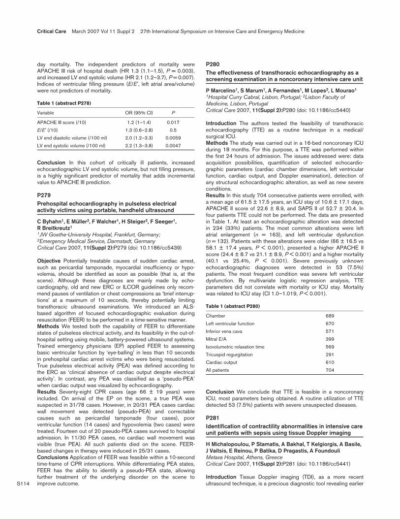
S114
day mortality. The independent predictors of mortality wereAPACHE III risk of hospital death (HR 1.3 (1.1–1.5), P = 0.003),and increased LV end systolic volume (HR 2.1 (1.2–3.7), P = 0.007).Indices of ventricular filling pressure (E/E′, left atrial area/volume)were not predictors of mortality.
Table 1 (abstract P278)
Variable OR (95% CI) P
APACHE III score (/10) 1.2 (1–1.4) 0.017
E/E′ (/10) 1.3 (0.6–2.8) 0.5
LV end diastolic volume (/100 ml) 2.0 (1.2–3.3) 0.0059
LV end systolic volume (/100 ml) 2.2 (1.3–3.8) 0.0047
Conclusion In this cohort of critically ill patients, increasedechocardiographic LV end systolic volume, but not filling pressure,is a highly significant predictor of mortality that adds incrementalvalue to APACHE III prediction.
P279Prehospital echocardiography in pulseless electricalactivity victims using portable, handheld ultrasound
C Byhahn1, E Müller2, F Walcher1, H Steiger2, F Seeger1, R Breitkreutz1
1JW Goethe-University Hospital, Frankfurt, Germany; 2Emergency Medical Service, Darmstadt, GermanyCritical Care 2007, 11(Suppl 2):P279 (doi: 10.1186/cc5439)
Objective Potentially treatable causes of sudden cardiac arrest,such as pericardial tamponade, myocardial insufficiency or hypo-volemia, should be identified as soon as possible (that is, at thescene). Although these diagnoses are mainly made by echo-cardiography, old and new ERC or ILCOR guidelines only recom-mend pauses of ventilation or chest compressions as ‘brief interrup-tions’ at a maximum of 10 seconds, thereby potentially limitingtransthoracic ultrasound examinations. We introduced an ALS-based algorithm of focused echocardiographic evaluation duringresuscitation (FEER) to be performed in a time-sensitive manner.Methods We tested both the capability of FEER to differentiatestates of pulseless electrical activity, and its feasibility in the out-of-hospital setting using mobile, battery-powered ultrasound systems.Trained emergency physicians (EP) applied FEER to assessingbasic ventricular function by ‘eye-balling’ in less than 10 secondsin prehospital cardiac arrest victims who were being resuscitated.True pulseless electrical activity (PEA) was defined according tothe ERC as ‘clinical absence of cardiac output despite electricalactivity’. In contrast, any PEA was classified as a ‘pseudo-PEA’when cardiac output was visualized by echocardiography.Results Seventy-eight CPR cases (age 66 ± 19 years) wereincluded. On arrival of the EP on the scene, a true PEA wassuspected in 31/78 cases. However, in 20/31 PEA cases cardiacwall movement was detected (pseudo-PEA) and correctablecauses such as pericardial tamponade (four cases), poorventricular function (14 cases) and hypovolemia (two cases) weretreated. Fourteen out of 20 pseudo-PEA cases survived to hospitaladmission. In 11/30 PEA cases, no cardiac wall movement wasvisible (true PEA). All such patients died on the scene. FEER-based changes in therapy were induced in 25/31 cases.Conclusions Application of FEER was feasible within a 10-secondtime-frame of CPR interruptions. While differentiating PEA states,FEER has the ability to identify a pseudo-PEA state, allowingfurther treatment of the underlying disorder on the scene toimprove outcome.
P280The effectiveness of transthoracic echocardiography as ascreening examination in a noncoronary intensive care unit
P Marcelino1, S Marum1, A Fernandes1, M Lopes2, L Mourao1
1Hospital Curry Cabral, Lisbon, Portugal; 2Lisbon Faculty ofMedicine, Lisbon, PortugalCritical Care 2007, 11(Suppl 2):P280 (doi: 10.1186/cc5440)
Introduction The authors tested the feasibility of transthoracicechocardiography (TTE) as a routine technique in a medical/surgical ICU.Methods The study was carried out in a 16-bed noncoronary ICUduring 18 months. For this purpose, a TTE was performed withinthe first 24 hours of admission. The issues addressed were: dataacquisition possibilities, quantification of selected echocardio-graphic parameters (cardiac chamber dimensions, left ventricularfunction, cardiac output, and Doppler examination), detection ofany structural echocardiographic alteration, as well as new severeconditions.Results In this study 704 consecutive patients were enrolled, witha mean age of 61.5 ± 17.5 years, an ICU stay of 10.6 ± 17.1 days,APACHE II score of 22.6 ± 8.9, and SAPS II of 52.7 ± 20.4. Infour patients TTE could not be performed. The data are presentedin Table 1. At least an echocardiographic alteration was detectedin 234 (33%) patients. The most common alterations were leftatrial enlargement (n = 163), and left ventricular dysfunction(n = 132). Patients with these alterations were older (66 ± 16.5 vs58.1 ± 17.4 years, P < 0.001), presented a higher APACHE IIscore (24.4 ± 8.7 vs 21.1 ± 8.9, P < 0.001) and a higher mortality(40.1 vs 25.4%, P < 0.001). Severe previously unknownechocardiographic diagnoses were detected in 53 (7.5%)patients. The most frequent condition was severe left ventriculardysfunction. By multivariate logistic regression analysis, TTEparameters did not correlate with mortality or ICU stay. Mortalitywas related to ICU stay (CI 1.0–1.019, P < 0.001).
Table 1 (abstract P280)
Chamber 689
Left ventricular function 670
Inferior vena cava 571
Mitral E/A 399
Isovolumetric relaxation time 569
Tricuspid regurgitation 291
Cardiac output 610
All patients 704
Conclusion We conclude that TTE is feasible in a noncoronaryICU, most parameters being obtained. A routine utilization of TTEdetected 53 (7.5%) patients with severe unsuspected diseases.
P281Identification of contractility abnormalities in intensive careunit patients with sepsis using tissue Doppler imaging
H Michalopoulou, P Stamatis, A Bakhal, T Kelgiorgis, A Basile,J Vaitsis, E Reinou, P Batika, D Pragastis, A FoundouliMetaxa Hospital, Athens, GreeceCritical Care 2007, 11(Suppl 2):P281 (doi: 10.1186/cc5441)
Introduction Tissue Doppler imaging (TDI), as a more recentultrasound technique, is a precious diagnostic tool revealing earlier
Critical Care March 2007 Vol 11 Suppl 2 27th International Symposium on Intensive Care and Emergency Medicine
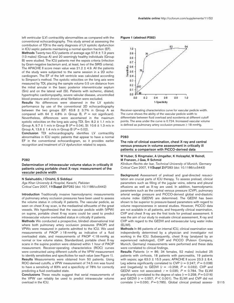
S115
left ventricular (LV) contractility abnormalities as compared with theconventional echocardiography. This study aimed at assessing thecontribution of TDI to the early diagnosis of LV systolic dysfunctionin ICU septic patients maintaining a normal ejection fraction (EF).Methods Twenty-two ICU patients of average age 57.6 ± 7.3 years(13 males) (Group A) and 20 seemingly healthy individuals (GroupB) were studied. The ICU patients met the sepsis criteria (infectionby Gram-negative bacterium and, at least, two of the SIRS criteria).The APACHE II score mean value was 21.2 ± 4.9. All the patientsof the study were subjected to the same session in a 2D echo-cardiogram. The EF of the left ventricle was calculated accordingto Simpson’s method. The systolic velocities on the long axis weremeasured by TDI, placing the sample volume 0.5 cm distance fromthe mitral annular in the basic posterior interventricular septum(Sm) and on the lateral wall (Sl). Patients with ischemic, dilated,hypertrophic cardiomyopathy, severe valvular disease, uncontrolledblood pressure and chronic atrial fibrillation were excluded.Results No differences were observed in the LV systolicperformance by use of the conventional 2D echocardiographybetween the two groups (EF: 63.8 ± 3.7% in Group A ascompared with 64 ± 4.8% in Group B, P = not significant).Nevertheless, differences were ascertained in the maximumsystolic velocities on the long axis using TDI. Sm: 8.2 ± 1.1 m/s inGroup A, 9.7 ± 1 m/s in Group B (P = 0.04), Sl: 10.6 ± 1.3 m/s inGroup A, 13.6 ± 1.4 m/s in Group B (P = 0.02).Conclusion TDI echocardiography identifies LV contractilityabnormalities in ICU septic patients that appear to have a normalEF in the conventional echocardiogram, so it provides earlierrecognition and treatment of LV dysfunction related to sepsis.
P282Determination of intravascular volume status in critically illpatients using portable chest X-rays: measurement of thevascular pedicle width
N Salahuddin, I Chishti, S SiddiquiAga Khan University & Hospital, Karachi, PakistanCritical Care 2007, 11(Suppl 2):P282 (doi: 10.1186/cc5442)
Introduction Traditionally invasive haemodynamic measurementsof pulmonary artery occlusion pressures have been used to assessthe volume status in critically ill patients. The vascular pedicle, asseen on chest X-ray scan, is the mediastinal silhouette of the greatvessels. We hypothesized that the vascular pedicle width (VPW)on supine, portable chest X-ray scans could be used to predictintravascular volume overloaded status in critically ill patients.Methods We conducted a prospective, blinded observational trialwhere both pulmonary artery occlusion pressures (PAOP) andVPWs were measured in patients admitted to the ICU. We usedmeasurements of PAOP ≥ 18 mmHg as indicative of a fluidoverloaded state, and measurements of PAOP < 18 mmHg asnormal or low volume states. Standardized, portable chest X-rayscans in the supine position were obtained within 1 hour of PAOPmeasurement. Receiver-operating characteristics (ROC) curveswere constructed using different cutoffs of the VPW measurementto identify sensitivities and specificities for each value (see Figure 1).Results Measurements were obtained from 50 patients. UsingROC-derived cutoffs, a VPW measurement of 74.5 mm was foundto have a sensitivity of 83% and a specificity of 78% for correctlypredicting a fluid overloaded state.Conclusions These results suggest that serial measurements ofthe VPW can reliably be used to predict intravascular volumeoverload in the ICU.
P283The role of clinical examination, chest X-ray and centralvenous pressure in volume assessment in critically illpatients: a comparison with PiCCO-derived data
W Huber, S Ringmaier, A Umgelter, K Holzapfel, W Reindl, M Franzen, J Gaa, R SchmidKlinikum Rechts der Isar, Technical University of Munich, GermanyCritical Care 2007, 11(Suppl 2):P283 (doi: 10.1186/cc5443)
Background Assessment of preload and goal-directed resusci-tation are crucial parts of ICU therapy. To assess preload, clinicalparameters such as filling of the jugular veins, edema and pleuraleffusions as well as X-ray are used. In addition, haemodynamicparameters such as the central venous pressure (CVP), pulmonaryarterial wedge pressure and PiCCO-derived global end-diastolicvolume index (GEDVI) are determined. The GEDVI has beenshown to be superior to pressure-based parameters with regard tovolume responsiveness in several studies. However, PiCCO dataare not available in all patients, and frequently clinical examination,CVP and chest X-ray are the first tools for preload assessment. Itwas the aim of our study to evaluate clinical assessment, X-ray andCVP with regard to the GEDVI and extravascular lung water index(ELWI).Methods In 86 patients of an internal ICU, clinical examination wasindependently determined by a physician and investigator notworking in the ICU. Subsequently, chest X-ray (analysed by anexperienced radiologist), CVP and PiCCO (Pulsion Company,Munich, Germany) measurements were performed and these datawere correlated to clinical findings.Results Patients (n = 86; 34 females, 52 males) included 25patients with cirrhosis, 18 patients with pancreatitis, 19 patientswith sepsis; age 63.0 ± 15.5 years; APACHE II score 23.3 ± 8.4.Leg edema significantly correlated to CVP (r = 0.247; P = 0.038)and (negatively) to GEDVI (r = –0.258; P = 0.032). CVP andGEDVI were not associated: r = 0.035; P = 0.784. The ELWIsignificantly correlated to the degree of rales (r = 0.258; P = 0.016)and GEDVI (r = 0.557; P < 0.001). The ELWI and CVP did notcorrelate (r = 0.030; P = 0.785). Global clinical preload assess-
Available online http://ccforum.com/supplements/11/S2
Figure 1 (abstract P282)
Receiver-operating characteristics curve for vascular pedicle width.The curve shows the ability of the vascular pedicle width todifferentiate between fluid overload and euvolemia at different cutoffpoints. The area under the curve is 0.724. Increased vascular volumeis defined as pulmonary artery occlusion pressure ≥ 18 mmHg.
S116
ment (scale 1–10) was not predictive for GEDVI. Radiologicalassessment significantly overestimated the GEDVI (901.41 ± 139.76vs 782.56 ± 183.80 ml/m2; P < 0.001) and underestimated theELWI (7.22 ± 1.38 vs 9.77 ± 4.51 ml/kg; P < 0.001).Conclusions (1) Leg edema and increased CVP do not excludepreload deficiency determined by the GEDVI, which wasoverestimated by X-ray. (2) CVP and leg edema are poorpredictors of the ELWI, which was significantly associated withaudible rales but underestimated by X-ray.
P284Clinicians’ prediction of advanced cardiopulmonaryvariables in critically ill patients: a multicenter study
A Perel1, M Maggiorini2, M Malbrain3, J Teboul4, J Belda5, E Fernández Mondéjar6, M Kirov7, J Wendon8
1Sheba Medical Center, Tel Hashomer, Israel; 2Universitatsspital,Zurich, Switzerland; 3Ziekenhuis Netwerk Antwerpen-HospitalCampus Stuivenberg, Antwerp, Belgium; 4Hôpital De Bicêtre, LeKremlin-Bicêtre, France; 5Hospital Clinico Universitario, Valencia,Spain; 6Hospital Universitario Virgen De Las Nieves, Granada,Spain; 7Northern State Medical University, Arkhangelsk, RussianFederation; 8King’s College Hospital, London, UKCritical Care 2007, 11(Suppl 2):P284 (doi: 10.1186/cc5444)
Introduction Lack of evidence that more advanced monitoringtechniques improve outcome may have led to insufficientmonitoring of critically ill patients. However, clinical judgment andconventional hemodynamic monitoring alone were shown to beinadequate for a reliable estimate of hemodynamic status. We havetherefore compared clinicians’ prediction of advanced cardio-pulmonary parameters with actual measurements.Methods Cardiopulmonary assessment was done in critically illpatients from 12 European ICUs just before the use of the PiCCOmonitor (Pulsion, Germany). Independent prediction of cardiacoutput (CO), systemic vascular resistance (SVR), indexed globalend-diastolic volume (GEDVi), stroke volume variation (SVV), andindexed extravascular lung water (EVLWi) was done by one to fourphysicians per patient. Following the first set of PiCCOmeasurements each physician self-rated the accuracy of his pre-PiCCO predictions.Results A total of 257 questionnaires of 165 patients (67 femalesand 98 males, age 59.8 ± 16.7 (range 16–93) years) werecompleted by 135 residents and 122 specialists. The mainreasons for using the PiCCO included unclear fluid status (109cases), sepsis/septic shock (70 cases), respiratory failure (42cases), cardiogenic shock (19 cases), renal failure (27 cases), andother (18 cases). Only 30–50% of the predicted values werecorrect (±20% of measured values) (Table 1). Ranges of errorswere: CO (–77/+100%), SVR (–94/+303%), GEDVi(–88/+135%), SVV (–91/+367%), EVLWi (–76/+650%). Therewas a significant underestimation of CO (P < 0.00001) andGEDVi (P < 0.0003), and overestimation of SVR (P < 0.003) andSVV (P < 0.0002).The 240 self-ratings (scale of 1–5, [1] = excellent; [5] = poor) ofpredictions accuracy included – [1] 1.6%, [2] 40%, [3] 38.8%, [4]
15.1%, [5] 4.5%. The mean self-rate was 2.8 ± 0.9, with that ofresidents (2.8 ± 0.9, n = 129) being similar to that of specialists(2.7 ± 0.8, n = 111), P < 0.31.Conclusions The ability of physicians to predict advancedcardiopulmonary parameters based on clinical evaluation andconventional monitoring alone has considerable limitations and isnot improved by experience.Acknowledgement The authors of this unsupported study aremembers of Pulsion’s medical advisory board.
P285Change of therapeutic plan following advancedcardiopulmonary monitoring in critically ill patients: amulticenter study
A Perel1, M Maggiorini2, M Malbrain3, J Teboul4, J Belda5, E Fernández Mondéjar6, M Kirov7, J Wendon8
1Sheba Medical Center, Tel Hashomer, Israel; 2Universitatsspital,Zurich, Switzerland; 3Ziekenhuisnetwerk Antwerpen, CampusStuivenberg, Antwerp, Belgium; 4Hopital De Bicetre, Le KremlinBicetre, France; 5Hospital Clinico Universitario, Valencia, Spain;6Hospital Universitario Virgen de las Nieves, Granada, Spain;7Northern State Medical University, Arkhangelsk, RussianFederation; 8Liver Intensive Care Unit, London, UKCritical Care 2007, 11(Suppl 2):P285 (doi: 10.1186/cc5445)
Introduction Many therapeutic decisions are made in the ICU onthe basis of clinical judgment and conventional monitoring alone,although these may be inadequate for a reliable estimate ofhemodynamic status. We therefore measured the effects of moreadvanced cardiopulmonary parameters (ACP) on major therapeuticdecisions.Methods Cardiopulmonary assessment was done in critically illpatients from 12 European ICUs independently by one to fourphysicians per patient just before the use of the PiCCO monitor(Pulsion, Germany). Following cardiopulmonary evaluation andprediction of ACP (reported elsewhere), each physician suggesteda therapeutic plan before and after the first set of PiCCO measure-ments (cardiac output, systemic vascular resistance, global end-diastolic volume, stroke volume variation, and extravascular lungwater), and then self-rated the accuracy of his original therapeuticplan.Results A total of 257 questionnaires of 165 patients (67 femalesand 98 males, age ± SD 59.8 ± 16.7 (range 16–93) years) werecompleted by 135 residents and 122 specialists. The mainreasons for using the PiCCO included unclear fluid status (109cases), sepsis/septic shock (70 cases), respiratory failure (42cases), cardiogenic shock (19 cases), renal failure (27 cases),other (18 cases). Changes (plan-and-not-give and no-plan-and-give) made in the pre-PiCCO therapeutic plans included: fluid,31.8%; inotropes, 23.3%; vasoconstrictors, 23.5%; diuretics, 15%(Table 1). The 240 overall self-ratings of the original plan (scale of1–5, [1] = not different; [5] = very different) included – [1] 33.7%,[2] 23.5%, [3] 25.9%, [4] 11.5%, [5] 5.3%. The mean self-ratewas 2.3 ± 1.2, with that of residents (2.3 ± 1.3, n = 129) beingsimilar to that of specialists (2.3 ± 1.1, n = 111), P < 0.88.
Critical Care March 2007 Vol 11 Suppl 2 27th International Symposium on Intensive Care and Emergency Medicine
Table 1 (abstract P284)
EVLWi % CO % SVR % GEDVi % SVV %
Under 69 29.4 127* 49.6 46 18.1 72 28 85 39.7
Correct 93 39.6 99 38.6 90 35.4 93 50.2 64 29.9
Over 73 31.1 30 11.7 116 45.7 73 21 65 30.4
*Number of predictions.
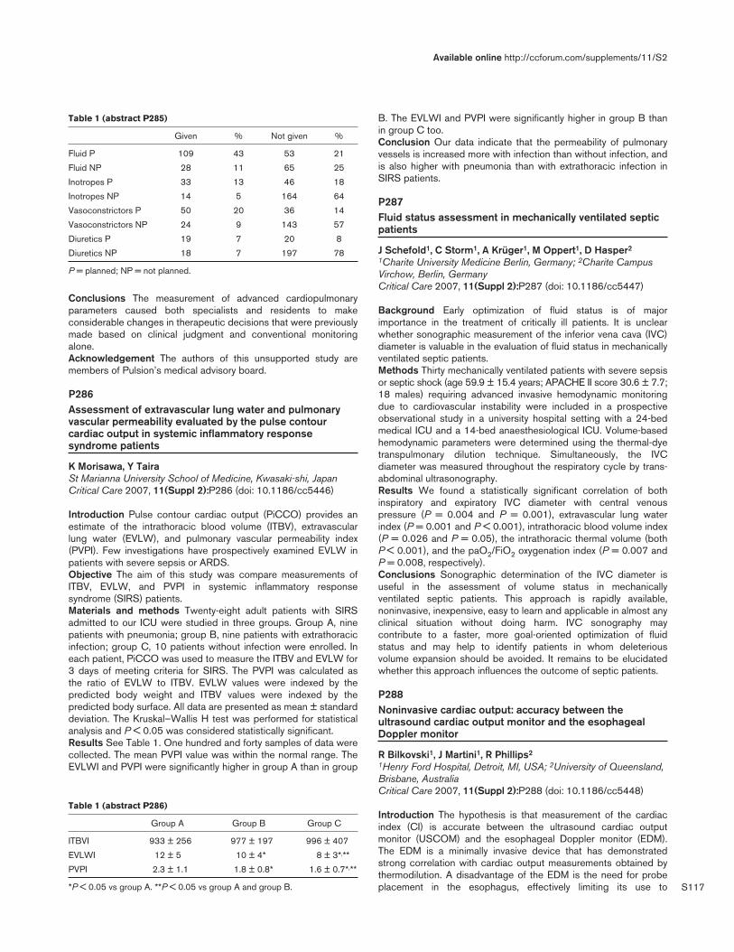
S117
Conclusions The measurement of advanced cardiopulmonaryparameters caused both specialists and residents to makeconsiderable changes in therapeutic decisions that were previouslymade based on clinical judgment and conventional monitoringalone.Acknowledgement The authors of this unsupported study aremembers of Pulsion’s medical advisory board.
P286Assessment of extravascular lung water and pulmonaryvascular permeability evaluated by the pulse contourcardiac output in systemic inflammatory responsesyndrome patients
K Morisawa, Y TairaSt Marianna University School of Medicine, Kwasaki-shi, JapanCritical Care 2007, 11(Suppl 2):P286 (doi: 10.1186/cc5446)
Introduction Pulse contour cardiac output (PiCCO) provides anestimate of the intrathoracic blood volume (ITBV), extravascularlung water (EVLW), and pulmonary vascular permeability index(PVPI). Few investigations have prospectively examined EVLW inpatients with severe sepsis or ARDS.Objective The aim of this study was compare measurements ofITBV, EVLW, and PVPI in systemic inflammatory responsesyndrome (SIRS) patients.Materials and methods Twenty-eight adult patients with SIRSadmitted to our ICU were studied in three groups. Group A, ninepatients with pneumonia; group B, nine patients with extrathoracicinfection; group C, 10 patients without infection were enrolled. Ineach patient, PiCCO was used to measure the ITBV and EVLW for3 days of meeting criteria for SIRS. The PVPI was calculated asthe ratio of EVLW to ITBV. EVLW values were indexed by thepredicted body weight and ITBV values were indexed by thepredicted body surface. All data are presented as mean ± standarddeviation. The Kruskal–Wallis H test was performed for statisticalanalysis and P < 0.05 was considered statistically significant.Results See Table 1. One hundred and forty samples of data werecollected. The mean PVPI value was within the normal range. TheEVLWI and PVPI were significantly higher in group A than in group
B. The EVLWI and PVPI were significantly higher in group B thanin group C too.Conclusion Our data indicate that the permeability of pulmonaryvessels is increased more with infection than without infection, andis also higher with pneumonia than with extrathoracic infection inSIRS patients.
P287Fluid status assessment in mechanically ventilated septicpatients
J Schefold1, C Storm1, A Krüger1, M Oppert1, D Hasper2
1Charite University Medicine Berlin, Germany; 2Charite CampusVirchow, Berlin, GermanyCritical Care 2007, 11(Suppl 2):P287 (doi: 10.1186/cc5447)
Background Early optimization of fluid status is of majorimportance in the treatment of critically ill patients. It is unclearwhether sonographic measurement of the inferior vena cava (IVC)diameter is valuable in the evaluation of fluid status in mechanicallyventilated septic patients.Methods Thirty mechanically ventilated patients with severe sepsisor septic shock (age 59.9 ± 15.4 years; APACHE II score 30.6 ± 7.7;18 males) requiring advanced invasive hemodynamic monitoringdue to cardiovascular instability were included in a prospectiveobservational study in a university hospital setting with a 24-bedmedical ICU and a 14-bed anaesthesiological ICU. Volume-basedhemodynamic parameters were determined using the thermal-dyetranspulmonary dilution technique. Simultaneously, the IVCdiameter was measured throughout the respiratory cycle by trans-abdominal ultrasonography.Results We found a statistically significant correlation of bothinspiratory and expiratory IVC diameter with central venouspressure (P = 0.004 and P = 0.001), extravascular lung waterindex (P = 0.001 and P < 0.001), intrathoracic blood volume index(P = 0.026 and P = 0.05), the intrathoracic thermal volume (bothP < 0.001), and the paO2/FiO2 oxygenation index (P = 0.007 andP = 0.008, respectively).Conclusions Sonographic determination of the IVC diameter isuseful in the assessment of volume status in mechanicallyventilated septic patients. This approach is rapidly available,noninvasive, inexpensive, easy to learn and applicable in almost anyclinical situation without doing harm. IVC sonography maycontribute to a faster, more goal-oriented optimization of fluidstatus and may help to identify patients in whom deleteriousvolume expansion should be avoided. It remains to be elucidatedwhether this approach influences the outcome of septic patients.
P288Noninvasive cardiac output: accuracy between theultrasound cardiac output monitor and the esophagealDoppler monitor
R Bilkovski1, J Martini1, R Phillips2
1Henry Ford Hospital, Detroit, MI, USA; 2University of Queensland,Brisbane, AustraliaCritical Care 2007, 11(Suppl 2):P288 (doi: 10.1186/cc5448)
Introduction The hypothesis is that measurement of the cardiacindex (CI) is accurate between the ultrasound cardiac outputmonitor (USCOM) and the esophageal Doppler monitor (EDM).The EDM is a minimally invasive device that has demonstratedstrong correlation with cardiac output measurements obtained bythermodilution. A disadvantage of the EDM is the need for probeplacement in the esophagus, effectively limiting its use to
Available online http://ccforum.com/supplements/11/S2
Table 1 (abstract P285)
Given % Not given %
Fluid P 109 43 53 21
Fluid NP 28 11 65 25
Inotropes P 33 13 46 18
Inotropes NP 14 5 164 64
Vasoconstrictors P 50 20 36 14
Vasoconstrictors NP 24 9 143 57
Diuretics P 19 7 20 8
Diuretics NP 18 7 197 78
P = planned; NP = not planned.
Table 1 (abstract P286)
Group A Group B Group C
ITBVI 933 ± 256 977 ± 197 996 ± 407
EVLWI 12 ± 5 10 ± 4* 8 ± 3*,**
PVPI 2.3 ± 1.1 1.8 ± 0.8* 1.6 ± 0.7*,**
*P < 0.05 vs group A. **P < 0.05 vs group A and group B.
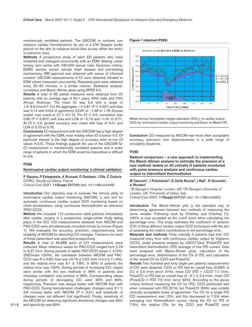
S118
mechanically ventilated patients. The USCOM, in contrast, canmeasure cardiac hemodynamics by use of a CW Doppler probeplaced on the skin to measure blood flow across either the aorticor pulmonic valve.Methods A prospective study of adult ED patients who wereintubated and managed concurrently with an EDM. Setting: urbantertiary care center with >90,000 annual visits. Exclusion criteria:ESRD, ascites, known valvular heart disease and pre-existingtracheotomy. IRB approval was obtained with waiver of informedconsent. USCOM measurements of CI were obtained, blinded toEDM values measured concurrently. Repeated pairs were obtainedevery 30–60 minutes, in a similar manner. Statistical analysis:correlation and Bland–Altman plots using SPSS 9.0.Results A total of 95 paired measures were obtained from 20patients with an average age of 60.1 years, 60% male and 70%African American. The mean CI was 5.2 with a range of1.4–6.6 l/min/m2. For the aggregate, r = 0.81 (P < 0.001) and biaswas 0.14 with limits of agreement (LOA) of –1.48 to 1.76. Excessscatter was noted at CI > 4.0 CI. For CI < 4.0, correlation was0.80 (P < 0.001) with bias and LOA of –0.15 and –1.01 to 0.71.At CI < 2.5, greater accuracy was noted with bias of 0.01 andLOA of 0.73 to 0.75.Conclusions CI measurement with the USCOM has a high degreeof agreement with the EDM, most notably when CI is below 4.0. Ofparticular interest is the high degree of accuracy seen at low CIvalues (<2.5). These findings support the use of the USCOM forCI measurement in mechanically ventilated patients and a widerrange of patients in which the EDM would be impractical or difficultto use.
P289Noninvasive cardiac output monitoring: a clinical validation
P Squara, P Estagnasie, A Brusset, D Denjean, J Dib, C DuboisCERIC, Neuilly-sur-Seine, FranceCritical Care 2007, 11(Suppl 2):P289 (doi: 10.1186/cc5449)
Introduction Our objective was to evaluate the clinical utility ofnoninvasive cardiac output monitoring (NICOM), a new tool forautomatic continuous cardiac output (CO) monitoring based onchest bioreactance, using continuous thermodilution as reference(PAC-CCO).Method We included 110 consecutive adult patients immediatelyafter cardiac surgery in a prospective, single-center study takingplace in the ICU. CO measurements obtained from NICOM andPAC-CCO were simultaneously recorded minute by minute (Figure1). We evaluated the accuracy, precision, responsiveness, andreliability of NICOM for detecting CO changes. Tolerance for eachof these parameters was specified prospectively.Results A total of 65,888 pairs of CO measurements werecollected. Mean reference values for PAC-CCO ranged from 2.79to 9.27 l/min. During periods of stable PAC-CCO (slope < ±10%,2SD/mean <20%), the correlation between NICOM and PAC-CCO was R = 0.82; bias was +0.16 ± 0.52 l/min (+4.0 ± 11.3%),and the relative error was 9.1 ± 7.8%. In 85% of patients therelative error was <20%. During periods of increasing CO, slopeswere similar with the two methods in 96% of patients andintraclass correlation was positive in 96%. Corresponding valuesduring periods of decreasing CO were 90% and 84%,respectively. Precision was always better with NICOM than withPAC-CCO. During hemodynamic challenges, changes were 3.1 ±3.8 minutes faster with NICOM (P < 0.01) and amplitude ofchanges were not different (not significant). Finally, sensitivity ofthe NICOM for detecting significant directional changes was 93%and specificity was 93%.
Conclusion CO measured by NICOM had most often acceptableaccuracy, precision, and responsiveness in a wide range ofcirculatory situations.
P290Method comparison – a new approach to implementingthe Bland–Altman analysis to estimate the precision of anew method: tested on 30 critically ill patients monitoredwith pulse pressure analysis and continuous cardiacoutput vs intermittent thermodilution
M Cecconi1, J Poloniecki2, G Della Rocca3, J Ball1, R Grounds1,A Rhodes1
1St George’s Hospital, London, UK; 2St George’s University ofLondon, UK; 3University of Udine, ItalyCritical Care 2007, 11(Suppl 2):P290 (doi: 10.1186/cc5450)
Introduction The Bland–Altman plot is the standard way ofdetermining agreement between two methods of measuring thesame variable. Following work by Critchley and Critchley [1],±30% is now accepted as the cutoff point when calculating thepercentage error. This study estimates the coefficient of variation(CV) of three different cardiac output (CO) techniques with the aimof assessing the relative contributions to the percentage error.Materials and methods Thirty critically ill patients had their COmeasured every hour with continuous cardiac output by Vigilance(CCO), pulse pressure analysis by LiDCO™plus (PulseCO) andintermittent thermodilution (ITD) (average of four ITD curves). Datawere analysed with Bland–Altman plots, calculation of thepercentage error, determination of the CV of ITD, and calculationof the overall CV for CCO and PulseCO.Results Two hundred and forty (eight per patient) measurementsof CO were obtained. CCO vs ITD had an overall bias (±2SD) of0.2 ± 2.4 l/min (error 31%), mean CO (ITD + CCO) 7.7 l/min.PulseCO vs ITD had an overall bias of –0.1 ± 2.4 l/min, mean CO(PulseCO + ITD) 7.5 l/min (error 33%). According to the abovecriteria (without measuring the CV for ITD), CCO performed wellwhen compared with ITD (31%) but PulseCO (33%) was outsideclinically acceptable levels of agreement. The CV for a single ITDCO measurement was 15%, and this decreased to 7.5% whenaveraging four thermodilution curves. Using the CV for ITD of7.5%, the relative CVs for the CCO and PulseCO were
Critical Care March 2007 Vol 11 Suppl 2 27th International Symposium on Intensive Care and Emergency Medicine
Figure 1 (abstract P289)
Mixed venous hemoglobin oxygen saturation (SvO2) vs cardiac output(CO) by noninvasive cardiac output monitoring and Swan on Mister197.
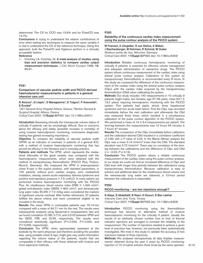
S119
determined. The CV for CCO was 13.6% and for PulseCO was14.7%.Conclusions In trying to understand the relative contributions oferror when testing two techniques to measure the same variable itis vital to understand the CV of the reference technique. Using thisapproach, both the PulseCO and Vigilance perform in a clinicallyacceptable fashion.Reference1. Critchley LA, Critchley JA: A meta-analysis of studies using
bias and precision statistics to compare cardiac outputmeasurement techniques. J Clin Monit Comput 1999, 15:85-91.
P291Comparison of vascular pedicle width and PiCCO-derivedhaemodynamic measurements in patients in a generalintensive care unit
S Aloizos1, G Liapis1, D Maragiannis1, E Tsigou2, P Aravosita2,E Evodia2
1401 General Army Hospital Athens, Greece; 2Mother General &Surgical Hospital, Athens, GreeceCritical Care 2007, 11(Suppl 2):P291 (doi: 10.1186/cc5451)
Introduction Assessing clinically the intravascular volume status ofcritically ill patients can be exceedingly difficult. Due to concernsabout the efficacy and safety (possible increase in mortality) ofusing invasive haemodynamic monitoring, noninvasive diagnostictesting has gained increasing importance.Objective To compare the reliability of vascular pedicle width(VPW) as an indicator of overload, in patients of a general ICU,with a method of invasive haemodynamic monitoring that hasproved its efficacy in the literature and in everyday practice.Patients and methods The VPW, which represents the media-stinal silhouette of the great vessels, was compared with thehaemodynamic measurements, which were obtained with themethod of transpulmonary thermodilution (PiCCO Plus; Pulsion,Munich, Germany). We measured the VPW in anteroposteriorchest X-rays in the supine position, with standard parameters, in100 patients without prior cardiac surgery, prior mediastinalirradiation, obesity, severe acute respiratory distress syndrome andpositive end-expiratory pressure > 7.5 cmH2O. In every patient weperformed invasive haemodynamic monitoring with the PiCCOPlus. An intrathoracic blood volume index (ITBI) > 1,000 ml/m2,global end-diastolic index (GEDI) > 800 ml/m2, and extravascularlung water index (ELWI) > 7.0 ml/kg were considered the markersof significant volume overload. After further refinement, 27 patientsfulfilled the above criteria and were considered eligible to beincluded in the study.Results The mean VPW in overloaded patients was 75.14 mmcompared with a mean of 64.71 mm for the rest. The results weresubsequently analyzed using Spearman’s nonparametric test andwe found correlation (0.785, 0.710, and 0.510) between VPW andthe GEDI, ITBI, and ELWI, respectively. The results wereconsidered statistically significant (P < 0.000, P < 0.000, andP < 0.005, respectively).Conclusion The VPW, when appropriately assessed at thebedside by the same physician and therefore avoiding the possiblebias, using portable chest X-rays, might give very useful informationregarding the volume status of the patients, results that arecomparable in their efficacy with those obtained with invasive andmore expensive methods.
P292Reliability of the continuous cardiac index measurementusing the pulse contour analysis of the PiCCO system
M Franzen, A Umgelter, S von Delius, A Weber, J Reichenberger, M Dohmen, R Schmid, W HuberKlinikum rechts der Isar, München, GermanyCritical Care 2007, 11(Suppl 2):P292 (doi: 10.1186/cc5452)
Introduction Reliable continuous hemodynamic monitoring ofcritically ill patients is essential for effective volume managementand adequate administration of vasoactive drugs. The PiCCOsystem allows continuous measurement of the cardiac index usingarterial pulse contour analysis. Calibration of this system bytranspulmonary thermodilution is recommended every 8 hours. Inthis study we compared the difference of the continuous measure-ment of the cardiac index using the arterial pulse contour analysis(CIpc) with the cardiac index acquired by the transpulmonarythermodilution (CItd) when calibrating the system.Methods Our study includes 140 measurements in 10 critically illpatients (eight males, two females, age 37–84 years, mean 64.1 ±13.0 years) requiring hemodynamic monitoring with the PiCCOsystem. Five patients had septic shock, three hepatorenalsyndrome and two acute heart failure. First the CIpc was recordedimmediately before the next calibration and afterwards the CItdwas measured three times, which resulted in a simultaneouscalibration of the pulse contour algorithm of the PiCCO system.We performed a mean of 14 ± 9.4 measurements per patient. Thetime-lag between the measurements was 12 hours 54 minutes ±7 hours 47 minutes.Results The comparison of the CIpc immediately before calibrationand the calibration-derived CItd resulted in a correlation coefficientof 0.84 with a P value of 0.02. In the Bland–Altman analysis theCIpc was a mean 0.14 l/min/m2 lower than the CItd. The standarddeviation was 0.72 l/min/m2. There was no correlation of the time-lag between the calibrations and the difference of CIpc and CItd(r = –0.03; P = 0.13).Conclusion The PiCCO system allows a reliable continuousmeasurement of the cardiac index using the pulse contour analysis.In our study we could not find an increased difference of CIpc andCItd even with longer time periods between the calibrations usingtranspulmonary thermodilution. Because calibration is easy toachieve and additional data for the intrathoracic blood volume andthe extravascular lung water are obtained, a 12-hour periodbetween the calibrations is reasonable.
P293PiCCO monitoring – are two injections enough?
S Alaya, S Abdellatif, R Nasri, H Ksouri, S Ben LakhalIntensive Care Unit, Tunis, TunisiaCritical Care 2007, 11(Suppl 2):P293 (doi: 10.1186/cc5453)
Introduction PiCCO monitoring using the thermodilutiontechnique has become an alternative method of invasivehaemodynamic monitoring for the critically ill patient. Usually theresults of an arbitrarily chosen number (one to five) of thermalindicator injections are averaged to increase the reliability of themeasurement. The number of injections needed to achieve a givenlevel of precision has, however, not previously been systematicallyinvestigated. We tried in this study to validate the accuracy of twoinjections instead of three injections.Methods We analysed retrospectively all data (triplate measure-ments) obtained during the past 2 years by PiCCO monitoring:injection of 10 ml saline solution three times by the same operator.
Available online http://ccforum.com/supplements/11/S2
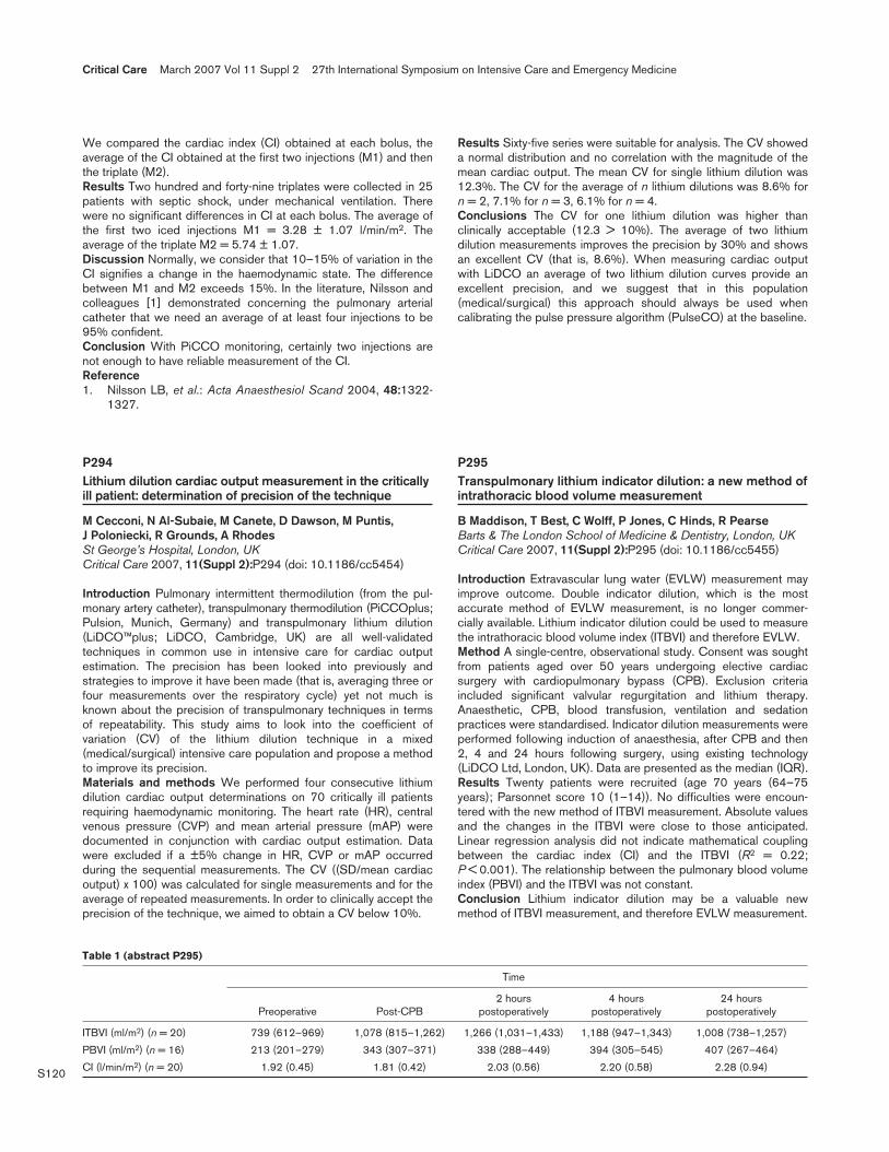
S120
We compared the cardiac index (CI) obtained at each bolus, theaverage of the CI obtained at the first two injections (M1) and thenthe triplate (M2).Results Two hundred and forty-nine triplates were collected in 25patients with septic shock, under mechanical ventilation. Therewere no significant differences in CI at each bolus. The average ofthe first two iced injections M1 = 3.28 ± 1.07 l/min/m2. Theaverage of the triplate M2 = 5.74 ± 1.07.Discussion Normally, we consider that 10–15% of variation in theCI signifies a change in the haemodynamic state. The differencebetween M1 and M2 exceeds 15%. In the literature, Nilsson andcolleagues [1] demonstrated concerning the pulmonary arterialcatheter that we need an average of at least four injections to be95% confident.Conclusion With PiCCO monitoring, certainly two injections arenot enough to have reliable measurement of the CI.Reference1. Nilsson LB, et al.: Acta Anaesthesiol Scand 2004, 48:1322-
1327.
P294Lithium dilution cardiac output measurement in the criticallyill patient: determination of precision of the technique
M Cecconi, N Al-Subaie, M Canete, D Dawson, M Puntis, J Poloniecki, R Grounds, A RhodesSt George’s Hospital, London, UKCritical Care 2007, 11(Suppl 2):P294 (doi: 10.1186/cc5454)
Introduction Pulmonary intermittent thermodilution (from the pul-monary artery catheter), transpulmonary thermodilution (PiCCOplus;Pulsion, Munich, Germany) and transpulmonary lithium dilution(LiDCO™plus; LiDCO, Cambridge, UK) are all well-validatedtechniques in common use in intensive care for cardiac outputestimation. The precision has been looked into previously andstrategies to improve it have been made (that is, averaging three orfour measurements over the respiratory cycle) yet not much isknown about the precision of transpulmonary techniques in termsof repeatability. This study aims to look into the coefficient ofvariation (CV) of the lithium dilution technique in a mixed(medical/surgical) intensive care population and propose a methodto improve its precision.Materials and methods We performed four consecutive lithiumdilution cardiac output determinations on 70 critically ill patientsrequiring haemodynamic monitoring. The heart rate (HR), centralvenous pressure (CVP) and mean arterial pressure (mAP) weredocumented in conjunction with cardiac output estimation. Datawere excluded if a ±5% change in HR, CVP or mAP occurredduring the sequential measurements. The CV ((SD/mean cardiacoutput) x 100) was calculated for single measurements and for theaverage of repeated measurements. In order to clinically accept theprecision of the technique, we aimed to obtain a CV below 10%.
Results Sixty-five series were suitable for analysis. The CV showeda normal distribution and no correlation with the magnitude of themean cardiac output. The mean CV for single lithium dilution was12.3%. The CV for the average of n lithium dilutions was 8.6% forn = 2, 7.1% for n = 3, 6.1% for n = 4.Conclusions The CV for one lithium dilution was higher thanclinically acceptable (12.3 > 10%). The average of two lithiumdilution measurements improves the precision by 30% and showsan excellent CV (that is, 8.6%). When measuring cardiac outputwith LiDCO an average of two lithium dilution curves provide anexcellent precision, and we suggest that in this population(medical/surgical) this approach should always be used whencalibrating the pulse pressure algorithm (PulseCO) at the baseline.
P295Transpulmonary lithium indicator dilution: a new method ofintrathoracic blood volume measurement
B Maddison, T Best, C Wolff, P Jones, C Hinds, R PearseBarts & The London School of Medicine & Dentistry, London, UKCritical Care 2007, 11(Suppl 2):P295 (doi: 10.1186/cc5455)
Introduction Extravascular lung water (EVLW) measurement mayimprove outcome. Double indicator dilution, which is the mostaccurate method of EVLW measurement, is no longer commer-cially available. Lithium indicator dilution could be used to measurethe intrathoracic blood volume index (ITBVI) and therefore EVLW.Method A single-centre, observational study. Consent was soughtfrom patients aged over 50 years undergoing elective cardiacsurgery with cardiopulmonary bypass (CPB). Exclusion criteriaincluded significant valvular regurgitation and lithium therapy.Anaesthetic, CPB, blood transfusion, ventilation and sedationpractices were standardised. Indicator dilution measurements wereperformed following induction of anaesthesia, after CPB and then2, 4 and 24 hours following surgery, using existing technology(LiDCO Ltd, London, UK). Data are presented as the median (IQR).Results Twenty patients were recruited (age 70 years (64–75years); Parsonnet score 10 (1–14)). No difficulties were encoun-tered with the new method of ITBVI measurement. Absolute valuesand the changes in the ITBVI were close to those anticipated.Linear regression analysis did not indicate mathematical couplingbetween the cardiac index (CI) and the ITBVI (R2 = 0.22;P < 0.001). The relationship between the pulmonary blood volumeindex (PBVI) and the ITBVI was not constant.Conclusion Lithium indicator dilution may be a valuable newmethod of ITBVI measurement, and therefore EVLW measurement.
Critical Care March 2007 Vol 11 Suppl 2 27th International Symposium on Intensive Care and Emergency Medicine
Table 1 (abstract P295)
Time
2 hours 4 hours 24 hours Preoperative Post-CPB postoperatively postoperatively postoperatively
ITBVI (ml/m2) (n = 20) 739 (612–969) 1,078 (815–1,262) 1,266 (1,031–1,433) 1,188 (947–1,343) 1,008 (738–1,257)
PBVI (ml/m2) (n = 16) 213 (201–279) 343 (307–371) 338 (288–449) 394 (305–545) 407 (267–464)
CI (l/min/m2) (n = 20) 1.92 (0.45) 1.81 (0.42) 2.03 (0.56) 2.20 (0.58) 2.28 (0.94)
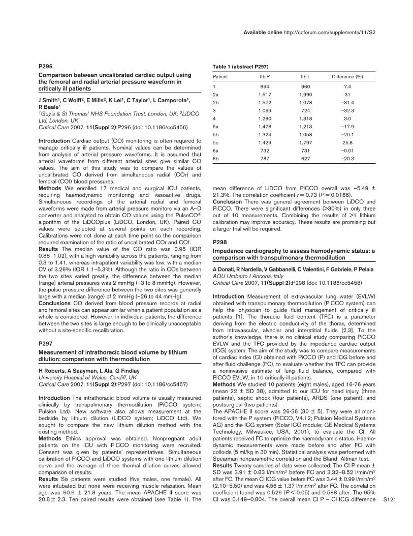
S121
P296Comparison between uncalibrated cardiac output usingthe femoral and radial arterial pressure waveform incritically ill patients
J Smith1, C Wolff2, E Mills2, K Lei1, C Taylor1, L Camporota1, R Beale1
1Guy’s & St Thomas’ NHS Foundation Trust, London, UK; 2LiDCOLtd, London, UKCritical Care 2007, 11(Suppl 2):P296 (doi: 10.1186/cc5456)
Introduction Cardiac output (CO) monitoring is often required tomanage critically ill patients. Nominal values can be determinedfrom analysis of arterial pressure waveforms. It is assumed thatarterial waveforms from different arterial sites give similar COvalues. The aim of this study was to compare the values ofuncalibrated CO derived from simultaneous radial (COr) andfemoral (COf) blood pressures.Methods We enrolled 17 medical and surgical ICU patients,requiring haemodynamic monitoring and vasoactive drugs.Simultaneous recordings of the arterial radial and femoralwaveforms were made from arterial pressure monitors via an A–Dconverter and analysed to obtain CO values using the PulseCO®
algorithm of the LiDCOplus (LiDCO, London, UK). Paired COvalues were selected at several points on each recording.Calibrations were not done at each time point so the comparisonrequired examination of the ratio of uncalibrated COr and COf.Results The median value of the CO ratio was 0.95 (IQR0.88–1.02), with a high variability across the patients, ranging from0.3 to 1.41, whereas intrapatient variability was low, with a medianCV of 3.26% (IQR 1.1–5.3%). Although the ratio in COs betweenthe two sites varied greatly, the difference between the median(range) arterial pressures was 2 mmHg (–3 to 8 mmHg). However,the pulse pressure difference between the two sites was generallylarge with a median (range) of 2 mmHg (–26 to 44 mmHg).Conclusions CO derived from blood pressure records at radialand femoral sites can appear similar when a patient population as awhole is considered. However, in individual patients, the differencebetween the two sites is large enough to be clinically unacceptablewithout a site-specific recalibration.
P297Measurement of intrathoracic blood volume by lithiumdilution: comparison with thermodilution
H Roberts, A Saayman, L Ala, G FindlayUniversity Hospital of Wales, Cardiff, UKCritical Care 2007, 11(Suppl 2):P297 (doi: 10.1186/cc5457)
Introduction The intrathoracic blood volume is usually measuredclinically by transpulmonary thermodilution (PiCCO system;Pulsion Ltd). New software also allows measurement at thebedside by lithium dilution (LiDCO system; LiDCO Ltd). Wesought to compare the new lithium dilution method with theexisting method.Methods Ethics approval was obtained. Nonpregnant adultpatients on the ICU with PiCCO monitoring were recruited.Consent was given by patients’ representatives. Simultaneouscalibration of PiCCO and LiDCO systems with one lithium dilutioncurve and the average of three thermal dilution curves allowedcomparison of results.Results Six patients were studied (five males, one female). Allwere intubated but none were receiving muscle relaxation. Meanage was 60.6 ± 21.8 years. The mean APACHE II score was20.8 ± 2.3. Ten paired results were obtained (see Table 1). The
mean difference of LiDCO from PiCCO overall was –5.49 ±21.3%. The correlation coefficient r = 0.73 (P = 0.0166).Conclusion There was general agreement between LiDCO andPiCCO. There were significant differences (>30%) in only threeout of 10 measurements. Combining the results of >1 lithiumcalibration may improve accuracy. These results are promising buta larger trial will be required.
P298Impedance cardiography to assess hemodynamic status: acomparison with transpulmonary thermodilution
A Donati, R Nardella, V Gabbanelli, C Valentini, F Gabriele, P PelaiaAOU Umberto I Ancona, ItalyCritical Care 2007, 11(Suppl 2):P298 (doi: 10.1186/cc5458)
Introduction Measurement of extravascular lung water (EVLW)obtained with transpulmonary thermodilution (PiCCO system) canhelp the physician to guide fluid management of critically illpatients [1]. The thoracic fluid content (TFC) is a parameterderiving from the electric conductivity of the thorax, determinedfrom intravascular, alveolar and interstitial fluids [2,3]. To theauthor's knowledge, there is no clinical study comparing PiCCOEVLW and the TFC provided by the impedance cardiac output(ICG) system. The aim of the study was to compare measurementsof cardiac index (CI) obtained with PiCCO (P) and ICG before andafter fluid challenge (FC), to evaluate whether the TFC can providea noninvasive estimate of lung fluid balance, compared withPiCCO EVLW, in 10 critically ill patients.Methods We studied 10 patients (eight males), aged 16-76 years(mean 22 ± SD 38), admitted to our ICU for head injury (threepatients), septic shock (four patients), ARDS (one patient), andpostsurgical (two patients).The APACHE II score was 26-36 (30 ± 5). They were all moni-tored with the P system (PiCCO, V4.12; Pulsion Medical SystemsAG) and the ICG system (Solar ICG module; GE Medical SystemsTechnology, Milwaukee, USA, 2001), to evaluate the CI. Allpatients received FC to optimize the haemodynamic status. Haemo-dynamic measurements were made before and after FC withcolloids (5 ml/kg in 30 min). Statistical analysis was performed withSpearman nonparametric correlation and the Bland–Altman test.Results Twenty samples of data were collected. The CI P mean ±SD was 3.91 ± 0.83 l/min/m2 before FC and 3.32–8.52 l/min/m2
after FC. The mean CI ICG value before FC was 3.44 ± 0.99 l/min/m2
(2.10–5.50) and was 4.56 ± 1.37 l/min/m2 after FC. The correlationcoefficient found was 0.526 (P < 0.05) and 0.588 after. The 95%CI was 0.149–0.804. The overall mean CI P – CI ICG difference
Available online http://ccforum.com/supplements/11/S2
Table 1 (abstract P297)
Patient ItbiP ItbiL Difference (%)
1 894 960 7.4
2a 1,517 1,990 31
2b 1,572 1,078 –31.4
3 1,069 724 –32.3
4 1,280 1,318 3.0
5a 1,478 1,213 –17.9
5b 1,324 1,058 –20.1
5c 1,429 1,797 25.8
6a 732 731 –0.01
6b 787 627 –20.3
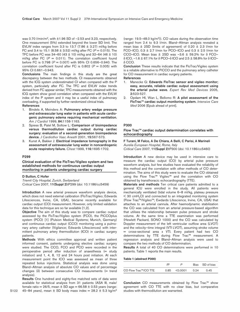
S122
was 0.70 l/min/m2, with ±1.96 SD of –2.53 and 3.23, respectively.One measurement (5%) extended beyond the lower SD limit. TheEVLW index ranges from 3.3 to 13.7 (7.86 ± 3.27) ml/kg beforeFC and 3.4 to 15.1 (8.58 ± 3.52) ml/kg after FC (P = 0.015). TheTFC before FC was 34–60 (43 ± 10) ml/kg and 32–64 (46 ± 10)ml/kg after FC (P = 0.011). The correlation coefficient foundbefore FC is 0.798 (P = 0.007) with 95% CI 0.656–0.940. Thecorrelation coefficient found after FC is 0.802 (P = 0.005) with95% CI 0.661–0.943.Conclusions The main findings in this study are the greatdiscrepancy between the two methods. CI measurements obtainedwith the ICG system underestimated CI when compared with the Psystem, particularly after FC. The TFC and EVLW index trendsderived from FC appear similar; TFC measurements obtained with theICG system show good correlation when compared with the EVLWindex of the P system and it may be a useful index of pulmonaryoverloading, if supported by further randomized clinical trials.References1. Bindels A, Meinders A: Pulmonary artery wedge pressure
and extravascular lung water in patients with acute cardio-genic pulmonary edema requiring mechanical ventilation.Am J Cardiol 1999, 84:1158-1163.
2. Spiess B, Patel M, Soltow L: Comparison of bioimpedanceversus thermodilution cardiac output during cardiacsurgery: evaluation of a second-generation bioimpedancedevice. J Cardiothor Vasc Anesth 2001, 15:567-573.
3. Kunst A, Bakker J: Electrical impedance tomography in theassessment of extravascular lung water in noncardiogenicacute respiratory failure. Chest 1999, 116:1695-1702.
P299Clinical evaluation of the FloTrac/Vigileo system and twoestablished methods for continuous cardiac outputmonitoring in patients undergoing cardiac surgery
D Button, C HoferTriemli City Hospital, Zurich, SwitzerlandCritical Care 2007, 11(Suppl 2):P299 (doi: 10.1186/cc5459)
Introduction A new arterial pressure waveform analysis device,which does not need external calibration (FloTrac/Vigileo; EdwardsLifesciences, Irvine, CA, USA), became recently available forcardiac output (CO) measurement. However, only limited validationdata for this technique are so far available [1,2].Objective The aim of this study was to compare cardiac outputassessed by the FloTrac/Vigileo system (FCO), the PiCCOplussystem (PCO) [1] (Pulsion Medical Systems; Munich, Germany)and continuous cardiac output (CCO) monitoring using a pulmo-nary artery catheter (Vigilance; Edwards Lifesciences) with inter-mittent pulmonary artery thermodilution (ICO) in cardiac surgerypatients.Methods With ethics committee approval and written patientinformed consent, patients undergoing elective cardiac surgerywere studied. The CCO, FCO and PCO were recorded in theperioperative period after induction of anaesthesia (= studyinitiation) and 1, 4, 8, 12 and 24 hours post initiation. At eachmeasurement point the ICO was assessed as mean of threerepeated bolus injections. Statistical analysis was done usingBland–Altman analysis of absolute CO values and of percentagechanges (δ) between consecutive CO measurements (= trendanalysis).Results One hundred and eighty-five matched sets of data wereavailable for statistical analysis from 31 patients (ASA III, male/female ratio = 26/5, mean ± SD age = 66.58 ± 0.53 years (range:45–84 years), mean ± SD body mass index = 28.2 ± 5.3 kg/m2
(range: 19.5–48.0 kg/m2)). CO values during the observation timeranged from 2.4 to 9.3 l/min. Bland–Altman analysis revealed amean bias ± 2SD (limits of agreement) of 0.20 ± 2.3 l/min forFCO–ICO, 0.3 ± 2.7 l/min for PCO–ICO and 0.3 ± 2.5 l/min forCCO–ICO. Mean bias ± 2SD was –3.6 ± 59.2% for δ FCO–δ ICO, –1.6 ± 67.1% for δ PCO–δ ICO and 2.5 ± 58.8% for δ ICO–δ CCO.Conclusion These results indicate that the FloTrac/Vigileo systemis a reliable alternative to PiCCO and the pulmonary artery catheterfor CO measurement in cardiac surgery patients.References1. Manecke G: Edwards FloTrac sensor and vigileo monitor;
easy, accurate, reliable cardiac output assessment usingthe arterial pulse wave. Expert Rev Med Devices 2005,2:523-527.
2. Opdam HI, Wan L, Bellomo R: A pilot assessment of theFloTrac™ cardiac output monitoring system. Intensive CareMed 2006 [Epub ahead of print].
P300Flow Trac™ cardiac output determination correlates withechocardiography
F Turani, M Falco, A De Chiara, A Belli, C Parisi, A MarineliAurelia European Hospital, Rome, ItalyCritical Care 2007, 11(Suppl 2):P300 (doi: 10.1186/cc5460)
Introduction A new device may be used in intensive care tomeasure the cardiac output (CO) by arterial pulse pressurewaveform analysis, but few studies have evaluated the reliability ofthis method and the correlation with other methods of CO deter-mination. The aims of this study were to evaluate the CO obtainedusing the Flow Trac™ Vigileo™ and the correlation with COobtained by transthoracic echocardiography (TTE).Materials and methods Ten critical care patients admitted to ageneral ICU were enrolled in the study. All patients weremechanically ventilated (tidal volume 6–8 ml/kg, plateau pressure< 30 cmH2O) and connected to an integrated monitoring system(Flow Trac™/Vigileo™; Ewdards Lifescience, Irvine, CA, USA) thatattaches to an arterial cannula. After haemodynamic stabilizationthe CO was calculated from an arterial pressure-based algorithmthat utilises the relationship between pulse pressure and strokevolume. At the same time a TTE examination was performed(Hewlett Packard, SONO 1000) and the CO was calculated byDoppler measurement of the left ventricular outflow area (LVOT)and the velocity–time integral (VTI LVOT), assuming stroke volume= cross-sectional area x VTI. Every patient had two COdeterminations by TTE during Flow Trac™ measurement. Aregression analysis and Bland–Altman analysis were used tocompare the two methods of CO determination.Results A total of 40 CO determinations were performed in 10patients. Table 1 reports the main results.
Table 1 (abstract P300)
R2 P Bias SD of bias
CO Flow Trac™/CO TTE 0.85 <0.0001 0.24 0.45
Conclusion CO measurements obtained by Flow Trac™ showagreement with CO TTE with no clear bias, but comparativestudies with thermodilution are warranted.
Critical Care March 2007 Vol 11 Suppl 2 27th International Symposium on Intensive Care and Emergency Medicine
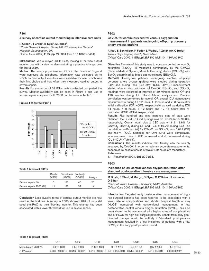
S123
P301A survey of cardiac output monitoring in intensive care units
R Green1, J Craig1, B Kyle1, M Jonas2
1Poole General Hospital, Poole, UK; 2Southampton GeneralHospital, Southampton, UKCritical Care 2007, 11(Suppl 2):P301 (doi: 10.1186/cc5461)
Introduction We surveyed adult ICUs, looking at cardiac outputmonitor use with a view to demonstrating a practice change overthe last 3 years.Method The senior physicians on ICUs in the South of Englandwere surveyed via telephone. Information was collected as towhich cardiac output monitors were available for use, which wastheir first choice and how often they measured cardiac output insevere sepsis.Results Forty-nine out of 52 ICUs units contacted completed thesurvey. Monitor availability can be seen in Figure 1 and use insevere sepsis compared with 2003 can be seen in Table 1.
Table 1 (abstract P301)
Rarely Sometimes Routinely (<5%) (<50%) (>50%) Always
Severe sepsis (%) 0 30 45 25
Severe sepsis 2003 (%) 11 36 37 14
Conclusion Less invasive forms of cardiac output monitor are nowused as the first line. A survey in 2005 showed 20% of units stillused the PAC as their first-line monitor. This change has beenassociated with a lower threshold for use in severe sepsis.
P302CeVOX for continuous central venous oxygenationmeasurement in patients undergoing off-pump coronaryartery bypass grafting
A Rist, S Schneider, P Fodor, L Weibel, A Zollinger, C HoferTriemli City Hospital, Zurich, SwitzerlandCritical Care 2007, 11(Suppl 2):P302 (doi: 10.1186/cc5462)
Objective The aim of this study was to compare central venous O2saturation (ScvO2) [1] measured continuously by the CeVOX(Pulsion Medical System, Munich, Germany) device (CScvO2) withScvO2 determined by blood gas co-oximetry (BScvO2).Methods Twenty-five patients undergoing elective off-pumpcoronary artery bypass grafting were studied during operation(OP) and during their ICU stay (ICU). OP/ICU measurementstarted after in vivo calibration of CeVOX. BScvO2 and CScvO2readings were recorded at intervals of 30 minutes during OP and120 minutes during ICU. Bland–Altman analysis and Pearsoncorrelation was performed for overall OP, overall ICU, consecutivemeasurements during OP ≤1 hour, 1–2 hours and 2–3 hours afterinitial calibration (OP1–OP3, respectively) as well as during ICU≤4 hours, 4–8 hours, 8–12 hours and 12–16 hours after re-calibration (ICU1–ICU4, respectively).Results Five hundred and nine matched sets of data wereobtained; the BScvO2/CScvO2 range was 36–98.9%/46.5–99.0%,respectively. Overall mean bias ± 2SD was –1.2 ± 13.8% forCScvO2–BScvO2 during OP and –2.6 ± 16.2% during ICU. Thecorrelation coefficient (r2) for CScvO2 vs BScvO2 was 0.614 (OP)and 0.174 (ICU). Statistics for OP1–OP3 were comparable,whereas mean bias ± 2SD increased and r2 decreased duringICU1–ICU4 (Table 1).Conclusions The results indicate that ScvO2 can be reliablyassessed by CeVOX. In order to maintain accurate measurements,scheduled re-calibrations at intervals <12 hours are mandatory.Reference1. Respiration 2001, 685:279-285.
P303Incidence of low central venous oxygen saturation afterstandard postoperative intensive care management
M Boyle, E Steel, M Murgo, G Flynn, M O’Brien, J Lawrence, D BihariPrince of Wales Hospital, Randwick, NSW, AustraliaCritical Care 2007, 11(Suppl 2):P303 (doi: 10.1186/cc5463)
Introduction Targeted early postoperative management of high-risk surgical patients has been reported to be associated with alower rate of complications and shorter hospital length of stay(HLOS) compared with conventional management. A lowpostoperative central venous oxygen saturation (ScVO2) has alsobeen shown to be associated with higher rates of complicationsand of HLOS for high-risk surgical patients. Benefit from early goal-directed therapy would be unlikely if ‘standard’ postoperativemanagement resulted in a low incidence of patients with a lowScVO2 in the early postoperative period.
Available online http://ccforum.com/supplements/11/S2
Figure 1 (abstract P301)
Table 1 (abstract P302)
OP1 OP2 OP3 ICU1 ICU2 ICU3 ICU4
Mean bias ± 2SD (%) –0.2 ± 13.9 +1.3 ± 9.6 +1.8 ± 16.6 –0.1 ± 12.0 –0.6 ± 15.4 –0.5 ± 13.8 –4.6 ± 16.9
r2 (P value) 0.580 (<0.001) 0.616 (<0.001) 0.513 (<0.001) 0.416 (<0.001) 0.514 (<0.001) 0.310 (0.001) 0.030 (0.247)
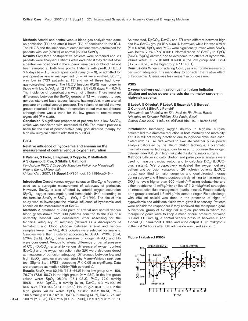
S124
Methods Arterial and central venous blood gas analysis was doneon admission (T1) and after 8 hours (T2) of admission to the ICU.The HLOS and the incidence of complications were determined forpatients with low (<70%) or normal (≥70%) ScVO2.Results Sixty-three postoperative patients were screened and 23patients were analysed. Patients were excluded if they did not havea central line positioned in the superior vena cava or blood had notbeen sampled at both time points. Patients with pre-ICU HLOS> 5 days (n = 10), acute spinal cord injury (n = 3), or admitted forpostoperative airway management (n = 4) were omitted. ScVO2was low in 7/23 patients at T2 and six of these had lowergastrointestinal surgery. The HLOS (median (IQR)) was longer inthose with low ScVO2 at T2 (17 (37.8) v 9.5 (5.0) days, P = 0.04).The incidence of complications was not different. There were nodifferences between the ScVO2 groups at T2 with respect to age,gender, standard base excess, lactate, haemoglobin, mean arterialpressure or central venous pressure. The volume of colloid the twogroups received in the 8-hour observation period was not differentalthough there was a trend for the low group to receive morecrystalloid (P = 0.08).Conclusion A significant proportion of patients had a low ScVO2,which was associated with increased HLOS. The results provide abasis for the trial of postoperative early goal-directed therapy forhigh-risk surgical patients admitted to our ICU.
P304Relative influence of hypoxemia and anemia on themeasurement of central venous oxygen saturation
F Valenza, S Froio, L Fagnani, S Coppola, M Maffioletti, A Sicignano, E Riva, S Sibilla, L GattinoniFondazione IRCCS Ospedale Maggiore Policlinico MangiagalliRegina Elena, Milano, ItalyCritical Care 2007, 11(Suppl 2):P304 (doi: 10.1186/cc5464)
Introduction Central venous oxygen saturation (ScvO2) is frequentlyused as a surrogate measurement of adequacy of perfusion.However, ScvO2 is also affected by arterial oxygen saturation(SpO2), oxygen consumption, and hemoglobin (Hb) according tothe formula: ScvO2 = SpO2 – (VO2 / Q*1/Hb). The aim of thisstudy was to investigate the relative influence of hypoxemia andanemia on the measurement of ScvO2.Methods A database of 700 pairs of arterial and central venousblood gases drawn from 300 patients admitted to the ICU of auniversity hospital was considered. After assessing for thetechnical adequacy of sampling (defined as a discrepancy ofhematocrit and blood glucose between arterial and venoussamples lower than 5%), 462 couples were selected for analysis.Samples were then clustered according to ScvO2: <70% (low),≥70% (high). SpO2, partial pressure of oxygen (PaO2) and Hbwere considered. Venous to arterial difference of partial pressureof CO2 (DpCO2), arterial to venous difference of oxygen content(DavO2) and the oxygen extraction ratio (ER) were also consideredas measures of perfusion adequacy. Differences between low andhigh ScvO2 samples were estimated by Mann–Whitney rank sumtest (Sigma Stat, SPSS), accepting P < 0.05 as significant. Dataare presented as median (25th–75th percentile).Results ScvO2 was 62.5% (56.3–66.2) in the low group (n = 180),76.7% (73.6–80.7) in the high group (n = 282). In the low groupvalues were: SpO2 95.0% (95.1–98.3), PaO2 72.0 mmHg(59.5–112.5), DpCO2 8 mmHg (6–9), DavO2 4.3 ml/100 ml(3.4–5.2), ER 0.346 (0.310–0.399), Hb 9.9 g/dl (8.9–11.1). In thehigh group values were: SpO2 98.3% (96.2–99.5), PaO2106.5 mmHg (81.0–167.0), DpCO2 6 mmHg (4–7), DavO2 2.9 ml/100 ml (2.3–3.6), ER 0.215 (0.185–0.250), Hb 9.9 g/dl (8.7–11.1).
As expected, DpCO2, DavO2 and ER were different between highand low ScvO2 groups (P < 0.001). However, while Hb was similar(P = 0.670), SpO2 and PaO2 were significantly lower when ScvO2was below 70% (P < 0.001). Normalization of ScvO2 to SpO2(ScvO2/SpO2) allowed one to overcome the effects of hypoxemia.Values were: 0.662 (0.603–0.693) in the low group and 0.794(0.757–0.828) in the high group (P < 0.001).Conclusions When considering ScvO2 as a surrogate measure ofperfusion adequacy, it is mandatory to consider the relative effectof hypoxemia. Anemia was less relevant in our case mix.
P305Oxygen delivery optimization using lithium indicatordilution and pulse power analysis during major surgery inhigh-risk patients
S Lobo1, N Oliveira1, F Lobo1, E Rezende2, B Borges1, G Cunrath1, J Silva2, L Ronchi11Faculdade de Medicina de São José do Rio Preto, Brazil;2Hospital do Servidor Público, São Paulo, BrazilCritical Care 2007, 11(Suppl 2):P305 (doi: 10.1186/cc5465)
Introduction Increasing oxygen delivery in high-risk surgicalpatients led to a dramatic reduction in both mortality and morbidity.Yet, it is still not widely practised due to logistical difficulties asso-ciated with its use. We aimed to evaluate whether pulse poweranalysis calibrated by the lithium dilution technique, a pragmaticminimally invasive technique, can be used to optimize the oxygendelivery index (DO2I) in high-risk patients during major surgery.Methods Lithium indicator dilution and pulse power analysis wereused to measure cardiac output and to calculate DO2I (LiDCO-plus system). We prospectively evaluated the oxygen deliverypattern and perfusion variables of 26 high-risk patients (LiDCOgroup) submitted to major surgeries and goal-directed therapyduring surgery and 8 hours postoperatively, aiming to maximize theDO2I to levels higher than 600 ml/min/m2 using dobutamine andeither ‘restrictive’ (4 ml/kg/min) or ‘liberal’ (12 ml/kg/min) strategiesof intraoperative fluid management (partial results). Postoperativelyboth groups received 1.5 ml/kg/min lactated ringer. Fluid challengewith 250 ml colloid was done in the presence of signs ofhypovolemia and additional fluids were given if necessary. Patientswere considered responders if they achieved the therapeutic goal.A historical group of 42 high-risk surgical patients in whom thetherapeutic goals were to keep a mean arterial pressure between80 and 110 mmHg, a central venous pressure between 6 and12 cmH2O, hematocrit > 30% and urine output > 0.5 ml/kg/hourin the first 24 hours after ICU admission was used as control.
Critical Care March 2007 Vol 11 Suppl 2 27th International Symposium on Intensive Care and Emergency Medicine
Figure 1 (abstract P305)
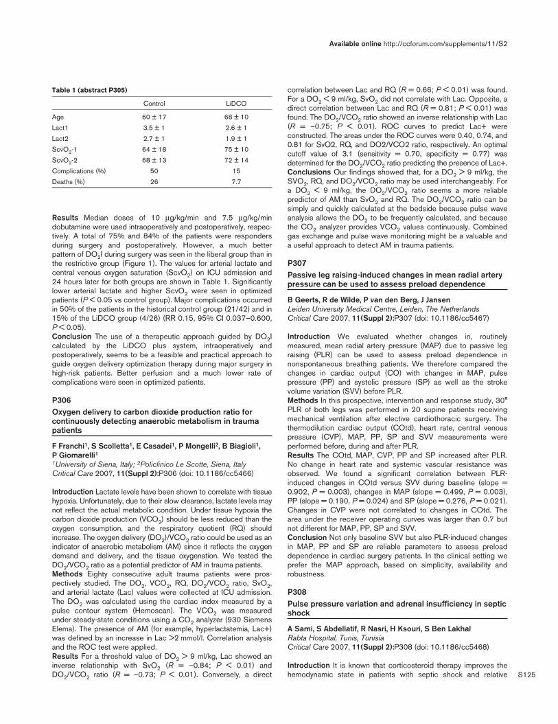
S125
Results Median doses of 10 µg/kg/min and 7.5 µg/kg/mindobutamine were used intraoperatively and postoperatively, respec-tively. A total of 75% and 84% of the patients were respondersduring surgery and postoperatively. However, a much betterpattern of DO2I during surgery was seen in the liberal group than inthe restrictive group (Figure 1). The values for arterial lactate andcentral venous oxygen saturation (ScvO2) on ICU admission and24 hours later for both groups are shown in Table 1. Significantlylower arterial lactate and higher ScvO2 were seen in optimizedpatients (P < 0.05 vs control group). Major complications occurredin 50% of the patients in the historical control group (21/42) and in15% of the LiDCO group (4/26) (RR 0.15, 95% CI 0.037–0.600,P < 0.05).Conclusion The use of a therapeutic approach guided by DO2Icalculated by the LiDCO plus system, intraoperatively andpostoperatively, seems to be a feasible and practical approach toguide oxygen delivery optimization therapy during major surgery inhigh-risk patients. Better perfusion and a much lower rate ofcomplications were seen in optimized patients.
P306Oxygen delivery to carbon dioxide production ratio forcontinuously detecting anaerobic metabolism in traumapatients
F Franchi1, S Scolletta1, E Casadei1, P Mongelli2, B Biagioli1, P Giomarelli11University of Siena, Italy; 2Policlinico Le Scotte, Siena, ItalyCritical Care 2007, 11(Suppl 2):P306 (doi: 10.1186/cc5466)
Introduction Lactate levels have been shown to correlate with tissuehypoxia. Unfortunately, due to their slow clearance, lactate levels maynot reflect the actual metabolic condition. Under tissue hypoxia thecarbon dioxide production (VCO2) should be less reduced than theoxygen consumption, and the respiratory quotient (RQ) shouldincrease. The oxygen delivery (DO2)/VCO2 ratio could be used as anindicator of anaerobic metabolism (AM) since it reflects the oxygendemand and delivery, and the tissue oxygenation. We tested theDO2/VCO2 ratio as a potential predictor of AM in trauma patients.Methods Eighty consecutive adult trauma patients were pros-pectively studied. The DO2, VCO2, RQ, DO2/VCO2 ratio, SvO2,and arterial lactate (Lac) values were collected at ICU admission.The DO2 was calculated using the cardiac index measured by apulse contour system (Hemoscan). The VCO2 was measuredunder steady-state conditions using a CO2 analyzer (930 SiemensElema). The presence of AM (for example, hyperlactatemia, Lac+)was defined by an increase in Lac >2 mmol/l. Correlation analysisand the ROC test were applied.Results For a threshold value of DO2 > 9 ml/kg, Lac showed aninverse relationship with SvO2 (R = –0.84; P < 0.01) andDO2/VCO2 ratio (R = –0.73; P < 0.01). Conversely, a direct
correlation between Lac and RQ (R = 0.66; P < 0.01) was found.For a DO2 < 9 ml/kg, SvO2 did not correlate with Lac. Opposite, adirect correlation between Lac and RQ (R = 0.81; P < 0.01) wasfound. The DO2/VCO2 ratio showed an inverse relationship with Lac(R = –0.75; P < 0.01). ROC curves to predict Lac+ wereconstructed. The areas under the ROC curves were 0.40, 0.74, and0.81 for SvO2, RQ, and DO2/VCO2 ratio, respectively. An optimalcutoff value of 3.1 (sensitivity = 0.70, specificity = 0.77) wasdetermined for the DO2/VCO2 ratio predicting the presence of Lac+.Conclusions Our findings showed that, for a DO2 > 9 ml/kg, theSVO2, RQ, and DO2/VCO2 ratio may be used interchangeably. Fora DO2 < 9 ml/kg, the DO2/VCO2 ratio seems a more reliablepredictor of AM than SvO2 and RQ. The DO2/VCO2 ratio can besimply and quickly calculated at the bedside because pulse waveanalysis allows the DO2 to be frequently calculated, and becausethe CO2 analyzer provides VCO2 values continuously. Combinedgas exchange and pulse wave monitoring might be a valuable anda useful approach to detect AM in trauma patients.
P307Passive leg raising-induced changes in mean radial arterypressure can be used to assess preload dependence
B Geerts, R de Wilde, P van den Berg, J JansenLeiden University Medical Centre, Leiden, The NetherlandsCritical Care 2007, 11(Suppl 2):P307 (doi: 10.1186/cc5467)
Introduction We evaluated whether changes in, routinelymeasured, mean radial artery pressure (MAP) due to passive legraising (PLR) can be used to assess preload dependence innonspontaneous breathing patients. We therefore compared thechanges in cardiac output (CO) with changes in MAP, pulsepressure (PP) and systolic pressure (SP) as well as the strokevolume variation (SVV) before PLR.Methods In this prospective, intervention and response study, 30°PLR of both legs was performed in 20 supine patients receivingmechanical ventilation after elective cardiothoracic surgery. Thethermodilution cardiac output (COtd), heart rate, central venouspressure (CVP), MAP, PP, SP and SVV measurements wereperformed before, during and after PLR.Results The COtd, MAP, CVP, PP and SP increased after PLR.No change in heart rate and systemic vascular resistance wasobserved. We found a significant correlation between PLR-induced changes in COtd versus SVV during baseline (slope =0.902, P = 0.003), changes in MAP (slope = 0.499, P = 0.003),PP (slope = 0.190, P = 0.024) and SP (slope = 0.276, P = 0.021).Changes in CVP were not correlated to changes in COtd. Thearea under the receiver operating curves was larger than 0.7 butnot different for MAP, PP, SP and SVV.Conclusion Not only baseline SVV but also PLR-induced changesin MAP, PP and SP are reliable parameters to assess preloaddependence in cardiac surgery patients. In the clinical setting weprefer the MAP approach, based on simplicity, availability androbustness.
P308Pulse pressure variation and adrenal insufficiency in septicshock
A Sami, S Abdellatif, R Nasri, H Ksouri, S Ben LakhalRabta Hospital, Tunis, TunisiaCritical Care 2007, 11(Suppl 2):P308 (doi: 10.1186/cc5468)
Introduction It is known that corticosteroid therapy improves thehemodynamic state in patients with septic shock and relative
Available online http://ccforum.com/supplements/11/S2
Table 1 (abstract P305)
Control LiDCO
Age 60 ± 17 68 ± 10
Lact1 3.5 ± 1 2.6 ± 1
Lact2 2.7 ± 1 1.9 ± 1
ScvO2-1 64 ± 18 75 ± 10
ScvO2-2 68 ± 13 72 ± 14
Complications (%) 50 15
Deaths (%) 26 7.7
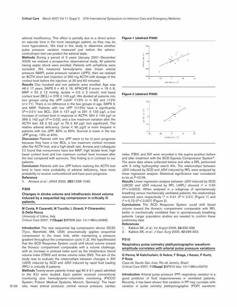
S126
adrenal insufficiency. This effect is partially due to a direct actionon vascular tone in the more vasoplegic patient, so they may bemore hypovolemic. We tried in this study to determine whetherpulse pressure variation measured just before the adreno-corticotropin test can predict the adrenal state.Methods During a period of 3 years (January 2001–December2003) we realized a prospective observational study. All patientshaving septic shock were enrolled. Patients with arrhythmia wereexcluded. We measured hemodynamic data (mean arterialpressure (MAP), pulse pressure variation (∆PP)), then we realizedan ACTH short test (injection of 250 mg ACTH with dosage of thecortisol level before the injection, at 30 and 60 minutes).Results One hundred and one patients were enrolled. Age was48 ± 17 years. SAPS II = 45 ± 16, APACHE II score = 18 ± 8,MAP = 52 ± 12 mmHg, lactate = 3.5 ± 2 mmol/l, and basalcortisol level (BCL) = 278 ± 143 µg/l. We divided all patients intotwo groups using the ∆PP cutoff: <12% (n = 30) and ≥12%(n = 71). There is no difference in the two groups in age, SAPS II,and MAP. Patients with low ∆PP (<12%) have a significantly(P = 0.01) low BCL: 204 ± 127 µg/l vs 291 ± 133 µg/l, a lowincrease of cortisol level in response to ACTH: 264 ± 144 µg/l vs369 ± 142 µg/l (P = 0.02), and a low maximum variation after theACTH test: 59 ± 52 µg/l vs 79 ± 63 µg/l (not significant). Therelative adrenal deficiency (∆max < 90 µg/l) is more frequent inpatients with low ∆PP: 80% vs 60%. Survival is lower in the low∆PP group, 13% vs 40%.Discussion Patients with low ∆PP seem to be of poor prognosisbecause they have a low BCL, a low maximum cortisol increaseafter the ACTH test, and a high death rate. Annane and colleagues[1] found that nonsurvivors have low MAP, high lactate level, highbasal cortisol level and low maximum cortisol level increase afterthe test compared with survivors. This finding is in contrast to ourpatients.Conclusion Patients with low ∆PP before realizing the ACTH testtend to have more probability of adrenal deficiency, have moreprobability to receive corticosteroid and have poor prognosis.Reference1. Annane et al.: JAMA 2000, 283:1038-1045.
P309Changes in stroke volume and intrathoracic blood volumeinduced by a sequential leg compression in critically illpatients.
M Costa, R Casaretti, M Tuccillo, L Girardi, P Chiarandini, G Della RoccaUniversity of Udine, ItalyCritical Care 2007, 11(Suppl 2):P309 (doi: 10.1186/cc5469)
Introduction The new sequential leg compression device (SCD)(Tyco, Mansfield, MA, USA) pneumatically applies sequentialcompression to the lower limb, while maintaining a pressuregradient throughout the compression cycle [1,2]. We hypothesizedthat the SCD Response System could shift blood volume towardthe thoracic compartment comparable with a volume challenge,with an increase in preload index such as the intrathoracic bloodvolume index (ITBVI) and stroke volume index (SVI). The aim of thestudy was to evaluate the relationships between changes in SVI(∆SVI) induced by SCD and ∆SVI induced by rapid fluid loading(RFL) in critically ill patients.Methods Twenty-seven patients (mean age 60 ± 9.1 years) admittedto the ICU were studied. Each patient received conventionalmonitoring plus hemodynamic–volumetric monitoring (PiCCOSystem; Pulsion Medical Systems, Munich, Germany). The heartrate, mean arterial pressure, central venous pressure, cardiac
index, ITBVI, and SVI were recorded in the supine position beforeand after treatment with the SCD Express Compression System®.The same data where collected before and after a RFL performedwith 3 ml/kg hydroxyethyl starch 6%. The relationships between∆SVI induced by SCD and ∆SVI induced by RFL were analyzed bylinear regression analysis. Statistical significance was consideredto be at P <0.05.Results Linear regression analysis between ∆SVI induced by SCD(∆SCD) and ∆SVI induced by RFL (∆RFL) showed r2 = 0.50(P = 0.0002). When analyzed in a subgroup of spontaneouslybreathing versus mechanically ventilated patients, the relationshipsobserved were respectively r2 = 0.41 (P < 0.01) (Figure 1) andr2 = 0.73 (P < 0.007) (Figure 2).Conclusions The SCD Response System could shift bloodvolume toward the thoracic compartment comparable with RFLbetter in mechanically ventilated than in spontaneously breathingpatients. Larger population studies are needed to confirm thesepreliminary data.References1. Kakkos SK, et al.: Int Angiol 2005, 24:330-335.2. Kakkos SK, et al.: J Vasc Surg 2005, 42:296-303.
P310Respiratory pulse oximetry plethysmographic waveformamplitude correlates with arterial pulse pressure variations
G Penna, M Kalichsztein, G Nobre, F Braga, J Kezen, P Kurtz, P RosaCasa de Saude Sao Jose, Rio de Janeiro, BrazilCritical Care 2007, 11(Suppl 2):P310 (doi: 10.1186/cc5470)
Introduction Arterial pulse pressure (PP) respiratory variation is agood predictor of fluid responsiveness in ventilated patients.Recently, it has been shown that variation in PP may correlate withvariation in pulse oximetry plethysmographic (POP) waveform
Critical Care March 2007 Vol 11 Suppl 2 27th International Symposium on Intensive Care and Emergency Medicine
Figure 1 (abstract P309)
Figure 2 (abstract P309)
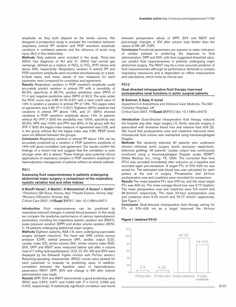
S127
amplitude, as they both depend on the stroke volume. Wedesigned a prospective study to evaluate the correlation betweenrespiratory arterial PP variation and POP waveform amplitudevariations in ventilated patients and the influence of acute lunginjury (ALI) in this relationship.Methods Sixty patients were included in the study. Thirty-nine(65%) had diagnosis of ALI and 21 (35%) had normal gasexchange, defined as a relation of PaO2 to FiO2 (P/F) below andabove 300, respectively. Respiratory variation in arterial PP andPOP waveform amplitude were recorded simultaneously on a beat-to-beat basis, and mean values of two measures for eachparameter were compared for correlation and agreement.Results Respiratory variation in POP waveform amplitude couldaccurately predict variation in arterial PP with a sensibility of83.3%, specificity of 85.7%, positive predictive value (PPV) of71.4 and negative predictive value (NPV) of 92.3. The area underthe ROC curve was 0.88 (0.79–0.97) with a best cutoff value of14% to predict a variation in arterial PP of 13%. The kappa indexof agreement was 0.65 (P < 0.001). Eighteen (30%) patients hadvariations in arterial PP above 13%, and 21 (35%) showedvariations in POP waveform amplitude above 14%. In patientswithout ALI (P/F > 300) the sensibility was 100%, specificity was93.3%, NPV was 100% and PPV was 80%. In the group with ALI(P/F < 300) the kappa index measure of agreement was 0.55, andin the group without ALI the kappa index was 0.85. PEEP levelswere not different between the groups.Conclusion Respiratory variation in arterial PP above 13% can beaccurately predicted by a variation in POP waveform amplitude of14% with good correlation and agreement. Our results confirm thefindings of a recent trial and suggest that the correlation is evenstronger when ALI is absent. These findings raise potential clinicalapplications of respiratory variation in POP waveform amplitude forhaemodynamic management of patients without an arterial catheter.
P311Assessing fluid responsiveness in patients undergoingabdominal major surgery: a comparison of the respiratorysystolic variation test and other indices
A Bisoffi Varani1, A Martini1, N Menestrina2, A Russo3, L Gottin1
1Policlinico GB Rossi, Verona, Italy; 2Hopital Erasme, Verona, Italy;3Romano D’Ezzelino Vicenza, ItalyCritical Care 2007, 11(Suppl 2):P311 (doi: 10.1186/cc5471)
Introduction Fluid responsiveness can be predicted byrespiratory-induced changes in arterial blood pressure. In this studywe compare the predictive performance of various haemodynamicparameters, including the respiratory systolic variation test (RSVT),pulse pressure variation (DPP) and stroke volume variation (SVV),in 18 patients undergoing abdominal major surgery.Methods Eighteen patients, ASA I–II, were undergoing pancreaticsurgery (whipple resection). The heart rate (HR) central venouspressure (CVP), arterial pressure (AP), cardiac output (CO),cardiac index (CI), stroke volume (SV), stroke volume index (SVI),SVV, DPP and RSVT were measured before and after a volumeload of 7 ml/kg hydroxyethylstarch. (CO, CI, SV, SVI and SVV weredisplayed by the Edwards Vigileo monitor with FloTrac sensor.)Receiving-operating characteristic (ROC) curves were plotted foreach parameter to evaluate its predicting value. In addition,correlation between the baseline value of haemodynamicparameters RSVT, DPP, SVV and change in SVI after volumeadministration was made.Results DPP, SVV and RSVT demonstrate a good predicting value(ROC area 0.870, 0.877 and 0.943 with P = 0.010, 0.009 and0.002, respectively). A statistically significant correlation was found
between preoperative values of DPP, SVV and RSVT andpercentage changes in SVI after volume load (better than thevalues of HR, AP, CVP).Conclusion Functional parameters are superior to static indicatorsof cardiac preload in predicting the response to fluidadministration. DPP and SVV, with their suggested threshold value,can predict fluid responsiveness in patients undergoing majorabdominal surgery. The RSVT may be a more accurate predictor offluid responsiveness although its performance demands a complexrespiratory manoeuvre and is dependent on offline measurementand calculations, which limits its clinical use.
P312Goal-directed intraoperative fluid therapy improvedpostoperative renal functions in aortic surgical patients
M Soliman, S Bajaj, K IsmailDepartment of Anaesthesia and Intensive Care Medicine, The MidYorkshire Hospitals, UKCritical Care 2007, 11(Suppl 2):P312 (doi: 10.1186/cc5472)
Introduction Goal-directed intraoperative fluid therapy reducedthe hospital stay after major surgery [1]. Aortic vascular surgery isassociated with excessive blood loss and massive fluid shift [2].We found that postoperative urea and creatinine improved whenintravascular fluid volume was maintained using transoesophagealDoppler.Methods We randomly selected 40 patients who underwentelective infrarenal aortic surgery (aortic aneurysm repair/aorto-bifemoral grafting). All patients’ cardiac output was continuouslymonitored using a transoesophageal Doppler probe (EDM™;Deltex Medical, Inc., Irving, TX, USA). The corrected flow time(FTc) was recorded immediately after induction as a baseline andrecorded again pre-extubation. A target FTc of 375–425 ms wasaimed for. The estimated total blood loss was calculated for eachpatient at the end of surgery. Preoperative and 24-hourpostoperative urea and creatinine were recorded for comparison.Results The mean baseline FTc was 278 ms, and the mean targetFTc was 405 ms. The mean average blood loss was 3.77 l/patient.The mean preoperative urea and creatinine were 5.9 mmol/l and95.3mmol/l, respectively. The mean 24-hour postoperative ureaand creatinine were 5.23 mmol/l and 76.77 mmol/l, respectively.See Figure 1.Conclusion Goal-directed intraoperative fluid therapy aiming forFTc of 375–425 ms as a target improved the 24-hour
Available online http://ccforum.com/supplements/11/S2
Figure 1 (abstract P312)
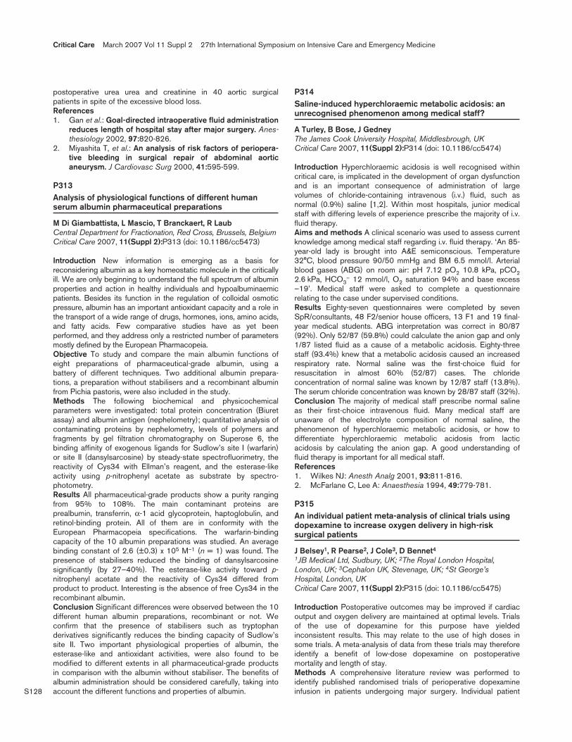
S128
postoperative urea urea and creatinine in 40 aortic surgicalpatients in spite of the excessive blood loss.References1. Gan et al.: Goal-directed intraoperative fluid administration
reduces length of hospital stay after major surgery. Anes-thesiology 2002, 97:820-826.
2. Miyashita T, et al.: An analysis of risk factors of periopera-tive bleeding in surgical repair of abdominal aorticaneurysm. J Cardiovasc Surg 2000, 41:595-599.
P313Analysis of physiological functions of different humanserum albumin pharmaceutical preparations
M Di Giambattista, L Mascio, T Branckaert, R LaubCentral Department for Fractionation, Red Cross, Brussels, BelgiumCritical Care 2007, 11(Suppl 2):P313 (doi: 10.1186/cc5473)
Introduction New information is emerging as a basis forreconsidering albumin as a key homeostatic molecule in the criticallyill. We are only beginning to understand the full spectrum of albuminproperties and action in healthy individuals and hypoalbuminaemicpatients. Besides its function in the regulation of colloidal osmoticpressure, albumin has an important antioxidant capacity and a role inthe transport of a wide range of drugs, hormones, ions, amino acids,and fatty acids. Few comparative studies have as yet beenperformed, and they address only a restricted number of parametersmostly defined by the European Pharmacopeia.Objective To study and compare the main albumin functions ofeight preparations of pharmaceutical-grade albumin, using abattery of different techniques. Two additional albumin prepara-tions, a preparation without stabilisers and a recombinant albuminfrom Pichia pastoris, were also included in the study.Methods The following biochemical and physicochemicalparameters were investigated: total protein concentration (Biuretassay) and albumin antigen (nephelometry); quantitative analysis ofcontaminating proteins by nephelometry, levels of polymers andfragments by gel filtration chromatography on Superose 6, thebinding affinity of exogenous ligands for Sudlow’s site I (warfarin)or site II (dansylsarcosine) by steady-state spectrofluorimetry, thereactivity of Cys34 with Ellman’s reagent, and the esterase-likeactivity using p-nitrophenyl acetate as substrate by spectro-photometry.Results All pharmaceutical-grade products show a purity rangingfrom 95% to 108%. The main contaminant proteins areprealbumin, transferrin, α-1 acid glycoprotein, haptoglobulin, andretinol-binding protein. All of them are in conformity with theEuropean Pharmacopeia specifications. The warfarin-bindingcapacity of the 10 albumin preparations was studied. An averagebinding constant of 2.6 (±0.3) x 105 M–1 (n = 1) was found. Thepresence of stabilisers reduced the binding of dansylsarcosinesignificantly (by 27–40%). The esterase-like activity toward p-nitrophenyl acetate and the reactivity of Cys34 differed fromproduct to product. Interesting is the absence of free Cys34 in therecombinant albumin.Conclusion Significant differences were observed between the 10different human albumin preparations, recombinant or not. Weconfirm that the presence of stabilisers such as tryptophanderivatives significantly reduces the binding capacity of Sudlow’ssite II. Two important physiological properties of albumin, theesterase-like and antioxidant activities, were also found to bemodified to different extents in all pharmaceutical-grade productsin comparison with the albumin without stabiliser. The benefits ofalbumin administration should be considered carefully, taking intoaccount the different functions and properties of albumin.
P314Saline-induced hyperchloraemic metabolic acidosis: anunrecognised phenomenon among medical staff?
A Turley, B Bose, J GedneyThe James Cook University Hospital, Middlesbrough, UKCritical Care 2007, 11(Suppl 2):P314 (doi: 10.1186/cc5474)
Introduction Hyperchloraemic acidosis is well recognised withincritical care, is implicated in the development of organ dysfunctionand is an important consequence of administration of largevolumes of chloride-containing intravenous (i.v.) fluid, such asnormal (0.9%) saline [1,2]. Within most hospitals, junior medicalstaff with differing levels of experience prescribe the majority of i.v.fluid therapy.Aims and methods A clinical scenario was used to assess currentknowledge among medical staff regarding i.v. fluid therapy. ‘An 85-year-old lady is brought into A&E semiconscious. Temperature32°C, blood pressure 90/50 mmHg and BM 6.5 mmol/l. Arterialblood gases (ABG) on room air: pH 7.12 pO2 10.8 kPa, pCO22.6 kPa, HCO3
– 12 mmol/l, O2 saturation 94% and base excess–19’. Medical staff were asked to complete a questionnairerelating to the case under supervised conditions.Results Eighty-seven questionnaires were completed by sevenSpR/consultants, 48 F2/senior house officers, 13 F1 and 19 final-year medical students. ABG interpretation was correct in 80/87(92%). Only 52/87 (59.8%) could calculate the anion gap and only1/87 listed fluid as a cause of a metabolic acidosis. Eighty-threestaff (93.4%) knew that a metabolic acidosis caused an increasedrespiratory rate. Normal saline was the first-choice fluid forresuscitation in almost 60% (52/87) cases. The chlorideconcentration of normal saline was known by 12/87 staff (13.8%).The serum chloride concentration was known by 28/87 staff (32%).Conclusion The majority of medical staff prescribe normal salineas their first-choice intravenous fluid. Many medical staff areunaware of the electrolyte composition of normal saline, thephenomenon of hyperchloraemic metabolic acidosis, or how todifferentiate hyperchloraemic metabolic acidosis from lacticacidosis by calculating the anion gap. A good understanding offluid therapy is important for all medical staff.References1. Wilkes NJ: Anesth Analg 2001, 93:811-816.2. McFarlane C, Lee A: Anaesthesia 1994, 49:779-781.
P315An individual patient meta-analysis of clinical trials usingdopexamine to increase oxygen delivery in high-risksurgical patients
J Belsey1, R Pearse2, J Cole3, D Bennet4
1JB Medical Ltd, Sudbury, UK; 2The Royal London Hospital,London, UK; 3Cephalon UK, Stevenage, UK; 4St George’sHospital, London, UKCritical Care 2007, 11(Suppl 2):P315 (doi: 10.1186/cc5475)
Introduction Postoperative outcomes may be improved if cardiacoutput and oxygen delivery are maintained at optimal levels. Trialsof the use of dopexamine for this purpose have yieldedinconsistent results. This may relate to the use of high doses insome trials. A meta-analysis of data from these trials may thereforeidentify a benefit of low-dose dopexamine on postoperativemortality and length of stay.Methods A comprehensive literature review was performed toidentify published randomised trials of perioperative dopexamineinfusion in patients undergoing major surgery. Individual patient
Critical Care March 2007 Vol 11 Suppl 2 27th International Symposium on Intensive Care and Emergency Medicine
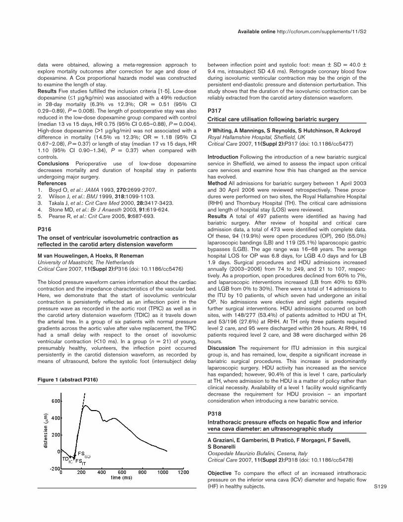
S129
data were obtained, allowing a meta-regression approach toexplore mortality outcomes after correction for age and dose ofdopexamine. A Cox proportional hazards model was constructedto examine the length of stay.Results Five studies fulfilled the inclusion criteria [1-5]. Low-dosedopexamine (≤1 µg/kg/min) was associated with a 49% reductionin 28-day mortality (6.3% vs 12.3%; OR = 0.51 (95% CI0.29–0.89), P = 0.008). The length of postoperative stay was alsoreduced in the low-dose dopexamine group compared with control(median 13 vs 15 days, HR 0.75 (95% CI 0.65–0.88), P = 0.004).High-dose dopexamine (>1 µg/kg/min) was not associated with adifference in mortality (14.5% vs 12.3%; OR = 1.18 (95% CI0.67–2.08), P = 0.37) or length of stay (median 17 vs 15 days, HR1.10 (95% CI 0.90–1.34), P = 0.37) when compared withcontrols.Conclusions Perioperative use of low-dose dopexaminedecreases mortality and duration of hospital stay in patientsundergoing major surgery.References1. Boyd O, et al.: JAMA 1993, 270:2699-2707.2. Wilson J, et al.: BMJ 1999, 318:1099-1103.3. Takala J, et al.: Crit Care Med 2000, 28:3417-3423.4. Stone MD, et al.: Br J Anaesth 2003, 91:619-624.5. Pearse R, et al.: Crit Care 2005, 9:687-693.
P316The onset of ventricular isovolumetric contraction asreflected in the carotid artery distension waveform
M van Houwelingen, A Hoeks, R RenemanUniversity of Maastricht, The NetherlandsCritical Care 2007, 11(Suppl 2):P316 (doi: 10.1186/cc5476)
The blood pressure waveform carries information about the cardiaccontraction and the impedance characteristics of the vascular bed.Here, we demonstrate that the start of isovolumic ventricularcontraction is persistently reflected as an inflection point in thepressure wave as recorded in the aortic root (TPIC) as well as inthe carotid artery distension waveform (TDIC) as it travels downthe arterial tree. In a group of six patients with normal pressuregradients across the aortic valve after valve replacement, the TPIChad a small delay with respect to the onset of isovolumicventricular contraction (<10 ms). In a group (n = 21) of young,presumably healthy, volunteers, the inflection point occurredpersistently in the carotid distension waveform, as recorded bymeans of ultrasound, before the systolic foot (intersubject delay
between inflection point and systolic foot: mean ± SD = 40.0 ±9.4 ms, intrasubject SD 4.6 ms). Retrograde coronary blood flowduring isovolumic ventricular contraction may be the origin of thepersistent end-diastolic pressure and distension perturbation. Thisstudy shows that the duration of the isovolumic contraction can bereliably extracted from the carotid artery distension waveform.
P317Critical care utilisation following bariatric surgery
P Whiting, A Mannings, S Reynolds, S Hutchinson, R AckroydRoyal Hallamshire Hospital, Sheffield, UKCritical Care 2007, 11(Suppl 2):P317 (doi: 10.1186/cc5477)
Introduction Following the introduction of a new bariatric surgicalservice in Sheffield, we aimed to assess the impact upon criticalcare services and examine how this has changed as the servicehas evolved.Method All admissions for bariatric surgery between 1 April 2003and 30 April 2006 were reviewed retrospectively. These proce-dures were performed on two sites, the Royal Hallamshire Hospital(RHH) and Thornbury Hospital (TH). The critical care admissionsand length of hospital stay (LOS) were reviewed.Results A total of 497 patients were identified as having hadbariatric surgery. After review of hospital and critical careadmission data, a total of 473 were identified with complete data.Of these, 94 (19.9%) were open procedures (OP), 260 (55.0%)laparoscopic bandings (LB) and 119 (25.1%) laparoscopic gastricbypasses (LGB). The age range was 16–68 years. The averagehospital LOS for OP was 6.8 days, for LGB 4.0 days and for LB1.9 days. Surgical procedures and HDU admissions increasedannually (2003–2006) from 74 to 249, and 21 to 107, respec-tively. As a proportion, open procedures declined from 60% to 7%,and laparoscopic interventions increased (LB from 40% to 63%and LGB from 0% to 30%). There were a total of 14 admissions tothe ITU by 10 patients, of which seven had undergone an initialOP. No admissions were elective and eight patients requiredfurther surgical interventions. HDU admissions occurred on bothsites, with 148/277 (53.4%) of patients admitted to HDU at TH,and 53/196 (27.6%) at RHH. At TH only three patients requiredlevel 2 care, and 95 were discharged within 26 hours. At RHH, 16patients required level 2 care, and 38 were discharged within 26hours.Discussion The requirement for ITU admission in this surgicalgroup is, and has remained, low, despite a significant increase inbariatric surgical procedures. This increase is predominantlylaparoscopic surgery. HDU activity has increased as the servicehas expanded; however, 90.4% of this is level 1 care, particularlyat TH, where admission to the HDU is a matter of policy rather thanclinical necessity. Availability of a level 1 facility would significantlydecrease the requirement for HDU provision – an importantconsideration when introducing a new bariatric service.
P318Intrathoracic pressure effects on hepatic flow and inferiorvena cava diameter: an ultrasonographic study
A Graziani, E Gamberini, B Praticò, F Morgagni, F Savelli, S BonarelliOospedale Maurizio Bufalini, Cesena, ItalyCritical Care 2007, 11(Suppl 2):P318 (doi: 10.1186/cc5478)
Objective To compare the effect of an increased intrathoracicpressure on the inferior vena cava (ICV) diameter and hepatic flow(HF) in healthy subjects.
Available online http://ccforum.com/supplements/11/S2
Figure 1 (abstract P316)
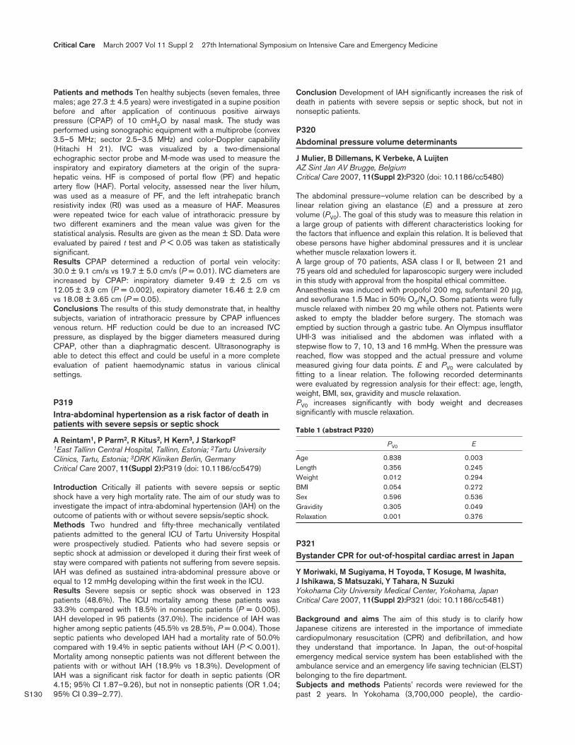
S130
Patients and methods Ten healthy subjects (seven females, threemales; age 27.3 ± 4.5 years) were investigated in a supine positionbefore and after application of continuous positive airwayspressure (CPAP) of 10 cmH2O by nasal mask. The study wasperformed using sonographic equipment with a multiprobe (convex3.5–5 MHz; sector 2.5–3.5 MHz) and color-Doppler capability(Hitachi H 21). IVC was visualized by a two-dimensionalechographic sector probe and M-mode was used to measure theinspiratory and expiratory diameters at the origin of the supra-hepatic veins. HF is composed of portal flow (PF) and hepaticartery flow (HAF). Portal velocity, assessed near the liver hilum,was used as a measure of PF, and the left intrahepatic branchresistivity index (RI) was used as a measure of HAF. Measureswere repeated twice for each value of intrathoracic pressure bytwo different examiners and the mean value was given for thestatistical analysis. Results are given as the mean ± SD. Data wereevaluated by paired t test and P < 0.05 was taken as statisticallysignificant.Results CPAP determined a reduction of portal vein velocity:30.0 ± 9.1 cm/s vs 19.7 ± 5.0 cm/s (P = 0.01). IVC diameters areincreased by CPAP: inspiratory diameter 9.49 ± 2.5 cm vs12.05 ± 3.9 cm (P = 0.002), expiratory diameter 16.46 ± 2.9 cmvs 18.08 ± 3.65 cm (P = 0.05).Conclusions The results of this study demonstrate that, in healthysubjects, variation of intrathoracic pressure by CPAP influencesvenous return. HF reduction could be due to an increased IVCpressure, as displayed by the bigger diameters measured duringCPAP, other than a diaphragmatic descent. Ultrasonography isable to detect this effect and could be useful in a more completeevaluation of patient haemodynamic status in various clinicalsettings.
P319Intra-abdominal hypertension as a risk factor of death inpatients with severe sepsis or septic shock
A Reintam1, P Parm2, R Kitus2, H Kern3, J Starkopf2
1East Tallinn Central Hospital, Tallinn, Estonia; 2Tartu UniversityClinics, Tartu, Estonia; 3DRK Kliniken Berlin, GermanyCritical Care 2007, 11(Suppl 2):P319 (doi: 10.1186/cc5479)
Introduction Critically ill patients with severe sepsis or septicshock have a very high mortality rate. The aim of our study was toinvestigate the impact of intra-abdominal hypertension (IAH) on theoutcome of patients with or without severe sepsis/septic shock.Methods Two hundred and fifty-three mechanically ventilatedpatients admitted to the general ICU of Tartu University Hospitalwere prospectively studied. Patients who had severe sepsis orseptic shock at admission or developed it during their first week ofstay were compared with patients not suffering from severe sepsis.IAH was defined as sustained intra-abdominal pressure above orequal to 12 mmHg developing within the first week in the ICU.Results Severe sepsis or septic shock was observed in 123patients (48.6%). The ICU mortality among these patients was33.3% compared with 18.5% in nonseptic patients (P = 0.005).IAH developed in 95 patients (37.0%). The incidence of IAH washigher among septic patients (45.5% vs 28.5%, P = 0.004). Thoseseptic patients who developed IAH had a mortality rate of 50.0%compared with 19.4% in septic patients without IAH (P < 0.001).Mortality among nonseptic patients was not different between thepatients with or without IAH (18.9% vs 18.3%). Development ofIAH was a significant risk factor for death in septic patients (OR4.15; 95% CI 1.87–9.26), but not in nonseptic patients (OR 1.04;95% CI 0.39–2.77).
Conclusion Development of IAH significantly increases the risk ofdeath in patients with severe sepsis or septic shock, but not innonseptic patients.
P320Abdominal pressure volume determinants
J Mulier, B Dillemans, K Verbeke, A LuijtenAZ Sint Jan AV Brugge, BelgiumCritical Care 2007, 11(Suppl 2):P320 (doi: 10.1186/cc5480)
The abdominal pressure–volume relation can be described by alinear relation giving an elastance (E) and a pressure at zerovolume (PV0). The goal of this study was to measure this relation ina large group of patients with different characteristics looking forthe factors that influence and explain this relation. It is believed thatobese persons have higher abdominal pressures and it is unclearwhether muscle relaxation lowers it.A large group of 70 patients, ASA class I or II, between 21 and75 years old and scheduled for laparoscopic surgery were includedin this study with approval from the hospital ethical committee.Anaesthesia was induced with propofol 200 mg, sufentanil 20 µg,and sevoflurane 1.5 Mac in 50% O2/N2O. Some patients were fullymuscle relaxed with nimbex 20 mg while others not. Patients wereasked to empty the bladder before surgery. The stomach wasemptied by suction through a gastric tube. An Olympus insufflatorUHI-3 was initialised and the abdomen was inflated with astepwise flow to 7, 10, 13 and 16 mmHg. When the pressure wasreached, flow was stopped and the actual pressure and volumemeasured giving four data points. E and PV0 were calculated byfitting to a linear relation. The following recorded determinantswere evaluated by regression analysis for their effect: age, length,weight, BMI, sex, gravidity and muscle relaxation.PV0 increases significantly with body weight and decreasessignificantly with muscle relaxation.
Table 1 (abstract P320)
PV0 E
Age 0.838 0.003Length 0.356 0.245Weight 0.012 0.294BMI 0.054 0.272Sex 0.596 0.536Gravidity 0.305 0.049Relaxation 0.001 0.376
P321Bystander CPR for out-of-hospital cardiac arrest in Japan
Y Moriwaki, M Sugiyama, H Toyoda, T Kosuge, M Iwashita, J Ishikawa, S Matsuzaki, Y Tahara, N SuzukiYokohama City University Medical Center, Yokohama, JapanCritical Care 2007, 11(Suppl 2):P321 (doi: 10.1186/cc5481)
Background and aims The aim of this study is to clarify howJapanese citizens are interested in the importance of immediatecardiopulmonary resuscitation (CPR) and defibrillation, and howthey understand that importance. In Japan, the out-of-hospitalemergency medical service system has been established with theambulance service and an emergency life saving technician (ELST)belonging to the fire department.Subjects and methods Patients’ records were reviewed for thepast 2 years. In Yokohama (3,700,000 people), the cardio-
Critical Care March 2007 Vol 11 Suppl 2 27th International Symposium on Intensive Care and Emergency Medicine
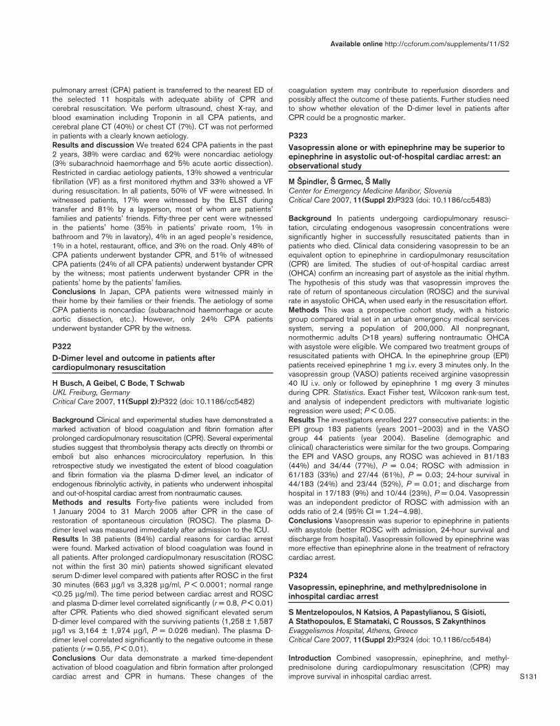
S131
pulmonary arrest (CPA) patient is transferred to the nearest ED ofthe selected 11 hospitals with adequate ability of CPR andcerebral resuscitation. We perform ultrasound, chest X-ray, andblood examination including Troponin in all CPA patients, andcerebral plane CT (40%) or chest CT (7%). CT was not performedin patients with a clearly known aetiology.Results and discussion We treated 624 CPA patients in the past2 years, 38% were cardiac and 62% were noncardiac aetiology(3% subarachnoid haemorrhage and 5% acute aortic dissection).Restricted in cardiac aetiology patients, 13% showed a ventricularfibrillation (VF) as a first monitored rhythm and 33% showed a VFduring resuscitation. In all patients, 50% of VF were witnessed. Inwitnessed patients, 17% were witnessed by the ELST duringtransfer and 81% by a layperson, most of whom are patients'families and patients’ friends. Fifty-three per cent were witnessedin the patients' home (35% in patients’ private room, 1% inbathroom and 7% in lavatory), 4% in an aged people’s residence,1% in a hotel, restaurant, office, and 3% on the road. Only 48% ofCPA patients underwent bystander CPR, and 51% of witnessedCPA patients (24% of all CPA patients) underwent bystander CPRby the witness; most patients underwent bystander CPR in thepatients’ home by the patients’ families.Conclusions In Japan, CPA patients were witnessed mainly intheir home by their families or their friends. The aetiology of someCPA patients is noncardiac (subarachnoid haemorrhage or acuteaortic dissection, etc.). However, only 24% CPA patientsunderwent bystander CPR by the witness.
P322D-Dimer level and outcome in patients aftercardiopulmonary resuscitation
H Busch, A Geibel, C Bode, T SchwabUKL Freiburg, GermanyCritical Care 2007, 11(Suppl 2):P322 (doi: 10.1186/cc5482)
Background Clinical and experimental studies have demonstrated amarked activation of blood coagulation and fibrin formation afterprolonged cardiopulmonary resuscitation (CPR). Several experimentalstudies suggest that thrombolysis therapy acts directly on thrombi oremboli but also enhances microcirculatory reperfusion. In thisretrospective study we investigated the extent of blood coagulationand fibrin formation via the plasma D-dimer level, an indicator ofendogenous fibrinolytic activity, in patients who underwent inhospitaland out-of-hospital cardiac arrest from nontraumatic causes.Methods and results Forty-five patients were included from1 January 2004 to 31 March 2005 after CPR in the case ofrestoration of spontaneous circulation (ROSC). The plasma D-dimer level was measured immediately after admission to the ICU.Results In 38 patients (84%) cardial reasons for cardiac arrestwere found. Marked activation of blood coagulation was found inall patients. After prolonged cardiopulmonary resuscitation (ROSCnot within the first 30 min) patients showed significant elevatedserum D-dimer level compared with patients after ROSC in the first30 minutes (663 µg/l vs 3,328 µg/ml, P < 0.0001; normal range<0.25 µg/ml). The time period between cardiac arrest and ROSCand plasma D-dimer level correlated significantly (r = 0.8, P < 0.01)after CPR. Patients who died showed significant elevated serumD-dimer level compared with the surviving patients (1,258 ± 1,587µg/l vs 3,164 ± 1,974 µg/l, P = 0.026 median). The plasma D-dimer level correlated significantly to the negative outcome in thesepatients (r = 0.55, P < 0.01).Conclusions Our data demonstrate a marked time-dependentactivation of blood coagulation and fibrin formation after prolongedcardiac arrest and CPR in humans. These changes of the
coagulation system may contribute to reperfusion disorders andpossibly affect the outcome of these patients. Further studies needto show whether elevation of the D-dimer level in patients afterCPR could be a prognostic marker.
P323Vasopressin alone or with epinephrine may be superior toepinephrine in asystolic out-of-hospital cardiac arrest: anobservational study
M ÐÐpindler, ÐÐ Grmec, ÐÐ MallyCenter for Emergency Medicine Maribor, SloveniaCritical Care 2007, 11(Suppl 2):P323 (doi: 10.1186/cc5483)
Background In patients undergoing cardiopulmonary resusci-tation, circulating endogenous vasopressin concentrations weresignificantly higher in successfully resuscitated patients than inpatients who died. Clinical data considering vasopressin to be anequivalent option to epinephrine in cardiopulmonary resuscitation(CPR) are limited. The studies of out-of-hospital cardiac arrest(OHCA) confirm an increasing part of asystole as the initial rhythm.The hypothesis of this study was that vasopressin improves therate of return of spontaneous circulation (ROSC) and the survivalrate in asystolic OHCA, when used early in the resuscitation effort.Methods This was a prospective cohort study, with a historicgroup compared trial set in an urban emergency medical servicessystem, serving a population of 200,000. All nonpregnant,normothermic adults (>18 years) suffering nontraumatic OHCAwith asystole were eligible. We compared two treatment groups ofresuscitated patients with OHCA. In the epinephrine group (EPI)patients received epinephrine 1 mg i.v. every 3 minutes only. In thevasopressin group (VASO) patients received arginine vasopressin40 IU i.v. only or followed by epinephrine 1 mg every 3 minutesduring CPR. Statistics. Exact Fisher test, Wilcoxon rank-sum test,and analysis of independent predictors with multivariate logisticregression were used; P < 0.05.Results The investigators enrolled 227 consecutive patients: in theEPI group 183 patients (years 2001–2003) and in the VASOgroup 44 patients (year 2004). Baseline (demographic andclinical) characteristics were similar for the two groups. Comparingthe EPI and VASO groups, any ROSC was achieved in 81/183(44%) and 34/44 (77%), P = 0.04; ROSC with admission in61/183 (33%) and 27/44 (61%), P = 0.03; 24-hour survival in44/183 (24%) and 23/44 (52%), P = 0.01; and discharge fromhospital in 17/183 (9%) and 10/44 (23%), P = 0.04. Vasopressinwas an independent predictor of ROSC with admission with anodds ratio of 2.4 (95% CI = 1.24–4.98).Conclusions Vasopressin was superior to epinephrine in patientswith asystole (better ROSC with admission, 24-hour survival anddischarge from hospital). Vasopressin followed by epinephrine wasmore effective than epinephrine alone in the treatment of refractorycardiac arrest.
P324Vasopressin, epinephrine, and methylprednisolone ininhospital cardiac arrest
S Mentzelopoulos, N Katsios, A Papastylianou, S Gisioti, A Stathopoulos, E Stamataki, C Roussos, S ZakynthinosEvaggelismos Hospital, Athens, GreeceCritical Care 2007, 11(Suppl 2):P324 (doi: 10.1186/cc5484)
Introduction Combined vasopressin, epinephrine, and methyl-prednisolone during cardiopulmonary resuscitation (CPR) mayimprove survival in inhospital cardiac arrest.
Available online http://ccforum.com/supplements/11/S2
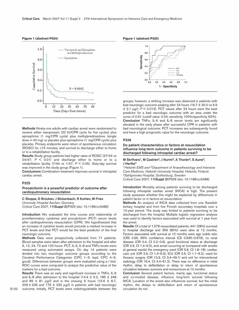
S132
Methods Ninety-one adults with cardiac arrest were randomized toreceive either vasopressin (20 IU/CPR cycle for five cycles) plusepinephrine (1 mg/CPR cycle) plus methylprednisolone (singledose = 40 mg) or placebo plus epinephrine (1 mg/CPR cycle) plusplacebo. Primary endpoints were return of spontaneous circulation(ROSC) for ≥15 minutes, and survival to discharge either to homeor to a rehabilitation facility.Results Study group patients had higher rates of ROSC (37/44 vs24/47; P < 0.01) and discharge either to home or to arehabilitation facility (7/44 vs 1/47; P < 0.05). Sixty-day survivalwas improved in the study group (Figure 1).Conclusions Combination treatment improves survival in inhospitalcardiac arrest.
P325Procalcitonin is a powerful predictor of outcome aftercardiopulmonary resuscitation
C Stoppe, D Brücken, J Bickenbach, R Kuhlen, M FriesUniversity Hospital Aachen, GermanyCritical Care 2007, 11(Suppl 2):P325 (doi: 10.1186/cc5485)
Introduction We evaluated the time course and relationship ofproinflammatory cytokines and procalcitonin (PCT) serum levelsafter cardiopulmonary resuscitation (CPR). We hypothesized thatan increase of cytokine levels would precede a marked increase inPCT levels and that PCT would be the best predictor of the finalneurologic outcome.Methods Data were prospectively collected from 71 patients.Blood samples were taken after admission to the hospital and after6, 12, 24, 72 and 120 hours. PCT, IL-6, IL-8 and TNFα levels weremeasured using automated assays. On day 14 patients weredivided into two neurologic outcome groups according to theCerebral Performance Categories (CPC 1–3: bad; CPC 4–5:good). Differences between groups were evaluated using a t test.ROC curves were computed to analyze the predictive value of themarkers for a bad outcome.Results There was an early and significant increase in TNFα, IL-6and IL-8 after admission to the hospital (14.4 ± 5.2, 185 ± 248and 89 ± 81 µg/l) and in the ensuing 6 hours (15.6 ± 8.7,209 ± 239 and 176 ± 232 µg/l) in patients with bad neurologicoutcome. Initially, PCT levels were indistinguishable between the
groups; however, a striking increase was observed in patients withbad neurologic outcome peaking after 24 hours (16.7 ± 30.0 vs 6.9± 2.1 µg/l; P < 0.013). PCT values after 24 hours were the bestpredictor for a bad neurologic outcome with an area under thecurve of 0.91 (cutoff value: 0.44; sensitivity 100%/specificity 62%).Conclusion TNFα, IL-6 and IL-8 serum levels are significantlyelevated in the early phase after successful CPR in patients withbad neurological outcome. PCT increases are subsequently foundand have a high prognostic value for the neurologic outcome.
P326Do patient characteristics or factors at resuscitationinfluence long-term outcome in patients surviving to bedischarged following inhospital cardiac arrest?
M Skrifvars1, M Castrén2, J Nurmi2, A Thorén3, S Aune3, J Herlitz3
1Helsinki EMS and 2Department of Anaesthesiology and IntensiveCare Medicine, Helsinki University Hospital, Helsinki, Finland;3Sahlgrenska Hospital, Gothenburg, SwedenCritical Care 2007, 11(Suppl 2):P326 (doi: 10.1186/cc5486)
Introduction Mortality among patients surviving to be dischargedfollowing inhospital cardiac arrest (IHCA) is high. The presentstudy assesses whether this might be explained by differences inpatient factor or in factors at resuscitation.Methods An analysis of IHCA data collected from one Swedishtertiary hospital and from five Finnish secondary hospitals over a10-year period. The study was limited to patients surviving to bedischarged from the hospital. Multiple logistic regression analysiswas used to identify factors associated with survival at 1 year fromthe arrest.Results Of a total of 1,578 resuscitated patients, 441 (28%) survivedto hospital discharge and 359 (80%) were alive at 12 months.Factors associated with survival at 12 months were age (odds ratio(OR) 0.96, 95% confidence interval (CI) 0.935–0.979), no renaldisease (OR 0.4, CI 0.2–0.9), good functional status at discharge(OR 2.9, CI 1.4–6.0), and arrest occurring at (compared with arrestsat general wards) the emergency ward (OR 5.8, CI 1.8–18), cardiaccare unit (OR 2.9, CI 1.3–6.3), ICU (OR 2.6, CI 1.1–6.2), ward forthoracic surgery (OR 12.9, CI 3.4–49.1) and unit for interventionalradiology (OR 16.4, CI 4.4–61.2). There was no difference in initialrhythm, delay to defibrillation or delay to return of spontaneouscirculation between survivors and nonsurvivors at 12 months.Conclusion Several patient factors, mainly age, functional statusand co-morbid disease, influence long-term survival followingIHCA. Location of the arrest also influences survival, but the initialrhythm, the delays to defibrillation and return of spontaneouscirculation do not.
Critical Care March 2007 Vol 11 Suppl 2 27th International Symposium on Intensive Care and Emergency Medicine
Figure 1 (abstract P324) Figure 1 (abstract P325)
S133
P327Out-of-hospital surface cooling with a cooling-blanket toinduce mild hypothermia in humans after cardiac arrest: afeasibility trial
T Uray1, R Malzer2, A Auer2, A Zajicek2, F Sterz1, J Arrich1, R Fleischhackl1, A Janata1, M Holzer1, A Laggner1, W Behringer1
1Medical University Vienna, Austria; 2Ambulance Service of Vienna,AustriaCritical Care 2007, 11(Suppl 2):P327 (doi: 10.1186/cc5487)
Introduction Mild hypothermia (32–34°C) is a promising newtherapy for patients resuscitated from cardiac arrest. Animalstudies suggest that early and fast cooling is crucial for beneficialeffect on neurological outcome. Inducing mild hypothermiaimmediately after successful restoration of spontaneous circulation(ROSC) in the out-of-hospital setting remains a challenge.Therefore, a novel cooling-blanket (EMCOOLSpad®), independentof any energy source during use, was developed. The aim of thestudy was to evaluate feasibility and safety of out-of-hospitalsurface cooling with EMCOOLSpad® in patients successfullyresuscitated from cardiac arrest.Methods We included patients successfully resuscitated from out-of-hospital cardiac arrest with an oesophageal temperature (Tes)>34°C. The EMCOOLSpad® consists of multiple cooling units(12 mm thick), filled with a mixture of graphite/water, which arestored in a cooling box at –3°C in the ambulance car. Cooling wasinitiated as soon as feasible by the first treating paramedics andemergency physicians, and was continued in the emergency room.The cooling-blanket was removed when the Tes reached 34°C. Thetarget temperature of Tes 33°C was maintained for 24 hours. Dataare presented as the median and interquartile range (25–75%).Results From September 2006 to December 2006, 10 patients,weighing 70 (64–93) kg, were included in the study. Cooling wasinitiated 14 (7–20) minutes after ROSC. The cooling-blanketdecreased the Tes from 36.5 (36.2–36.7)°C at the start of coolingto 34.0°C within 61 (47–93) minutes, and to target temperatureTes 33°C within 83 (61–119) minutes, resulting in a cooling rate of2.6 (1.6–3.6)°C/hour. Hospital admission was 45 (40–53) minutesafter ROSC, and Tes 33°C was achieved 78 (32–107) minutesafter admission. In eight patients, precooled parts of the cooling-blanket had to be applied repeatedly on the chest and abdomen tomaintain the target temperature of Tes 33°C for 24 hours. No skinlesions were observed.Conclusion Noninvasive surface cooling with the EMCOOLSpad®
immediately after resuscitation from cardiac arrest, in the out-of-hospital setting, was shown to be feasible and safe. Whether earlycooling, as compared with delayed cooling in the hospital, willimprove neurological outcome needs to be determined in aprospective randomized trial.
P328Mild hypothermia induction following cardiac arrest using awater-circulating cooling device
E Miñambres, B Suberviola, A González-Castro, J San Jose, J Gutierrez MorloteHospital Universitario Marques de Valdecilla, Santander, SpainCritical Care 2007, 11(Suppl 2):P328 (doi: 10.1186/cc5488)
Introduction The use of mild hypothermia for comatose survivorsof cardiac arrest has been endorsed by the American HeartAssociation and the International Liaison Committee on Resusci-tation [1,2]. Unintentional overcooling is common with sometechniques such as cool intravascular fluid or the use of ice packs.
Nowadays, the maintenance of hypothermia can be facilitated withnew technology to avoid unintentional overcooling.Methods A 77-year-old male with a history of hypertension,previous replacement of aortic valve and a right coronary arterybypass was admitted to our ICU after cardiac arrest. He suffered acollapse while walking. The emergency service arrived within5 minutes. The initial cardiac rhythm was ventricular fibrillation. Theestimated time to return to spontaneous circulation was20 minutes. The patient arrived in the hospital 50 minutes aftercollapse and was immediately admitted to the ICU. Thirty minutesafter ICU admission, he was unconscious with a Glasgow comascore of 5. Hypothermia was induced by the Artic Sun 2000cooling system (Medivance, Louisville, CO, USA), and the goaltemperature was obtained 105 minutes after induction. The bodytemperature was monitored continuously with a Foley catheter.Hypothermia was maintained for 24 hours at 33ºC and rewarmingto the target temperature of 37ºC was achieved over 12 hours. Noelectrolyte imbalances or coagulopathies were observed. Noovercooling was observed at any moment. The patient wasextubated on day 6 after admission and discharged from the ICUon day 10 without neurological sequelae.Conclusions Careful monitoring of temperature is important duringuse of therapeutic hypothermia because unintentional overcoolingbelow 32°C may place the patient at risk for serious complicationssuch as arrhythmias, infection, and coagulopathy. Cooling with awater-circulating cooling device is fast and safe. Clinicians shouldwork to institute protocols for mild hypothermia treatment for suchpatients as a part of their critical care treatment.References1. Bernard SA, Gray TW, Buist MD, et al.: Treatment of
comatose survivors of out-of-hospital cardiac arrest withinduced hypothermia. N Engl J Med 2002; 346:557-563.
2. 2005 International Consensus on CardiopulmonaryResuscitation (CPR) and Emergency Cardiovascular Care(ECC) Science with Treatment Recommendations. Circula-tion 2005, 112 (Suppl):III-1-III-136.
P329Induction of mild hypothermia in cardiac arrest survivorswith cardiogenic shock syndrome
R Skulec, J Belohlavek, V Dytrych, T Kovarnik, M Aschermann,A LinhartGeneral Teaching Hospital, Prague, Czech RepublicCritical Care 2007, 11(Suppl 2):P329 (doi: 10.1186/cc5489)
Introduction Induction of mild hypothermia (MH) in patientsresuscitated from cardiac arrest can improve their outcome.However, benefits and risks of MH induction in patients whoremain in cardiogenic shock after the return of spontaneouscirculation (ROSC) are unclear. We therefore analysed a group ofall cardiac arrest survivors who were indicated for MH induction inour coronary care unit (CCU) and compared the outcome ofpatients with cardiogenic shock syndrome after ROSC with theoutcome of those who were relatively haemodynamically stable.Methods We performed retrospective analysis of all consecutivecardiac arrest survivors treated by MH in our CCU from November2002 to August 2006. They were classified into two groups,according to whether they met the criteria for cardiogenic shock ornot after ROSC and just before MH initiation. Primary outcomemeasures were inhospital mortality, and the best inhospital anddischarge neurological result. Predicted mortality was evaluated bythe APACHE II score, and neurological outcome by CerebralPerformance Category score. MH was initiated as soon as
Available online http://ccforum.com/supplements/11/S2
S134
possible after ROSC and patients were cooled to bodytemperature 32–34ºC for 12 hours.Results From 50 consecutive patients, 28 fulfilled criteria ofcardiogenic shock before MH initiation (group A), and 22 wererelatively hemodynamically stable (group B). While predictedmortality was 83.1 ± 13.1% in group A and 63.2 ± 19.0% in groupB (P < 0.001), real inhospital mortality was 55.6% in group A andonly 18.2% in group B patients (P = 0.009). The best inhospitalneurological outcome was found favourable in 71.4% patients ingroup A and in 86.3% in group B (P = 0.306). Favourabledischarge neurological outcome was reached in 100% in group Aand in 94% in group B (P = 1.000). Patients in both groups didnot differ in rate of complications.Conclusions While inhospital mortality in cardiac arrest survivorstreated by MH was expectably higher in those with cardiogenicshock than in stable patients, favourable neurological outcome wasfrequent and comparable in both groups of patients. Moreover, MHapplication was safe in both groups. Therefore, induction of MHshould be considered also in cardiac arrest survivors withcardiogenic shock syndrome after ROSC.
P330Changes in urinary 8-hydroxy-2-deoxyguanosine inpatients with global brain ischemia undergoing brainhypothermia therapy: comparison of whole body andselective head cooling
K Ikeda, Y Kuroki, K Yosikawa, T Yokoyama, H UtinoHachiouji Medical Center, Tokyo, JapanCritical Care 2007, 11(Suppl 2):P330 (doi: 10.1186/cc5490)
Introduction Oxygen free radicals play an important role in globalbrain ischemia after cardiac arrest. Brain hypothermia therapy iseffective in suppressing free radical expression. The aim of thisstudy was to assess free radical expression under brainhypothermia, and to compare the expression between whole bodyand selective head cooling.Methods The subjects were 12 patients treated with mild brainhypothermia (34 ± 1°C) after resuscitation following cardiac arrestin our ICU; five patients received whole body cooling and sevenpatients received selective head cooling. We examined thehemodynamic changes and the urinary concentration of 8-hydroxy-2-deoxyguanosine (determined by HPLC) during brain hypothermiatherapy. Furthermore, we compared the prognosis at 28 days afteradmission to the ICU.Results The induction time for whole body cooling wassignificantly shorter than that for selective head cooling. Therewarming time for head cooling was significantly shorter than thatfor whole body cooling. The mean arterial pressure and heart ratewere both stable in the head cooling group. The urinary 8-hydroxy-2-deoxyguanosine concentrations decreased significantly in bothgroups, but data were significantly lower in the whole body coolinggroup compared with the selective head cooling group. Five andseven patients, respectively, exhibited good recovery 28 days afteradmission, in the whole body and selective head cooling groups.Conclusions Mild brain hypothermia therapy suppressed theproduction of free radicals following global brain ischemia. Wholebody cooling had a stronger effect of suppression of free radicalscompare with selective head cooling. It is considered that selectivehead cooling exhibits neuroprotection similar to whole bodycooling.
P331Reduction of magnetic resonance spectroscopy braintemperature by convective head cooling in healthy humans
B Harris1, P Andrews1, I Marshall1, T Robinson2, G Murray1
1University of Edinburgh, UK; 2KCI, Ferndown, UKCritical Care 2007, 11(Suppl 2):P331 (doi: 10.1186/cc5491)
This pilot study assessed the effect of forced convective head andneck cooling on brain temperature, measured by magnetic resonancespectroscopy (MRS), in five healthy adult humans (three males).Following a 10-minute baseline, subjects received 30 minuteshead cooling followed by 30 minutes head and neck cooling. Thecooling device delivered air at 11ºC and 15 m/s through a hoodand separate neck collar made of a double layer of nylon sheeting,the inner layer pierced with holes. Subjects wore a windproofwaistcoat taped round the base of their neck and were wrapped inblankets from the base of the neck down. Bilateral foot warmingwith chemical hot packs was used to encourage heat loss in thepresence of normothermia.MRS temperature data were collected at the level of the basalganglia over the baseline and the last 10 minutes of each coolingintervention. MRS detects naturally occurring brain metabolitesand interpretation of the relative frequencies of N-acetyl aspartateand water allows estimation of tissue temperature in 1 cm3 voxels.For assessment of regional cooling, voxels lying within the regionformed by joining the tips of the lateral ventricles were defined as‘core’, voxels within approximately one voxel of the brain surfacewere defined as ‘outer’, and all other voxels were defined as‘intermediate’. The oesophageal temperature was measuredcontinuously with a fluoroptic thermometer.The mean baseline-corrected MRS brain temperature over allvoxels reduced by –0.45ºC (SD 0.23ºC, P = 0.01, 5% CI –0.74 to–0.17ºC) with head cooling and –0.37ºC (SD 0.30ºC, P = 0.049,95% CI –0.74 to 0.00ºC) with head and neck cooling. Headcooling reduced the mean baseline-corrected MRS braintemperature in core voxels in all subjects. The formal test forgradient was not significant (P = 0.43; 95% CI –0.15 to 0.29ºC).Head and neck cooling reduced the temperature in core voxels inthree subjects; the test for gradient was not significant (P = 0.07;95% CI –0.03 to 0.58ºC). The mean baseline-correctedoesophageal temperature reductions for the last 10 minutes ofeach intervention were –0.16ºC (SD 0.04ºC) with head coolingand –0.36ºC (SD 0.12ºC) with head and neck cooling.Forced convective head cooling reduced the MRS braintemperature at an equivalent of 1.35ºC per hour in healthysubjects, and the reduction was apparent across the brain.
P332Induction of therapeutic mild hypothermia after cardiacarrest: a new combined method to achieve the targettemperature
H Busch, T Schwab, K Fink, C BodeUKL Freiburg, GermanyCritical Care 2007, 11(Suppl 2):P332 (doi: 10.1186/cc5492)
Background Clinical and experimental investigations have demon-strated that induction of mild hypothermia works after successfulcardiopulmonal resuscitation (CPR) neuroprotection. After thepresentation of controlled studies, therapeutic hypothermia movedinto the topical international guidelines.Methods and results A total of 50 patients were examined aftersuccessful CPR. Twenty-nine patients received 4°C cold infusionsafter arrival in the heart catheter laboratory. Retrospective data of
Critical Care March 2007 Vol 11 Suppl 2 27th International Symposium on Intensive Care and Emergency Medicine
S135
21 patients who had received a volume substitute by means ofdrips from ambient temperature served as a control group. Afteradmission to the ICU, both groups were immediately connected toan external cooling device (CoolGard® or Thermo Wrap®) and werecooled to a target temperature of 33°C (bladder temperature).Results The average temperature at admission did not differ inboth groups (35.5 ± 0.9°C vs 35.89 ± 0.8°C). In the group withinitial cooling by means of 4°C cold infusions, a significanttemperature decrease could be reached during the invasivecoronary diagnostics to admission to the ICU of an average 0.84°C(35.88 ± 0.9°C vs 35.04 ± 0.9°C, P < 0.0001). The middle chillduration up to the achievement of the target temperature afteradmission was significantly shorter with the combined method(341 ± 113 min versus 553 ± 342 min, P < 0.01). The period tothe achievement of the target temperature after the beginning ofthe external cooling device with the group of the combined methodwas significantly shorter (163 ± 91 min versus 342 ± 258 min,P < 0.01).Conclusions The combined method with initial cooling with 4°Ccold solutions shows a sure and actual prestationary coolingprocedure to the introduction or realisation of mild hypothermia andoffers the possibility to reach the purpose temperature significantlyfaster. Preclinical introduction of mild hypothermia by means of 4°Ccold solutions could be a beneficial criteria in the future treatment,and probably affects the outcome of these patients.
P333A comparison of complications during therapeutichypothermia between surface cooling and endovascularcooling techniques
P Hayden, A Salam, R Beale, M GilliesGuy’s & St Thomas’ NHS Foundation Trust, London, UKCritical Care 2007, 11(Suppl 2):P333 (doi: 10.1186/cc5493)
Introduction Therapeutic hypothermia (TH) following cardiacarrest is associated with several complications including sympto-matic bradycardia, coagulopathy, and pneumonia [1]. Furthermore,hyperthermia is associated with poor outcome following braininjury. The incidence of these complications may be increased byexcessive temperature fluctuations. We sought to comparecomplications between two techniques used to induce TH; surfacecooling (SC) using ice packs, and endovascular cooling (EV),using the Coolgard™ system (Alsius Corp., USA).Methods A retrospective review was performed of all cardiacarrest patients undergoing TH and surviving ≥48 hours betweenJune 2005 and November 2006.Results Thirty-five patients underwent our TH protocol (SC group =21, EV group = 14). The incidence of overcooling (<32°C) in theSC group was significantly higher than the EV group (10 vs 1,P = 0.01), whilst a trend towards more episodes of symptomaticbradycardia (SC 9 vs EV 2, P = 0.07) and rebound hyperthermia(SC 9 vs EV 2, P = 0.07) was also present. The incidence ofpneumonia (SC 7 vs EV 4, P = 0.77) and coagulopathy/bleeding(SC 2 vs EV 3, P = 0.32) were similar between groups.Conclusions (1) SC is associated with a significantly higherincidence of overcooling than EC and may be associated with anincrease in complications such as symptomatic bradycardia. (2)SC may also be associated with an increase in reboundhyperthermia.Reference1. Polderman KH: Application of therapeutic hypothermia in
the intensive care unit. Opportunities and pitfalls of apromising treatment modality – Part 2: practical aspectsand side effects. Intensive Care Med 2004, 30:757-769.
P334Buspirone and dexmedetomidine synergistically reducethe shivering threshold in humans
R Lenhardt, R Komatsu, M Orhan-SungurUniversity of Louisville, KY, USACritical Care 2007, 11(Suppl 2):P334 (doi: 10.1186/cc5494)
Introduction Hypothermia may be therapeutically beneficial instroke victims; however, it provokes vigorous shivering. Buspirone,a partial serotonin 1A antagonist, and dexmedetomidine, an α2agonist, linearly reduce the shivering threshold (triggering coretemperature) with minimal sedation and respiratory depression. Wetested the hypothesis that buspirone and dexmedetomidinesynergistically reduce the shivering threshold without producingsubstantial sedation or respiratory depression.Methods We studied four healthy male volunteers (18–40) on4 days: (1) control (no drug); (2) buspirone only (60 mg orally); (3)dexmedetomidine only (target plasma concentration 0.6 ng/ml);and (4) combined buspirone and dexmedetomidine in the samedoses. Lactated Ringer’s solution (3°C) was infused via a centralvenous catheter to decrease tympanic membrane temperature by≈2.2°C/hour; the mean skin temperature was maintained at 31°C.An increase in oxygen consumption more than 25% of baselineidentified the shivering threshold. Sedation was evaluated usingthe Observer’s Assessment Sedation/Alertness scale. Two-wayrepeated-measures analysis of variance was used to identifyinteractions between drugs. Data are presented as means ± SDs;P < 0.05 was statistically significant.Results The shivering thresholds were 36.4 ± 0.5°C on the controlday; 34.9 ± 0.6°C (P < 0.01 from control) on the buspirone onlyday; 36.1 ± 0.6°C (P < 0.01 from control) on the dexmedeto-midine only day; and 34.2 ± 0.5°C (P < 0.01 from control) on thecombined buspirone and dexmedetomidine day. The calculatedmean difference between the thresholds on the combined and thecontrol days was 1.9 ± 0.4°C, while the measured mean differencederived from the difference between the combined and controldays was 2.3 ± 0.4°C. There was only trivial sedation with eitherdrug alone or in combination. The respiratory rate and end-tidalPCO2 were well preserved on all days.Conclusion Buspirone and dexmedetomidine act synergistically toreduce the shivering threshold with only mild sedation and norespiratory depression. This combination might be a valid treatmentto prevent shivering in stroke patients during therapeutichypothermia.
P335Bispectral index and suppression ratio are very earlypredictors of neurological outcome during therapeutichypothermia after cardiac arrest
D Seder, R Riker, G Fraser, H Bruce, T RobbinsMaine Medical Center, Portland, ME, USACritical Care 2007, 11(Suppl 2):P335 (doi: 10.1186/cc5495)
Introduction The bispectral index (BIS) is calculated from fronto-temporal electroencephalogram (EEG), and the suppression ratio(SR) estimates the percentage of EEG suppression. We monitoredthe BIS and SR during therapeutic hypothermia (TH) andcompared them with neurological outcomes of encephalopathicsurvivors of out-of-hospital cardiac arrest (OHCA).Methods Thirty-two patients with anoxic encephalopathy afterOHCA received 18 hours of TH at 32–34°C. BIS monitoring wasinitiated at the onset of TH, and neuromuscular blockade (NMB)was dosed in response to shivering. Blinded BIS and SR data
Available online http://ccforum.com/supplements/11/S2
S136
were recorded after the first dose of NMB, and compared with theCerebral Performance Category (CPC) at discharge and 6 months.CPC 1 or CPC 2 was considered a good outcome (GO).Results Fourteen out of 32 patients (44%) survived, 11 (34%)with GO. Five of the remaining 18 patients died before neuro-logical evaluation at 72 hours, and one patient recoveredneurological function but died of cardiogenic shock. No survivorrecalled the period of NMB. First NMB was administered a medianof 5 hours after cardiac arrest or 87 minutes after initiation of TH,at 35.6 ± 1.7°C. Patients with GO had a higher first post-NMB BIS(39 ± 6 vs 13 ± 14, P < 0.001) and a lower SR (10 ± 12 vs69 ± 29, P < 0.001) than those with CPC 3–5. Initial NMBreduced frontotemporal electromyogram (EMG) power from52 ± 8 to 27 ± 1 db, P < 0.001. In 17 of the patients withdownloaded EEG data, an increase in EMG power of 17 dB (IQR10–27) from baseline was associated with clinically detectableshivering. Epileptiform discharges were noted on the monitorduring NMB in two patients, and seizure activity was confirmed byformal EEG in both.Conclusions In cardiac arrest survivors receiving TH, a higherpost-NMB BIS score and a lower SR are very early predictors ofneurological outcome. The potential benefits of monitoring BIS andSR, as well as EMG power for early recognition of shivering, andcontinuous frontotemporal EEG to detect seizures, warrant furtherstudy.
P336Role of bedside electroencephalogram in intensive care: a critical review
R Jayaram Ramachandran, C Hargreaves, S SinhaWhittington Hospital NHS Trust, London, UKCritical Care 2007, 11(Suppl 2):P336 (doi: 10.1186/cc5496)
Electroencephalogram (EEG) is an appropriate monitoring tool inintensive care because it is linked to cerebral metabolism, issensitive to ischaemia/hypoxia, can detect neuronal dysfunction ata reversible stage and is the best method to detect seizure activity.Scientific data have proved utility of continuous EEG monitoring inintensive care [1,2]. But there is a paucity of data relating to singlerecordings of EEG especially in general ICUs.A retrospective chart review of patients who had bedside EEG in amedical–surgical ICU was done. Data were collected with a focuson: indication for requesting EEG, technical difficulties during thestudy, the report and its influence on subsequent clinical manage-ment.Forty-two charts were reviewed. The indications were: evaluationof persistent comatose state (n = 27), to diagnose/exclude seizureactivity and nonconvulsive status epilepsy (n = 12), and as anadjunct to support clinical diagnosis of suspected brain death priorto formal testing (n = 3). Movement artifacts led to technicaldifficulty in four studies.EEG confirmed: moderate to severe nonspecific brain dysfunctionas the cause for persistent comatose state by the presence ofeither diffuse slowing with theta/delta activity, absence of cerebralactivity, continuous rhythmic and semi rhythmic lateralized/bilateralepileptiform discharges, burst suppression pattern or continuousbilateral slow U-shaped waves; anoxic brain damage by absence ofchanges in electrical signals following external application ofnoxious stimuli; and seizure activity by epileptiform discharges.Twelve reports stated that use of sedation interfered with EEGinterpretation.The following clinical decisions were made based on the EEGreport in conjunction with clinical findings: initiating withdrawal oflife support or ‘do-not-resuscitate orders’ in patients diagnosed to
have hypoxic ischaemic encephalopathy (n = 15); adding/escalating or stopping antiseizure drugs based on the presence/absence of seizure activity (n = 12); and continuing supportivecare in comatose patients diagnosed to have metabolic encephalo-pathy/prolonged sedation effect as the cause for coma (n = 8).Based on these results it can be concluded that, despite limitationssuch as motion artifacts and influence of sedation on electricalsignals, EEG impacts on clinical decision-making processes incritical care. Hence it is beneficial, and more widespread usewould improve its diagnostic potential.References1. Hirsch LJ, Kull LL:Am J Electroneurodiagnostic Technol
2004, 44:137-158.2. Newton DEF: Electrophysiological monitoring of general
intensive care patients. Intensive Care Med 1999, 25:350-352.
P337Abstract withdrawn
Critical Care March 2007 Vol 11 Suppl 2 27th International Symposium on Intensive Care and Emergency Medicine
S137
P338Aneurysmal subarachnoid hemorrhage induces theexpression of Pentraxin3 in patients
G Brandi1, E Roncati Zanier1, L Longhi1, G Peri2, G De Simon3,M Tettamanti3, A Mantovani2, N Stocchetti11Milan University, Ospedale Maggiore Policlinico IRCCS, Milan,Italy; 2Clinical Institute Humanitas, Milan, Italy; 3Mario NegriInstitute, Milan, ItalyCritical Care 2007, 11(Suppl 2):P338 (doi: 10.1186/cc5498)
Introduction Aneurismal subarachnoid hemorrhage (SAH) is anextremely severe illness associated with a high mortality rate andpermanent severe neurological dysfunction in two-thirds of allaffected patients. One of the major complications of SAH isvasospasm-associated cerebral ischemia. Clinical and experi-mental data suggest that vasospasm is linked to the inflammatoryresponse associated with SAH. The goal of this study was toinvestigate the expression of Pentraxin3 (PTX3), a prototypic longpentraxin protein induced by proinflammatory signals in the brain,in SAH patients to test the hypothesis that SAH is followed by anupregulation of PTX3, and establish a temporal relationshipbetween the expression of PTX3 and the induction of vasospasm.We also attempted to establish that PTX3 is detectable in cerebro-spinal fluid (CSF).Methods We studied eight severe SAH patients admitted to ourneuroscience ICU with a median World Federation NeurosurgicalScore of 4 and a Fisher score of 4. Arterial, jugular venous bloodand CSF samples were routinely obtained every 12 hours for7 days. PTX3 levels were measured by ELISA in plasma and CSFsamples.Results Compared with plasma levels of PTX3 in normalvolunteers (<2 ng/ml [1]), SAH induced a marked increase inplasma PTX3 expression. During the first 48 hours following SAH(acute phase), PTX3 arterial and jugular venous levels increased to36.93 ± 24.32 ng/ml and 33.64 ± 28.76 ng/ml, respectively, andthen subsequently decreased concomitantly with the reduction ofthe inflammation (48–96 hours: subacute phase). PTX3 isdetectable in the CSF: mean CSF levels of PTX3 were4.07 ± 3.64 ng/ml during the acute phase and 0.69 ± 0.44 ng/mlduring the subacute phase (t test: P < 0.05 compared with theacute phase). In the presence of vasospasm (four patients), wedetected a second peak of PTX3 (4.03 ± 2.85 ng/ml) in CSFsamples (t test: P < 0.05 compared with the subacute phase) thatwas not detectable in plasma.Conclusions SAH is characterized by the production of PTX3 andthe induction of vasospasm is associated with an upregulation ofPTX3 in the CSF that is not detectable in plasma.Reference1. Muller et al.: Crit Care Med 2001, 29:1404-1407.
P339Outcome of intensive care unit patients with spontaneousintracerebral hemorrhage
M Sartzi, A Papaeveggelou, A Stogiannidi, P Kouki, B Romanou, E Panagiotakopoulou, G Kallitsi, K Mihas, F Tsidemiadou, P Clouva-MolyvdaGeneral Hospital of Eleusis, Athens, GreeceCritical Care 2007, 11(Suppl 2):P339 (doi: 10.1186/cc5499)
Introduction We evaluated the factors that may influence theoutcome of patients with spontaneous intracerebral hemorrhage(SICH).
Methods Retrospective analysis of prospectively gathered data of102 patients with SICH treated in our ICU during the past 8 years.On admission the following data were registered: vascular riskfactors (high blood pressure, diabetes mellitus), age, gender,APACHE II score, GCS, hemorrhage characteristics (location, side,volume, mass effect), surgical procedure, MODS, blood pressure(systolic, diastolic, mean), pulse pressure, pulse rate, laboratoryparameters (hemoglobin, white cell and platelet count, INR, serumvalues for Na, glucose, lactate, creatinin, bilirubin). Also registeredwere length of stay (LOS), duration of mechanical ventilation (MV),time of intubation (TT) and patient outcome. Haemodynamicinstability was defined as low mean blood pressure and support withvasoactive and inotrop drugs. Statistical evaluation was performedusing univariate and multivariate logistic regression, Student’s t testPearson’s chi-square test and Fisher’s exact statistic were used.Results From the 102 patients (56 men and 48 women) 38(37.5%) died within the first 30 days, most of them in the first10 days. Age (OR 13.801, P < 0.04), APACHE II score (OR1.114, P < 0.008), GCS (OR 2.158, P < 0.002), ICH score (OR1.183, P < 0.001), FiO2/pO2 (OR 0.996, P < 0.009), haemodynamicinstability (OR 2.340, P < 0.002), fever (OR 1.245, P < 0.002),and INR (OR 13.801, P < 0.04) were the strongest associatedfactors of 30-day mortality. Gender (OR 0.652, P < 0.301), priorillness (OR 1.070, P < 0.870), MODS (OR 0.978, P < 0.803),LOS (OR 0.988, P < 0.266), MV (OR 0.994, P < 0.356) and TT(OR 0.990 P < 0.371) were not associated with mortality. Patientswho where operated on had higher mortality but were also moreseverely ill.Conclusion Age, severity of illness, ICH score, hypoxemia,haemodynamic instability, and increased temperature are directlyrelated with the outcome of patients with SICH. Gender, LOS, MV,TT, and MODS did not influence mortality.
P340Maintenance of prehospital medical systems due to clinicaladvance in acute stroke
K Ishii1, Y Wakabayashi1, Y Momii2, T Asano2, H Kenai2, M Yamashita2, M Mori2, Y Hori2, H Nagatomi21Oita University School of Medicine, Oita, Japan; 2NagatomiNeurosurgical Hospital, Oita, JapanCritical Care 2007, 11(Suppl 2):P340 (doi: 10.1186/cc5500)
Introduction Recently, the medical treatment in acute stroke hasbeen making rapid progress. Especially, in the ischemic stroke ofacute stage, the efficacy of thrombolysis, systemic t-PA or localtransarterial urokinase infusion has been proved. However, theeffective treatment time is still quite limited. The patients must bebrought to the stroke center as soon as possible. We analyzed thereason why most stroke patients delay coming to the stroke center.We extracted the problems and proposed some solutions.Patients and methods The clinical subjects consisted of 1,112consecutive patients with ischemic stroke in the acute stage,hospitalized in our hospital between April 2003 and September2006. We investigated the clinical course, especially the time fromthe onset to the physical examination, and radiological exami-nations (CT, MRI, MRA and/or cerebral angiography). The meanage was 72.3 years. Among them, 334 patients were classified asatherothrombosis, 232 were cardiac embolism, 439 were lacunarinfarction and 107 were transient ischemic attack. Only 19 patientsunderwent acute thrombolytic therapy.Results Two hundred and forty-one patients (21.7%) werehospitalized within 3 hours from the onset, and 365 patients(32.9%) were within 6 hours. Among them, only 438 were admittedby ambulance. We found the following results. The main reason for
Available online http://ccforum.com/supplements/11/S2
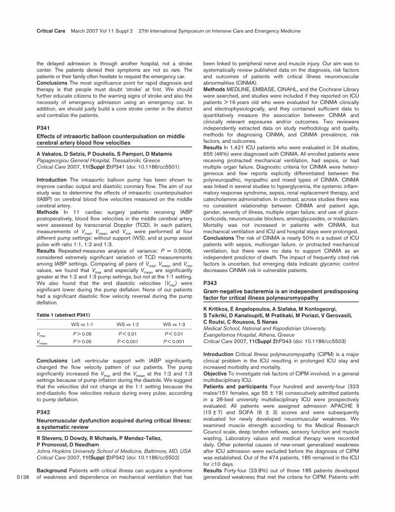
S138
the delayed admission is through another hospital, not a strokecenter. The patients denied their symptoms are not so rare. Thepatients or their family often hesitate to request the emergency car.Conclusions The most significance point for rapid diagnosis andtherapy is that people must doubt ‘stroke’ at first. We shouldfurther educate citizens to the warning signs of stroke and also thenecessity of emergency admission using an emergency car. Inaddition, we should justly build a core stroke center in the districtand centralize the patients.
P341Effects of intraaortic balloon counterpulsation on middlecerebral artery blood flow velocities
A Vakalos, D Setzis, P Doukelis, S Pampori, D MatamisPapageorgiou General Hospital, Thessaloniki, GreeceCritical Care 2007, 11(Suppl 2):P341 (doi: 10.1186/cc5501)
Introduction The intraaortic balloon pump has been shown toimprove cardiac output and diastolic coronary flow. The aim of ourstudy was to determine the effects of intraaortic counterpulsation(IABP) on cerebral blood flow velocities measured on the middlecerebral artery.Methods In 11 cardiac surgery patients receiving IABPpostoperatively, blood flow velocities in the middle cerebral arterywere assessed by transcranial Doppler (TCD). In each patient,measurements of Vmax, Vmean and Vmin were performed at fourdifferent pump settings: without support (WS), and at pump assistpulse with ratio 1:1, 1:2 and 1:3.Results Repeated-measures analysis of variance: P = 0.0006,considered extremely significant variation of TCD measurementsamong IABP settings. Comparing all pairs of Vmax, Vmean and Vminvalues, we found that Vmax and especially Vmean are significantlygreater at the 1:2 and 1:3 pump settings, but not at the 1:1 setting.We also found that the end diastolic velocities (Vmin) weresignificant lower during the pump deflation. None of our patientshad a significant diastolic flow velocity reversal during the pumpdeflation.
Table 1 (abstract P341)
WS vs 1:1 WS vs 1:2 WS vs 1:3
Vmax P > 0.05 P < 0.01 P < 0.01
Vmean P > 0.05 P < 0.001 P < 0.001
Conclusions Left ventricular support with IABP significantlychanged the flow velocity pattern of our patients. The pumpsignificantly increased the Vmax and the Vmean at the 1:2 and 1:3settings because of pump inflation during the diastole. We suggestthat the velocities did not change at the 1:1 setting because theend-diastolic flow velocities reduce during every pulse, accordingto pump deflation.
P342Neuromuscular dysfunction acquired during critical illness:a systematic review
R Stevens, D Dowdy, R Michaels, P Mendez-Tellez, P Pronovost, D NeedhamJohns Hopkins University School of Medicine, Baltimore, MD, USACritical Care 2007, 11(Suppl 2):P342 (doi: 10.1186/cc5502)
Background Patients with critical illness can acquire a syndromeof weakness and dependence on mechanical ventilation that has
been linked to peripheral nerve and muscle injury. Our aim was tosystematically review published data on the diagnosis, risk factorsand outcomes of patients with critical illness neuromuscularabnormalities (CINMA).Methods MEDLINE, EMBASE, CINAHL, and the Cochrane Librarywere searched, and studies were included if they reported on ICUpatients > 16 years old who were evaluated for CINMA clinicallyand electrophysiologically, and they contained sufficient data toquantitatively measure the association between CINMA andclinically relevant exposures and/or outcomes. Two reviewersindependently extracted data on study methodology and quality,methods for diagnosing CINMA, and CINMA prevalence, riskfactors, and outcomes.Results In 1,421 ICU patients who were evaluated in 24 studies,655 (46%) were diagnosed with CINMA. All enrolled patients werereceiving protracted mechanical ventilation, had sepsis, or hadmultiple organ failure. Diagnostic criteria for CINMA were hetero-geneous and few reports explicitly differentiated between thepolyneuropathic, myopathic and mixed types of CINMA. CINMAwas linked in several studies to hyperglycemia, the systemic inflam-matory response syndrome, sepsis, renal replacement therapy, andcatecholamine administration. In contrast, across studies there wasno consistent relationship between CINMA and patient age,gender, severity of illness, multiple organ failure, and use of gluco-corticoids, neuromuscular blockers, aminoglycosides, or midazolam.Mortality was not increased in patients with CINMA, butmechanical ventilation and ICU and hospital stays were prolonged.Conclusions The risk of CINMA is nearly 50% in a subset of ICUpatients with sepsis, multiorgan failure, or protracted mechanicalventilation, but there were no data to support CINMA as anindependent predictor of death. The impact of frequently cited riskfactors is uncertain, but emerging data indicate glycemic controldecreases CINMA risk in vulnerable patients.
P343Gram-negative bacteremia is an independent predisposingfactor for critical illness polyneuromyopathy
K Kritikos, E Angelopoulos, A Siafaka, M Kontogeorgi, S Tsikriki, D Kanaloupiti, M Pratikaki, M Poriazi, V Gerovasili, C Routsi, C Roussos, S NanasMedical School, National and Kapodistrian University,Evangelismos Hospital, Athens, GreeceCritical Care 2007, 11(Suppl 2):P343 (doi: 10.1186/cc5503)
Introduction Critical illness polyneuromyopathy (CIPM) is a majorclinical problem in the ICU resulting in prolonged ICU stay andincreased morbidity and mortality.Objective To investigate risk factors of CIPM involved, in a generalmultidisciplinary ICU.Patients and participants Four hundred and seventy-four (323males/151 females, age 55 ± 19) consecutively admitted patientsin a 28-bed university multidisciplinary ICU were prospectivelyevaluated. All patients were assigned admission APACHE II(15 ± 7) and SOFA (6 ± 3) scores and were subsequentlyevaluated for newly developed neuromuscular weakness. Weexamined muscle strength according to the Medical ResearchCouncil scale, deep tendon reflexes, sensory function and musclewasting. Laboratory values and medical therapy were recordeddaily. Other potential causes of new-onset generalized weaknessafter ICU admission were excluded before the diagnosis of CIPMwas established. Out of the 474 patients, 185 remained in the ICUfor ≥10 days.Results Forty-four (23.8%) out of those 185 patients developedgeneralized weakness that met the criteria for CIPM. Patients with
Critical Care March 2007 Vol 11 Suppl 2 27th International Symposium on Intensive Care and Emergency Medicine
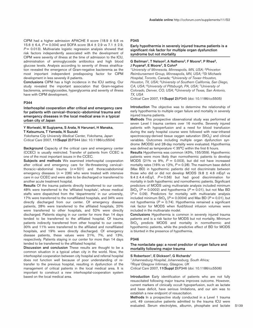
S139
CIPM had a higher admission APACHE II score (18.9 ± 6.6 vs15.6 ± 6.4, P = 0.004) and SOFA score (8.4 ± 2.9 vs 7.1 ± 2.9,P = 0.013). Multivariate logistic regression analysis showed thatrisk factors independently associated with the development ofCIPM were severity of illness at the time of admission to the ICU,administration of aminoglycoside antibiotics and high bloodglucose levels. Analysis according to severity of illness stratifica-tion revealed the emergence of Gram-negative bacteremia as themost important independent predisposing factor for CIPMdevelopment in less severely ill patients.Conclusions CIPM has a high incidence in the ICU setting. Ourstudy revealed the important association that Gram-negativebacteremia, aminoglycosides, hyperglycemia and severity of illnesshave with CIPM development.
P344Interhospital cooperation after critical and emergency carefor patients with cervical–thoracic–abdominal trauma andemergency diseases in the local medical area in a typicalurban city of Japan
Y Moriwaki, M Sugiyama, S Arata, N Harunari, H Manaka, T Katsumura, T Yamada, N SuzukiYokohama City University Medical Center, Yokohama, JapanCritical Care 2007, 11(Suppl 2):P344 (doi: 10.1186/cc5504)
Background Capacity of the critical care and emergency center(CCEC) is usually restricted. Transfer of patients from CCEC isone of the most important issues in the CCEC.Subjects and methods We examined interhospital cooperationafter critical and emergency care for life-threatening cervical–thoracic–abdominal trauma (n = 501) and thoracoabdominalemergency diseases (n = 236) who were treated with intensivecare in our CCEC and were able to be discharged or transferred toanother acute treatment hospital.Results Of the trauma patients directly transferred to our center,48% were transferred to the ‘affiliated hospitals’, whose medicalstaffs were dispatched from the ‘departments’ in our university,17% were transferred to the nonaffiliated hospitals, and 34% weredirectly discharged from our center. Of emergency diseasepatients, 28% were transferred to the affiliated hospitals, 20%were transferred to other hospitals, and 52% were directlydischarged. Patients staying in our center for more than 14 daystended to be transferred to the affiliated hospital. Of traumapatients indirectly transferred from other hospital to our center,30% and 11% were transferred to the affiliated and nonaffiliatedhospitals, and 19% were directly discharged. Of emergencydisease patients, these values were 21%, 7%, and 13%,respectively. Patients staying in our center for more than 14 daystended to be transferred to the affiliated hospital.Discussion and conclusion These results are thought to be acommon situation in a typical urban city in the world. Now, theinterhospital cooperation between city hospital and referral hospitaldoes not function well because of poor understanding of re-transfer to the previous hospital, resulting in dysfunction of themanagement of critical patients in the local medical area. It isimportant to construct a new interhospital-cooperation systembased on the local medical area.
P345Early hypothermia in severely injured trauma patients is asignificant risk factor for multiple organ dysfunctionsyndrome but not mortality
G Beilman1, T Nelson2, A Nathens3, F Moore4, P Rhee5, J Puyana6, E Moore7, S Cohn8
1University of Minnesota, Minneapolis, MN, USA; 2PrincetonReimbursement Group, Minneapolis, MN, USA; 3St MichaelsHospital, Toronto, Canada; 4University of Texas–Houston,Houston, TX, USA; 5University of Southern California, San Diego,CA, USA; 6University of Pittsburgh, PA, USA; 7University ofColorado, Denver, CO, USA; 8University of Texas, San Antonio,TX, USACritical Care 2007, 11(Suppl 2):P345 (doi: 10.1186/cc5505)
Introduction The objective was to determine the relationship ofearly hypothermia to multiple organ failure and mortality in severelyinjured trauma patients.Methods This prospective observational study was performed atseven Level I trauma centers over 16 months. Severely injuredpatients with hypoperfusion and a need for blood transfusionduring the early hospital course were followed with near-infraredspectroscopy-derived tissue oxygen saturation (StO2) and clinicalvariables. Outcomes including multiple organ dysfunction syn-drome (MODS) and 28-day mortality were evaluated. Hypothermiawas defined as temperature < 35°C within the first 6 hours.Results Hypothermia was common (43%, 155/359). Hypothermicpatients were more likely than normothermic patients to developMODS (21% vs 9%, P = 0.003), but did not have increasedmortality rates (16% vs 12%, P = 0.28). The maximum base deficit(Max BD) in hypothermic patients did not discriminate betweenthose who did or did not develop MODS (9.8 ± 4.6 mEq/l vs9.4 ± 4.4 mEq/l, P = 0.56) but had good discrimination formortality in both hypothermic and normothermic patients. Significantpredictors of MODS using multivariate analysis included minimumStO2 (P = 0.0002) and hypothermia (P = 0.01), but not Max BD(P = 0.09). Predictors for mortality with multivariate analysisincluded minimum StO2 (P = 0.0004) and Max BD (P = 0.01), butnot hypothermia (P = 0.74). Hypothermia remained a significantrisk factor for MODS when fluid/blood infusion volumes wereincluded in the multivariate model.Conclusions Hypothermia is common in severely injured traumapatients and is a risk factor for MODS but not mortality. MinimumStO2 predicts MODS and mortality in normothermic andhypothermic patients, while the predictive effect of BD for MODSis blunted in the presence of hypothermia.
P346The nonlactate gap: a novel predictor of organ failure andmortality following major trauma
S Robertson1, E Dickson2, G Richards1
1Johannesburg Hospital, Johannesburg, South Africa; 2Royal Glasgow Infirmary, Glasgow, UKCritical Care 2007, 11(Suppl 2):P346 (doi: 10.1186/cc5506)
Introduction Early identification of patients who are not fullyresuscitated following major trauma improves outcome. However,current markers of clinically occult hypoperfusion, such as lactateand base deficit, have serious limitations, and our aim was toestablish a new endpoint of resuscitation.Methods In a prospective study conducted in a Level 1 traumaunit, 49 consecutive patients admitted to the trauma ICU wereevaluated. Serum electrolytes, albumin, phosphate and lactate
Available online http://ccforum.com/supplements/11/S2
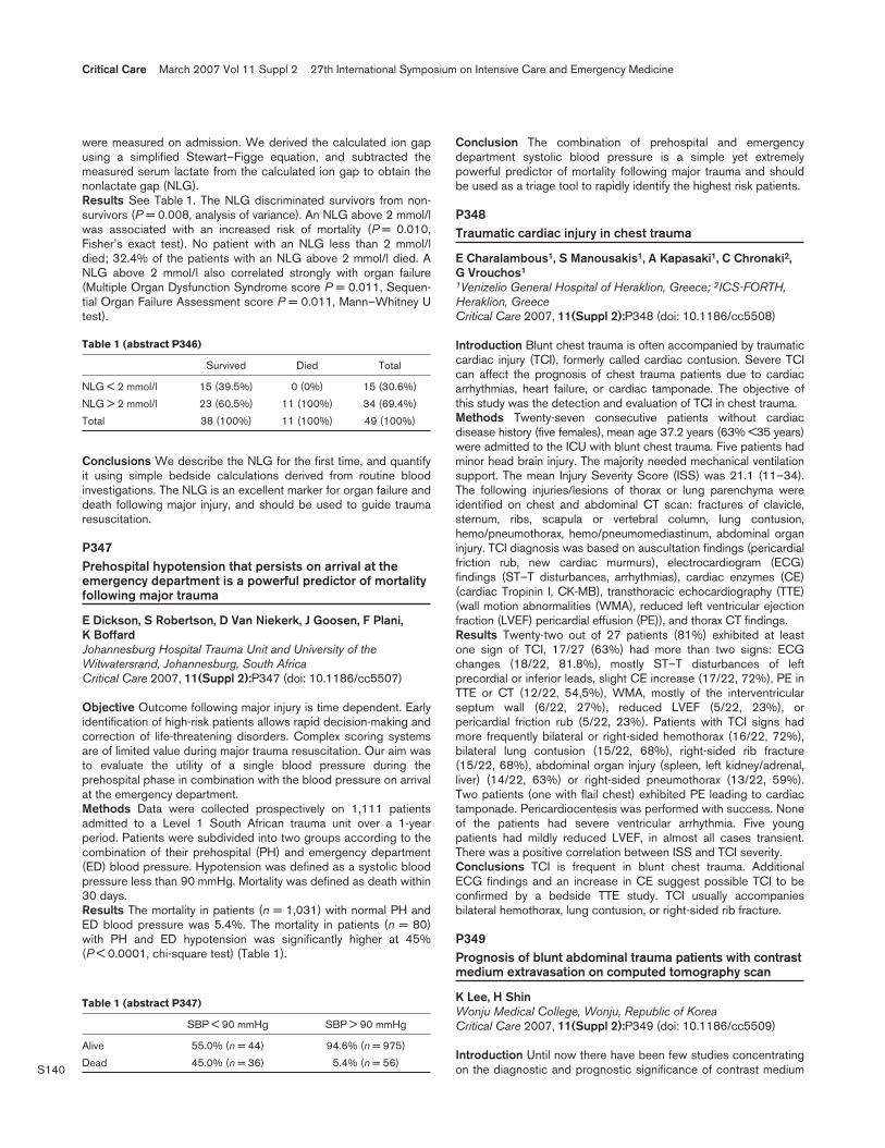
S140
were measured on admission. We derived the calculated ion gapusing a simplified Stewart–Figge equation, and subtracted themeasured serum lactate from the calculated ion gap to obtain thenonlactate gap (NLG).Results See Table 1. The NLG discriminated survivors from non-survivors (P = 0.008, analysis of variance). An NLG above 2 mmol/lwas associated with an increased risk of mortality (P = 0.010,Fisher’s exact test). No patient with an NLG less than 2 mmol/ldied; 32.4% of the patients with an NLG above 2 mmol/l died. ANLG above 2 mmol/l also correlated strongly with organ failure(Multiple Organ Dysfunction Syndrome score P = 0.011, Sequen-tial Organ Failure Assessment score P = 0.011, Mann–Whitney Utest).
Table 1 (abstract P346)
Survived Died Total
NLG < 2 mmol/l 15 (39.5%) 0 (0%) 15 (30.6%)
NLG > 2 mmol/l 23 (60.5%) 11 (100%) 34 (69.4%)
Total 38 (100%) 11 (100%) 49 (100%)
Conclusions We describe the NLG for the first time, and quantifyit using simple bedside calculations derived from routine bloodinvestigations. The NLG is an excellent marker for organ failure anddeath following major injury, and should be used to guide traumaresuscitation.
P347Prehospital hypotension that persists on arrival at theemergency department is a powerful predictor of mortalityfollowing major trauma
E Dickson, S Robertson, D Van Niekerk, J Goosen, F Plani, K BoffardJohannesburg Hospital Trauma Unit and University of theWitwatersrand, Johannesburg, South AfricaCritical Care 2007, 11(Suppl 2):P347 (doi: 10.1186/cc5507)
Objective Outcome following major injury is time dependent. Earlyidentification of high-risk patients allows rapid decision-making andcorrection of life-threatening disorders. Complex scoring systemsare of limited value during major trauma resuscitation. Our aim wasto evaluate the utility of a single blood pressure during theprehospital phase in combination with the blood pressure on arrivalat the emergency department.Methods Data were collected prospectively on 1,111 patientsadmitted to a Level 1 South African trauma unit over a 1-yearperiod. Patients were subdivided into two groups according to thecombination of their prehospital (PH) and emergency department(ED) blood pressure. Hypotension was defined as a systolic bloodpressure less than 90 mmHg. Mortality was defined as death within30 days.Results The mortality in patients (n = 1,031) with normal PH andED blood pressure was 5.4%. The mortality in patients (n = 80)with PH and ED hypotension was significantly higher at 45%(P < 0.0001, chi-square test) (Table 1).
Conclusion The combination of prehospital and emergencydepartment systolic blood pressure is a simple yet extremelypowerful predictor of mortality following major trauma and shouldbe used as a triage tool to rapidly identify the highest risk patients.
P348Traumatic cardiac injury in chest trauma
E Charalambous1, S Manousakis1, A Kapasaki1, C Chronaki2, G Vrouchos1
1Venizelio General Hospital of Heraklion, Greece; 2ICS-FORTH,Heraklion, GreeceCritical Care 2007, 11(Suppl 2):P348 (doi: 10.1186/cc5508)
Introduction Blunt chest trauma is often accompanied by traumaticcardiac injury (TCI), formerly called cardiac contusion. Severe TCIcan affect the prognosis of chest trauma patients due to cardiacarrhythmias, heart failure, or cardiac tamponade. The objective ofthis study was the detection and evaluation of TCI in chest trauma.Methods Twenty-seven consecutive patients without cardiacdisease history (five females), mean age 37.2 years (63% <35 years)were admitted to the ICU with blunt chest trauma. Five patients hadminor head brain injury. The majority needed mechanical ventilationsupport. The mean Injury Severity Score (ISS) was 21.1 (11–34).The following injuries/lesions of thorax or lung parenchyma wereidentified on chest and abdominal CT scan: fractures of clavicle,sternum, ribs, scapula or vertebral column, lung contusion,hemo/pneumothorax, hemo/pneumomediastinum, abdominal organinjury. TCI diagnosis was based on auscultation findings (pericardialfriction rub, new cardiac murmurs), electrocardiogram (ECG)findings (ST–T disturbances, arrhythmias), cardiac enzymes (CE)(cardiac Tropinin I, CK-MB), transthoracic echocardiography (TTE)(wall motion abnormalities (WMA), reduced left ventricular ejectionfraction (LVEF) pericardial effusion (PE)), and thorax CT findings.Results Twenty-two out of 27 patients (81%) exhibited at leastone sign of TCI, 17/27 (63%) had more than two signs: ECGchanges (18/22, 81.8%), mostly ST–T disturbances of leftprecordial or inferior leads, slight CE increase (17/22, 72%), PE inTTE or CT (12/22, 54,5%), WMA, mostly of the interventricularseptum wall (6/22, 27%), reduced LVEF (5/22, 23%), orpericardial friction rub (5/22, 23%). Patients with TCI signs hadmore frequently bilateral or right-sided hemothorax (16/22, 72%),bilateral lung contusion (15/22, 68%), right-sided rib fracture(15/22, 68%), abdominal organ injury (spleen, left kidney/adrenal,liver) (14/22, 63%) or right-sided pneumothorax (13/22, 59%).Two patients (one with flail chest) exhibited PE leading to cardiactamponade. Pericardiocentesis was performed with success. Noneof the patients had severe ventricular arrhythmia. Five youngpatients had mildly reduced LVEF, in almost all cases transient.There was a positive correlation between ISS and TCI severity.Conclusions TCI is frequent in blunt chest trauma. AdditionalECG findings and an increase in CE suggest possible TCI to beconfirmed by a bedside TTE study. TCI usually accompaniesbilateral hemothorax, lung contusion, or right-sided rib fracture.
P349Prognosis of blunt abdominal trauma patients with contrastmedium extravasation on computed tomography scan
K Lee, H ShinWonju Medical College, Wonju, Republic of KoreaCritical Care 2007, 11(Suppl 2):P349 (doi: 10.1186/cc5509)
Introduction Until now there have been few studies concentratingon the diagnostic and prognostic significance of contrast medium
Critical Care March 2007 Vol 11 Suppl 2 27th International Symposium on Intensive Care and Emergency Medicine
Table 1 (abstract P347)
SBP < 90 mmHg SBP > 90 mmHg
Alive 55.0% (n = 44) 94.6% (n = 975)
Dead 45.0% (n = 36) 5.4% (n = 56)
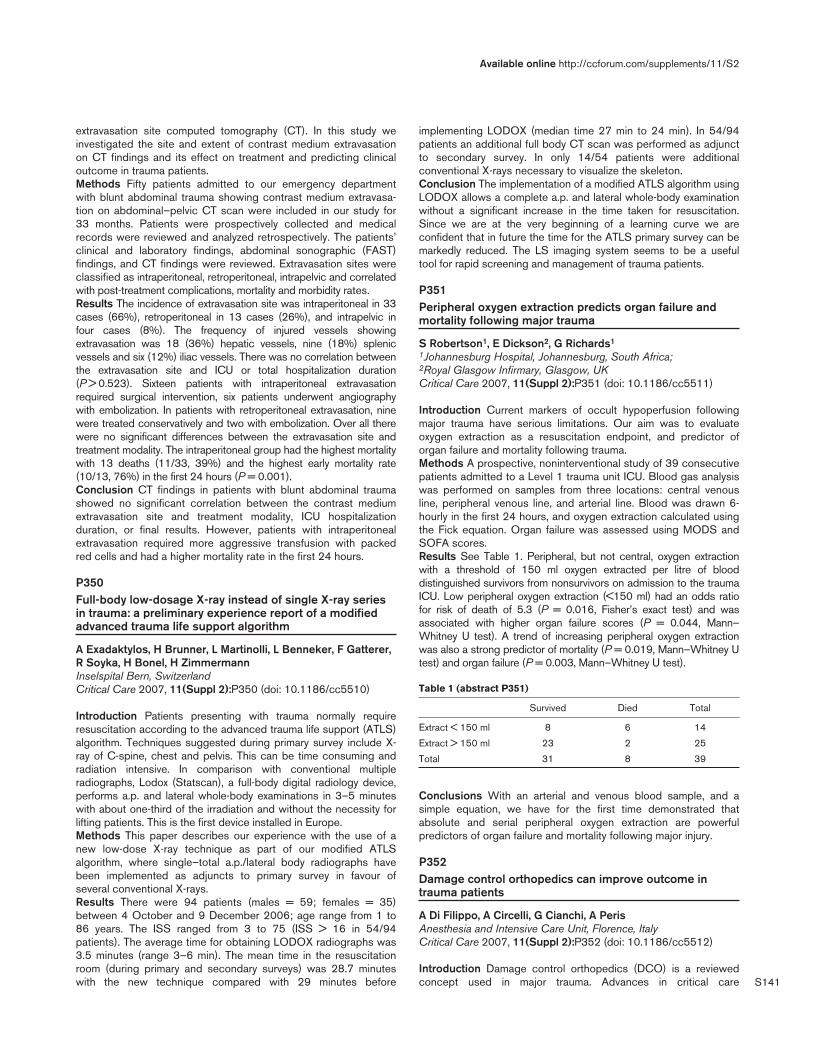
S141
extravasation site computed tomography (CT). In this study weinvestigated the site and extent of contrast medium extravasationon CT findings and its effect on treatment and predicting clinicaloutcome in trauma patients.Methods Fifty patients admitted to our emergency departmentwith blunt abdominal trauma showing contrast medium extravasa-tion on abdominal–pelvic CT scan were included in our study for33 months. Patients were prospectively collected and medicalrecords were reviewed and analyzed retrospectively. The patients’clinical and laboratory findings, abdominal sonographic (FAST)findings, and CT findings were reviewed. Extravasation sites wereclassified as intraperitoneal, retroperitoneal, intrapelvic and correlatedwith post-treatment complications, mortality and morbidity rates.Results The incidence of extravasation site was intraperitoneal in 33cases (66%), retroperitoneal in 13 cases (26%), and intrapelvic infour cases (8%). The frequency of injured vessels showingextravasation was 18 (36%) hepatic vessels, nine (18%) splenicvessels and six (12%) iliac vessels. There was no correlation betweenthe extravasation site and ICU or total hospitalization duration(P > 0.523). Sixteen patients with intraperitoneal extravasationrequired surgical intervention, six patients underwent angiographywith embolization. In patients with retroperitoneal extravasation, ninewere treated conservatively and two with embolization. Over all therewere no significant differences between the extravasation site andtreatment modality. The intraperitoneal group had the highest mortalitywith 13 deaths (11/33, 39%) and the highest early mortality rate(10/13, 76%) in the first 24 hours (P = 0.001).Conclusion CT findings in patients with blunt abdominal traumashowed no significant correlation between the contrast mediumextravasation site and treatment modality, ICU hospitalizationduration, or final results. However, patients with intraperitonealextravasation required more aggressive transfusion with packedred cells and had a higher mortality rate in the first 24 hours.
P350Full-body low-dosage X-ray instead of single X-ray seriesin trauma: a preliminary experience report of a modifiedadvanced trauma life support algorithm
A Exadaktylos, H Brunner, L Martinolli, L Benneker, F Gatterer,R Soyka, H Bonel, H ZimmermannInselspital Bern, SwitzerlandCritical Care 2007, 11(Suppl 2):P350 (doi: 10.1186/cc5510)
Introduction Patients presenting with trauma normally requireresuscitation according to the advanced trauma life support (ATLS)algorithm. Techniques suggested during primary survey include X-ray of C-spine, chest and pelvis. This can be time consuming andradiation intensive. In comparison with conventional multipleradiographs, Lodox (Statscan), a full-body digital radiology device,performs a.p. and lateral whole-body examinations in 3–5 minuteswith about one-third of the irradiation and without the necessity forlifting patients. This is the first device installed in Europe.Methods This paper describes our experience with the use of anew low-dose X-ray technique as part of our modified ATLSalgorithm, where single–total a.p./lateral body radiographs havebeen implemented as adjuncts to primary survey in favour ofseveral conventional X-rays.Results There were 94 patients (males = 59; females = 35)between 4 October and 9 December 2006; age range from 1 to86 years. The ISS ranged from 3 to 75 (ISS > 16 in 54/94patients). The average time for obtaining LODOX radiographs was3.5 minutes (range 3–6 min). The mean time in the resuscitationroom (during primary and secondary surveys) was 28.7 minuteswith the new technique compared with 29 minutes before
implementing LODOX (median time 27 min to 24 min). In 54/94patients an additional full body CT scan was performed as adjunctto secondary survey. In only 14/54 patients were additionalconventional X-rays necessary to visualize the skeleton.Conclusion The implementation of a modified ATLS algorithm usingLODOX allows a complete a.p. and lateral whole-body examinationwithout a significant increase in the time taken for resuscitation.Since we are at the very beginning of a learning curve we areconfident that in future the time for the ATLS primary survey can bemarkedly reduced. The LS imaging system seems to be a usefultool for rapid screening and management of trauma patients.
P351Peripheral oxygen extraction predicts organ failure andmortality following major trauma
S Robertson1, E Dickson2, G Richards1
1Johannesburg Hospital, Johannesburg, South Africa; 2Royal Glasgow Infirmary, Glasgow, UKCritical Care 2007, 11(Suppl 2):P351 (doi: 10.1186/cc5511)
Introduction Current markers of occult hypoperfusion followingmajor trauma have serious limitations. Our aim was to evaluateoxygen extraction as a resuscitation endpoint, and predictor oforgan failure and mortality following trauma.Methods A prospective, noninterventional study of 39 consecutivepatients admitted to a Level 1 trauma unit ICU. Blood gas analysiswas performed on samples from three locations: central venousline, peripheral venous line, and arterial line. Blood was drawn 6-hourly in the first 24 hours, and oxygen extraction calculated usingthe Fick equation. Organ failure was assessed using MODS andSOFA scores.Results See Table 1. Peripheral, but not central, oxygen extractionwith a threshold of 150 ml oxygen extracted per litre of blooddistinguished survivors from nonsurvivors on admission to the traumaICU. Low peripheral oxygen extraction (<150 ml) had an odds ratiofor risk of death of 5.3 (P = 0.016, Fisher’s exact test) and wasassociated with higher organ failure scores (P = 0.044, Mann–Whitney U test). A trend of increasing peripheral oxygen extractionwas also a strong predictor of mortality (P = 0.019, Mann–Whitney Utest) and organ failure (P = 0.003, Mann–Whitney U test).
Table 1 (abstract P351)
Survived Died Total
Extract < 150 ml 8 6 14
Extract > 150 ml 23 2 25
Total 31 8 39
Conclusions With an arterial and venous blood sample, and asimple equation, we have for the first time demonstrated thatabsolute and serial peripheral oxygen extraction are powerfulpredictors of organ failure and mortality following major injury.
P352Damage control orthopedics can improve outcome intrauma patients
A Di Filippo, A Circelli, G Cianchi, A PerisAnesthesia and Intensive Care Unit, Florence, ItalyCritical Care 2007, 11(Suppl 2):P352 (doi: 10.1186/cc5512)
Introduction Damage control orthopedics (DCO) is a reviewedconcept used in major trauma. Advances in critical care
Available online http://ccforum.com/supplements/11/S2
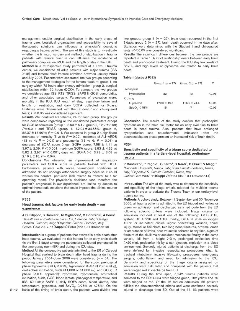
S142
management enable surgical stabilization in the early phase oftrauma care. Logistical organization and accessibility to severaltherapeutic solutions can influence a physician’s decisionsregarding a trauma patient. The aim of this study is to investigatewhether the timing of surgery and method of stabilization in traumapatients with femoral fracture can influence the incidence ofpulmonary complication, MOF and the length of stay in the ICU.Method In a retrospective study performed at a Level I traumacenter, we considered all adult patients with major trauma (ISS> 15) and femoral shaft fracture admitted between January 2003and July 2006. Patients were separated into two groups accordingto the management strategies for the femoral fracture: group 1, nosurgery within 72 hours after primary admission; group 2, surgicalstabilization within 72 hours (DCO). To compare the two groupswe considered age, ISS, RTS, TRISS, SAPS II, GCS, comorbidity,and other associated surgery. Parameters of evaluation were:mortality in the ICU, ICU length of stay, respiratory failure andlength of ventilation, and daily SOFA collected for 8 days.Statistics were determined with the Student t and chi-squaredtests; P < 0.05 was considered significant.Results We identified 48 patients, 24 for each group. The groupswere comparable regarding all the considered parameters exceptfor GCS at admission (group 1, 8.63 ± 5.12; group 2, 12.2 ± 3.99;P = 0.01) and TRISS (group 1, 62.04 ± 34.55%; group 2,82.37 ± 18.60%; P = 0.01). We observed in group 2 a significantdecrease of mortality (5 vs 0; P = 0.02), incidence of ALI–ARDS(13 vs 4; P = 0.01) and pneumonia (18 vs 6; P = 0.01), adecrease of SOFA score (mean SOFA score: 7.58 ± 4.11 vs3.97 ± 2.39, P < 0.001; maximum SOFA score: 9.83 ± 4.36 vs5.62 ± 2.97, P < 0.001; days with SOFA >6: 3.79 ± 3.08 vs2.16 ± 2.18, P = 0.04).Conclusions We observed an improvement of respiratoryparameters and SOFA score in patients treated with DCO.Furthermore, patients with worse neurological conditions atadmission do not undergo orthopaedic surgery because it couldworsen the cerebral perfusion (risk related to transfer to a faroperating room). The physician’s decisions (and therefore thepatient’s prognosis), in our experience, are limited by access tooptimal therapeutic solutions that could improve the clinical courseof the patient.
P353Head trauma: risk factors for early brain death – ourexperience
A Di Filippo1, S Damiani1, M Migliaccio1, M Bonizzoli2, A Peris3
1Anesthesia and Intensive Care Unit, Florence, Italy; 2CareggiHospital, Florence, Italy; 3Ospedale Firenze, Florence, ItalyCritical Care 2007, 11(Suppl 2):P353 (doi: 10.1186/cc5513)
Introduction In a group of patients that evolved in brain death afterhead trauma, we evaluated the risk factors for an early brain death(in the first 3 days) among the parameters collected prehospital, inthe emergency room (ER) and during the ICU stay.Method All the consecutive patients admitted to the ER of CareggiHospital that evolved to brain death after head trauma during theperiod January 2004–June 2006 were considered (n = 54). Thefollowing parameters were considered for the study: prehospitalphase: hypoxemia (SaO2 < 95%), hypotension (SAPS II < 90 mmHg),orotracheal intubation, fluids (>1,000 or ≤1,000 ml), and GCS; ERphase (ATLS approach): hypoxemia, hypotension, orotrachealintubation, fluids, GCS, blood lactate, pharyngeal temperature, andISS; ICU stay: SAPS II, daily SOFA score, blood lactate, coretemperature, glycaemia, and ScVO2 (>75% or ≤75%). On thebasis of the timing of brain death, the patients were divided into
two groups: group 1 (n = 27), brain death occurred in the first3 days; group 2 (n = 27), brain death occurred in the days after.Statistics were determined with the Student t and chi-squaredtests; P < 0.05 was considered significant.Results The significant differences between the two groups arereported in Table 1. A strict relationship exists between early braindeath and prehospital treatment. During the ICU stay low levels ofScVO2 and high levels of glycaemia are related to early braindeath.
Table 1 (abstract P353)
Group 1 (n = 27) Group 2 (n = 27) P
Prehospital
Hypotension 22 13 <0.05
ICU stay
Glycaemia 170.8 ± 49.5 116.6 ± 24.4 <0.05
ScVO2 < 75% 15 7 <0.05
Conclusion The results of the study confirm that prehospitalhypotension is the main risk factor for an early evolution to braindeath in head trauma. Also, patients that have prolongedhypoperfusion and neurohormonal imbalance after thepostresuscitation phase present an increased risk of brain death.
P354Sensitivity and specificity of a triage score dedicated totrauma patients in a tertiary-level hospital: preliminaryresults
P Masturzo1, R Regolo1, G Ferro2, G Nardi3, D Orazi2, V Maggi21Seconda Università, Napoli, Italy; 2San Camillo Forlanini, Roma,Italy; 3Ospedale S. Camillo-Forlanini, Roma, ItalyCritical Care 2007, 11(Suppl 2):P354 (doi: 10.1186/cc5514)
Introduction The aim of the study was to determine the sensitivityand specificity of the triage criteria adopted for multiple traumapatients in order to activate the Trauma Team in our tertiary-leveltrauma centre.Methods A cohort study. Between 1 September and 30 November2006, all trauma patients admitted to the ED triaged red, yellow orgreen on admission and discharged as a red code from the EDfollowing specific criteria were included. Triage criteria onadmission included at least one of the following: GCS < 13,systolic BP > 220 and < 100 mmHg, SaO2 < 95% on oxygen100% or intubated; clinical signs: penetrating wounds, spinalinjury, sternal or flail chest, two long-bone fractures, proximal crashor amputation of limbs, post-traumatic seizures at any time, signs offracture of the skull; major accident mechanics: fatality in the samevehicle, fall from a height > 3 m, prolonged extrication time(> 20 min), pedestrian hit by a car, ejection, explosion in a closeenvironment. Severely injured patients at discharge from the EDwere defined by: invasive resuscitating procedures (that is,tracheal intubation), invasive life-saving procedures (emergencysurgery, defibrillation) and need for admission to the ICU.Sensitivity and specificity of the triage criteria assigned onadmission were calculated and compared with the patients thatwere triaged red at discharge from ED.Results During the time span, 5,142 trauma patients wereadmitted to the ED: 4,884 were triaged green, 182 yellow and 76were triaged as red. Of the 76 red on admission, 55 patientsfulfilled the abovementioned criteria and were confirmed severelyinjured at discharge from ED. Out of the 55, 53 patients were
Critical Care March 2007 Vol 11 Suppl 2 27th International Symposium on Intensive Care and Emergency Medicine
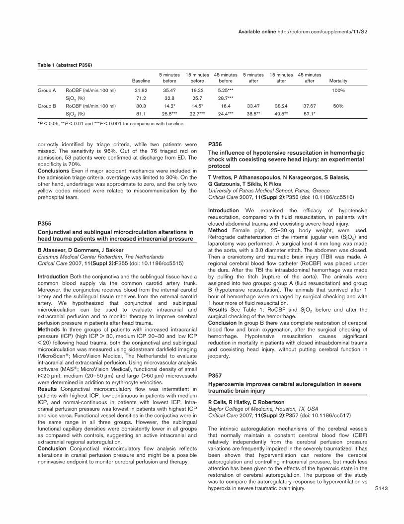
S143
correctly identified by triage criteria, while two patients weremissed. The sensitivity is 96%. Out of the 76 triaged red onadmission, 53 patients were confirmed at discharge from ED. Thespecificity is 70%.Conclusions Even if major accident mechanics were included inthe admission triage criteria, overtriage was limited to 30%. On theother hand, undertriage was approximate to zero, and the only twoyellow codes missed were related to miscommunication by theprehospital team.
P355Conjunctival and sublingual microcirculation alterations inhead trauma patients with increased intracranial pressure
B Atasever, D Gommers, J BakkerErasmus Medical Center Rotterdam, The NetherlandsCritical Care 2007, 11(Suppl 2):P355 (doi: 10.1186/cc5515)
Introduction Both the conjunctiva and the sublingual tissue have acommon blood supply via the common carotid artery trunk.Moreover, the conjunctiva receives blood from the internal carotidartery and the sublingual tissue receives from the external carotidartery. We hypothesized that conjunctival and sublingualmicrocirculation can be used to evaluate intracranial andextracranial perfusion and to monitor therapy to improve cerebralperfusion pressure in patients after head trauma.Methods In three groups of patients with increased intracranialpressure (ICP) (high ICP > 30, medium ICP 20–30 and low ICP< 20) following head trauma, both the conjunctival and sublingualmicrocirculation was measured using sidestream darkfield imaging(MicroScan®; MicroVision Medical, The Netherlands) to evaluateintracranial and extracranial perfusion. Using microvascular analysissoftware (MAS®; MicroVision Medical), functional density of small(<20 µm), medium (20–50 µm) and large (>50 µm) microvesselswere determined in addition to erythrocyte velocities.Results Conjunctival microcirculatory flow was intermittent inpatients with highest ICP, low-continuous in patients with mediumICP, and normal-continuous in patients with lowest ICP. Intra-cranial perfusion pressure was lowest in patients with highest ICPand vice versa. Functional vessel densities in the conjuctiva were inthe same range in all three groups. However, the sublingualfunctional capillary densities were consistently lower in all groupsas compared with controls, suggesting an active intracranial andextracranial regional autoregulation.Conclusion Conjunctival microcirculatory flow analysis reflectsalterations in cranial perfusion pressure and might be a possiblenoninvasive endpoint to monitor cerebral perfusion and therapy.
P356The influence of hypotensive resuscitation in hemorrhagicshock with coexisting severe head injury: an experimentalprotocol
T Vrettos, P Athanasopoulos, N Karageorgos, S Balasis, G Gatzounis, T Siklis, K FilosUniversity of Patras Medical School, Patras, GreeceCritical Care 2007, 11(Suppl 2):P356 (doi: 10.1186/cc5516)
Introduction We examined the efficacy of hypotensiveresuscitation, compared with fluid resuscitation, in patients withclosed abdominal trauma and coexisting severe head injury.Method Female pigs, 25–30 kg body weight, were used.Retrograde catheterization of the internal jugular vein (SjO2) andlaparotomy was performed. A surgical knot 4 mm long was madeat the aorta, with a 3.0 diameter stitch. The abdomen was closed.Then a craniotomy and traumatic brain injury (TBI) was made. Aregional cerebral blood flow catheter (RoCBF) was placed underthe dura. After the TBI the intraabdominal hemorrhage was madeby pulling the titch (rupture of the aorta). The animals wereassigned into two groups: group A (fluid resuscitation) and groupB (hypotensive resuscitation). The animals that survived after 1hour of hemorrhage were managed by surgical checking and with1 hour more of fluid resuscitation.Results See Table 1: RoCBF and SjO2 before and after thesurgical checking of the hemorrhage.Conclusion In group B there was complete restoration of cerebralblood flow and brain oxygenation, after the surgical checking ofhemorrhage. Hypotensive resuscitation causes significantreduction in mortality in patients with closed intraabdominal traumaand coexisting head injury, without putting cerebral function injeopardy.
P357Hyperoxemia improves cerebral autoregulation in severetraumatic brain injury
R Celis, R Hlatky, C RobertsonBaylor College of Medicine, Houston, TX, USACritical Care 2007, 11(Suppl 2):P357 (doi: 10.1186/cc517)
The intrinsic autoregulation mechanisms of the cerebral vesselsthat normally maintain a constant cerebral blood flow (CBF)relatively independently from the cerebral perfusion pressurevariations are frequently impaired in the severely traumatized. It hasbeen shown that hyperventilation can restore the cerebralautoregulation and controlling intracranial pressure, but much lessattention has been given to the effects of the hyperoxic state in therestoration of cerebral autoregulation. The purpose of the studywas to compare the autoregulatory response to hyperventilation vshyperoxia in severe traumatic brain injury.
Available online http://ccforum.com/supplements/11/S2
Table 1 (abstract P356)
5 minutes 15 minutes 45 minutes 5 minutes 15 minutes 45 minutes Baseline before before before after after after Mortality
Group A RoCBF (ml/min.100 ml) 31.92 35.47 19.32 5.25*** 100%
SjO2 (%) 71.2 32.8 25.7 28.7***
Group B RoCBF (ml/min.100 ml) 30.3 14.2* 14.5* 16.4 33.47 38.24 37.67 50%
SjO2 (%) 81.1 25.8*** 22.7*** 24.4*** 38.5** 49.5** 57.1*
*P < 0.05, **P < 0.01 and ***P < 0.001 for comparison with baseline.
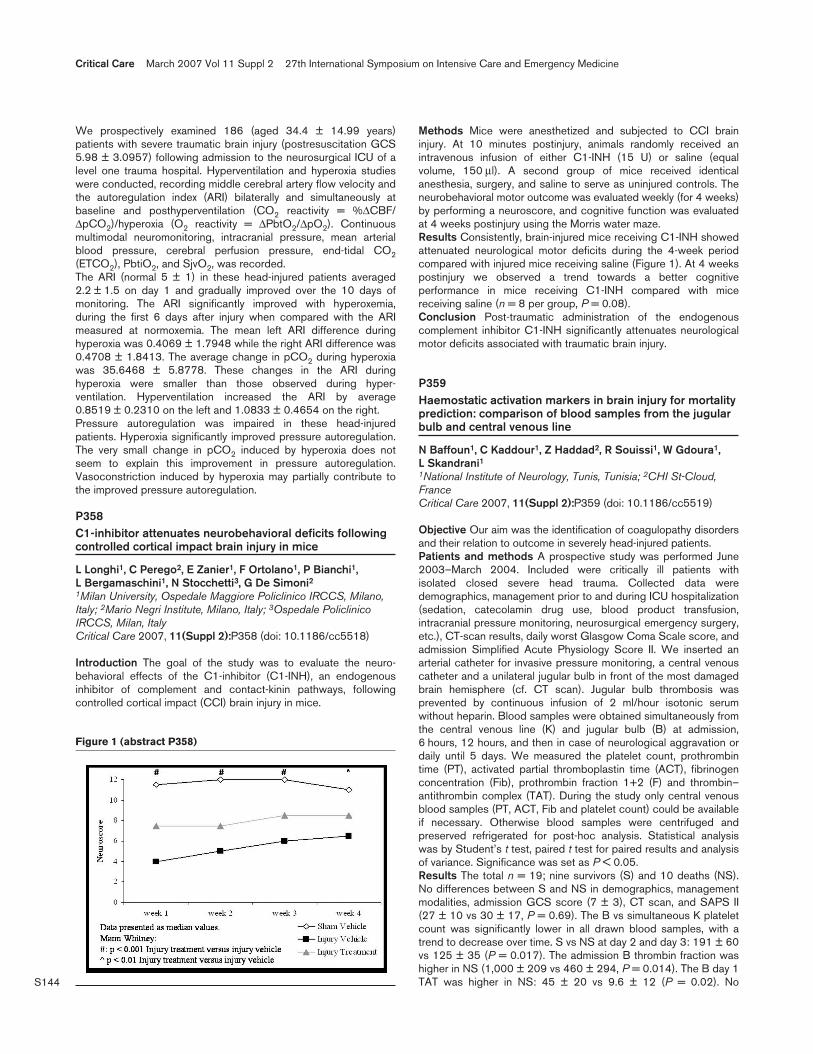
S144
We prospectively examined 186 (aged 34.4 ± 14.99 years)patients with severe traumatic brain injury (postresuscitation GCS5.98 ± 3.0957) following admission to the neurosurgical ICU of alevel one trauma hospital. Hyperventilation and hyperoxia studieswere conducted, recording middle cerebral artery flow velocity andthe autoregulation index (ARI) bilaterally and simultaneously atbaseline and posthyperventilation (CO2 reactivity = %∆CBF/∆pCO2)/hyperoxia (O2 reactivity = ∆PbtO2/∆pO2). Continuousmultimodal neuromonitoring, intracranial pressure, mean arterialblood pressure, cerebral perfusion pressure, end-tidal CO2(ETCO2), PbtiO2, and SjvO2, was recorded.The ARI (normal 5 ± 1) in these head-injured patients averaged2.2 ± 1.5 on day 1 and gradually improved over the 10 days ofmonitoring. The ARI significantly improved with hyperoxemia,during the first 6 days after injury when compared with the ARImeasured at normoxemia. The mean left ARI difference duringhyperoxia was 0.4069 ± 1.7948 while the right ARI difference was0.4708 ± 1.8413. The average change in pCO2 during hyperoxiawas 35.6468 ± 5.8778. These changes in the ARI duringhyperoxia were smaller than those observed during hyper-ventilation. Hyperventilation increased the ARI by average0.8519 ± 0.2310 on the left and 1.0833 ± 0.4654 on the right.Pressure autoregulation was impaired in these head-injuredpatients. Hyperoxia significantly improved pressure autoregulation.The very small change in pCO2 induced by hyperoxia does notseem to explain this improvement in pressure autoregulation.Vasoconstriction induced by hyperoxia may partially contribute tothe improved pressure autoregulation.
P358C1-inhibitor attenuates neurobehavioral deficits followingcontrolled cortical impact brain injury in mice
L Longhi1, C Perego2, E Zanier1, F Ortolano1, P Bianchi1, L Bergamaschini1, N Stocchetti3, G De Simoni21Milan University, Ospedale Maggiore Policlinico IRCCS, Milano,Italy; 2Mario Negri Institute, Milano, Italy; 3Ospedale PoliclinicoIRCCS, Milan, ItalyCritical Care 2007, 11(Suppl 2):P358 (doi: 10.1186/cc5518)
Introduction The goal of the study was to evaluate the neuro-behavioral effects of the C1-inhibitor (C1-INH), an endogenousinhibitor of complement and contact-kinin pathways, followingcontrolled cortical impact (CCI) brain injury in mice.
Methods Mice were anesthetized and subjected to CCI braininjury. At 10 minutes postinjury, animals randomly received anintravenous infusion of either C1-INH (15 U) or saline (equalvolume, 150 µl). A second group of mice received identicalanesthesia, surgery, and saline to serve as uninjured controls. Theneurobehavioral motor outcome was evaluated weekly (for 4 weeks)by performing a neuroscore, and cognitive function was evaluatedat 4 weeks postinjury using the Morris water maze.Results Consistently, brain-injured mice receiving C1-INH showedattenuated neurological motor deficits during the 4-week periodcompared with injured mice receiving saline (Figure 1). At 4 weekspostinjury we observed a trend towards a better cognitiveperformance in mice receiving C1-INH compared with micereceiving saline (n = 8 per group, P = 0.08).Conclusion Post-traumatic administration of the endogenouscomplement inhibitor C1-INH significantly attenuates neurologicalmotor deficits associated with traumatic brain injury.
P359Haemostatic activation markers in brain injury for mortalityprediction: comparison of blood samples from the jugularbulb and central venous line
N Baffoun1, C Kaddour1, Z Haddad2, R Souissi1, W Gdoura1, L Skandrani11National Institute of Neurology, Tunis, Tunisia; 2CHI St-Cloud,FranceCritical Care 2007, 11(Suppl 2):P359 (doi: 10.1186/cc5519)
Objective Our aim was the identification of coagulopathy disordersand their relation to outcome in severely head-injured patients.Patients and methods A prospective study was performed June2003–March 2004. Included were critically ill patients withisolated closed severe head trauma. Collected data weredemographics, management prior to and during ICU hospitalization(sedation, catecolamin drug use, blood product transfusion,intracranial pressure monitoring, neurosurgical emergency surgery,etc.), CT-scan results, daily worst Glasgow Coma Scale score, andadmission Simplified Acute Physiology Score II. We inserted anarterial catheter for invasive pressure monitoring, a central venouscatheter and a unilateral jugular bulb in front of the most damagedbrain hemisphere (cf. CT scan). Jugular bulb thrombosis wasprevented by continuous infusion of 2 ml/hour isotonic serumwithout heparin. Blood samples were obtained simultaneously fromthe central venous line (K) and jugular bulb (B) at admission,6 hours, 12 hours, and then in case of neurological aggravation ordaily until 5 days. We measured the platelet count, prothrombintime (PT), activated partial thromboplastin time (ACT), fibrinogenconcentration (Fib), prothrombin fraction 1+2 (F) and thrombin–antithrombin complex (TAT). During the study only central venousblood samples (PT, ACT, Fib and platelet count) could be availableif necessary. Otherwise blood samples were centrifuged andpreserved refrigerated for post-hoc analysis. Statistical analysiswas by Student’s t test, paired t test for paired results and analysisof variance. Significance was set as P < 0.05.Results The total n = 19; nine survivors (S) and 10 deaths (NS).No differences between S and NS in demographics, managementmodalities, admission GCS score (7 ± 3), CT scan, and SAPS II(27 ± 10 vs 30 ± 17, P = 0.69). The B vs simultaneous K plateletcount was significantly lower in all drawn blood samples, with atrend to decrease over time. S vs NS at day 2 and day 3: 191 ± 60vs 125 ± 35 (P = 0.017). The admission B thrombin fraction washigher in NS (1,000 ± 209 vs 460 ± 294, P = 0.014). The B day 1TAT was higher in NS: 45 ± 20 vs 9.6 ± 12 (P = 0.02). No
Critical Care March 2007 Vol 11 Suppl 2 27th International Symposium on Intensive Care and Emergency Medicine
Figure 1 (abstract P358)
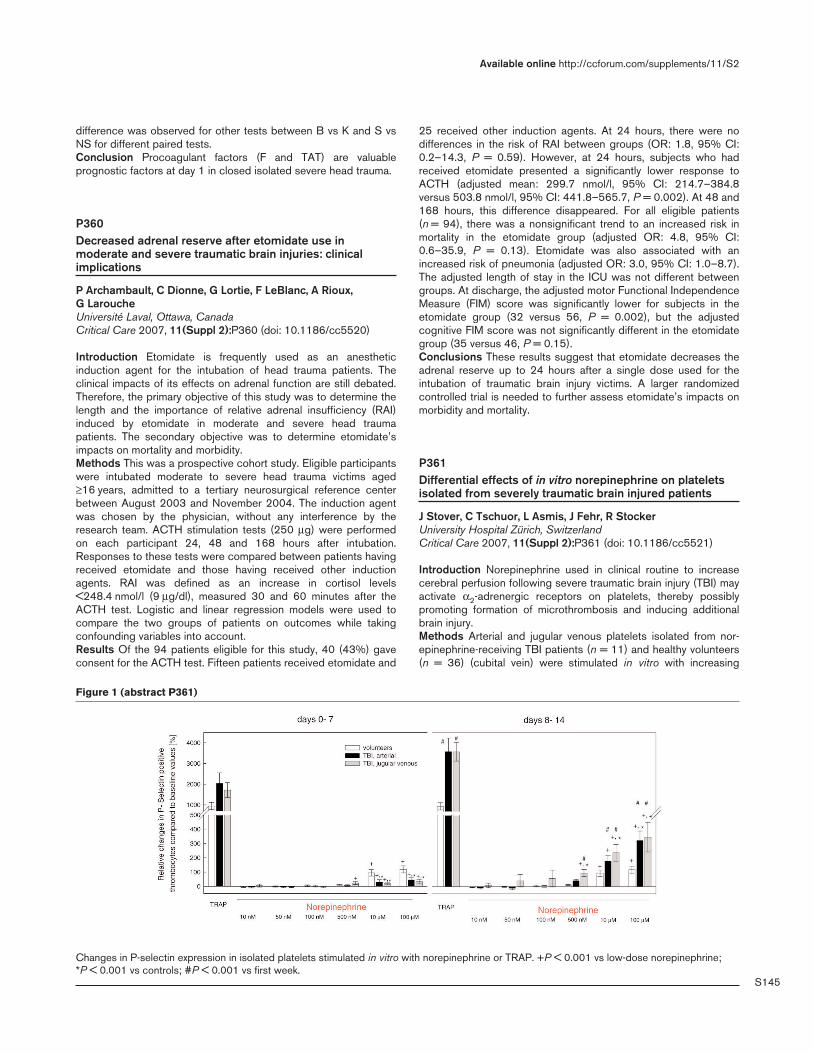
S145
difference was observed for other tests between B vs K and S vsNS for different paired tests.Conclusion Procoagulant factors (F and TAT) are valuableprognostic factors at day 1 in closed isolated severe head trauma.
P360Decreased adrenal reserve after etomidate use inmoderate and severe traumatic brain injuries: clinicalimplications
P Archambault, C Dionne, G Lortie, F LeBlanc, A Rioux, G LaroucheUniversité Laval, Ottawa, CanadaCritical Care 2007, 11(Suppl 2):P360 (doi: 10.1186/cc5520)
Introduction Etomidate is frequently used as an anestheticinduction agent for the intubation of head trauma patients. Theclinical impacts of its effects on adrenal function are still debated.Therefore, the primary objective of this study was to determine thelength and the importance of relative adrenal insufficiency (RAI)induced by etomidate in moderate and severe head traumapatients. The secondary objective was to determine etomidate’simpacts on mortality and morbidity.Methods This was a prospective cohort study. Eligible participantswere intubated moderate to severe head trauma victims aged≥16 years, admitted to a tertiary neurosurgical reference centerbetween August 2003 and November 2004. The induction agentwas chosen by the physician, without any interference by theresearch team. ACTH stimulation tests (250 µg) were performedon each participant 24, 48 and 168 hours after intubation.Responses to these tests were compared between patients havingreceived etomidate and those having received other inductionagents. RAI was defined as an increase in cortisol levels<248.4 nmol/l (9 µg/dl), measured 30 and 60 minutes after theACTH test. Logistic and linear regression models were used tocompare the two groups of patients on outcomes while takingconfounding variables into account.Results Of the 94 patients eligible for this study, 40 (43%) gaveconsent for the ACTH test. Fifteen patients received etomidate and
25 received other induction agents. At 24 hours, there were nodifferences in the risk of RAI between groups (OR: 1.8, 95% CI:0.2–14.3, P = 0.59). However, at 24 hours, subjects who hadreceived etomidate presented a significantly lower response toACTH (adjusted mean: 299.7 nmol/l, 95% CI: 214.7–384.8versus 503.8 nmol/l, 95% CI: 441.8–565.7, P = 0.002). At 48 and168 hours, this difference disappeared. For all eligible patients(n = 94), there was a nonsignificant trend to an increased risk inmortality in the etomidate group (adjusted OR: 4.8, 95% CI:0.6–35.9, P = 0.13). Etomidate was also associated with anincreased risk of pneumonia (adjusted OR: 3.0, 95% CI: 1.0–8.7).The adjusted length of stay in the ICU was not different betweengroups. At discharge, the adjusted motor Functional IndependenceMeasure (FIM) score was significantly lower for subjects in theetomidate group (32 versus 56, P = 0.002), but the adjustedcognitive FIM score was not significantly different in the etomidategroup (35 versus 46, P = 0.15).Conclusions These results suggest that etomidate decreases theadrenal reserve up to 24 hours after a single dose used for theintubation of traumatic brain injury victims. A larger randomizedcontrolled trial is needed to further assess etomidate’s impacts onmorbidity and mortality.
P361Differential effects of in vitro norepinephrine on plateletsisolated from severely traumatic brain injured patients
J Stover, C Tschuor, L Asmis, J Fehr, R StockerUniversity Hospital Zürich, SwitzerlandCritical Care 2007, 11(Suppl 2):P361 (doi: 10.1186/cc5521)
Introduction Norepinephrine used in clinical routine to increasecerebral perfusion following severe traumatic brain injury (TBI) mayactivate α2-adrenergic receptors on platelets, thereby possiblypromoting formation of microthrombosis and inducing additionalbrain injury.Methods Arterial and jugular venous platelets isolated from nor-epinephrine-receiving TBI patients (n = 11) and healthy volunteers(n = 36) (cubital vein) were stimulated in vitro with increasing
Available online http://ccforum.com/supplements/11/S2
Figure 1 (abstract P361)
Changes in P-selectin expression in isolated platelets stimulated in vitro with norepinephrine or TRAP. +P < 0.001 vs low-dose norepinephrine; *P < 0.001 vs controls; #P < 0.001 vs first week.
S146
norepinephrine concentrations (10 nM to 100 µM); thrombinreceptor activator peptide (TRAP) served as positive control.P-selectin expression was determined by flow cytometry (FACS).Results Following TBI, the number of unstimulated P-selectin-positive platelets was significantly decreased in the second weekby 60%. During the first week, the in vitro stimulatory effect wassignificantly reduced; in the second week, however,norepinephrine-mediated effects exceeded changes in controlsand the first week without a difference between arterial and jugularvenous platelets (Figure 1).Conclusion Clinically relevant norepinephrine concentrations are<25 nM. The present in vitro effects occurred at concentrations>500 nM. Thus, a clinically relevant impact appears doubtful.
P362Ultrasound evaluation and risk factors for deep venousthrombosis in the intensive care unit
M Boddi1, G Palano1, L Tini2, E Lucente1, S Berardino1, G Gensini1, A Peris3
1Department of Medical and Surgical Critical Care, CareggiTeaching Hospital, Florence, Italy; 2Department of Emergency,Careggi Teaching Hospital, Florence, Italy; 3Ospedale Firenze,Florence, ItalyCritical Care 2007, 11(Suppl 2):P362 (doi: 10.1186/cc5522)
Introduction Critically ill patients have a high risk of deep venousthrombosis (DVT); however, data about prevalence and specificrisk factors in the ICU are conflicting and the prophylaxis strategiesare still debated. The aim of this study is to evaluate the prevalenceand risk factors in an ICU dealing with trauma and major surgerypatients.Methods We analyzed retrospectively data from 142 patients(56 ± 4.8 years) admitted to the ICU from December 2004 toDecember 2005. We recorded the history, diagnosis, SAPS II,length of stay and major surgery. All patients received standardprophylaxis (LMWH and/or mechanical device). For DVT diagnosis,three compression ultrasound examinations were performed (a) within 48 hours from admission, (b) between the 7th and 10thdays, and (c) between the 13th and 16th days. The prevalence ofDVT and risk factors were analyzed in the whole population and inthe following four groups: ≤40 years, 41–59 years, 60–74 years,and ≥75 years. Statistics were determined using the Wilcoxon andMann–Whitney tests and one-way analysis of variance on SPSS;P < 0.5 was considered significant.Results One hundred (70%) patients were admitted after trauma,15 (11%) after surgery, 27 (19%) had medical disease. The meanSAPS II score was 45 ± 15.8 and the mean length of stay was16 ± 9.84 days. Forty-six patients (32.4%) underwent major surgeryfollowing the admission. The overall prevalence of DVT was 17.6%(25/142), with the highest value (24.2%) in the 41–59 year groupand the lowest (12.5%) in the >75 year group. DVT wasdiagnosed in 12/25 (48%) patients within 48 hours from ICUadmission and 7/12 (58.3%) had direct venous injury. DVT wasdiagnosed in the remaining patients after 10 days. We found astrong relationship (P < 0.06) between the length of stay and DVTin patients <40 years. No significant differences were foundregarding SAPS.Conclusions In our experience, DVT prevalence showed a bimodaloccurrence with a first short-term peak, associated with traumaticor surgical direct venous injury, and a medium-term peak, related toan ICU stay >10 days. We therefore oriented the surveillance tothe two periods of higher DVT prevalence and we established atraining program for the intensivists to improve DVT detection,relieving the workload of the ultrasound physician.
P363Venous thromboembolism in critically ill patients:incidence and risk factors
S Al-Qahtani, Y Arabi, A AldawoodKIing Abdulaziz Medical City, Riyadh, Saudi ArabiaCritical Care 2007, 11(Suppl 2):P363 (doi: 10.1186/cc5523)
Objective Despite the high risk of venous thromboembolism (VTE)in ICU patients, only few studies have examined this group ofpatients systematically. The objective of this study is to examine theincidence and risk factors of VTE among critically ill patients.Design A prospective cohort study.Setting A closed university-affiliated ICU in a medical–surgical ICUin the Kingdom of Saudi Arabia.Patients We enrolled consecutive patients >12 years of ageexpected to stay in the ICU >48 hours. We excluded patients onsystemic anticoagulation and patients with pulmonary embolism(PE) or deep venous thrombosis (DVT) on admission to the ICU ordiagnosed within 24 hours of ICU admission. We recorded a prioridefined VTE risk factors at baseline and daily. Our ICU implementsa protocol for thromboprophylaxis, which is based on the evidence-based ACCP guidelines. The primary endpoint was the develop-ment of PE or DVT during ICU stay. We used multivariateregression analysis to determine independent predictors of VTE.Results Among 277 patients with a mean APACHE II score of 25(+9), the incidence of VTE was 7.2% (95% CI 4.5–11). Weidentified three independent risk factors for ICU-acquired venousthromboembolism: stroke (OR 13.5, 95% CI 1.9–91.19,P = 0.008), femur fracture (OR 4.5, 95% CI 1.18–17.10, P = 0.03),and ICU length of stay (OR for each day increment 1.08, 95% CI1.03–1.13, P = 0.002). After adjustment for APACHE II score,VTE was an independent predictor of mortality (OR 3.85, 95% CI1.11–13.29, P = 0.03).Conclusions VTE is relatively common complication in critically illpatients and is associated with significant mortality. Longer ICUlength of stay, stroke and femur fracture are independentpredictors for VTE. These findings suggest the need for moreeffective prophylactic strategies in critically ill patients, especiallythose at higher risk.Acknowledgement Sponsored by Sanofi-Aventis.
P364Cardiopulmonary bypass and recombinant plasminogenactivator for treatment of experimental fatal pulmonaryembolism
B Kjærgaard, M Risom, S Risom Kristensen, E Tonnesen, A LarssonAalborg–Aarhus University Hospital, Aalborg, DenmarkCritical Care 2007, 11(Suppl 2):P364 (doi: 10.1186/cc5524)
Introduction Treatment of acute pulmonary embolism (PE), whichcauses life-threatening cardiovascular collapse, consists of cardio-pulmonary bypass (CPB) and surgical embolectomy. In the clinicalpractice we have treated three such patients successfully withCPB but instead of performing embolectomy, administeredrecombinant plasminogen activator (rt-PA). We studied thecirculatory and respiratory effects of this combined therapy in aswine model of fatal PE.Methods Seven pigs (90 kg) were i.v. anesthetized, musclerelaxed, tracheally intubated and mechanically ventilated (FiO21.0). A large-bore catheter (8.5 mm ID) was inserted in the rightsuperior vena cava (for injection of preformed blood clots) andlarge-bore catheters were inserted in the inferior vena cava via the
Critical Care March 2007 Vol 11 Suppl 2 27th International Symposium on Intensive Care and Emergency Medicine
S147
femoral vein and in the aorta via the femoral artery (for accessingCPB). We measured the mean arterial pressure (MAP), cardiacoutput (CO), blood gases, pulmonary artery pressure measurement(MPAP), end-tidal CO2 (ETCO2) and blood gases. Then 100–300 mlpreformed blood clot was injected until systemic circulationceased (systolic AP < 25 mmHg) and a 5 minute interval wasallowed before start of CPB (flow rate of 4–7 l/min). Heparin10,000 IE was given i.v., and rt-PA 10 mg as an i.v. bolus followedby an i.v. infusion of 90 mg during 2 hours. If ventricular fibrillationoccurred, cardioversion was performed. After 145 minutes CPBwas weaned off, and after a further 40 minutes the experiment wasended.Results Five animals developed ventricular fibrillation, while oneanimal maintained sinus rhythm. After 2 hours all animals had anatrial rhythm. All animals were weaned off CPB and survived untilthe experiment ended. Values before and after CPB (median andrange): MAP (mmHg) 101 (86, 109) and 75 (46, 106); CO (l/min)6.7 (4.3, 11) and 6.2 (3.6, 7.8); MPAP (mmHg) 22 (19, 36) and 44(31, 82) (P < 0.05); ETCO2 (kPa) 5.6 (4.3, 6.5) and 2.7 (2.4, 3.4)(P < 0.05); PaCO2 (kPa) 6.2 (5.2, 7.2) and 6.1 (4.8, 7.3); PaO2(kPa) 60 (50, 68) and 55 (30, 64).Conclusion Although there were signs – that is, lower ETCO2 andhigher MPAP – that the massive clots were not fully dissolved after185 minutes, this study shows that fatal PE might be treatedeffectively with CPB combined with simultaneous thrombolytictherapy.
P365Early coagulation alterations in intensive care unit burnpatients
A Lavrendieva1, A Parlapani2, O Thomareis2, V Soulountsi1, M Bitzani11G Papanikolaou Hospital, Thessalonki, Greece; 2AristotelianUniversity, Thessaloniki, GreeceCritical Care 2007, 11(Suppl 2):P365 (doi: 10.1186/cc5525)
Introduction The aim of the present study was to examine thecoagulation status of patients in the early postburn period.Method Coagulation and fibrinolysis parameters – antithrombin III(ATIII), protein C (PrC), free protein S (PrS), plasminogen activatorinhibitor 1 (PAI-1), tissue plasminogen activator (t-PA), thrombin/antithrombin complexes (TAT), plasmin/α2-antiplasmin complexes(PAP), fibrin degradation products (F1.2) – were measured at ICUadmission and daily thereafter for 7 postburn days.Results Forty-five patients were screened (nine nonsurvivors and36 survivors). All patients had a severe deficiency of the coagulationinhibitors on admission. Normalization of these levels in survivorswas observed at day 5 for ATIII and PrC, and at day 7 for PrS. Allpatients had elevated levels of TAT during the investigation period,but survivors had significantly lower levels at day 7 postburn(6.2 ± 4.9 vs 11.4 ± 4.5 µg/l, P < 0.001). PAP levels were withinthe physiological range in both groups at day 1, remained low insurvivors, but raised significantly in nonsurvivors at day 7(19.3 ± 14 vs 80.9 ± 10.4 µg/l, P = 0.003). The t-PA levels wereelevated permanently only in nonsurvivors. PAI-1 levels wereincreased at day 1 in both groups, but returned to normal values atday 5 in survivors. The degree of PAI-1 activation was significantlyhigher than this of t-PA. The F1.2 levels were permanently elevatedand there was no statistically significant difference in both groups.A logistic regression analysis revealed that ATIII and PrS at days 3,5 and 7, Pr C at days 5 and 7 and TAT at day 7 were independentpredictors of ICU death.Conclusion Our findings indicate the early postburn dysregulationof the hemostatic balance characterized by the activation of
procoagulant pathways. Although fibrinolysis was activated, theinhibition of fibrinolysis was more pronounced at the same time.The coagulation inhibitors and TAT levels seem to be earlypredictors of ICU mortality.
P366Fibrinolysis during cardiopulmonary bypass detected withthromboelastography
R Hájek, J Ruþþièèková, I Fluger, P Nemec, V BrukUniversity Hospital, Olomouc, Czech RepublicCritical Care 2007, 11(Suppl 2):P366 (doi: 10.1186/cc5526)
Background Fibrinolysis is a common haemostatic abnormalityduring cardiopulmonary bypass (CPB). Thromboelastography(TEG) is a good method to detect both types of fibrinolysis.Methods Four hundred and ninety-nine patients during mildhypothermic CPB and elective surgery were monitored with TEG(first – after the induction, second – after rewarming, third andfourth – at the end of surgery native and heparinase). No prophy-lactic antifibrinolytics were used. The data of the study group werecompared with a control group of 475 patients monitored only withlaboratory tests (fibrin degradation products (FDP) and D-dimers).Peroperative and 24 hour postoperative bleeding, number oftransfusions, aprotinin therapy and reexploration were recorded.Correlations between the presence of fibrinolysis and blood lossand transfusion therapy and between aprotinin administration andblood loss and number of transfusions were evaluated.Results The frequency of fibrinolysis measured with TEG: beforesurgery – primary 3.2%/secondary 3.4%; during CPB – 18.8%/0.6%; after surgery – 7%/1.4% (native), 5.6%/0.6% (heparinase).Positivity of fibrinolysis detected with laboratory tests was 100%.The TEG parameter of fibrinolysis (LY30) was significantlyincreased during CPB. The frequency of aprotinin administrationwas 12% TEG, 10.7% control. No correlation between positivity offibrinolysis and peroperative/postoperative blood loss and redblood cells (RBC) and fresh frozen plasma (FFP) transfusions wererecorded. No correlation between aprotinin administration andperoperative/postoperative blood loss and RBC transfusion wererecorded. Positive correlation between aprotinin administration andFFP transfusion were recordedConclusion Fibrinolysis was usually not associated with seriousbleeding. There was no positive effect of aprotinin to reducebleeding or transfusion therapy. FDP and D-dimers are not usefulto detect fibrinolysis in cardiac surgery.
P367ROTEM® thrombelastometry in on-pump cardiac surgerypatients
W Loesche, M Reinhöfer, C Macholdt, M Brauer, D Barz, J Gummert, K Reinhart, G MarxUniversity Hospital Jena, GermanyCritical Care 2007, 11(Suppl 2):P367 (doi: 10.1186/cc5527)
Objective On-pump cardiac surgery is frequently associated withintraoperative and postoperative bleeding. ROTEM® is a point-of-care method that reflects more closely than classical coagulationtests the in vivo haemostatic activity and the contribution offibrinogen and platelets to clot formation.Methods Two hundred and thirty-two patients (age 67 ± 10 years)with elective cardiac surgery and an on-pump time >45 minutes(mean 112 ± 52 min) were enrolled. Preoperative, intraoperativeand postoperative blood samples were taken from an arterialcatheter. The ROTEM® system and test kits (INTEM/EXTEM =
Available online http://ccforum.com/supplements/11/S2
S148
intrinsic/extrinsic activation, FIBTEM = EXTEM with inhibition ofplatelets) were from Pentapharm (Munich, Germany).Results The clot formation time (CFT) and maximum clot firmness(MCF), but not clotting time (CT), were strongly correlated with thefibrinogen level and platelet count. Surgery significantly decreasedthe ROTEM haemostatic activity, but normalised in most patientswithin 14–18 h postoperation. Lowest haemostatic activity (dramaticincrease in CT and CFT, decrease in MCF) was seen whenpatients were conditioned for cardiopulmonary bypass (CPB).When connected to CPB, the CT and CFT turned to recover, butMCF in EXTEM remained unchanged and MCF in FIBTEMdeclined further indicating continuous fibrinogen consumption. In12.5% of our patients, postoperative MCF in FIBTEM was reducedto <9 mm indicating a need for fibrinogen substitution. Lowpostoperative activity in ROTEM® was associated with highpostoperative blood loss. The positive predictive value andspecificity of FIBTEM were clearly superior to those of the APTT orprothrombin time. Up to 50% of patients had an increasedhaemostatic activity in preoperative ROTEM®, and this wasassociated with high CRP levels and intraoperative blood loss.Conclusions ROTEM® is a valuable tool to monitor peri-operative haemostasis. The decreases in haemostatic activityand postoperative bleeding are probably due to anticoagulanttherapy as well as fibrinogen and platelet consumption. Anincreased preoperative haemostatic activity is probably due toan acute phase reaction associated with advanced athero-sclerosis, and the high intraoperative bleeding in these patientsmight be due to the atherosclerotic vessels rather than due to aninsufficient haemostasis.
P368Low-frequency hemoviscoelastography: a new method ofdiagnostics for coagulation disorders after abdominalsurgery for cancer
O Tarabrin, A Simovskykh, V Mazur, A SuhanovOdessa Medical University, Marine Central Hospital, Odessa,UkraineCritical Care 2007, 11(Suppl 2):P368 (doi: 10.1186/cc5528)
Background Venous thromboembolism is one of the mostcommon complications seen in cancer patients and may be due tothe hypercoagulable state of the malignancy and to its surgicaltreatment. Despite clinical and laboratory evidence of perioperativehypercoagulability, there are no consistent data evaluating theextent, duration, and specific contribution of platelets and pro-coagulatory proteins by in vitro testing.Materials and methods Patients undergoing planned curativeopen surgery for abdominal cancer received MEDNORD (UkraineCo analyser) analysis (HVG), a viscoelastic test that measures clotformation and includes information on the cellular, as well as theplasmatic coagulation, system. We examined the efficacy of avariety of coagulation tests. A complete coagulation screen,activated clotting time, thromboelastography (TEG) and hemo-viscoelastography (HVG) were performed before surgery, at theend of surgery, and on postoperative days 1, 2, 3, and 7; they wereanalyzed for the reaction time and the maximal amplitude (MA). Wetested the hypothesis that the parallel use of standard TEG andHVG can assess postoperative hypercoagulability and canestimate the independent contribution of procoagulatory proteinsand platelets.Results and discussion We calculated the elastic shear modulusof standard MA (Gt) and HVG MA (GH), which reflect the total clotstrength and procoagulatory protein component, respectively. Thedifference was an estimate of the platelet component (Gp). There
was a 14% perioperative increase of standard MA, correspondingto a 48% increase of Gt (P < 0. 05) and an 80–86% contributionof the calculated Gp to Gt. We conclude that serial standard TEGand the HVG viscoelastic test may reveal the independentcontribution of platelets and procoagulatory proteins to clotstrength. Using multiple linear regression, all coagulation, TEG andHVG variabilities were used to model postoperative hyper-coagulation. Results showed that some components of the TEGfailed to identify hypercoagulation (r < 0.2, P > 0.75). However,three components of the routine coagulation assay, includingbleeding time, prothrombin time, and platelet count, could bemodeled to show prolonged postoperative hypercoagulability(P < 0.01). We conclude that all components of the HVG testreflect postoperative coagulopathies; these results suggest that itmay be useful in determining the coagulation status of cancerpatients perioperatively.Conclusion Postoperative hypercoagulability, occurring for at least1 week after major cancer abdominal surgery, may bedemonstrated by the HVG viscoelastic test. This hypercoagulabilityis not reflected completely by standard coagulation monitoring andTEG, and seems to be predominantly caused by increased plateletreactivity. The HVG viscoelastic test provides a fast and easy toperform bedside test to quantify in vitro hemocoagulation.
P369Thrombin generation in on-pump cardiac surgery patients
W Loesche, V König, V Oberle, M Reinhöfer, D Barz, J Gummert, G MarxUniversity Hospital Jena, GermanyCritical Care 2007, 11(Suppl 2):P369 (doi: 10.1186/cc5529)
Objective Cardiac surgery, in particular when done with cardio-pulmonary bypass (CPB), is frequently associated with excessiveintraoperative and postoperative bleeding. Here we employed theTechnothrombin® thrombin generation assay (TGA; Technoclone)to monitor changes in perioperative haemostasis.Methods One hundred and forty-eight patients (age 66.0 ± 9.8 years;103 males, 45 females) with elective cardiac surgery and a CPBtime >45 minutes (mean 114 ± 56 min) were enrolled. Arterialblood samples were obtained preoperative, postoperative and14–18 hours postoperative and centrifuged within 30 minutesafter withdrawal, for 5 minutes at 5,000 x g. Plasma samples werestored until analysis at –80°C. Thrombin generation (TG) inducedby the TGA RC low reagent (71.6 pM tissue factor) was measuredin 96-well multiplates using a FLUOstar OPTIMA fluorescencereader (MWG Labtech).Results Due to cardiac surgery, the lag-phase and time-to-peak ofTG increased by 45% and 35%, respectively. In parallel, the peakthrombin concentration and maximum slope decreased by 42%and 51%, respectively (P < 0.000001). Both the lag-phase andtime-to-peak returned to basal values within 14–18 hourspostoperative, but the peak thrombin and maximum slope of TGrose above the preoperative values (+39% and +68%, P < 0.0005).The on-pump time was positively correlated with lag phase andtime to peak and negatively correlated with peak thrombin andmaximum slope of TG when measured at 14–18 hourspostoperative, but there was no correlation at early postoperative.With respect to classical coagulation parameters, significantcorrelations were observed between TG and activated partialthromboplastin time at preoperative and 14–18 hours post-operative, and between TG and prothrombin time at postoperative(P < 0.025–0.001). At 14–18 hours postoperative there was asignificant correlation of TG with platelet as well as leukocytecounts (P < 0.025).
Critical Care March 2007 Vol 11 Suppl 2 27th International Symposium on Intensive Care and Emergency Medicine
S149
Conclusions The data provide clear evidence for a markeddecrease of TG during cardiac surgery followed by an excessrestoration in the postoperative phase. Factors released fromplatelets and leukocytes (procoagulant microvesicles?) mightcontribute to the enhanced TG observed at 14–18 hourspostoperative.
P370Activated clotting time (ACT) measuring devices usedsimultaneously do not produce correlating ACT values
I Lorenz, J Walde, C Schnuerer, C Velik-Salchner, P Muessigang,A Jeller, J Engel, R Gruber, C Falbesoner, C KolbitschUniversity Innsbruck, AustriaCritical Care 2007, 11(Suppl 2):P370 (doi: 10.1186/cc5530)
Introduction The lack of a ‘gold standard’ activated clotting time(ACT) measuring device gives rise to the broad range of ACTmeasuring devices currently on the market. The present studyfocused on the inter-device and intra-device differences (forexample, reproducibility of ACT measurement) in four different ACTmeasuring systems.Methods (1) Hemochron celite tubes, (2) Aktalyke celite tubes, (3) Actalyke ACT-Max tubes and (4) Hemochron Junior low-range(LR) or high-range (HR) cartridges were simultaneously filled withblood drawn (n = 3,997) at baseline, at 3 minutes followingadministration of heparin (300 IU/ kg), 5 minutes after startingcardiopulmonary bypass (CPB), every 30 minutes during CPB andthereafter at 15 minutes following protamine administration.Results The ACT values measured simultaneously using fourdifferent ACT measuring devices did not correlate with each otherat any measurement time. Reproducibility of ACT measurement (forexample, intra-device difference) was, in descending order, best forHemochron Junior, Actalyke ACT-Max, Hemochron celite andAktalyke celite.Conclusion The Hemochron Junior ACT measuring deviceshowed the smallest intra-device measuring error. No correlation,however, could be established between ACT values measuredwith the four devices tested.
P371Orgaran® use in intensive care unit patients with heparin-induced thrombocytopenia and acute renal failure
H Magnani1, J Wester2
1NV Organon, Oss, The Netherlands; 2Onze Lieve VrouweGasthuis, Amsterdam, The NetherlandsCritical Care 2007, 11(Suppl 2):P371 (doi: 10.1186/cc5531)
Introduction Orgaran® (danaparoid sodium) is a low-molecular-weight, nonheparin glycosaminoglycan antithrombotic that iscurrently a first-line treatment for heparin-induced thrombocyto-penia (HIT). This study reports on the safety of Orgaran® in ICUpatients suffering from HIT and acute renal failure requiringcontinuous renal replacement therapy (haemofiltration).Methods Data on 96 case reports from personal experience andpublications have been collated.Results Nineteen females and 59 males (18 unknown gender)with a median age of 60 years (range 22–95 years) presented witha variety of clinical problems that either preceded or complicatedtheir HIT; for example, postoperative/accidental trauma or overtbleeding in 61, sepsis/septicaemia/septic shock in 37, thrombo-embolism in 23, multiple organ dysfunction syndrome in 14,disseminated intravascular coagulation in eight and other miscel-laneous serious problems in 38.
A variety of continuous extracorporeal haemofiltration circuits wasused for from 1 to 39 days (median 7 days). The Orgaran® dosingschedule was usually initiated with a 2,000–2,500 U intravenousbolus injection. After two step-down dose periods the subsequentmaintenance infusion was usually titrated according to eachpatient’s thromboembolic and bleeding risk status. Hence mostpatients received 100–400 U/hour, but 11 patients required up to600 U/hour temporarily to control extracorporeal circuit clotting.There were 12 minor bleeding events, occurring mostly during themaintenance infusion rate adjustment period and which respondedto transient interruption and/or lowering of the infusion rate. Elevennonfatal major bleeding events occurred (four due to proceduralerrors) and eight fatal major bleeds, two of which occurred 3 daysand 8 days after Orgaran® discontinuation.Plasma anti-FXa levels were reported for 38 patients. During sevenof the eight fatal bleeding episodes the plasma anti-FXa levelswere ≥0.8 U/ml in three patients and 0.31–0.66 U/ml (that is,within the target range) in the other four patients. The highest anti-FXa response of 2.00 U/ml was associated with minor bleedingfrom an angiomatous malformation and stopped when theOrgaran® dose was reduced. However, of the 11 patientsreceiving >400 U/hour, four suffered major bleeding (three fatal)and two developed minor bleeding. Most of these patientsreceived continuous arterio-venous haemodiafiltration treatment,which may have been a contributory factor.Conclusions We recommend that maintenance Orgaran® infusionrates ≥400 U/hour should be avoided unless serious circuitclotting is grossly affecting the haemofilter life. The infusion rateshould be monitored clinically; that is, on the basis of bleeding andcircuit clotting rather than the patients’ plasma anti-FXa responses.
P372High incidence of positive heparin antibodies in amultidisciplinary intensive care unit
P Renieris, V Gerovasili, M El Ali, T Theodoridis, M Poriazi, A Bouhla, V Markaki, S Mentzelopoulos, D Zervakis, C Roussos, S NanasNational and Kapodistrian University of Athens, GreeceCritical Care 2007, 11(Suppl 2):P372 (doi: 10.1186/cc5532)
Introduction Heparin-induced thrombocytopenia (HIT) presents withthrombocytopenia and high risk for venous and/or arterialthrombosis, as an adverse effect of heparin (especially unfractioned).Objective To evaluate the incidence of positive heparin antibodytest in a multidisciplinary ICU for the time period from 1 January2006 to 30 November 2006.Methods We retrospectively reviewed all laboratory tests forheparin antibodies requested from the ICU for the period ofJanuary–November 2006. The blood sample analysis wasperformed with ELISA (Asserachrom® HPIA; Diagnostica Stago,Asnieres, France). All patients were receiving fractioned heparinsubcutaneously for prophylaxis when indicated. Flushes ofunfractioned heparin for catheter clotting prevention were given toall patients. Only ‘positive’ results were considered positive, whilst‘mildly positive’ and ‘uncertain’ results were considered negative.Results During that period 300 patients were admitted (meanduration of stay 15 ± 16 days, 25th–75th 4–22 days) to the ICU;212 (70%) were survivors and 88 (30%) were nonsurvivors. Allpatients presenting with thrombocytopenia (platelets < 150,000/dl)or showing a decrease >50% of their admission day’s plateletcount were checked for heparin antibodies. Fifty-two samplesretrieved from 48 patients suspicious for HIT were sent for heparinantibody analysis. Of the 48 patients checked, 15 (31%) werepositive and 33 (68%) negative. Three of the suspected patients
Available online http://ccforum.com/supplements/11/S2
S150
suffered from pulmonary embolism and were treated withtherapeutic doses of i.v. unfractioned heparin; one of them waspositive for heparin antibodies.Conclusions Our findings of high incidence of positive heparinantibodies may be mainly due to unfractioned heparin flushes. Theuse of heparin flushes with the new high-quality catheters andmonitoring kit is questionable. Since low doses of unfractionedheparin could lead to the production of antibodies and subsequentlyto HIT, further studies should examine the risk-to-benefit ratio of theuse of unfractioned heparin flushes in the ICU setting.
P373The beneficial effects of the combined methods ininvestigating heparin-induced thrombocytopenia
M Kounavi, C Kroupis, E Iliopoulou, D Karamichaleli, M Katafigioti, D Degiannis, E MelissariOnassis Cardiac Center, Athens, GreeceCritical Care 2007, 11(Suppl 2):P373 (doi: 10.1186/cc5533)
Introduction Heparin-induced thrombocytopenia (HIT) type II is aserious side effect of unfractionated heparin, and to a lesser extentof low molecular weight heparins. It is mediated through animmunological mechanism and leads to platelet aggregation. Theoccurrence of HIT varies between 1% and 5% of patientsreceiving heparin while a substantial fraction of them developsthrombosis (HITT). Therefore, the rapid and accurate confirmationof HIT is a necessity. The laboratory assays available for thediagnosis of HIT are based on the detection of HIT antibodies oron the consequences of platelet activation (functional methods).The purpose of the present study was to improve HIT recognition bythe combination of functional and antibody determination assays.Patients and methods Fifty-three patients who presentedthrombocytopenia while receiving UFH or LMWH participated inthe study. A positive control group consisting of 15 patients knownto suffer from HIT/HITT was used. Moreover, 19 healthy donorsnever exposed to heparin also participated as negative controls.The antibody determination was performed using commerciallyavailable ELISA kits. In the functional assays, heparin-inducedplatelet activation/aggregation (HIPA) and a flow cytometrictechnique for the detection of platelet microparticles released(platelet microparticle assay) were included.Results None of the negative control individuals were positive inany applied method. In contrast, all the participants of the positivecontrol group were found positive in all the applied methods. NoHIT was detected in 10 patients, seven of whom were pregnant atdifferent gestational ages. The remaining 43 thrombocytopenicpatients were positive in HIPA and 39 of them were also positive inthe platelet microparticle assay. Out of these 39 patients, only 16were positive in the ELISA assay.Conclusions In comparison with the HIT-positive control group itwas found that: a 92% agreement existed between the HIPA andthe platelet microparticle assays; and the positive results in theELISA were significantly lower than in the functional methods,indicating perhaps a lower sensitivity (41% agreement with theflow cytometric method and 32% with the HIPA). Therefore, thecombination of functional and antibody detection assays is anecessity for the HIT recognition since both could give pseudo-positive or pseudo-negative results.
P374The effect of male-donor-only fresh frozen plasma on theincidence of acute lung injury following rupturedabdominal aortic aneurysm repair
S Wright, S Athey, A Leaver, C Snowden, D Roberts, J Clarkson, C Chapman, J WallisFreeman Hospital and National Blood Service, Newcastle uponTyne, UKCritical Care 2007, 11(Suppl 2):P374 (doi: 10.1186/cc5534)
Introduction Transfusion-related acute lung injury, due to plasmafrom female donors containing antileucocyte antibodies, may be acommon contributor to the development of acute lung injury (ALI)in the critically ill. In July 2003 the English Blood Service stoppedusing female donor plasma for the manufacture of fresh frozenplasma (FFP). Patients undergoing repair of ruptured abdominalaortic aneurysm (AAA) receive large amounts of FFP and oftendevelop ALI. We investigated whether the change to male-onlyFFP was associated with a change in incidence of ALI in patientsundergoing emergency AAA repair.Methods A before-and-after, observational, single-centre study.Subjects were 211 consecutive patients undergoing open repair ofa ruptured AAA between 1998 and 2006. Primary outcome wasdevelopment of ALI (PaO2/FiO2 < 300 and bilateral pulmonaryinfiltrates on chest X-ray) in the first 6 hours after surgery.Secondary outcomes were time to extubation, and survival at30 days. Chest X-rays were examined independently by tworadiologists who were blinded to the study hypothesis.Results One hundred and twenty-nine patients were operated onbefore and 82 after the change in FFP procurement. Groups werewell matched, with respect to age, sex, co-morbidities and severityof illness, and received similar volumes of i.v. fluids and bloodproducts from admission to 6 hours postoperatively (mean units ofFFP, 8.6 before and 8.39 after, P = 0.833). The maximum tidalvolume, PEEP, and CVP were similar in both groups.Norepinephrine was given to 8.5% of patients in the before groupcompared with 24.4% after (P = 0.001), otherwise inotrope usewas similar. Primary outcome: there was significantly less ALIfollowing the change to male-only FFP (36% before vs 21% after,P = 0.042). Secondary outcomes were not statistically differentbetween groups; however, patients with ALI in either group had apoorer 30-day survival (59% vs 80%, P = 0.005).Conclusion Exclusion of female-donor FFP was associated with astatistically significant reduction in the incidence of ALI in patientsundergoing repair of a ruptured AAA.
P375A biochemical comparison of Octaplas with a universallyapplicable development product (Uniplas) and single-donor fresh-frozen plasmas subjected to methylene-bluedye and white-light treatment
A Heger, J Römisch, T SvaeOctapharma PPGmbH, Vienna, AustriaCritical Care 2007, 11(Suppl 2):P375 (doi: 10.1186/cc5535)
Introduction The aim of this study was to perform an extensivebiochemical comparison of the pharmaceutically licensedcoagulation active plasma Octaplas with an identical, butuniversally applicable, development product (Uniplas) and single-donor fresh-frozen plasma (FFP) units subjected to a medicaldevice treatment using a combination of methylene-blue dye andsubsequent white-light exposure (MB plasma).
Critical Care March 2007 Vol 11 Suppl 2 27th International Symposium on Intensive Care and Emergency Medicine
S151
Methods Twenty-four batches of Octaplas of different bloodgroups and different plasma sources, three batches of Uniplas(both products manufactured by Octapharma PPGmbH, Vienna,Austria) and 20 random commercially available bags of MB plasmaof different blood groups were analysed. Beyond the globalcoagulation parameters, the activities of coagulation factors andprotease inhibitors, as well as plasminogen, activated factor VII,plasma turbidity and lipid components, were quantified.Results Similar to Octaplas, Uniplas showed standardised levelsof coagulation factors, plasminogen and protease inhibitors(decreased protein S and antiplasmin activities) according to theproduct specifications. MB plasma revealed fibrinogen levels closeto or below the physiological range (<1.5 mg/ml). Coagulationfactor activities in single MB plasma units both below and abovethe normal ranges for FFP were found in this study, reflecting theconsiderable variability of clinically important plasma proteins.Moreover, MB plasma revealed a higher turbidity after thawing,probably due to the elevated lipid parameters.Conclusion This study showed that there are significant differencesin the biochemical characteristics between Octaplas and MBplasma, while Uniplas revealed the same high quality as Octaplas.The variability of several plasma proteins in the 20 individual MBplasma units tested was high compared with Octaplas/Uniplas. Forplasma prescribers and physicians it is also important to considerthe significant loss of functional fibrinogen in MB plasma whenplanning and monitoring the treatment of severely ill patients.
P376Octaplex in routine clinical use for prophylaxis and therapyof bleeding in patients with prothrombin complex factordeficiency
T Franken1, W Rees2, N Hübner2, J Albers3, B Gansera4, M Uhl5,C Niessner6, B Sedemund-Adip7, T Tödter8
1Octapharma GmbH, Langenfeld, Germany; 2Schüchtermannklinik,Bad Rothenfelde, Germany; 3Klinikum der Joh Gutenberg Universität,Mainz, Germany; 4Städt Krankenhaus München-Bogenhausen,München, Germany; 5Klinikum Starnberg, Germany; 6DiakonissenKrankenhaus, Karlsruhe, Germany; 7Universitätsklinikum Lübeck,Germany; 8Albertinen-Krankenhaus, Hamburg, GermanyCritical Care 2007, 11(Suppl 2):P376 (doi: 10.1186/cc5536)
Introduction Octaplex is a new prothrombin complex concentratethat is indicated for treatment or perioperative prophylaxis ofbleeding in patients with deficiency of the prothrombin complexcoagulation factors, such as deficiency caused by treatment withvitamin K antagonists or by liver failure, when rapid correction ofbleeding is required. The study was conducted to demonstrateboth prevention of bleeding and achievement of haemostasis inacute bleeding and to obtain further information about the safety ofadministration of Octaplex.Methods One hundred and one patients were included in thisobservational study with determination of the INR as part of routineclinical management. The endpoint of this study was perioperativeprophylaxis of bleeding or successful treatment of acute bleedingaccording to clinical signs.Results The total dose administered per patient varied from 5.6 to63.5 IU/kg bodyweight with a median dose of 20.4 IU/kg bodyweight.The median infusion rate was 3 ml/minute; in some cases up to10 ml/minute were administered without any adverse reactions. Theadministration of Octaplex reduced the INR on average from 2.3 to1.5. Infusion of 25 IU Octaplex per kg body weight resulted in anaverage decrease of about 1.2 in INR. A subgroup analysis of thosepatients with a postinfusion measurement within 1 hour afterapplication of Octaplex showed a fast onset of action. The
investigators evaluated the overall efficacy of Octaplex in 84.2% ofcases as ‘very good’ and in 14.9% of cases as ‘moderate’. Noadverse drug reactions or interactions were reported.Conclusions Octaplex is effective in perioperative prophylaxis ofbleeding in patients with a coagulation deficit caused by treatmentwith vitamin K antagonists. Octaplex is also effective in treatmentof acute bleeding. Octaplex was well tolerated, with no adversereactions being reported during the study.
P377Reversal of oral anticoagulation with prothrombin complexconcentrate (Octaplex)
H Riess1, A Meier-Hellmann2, J Motsch3, M Elias4, F Kursten5, C Dempfle6
1Charité-Universitätsmedizin Berlin, Germany; 2HELIOS KlinikumErfurt GmbH, Erfurt, Germany; 3Universitätsklinikum Heidelberg,Germany; 4HaEmek Medical Center, Afula, Israel; 5OctapharmaPharmazeutika Produktions ges.m.b.H., Wien, Austria;6Universitätsklinikum Mannheim, GermanyCritical Care 2007, 11(Suppl 2):P377 (doi: 10.1186/cc5537)
Introduction Oral anticoagulant therapy with vitamin K antagonistsmay need to be rapidly reversed if acute bleeding occurs or surgicalintervention is required. This can most effectively be achieved byadministering prothrombin complex concentrates (PCCs), whichcorrect the INR more quickly than fresh frozen plasma without theproblem of volume overload. Octaplex, a virus-inactivated PCC con-taining balanced potencies of coagulation factors and the regulatingproteins C and S, was developed with the intention of making arapid contribution to coagulation whilst limiting the risk of thrombosis.Methods The objective of this prospective, open-label study wasto demonstrate that Octaplex, when individually dosed, efficientlycorrects the INR within 1 hour post infusion. Sixty patients wereincluded, 56 of them evaluable in terms of efficacy.Results The median total dose was 41.1 IU/kg body weight (range15.3–83.3 IU/kg body weight). In total the mean infusion rate was6.42 ml/minute for the first infusion. In about one-third of thepatients an average infusion rate of ≥8 ml/minute was used. Themedian INR decreased from 2.8 (1.5–9.5) to 1.1 (1.0–1.9) after10 minutes and remained at that level at measurements 30 and60 minutes after infusion. There was a rapid increase in coagulationfactor activity within the first 10 minutes as well. This activityremained stably elevated for at least 4–6 hours, confirming the INRresults. Of 56 patients evaluable for efficacy, for 51 (91%) thegeometric mean of postinfusion values was equal to or less thanthe predetermined target INR. Overall haemostatic efficacy wasassessed as ‘excellent’ by investigators in all 56 patients. Three ofthe 60 patients had minor adverse drug reactions possibly relatedto Octaplex. No evidence of thrombotic side effects was observed.Conclusions Octaplex corrects quickly, effectively and safely theINR to a predetermined level in patients with vitamin K antagonist-related deficiency of prothrombin complex coagulation factors.
P378Experience of NovoSeven administration in themanagement of severe haemorrhage following cardiacsurgery of nonhaemophilic patients
G Michalska, R StanekClinical Hospital, Szczecin-Police, PolandCritical Care 2007, 11(Suppl 2):P378 (doi: 10.1186/cc5538)
Introduction Severe bleeding in cardiac surgery is often difficultto manage. The aim of this study is clinical evaluation of efficacy of
Available online http://ccforum.com/supplements/11/S2
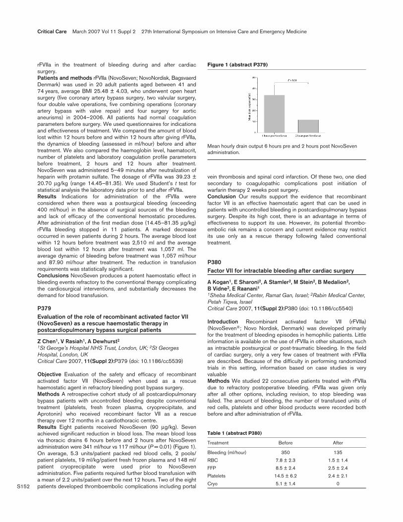
S152
rFVIIa in the treatment of bleeding during and after cardiacsurgery.Patients and methods rFVIIa (NovoSeven; NovoNordisk, BagsvaerdDenmark) was used in 20 adult patients aged between 41 and74 years, average BMI 25.48 ± 4.03, who underwent open heartsurgery (five coronary artery bypass surgery, two valvular surgery,four double valve operations, five combining operations (coronaryartery bypass with valve repair) and four surgery for aorticaneurisms) in 2004–2006. All patients had normal coagulationparameters before surgery. We used questionnaires for indicationsand effectiveness of treatment. We compared the amount of bloodlost within 12 hours before and within 12 hours after giving rFVIIa,the dynamics of bleeding (assessed in ml/hour) before and aftertreatment. We also compared the haemoglobin level, haematocrit,number of platelets and laboratory coagulation profile parametersbefore treatment, 2 hours and 12 hours after treatment.NovoSeven was administered 5–49 minutes after neutralization ofheparin with protamin sulfate. The dosage of rFVIIa was 39.23 ±20.70 µg/kg (range 14.45–81.35). We used Student’s t test forstatistical analysis the laboratory data prior to and after rFVIIa.Results Indications for administration of the rFVIIa wereconsidered when there was a postsurgical bleeding (exceeding400 ml/hour) in the absence of surgical sources of the bleedingand lack of efficacy of the conventional hemostatic procedures.After administration of the first median dose (14.45–81.35 µg/kg)rFVIIa bleeding stopped in 11 patients. A marked decreaseoccurred in seven patients during 2 hours. The average blood lostwithin 12 hours before treatment was 2,510 ml and the averageblood lost within 12 hours after treatment was 1,057 ml. Theaverage dynamic of bleeding before treatment was 1,057 ml/hourand 87.90 ml/hour after treatment. The reduction in transfusionrequirements was statistically significant.Conclusions NovoSeven produces a potent haemostatic effect inbleeding events refractory to the conventional therapy complicatingthe cardiosurgical interventions, and substantially decreases thedemand for blood transfusion.
P379Evaluation of the role of recombinant activated factor VII(NovoSeven) as a rescue haemostatic therapy inpostcardiopulmonary bypass surgical patients
Z Chen1, V Rasiah1, A Dewhurst2
1St George’s Hospital NHS Trust, London, UK; 2St GeorgesHospital, London, UKCritical Care 2007, 11(Suppl 2):P379 (doi: 10.1186/cc5539)
Objective Evaluation of the safety and efficacy of recombinantactivated factor VII (NovoSeven) when used as a rescuehaemostatic agent in refractory bleeding post bypass surgery.Methods A retrospective cohort study of all postcardiopulmonarybypass patients with uncontrolled bleeding despite conventionaltreatment (platelets, fresh frozen plasma, cryoprecipitate, andAprotonin) who received recombinant factor VII as a rescuetherapy over 12 months in a cardiothoracic centre.Results Eight patients received NovoSeven (90 µg/kg). Sevenachieved significant reduction in blood loss. The mean blood lossvia thoracic drains 6 hours before and 2 hours after NovoSevenadministration were 341 ml/hour vs 117 ml/hour (P = 0.01) (Figure 1).On average, 5.3 units/patient packed red blood cells, 2 pools/patient platelets, 19 ml/kg/patient fresh frozen plasma and 148 ml/patient cryoprecipitate were used prior to NovoSevenadministration. Five patients required further blood transfusion witha mean of 2.2 units/patient over the next 12 hours. Two of the eightpatients developed thromboembolic complications including portal
vein thrombosis and spinal cord infarction. Of these two, one diedsecondary to coagulopathic complications post initiation ofwarfarin therapy 2 weeks post surgery.Conclusion Our results support the evidence that recombinantfactor VII is an effective haemostatic agent that can be used inpatients with uncontrolled bleeding in postcardiopulmonary bypasssurgery. Despite its high cost, there is an advantage in terms ofeffectiveness to support its use. However, its potential thrombo-embolic risk remains a concern and current evidence may restrictits use only as a rescue therapy following failed conventionaltreatment.
P380Factor VII for intractable bleeding after cardiac surgery
A Kogan1, E Sharoni2, A Stamler2, M Stein2, B Medalion2, B Vidne2, E Raanani11Sheba Medical Center, Ramat Gan, Israel; 2Rabin Medical Center,Petah Tiqwa, IsraelCritical Care 2007, 11(Suppl 2):P380 (doi: 10.1186/cc5540)
Introduction Recombinant activated factor VII (rFVIIa)(NovoSeven®; Novo Nordisk, Denmark) was developed primarilyfor the treatment of bleeding episodes in hemophilic patients. Littleinformation is available on the use of rFVIIa in other situations, suchas intractable postsurgical or post-traumatic bleeding. In the fieldof cardiac surgery, only a very few cases of treatment with rFVIIaare described. Because of the difficulty in performing randomizedtrials in this setting, information based on case studies is veryvaluableMethods We studied 22 consecutive patients treated with rFVIIadue to refractory postoperative bleeding. rFVIIa was given onlyafter all other options, including revision, to stop bleeding wasfailed. The amount of bleeding, the number of transfused units ofred cells, platelets and other blood products were recorded bothbefore and after administration of rFVIIa.
Critical Care March 2007 Vol 11 Suppl 2 27th International Symposium on Intensive Care and Emergency Medicine
Figure 1 (abstract P379)
Mean hourly drain output 6 hours pre and 2 hours post NovoSevenadministration.
Table 1 (abstract P380)
Treatment Before After
Bleeding (ml/hour) 350 135
RBC 7.8 ± 2.3 1.5 ± 1.4
FFP 8.5 ± 2.4 2.5 ± 2.4
Platelets 14.5 ± 6.2 2.4 ± 2.1
Cryo 5.1 ± 1.4 0
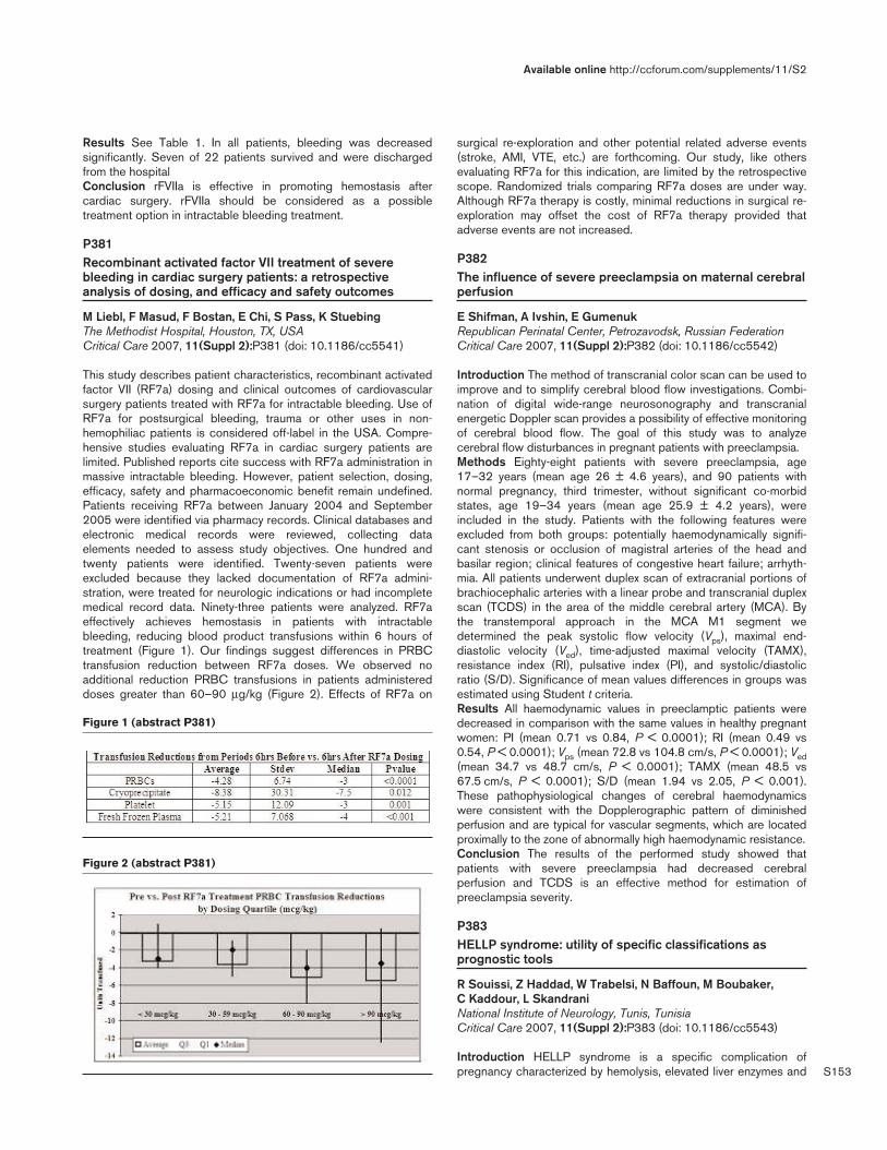
S153
Results See Table 1. In all patients, bleeding was decreasedsignificantly. Seven of 22 patients survived and were dischargedfrom the hospitalConclusion rFVIIa is effective in promoting hemostasis aftercardiac surgery. rFVIIa should be considered as a possibletreatment option in intractable bleeding treatment.
P381Recombinant activated factor VII treatment of severebleeding in cardiac surgery patients: a retrospectiveanalysis of dosing, and efficacy and safety outcomes
M Liebl, F Masud, F Bostan, E Chi, S Pass, K StuebingThe Methodist Hospital, Houston, TX, USACritical Care 2007, 11(Suppl 2):P381 (doi: 10.1186/cc5541)
This study describes patient characteristics, recombinant activatedfactor VII (RF7a) dosing and clinical outcomes of cardiovascularsurgery patients treated with RF7a for intractable bleeding. Use ofRF7a for postsurgical bleeding, trauma or other uses in non-hemophiliac patients is considered off-label in the USA. Compre-hensive studies evaluating RF7a in cardiac surgery patients arelimited. Published reports cite success with RF7a administration inmassive intractable bleeding. However, patient selection, dosing,efficacy, safety and pharmacoeconomic benefit remain undefined.Patients receiving RF7a between January 2004 and September2005 were identified via pharmacy records. Clinical databases andelectronic medical records were reviewed, collecting dataelements needed to assess study objectives. One hundred andtwenty patients were identified. Twenty-seven patients wereexcluded because they lacked documentation of RF7a admini-stration, were treated for neurologic indications or had incompletemedical record data. Ninety-three patients were analyzed. RF7aeffectively achieves hemostasis in patients with intractablebleeding, reducing blood product transfusions within 6 hours oftreatment (Figure 1). Our findings suggest differences in PRBCtransfusion reduction between RF7a doses. We observed noadditional reduction PRBC transfusions in patients administereddoses greater than 60–90 µg/kg (Figure 2). Effects of RF7a on
surgical re-exploration and other potential related adverse events(stroke, AMI, VTE, etc.) are forthcoming. Our study, like othersevaluating RF7a for this indication, are limited by the retrospectivescope. Randomized trials comparing RF7a doses are under way.Although RF7a therapy is costly, minimal reductions in surgical re-exploration may offset the cost of RF7a therapy provided thatadverse events are not increased.
P382The influence of severe preeclampsia on maternal cerebralperfusion
E Shifman, A Ivshin, E GumenukRepublican Perinatal Center, Petrozavodsk, Russian FederationCritical Care 2007, 11(Suppl 2):P382 (doi: 10.1186/cc5542)
Introduction The method of transcranial color scan can be used toimprove and to simplify cerebral blood flow investigations. Combi-nation of digital wide-range neurosonography and transcranialenergetic Doppler scan provides a possibility of effective monitoringof cerebral blood flow. The goal of this study was to analyzecerebral flow disturbances in pregnant patients with preeclampsia.Methods Eighty-eight patients with severe preeclampsia, age17–32 years (mean age 26 ± 4.6 years), and 90 patients withnormal pregnancy, third trimester, without significant co-morbidstates, age 19–34 years (mean age 25.9 ± 4.2 years), wereincluded in the study. Patients with the following features wereexcluded from both groups: potentially haemodynamically signifi-cant stenosis or occlusion of magistral arteries of the head andbasilar region; clinical features of congestive heart failure; arrhyth-mia. All patients underwent duplex scan of extracranial portions ofbrachiocephalic arteries with a linear probe and transcranial duplexscan (TCDS) in the area of the middle cerebral artery (MCA). Bythe transtemporal approach in the MCA M1 segment wedetermined the peak systolic flow velocity (Vps), maximal end-diastolic velocity (Ved), time-adjusted maximal velocity (TAMX),resistance index (RI), pulsative index (PI), and systolic/diastolicratio (S/D). Significance of mean values differences in groups wasestimated using Student t criteria.Results All haemodynamic values in preeclamptic patients weredecreased in comparison with the same values in healthy pregnantwomen: PI (mean 0.71 vs 0.84, P < 0.0001); RI (mean 0.49 vs0.54, P < 0.0001); Vps (mean 72.8 vs 104.8 cm/s, P < 0.0001); Ved(mean 34.7 vs 48.7 cm/s, P < 0.0001); TAMX (mean 48.5 vs67.5 cm/s, P < 0.0001); S/D (mean 1.94 vs 2.05, P < 0.001).These pathophysiological changes of cerebral haemodynamicswere consistent with the Dopplerographic pattern of diminishedperfusion and are typical for vascular segments, which are locatedproximally to the zone of abnormally high haemodynamic resistance.Conclusion The results of the performed study showed thatpatients with severe preeclampsia had decreased cerebralperfusion and TCDS is an effective method for estimation ofpreeclampsia severity.
P383HELLP syndrome: utility of specific classifications asprognostic tools
R Souissi, Z Haddad, W Trabelsi, N Baffoun, M Boubaker, C Kaddour, L SkandraniNational Institute of Neurology, Tunis, TunisiaCritical Care 2007, 11(Suppl 2):P383 (doi: 10.1186/cc5543)
Introduction HELLP syndrome is a specific complication ofpregnancy characterized by hemolysis, elevated liver enzymes and
Available online http://ccforum.com/supplements/11/S2
Figure 1 (abstract P381)
Figure 2 (abstract P381)
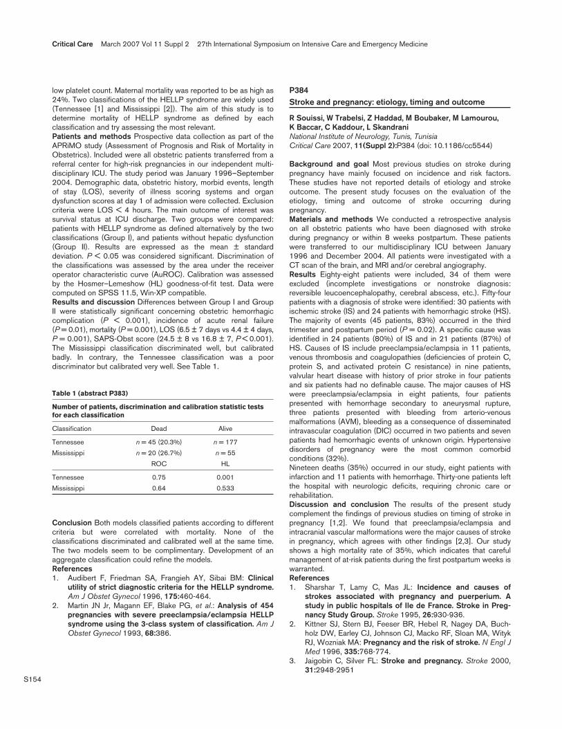
S154
low platelet count. Maternal mortality was reported to be as high as24%. Two classifications of the HELLP syndrome are widely used(Tennessee [1] and Mississippi [2]). The aim of this study is todetermine mortality of HELLP syndrome as defined by eachclassification and try assessing the most relevant.Patients and methods Prospective data collection as part of theAPRiMO study (Assessment of Prognosis and Risk of Mortality inObstetrics). Included were all obstetric patients transferred from areferral center for high-risk pregnancies in our independent multi-disciplinary ICU. The study period was January 1996–September2004. Demographic data, obstetric history, morbid events, lengthof stay (LOS), severity of illness scoring systems and organdysfunction scores at day 1 of admission were collected. Exclusioncriteria were LOS < 4 hours. The main outcome of interest wassurvival status at ICU discharge. Two groups were compared:patients with HELLP syndrome as defined alternatively by the twoclassifications (Group I), and patients without hepatic dysfunction(Group II). Results are expressed as the mean ± standarddeviation. P < 0.05 was considered significant. Discrimination ofthe classifications was assessed by the area under the receiveroperator characteristic curve (AuROC). Calibration was assessedby the Hosmer–Lemeshow (HL) goodness-of-fit test. Data werecomputed on SPSS 11.5, Win-XP compatible.Results and discussion Differences between Group I and GroupII were statistically significant concerning obstetric hemorrhagiccomplication (P < 0.001), incidence of acute renal failure(P = 0.01), mortality (P = 0.001), LOS (6.5 ± 7 days vs 4.4 ± 4 days,P = 0.001), SAPS-Obst score (24.5 ± 8 vs 16.8 ± 7, P < 0.001).The Mississippi classification discriminated well, but calibratedbadly. In contrary, the Tennessee classification was a poordiscriminator but calibrated very well. See Table 1.
Table 1 (abstract P383)
Number of patients, discrimination and calibration statistic testsfor each classification
Classification Dead Alive
Tennessee n = 45 (20.3%) n = 177
Mississippi n = 20 (26.7%) n = 55
ROC HL
Tennessee 0.75 0.001
Mississippi 0.64 0.533
Conclusion Both models classified patients according to differentcriteria but were correlated with mortality. None of theclassifications discriminated and calibrated well at the same time.The two models seem to be complimentary. Development of anaggregate classification could refine the models.References1. Audibert F, Friedman SA, Frangieh AY, Sibai BM: Clinical
utility of strict diagnostic criteria for the HELLP syndrome.Am J Obstet Gynecol 1996, 175:460-464.
2. Martin JN Jr, Magann EF, Blake PG, et al.: Analysis of 454pregnancies with severe preeclampsia/eclampsia HELLPsyndrome using the 3-class system of classification. Am JObstet Gynecol 1993, 68:386.
P384Stroke and pregnancy: etiology, timing and outcome
R Souissi, W Trabelsi, Z Haddad, M Boubaker, M Lamourou, K Baccar, C Kaddour, L SkandraniNational Institute of Neurology, Tunis, TunisiaCritical Care 2007, 11(Suppl 2):P384 (doi: 10.1186/cc5544)
Background and goal Most previous studies on stroke duringpregnancy have mainly focused on incidence and risk factors.These studies have not reported details of etiology and strokeoutcome. The present study focuses on the evaluation of theetiology, timing and outcome of stroke occurring duringpregnancy.Materials and methods We conducted a retrospective analysison all obstetric patients who have been diagnosed with strokeduring pregnancy or within 8 weeks postpartum. These patientswere transferred to our multidisciplinary ICU between January1996 and December 2004. All patients were investigated with aCT scan of the brain, and MRI and/or cerebral angiography.Results Eighty-eight patients were included, 34 of them wereexcluded (incomplete investigations or nonstroke diagnosis:reversible leucoencephalopathy, cerebral abscess, etc.). Fifty-fourpatients with a diagnosis of stroke were identified: 30 patients withischemic stroke (IS) and 24 patients with hemorrhagic stroke (HS).The majority of events (45 patients, 83%) occurred in the thirdtrimester and postpartum period (P = 0.02). A specific cause wasidentified in 24 patients (80%) of IS and in 21 patients (87%) ofHS. Causes of IS include preeclampsia/eclampsia in 11 patients,venous thrombosis and coagulopathies (deficiencies of protein C,protein S, and activated protein C resistance) in nine patients,valvular heart disease with history of prior stroke in four patientsand six patients had no definable cause. The major causes of HSwere preeclampsia/eclampsia in eight patients, four patientspresented with hemorrhage secondary to aneurysmal rupture,three patients presented with bleeding from arterio-venousmalformations (AVM), bleeding as a consequence of disseminatedintravascular coagulation (DIC) occurred in two patients and sevenpatients had hemorrhagic events of unknown origin. Hypertensivedisorders of pregnancy were the most common comorbidconditions (32%).Nineteen deaths (35%) occurred in our study, eight patients withinfarction and 11 patients with hemorrhage. Thirty-one patients leftthe hospital with neurologic deficits, requiring chronic care orrehabilitation.Discussion and conclusion The results of the present studycomplement the findings of previous studies on timing of stroke inpregnancy [1,2]. We found that preeclampsia/eclampsia andintracranial vascular malformations were the major causes of strokein pregnancy, which agrees with other findings [2,3]. Our studyshows a high mortality rate of 35%, which indicates that carefulmanagement of at-risk patients during the first postpartum weeks iswarranted.References1. Sharshar T, Lamy C, Mas JL: Incidence and causes of
strokes associated with pregnancy and puerperium. Astudy in public hospitals of Ile de France. Stroke in Preg-nancy Study Group. Stroke 1995, 26:930-936.
2. Kittner SJ, Stern BJ, Feeser BR, Hebel R, Nagey DA, Buch-holz DW, Earley CJ, Johnson CJ, Macko RF, Sloan MA, WitykRJ, Wozniak MA: Pregnancy and the risk of stroke. N Engl JMed 1996, 335:768-774.
3. Jaigobin C, Silver FL: Stroke and pregnancy. Stroke 2000,31:2948-2951
Critical Care March 2007 Vol 11 Suppl 2 27th International Symposium on Intensive Care and Emergency Medicine
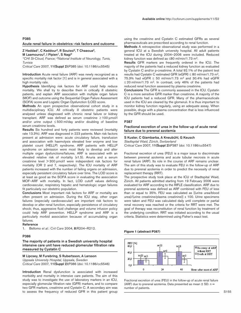
S155
P385Acute renal failure in obstetrics: risk factors and outcome
Z Haddad1, C Kaddour2, R Souissi2, T Chaaoua2, M Laamourou2, I Pipien1, S Nagi21CHI St-Cloud, France; 2National Institute of Neurology, Tunis,TunisiaCritical Care 2007, 11(Suppl 2):P385 (doi: 10.1186/cc5545)
Introduction Acute renal failure (ARF) was newly recognized as aspecific mortality risk factor [1] and is in general associated with ahigh mortality rate.Hypothesis Identifying risk factors for ARF could help reducemortality. We shall try to describe them in critically ill obstetricpatients, and explain ARF association with multiple organ failure(MOF) and outcome using the Sequential Organ Failure Assessment(SOFA) score and Logistic Organ Dysfunction (LOD) score.Methods An open prospective observational cohort study in amultidisciplinary ICU. All critically ill obstetric patients wereanalysed unless diagnosed with chronic renal failure or kidneytransplant. ARF was defined as serum creatinine ≥ 100 µmol/land/or urine output ≤ 500 ml/day and/or doubling of baselineserum creatinine levels.Results Six hundred and forty patients were reviewed (mortalityrate 13.3%). ARF was diagnosed in 223 patients. Main risk factorspresent at admission were: acute circulatory failure, transfusionand association with haemolysis elevated liver enzyme and lowplatelet count (HELLP) syndrome. ARF patients with HELLPsyndrome on admission were most likely to develop and altermultiple organ dysfunctions/failures. ARF is associated with anelevated relative risk of mortality (x1.5). Anuria and a serumcreatinine level > 300 µmol/l were independent risk factors formortality (OR 2 and 7, respectively). The ICU mortality of ARFpatients increased with the number of failing organs on admission,especially persistent circulatory failure over time. The LOD score isat least as good as the SOFA score in evaluating the associationMOF–ARF with mortality. In fact, LOD cutoff values definingcardiovascular, respiratory hepatic and hematologic organ failuresfit particularly our obstetric population.Conclusions Most important risk factors for ARF or mortality areoften present on admission. During the ICU stay, other organfailures (especially cardiovascular) are important risk factors todevelop or alter renal function, especially persistence of circulatoryshock; thus, aggressive fluid challenge and volume infusion policycould help ARF prevention. HELLP syndrome and ARF is aparticularly morbid association because of accumulating organfailures.Reference1. Bellomo et al.: Crit Care 2004, 8:R204–R212.
P386The majority of patients in a Swedish university hospitalintensive care unit have reduced glomerular filtration ratemeasured by Cystatin C
M Lipcsey, M Furebring, S Rubertsson, A LarssonUppsala University Hospital, Uppsala, SwedenCritical Care 2007, 11(Suppl 2):P386 (doi: 10.1186/cc5546)
Introduction Renal dysfunction is associated with increasedmorbidity and mortality in intensive care patients. The aim of thisstudy was to investigate the use of laboratory markers in an ICU,especially glomerular filtration rate (GFR) markers, and to comparetwo GFR markers, creatinine and Cystatin C. A secondary aim wasto assess the frequency of reduced GFR in this patient group
using the creatinine and Cystatin C estimated GFRs as severalpharmaceuticals are prescribed according to renal function.Methods A retrospective observational study was performed in ageneral ICU at a Swedish university hospital. All adult patientstreated at the ICU during 2004–2006 were included. Reducedkidney function was defined as ≤80 ml/min/1.73 m2.Results GFR markers are frequently ordered in the ICU. Themajority of the patients had a reduced kidney function as evaluatedby Cystatin C and/or p-creatinine. A total 92.1% of the patient testresults had Cystatin C estimated GFR (eGFR) ≤ 80 ml/min/1.73 m2,75.3% had eGFR ≤ 50 ml/min/1.73 m2 and 30.4% had eGFR≤ 20 ml/min/1.73 m2. In contrast, only 46% of the patients hadreduced renal function assessed by plasma creatinine.Conclusions The GFR is commonly assessed in the ICU. CystatinC is a more sensitive GFR marker than creatinine. A majority of theICU patients had a reduced GFR. Many of the pharmaceuticalsused in the ICU are cleared by the glomeruli. It is thus important tomonitor kidney function regularly, using an adequate assay. Whenpossible, drugs with a plasma concentration that is less influencedby the GFR should be used.
P387Fractional excretion of urea in the follow-up of acute renalfailure due to prerenal azotemia
S Kuster, C Giambarba, A Kneubühl, G KeuschStadtspital Waid, Zürich, SwitzerlandCritical Care 2007, 11(Suppl 2):P387 (doi: 10.1186/cc5547)
Fractional excretion of urea (FEU) is a major issue to discriminatebetween prerenal azotemia and acute tubular necrosis in acuterenal failure (ARF). Its role in the course of ARF remains unclear.The aim of this study was to evaluate FEU in the follow-up of ARFdue to prerenal azotemia in order to predict the necessity of renalreplacement therapy (RRT).The prospective study took place at the ICU of Stadtspital Waid,Zürich. All patients admitted starting from 19 February 2006 wereevaluated for ARF according to the RIFLE classification. ARF due toprerenal azotemia was defined as ARF combined with FEU of lessthan or equal to 35%. FEU was calculated as [(urine urea/bloodurea)/(urine creatinine/plasma creatinine)] x 100. Urine specimenswere taken and FEU was calculated daily until complete or partialrenal recovery was reached or the criteria for RRT were met. Thegoal of therapy was reconstitution of renal function by treatment ofthe underlying condition. RRT was initiated according to the usualcriteria. Statistics were determined using Fisher’s exact test.
Available online http://ccforum.com/supplements/11/S2
Figure 1 (abstract P387)
Fractional excretion of urea (FEU) in the follow-up of acute renal failure(ARF) due to prerenal azotemia. Data presented as mean ± SD. n =number of patients.
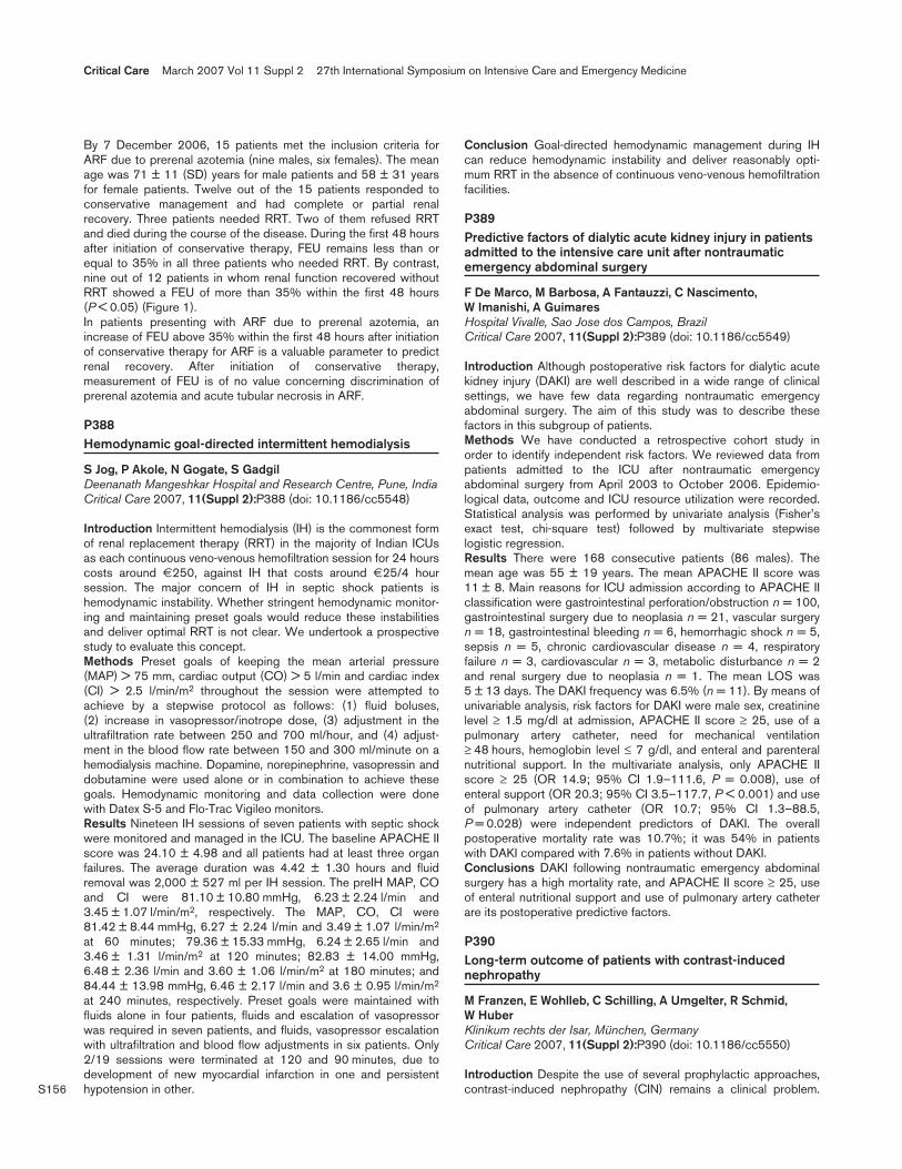
S156
By 7 December 2006, 15 patients met the inclusion criteria forARF due to prerenal azotemia (nine males, six females). The meanage was 71 ± 11 (SD) years for male patients and 58 ± 31 yearsfor female patients. Twelve out of the 15 patients responded toconservative management and had complete or partial renalrecovery. Three patients needed RRT. Two of them refused RRTand died during the course of the disease. During the first 48 hoursafter initiation of conservative therapy, FEU remains less than orequal to 35% in all three patients who needed RRT. By contrast,nine out of 12 patients in whom renal function recovered withoutRRT showed a FEU of more than 35% within the first 48 hours(P < 0.05) (Figure 1).In patients presenting with ARF due to prerenal azotemia, anincrease of FEU above 35% within the first 48 hours after initiationof conservative therapy for ARF is a valuable parameter to predictrenal recovery. After initiation of conservative therapy,measurement of FEU is of no value concerning discrimination ofprerenal azotemia and acute tubular necrosis in ARF.
P388Hemodynamic goal-directed intermittent hemodialysis
S Jog, P Akole, N Gogate, S GadgilDeenanath Mangeshkar Hospital and Research Centre, Pune, IndiaCritical Care 2007, 11(Suppl 2):P388 (doi: 10.1186/cc5548)
Introduction Intermittent hemodialysis (IH) is the commonest formof renal replacement therapy (RRT) in the majority of Indian ICUsas each continuous veno-venous hemofiltration session for 24 hourscosts around €250, against IH that costs around €25/4 hoursession. The major concern of IH in septic shock patients ishemodynamic instability. Whether stringent hemodynamic monitor-ing and maintaining preset goals would reduce these instabilitiesand deliver optimal RRT is not clear. We undertook a prospectivestudy to evaluate this concept.Methods Preset goals of keeping the mean arterial pressure(MAP) > 75 mm, cardiac output (CO) > 5 l/min and cardiac index(CI) > 2.5 l/min/m2 throughout the session were attempted toachieve by a stepwise protocol as follows: (1) fluid boluses, (2) increase in vasopressor/inotrope dose, (3) adjustment in theultrafiltration rate between 250 and 700 ml/hour, and (4) adjust-ment in the blood flow rate between 150 and 300 ml/minute on ahemodialysis machine. Dopamine, norepinephrine, vasopressin anddobutamine were used alone or in combination to achieve thesegoals. Hemodynamic monitoring and data collection were donewith Datex S-5 and Flo-Trac Vigileo monitors.Results Nineteen IH sessions of seven patients with septic shockwere monitored and managed in the ICU. The baseline APACHE IIscore was 24.10 ± 4.98 and all patients had at least three organfailures. The average duration was 4.42 ± 1.30 hours and fluidremoval was 2,000 ± 527 ml per IH session. The preIH MAP, COand CI were 81.10 ± 10.80 mmHg, 6.23 ± 2.24 l/min and3.45 ± 1.07 l/min/m2, respectively. The MAP, CO, CI were81.42 ± 8.44 mmHg, 6.27 ± 2.24 l/min and 3.49 ± 1.07 l/min/m2
at 60 minutes; 79.36 ± 15.33 mmHg, 6.24 ± 2.65 l/min and3.46 ± 1.31 l/min/m2 at 120 minutes; 82.83 ± 14.00 mmHg,6.48 ± 2.36 l/min and 3.60 ± 1.06 l/min/m2 at 180 minutes; and84.44 ± 13.98 mmHg, 6.46 ± 2.17 l/min and 3.6 ± 0.95 l/min/m2
at 240 minutes, respectively. Preset goals were maintained withfluids alone in four patients, fluids and escalation of vasopressorwas required in seven patients, and fluids, vasopressor escalationwith ultrafiltration and blood flow adjustments in six patients. Only2/19 sessions were terminated at 120 and 90 minutes, due todevelopment of new myocardial infarction in one and persistenthypotension in other.
Conclusion Goal-directed hemodynamic management during IHcan reduce hemodynamic instability and deliver reasonably opti-mum RRT in the absence of continuous veno-venous hemofiltrationfacilities.
P389Predictive factors of dialytic acute kidney injury in patientsadmitted to the intensive care unit after nontraumaticemergency abdominal surgery
F De Marco, M Barbosa, A Fantauzzi, C Nascimento, W Imanishi, A GuimaresHospital Vivalle, Sao Jose dos Campos, BrazilCritical Care 2007, 11(Suppl 2):P389 (doi: 10.1186/cc5549)
Introduction Although postoperative risk factors for dialytic acutekidney injury (DAKI) are well described in a wide range of clinicalsettings, we have few data regarding nontraumatic emergencyabdominal surgery. The aim of this study was to describe thesefactors in this subgroup of patients.Methods We have conducted a retrospective cohort study inorder to identify independent risk factors. We reviewed data frompatients admitted to the ICU after nontraumatic emergencyabdominal surgery from April 2003 to October 2006. Epidemio-logical data, outcome and ICU resource utilization were recorded.Statistical analysis was performed by univariate analysis (Fisher’sexact test, chi-square test) followed by multivariate stepwiselogistic regression.Results There were 168 consecutive patients (86 males). Themean age was 55 ± 19 years. The mean APACHE II score was11 ± 8. Main reasons for ICU admission according to APACHE IIclassification were gastrointestinal perforation/obstruction n = 100,gastrointestinal surgery due to neoplasia n = 21, vascular surgeryn = 18, gastrointestinal bleeding n = 6, hemorrhagic shock n = 5,sepsis n = 5, chronic cardiovascular disease n = 4, respiratoryfailure n = 3, cardiovascular n = 3, metabolic disturbance n = 2and renal surgery due to neoplasia n = 1. The mean LOS was5 ± 13 days. The DAKI frequency was 6.5% (n = 11). By means ofunivariable analysis, risk factors for DAKI were male sex, creatininelevel ≥ 1.5 mg/dl at admission, APACHE II score ≥ 25, use of apulmonary artery catheter, need for mechanical ventilation≥ 48 hours, hemoglobin level ≤ 7 g/dl, and enteral and parenteralnutritional support. In the multivariate analysis, only APACHE IIscore ≥ 25 (OR 14.9; 95% CI 1.9–111.6, P = 0.008), use ofenteral support (OR 20.3; 95% CI 3.5–117.7, P < 0.001) and useof pulmonary artery catheter (OR 10.7; 95% CI 1.3–88.5,P = 0.028) were independent predictors of DAKI. The overallpostoperative mortality rate was 10.7%; it was 54% in patientswith DAKI compared with 7.6% in patients without DAKI.Conclusions DAKI following nontraumatic emergency abdominalsurgery has a high mortality rate, and APACHE II score ≥ 25, useof enteral nutritional support and use of pulmonary artery catheterare its postoperative predictive factors.
P390Long-term outcome of patients with contrast-inducednephropathy
M Franzen, E Wohlleb, C Schilling, A Umgelter, R Schmid, W HuberKlinikum rechts der Isar, München, GermanyCritical Care 2007, 11(Suppl 2):P390 (doi: 10.1186/cc5550)
Introduction Despite the use of several prophylactic approaches,contrast-induced nephropathy (CIN) remains a clinical problem.
Critical Care March 2007 Vol 11 Suppl 2 27th International Symposium on Intensive Care and Emergency Medicine
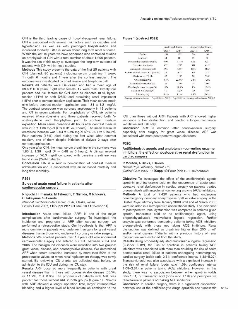
S157
CIN is the third leading cause of hospital-acquired renal failure.CIN is associated with several risk factors such as diabetes andhypertension as well as with prolonged hospitalization andincreased mortality. Little is known about long-term renal outcome.Within the last 10 years we have performed nine controlled studieson prophylaxis of CIN with a total number of about 1,200 patients.It was the aim of this study to investigate the long-term outcome ofpatients with CIN within these studies.Methods This study presents the data of the first 25 patients withCIN (planned: 80 patients) including serum creatinine 1 week,1 month, 6 months and 1 year after the contrast medium. Theoutcome was investigated by chart review and telephone call.Results All patients were Caucasian and had a mean age of69.6 ± 10.6 years. Eight were female, 17 were male. Twenty-fourpatients had risk factors for CIN such as diabetes (8%), hyper-tension (44%) or both (28%) and preexisting renal impairment(15%) prior to contrast medium application. Their mean serum creati-nine before contrast medium application was 1.81 ± 1.21 mg/dl.The contrast procedure was coronary angiography in 18 patientsand CT in seven patients. For prophylaxis of CIN, six patientsreceived N-acetylcysteine and three patients received both N-acetylcysteine and theophylline prior to contrast mediumexposition. Mean serum creatinine 48 hours after contrast mediumwas 2.36 ± 1.36 mg/dl (P < 0.01 vs 0 hours). The mean maximumcreatinine increase was 0.64 ± 0.26 mg/dl (P < 0.01 vs 0 hours).Four patients (16%) died during the first week after contrastmedium, one of them despite initiation of dialysis 2 days aftercontrast application.One year after CIN, the mean serum creatinine in the survivors was1.85 ± 1.39 mg/dl (P = 0.48 vs 0 hours). A clinical relevantincrease of >0.3 mg/dl compared with baseline creatinine wasfound in six (24%) patients.Conclusion CIN is a serious complication of contrast mediumadministration and is associated with an increased mortality andlong-time morbidity.
P391Survey of acute renal failure in patients aftercardiovascular surgery
N Iguchi, H Imanaka, M Takeuchi, T Nishida, M Ichikawa, C Takayama, S AkaedaNational Cardiovascular Center, Suita, Osaka, JapanCritical Care 2007, 11(Suppl 2):P391 (doi: 10.1186/cc5551)
Introduction Acute renal failure (ARF) is one of the majorcomplications after cardiovascular surgery. To investigate theincidence and prognosis of ARF after cardiac surgery, weperformed a retrospective study. Our hypothesis is that ARF ismore common in patients who underwent surgery for great vesseldiseases than in those who underwent coronary or valve surgery.Methods We enrolled patients over 18 years old who underwentcardiovascular surgery and entered our ICU between 2004 and2005. The background diseases were classified into two groups:great vessel disease, and coronary/valve disease. We determinedARF when serum creatinine increased by more than 50% of thepreoperative values, or when renal replacement therapy was newlystarted. By reviewing ICU charts, we collected data before, onadmission to the ICU and during the ICU stay.Results ARF occurred more frequently in patients with greatvessel disease than in those with coronary/valve disease (33.5%vs 11.2%, P < 0.05). The prognosis of patients with ARF waspoorer than those without ARF in both groups (Figure 1). Patientswith ARF showed a longer operation time, larger intraoperativebleeding and a higher level of blood lactate on admission to the
ICU than those without ARF. Patients with ARF showed higherincidence of liver dysfunction, and needed a longer mechanicalventilation and ICU stay.Conclusion ARF is common after cardiovascular surgery,especially after surgery for great vessel disease. ARF wasassociated with more postoperative organ disorders.
P392Antifibrinolytic agents and angiotensin-converting enzymeinhibitors: the effect on postoperative renal dysfunction incardiac surgery
R Mouton, A Binks, I DaviesBristol Royal Infirmary, Bristol, UKCritical Care 2007, 11(Suppl 2):P392 (doi: 10.1186/cc5552)
Objective To investigate the effect of the antifibrinolytic agentsaprotinin and tranexamic acid on the occurrence of acute post-operative renal dysfunction in cardiac surgery on patients treatedpreoperatively with angiotensin-converting enzyme (ACE) inhibitors.Methods A total of 7,420 patients who had undergonenonemergency coronary artery bypass graft or valve surgery in theBristol Royal Infirmary from January 2000 until end of March 2006were included in a retrospective observational study. The incidenceof postoperative renal dysfunction was compared in patients givenaprotin, tranexamic acid or no antifibrinolytic agent, usingpropensity-adjusted multivariable logistic regression. Furtheranalysis was performed comparing patients taking ACE inhibitorspreoperatively with those not taking ACE inhibitors. Renaldysfunction was defined as creatinine higher than 200 µmol/land/or renal dialysis. Patients with a previous history of renaldysfunction were excluded from the study.Results Using propensity-adjusted multivariable logistic regression(C-index, 0.82), the use of aprotinin in patients taking ACEinhibitors was associated with more than doubling the risk of acutepostoperative renal failure in patients undergoing nonemergencycardiac surgery (odds ratio 2.64; confidence interval 1.32–5.27).Tranexamic acid was also associated with a significant increase inthe risk of renal failure (odds ratio 1.59; confidence interval1.09–2.31) in patients taking ACE inhibitors. However, in thisstudy, there was no association between either aprotinin (oddsratio 1.01) or tranexamic acid (odds ratio 1.19) and postoperativerenal failure in patients not taking ACE inhibitors.Conclusion In cardiac surgery, there is a significant associationbetween use of the antifibrinolytic drugs aprotinin and tranexamic
Available online http://ccforum.com/supplements/11/S2
Figure 1 (abstract P391)
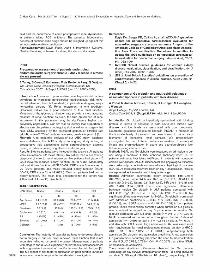
S158
acid and the occurrence of acute postoperative renal dysfunction,in patients taking ACE inhibitors. The potential blood-savingbenefits of antifibrinolytic drugs should be weighed up against thisserious postoperative complication.Acknowledgement David Finch, Audit & Information Systems,Cardiac Services, is thanked for doing the statistical analysis.
P393Preoperative assessment of patients undergoingabdominal aortic surgery: chronic kidney disease is almostalways present
A Turley, S Owen, E Kothmann, M de Belder, A Parry, G DanjouxThe James Cook University Hospital, Middlesbrough, UKCritical Care 2007, 11(Suppl 2):P393 (doi: 10.1186/cc5553)
Introduction A number of preoperative patient-specific risk factorscontribute to increased perioperative cardiovascular risk (myo-cardial infarction, heart failure, death) in patients undergoing majornoncardiac surgery [1]. Renal impairment is one predictor.Creatinine values are a poor reflection of true renal function.Measures of the glomerular filtration rate provide a more accuratemeasure of renal function; as such, the true prevalence of renalimpairment in this population may be significantly higher thanpreviously appreciated. Our aim was to identify that proportion ofelective vascular patients undergoing abdominal aortic surgery thathave CKD, assessed by the estimated glomerular filtration rate(eGFR, ml/min/1.73 m2 body surface area; creatinine, µmol/l) [2].Methods A retrospective analysis of our ‘ABC study’ databasewas undertaken. The ABC study is an ongoing study looking atpreoperative risk assessment using cardiopulmonary exercisetesting in patients undergoing elective aortic surgery.Results Sixty-six patients were included in the analysis. All patientswere Caucasians, 62 males (94%). No patient had a pre-existingdiagnosis of chronic renal impairment. No patients had stage 4/5CKD (severely reduced kidney function, eGFR < 30). Moderatelyreduced kidney function (eGFR 30–59, CKD stage 3) was seen in20 (30%) patients, and mildly reduced kidney function (eGFR60–89, CKD stage 2) in 44 (67%). Only two patients had normalkidney function. The mean total cholesterol for the cohort was4.6 mmol/l (±1 mmol/l). See Table 1.
Table 1 (abstract P393)
CKD stage Stage 1 Stage 2 Stage 3 Total
n 2 44 20 66
Age (years) 64.7 (3.4) 69.9 (8.3) 75.5 (7.7) 71.5 (8.4)
eGFR 92.5 (0.7) 69.4 (7.2) 52.35 (7.4) 64.9 (11.6)
Creatinine 78 (1.4) 97.8 (9) 125.2 (19.9) 105.5 (18.8)
Cholesterol 4.5 (0.5) 4.8 (1.1) 4.3 (0.9) 4.6 (1)
BP 1 (50%) 21 (48%) 9 (45%) 31 (47%)
Smoking 2 (100%) 31 (70%) 14 (70%) 49 (74%)
Diabetes 0 1 (3%) 2 (10%) 3 (5%)
Conclusion The majority of vascular patients undergoing electiveaortic surgery in our unit have impaired renal function that is notaccurately reflected by creatinine values. Management of patientswith stage 2 and 3 CKD is primarily cardiovascular risk assessmentwith aggressive treatment of modifiable vascular risk factors [3].The full impact of risk factor modification on perioperative outcomein vascular patients requires further detailed investigation.
References1. Eagle KA, Berger PB, Calkins H, et al.: ACC/AHA guideline
update for perioperative cardiovascular evaluation fornoncardiac surgery – executive summary. A report of theAmerican College of Cardiology/American Heart Associa-tion Task Force on Practice Guidelines (committee toupdate the 1996 guidelines on perioperative cardiovascu-lar evaluation for noncardiac surgery). Anesth Analg 2002,94:1052-1064.
2. K/DOQI clinical practice guidelines for chronic kidneydisease: evaluation, classification, and stratification. Am JKidney Dis 2002, 39:S1-S266.
3. JBS 2: Joint British Societies’ guidelines on prevention ofcardiovascular disease in clinical practice. Heart 2005, 91(Suppl 5):v1-v52.
P394A comparison of Gc globulin and neutrophil gelatinase-associated lipocalin in patients with liver disease
A Portal, M Austin, M Bruce, E Sizer, G Auzinger, M Heneghan,J WendonKings College Hospital, London, UKCritical Care 2007, 11(Suppl 2):P394 (doi: 10.1186/cc5554)
Introduction Gc globulin, a hepatically synthesized actin bindingprotein, is known to decrease in both acute and chronic liverdisease, and low levels are associated with poor prognosis.Neutrophil gelatinase-associated lipocalin (NGAL), a member ofthe lipocalin family of proteins, has been shown to be an earlybiomarker of ischaemic renal damage. We prospectivelyinvestigated the use of these proteins as markers of severity ofillness and prognostication in acute and acute-on-chronic liverfailure requiring intensive care.Methods NGAL and Gc globulin were measured on admission to ourunit using a sandwich ELISA technique (AntibodyShop®) in 17patients with acute liver failure (ALF) and 11 patients with acute-on-chronic liver disease (ACLD). Biochemical and physiological variableswere collected prospectively and entered into a physiological database(ICARE). All measurements were taken on day 1 of admission. Resultsare expressed as the median and interquartile range.Results Admission parameters: serum creatinine 185 µmol/l(89–266), urine output/24 hours: 340 ml (0–1,111), APACHE IIscore 20 (15–25), lactate 2.6 (1.8–4.68), INR 2.6 (1.6–3.8) andAST 1,404 (124–6,349). There were significant differencesbetween median Gc globulin in ALF patients compared withACLD: 25 mg/l (10–50) vs 50 mg/l (25–129), P = 0.036. Nosignificant differences were seen for NGAL. Gc globulin correlatedwith admission creatinine (r = 0.44, P < 0.01), INR (r = 0.68,P < 0.01), and SOFA score (r = 0.419, P < 0.01) in both patientgroups. These relationships persisted when admission Gc globulinwas examined in regard to day 5 parameters. Admission Gcglobulin correlated with D3 urine output (r = 0.619, P < 0.001).NGAL correlated with urine output throughout the first 5 days ofadmission (r = –0.593 on day 1, r = –0.674 on day 3, P < 0.001)and also with SOFA score. High admission NGAL was associatedwith requirement for renal replacement therapy on day 3 (ROCAUC 0.91 (0.895–1.022, P < 0.001)), outperforming bothadmission Gc globulin and admission creatinine. A low admissionGc globulin more accurately predicted the need for haemofiltrationon day 5 (AUC 0.889, 0.734–1.044, P < 0.007) than either NGALor creatinine on admission.There were significant differences observed for Gc globulinexamining 30-day survival (transplantation from ITU being analysedas ‘death’): 50 mg/l (29–94) vs 18 (4–40), respectively, AUC
Critical Care March 2007 Vol 11 Suppl 2 27th International Symposium on Intensive Care and Emergency Medicine
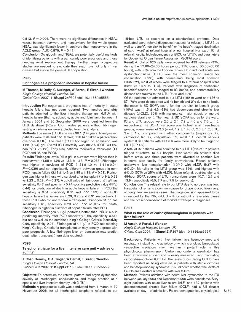
S159
0.813, P = 0.006. There were no significant differences in NGALvalues between survivors and nonsurvivors for the whole group.NGAL was significantly lower in survivors than nonsurvivors in theACLD group (AUC 0.875, P = 0.47).Conclusion Gc globulin and NGAL are potentially useful methodsof identifying patients with a particularly poor prognosis and thoseneeding renal replacement therapy. Further larger prospectivestudies are needed to elucidate their exact role not only in liverdisease but also in the general ITU population.
P395Fibrinogen as a prognostic indicator in hepatic failure
M Thomas, M Duffy, G Auzinger, W Bernal, E Sizer, J WendonKing’s College Hospital, London, UKCritical Care 2007, 11(Suppl 2):P395 (doi: 10.1186/cc5555)
Introduction Fibrinogen as a prognostic test of mortality in acutehepatic failure has not been reported. Two hundred and sixtypatients admitted to the liver intensive therapy unit (LITU) withhepatic failure (that is, subacute, acute and fulminant) between 1January 2004 and 30 September 2006 were identified from theLITU database (I-Care, UK). Twelve patients without fibrinogentesting on admission were excluded from the analysis.Methods The mean (±SD) age was 38.1 (14) years. Ninety-sevenpatients were male and 151 female; 116 had taken a paracetamoloverdose (POD). The mean (±SD) fibrinogen on admission was1.88 (1.24) g/l. Overall ICU mortality was 30.2% (POD 43.4%;non-POD 26.1%). Forty-nine patients received a transplant (14POD and 35 non-POD).Results Fibrinogen levels (all in g/l) in survivors were higher than innonsurvivors (1.98 ± 1.26 vs 1.63 ± 1.15; P = 0.033). Fibrinogenwas higher in survivors of POD (1.81 ± 1.0 vs 1.41 ± 0.94;P = 0.039) and not significantly different between groups in non-POD hepatic failure (2.13 ± 1.43 vs 1.85 ± 1.31; P = 0.28). Fibrino-gen was higher in those who survived after transplant (1.49 ± 0.83vs 1.23 ± 0.23; P = 0.27). A cutoff value of fibrinogen of ≤1 g/l hassensitivity 0.47 and specificity 0.74 (positive predictive value (PPV)0.44) for prediction of death in acute hepatic failure. In POD thesensitivity is 0.51, specificity 0.81 and PPV 0.59; in non-PODhepatic failure the values are 0.29, 0.73 and 0.31 respectively. Inthose POD who did not receive a transplant, fibrinogen ≤1 g/l hassensitivity 0.61, specificity 0.78 and PPV of 0.57 for death.Fibrinogen is higher in survivors of hepatic failure after POD.Conclusion Fibrinogen ≤1 g/l performs better than INR > 6.5 inpredicting mortality after POD (sensitivity 0.69, specificity 0.61),but not as well as the combined King’s College Criteria (sensitivity0.69, specificity 0.96). Fibrinogen of ≤1 g/l in POD not meetingKing’s College Criteria for transplantation may identify a group withpoor prognosis. A low fibrinogen level on admission may predictdeath after transplant (more data required).
P396Telephone triage for a liver intensive care unit – advise oradmit?
A Chan-Dominy, G Auzinger, W Bernal, E Sizer, J WendonKing’s College Hospital, London, UKCritical Care 2007, 11(Suppl 2):P396 (doi: 10.1186/cc5556)
Objective To determine the referral pattern and organ dysfunctionseverity of interhospital consultations, and triage practice at aspecialised liver intensive therapy unit (LITU).Methods A prospective audit was conducted from 1 March to 30November 2006, for all interhospital telephone referrals to our
15-bed LITU as recorded on a standardised proforma. Dataevaluated were: referral diagnoses; reasons for refusal to LITU (‘toowell to benefit’, ‘too sick to benefit’ or ‘no beds’); triaged destinationof care (‘ward’ at referral hospital or our hospital liver ward, ‘IC’ atreferral hospital high-dependency unit/ICU or ‘LITU’); and parametersfor Sequential Organ Failure Assessment (SOFA) score.Result A total of 620 calls were received for 439 referrals (37%during the 17:00–24:00 hours period, 11% during 00:00–08:00hours), with 38% from the London region. Drug-induced acute liverdysfunction/failure (ALDF) was the most common reason forconsultation (39%), with paracetamol being most common(163/172), most of whom were triaged to a referral hospital ward(63% vs 14% to LITU). Patients with diagnosis of ‘ischaemichepatitis’ tended to be triaged to IC (82%), and pancreatobiliarydisease and trauma to the LITU (69% and 80%).Of the patients not admitted to our LITU (162 to ward and 176 toIC), 79% were deemed too well to benefit and 2% due to no beds.the mean ± SD SOFA score for the too sick to benefit group(19%) was 11.5 ± 4.3 (63% had decompensated chronic liverdisease (d-CLD), 36% with malignancy, major sepsis or morbidcardiocerebral event). The mean ± SD SOFA scores for the ward,IC and LITU groups were 2.5 ± 2.4, 7.9 ± 4.6 and 7.8 ± 4.5,respectively. The SOFA liver score was highest in all three triagegroups, overall mean of 2.3 (ward, 1.9 ± 1.4; IC, 2.6 ± 1.2; LITU,2.4 ± 1.2), compared with other components (respiratory 0.9,cardiovascular 0.7, coagulation 0.9, renal 1.1, central nervoussystem 0.8). Patients with INR > 6 were more likely to be triaged toLITU (OR 4.2).A total of 97 patients were admitted to our LITU (five of 17 patientstriaged at referral to our hospital liver ward); six patients diedbefore arrival and three patients were diverted to another liverintensive care facility for family convenience. Fifteen patientsunderwent liver transplantation (10/49 with ALDF, 5/37 withd-CLD). Mortality in the LITU group was 36%, and highest withd-CLD (57% vs 25% with ALDF). Mean referral, post-transfer and48-hour SOFA scores of LITU nonsurvivors were 10.7, 12.7 and13.9, respectively (6.5, 7.7 and 7.5 for survivors).Conclusions The refusal rate to our LITU due to no beds was low.Paracetamol remains a common cause for drug-induced liver injury,although few are severe cases. The triage decision appeared to beinfluenced by the INR, d-CLD with or without a reversible causeand the presence/absence of morbid extrahepatic diagnoses.
P397What is the role of carboxyhaemoglobin in patients withliver failure?
M Austin, A Portal, J WendonKing’s College Hospital, London, UKCritical Care 2007, 11(Suppl 2):P397 (doi: 10.1186/cc5557)
Background Patients with liver failure have haemodynamic andrespiratory instability, the aetiology of which is unclear. Unregulatedvasoactive mediators may have an important role in thisphysiological phenomenon. Carbon monoxide, a vasodilator, hasbeen extensively studied and is easily measured using circulatingcarboxyhaemoglobin (COHb). The levels of circulating COHb havebeen reported as being elevated in patients with stable cirrhosisand hepatopulmonary syndrome. It is unknown whether the levels ofCOHb are elevated in patients with liver failure.Methods Patients admitted with acute liver dysfunction to the ITUbetween January 2003 and December 2005 were considered. Sixty-eight patients with acute liver failure (ALF) and 132 patients withdecompensated chronic liver failure (DCLF) had a full datasetavailable on day 1 of admission. Patient demographics, physiological
Available online http://ccforum.com/supplements/11/S2
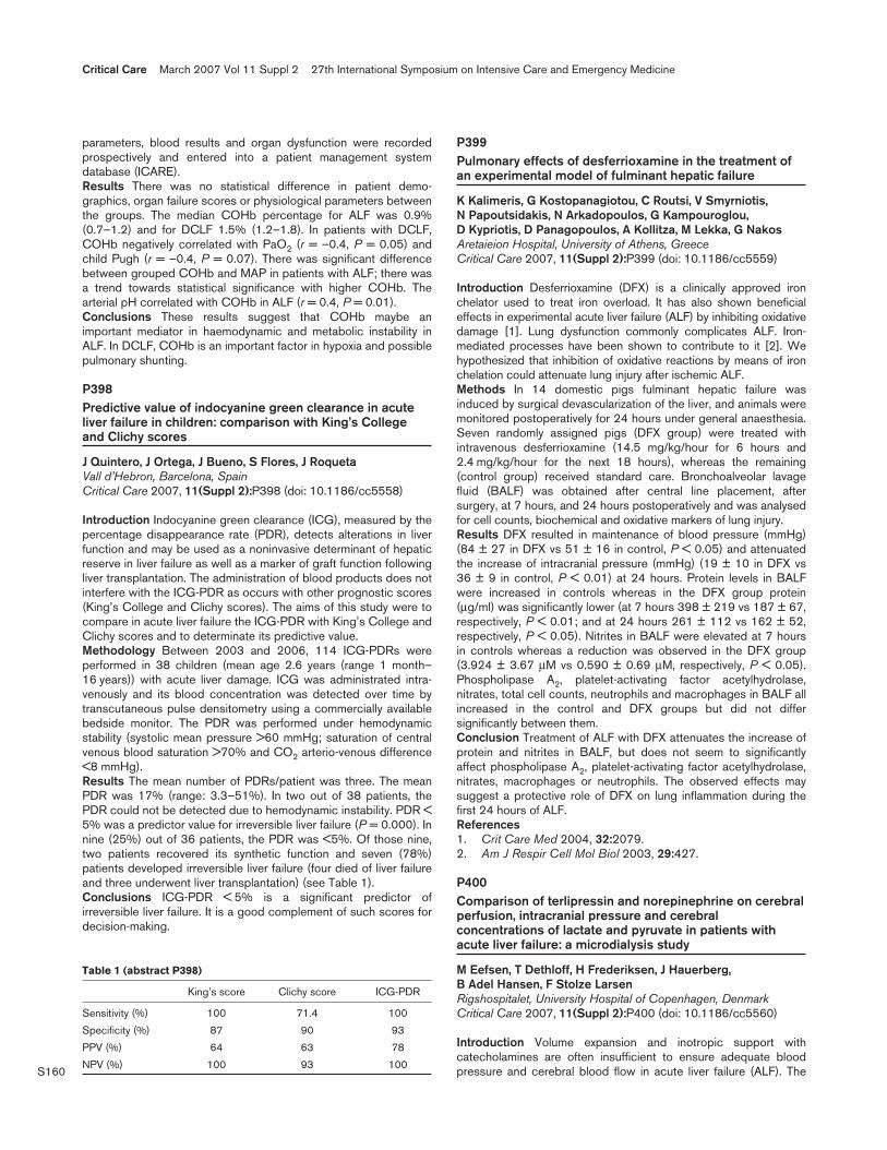
S160
parameters, blood results and organ dysfunction were recordedprospectively and entered into a patient management systemdatabase (ICARE).Results There was no statistical difference in patient demo-graphics, organ failure scores or physiological parameters betweenthe groups. The median COHb percentage for ALF was 0.9%(0.7–1.2) and for DCLF 1.5% (1.2–1.8). In patients with DCLF,COHb negatively correlated with PaO2 (r = –0.4, P = 0.05) andchild Pugh (r = –0.4, P = 0.07). There was significant differencebetween grouped COHb and MAP in patients with ALF; there wasa trend towards statistical significance with higher COHb. Thearterial pH correlated with COHb in ALF (r = 0.4, P = 0.01).Conclusions These results suggest that COHb maybe animportant mediator in haemodynamic and metabolic instability inALF. In DCLF, COHb is an important factor in hypoxia and possiblepulmonary shunting.
P398Predictive value of indocyanine green clearance in acuteliver failure in children: comparison with King’s Collegeand Clichy scores
J Quintero, J Ortega, J Bueno, S Flores, J RoquetaVall d’Hebron, Barcelona, SpainCritical Care 2007, 11(Suppl 2):P398 (doi: 10.1186/cc5558)
Introduction Indocyanine green clearance (ICG), measured by thepercentage disappearance rate (PDR), detects alterations in liverfunction and may be used as a noninvasive determinant of hepaticreserve in liver failure as well as a marker of graft function followingliver transplantation. The administration of blood products does notinterfere with the ICG-PDR as occurs with other prognostic scores(King’s College and Clichy scores). The aims of this study were tocompare in acute liver failure the ICG-PDR with King’s College andClichy scores and to determinate its predictive value.Methodology Between 2003 and 2006, 114 ICG-PDRs wereperformed in 38 children (mean age 2.6 years (range 1 month–16 years)) with acute liver damage. ICG was administrated intra-venously and its blood concentration was detected over time bytranscutaneous pulse densitometry using a commercially availablebedside monitor. The PDR was performed under hemodynamicstability (systolic mean pressure >60 mmHg; saturation of centralvenous blood saturation >70% and CO2 arterio-venous difference<8 mmHg).Results The mean number of PDRs/patient was three. The meanPDR was 17% (range: 3.3–51%). In two out of 38 patients, thePDR could not be detected due to hemodynamic instability. PDR <5% was a predictor value for irreversible liver failure (P = 0.000). Innine (25%) out of 36 patients, the PDR was <5%. Of those nine,two patients recovered its synthetic function and seven (78%)patients developed irreversible liver failure (four died of liver failureand three underwent liver transplantation) (see Table 1).Conclusions ICG-PDR < 5% is a significant predictor ofirreversible liver failure. It is a good complement of such scores fordecision-making.
P399Pulmonary effects of desferrioxamine in the treatment ofan experimental model of fulminant hepatic failure
K Kalimeris, G Kostopanagiotou, C Routsi, V Smyrniotis, N Papoutsidakis, N Arkadopoulos, G Kampouroglou, D Kypriotis, D Panagopoulos, A Kollitza, M Lekka, G NakosAretaieion Hospital, University of Athens, GreeceCritical Care 2007, 11(Suppl 2):P399 (doi: 10.1186/cc5559)
Introduction Desferrioxamine (DFX) is a clinically approved ironchelator used to treat iron overload. It has also shown beneficialeffects in experimental acute liver failure (ALF) by inhibiting oxidativedamage [1]. Lung dysfunction commonly complicates ALF. Iron-mediated processes have been shown to contribute to it [2]. Wehypothesized that inhibition of oxidative reactions by means of ironchelation could attenuate lung injury after ischemic ALF.Methods In 14 domestic pigs fulminant hepatic failure wasinduced by surgical devascularization of the liver, and animals weremonitored postoperatively for 24 hours under general anaesthesia.Seven randomly assigned pigs (DFX group) were treated withintravenous desferrioxamine (14.5 mg/kg/hour for 6 hours and2.4 mg/kg/hour for the next 18 hours), whereas the remaining(control group) received standard care. Bronchoalveolar lavagefluid (BALF) was obtained after central line placement, aftersurgery, at 7 hours, and 24 hours postoperatively and was analysedfor cell counts, biochemical and oxidative markers of lung injury.Results DFX resulted in maintenance of blood pressure (mmHg)(84 ± 27 in DFX vs 51 ± 16 in control, P < 0.05) and attenuatedthe increase of intracranial pressure (mmHg) (19 ± 10 in DFX vs36 ± 9 in control, P < 0.01) at 24 hours. Protein levels in BALFwere increased in controls whereas in the DFX group protein(µg/ml) was significantly lower (at 7 hours 398 ± 219 vs 187 ± 67,respectively, P < 0.01; and at 24 hours 261 ± 112 vs 162 ± 52,respectively, P < 0.05). Nitrites in BALF were elevated at 7 hoursin controls whereas a reduction was observed in the DFX group(3.924 ± 3.67 µM vs 0.590 ± 0.69 µM, respectively, P < 0.05).Phospholipase A2, platelet-activating factor acetylhydrolase,nitrates, total cell counts, neutrophils and macrophages in BALF allincreased in the control and DFX groups but did not differsignificantly between them.Conclusion Treatment of ALF with DFX attenuates the increase ofprotein and nitrites in BALF, but does not seem to significantlyaffect phospholipase A2, platelet-activating factor acetylhydrolase,nitrates, macrophages or neutrophils. The observed effects maysuggest a protective role of DFX on lung inflammation during thefirst 24 hours of ALF.References1. Crit Care Med 2004, 32:2079.2. Am J Respir Cell Mol Biol 2003, 29:427.
P400Comparison of terlipressin and norepinephrine on cerebralperfusion, intracranial pressure and cerebralconcentrations of lactate and pyruvate in patients withacute liver failure: a microdialysis study
M Eefsen, T Dethloff, H Frederiksen, J Hauerberg, B Adel Hansen, F Stolze LarsenRigshospitalet, University Hospital of Copenhagen, DenmarkCritical Care 2007, 11(Suppl 2):P400 (doi: 10.1186/cc5560)
Introduction Volume expansion and inotropic support withcatecholamines are often insufficient to ensure adequate bloodpressure and cerebral blood flow in acute liver failure (ALF). The
Critical Care March 2007 Vol 11 Suppl 2 27th International Symposium on Intensive Care and Emergency Medicine
Table 1 (abstract P398)
King’s score Clichy score ICG-PDR
Sensitivity (%) 100 71.4 100
Specificity (%) 87 90 93
PPV (%) 64 63 78
NPV (%) 100 93 100
S161
aim of this study was to determine whether terlipressin increasescerebral perfusion and to compare the effect with that of nor-epinephrine (NE). Furthermore, the effect on intracranial pressure(ICP) and cerebral concentrations of lactate and pyruvate wererecorded.Methods A retrospective study in a national four-bed liver ICU thattreated 127 patients with ALF between 2001 and 2006. Thirty-eightof these patients were treated with terlipressin, and 10 patients(median age 42.5 years; range 15–66 years; five females) who alsohad an ICP and a microdialysis catheter placed in the cerebral cortexwere included in this study. Concomitant measurements of meanarterial pressure (MAP), ICP, cerebral perfusion using transcranialDoppler sonography (Vmean) and cerebral concentrations of lactateand pyruvate were made before and after an increase in the NEinfusion rate and i.v. injection of 1 mg terlipressin.Results NE infusion and terlipressin injection increased the MAPand Vmean (P < 0.01). Also, the ICP increased during NE infusion(P < 0.01) but not after terlipressin. The cerebral lactateconcentration was unchanged during NE infusion, while itdecreased after terlipressin (P < 0.05).Conclusion This study shows that terlipressin increases the MAPand cerebral perfusion in patients with ALF with no influence uponICP and the cerebral concentrations of lactate and pyruvate. Thesefindings indicate that terlipressin may be valuable, as an additive, oralternative, treatment of arterial hypotension in patients with ALF tosecure brain viability.
P401Relative adrenal insufficiency in patients with severe acutepancreatitis
J De Waele1, E Hoste1, D Baert2, K Heyndrickx3, D Rijkckaert4,P Thibo5, P Van Biervliet6, F Colardyn1
1Ghent University Hospital, Ghent, Belgium; 2AZ Maria Middelares,Ghent, Belgium; 3OLV, Aalst, Belgium; 4AZ St Lucas, Ghent, Belgium;5AZ Palfijn, Ghent, Belgium; 6AZ Aurora, Oudenaarde, BelgiumCritical Care 2007, 11(Suppl 2):P401 (doi: 10.1186/cc5561)
Background Inadequate cortisol levels and adrenal dysfunctionmay play a role in the pathophysiology of severe acute pancreatitis.This study aimed to analyse the incidence of relative adrenalinsufficiency (RAI) in these patients, to identify factors associatedwith relative adrenal insufficiency and to describe how adrenalresponsiveness affects outcome.Methods In a prospective observational multicenter study, a shortSynacthen test (SST) was performed within 5 days after admissionto the hospital in 25 patients with severe acute pancreatitis, aftersigned informed consent was obtained. The incidence of RAI,defined as an increment after SST of less than 9 µg/dl, was theprimary endpoint of the study. Serum cortisol was measured atbaseline and 30 and 60 minutes after 250 µg adrenocorticotropichormone administration.Results The median baseline cortisol level was 26.6 µg/dl, andincreased to 43.2 µg/dl and 48.8 µg/dl after 30 and 60 minutes,respectively. RAI was found in 16% of all patients, and in 27% ofpatients with organ dysfunction. Patients with RAI were moreseverely ill and had higher SOFA scores from day 4 through day 7after admission. All patients with RAI developed pancreaticnecrosis, and all of them needed surgical intervention. Mortalitywas significantly higher in patients with RAI (75% vs 10%,P = 0.016). Patients who died had a lower increment in cortisollevels after the SST than patients who survived.Conclusion RAI is frequent in patients with severe acute pancreatitisand organ dysfunction. It occurs in patients with more severepancreatitis and is associated with an increased mortality rate.
P402The relative sensitivity of serum lipase versus amylase forradiological image-positive pancreatitis
K Delaney, S LuberUT Southwestern Medical School, Dallas, TX, USACritical Care 2007, 11(Suppl 2):P402 (doi: 10.1186/cc5562)
Background Due to the absence of a ‘gold standard’ test for thediagnosis of pancreatitis, the sensitivities of pancreatic enzymetests for pancreatitis are still debated. We compared the relativesensitivities of lipase and amylase at their upper limit of normalvalues for image-positive pancreatitis in a large consecutive seriesof patients who had simultaneous tests of amylase and lipase.Methods Consecutive patients with a clinical diagnosis ofpancreatitis defined by constant epigastric pain and elevation ofthe amylase or lipase to greater than 106 U/l or 59 U/l,respectively, were imaged by abdominal sonography orcomputerized axial tomography (CAT) scan. All included patientshad positive radiological evidence of acute pancreatitis.Results The pancreas was visualized in 399/473 (84%) patientswith suspected pancreatitis and 127/399 (38%) had radiologicalevidence of pancreatitis. Elevation of the lipase to >59 U/ldetected 127/127 cases of image-positive pancreatitis, whileelevation of the amylase to >106 U/l detected 113/127 cases(88% sensitive (0.82–0.94)). All cases of acute biliary pancreatitiswere detected by both the lipase and amylase. The amylasemissed 14 of 65 patients with nonbiliary etiologies of pancreatitis.Four of these were alcoholics.Conclusion Our study shows a high degree of relative sensitivityof the lipase compared with amylase for pancreatic injury that isdemonstrable on CAT or sonographic imaging. Our findingsdemonstrate a relative lack of sensitivity of amylase for nonbiliaryetiologies of pancreatitis. We recognize that the absolutesensitivity of lipase for image-positive pancreatitis cannot bedetermined by this retrospective methodology that requiredelevation of one marker for inclusion.
P403The significance of gallbladder sludge in the patient withacute pancreatitis
K Delaney, L VelezUT Southwestern Medical School, Dallas, TX, USACritical Care 2007, 11(Suppl 2):P403 (doi: 10.1186/cc5563)
Background The significance of gallbladder sludge as a potentialcause of acute biliary pancreatitis is debated. We report theincidence and outcome of patients with gallbladder sludge in alarge population of patients with pancreatitis.Methods Pancreatitis was defined as constant epigastric pain withlipase greater than three times the upper reference value for ourlaboratory (177 U/l). Consecutive patients with first episodes ofacute pancreatitis were identified over a 2-year period and datawere evaluated retrospectively. Patients were followedprospectively for 2 years more.Results All patients had gallbladder ultrasound examinations. Firstepisodes of acute pancreatitis were identified in 356 patients.Initially 236 patients had stones directly visualized in thegallbladder. Of the remaining 120 patients, 13 had sludge, 11 hada dilated common bile duct, one had a positive sonographicMurphy sign, and 95 had no abnormalities. During the time courseof the study, 23 of these 120 patients were demonstrated to havestones; by the surgical pathology report (12), endoscopicretrograde cholangio-pancreatography (10), and cholecystostomy
Available online http://ccforum.com/supplements/11/S2
S162
(one). Seven of 13 patients with sludge underwent cholestectomyand all had evidence of stones. Four of six nonoperated patientswith sludge (67%) returned with recurrent pancreatitis over thecourse of the study. Ten of 97 patients (10%) with suspectednonbiliary etiology of the pancreatitis returned with recurrentpancreatitis over the same period. The nonoperated patients withsludge were more likely to have other risk factors for nonbiliarypancreatitis than were the operated patients.Conclusions The presence of sludge on the gallbladderultrasound suggests the presence of stones and is associated witha high rate of recurrence of pancreatitis in nonoperated patients.
P404Recurrence rates in patients with first episodes of acutepancreatitis
K Delaney, S LuberUT Southwestern Medical School, Dallas, TX, USACritical Care 2007, 11(Suppl 2):P404 (doi: 10.1186/cc5564)
Introduction The timing of operative intervention in patients withsuspected biliary pancreatitis is debated. Recurrence rates ofpancreatitis in patients with biliary lithiasis are compared inoperated and nonoperated patients. Recurrences in patients withsuspected nonbiliary pancreatitis are also reported.Methods First episodes of acute pancreatitis were identified inconsecutive emergency department patients over a 2-year period.Data were evaluated retrospectively and then the identifiedpatients were then followed prospectively for 2 years more.Results Pancreatitis was defined clinically as constant epigastricpain associated with elevation of the serum lipase to greater thanthree times the upper reference value (177 U/l) and no otheridentified cause of abdominal pain. Of 356 patients with firstepisodes of acute pancreatitis, 259 had pancreatitis that wasultimately associated with biliary lithiasis based on abdominalultrasound (n = 236), positive endoscopic retrograde pancreoto-graphy (n = 11), surgical pathology report (n = 11), or cholecystos-tomy (n = 1). Ninety-seven patients had no identified stones duringthe study period. Cholecystectomy was performed in 235/259 atthe time of admission for pancreatitis. Reasons for nonoperativemanagement were death (three cases), medical contraindications(15 cases), pregnancy (two cases) and delayed diagnosis due tonegative abdominal sonogram (four cases). During the studyperiod 10% (10/97) of patients without demonstrated stonesreturned with recurrent episodes of pancreatitis. There were tworecurrences in 235 operated patients with stones (0.85%). Twelveof 19 nonoperated survivors with biliary lithiasis returned withcomplications of biliary lithiasis, including 10 recurrences of biliarypancreatitis (53%) and two episodes of common bile ductobstruction, one of which resulted in death from cholangitis. Themedian time to recurrence of pancreatitis in nonoperated patientswith stones was 50 days, range 26–581 days.Conclusion Cholecystectomy reduced the incidence of recurrenceof pancreatitis in patients with biliary lithiasis.
P405Replacement of albumin after abdominal surgery
K Mahkovic Hergouth1, L Kompan2
1Institute of Oncology, Ljubljana, Slovenia; 2Clinical Center,Ljubljana, SloveniaCritical Care 2007, 11(Suppl 2):P405 (doi: 10.1186/cc5565)
Introduction Replacement of albumin in hypoalbuminemic patientsis not proven to reduce postoperative morbidity and mortality but
no broad consensus is reached yet on abandoning the use ofalbumin in intensive care and perioperative settings for the badprognostic value of hypoalbuminemia. As the albumin decrease inmajor surgery is mostly due to extravascular leakage of albumin(systemic inflammatory response), we regard hypoalbuminemia justas a marker of inflammatory response to surgery that albuminreplacement cannot change. So the postoperative morbidity,mortality and length of stay would not differ in patients withoutalbumin replacement.Materials and methods We retrospectively studied 76successive patients operated on in the abdomen at the OncologicInstitute in Ljubljana in 1997/98 (group 1 – postoperativehypoalbuminemia treated with 20% albumin solution) and in2000/01 (group 2 – no albumin treatment), because ofabandoning albumin use in our surgical department. We comparedserum albumin concentrations in the first week after surgery (threevalues) as well as the postoperative complication rate and thelength of hospital stay. We looked for correlation between thepostoperative albumin concentration and the duration of surgery,amount of transfusion and amount of infusion during surgery.Results The two groups of 38 patients were comparable in age(52.4 and 56.5 years), ASA physical status (1.9 and 2.0),preoperative albumin concentration (39.0 and 38.1 g/l), duration ofoperation (5.9 and 6.1 hours), transfused red blood cells (3.3 and2.0 l) and crystalloid infusion during surgery (5.3 and 4.5 l). In bothgroups there was very significant drop of albumin concentration inthe first week after surgery (P < 0.001). In group 2 albuminconcentrations were very significantly lower than in group 1 untilthe fifth postoperative day (P < 0.001). The difference diminishedafter the sixth postoperative day (P < 0.03). There was negativecorrelation between the postoperative albumin concentration andthe duration of surgery (r = –0.44, P < 0.008). We found nodifference in the postoperative complication rate (surgical ormedical), length of stay and mortality between the groups.Conclusion Postoperative serum albumin concentrations werereduced in both groups, but more in group 2 with no albumintreatment, and in longer operations. Morbidity, mortality and lengthof stay were not influenced by albumin replacement.
P406Extravascular lung water following resuscitation ofhemorrhagic shock in swine: comparison between Ringers’lactate and normal saline
C Phillips, B Tieu, D Hagg, M SchreiberOregon Health & Science University, Portland, OR, USACritical Care 2007, 11(Suppl 2):P406 (doi: 10.1186/cc5566)
Introduction Pulmonary edema is a common consequence ofhemorrhagic shock resuscitation. The type and amount of fluidused in resuscitation may be important determinants of the amountof edema formed. Ringers’ lactate (RL) and normal saline (NS)remain common resuscitative fluids. These experiments weredesigned to measure the extravascular lung water (EVLW) afterresuscitation from hemorrhagic shock with RL vs NS, to determinewhether the fluid type results in differences in the amount ofEVLW, and to determine whether there exists a threshold amountof fluid that results in the development of edema.Methods This was a randomized controlled trial using 20 femaleYorkshire crossbred pigs. Animals were mechanically ventilated.Anesthesia was maintained using 2% isofluorane in 100% oxygen.Continuous hemodynamic monitoring, blood sampling, anddetermination of EVLW by single indicator transpulmonary dilutionwas done using a PiCCO plus monitor (Pulsion Medical System,Munich, Germany). The animals underwent a midline celiotomy,
Critical Care March 2007 Vol 11 Suppl 2 27th International Symposium on Intensive Care and Emergency Medicine
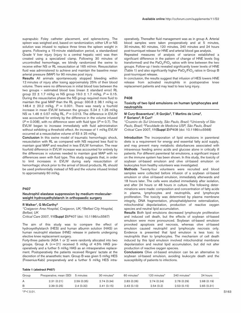
S163
suprapubic Foley catheter placement, and splenectomy. Thespleen was weighed and, based on randomization, either LR or NSsolution was infused to replace three times the spleen weight ingrams. Following a 15-minute stabilization period, a standardizedGrade V liver injury (injury to a central hepatic vein) was thencreated using a specialized clamp. Following 30 minutes ofuncontrolled hemorrhage, we blindly randomized the swine toreceive either NS or RL resuscitation at 165 ml/min. Resuscitationfluid was administered to achieve and maintain the baseline meanarterial pressure (MAP) for 90 minutes post injury.Results All animals spontaneously stopped bleeding within12 minutes of injury after losing approximately 25% of their bloodvolume. There were no differences in initial blood loss between thetwo groups – estimated blood loss (mean ± standard error) RLgroup 22 ± 1.7 ml/kg vs NS group 19.0 ± 1.7 ml/kg, P = 0.15.During the resuscitative phase the NS group required more fluid tomaintain the goal MAP than the RL group: 330.8 ± 38.1 ml/kg vs148.4 ± 20.2 ml/kg, P = 0.001. There was nearly a fourfoldincrease in mean EVLW between the groups: 5.24 ± 1.26 ml/kgNS vs 1.46 ± 0.57 ml/kg RL, P = 0.013. The difference in EVLWwas accounted for entirely by the difference in the volume infused(P = 0.008), with no difference seen with fluid type (P = 0.7). TheEVLW began to increase immediately with fluid administrationwithout exhibiting a threshold effect. An increase of 1 ml/kg EVLWoccurred at a resuscitative volume of 63 ± 25 ml/kg.Conclusion In this swine model of traumatic hemorrhagic shock,resuscitation with RL as compared with NS required less fluid tomaintain goal MAP and resulted in less EVLW formation. The nearfourfold difference in EVLW increase was accounted for entirely bythe differences in volumes needed to maintain goal MAP with nodifferences seen with fluid type. This study suggests that, in orderto limit increases in EVLW during early resuscitation ofhemorrhagic shock prior to the arrival of blood products, RL shouldbe used preferentially instead of NS and the volume infused limitedto approximately 60 ml/kg.
P407Neutrophil elastase suppression by medium-molecular-weight hydroxyethylstarch in orthopaedic surgery
R Walker1, G McCarthy2
1Craigavon Area Hospital, Craigavon, UK; 2Belfast City Hospital,Belfast, UKCritical Care 2007, 11(Suppl 2):P407 (doi: 10.1186/cc5567)
The aim of this study was to compare the effect ofhydroxyethylstarch (HES) and human albumin solution (HAS) onhuman neutrophil elastase (HNE) release in patients undergoingelective knee replacement surgery.Forty-three patients (ASA 1 or 2) were randomly allocated into twogroups. Group A (n = 21) received 5 ml/kg of 4.5% HAS pre-operatively and a further 5 ml/kg HAS as an intraoperative replace-ment. Postoperatively the patients received Ringers’ lactate at thediscretion of the anaesthetic team. Group B was given 5 ml/kg HES(Fresenius-Kabi) preoperatively and a further 5 ml/kg HES intra-
operatively. Thereafter fluid management was as in group A. Arterialblood samples were taken preoperatively and at 5 minutes,30 minutes, 60 minutes, 120 minutes, 240 minutes and 24 hourspost-tourniquet release for HNE and arterial blood gas analysis.Repeated measures of analysis of variance established asignificant difference in the pattern of change of HNE levels (logtransformed) and the PaO2/FiO2 ratios with time between the twogroups. Follow-up t tests revealed significantly lower levels of HNE(Table 1) and also significantly higher PaO2/FiO2 ratios in Group Bpost-tourniquet release.In conclusion, the results suggest that infusion of HES lowers HNErelease from activated neutrophils in postoperative kneereplacement patients and may lead to less lung injury.
P408Toxicity of two lipid emulsions on human lymphocytes andneutrophils
M Cury-Boaventura1, R Gorjão2, T Martins de Lima2, F Soriano3, R Curi21Cruzeiro do Sul University, São Paulo, Brazil; 2University of SãoPaulo, Brazil; 3Faculdade de Medicina USP, São Paulo, BrazilCritical Care 2007, 11(Suppl 2):P408 (doi: 10.1186/cc5568)
Introduction The incorporation of lipid emulsions in parenteraldiets is a requirement for energy and essential fatty acid supply,and may prevent many metabolic disturbances associated withintravenous feeding amino acids and glucose alone in critically illpatients. For different parenteral fat emulsions, a significant impacton the immune system has been shown. In this study, the toxicity ofsoybean oil-based emulsion and olive oil-based emulsion onleukocytes from healthy volunteers was investigated.Methods Twenty-four volunteers were recruited and bloodsamples were collected before infusion of a soybean oil-basedemulsion or olive oil-based emulsion, immediately afterwards and18 hours later. The cells were studied immediately after isolation,and after 24 hours or 48 hours in culture. The following deter-minations were made: composition and concentration of fatty acidsin plasma, lymphocytes and neutrophils, and lymphocyteproliferation. The toxicity was determined by plasma membraneintegrity, DNA fragmentation, phosphatidylserine externalization,mitochondrial depolarization, production of reactive oxygenspecies and neutral lipid accumulation.Results Both lipid emulsions decreased lymphocyte proliferationand induced cell death, but the effects of soybean oil-basedemulsion were more pronounced. Soybean oil-based emulsionprovoked apoptosis and necrosis, whereas olive oil-basedemulsion caused neutrophil and lymphocyte necrosis only.Evidence is presented that lipid emulsion is less toxic toneutrophils than to lymphocytes. The mechanism of cell deathinduced by this lipid emulsion involved mitochondrial membranedepolarization and neutral lipid accumulation, but did not alterproduction of reactive oxygen species.Conclusions Olive oil-based emulsion can be an alternative tosoybean oil-based emulsion, avoiding leukocyte death and thesusceptibility of patients to infections.
Available online http://ccforum.com/supplements/11/S2
Table 1 (abstract P407)
Group Preoperative, mean (SD) 5 minutes 30 minutes* 60 minutes* 120 minutes* 240 minutes* 24 hours
A 2.31 (0.21) 2.59 (0.35) 2.74 (0.34) 2.83 (0.26) 2.74 (0.24) 2.78 (0.29) 2.68 (0.19)
B 2.39 (0.25) 2.4 (0.32) 2.41 (0.15) 2.43 (0.13) 2.54 (0.2) 2.53 (0.15) 2.65 (0.31)
*P < 0.01.
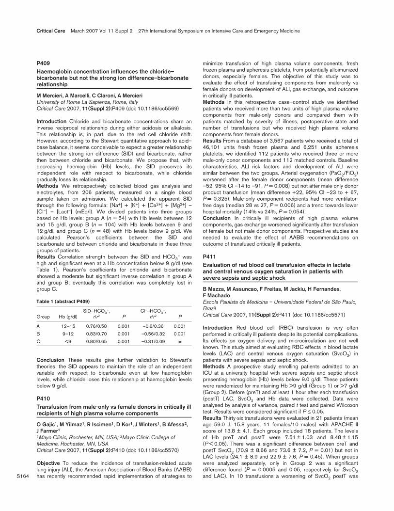
S164
P409Haemoglobin concentration influences the chloride–bicarbonate but not the strong ion difference–bicarbonaterelationship
M Mercieri, A Marcelli, C Claroni, A MercieriUniversity of Rome La Sapienza, Rome, ItalyCritical Care 2007, 11(Suppl 2):P409 (doi: 10.1186/cc5569)
Introduction Chloride and bicarbonate concentrations share aninverse reciprocal relationship during either acidosis or alkalosis.This relationship is, in part, due to the red cell chloride shift.However, according to the Stewart quantitative approach to acid–base balance, it seems conceivable to expect a greater relationshipbetween the strong ion difference (SID) and bicarbonate, ratherthen between chloride and bicarbonate. We propose that, withdecreasing haemoglobin (Hb) levels, the SID preserves itsindependent role with respect to bicarbonate, while chloridegradually loses its relationship.Methods We retrospectively collected blood gas analysis andelectrolytes, from 206 patients, measured on a single bloodsample taken on admission. We calculated the apparent SIDthrough the following formula: [Na+] + [K+] + [Ca2+] + [Mg2+] –[Cl–] – [Lact–] (mEq/l). We divided patients into three groupsbased on Hb levels: group A (n = 54) with Hb levels between 12and 15 g/dl, group B (n = 104) with Hb levels between 9 and12 g/dl, and group C (n = 48) with Hb levels below 9 g/dl. Wecalculated Pearson’s coefficients between the SID andbicarbonate and between chloride and bicarbonate in these threegroups of patients.Results Correlation strength between the SID and HCO3
– washigh and significant even at a Hb concentration below 9 g/dl (seeTable 1). Pearson’s coefficients for chloride and bicarbonateshowed a moderate but significant inverse correlation in group Aand group B; eventually this correlation was completely lost ingroup C.
Table 1 (abstract P409)
SID–HCO3–, Cl––HCO3
–, Group Hb (g/dl) r/r2 P r/r2 P
A 12–15 0.76/0.58 0.001 –0.6/0.36 0.001
B 9–12 0.83/0.70 0.001 –0.56/0.32 0.001
C <9 0.80/0.65 0.001 –0.31/0.09 ns
Conclusion These results give further validation to Stewart’stheories: the SID appears to maintain the role of an independentvariable with respect to bicarbonate even at low haemoglobinlevels, while chloride loses this relationship at haemoglobin levelsbelow 9 g/dl.
P410Transfusion from male-only vs female donors in critically illrecipients of high plasma volume components
O Gajic1, M Yilmaz1, R Iscimen1, D Kor1, J Winters1, B Afessa2, J Farmer1
1Mayo Clinic, Rochester, MN, USA; 2Mayo Clinic College ofMedicine, Rochester, MN, USACritical Care 2007, 11(Suppl 2):P410 (doi: 10.1186/cc5570)
Objective To reduce the incidence of transfusion-related acutelung injury (ALI), the American Association of Blood Banks (AABB)has recently recommended rapid implementation of strategies to
minimize transfusion of high plasma volume components, freshfrozen plasma and apheresis platelets, from potentially alloimunizeddonors, especially females. The objective of this study was toevaluate the effect of transfusing components from male-only vsfemale donors on development of ALI, gas exchange, and outcomein critically ill patients.Methods In this retrospective case–control study we identifiedpatients who received more than two units of high plasma volumecomponents from male-only donors and compared them withpatients matched by severity of illness, postoperative state andnumber of transfusions but who received high plasma volumecomponents from female donors.Results From a database of 3,567 patients who received a total of46,101 units fresh frozen plasma and 6,251 units apheresisplatelets, we identified 112 patients who received three or moremale-only donor components and 112 matched controls. Baselinecharacteristics, ALI risk factors and development of ALI weresimilar between the two groups. Arterial oxygenation (PaO2/FiO2)worsened after the female donor components (mean difference–52, 95% CI –14 to –91, P = 0.008) but not after male-only donorproduct transfusion (mean difference +22, 95% CI –23 to + 67,P = 0.325). Male-only component recipients had more ventilator-free days (median 28 vs 27, P = 0.006) and a trend towards lowerhospital mortality (14% vs 24%, P = 0.054).Conclusion In critically ill recipients of high plasma volumecomponents, gas exchange worsened significantly after transfusionof female but not male donor components. Prospective studies areneeded to evaluate the effect of AABB recommendations onoutcome of transfused critically ill patients.
P411Evaluation of red blood cell transfusion effects in lactateand central venous oxygen saturation in patients withsevere sepsis and septic shock
B Mazza, M Assuncao, F Freitas, M Jackiu, H Fernandes, F MachadoEscola Paulista de Medicina – Universidade Federal de São Paulo,BrazilCritical Care 2007, 11(Suppl 2):P411 (doi: 10.1186/cc5571)
Introduction Red blood cell (RBC) transfusion is very oftenperformed in critically ill patients despite its potential complications.Its effects on oxygen delivery and microcirculation are not wellknown. This study aimed at evaluating RBC effects in blood lactatelevels (LAC) and central venous oxygen saturation (SvcO2) inpatients with severe sepsis and septic shock.Methods A prospective study enrolling patients admitted to anICU at a university hospital with severe sepsis and septic shockpresenting hemoglobin (Hb) levels below 9.0 g/dl. These patientswere randomized for maintaining Hb >9 g/dl (Group 1) or >7 g/dl(Group 2). Before (preT) and at least 1 hour after each transfusion(postT) LAC, SvcO2 and Hb data were collected. Data wereanalysed by analysis of variance, paired t test and paired Wilcoxontest. Results were considered significant if P ≤ 0.05.Results Thirty-six transfusions were evaluated in 21 patients (meanage 59.0 ± 15.8 years, 11 females/10 males) with APACHE IIscore of 13.8 ± 4.1. Each group included 18 patients. The levelsof Hb preT and postT were 7.51 ± 1.03 and 8.48 ± 1.15(P < 0.05). There was a significant difference between preT andpostT SvcO2 (70.9 ± 8.66 and 73.6 ± 7.2, P = 0.01) but not inLAC levels (24.1 ± 8.9 and 22.9 ± 7.6, P = 0.45). When groupswere analyzed separately, only in Group 2 was a significantdifference found (P = 0.0005 and 0.05, respectively for SvcO2and LAC). In 10 transfusions a worsening of SvcO2 postT was
Critical Care March 2007 Vol 11 Suppl 2 27th International Symposium on Intensive Care and Emergency Medicine
S165
observed and all these patients had preT SvcO2 > 70%. Althoughthere was no significant correlation between a worsening in SvcO2and preT Hb, eight of these patients were allocated to Group 1.Another 13 transfusions were done with a SvcO2 preT < 70, and10 of them improved (> 5%) after transfusion (mean percentage ofimprovement = 18.9%). Only four of these patients were allocatedto Group 1. Patients with high levels of preT LAC (n = 21) onlyimproved (reduction > 10%) in 42.9% of cases. A total 53.3% ofpatients with normal preT LAC levels worsened (rising > 10%)postT. The mean preT Hb from these patients was 8.18 ± 0.9.Conclusion In patients with SvcO2 < 70 and/or Hb < 7.0 g/dl,transfusion seems to result in an improvement of perfusionparameters. However, in patients with SvcO2 > 70 or normallactate levels, transfusion seems to impair tissue perfusion.
P412Transfusion profiles in intensive care units from auniversity hospital
M Assuncao, I Paula, L Falcao, B Mazza, M Barros, M Jackiu, H Fernandes, F MachadoUniversidade Federal de São Paulo, BrazilCritical Care 2007, 11(Suppl 2):P412 (doi: 10.1186/cc5572)
Introduction Red blood cell (RBC) transfusion is very oftenperformed in critically ill patients despite its potential complica-tions. New guidelines recommend that doctors should haveconservative behavior regarding its use. The objective of this studywas to evaluate the transfusion profile among patients in ICUs at auniversity hospital in Brazil.Methods A prospective evaluation of all patients admitted to sixICUs (surgical–medical, private, neurosurgery, medical, pneu-mology and coronary units) that have received a RBC transfusionas indicated by assistant physicians during October/November2005. Clinical data as well as the characteristics of the transfusionwere collected and submitted to univariate statistical analysis (chi-squared and Student’s t test). Results were considered significantif P ≤ 0.05.Results Four hundred and eight transfusions were made in 71patients (38 females, 33 males), 35 medical/36 surgical, with amean age 57.2 ± 8.4 years, mean APACHE II score 17.7 ± 5.3,and mean SOFA score on the day of transfusion 6.09 ± 3.99. Atadmission, 60 patients (84.5%) had comorbidities, 10 (14.1%)had chronic coronary disease. At transfusion, 54.9% had sepsis,severe sepsis or septic shock, and 9.9% had acute coronarysyndrome. The mean hemoglobin (Hb) level at ICU admission was9.69 ± 2.3 g/dl and the mean level that triggered transfusion was6.88 ± 1.1 g/dl. The most important transfusion indication was Hblevels (49.8%), followed by active bleeding (31.8%). The meannumber of RBC transfused per time was 1.68 ± 0.96 and themean age of RBC was 14.3 ± 7.83 days (46.6% had more than14 days). Adverse events occurred in 3.4%. The 28-day mortalityrate was 47.1%. Only the SOFA score at the day of transfusioncorrelated with mortality (P = 0.004). There was no correlation withage, type of ICU, APACHE II score, total number or age of RBC,Hb at admission or Hb pretransfusion. There was a significantdifference between the pretransfusion Hb (P < 0.00001) and thenumber of RBC transfused at the same time considering all ICUenrolled in the study (P < 0.00001).Conclusions The Hb level that triggered transfusion was inagreement with recent guidelines regarding critically ill patients,although there was a difference between all ICUs. Despite the factthat there is a scarceness of RBC, the RBC were higher in age.The missing correlation with mortality can be due to the smallsample size.
P413National survey of transfusion practices
N Frikha, R Ouezini, T Mestiri, M Bechikh, M Mebazaa, M Ben AmmarMongi Slim Hospital, La Marsa, TunisiaCritical Care 2007, 11(Suppl 2):P413 (doi: 10.1186/cc5573)
Introduction The awakening of the residual risks of blood trans-fusion leads, everywhere in the world, to efforts to reduce them. Theaim of the study was to assess the transfusion practices in Tunisia.Patients and methods A multicentric prospective observationalstudy of about 1,000 transfusions practiced during 2004 inTunisia. Data were determined by the prescriptor of the trans-fusion. The data were analyzed with SPSS 12.0.Results The mean age of the transfused patients was 35.78 years.The sex ratio was 1.2. A total of 48.3% of the patients had trans-fusion history. Allogeneic immunization and viral serology conver-sion were reported in 2.1% of these patients. The indications weregenerally carried in front of chronic medical pathologies (36.9%),acute medical pathologies (28.2%) and surgical pathologies(elective 11.7%, urgent 11.2%). The haemoglobin threshold fortransfusion was 7.29 g/dl and depended on the indication of thered cell transfusion: 6.16 g/dl for urgent medical pathologies,6.22 g/dl for chronic medical pathologies, 7.74 g/dl for urgentsurgical pathologies, 10.38 g/dl for elective surgery, 6.15 g/dl forurgent obstetrical pathologies. The mean platelet count was24,000 (patients transfused by platelet units). The ABO andrhesus determination were made in 99% of the cases. Aphenotypic determination was required in only 34.5% of the cases.The search for irregular agglutinins was made in 20.7% of thecases. The test of compatibility at the laboratory was practiced in95.4% of the cases. The amount of blood transfused was 2 units.Immediate incidents were reported in 2.5% of the cases. The post-transfusion haemoglobin average was of 9.15 g/dl.Discussion and conclusion The evolution of the blood transfusionwas remarkable, since the use of total blood in the 1980s, with theacquisition of the first techniques of separation of the bloodcomponents. The transfusion practice in Tunisia is far from being tothe standards. The results obtained make it possible to transmit tothe clinician the failures of the system, to better include how toprescribe a blood product, to follow its effectiveness and itspossible side effects, and to apprehend the impact of theinnovated biotechnologies to improve quality of transfusionmedicine in coherence with the security requirements.
P414Could the combination of bleeding time and plateletfunction predict the perioperative transfusion requirementsin cardiac surgery patients?
M Kataphigioti, D Karamichaleli, M Kounavi, E Iliopoulou, G Palatianos, E MelissariOnassis Cardiac Surgery Center, Athens, GreeceCritical Care 2007, 11(Suppl 2):P414 (doi: 10.1186/cc5574)
Introduction The reduction in platelet count and function is themost important, unsolved, nonsurgical cause of postoperativebleeding after open heart surgery. On the other hand, the bleedingtime (BT), the only comprehensive test to explore primaryhaemostasis, detects otherwise unknown defects in platelet–vessel wall interactions. The present study was undertaken in orderto clarify whether the BT and platelet function tested preoperativelycould predict the perioperative transfusion requirements in cardiacsurgery patients.
Available online http://ccforum.com/supplements/11/S2
S166
Patients and methods Sixty-eight patients (54 males/14 females)participated in the study. Thirty-two patients underwent valve(s)replacement (group A) and the remaining underwent coronaryartery bypass grafting(s) (group B). The BT determination wasperformed according to the Mielke technique using Surgicuttdevices (ITC, USA). Platelet function was evaluated by theaggregation procedure using four agonists: ADP, arachidonic acid,collagen and ristocetin at a final concentration of 4 x 10⁻⁶ M,0.5 mg/ml, 0.19 mg/ml and 1.2 mg/ml, respectively.Results (1) In the immediate postoperative time, a significantreduction in haemoglobulin levels was observed in both groupscompared with that of the preoperative time (13.3%, P < 0.05 forgroup A and 28.4%, P < 0.01 for group B). No difference existedin haemoglobulin levels between groups postoperatively. (2) Plateletvalues were slightly different between the groups. A significantdecrease in platelet count was observed in both groupspostoperatively (28.7%, P < 0.03 for group A and 22.4%, P < 0.05for group B). (3) The results of BT and platelet activation (per-formed preoperatively) were similar for patients who underwentvalve replacement and patients who underwent coronary arterybypass grafting – although in this group platelet activation witharachidonic acid and ADP was ~11% lower with both agonists. (4) The transfusion requirements were slightly higher for patients ingroup A, and more patients in group B received no transfusion(one vs four patients).Conclusion In patients undergoing cardiac surgery with a negativehistory of bleeding and early interruption of antiplatelet treatment,the BT and platelet function do not offer much in the setting topredict perioperative bleeding.
P415Emergency staff is in danger
B Gulalp1, O Karcioglu2
1Adana State Hospital, Adana, Turkey; 2Dokuz Eylul University,School of Medicine, Izmir, TurkeyCritical Care 2007, 11(Suppl 2):P415 (doi: 10.1186/cc5575)
Objective To investigate the ratio and characteristics ofaggression, threat and physical violence directed towards staff inemergency departments as a model of state hospitals.Methods A questionnaire were filled in by the staff working in theemergency department of three high-volume inner-city statehospitals. The individualized data collected were relevant to thepattern of violence, age, sex, number of years in the profession,nature of the job, and the behavioral characteristics of assailants,and outcome of incidents. The data were abstracted between1 May and 31 May 2006.Results A total of 109 staff reports were reviewed. Therelationship of aggression with sex, age and years of experiencewere insignificant (P values were 0.464, 0.692, and 0.298,respectively), while profession was very significantly related(P = 0.000). The relation between threat and sex is P = 0.311,experience 0.994, profession 0.326, age 0.278. The relationship ofthreat with sex, years of experience, profession and age wereinsignificant (P values were 0.311, 0.994, 0.326, and 0.278,respectively). On the other hand, physical assault was foundsignificantly related to sex, years of experience, profession and age(P values were 0.042, 0.011, 0.000, and 0.000, respectively).Conclusion Violence to the staff is common. There is not asignificant relationship between aggression, threat and personalcharacters. However, male sex, >5 years experience, emergencydoctor, ≥31 years of age are the risk factors for physical violence.
P416Shaken baby syndrome: the classical clinical triad is stillvalid in recent court rulings
M De Leeuw1, W Jacobs2
1Algemeen Stedelijk Ziekenhuis Aalst, Essene, Belgium; 2University Hospital Antwerp, Edegem, BelgiumCritical Care 2007, 11(Suppl 2):P416 (doi: 10.1186/cc5576)
Introduction Subdural haemorrhage, retinal bleeding and hypoxae-mic encephalopathy have long been considered a diagnostic clinicaltriad for the so-called shaken baby syndrome (SBS). The classicaltriad, however, has been challenged in the recent past by the so-called ‘unified hypothesis’ by Geddes and colleagues [1] withsubsequent implications in court rulings in suspected cases of SBS.Judicial and scientific dilemma The unified hypothesis by Geddessuggested an alternative cause for SBS injuries that did not involvesignificant shaking. The Geddes theory led to the speculation thatsubdural and retinal haemorrhage was not caused by traumaticshearing of subdural and retinal veins but by a combination ofcerebral hypoxia, raised intracranial pressure and raised arterial andcentral venous pressure. The publication of this theory was met withscepticism by many forensic and paediatric pathologists but wasenthusiastically embraced by defence attorneys. This dilemmaresulted in the UK with several appeals against prior convictions ofmurder/manslaughter because of alleged traumatic shaking ofyoung children. The forensic community awaited with great interestthe ruling of the Court of Appeal in London on 21 July 2005.Court ruling of 21 July 2005 Four cases of alleged SBS werebrought to the Court of Appeal in London. Two convictions wereupheld, one conviction was dismissed and one conviction wasreduced from murder to manslaughter. In their written judgementtheir Lordships clearly stated: ‘In our judgment, it follows that theunified hypothesis can no longer be regarded as a credible oralternative cause of the triad of injuries’. The Crown ProsecutionService made a press release that ‘Today’s judgement sends a clearsignal validating the CPS in prosecuting Shaken Baby Syndromecases. The Geddes theory will no longer be used by the defence.’Conclusions Diagnosing SBS is a very complex and delicate matter.The mere presence of the classic triad does not automatically ornecessarily lead to a diagnosis of nonaccidental head injury or aconclusion of unlawful killing. Diagnosis of (intentional) SBS must bebased on the combination of: medical elements, elements frompolice inquiry, and forensic and crime scene elements. Physicians,particularly those working at the medico-legal interface (such asemergency physicians), should realise that medical observations mayplay a pivotal role in the diagnosis of SBS. As important is therealisation that, despite alternative hypothesis in medical literature,the classical triad of symptoms (subdural haemorrhage, retinalbleeding, hypoxaemic encephalopathy) is still valid as diagnostic forSBS according to recent (UK) court ruling.Reference1. Geddes J, et al.: Neuropathol Appl Neurobiol 2003, 29:14-22.
P417Organophosphate poisoning and related mortality withoxime perfusion
A Bartolo1, O Caetano1, M Costa1, R Milheiro1, A Carvalho1, A Braga2
1Hospital Senhora Oliveira, Guimaraes, Portugal; 2University ofMinho, Braga, PortugalCritical Care 2007, 11(Suppl 2):P417 (doi: 10.1186/cc5577)
Introduction A retrospective study performed between 1 January2001 and 31 October 2006 in patients admitted to the ICU with
Critical Care March 2007 Vol 11 Suppl 2 27th International Symposium on Intensive Care and Emergency Medicine
S167
organophosphate poisoning (OPP). The aim of the study was todetermine the relation between mortality and: (a) toxicity oforganophosphate, (b) time between ingestion and management ofthe patient, (c) coligernic manifestations, (d) time and doses ofoximes and atropine.Methods Were admitted to the ICU 29 patients with OPP. The datawere treated by SPSS 14 for Windows and the analysis consisted ofa descriptive study, analytic study (chi-square analysis, Spearmanassociation analysis) and comparison between groups (Studentt analysis, Wilcoxon Mann–Whitney and Kolmogorov–Smirnov tests).The receiver operating characteristic (ROC) was applied for theSequential Organ Failure Assessment (SOFA) score.Results Twenty-nine patients were included in the study, 21 malesand eight females. The mean age of the males was 47.71 years(SD = 13.58) and of females was 41.0 years (SD = 11.66), and62.7% were from a rural area and 37.3% from an urban area.Mortality does not have a significant statistic relation (P > 0.05)with toxicity of organophosphate, time and doses of atropine.However, statistical significance was found between mortality and:(a) time between ingestion of the poison and treatment (Spearmantest, rs = –0.596, P < 0.05), (b) muscarinic manifestations (chi-square test, χ2 = 4.152, P < 0.05), (c) time of oximes(Kolmogorov–Smirnov test with Z = 1.439, P < 0.05) and doses ofoximes (Kolmogorov–Smirnov test with Z = 1.412, P < 0.05). TheROC analysis reveals that for the respiratory SOFA at 9 days, thearea under the ROC curve was 0.917; this means that this SOFAscore can predict correctly in 91.7% of the cases.Conclusion In our study we concluded that the mortality rate wasincreased with prolonged perfusion of oximes and with muscarinicmanifestations of OPP but not with the time and dose of atropine.The respiratory SOFA score at 9 days matches with prediction inabove 90% of the cases.
P418Approaches of Turkish anesthesiologists to deliriumobserved in intensive care unit patients
N Gokmen, L Iyilikci, S Kucukguclu, B Kuvaki, L Ciftci, A GunerliDokuz Eylul University, School of Medicine, Izmir, TurkeyCritical Care 2007, 11(Suppl 2):P418 (doi: 10.1186/cc5578)
Objective To determine attitudes and practices of the Turkishanesthesiologists and residents about delirium in the ICU.Methods An anonymous questionnaire consisting of 22 questions[1] was mailed to 258 anesthesiologists and residents.Results One hundred and fifty-four questionnaires were returned(60% response). Of the respondents, 57% were male and 61%were residents. One-half of respondents work in hospitals withmore than 800 beds; 65% of respondents had an ICU facility of7–12 beds. Seventy-two percent of the respondents had seendelirium in the ICU and also 70.2% of these respondents observeddelirium in <25% of patients who were on mechanical ventilation.Although delirium was accepted a significant or very seriousproblem by 92.5% of the respondents, underdiagnosis wasacknowledged by 74%. Routine screening for delirium wasperformed by 41.6% of the anesthesiologists and 88.1% of themwere repeating daily. Clinical assessment was used in 76.7% ofthe screenings. Delirium was treated with haloperidol andbenzodiazepine by 61.5% and 24% of the respondents. Of therespondents, 93.4% were not able to attend a meeting related todelirium and 67.6% did not read even an article about delirium.Conclusions Turkish anesthesiologists and residents considerdelirium a relatively common and serious problem. However, theyseldom perform screening tests and try to update their knowledgeregarding delirium.
Reference1. Ely EW, et al.: Crit Care Med 2004, 32:106-112.
P419A comparison of the confusion assessment method for theintensive care unit and the NEECHAM confusion scale inintensive care delirium assessment
B Van Rompaey1, L Bossaert2, L Shortridge-Bagett1, M Schuurmans1, S Truijen1
1Universiteit Antwerpen, Wilrijk, Belgium; 2Universitair Ziekenhuis,Edegem, BelgiumCritical Care 2007, 11(Suppl 2):P419 (doi: 10.1186/cc5579)
Introduction Reports indicate an incidence of intensive caredelirium of 11–87%. The confusion assessment method for theintensive care unit (CAM-ICU) is widely used in delirium assess-ment in ICUs. However, its binomial results constrain the evaluationof severity. The NEECHAM confusion scale has recently beenvalidated for use in the ICU and uses a numeric assessment. Thisscale allows the patients to be classified in four categories ofdelirium severity (normal, at-risk, mild to early, moderate to severe).In this study we investigated the diagnostic value of theNEECHAM referring to the CAM-ICU.Methods A consecutive sample of 106 patients in a mixed ICU(cardiac surgery (CS) 35%, noncardiac surgery (NCS) 26%,internal medicine (IM) 39%, age 62 ± 14 years, male 63%) wasassessed after a stay in the ICU ≥24 hours. All patients with aGlasgow Coma Scale ≥ 10 and age ≥ 18 years were included. Anurse researcher simultaneously assessed both scales once dailyin the morning. A total of 272 paired observations were made. Datawere analyzed using the NEECHAM cut-off values of ≤26 (at-risk),≤24 (mild delirium), and ≤19 (severe delirium).Results Using the CAM-ICU the overall prevalence was 15%.Prevalences in CS, NCS and IM were 9%, 14% and 21%,respectively. Using the NEECHAM scale, the overall prevalencewas 33% (16.5% mild, 16.5% severe) and 36%, 21% and 38%for the three patient categories, respectively. Sensitivity was100%, specificity was 79%, positive predictive value was 46% andnegative predictive value was 100%. Using the cut-off value ≤19,sensitivity was 83% and specificity was 96%. All positive CAM-ICU patients were detected by the NEECHAM (85% severe, 15%mild). However, 21% of the CAM-ICU negative patients had aNEECHAM value that diagnoses delirium (4% severe, 17% mild).Consequently, 27% of the CS group (19% severe, 8% mild), 7%of the NCS group (7% mild) and 17% of the IM group (2% severe,15% mild) were diagnosed to be delirious using the NEECHAMand not delirious using the CAM-ICU.Conclusion The NEECHAM delirium scale identified all cases ofdelirium that were detected by the CAM-ICU. Moreover, additionaldelirious patients were identified, especially in the CS group. In thispilot experience, the NEECHAM scale was a valuable screeningtool for intensive care.
P420The occurrence of delirium is severely underestimated byintensivists and intensive care unit nurses during daily ICU care
P Spronk, B Riekerk, S Elias, J Rommes, J HofhuisGelre Ziekenhuizen Location Lukas, Apeldoorn, The NetherlandsCritical Care 2007, 11(Suppl 2):P420 (doi: 10.1186/cc5580)
Introduction Patients improve faster in the ICU if sedatives arestopped as soon as possible with inherent occurrence of sleepingdisorders, and delirium, which could compromise the recovery
Available online http://ccforum.com/supplements/11/S2
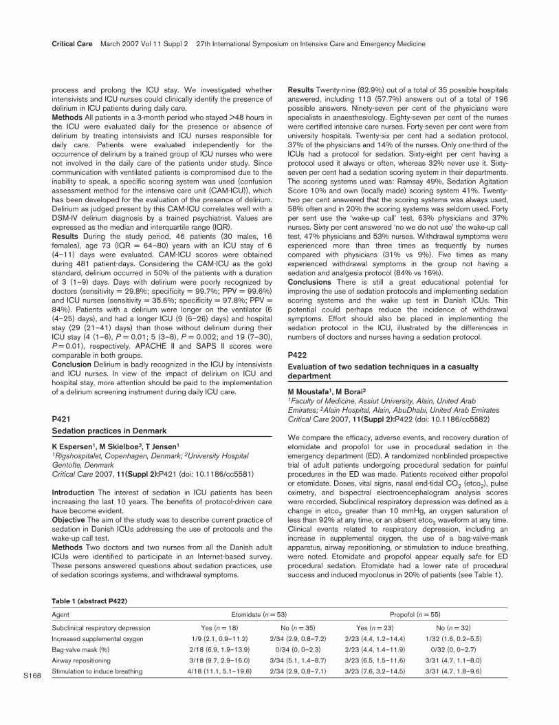
S168
process and prolong the ICU stay. We investigated whetherintensivists and ICU nurses could clinically identify the presence ofdelirium in ICU patients during daily care.Methods All patients in a 3-month period who stayed >48 hours inthe ICU were evaluated daily for the presence or absence ofdelirium by treating intensivists and ICU nurses responsible fordaily care. Patients were evaluated independently for theoccurrence of delirium by a trained group of ICU nurses who werenot involved in the daily care of the patients under study. Sincecommunication with ventilated patients is compromised due to theinability to speak, a specific scoring system was used (confusionassessment method for the intensive care unit (CAM-ICU)), whichhas been developed for the evaluation of the presence of delirium.Delirium as judged present by this CAM-ICU correlates well with aDSM-IV delirium diagnosis by a trained psychiatrist. Values areexpressed as the median and interquartile range (IQR).Results During the study period, 46 patients (30 males, 16females), age 73 (IQR = 64–80) years with an ICU stay of 6(4–11) days were evaluated. CAM-ICU scores were obtainedduring 481 patient-days. Considering the CAM-ICU as the goldstandard, delirium occurred in 50% of the patients with a durationof 3 (1–9) days. Days with delirium were poorly recognized bydoctors (sensitivity = 29.8%; specificity = 99.7%; PPV = 99.6%)and ICU nurses (sensitivity = 35.6%; specificity = 97.8%; PPV =84%). Patients with a delirium were longer on the ventilator (6(4–25) days), and had a longer ICU (9 (6–26) days) and hospitalstay (29 (21–41) days) than those without delirium during theirICU stay (4 (1–6), P = 0.01; 5 (3–8), P = 0.002; and 19 (7–30),P = 0.01), respectively. APACHE II and SAPS II scores werecomparable in both groups.Conclusion Delirium is badly recognized in the ICU by intensivistsand ICU nurses. In view of the impact of delirium on ICU andhospital stay, more attention should be paid to the implementationof a delirium screening instrument during daily ICU care.
P421Sedation practices in Denmark
K Espersen1, M Skielboe2, T Jensen1
1Rigshospitalet, Copenhagen, Denmark; 2University HospitalGentofte, DenmarkCritical Care 2007, 11(Suppl 2):P421 (doi: 10.1186/cc5581)
Introduction The interest of sedation in ICU patients has beenincreasing the last 10 years. The benefits of protocol-driven carehave become evident.Objective The aim of the study was to describe current practice ofsedation in Danish ICUs addressing the use of protocols and thewake-up call test.Methods Two doctors and two nurses from all the Danish adultICUs were identified to participate in an Internet-based survey.These persons answered questions about sedation practices, useof sedation scorings systems, and withdrawal symptoms.
Results Twenty-nine (82.9%) out of a total of 35 possible hospitalsanswered, including 113 (57.7%) answers out of a total of 196possible answers. Ninety-seven per cent of the physicians werespecialists in anaesthesiology. Eighty-seven per cent of the nurseswere certified intensive care nurses. Forty-seven per cent were fromuniversity hospitals. Twenty-six per cent had a sedation protocol,37% of the physicians and 14% of the nurses. Only one-third of theICUs had a protocol for sedation. Sixty-eight per cent having aprotocol used it always or often, whereas 32% never use it. Sixty-seven per cent had a sedation scoring system in their departments.The scoring systems used was: Ramsay 49%, Sedation AgitationScore 10% and own (locally made) scoring system 41%. Twenty-two per cent answered that the scoring systems was always used,58% often and in 20% the scoring systems was seldom used. Fortyper sent use the ‘wake-up call’ test, 63% physicians and 37%nurses. Sixty per cent answered ‘no we do not use’ the wake-up calltest, 47% physicians and 53% nurses. Withdrawal symptoms wereexperienced more than three times as frequently by nursescompared with physicians (31% vs 9%). Five times as manyexperienced withdrawal symptoms in the group not having asedation and analgesia protocol (84% vs 16%).Conclusions There is still a great educational potential forimproving the use of sedation protocols and implementing sedationscoring systems and the wake up test in Danish ICUs. Thispotential could perhaps reduce the incidence of withdrawalsymptoms. Effort should also be placed in implementing thesedation protocol in the ICU, illustrated by the differences innumbers of doctors and nurses having a sedation protocol.
P422Evaluation of two sedation techniques in a casualtydepartment
M Moustafa1, M Borai21Faculty of Medicine, Assiut University, Alain, United ArabEmirates; 2Alain Hospital, Alain, AbuDhabi, United Arab EmiratesCritical Care 2007, 11(Suppl 2):P422 (doi: 10.1186/cc5582)
We compare the efficacy, adverse events, and recovery duration ofetomidate and propofol for use in procedural sedation in theemergency department (ED). A randomized nonblinded prospectivetrial of adult patients undergoing procedural sedation for painfulprocedures in the ED was made. Patients received either propofolor etomidate. Doses, vital signs, nasal end-tidal CO2 (etco2), pulseoximetry, and bispectral electroencephalogram analysis scoreswere recorded. Subclinical respiratory depression was defined as achange in etco2 greater than 10 mmHg, an oxygen saturation ofless than 92% at any time, or an absent etco2 waveform at any time.Clinical events related to respiratory depression, including anincrease in supplemental oxygen, the use of a bag-valve-maskapparatus, airway repositioning, or stimulation to induce breathing,were noted. Etomidate and propofol appear equally safe for EDprocedural sedation. Etomidate had a lower rate of proceduralsuccess and induced myoclonus in 20% of patients (see Table 1).
Critical Care March 2007 Vol 11 Suppl 2 27th International Symposium on Intensive Care and Emergency Medicine
Table 1 (abstract P422)
Agent Etomidate (n = 53) Propofol (n = 55)
Subclinical respiratory depression Yes (n = 18) No (n = 35) Yes (n = 23) No (n = 32)
Increased supplemental oxygen 1/9 (2.1, 0.9–11.2) 2/34 (2.9, 0.8–7.2) 2/23 (4.4, 1.2–14.4) 1/32 (1.6, 0.2–5.5)
Bag-valve mask (%) 2/18 (6.9, 1.9–13.9) 0/34 (0, 0–2.3) 2/23 (4.4, 1.4–11.9) 0/32 (0, 0–2.7)
Airway repositioning 3/18 (9.7, 2.9–16.0) 3/34 (5.1, 1.4–8.7) 3/23 (6.5, 1.5–11.6) 3/31 (4.7, 1.1–8.0)
Stimulation to induce breathing 4/18 (11.1, 5.1–19.6) 2/34 (2.9, 0.8–7.1) 3/23 (7.6, 3.2–14.5) 3/31 (4.7, 1.8–9.6)
S169
P423Comparison of dexmedetomidine with propofol/midazolam in sedation of long-stay intensive care patients:a prospective randomized, controlled, multicenter trial
J Takala1, S Nunes2, I Parviainen3, S Jakob1, M Kaukonen4, S Shepherd5, R Bratty5, E Ruokonen3
1University Hospital Bern, Switzerland; 2Tampere UniversityHospital, Tampere, Finland; 3Kuopio University Hospital, Kuopio,Finland; 4Helsinki University Hospital, Helsinki, Finland; 5OrionPharma, Helsinki, FinlandCritical Care 2007, 11(Suppl 2):P423 (doi: 10.1186/cc5583)
Introduction Sedation is a major problem in long-stay intensivecare patients despite use of sedation stops and scores. Wehypothesized that the α2-adrenoceptor agonist dexmedetomidine(DEX) is at least equivalent to standard-of-care sedation (SOC),and may reduce the length of ICU stay and improve other clinicallyrelevant outcomes.Methods We performed a pilot (n = 85), phase III, multicenter,prospective, randomized, double-blind, double-dummy, activecomparator (SOC: either propofol or midazolam) study to definethe feasibility and size of a pivotal trial. Patients with expected ICUstays ≥48 hours and a need for sedation for at least 24 hours afterrandomization were included within the first 72 hours of ICU stay.The maximum duration of study sedation was 14 days, with a 45-day follow-up from randomization. Sedation was RichmondAgitation Sedation Scale (RASS)-score targeted, with dailysedation stops.Results Forty-one patients received DEX and 44 SOC (28propofol). The goal was moderate (RASS 0 to –3) sedation in mostpatients (78% in DEX and 80% in SOC). Patients were at thetarget RASS 55% (DEX) and 57% (SOC) of the sedation time (notsignificant): for RASS target 0 to –3, 68% (DEX) and 64% (SOC)of the time (not significant) and for RASS target –4, 31% (DEX)and 63% (SOC), respectively (P = 0.006). Median time fromadmission/randomization to ICU discharge was similar (DEX6.6/5.7 days, SOC 6.7/5.5 days, not significant). Mechanicalventilation was shorter for DEX with RASS target 0 to –3 (DEX70.2 hours, SOC 92.5 hours, P = 0.027), and patients’ ability tocommunicate (multidimensional visual analog scale) was betterwith DEX (P < 0.001). Occurrence rates and number of patientswith overall and serious adverse events were similar.Conclusion DEX is well tolerated and comparable with SOC inlong-term sedation, but not suitable as the sole agent for deepsedation. DEX enhances the patient’s ability to communicate. Itseffects on relevant outcomes (for example, duration of mechanicalventilation) should be tested in a large randomized controlled trial.
P424Multimodal short acting sedation using NMDA antagonistand remifentanil in brain trauma patients: a prospectiverandomised study
F Meurant, Z AhdachKirchberg, Luxembourg State, LuxembourgCritical Care 2007, 11(Suppl 2):P424 (doi: 10.1186/cc5584)
Introduction We hypothesize that using a multimodal short-actingsedation regimen based on remifentanil and NMDA-antagonistreceptors such as ketamine, clonidine and magnesium will improvecerebral protection and make clinical patient examination easierwithout hemodynamic impairments.Methods Sixty-eight ventilated brain trauma patients (meanGlasgow Coma Scale: 5 ± 3) with controlled invasive ventilation
during 6.4 (± 4) days were prospectively randomized into twogroups (G1 n = 32; G2 n = 36) using different sedation protocolsto reach a mean hourly Ramsay Score of 4. Sedation in G1 wasbased on morphine (0.1 ± 0.1 mg/kg/hour) and midazolam(0.4 ± 0.4 mg/kg/hour); in G2 on remifentanil (0.25 ± 0.25 µg/kg/min),magnesium (0.08 g/kg/day), ketamine (0.15 ± 0.15 µg/kg/min),clonidine (0.001 ± 0.001 µg/kg/min) and propofol (2 ± 1.5 mg/kg/hour). The cerebral parameters (mean intracranial continuouspressure (mICP); mean cerebral perfusion pressure (mCPP)) andthe needs of norepinephrine (Ne) were evaluated hourly.Preloading was adapted by a continuous central venous pressuremeasurement before Ne adaptation requirements to keep themCPP over 60 mmHg. For statistical analysis a Shapiro–Wilk test,a Wilcox test and a Student t test were used.Results Demographic data (age, gender, trauma severity score)were comparable in both groups. The waking time was significantlyshorter in G2 (5 ± 8 min) compared with G1 (35 ± 20 min)(P < 0.05). The mICP was more stable in G2 (9 ± 4 mmHg)compared with G1 (10 ± 9 mmHg) (P = 0.02). The mCPP werecomparable in G1 (62 ± 10 mmHg) and in G2 (63 ± 0.2 mmHg)but with a 24% swing in dose requirement adaptation of Ne in G1compared with a 6% daily swing in G2 (P < 0.02).Conclusion By using a multimodal short-acting sedation protocolbased on remifentanil and NMDA-antagonist receptors we wereable to provide adequate sedation in brain trauma patients.Neurological parameters were respected with this regimen,avoiding the risk of secondary patient hemodynamic destabilisationduring the waking periods.
P425Inhalational sedation during transport to the intensive careunit
M Bellgardt, C Sirtl, M Bergmann, D Weyhe, A Terporten, H Laubenthal, A MeiserSt Josef-Hospital, Ruhr-Universität, Bochum, GermanyCritical Care 2007, 11(Suppl 2):P425 (doi: 10.1186/cc5585)
Introduction Inhalational ICU sedation is increasingly appliedsince the introduction of AnaConDa® (Sedana Medical, Sweden).This anaesthetic-conserving device (ACD) retains exhaledsevofluorane (SEV) and resupplies it during inspiration [1]. Asyringe pump delivers liquid SEV into the device. Since January2004 we have used the ACD as a standard practice. Our patientsanaesthetised with SEV in the OR and scheduled for ICU sedationwith SEV only need propofol on transport. Could this be avoidedwhen using the ACD during transport?Methods Forty-one patients after major abdominal surgery wereincluded in this quality assurance project. In 20 patients the ACDwas inserted into the anaesthesia circuit to take up warmth,humidity and SEV for 15 minutes and used for transport. SEVinfusion was started in the ICU after gas monitoring. Twenty-onepatients scheduled for propofol sedation served as controls.During transport all patients were ventilated with Oxylog2000(Dräger, Germany), vital parameters were monitored, and theRamsay Score (RS) was assessed at five time points. If necessary,propofol injections of 0.5 mg/kg were given. Statistics were t testfor parametric data (mean ± standard deviation), U test fornonparametric data (median (interquartile range)), SPSS 11.04.Results The age, weight, duration of anaesthesia (ACD/controls7.3 ± 2.0/6.3 ± 2.2 hours), total sufentanil (124 ± 75/118 ± 57 µg)and transport time (16.3 ± 2.7/17.0 ± 2.7 min) were not differentbetween groups, and neither were heart rates, mean arterialpressures and RS at five time points during transport. ACDpatients needed less propofol injections (0 (0–1)/3 (2–4), P < 0.001)
Available online http://ccforum.com/supplements/11/S2
S170
and reached the ICU with a similar RS (5 (4.5–5)/5 (4.75–5)).End-tidal SEV concentrations were similar in the OR (1.3 ± 0.2/1.2 ± 0.2 vol%), but different when arriving in the ICU (0.6 ± 0.2/0.2 ± 0.1 vol%, P < 0.001).Conclusions AnaConDa® effectively retains SEV in patients andpermits inhalational sedation during >15 minutes transport. Hemo-dynamic stability and depth of sedation are as good as thestandard regime with Propofol. Less SEV exhaled by the patientsduring transport also means less contamination of the workplace.Reference1. A Meiser, H Laubenthal: Best Pract Res Clin Anaesthesiol
2005, 19:523-538.
P426Pharmacokinetics of single intravenous bolusadministration of propofol in preterm and term neonates
K Allegaert1, M Rayyan1, A Debeer1, H Devlieger2, G Naulaers1
1University Hospital Gasthuisberg, Leuven, Belgium; 2A.Z. Gasthuisberg, Leuven, BelgiumCritical Care 2007, 11(Suppl 2):P426 (doi: 10.1186/cc5586)
Background The aim of this study is to describe maturationalaspects of propofol pharmacokinetics following single intravenousbolus administration in childhood.Methods Seventy propofol blood–time profiles were collected innine neonates (mean weight 2.4, range 0.91–3.8 kg) by arterialblood samples up to 24 hours after administration of a singleintravenous bolus of propofol (3 mg/kg over 10 s) before electivechest tube removal. Concentration–time curves obtained for everyindividual neonate were interpreted by two-stage analysis as two-compartment and three-compartment open models. These newlycollected observations following intravenous bolus administrationof propofol in preterm and term neonates (n = 9) were combinedwith individual pharmacokinetic estimates in toddlers (n = 12) andyoung children (n = 10) [1,2]. Data were reported by the medianand range. The Wilcoxon test or linear correlation were used toanalyse the pharmacokinetic findings in neonates, toddlers andyoung children.Results The blood–concentration curves obtained for everyindividual patient were interpreted by two-stage analysis as a three-compartment open model in a cohort of 31 patients with a medianweight of 11.2 (range 0.91–24) kg and a median postmenstrualage of 108 (range 27–405) weeks. The median clearance was36.8 (range 3.7–78.1) ml/kg/min, the median apparent volume ofdistribution at steady state (Vss) was 7.6 (1.33–15.6) l/kg and themedian final serum elimination half-life was 377 (range 27–1134)minutes. Median clearance was significantly lower in neonatescompared with toddlers and older children (P < 0.01) and thesedifferences remained significant after allometric scaling (ml/kg0.75/min). A significant correlation between Vss and postmenstrualage (r = 0.61, 95% CI 0.32–0.8, P < 0.004) was observed.Conclusions Propofol disposition is significantly different inneonates compared with toddlers and young children, reflectingboth ontogeny and differences in body composition. Based on thereduced clearance of propofol, accumulation during repeatedadministration and longer recovery time are more likely to occur inneonates.References1. Murat I, et al.: Anesthesiology 1996, 84:526-532.2. Saint-Maurice C, et al.: Br J Anaesth 1989, 63:667-670.
P427Remifentanil vs conventional sedation in The Netherlands:a pharmacoeconomic model analysis
M Al, L Hakkaart, S Tan, P Mulder, J BakkerErasmus MC, Rotterdam, The NetherlandsCritical Care 2007, 11(Suppl 2):P427 (doi: 10.1186/cc5587)
Introduction The goal of this study was to compare the duration ofmechanical ventilation (MV), the length of stay (LOS) and the directmedical costs of remifentanil-based sedation (RS) vs conventionalsedation (CS) in ICU patients requiring MV.Methods A Markov model was developed based on UltiSAFE, arecent Dutch open-label randomized controlled study that includedpatients with an expected MV time of 2–3 days. Study medicationwas either CS (morphine or fentanyl combined with propofol,midazolam or lorazepam according to Dutch guidelines) or RS(remifentanil, combined with propofol when required). The LOS onthe ICU, the time at which the patient was eligible for weaning orextubation and the actual time of weaning and extubation, plus allstudy drugs with all adjustments in dosage, were recorded. Themodel describes the patient flow on the ICU. Three states weredefined: MV before weaning, MV after weaning has started beforeextubation, post-MV before discharge. At every hour, patients eitherstay at the current state, move to the next state or die. Transitionprobabilities and the costs of the study drugs were derived fromUltiSAFE, whereas all other direct medical costs on the ICU wereestimated in a separate Dutch monocenter micro-costing study. Allcosts were measured from the hospital perspective with 2006 asthe reference year. The time horizon used in the model was 28 days.Results From the trial data, it was estimated that the costs of RS onMV before the start of weaning amount to €22 per hour, comparedwith €15 for the CS treatment. After the start of weaning, thesecosts decrease to €8 per hour for RS and €2 per hour for CS. TheLOS on the ICU was 9.2 days in the CS group vs 8.1 days in the RSgroup (difference 1.1, 95% CI 0.6–1.5), whereas the length of timeon MV was 6.3 days and 5.2 days, respectively, with a difference of1.1 day (95% CI 0.6–1.6). The average total 28-day costs were€15,911 in the CS group vs €14,855 in the RS group, resulting inRS related cost-savings of €1,056 (95% CI €58–2,054).Conclusion Compared with CS, RS seems to be the preferredregimen for patients with an expected MV time of 2–3 days. It notonly significantly decreases the length of ICU stay and the totalcosts but also significantly reduces the duration of MV, which is arisk factor for ventilator-associated morbidity.
P428Assessment and management of pain in children in A&E:are we doing it the right way?
A Kage, A MohammedCalderdale Royal Hospital, Halifax, UKCritical Care 2007, 11(Suppl 2):P428 (doi: 10.1186/cc5588)
Introduction Pain management is one of the most importantcomponents in patient care. The severity of the pain should beassessed effectively and weight-based analgesia should be given.The BAEM Clinical Effectiveness Committee standard of analgesiafor moderate and severe pain within 20 minutes of arrival in A&Eshould be applied to children in all A&E DepartmentsMethods A retrospective study. Five casualty cards with soft tissueinjury and fractures in children under 16 years old were pickedrandomly every day from January 2005. The assessment andmanagement of pain was recorded in each case. A validated painscore tool – Alder Hey triage pain score – was introduced April
Critical Care March 2007 Vol 11 Suppl 2 27th International Symposium on Intensive Care and Emergency Medicine
S171
2005. Five casualty cards were picked randomly every day fromMay 2005. The assessment and management of pain in each casewas recorded.Results There were 155 patients in each month. In January, noneof the patients were assessed for the severity of their pain; 34patients received analgesia, of which 15 received weight-basedand 19 received age-based analgesia. In May, 84 patients wereassessed for the severity of their pain with the Alder Hey triagepain score; 63 received weight-based and two received age-basedanalgesia. The rest had no pain.Conclusion The Alder Hey triage pain score should be introducedin A&E as it serves as an effective means of assessing pain inchildren of all age groups. Analgesia should be prescribed basedon the weight of the children.
P429Pain after cardiac surgery
A Kianfar1, K Shadvar1, A Mahoori2, R Azarfarin1
1Madani Heart Center, Tabriz, Israel; 2Cardiac Anesthesia, Urumie,IsraelCritical Care 2007, 11(Suppl 2):P429 (doi: 10.1186/cc5589)
Background Acute pain is common after cardiac surgery and cankeep patients from participating in activities that prevent postoperativecomplications especially respiratory complications. Accurate assess-ment and understanding of pain are vital for providing satisfactorypain control and optimizing recovery. This study was performed to findthe location, distribution, and intensity of pain in a sample of adultcardiac surgery patients during their postoperative ICU stay.Methods In a prospective study, pain location, distribution (numberof pain areas per patient), and intensity (0–10 numerical ratingscale) were documented on 250 consecutive adult patients on thefirst, second and third postoperative day (POD). Patientcharacteristics (age, sex, size, and body mass index) were analyzedfor their impact on pain intensity. There were 140 male and 110female patients, with a mean ± SD age of 65.7 ± 13.5 years.Results The maximal pain intensity was significantly higher onPOD 1 and 2 (3.7 ± 2 and 3.9 ± 1.9, respectively) and lower onPOD 3 (3.2 ± 1.5). The order of overall pain scores amongactivities (P < 0.001) from highest to lowest was coughing, movingor turning in bed, getting up, deep breathing or using the incentivespirometer, and resting. After chest tubes were discontinued,patients had lower pain levels at rest (P = 0.01), with coughing(P = 0.05). Age and sex was found to have an impact on painintensity, with patients <60 years old and male patients having ahigher pain intensity than older patients on POD 2 (4.7 ± 2.0 vs3.2 ± 2.4, P = 0.02 and 4.5 ± 2.3 vs 2.9 ± 2.2, respectively).Conclusions Pain relief is an important outcome of care. Acomprehensive, individualized assessment of pain that incorporatesactivity levels is necessary to promote satisfactory management ofpain. We recommend the use of remifentanil infusion forpostoperative pain relief in suitable cardiac surgery patients.
P430Opioid receptor expression on neutrophils: effect oftumour necrosis factor alpha treatment
A Schmidt1, I Welters2
1University of Giessen, Germany; 2Academic Unit of Critical CareMedicine, Liverpool, UKCritical Care 2007, 11(Suppl 2):P430 (doi: 10.1186/cc5590)
Introduction Opioids and endogenous opioid peptides possessimmunomodulating properties and are involved in the regulation of
immunocyte activity. These effects are mediated by opioid receptors(OR) on peripheral white blood cells that can be detected andquantified by flow cytometry. We investigated OR expression onneutrophils using polyclonal antibodies against δ-opioid, κ-opioidand µ-opioid receptors (DOR, KOR, MOR) to assess spontaneousand TNFα-induced OR expression on neutrophils.Methods After approval by the local ethics committee andinformed consent, 100 µl whole blood samples from 11 healthyvolunteers (EDTA served as anticoagulant) was incubated with10 µl TNF-α (100 ng/ml) for 3, 6, 12 and 24 hours. Samples werewashed and incubated with 5 µl fluorescein-isothiocynate (FITC)-labelled polyclonal antibodies against human MOR, KOR andDOR. Rabbit IgG antibodies served as a negative control. After redcell lysis, flow cytometry was performed to quantify OR expressionusing live gating on neutrophils. The percentage of positive cells aswell as mean fluorescent intensities (MFIs) were determined.Results Stimulation with TNFα increased the percentage of DOR-expressing cells significantly from 2.1% to 12.8% positive cellsafter 6 hours. After 12 hours of stimulation 28.6%, and after 24 hourseven 68.1%, of neutrophils expressed DOR. The MFI increasedduring TNFα stimulation from 47.6 ± 15.7 to 254.8 ± 110 after6 hours, staying levelled at this height. After 6 hours of stimulation,the MFI for KOR reached a maximum, rising from 59.5 ± 19.4 to513.9 ± 162. During TNFα stimulation, the percentage of positivecells increased from 5.4 to 65.0% after 24 hours (17.5% at6 hours). Fourteen per cent of neutrophils expressed MOR after6 hours (initially 5.4%) and up to 57.2% after 24 hours. The MORMFI was measured at 43.4 ± 16.3 to 410 ± 263.6 6 hours afterstimulation with TNFα.Conclusion Our results display that stimulation of whole blood withTNFα amplifies OR expression of all subtypes significantly onneutrophils. We suggest further studies to clarify the specific actionsof opioids and their receptors in health and acute inflammation.
P431Intravenous anesthesia with S-(+)-ketamine for ‘on-pump’coronary artery bypass surgery: hemodynamic profile andeffect on troponin T levels
C Neuhaeuser1, V Preiss1, M Mueller1, S Scholz1, M Kwapizs1, I Welters2
1University Hospital, Giessen, Germany; 2University of Liverpool,School of Clinical Science, Liverpool, UKCritical Care 2007, 11(Suppl 2):P431 (doi: 10.1186/cc5591)
Introduction In patients with ischemic coronary artery disease the‘sympathomimetic’ effects of ketamine can cause myocardialdamage. However, the S-isomer of ketamine may have variousadvantages. We studied the cardiovascular stability and safety ofintravenous anesthesia with S-(+)-ketamine for coronary arterybypass graft surgery (CABGS).Methods After approval of the local ethics committee and writteninformed consent, 315 patients scheduled for elective ‘on-pump’CABGS were enrolled in the study. Patients were randomlyallocated to three anesthetic protocols: sufentanil–sevofluorane–propofol (SSP), sufentanil–propofol (SP), and S-(+)-ketamine–midazolam–propofol (KMP). Standard invasive hemodynamicmonitoring was performed using a pulmonary artery catheter andhemodynamic variables were reported. Measurements were takenafter induction of anesthesia, after weaning from cardiopulmonarybypass, and 6 hours postoperatively. Serial plasma troponin Tlevels were taken: before induction of anesthesia, after surgery,and 6 and 24 hours postoperatively. All cardiovascular adverseevents were recorded (such as electrocardiographic signs ofischemia, myocardial infarction, 28-day mortality).
Available online http://ccforum.com/supplements/11/S2
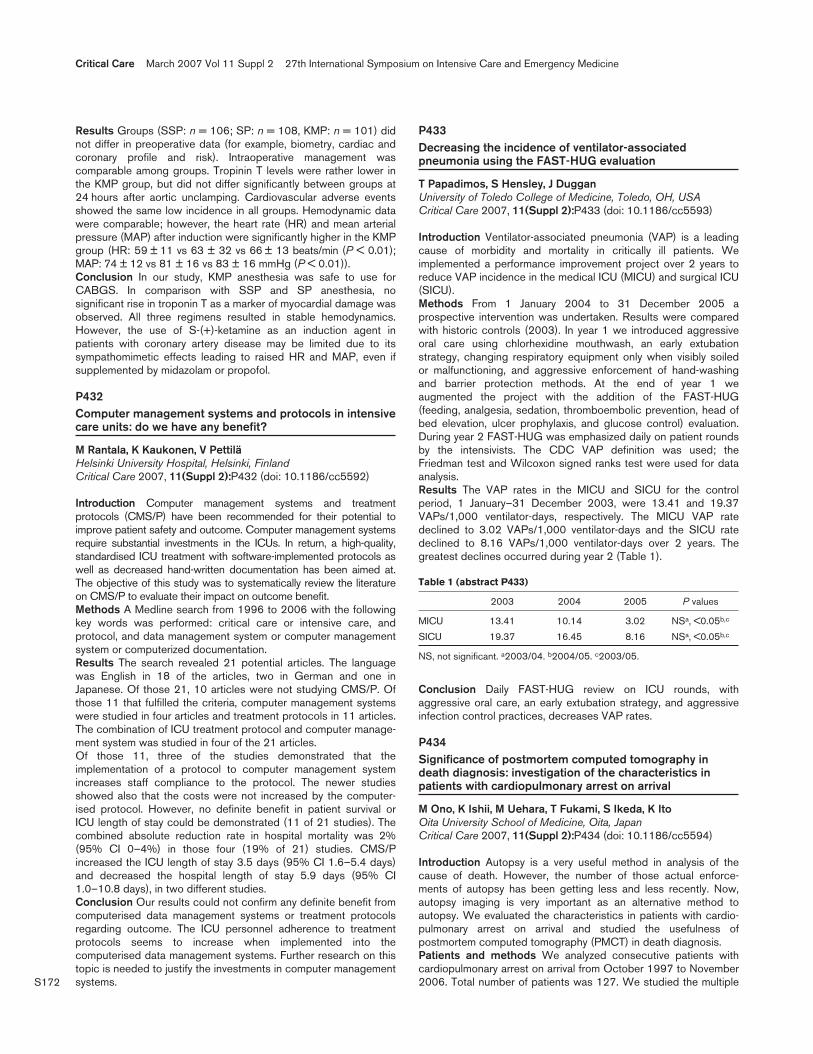
S172
Results Groups (SSP: n = 106; SP: n = 108, KMP: n = 101) didnot differ in preoperative data (for example, biometry, cardiac andcoronary profile and risk). Intraoperative management wascomparable among groups. Tropinin T levels were rather lower inthe KMP group, but did not differ significantly between groups at24 hours after aortic unclamping. Cardiovascular adverse eventsshowed the same low incidence in all groups. Hemodynamic datawere comparable; however, the heart rate (HR) and mean arterialpressure (MAP) after induction were significantly higher in the KMPgroup (HR: 59 ± 11 vs 63 ± 32 vs 66 ± 13 beats/min (P < 0.01);MAP: 74 ± 12 vs 81 ± 16 vs 83 ± 16 mmHg (P < 0.01)).Conclusion In our study, KMP anesthesia was safe to use forCABGS. In comparison with SSP and SP anesthesia, nosignificant rise in troponin T as a marker of myocardial damage wasobserved. All three regimens resulted in stable hemodynamics.However, the use of S-(+)-ketamine as an induction agent inpatients with coronary artery disease may be limited due to itssympathomimetic effects leading to raised HR and MAP, even ifsupplemented by midazolam or propofol.
P432Computer management systems and protocols in intensivecare units: do we have any benefit?
M Rantala, K Kaukonen, V PettiläHelsinki University Hospital, Helsinki, FinlandCritical Care 2007, 11(Suppl 2):P432 (doi: 10.1186/cc5592)
Introduction Computer management systems and treatmentprotocols (CMS/P) have been recommended for their potential toimprove patient safety and outcome. Computer management systemsrequire substantial investments in the ICUs. In return, a high-quality,standardised ICU treatment with software-implemented protocols aswell as decreased hand-written documentation has been aimed at.The objective of this study was to systematically review the literatureon CMS/P to evaluate their impact on outcome benefit.Methods A Medline search from 1996 to 2006 with the followingkey words was performed: critical care or intensive care, andprotocol, and data management system or computer managementsystem or computerized documentation.Results The search revealed 21 potential articles. The languagewas English in 18 of the articles, two in German and one inJapanese. Of those 21, 10 articles were not studying CMS/P. Ofthose 11 that fulfilled the criteria, computer management systemswere studied in four articles and treatment protocols in 11 articles.The combination of ICU treatment protocol and computer manage-ment system was studied in four of the 21 articles.Of those 11, three of the studies demonstrated that theimplementation of a protocol to computer management systemincreases staff compliance to the protocol. The newer studiesshowed also that the costs were not increased by the computer-ised protocol. However, no definite benefit in patient survival orICU length of stay could be demonstrated (11 of 21 studies). Thecombined absolute reduction rate in hospital mortality was 2%(95% CI 0–4%) in those four (19% of 21) studies. CMS/Pincreased the ICU length of stay 3.5 days (95% CI 1.6–5.4 days)and decreased the hospital length of stay 5.9 days (95% CI1.0–10.8 days), in two different studies.Conclusion Our results could not confirm any definite benefit fromcomputerised data management systems or treatment protocolsregarding outcome. The ICU personnel adherence to treatmentprotocols seems to increase when implemented into thecomputerised data management systems. Further research on thistopic is needed to justify the investments in computer managementsystems.
P433Decreasing the incidence of ventilator-associatedpneumonia using the FAST-HUG evaluation
T Papadimos, S Hensley, J DugganUniversity of Toledo College of Medicine, Toledo, OH, USACritical Care 2007, 11(Suppl 2):P433 (doi: 10.1186/cc5593)
Introduction Ventilator-associated pneumonia (VAP) is a leadingcause of morbidity and mortality in critically ill patients. Weimplemented a performance improvement project over 2 years toreduce VAP incidence in the medical ICU (MICU) and surgical ICU(SICU).Methods From 1 January 2004 to 31 December 2005 aprospective intervention was undertaken. Results were comparedwith historic controls (2003). In year 1 we introduced aggressiveoral care using chlorhexidine mouthwash, an early extubationstrategy, changing respiratory equipment only when visibly soiledor malfunctioning, and aggressive enforcement of hand-washingand barrier protection methods. At the end of year 1 weaugmented the project with the addition of the FAST-HUG(feeding, analgesia, sedation, thromboembolic prevention, head ofbed elevation, ulcer prophylaxis, and glucose control) evaluation.During year 2 FAST-HUG was emphasized daily on patient roundsby the intensivists. The CDC VAP definition was used; theFriedman test and Wilcoxon signed ranks test were used for dataanalysis.Results The VAP rates in the MICU and SICU for the controlperiod, 1 January–31 December 2003, were 13.41 and 19.37VAPs/1,000 ventilator-days, respectively. The MICU VAP ratedeclined to 3.02 VAPs/1,000 ventilator-days and the SICU ratedeclined to 8.16 VAPs/1,000 ventilator-days over 2 years. Thegreatest declines occurred during year 2 (Table 1).
Table 1 (abstract P433)
2003 2004 2005 P values
MICU 13.41 10.14 3.02 NSa, <0.05b,c
SICU 19.37 16.45 8.16 NSa, <0.05b,c
NS, not significant. a2003/04. b2004/05. c2003/05.
Conclusion Daily FAST-HUG review on ICU rounds, withaggressive oral care, an early extubation strategy, and aggressiveinfection control practices, decreases VAP rates.
P434Significance of postmortem computed tomography indeath diagnosis: investigation of the characteristics inpatients with cardiopulmonary arrest on arrival
M Ono, K Ishii, M Uehara, T Fukami, S Ikeda, K ItoOita University School of Medicine, Oita, JapanCritical Care 2007, 11(Suppl 2):P434 (doi: 10.1186/cc5594)
Introduction Autopsy is a very useful method in analysis of thecause of death. However, the number of those actual enforce-ments of autopsy has been getting less and less recently. Now,autopsy imaging is very important as an alternative method toautopsy. We evaluated the characteristics in patients with cardio-pulmonary arrest on arrival and studied the usefulness ofpostmortem computed tomography (PMCT) in death diagnosis.Patients and methods We analyzed consecutive patients withcardiopulmonary arrest on arrival from October 1997 to November2006. Total number of patients was 127. We studied the multiple
Critical Care March 2007 Vol 11 Suppl 2 27th International Symposium on Intensive Care and Emergency Medicine
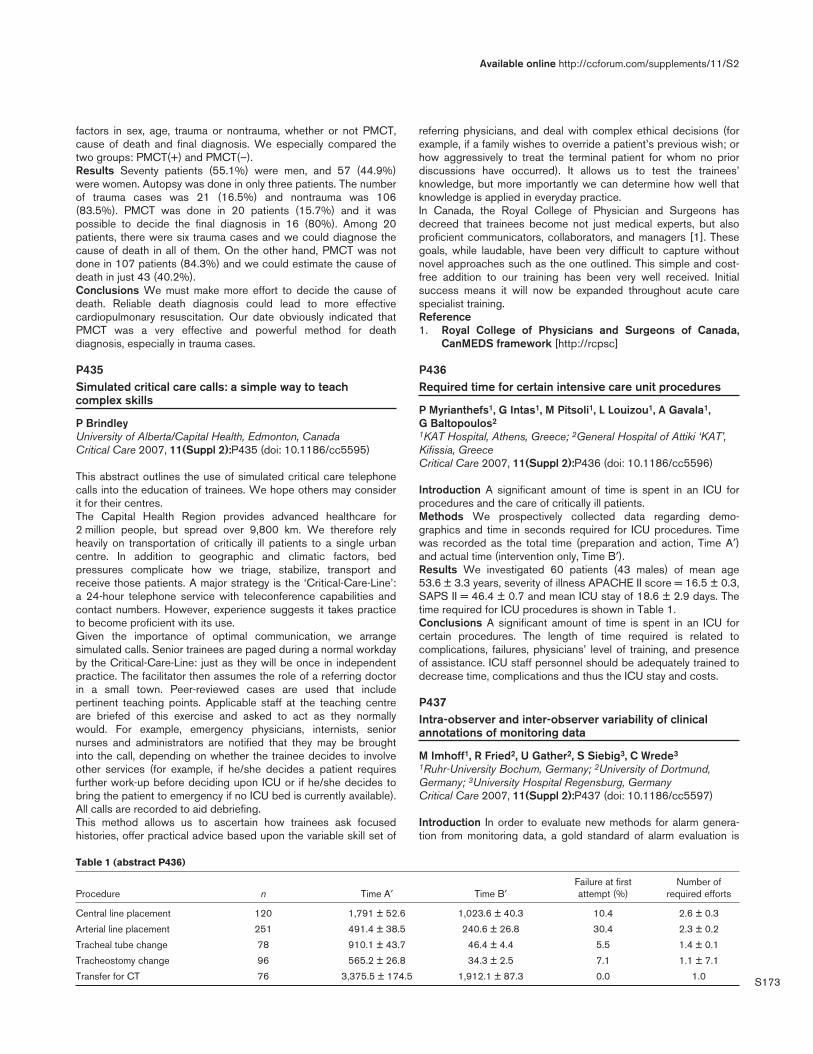
S173
factors in sex, age, trauma or nontrauma, whether or not PMCT,cause of death and final diagnosis. We especially compared thetwo groups: PMCT(+) and PMCT(–).Results Seventy patients (55.1%) were men, and 57 (44.9%)were women. Autopsy was done in only three patients. The numberof trauma cases was 21 (16.5%) and nontrauma was 106(83.5%). PMCT was done in 20 patients (15.7%) and it waspossible to decide the final diagnosis in 16 (80%). Among 20patients, there were six trauma cases and we could diagnose thecause of death in all of them. On the other hand, PMCT was notdone in 107 patients (84.3%) and we could estimate the cause ofdeath in just 43 (40.2%).Conclusions We must make more effort to decide the cause ofdeath. Reliable death diagnosis could lead to more effectivecardiopulmonary resuscitation. Our date obviously indicated thatPMCT was a very effective and powerful method for deathdiagnosis, especially in trauma cases.
P435Simulated critical care calls: a simple way to teachcomplex skills
P BrindleyUniversity of Alberta/Capital Health, Edmonton, CanadaCritical Care 2007, 11(Suppl 2):P435 (doi: 10.1186/cc5595)
This abstract outlines the use of simulated critical care telephonecalls into the education of trainees. We hope others may considerit for their centres.The Capital Health Region provides advanced healthcare for2 million people, but spread over 9,800 km. We therefore relyheavily on transportation of critically ill patients to a single urbancentre. In addition to geographic and climatic factors, bedpressures complicate how we triage, stabilize, transport andreceive those patients. A major strategy is the ‘Critical-Care-Line’:a 24-hour telephone service with teleconference capabilities andcontact numbers. However, experience suggests it takes practiceto become proficient with its use.Given the importance of optimal communication, we arrangesimulated calls. Senior trainees are paged during a normal workdayby the Critical-Care-Line: just as they will be once in independentpractice. The facilitator then assumes the role of a referring doctorin a small town. Peer-reviewed cases are used that includepertinent teaching points. Applicable staff at the teaching centreare briefed of this exercise and asked to act as they normallywould. For example, emergency physicians, internists, seniornurses and administrators are notified that they may be broughtinto the call, depending on whether the trainee decides to involveother services (for example, if he/she decides a patient requiresfurther work-up before deciding upon ICU or if he/she decides tobring the patient to emergency if no ICU bed is currently available).All calls are recorded to aid debriefing.This method allows us to ascertain how trainees ask focusedhistories, offer practical advice based upon the variable skill set of
referring physicians, and deal with complex ethical decisions (forexample, if a family wishes to override a patient’s previous wish; orhow aggressively to treat the terminal patient for whom no priordiscussions have occurred). It allows us to test the trainees’knowledge, but more importantly we can determine how well thatknowledge is applied in everyday practice.In Canada, the Royal College of Physician and Surgeons hasdecreed that trainees become not just medical experts, but alsoproficient communicators, collaborators, and managers [1]. Thesegoals, while laudable, have been very difficult to capture withoutnovel approaches such as the one outlined. This simple and cost-free addition to our training has been very well received. Initialsuccess means it will now be expanded throughout acute carespecialist training.Reference1. Royal College of Physicians and Surgeons of Canada,
CanMEDS framework [http://rcpsc]
P436Required time for certain intensive care unit procedures
P Myrianthefs1, G Intas1, M Pitsoli1, L Louizou1, A Gavala1, G Baltopoulos2
1KAT Hospital, Athens, Greece; 2General Hospital of Attiki ‘KAT’,Kifissia, GreeceCritical Care 2007, 11(Suppl 2):P436 (doi: 10.1186/cc5596)
Introduction A significant amount of time is spent in an ICU forprocedures and the care of critically ill patients.Methods We prospectively collected data regarding demo-graphics and time in seconds required for ICU procedures. Timewas recorded as the total time (preparation and action, Time A′)and actual time (intervention only, Time B′).Results We investigated 60 patients (43 males) of mean age53.6 ± 3.3 years, severity of illness APACHE II score = 16.5 ± 0.3,SAPS II = 46.4 ± 0.7 and mean ICU stay of 18.6 ± 2.9 days. Thetime required for ICU procedures is shown in Table 1.Conclusions A significant amount of time is spent in an ICU forcertain procedures. The length of time required is related tocomplications, failures, physicians’ level of training, and presenceof assistance. ICU staff personnel should be adequately trained todecrease time, complications and thus the ICU stay and costs.
P437Intra-observer and inter-observer variability of clinicalannotations of monitoring data
M Imhoff1, R Fried2, U Gather2, S Siebig3, C Wrede3
1Ruhr-University Bochum, Germany; 2University of Dortmund,Germany; 3University Hospital Regensburg, GermanyCritical Care 2007, 11(Suppl 2):P437 (doi: 10.1186/cc5597)
Introduction In order to evaluate new methods for alarm genera-tion from monitoring data, a gold standard of alarm evaluation is
Available online http://ccforum.com/supplements/11/S2
Table 1 (abstract P436)
Failure at first Number of Procedure n Time A′ Time B′ attempt (%) required efforts
Central line placement 120 1,791 ± 52.6 1,023.6 ± 40.3 10.4 2.6 ± 0.3
Arterial line placement 251 491.4 ± 38.5 240.6 ± 26.8 30.4 2.3 ± 0.2
Tracheal tube change 78 910.1 ± 43.7 46.4 ± 4.4 5.5 1.4 ± 0.1
Tracheostomy change 96 565.2 ± 26.8 34.3 ± 2.5 7.1 1.1 ± 7.1
Transfer for CT 76 3,375.5 ± 174.5 1,912.1 ± 87.3 0.0 1.0
S174
needed. Nearly all clinical studies into monitoring alarms usedclinician judgement and annotation as the reference standard. Weinvestigated the intra-observer and inter-observer variability betweentwo intensivists in the classification of monitoring time series.Methods A total of 3,092 time series segments (heart rate andblood pressures) of 30 minutes each from six critically ill patientswere presented to two experienced intensivists (MD1 and MD2)offline and were visually classified into clinically relevant patterns(no change, level shift, trend) by the physicians separately. Oneintensivist (MD2) repeated the classification 4 weeks after the firstanalysis on the same dataset.Results MD1 found clinically relevant events in 36%, and MD2 in29% of all time series. In 16% of all cases both intensivists cameto different classifications. In 10% even the direction of changewas classified differently. MD2 classified 10% of all casesdifferently between the first and second analysis. Even if levelchanges and trends were treated as one universal pattern ofchange, intra-individual variability (MD2 first analysis vs MD2second analysis) was still 5% and inter-individual variability (MD1vs MD2, only unequivocal classifications) was 10%.Conclusion Although this study is small with only two observerswho were investigated, it clearly shows that there is a significantintra-individual and inter-individual variability in the classification ofmonitoring events done by experienced clinicians. These findingsare supported by studies into image analysis that also found highintra-individual and inter-individual variability. High inter-observerand intra-observer variability is a challenge for clinical studies intonew alarm algorithms. Our findings also show a need for reliableclassification methods.
P438Robust regression methods for intensive care monitoring
M Imhoff1, K Schettlinger2, R Fried2, U Gather2, S Siebig3, C Wrede3
1Ruhr-University Bochum, Germany; 2University of Dortmund,Germany; 3University Hospital Regensburg, GermanyCritical Care 2007, 11(Suppl 2):P438 (doi: 10.1186/cc5598)
Introduction Alarm generation of modern patient monitoringsystems still predominantly relies on simple threshold methods. Thisleads to an unacceptably high rate of false positive alarms. Manyfalse positive alarms are generated by measurement artefacts andmeasurement noise. One approach to address this problem is toalarm on the underlying signal (that is, the noise-free time series ofthe physiological variable), instead of the raw measurement.Methods Monitoring time series were simulated. Against thesedata four robust regression methods were evaluated: least trimmedsquares (LTS), least median of squares (LMS), repeated median(RM), and deepest regression (DR). Moreover, online monitoringseries from critically ill patients during multiparameter monitoringwere also compared.Results LTS and LMS showed comparable behaviour, as did RMand DR. LMS and LTS provided only 20% efficiency, DR 61% andRM 70% (least squares regression = 100%). RM and DR hadsmaller standard deviations and smaller mean-squared errors thanLMS and LTS under different noise distributions (standarddeviation of online estimates based on sliding windows of size n =21 for simulated standard normal errors: LMS: 0.875, LTS: 0.887,RM: 0.500, DR: 0.533). Analyses with clinical monitoring data alsoshowed that LMS and LTS preserve sudden level shifts but areunstable and perform poorly with trend changes; RM and DR blurshifts but yield more stable estimations.
Conclusion All four methods allow one to extract the underlyingsignal from physiological time series in a way that is robust againstmeasurement artefacts and noise. However, there are significantdifferences between the methods. Overall, repeated medianregression seems the best choice for intensive care monitoringsince it is not only the most stable but also the fastest method.
P439ISIS program: a new tool for medical research at thebedside in critical care units
H Mehdaoui1, B Sarrazin1, I El Zein1, L Allart2, C Vilhelm2, S Guerra2, D Zitouni2, M Lemdani2, R Valentino1, A Herbland1,P Ravaux2
1Fort De France University Hospital, Fort De France, Martinique;2Lille 2 University, Lille, FranceCritical Care 2007, 11(Suppl 2):P439 (doi: 10.1186/cc5599)
Introduction The goal of this program is to develop an experi-mental tool able to record, store and analyse data issued fromcritical care patients. Due to technical limitations and medicalconstraints, information systems able to manage such data flow aredifficult to deploy.Methods Data recording is done through a laptop connected tothe medical devices, allowing analogical and digital signaltransmission through a high-speed network. Several servers arededicated to specialised tasks: mass storage, model generation,artificial intelligence (AI), telecommunications, and security. A 3Teraflops supercomputer is dedicated to intensive computationwhen necessary. Twenty applications are dedicated to electivetasks, most of them running using the Linux operating system.The ‘Aiddiag’ data-acquisition software is a standalone applicationadapted to patient data recording from the biomedical devices andcaregiver’s inputs. It has a friendly designed user-interfacetouchscreen at the bedside and was adapted according tocaregivers’ feedback. Data are also stored in a repository and aselective secondary extraction is possible. Online and offlineanalysis by the AI engine is allowed. Software had to consider timespecifications and uses distributed computation to achieve highworkload tasks. We complied to the French legal patient datamanagement constraints.Results After 2 years, our system is fully deployed. It recordedmore than 2,500 patient-hours over a 3-month period. Signal lossis less than 1%. Our tool allows recording of more than 40 digitalsignals, eight analogical signals sampled at a rate of 1 kHz, andcaregiver comments and actions. CPU resources of the laptop areavailable for supplemental AI developments during dataacquisition. Transfer of data to the repository is either a hotplug-automated process or delayed with 5 days of buffering in thelaptop. Automated artefacts’ cleaning allows time-series analysis(GARCH method) to extract behavioural models after intensivecomputation. The AI engine is used for medical guidelineimplementation (that is, severe brain trauma care algorithms) andlater comparison with caregiver’s behaviour. Remote use of oursystem is possible and schedulable, allowing other research teamsto work on the data. Limitations have been detected duringintensive calculation. Fine-tuning of the network will suppressthese limitations.Conclusion ISIS is the first program to achieve an easy-to-userecording tool able to build a very large medical repository. Dataanalysis methods and AI-controlled automated complex medicalguidelines are under evaluation.
Critical Care March 2007 Vol 11 Suppl 2 27th International Symposium on Intensive Care and Emergency Medicine
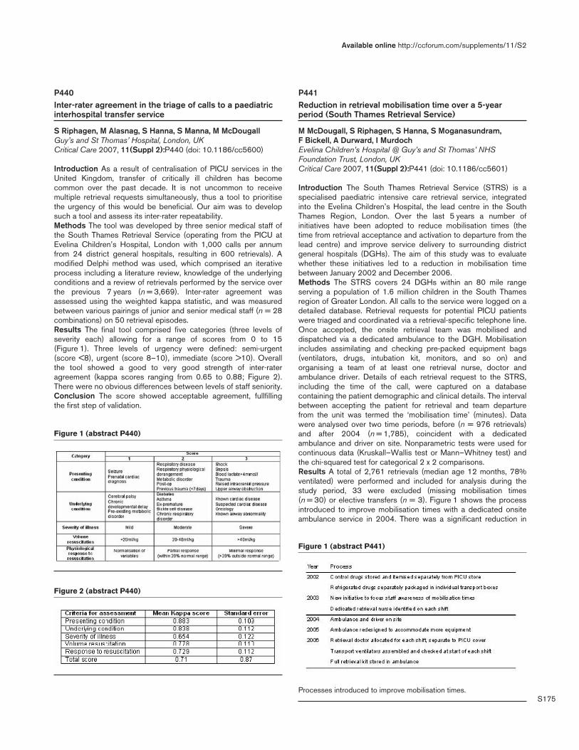
S175
P440Inter-rater agreement in the triage of calls to a paediatricinterhospital transfer service
S Riphagen, M Alasnag, S Hanna, S Manna, M McDougallGuy’s and St Thomas’ Hospital, London, UKCritical Care 2007, 11(Suppl 2):P440 (doi: 10.1186/cc5600)
Introduction As a result of centralisation of PICU services in theUnited Kingdom, transfer of critically ill children has becomecommon over the past decade. It is not uncommon to receivemultiple retrieval requests simultaneously, thus a tool to prioritisethe urgency of this would be beneficial. Our aim was to developsuch a tool and assess its inter-rater repeatability.Methods The tool was developed by three senior medical staff ofthe South Thames Retrieval Service (operating from the PICU atEvelina Children’s Hospital, London with 1,000 calls per annumfrom 24 district general hospitals, resulting in 600 retrievals). Amodified Delphi method was used, which comprised an iterativeprocess including a literature review, knowledge of the underlyingconditions and a review of retrievals performed by the service overthe previous 7 years (n = 3,669). Inter-rater agreement wasassessed using the weighted kappa statistic, and was measuredbetween various pairings of junior and senior medical staff (n = 28combinations) on 50 retrieval episodes.Results The final tool comprised five categories (three levels ofseverity each) allowing for a range of scores from 0 to 15(Figure 1). Three levels of urgency were defined: semi-urgent(score <8), urgent (score 8–10), immediate (score >10). Overallthe tool showed a good to very good strength of inter-rateragreement (kappa scores ranging from 0.65 to 0.88; Figure 2).There were no obvious differences between levels of staff seniority.Conclusion The score showed acceptable agreement, fullfillingthe first step of validation.
P441Reduction in retrieval mobilisation time over a 5-yearperiod (South Thames Retrieval Service)
M McDougall, S Riphagen, S Hanna, S Moganasundram, F Bickell, A Durward, I MurdochEvelina Children’s Hospital @ Guy’s and St Thomas’ NHSFoundation Trust, London, UKCritical Care 2007, 11(Suppl 2):P441 (doi: 10.1186/cc5601)
Introduction The South Thames Retrieval Service (STRS) is aspecialised paediatric intensive care retrieval service, integratedinto the Evelina Children’s Hospital, the lead centre in the SouthThames Region, London. Over the last 5 years a number ofinitiatives have been adopted to reduce mobilisation times (thetime from retrieval acceptance and activation to departure from thelead centre) and improve service delivery to surrounding districtgeneral hospitals (DGHs). The aim of this study was to evaluatewhether these initiatives led to a reduction in mobilisation timebetween January 2002 and December 2006.Methods The STRS covers 24 DGHs within an 80 mile rangeserving a population of 1.6 million children in the South Thamesregion of Greater London. All calls to the service were logged on adetailed database. Retrieval requests for potential PICU patientswere triaged and coordinated via a retrieval-specific telephone line.Once accepted, the onsite retrieval team was mobilised anddispatched via a dedicated ambulance to the DGH. Mobilisationincludes assimilating and checking pre-packed equipment bags(ventilators, drugs, intubation kit, monitors, and so on) andorganising a team of at least one retrieval nurse, doctor andambulance driver. Details of each retrieval request to the STRS,including the time of the call, were captured on a databasecontaining the patient demographic and clinical details. The intervalbetween accepting the patient for retrieval and team departurefrom the unit was termed the ‘mobilisation time’ (minutes). Datawere analysed over two time periods, before (n = 976 retrievals)and after 2004 (n = 1,785), coincident with a dedicatedambulance and driver on site. Nonparametric tests were used forcontinuous data (Kruskall–Wallis test or Mann–Whitney test) andthe chi-squared test for categorical 2 x 2 comparisons.Results A total of 2,761 retrievals (median age 12 months, 78%ventilated) were performed and included for analysis during thestudy period, 33 were excluded (missing mobilisation times(n = 30) or elective transfers (n = 3). Figure 1 shows the processintroduced to improve mobilisation times with a dedicated onsiteambulance service in 2004. There was a significant reduction in
Available online http://ccforum.com/supplements/11/S2
Figure 1 (abstract P440)
Figure 2 (abstract P440)
Figure 1 (abstract P441)
Processes introduced to improve mobilisation times.
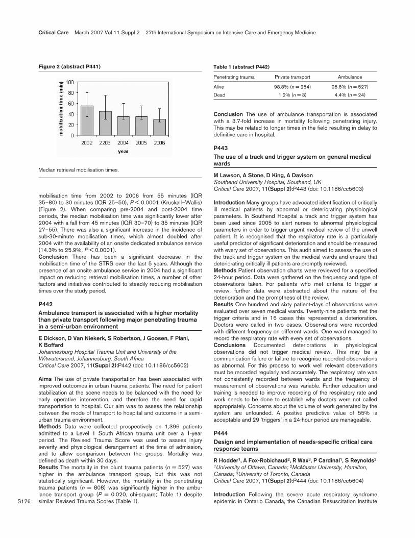
S176
mobilisation time from 2002 to 2006 from 55 minutes (IQR35–80) to 30 minutes (IQR 25–50), P < 0.0001 (Kruskall–Wallis)(Figure 2). When comparing pre-2004 and post-2004 timeperiods, the median mobilisation time was significantly lower after2004 with a fall from 45 minutes (IQR 30–70) to 35 minutes (IQR27–55). There was also a significant increase in the incidence ofsub-30-minute mobilisation times, which almost doubled after2004 with the availability of an onsite dedicated ambulance service(14.3% to 25.9%, P < 0.0001).Conclusion There has been a significant decrease in themobilisation time of the STRS over the last 5 years. Although thepresence of an onsite ambulance service in 2004 had a significantimpact on reducing retrieval mobilisation times, a number of otherfactors and initiatives contributed to steadily reducing mobilisationtimes over the study period.
P442Ambulance transport is associated with a higher mortalitythan private transport following major penetrating traumain a semi-urban environment
E Dickson, D Van Niekerk, S Robertson, J Goosen, F Plani, K BoffardJohannesburg Hospital Trauma Unit and University of theWitwatersrand, Johannesburg, South AfricaCritical Care 2007, 11(Suppl 2):P442 (doi: 10.1186/cc5602)
Aims The use of private transportation has been associated withimproved outcomes in urban trauma patients. The need for patientstabilization at the scene needs to be balanced with the need forearly operative intervention, and therefore the need for rapidtransportation to hospital. Our aim was to assess the relationshipbetween the mode of transport to hospital and outcome in a semi-urban trauma environment.Methods Data were collected prospectively on 1,396 patientsadmitted to a Level 1 South African trauma unit over a 1-yearperiod. The Revised Trauma Score was used to assess injuryseverity and physiological derangement at the time of admission,and to allow comparison between the groups. Mortality wasdefined as death within 30 days.Results The mortality in the blunt trauma patients (n = 527) washigher in the ambulance transport group, but this was notstatistically significant. However, the mortality in the penetratingtrauma patients (n = 808) was significantly higher in the ambu-lance transport group (P = 0.020, chi-square; Table 1) despitesimilar Revised Trauma Scores (Table 1).
Conclusion The use of ambulance transportation is associatedwith a 3.7-fold increase in mortality following penetrating injury.This may be related to longer times in the field resulting in delay todefinitive care in hospital.
P443The use of a track and trigger system on general medicalwards
M Lawson, A Stone, D King, A DavisonSouthend University Hospital, Southend, UKCritical Care 2007, 11(Suppl 2):P443 (doi: 10.1186/cc5603)
Introduction Many groups have advocated identification of criticallyill medical patients by abnormal or deteriorating physiologicalparameters. In Southend Hospital a track and trigger system hasbeen used since 2005 to alert nurses to abnormal physiologicalparameters in order to trigger urgent medical review of the unwellpatient. It is recognised that the respiratory rate is a particularlyuseful predictor of significant deterioration and should be measuredwith every set of observations. This audit aimed to assess the use ofthe track and trigger system on the medical wards and ensure thatdeteriorating critically ill patients are promptly reviewed.Methods Patient observation charts were reviewed for a specified24-hour period. Data were gathered on the frequency and type ofobservations taken. For patients who met criteria to trigger areview, further data were abstracted about the nature of thedeterioration and the promptness of the review.Results One hundred and sixty patient-days of observations wereevaluated over seven medical wards. Twenty-nine patients met thetrigger criteria and in 16 cases this represented a deterioration.Doctors were called in two cases. Observations were recordedwith different frequency on different wards. One ward managed torecord the respiratory rate with every set of observations.Conclusions Documented deteriorations in physiologicalobservations did not trigger medical review. This may be acommunication failure or failure to recognise recorded observationsas abnormal. For this process to work well relevant observationsmust be recorded regularly and accurately. The respiratory rate wasnot consistently recorded between wards and the frequency ofmeasurement of observations was variable. Further education andtraining is needed to improve recording of the respiratory rate andwork needs to be done to establish why doctors were not calledappropriately. Concerns about the volume of work generated by thesystem are unfounded. A positive predictive value of 55% isacceptable and 29 ‘triggers’ in a 24-hour period are manageable.
P444Design and implementation of needs-specific critical careresponse teams
R Hodder1, A Fox-Robichaud2, R Wax3, P Cardinal1, S Reynolds3
1University of Ottawa, Canada; 2McMaster University, Hamilton,Canada; 3University of Toronto, CanadaCritical Care 2007, 11(Suppl 2):P444 (doi: 10.1186/cc5604)
Introduction Following the severe acute respiratory syndromeepidemic in Ontario Canada, the Canadian Resuscitation Institute
Critical Care March 2007 Vol 11 Suppl 2 27th International Symposium on Intensive Care and Emergency Medicine
Figure 2 (abstract P441)
Median retrieval mobilisation times.
Table 1 (abstract P442)
Penetrating trauma Private transport Ambulance
Alive 98.8% (n = 254) 95.6% (n = 527)
Dead 1.2% (n = 3) 4.4% (n = 24)
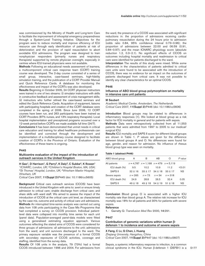
S177
was commissioned by the Ministry of Health and Long-term Careto facilitate the improvement of inhospital emergency preparednessthrough a System-Level Training Initiative. Objectives for theprogram were to reduce mortality and improve efficiency of ICUresource use through early identification of patients at risk ofdeterioration and the provision of rapid resuscitation to abortavoidable ICU admissions. The program was designed to trainnonphysician responders (primarily nurses and respiratorytherapists) supported by remote physician oversight, especially incentres where ICU-trained physicians were not available.Methods Following an educational needs assessment of learners,a multicomponent critical care response team (CCRT) trainingcourse was developed. The 2-day course consisted of a series ofsmall group, interactive, case-based seminars, high-fidelitysimulation training, and the publication of a CCRT Provider Manualand Quick Reference Cards. A database for monitoring theeffectiveness and impact of the CCRTs was also developed.Results Beginning in October 2005, 24 CCRT physician instructorswere trained in one of two streams: (i) simulator instructors with skillsin constructive feedback and assessment of crisis management skills;(ii) instructors who further refined the case-based seminars andedited the Quick Reference Cards. Acquisition of equipment, liaisonswith participating hospitals and creation of the CCRT database werecompleted in the spring of 2006. Since June 2006, 12 CCRTcourses have been run, and 263 participants have been trained asCCRT Providers (87% nurses, and 13% respiratory therapists). Localhospital implementation and preceptored programs occurred over a12-week period before CCRTs we made available full time (24/7).Conclusions It has been demonstrated that unmet needs in criticalcare education and training for allied healthcare professionals canbe identified and corrected through the development andimplementation of a multidisciplinary course designed to facilitatecreation of CCRTs in the Province of Ontario. Evaluation of theeffectiveness of these teams is ongoing.
P445Multicentre evaluation of the impact of the introduction ofoutreach services in the United Kingdom
H Gao1, D Harrison1, G Parry2, K Daly3, C Subbe4, K Rowan1
1ICNARC, London, UK; 2Children’s Hospital Boston, MA, USA; 3St Thomas’ Hospital, London, UK; 4Wrexham Maelor Hospital,Wrexham, UKCritical Care 2007, 11(Suppl 2):P445 (doi: 10.1186/cc5605)
Background Critical care outreach services (CCOS) have beenintroduced in the United Kingdom with aims to: avert or ensure timelyadmission to critical care; enable discharge from critical care; andshare skills with ward staff. We aimed to assess the impact of theintroduction of CCOS at the critical care unit level, as characterisedby the case mix, outcome and activity of critical care unit admissions.Methods An interrupted time-series analysis was carried out usingdata from 108 units participating in the Case Mix Programme thathad completed a survey on CCOS provision. Individual patient-level data were collapsed into monthly time series for each unit(panel data). Population-averaged panel-data models were fittedusing a generalised estimating equation approach. Variousoutcomes reflecting the stated aims of CCOS were considered forthree groups of admissions: all admissions to the unit; admissionsfrom the ward; and unit survivors discharged to the ward. Theprimary exposure variable was the presence of a formal CCOSwith secondary exposures of CCOS activities, coverage andstaffing, identified from the survey data.Results Of 108 units in the analysis, 79 (73%) had a formalCCOS introduced between 1996 and 2004. For admissions from
the ward, the presence of a CCOS was associated with significantreductions in: the proportion of admissions receiving cardio-pulmonary resuscitation during the 24 hours prior to admission(odds ratio 0.84, 95% confidence interval 0.73–0.96); theproportion of admissions between 22:00 and 06:59 (0.91,0.84–0.97); and the mean ICNARC physiology score (absolutereduction 1.2, 0.3–2.1). No significant effects of CCOS onoutcomes including hospital mortality and readmission to criticalcare were identified for patients discharged to the ward.Interpretation The results of this study were mixed. While somedifferences in the characteristics of patients admitted to criticalcare units were found to be associated with the introduction ofCCOS, there was no evidence for an impact on the outcomes ofpatients discharged from critical care. It was not possible toidentify any clear characteristics for an optimal CCOS.
P446Influence of ABO blood group polymorphism on mortalityin intensive care unit patients
M SeubertAcademic Medical Center, Amsterdam, The NetherlandsCritical Care 2007, 11(Suppl 2):P446 (doi: 10.1186/cc5606)
Introduction Blood groups may be related to differences ininflammatory responses [1]. We looked at blood group as a riskfactor for ICU mortality in general and for patients with sepsis.Methods Data were retrospectively collected from all 11,553patients that were admitted from 1997 to 2005 to our medical/surgical ICU.Results ICU mortality and SAPS II score for different blood groupsare shown in Table 1. P values are given for the differencebetween blood groups A and O. No differences were found forage, gender, and reason for admission. No influence of rhesusblood group type was seen on mortality.
Table 1 (abstract P446)
ABO blood group A B AB O P value
All patients n = 4,787 n = 1,168 n = 479 n = 5,119
ICU death (%) 9.5 10.2 10.9 11.2 <0.01
SAPS II 32 ± 16 33 ± 17 34 ± 18 33 ± 17 NS
Severe sepsis n = 265 n = 73 n = 34 n = 318
ICU death (%) 24.9 28.8 26.5 32.4 0.05
SAPS II 49 ± 18 49 ± 18 54 ± 19 51 ± 18 NS
Conclusion Blood group O is associated with a higher ICUmortality rate than blood group A. The relative risk increase for ICUmortality was 18% for all patients and 30% for patients with severesepsis.Reference1. Garratty G: Transfusion Med Rev 2000, 14:291.
P447Contribution of genomic variations within human ββ-defensin 1 to incidence and outcome of severe sepsis
X Fang, C Lv, Q Chen, L HuangZhejiang University, Hangzhou, ChinaCritical Care 2007, 11(Suppl 2):P447 (doi: 10.1186/cc5607)
Sepsis, a systemic inflammatory response to infection, is a commonclinical syndrome in the ICU. Human β-defensin 1 (DEFB1) is a
Available online http://ccforum.com/supplements/11/S2
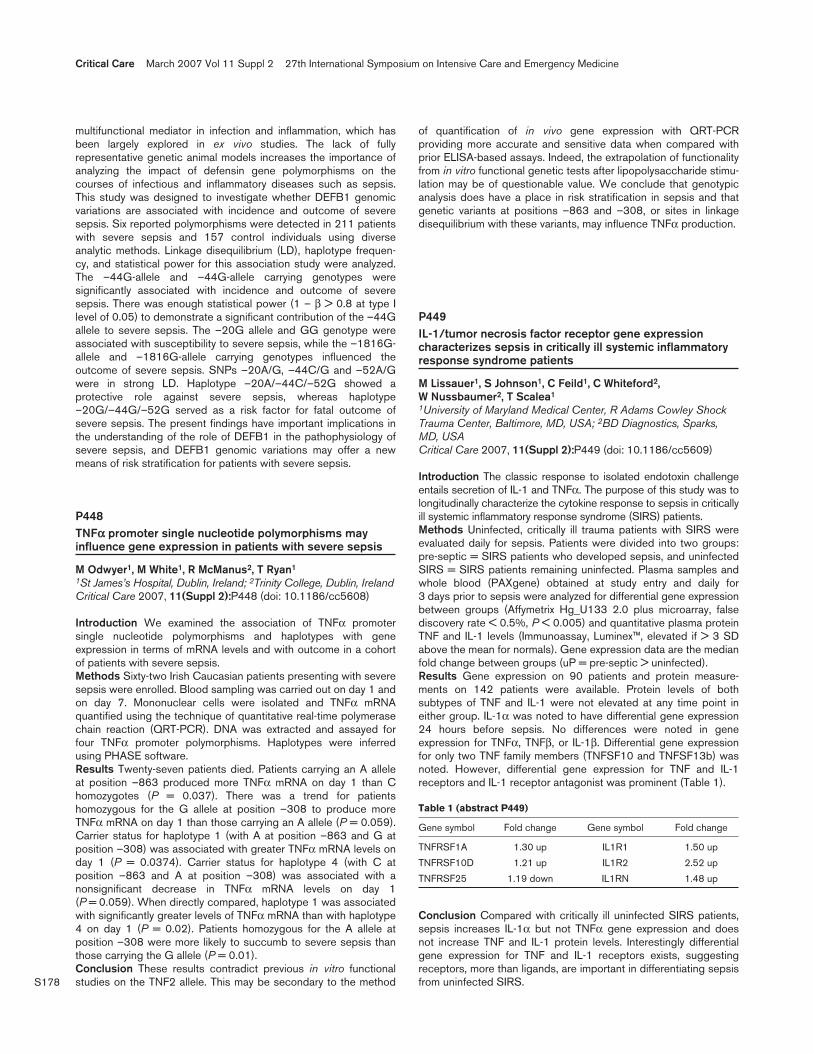
S178
multifunctional mediator in infection and inflammation, which hasbeen largely explored in ex vivo studies. The lack of fullyrepresentative genetic animal models increases the importance ofanalyzing the impact of defensin gene polymorphisms on thecourses of infectious and inflammatory diseases such as sepsis.This study was designed to investigate whether DEFB1 genomicvariations are associated with incidence and outcome of severesepsis. Six reported polymorphisms were detected in 211 patientswith severe sepsis and 157 control individuals using diverseanalytic methods. Linkage disequilibrium (LD), haplotype frequen-cy, and statistical power for this association study were analyzed.The –44G-allele and –44G-allele carrying genotypes weresignificantly associated with incidence and outcome of severesepsis. There was enough statistical power (1 – β > 0.8 at type Ilevel of 0.05) to demonstrate a significant contribution of the –44Gallele to severe sepsis. The –20G allele and GG genotype wereassociated with susceptibility to severe sepsis, while the –1816G-allele and –1816G-allele carrying genotypes influenced theoutcome of severe sepsis. SNPs –20A/G, –44C/G and –52A/Gwere in strong LD. Haplotype –20A/–44C/–52G showed aprotective role against severe sepsis, whereas haplotype–20G/–44G/–52G served as a risk factor for fatal outcome ofsevere sepsis. The present findings have important implications inthe understanding of the role of DEFB1 in the pathophysiology ofsevere sepsis, and DEFB1 genomic variations may offer a newmeans of risk stratification for patients with severe sepsis.
P448TNFαα promoter single nucleotide polymorphisms mayinfluence gene expression in patients with severe sepsis
M Odwyer1, M White1, R McManus2, T Ryan1
1St James’s Hospital, Dublin, Ireland; 2Trinity College, Dublin, IrelandCritical Care 2007, 11(Suppl 2):P448 (doi: 10.1186/cc5608)
Introduction We examined the association of TNFα promotersingle nucleotide polymorphisms and haplotypes with geneexpression in terms of mRNA levels and with outcome in a cohortof patients with severe sepsis.Methods Sixty-two Irish Caucasian patients presenting with severesepsis were enrolled. Blood sampling was carried out on day 1 andon day 7. Mononuclear cells were isolated and TNFα mRNAquantified using the technique of quantitative real-time polymerasechain reaction (QRT-PCR). DNA was extracted and assayed forfour TNFα promoter polymorphisms. Haplotypes were inferredusing PHASE software.Results Twenty-seven patients died. Patients carrying an A alleleat position –863 produced more TNFα mRNA on day 1 than Chomozygotes (P = 0.037). There was a trend for patientshomozygous for the G allele at position –308 to produce moreTNFα mRNA on day 1 than those carrying an A allele (P = 0.059).Carrier status for haplotype 1 (with A at position –863 and G atposition –308) was associated with greater TNFα mRNA levels onday 1 (P = 0.0374). Carrier status for haplotype 4 (with C atposition –863 and A at position –308) was associated with anonsignificant decrease in TNFα mRNA levels on day 1(P = 0.059). When directly compared, haplotype 1 was associatedwith significantly greater levels of TNFα mRNA than with haplotype4 on day 1 (P = 0.02). Patients homozygous for the A allele atposition –308 were more likely to succumb to severe sepsis thanthose carrying the G allele (P = 0.01).Conclusion These results contradict previous in vitro functionalstudies on the TNF2 allele. This may be secondary to the method
of quantification of in vivo gene expression with QRT-PCRproviding more accurate and sensitive data when compared withprior ELISA-based assays. Indeed, the extrapolation of functionalityfrom in vitro functional genetic tests after lipopolysaccharide stimu-lation may be of questionable value. We conclude that genotypicanalysis does have a place in risk stratification in sepsis and thatgenetic variants at positions –863 and –308, or sites in linkagedisequilibrium with these variants, may influence TNFα production.
P449IL-1/tumor necrosis factor receptor gene expressioncharacterizes sepsis in critically ill systemic inflammatoryresponse syndrome patients
M Lissauer1, S Johnson1, C Feild1, C Whiteford2, W Nussbaumer2, T Scalea1
1University of Maryland Medical Center, R Adams Cowley ShockTrauma Center, Baltimore, MD, USA; 2BD Diagnostics, Sparks,MD, USACritical Care 2007, 11(Suppl 2):P449 (doi: 10.1186/cc5609)
Introduction The classic response to isolated endotoxin challengeentails secretion of IL-1 and TNFα. The purpose of this study was tolongitudinally characterize the cytokine response to sepsis in criticallyill systemic inflammatory response syndrome (SIRS) patients.Methods Uninfected, critically ill trauma patients with SIRS wereevaluated daily for sepsis. Patients were divided into two groups:pre-septic = SIRS patients who developed sepsis, and uninfectedSIRS = SIRS patients remaining uninfected. Plasma samples andwhole blood (PAXgene) obtained at study entry and daily for3 days prior to sepsis were analyzed for differential gene expressionbetween groups (Affymetrix Hg_U133 2.0 plus microarray, falsediscovery rate < 0.5%, P < 0.005) and quantitative plasma proteinTNF and IL-1 levels (Immunoassay, Luminex™, elevated if > 3 SDabove the mean for normals). Gene expression data are the medianfold change between groups (uP = pre-septic > uninfected).Results Gene expression on 90 patients and protein measure-ments on 142 patients were available. Protein levels of bothsubtypes of TNF and IL-1 were not elevated at any time point ineither group. IL-1α was noted to have differential gene expression24 hours before sepsis. No differences were noted in geneexpression for TNFα, TNFβ, or IL-1β. Differential gene expressionfor only two TNF family members (TNFSF10 and TNFSF13b) wasnoted. However, differential gene expression for TNF and IL-1receptors and IL-1 receptor antagonist was prominent (Table 1).
Table 1 (abstract P449)
Gene symbol Fold change Gene symbol Fold change
TNFRSF1A 1.30 up IL1R1 1.50 up
TNFRSF10D 1.21 up IL1R2 2.52 up
TNFRSF25 1.19 down IL1RN 1.48 up
Conclusion Compared with critically ill uninfected SIRS patients,sepsis increases IL-1α but not TNFα gene expression and doesnot increase TNF and IL-1 protein levels. Interestingly differentialgene expression for TNF and IL-1 receptors exists, suggestingreceptors, more than ligands, are important in differentiating sepsisfrom uninfected SIRS.
Critical Care March 2007 Vol 11 Suppl 2 27th International Symposium on Intensive Care and Emergency Medicine
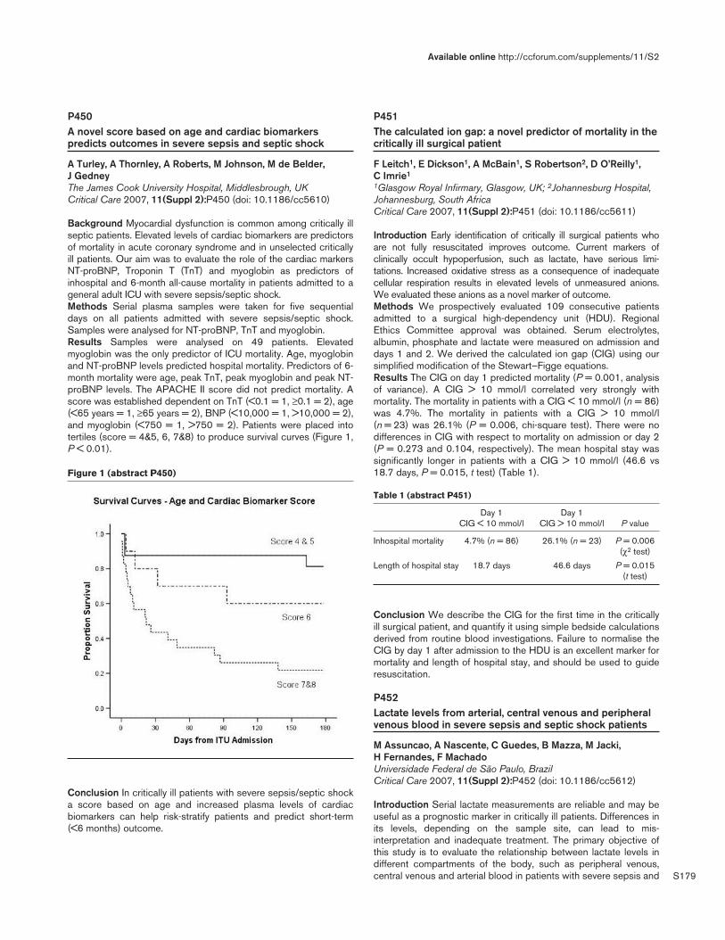
S179
P450A novel score based on age and cardiac biomarkerspredicts outcomes in severe sepsis and septic shock
A Turley, A Thornley, A Roberts, M Johnson, M de Belder, J GedneyThe James Cook University Hospital, Middlesbrough, UKCritical Care 2007, 11(Suppl 2):P450 (doi: 10.1186/cc5610)
Background Myocardial dysfunction is common among critically illseptic patients. Elevated levels of cardiac biomarkers are predictorsof mortality in acute coronary syndrome and in unselected criticallyill patients. Our aim was to evaluate the role of the cardiac markersNT-proBNP, Troponin T (TnT) and myoglobin as predictors ofinhospital and 6-month all-cause mortality in patients admitted to ageneral adult ICU with severe sepsis/septic shock.Methods Serial plasma samples were taken for five sequentialdays on all patients admitted with severe sepsis/septic shock.Samples were analysed for NT-proBNP, TnT and myoglobin.Results Samples were analysed on 49 patients. Elevatedmyoglobin was the only predictor of ICU mortality. Age, myoglobinand NT-proBNP levels predicted hospital mortality. Predictors of 6-month mortality were age, peak TnT, peak myoglobin and peak NT-proBNP levels. The APACHE II score did not predict mortality. Ascore was established dependent on TnT (<0.1 = 1, ≥0.1 = 2), age(<65 years = 1, ≥65 years = 2), BNP (<10,000 = 1, >10,000 = 2),and myoglobin (<750 = 1, >750 = 2). Patients were placed intotertiles (score = 4&5, 6, 7&8) to produce survival curves (Figure 1,P < 0.01).
Conclusion In critically ill patients with severe sepsis/septic shocka score based on age and increased plasma levels of cardiacbiomarkers can help risk-stratify patients and predict short-term(<6 months) outcome.
P451The calculated ion gap: a novel predictor of mortality in thecritically ill surgical patient
F Leitch1, E Dickson1, A McBain1, S Robertson2, D O’Reilly1, C Imrie1
1Glasgow Royal Infirmary, Glasgow, UK; 2Johannesburg Hospital,Johannesburg, South AfricaCritical Care 2007, 11(Suppl 2):P451 (doi: 10.1186/cc5611)
Introduction Early identification of critically ill surgical patients whoare not fully resuscitated improves outcome. Current markers ofclinically occult hypoperfusion, such as lactate, have serious limi-tations. Increased oxidative stress as a consequence of inadequatecellular respiration results in elevated levels of unmeasured anions.We evaluated these anions as a novel marker of outcome.Methods We prospectively evaluated 109 consecutive patientsadmitted to a surgical high-dependency unit (HDU). RegionalEthics Committee approval was obtained. Serum electrolytes,albumin, phosphate and lactate were measured on admission anddays 1 and 2. We derived the calculated ion gap (CIG) using oursimplified modification of the Stewart–Figge equations.Results The CIG on day 1 predicted mortality (P = 0.001, analysisof variance). A CIG > 10 mmol/l correlated very strongly withmortality. The mortality in patients with a CIG < 10 mmol/l (n = 86)was 4.7%. The mortality in patients with a CIG > 10 mmol/l(n = 23) was 26.1% (P = 0.006, chi-square test). There were nodifferences in CIG with respect to mortality on admission or day 2(P = 0.273 and 0.104, respectively). The mean hospital stay wassignificantly longer in patients with a CIG > 10 mmol/l (46.6 vs18.7 days, P = 0.015, t test) (Table 1).
Table 1 (abstract P451)
Day 1 Day 1 CIG < 10 mmol/l CIG > 10 mmol/l P value
Inhospital mortality 4.7% (n = 86) 26.1% (n = 23) P = 0.006 (χ2 test)
Length of hospital stay 18.7 days 46.6 days P = 0.015 (t test)
Conclusion We describe the CIG for the first time in the criticallyill surgical patient, and quantify it using simple bedside calculationsderived from routine blood investigations. Failure to normalise theCIG by day 1 after admission to the HDU is an excellent marker formortality and length of hospital stay, and should be used to guideresuscitation.
P452Lactate levels from arterial, central venous and peripheralvenous blood in severe sepsis and septic shock patients
M Assuncao, A Nascente, C Guedes, B Mazza, M Jacki, H Fernandes, F MachadoUniversidade Federal de São Paulo, BrazilCritical Care 2007, 11(Suppl 2):P452 (doi: 10.1186/cc5612)
Introduction Serial lactate measurements are reliable and may beuseful as a prognostic marker in critically ill patients. Differences inits levels, depending on the sample site, can lead to mis-interpretation and inadequate treatment. The primary objective ofthis study is to evaluate the relationship between lactate levels indifferent compartments of the body, such as peripheral venous,central venous and arterial blood in patients with severe sepsis and
Available online http://ccforum.com/supplements/11/S2
Figure 1 (abstract P450)
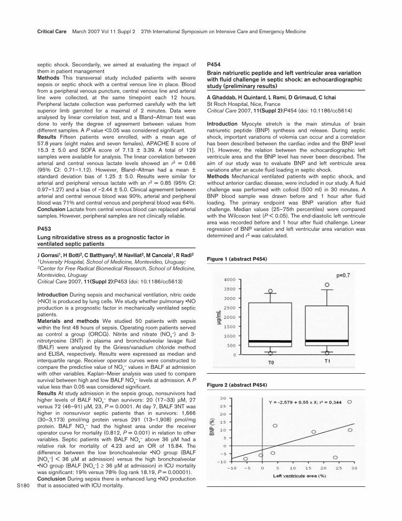
S180
septic shock. Secondarily, we aimed at evaluating the impact ofthem in patient managementMethods This transversal study included patients with severesepsis or septic shock with a central venous line in place. Bloodfrom a peripheral venous puncture, central venous line and arterialline were collected, at the same timepoint each 12 hours.Peripheral lactate collection was performed carefully with the leftsuperior limb garroted for a maximal of 2 minutes. Data wereanalysed by linear correlation test, and a Bland–Altman test wasdone to verify the degree of agreement between values fromdifferent samples. A P value <0.05 was considered significant.Results Fifteen patients were enrolled, with a mean age of57.8 years (eight males and seven females), APACHE II score of15.3 ± 5.0 and SOFA score of 7.13 ± 3.39. A total of 129samples were available for analysis. The linear correlation betweenarterial and central venous lactate levels showed an r2 = 0.66(95% CI: 0.71–1.12). However, Bland–Altman had a mean ±standard deviation bias of 1.25 ± 5.0. Results were similar forarterial and peripheral venous lactate with an r2 = 0.85 (95% CI:0.97–1.27) and a bias of –2.44 ± 5.0. Clinical agreement betweenarterial and central venous blood was 90%, arterial and peripheralblood was 71% and central venous and peripheral blood was 64%.Conclusion Lactate from central venous blood can replaced arterialsamples. However, peripheral samples are not clinically reliable.
P453Lung nitroxidative stress as a prognostic factor inventilated septic patients
J Gorrasi1, H Botti2, C Batthyany2, M Naviliat2, M Cancela1, R Radi21University Hospital, School of Medicine, Montevideo, Uruguay;2Center for Free Radical Biomedical Research, School of Medicine,Montevideo, UruguayCritical Care 2007, 11(Suppl 2):P453 (doi: 10.1186/cc5613)
Introduction During sepsis and mechanical ventilation, nitric oxide(•NO) is produced by lung cells. We study whether pulmonary •NOproduction is a prognostic factor in mechanically ventilated septicpatients.Materials and methods We studied 50 patients with sepsiswithin the first 48 hours of sepsis. Operating room patients servedas control a group (ORCG). Nitrite and nitrate (NOx
–) and 3-nitrotyrosine (3NT) in plasma and bronchoalveolar lavage fluid(BALF) were analyzed by the Griess/vanadium chloride methodand ELISA, respectively. Results were expressed as median andinterquartile range. Receiver operator curves were constructed tocompare the predictive value of NOx
– values in BALF at admissionwith other variables. Kaplan–Meier analysis was used to comparesurvival between high and low BALF NOx
– levels at admission. A Pvalue less than 0.05 was considered significant.Results At study admission in the sepsis group, nonsurvivors hadhigher levels of BALF NOx
– than survivors: 20 (17–33) µM, 27versus 72 (46–91) µM, 23, P = 0.0001. At day 7, BALF 3NT washigher in nonsurvivor septic patients than in survivors: 1,666(30–3,173) pmol/mg protein versus 291 (13–1,908) pmol/mgprotein. BALF NOx
– had the highest area under the receiveroperator curve for mortality (0.812, P = 0.001) in relation to othervariables. Septic patients with BALF NOx
– above 36 µM had arelative risk for mortality of 4.23 and an OR of 15.84. Thedifference between the low bronchoalveolar •NO group (BALF[NOx
–] < 36 µM at admission) versus the high bronchoalveolar•NO group (BALF [NOx
–] ≥ 36 µM at admission) in ICU mortalitywas significant: 19% versus 78% (log rank 18.19, P = 0.00001).Conclusion During sepsis there is enhanced lung •NO productionthat is associated with ICU mortality.
P454Brain natriuretic peptide and left ventricular area variationwith fluid challenge in septic shock: an echocardiographicstudy (preliminary results)
A Ghaddab, H Quintard, L Rami, D Grimaud, C IchaiSt Roch Hospital, Nice, FranceCritical Care 2007, 11(Suppl 2):P454 (doi: 10.1186/cc5614)
Introduction Myocyte stretch is the main stimulus of brainnatriuretic peptide (BNP) synthesis and release. During septicshock, important variations of volemia can occur and a correlationhas been described between the cardiac index and the BNP level[1]. However, the relation between the echocardiographic leftventricule area and the BNP level has never been described. Theaim of our study was to evaluate BNP and left ventricule areavariations after an acute fluid loading in septic shock.Methods Mechanical ventilated patients with septic shock, andwithout anterior cardiac disease, were included in our study. A fluidchallenge was performed with colloid (500 ml) in 30 minutes. ABNP blood sample was drawn before and 1 hour after fluidloading. The primary endpoint was BNP variation after fluidchallenge. Median values (25–75th percentiles) were comparedwith the Wilcoxon test (P < 0.05). The end-diastolic left ventriculearea was recorded before and 1 hour after fluid challenge. Linearregression of BNP variation and left ventricular area variation wasdetermined and r2 was calculated.
Critical Care March 2007 Vol 11 Suppl 2 27th International Symposium on Intensive Care and Emergency Medicine
Figure 1 (abstract P454)
Figure 2 (abstract P454)
S181
Results Eight patients (median age 68 years; six males/two females;SOFA score = 12) were enrolled in our study. The initial BNP levelmedian increased from 695 (417–2,738) to 715 (478–2,596) µg/mlafter a fluid loading (P = 0.7) (Figure 1).We did not find a statisticallysignificant relationship between BNP variation and left ventriculearea variation after fluid challenge (P = 0.13) (Figure 2).Conclusion There is no increase in BNP level in patients withseptic shock after fluid challenge. To our knowledge, thispreliminary study is the first to evaluate the relationship betweenBNP and left ventricule area variation in patient with septic shock.Although no statistical significance between left ventricule areavariation and BNP variation after fluid challenge, there is a trend tocorrelation between these two parameters. More patients have tobe included to confirm this result.Reference1. McLean AS: The effects of acute fluid loading on plasma
B-type natriuretic peptide levels in a septic shock patient.Anesth Intensive Care 2005, 33:528-530.
P455Atrial natriuretic peptide reduces the ischemia/reperfusion-induced renal injury in rats by enhancingsensory neuron activation
N Harada, K Okajima, Y MizuochiNagoya City University Graduate School of Medical Sciences,Nagoya, JapanCritical Care 2007, 11(Suppl 2):P455 (doi: 10.1186/cc5615)
Introduction Although carpertide, a synthetic α-human atrialnatriuretic peptide (ANP), reduces ischemia/reperfusion (I/R)-induced tissue injury, the precise therapeutic mechanism(s)remains to be elucidated. Calcitonin gene-related peptide (CGRP)released from sensory neurons reduces I/R-induced liver injury byinhibiting neutrophil activation through an increase in theendothelial production of prostacyclin (PGI2). In the present study,we examined in rats whether ANP reduces I/R-induced renal injuryby enhancing sensory neuron activation.Methods The right renal vessels were clamped in rats for 45minutes after left nephrectomy. ANP (0.3 µg/kg/min) wascontinuously infused from 30 minutes before ischemia to 60minutes after reperfusion. We attempted to determine whetherANP promotes CGRP release from cultured dorsal root ganglionneurons isolated from adult rats in vitro.Results Intravenous infusion of ANP reduced I/R-induced increasein serum levels of blood urea nitrogen and creatinine at 24 hoursafter reperfusion. ANP inhibited I/R-induced increases in renaltissue levels of TNF and myeloperoxidase at 3 and 6 hours afterreperfusion, respectively. ANP significantly enhanced I/R-inducedincreases in renal tissue levels of CGRP and 6-keto-PGF1α, astable metabolite of PGI2, at 1 hour after reperfusion. ANP-inducedincreases in renal tissue levels of CGRP were significantly inhibitedby pretreatment with SB366791, a specific vanilloid receptor-1antagonist. ANP-induced increases in renal tissue levels of 6-keto-PGF1α were significantly inhibited by pretreatment withSB366791, CGRP(8-37), a CGRP receptor antagonist, andindomethacin. Reduction of I/R-induced increases in serum levelsof blood urea nitrogen and creatinine and those in renal tissuelevels of TNF and myeloperoxidase in rats treated with ANP werecompletely abrogated by pretreatment with SB366791, CGRP(8-37), and indomethacin. ANP significantly increased CGRP releasefrom dorsal root ganglion neurons in vitro.Conclusions These results strongly suggested that ANP mightreduce I/R-induced renal injury in rats by inhibiting neutrophilactivation through enhancement of sensory neuron activation.
P456Mid-regional pro-atrial natriuretic peptide is a strong predictorof outcome in an unselected cohort of critically ill patients
B Meyer1, P Wexberg1, R Berger1, D Moertl1, N Morgenthaler2, J Struck2, A Bergmann2, G Heinz1, R Pacher1, M Huelsmann1
1Medical University of Vienna, Austria; 2B.R.A.H.M.S. Ag,Henningdorf, GermanyCritical Care 2007, 11(Suppl 2):P456 (doi: 10.1186/cc5616)
Introduction Atrial natriuretic peptide (ANP) is a strong predictorof outcome in patients with heart failure, various othercardiovascular diseases and in patients with systemic inflammatoryresponse syndrome and sepsis. Furthermore, ANP is elevated inpatients with kidney disease. We aimed to test the prognosticpotency of the precursor molecule mid-regional pro-atrial natriu-retic peptide (MR-pro-ANP) in an unselected cohort of critically illpatients.Methods Between August 2004 and February 2006, a total of294 patients (191 males, age 63.8 ± 14.7 years) admitted to ourICU were studied. The mean SAPS2 and APACHE II score were52 ± 23 and 24 ± 11, respectively. Two hundred and threepatients (69.1%) were on intravenous inotropic support, 30 patientshad additional mechanical circulatory support (23 intraaorticballoon counterpulsation (7.8%), eight extracorporeal membraneoxygenation (2.7%), three left ventricular assist device (0.9%)). Twohundred and five patients (69.7%) were mechanically ventilated,and 59 patients (20.1%) presented with acute renal failure. Plasmasamples for determination of MR-pro-ANP were obtained onadmission in all patients. As MR-pro-ANP values were not normallydistributed, log MR-pro-ANP values were used for analysis.Results Two hundred and thirty-five patients (79.9%) survived toICU discharge and 59 patients died (21.1%). Log MR-pro-ANPplasma levels were significantly higher in patients who died than inICU survivors (2.76 ± 0.39 pmol/l vs 2.50 ± 0.38 pmol/l,respectively, P < 0.0001). In the Kaplan–Meier analysis of 28-daysurvival, patients with log MR-pro-ANP plasma-levels above themedian had significantly lower survival rates compared withpatients with log MR-pro-ANP plasma levels below the median(P = 0.02).Conclusion Our data show that elevated plasma levels of MR-pro-ANP at ICU admission are associated with an adverse outcome inan unselected cohort of critically ill patients.
P457Clinical meaning of brain natriuretic peptide in theintensive care unit
T Principi, D Elisei, M Strovegli, G Falzetti, P PelaiaAnaesthesia and Intensive Care Clinic – Politechnical University ofMarche, Ancona, ItalyCritical Care 2007, 11(Suppl 2):P457 (doi: 10.1186/cc5617)
Introduction The aim of this study is to evaluate differences inbrain natriuretic peptide (BNP) dosage (vn < 100 pg/ml) duringthe weaning procedure, in patients with heart disease (HD) vspatients without HD, admitted to the ICU.Methods Ten patients with HD (group A) and 33 patients withoutHD (group B) were studied by BNP dosage in three specific steps:(1) admission to the ICU; (2) before the extubation (performed ifpatients, after the end of mechanical ventilation and 1 hour ofconnection to T-tube, were considered suitable for extubation); and(3) 24 hours after extubation. Necessity of noninvasive ventilationor reintubation after the extubation was considered as weaningfailure. Data are shown as the mean ± standard error of the mean;
Available online http://ccforum.com/supplements/11/S2
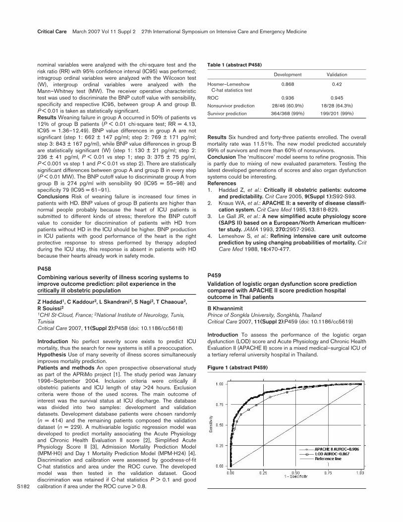
S182
nominal variables were analyzed with the chi-square test and therisk ratio (RR) with 95% confidence interval (IC95) was performed;intragroup ordinal variables were analyzed with the Wilcoxon test(W), intergroup ordinal variables were analyzed with theMann–Whitney test (MW). The receiver operative characteristictest was used to discriminate the BNP cutoff value with sensibility,specificity and respective IC95, between group A and group B.P < 0.01 is taken as statistically significant.Results Weaning failure in group A occurred in 50% of patients vs12% of group B patients (P < 0.01 chi-square test; RR = 4.13,IC95 = 1.36–12.49). BNP value differences in group A are notsignificant (step 1: 662 ± 147 pg/ml; step 2: 769 ± 171 pg/ml;step 3: 843 ± 167 pg/ml), while BNP value differences in group Bare statistically significant (W) (step 1: 130 ± 21 pg/ml; step 2:236 ± 41 pg/ml, P < 0.01 vs step 1; step 3: 375 ± 75 pg/ml,P < 0.001 vs step 1 and P < 0.01 vs step 2). There are statisticallysignificant differences between group A and group B in every step(P < 0.01 MW). The BNP cutoff value to discriminate group A fromgroup B is 274 pg/ml with sensibility 90 (IC95 = 55–98) andspecificity 79 (IC95 = 61–91).Conclusions Risk of weaning failure is increased four times inpatients with HD. BNP values of group B patients are higher thannormal people probably because the heart of ICU patients issubmitted to different kinds of stress; therefore the BNP cutoffvalue to consider for discrimination of patients with HD frompatients without HD in the ICU should be higher. BNP productionin ICU patients with good performance of the heart is the rightprotective response to stress performed by therapy adoptedduring the ICU stay, this response is absent in patients with HDbecause their hearts already work in safety mode.
P458Combining various severity of illness scoring systems toimprove outcome prediction: pilot experience in thecritically ill obstetric population
Z Haddad1, C Kaddour2, L Skandrani2, S Nagi2, T Chaaoua2, R Souissi21CHI St-Cloud, France; 2National Institute of Neurology, Tunis,TunisiaCritical Care 2007, 11(Suppl 2):P458 (doi: 10.1186/cc5618)
Introduction No perfect severity score exists to predict ICUmortality, thus the search for new systems is still a preoccupation.Hypothesis Use of many severity of illness scores simultaneouslyimproves mortality prediction.Patients and methods An open prospective observational studyas part of the APRiMo project [1]. The study period was January1996–September 2004. Inclusion criteria were critically illobstetric patients and ICU length of stay >24 hours. Exclusioncriteria were those of the used scores. The main outcome ofinterest was the survival status at ICU discharge. The databasewas divided into two samples: development and validationdatasets. Development database patients were chosen randomly(n = 414) and the remaining patients composed the validationdataset (n = 229). A multivariable logistic regression model wasdeveloped to predict mortality associating the Acute Physiologyand Chronic Health Evaluation II score [2], Simplified AcutePhysiology Score II [3], Admission Mortality Prediction Model(MPM-H0) and Day 1 Mortality Prediction Model (MPM-H24) [4].Discrimination and calibration were assessed by goodness-of-fitC-hat statistics and area under the ROC curve. The developedmodel was then tested in the validation dataset. Gooddiscrimination was retained if C-hat statistics P > 0.1 and goodcalibration if area under the ROC curve > 0.8.
Results Six hundred and forty-three patients enrolled. The overallmortality rate was 11.51%. The new model predicted accurately99% of survivors and more than 60% of nonsurvivors.Conclusion The ‘multiscore’ model seems to refine prognosis. Thisis partly due to mixing of new evaluated parameters. Testing thelatest developed generations of scores and also organ dysfunctionsystems could be interesting.References1. Haddad Z, et al.: Critically ill obstetric patients: outcome
and predictability. Crit Care 2005, 9(Suppl 1):S92-S93.2. Knaus WA, et al.: APACHE II: a severity of disease classifi-
cation system. Crit Care Med 1985, 13:818-829.3. Le Gall JR, et al.: A new simplified acute physiology score
(SAPS II) based on a European/North American multicen-ter study. JAMA 1993, 270:2957-2963.
4. Lemeshow S, et al.: Refining intensive care unit outcomeprediction by using changing probabilities of mortality. CritCare Med 1988, 16:470-477.
P459Validation of logistic organ dysfunction score predictioncompared with APACHE II score prediction hospitaloutcome in Thai patients
B KhwannimitPrince of Songkla University, Songkhla, ThailandCritical Care 2007, 11(Suppl 2):P459 (doi: 10.1186/cc5619)
Introduction To assess the performance of the logistic organdysfunction (LOD) score and Acute Physiology and Chronic HealthEvaluation II (APACHE II) score in a mixed medical–surgical ICU ofa tertiary referral university hospital in Thailand.
Critical Care March 2007 Vol 11 Suppl 2 27th International Symposium on Intensive Care and Emergency Medicine
Table 1 (abstract P458)
Development Validation
Hosmer–Lemeshow 0.868 0.42C-hat statistics test
ROC 0.936 0.945
Nonsurvivor prediction 28/46 (60.9%) 18/28 (64.3%)
Survivor prediction 364/368 (99%) 199/201 (99%)
Figure 1 (abstract P459)
S183
Methods The data were collected prospectively on consecutivepatients admitted to the ICU of Songklanagarind Hospital over a24-month period from 1 July 2004 until 30 June 2006.Results A total of 1,962 patients were enrolled, with 432 deaths(22%) prior to hospital discharge. Both systems providedoverprediction of hospital mortality. LOD and APACHE II scorespredicted hospital mortality of 25.4 ± 26.5 and 29.6 ± 27.8,respectively. Both models showed excellent discrimination. Thereceiver operating characteristic curves of both systems are shown inFigure 1. The area under the receiver operating characteristic curve(AUROC) of LOD was 0.867 (95% CI = 0.846–0.886) and theAUROC was 0.906 (95% CI = 0.889–0.923) for APACHE II. Bothmodels presented a poor calibration in overall population. However,the LOD score had good discrimination and calibration in subgroupsof nonoperative patients (AUROC 0.854, the Hosmer–Lemeshowgoodness-of-fit H statistic 11.67, P = 0.166) and patients thatexclude coronary artery disease and cardiac surgery (AUROC 0.860,the Hosmer–Lemeshow goodness-of-fit H statistic 10.03, P = 0.263).Conclusion The LOD score showed good accuracy to predicthospital mortality in subgroups of nonoperative critically ill patientsand excluded coronary heart disease and cardiac surgical criticallyill patients in Thailand.
P460Could we use the admission Acute Physiology and ChronicHealth Evaluation II score for outcome prediction incritically ill obstetric patients?
Z Haddad1, R Souissi2, C Kaddour2, W Trabelsi2, M Bouchnak3,S Nagi21CHI St-Cloud, France; 2National Institute of Neurology, Tunis,Tunisia; 3Centre de Maternité et de Néonatologie, Tunis, TunisiaCritical Care 2007, 11(Suppl 2):P460 (doi: 10.1186/cc5620)
Introduction The APACHE II score (APII) has widespread use inICUs for research and benchmarking. Physiological data forcalculation of the APII score derive from worst values in the first24 hours after ICU admission.Hypothesis Mortality prediction by the APII system depends ondata sampling. Use of ICU admission data (first hour) could beaccurate to predict mortality.Methods An open prospective data-sampling part of the APRiMostudy. Included were critically ill obstetric patients, with ICU lengthof stay (LOS) ≥6 hours. Admission (H1: first-hour worstphysiological data) and H24 (worst 24-hour physiological variablesincluding H1 collected data) were used to generate, respectively:the admission APII score (H1-APII) and H24-APII. The formulae tocalculate individual mortality for H1 and H24 APII were thosevalidated for H24-APII as stated by Knaus and colleagues [1],adjusting for admission diagnosis. We compared both scores bydiscrimination and calibration statistical tests. P < 0.05 was thethreshold for statistical significance.Results The study period was January 1996–September 2004.We included 541 patients, overall mortality was 10.5%. Mean H1-APII and H24-APII scores. respectively. were 7.6 ± 6.1 and 8.6 ± 7,with derived mean predicted mortality, respectively, of 8.63% and9.86%. The H24-APII score was higher than the H1-APII score in135 patients (25%), among those patients 32 died (24% ofpatients with worsened APII) vs 6.16% if H1 = H24 (P < 0.01).Running a multiple logistic regression with mortality as thedependent parameter, we found that worsening of the APII scoreover time is not significantly associated with mortality (P = 0.791),whereas the H1-APII score (P < 0.001) and ∆APII score (H24-APII
minus H1-APII score) (P = 0.04) are correlated with mortality.Respective ORs are 1.28 and 1.45. Overall discrimination abilityassessed by receiver operating characteristic curves was good forH1-APII (0.78) and H24-APII (0.784) (P = 0.834).Conclusion To avoid variation in APII mortality prediction causedby variable sample rates, the admission APII is reliable.Customizing mortality formulae could improve performances ofAPII-H1.Reference1. Knaus WA, et al.: APACHE II: a severity of disease classifi-
cation system. Crit Care Med 1985, 13:818-829.
P461Two-day intensive care unit outcome prediction score: atrial to improve outcome prediction in critically ill obstetricpatients
Z Haddad1, S Nagi2, R Souissi2, C Kaddour2
1CHI St-Cloud, France; 2National Institute of Neurology, Tunis,TunisiaCritical Care 2007, 11(Suppl 2):P461 (doi: 10.1186/cc5621)
Introduction The critically ill obstetric population median ICU stayis 4 days, thus general severity of illness scores are supposed tobe accurate in mortality prediction. A literature review makes ussceptical.Objective Development of a new prognostic model based onassociation of one of the generalistic severity scores (SAPS II,APACHE II), one of the organ dysfunction scores (LOD, MODS,SOFA) and evolution of these scores during the first 2 days of ICUhospitalization.Methods An open prospective analysis part of the APRiMo study[1] ranging from January 1996 to September 2004. Inclusioncriteria were critically ill obstetric patients with an ICU length ofstay > 24 hours. Exclusion criteria were those of the used scores.The main outcome of interest was survival status at ICU discharge.The database was divided into two samples: a developmentsample by random choice of 450 patients, and the remainingpatients in the validation dataset. Multivariable logistic regressionmodels were developed. We chose among different developedmodels the best performer as assessed by Hosmer–Lemeshow(HL) goodness-of-fit statistics (calibration) and the area under thereceiver operating characteristic curve (AUROC) for discrimination.Accuracy of the developed model was verified on the validationdataset using the same statistical tests. Results are expressed asthe mean ± standard deviation unless stated elsewhere. Data werecomputed on SPSS 11.5 Win-XP version.Results Six hundred and forty patients included. Age 31 ± 6 years,length of stay 5 ± 5 days, SAPS II 27 ± 16, SOFA score 5 ± 4,LOD score 2 ± 1.7. The overall mortality rate was 13.3%. The bestmodel was the one combining SAPS II and LOD scores. The LODscore and SAPS II alone discriminated well but calibrated poorly inoutcome prediction. Discrimination was optimal for the newdeveloped model in both development and validation datasets,with AUROC respectively of 0.87 and 0.85. Calibration was goodin the developed and validated datasets, respectively P = 0.176and 0.34. The developed model predicts death accurately in 2/3cases.Discussion and conclusion The SAPS II and LOD scores arecomplementary. Development of dynamic models in time helps torefine prognosis prediction.Reference1. Haddad Z, et al.: Critically ill obstetric patients: outcome
and predictability. Crit Care 2005, 9(Suppl 1):S92-S93.
Available online http://ccforum.com/supplements/11/S2
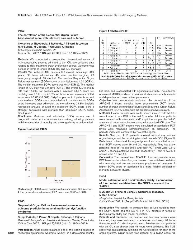
S184
P462The correlation of the Sequential Organ FailureAssessment score with intensive care unit outcome
I Ketchley, A Theodoraki, T Reynolds, A Tillyard, R Lawson, N Al-Subaie, M Cecconi, R Grounds, A RhodesSt George’s Hospital, London, UKCritical Care 2007, 11(Suppl 2):P462 (doi: 10.1186/cc5622)
Methods We conducted a prospective observational review of100 consecutive patients admitted to our ICU. We collected datarelating to daily maximum organ dysfunction scores. Outcome wasdefined in terms of length of ICU stay and ICU mortality.Results We included 100 patients (62 males), mean age 60.9years. Of these admissions, 45 were elective surgical, 22emergency surgical, 33 medical. The median Sequential OrganFailure Assessment (SOFA) score on admission was 4.50 (IQR 4).The median maximum SOFA score was 5.00 (IQR 5). The medianlength of ICU stay was 3.0 days (IQR 3). The overall ICU mortalityrate was 14.0%. For patients with a maximum SOFA score ≤8,mortality was 5.1% – vs 45.5% for those whose maximum SOFAscore was >8 (P < 0.001). Sixty-four per cent of patients scoredtheir maximum SOFA score on admission. In patients whose SOFAscore increased after admission, the mortality was 24.3%. Logisticregression analysis showed the maximum SOFA score bore astronger correlation with mortality than admission SOFA score.See Figure 1.Conclusion Maximum and admission SOFA scores are ofprognostic value in the intensive care setting; allowing patientswith increased risk of mortality and prolonged stay to be identified.
P463Sequential Organ Failure Assessment score as anoutcome predictor in malarial multiorgan dysfunctionsyndrome
S Jog, P Akole, B Pawar, N Gogate, S Gadgil, P RajhansDeenanath Mangeshkar Hospital and Research Centre, Pune, IndiaCritical Care 2007, 11(Suppl 2):P463 (doi: 10.1186/cc5623)
Introduction Acute severe malaria is one of the leading causes ofmultiorgan dysfunction syndrome (MODS) in a developing country
like India, and is associated with significant mortality. The outcomeof malarial MODS predicted in various studies is extremely variableand dependent on many patient parameters.Objective We prospectively evaluated the correlation of theAPACHE II score, parasite index, procalcitonin (PCT) levels,number of organ dysfunctions/failures and Sequential Organ FailureAssessment (SOFA) score with the outcome of severe malaria.Methods Eleven patients with acute severe malaria with MODSwere treated in our ICU in the last 5 months. All these patientswere treated with artesunate and/or quinine as per the WHOantimalarial treatment schedule, along with standard ICU care. TheAPACHE II and SOFA scores were calculated on admission. PCTlevels were measured semiquantitatively on admission. Theparasite index was confirmed by two pathologists.Results Nine out of 11 patients survived without any residualorgan damage, and the remaining two died due to MODS (Figure 1).Both these patients had five organ dysfunctions on admission, andtheir SOFA scores were 18 and 20, respectively. They had a lowparasitic index of 1% and 2.5% and their PCT levels were 0.5–2and >10 (semiquantitative method), respectively. Their APACHE IIscores were 16 and 10.Conclusion The pretreatment APACHE II score, parasite index,PCT levels and number of organs involved have variable correlationwith mortality and are not consistent predictors of outcome. Ahigher SOFA score on admission is a more reliable predictor ofmortality in malarial MODS.
P464Model calibration and discriminatory ability: a comparisonof four derived variables from the SOFA score and theSAPS II
R Ouezini, N Frikha, N Belhaj, S Ouerghi, M Mebazaa, M Ben AmmarMongi slim Hospital, La Marsa, TunisiaCritical Care 2007, 11(Suppl 2):P464 (doi: 10.1186/cc5624)
Introduction We sought to compare four derived variables fromthe SOFA score and the SAPS II in ICU patients in terms ofdiscriminatory ability and model calibration.Patients and methods Four hundred and fourteen patients wereincluded; they were evaluated on admission and every 48 hoursthereafter until ICU discharge or death. Readmission and patientswith an ICU stay shorter than 48 hours were excluded. The TMSscore was calculated by summing the worst scores for each of theorgan systems. Organ failure was defined by a SOFA score ≥3.
Critical Care March 2007 Vol 11 Suppl 2 27th International Symposium on Intensive Care and Emergency Medicine
Figure 1 (abstract P462)
Median length of ICU stay in patients with an admission SOFA score>8 vs those whose admission SOFA score was ≤8 (P = 0.001).
Figure 1 (abstract P463)
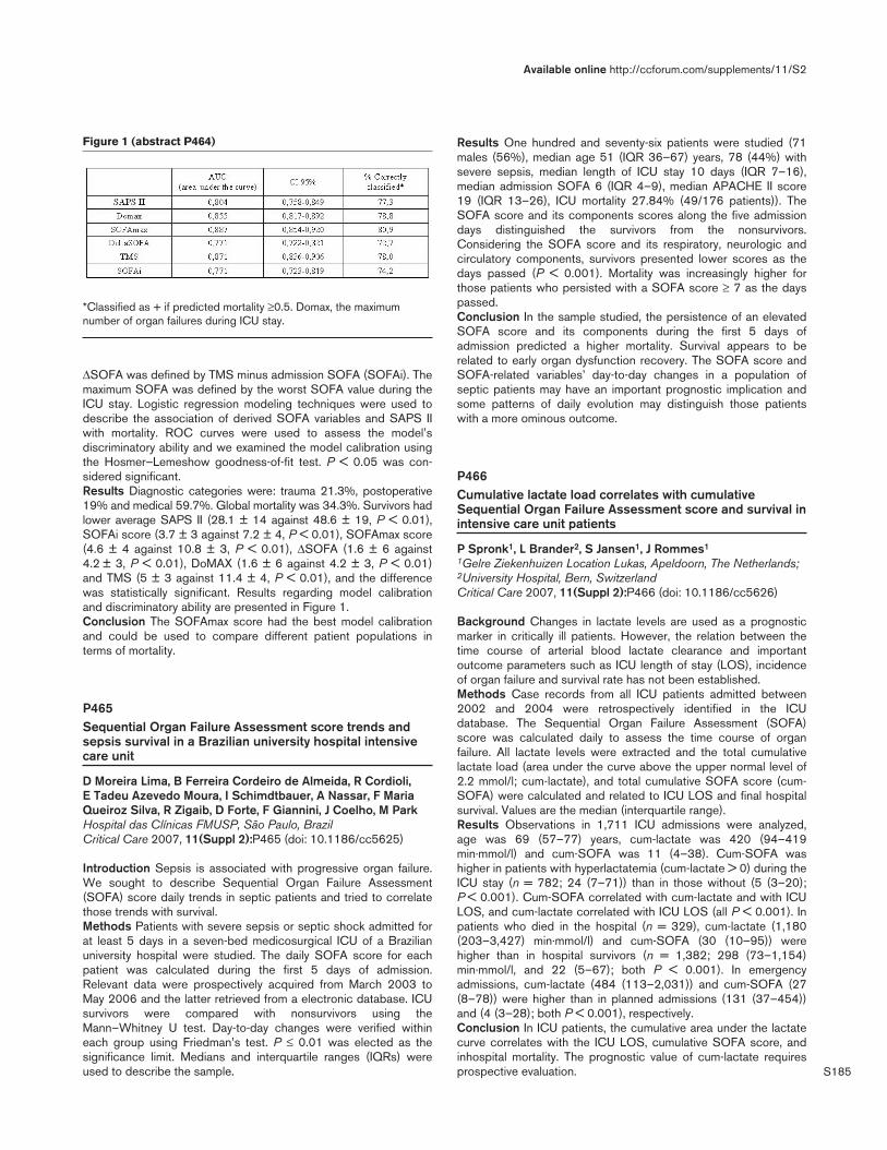
S185
∆SOFA was defined by TMS minus admission SOFA (SOFAi). Themaximum SOFA was defined by the worst SOFA value during theICU stay. Logistic regression modeling techniques were used todescribe the association of derived SOFA variables and SAPS IIwith mortality. ROC curves were used to assess the model’sdiscriminatory ability and we examined the model calibration usingthe Hosmer–Lemeshow goodness-of-fit test. P < 0.05 was con-sidered significant.Results Diagnostic categories were: trauma 21.3%, postoperative19% and medical 59.7%. Global mortality was 34.3%. Survivors hadlower average SAPS II (28.1 ± 14 against 48.6 ± 19, P < 0.01),SOFAi score (3.7 ± 3 against 7.2 ± 4, P < 0.01), SOFAmax score(4.6 ± 4 against 10.8 ± 3, P < 0.01), ∆SOFA (1.6 ± 6 against4.2 ± 3, P < 0.01), DoMAX (1.6 ± 6 against 4.2 ± 3, P < 0.01)and TMS (5 ± 3 against 11.4 ± 4, P < 0.01), and the differencewas statistically significant. Results regarding model calibrationand discriminatory ability are presented in Figure 1.Conclusion The SOFAmax score had the best model calibrationand could be used to compare different patient populations interms of mortality.
P465Sequential Organ Failure Assessment score trends andsepsis survival in a Brazilian university hospital intensivecare unit
D Moreira Lima, B Ferreira Cordeiro de Almeida, R Cordioli, E Tadeu Azevedo Moura, I Schimdtbauer, A Nassar, F MariaQueiroz Silva, R Zigaib, D Forte, F Giannini, J Coelho, M ParkHospital das Clínicas FMUSP, São Paulo, BrazilCritical Care 2007, 11(Suppl 2):P465 (doi: 10.1186/cc5625)
Introduction Sepsis is associated with progressive organ failure.We sought to describe Sequential Organ Failure Assessment(SOFA) score daily trends in septic patients and tried to correlatethose trends with survival.Methods Patients with severe sepsis or septic shock admitted forat least 5 days in a seven-bed medicosurgical ICU of a Brazilianuniversity hospital were studied. The daily SOFA score for eachpatient was calculated during the first 5 days of admission.Relevant data were prospectively acquired from March 2003 toMay 2006 and the latter retrieved from a electronic database. ICUsurvivors were compared with nonsurvivors using theMann–Whitney U test. Day-to-day changes were verified withineach group using Friedman’s test. P ≤ 0.01 was elected as thesignificance limit. Medians and interquartile ranges (IQRs) wereused to describe the sample.
Results One hundred and seventy-six patients were studied (71males (56%), median age 51 (IQR 36–67) years, 78 (44%) withsevere sepsis, median length of ICU stay 10 days (IQR 7–16),median admission SOFA 6 (IQR 4–9), median APACHE II score19 (IQR 13–26), ICU mortality 27.84% (49/176 patients)). TheSOFA score and its components scores along the five admissiondays distinguished the survivors from the nonsurvivors.Considering the SOFA score and its respiratory, neurologic andcirculatory components, survivors presented lower scores as thedays passed (P < 0.001). Mortality was increasingly higher forthose patients who persisted with a SOFA score ≥ 7 as the dayspassed.Conclusion In the sample studied, the persistence of an elevatedSOFA score and its components during the first 5 days ofadmission predicted a higher mortality. Survival appears to berelated to early organ dysfunction recovery. The SOFA score andSOFA-related variables’ day-to-day changes in a population ofseptic patients may have an important prognostic implication andsome patterns of daily evolution may distinguish those patientswith a more ominous outcome.
P466Cumulative lactate load correlates with cumulativeSequential Organ Failure Assessment score and survival inintensive care unit patients
P Spronk1, L Brander2, S Jansen1, J Rommes1
1Gelre Ziekenhuizen Location Lukas, Apeldoorn, The Netherlands;2University Hospital, Bern, SwitzerlandCritical Care 2007, 11(Suppl 2):P466 (doi: 10.1186/cc5626)
Background Changes in lactate levels are used as a prognosticmarker in critically ill patients. However, the relation between thetime course of arterial blood lactate clearance and importantoutcome parameters such as ICU length of stay (LOS), incidenceof organ failure and survival rate has not been established.Methods Case records from all ICU patients admitted between2002 and 2004 were retrospectively identified in the ICUdatabase. The Sequential Organ Failure Assessment (SOFA)score was calculated daily to assess the time course of organfailure. All lactate levels were extracted and the total cumulativelactate load (area under the curve above the upper normal level of2.2 mmol/l; cum-lactate), and total cumulative SOFA score (cum-SOFA) were calculated and related to ICU LOS and final hospitalsurvival. Values are the median (interquartile range).Results Observations in 1,711 ICU admissions were analyzed,age was 69 (57–77) years, cum-lactate was 420 (94–419min·mmol/l) and cum-SOFA was 11 (4–38). Cum-SOFA washigher in patients with hyperlactatemia (cum-lactate > 0) during theICU stay (n = 782; 24 (7–71)) than in those without (5 (3–20);P < 0.001). Cum-SOFA correlated with cum-lactate and with ICULOS, and cum-lactate correlated with ICU LOS (all P < 0.001). Inpatients who died in the hospital (n = 329), cum-lactate (1,180(203–3,427) min·mmol/l) and cum-SOFA (30 (10–95)) werehigher than in hospital survivors (n = 1,382; 298 (73–1,154)min·mmol/l, and 22 (5–67); both P < 0.001). In emergencyadmissions, cum-lactate (484 (113–2,031)) and cum-SOFA (27(8–78)) were higher than in planned admissions (131 (37–454))and (4 (3–28); both P < 0.001), respectively.Conclusion In ICU patients, the cumulative area under the lactatecurve correlates with the ICU LOS, cumulative SOFA score, andinhospital mortality. The prognostic value of cum-lactate requiresprospective evaluation.
Available online http://ccforum.com/supplements/11/S2
Figure 1 (abstract P464)
*Classified as + if predicted mortality ≥0.5. Domax, the maximumnumber of organ failures during ICU stay.
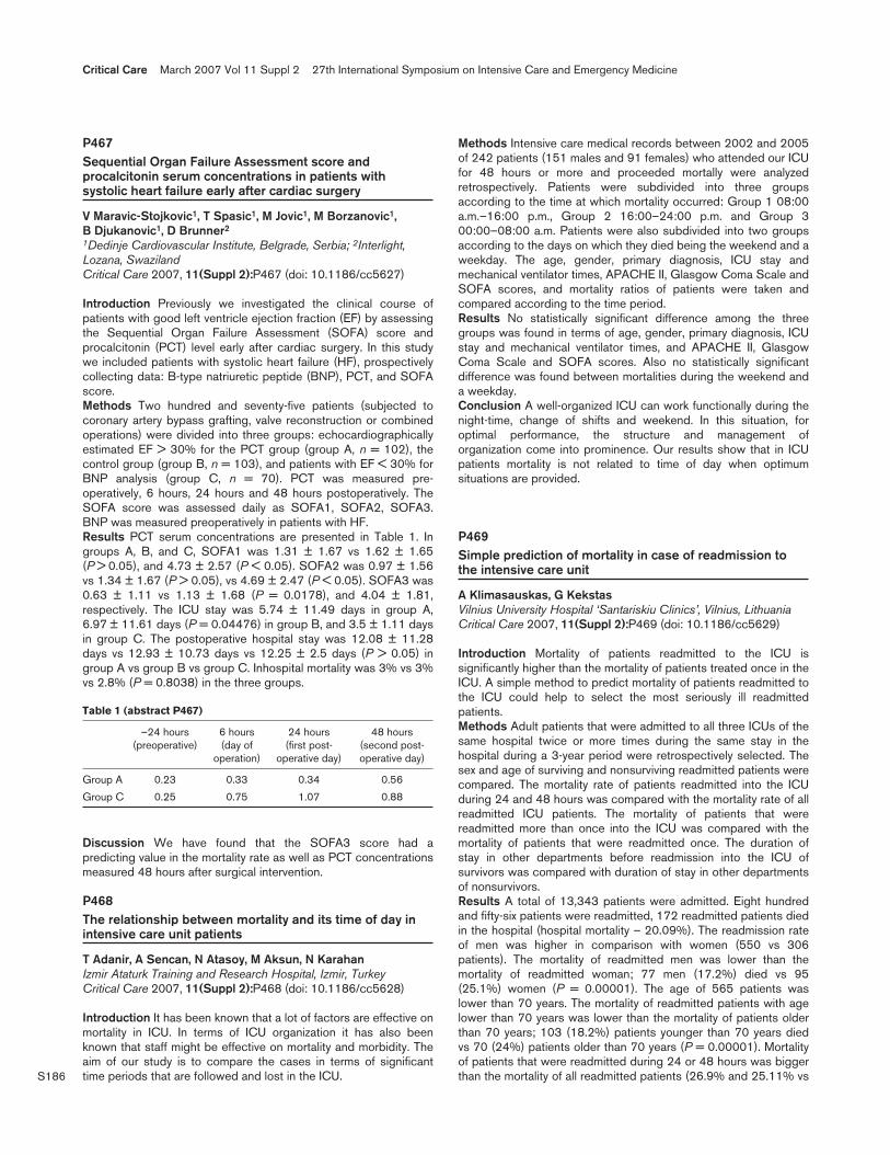
S186
P467Sequential Organ Failure Assessment score andprocalcitonin serum concentrations in patients withsystolic heart failure early after cardiac surgery
V Maravic-Stojkovic1, T Spasic1, M Jovic1, M Borzanovic1, B Djukanovic1, D Brunner2
1Dedinje Cardiovascular Institute, Belgrade, Serbia; 2Interlight,Lozana, SwazilandCritical Care 2007, 11(Suppl 2):P467 (doi: 10.1186/cc5627)
Introduction Previously we investigated the clinical course ofpatients with good left ventricle ejection fraction (EF) by assessingthe Sequential Organ Failure Assessment (SOFA) score andprocalcitonin (PCT) level early after cardiac surgery. In this studywe included patients with systolic heart failure (HF), prospectivelycollecting data: B-type natriuretic peptide (BNP), PCT, and SOFAscore.Methods Two hundred and seventy-five patients (subjected tocoronary artery bypass grafting, valve reconstruction or combinedoperations) were divided into three groups: echocardiographicallyestimated EF > 30% for the PCT group (group A, n = 102), thecontrol group (group B, n = 103), and patients with EF < 30% forBNP analysis (group C, n = 70). PCT was measured pre-operatively, 6 hours, 24 hours and 48 hours postoperatively. TheSOFA score was assessed daily as SOFA1, SOFA2, SOFA3.BNP was measured preoperatively in patients with HF.Results PCT serum concentrations are presented in Table 1. Ingroups A, B, and C, SOFA1 was 1.31 ± 1.67 vs 1.62 ± 1.65(P > 0.05), and 4.73 ± 2.57 (P < 0.05). SOFA2 was 0.97 ± 1.56vs 1.34 ± 1.67 (P > 0.05), vs 4.69 ± 2.47 (P < 0.05). SOFA3 was0.63 ± 1.11 vs 1.13 ± 1.68 (P = 0.0178), and 4.04 ± 1.81,respectively. The ICU stay was 5.74 ± 11.49 days in group A,6.97 ± 11.61 days (P = 0.04476) in group B, and 3.5 ± 1.11 daysin group C. The postoperative hospital stay was 12.08 ± 11.28days vs 12.93 ± 10.73 days vs 12.25 ± 2.5 days (P > 0.05) ingroup A vs group B vs group C. Inhospital mortality was 3% vs 3%vs 2.8% (P = 0.8038) in the three groups.
Table 1 (abstract P467)
–24 hours 6 hours 24 hours 48 hours (preoperative) (day of (first post- (second post-
operation) operative day) operative day)
Group A 0.23 0.33 0.34 0.56
Group C 0.25 0.75 1.07 0.88
Discussion We have found that the SOFA3 score had apredicting value in the mortality rate as well as PCT concentrationsmeasured 48 hours after surgical intervention.
P468The relationship between mortality and its time of day inintensive care unit patients
T Adanir, A Sencan, N Atasoy, M Aksun, N KarahanIzmir Ataturk Training and Research Hospital, Izmir, TurkeyCritical Care 2007, 11(Suppl 2):P468 (doi: 10.1186/cc5628)
Introduction It has been known that a lot of factors are effective onmortality in ICU. In terms of ICU organization it has also beenknown that staff might be effective on mortality and morbidity. Theaim of our study is to compare the cases in terms of significanttime periods that are followed and lost in the ICU.
Methods Intensive care medical records between 2002 and 2005of 242 patients (151 males and 91 females) who attended our ICUfor 48 hours or more and proceeded mortally were analyzedretrospectively. Patients were subdivided into three groupsaccording to the time at which mortality occurred: Group 1 08:00a.m.–16:00 p.m., Group 2 16:00–24:00 p.m. and Group 300:00–08:00 a.m. Patients were also subdivided into two groupsaccording to the days on which they died being the weekend and aweekday. The age, gender, primary diagnosis, ICU stay andmechanical ventilator times, APACHE II, Glasgow Coma Scale andSOFA scores, and mortality ratios of patients were taken andcompared according to the time period.Results No statistically significant difference among the threegroups was found in terms of age, gender, primary diagnosis, ICUstay and mechanical ventilator times, and APACHE II, GlasgowComa Scale and SOFA scores. Also no statistically significantdifference was found between mortalities during the weekend anda weekday.Conclusion A well-organized ICU can work functionally during thenight-time, change of shifts and weekend. In this situation, foroptimal performance, the structure and management oforganization come into prominence. Our results show that in ICUpatients mortality is not related to time of day when optimumsituations are provided.
P469Simple prediction of mortality in case of readmission tothe intensive care unit
A Klimasauskas, G KekstasVilnius University Hospital ‘Santariskiu Clinics’, Vilnius, LithuaniaCritical Care 2007, 11(Suppl 2):P469 (doi: 10.1186/cc5629)
Introduction Mortality of patients readmitted to the ICU issignificantly higher than the mortality of patients treated once in theICU. A simple method to predict mortality of patients readmitted tothe ICU could help to select the most seriously ill readmittedpatients.Methods Adult patients that were admitted to all three ICUs of thesame hospital twice or more times during the same stay in thehospital during a 3-year period were retrospectively selected. Thesex and age of surviving and nonsurviving readmitted patients werecompared. The mortality rate of patients readmitted into the ICUduring 24 and 48 hours was compared with the mortality rate of allreadmitted ICU patients. The mortality of patients that werereadmitted more than once into the ICU was compared with themortality of patients that were readmitted once. The duration ofstay in other departments before readmission into the ICU ofsurvivors was compared with duration of stay in other departmentsof nonsurvivors.Results A total of 13,343 patients were admitted. Eight hundredand fifty-six patients were readmitted, 172 readmitted patients diedin the hospital (hospital mortality – 20.09%). The readmission rateof men was higher in comparison with women (550 vs 306patients). The mortality of readmitted men was lower than themortality of readmitted woman; 77 men (17.2%) died vs 95(25.1%) women (P = 0.00001). The age of 565 patients waslower than 70 years. The mortality of readmitted patients with agelower than 70 years was lower than the mortality of patients olderthan 70 years; 103 (18.2%) patients younger than 70 years diedvs 70 (24%) patients older than 70 years (P = 0.00001). Mortalityof patients that were readmitted during 24 or 48 hours was biggerthan the mortality of all readmitted patients (26.9% and 25.11% vs
Critical Care March 2007 Vol 11 Suppl 2 27th International Symposium on Intensive Care and Emergency Medicine
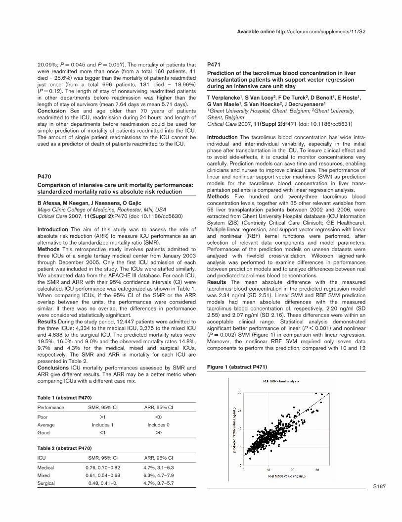
S187
20.09%; P = 0.045 and P = 0.097). The mortality of patients thatwere readmitted more than once (from a total 160 patients, 41died – 25.6%) was bigger than the mortality of patients readmittedjust once (from a total 696 patients, 131 died – 18.96%)(P = 0.12). The length of stay of nonsurviving readmitted patientsin other departments before readmission was higher than thelength of stay of survivors (mean 7.64 days vs mean 5.71 days).Conclusion Sex and age older than 70 years of patientsreadmitted to the ICU, readmission during 24 hours, and length ofstay in other departments before readmission could be used forsimple prediction of mortality of patients readmitted into the ICU.The amount of single patient readmissions to the ICU cannot beused as a predictor of death of patients readmitted to the ICU.
P470Comparison of intensive care unit mortality performances:standardized mortality ratio vs absolute risk reduction
B Afessa, M Keegan, J Naessens, O GajicMayo Clinic College of Medicine, Rochester, MN, USACritical Care 2007, 11(Suppl 2):P470 (doi: 10.1186/cc5630)
Introduction The aim of this study was to assess the role ofabsolute risk reduction (ARR) to measure ICU performance as analternative to the standardized mortality ratio (SMR).Methods This retrospective study involves patients admitted tothree ICUs of a single tertiary medical center from January 2003through December 2005. Only the first ICU admission of eachpatient was included in the study. The ICUs were staffed similarly.We abstracted data from the APACHE III database. For each ICU,the SMR and ARR with their 95% confidence intervals (CI) werecalculated. ICU performance was categorized as shown in Table 1.When comparing ICUs, if the 95% CI of the SMR or the ARRoverlap between the units, the performances were consideredsimilar. If there was no overlap, the differences in performancewere considered statistically significant.Results During the study period, 12,447 patients were admitted tothe three ICUs: 4,334 to the medical ICU, 3,275 to the mixed ICUand 4,838 to the surgical ICU. The predicted mortality rates were19.5%, 16.0% and 9.0% and the observed mortality rates 14.8%,9.7% and 4.3% for the medical, mixed and surgical ICUs,respectively. The SMR and ARR in mortality for each ICU arepresented in Table 2.Conclusions ICU mortality performances assessed by SMR andARR give different results. The ARR may be a better metric whencomparing ICUs with a different case mix.
P471Prediction of the tacrolimus blood concentration in livertransplantation patients with support vector regressionduring an intensive care unit stay
T Verplancke1, S Van Looy2, F De Turck2, D Benoit1, E Hoste1,G Van Maele1, S Van Hoecke2, J Decruyenaere1
1Ghent University Hospital, Ghent, Belgium; 2Ghent University,Ghent, BelgiumCritical Care 2007, 11(Suppl 2):P471 (doi: 10.1186/cc5631)
Introduction The tacrolimus blood concentration has wide intra-individual and inter-individual variability, especially in the initialphase after transplantation in the ICU. To insure clinical effect andto avoid side-effects, it is crucial to monitor concentrations verycarefully. Prediction models can save time and resources, enablingclinicians and nurses to improve clinical care. The performance oflinear and nonlinear support vector machines (SVM) as predictionmodels for the tacrolimus blood concentration in liver trans-plantation patients is compared with linear regression analysis.Methods Five hundred and twenty-three tacrolimus bloodconcentration levels, together with 35 other relevant variables from56 liver transplantation patients between 2002 and 2006, wereextracted from Ghent University Hospital database (ICU InformationSystem IZIS) (Centricity Critical Care Clinisoft; GE Healthcare).Multiple linear regression, and support vector regression with linearand nonlinear (RBF) kernel functions were performed, afterselection of relevant data components and model parameters.Performances of the prediction models on unseen datasets wereanalyzed with fivefold cross-validation. Wilcoxon signed-rankanalysis was performed to examine differences in performancesbetween prediction models and to analyze differences between realand predicted tacrolimus blood concentrations.Results The mean absolute difference with the measuredtacrolimus blood concentration in the predicted regression modelwas 2.34 ng/ml (SD 2.51). Linear SVM and RBF SVM predictionmodels had mean absolute differences with the measuredtacrolimus blood concentration of, respectively, 2.20 ng/ml (SD2.55) and 2.07 ng/ml (SD 2.16). These differences were within anacceptable clinical range. Statistical analysis demonstratedsignificant better performance of linear (P < 0.001) and nonlinear(P = 0.002) SVM (Figure 1) in comparison with linear regression.Moreover, the nonlinear RBF SVM required only seven datacomponents to perform this prediction, compared with 10 and 12
Available online http://ccforum.com/supplements/11/S2
Table 1 (abstract P470)
Performance SMR, 95% CI ARR, 95% CI
Poor >1 <0
Average Includes 1 Includes 0
Good <1 >0
Table 2 (abstract P470)
ICU SMR, 95% CI ARR, 95% CI
Medical 0.76, 0.70–0.82 4.7%, 3.1–6.3
Mixed 0.61, 0.54–0.68 6.3%, 4.7–7.9
Surgical 0.48, 0.41–0. 4.7%, 3.7–5.7
Figure 1 (abstract P471)
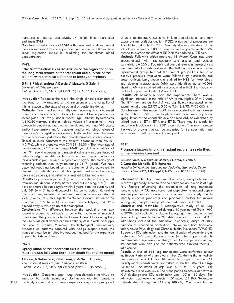
S188
components needed, respectively, by multiple linear regressionand linear SVM.Conclusion Performance of SVM with linear and nonlinear kernelfunction was excellent and superior in comparison with the multiplelinear regression model in predicting the tacrolimus bloodconcentration.
P472Effects of the clinical characteristics of the organ donor onthe long-term results of the transplant and survival of thepatient, with particular reference to kidney transplants
S Pirri, R Mastrandrea, A Barcia, A Mazzola, S SalemiUniversity of Palermo, ItalyCritical Care 2007, 11(Suppl 2):P472 (doi: 10.1186/cc5632)
Introduction To assess the role of the single clinical parameters ofthe donor on the outcome of the transplant and the variability ofthis in relation to the state of an optimal or borderline donor.Methods One hundred and fifty-one receiving and deceaseddonor brace subordinates to kidney transplant. Clinical parametersinvestigated for every donor were: age, arterial hypertension(≥140/80 mmHg), diabetes, blood values of creatinine. It waschosen to classify as marginal all the donors with age >55 years,and/or hypertension, and/or diabetes, and/or with blood values ofcreatinine >1.5 mg/dl, and/or whose death has happened becauseof one whichever pathology that has determined cerebral anoxia.Based on such parameters the donors’ borderline was 72/151(47.7%), while the optimal was 79/151 (52.3%). The mean age ofthe donors was 47.5 years (range 14–81 years). The population ofthe 151 receiving optimal and marginal kidneys was constituted ofpatients judged suitable for the transplant with typical risk factorsfor a standard population of subjects on dialysis. The mean age ofreceiving patients was 46 years (range 21–71 years). We haveclassified receiving based on the outcome of the transplant to5 years, as: patients alive with transplanted kidney still working,deceased patients, and patients re-entered to haemodialysis.Results Eighty-seven per cent (n = 69) of kidneys transplantedfrom optimal donors, in fact, have turned out working, 4% (n = 3)have re-entered haemodialysis within 5 years from the surgery, andonly 9% (n = 7) have deceased in the same period. Regardingmarginal kidney receiving, it has been possible to demonstrate that72% (n = 52) of such subjects maintained a good function of thetransplant, 11% (n = 8) re-entered haemodialysis, and 17%passed away within 5 years of the transplant.Conclusions The difference between the survival of the tworeceiving groups is not such to justify the exclusion of marginaldonors from the ‘pool’ of potential kidney donors. Considering that,the use of marginal donors can be a valid system in order to supplythe lack of organs. Moreover the histological examinations,executed on patterns captured with wedge biopsy before thetransplant, can be an effective strategy finalized for the expansionof potential kidney donors.
P473Upregulation of the endothelin axis in alveolarmacrophages following brain stem death in a murine model
J Fraser, A Sutherland, F Kermeen, K McNeil, J DunningThe Prince Charles Hospital, Brisbane, AustraliaCritical Care 2007, 11(Suppl 2):P473 (doi: 10.1186/cc5633)
Introduction Outcomes post lung transplantation continue toimprove, but early pulmonary dysfunction dictates long-termmorbidity and mortality. Ischaemia reperfusion injury is a precipitant
of poor postoperative outcome in lung transplantation and maycause primary graft dysfunction (PGD). A number of processes arethought to contribute to PGD. Relatively little is understood of therole of brain stem death (BSD) in subsequent organ dysfunction. Wewished to examine the effect of BSD on the endothelin (ET) axis.Methods Following ethics approval, 14 Wistar–Kyoto rats wereanaesthetised, with tracheostomy and arterial and venouscannulation. A 200 µl Fogarty’s balloon catheter was inserted via aburr hole into the subdural vault. The balloon was inflated in theexperimental group but not the control group. Four hours ofpositive pressure ventilation were followed by euthanasia andorgan retrieval. Lung tissue was stained for H&E for morphology,and alveolar macrophages (AM) were identified by anti-CD68staining. AM were stained with a monoclonal anti-ET-1 antibody, aswell as the polyclonal anti-ET-A and ET-B.Results All animals survived the experiment. There was asignificant increase in the ratio of AM to neutrophils (P = 0.002).The ET-1 content on the AM was significantly increased in theexperimental group (27.57 ± 5.26 vs 7.01 ± 1.75, P < 0.0001).Conclusions In this model, BSD was associated with an increasein the ratio of AM to neutrophils, and there was significantupregulation of the endothelin axis on these AM, as evidenced byraised levels of ET-1, ET-A and ET-B. There may be a role forendothelin blockade in the BSD organ donor. This may increasethe yield of organs that can be accepted for transplantation andimprove early graft function in the recipient.
P474Prognosis factors in lung transplant recipients readmittedto the intensive care unit
B Suberviola, A Gonzalez Castro, J Llorca, A Vallejo, C Gonzalez Mansilla, E MiñambresHospital Universitario Marques de Valdecilla, Santander, SpainCritical Care 2007, 11(Suppl 2):P474 (doi: 10.1186/cc5634)
Introduction The short-term survival after lung transplantation hasimproved gradually. Despite this the peritransplant period is of highrisk. Factors influencing the readmission of lung transplantrecipients to the ICU are diverse, but respiratory failure and sepsisare the predominant causes. The objective of our study was toidentify outcome predictors and prognostic factors for survivalamong lung transplant recipients on readmission to the ICU.Materials and methods A retrospective study of all lungtransplant recipients achieved during a 10-year period (from 1997to 2006). Data collection included the age, gender, reason for andtype of lung transplantation. Variables specific to individual ICUadmissions included the admission diagnosis, length of stay,duration of mechanical ventilation, interval time from transplan-tation, Acute Physiology and Chronic Health Evaluation (APACHE)II score on ICU admission, and the identification of systemic organdysfunction. We used Student’s t test (or, where appropriate, itsnonparametric equivalent) or the χ2 test for comparisons amongthe patients who died and the patients who survived their ICUadmissions.Results A total of 144 lung transplants were performed at ourinstitution. Forty-six of them died on the ICU during the immediateperioperative period. Finally, 98 were discharged from the ICU.Twenty-eight patients were readmitted to the ICU after discharge(28.57%). The mean of age was 51.3 ± 11.6 years. Themale/female ratio was 23/5. The mean period transcurred betweenICU discharge and ICU readmission was 107 ± 162 days. Theadmission diagnosis was sepsis in 20 cases (71.4%). Seventeenpatients died during the ICU stay (60.7%). We found that an
Critical Care March 2007 Vol 11 Suppl 2 27th International Symposium on Intensive Care and Emergency Medicine
S189
increase in APACHE II score, delay to ICU readmission, need ofmechanical ventilation and three or more organ dysfunctions weresignificantly associated with mortality.Conclusions Admission to the ICU is common in lung transplantrecipients, and it is associated with a high mortality. Sepsis is themain cause of ICU readmission and the most frequent cause ofdeath. Lung transplant recipients with higher APACHE II score andthree or more organ dysfunction present higher mortality. The delayon ICU readmission is also associated with higher mortality.
P475Predictors of intensive care unit readmission within 48hours after discharge
E Boudesteijn, S Arbous, P Berg van denLUMC, Leiden, The NetherlandsCritical Care 2007, 11(Suppl 2):P475 (doi: 10.1186/cc5635)
Introduction Readmission to the ICU during the samehospitalization is associated with significant morbidity and mortalityand results in a longer length of stay and higher costs. There istherefore growing interest to identify reliable predictors forreadmission. The aim of our study was to assess the incidence ofICU readmissions, identify predictors of ICU readmission, anddetermine patient outcome.Methods We performed a retrospective case–control study. Thestudy population consisted of all patients who were dischargedalive from our 28-bed surgical, thoracic–surgical and medical ICUin a university teaching hospital in a 1-year period. A case wasdefined as a patient readmitted to the ICU within 48 hours afterdischarge. For each case, three control patients were randomlyselected from the study population. The following information wascollected: demographic parameters and APACHE II score,parameters of hemodynamic, respiratory and renal function, lengthof ICU stay, duration of invasive ventilatory support (ventilator time),and time between extubation and discharge. To determine apredictive model, covariate selection was done by the two-samplet test, Mann–Whitney test and univariate logistic regression. Fromsignificant (P < 0.10), plausible and clinically relevant variables, apredictive model was generated using multivariate logisticregression.Results During a 1-year period 1,635 patients were admitted to ourICU. Of 1,393 patients at risk for readmission, 25 (1.8%)readmissions occurred in 23 patients. Nine of the 23 (39%)readmitted patients died during their hospitalization, while theoverall ICU mortality was 10.6%. The most important reason forreadmission (68% of the cases) was respiratory deterioration. In theunivariate analysis, age, ventilator time during first admission andtime between extubation and ICU discharge were significantpredictors of readmission. In the multivariate analysis, age (OR 1.1;95% CI 1.00–1.13; P = 0.03) and ventilator time during firstadmission (OR 1.1; 95% CI 1.00–1.10; P = 0.03) were significantpredictors, corrected for patient characteristics. Furthermore,patients who were readmitted had a significant longer duration oftotal (first and second admission) ventilator time (188 vs 106 hours,P = 0.012), and total ICU stay (400 vs 127 hours, P = 0.009).Conclusion Patients readmitted to the ICU have significant longeroverall ventilator time, ICU stay, and a higher ICU mortality. Theventilator time during first admission (especially beyond 300 hours)is an important predictor of readmission. The time it takes to getpatients ready for discharge after extubation also differedsignificantly. The data suggest that elderly patients who have beenventilated for a long period are at particular risk for readmissionand should receive additional care before discharge from the ICU.
P476Intensive care unit readmissions after lung transplantation:epidemiology and outcome
F Klein, P Klin, J Osses, J Díaz, A Bertolotti, R FavaloroFavaloro Foundation, Buenos Aires, ArgentinaCritical Care 2007, 11(Suppl 2):P476 (doi: 10.1186/cc5636)
Introduction Significant improvement of short-term and long-termsurvival after lung transplantation (LT) has been observed. Never-theless, a significant number of patients need to be readmitted tothe ICU. The aim of our study was to analyse the epidemiology,outcome and risk factors for LT patients readmitted to the ICU afteran initial discharge.Methods From February 1996 until May 2006 we studied all LTpatients from a single centre initially discharged from the ICU whoneeded to be readmitted. Demographic data included the type anddate of LT, best post-LT FEV1, last pre-ICU readmission FEV1,admission diagnosis, time from LT to ICU admission, mechanicalventilation (MV) use, rejection episodes and infections. Actuarialsurvival rates (ASR) were calculated with Kaplan–Meier curves.Results A total of 103 LT patients were discharged from the ICU,41 patients (39.8%) were readmitted (males 53.6% (22 patients)with a mean age of 42 years (15–66)). Indications were emphy-sema in 13 patients (31.7%), idiopathic pulmonary fibrosis in eightpatients (19.5%), bronchiectasis in five patients (12.2%), cysticfibrosis in five patients and others in seven patients (17%).Seventeen patients underwent bilateral LT, 11 patients right LT(26.8%) and eight patients left LT (19.5%), while five patientsreceived a heart–lung transplantation. Respiratory failure was theprincipal ICU admission diagnosis (68.3%), followed by seizures(7%) and septic shock (4.8%). MV was required in 35 patients(85.3%). ICU mortality for readmitted patients was 68.3% with a1-year, 3-year and 5-year ASR of 67.3%, 62.9% and 47.4%. Thesurvival median was 1,761 days (1,134–2,388). In the MVpatients, a 1-year, 3-year and 5-year ASR of 63.1%, 58.9% and44.2% was found with a median survival of 1,618 days(132–3,104). The time to ICU admission was 1,303 (4–3,096days). ICU admission timing was not found to be a predictor forearly (<30 days; 53.8%) vs late (>30 days: 46.4%), P = 0.65.Deceased patients required significantly more MV (71.4% vs38.5%; P = 0.044 (chi-square); OR: 4; 95% CI: 1–15.99).Emphysema was not more prevalent in the deceased patientgroup, and neither was the pre-ICU readmission FEV1 nor theoccurrence of opportunistic infections. Steroid-resistant acuterejection was found to correlate with mortality.Conclusion ICU readmissions are frequent among LT patients. Inour study group, respiratory failure was the more prevalentadmission diagnosis. The need for MV was associated with a worseprognosis as well as steroid-resistant acute rejection episodes.Early or late ICU admission after LT has not influenced mortality.
P477The functional outcome of patients requiring intensive carereadmission
N Conlon, B O’Brien, B MarshMater Misericordiae University Hospital, Dublin, IrelandCritical Care 2007, 11(Suppl 2):P477 (doi: 10.1186/cc5637)
Introduction Rates of readmission to the ICU are often cited ascontroversial indices of quality of intensive care, adequacy offollow-up and as guides to resource allocation. Nonetheless thereare few data on the long-term functional outcome of ICUrecidivists: we set out to study this.
Available online http://ccforum.com/supplements/11/S2
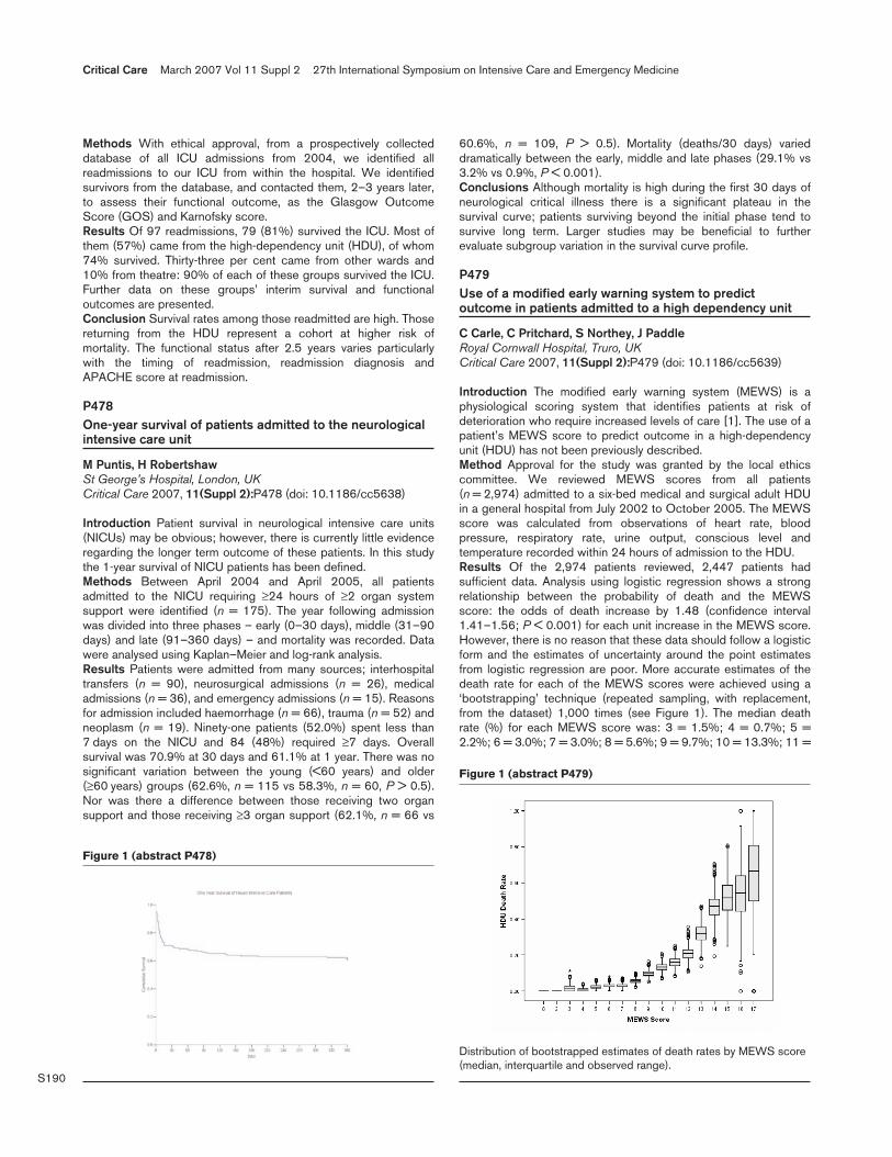
S190
Methods With ethical approval, from a prospectively collecteddatabase of all ICU admissions from 2004, we identified allreadmissions to our ICU from within the hospital. We identifiedsurvivors from the database, and contacted them, 2–3 years later,to assess their functional outcome, as the Glasgow OutcomeScore (GOS) and Karnofsky score.Results Of 97 readmissions, 79 (81%) survived the ICU. Most ofthem (57%) came from the high-dependency unit (HDU), of whom74% survived. Thirty-three per cent came from other wards and10% from theatre: 90% of each of these groups survived the ICU.Further data on these groups’ interim survival and functionaloutcomes are presented.Conclusion Survival rates among those readmitted are high. Thosereturning from the HDU represent a cohort at higher risk ofmortality. The functional status after 2.5 years varies particularlywith the timing of readmission, readmission diagnosis andAPACHE score at readmission.
P478One-year survival of patients admitted to the neurologicalintensive care unit
M Puntis, H RobertshawSt George’s Hospital, London, UKCritical Care 2007, 11(Suppl 2):P478 (doi: 10.1186/cc5638)
Introduction Patient survival in neurological intensive care units(NICUs) may be obvious; however, there is currently little evidenceregarding the longer term outcome of these patients. In this studythe 1-year survival of NICU patients has been defined.Methods Between April 2004 and April 2005, all patientsadmitted to the NICU requiring ≥24 hours of ≥2 organ systemsupport were identified (n = 175). The year following admissionwas divided into three phases – early (0–30 days), middle (31–90days) and late (91–360 days) – and mortality was recorded. Datawere analysed using Kaplan–Meier and log-rank analysis.Results Patients were admitted from many sources; interhospitaltransfers (n = 90), neurosurgical admissions (n = 26), medicaladmissions (n = 36), and emergency admissions (n = 15). Reasonsfor admission included haemorrhage (n = 66), trauma (n = 52) andneoplasm (n = 19). Ninety-one patients (52.0%) spent less than7 days on the NICU and 84 (48%) required ≥7 days. Overallsurvival was 70.9% at 30 days and 61.1% at 1 year. There was nosignificant variation between the young (<60 years) and older(≥60 years) groups (62.6%, n = 115 vs 58.3%, n = 60, P > 0.5).Nor was there a difference between those receiving two organsupport and those receiving ≥3 organ support (62.1%, n = 66 vs
60.6%, n = 109, P > 0.5). Mortality (deaths/30 days) varieddramatically between the early, middle and late phases (29.1% vs3.2% vs 0.9%, P < 0.001).Conclusions Although mortality is high during the first 30 days ofneurological critical illness there is a significant plateau in thesurvival curve; patients surviving beyond the initial phase tend tosurvive long term. Larger studies may be beneficial to furtherevaluate subgroup variation in the survival curve profile.
P479Use of a modified early warning system to predictoutcome in patients admitted to a high dependency unit
C Carle, C Pritchard, S Northey, J PaddleRoyal Cornwall Hospital, Truro, UKCritical Care 2007, 11(Suppl 2):P479 (doi: 10.1186/cc5639)
Introduction The modified early warning system (MEWS) is aphysiological scoring system that identifies patients at risk ofdeterioration who require increased levels of care [1]. The use of apatient’s MEWS score to predict outcome in a high-dependencyunit (HDU) has not been previously described.Method Approval for the study was granted by the local ethicscommittee. We reviewed MEWS scores from all patients(n = 2,974) admitted to a six-bed medical and surgical adult HDUin a general hospital from July 2002 to October 2005. The MEWSscore was calculated from observations of heart rate, bloodpressure, respiratory rate, urine output, conscious level andtemperature recorded within 24 hours of admission to the HDU.Results Of the 2,974 patients reviewed, 2,447 patients hadsufficient data. Analysis using logistic regression shows a strongrelationship between the probability of death and the MEWSscore: the odds of death increase by 1.48 (confidence interval1.41–1.56; P < 0.001) for each unit increase in the MEWS score.However, there is no reason that these data should follow a logisticform and the estimates of uncertainty around the point estimatesfrom logistic regression are poor. More accurate estimates of thedeath rate for each of the MEWS scores were achieved using a‘bootstrapping’ technique (repeated sampling, with replacement,from the dataset) 1,000 times (see Figure 1). The median deathrate (%) for each MEWS score was: 3 = 1.5%; 4 = 0.7%; 5 =2.2%; 6 = 3.0%; 7 = 3.0%; 8 = 5.6%; 9 = 9.7%; 10 = 13.3%; 11 =
Critical Care March 2007 Vol 11 Suppl 2 27th International Symposium on Intensive Care and Emergency Medicine
Figure 1 (abstract P478)
Figure 1 (abstract P479)
Distribution of bootstrapped estimates of death rates by MEWS score(median, interquartile and observed range).
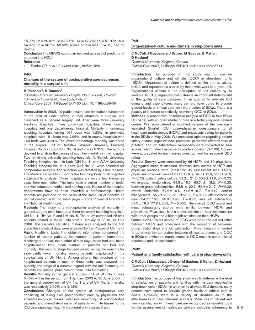
S191
15.9%; 12 = 20.9%; 13 = 32.0%; 14 = 47.2%; 15 = 51.9%; 16 =54.6%; 17 = 66.7%. MEWS scores of 2 or less (n = 19) had nodeaths.Conclusion The MEWS score can be used as a useful predictor ofoutcome in a HDU.Reference1. Subbe CP, et al.: Q J Med 2001, 94:521-526.
P480Changes of the system of postoperative care decreasesmortality in a surgical unit
M Piechota1, M Banach2
1Bolesùaw Szarecki University Hospital No. 5 in Lodz, Poland;2University Hospital No. 3 in Lodz, PolandCritical Care 2007, 11(Suppl 2):P480 (doi: 10.1186/cc5640)
Introduction In 2005, 10 public health care institutions functionedin the area of Lodz, having in their structure a surgical unitclassified as a general surgery unit. They were three universityteaching hospitals, three provincial hospitals, three countyhospitals and one departmental hospital. Mortality in universityteaching hospitals having 167 beds was 1.25%, in provincialhospitals with 191 beds was 2.96%, and in county hospitals with140 beds was 3.98%. The lowest percentage mortality was notedin the surgical unit of Bolesùaw Szarecki University TeachingHospital No. 5 in Lodz (UH No. 5) and it was 0.35%. The authorsdecided to analyse the causes of such low mortality in this hospital.Two remaining university teaching hospitals, N. Barlicki UniversityTeaching Hospital No. 1 in Lodz (UH No. 1) and WAM UniversityTeaching Hospital No. 2 in Lodz (UH No. 2), were selected forcomparative analysis. The selection was dictated by a few reasons.The Medical University in Lodz is the founding body of all hospitalssubjected to analysis. These hospitals are only a few kilometresaway from each other. The units have a similar number of beds,and well-educated medical and nursing staff. Heads of the hospitaldepartments have all been awarded a professorship. Healthbenefits are provided on the basis of the same list of benefits aspart of contract with the same payer – Lodz Provincial Branch ofthe National Health Fund.Methods The study is a retrospective analysis of mortality ingeneral surgery units located at three university teaching hospitals:UH No. 1, UH No. 2 and UH No. 5. The study comprised 25,921patients treated in these units from 1 January 2003 to 30 June2006. The available statistical material was analysed. In the firststage the statistical data were analysed by the Provincial Centre ofPublic Health in Lodz. The obtained information concerned thenumber of treated patients, the number of patients transferred,discharged or dead, the number of man-days, mean bed use, meanhospitalisation time, mean number of patients per bed andmortality. The second stage focused on explaining the reasons forsignificantly lower mortality among patients hospitalised in thesurgical unit of UH No. 5. Among others, the structure of thehospitalised patients in each of these units was analysed, thequantity and range of a contract signed with the unit financing thebenefits and internal principles of these units functioning.Results Mortality in the general surgery unit of UH No. 5 was0.40% within the period from 1 January 2003 to 30 June 2006. Inthe general surgery unit of UH No. 1 and of UH No. 2, mortalitywas respectively 2.70% and 2.13%.Conclusions Changes of the system of postoperative careconsisting of taking over postoperative care by physicians andanaesthesiological nurses, intensive monitoring of postoperativepatients, and immediate transfer of patients with life hazard to theICU decreases significantly the mortality in a surgical unit.
P481Organizational culture and climate in step-down units
C McColl, J Muscedere, J Drover, M Squires, B Mahon, D HeylandQueen’s University, Kingston, CanadaCritical Care 2007, 11(Suppl 2):P481 (doi: 10.1186/cc5641)
Introduction The purpose of this study was to examineorganizational culture and climate (OCC) in step-down units(SDUs). Organizational culture is defined as the norms, values,beliefs and expectations shared by those who work in a given unit.Organizational climate is the perception of unit culture by itsworkers. In ICUs, organizational culture is an important determinantof the quality of care delivered. In an attempt to alleviate ICUdemand and expenditures, many centers have opted to providegraded levels of critical care with the creation of SDUs. There is apaucity of literature specifically examining OCC in SDUs.Methods A prospective descriptive analysis of OCC in four SDUs(18 beds) with an open model of care in a tertiary regional referralcentre. We administered a modified version of the previouslyvalidated Shortell ICU nurse–physician questionnaire to allhealthcare professionals (HCPs) and physicians caring for patientsin the SDUs in May 2006. We measured opinion regarding patientsafety culture, organizational practices, perceived effectiveness ofpractice, and job satisfaction. Responses were converted to itemscores, which reflect negative to positive opinion (0–100). Scoreswere aggregated for each survey construct and for an overall SDUrating.Results Surveys were completed by 68 HCPs and 69 physicians.Aggregated mean ± standard deviation item scores of HCP andphysician opinions were (presented as assessment area, HCPs,physicians, P value): overall OCC in SDUs, 54.8 ± 19.9, 57.0 ± 20.3,P = 0.52; patient safety culture, 52.2 ± 21.2, 50.9 ± 21.0, P = 0.72;within-group relationships, 66.5 ± 16.0, 63.7 ± 18.2, P = 0.33;between-group relationships, 53.8 ± 22.2, 62.4 ± 21.7, P = 0.02;overall leadership, 52.2 ± 19.6, 54.8 ± 18.7, P = 0.42; conflictmanagement, 55.2 ± 20.1, 61.3 ± 20.1, P = 0.08; effectiveness ofcare, 54.7 ± 16.8, 55.8 ± 19.3, P = 0.72; and job satisfaction,67.5 ± 19.0, 77.9 ± 20.3, P = 0.002. The overall OCC score andmost subcategory scores were similar between HCPs andphysicians. Physicians had a better opinion of their relationshipswith other groups and a higher job satisfaction than HCPs.Conclusions Overall scores of OCC were poor and did not differbetween HCPs and physicians with the exception of between-group relationships and job satisfaction. More research is neededto determine the correlation between clinical outcomes and OCCin SDUs and whether improvements in OCC result in better clinicaloutcomes and job satisfaction.
P482Patient and family satisfaction with care in step-down units
C McColl, J Muscedere, J Drover, M Squires, B Mahon, D HeylandQueen’s University, Kingston, CanadaCritical Care 2007, 11(Suppl 2):P482 (doi: 10.1186/cc5642)
Introduction The purpose of this study was to determine the levelof satisfaction of patients and families with the care received instep-down units (SDUs). In an effort to alleviate ICU demand, manycenters have opted to provide graded levels of critical care inSDUs. However, there is a paucity of literature as to theeffectiveness of care delivered in SDUs. Measures of patient andfamily satisfaction with healthcare are recognized as valuable toolsfor the assessment of healthcare delivery including adherence to
Available online http://ccforum.com/supplements/11/S2
S192
patient-centered and family-centered quality care. Literatureexamining patient or family satisfaction in SDUs is limited.Methods We administered a modified version of the previouslyvalidated Family Satisfaction with ICU care survey to patients andfamilies of patients who were cared for in the SDUs (18 beds infour separate units) of a tertiary regional referral center. Weobtained self-reported levels of patient and family satisfaction with27 aspects of care related to SDU experience, communication,and decision-making. Responses were converted to item scores,which reflect poor to excellent satisfaction with care (0–100).Results A total of 120 patient surveys (60% response) and 99family surveys (45% response) were completed. Patients had amean SDU length of stay of 2.5 days, APACHE II score of 9.9 andan SDU mortality of 2.4%. The highest levels of satisfaction withcare were (mean ± standard deviation item score; presented asaspect of care, patients, families, P value): overall care (aggregatescore), 81.1 ± 21.5, 80.1 ± 22.3, NS; concern and caringreceived from SDU staff, 87.9 ± 17.1, 90.4 ± 5.0, NS; and nurses’skill and competence, 88.7 ± 16.0, 88.8 ± 16.6, NS. The lowestlevels of satisfaction were: frequency of communication withphysicians, 71.6 ± 27.8, 62.7 ± 32.2, P = 0.03 and decision-making (aggregate score), 67.5 ± 29.9, 62.7 ± 30.5, NS. For thedecision-making process, a proportion of respondents felt theywere not included (patients, families, P value; 40.3%, 42.7%, NS),not supported (31%, 38.6%, NS), and not in control (32.7%,55.3%, P = 0.001). Patients and families were least satisfied withthe frequency of communication with physicians and participationin decision-making. Patients and families were similar in theirassessments with the exception of the frequency of communicationwith physicians and control of the care delivered.Conclusions While most patients and family members weresatisfied with care received, these data identify opportunities forimprovement. Specifically, attention must be paid tocommunication and decision-making processes in SDUs.
P483Abstract withdrawn
P484Causes and consequences of failure of implementation ofmanagement plans in critical care
M El Toukhy, P McQuillanPortsmouth Hospital NHS Trust, Portsmouth, UKCritical Care 2007, 11(Suppl 2):P484 (doi: 10.1186/cc5644)
Introduction We investigated patient management plans toascertain the total number made, types of plan, priority, personnelresponsible and expected time frame, proportion completed andthe causes and consequences of failed plans (on the patient, thefamily and the critical care service).Methods Over seven consecutive days, details of all consultantdetermined management plans were recorded by a dedicatednurse auditor. A plan was defined as an identifiable do-able, short-term action. Data on type, (arbitrary) priority, involved personneland time frame were noted. The auditor later returned at the end ofshift to determine whether plans had been completed in theappropriate time frame (successful plan) or not (unsuccessfulplan). For unsuccessful plans, the nurse, senior nurse, seniorhouse officer, fellow and consultant were all independently quizzedon causes and consequences (for patient, family, service) from apredetermined list of possibilities.Results Of 200 plans, 130 were successful, for three plans datawere missing and 67 (34%) plans were unsuccessful. Of
unsuccessful plans, 36 were completed late, 22 were nevercompleted and nine had missing data. Thirty-six per cent, 34% and18% of arbitrarily defined high-priority, medium-priority and low-priority plans were unsuccessful. Most plans were to be actionedby nurse or senior house officer, and 36% and 28% were unsuc-cessful, respectively. More unsuccessful plans than successfulplans were recorded in the computerised notes, 79% vs 67%.Only 40% of data (staff opinions) on perceptions of causes andconsequences were gathered. Patient consequences of failedplans included increased ICU stay in 24%, increased morbiditysuch as risk of inadequate nutrition in 9%, delayed definitive treat-ment in 7%, delayed weaning in 6%, increased risk of infection in5% and no impact on patient in 44%. Consequences for familyincluded no impact in 53%, misinformation given in 8%, delayedpatient access in 2%, and delayed communication in 2%. Serviceconsequences were bed blocking/increased workload in 20%,delayed admission of another patient in 14%, cancelled electiveoperations in 4%, and loss of unit capacity and cohesion in 7%.Conclusions We failed to achieve 100% successful plans. Smallnumbers and failure to gather more than 40% of staff opinions oncauses and consequences of failed plans limit this pilot study.Documentation in (electronic) notes did not improve completion ofplans. The process and efficiency of care has an impact on at leastaspects of morbidity and length of stay, and deserve further study.
P485Outcome of very old patients on mechanical ventilation
R Espinoza, R Serafim, F Gago, F Saddy, B Tura, J CastroHospital Copa D’Or, Rio de Janeiro, BrazilCritical Care 2007, 11(Suppl 2):P485 (doi: 10.1186/cc5645)
Background The population group of 85 years old or more,classified as very old, are the most rapidly growing group indeveloped countries, although it still represents 0.46% of theBrazilian population. Aging is associated with decreasedcardiopulmonary and renal reserve as well as the development ofprogressive organ failure.Objective To evaluate outcomes of very old patients in mechanicalventilation.Patients and methods A prospective cohort study in themedical/surgical ICU of a tertiary-care Brazilian hospital. Twohundred and forty-four patients aged 85 years old or more wereselected from 7,410 patients admitted to the ICU from October2002 to September 2006. Data were extracted from the QUATI(Dixtal-Brazil) database and included sex, age, APACHE II score,ventilation-days, length of stay, incidence of sepsis, tracheotomy,dialysis therapy and hemodynamic monitoring. For statisticalanalysis we used the chi-square test for evaluated difference ofproportion, and considered statistical significance as P < 0.05.Results There were 168 female (68.9%) and 75 male (30.7%)patients. The mean age of the study population and the APACHE IIscore were 89.55 ± 3.61 years and 17.98 ± 6.3, respectively.Median ventilation-days and length of stay were 6 and 8.14 days,respectively. Tracheotomy was performed in 44.1%, dialysistherapy in 15.2% and hemodynamic monitoring in 19.8%. Only thegroup above 95 years old had a significant increase of days ofventilation and length of stay: 18.77 vs 10.47 days (P = 0.01) and19.74 vs 12.86 days (P = 0.07), respectively. The predictedAPACHE II mortality for the studied population was26.9 ± 17.21% and the present rate to the population studied was47.7%. Patients in dialysis and with diagnosis of sepsis atadmission had poorer prognosis (respectively a 1.6 and 1.52 timeslikely ratio to die).
Critical Care March 2007 Vol 11 Suppl 2 27th International Symposium on Intensive Care and Emergency Medicine
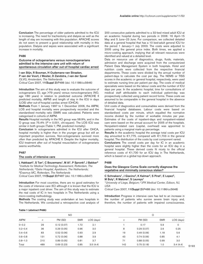
S193
Conclusion The percentage of older patients admitted to the ICUis increasing. The need for tracheotomy and dialysis as well as thelength of stay are increasing with this population. APACHE scoresdo not seem to present a good relationship with mortality in thispopulation. Dialysis and sepsis were associated with a significantincrease in mortality.
P486Outcome of octogenarians versus nonoctogenariansadmitted to the intensive care unit with return ofspontaneous circulation after out-of-hospital cardiac arrest
I van Stijn, R Bosman, H Oudemans-van Straaten, P van der Voort, J Wester, D Zandstra, J van der SpoelOLVG, Amsterdam, The NetherlandsCritical Care 2007, 11(Suppl 2):P486 (doi: 10.1186/cc5646)
Introduction The aim of this study was to evaluate the outcome ofoctogenarians (O, age >79 years) versus nonoctogenarians (NO,age <80 years) in relation to predicted outcome (APACHE IIpredicted mortality, AIIPM) and length of stay in the ICU in days(LOS) after out-of-hospital cardiac arrest (OHCA).Methods From 1 January 1997 to 1 December 2006, the AIIPM,LOS and hospital mortality were prospectively recorded and thestandardised mortality ratio (SMR) was calculated. Patients werecategorised in cohorts of AIIPM.Results Hospital mortality in the NO group was 58.9%, and in theO group was 75.4% (P = 0.001, chi-square). The LOS ICU wassimilar in both groups (Table 1; PM, predicted mortality).Conclusion In octogenarians admitted in the ICU after OHCA,hospital mortality is higher than in the younger group but still animportant proportion survives. Non-octogenarians survived moreoften than predicted by APACHE II. Despite the higher mortality,ICU treatment after out of hospital resuscitation of octogenariansseems worthwhile.
P487The costs of intensive care
L Hakkaart1, S Tan1, C Bouwmans1, M Al1, P Spronk2, J Bakker3
1Institute for Medical Technology Assessment, Rotterdam, TheNetherlands; 2Gelre Hospital, Apeldoorn, The Netherlands;3Erasmus MC, Rotterdam, The NetherlandsCritical Care 2007, 11(Suppl 2):P487 (doi: 10.1186/cc5647)
Introduction For most countries, there are no good estimates forthe costs of intensive care (IC) although it is known that the ICU isa major inpatient cost driver. The aim of this study was to estimatethe real costs of IC in two hospitals in The Netherlands using amicro-costing methodology.Methods The costing study was undertaken at two hospitals inThe Netherlands. We conducted a retrospective cost analysis of
200 consecutive patients admitted to a 32-bed mixed adult ICU atan academic hospital during two periods in 2006: 16 April–15May and 5 June–23 June. For comparison, we collected detaileddata at a general hospital that has a 10 bed-adult general ICU forthe period 1 January–1 July 2003. The costs were adjusted to2005 using the general price index. Both times, we applied amicro-costing approach, implying that all relevant resources wereidentified and valued at a detailed level.Data on resource use of diagnostics, drugs, fluids, materials,admission and discharge were acquired from the computerizedPatient Data Management System in both hospitals. Hotel andnutrition costs were collected from the respective financialdepartments. These costs were divided by the annual number ofpatient-days to calculate the cost per day. The NEMS or TISSscores in the academic or general hospital, respectively, were usedto estimate nursing time per patient per day. The costs of medicalspecialists were based on the labour costs and the number of ICUdays per year. In the academic hospital, time for consultations ofmedical staff attributable to each individual patient-day wasprospectively collected using patient record forms. These costs weassumed to be comparable in the general hospital in the absenceof detailed data.Unit costs of diagnostics and consumables were derived from thefinancial hospital databases. Labour costs were based onstandardised costs per minute, which equalled the normativeincome divided by the number of workable minutes per year.Estimates of the costs of inpatient-days and nonpatient-relatedcare were based on the annual account for 2005 of the hospitals.Nonpatient-related care (capital, overhead) was appointed topatients using a marginal mark-up percentage.Results In the academic hospital the average total costs per ICUday amounted to €1,775, compared with €1,703 in the generalhospital. The distribution of the costs by cost component varied.Conclusions The overall costs per day for IC in an academichospital were slightly higher than the costs for an ICU day in ageneral hospital. These derived costs fit nicely to the officialreference costs of €1,730 for an ICU day in The Netherlands,which is based on a global top-down approach.
P488Does the Glasgow Coma Scale correctly diagnose thevegetative and minimally conscious states?
C Schnakers1, J Giacino2, K Kalmar2, S Piret1, E Lopez2, M Boly1, R Malone2, S Laureys1
1University of Liege, Belgium; 2JFK Medical Center, Edison, NJ,USACritical Care 2007, 11(Suppl 2):P488 (doi: 10.1186/cc5648)
Introduction Progress in intensive care has led to an increase inthe number of patients who survive severe brain injury and,therefore, the number of patients with impaired consciousness.
Available online http://ccforum.com/supplements/11/S2
Table 1 (abstract P486)
NO O
AIIPM n PM (SD) SMR LOS (days) n PM (SD) SMR LOS (days)
0–0.2 29 0.12 (0.07) 1.72 2.1 1 0.17 5.9 0
0.2–0.4 36 0.29 (0.05) 0.96 3.0 8 0.29 (0.07) 2.6 0.25
0.4–0.6 83 0.52 (0.06) 0.93 2.9 19 0.49 (0.06) 1.18 5.6
0.6–0.8 131 0.72 (0.06) 0.88 3.9 43 0.74 (0.06) 0.85 4.1
0.8–1.0 210 0.89 (0.05) 0.81 3.7 71 0.88 (0.05) 0.99 3.0
Total 489 0.69 (0.23) 0.85 3.5 (4.4) 142 0.75 (0.18) 1.0 3.4 (4.4)
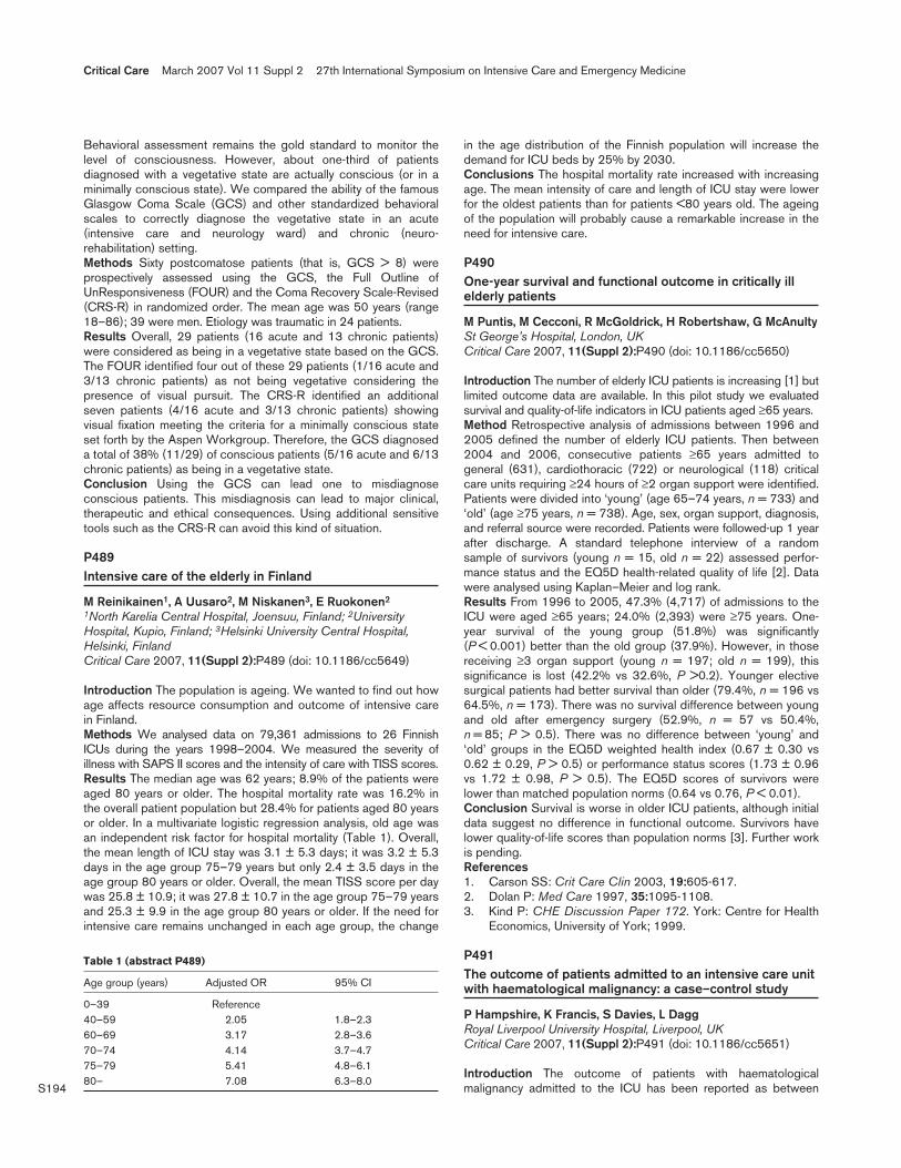
S194
Behavioral assessment remains the gold standard to monitor thelevel of consciousness. However, about one-third of patientsdiagnosed with a vegetative state are actually conscious (or in aminimally conscious state). We compared the ability of the famousGlasgow Coma Scale (GCS) and other standardized behavioralscales to correctly diagnose the vegetative state in an acute(intensive care and neurology ward) and chronic (neuro-rehabilitation) setting.Methods Sixty postcomatose patients (that is, GCS > 8) wereprospectively assessed using the GCS, the Full Outline ofUnResponsiveness (FOUR) and the Coma Recovery Scale-Revised(CRS-R) in randomized order. The mean age was 50 years (range18–86); 39 were men. Etiology was traumatic in 24 patients.Results Overall, 29 patients (16 acute and 13 chronic patients)were considered as being in a vegetative state based on the GCS.The FOUR identified four out of these 29 patients (1/16 acute and3/13 chronic patients) as not being vegetative considering thepresence of visual pursuit. The CRS-R identified an additionalseven patients (4/16 acute and 3/13 chronic patients) showingvisual fixation meeting the criteria for a minimally conscious stateset forth by the Aspen Workgroup. Therefore, the GCS diagnoseda total of 38% (11/29) of conscious patients (5/16 acute and 6/13chronic patients) as being in a vegetative state.Conclusion Using the GCS can lead one to misdiagnoseconscious patients. This misdiagnosis can lead to major clinical,therapeutic and ethical consequences. Using additional sensitivetools such as the CRS-R can avoid this kind of situation.
P489Intensive care of the elderly in Finland
M Reinikainen1, A Uusaro2, M Niskanen3, E Ruokonen2
1North Karelia Central Hospital, Joensuu, Finland; 2UniversityHospital, Kupio, Finland; 3Helsinki University Central Hospital,Helsinki, FinlandCritical Care 2007, 11(Suppl 2):P489 (doi: 10.1186/cc5649)
Introduction The population is ageing. We wanted to find out howage affects resource consumption and outcome of intensive carein Finland.Methods We analysed data on 79,361 admissions to 26 FinnishICUs during the years 1998–2004. We measured the severity ofillness with SAPS II scores and the intensity of care with TISS scores.Results The median age was 62 years; 8.9% of the patients wereaged 80 years or older. The hospital mortality rate was 16.2% inthe overall patient population but 28.4% for patients aged 80 yearsor older. In a multivariate logistic regression analysis, old age wasan independent risk factor for hospital mortality (Table 1). Overall,the mean length of ICU stay was 3.1 ± 5.3 days; it was 3.2 ± 5.3days in the age group 75–79 years but only 2.4 ± 3.5 days in theage group 80 years or older. Overall, the mean TISS score per daywas 25.8 ± 10.9; it was 27.8 ± 10.7 in the age group 75–79 yearsand 25.3 ± 9.9 in the age group 80 years or older. If the need forintensive care remains unchanged in each age group, the change
in the age distribution of the Finnish population will increase thedemand for ICU beds by 25% by 2030.Conclusions The hospital mortality rate increased with increasingage. The mean intensity of care and length of ICU stay were lowerfor the oldest patients than for patients <80 years old. The ageingof the population will probably cause a remarkable increase in theneed for intensive care.
P490One-year survival and functional outcome in critically illelderly patients
M Puntis, M Cecconi, R McGoldrick, H Robertshaw, G McAnultySt George’s Hospital, London, UKCritical Care 2007, 11(Suppl 2):P490 (doi: 10.1186/cc5650)
Introduction The number of elderly ICU patients is increasing [1] butlimited outcome data are available. In this pilot study we evaluatedsurvival and quality-of-life indicators in ICU patients aged ≥65 years.Method Retrospective analysis of admissions between 1996 and2005 defined the number of elderly ICU patients. Then between2004 and 2006, consecutive patients ≥65 years admitted togeneral (631), cardiothoracic (722) or neurological (118) criticalcare units requiring ≥24 hours of ≥2 organ support were identified.Patients were divided into ‘young’ (age 65–74 years, n = 733) and‘old’ (age ≥75 years, n = 738). Age, sex, organ support, diagnosis,and referral source were recorded. Patients were followed-up 1 yearafter discharge. A standard telephone interview of a randomsample of survivors (young n = 15, old n = 22) assessed perfor-mance status and the EQ5D health-related quality of life [2]. Datawere analysed using Kaplan–Meier and log rank.Results From 1996 to 2005, 47.3% (4,717) of admissions to theICU were aged ≥65 years; 24.0% (2,393) were ≥75 years. One-year survival of the young group (51.8%) was significantly(P < 0.001) better than the old group (37.9%). However, in thosereceiving ≥3 organ support (young n = 197; old n = 199), thissignificance is lost (42.2% vs 32.6%, P >0.2). Younger electivesurgical patients had better survival than older (79.4%, n = 196 vs64.5%, n = 173). There was no survival difference between youngand old after emergency surgery (52.9%, n = 57 vs 50.4%,n = 85; P > 0.5). There was no difference between ‘young’ and‘old’ groups in the EQ5D weighted health index (0.67 ± 0.30 vs0.62 ± 0.29, P > 0.5) or performance status scores (1.73 ± 0.96vs 1.72 ± 0.98, P > 0.5). The EQ5D scores of survivors werelower than matched population norms (0.64 vs 0.76, P < 0.01).Conclusion Survival is worse in older ICU patients, although initialdata suggest no difference in functional outcome. Survivors havelower quality-of-life scores than population norms [3]. Further workis pending.References1. Carson SS: Crit Care Clin 2003, 19:605-617.2. Dolan P: Med Care 1997, 35:1095-1108.3. Kind P: CHE Discussion Paper 172. York: Centre for Health
Economics, University of York; 1999.
P491The outcome of patients admitted to an intensive care unitwith haematological malignancy: a case–control study
P Hampshire, K Francis, S Davies, L DaggRoyal Liverpool University Hospital, Liverpool, UKCritical Care 2007, 11(Suppl 2):P491 (doi: 10.1186/cc5651)
Introduction The outcome of patients with haematologicalmalignancy admitted to the ICU has been reported as between
Critical Care March 2007 Vol 11 Suppl 2 27th International Symposium on Intensive Care and Emergency Medicine
Table 1 (abstract P489)
Age group (years) Adjusted OR 95% CI
0–39 Reference40–59 2.05 1.8–2.360–69 3.17 2.8–3.670–74 4.14 3.7–4.775–79 5.41 4.8–6.180– 7.08 6.3–8.0
S195
34% and 75%. Historically, these patients have been regarded ashaving a poor prognosis once admitted to the ICU. We decided tocompare the ICU and hospital mortality of these patients withpatients of a similar age and severity of acute illness.Methods Twenty-four patients were admitted to the ICU fromAugust 2004 to August 2006 with a haematological malignancy.These were case-matched using sex, age (±2 years), APACHE IIscore (±2) and admission diagnosis with patients admitted to theICU without a diagnosis of haematological malignancy. Eighteenpatients were matched to one case control; however, in sixpatients, two matches were found. Where it was impossible todifferentiate between cases on the grounds of diagnosis, age orAPACHE II score they were both included. We compared ICU andhospital mortality between the two groups.Results Patients with a haematological malignancy had an ICUmortality of 50%, and a hospital mortality of 58%. Control patientshad an ICU mortality of 60%, and a hospital mortality of 67%(statistically nonsignificant). The length of time to admissionbetween the two groups was significantly longer in thehaematology group at 12.4 days, compared with 2.8 days in thecontrol patients (P < 0.05). The level of organ support was thesame between the two cohorts.Conclusion We have demonstrated that, for our unit, there was nostatistically significant difference in hospital or ICU mortalitybetween the two groups. In fact, the group with a haematologicalmalignancy had a lower mortality than the control group. Thepresence of haematological malignancy, of itself, does not appearto increase the mortality risk, when compared with a population ofpatients without haematological malignancy of a similar age,APACHE II score and admission diagnosis.
P492Quality of life following prolonged critical illness: insightsfrom a qualitative approach
P RamsayRoyal Infirmary of Edinburgh, UKCritical Care 2007, 11(Suppl 2):P492 (doi: 10.1186/cc5652)
Introduction The measurement of health-related quality of life(HRQoL) among survivors of critical illness has become aprominent feature of our outcomes research. Local researchexperience suggests that existing measures may fail to capture thebroad spectrum of morbidity that survivors experience. Thepurpose of this research is to explore, through predominantlyqualitative methods, critical-illness mediated morbidity amongsurvivors of prolonged critical illness; a group in whom this type ofmorbidity appears to be most prevalent. An important secondaryaim is to explore the contribution of the processes of rehabilitationand recovery to perceptions of HRQoL.Methods Survivors who experienced prolonged critical illness(defined as ≥14 days mechanical ventilation) were identified fromthe Scottish Intensive Care Society Audit Group (SICSAG)database, Wardwatcher®. Participants were contacted ≤6 monthsfollowing ICU discharge, and were invited to completeprofessionally recommended quality-of-life questionnaires (theShort Form 36 and EuroQol-5D) and to participate in a semi-structured interview. Interviews explored everyday experiences ofongoing morbidity and were analysed with regard to their corre-lation with the domains and scores of the HRQoL questionnaires.Purposive sampling provided clinically important insights intoexperiences and perceptions of health and pre-existing morbidityand, importantly, the processes of rehabilitation and recovery; thatis, through comparison of (i) patients with/without appreciable pre-
existing disease and (ii) patients receiving ward-basedrehabilitation with those who receive formalised rehabilitation indedicated facilities.Preliminary results Ten of the required 40 interviewees have sofar been recruited. Preliminary analysis confirms that survivorsexperience a range of morbidity not well captured by professionallyrecommended measures, and that pre-existing disease is animportant factor in both coping with new morbidity superimposedby critical illness, and in marshalling support. The process ofrehabilitation appears to have important effects on perceptions ofrecovery, self-management strategies, and perceptions of HRQoL.Conclusions This qualitative enquiry has already provided, and willcontinue to provide, new and clinically relevant insights intopatients’ experiences of morbidity, the processes of rehabilitation,recovery and perceived HRQoL following discharge into thecommunity.
P493Quality of life before intensive care unit admission is astrong predictor of survival
P Spronk1, J Hofhuis1, H van Stel2, J Rommes1, A Schrijvers2, J Bakker3
1Gelre Ziekenhuizen Location Lukas, Apeldoorn, The Netherlands;2Julius Institute, Utrecht, The Netherlands; 3Erasmus MC,Rotterdam, The NetherlandsCritical Care 2007, 11(Suppl 2):P493 (doi: 10.1186/cc5653)
Objective To examine whether health-related quality of life(HRQOL) before admission to the ICU can be used as a predictorof mortalityDesign and methods A prospective cohort study in an university-affiliated teaching hospital. Patients admitted to the ICU for >48hours were included. Close relatives completed the Short-form 36(SF-36) within the first 48 hours of admission to assess the pre-morbid HRQOL of the patient. Mortality was evaluated from ICUadmittance until 6 months after ICU discharge. Logistic regressionand ROC analysis were used to assess the predictive value formortality of the first general health question of the SF-36 (‘ingeneral would you say the health of your relative is excellent, verygood, good, fair or poor’), as well as HRQOL measured by thecomplete SF-36. The Acute Physiologic and Chronic HealthEvaluation (APACHE) II score was used as an accepted mortalityprediction model in ICU patients. Three models were constructedincluding the HRQOL (model A), APACHE II score (model B), orboth (model C) to age and gender. Percentages of correctsurvival/death predictions were calculated.Results Four hundred and fifty-one patients were included atadmission to the ICU. At 6 months follow-up, 159 patients haddied and 40 patients were lost to follow-up. When the generalhealth item was used as an estimate of HRQOL, the area under thecurve (AUC) for model A (0.719) was comparable with model B(0.721), and slightly better in model C (0.760). The percentage ofwrong predictions was lower in model C (survival 27%; death37%) compared with model A (30% and 41%). Similar resultswere found when using the complete SF-36.Conclusions This study shows that the pre-admission HRQOLmeasured with either the one-item general health question or thecomplete SF-36 are as good in predicting survival/mortality in ICUpatients as the APACHE II score and improves prediction slightlywhen combined. As the one-item general health question is easilyand quickly obtained, assessment of HRQOL before admission tothe ICU may facilitate the decision process in determining whichpatients will benefit from ICU treatment.
Available online http://ccforum.com/supplements/11/S2
S196
P494Five years experience of critical care bereavement follow-up
S Voisey, J Davies, J Parry-Jones, N StallardUniversity Hospital of Wales, Cardiff, UKCritical Care 2007, 11(Suppl 2):P494 (doi: 10.1186/cc5654)
Introduction We believe that by explaining and answering thequestions of those relatives that wish it, we can alleviate the griefcaused by the death of a relative in critical care. Death is acommon outcome of critical illness and relatives are at high risk ofproblems with complicated or unresolved grief due to the sudden,often unexpected, manner of that death. We describe theestablishment and results of a bereavement follow-up service.Methods All relatives, since January 2001, have been offeredbereavement follow-up. Following the death of a patient in criticalcare, relatives are informed of the bereavement follow-up service.After death, a booklet is given to the next of kin with informationabout the service. Four to six weeks later a condolence card issent, which includes a reminder of the follow-up service. We donot offer a counseling service and details of other organizationsproviding this are given. Appointments are made by telephone. Theinterview usually takes place outside critical care and is led by aconsultant and a specially trained nurse. Relatives are given theopportunity to ask questions. A monitoring form is completed. Aletter is sent to the GP detailing the issues discussed.Results In the 30-bed critical care unit in a teaching hospital,during study period January 2001–November 2006, 8,964admissions and 1,560 deaths (17.4% of patients) occurred(male:female ratio 1.27:1). The average age of deaths was 64.7years. Eighty-nine families were seen in bereavement follow-up(5.7% of total deaths) and two families attended for a second visit.The male:female ratio was 2.18:1.0, and the average age of deathwas 42.9 years. A length of stay before death of 1 week or lessoccurred in 59.6%. Forty-seven per cent of families were seenbetween 2 and 4 months after the death of their relative. Theissues most commonly raised were specific questions about thepatient (56.2%), review of information in the notes (34.8%),complaints (23.6%), clarifying misunderstandings (18%) andcontacting other health professionals (21.3%).Conclusions We describe the results of a bereavement follow-upservice for families whose relatives died on the critical care unit atUniversity Hospital of Wales, Cardiff. In total, 5.7% of families tookup the service offered. These were usually the families of youngerthan average male patients, who died with a critical care stay ofless than 1 week. We believe that by answering questions stilltroubling the families 2–4 months after death, we can assist themwith their grief. We also believe that this follow-up service byproviding further communication can resolve some difficult issuesbefore they develop into formal complaints.
P495Quality of life aspects in oncologic patients who survivedan intensive care unit admission
D Forte, O Ranzani, N Stape, F Gianinni, R Cordioli, D Lima, J Coelho, P Nassar, R Zigaib, E Azevedo, I Schimidtbauer, F Silva, B Cordeiro, A Toledo-Maciel, M ParkUniversity of São Paulo, BrazilCritical Care 2007, 11(Suppl 2):P495 (doi: 10.1186/cc5655)
Introduction The number of organ failures in oncologic patientsadmitted to the ICU is a good predictor of mortality. We proposeto analyze the association of this variable and quality of life (QOL)aspects in oncologic patients who survived an ICU admission.
Methods ICU data were prospectively collected from March 2003to November 2005. Oncologic patients were selected. QOLaspects were evaluated through the analysis of independence toaccomplish daily living activities (IADL) after ICU discharge,defined as the patient’s ability to walk, eat by mouth, maintain anoriented conversation and bath himself. The number of organfailures was assessed through the SOFA score. Severe organdysfunction (SOD) was defined if the patient had three or fourpoints in any of the six domains of the SOFA score. According tothe number of SOD, patients were divided into two groups: onewith two or less SODs and another with three or more SODs.Groups were compared using Fisher’s exact test.Results Seventy out of 793 patients had an oncologic diagnosis.The mean age was 52 ± 19 years, male gender 56%, medicaladmission 77%, hematological malignancies 47% and meanAPACHE II score 20 ± 8. ICU mortality was 53% and inhospitalmortality was 71%. Nine (13%) patients were discharged of thehospital with complete IADL. Mortality in the group admitted withthree or more SODs was 100%, while in the other group it was39% (P = 0.01, OR 2.5 95% CI 1.8–3.6). During the ICU stay,patients who developed three or more SODs had a higher ICU andinhospital mortality than the ones who did not (89 x 23%,P < 0.001, OR 7, 95% CI 1.8–26 and 100 x 53%, P < 0.001,OR 2, 95% CI 1.4–2.6) and a smaller proportion of IADL after ICUand hospital discharge (0 x 22%, P = 0.04, OR 1.7, 95% CI1.3–2.2 and 0 x 47%, P < 0.001, OR 2, 95% CI 1.4–2.6).Conclusion Mortality should not be the only aspect analyzed whenconsidering an ICU admission for an oncologic patient. The QOL,including IADL, must be taken into account. A higher number ofSODs during the entire ICU stay is associated with higher ICU andinhospital mortality. Beyond this association, a smaller number ofSODs may be associated with higher probability of IADL.
P496Intensive care unit utilization after esophagectomy
R Iscimen1,2, D Brown2, F Whalen2, T Roy2, S Cassivi2, M Keegan2
1Uludag University Faculty of Medicine, Bursa, Turkey; 2Mayo Clinic College of Medicine, Rochester, MN, USACritical Care 2007, 11(Suppl 2):P496 (doi: 10.1186/cc5656)
Introduction At our institution, postesophagectomy patients areusually managed in a progressive care unit with ICU admissionreserved for those of high acuity. We hypothesized that ICUadmission after esophagectomy is predictable and associated withhigh mortality.Methods A retrospective analysis of all patients afteresophagectomy between January 2000 and June 2004 at a tertiaryreferral center. Data regarding demographics, preoperativemorbidities, perioperative complications, APACHE III predictions,mortality, and lengths of stay were collected.Results Four hundred and thirty-two patients underwentesophagectomy during the study period: 123 (28.5%) wereadmitted to the ICU (ICUGP) and 309 (71.5%) were not(NICUGP). Overall mortality was 3.7% (16 of 432 patients).Fifteen of 123 in ICUGP died in hospital (12.2%) compared withone of 309 in NICUGP. For ICUGP, mean (±standard deviation)acute physiology and APACHE III scores were 41.8 (±16.6) and54.5 (±18.1), respectively. Forty-seven percent of ICUGP had anew (versus pre-existing postoperative) infiltrate on chest X-ray,21.8% had positive sputum/bronchial culture and 5% positiveblood culture within 48 hours of ICU admission. A total 13.8% ofICUGP had ‘aspiration’ documented in physician notes. Themedian (IQR) ICU and hospital lengths of stay were 3.6 (1.7–9.9)
Critical Care March 2007 Vol 11 Suppl 2 27th International Symposium on Intensive Care and Emergency Medicine
S197
and 17.0 (11.3–33.9) days, respectively. Compared with NICUGP,patients in ICUGP were more likely to have developed post-operative arrhythmia (57.9% vs 12.9%, P < 0.001), were older, ofhigher ASA status, and more likely to have diabetes, coronaryartery disease, hypertension, a higher cancer stage, and to havereceived more intraoperative blood products. Of 352 patientsoriginally not sent to the ICU, 43 (12.2%) were subsequentlyadmitted to the ICU. These patients had higher APACHE III scoresand were more likely to have ‘aspiration’ documented, althoughtheir mortality was not higher than direct ICU admissions.Conclusions After esophagectomy, overall mortality is low, butmany patients require ICU admission. Postoperative arrhythmiasand aspiration pneumonitis are especially problematic.
P497Visiting policies in Italian intensive care units: a nationalsurvey
A Giannini1, S Leoncino1, G Miccinesi21Fondazione IRCCS Ospedale Maggiore Policlinico, Mangiagalli eRegina Elena, Milan, Italy; 2Centro per lo Studio e la PrevenzioneOncologica, Florence, ItalyCritical Care 2007, 11(Suppl 2):P497 (doi: 10.1186/cc5657)
Introduction Today no published data are as yet available onvisiting policies in Italy’s approximately 600 ICUs. We carried out amulticentre survey to evaluate visiting policies in Italian ICUs.Methods An email questionnaire was sent to all 303 ICUs (generaland specialized) in the Italian collaborative Gruppo Italiano per laValutazione degli Interventi in Terapia Intensiva, asking about theirvisiting policies.Results The response rate was 84.8% (257/303). The mediandaily visiting time was 60 minutes (10th percentile: 30 minutes;90th percentile: 120 minutes); however, 2% of ICUs allowed novisiting whatsoever. A total 54.8% of ICUs surveyed had only onedaily visiting slot, and 44% two. Only 1.2% had more than twovisiting slots. The number of visitors was restricted in 91.8% ofICUs. The type of visitors (immediate family only) was restricted in17.5% of ICUs. Children were not permitted to visit in 69.1% ofICUs and 17.5% had a minimum age limit for visitors. In the case ofa dying patient, 20.6% of ICUs did not alter the visiting policy;49% extended visiting hours; 44% increased the number of slots;and 53% allowed more visitors. A gowning procedure wascompulsory for visitors in 95.3% of ICUs. No waiting room wasprovided by 25.4% of ICUs.Conclusions This is the first survey on visiting policies in ItalianICUs. Despite the widely-held conviction that there is no soundscientific basis for restricting visitors in ICUs [1-3], our findingsshow a clear tendency in Italian ICUs to apply restricted visitingpolicies (concerning visiting hours, number and type of visitors),which are only partially liberalized when the patient is dying. Oursurvey could contribute towards modifying current policies infavour of opening ICUs that are still ‘closed’ and promoting moreappropriate and attentive care for the patient and his/her family.Acknowledgement We thank Gruppo Italiano per la Valutazionedegli Interventi in Terapia Intensiva for their valuable help.References1. Burchardi H: Let’s open the door! Intensive Care Med 2002,
8:1371-1372.2. Berwick DM, et al.: Restricted visiting hours in ICUs: time
to change. JAMA 2004, 292:736-737.3. Slota M, et al.: Perspectives on family-centered, flexible
visitation in the intensive care unit setting. Crit Care Med2003, 31(Suppl):S362-S366.
P498Communication with patients during ward rounds on theintensive care unit: a prospective, observational, semiblindstudy
K Francis, D Langhor, J Walker, I WeltersRoyal Liverpool University Hospital, Liverpool, UKCritical Care 2007, 11(Suppl 2):P498 (doi: 10.1186/cc5658)
Introduction It is good medical practice to communicate withpatients regarding their condition and proposed treatment. Com-munication is essential to allow them to express their concerns andexercise their own autonomy, in a situation where they otherwisemay have little control. Poor communication may contribute tounnecessary anxiety or depression. The aim of this audit was toobserve whether the doctors communicated with patients on wardrounds.Methods We audited 28 ICU ward rounds. It was noted whetherthe patient was sedated or not sedated, and whether they had theireyes open or closed. Negative comments were recorded, anddefined as comments regarding a patient’s poor progress orprognosis made at the bedside during the ward round. We alsonoted whether the patient was informed that the ward round was inprogress, whether they were informed of the plan for the day, andwhether any reassurance was offered. The other members of theteam were not aware of the audit.Results Twenty-eight ward rounds were audited, a total of 328patient reviews. The total number of patients with their eyes openwas 171, while 157 had their eyes closed. An adverse commentwas made during 8.2% of reviews within earshot of the patient. Anegative comment was made on 27 episodes, 13 of these werewhen the patient was not sedated but had their eyes closed. Fourepisodes occurred when the patient was not sedated and had theireyes open, while 10 were sedated and had their eyes closed. Ofthe 171 patients with their eyes open, 74 (43%) were informedthat we were the team doing the ward round, while 97 (57%) werenot spoken to at the start of the review. Fifty-three (31%) wereinformed of our management plan for the day and 43 (25%) wereoffered reassurance. In total, 103/171 patients (60%) with theireyes open were spoken to during the ward round, and we did notspeak to 68 (40%).Conclusion On intensive care ward rounds, patients are often notengaged in conversations or allowed to express their concerns.The majority of patients were not offered reassurance or informedabout the treatment plan. Improved communication skills may helpto lessen patients’ anxiety and unnecessary stress both during theirstay on ICU and following their discharge.
P499Hospital volume and outcome following mechanicalventilation on the intensive care unit
S Gopal1, R O’Brien1, J Pooni1, S Macfarlane2
1The Royal Wolverhampton Hospitals NHS Trust, Wolverhampton,UK; 2ICNARC, Birmingham, UKCritical Care 2007, 11(Suppl 2):P499 (doi: 10.1186/cc5659)
Introduction The study was undertaken to determine therelationship between hospital volume and mortality in mechanicallyventilated adult medical and surgical patients. The regionalisationof adult critical care services has been suggested by healthcareproviders and health policy-makers as a means of achieving theideal balance of providing high-quality care that is both cost-effective and accessible. Recent studies have had conflictingresults.
Available online http://ccforum.com/supplements/11/S2
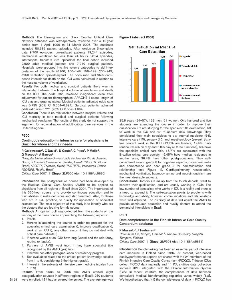
S198
Methods The Birmingham and Black Country Critical CareNetwork database was retrospectively reviewed over a 10-yearperiod from 1 April 1996 to 31 March 2006. The databaseincluded 50,686 patient episodes. After exclusion (incompletedata 9,700 episodes, unventilated patients 19,244 episodes,mechanical ventilation for less than 24 hours 3,814 episodes,interhospital transfers 795 episodes) the final cohort included9,920 adult medical patients and 7,210 surgical patients.Hospitals were grouped into five volume categories to aid inter-pretation of the results (<100; 100–149; 150–199; 200–249;≥250 ventilation episodes/year). The odds ratio and 95% confi-dence intervals for death on the ICU were calculated in relation tothe hospital volume of ventilation.Results For both medical and surgical patients there was norelationship between the hospital volume of ventilation and deathon the ICU. The odds ratio remained insignificant even afteradjustment for patient demographics, APACHE II score, length ofICU stay and urgency status. Medical patients’ adjusted odds ratiowas 0.735 (95% CI 0.604–0.894). Surgical patients’ adjustedodds ratio was 0.771 (95% CI 0.559–1.064).Conclusion There is no relationship between hospital volume andICU mortality in both medical and surgical patients followingmechanical ventilation. The results of this study do not support theargument for regionalisation of adult critical care services in theUnited Kingdom.
P500Continuous education in intensive care for physicians inBrazil: for whom and their needs
R Goldwasser1, C David1, D Costa2, C Piras3, P Mello4, G Macedo5, A Barros6
1Hospital Universitario-Universidade Federal do Rio de Janeiro,Brazil; 2Hospital Universitario, Cuiaba, Brazil; 3SOESTI, Vitoria,Brazil; 4SOTIPI, Terezina, Brazil; 5SOTIERJ, Vassouras, Brazil;6SOTIPE, Recife, BrazilCritical Care 2007, 11(Suppl 2):P500 (doi: 10.1186/cc5660)
Introduction The postgraduation course had been developed bythe Brazilian Critical Care Society (AMIB) to be applied tophysicians from all regions of Brazil since 2004. The importance ofthis 360-hour course is to provide continuous education and totrain abilities to make decisions and start treatment. Also, for thosewho are in ICU practice, to qualify for application of specialistexamination. The main objective of this study is to identify who arethe doctors that are looking for this course.Methods An opinion poll was collected from the students on thefirst day of the class course approaching the following aspects:1. Profile.2. He/she is attending the course in order to: prepare for the
specialist critical care examination (), improve qualification (),work at an ICU (), any other reason if they do not deal withcritical care patients ().
3. If he/she works at an ICU: how long (years) and the role (duty,routine or leader).
4. Partners of AMIB (yes) (no); if they have specialist titlerecognized by the AMIB (yes) (no).
5. If he/she has other postgraduation or residency program.6. Self-evaluation related to the critical patient knowledge (scales
from 1 to 9, considering 9 the highest grade).7. Interest in the subjects of intensive care medicine (scales from
1 to 9).Results From 2004 to 2005 the AMIB started eightpostgraduation courses in different regions of Brazil; 250 studentswere enrolled; 184 had answered the survey. The average age was
35.8 years (24–57); 133 men, 51 women. One hundred and fivestudents are attending the course in order to improve theirqualification, 87 are studying for the specialist title examination, 58to work in the ICU and 47 to acquire new knowledge. Theyconsidered their main specialties to be: internal medicine (54),intensive care (15), surgery (10) and anesthesiology (seven). Sixty-five percent work in the ICU (12.7% are leaders, 19.5% dailyroutine, 86.4% on duty and 6.8% play all three functions), 6% havethe specialist critical care title, 15.7% are associated with theBrazilian critical care society, 49.45% have medical residence inanother area, 36.4% have other postgraduations. They self-considered around grade 6 for cognitive aspects, procedural skillsand competence and near grade 8 for communication andrelationship (see Figure 1). Cardiopulmonary resuscitation,mechanical ventilation, haemodynamics and neurointensivism arethe most desirable subjects.Conclusions Doctors are mainly from the fourth decade, want toimprove their qualification, and are usually working in ICUs. Thelow number of specialists who works in ICU is a reality and there isa need to expand it. The self-evaluation points to problems withknowledge and ability; however, communications and relationshipswere well adjusted. The diversity of data will assist the AMIB toprovide continuous education and qualify doctors to attend thedemand of intensivists in Brazil.
P501Data completeness in the Finnish Intensive Care QualityConsortium database
P Mussalo1, J Tenhunen2
1Intensium Ltd, Kuopio, Finland; 2Tampere University Hospital,Tampere, FinlandCritical Care 2007, 11(Suppl 2):P501 (doi: 10.1186/cc5661)
Introduction Benchmarking has been an essential part of intensivecare medicine in Finland since 1994. At present, web-basedquality/performance reports are shared with the 24 members of theFinnish Intensive Care Quality Consortium (FICQC). Thirteen ICUscollect FICQC data manually and 11 ICUs utilize data collectionsoftware (IVT) integrated with the Clinical Information System(CIS). In recent literature, the completeness of data betweencentralized medical benchmarking registries varies widely [1,2].We hypothesized that: (1) the completeness of data in FICQC has
Critical Care March 2007 Vol 11 Suppl 2 27th International Symposium on Intensive Care and Emergency Medicine
Figure 1 (abstract P500)
S199
increased over the years and (2) the variation between the differentunits still exists.Methods We assessed data completeness of a Finnish ICU qualitybenchmarking database from 1998 to March 2006 containing93,964 admission records. The data completeness was defined asa ratio of available and required data at ICU admission level. Weevaluated the dataset and selected 19 most significant admissionscheme variables to be included in completeness ratio calculations.Results The majority of data (77.5%) was collected with themanual system and the remaining 22.5% with an integrationsoftware. The mean admission data completeness ratio (CR)increased from 85.3% at 1998 to 97.9% at 2005 (P = 0.01).Between the ICUs, the mean CR varied from 91.6% to 99.6%(P = 0.01). The mean CR of the data collected with the IVT was98.7% and with the manual system was 95.1% (P = 0.01). Therate of 100% complete records per patient was 48.7% and itincreased from 0.0% in 1998 to 71% in 2005.Conclusion Data completeness in the FICQC has improved duringthe study period, although there is still significant variation betweenICUs. Improved data completeness and decreased proportion ofmissing data are most likely due to the increasingly common use ofCIS and automated data collection. We conclude thatmeasuring/reporting the amount of missing data is mandatorywhen data collection and data management procedures forbenchmarking are being developed.References1. Arts D, et al.: Intensive Care Med 2002, 28:656-659.2. Afessa B, et al.: Intensive Care Med 2005, 31:1537-1543.
P502Analysis of critical incidents during the interhospitaltransport of critically ill patients
P Doherty, B DigbyWestern Infirmary, Glasgow, UKCritical Care 2007, 11(Suppl 2):P502 (doi: 10.1186/cc5662)
Introduction In this study we created a database to analyse theincidence and types of critical incidents that occurred during theinterhospital transfer of critically ill patients. The transfer of criticallyill patients presents important risks and the safety of patients hasbeen shown to be facilitated by the development of standardequipment and specialist teams [1]. The West of Scotland Shockteam is a designated regional transfer service based in Glasgow.We are involved in the interhospital transfer of patients and notprimary retrieval. A recent study looking at critical incidents duringthe intrahospital transport of the critically ill highlighted the risksposed, and recommended the monitoring of incidents in order toaid the continuous improvement in patient safety [2]. No similarstudy has been carried out looking at the interhospital transport ofthe critically ill patient.Methods The study was a cross-sectional analysis of criticalincidents occurring during interhospital transport that were reportedto the West of Scotland Shock Team critical incident database setup in September 2005. The information obtained was categorisedinto: (a) where the incident took place, (b) type of incident, (c)written description of events, (d) outcome (potential or actual harmto the patient) and (e) designation of the staff member reportingthe incident.Results A total of 199 transfers were performed over the 6-monthperiod. Thirty-four critical incidents were reported. Twenty-four(70%) incidents took place before, seven (21%) during and three(9%) after transfer. No patients sustained actual harm, 29 (85%)were perceived by the reporter to have suffered potential harm andthe most common cause of this were delays in the transfer. No
potential or actual harm was perceived in five (15%) of theincidents. Fifty-three per cent of events were reported by seniorhouse officer grade and 47% of incidents were reported by aspecialist registrar. Only one incident was reported by a nurse onthe team.Conclusions Interhospital transport of critically ill patients canpose important risks. In our study no actual patient harm occurredalthough most incidents had the potential to cause harm. Themajority of incidents were caused by system-based factors. Thisdatabase has allowed us to perform continuous servicedevelopment and education of staff.References1. Faculty of Intensive Care and Australasian College for Emer-
gency Medicine: Minimum Standards for the Transport of theCritically Ill Patient; 2003 [http://www.acem.org.au/media/policies_and_guidelines/min_standard_crit_ill.pdf]
2. Beckmann U, Gillies D, Berenholtz S, et al.: Incidents relat-ing to the intra-hospital transfer of critically ill patients.Intensive Care Med 2004, 30:1579-1585.
P503Maximisation of heart and lung donation in a neurosurgicalintensive care unit
S Aerdts, J HofstraIsala Klinieken, Zwolle, The NetherlandsCritical Care 2007, 11(Suppl 2):P503 (doi: 10.1186/cc5663)
Introduction The number of heart-beating donors in The Netherlandsis decreasing. This decrease is only partially compensated for byan increase of nonheart-beating donations, resulting in anincreasing shortage of donor organs, especially of donor heartsand lungs. In 2005 compared with 2004 the number of patientswaiting for a donor heart increased from 38 to 50 (32%). In lungdonation the increase was 37% (from 79 to 108). One approachto reduce this shortage is to maximize the number of organs perdonor by optimisation of donor treatment in the ICU.Methods We investigated the possibilities for improvement ofdonor management in our ICU by a retrospective study in 37 heart-beating organ donors hospitalised in our ICU from 1993 to 2005.There was no protocol for the treatment of organ donors in ourinstitution.Results The heart was donated in 18 of 37 patients (49%). Lungdonation was possible in only eight of 37 donors (22%). Mosthearts and lungs were rejected for transplantation for validreasons. In some patients there was room for improvement: in twoof the three cases where hemodynamic instability impeded heartand lung donation (one dying from subarachnoidal bleeding andone from ischemic cerebral infarction), hemodynamic instabilitywas closely associated with the moment of cerebral death. In threefurther patients heart donation was not carried out because of wallmovement abnormality or electrocardiogram abnormalities. None of
Available online http://ccforum.com/supplements/11/S2
Table 1 (abstract P502)
Causes of all incidents (irrespective of patient outcome)
Communication problem 10 (29%)
Organisational delay 7 (21%)
Lack of staff 2 (6%)
Equipment failure 9 (26%)
Poor preparation of patient 5 (15%)
Staff injury 1 (3%)
S200
them had previous cardiac disease. All three had disturbances incardiac rhythm closely related to the occurrence of cerebral death.Two of these three patients also developed neurogenic pulmonaryedema. We speculate that in all five patients the instability leadingto the decision not to perform heart and lung donation was causedby excessive sympathic stimulation at the time of cerebral deathleading to impaired myocardial function and neurogenic pulmonaryedema. These disturbances may be reversible within a few hours.Inotropic therapy, judicious fluid administration guided by closehemodynamic monitoring together with a trial of treatment withtriple hormonal therapy (corticosteroid, vasopressin and thyroidhormone) might have improved cardiac and pulmonary function,rendering heart and lung donation possible.Conclusion On the basis of this retrospective study we concludethat donor management in our ICU can be improved. Amanagement protocol with special attention for treatment ofdisturbance in cardiac and pulmonary function caused bysympathic overstimulation might considerably increase theamount of hearts and lungs donated, contributing to a decreasein organ shortage.
P504Medical practices during the last 48 hours of life inchildren admitted to seven Brazilian Pediatric intensivecare units
P Lago, J Piva, P GarciaPUCRS, Porto Alegre, BrazilCritical Care 2007, 11(Suppl 2):P504 (doi: 10.1186/cc5664)
Introduction During the last decades life support limitation (LSL)practices have been offered more frequently in Latin Americanpediatric intensive care units (PICUs). We hypothesize that,depending on the Brazilian region, the incidence of LSL and themedical management may differ.Objective To evaluate the incidence of LSL practices and themedical management during the last 48 hours of life of childrenadmitted to seven PICUs located in regions of Brazil.Methods A multicenter, observational and retrospective chartreview study. The medical chart of all deaths occurring betweenJanuary 2003 and December 2004 in seven Brazilian PICUslocated in Porto Alegre (two), Sao Paulo (two) and Salvador(three) were evaluated. Two pediatric intensive care residents ofeach service filled a standard protocol searching for: demographicdata, mode of death (full reanimation, nonreanimation orders orwithdrawn treatment) and medical management during the last 48hours of life. Student’s t test, analysis of variance, chi-square testand relative risk were used for comparing the data.Results There were 561 deaths, 36 being excluded that died withless than 24 hours, 61 with brain death and 36 missing charts. Fullcardiopulmonary reanimation was offered to 56.5%, withdifferences between the northeast and southeast regions (P <0.001). Higher age (P = 0.02) and long length of PICU stay wereassociated with nonreanimation orders. The plan for LSL wasrecorded in a clear manner in just 52.7%. No respiratory supportwas observed in 14 dying children. For 66% patients with do-not-resuscitate orders the inotrope drugs were maintained orincreased in the last 48 hours.Conclusions The incidence of LSV has increased among theBrazilian PICUs with a difference between the regions. Thenonreanimation order is still the most common practice with scarceinitiative for withdrawn life support.
P505End-of-life care in the critically ill: a description ofknowledge, attitudes and practices of physicians andnurses from Karachi, Pakistan
N Salahuddin, A Ahmed, S ShafquatAga Khan University & Hospital, Karachi, PakistanCritical Care 2007, 11(Suppl 2):P505 (doi: 10.1186/cc5665)
Introduction As the numbers of people admitted to ICUs areincreasing, physicians are faced with obligations beyondattempting to reverse illness and include providing quality end-of-life care. Barriers to this include inadequate understanding of thedying patient and withdrawal or limitation of care. The objectives ofthis study were to document the comprehensions of physiciansand nurses dealing with these situations.Methods We carried out a cross-sectional survey of cliniciansworking at three hospitals in Karachi (one private, universityhospital, one mixed public and private, tertiary care hospital andone large government-funded hospital). A 13-question instrumentwas developed to assess recognition of end-of-life in the ICU,knowledge of commonly used terms to describe limitations of care,and attitudes and practices towards withdrawal and limitation oflife-support measures and organ harvest for transplantation. Aftermeasuring the frequencies for presentation of the data, differencesbetween the three respondent subgroups were compared using achi-square analysis. Fisher’s exact test was used where theindividual cell count was <5. A one-way analysis of variance wasused to compare differences in age and years of practice. A two-sided P value of <0.05 was considered statistically significant.Results A total of 137 physicians and critical care nursescompleted the survey. The average age was 34 years and 58%were males. ‘Brain death’ was defined as an ‘irreversible cessationof brainstem function’ by 85% of respondents; 77% relying onclinical examination, 49.6% consulting neurophysicians and 28.3%ordering further testing to confirm the diagnosis. Withdrawal of lifesupport is practiced by 83.2%; most frequently in the setting ofabsent brainstem and cortical functioning (74.3%), followed byacute, progressive multiorgan failure (39.8%). Physicians are morelikely (P value 0.000) to withdraw mechanical ventilation,compared with nurses who would withdraw vasopressors (P value0.006). The primary physician is the most frequent caregiver(60.2%) to start a discussion on withdrawal of life support, with72.6% respondents consulting the Hospital Ethics Committee.Only 13.3% respondents never withdraw life support; 28.3%considered it their responsibility to ‘sustain life at all costs’ and only8% gave religious beliefs as a reason. Only 56.6% favored organharvest for transplantation from cadavers, while 64.6% supportedharvest from brain dead individuals. Nurses were significantly morelikely to support organ harvest for transplant from heart-beating,brain dead individuals (P value 0.025) than cadavers (P value0.000).Conclusion There are deficiencies and disparities in theunderstandings of physicians and nurses on the recognition andmanagement of end-of-life in the ICU.
Critical Care March 2007 Vol 11 Suppl 2 27th International Symposium on Intensive Care and Emergency Medicine
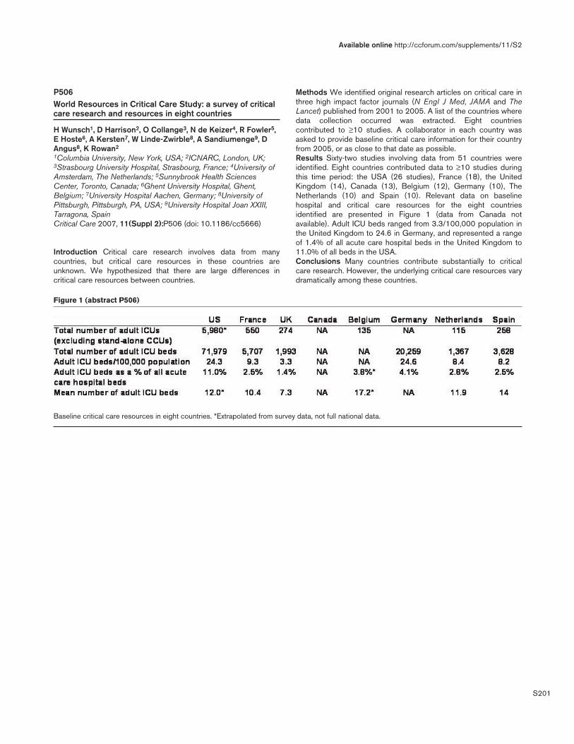
S201
P506World Resources in Critical Care Study: a survey of criticalcare research and resources in eight countries
H Wunsch1, D Harrison2, O Collange3, N de Keizer4, R Fowler5,E Hoste6, A Kersten7, W Linde-Zwirble8, A Sandiumenge9, DAngus8, K Rowan2
1Columbia University, New York, USA; 2ICNARC, London, UK;3Strasbourg University Hospital, Strasbourg, France; 4University ofAmsterdam, The Netherlands; 5Sunnybrook Health SciencesCenter, Toronto, Canada; 6Ghent University Hospital, Ghent,Belgium; 7University Hospital Aachen, Germany; 8University ofPittsburgh, Pittsburgh, PA, USA; 9University Hospital Joan XXIII,Tarragona, SpainCritical Care 2007, 11(Suppl 2):P506 (doi: 10.1186/cc5666)
Introduction Critical care research involves data from manycountries, but critical care resources in these countries areunknown. We hypothesized that there are large differences incritical care resources between countries.
Methods We identified original research articles on critical care inthree high impact factor journals (N Engl J Med, JAMA and TheLancet) published from 2001 to 2005. A list of the countries wheredata collection occurred was extracted. Eight countriescontributed to ≥10 studies. A collaborator in each country wasasked to provide baseline critical care information for their countryfrom 2005, or as close to that date as possible.Results Sixty-two studies involving data from 51 countries wereidentified. Eight countries contributed data to ≥10 studies duringthis time period: the USA (26 studies), France (18), the UnitedKingdom (14), Canada (13), Belgium (12), Germany (10), TheNetherlands (10) and Spain (10). Relevant data on baselinehospital and critical care resources for the eight countriesidentified are presented in Figure 1 (data from Canada notavailable). Adult ICU beds ranged from 3.3/100,000 population inthe United Kingdom to 24.6 in Germany, and represented a rangeof 1.4% of all acute care hospital beds in the United Kingdom to11.0% of all beds in the USA.Conclusions Many countries contribute substantially to criticalcare research. However, the underlying critical care resources varydramatically among these countries.
Available online http://ccforum.com/supplements/11/S2
Figure 1 (abstract P506)
Baseline critical care resources in eight countries. *Extrapolated from survey data, not full national data.
Related Documents