10.1128/AEM.71.4.1729-1736.2005. 2005, 71(4):1729. DOI: Appl. Environ. Microbiol. Dhana Rao, Jeremy S. Webb and Staffan Kjelleberg Pseudoalteromonas tunicata Biofilms Containing the Marine Bacterium Competitive Interactions in Mixed-Species http://aem.asm.org/content/71/4/1729 Updated information and services can be found at: These include: REFERENCES http://aem.asm.org/content/71/4/1729#ref-list-1 at: This article cites 60 articles, 28 of which can be accessed free CONTENT ALERTS more» articles cite this article), Receive: RSS Feeds, eTOCs, free email alerts (when new http://journals.asm.org/site/misc/reprints.xhtml Information about commercial reprint orders: http://journals.asm.org/site/subscriptions/ To subscribe to to another ASM Journal go to: on November 12, 2013 by guest http://aem.asm.org/ Downloaded from on November 12, 2013 by guest http://aem.asm.org/ Downloaded from

Welcome message from author
This document is posted to help you gain knowledge. Please leave a comment to let me know what you think about it! Share it to your friends and learn new things together.
Transcript
10.1128/AEM.71.4.1729-1736.2005.
2005, 71(4):1729. DOI:Appl. Environ. Microbiol. Dhana Rao, Jeremy S. Webb and Staffan Kjelleberg Pseudoalteromonas tunicataBiofilms Containing the Marine Bacterium Competitive Interactions in Mixed-Species
http://aem.asm.org/content/71/4/1729Updated information and services can be found at:
These include:
REFERENCEShttp://aem.asm.org/content/71/4/1729#ref-list-1at:
This article cites 60 articles, 28 of which can be accessed free
CONTENT ALERTS more»articles cite this article),
Receive: RSS Feeds, eTOCs, free email alerts (when new
http://journals.asm.org/site/misc/reprints.xhtmlInformation about commercial reprint orders: http://journals.asm.org/site/subscriptions/To subscribe to to another ASM Journal go to:
on Novem
ber 12, 2013 by guesthttp://aem
.asm.org/
Dow
nloaded from
on Novem
ber 12, 2013 by guesthttp://aem
.asm.org/
Dow
nloaded from
APPLIED AND ENVIRONMENTAL MICROBIOLOGY, Apr. 2005, p. 1729–1736 Vol. 71, No. 40099-2240/05/$08.00�0 doi:10.1128/AEM.71.4.1729–1736.2005Copyright © 2005, American Society for Microbiology. All Rights Reserved.
Competitive Interactions in Mixed-Species Biofilms Containing theMarine Bacterium Pseudoalteromonas tunicata
Dhana Rao, Jeremy S. Webb, and Staffan Kjelleberg*School of Biotechnology and Biomolecular Sciences and Centre for Marine Biofouling and Bio-Innovation,
University of New South Wales, Sydney, Australia
Received 20 August 2004/Accepted 4 November 2004
Pseudoalteromonas tunicata is a biofilm-forming marine bacterium that is often found in association with thesurface of eukaryotic organisms. It produces a range of extracellular inhibitory compounds, including anantibacterial protein (AlpP) thought to be beneficial for P. tunicata during competition for space and nutrientson surfaces. As part of our studies on the interactions between P. tunicata and the epiphytic bacterialcommunity on the marine plant Ulva lactuca, we investigated the hypothesis that P. tunicata is a superiorcompetitor compared with other bacteria isolated from the plant. A number of U. lactuca bacterial isolates were(i) identified by 16S rRNA gene sequencing, (ii) characterized for the production of or sensitivity to extracel-lular antibacterial proteins, and (iii) labeled with a fluorescent color tag (either the red fluorescent proteinDsRed or green fluorescent protein). We then grew single- and mixed-species bacterial biofilms containingP. tunicata in glass flow cell reactors. In pure culture, all the marine isolates formed biofilms containingmicrocolony structures within 72 h. However, in mixed-species biofilms, P. tunicata removed the competingstrain unless its competitor was relatively insensitive to AlpP (Pseudoalteromonas gracilis) or produced stronginhibitory activity against P. tunicata (Roseobacter gallaeciensis). Moreover, biofilm studies conducted with anAlpP� mutant of P. tunicata indicated that the mutant was less competitive when it was introduced intopreestablished biofilms, suggesting that AlpP has a role during competitive biofilm formation. When single-species biofilms were allowed to form microcolonies before the introduction of a competitor, these microcolo-nies coexisted with P. tunicata for extended periods of time before they were removed. Two marine bacteria(R. gallaeciensis and P. tunicata) were superior competitors in this study. Our data suggest that this dominancecan be attributed to the ability of these organisms to rapidly form microcolonies and their ability to produceextracellular antibacterial compounds.
Biofouling is ubiquitous in the marine environment, andbacteria are among the first organisms to foul surfaces. Theyform biofilms which serve as a focus for the attachment andgrowth of other organisms, such as invertebrates, sessile plants,and animals (15). Mature marine biofouling communities arecomplex, highly dynamic ecosystems and, once established, areextremely difficult to eradicate (28).
Many marine organisms have evolved efficient strategies tocombat epibiosis. Seaweeds employ a number of physical andchemical defense systems to prevent fouling, such as the shed-ding of outer layers of cells (35) or production of inhibitorycompounds (17). However, antifouling defense is costly (57),and it has been hypothesized that the seaweed Ulva lactuca,which has neither physical nor chemical defenses, relies onmicrobial defense (19, 32). It has been demonstrated that bac-terial biofilms are present on the surface of U. lactuca (53), andit has been shown that biofilms can be beneficial for their hostsby enhancing their antifouling strategies (2, 7, 16, 24, 30, 46).
One important group of marine bacteria that is found inassociation with living surfaces is Pseudoalteromonas spp. Thisgenus produces a diverse range of biologically active com-pounds that specifically target marine fouling organisms. Per-haps the most extensively studied species in the genus is Pseu-
doalteromonas tunicata. P. tunicata is a green-pigmented gram-negative gamma-proteobacterium. It colonizes living marinesurfaces, including U. lactuca (29), and produces at least fivenovel inhibitory compounds. One of these compounds is a190-kDa multisubunit antibacterial protein designated AlpPwhich is effective against both gram-negative and gram-positivebacteria from a range of environments (33). However, thisprotein was also found to be active against P. tunicata itself,which raises a question about the ecological role of AlpP. It hasbeen speculated that AlpP might provide a competitive advan-tage to P. tunicata during biofilm growth in the marine envi-ronment.
P. tunicata has been shown to form complex differentiatedbiofilms (39). Biofilm formation appears to typically follow asequence. Single cells attach to a surface and differentiate intomatrix-enclosed microcolonies separated by a network of openwater channels. Dispersal of bacteria from the interior regionsof the microcolonies has been observed, resulting in formationof hollow voids inside the microcolonies (39). Microcolonydevelopment is thought to be a coordinated adaptive responsethat facilitates continued biofilm development and dispersal(60). Very little is known about the role of microcolonieswithin biofilms. Recent findings suggest that the formation ofmicrocolonies by Pseudomonas aeruginosa allows it to escapepredation by flagellates (41). Generally, microcolonies may bean adaptive strategy for competing under stressful conditions.
The microcolonies that constitute a biofilm can be composedof single-species populations or mixed populations with various
* Corresponding author. Mailing address: School of Biotechnologyand Biomolecular Sciences and Centre for Marine Biofouling andBio-Innovation, Biological Sciences Building, University of New SouthWales, Sydney, NSW 2052, Australia. Phone: 61 (2) 9385 2102. Fax: 61(2) 9385 1779. E-mail: [email protected].
1729
on Novem
ber 12, 2013 by guesthttp://aem
.asm.org/
Dow
nloaded from
degrees of interaction, depending on the environmental con-ditions under which they were formed. Two species formedmixed microcolonies when they were grown under commensalconditions and formed separate microcolonies under noncom-mensal conditions (48). Thus, commensal relationships canplay a role in determining the spatial distribution of the organ-isms in microbial communities. Meanwhile, in competitive in-teractions between bacteriocin-producing and bacteriocin-sen-sitive strains, the bacteria formed biofilms in which each strainformed its own microcolonies (55). Generally, surprisingly lit-tle is known about the factors that govern the establishmentand distribution of bacteria within multispecies biofilms inmarine environments. A few studies on epiphytic microbialcommunities present on macroalgae have highlighted the com-plex spatial distribution of bacterial populations (12), but thestrategies that marine bacteria use to colonize surfaces and tocompete with other bacteria are poorly understood.
In this study we investigated the hypothesis that P. tunicatacompetes effectively with other marine bacterial isolates duringbiofilm formation based on the isolation of this organism frommarine plants in repeated sampling and based on the produc-tion of the potent antibacterial compound AlpP by this species.We observed that in pure cultures all the marine isolatesformed stable biofilms within 72 h. P. tunicata was found to bethe dominant isolate and totally removed a competing strainunless the competitor was not sensitive to the P. tunicata an-tibacterial protein or exhbited strong inhibitory activity againstP. tunicata. In competition studies in which microcolonies wereallowed to form before the introduction of P. tunicata, biofilmscoexisted for greatly extended periods. Studies with a P. tuni-cata alpP mutant suggested that AlpP provides an advantageduring colonization of biofilms formed by other marine bacte-ria. Our data also suggest that microcolonies may be protectivestructures during biofilm development that enhance the per-sistence of an organism during competitive interactions.
MATERIALS AND METHODS
Isolation of marine strains. P. tunicata and other isolates were originallyisolated from the surface of the common marine alga U. lactuca, which wascollected from the rocky intertidal zone near Sydney, Australia. The algal thalluswas suspended in sterile nine-salt solution, and surface bacteria were removed byvortexing. Aliquots of the samples were then spread on the complex marinemedium Vaatanen nine-salt solution (VNSS) (40) containing 1.5% agar andincubated at 23°C for 48 h. Morphologically distinct bacterial colonies wereselected. Bacteria were stored at �70°C in 30% glycerol. While over 50 differentcolony phenotypes were observed, only 20 strains survived repeated subculturingon VNSS. These strains were routinely grown and maintained on VNSS agar at25°C. Sequencing of these strains was part of another project and was carried outby Tujula et al. (N. Tujula, J. S. Webb, I. Dahllof, C. Holmstrom, and S.Kjelleberg, unpublished data). Four of the 20 culturable strains were chosen inaddition to P. tunicata, as these strains are commonly found growing on algae.The strains were Pseudoalteromonas gracilis, Alteromonas sp., Cellulophaga fuci-cola, and Roseobacter gallaeciensis strains.
Fluorescent labeling of marine isolates. Marine strains were labeled with agreen fluorescent protein (GFP) color tag by transconjugation by using theconstitutive GFP expression plasmid pCJS10. This plasmid contains the gfpmut3gene (11) on an RSF1010 backbone from the broad-host-range vector pHRP304(3). In addition, strains were labeled with a red fluorescent protein (RFP) colortag by using the pCJS10-derived plasmid pCJS10R. This plasmid contains theRFP gene dsred (Clontech) in place of gfpmut3 on pCJS10. Plasmids pCJS10 andpCJS10R were gifts from Charles Svenson, University of New South Wales,Sydney, Australia. GFP triparental conjugations were carried out as describedpreviously (19), and labeled transconjugants were grown on VNSS agar platescontaining 15 �g of chloramphenicol per ml and 100 �g of streptomycin per ml.
GFP- and RFP-labeled strains showed bright fluorescence after overnight cul-ture, and we observed no differences in the growth rates or surface attachmentproperties of the labeled strains compared with the unlabeled parent strain (datanot shown).
Detection of inhibitory compounds produced by marine bacterial isolates. Inorder to detect inhibitory compounds produced by marine bacterial isolates,concentrated supernatants from P. tunicata and other marine isolates were pre-pared by the method described previously (33), with some modifications. Thestrains were grown in 3M medium with 0.2% (wt/vol) trehalose and 0.2% (wt/vol)glucose as the carbon sources for 48 h, harvested by centrifugation (12,000 � gfor 20 min), and resuspended in 3M medium (47) containing trehalose at adensity of 0.7 g ml�1. Each concentrated cell suspension was incubated withoutshaking at 28°C for 24 h. Cells were removed by centrifugation (14,000 � g for1.5 h), and the concentrated supernatant was sterilized by using a 0.2-�m-pore-size sterile filter (Millipore).
Supernatant samples were assayed for inhibitory activity by the drop test assay(33). Briefly, 100 �l of an overnight broth culture of the target strain was spreadon a VNSS agar plate, and the plate was dried at 30°C for 30 min. Dropscontaining 20 �l of the concentrated supernatant, as well as a control (nine-saltsolution), were placed on the agar surface and incubated overnight at roomtemperature to allow formation of inhibition zones.
Biofilm experiments. Biofilms were grown in continuous-culture flow cells(channel dimensions, 1 by 4 by 40 mm) at room temperature as previouslydescribed (44). Channels were inoculated with overnight cultures and incubatedwith no flow for 1 h at room temperature. Cultures were adjusted so that biofilmswere established with a flow rate of 150 �l min�1. R. gallaeciensis, which couldnot be labeled with a fluorescent color tag, was stained with Syto 59 diluted to aconcentration of 3 �l ml�1 in biofilm media. (Biofilms were grown in 3Mmedium containing 0.01% trehalose, 0.01% glucose, and 0.01% fructose.) Bio-films were visualized with a confocal laser scanning microscope (Olympus) byusing fluorescein isothiocyanate and tetramethyl rhodamine isocyanate opticalfilters.
To cultivate mixed biofilms, flow chambers were inoculated with 500 �l of amixture of stationary-phase cultures of P. tunicata and a competitive marinestrain. In order to ensure that the initial ratio of attached cells of the twocompeting strains was 1:1, we first monitored the initial attachment of the mixedculture after a 1-h adhesion period. In most cases, the initial levels of attachmentto the glass surface were approximately equal, as determined by counting thenumbers of red- and green-labeled cells by epifluorescence microscopy by usingan eyepiece grid. However, for C. fucicola and Alteromonas it was necessary toincrease the ratios of cells to 4:1 and 2:1, respectively, in order to achieve equallevels of attachment for the two competing strains.
To investigate in more detail whether microcolonies improved the competi-tiveness of strains, marine strains were allowed to preestablish and form micro-colonies for 24 h (P. gracilis) and 48 h (C. fucicola and Alteromonas sp.). Eachpreformed biofilm was inoculated with �10 7 cells of wild-type P. tunicata, andthe flow was stopped for 1 h. After resumption of the flow, the biofilm wasexamined for red and green fluorescence. Experiments were repeated in threeseparate rounds with three independent flow cells running in parallel.
RESULTS AND DISCUSSION
Characterization of marine strains. (i) 16S rRNA gene se-quencing. We first carried out partial 16S rRNA gene sequenc-ing of bacteria isolated from U. lactuca (Table 1). We foundthat the four isolates studied all exhibited 16S rRNA genehomology with organisms that are commonly isolated frommarine eukaryotic surfaces. C. fucicola belongs to the Fla-vobacterium group of the Bacteroidetes, a diverse group with
TABLE 1. 16S rRNA gene identification of bacteria isolatedfrom the marine alga U. lactuca
Strain % Identity to sequencein database Closest relative
2.06 97 Cytophaga fucicola2.10 99 Roseobacter gallaeciensis2.14 95 Pseudoalteromonas gracilis2.19 99 Alteromonas sp.
1730 RAO ET AL. APPL. ENVIRON. MICROBIOL.
on Novem
ber 12, 2013 by guesthttp://aem
.asm.org/
Dow
nloaded from
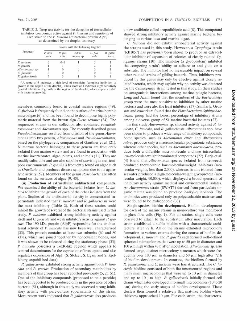
members commonly found in coastal marine regions (49).C. fucicola is frequently found on the surface of marine benthicmacroalgae (6) and has been found to decompose highly poly-meric material from the brown alga Fucus serratus (34). Thesequenced strains in our collection also contained Pseudoal-teromonas and Alteromonas spp. The recently described genusPseudoalteromonas resulted from division of the genus Altero-monas into two genera, Alteromonas and Pseudoalteromonas,based on the phylogenetic comparison of Gauthier et al. (21).Numerous bacteria belonging to these genera are frequentlyisolated from marine waters and are found in association withmarine invertebrates, algae, plants, and animals (31). They arereadily culturable and are also capable of surviving in nutrient-poor environments. P. gracilis is frequently found on algae suchas Gracilaria and produces disease symptoms due to its agaro-lytic activity (52). Members of the genus Roseobacter are oftenfound on the surfaces of algae (9, 43).
(ii) Production of extracellular antibacterial compounds.We examined the ability of the bacterial isolates from U. lac-tuca to inhibit the growth of each of the other isolates from theplant. Studies of the antibacterial activity in concentrated su-pernatants indicated that P. tunicata and R. gallaeciensis werethe most inhibitory (Table 2). Each of these strains couldinhibit the growth of several of the bacterial strains used in thisstudy. P. tunicata exhibited strong inhibitory activity againstitself and C. fucicola and weak inhibitory activity against P. gra-cilis. The 190-kDa protein (AlpP) responsible for the antibac-terial activity of P. tunicata has now been well characterized(33). This protein contains at least two subunits (60 and 80kDa), which are joined together by noncovalent bonds, andit was shown to be released during the stationary phase (33).P. tunicata possesses a ToxR-like regulon which appears tocontrol determinants for the expression of iron uptake and alsoregulates expression of AlpP (S. Stelzer, S. Egan, and S. Kjel-leberg unpublished data).
R. gallaeciensis exhibited strong activity against both P. tuni-cata and P. gracilis. Production of secondary metabolites bymembers of this group has been reported previously (5, 25, 51).One of the inhibitory compounds (proposed to be a peptide)has been reported to be produced only in the presence of otherbacteria (51), although in this study we observed strong inhib-itory activity with pure-culture supernatants of this strain.More recent work indicated that R. gallaeciensis also produces
a new antibiotic called tropodithietic acid (8). This compoundshowed strong inhibitory activity against marine bacteria be-longing to various taxa and marine algae.
C. fucicola did not exhibit antibacterial activity againstthe strains used in this study. However, a Cytophaga strain(RB1057) has previously been shown to produce an extracel-lular inhibitor of expansion of colonies of closely related Cy-tophaga strains (10). The inhibitor (a glycoprotein) inhibitedthe competing strain’s ability to adhere to and glide on asubstrate. The inhibitor had no measurable impact on severalother related strains of gliding bacteria. Thus, inhibitors pro-duced by this genus may only be effective against closely re-lated bacteria, which may explain why no activity was detectedfor the Cellulophaga strain tested in this study. In their studieson antagonistic interactions among marine pelagic bacteria,Long and Azam found that the members of the Bacteroidetesgroup were the most sensitive to inhibition by other marinebacteria and were also the least inhibitory (37). Similarly, Gros-sart and coworkers found that the Flavobacterium-Sphingobac-terium group had the lowest percentage of inhibitory strainsamong a diverse group of 51 marine bacterial isolates (27).
In this study Alteromonas sp. showed activity against P. tu-nicata, C. fucicola, and R. gallaeciensis. Alteromonas spp. havebeen shown to produce a wide range of inhibitory compounds.Some species, such as Alteromonas citrea and Alteromonasrubra, produce only a macromolecular polyanionic substance,whereas other species, such as Alteromonas luteoviolacea, pro-duce both a diffusible macromolecule and two intracellularlow-molecular-weight brominated compounds (22). Barja et al.(4) found that Alteromonas species isolated from seaweedsproduced thermolabile low-molecular-weight inhibitors (mo-lecular weights, less than 2,000), whereas strains isolated fromseawater produced a high-molecular-weight glycoprotein (mo-lecular weight, 90,000), which displayed a broad spectrum ofinhibitory activity against medical and environmental isolates.An Alteromonas strain (SWAT5) derived from particulate or-ganic matter was found to produce 2-alkyl-quinolinols. Theantibiotics were produced only on polysaccharide matrices andwere found to be hydrophobic (38).
Single-species biofilm development. Biofilm developmentfor each of the marine strains in monoculture was monitoredin glass flow cells (Fig. 1). For all strains, single cells wereobserved to attach to the substratum after inoculation. Eachstrain established a stable biofilm with a characteristic archi-tecture after 72 h. All of the strains exhibited microcolonyformation to various extents during the course of biofilm de-velopment. P. tunicata and P. gracilis each formed well-definedspherical microcolonies that were up to 50 �m in diameter and100 �m high within 48 h after inoculation. Alteromonas sp. alsoformed large, distinct microcolony structures which were fre-quently over 100 �m in diameter and 50 �m high after 72 hof biofilm development. In contrast, the biofilms formed byR. gallaeciensis and C. fucicola were less structured. The C. fu-cicola biofilms consisted of both flat unstructured regions andmany small microcolonies that were up to 10 �m in diameterand up to 10 �m high. R. gallaeciensis initially formed cellchains which later developed into small microcolonies (10 to 20�m) during the early stages of biofilm development. Theseclusters then formed a relatively flat, mat-like biofilm whosethickness approached 10 �m. For each strain, the characteris-
TABLE 2. Drop test activity for the detection of extracellularinhibitory compounds active against P. tunicata and sensitivity of
each strain to the P. tunicata antibacterial protein AlpP,as determined by the droplet test assay
Producer
Scores with the following targetsa:
P. tuni-cata
P. gra-cilis
Altero-monas sp.
C. fuci-cola
R. gallae-ciensis
P. tunicata 3 0–1 1 3 1P. gracilis 1 1 0 2 1Alteromonas sp. 1 0 0 2 1C. fucicola 0 0 0 0 0R. gallaeciensis 3 3 1 0 1
a A score of 3 indicates a high level of sensitivity (complete inhibition ofgrowth in the region of the droplet), and a score of 1 indicates slight sensitivity(partial inhibition of growth in the region of the droplet, which appears turbidwith bacterial growth).
VOL. 71, 2005 COMPETITION IN P. TUNICATA BIOFILMS 1731
on Novem
ber 12, 2013 by guesthttp://aem
.asm.org/
Dow
nloaded from
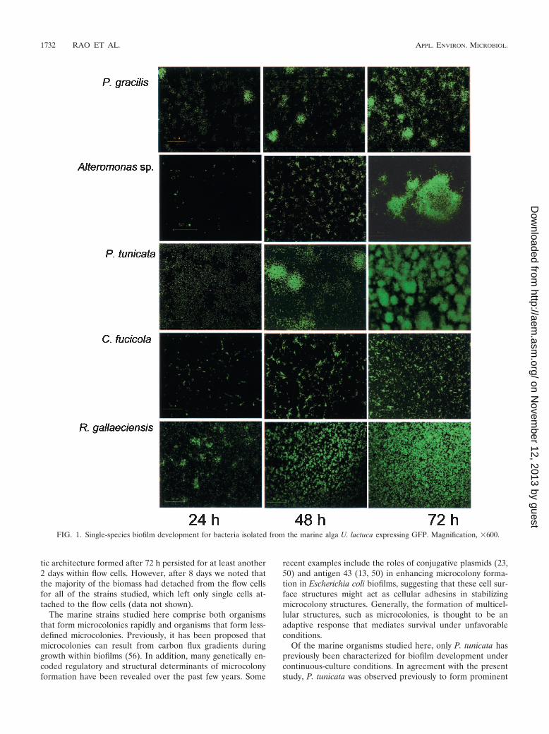
tic architecture formed after 72 h persisted for at least another2 days within flow cells. However, after 8 days we noted thatthe majority of the biomass had detached from the flow cellsfor all of the strains studied, which left only single cells at-tached to the flow cells (data not shown).
The marine strains studied here comprise both organismsthat form microcolonies rapidly and organisms that form less-defined microcolonies. Previously, it has been proposed thatmicrocolonies can result from carbon flux gradients duringgrowth within biofilms (56). In addition, many genetically en-coded regulatory and structural determinants of microcolonyformation have been revealed over the past few years. Some
recent examples include the roles of conjugative plasmids (23,50) and antigen 43 (13, 50) in enhancing microcolony forma-tion in Escherichia coli biofilms, suggesting that these cell sur-face structures might act as cellular adhesins in stabilizingmicrocolony structures. Generally, the formation of multicel-lular structures, such as microcolonies, is thought to be anadaptive response that mediates survival under unfavorableconditions.
Of the marine organisms studied here, only P. tunicata haspreviously been characterized for biofilm development undercontinuous-culture conditions. In agreement with the presentstudy, P. tunicata was observed previously to form prominent
FIG. 1. Single-species biofilm development for bacteria isolated from the marine alga U. lactuca expressing GFP. Magnification, �600.
1732 RAO ET AL. APPL. ENVIRON. MICROBIOL.
on Novem
ber 12, 2013 by guesthttp://aem
.asm.org/
Dow
nloaded from
microcolony structures during biofilm development (39). Bio-film development has been well characterized in the marinebacterium Vibrio cholerae, which also can form microcolonies(45, 58, 59). To our knowledge, however, there have been noreports that have considered the ecological role of biologicalstructures in mediating interactions between bacteria in mixed-community marine biofilms. In several studies the workersinvestigated interactions in marine biofilm communities (14,26, 36). However, these workers focused mainly on successionevents or addressed the issue of attachment and detachment ofdefined pure and mixed cultures on agar beads. In this study,we also examined the role of microcolonies during mixed-species biofilm development.
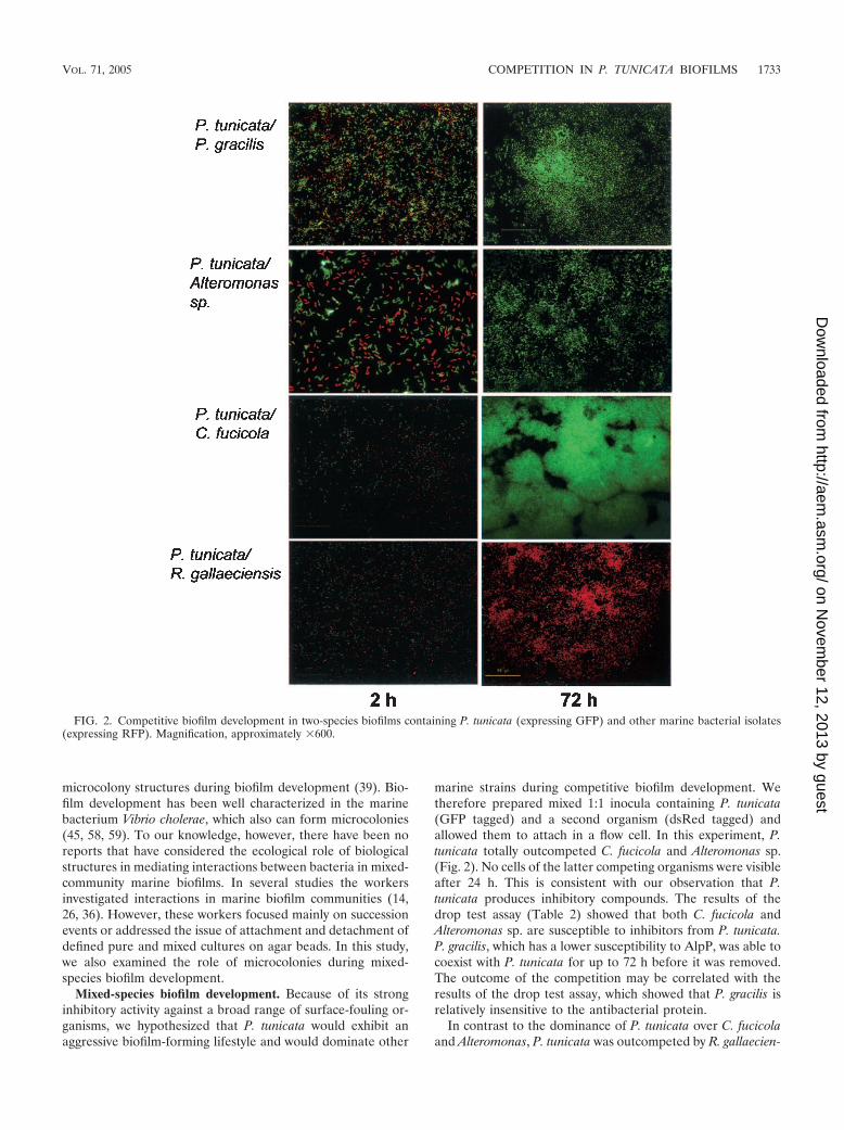
Mixed-species biofilm development. Because of its stronginhibitory activity against a broad range of surface-fouling or-ganisms, we hypothesized that P. tunicata would exhibit anaggressive biofilm-forming lifestyle and would dominate other
marine strains during competitive biofilm development. Wetherefore prepared mixed 1:1 inocula containing P. tunicata(GFP tagged) and a second organism (dsRed tagged) andallowed them to attach in a flow cell. In this experiment, P.tunicata totally outcompeted C. fucicola and Alteromonas sp.(Fig. 2). No cells of the latter competing organisms were visibleafter 24 h. This is consistent with our observation that P.tunicata produces inhibitory compounds. The results of thedrop test assay (Table 2) showed that both C. fucicola andAlteromonas sp. are susceptible to inhibitors from P. tunicata.P. gracilis, which has a lower susceptibility to AlpP, was able tocoexist with P. tunicata for up to 72 h before it was removed.The outcome of the competition may be correlated with theresults of the drop test assay, which showed that P. gracilis isrelatively insensitive to the antibacterial protein.
In contrast to the dominance of P. tunicata over C. fucicolaand Alteromonas, P. tunicata was outcompeted by R. gallaecien-
FIG. 2. Competitive biofilm development in two-species biofilms containing P. tunicata (expressing GFP) and other marine bacterial isolates(expressing RFP). Magnification, approximately �600.
VOL. 71, 2005 COMPETITION IN P. TUNICATA BIOFILMS 1733
on Novem
ber 12, 2013 by guesthttp://aem
.asm.org/
Dow
nloaded from
sis. While some cells of P. tunicata persisted on the surfacethroughout the experimental period, these cells did not grow,and R. gallaeciensis rapidly grew and formed a biofilm. This isagain consistent with the results of the drop test assay, whichshowed that P. tunicata is highly susceptible to the inhibitorycompounds produced by R. gallaeciensis.
Role of the P. tunicata antibacterial protein AlpP in com-petitive biofilm development. To evaluate whether the antibac-terial protein AlpP can play a role in competitive biofilm de-velopment, a �alpP insertional knockout mutant (39) was usedin competition studies. We first repeated the mixed-inoculumbiofilm experiments described above by using the �alpP straininstead of the wild type. In these experiments we observed nodifference in the outcome of the biofilm competition experi-ments when the wild-type and �alpP strains were used. Wheninoculated together with C. fucicola, Alteromonas sp., or P.gracilis, the �alpP mutant became dominant after 72 h, similarto the results obtained for the wild-type strain (data notshown). Also, R. gallaeciensis was dominant over the P. tunicata�alpP strain in a manner similar to the manner observed withwild-type P. tunicata.
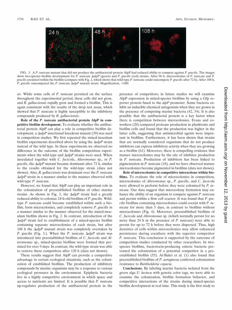
However, we found that AlpP can play an important role inthe colonization of preestablished biofilms of other marinestrains. As shown in Fig. 3, the �alpP strain had a greatlyreduced ability to colonize 24-h-old biofilms of P. gracilis. Wild-type P. tunicata could become established within such a bio-film, form microcolonies, and completely remove P. gracilis ina manner similar to the manner observed for the mixed-inoc-ulum biofilm shown in Fig. 2. In contrast, introduction of the�alpP strain led to establishment of a mixed-species biofilmcontaining separate microcolonies of each strain, but after108 h the �alpP mutant strain was completely overtaken byP. gracilis (Fig. 3.). When the P. tunicata �alpP strain wasintroduced into preestablished biofilms of C. fucicola and Al-teromonas sp., mixed-species biofilms were formed that per-sisted for over 9 days. In contrast, the wild-type strain was ableto remove these competitors after 120 h (data not shown).
These results suggest that AlpP can provide a competitiveadvantage in certain ecological situations, such as the coloni-zation of established biofilms. The production of inhibitorycompounds by marine organisms may be a response to variousecological pressures in the environment. Epiphytic bacterialive in a highly competitive environment in which space andaccess to nutrients are limited. It is possible that P. tunicataup-regulates production of the antibacterial protein in the
presence of competitors; in future studies we will examineAlpP expression in mixed-species biofilms by using a Gfp re-porter protein fused to the alpP promoter. Some bacteria ex-hibit an inducible chemical antagonism when they are grown inthe presence of competing marine bacteria (42, 54). It is alsopossible that the antibacterial protein is a key factor whenthere is competition between microcolonies. Evans and co-workers (20) compared protease production in planktonic andbiofilm cells and found that the production was higher in thelatter cells, suggesting that antimicrobial agents were impor-tant in biofilms. Furthermore, it has been shown that strainsthat are normally considered organisms that do not produceinhibitors can express inhibitory activity when they are growingas a biofilm (61). Moreover, there is compelling evidence thatmature microcolonies may be the site of inhibitor productionin P. tunicata. Production of inhibitors has been linked topigmentation in P. tunicata (18), and we have observed maturemicrocolonies become pigmented in biofilms (data not shown).
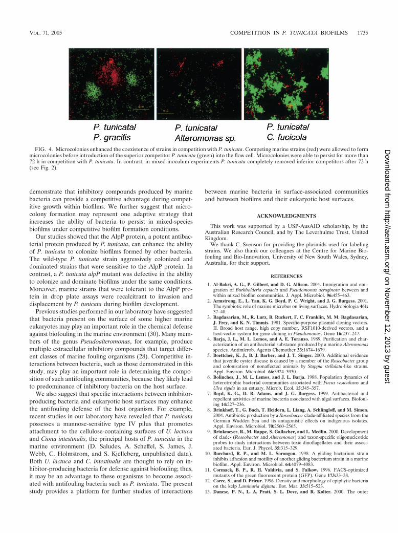
Role of microcolonies in competitive interactions within bio-films. To evaluate the role of microcolonies in competition,microcolonies of Alteromonas sp., P. gracilis, and C. fucicolawere allowed to preform before they were colonized by P. tu-nicata. Our data suggest that microcolony formation may en-hance the ability of an organism to compete against P. tunicataand persist within a flow cell reactor. It was found that P. gra-cilis biofilms containing microcolonies could coexist with P. tu-nicata for more than 5 days, in contrast to biofilms withoutmicrocolonies (Fig. 4). Moreover, preestablished biofilms ofC. fucicola and Alteromonas sp. (which normally persist for nomore than 24 h in the presence of P. tunicata) were able topersist for up to 72 h before they were dispersed. Thus, highdensities of cells within microcolonies may allow enhancedpersistence during coculture with the superior competitorP. tunicata. This conclusion is supported by the outcome ofcompetition studies conducted by other researchers. In two-species biofilms, bacteriocin-producing enteric bacteria pre-vented the colonization of a potential competitor in a pre-established biofilm (55). Al-Bakri et al. (1) also found thatpreestablished biofilms of P. aeruginosa conferred colonizationresistance to Burkholderia cepacia.
Conclusions. By labeling marine bacteria isolated from thegreen alga U. lactuca with genetic color tags, we were able toexamine the colonization, biofilm formation behavior, andcompetitive interactions of the strains during mixed-speciesbiofilm development in real time. This study is the first study to
FIG. 3. A P. tunicata mutant that did not produce the antibacterial protein AlpP had reduced ability to compete against P. gracilis. The imagesshow two-species biofilm development for P. tunicata �alpP (green) and P. gracilis (red) strains. After 96 h, microcolonies of P. tunicata and P.gracilis coexisted within the biofilm (compare with Fig. 2, which shows that wild-type P. tunicata could outcompete P. gracilis after 72 h). After 108 h,P. gracilis outcompeted the P. tunicata �alpP mutant strain. Magnification, �600.
1734 RAO ET AL. APPL. ENVIRON. MICROBIOL.
on Novem
ber 12, 2013 by guesthttp://aem
.asm.org/
Dow
nloaded from
demonstrate that inhibitory compounds produced by marinebacteria can provide a competitive advantage during compet-itive growth within biofilms. We further suggest that micro-colony formation may represent one adaptive strategy thatincreases the ability of bacteria to persist in mixed-speciesbiofilms under competitive biofilm formation conditions.
Our studies showed that the AlpP protein, a potent antibac-terial protein produced by P. tunicata, can enhance the abilityof P. tunicata to colonize biofilms formed by other bacteria.The wild-type P. tunicata strain aggressively colonized anddominated strains that were sensitive to the AlpP protein. Incontrast, a P. tunicata alpP mutant was defective in the abilityto colonize and dominate biofilms under the same conditions.Moreover, marine strains that were tolerant to the AlpP pro-tein in drop plate assays were recalcitrant to invasion anddisplacement by P. tunicata during biofilm development.
Previous studies performed in our laboratory have suggestedthat bacteria present on the surface of some higher marineeukaryotes may play an important role in the chemical defenseagainst biofouling in the marine environment (30). Many mem-bers of the genus Pseudoalteromonas, for example, producemultiple extracellular inhibitory compounds that target differ-ent classes of marine fouling organisms (28). Competitive in-teractions between bacteria, such as those demonstrated in thisstudy, may play an important role in determining the compo-sition of such antifouling communities, because they likely leadto predominance of inhibitory bacteria on the host surface.
We also suggest that specific interactions between inhibitor-producing bacteria and eukaryotic host surfaces may enhancethe antifouling defense of the host organism. For example,recent studies in our laboratory have revealed that P. tunicatapossesses a mannose-sensitive type IV pilus that promotesattachment to the cellulose-containing surfaces of U. lactucaand Ciona intestinalis, the principal hosts of P. tunicata in themarine environment (D. Saludes, A. Scheffel, S. James, J.Webb, C. Holmstrom, and S. Kjelleberg, unpublished data).Both U. lactuca and C. intestinalis are thought to rely on in-hibitor-producing bacteria for defense against biofouling; thus,it may be an advantage to these organisms to become associ-ated with antifouling bacteria such as P. tunicata. The presentstudy provides a platform for further studies of interactions
between marine bacteria in surface-associated communitiesand between biofilms and their eukaryotic host surfaces.
ACKNOWLEDGMENTS
This work was supported by a USP-AusAID scholarship, by theAustralian Research Council, and by The Leverhulme Trust, UnitedKingdom.
We thank C. Svenson for providing the plasmids used for labelingstrains. We also thank our colleagues at the Centre for Marine Bio-fouling and Bio-Innovation, University of New South Wales, Sydney,Australia, for their support.
REFERENCES
1. Al-Bakri, A. G., P. Gilbert, and D. G. Allison. 2004. Immigration and emi-gration of Burkholderia cepacia and Pseudomonas aeruginosa between andwithin mixed biofilm communities. J. Appl. Microbiol. 96:455–463.
2. Armstrong, E., L. Yan, K. G. Boyd, P. C. Wright, and J. G. Burgess. 2001.The symbiotic role of marine microbes on living surfaces. Hydrobiologia 461:37–40.
3. Bagdasarian, M., R. Lurz, B. Ruckert, F. C. Franklin, M. M. Bagdasarian,J. Frey, and K. N. Timmis. 1981. Specific-purpose plasmid cloning vectors.II. Broad host range, high copy number, RSF1010-derived vectors, and ahost-vector system for gene cloning in Pseudomonas. Gene 16:237–247.
4. Barja, J. L., M. L. Lemos, and A. E. Toranzo. 1989. Purification and char-acterization of an antibacterial substance produced by a marine Alteromonasspecies. Antimicrob. Agents Chemother 33:1674–1679.
5. Boettcher, K. J., B. J. Barber, and J. T. Singer. 2000. Additional evidencethat juvenile oyster disease is caused by a member of the Roseobacter groupand colonization of nonaffected animals by Stappia stellulata-like strains.Appl. Environ. Microbiol. 66:3924–3930.
6. Bolinches, J., M. L. Lemos, and J. L. Barja. 1988. Population dynamics ofheterotrophic bacterial communities associated with Fucus vesiculosus andUlva rigida in an estuary. Microb. Ecol. 15:345–357.
7. Boyd, K. G., D. R. Adams, and J. G. Burgess. 1999. Antibacterial andrepellent activities of marine bacteria associated with algal surfaces. Biofoul-ing 14:227–236.
8. Brinkhoff, T., G. Bach, T. Heidorn, L. Liang, A. Schlingloff, and M. Simon.2004. Antibiotic production by a Roseobacter clade-affiliated species from theGerman Wadden Sea and its antagonistic effects on indigenous isolates.Appl. Environ. Microbiol. 70:2560–2565.
9. Brinkmeyer, R., M. Rappe, S. Gallacher, and L. Medlin. 2000. Developmentof clade- (Roseobacter and Alteromonas) and taxon-specific oligonucleotideprobes to study interactions between toxic dinoflagellates and their associ-ated bacteria. Eur. J. Phycol. 35:315–329.
10. Burchard, R. P., and M. L. Sorongon. 1998. A gliding bacterium straininhibits adhesion and motility of another gliding bacterium strain in a marinebiofilm. Appl. Environ. Microbiol. 64:4079–4083.
11. Cormack, B. P., R. H. Valdivia, and S. Falkow. 1996. FACS-optimizedmutants of the green fluorescent protein (GFP). Gene 173:33–38.
12. Corre, S., and D. Prieur. 1996. Density and morphology of epiphytic bacteriaon the kelp Laminaria digitata. Bot. Mar. 33:515–523.
13. Danese, P. N., L. A. Pratt, S. L. Dove, and R. Kolter. 2000. The outer
FIG. 4. Microcolonies enhanced the coexistence of strains in competition with P. tunicata. Competing marine strains (red) were allowed to formmicrocolonies before introduction of the superior competitor P. tunicata (green) into the flow cell. Microcolonies were able to persist for more than72 h in competition with P. tunicata. In contrast, in mixed-inoculum experiments P. tunicata completely removed inferior competitors after 72 h(see Fig. 2).
VOL. 71, 2005 COMPETITION IN P. TUNICATA BIOFILMS 1735
on Novem
ber 12, 2013 by guesthttp://aem
.asm.org/
Dow
nloaded from
membrane protein, antigen 43, mediates cell-to-cell interactions within Esch-erichia coli biofilms. Mol. Microbiol. 37:424–432.
14. Dang, H., and C. R. Lovell. 2000. Bacterial primary colonization and earlysuccession on surfaces in marine waters as determined by amplified rRNAgene restriction analysis and sequence analysis of 16S rRNA genes. Appl.Environ. Microbiol. 66:467–475.
15. Davis, A. R., N. M. Targett, O. J. McConnel, and C. M. Young. 1989.Epibiosis of marine algae and benthic invertebrates: natural products chem-istry and other mechanisms inhibiting settlement and overgrowth. Bioorg.Mar. Chem. 3:85–114.
16. Dobretsov, S. V., and P. Y. Qian. 2002. Effect of bacteria associated with thegreen alga Ulva reticulata on marine micro- and macrofouling. Biofouling 18:217–228.
17. Dworjanyn, S. A., R. de Nys, and P. D. Steinberg. 1999. Localization andsurface quantification of secondary metabolites in the red alga Delisea pul-chra. Mar. Biol. 133:727–736.
18. Egan, S., S. James, C. Holmstrom, and S. Kjelleberg. 2002. Correlationbetween pigmentation and antifouling compounds produced by Pseudoal-teromonas tunicata. Environ. Microbiol. 4:433–442.
19. Egan, S., S. James, and S. Kjelleberg. 2002. Identification and characteriza-tion of a putative transcriptional regulator controlling the expression offouling inhibitors in Pseudoalteromonas tunicata. Appl. Environ. Microbiol.68:372–378.
20. Evans, E., M. R. Brown, and P. Gilbert. 1994. Iron chelator, exopolysaccha-ride and protease production in Staphylococcus epidermidis: a comparativestudy of the effects of specific growth rate in biofilm and planktonic culture.Microbiology 140:153–157.
21. Gauthier, G., M. Gauthier, and R. Christen. 1995. Phylogenetic analysis ofthe genera Alteromonas, Shewanella, and Moritella using genes coding forsmall-subunit rRNA sequences and division of the genus Alteromonas intotwo genera, Alteromonas (emended) and Pseudoalteromonas gen. nov., andproposal of twelve new species combinations. Int. J. Syst. Bacteriol. 45:755–761.
22. Gauthier, M. J., and G. N. Flatau. 1976. Antibacterial activity of marineviolet pigmented Alteromonas with special reference to the production ofbrominated compounds. Can. J. Microbiol. 22:349–354.
23. Ghigo, J.-M. 2001. Natural conjugative plasmids induce bacterial biofilmdevelopment. Nature 412:442–445.
24. Gil-Turness, M. S., and W. Fenical. 1992. Embryos of Homarus americanusare protected by epibiotic bacteria. Biol. Bull. 182:105–108.
25. Gram, L., H. P. Grossart, A. Schlingloff, and T. Kiorboe. 2002. Possiblequorum sensing in marine snow bacteria: production of acylated homoserinelactones by Roseobacter strains isolated from marine snow. Appl. Environ.Microbiol. 68:4111–4116.
26. Grossart, H.-P., T. Kiorboe, K. Tang, and H. Ploug. 2003. Bacterial coloni-zation of particles: growth and interactions. Appl. Environ. Microbiol. 69:3500–3509.
27. Grossart, H. P., A. Schlingloff, M. Bernhard, M. Simon, and T. Brinkhoff.2004. Antagonistic activity of bacteria isolated from organic aggregates of theGerman Wadden Sea. FEMS Microbiol. Ecol. 47:387–396.
28. Holmstrom, C., S. Egan, A. Franks, S. McCloy, and S. Kjelleberg. 2002.Antifouling activities expressed by marine surface associated Pseudoaltero-monas species. FEMS Microbiol. Ecol. 41:47–58.
29. Holmstrom, C., S. James, B. A. Neilan, D. C. White, and S. Kjelleberg. 1998.Pseudoalteromonas tunicata sp. nov., a bacterium that produces antifoulingagents. Int. J. Syst. Bacteriol. 48:1205–1212.
30. Holmstrom, C., and S. Kjelleberg. 1994. The effect of external biologicalfactors on settlement of marine invertebrate and new antifouling technology.Biofouling 8:147–160.
31. Holmstrom, C., and S. Kjelleberg. 1999. Marine Pseudoalteromonas speciesare associated with higher organisms and produce biologically active extra-cellular agents. FEMS Microbiol. Ecol. 30:285–293.
32. Holmstrom, C., D. Rittschof, and S. Kjelleberg. 1992. Inhibition of settle-ment by larvae of Balanus amphitrite and Ciona intestinalis by a surface-colonizing marine bacterium. Appl. Environ. Microbiol. 58:2111–2115.
33. James, S., C. Holmstrom, and S. Kjelleberg. 1996. Purification and charac-terization of a novel antibacterial protein from the marine bacterium D2.Appl. Environ. Microbiol. 62:2783–2788.
34. Johansen, J. E., P. Nielsen, and C. Sjoholm. 1999. Description of Cellu-lophaga baltica gen. nov., sp. nov. and Cellulophaga fucicola gen. nov., sp.nov. and reclassification of Cytophaga lytica to Cellulophaga lytica gen. nov.,comb. nov. Int. J. Syst. Bacteriol. 49:1231–1240.
35. Keats, D. W., M. A. Knight, and C. M. Pueschel. 1997. Antifouling effects ofepithallial shedding in three crustose coralling algae (Rhodophyta, Cora-lines) on a coral reef. J. Exp. Mar. Biol. Ecol. 213:281–293.
36. Kiorboe, T., K. Tang, H.-P. Grossart, and H. Ploug. 2003. Dynamics ofmicrobial communities on marine snow aggregates: colonization, growth,detachment, and grazing mortality of attached bacteria. Appl. Environ. Mi-crobiol. 69:3036–3047.
37. Long, R. A., and F. Azam. 2001. Antagonistic interactions among marinepelagic bacteria. Appl. Environ. Microbiol. 67:4975–4983.
38. Long, R. A., A. Qureshi, D. J. Faulkner, and F. Azam. 2003. 2-n-Pentyl-4-quinolinol produced by a marine Alteromonas sp. and its potential ecologicaland biogeochemical roles. Appl. Environ. Microbiol. 69:568–576.
39. Mai-Prochnow, A., F. Evans, D. Dalisay-Saludes, S. Stelzer, S. Egan, S.James, J. S. Webb, and S. Kjelleberg. 2004. Biofilm development and celldeath in the marine bacterium Pseudoalteromonas tunicata. Appl. Environ.Microbiol. 70:3232–3238.
40. Marden, P., A. Tunlid, K. Malmcrona-Friberg, G. Odham, and S. Kjelle-berg. 1985. Physiological and morphological changes during short term star-vation of marine bacteria isolates. Arch. Microbiol. 142:326–332.
41. Matz, C., T. Bergfeld, S. A. Rice, and S. Kjelleberg. 2004. Microcolonies,quorum sensing and cytotoxicity determine the survival of Pseudomonasaeruginosa biofilms exposed to protozoan grazing. Environ. Microbiol. 6:218–226.
42. Mearns-Spragg, A., M. Bregu, K. G. Boyd, and J. G. Burgess. 1998. Cross-species induction and enhancement of antimicrobial activity produced byepibiotic bacteria from marine algae and invertebrates after exposure toterrestrial bacteria. Lett. Appl. Microbiol. 27:142–146.
43. Meusnier, I., J. L. Olsen, W. T. Stam, C. Destombe, and M. Valero. 2001.Phylogenetic analysis of Caulerpa taxifolia (Chlorophyta) and its associatedbacterial microflora provide clues to the origin of the Mediterranean intro-duction. Mol. Ecol. 10:931–946.
44. Moller, S., C. Sternberg, J. B. Andersen, B. B. Christensen, J. L. Ramos, M.Givskov, and S. Molin. 1998. In situ gene expression of mixed-culture bio-films: evidence of metabolic interactions between community members.Appl. Environ. Microbiol. 64:721–732.
45. Moorthy, S., and P. I. Watnick. 2004. Genetic evidence that the Vibriocholerae monolayer is a distinct stage in biofilm development. Mol. Micro-biol. 52:573–587.
46. Nakasono, S., J. G. Burgess, K. Takashi, M. Koike, C. Murayama, S. Na-kamura, and T. Matsunaga. 1993. Electrochemical prevention of marinebiofouling with a carbon-chloroprene sheet. Appl. Environ. Microbiol. 59:3757–3762.
47. Neidhart, F. C., P. L. Bloch, and D. F. Smith. 1974. Culture medium forenterobacteria. J. Bacteriol. 119:736–747.
48. Nielsen, A., T. Tolker-Neilsen, K. Barken, and S. Molin. 2000. Role ofcommensal relationships on the spatial structure of a surface-attached mi-crobial consortium. Environ. Microbiol. 2:59–68.
49. Pinhassi, J., U.-L. Zweifel, and A. Hagstrom. 1997. Dominant marine bac-terioplankton species found among colony-forming bacteria. Appl. Environ.Microbiol. 63:3359–3366.
50. Reisner, A., J. A. J. Haagensen, M. A. Schembri, E. L. Zechner, and S. Molin.2003. Development and maturation of Escherichia coli K-12 biofilms. Mol.Microbiol. 48:933–946.
51. Ruiz-Ponte, C., V. Cilia, C. Lambert, and J. L. Nicolas. 1998. Roseobactergallaeciensis sp. nov., a new marine bacterium isolated from rearings andcollectors of the scallop Pecten maximus. Int. J. Syst. Bacteriol. 48:537–542.
52. Schroeder, D. C., M. A. Jaffer, and V. E. Coyne. 2003. Investigation of therole of a �(1-4)agarase produced by Pseudoalteromonas gracilis B9 in elicit-ing disease symptoms in the red alga Gracilaria gracilis. Microbiology 149:2919–2929.
53. Sieburth, J. M. 1975. Microbial seascapes: a pictoral essay on marine mi-croorganisms and their environments. University Park Press, Baltimore, Md.
54. Slattery, M., I. Rajbhandari, and K. Wesson. 2001. Competition-mediatedantibiotic induction in the marine bacterium Streptomyces tenjimariensis.Microb. Ecol. 41:90–96.
55. Tait, K., and I. W. Sutherland. 2002. Antagonistic interactions among bac-teriocin-producing enteric bacteria in dual species biofilms. J. Appl. Micro-biol. 93:345–352.
56. van Loosdrecht, M. C., J. J. Heijnen, H. Eberl, J. Kreft, and C. Picioreanu.2002. Mathematical modeling of biofilm structures. Antonie Leeuwenhoek81:245–256.
57. Wahl, M. 1989. Marine epibosis. 1. Fouling and antifouling: some basicaspects. Mar. Ecol. Prog. Ser. 58:175–189.
58. Watnick, P. I., K. J. Fullner, and R. Kolter. 1999. A role for the mannose-sensitive hemagglutinin in biofilm formation by Vibrio cholerae El Tor.J. Bacteriol. 181:3606–3609.
59. Watnick, P. I., and R. Kolter. 1999. Steps in the development of a Vibriocholerae El Tor biofilm. Mol. Microbiol. 34:586–595.
60. Webb, J. S., L. S. Thompson, S. James, T. Charlton, T. Tolker-Nielsen, B.Koch, M. Givskov, and S. Kjelleberg. 2003. Cell death in Pseudomonasaeruginosa biofilm development. J. Bacteriol. 185:4585–4592.
61. Yan, L., K. G. Boyd, D. R. Adams, and J. G. Burgess. 2003. Biofilm-specificcross-species induction of antimicrobial compounds in bacilli. Appl. Environ.Microbiol. 69:3719–3727.
1736 RAO ET AL. APPL. ENVIRON. MICROBIOL.
on Novem
ber 12, 2013 by guesthttp://aem
.asm.org/
Dow
nloaded from
Related Documents