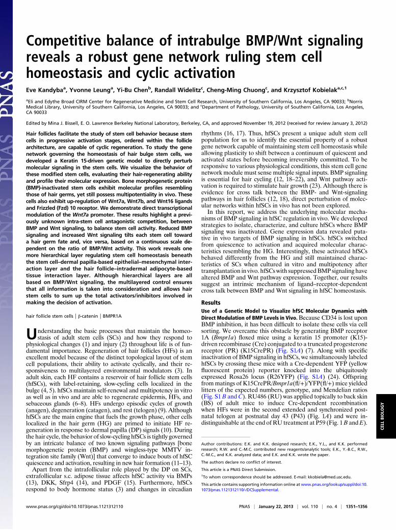
Competitive balance of intrabulge BMP/Wnt signaling reveals a robust gene network ruling stem cell homeostasis and cyclic activation Eve Kandyba a , Yvonne Leung a , Yi-Bu Chen b , Randall Widelitz c , Cheng-Ming Chuong c , and Krzysztof Kobielak a,c,1 a Eli and Edythe Broad CIRM Center for Regenerative Medicine and Stem Cell Research, University of Southern California, Los Angeles, CA 90033; b Norris Medical Library, University of Southern California, Los Angeles, CA 90033; and c Department of Pathology, University of Southern California, Los Angeles, CA 90033 Edited by Mina J. Bissell, E. O. Lawrence Berkeley National Laboratory, Berkeley, CA, and approved November 19, 2012 (received for review January 3, 2012) Hair follicles facilitate the study of stem cell behavior because stem cells in progressive activation stages, ordered within the follicle architecture, are capable of cyclic regeneration. To study the gene network governing the homeostasis of hair bulge stem cells, we developed a Keratin 15-driven genetic model to directly perturb molecular signaling in the stem cells. We visualize the behavior of these modified stem cells, evaluating their hair-regenerating ability and profile their molecular expression. Bone morphogenetic protein (BMP)-inactivated stem cells exhibit molecular profiles resembling those of hair germs, yet still possess multipotentiality in vivo. These cells also exhibit up-regulation of Wnt7a, Wnt7b, and Wnt16 ligands and Frizzled (Fzd) 10 receptor. We demonstrate direct transcriptional modulation of the Wnt7a promoter. These results highlight a previ- ously unknown intra-stem cell antagonistic competition, between BMP and Wnt signaling, to balance stem cell activity. Reduced BMP signaling and increased Wnt signaling tilts each stem cell toward a hair germ fate and, vice versa, based on a continuous scale de- pendent on the ratio of BMP/Wnt activity. This work reveals one more hierarchical layer regulating stem cell homeostasis beneath the stem cell–dermal papilla-based epithelial–mesenchymal inter- action layer and the hair follicle–intradermal adipocyte-based tissue interaction layer. Although hierarchical layers are all based on BMP/Wnt signaling, the multilayered control ensures that all information is taken into consideration and allows hair stem cells to sum up the total activators/inhibitors involved in making the decision of activation. hair follicle stem cells | β-catenin | BMPR1A U nderstanding the basic processes that maintain the homeo- stasis of adult stem cells (SCs) and how they respond to physiological changes (1) and injury (2) throughout life is of fun- damental importance. Regeneration of hair follicles (HFs) is an excellent model because of the distinct topological layout of stem cell populations, their ability to activate cyclically, and their re- sponsiveness to multilayered environmental modulators (3). In adult skin, each HF contains a reservoir of hair follicle stem cells (hfSCs), with label-retaining, slow-cycling cells localized in the bulge (4, 5). hfSCs maintain self-renewal and multipotency in vitro as well as in vivo and are able to regenerate epidermis, HFs, and sebaceous glands (6–8). HFs undergo episodic cycles of growth (anagen), degeneration (catagen), and rest (telogen) (9). Although hfSCs are the main engine that fuels the growth phase, other cells localized in the hair germ (HG) are primed to initiate HF re- generation in response to dermal papilla (DP) signals (10). During the hair cycle, the behavior of slow-cycling hfSCs is tightly governed by an intricate balance of two known signaling pathways [bone morphogenetic protein (BMP) and wingless-type MMTV in- tegration site family (Wnt)] that converge to induce bouts of hfSC quiescence and activation, resulting in new hair formation (11–13). Apart from the intrafollicular role played by the DP on SCs, extrafollicular s.c. adipose tissue affects hfSC activity via BMPs (13), DKK, Sfrp4 (14), and PDGF (15). Furthermore, hfSCs respond to body hormone status (3) and changes in circadian rhythms (16, 17). Thus, hfSCs present a unique adult stem cell population for us to identify the essential property of a robust gene network capable of maintaining stem cell homeostasis while allowing plasticity to shift between a continuum of quiescent and activated states before becoming irreversibly committed. To be responsive to various physiological conditions, this stem cell gene network module must sense multiple signal inputs. BMP signaling is essential for hair cycling (12, 18–22), and Wnt pathway acti- vation is required to stimulate hair growth (23). Although there is evidence for cross talk between the BMP- and Wnt-signaling pathways in hair follicles (12, 18), direct perturbation of molec- ular networks within hfSCs in vivo has not been explored. In this report, we address the underlying molecular mecha- nisms of BMP signaling in hfSC regulation in vivo. We developed strategies to isolate, characterize, and culture hfSCs where BMP signaling was inactivated. Gene expression data revealed puta- tive in vivo targets of BMP signaling in hfSCs. hfSCs switched from quiescence to activation and acquired molecular charac- teristics resembling the HG. Interestingly, these activated hfSCs behaved differently from the HG and still maintained charac- teristics of SCs when cultured in vitro and multipotency after transplantation in vivo. hfSCs with suppressed BMP signaling have altered BMP and Wnt pathway expression. Together, our results suggest an intrinsic mechanism of ligand–receptor-dependent cross talk between BMP and Wnt signaling in hfSC homeostasis. Results Use of a Genetic Model to Visualize hfSC Molecular Dynamics with Direct Modulation of BMP Levels in Vivo. Because CD34 is lost upon BMP inhibition, it has been difficult to isolate these cells via cell sorting. We overcame this obstacle by generating BMP receptor 1A (Bmpr1a) floxed mice using a keratin 15 promoter (K15)- driven recombinase (Cre) conjugated to a truncated progesterone receptor (PR) (K15CrePR) (Fig. S1A) (7). Along with specific inactivation of BMP signaling in hfSCs, we simultaneously labeled hfSCs by crossing these mice with a Cre-dependent YFP (yellow fluorescent protein) reporter knocked into the ubiquitously expressed Rosa26 locus (R26YFP) (Fig. S1A) (24). Offspring from matings of K15CrePR/Bmpr1a(fl/+)/YFP(fl/+) mice yielded litters of the expected numbers, genotype, and Mendelian ratios (Fig. S1 B and C). RU486 (RU) was applied topically to back skin (BS) of adult mice to induce Cre-dependent recombination when HFs were in the second extended and synchronized post- natal telogen at postnatal day 43 (P43) (Fig. 1A) and were in- distinguishable at the end of RU treatment at P59 (Fig. 1 B and E). Author contributions: E.K. and K.K. designed research; E.K., Y.L., and K.K. performed research; R.W. and C.-M.C. contributed new reagents/analytic tools; E.K., Y.-B.C., R.W., C.-M.C., and K.K. analyzed data; and E.K. and K.K. wrote the paper. The authors declare no conflict of interest. This article is a PNAS Direct Submission. 1 To whom correspondence should be addressed. E-mail: [email protected]. This article contains supporting information online at www.pnas.org/lookup/suppl/doi:10. 1073/pnas.1121312110/-/DCSupplemental. www.pnas.org/cgi/doi/10.1073/pnas.1121312110 PNAS | January 22, 2013 | vol. 110 | no. 4 | 1351–1356 CELL BIOLOGY

Welcome message from author
This document is posted to help you gain knowledge. Please leave a comment to let me know what you think about it! Share it to your friends and learn new things together.
Transcript
Competitive balance of intrabulge BMP/Wnt signalingreveals a robust gene network ruling stem cellhomeostasis and cyclic activationEve Kandybaa, Yvonne Leunga, Yi-Bu Chenb, Randall Widelitzc, Cheng-Ming Chuongc, and Krzysztof Kobielaka,c,1
aEli and Edythe Broad CIRM Center for Regenerative Medicine and Stem Cell Research, University of Southern California, Los Angeles, CA 90033; bNorrisMedical Library, University of Southern California, Los Angeles, CA 90033; and cDepartment of Pathology, University of Southern California, Los Angeles,CA 90033
Edited by Mina J. Bissell, E. O. Lawrence Berkeley National Laboratory, Berkeley, CA, and approved November 19, 2012 (received for review January 3, 2012)
Hair follicles facilitate the study of stem cell behavior because stemcells in progressive activation stages, ordered within the folliclearchitecture, are capable of cyclic regeneration. To study the genenetwork governing the homeostasis of hair bulge stem cells, wedeveloped a Keratin 15-driven genetic model to directly perturbmolecular signaling in the stem cells. We visualize the behavior ofthese modified stem cells, evaluating their hair-regenerating abilityand profile their molecular expression. Bone morphogenetic protein(BMP)-inactivated stem cells exhibit molecular profiles resemblingthose of hair germs, yet still possess multipotentiality in vivo. Thesecells also exhibit up-regulation of Wnt7a, Wnt7b, and Wnt16 ligandsand Frizzled (Fzd) 10 receptor. We demonstrate direct transcriptionalmodulation of the Wnt7a promoter. These results highlight a previ-ously unknown intra-stem cell antagonistic competition, betweenBMP and Wnt signaling, to balance stem cell activity. Reduced BMPsignaling and increased Wnt signaling tilts each stem cell towarda hair germ fate and, vice versa, based on a continuous scale de-pendent on the ratio of BMP/Wnt activity. This work reveals onemore hierarchical layer regulating stem cell homeostasis beneaththe stem cell–dermal papilla-based epithelial–mesenchymal inter-action layer and the hair follicle–intradermal adipocyte-basedtissue interaction layer. Although hierarchical layers are allbased on BMP/Wnt signaling, the multilayered control ensuresthat all information is taken into consideration and allows hairstem cells to sum up the total activators/inhibitors involved inmaking the decision of activation.
hair follicle stem cells | β-catenin | BMPR1A
Understanding the basic processes that maintain the homeo-stasis of adult stem cells (SCs) and how they respond to
physiological changes (1) and injury (2) throughout life is of fun-damental importance. Regeneration of hair follicles (HFs) is anexcellent model because of the distinct topological layout of stemcell populations, their ability to activate cyclically, and their re-sponsiveness to multilayered environmental modulators (3). Inadult skin, each HF contains a reservoir of hair follicle stem cells(hfSCs), with label-retaining, slow-cycling cells localized in thebulge (4, 5). hfSCs maintain self-renewal and multipotency in vitroas well as in vivo and are able to regenerate epidermis, HFs, andsebaceous glands (6–8). HFs undergo episodic cycles of growth(anagen), degeneration (catagen), and rest (telogen) (9). AlthoughhfSCs are the main engine that fuels the growth phase, other cellslocalized in the hair germ (HG) are primed to initiate HF re-generation in response to dermal papilla (DP) signals (10). Duringthe hair cycle, the behavior of slow-cycling hfSCs is tightly governedby an intricate balance of two known signaling pathways [bonemorphogenetic protein (BMP) and wingless-type MMTV in-tegration site family (Wnt)] that converge to induce bouts of hfSCquiescence and activation, resulting in new hair formation (11–13).Apart from the intrafollicular role played by the DP on SCs,
extrafollicular s.c. adipose tissue affects hfSC activity via BMPs(13), DKK, Sfrp4 (14), and PDGF (15). Furthermore, hfSCsrespond to body hormone status (3) and changes in circadian
rhythms (16, 17). Thus, hfSCs present a unique adult stem cellpopulation for us to identify the essential property of a robustgene network capable of maintaining stem cell homeostasis whileallowing plasticity to shift between a continuum of quiescent andactivated states before becoming irreversibly committed. To beresponsive to various physiological conditions, this stem cell genenetwork module must sense multiple signal inputs. BMP signalingis essential for hair cycling (12, 18–22), and Wnt pathway acti-vation is required to stimulate hair growth (23). Although there isevidence for cross talk between the BMP- and Wnt-signalingpathways in hair follicles (12, 18), direct perturbation of molec-ular networks within hfSCs in vivo has not been explored.In this report, we address the underlying molecular mecha-
nisms of BMP signaling in hfSC regulation in vivo. We developedstrategies to isolate, characterize, and culture hfSCs where BMPsignaling was inactivated. Gene expression data revealed puta-tive in vivo targets of BMP signaling in hfSCs. hfSCs switchedfrom quiescence to activation and acquired molecular charac-teristics resembling the HG. Interestingly, these activated hfSCsbehaved differently from the HG and still maintained charac-teristics of SCs when cultured in vitro and multipotency aftertransplantation in vivo. hfSCswith suppressedBMP signaling havealtered BMP and Wnt pathway expression. Together, our resultssuggest an intrinsic mechanism of ligand–receptor-dependentcross talk between BMP and Wnt signaling in hfSC homeostasis.
ResultsUse of a Genetic Model to Visualize hfSC Molecular Dynamics withDirect Modulation of BMP Levels in Vivo.Because CD34 is lost uponBMP inhibition, it has been difficult to isolate these cells via cellsorting. We overcame this obstacle by generating BMP receptor1A (Bmpr1a) floxed mice using a keratin 15 promoter (K15)-driven recombinase (Cre) conjugated to a truncated progesteronereceptor (PR) (K15CrePR) (Fig. S1A) (7). Along with specificinactivation of BMP signaling in hfSCs, we simultaneously labeledhfSCs by crossing these mice with a Cre-dependent YFP (yellowfluorescent protein) reporter knocked into the ubiquitouslyexpressed Rosa26 locus (R26YFP) (Fig. S1A) (24). Offspringfrommatings of K15CrePR/Bmpr1a(fl/+)/YFP(fl/+) mice yieldedlitters of the expected numbers, genotype, and Mendelian ratios(Fig. S1 B and C). RU486 (RU) was applied topically to back skin(BS) of adult mice to induce Cre-dependent recombinationwhen HFs were in the second extended and synchronized post-natal telogen at postnatal day 43 (P43) (Fig. 1A) and were in-distinguishable at the end of RU treatment at P59 (Fig. 1B andE).
Author contributions: E.K. and K.K. designed research; E.K., Y.L., and K.K. performedresearch; R.W. and C.-M.C. contributed new reagents/analytic tools; E.K., Y.-B.C., R.W.,C.-M.C., and K.K. analyzed data; and E.K. and K.K. wrote the paper.
The authors declare no conflict of interest.
This article is a PNAS Direct Submission.1To whom correspondence should be addressed. E-mail: [email protected].
This article contains supporting information online at www.pnas.org/lookup/suppl/doi:10.1073/pnas.1121312110/-/DCSupplemental.
www.pnas.org/cgi/doi/10.1073/pnas.1121312110 PNAS | January 22, 2013 | vol. 110 | no. 4 | 1351–1356
CELL
BIOLO
GY

Before RU treatment, K15CrePRRU/Bmpr1a(fl/fl)/YFP(fl/+) orK15CrePRRU/Bmpr1a(fl/fl)/YFP(fl/fl) (cKO) were indistinguish-able from control (CON) animals. As expected, RU inducibleconditional knockout (cKORU) mice at P120 showed a strongphenotype with visible hair loss [Fig. S1E vs. Fig. S1D, controlafter RU treatment (CONRU)], which confirmed high re-combination efficiency in vivo (12). At P59, after 16 d of RUtreatment, activated YFP expression in both cKORU (Fig. 1 F–G)and CONRU (Fig. 1 C and D) bulges displayed telogen HFs withquiescent morphology verified by BrdU incorporation into YFP+hfSCs (Fig. S1G and F). This was also confirmed by fluorescence-activated cell sorting (FACS) analysis, checking selective BrdUincorporation into YFP-positive hfSCs at P59 (Fig. S1 J–K′).However, at P62, cKORU follicles displayed precocious anagenactivation with numerous BrdU-labeled cells in the bulge and HG(Fig. S1I), whereas CONRU HFs were still in telogen (Fig. S1H).Thus, before morphological changes occurred at P59, we isolatedYFP+ hfSCs from cKORU or CONRU by FACS (Fig. 1 J and H,respectively). Approximately 1–2% of the whole BS cell pop-ulation was YFP+. These YFP+ cKORU or CONRU hfSC pop-ulations were then fractionated into three distinct subpopulationsby using α6-integrin and CD34 antibody staining as previouslydescribed (6): YFP+ α6+; YFP+ CD34+ (suprabasal hfSCs) andYFP+ α6+ CD34+ (basal hfSCs, marked by R1 gate) (Fig. 1 Kand I, R1 gates, respectively). Although morphologically the HFsremained in telogen phase, upon BMP signaling inactivation,hfSC CD34 marker expression was decreased in cKORU cells(Fig. S1M), compared with CONRU YFP+ CD34 high fractions(Fig. S1L). This was confirmed by immunofluorescent staining forCD34 (compare Fig. S1 O and Q with Fig. S1 N and P).
Reducing BMP Signaling in hfSCs Induces Hair Germ-Like MolecularCharacteristics. To characterize target genes relevant for BMPsignaling, total RNAs from cKORU and CONRU basal hfSC pop-
ulations (b-hfSCs; YFP+ α6+ CD34+; Fig. 1 K and I, R1) wereused to perform microarray analysis. We confirmed that Bmpr1awas efficiently targeted in cKORU hfSC populations by RT-PCRdetection of an exon 2 deletion in the sorted YFP+ b-hfSCsfraction (Fig. S2A). In our microarray dataset, we first focused onchanges in the signature genes commonly up-regulated in qui-escent hfSCs (5, 6, 10, 25). Inhibition of BMP signaling in thehair bulge resulted in the down-regulation of 103 gene probes(∼24%) whereas only 16 of 426 probes tested (∼4%) were up-regulated (Table S1). We then investigated potential similaritiesbetween cKORU hfSCs and the HG by performing immunos-taining against P-cadherin (Pcad), which is highly expressed by
A
P59 CONRU
B
YFP+ CONRU
Bu
Bu
Bu
Bu
Bu
C DP59 cKO
RU
E
BuBu
BuBu Bu
F G
CD
34
a6-integrin
YFP+ CONRU
Forward scatter
YF
P+
CD
34
a6-integrinForward scatter
YF
P+
YFP+ cKORU
YFP+ CONRU
YFP+ cKORU
KJIH
RU486
Anagen
Anagen
Anagen
Cata
gen
Cata
gen
Telo
gen
Telo
gen
P1 P7 P14 P21 P28 P35 P42 P49 P56 P63 P70 P77
P43 P59
YFP+ cKORU
R1 R1
Fig. 1. Labeling and isolation hfSCs after BMP-signaling inhibition in vivo. (A)Chart illustrating the first and second postnatal hair cycles with RU adminis-tration. (B and E) CONRU and cKORU mice showed no differences phenotypi-cally at the end of RU treatment (P59). (C and F) Whole-mount back-skin viewof the dermis from CONRU and cKORU mice showing specific activation of YFPin the hfSCs after 16 d of RU treatment. (D and G) Sections through the HFsfrom CONRU and cKORU skin showing YFP-positive bulges in telogen. (H and J)Isolation of YFP+ CONRU and cKORU bulge cells fromwhole-mount back skin byFACS. (I and K) YFP+ fractions from CONRU and cKORU were further gated intothree distinct subpopulations: YFP+CD34+ (suprabasal hfSCs); YFP+α6+CD34+(basal hfSCs, R1 gates) and YFP+ α6+. Bu, bulge; RU, RU486. (Scale bars: 50 μm.)
YFP+ CONRU
8.4%
P-cadherin
YFP+
PcadDAPI
HG
Bu
PcadYFP
DAPI
HG
DP
Bu
YFP+ cKORU
59.4%
P-cadherin
YFP+
PcadDAPI
HG
DP
Bu
PcadYFP
DAPI
HG
DP
Bu
G I
K
H J
L
P62 P62
P62 cKORUP62 CONRU
BuHG
DP
PcadDAPI
BuHG
DP
PcadYFP
DAPI
BuHG
DP
PcadDAPI
PcadYFP
DAPI
BuHG
DP
A C
B D
P59 CONRU
YFP+
P-cadherin
0.6%YFP+ CONRUE
P59YF
P+P-cadherin
10%YFP+ cKORUF
P59
P59 cKORU
DP
M
Cell Cycle
Signaling
Transcription Factors
ECM / Cell Adhesion
Down-regulated genes Up-regulated genes
Hair Germ Signature
Arrest: Pmp22(-9x), Nbl1(-6x; -2x), Gas1(-3x);Sesn1(-2x)
Progression: Gsg2(30x), Mki67(14x) Cks2(11x), Ccnb1-rs1(9x), Ccnb1(9x; 2x), Ccna2(8x), Cdc2a(5x), Ccnl1(4x), Ccnb2(4x), Ccnd2(4x), Wee(4x), Ccnl2(3x), Csnk2a2(3x), Ccnd1(2x)
FGF18(-239x; -6x), Sfrp(-73x; -2x), BMP6(-37x; 3x), Grem1(-18x; -3x), Fzd2(-15x; -4x; -2x; -2x), Ppa2a(-15x; -2x; -2x), Dkk3(-9x; -3x; -3x), Sema3e(-7x), Fzd7(-5x), Lgr5(-5x; -9x; -5x), Ltbp2(-4x; -11x), Ltbp3(-3x; -3x), Ltbp4(-3x), Fzd3(-3x; -2x; -2x), Fst(-2x) Id2(-52x; -9x; -2x), Lhx2(-9x), NFATc1(-8x; 2x), Sox9(-6x), Id3(-5x; -3x), Id4(-5x), Vdr(-3x), Tcf3(-3x), Nfib(-2x) CD34(-776x; -5x), Col6a1(-26x; -3x), Ecm1(-9x; -2x), Pnf2(-5x), cdh13(-4x), S100A6(-3x), Macf1(ACF7)(-2x)
Areg(223x; 5x), Ptgs1(90x), Wnt5a (49x), S100A8(49x; 6x), S100A9(34x; 5x), Wnt16(26x; 2x), Clca1(21x), Clca2(11x), HbEGF(20x; 7x; 4x), Lrp4(7x), Map4K5(6x), Ptgs2(5x), Map3K12(5x), Hmgn3(3x), Axin2(3x), Wnt10a(2x), Wnt4a(2x)
Sox6(37x), Ovo1(24x), Ap2y(16x), Sox7(6x), Sox4(5x), Cox5a(3x), Mybl2(3x), Fosb(3x; 3x), Skp2(3x), Klf5(3x), Gata(3x)Cdh4(6x), Col16a1(4x), Col20a1 (3x; -6x; -3x), Col4a5(2x)
Overlapping cKORU/HG down-regulated gene Oppositely regulated cKORU/HG geneOverlapping cKORU/HG up-regulated gene Overlapping Wnt/HG up-regulated geneOverlapping cKORU / Wnt HG up-regulated gene
Fig. 2. hfSCs after BMP inhibition in vivo switch from quiescence to acti-vation and acquire molecular characteristics resembling the HG. (A–B andC–D) At P59, in CONRU HFs, strong P-cadherin staining was restricted to theHG and did not overlap with YFP+ cells in the bulge (arrows). In the bulge ofcKORU HFs, P-cadherin staining was expanded and increased with over-lapping expression observed by YFP+ cells in both the HG and the bulge(arrows). (I–J and G–H) At P62 in the bulge of cKORU, elevated P-cadherincorrelated with YFP activation (arrows), whereas, in the CONRU bulge, P-cadherin staining was absent. (E–F and K–L) At P59 and P62, FACS analysisconfirmed increased P-cadherin staining in YFP+ cKORU hfSCs compared withCONRU, respectively. (M) Changes in the pool of HG signature genes in cKORU
hfSCs were either up-regulated (red type) or down-regulated (green type).Only three genes characterized in cKORU hfSCs were inversely regulated,namely BMP6, NFATc1, and Col20α1 (marked in blue). (Scale bars: 50 μm.)
1352 | www.pnas.org/cgi/doi/10.1073/pnas.1121312110 Kandyba et al.

the HG (10). At P59, strong Pcad staining was restricted to theCONRU HG with the majority of bulge cells expressing YFPalone (Fig. 2 A and B), whereas Pcad staining was expandedin the cKORU bulge and overlapped with activated YFP+ HGand bulge cells (Fig. 2 C and D). Furthermore, 3 d later at P62,stronger Pcad staining in the cKORU bulge correlated with YFPactivation (Fig. 2 I and J), whereas the CONRU bulge remainedPcad-negative (Fig. 2 G and H). These findings were confirmedusing FACS analysis at P59 (Fig. 2 E and F) and P62 (Fig. 2 Kand L). Then we tested changes in the pool of HG signaturegenes in cKORU hfSCs (10). Our data demonstrated that cKORU
hfSCs acquire some molecular characteristics resembling theHG; 32% of the previously characterized HG signature geneswere either up- or down-regulated following BMP inhibition (Fig.2M; genes in red and green type, respectively; Table S2). Sur-prisingly, only three genes characterized in cKORU hfSCs did notfollow similar changes observed in the HG signature genes. Infact, BMP6, NFATc1, and Col20α1 were inversely regulated(Fig. 2M; genes in blue type; Table S2). These changes in geneexpression were verified by real-time PCR using independentFACS-isolated biological samples (Fig. S3).
BMP-Inactivated Stem Cells Retain Multipotentiality. We thenevaluated the self-renewing capacity of FACS isolated andcultured b-hfSCs from cKORU and CONRU (Fig. S2B). Afterinitially similar attachment rates (Fig. S2C, 24 h) over a periodof 7 d, cKORU hfSCs displayed a faster proliferation rate witha larger overall colony size (defined as more than four cells)over CONRU hfSCs (Fig. S2B, days 3–7 and Fig. S2C, day 7 andFACS). Indeed, although colony-forming efficiency was rela-tively similar during the first 3 d (Fig. S2D), cKORU hfSC colonieswere significantly larger than CONRU ones with a higher aver-age cell number per colony (Fig. S2E). Both cKORU and CONRU
hfSC lines could be passaged multiple times (>20 passages). Next,we checked if cKORU hfSCs retained multipotency characteristicsand regenerative potential in vivo by performing chamber graftexperiments (Fig. S2F) (26). Cultured YFP+ hfSCs from bothcKORU and CONRU were mixed with freshly isolated newborndermal fibroblasts and engrafted into athymic mice (Fig. 3 A and Fand Fig. S2F). Following removal of the chamber dome 2 wk afterengraftment, the grafts became covered by YFP+ epidermis (Fig.3 B and G), indicating survival of the hfSCs (Fig. 3 C and H). At 8wk post engraftment, we observed visible hair (with shafts) on thesurface of the CONRU but not the cKORU grafted regions (Fig. 3D and I, respectively). However, graft cryosections revealed thatYFP+ hfSCs from cKORU and CONRU retained multipotency byregenerating YFP+ epidermis and HFs in vivo, although cKORU
hair shaft formation was compromised (Fig. 3 J and E).
Altered Gene Profile in BMP-Inactivated Stem Cells Reveals a DynamicMolecular Equilibrium Within hfSCs. Both BMP and Wnt signalingare known to be crucial for hfSC homeostasis regulation (5, 7, 12,23, 25, 27). Therefore, we looked for changes in our cKORU
hfSC microarray data and found profoundly altered expressionof genes involved in both pathways (Fig. 4A). The majority ofthese gene changes were consistent with patterns of gene ex-pression obtained from another microarray dataset generatedusing independent biological samples from the whole fractionsof YFP+ CONRU and cKORU hfSCs (Fig. 4A). These data wereconfirmed by qPCR (Fig. 4B). After initial BMP inactivation, weobserved down-regulation of Gremlin and Bambi (BMP antag-onists) and up-regulation of BMP6 (BMP agonist). Additionally,Wnt signaling-associated genes such as Dkk3, Fzd2, and Fzd3(up-regulated in hfSCs) (5–7) were down-regulated in cKORU
hfSCs (Fig. 4A). Genes proposed to be direct targets of BMPsignaling, Id2 and Id3 (28, 29), and known to be up-regulated inhfSCs (5–7), were consistently down-regulated in cKORU hfSCs(Fig. 4 A and B). Unexpectedly, BMP inhibition in hfSCs up-regulated the expression of three other Wnt ligands: Wnt7a,Wnt7b, andWnt16, previously not described to play a role in hfSCregulation. This was accompanied by activation of only one Wntreceptor, Fzd 10 (Fig. 4A). We focused on Wnt7a and confirmedits up-regulation in cKORU hfSCs by qPCR in independentFACS-sorted YFP+ biological samples (Fig. 4B). Immunostain-ing localized the up-regulated Wnt7a to the bulge and HG ofcKORU hfSCs at P59 (Fig. 4D, arrows) whereas CONRU hfSCsremained negative (Fig. 4C). During precocious anagen incKORU hfSCs at P62, we observed stronger Wnt7a staining inboth theHG and the bulge over negative CONRU (Fig. 4H andG,arrows, respectively). It correlated well with stabilized nuclearβ-catenin staining in cKORU hfSCs at both P59 and P62 (Fig. 4 Fand J) whereas telogen CONRU hfSCs remained negative (Fig. 4E and I). We confirmed that Wnt7a was present in the bulge andHG during the physiological onset of anagen at P21 (Fig. 4M).This staining was negative in telogen HFs at P18 (Fig. 4K). Againthere was a correlation with stabilized nuclear β-catenin stainingin the hfSCs (Fig. 4 N vs. L).In contrast, the normal up-regulation of Wnt7a at P21 was
fully inhibited in FACS-sorted K15-GFP+/double-transgenic(dTg) hfSCs (Fig. S4A) (12) after in vivo BMP pathway activa-tion produced by 3 d of doxycycline (Doxy) treatment (Fig. S4B,Dox; Fig. 4O, P21 vs. P21+Dox). This was confirmed by qPCR.Skin sections showed that anagen onset was blocked in K15-GFP+/dTg (Fig. S4 D′ vs. D and C, P21 and P18), keeping theskin in a prolonged telogen (Fig. S4 E′ vs. E, P25). Long-termDoxy treatment of these mice resulted in baldness (Fig. S4 G′ vs.G). Chromatin immunoprecipitation (ChIP) assays of P21FACS-isolated K15-GFP+/dTg hfSCs (Fig. S4D′) confirmed thatBMP activation results in Phospho-Smads (P-Smads) directlybinding to the Wnt7a promoter and to a known control target,Id2 (Fig. 4T). These interactions were not detected in P21 con-trol hfSCs at anagen onset (Fig. 4T). In addition, nonspecific IgGprecipitations were also negative (Fig. 4T). Additionally, at P25,after 7 d of BMP activation by Doxy treatment, K15-GFP+/dTgHFs exhibiting K15-GFP in the bulge region remained in telogen(Fig. S4E′). In contrast, control hfSCs had transitioned into
hair shaftB E
2wks 8wks
no hair shaftG J
2wks 8wks
F
A
CONRU
hfS
Cs
cKORU graft
H
CONRU graft
C D
8wks
CONRU
graft
I
8wks
cKORU
graft YFP+
cKO
RU h
fSCs
YFP+ YFP+
Fig. 3. hfSCs without BMP signaling maintainstem cell characteristics and their multipotency invivo. (A and F ) Cultured YFP+ hfSCs from CONRU
and cKORU HFs were mixed with freshly isolateddermal fibroblasts from newborn mice (1:1 ratio)and engrafted into athymic mice (B, G, C, and H).Two weeks after engraftment both grafted re-gions from CONRU and cKORU were healed andcovered by YFP+ epidermis. (D and I) Eight weeksafter engraftment, the CONRU but not cKORU
grafted region presented visible hair shafts onthe skin surface. (E and J) Skin sections of theCONRU and cKORU grafts showed YFP+ epidermisand HFs, but the cKORU grafts lacked differenti-ated hair shafts. In vitro and in vivo experimentswere repeated in triplicate (n = 3) using two independent FACS-isolated cell lines for both the cKORU and CONRU hfSCs. (Scale bars: 50 μm.)
Kandyba et al. PNAS | January 22, 2013 | vol. 110 | no. 4 | 1353
CELL
BIOLO
GY

anagen (Fig. S4E, Inset). At this time point, flow cytometryrevealed more P-Smad–positive cells in K15-GFP+/dTg (Fig.S4F′) than in anagen K15-GFP+/control hfSCs (Fig. S4F). Wefurther confirmed that other Wnt pathway components identifiedin our microarray, such as Wnt7b and Fzd10 (Fig. 4A), were alsoexpressed at the protein level in the bulge and HG duringphysiological anagen activation at P21 (Fig. 4 P–S).
Augmentation of Wnt Signaling by Ectopic Wnt7a Induces PrecociousActivation of hfSCs. Recently, s.c.-injected Wnt7a soaked beadswere shown to promote nuclear β-catenin stabilization in the HGof epithelial stem cells (EpSCs) and melanocyte stem cells(McSCs) (30). However, that research did not show whetherWnt7a promoted hfSC and HG activation as well as self-renewal.Thus, to test the functional significance of Wnt7a expression inhfSC homeostasis in vivo, we s.c.-injected CON mice with PBS(control) or recombinant Wnt7a-coated agarose beads duringthe second prolonged quiescent telogen (Fig. S5A). After 5 d ofdaily bead injections evaluated at P59 and P62, H&E stainingrevealed an increased HG size in HFs in close proximity to theWnt7a-coated beads (Fig. 4 V and V′ and Fig. S5C), showingmorphological signs of precocious synchronized hfSC activation(>90% of HFs). In contrast, controls remained in telogen (Fig. 4U and U′ and Fig. S5B). To test this possibility further, we ad-ministered 3-h pulses of BrdU before skin samples were pro-cessed. At P59, Wnt7a-treated HFs displayed numerous BrdU-labeled HG and lower bulge cells (Fig. 4X, Inset, arrows),whereas PBS-treated control HFs were in telogen without anyvisible bulge/HG cells incorporating BrdU (Fig. 4W). The pro-liferation status of HFs after ectopic Wnt7a exposure was alsoconfirmed by proliferating cell nuclear antigen (PCNA) stainingat P62 (Fig. S5E). PCNA staining was negative in control HFs(Fig. S5D). In addition, we confirmed triple-positive staining forKi67 staining, CD34, and YFP in the lower bulge region (Fig.S5G), whereas control telogen HFs remained Ki67-negative (Fig.S5F). Consequently, with concomitant synchronized hfSCs acti-vation, nuclear β-catenin stabilization was observed in lower bulgehfSCs, predominantly in the HG adjacent to Wnt7a-soakedbeads. This demonstrates that canonical Wnt pathway inductionoccurs precociously in the HG and lower bulge hfSCs in response
GA
PD
H
BA
MB
I
BM
P6
ID2
ID3
Wnt7a
Wnt7b
Wnt16
Fzd2
Fo
ld
C
han
ge
*
A B 6
+CD34
+
P59
YFP+
Smad
Sites
19
22
11
13
22
20
15
33
15
20
8
14
8
BAMBI
BMP6
Id2
Id3
Grem1
Dkk3
Wnt16
Wnt5b
Wnt7a
Wnt7b
Fzd2
Fzd3
Fzd10
GeneProbe
1450759_at
1423753_at
1425357_a_at
1.68
-10.79
-2.43
-7.69
-2.07
-2.17
1.68
-16.03
38.97
2.38
-1.54
-2.02
10.89
3.07
-4.48
-3.35
-9.58
-3.21
-3.39
2.04
-3.03
2.36
2.04
-2.23
-2.06
2.50
1448669_at
1422537_a_at
1416630_at
1422941_at
1422602_a_at
1423367_at
1420891_at
1418534_at
1450135_at
1455689_at
α
P59 YFP+
O
P18 P21 P21 +DOX
Wnt7ax4.9
x0.9x1.0
T In vivo hfSCs ChIP
Control
dTg
ID2
ExonUTRIntronPredicted Smad4 site
Predicted Smad1,5,8 site
IgGInput P-Smad
Wnt7a
IgGInput P-Smad
Control
dTg
Bu
SG
Bu
HG
Wnt7aWnt7a
P59 cKORU
Bu
Bu
SG
HG
Bu
SG
HG
Wnt7a
P62 cKORU
Bu
SG
HG
P62 cKORU
C D F
P59 CONRU
Bu
SG
HG
Wnt7a
P18
Telogen
Bu
HG
Bu
Bu
HG
HG
Wnt7a ß-catenin
H J
K M N
ß-catenin
ß-catenin
P59 cKORU
HG
Bu
SG
HG
Lß-catenin
P18
Telogen
Bu
SG
Wnt7a
P62 CONRU
G
P21
Tel-Anagen
Iß-catenin
P62 CONRU
HG
Bu
Eß-catenin
SG
DP
P59 CONRU
P21
Tel-Anagen
Bu
HG
DP
Bu
HG
DP
Bu
HG
DP
Bu
HG
DP
P18-Wnt7b P21-Wnt7b
P18-Fzd10 P21-Fzd10
P Q
R S
U U’ YW
V V’ ZX
Bu
Bu
Bu
Bu
Bu
Bu
Bu
HG
HG
HG
HG
HG
HG
ß-catenin
Wn
t7
a B
ea
ds
PB
S B
ea
ds
Bead
ß-catenin
Bu
HG
Wnt7a Beads
PBS Beads
YFP / BrdU / DAPI
Fig. 4. Intrinsic changes in gene expression of BMP and Wnt signaling inhfSCs upon BMP inhibition. Subcutaneous injection of Wnt7a induces pre-cocious telogen-to-anagen transition. (A) Microarray datasets of P59 cKORU
b-hfSCs (YFP + α6+ CD34+ subpopulation) and cKORU YFP+ hfSCs from twoindependent biological samples showed similar expression patterns of genesinvolved in both the BMP and Wnt pathways. (B) qPCR analysis confirmedmicroarray data, using FACS-sorted YFP+ CONRU and cKORU hfSCs at P59. (C
and D) Wnt7a protein staining was up-regulated in the bulge and HG ofcKORU but not present in CONRU hfSCs at P59 when HFs remained in telogen(arrows). (G and H) At P62, when precocious anagen takes place in cKORU
HFs, strong Wnt7a staining is found in both the bulge and the HG regions ofcKORU HFs, but not in the CONRU. (F and J) Nuclear β-catenin staining incKORU hfSCs at P59 and P62. (E and I) No nuclear β-catenin staining wasvisible in CONRU hfSCs at P59 and P62. (M and K) Wnt7a staining was presentat the onset of the physiological anagen at P21 in the bulge and HG, but notat P18 telogen HFs. (N and L) At P21, physiological up-regulation of Wnt7astaining correlated with nuclear β-catenin staining in bulge hfSCs, but nonuclear β-catenin stabilization was observed in telogen HFs at P18. (O) qPCRanalysis showed a physiological ∼5×-fold increase in Wnt7a gene expressionbetween P18 and P21 in FACS-sorted GFP+ hfSCs without Doxy treatment.After activation of BMP signaling by Doxy treatment in hfSCs at P18, Wnt7aup-regulation was inhibited at P21. (Q and S) Wnt7b and Fzd10 up-regula-tion in the P21 bulge and HG at physiological telogen–anagen transition,respectively. (P and R) At telogen P18, controls remain negative in HG andbulge. (T) In vivo ChIP PCR reveals selective precipitation of DNA fragmentsthat possess canonical Smad-binding motifs. Schematic representation ofprimers designed to flank conserved Smad-binding elements within thepromoter regions of mouse ID2 and Wnt7a. (U and V) H&E staining after s.c.injection of Wnt7a- or PBS-coated agarose beads during the second telogenshowed increased HG size of HFs in proximity to Wnt7a-coated beads, butHFs adjacent to PBS-coated agarose beads remained in telogen. (U′ and V′)Higher magnification of the H&E staining from U and V. (X and W) At P62,Wnt7a-treated HFs displayed BrdU-labeled HG and lower bulge cells(arrows), whereas no BrdU incorporation was visible in PBS-treated controlHFs. (Z and Y) The lower bulge hfSCs and most of the HG adjacent to theWnt7a-soaked beads showed nuclear β-catenin staining, whereas PBS con-trol HFs remained negative. (Scale bars: 50 μm.)
1354 | www.pnas.org/cgi/doi/10.1073/pnas.1121312110 Kandyba et al.
to exogenous Wnt7a ligand (Fig. 4Z) whereas the PBS controlremains negative (Fig. 4Y).
DiscussionHere we demonstrate the salient features of the hfSC gene net-work. We show how the delicate balance between BMP and Wntactivity within each bulge stem cell can maintain stem cell status ina state ready to be activated toward hair germ fate upon reductionof BMP signaling or enhanced Wnt signaling. On the other hand,excessive BMP signaling, whether derived from an intra- or extra-folliclular source, can lock hfSCs in quiescent states, as observed inrefractory telogen. Such a competitive equilibrium is at the core ofthis unique stem cell gene network module, making it able to sensemultilayered environmental regulation (Fig. 5A) to reversibly tilttoward either activation or quiescent states (Fig. 5 C–C′′).
Characterization of BMP-Inactivated hfSC Reveals a Molecular ModuleCapable of Switching Back and Forth Between Activated and QuiescentStates, Allowing Cyclic Regeneration. Our results demonstrate thatmore than 50% of all down-regulated signature genes in the HG(18 of 35 genes) were affected by BMP inhibition (Figs. 2M and 5D;Fig. S6; Table S2). Strikingly, all of these BMP-dependent down-
regulatedHGgenes are signature genes of the quiescent bulge (Fig.5D; Fig. S6; Table S2). This shows that inhibition of BMP signalingin the bulge works as a “switch” to activate quiescent hfSCs in a verydefined and synchronized manner, instructing these cells to par-tially adopt an intermediate active state resembling the HG (Fig.5D). Interestingly, in cKORU hfSCs, three genes—BMP6, NFATc1,and Col20α1—did not adopt the early HG signature expressionpattern and were inversely regulated (Fig. 2M; genes in blue). Thissuggests an auto-regulatory feedback loop after BMP inhibition,which could work as a very early mechanism to reversibly inhibithfSCs and return them to a quiescent state, preventing conse-quences of hfSC overactivation. That is consistent with previouslypublished data reporting that BMP6 and NFATc1 were involved inSC quiescence along with FGF18, which has the opposing trend inour array (10). Although cKORU cells acquired HG-like geneprofiles, these cells maintain multipotency in vivo (Fig. 3 and Fig.S2). Thus, this experimental condition reflects a status between theSC and HG states (Fig. 5B), which may be very transient in vivo.One attractive scenario in light of two recently published reports(31, 32) is that these cells may be activated to exit the old bulge andtransiently adopt characteristics of upper outer root sheath (ORS)cells in the direct vicinity of the old bulge. It will be interesting totest these possibilities in the future.Putting our and other’s work together, we can appreciate that
the hfSC gene network is capable of tilting toward either the ac-tivated or the quiescent state and that such conversion is reversiblewhen the activator/inhibitor ratio is balanced, but gradually canbecome committed toward either extreme (Fig. 5 C–C′′).
Cellular-Autonomous Loop Regulates BMP Signaling Within hfSCs.Our system gives us a unique opportunity to look at hfSCproperties directly at early time points of the telogen–anagentransition. Surprisingly, hfSCs with suppressed BMP signalingrevealed profound altered expression in the BMP pathway itself.These data led us to propose a model (Fig. 5 C–C′′) in whichBMP inhibition in hfSCs leads to a cell-autonomous secretion ofBMP6 and suppression of the BMP antagonists Gremlin andBambi (Fig. 4 A and B). Our data indicate that the initial con-sequences of BMP inactivation will be the temporal activation ofhfSCs in telogen HFs (Fig. 5C′′). Then, these activated hfSCswill gradually start to re-express the BMP agonist, BMP6 (Fig.5C), which correlates with the simultaneous inhibition of theBMP antagonists Gremlin and Bambi. The feedback loop willthen reverse after BMP reaches full activation in hfSCs, resultingin progressive activation of their own antagonists, Gremlin andBambi, completing the cycle (Fig. 5C′). In this way, cyclic regu-lation of agonists (e.g., BMP6) or antagonists (e.g., Bambi,Grem1) is directly regulated by BMP canonical signaling in hfSCs.
Cross Talk Between BMP and Wnt Signaling Gives hfSCs UniqueFlexibility to Integrate Multilayered Signaling Inputs. Although therole of β-catenin in hfSCs was discovered more than a decade ago,whether canonical Wnt-signaling activation is ligand(s)-dependentor -independent still remains elusive. Here, our data propose anintrinsic mechanism of hfSC regulation whereby BMP inhibitionregulates ligand–receptor-dependent canonical Wnt activation(Fig. 5 C–C′′). The data suggest that, after the initial BMP in-activation, there is intrinsic activation of Wnt7a, Wnt7b, andWnt16 ligands in hfSCs, whereas the Wnt antagonist Dkk3 issuppressed (Figs. 4 A and B and 5 C–C′′). At the same time, theexpression of Wnt receptor Fzd10 is increased. Thus, the decreaseof BMP signaling unleashes Wnt pathway activation via ligand andreceptor up-regulation and antagonist down-regulation.Is the Wnt activation effect generic or specific? The role of
Wnt7a as an activator for hair regeneration is further evaluated.Overexpression of Wnt7a in K14-Wnt7a transgenic mice showsenhanced HF neogenesis after wounding (33). K14-Wnt7amice exhibit shortened telogen and continuous propagation ofregenerative waves (14). Wnt7a-soaked beads cause precociousnuclear β-catenin stabilization in EpSCs and McSCs (30). Wnt7acan also mediate epidermal–mesenchymal interactions as it is
ActivationhfSCshfSCs
Quiescence
EquilibriumhfSCs
WNT Activators
WNTInhibitors↑Dkk3
↑BMP
ActivatorsBMP6
Active
C C’ C”
↑BMP
ActivatorsBMP6
Quiescence Active
Quiescence
↑BMP
InhibitorsBAMBI
↑Grem1WNT
Activators
WNT Activators WNT7aWNT7bFzd10
↑↑↑
Inhibited Pathway
BMP
Nuclear -cateninß
Quiescence Active
D
FGF18, Fzd2, Fzd3, Grem1, Lgr5, Ltbp2,
Nbl1BMP6, Dkk3
Ltbp3, Ppap2a, Sfrp1ID2, ID3, Nfatc1, CD34, Col6a1,
Ecm1
Down-regulated Hair germ
Signature Genes
Up-regulated Telogen Bulge hfSC Signature
Genes
Common Signature Genes Affected By BMP Signaling↑
BMP as a Master Molecular Switch
B
WntBMP
BMP
HG
Diff
TA1
SC1
SC2
TA2
Irreversible
A
TA1
Differentiation
TA2
MatrixDP
Bulge niche
Hairgerm
Stem cells
Intradermaladipose tissue
ORS
Hairfilament
SCHGTA1
ORS
DP
Intrinsic SC regulation layer
DP-SC Interaction layer
TelogenAnagen
Adipocyte-HF Interaction layer
SC/HG
Selfrenewal
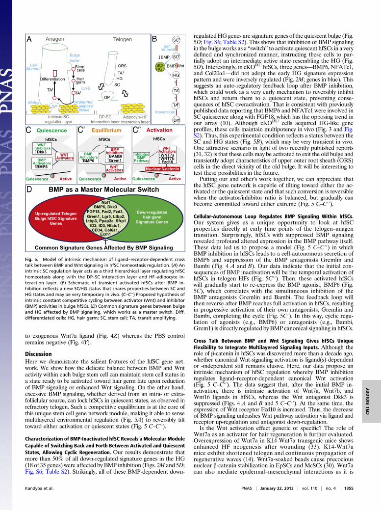
Fig. 5. Model of intrinsic mechanism of ligand–receptor-dependent crosstalk between BMP and Wnt signaling in hfSC homeostasis regulation. (A) Anintrinsic SC regulation layer acts as a third hierarchical layer regulating hfSChomeostasis along with the DP–SC interaction layer and HF–adipocyte in-teraction layer. (B) Schematic of transient activated hfSCs after BMP in-hibition reflects a new SC/HG status that shares properties between SC andHG states and may be very temporary in vivo. (C–C′′) Proposed hypothesis ofintrinsic constant competitive cycling between activator (Wnt) and inhibitor(BMP) activities in bulge hfSCs. (D) Common signature genes between bulgeand HG affected by BMP signaling, which works as a master switch. Diff,differentiated cells; HG, hair germ; SC, stem cell; TA, transit amplifying.
Kandyba et al. PNAS | January 22, 2013 | vol. 110 | no. 4 | 1355
CELL
BIOLO
GY

important in maintaining the HFs inducing activity of culturedDP cells (34). Other Wnts such as Wnt10b were visualized in thelower, permanent portion of the follicular epithelium (ISH) (27,30). The exogeneous delivery of Wnt10b can lead to anagenactivation (35). The stem cell module here may be able to sensedifferent Wnts as activators, and we will focus more on the in-crease of β-catenin activity in hfSC, rather than on specific Wnts.Is increasedWnt signaling a direct effect of BMP inactivation or
a consequence of hair germ activation? We consistently observedWnt ligand and receptor expression changes immediately follow-ing BMP inactivation in quiescent hfSCs (Fig. 4 A and B and Fig.S1 F–G and J–K′), whenmost of the canonicalWnt signaling targetgenes were not yet affected (Fig. 2M, overlapping Wnt/HG up-regulated genes, in brown) (10, 25). At this early point, only onegene, cyclin B1 (ccnb1), overlapped between the Wnt and BMPpathways following BMP inhibition in hfSCs (Fig. 2M, overlappingcKORUand Wnt/HG up-regulated genes, underlined in brown).This delayed activation of most canonical Wnt-dependent cellcycle target genes after BMP inactivation is consistent with ourmodel emphasizing that BMP inhibition precedes ligand–receptor-dependent canonical Wnt up-regulation and hfSC activation.Finally, we wonder if BMP regulation of Wnt is at the tran-
scriptional level. We were able to repress Wnt7a in vivo usingour BMP gain-of-function, inducible K15-GFP+/dTg system(Fig. 4O, P21+Dox) and confirmed direct binding of P-Smads tothe Wnt7a promoter in vivo in FACS-isolated hfSCs by ChIPassay (Fig. 4T) when HFs remained in a BMP-induced telogen atP21 (Fig. S4D′).In summary, we are able to demonstrate the key role of BMP
signaling and its cross talk in the gene network governing thehomeostasis of hfSCs. Inactivation of BMP signaling in K15-positive bulge stem cells reveals intracellular cross talk between
the BMP/Wnt pathways. Such dynamic balance confers hfSCswith a robust ability to regenerate cyclically and to sense genericactivators and inhibitors when deciding whether to activate or not.
Materials and MethodsMice and RU486 Treatment Time Line. All mice were housed and bred withinthe animal facility at the University of Southern California in accordancewith the Institutional Animal Care and Use Committee (Protocol 11543). Aseries of matings were set up using Bmpr1afl/fl mice (36), K15-CrePR1 mice(7), and Rosa26-STOP-eYFP (24) mice to generate offspring Bmpr1a+/+
(control, CON), Bmpr1afl/+(CON), or Bmpr1afl/fl (knockout, cKO) mice,which were genotypically positive for K15CrePR and Rosa-STOP-eYFP.Targeting was achieved by daily application of 2.5 mg/mouse RU486 [(wt/vol) in 100% ethanol; VWR] for 16 d to shaved back skins at P43 (corre-sponding to the start of the second postnatal telogen) and ending at P59.
Mice and Doxy Treatment Time Line. Previously generated doxycycline (Doxy)-inducible double-transgenic (dTg) mice that express a constitutively activeform of Bmpr1a gene (12) were crossed in the background of K15-GFP re-porter mice (37). GFP+ hair follicle stem cells (hfSCs) for quantitative PCR(qPCR) analysis were sorted by FACS from either untreated or Doxytreated(3 d) postnatal day 21 (P21) mice.
ACKNOWLEDGMENTS. We thank Dr. Richard R. Behringer (MD AndersonCancer Center) for floxed-Bmpr1a mice and Dr. Peggy Farnham (Universityof Southern California) for help with ChIP assay optimization. We thank theGenomics Core Facility, Children’s Hospital Los Angeles, and the University ofSouthern California Flow Cytometry Core and Animal Facility for mousehusbandry. E.K. is a fellow of the California Institute for Regenerative Med-icine (CIRM)–Research Training Program II in Stem Cell Biology. This workwas supported initially by the Donald E. and Delia B. Baxter FoundationAward (to K.K.) and National Institute of Arthritis and Musculoskeletaland Skin Diseases of the National Institutes of Health Grants R01-AR061552 (to K.K.), AR42177 (to C.-M.C.), and AR060306 (to C.-M.C.).
1. Chuong CM, Randall VA, Widelitz RB, Wu P, Jiang TX (2012) Physiological re-generation of skin appendages and implications for regenerative medicine. Physiol-ogy (Bethesda) 27(2):61–72.
2. Ito M, et al. (2005) Stem cells in the hair follicle bulge contribute to wound repair butnot to homeostasis of the epidermis. Nat Med 11(12):1351–1354.
3. Chen CC, Chuong CM (2012) Multi-layered environmental regulation on the homeo-stasis of stem cells: The saga of hair growth and alopecia. J Dermatol Sci 66(1):3–11.
4. Cotsarelis G, Sun TT, Lavker RM (1990) Label-retaining cells reside in the bulge area ofpilosebaceous unit: Implications for follicular stem cells, hair cycle, and skin carcino-genesis. Cell 61(7):1329–1337.
5. Tumbar T, et al. (2004) Defining the epithelial stem cell niche in skin. Science 303(5656):359–363.
6. Blanpain C, Lowry WE, Geoghegan A, Polak L, Fuchs E (2004) Self-renewal, multi-potency, and the existence of two cell populations within an epithelial stem cell niche.Cell 118(5):635–648.
7. Morris RJ, et al. (2004) Capturing and profiling adult hair follicle stem cells. Nat Bi-otechnol 22(4):411–417.
8. Claudinot S, Nicolas M, Oshima H, Rochat A, Barrandon Y (2005) Long-term re-newal of hair follicles from clonogenic multipotent stem cells. Proc Natl Acad SciUSA 102(41):14677–14682.
9. Müller-Röver S, Peters EJ, Botchkarev VA, Panteleyev A, Paus R (1998) Distinct pat-terns of NCAM expression are associated with defined stages of murine hair folliclemorphogenesis and regression. J Histochem Cytochem 46(12):1401–1410.
10. Greco V, et al. (2009) A two-step mechanism for stem cell activation during hair re-generation. Cell Stem Cell 4(2):155–169.
11. Blanpain C, Fuchs E (2009) Epidermal homeostasis: A balancing act of stem cells in theskin. Nat Rev Mol Cell Biol 10(3):207–217.
12. Kobielak K, Stokes N, de la Cruz J, Polak L, Fuchs E (2007) Loss of a quiescent niche butnot follicle stem cells in the absence of bone morphogenetic protein signaling. ProcNatl Acad Sci USA 104(24):10063–10068.
13. Plikus MV, et al. (2008) Cyclic dermal BMP signalling regulates stem cell activationduring hair regeneration. Nature 451(7176):340–344.
14. Plikus MV, et al. (2011) Self-organizing and stochastic behaviors during the re-generation of hair stem cells. Science 332(6029):586–589.
15. Festa E, et al. (2011) Adipocyte lineage cells contribute to the skin stem cell niche todrive hair cycling. Cell 146(5):761–771.
16. Janich P, et al. (2011) The circadian molecular clock creates epidermal stem cell het-erogeneity. Nature 480(7376):209–214.
17. Lin KK, et al. (2009) Circadian clock genes contribute to the regulation of hair folliclecycling. PLoS Genet 5(7):e1000573.
18. Zhang J, et al. (2006) Bone morphogenetic protein signaling inhibits hair follicleanagen induction by restricting epithelial stem/progenitor cell activation and ex-pansion. Stem Cells 24(12):2826–2839.
19. Plikus M, et al. (2004) Morpho-regulation of ectodermal organs: Integument pa-thology and phenotypic variations in K14-Noggin engineered mice through modu-lation of bone morphogenic protein pathway. Am J Pathol 164(3):1099–1114.
20. Botchkarev VA, et al. (2001) Noggin is required for induction of the hair folliclegrowth phase in postnatal skin. FASEB J 15(12):2205–2214.
21. Andl T, et al. (2004) Epithelial Bmpr1a regulates differentiation and proliferation in post-natal hair follicles and is essential for tooth development. Development 131(10):2257–2268.
22. Kobielak K, Pasolli HA, Alonso L, Polak L, Fuchs E (2003) Defining BMP functions in thehair follicle by conditional ablation of BMP receptor IA. J Cell Biol 163(3):609–623.
23. Gat U, DasGupta R, Degenstein L, Fuchs E (1998) De novo hair follicle morphogenesisand hair tumors in mice expressing a truncated beta-catenin in skin. Cell 95(5):605–614.
24. Srinivas S, et al. (2001) Cre reporter strains produced by targeted insertion of EYFPand ECFP into the ROSA26 locus. BMC Dev Biol 1:4.
25. Lowry WE, et al. (2005) Defining the impact of beta-catenin/Tcf transactivation onepithelial stem cells. Genes Dev 19(13):1596–1611.
26. Weinberg WC, et al. (1993) Reconstitution of hair follicle development in vivo:Determination of follicle formation, hair growth, and hair quality by dermal cells. JInvest Dermatol 100(3):229–236.
27. Reddy S, et al. (2001) Characterization of Wnt gene expression in developing andpostnatal hair follicles and identification of Wnt5a as a target of Sonic hedgehog inhair follicle morphogenesis. Mech Dev 107(1–2):69–82.
28. Nakahiro T, Kurooka H, Mori K, Sano K, Yokota Y (2010) Identification of BMP-responsiveelements in the mouse Id2 gene. Biochem Biophys Res Commun 399(3):416–421.
29. Ho CC, Zhou X, Mishina Y, Bernard DJ (2011) Mechanisms of bone morphogeneticprotein 2 (BMP2) stimulated inhibitor of DNA binding 3 (Id3) transcription. Mol CellEndocrinol 332(1-2):242–252.
30. Rabbani P, et al. (2011) Coordinated activation of Wnt in epithelial and melanocytestem cells initiates pigmented hair regeneration. Cell 145(6):941–955.
31. Jaks V, et al. (2008) Lgr5 marks cycling, yet long-lived, hair follicle stem cells. NatGenet 40(11):1291–1299.
32. Hsu YC, Pasolli HA, Fuchs E (2011) Dynamics between stem cells, niche, and progeny inthe hair follicle. Cell 144(1):92–105.
33. Ito M, et al. (2007) Wnt-dependent de novo hair follicle regeneration in adult mouseskin after wounding. Nature 447(7142):316–320.
34. Kishimoto J, Burgeson RE, Morgan BA (2000) Wnt signaling maintains the hair-in-ducing activity of the dermal papilla. Genes Dev 14(10):1181–1185.
35. Li YH, et al. (2012) Adenovirus-mediated Wnt10b overexpression induces hair follicleregeneration. J Invest Dermatol, 10.1038/jid.2012.235.
36. Mishina Y, Hanks MC, Miura S, Tallquist MD, Behringer RR (2002) Generation of Bmpr/Alk3 conditional knockout mice. Genesis 32(2):69–72.
37. Liu Y, Lyle S, Yang Z, Cotsarelis G (2003) Keratin 15 promoter targets putative epi-thelial stem cells in the hair follicle bulge. J Invest Dermatol 121(5):963–968.
1356 | www.pnas.org/cgi/doi/10.1073/pnas.1121312110 Kandyba et al.
Related Documents