Graduate Theses, Dissertations, and Problem Reports 2006 Comparison of the obturation density of gutta percha using cold Comparison of the obturation density of gutta percha using cold lateral condensation and varying continuous wave of lateral condensation and varying continuous wave of condensation techniques condensation techniques Jamie Michelle Day West Virginia University Follow this and additional works at: https://researchrepository.wvu.edu/etd Recommended Citation Recommended Citation Day, Jamie Michelle, "Comparison of the obturation density of gutta percha using cold lateral condensation and varying continuous wave of condensation techniques" (2006). Graduate Theses, Dissertations, and Problem Reports. 2376. https://researchrepository.wvu.edu/etd/2376 This Thesis is protected by copyright and/or related rights. It has been brought to you by the The Research Repository @ WVU with permission from the rights-holder(s). You are free to use this Thesis in any way that is permitted by the copyright and related rights legislation that applies to your use. For other uses you must obtain permission from the rights-holder(s) directly, unless additional rights are indicated by a Creative Commons license in the record and/ or on the work itself. This Thesis has been accepted for inclusion in WVU Graduate Theses, Dissertations, and Problem Reports collection by an authorized administrator of The Research Repository @ WVU. For more information, please contact [email protected].

Welcome message from author
This document is posted to help you gain knowledge. Please leave a comment to let me know what you think about it! Share it to your friends and learn new things together.
Transcript
Graduate Theses, Dissertations, and Problem Reports
2006
Comparison of the obturation density of gutta percha using cold Comparison of the obturation density of gutta percha using cold
lateral condensation and varying continuous wave of lateral condensation and varying continuous wave of
condensation techniques condensation techniques
Jamie Michelle Day West Virginia University
Follow this and additional works at: https://researchrepository.wvu.edu/etd
Recommended Citation Recommended Citation Day, Jamie Michelle, "Comparison of the obturation density of gutta percha using cold lateral condensation and varying continuous wave of condensation techniques" (2006). Graduate Theses, Dissertations, and Problem Reports. 2376. https://researchrepository.wvu.edu/etd/2376
This Thesis is protected by copyright and/or related rights. It has been brought to you by the The Research Repository @ WVU with permission from the rights-holder(s). You are free to use this Thesis in any way that is permitted by the copyright and related rights legislation that applies to your use. For other uses you must obtain permission from the rights-holder(s) directly, unless additional rights are indicated by a Creative Commons license in the record and/ or on the work itself. This Thesis has been accepted for inclusion in WVU Graduate Theses, Dissertations, and Problem Reports collection by an authorized administrator of The Research Repository @ WVU. For more information, please contact [email protected].
COMPARISON OF THE OBTURATION DENSITY OF GUTTA PERCHA USING COLD LATERAL
CONDENSATION AND VARYING CONTINUOUS WAVE OF CONDENSATION TECHNIQUES
Jamie Michele Day, D.D.S.
Thesis submitted to the School of Dentistry
At West Virginia UniversityIn Partial Fulfillment of the Requirements
For the Degree of
Master of Sciencein
Endodontics
C. Russell Jackson, D.D.S., M.S., ChairMichael Bagby, D.D.S., M.S., PhDLouise T. Veselicky, D.D.S., M.S.D.
Department of Endodontics
Morgantown, West Virginia2006
Keywords: Obturation Density, Continuous Wave of CondensationCopyright 2006 Jamie M. Day
ABSTRACT
COMPARISON OF THE OBTURATION DENSITY OF GUTTA PERCHA USING COLD LATERAL CONDENSATION AND VARYING CONTINUOUS WAVE OF
CONDENSATION TECHNIQUES
Jamie Michele Day, D.D.S.
This study compared the density of gutta percha when using cold lateral condensation (CLC) and continuous wave of condensation (CWC) under varying temperature and plugger depths. Seventy acrylic blocks were instrumented. The blocks were weighed and assigned to seven groups. Group 1 was obturated with CLC. The remaining six groups were obturated with CWC. Groups 2-4 were obturated by advancing the plugger to 3 mm short of the working length at 100C, 200C, and 300C, respectively, and then backfilled. In groups 5-7 the plugger was advanced to 5 mm short of working length. The blocks were weighed again after obturation. Data were analyzed using a one-way ANOVA and Tukey’s tests. Results indicated a significantly greater density of gutta percha when using CWC compared to CLC and when the plugger was advanced to 3 mm short of working length (p<.05). No significant differences were noted between the three different temperatures (p>.05).
iii
DEDICATION
For Mom, Dad and JoshNo support could ever compare to what I have received.
Thanks.
iv
ACKNOWLEDGEMENTS
Thanks to Dr. Russ Jackson for giving me the opportunity to become an endodontist and for guidance along the way.
Thanks to Drs. Bagby and Veselicky for help and support during my thesis from start to finish.
Thanks to Marilyn, Cathy and Gina for all of their help in everything and for wonderful friendships.
Thanks to Drs. Lora Ford and Pam Harrington for making the first year fun and for helping us learn the ropes.
Thanks to Drs. Nic Taylor and Kent McBride for all of the laughs, memories and for never letting any moment become a dull one. I really value the friendships we have developed.
Thanks to Dr. Becky Tolley for being there day in and day out when I needed to vent about something.
Finally, thanks to Dr. Rodney Southern. Where should I begin? I would not have wanted to share this experience with anyone else. No other person can appreciate what the last two years have been like but the two of us. It is truly a special thing to be thrown into a program with a complete stranger only to leave it two years later with one of the best friends you will ever have. There are too many things to attempt to thank you for them all. But, you know I appreciate everything you have ever done and continue to do for me.
v
TABLE OF CONTENTS
ABSTRACT.............................................................................................................................. iiDEDICATION......................................................................................................................... iiiACKNOWLEDGEMENTS..................................................................................................... ivTABLE OF CONTENTS.......................................................................................................... vLIST OF FIGURES ................................................................................................................. viLIST OF TABLES.................................................................................................................. viiCHAPTER I .............................................................................................................................. 1
INTRODUCTION ................................................................................................................ 1STATEMENT OF THE PROBLEM................................................................................ 2SIGNIFICANCE OF THE STUDY.................................................................................. 2HYPOTHESIS .................................................................................................................. 3DEFINITION OF TERMS ............................................................................................... 3ASSUMPTIONS............................................................................................................... 5LIMITATIONS................................................................................................................. 5DELIMITATIONS ........................................................................................................... 5
CHAPTER II............................................................................................................................. 6REVIEW OF LITERATURE ............................................................................................... 6
CHAPTER III ......................................................................................................................... 15MATERIALS AND METHODS........................................................................................ 15
SAMPLE DESCRIPTION.............................................................................................. 15INSTRUMENTATION METHODOLOGY .................................................................. 15OBTURATION METHODOLOGY .............................................................................. 16DATA COLLECTION ................................................................................................... 18MATERIALS AND EQUIPMENT................................................................................ 19
CHAPTER IV ......................................................................................................................... 20RESULTS AND DISCUSSION......................................................................................... 20
RESULTS ....................................................................................................................... 20DISCUSSION................................................................................................................. 21
CHAPTER V .......................................................................................................................... 25SUMMARY AND CONCLUSIONS ................................................................................. 25
SUMMARY.................................................................................................................... 25CONCLUSIONS............................................................................................................. 26
REFERENCES ....................................................................................................................... 27APPENDIX A......................................................................................................................... 31
EXAMINER DATA SHEET.............................................................................................. 31APPENDIX B ......................................................................................................................... 33
STATISTICAL RESULTS................................................................................................. 33CURRICULMN VITAE......................................................................................................... 38
vi
LIST OF FIGURES
Figure 1. Sample Acrylic Block .......................................................................................... 15Figure 2. Blinder .................................................................................................................. 18Figure 3. Comparison of Gutta Percha Mass between Groups ....................................... 21
vii
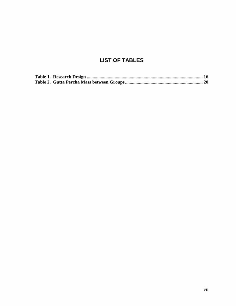
LIST OF TABLES
Table 1. Research Design .................................................................................................... 16Table 2. Gutta Percha Mass between Groups ................................................................... 20
1
CHAPTER IINTRODUCTION
Major advancements have been made in the techniques used to obturate root canal
anatomy during endodontic therapy over the last half century. Many of these improvements
have come about, in part, due to improved techniques to the cleaning and shaping stage of
root canal therapy. The cleaning and shaping procedure allows the clinician to produce a
canal that is cleaned mechanically and chemically and shaped to the optimal configuration to
accept obturation. The obturation procedure attempts to eliminate all routes of leakage that
may occur from the oral cavity and seals in any other irritants that could not fully be removed
during the preparation of the canal.
The continuous wave of condensation obturation technique developed by Dr. L.
Stephen Buchanan has shown much promise in three-dimensional filling of the root canal
system. (1) During this procedure, gutta percha is heated with one continuous wave of heat to
facilitate movement of gutta percha and sealer into even the smallest of canal intricacies.
The mass of apical gutta percha is then compacted vertically to produce a dense apical fill.
With the increasing popularity of this technique, come many concerns about the
potential complications that can occur when periradicular temperatures rise above acceptable
conditions. Gutta percha is still the most widely used and accepted obturation material
because of its biocompatibility, inertness, dimensional stability, compactibility, plasticity
when heated and ease of removal is retreatment becomes necessary. (2) However, to heat
2
gutta percha to the state necessary for a complete three-dimensional fill, high and prolonged
heat is recommended.
This study was designed to compare the density of gutta percha produced under
varying temperatures and plugger depth penetration of a System B Heatsource plugger. The
possibility exists that a lower temperature setting and less penetration into the canal with a
heated instrument could produce equal fill of the canal system and reduce possible
complications that may occur with higher temperatures.
STATEMENT OF THE PROBLEM
There have been no studies to evaluate the density of gutta percha under varying
temperature conditions and plugger depth of the System B Heatsource when utilizing the
continuous wave of condensation obturation technique.
SIGNIFICANCE OF THE STUDY
Cleaning and shaping of a root canal system is critical in the ability to properly
obturate the anatomy of the system. New techniques in cleaning and shaping have led to
improved techniques in obturation as well. The continuous wave of condensation technique
is a warm vertical technique that has shown promise in more complete three-dimensional
filling of canal systems. However, the technique produces potentially high temperatures that
may have damaging implications to the periradicular tissues. The results of this study could
suggest the proper temperature setting and depth of plugger penetration of the System B
Heatsource for optimal obturation when utilizing the continuous wave of condensation
obturation technique.
3
HYPOTHESIS
The null hypotheses are: (1) There is no significant difference in the mass of
obturation material between the lateral condensation group (control group) and the
continuous wave of condensation groups (experimental groups). (2) There is no significant
difference in the mass of obturation material among the experimental groups when
comparing plugger temperature and depth of plugger penetration of the System B Heatsource
when utilizing the continuous wave of condensation technique.
DEFINITION OF TERMS
Backfilling- The injection and/or compaction of gutta percha into a canal after creation of an
apical seal.
Cold lateral condensation- An obturation technique which involves the placement of sealer
and a fitted master gutta percha point, which is laterally compacted by a spreader to make
room for additional accessory gutta percha points.
Continuous wave of condensation (CWC)- A technique of warm vertical gutta percha
compaction developed by L. Stephen Buchanan, D.D.S. This technique utilizes a wave of
continuous heat from an electronic heat source to warm the gutta percha during apical
compaction.
Crown-down preparation- A technique of canal preparation, which enlarges the coronal
portion of the canal initially. The canal is enlarged in a step-wise fashion from the coronal
orifice towards the apical foramen with progressively smaller files until the desired length is
achieved.
4
Downpacking- The compaction of incremental segments of heat-softened gutta-percha to
create an apical plug.
Master apical file (MAF)- The largest file used to the full working length of the completely
prepared root canal.
Obtura II- A device used to expel warm gutta percha into the root canal space.
Obturation density- The amount of compacted gutta percha in the canal space.
Plugger- A smooth, flat-ended and slightly tapered metal instrument designed to compact
materials vertically within a prepared root canal.
Reference point- A repeatable landmark at the top of the acrylic block used to maintain
working length.
Rotary instrumentation- A technique of canal instrumentation, which utilizes an engine
driven handpiece to rotate endodontic files in a 360-degree clockwise direction.
Spreader- A smooth, pointed, slightly tapered metal instrument designed to compact
materials within a prepared root canal.
System B Heatsource- An electronic device, which provides the delivery of a heated plugger
type instrument in the canal. The System B heating units have tapered plugger units that can
be sized to fit most canal spaces.
Warm vertical condensation- An obturation technique that utilizes a heated plugger or an
electronic heat source instrument. Gutta percha is burned off with an instrument and then
immediately compacted apically with a plugger.
Working length- The length from the top of the acrylic block to the canal terminus.
5
ASSUMPTIONS
(1) Acrylic blocks can be satisfactorily instrumented and obturated to be evaluated for
density of obturation material. (2) Density of obturation material is a good model in
evaluating complete fill of the root canal system.
LIMITATIONS
(1) This in-vitro study attempted to represent root canal obturation concepts that can be
applied to a clinical setting; however, results may vary from in-vivo conditions. (2) Heat
produced from the System B could potentially alter anatomy of the acrylic blocks.
DELIMITATIONS
(1) Acrylic blocks were used to provide least amount of variability possible. (2) All
simulated canals had the same degree of curvature. (3) All canals were worked to an MAF of
#35. (4) All blocks were instrumented, obturated, and weighed by the principal investigator.
(5) Sealer was not used so only gutta percha mass could be studied. (6) Any block with
obvious signs of charring or melting of the acrylic after use of the System B Heatsource was
eliminated.
6
CHAPTER IIREVIEW OF LITERATURE
The three-dimensional obturation of the root canal system is widely accepted as a key
factor for successful endodontic therapy. (3) Studies have come to show that one can achieve
a more complete root canal filling by utilizing a warm obturation technique. Techniques and
armamentarium have evolved greatly to allow practitioners a more efficient, reliable and
predicable way to incorporate this type of obturation into their practices. Schilder described
the warm vertical obturation technique in 1967 and since then, modifications to this
technique have flooded the endodontic literature. (4) Regardless of the way endodontic
obturation procedures are completed, the objectives have remained the same.
The purposes of obturating the prepared root canal space are as follows: (1) to
eliminate all avenues of leakage from the oral cavity and periradicular tissues into the root
canal system, and (2) to seal within the system any irritants that cannot be fully removed
during the cleaning and shaping procedures. These objectives recognize the importance of
microbial irritants and pulp tissue degeneration products as a prime cause for pulpal decline
and that failure to remove these factors adequately will cause continued irritation from
contamination and subsequently root canal therapy failure. (5) Therefore, three-dimensional
obturation is of utmost importance when evaluating a tooth following root canal therapy
completion.
Cold lateral condensation is the foundation obturation technique that is understood by
students and practitioners alike since is has been the technique in which most all dental
7
students have been taught in the past century. It is typically the technique used to compare
all obturation techniques against in studies examining obturation procedures. It is not
accepted that this is necessarily a superior technique, but rather that it is reliable,
reproducible and understandable to most clinicians. (2)
The cold lateral condensation procedure involves cleaning and shaping the root canal
system to produce a conical shape that forms a continuous taper from its coronal aspect to the
apex. A master gutta percha cone is fitted to place with sealer and successive accessory
cones are inserted alongside the master cone. Due to the funnel shape of the canal, a
spreader is easily inserted making room for each of the accessory points to be placed. During
this procedure, the gutta percha is compacted into a dense mass that fills the root canal space.
(6)
Before any type of obturation procedure can be successfully accomplished, one must
first shape and clean the canal with the objectives of three-dimensional obturation in mind.
The shaping procedure removes the bulk contents from the canal system and creates the
smooth, tapered opening to the terminus. Shaping also facilitates cleaning and obturation by
removing restrictive dentin and allowing effective volumes of irrigants and instruments to
work deeper and more quickly into the entire system. With the invention of greater taper
files, cleaning and shaping has become more efficient especially in the coronal third of the
root canal allowing these objectives to be more easily met. Any clinician must realize that
shape is critical not only for effective cleaning, but also for three-dimensional obturations.
Consistently producing optimal shape will continuously produce well-obturated canals and
become the foundation in a clinician’s endodontic success. (7)
8
Gutta percha is the most commonly used obturating material in endodontics. Dental
gutta percha contains about 20% gutta percha, 66% zinc oxide, 11% heavy metal sulfates,
and 3% waxes and resins. (8) It exists in two distinct crystalline forms, a beta form and an
alpha form, which can be converted into each other. The alpha form comes from the natural
tree product and the beta form is that which is processed and used in root canal systems.
During heating and cooling, phase transitions occur. At around 46-52° C, the crystalline
structure changes from the beta phase to alpha phase and ultimately into an amorphous melt
at around 56-62° C. (5) Normal cooling returns it to a beta phase as temperatures fall to
around 42-40°, however a rapid decrease in volume occurs as this cooling takes place.
Shrinkage can be prevented if it is not heated above 45° C, however a temperature of 64° C is
necessary to soften gutta percha for warm techniques. Therefore, it is necessary to maintain
vertical pressure in all warm gutta percha techniques to compensate for the volume changes
that occur as cooling takes place. (9) Newer alpha form gutta percha has been marketed for
dental use under trade names such as Thermafil, Densfil and Microseal. These have a lower
melting point, lower viscosity and will flow with less pressure and stress while exhibiting an
increase in tackiness to form a more homogenous fill. They also exhibit less shrinkage, but
are technique sensitive procedures. (10)
The essentials of warm vertical compaction have been around for almost a century,
but Dr. Herb Schilder popularized this technique in the 1960’s and it now is commonly called
the Schilder technique. This technique utilizes a system of varying sized pluggers to burn off
and compact warm gutta percha apically. A master cone is selected, fit for size, coated with
sealer and inserted into the root canal. The largest plugger is used to burn off the coronal
portion of the gutta percha with immediate apical compaction. This process continues with
9
smaller pluggers until 5.0mm of gutta percha remains in the root canal. At this stage the
space can be left if a post space is required, or backfilled with another technique. During this
process, gutta percha is forced into canal irregularities. (4)
In 1986, Buchanan introduced the continuous wave of condensation technique. (1)
This technique, utilizes an electrical heat source (i.e. System B) with a heated plugger
delivery unit, standard gutta percha and sealer. Once cleaning and shaping have been
accomplished, a heated plugger delivery unit is selected based on the canal size and
configuration and pre-fitted to make certain the plugger can advance to between 4-5 mm
from the working length. Once this has been accomplished, a gutta percha cone is selected
and fit to the working length. The heated plugger is used to sear off the gutta percha at the
canal orifice and a plugger is used to compact this gutta percha. The plugger is then
activated and introduced into the canal at 200° C to about 3-4 mm short of its binding point
before the plugger is deactivated. The plugger will continue to advance to one mm short of
its binding point at which firm apical pressure should be maintained for 10 seconds. This
action compensates for any shrinkage that may occur during cooling. While still maintaining
pressure, the plugger is activated with a one second burst of heat and the plugger is quickly
removed. With a smaller plugger, it is confirmed that the apical mass of gutta percha has not
become dislodged by maintaining firm apical pressure. At this point the space can remain for
a post space, or backfilled. (11) Buchanan asserts that it is not necessary to advance the
plugger further than 4-5 mm from the working length because the gutta percha is heated 4-5
mm past the tip of the plugger unit. (12) Recently, Bowman and Baumgartner found that
there is better gutta percha flow into lateral grooves when the plugger is advanced to 3 mm
from working length. (13)
10
There have been many studies comparing these two warm vertical techniques to one
another and to cold lateral condensation. Many advantages have been found when using a
warm obturation technique. Blum et. al. used a force analyzer device and found a decrease in
the wedging effect for warm vertical compaction compared to lateral condensation. This may
have an effect on the fracture resistance of teeth during root canal therapy. (14)
Studies have overwhelming shown that warm vertical obturation techniques
accomplish a more complete three-dimensional filling than cold techniques. A statistically
significant greater number of simulated lateral canals were obturated when using the System
B plus the Obtura II as compared to cold lateral or the Obtura II alone. (15) Warm vertical
compaction using the continuous wave condensation technique in acrylic blocks resulted in a
greater gutta percha fill by mass compared with standard cold lateral. (16) In a study
comparing three obturation techniques, the warm obturation techniques had significantly
more gutta percha in lateral canals. (3) The percentage of gutta percha filled canal area using
warm gutta percha was greater than that of cold gutta percha in oval canals. (17)
Jacobson et. al. studied the microleakage of the continuous wave of condensation
technique and found that microbial coronal leakage occurs more quickly using lateral
condensation than with the System B continuous wave of condensation and Obtura II
backfill. (18) When using the System B, Nelson et al. found a 23.97% increase in the weight
of gutta percha after one heat application compared to standard cold lateral condensation.
After a second application of heat, an additional of 2.59% increase was produced. The
results indicate that warm techniques using the System B result in a denser gutta percha fill
by weight when compared to standard cold lateral. (19) Under laboratory conditions,
Gilhooley et. al. found that warm gutta percha had a better sealabilty as assessed with a dye
11
penetration method than cold lateral condensation. (20) Wong et. al. found that warm vertical
condensation was significantly better at reproducing canal anatomy than cold lateral
condensation. (21) The results of these studies indicate the potential for a more complete
dense fill when using a warm obturation technique. Eldeeb found a correlation between
radiographic density and leakage especially in the middle one third of the root. (22) Echoing
this, Chugal et. al. found the risk of failure to be higher for a poor or fair density of
obturation than for a good density for any type of periradicular diagnosis prior to treatment.
(23)
Although most studies find warm vertical a superior technique, not all studies have
found this to be true. Benner et. al. found no difference in leakage between cold lateral and
warm vertical condensation. (24) Larder et. al. found that there was no significant difference
in cold lateral and warm vertical condensation at reproducing canal anatomy. (25)
There are some significant disadvantages that accompany the use of warm vertical
techniques as well. First, when filling a canal system with a warm technique, care must be
taken to avoid excessive extrusion of material through the apex. Root canals filled with
warm gutta percha had significantly more extrusion of sealer and gutta percha than canals
filled by lateral condensation. (26) Many of the newer techniques allow this factor to be more
controlled and predictable. Furthermore, if the root canal is optimally shaped, an
environment is created to force excess gutta percha in a coronal direction instead of through
the apex.
Another concern is the heat that is generated when using these techniques. Eriksson
and Albrektsson found that in a rabbit tibia, a temperature of 53° C, i.e. below the
12
denaturation point of alkaline phosphatase, caused an irreversible bone injury, after which
healing occurred from the surrounding tissues. (27) It is generally accepted that a temperature
of 47° C is the critical temperature when permanent damage to the supporting structures of
the tooth root may occur. This is about a 10° C rise from average temperature. (28) Studies
by Fors et. al. and Gutmann et. al. agree that 10C above normal body temperature is the
critical level where irreversible damage to the periodontium is of concern. (29,30) Studies are
conflicting in whether or not using warm techniques allow temperatures to rise above these
critical levels. Lee used a thermocouple 2 mm below the cementoenamel junction to transfer
heat to a digital thermometer and found that the System B had <10° C rise in all experimental
teeth. The Touch n’ Heat electronic heating device demonstrated a rise of <10° C in all teeth
expect mandibular incisors, and a flame-heated carrier demonstrated >10° C in all
experimental teeth. (31) Lipiski studied the root surface temperature rise during root canal
obturation using the System B and continuous wave of condensation technique with an
infrared thermal imaging camera. He found that the System B produced temperature changes
on the outer root surfaces, which, in the case of teeth with relatively thin dentinal walls, can
reach high values. In the case of mandibular central incisors, temperatures elevated more
than 10° C. (32)
Floren et. al. found that temperatures above 250° C as those recommended for
removal of plastic obturation carriers during endodontic retreatment has the potential to cause
root surface temperatures to rise above 10° C. Using 10 thermocouples at one mm
increments along the tooth, it was found that a site 5 mm from the apex had the highest
increase in surface temperature when the plugger was inserted to a depth of 3 mm short of
working length. This temperature exceeded the critical value for over one minute indicating
13
that damage could occur. (33) Sweatman et. al. disputed these findings and found that at no
time did the external surface of the root exceed an increase of 10° C at any thermocouple
level even when the plugger was set to a temperature of 300° C. (34) Venturi et. al. agreed
that using the System B Heatsource has negligible effects on outer root temperature. (35)
With conflicting evidence in the literature, it is safe to say the care must be taken
when utilizing any warm technique and the lowest temperature possible should be used. It is
uncertain how these studies convert to in vivo conditions, however, it is certain that the
potential for complications can occur if temperatures rise above that critical level.
With this in mind, a few studies have attempted to find the lowest temperature to
achieve clinical acceptable obturation results. The depth of plugger penetration has also been
studied. Jung et. al. used acrylic blocks with artificially created oval canals and obturated
them with the continuous wave of condensation technique at 100°, 200° and 350° C
temperature settings and at plugger depth penetration of 4 mm. He also went to 2 mm from
the apex with a plugger set at 200° C. Plugger temperatures within the range used did not
affect the percentage of gutta percha (PGP) filled area, however, the penetration depth of the
plugger was significantly related to the PGP. (36) Yared et. al. found similar results. He
found that deeper plugger penetration showed significantly less apical leakage. (37) Wu et.
al. found an average of 96.1% gutta percha filled area when heat was applied 2 mm short of
working length compared to 87% when applied to only 4 mm short of working length. (38)
Guess et. al. obtained best results with a plugger depth of 3.5 to 4.5 mm from the working
length. (39)
14
This study attempted to find the lowest temperature setting and proper depth of
plugger penetration of a System B plugger when using the continuous wave of condensation
technique. An examination of obturation density was accomplished using acrylic blocks with
simulated root canal anatomy. While this was an in vitro study, all obturation concepts were
followed as they apply in a clinical setting.
15
CHAPTER IIIMATERIALS AND METHODS
SAMPLE DESCRIPTION
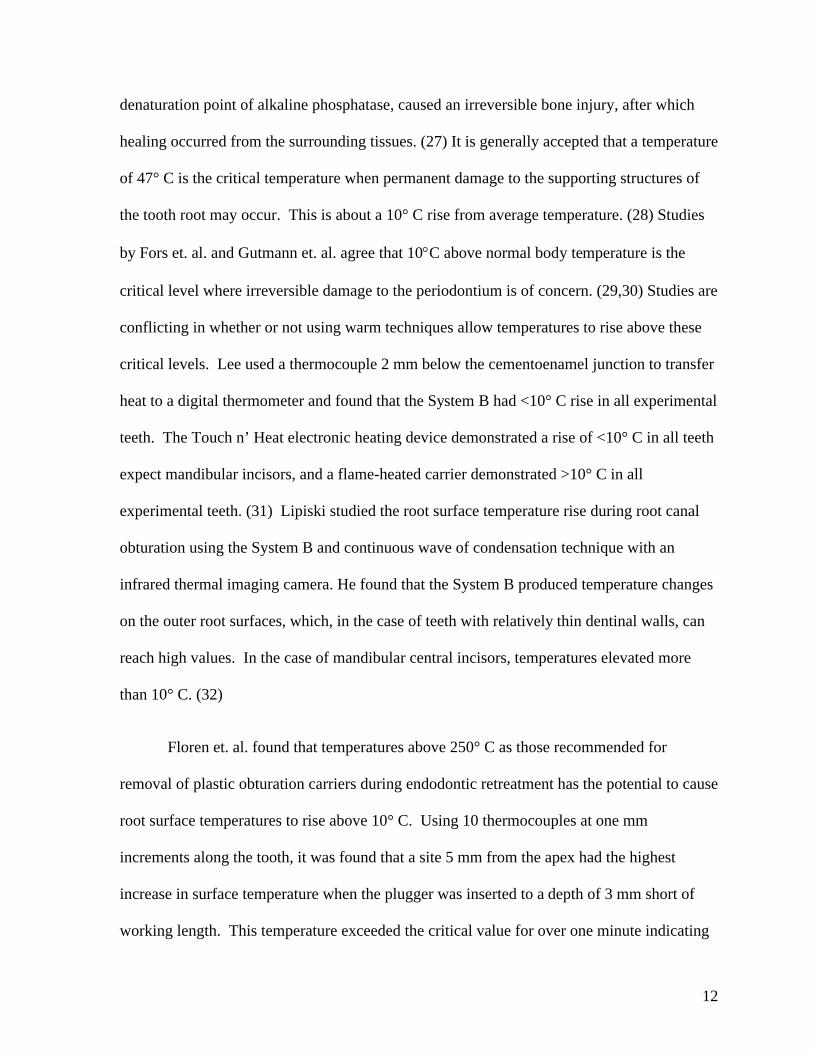
Acrylic blocks simulating root canal anatomy were obtained from Kilgore
International for this study. (see Figure 1.) All of the blocks had a 30-degree canal curvature.
They were chosen to minimize variability between the samples. Each block was
instrumented in the same manner and to the same master apical file.
Figure 1. Sample Acrylic Block
INSTRUMENTATION METHODOLOGY
A standard instrumentation procedure was carried out on every block. An ISO #10
hand file was inserted into the canal to determine working length. When the tip of the file
became visible at the terminus of the canal, the stopper was set to the reference point and
measured. This was considered the actual length of the canal. The exact working length was
determined by subtracting 1.0 mm from the measured actual length.
16
Once working length had been established, Gates Glidden burs #2, #3, and #4 were
used to enlarge the coronal half of the canal. Irrigation with at least 3.0 ml of water was used
to flush debris from the canal between Gates Glidden burs and files. ISO ProFile rotary files
were used in a crown-down preparation. A #40 file was used to prepare the top two-thirds of
the canal followed by a #35 file to the working length. Once instrumentation was complete,
each block was given a final irrigation and dried with paper points. At this point each block
was randomly assigned to a group with 10 blocks in each group. Initial weight was taken
with a digital scale and recorded to the nearest .0001 g. This weight was referred to as the
weight of block after instrumentation.
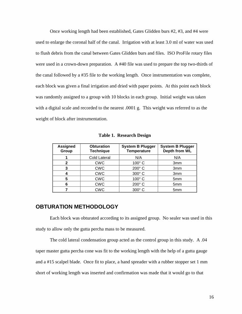
Table 1. Research Design
Assigned Group
Obturation Technique
System B Plugger Temperature
System B Plugger Depth from WL
1 Cold Lateral N/A N/A2 CWC 100° C 3mm3 CWC 200° C 3mm4 CWC 300° C 3mm5 CWC 100° C 5mm6 CWC 200° C 5mm
7 CWC 300° C 5mm
OBTURATION METHODOLOGY
Each block was obturated according to its assigned group. No sealer was used in this
study to allow only the gutta percha mass to be measured.
The cold lateral condensation group acted as the control group in this study. A .04
taper master gutta percha cone was fit to the working length with the help of a gutta gauge
and a #15 scalpel blade. Once fit to place, a hand spreader with a rubber stopper set 1 mm
short of working length was inserted and confirmation was made that it would go to that
17
depth. A #25 .02 taper accessory gutta percha cone was placed and the cold lateral
condensation procedure continued until the spreader could not penetrate more than 4 mm into
the canal from the reference point. The System B was used to sear off the cones from the
canal orifice and a large Buchanan hand plugger was used to condense the softened gutta
percha at the orifice. A #15 scalpel blade was used to trim the gutta percha flush to the top of
the block.
The Continuous wave of condensation groups were the experimental groups in this
study. A System B plugger was chosen and placed into the canal to confirm it would
advance to the needed depth. A .06 taper master gutta percha cone was fit to the working
length with the help of a gutta gauge and #15 scalpel blade. Once fit to place, the System B
was used to sear off the cone at the canal orifice and a large Buchanan hand plugger was used
to condense the softened gutta percha at the orifice. The downpack procedure varied
according to the assigned experimental group. The temperature was set at 100°C for groups 2
and 5, 200°C for groups 3 and 6, and 300°C for groups 4 and 7. In groups 2-4, the plugger
was allowed to advance to 3 mm short of the working length. In groups 5-7, the plugger was
allowed to advance to 5 mm short of the working length. The downpack procedure in all
groups consisted of activation of the plugger for no more than 3 seconds as it advanced to
within 3-4 mm of the intended depth before it was deactivated. The plugger was allowed to
continue to advance to its intended depth. When that depth had been reached, apical pressure
was maintained for 10 seconds. A separation burst of heat was applied to the plugger and
quickly removed from the canal. A small Buchanan hand plugger was used to condense the
softened gutta percha and to confirm that it had not been dislodged.
18
At this point, the canal was ready to be backfilled with the Obtura II. The Obtura II
gun was set to a temperature of 150°C and the needle was inserted into the canal and allowed
to make contact with the gutta percha mass. Flow 150 gutta percha was then extruded into
the canal as the gun was allowed to back its way out of the canal. The space was filled in one
increment of the gun and a large Buchanan hand plugger was used to condense the softened
gutta percha at the canal orifice. A #15 scalpel blade was used to trim the gutta percha flush
to the top of the block.
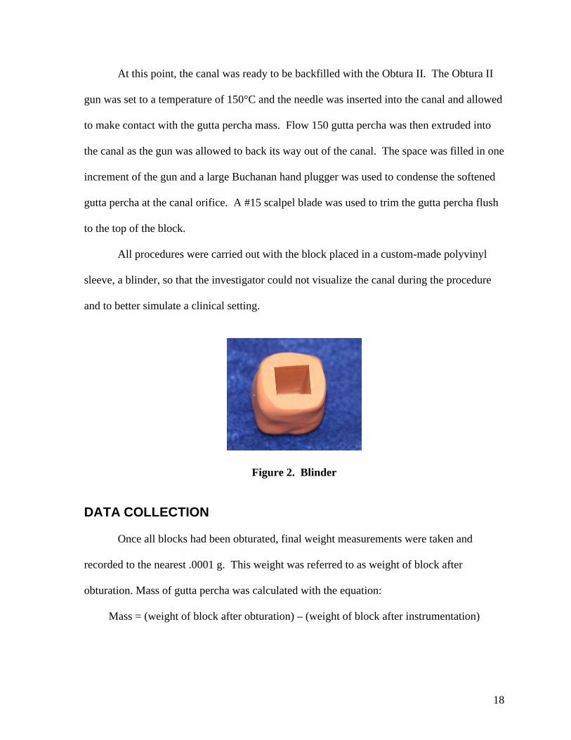
All procedures were carried out with the block placed in a custom-made polyvinyl
sleeve, a blinder, so that the investigator could not visualize the canal during the procedure
and to better simulate a clinical setting.
Figure 2. Blinder
DATA COLLECTION
Once all blocks had been obturated, final weight measurements were taken and
recorded to the nearest .0001 g. This weight was referred to as weight of block after
obturation. Mass of gutta percha was calculated with the equation:
Mass = (weight of block after obturation) – (weight of block after instrumentation)
19
Since all of the blocks were worked to the same size, it is assumed that the volume of space
obturated is the same in every block; therefore, any increase in mass would also be an
increase in density.
MATERIALS AND EQUIPMENT
70 acrylic blocks (Kilgore International, Coldwater MI)
Dentsply/Tulsa Dental Electric rotary motor and handpiece (Tulsa Dental, Tulsa
OK)
Profile ISO .06 rotary files (Tulsa Dental, Tulsa OK)
Gates Glidden burs #2-4 (Dentsply Maillefer, Tulsa OK)
Flexofile ISO endodontic files (Dentsply Maillefer, Tulsa OK)
Monojet endodontic irrigation syringes (Sherwood Medical, St. Louis MO)
Absorbent paper points #35 (Dentsply Maillefer, Tulsa OK)
Gutta percha points .02, .04, and .06 taper (Dentsply Maillefer, Tulsa OK)
Gutta gauge (Dentsply Maillefer, Tulsa OK)
#15 scalpel blade (Miltex, York PA)
cotton forceps (Hu-Friedy, Chicago, IL)
System B Heatsource with pluggers (Analytic Technologies/Sybron Endo, Orange
CA)
Obtura II (Obtura Spartan Corporation, Fenton MO)
Obtura Flow 150 gutta percha (Obtura Spartan Corporation, Fenton MO)
Endodontic hand spreader (Miltex, York PA)
Buchanan hand pluggers (Sybron Endo, Orange CA)
digital balance (American Scientific Products)
20
CHAPTER IVRESULTS AND DISCUSSION
RESULTS
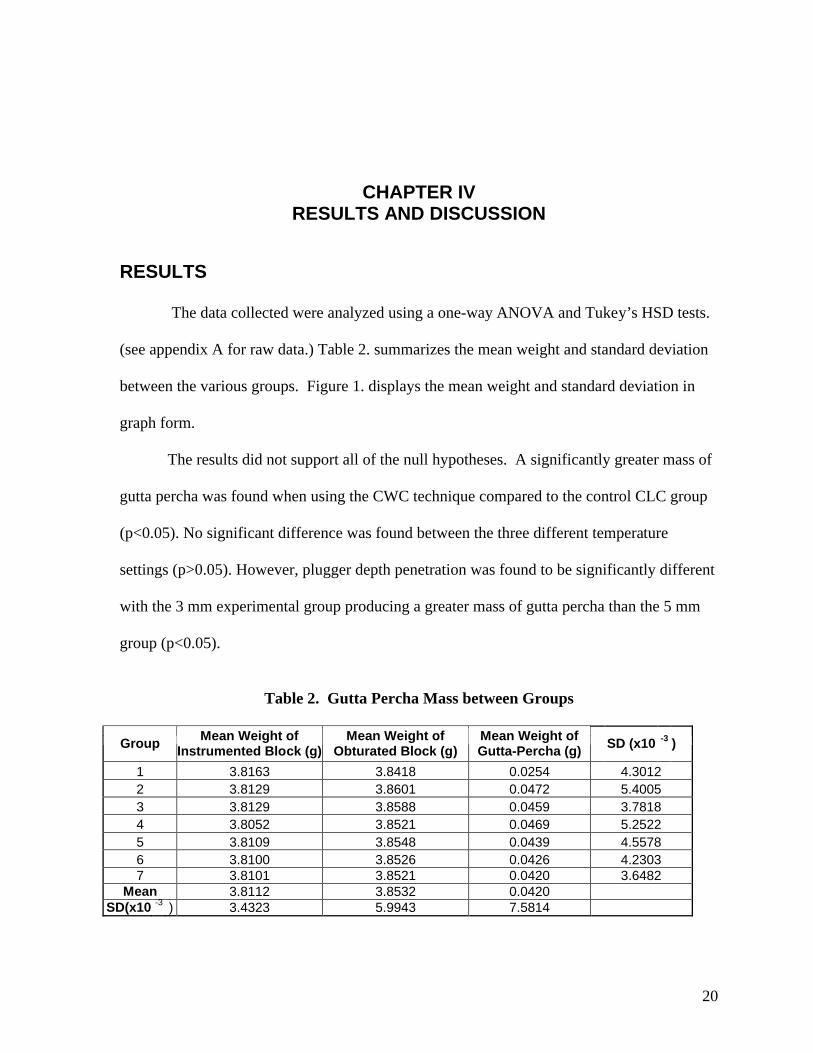
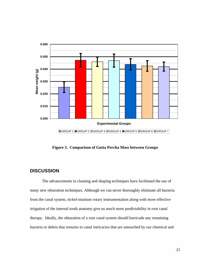
The data collected were analyzed using a one-way ANOVA and Tukey’s HSD tests.
(see appendix A for raw data.) Table 2. summarizes the mean weight and standard deviation
between the various groups. Figure 1. displays the mean weight and standard deviation in
graph form.
The results did not support all of the null hypotheses. A significantly greater mass of
gutta percha was found when using the CWC technique compared to the control CLC group
(p<0.05). No significant difference was found between the three different temperature
settings (p>0.05). However, plugger depth penetration was found to be significantly different
with the 3 mm experimental group producing a greater mass of gutta percha than the 5 mm
group (p<0.05).
Table 2. Gutta Percha Mass between Groups
GroupMean Weight of
Instrumented Block (g)Mean Weight of
Obturated Block (g)Mean Weight of Gutta-Percha (g)
SD (x10 -3 )
1 3.8163 3.8418 0.0254 4.30122 3.8129 3.8601 0.0472 5.40053 3.8129 3.8588 0.0459 3.78184 3.8052 3.8521 0.0469 5.25225 3.8109 3.8548 0.0439 4.55786 3.8100 3.8526 0.0426 4.23037 3.8101 3.8521 0.0420 3.6482
Mean 3.8112 3.8532 0.0420SD(x10 -3 ) 3.4323 5.9943 7.5814
21
0.000
0.010
0.020
0.030
0.040
0.050
0.060
Experimental Groups
Mea
n w
eig
ht
(g)
GROUP 1 GROUP 2 GROUP 3 GROUP 4 GROUP 5 GROUP 6 GROUP 7
Figure 3. Comparison of Gutta Percha Mass between Groups
DISCUSSION
The advancements in cleaning and shaping techniques have facilitated the use of
many new obturation techniques. Although we can never thoroughly eliminate all bacteria
from the canal system, nickel-titanium rotary instrumentation along with more effective
irrigation of the internal tooth anatomy give us much more predictability in root canal
therapy. Ideally, the obturation of a root canal system should barricade any remaining
bacteria or debris that remains in canal intricacies that are untouched by our chemical and
22
mechanical debridement. In order to accomplish this with consistent results, we must be
certain that we are accomplishing the most complete fill of the root canal system.
Although CLC has been the “gold standard” in root canal obturation for nearly a
century, newer techniques are proving to be superior in accomplishing our objectives of a
more complete root canal filling. In the years past, it has been thought that the CLC
technique was, in fact, a conceptually inferior technique; however, it was the only technique
that could produce reliable and reproducible results. (2) This is no longer true with the
introduction of greater taper files and techniques that are more understandable, reliable and
reproducible to most any prudent clinician.
With the popularization of warm vertical techniques we must be aware of the
potential complications that can occur when we subject the periradicular tissues to such high
temperatures. (32) Although studies are conflicting as to the clinical rise in temperature that
may occur during these procedures, the possibility does exist that these techniques can
produce a rise in temperature above the 47 C temperature that is agreed to be the critical
point where permanent damage may occur to the supporting structures of a tooth. (28,29,30)
This study explored the alternative of using much lower temperatures to accomplish the same
three-dimensional fill of the root canal system when using the continuous wave of
condensation technique.
The methods for carrying out this study were based as much as possible on the
clinical techniques and procedures we use everyday. The design, using acrylic blocks, has
been used in similar studies looking at other obturation techniques. Several other researchers
have carried out similar studies using acrylic blocks with much success. (16,19,36,40) The
blocks provided a means of minimizing variability between canals. Each block was
23
instrumented to the same size, therefore the volume of each canal should be the same. All
instrumentation and obturation was completed by the principle investigator to decrease inter-
examiner error. During all procedures, the investigator was unable to see the canal as it was
placed in a custom-made polyvinyl sleeve, a blinder, to simulate a clinical case. Once
obturation was complete, the blocks were removed from the blinder and inspected for any
sign of damage and distoration that may have occurred during the procedure. No sign of
alteration was noted on any block. Likewise, each block visually demonstrated a complete
fill along the entire canal length.
This study was in agreement with a recent study by Lea et. al. that using the CWC
technique produces a greater density of gutta percha than cold lateral condensation. (16) The
results also supported numerous other studies that demonstrated a greater density of gutta
percha with better replication of canal anatomy when heat was applied to various other
techniques. (3,13,15,17,18,19,21,40)
This study also suggested that a lower temperature setting on the System B
Heatsource would be acceptable in producing an equal fill of the root canal system, and
likely reduce the possible risk to the health of the periodontium. The temperature settings in
this study had no effect on the density of gutta percha. Jung et. al. varied temperature on the
plugger in a similar study and found no difference in percentage of gutta percha filled area
when using 100C, 200C, and 350C temperatures. (36) It has been suggested that a
temperature lower than the recommended 200C temperature makes obturation more difficult
in that the gutta percha will not always separate from the down-packed mass of gutta percha
after the separation burst. At no time during this study was this observed at any temperature
24
setting including 100C. The gutta percha appeared to handle the same in every
experimental group.
The results of this study suggest that deeper plugger penetration significantly
increased the density of gutta percha. This is in agreement with several other studies. (13,
36,37,38,39) Jung et.al. found similar results when they compared plugger penetration depths
of 2 mm and 4 mm short of working length and found a greater percentage of gutta percha
filled area when advancing the plugger to 2 mm short of working length. (36) Yared et.al.
studied the sealing ability of vertical compaction and found less apical leakage when taking
the plugger closer to the apex. (37) Bowman and Baumgartner found better replication of
lateral grooves when the plugger is advanced to 3 mm from the working length when using
the continuous wave of condensation technique. (13)
From the overall results of this study, we can suggest that the safest and most
predictable way of using the CWC obturation technique is at a 100C temperature setting and
advancing the plugger to 3 mm short of working length. However, this was an in-vitro study
and should be examined more closely in an in-vivo experimental design.
25
CHAPTER VSUMMARY AND CONCLUSIONS
SUMMARY
Techniques for obturating root canal anatomy continue to evolve in an attempt to
provide the most complete three-dimensional fill possible. This study looked in depth at a
gutta percha warm vertical condensation technique that has shown much promise over the
last decade in accomplishing just that. The purpose of this study was to quantitatively
compare the density of gutta percha under varying temperature settings and depths of plugger
penetration when using a System B Heatsource and the Continuous wave of condensation
technique.
It was hypothesized that that there would be no difference in the obturation density
between the control group (CLC) and the CWC groups. Likewise, it was hypothesized that
there would be no difference between the experimental groups (CWC) when varying the
temperature and plugger penetration.
The results of this study did not support all of the null hypotheses. There was a
significant difference in gutta percha density between the CLC and the CWC techniques.
There was no significant difference between the three different temperature settings.
However, a significant difference was found when comparing the two depths of plugger
penetration. The statistical analysis of the data was performed with a one-way ANOVA and
Tukey’s HSD tests.
26
CONCLUSIONS
This in-vitro study using acrylic blocks showed that the Continuous wave of
condensation technique was superior in obturation density of gutta percha than the cold
lateral condensation technique. Varying the temperature setting of the heatsource had no
influence on obturation density; however, by positioning the plugger closer to the working
length, a more dense gutta percha fill was achieved.
27
REFERENCES
1. Buchanan LS. The continuation wave of condensation technique: a convergence of conceptual and procedural advances in obturation. Dent Today 1994;13:80,82,84-5.
2. Walton RE, Johnson WT. Obturation. In: Walton RE, Torabinejad M. Principles and Practice of Endodontics. Third ed. Philadelphia: W.B. Saunders Co., 2002:246.
3. Reader CM, Himel VT, Germain LP, Hoen MM. Effect of three obturation techniques on the filling of lateral canals and the main canal. J Endod. 1993 Aug;19(8):404-8.
4. Schilder H. Filling root canals in three dimensions. Dent Clin North Am 1967;11:723-44.
5. Gutmann JL, Witherspoon DE. Obturation of the cleaned and shaped root canal system. In: Cohen S, Burns RC. Pathways of the pulp. Eighth ed. St Louis: Mosby Inc., 2002:295-99.
6. Ingle JI, Newton CW, West JD, Gutmann JL, Glickman GN, Korzon BH, Martin H. Obturation of the radicular space. In: Ingle JI, Bakland LK. Endodontics. Fifth ed. Hamilton: BC Decker Inc., 2002:599-615.
7. Ruddle C. Cleaning and shaping the root canal system. In: Cohen S, Burns RC. Pathways of the Pulp. Eighth ed. St Louis: Mosby Inc., 2002:235-6.
8. Friedman CM, Sandrik JL, Heuer MA, Rapp, GW. Composition and mechanical properties of gutta percha endodontic points. J Dent Res. 1975; Sept.-Oct.:921-25.
9. Schilder H, Goodman A, Aldrich W. The thermomechanical properties of gutta percha. Part V. Volume changes in the bulk gutta percha as a function of temperature and its relationship to molecular phase treansformation. Oral Surg Oral Med Oral Pathol Oral Radiol Endod. 1985;59:285-296.
10. Ingle JI, Newton CW, West JD, Gutmann JL, Glickman GN, Korzon BH, Martin H. Obturation of the radicular space. In: Ingle JI, Bakland LK. Endodontics. Fifth ed. Hamilton: BC Decker Inc., 2002:575.
11. Buchanan LS. The continuation wave of obturation technique: centered condensation of warm gutta-percha in 12 seconds. Dent Today 1996;15:60-7.
28
12. Buchanan LS. Continuous wave of condensation technique. Endod Prac. 1998 Dec;1(4):7-10,13-6,18.
13. Bowman C, Baumgartner J. Gutta-percha obturation of lateral grooves and depressions. J Endod 2002;28:220-3.
14. Blum JY, Machtou P, Micallef JP. Analysis of forces developed during obturation. Wedging effect: Part II. J Endod. 1998 Apr;24(4):223-8.
15. Goldberg F, Artaza LP, De Silvio A. Effectiveness of different obturation techniques in the filling of simulated lateral canals. J Endod 2001;27:362-4.
16. Lea CS, Apicella MJ, Mines P, Yancich PP, Parker MH. Comparison of the obturation density of cold lateral compaction versus warm vertical compaction using the continuous wave of condensation technique. J Endod 2005;31:37-9.
17. Wu MK, van der Sluis LW, Wesselink PR. A preliminary study of the percentage of gutta percha filled area in the apical canal filled with vertically compacted warm gutta percha. Int Endod J. 2002 Jun;35(6):527-35.
18. Jacobson HLJ, Xia T, Baumgartner JC, Marshall JG, Beller WJ. Microbial leakage evaluation of the continuous wave of condensation. J Endod 2002;28:269-71
19. Nelson EA, Liewehr FR, West LA. Increased density of gutta-percha using a controlled heat instrument with lateral condensation. J Endod 2000;26:748-50.
20. Gilhooly RM, Hayes SJ, Bryant ST, Dummer PM. Comparison of cold lateral condensation and a warm multiphase gutta percha technique for obturation curved root canals. Int Endod J. 2000 Sep;33(5):415-20.
21. Wong M, Peters DD, Lorton L. Comparison of gutta-percha filling techniques mechanical compaction, warm vertical, and lateral condensation techniques, part 1. J Endod 1981;7:551-8.
22. ElDeeb ME, Zucker KJ, Messer H. Apical leakage in relation to radiographic density of gutta percha using different obturation techniques. J Endod. 1985 Jan;11(1):25-9.
23. Chugal NM, Clive JM, Spangberg LS. Endodontic infection: some biologic and treatment factors associated with outcome. Oral Surg Oral Med Oral Pathol Oral Radiol Endod 2003; 96:81-90.
24. Benner MD, Peters DD, Grower M, Bernier WE. Evaluation of a new thermoplastic gutta-percha technique using 45Ca. J Endod 1981;7:500-8.
25. Larder TC, Prescott AJ, Brayton SM. Gutta-percha: a comparative study of three methods of obturation. J Endod 1976;2:289-94.
29
26. Gilhooly RM, Hayes SJ, Bryant ST, Dummer PM. Oral Surg Oral Med Oral Pathol Oral Radiol Endod. 2001 Jan;91(1):89-94.
27. Eriksson A, Albrektsson T, Grane B, McQueen D. Thermal injury to bone. A vital-microscopic description of heat effects. Int J Oral Surg. 1982 Apr;11(2):115-21.
28. Eriksson AR, Albrektsson T. J Prosthet Dent. 1983 Jul;50(1):101-7.
29. Fors U, Jonasson E, Berquist A, Berg J. Measurements of the root surface temperature during thermo-mechanical root canal filling in vitro. Int Endod J 1985;18:199-202.
30. Gutmann JL, Rakusin H, Powe R, Bowles WH. Evaluation of heat transfer during root canal obturation with thermoplasticized gutta-percha. Part II. In vivo response to heat levels generated. J Endod 1987;13:441-8.
31. Lee FS, Van Cura JE, BeGole E. A comparison of root surface temperatures using different obturation heat sources. J Endod 1998;24:617-20.
32. Lipski M. Root surface temperature rises during root canal obturation, in vitro, by the continuous wave of condensation technique using System B Heatsource. Oral Surg Oral Med Oral Pathol Oral Radiol Endod 2005;99:505-10.
33. Floren JW, Weller RN, Pashley DH, Kimbrough WF. Changes in root surface temperatures with in vitro use of the System B Heatsource. J Endod. 1999 Sep;25(9):593-5.
34. Sweatman TL, Baumgartner JC, Sakaguchi RL. Radicular temperatures associated with thermoplasticized gutta percha. J Endod. 2001 Aug;27(8):512-5.
35. Venturi M, Pasquantonio G, Falconi M, Breschi L. Temperature change within gutta percha induced by the System-B Heatsource. Int Endod J. 2002 Sep;35(9):740-6.
36. Jung IY, Lee SB, Kim ES, Lee CY, Lee SJ. Effect of different temperatures and penetration dnpths of a System B plugger in the filling of artificially created oval canals. Oral Surg Oral Med Oral Pathol Oral Radiol Endod. 2003 Oct;96(4):453-7.
37. Yared GM, BouDagher FE. Influence of plugger penetration on the sealing ability of vertical condensation. J Endod. 1995 Mar;21(3):152-3.
38. Wu MK, Kast’akova A, Wesselink PR. Quality of cold and warm gutta percha filling in oval canals in mandibular premolars. Int Endod J 2001 sep;34(6):485-91.
39. Guess GM, Edwards KR, Yang ML, Iqbal MK, Kim S. Analysis of continuous wave obturation using a single-cone and hybrid technique. J Endod 2003;29:509-12.
30
40. Liewehr FR, Kulild JC, Primack PD. Improved density of gutta-percha after warm lateral condensation. J Endod 1993;19:489-91.
31
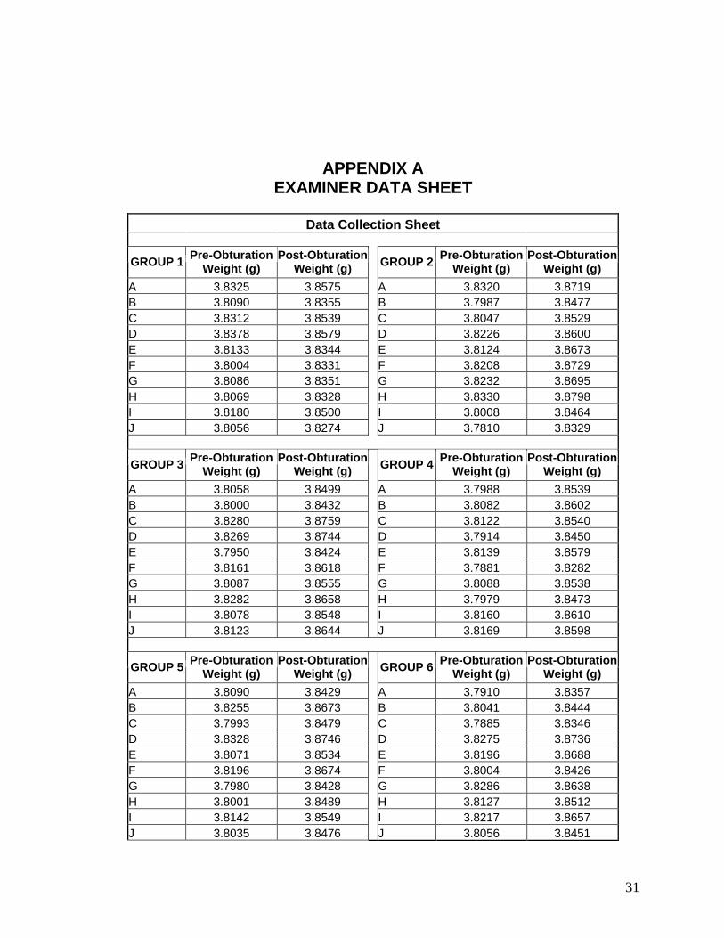
APPENDIX AEXAMINER DATA SHEET
Data Collection Sheet
GROUP 1Pre-Obturation
Weight (g)Post-Obturation
Weight (g)GROUP 2
Pre-Obturation Weight (g)
Post-Obturation Weight (g)
A 3.8325 3.8575 A 3.8320 3.8719B 3.8090 3.8355 B 3.7987 3.8477C 3.8312 3.8539 C 3.8047 3.8529D 3.8378 3.8579 D 3.8226 3.8600E 3.8133 3.8344 E 3.8124 3.8673F 3.8004 3.8331 F 3.8208 3.8729G 3.8086 3.8351 G 3.8232 3.8695H 3.8069 3.8328 H 3.8330 3.8798I 3.8180 3.8500 I 3.8008 3.8464J 3.8056 3.8274 J 3.7810 3.8329
GROUP 3Pre-Obturation
Weight (g)Post-Obturation
Weight (g)GROUP 4
Pre-Obturation Weight (g)
Post-Obturation Weight (g)
A 3.8058 3.8499 A 3.7988 3.8539B 3.8000 3.8432 B 3.8082 3.8602C 3.8280 3.8759 C 3.8122 3.8540D 3.8269 3.8744 D 3.7914 3.8450E 3.7950 3.8424 E 3.8139 3.8579F 3.8161 3.8618 F 3.7881 3.8282G 3.8087 3.8555 G 3.8088 3.8538H 3.8282 3.8658 H 3.7979 3.8473I 3.8078 3.8548 I 3.8160 3.8610J 3.8123 3.8644 J 3.8169 3.8598
GROUP 5Pre-Obturation
Weight (g)Post-Obturation
Weight (g)GROUP 6
Pre-Obturation Weight (g)
Post-Obturation Weight (g)
A 3.8090 3.8429 A 3.7910 3.8357B 3.8255 3.8673 B 3.8041 3.8444C 3.7993 3.8479 C 3.7885 3.8346D 3.8328 3.8746 D 3.8275 3.8736E 3.8071 3.8534 E 3.8196 3.8688F 3.8196 3.8674 F 3.8004 3.8426G 3.7980 3.8428 G 3.8286 3.8638H 3.8001 3.8489 H 3.8127 3.8512I 3.8142 3.8549 I 3.8217 3.8657J 3.8035 3.8476 J 3.8056 3.8451
32
GROUP 7Pre-Obturation
Weight (g)Post-Obturation
Weight (g)
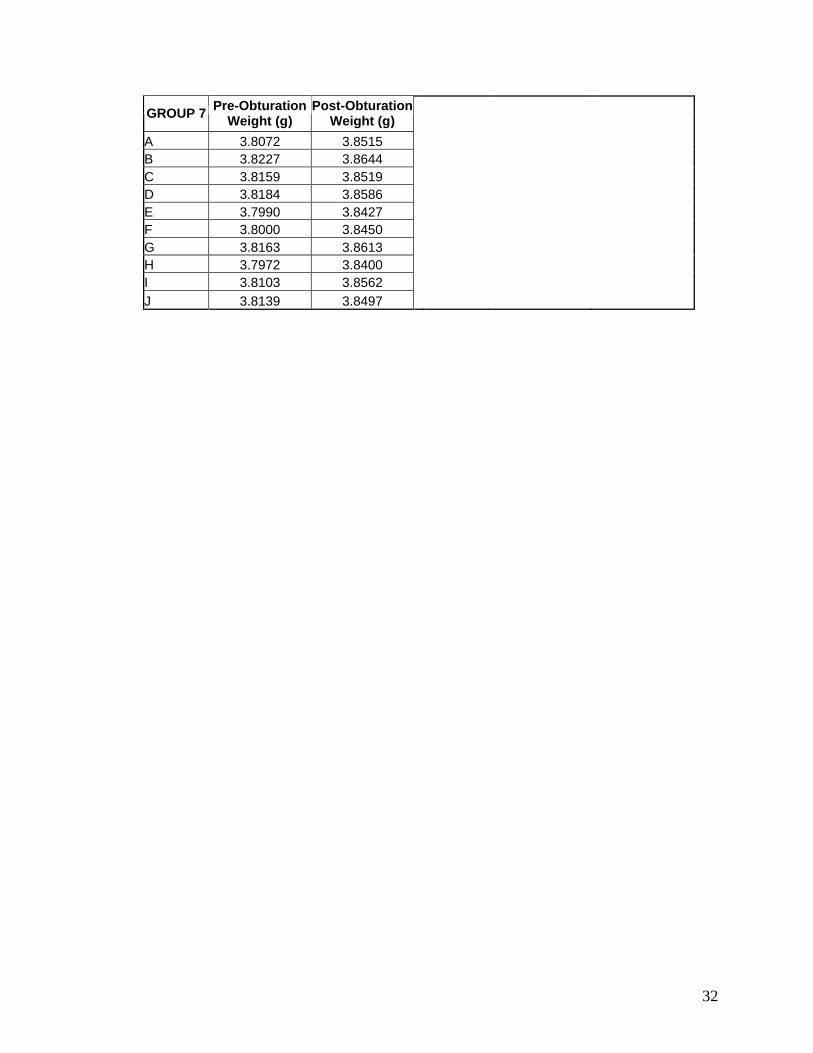
A 3.8072 3.8515B 3.8227 3.8644C 3.8159 3.8519D 3.8184 3.8586E 3.7990 3.8427F 3.8000 3.8450G 3.8163 3.8613H 3.7972 3.8400I 3.8103 3.8562
J 3.8139 3.8497
33
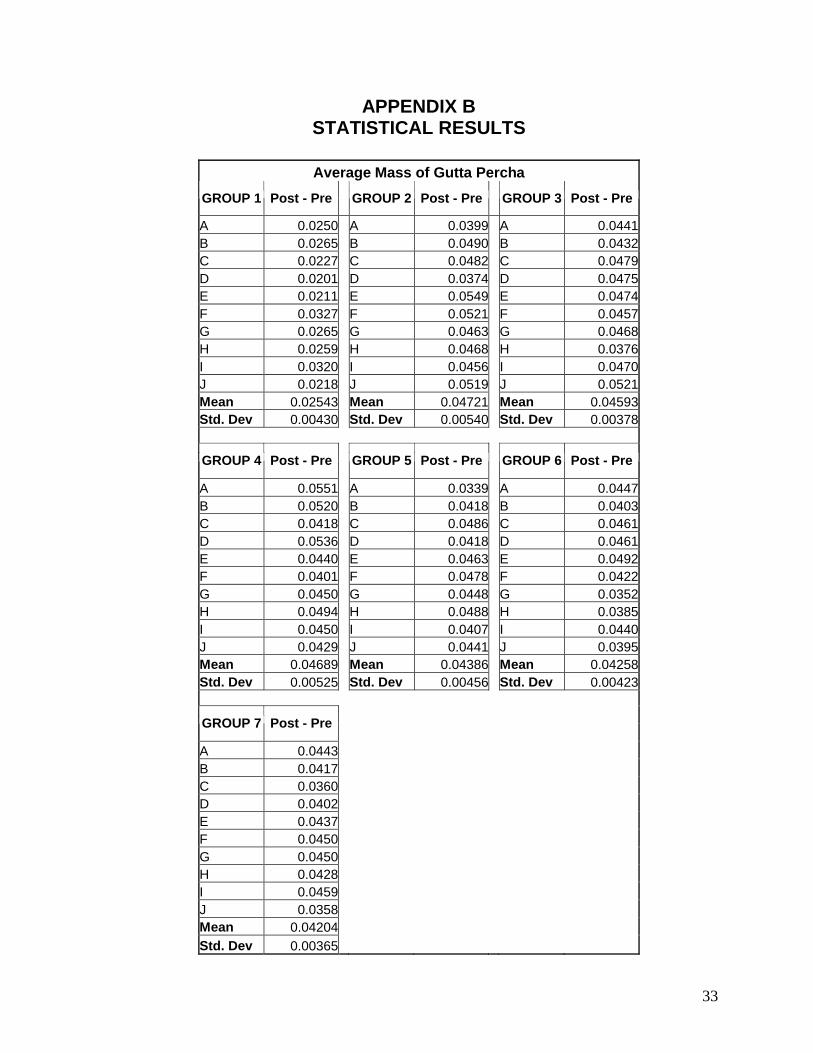
APPENDIX BSTATISTICAL RESULTS
Average Mass of Gutta Percha
GROUP 1 Post - Pre GROUP 2 Post - Pre GROUP 3 Post - Pre
A 0.0250 A 0.0399 A 0.0441B 0.0265 B 0.0490 B 0.0432C 0.0227 C 0.0482 C 0.0479D 0.0201 D 0.0374 D 0.0475E 0.0211 E 0.0549 E 0.0474F 0.0327 F 0.0521 F 0.0457G 0.0265 G 0.0463 G 0.0468H 0.0259 H 0.0468 H 0.0376I 0.0320 I 0.0456 I 0.0470J 0.0218 J 0.0519 J 0.0521Mean 0.02543 Mean 0.04721 Mean 0.04593Std. Dev 0.00430 Std. Dev 0.00540 Std. Dev 0.00378
GROUP 4 Post - Pre GROUP 5 Post - Pre GROUP 6 Post - Pre
A 0.0551 A 0.0339 A 0.0447B 0.0520 B 0.0418 B 0.0403C 0.0418 C 0.0486 C 0.0461D 0.0536 D 0.0418 D 0.0461E 0.0440 E 0.0463 E 0.0492F 0.0401 F 0.0478 F 0.0422G 0.0450 G 0.0448 G 0.0352H 0.0494 H 0.0488 H 0.0385I 0.0450 I 0.0407 I 0.0440J 0.0429 J 0.0441 J 0.0395Mean 0.04689 Mean 0.04386 Mean 0.04258Std. Dev 0.00525 Std. Dev 0.00456 Std. Dev 0.00423
GROUP 7 Post - Pre
A 0.0443B 0.0417C 0.0360D 0.0402E 0.0437F 0.0450G 0.0450H 0.0428I 0.0459J 0.0358Mean 0.04204
Std. Dev 0.00365
34
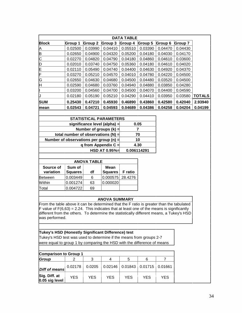
DATA TABLEBlock Group 1 Group 2 Group 3 Group 4 Group 5 Group 6 Group 7A 0.02500 0.03990 0.04410 0.05510 0.03390 0.04470 0.04430B 0.02650 0.04900 0.04320 0.05200 0.04180 0.04030 0.04170C 0.02270 0.04820 0.04790 0.04180 0.04860 0.04610 0.03600D 0.02010 0.03740 0.04750 0.05360 0.04180 0.04610 0.04020E 0.02110 0.05490 0.04740 0.04400 0.04630 0.04920 0.04370F 0.03270 0.05210 0.04570 0.04010 0.04780 0.04220 0.04500G 0.02650 0.04630 0.04680 0.04500 0.04480 0.03520 0.04500H 0.02590 0.04680 0.03760 0.04940 0.04880 0.03850 0.04280I 0.03200 0.04560 0.04700 0.04500 0.04070 0.04400 0.04590
J 0.02180 0.05190 0.05210 0.04290 0.04410 0.03950 0.03580 TOTALS
SUM 0.25430 0.47210 0.45930 0.46890 0.43860 0.42580 0.42040 2.93940
mean 0.02543 0.04721 0.04593 0.04689 0.04386 0.04258 0.04204 0.04199
STATISTICAL PARAMETERSsignificance level (alpha) = 0.05
Number of groups (k) = 7total number of observations (N) = 70
Number of observations per group (n) = 10q from Appendix C = 4.30
HSD AT 0.95%= 0.006114291
ANOVA TABLE
Source of variation
Sum of Squares df
Mean Squares F ratio
Between 0.003449 6 0.000575 28.4276Within 0.001274 63 0.000020
Total 0.004722 69
ANOVA SUMMARYFrom the table above it can be determined that the F ratio is greater than the tabulated F value of F(6,63) = 2.24. This indicates that at least one of the means is significantly different from the others. To determine the statistically different means, a Tukey's HSD was performed.
Tukey's HSD (Honestly Significant Difference) testTukey's HSD test was used to determine if the means from groups 2-7were equal to group 1 by comparing the HSD with the difference of means
Comparison to Group 1Group 2 3 4 5 6 7
Diff of means0.02178 0.0205 0.02146 0.01843 0.01715 0.01661
Sig. Diff. at 0.05 sig level
YES YES YES YES YES YES
35
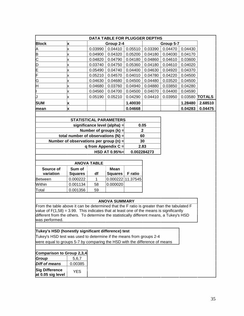
DATA TABLE FOR PLUGGER DEPTHSBlock x Group 2-4 Group 5-7A x 0.03990 0.04410 0.05510 0.03390 0.04470 0.04430B x 0.04900 0.04320 0.05200 0.04180 0.04030 0.04170C x 0.04820 0.04790 0.04180 0.04860 0.04610 0.03600D x 0.03740 0.04750 0.05360 0.04180 0.04610 0.04020E x 0.05490 0.04740 0.04400 0.04630 0.04920 0.04370F x 0.05210 0.04570 0.04010 0.04780 0.04220 0.04500G x 0.04630 0.04680 0.04500 0.04480 0.03520 0.04500H x 0.04680 0.03760 0.04940 0.04880 0.03850 0.04280I x 0.04560 0.04700 0.04500 0.04070 0.04400 0.04590
J x 0.05190 0.05210 0.04290 0.04410 0.03950 0.03580 TOTALS
SUM x 1.40030 1.28480 2.68510
mean x 0.04668 0.04283 0.04475
STATISTICAL PARAMETERSsignificance level (alpha) = 0.05
Number of groups (k) = 2total number of observations (N) = 60
Number of observations per group (n) = 30q from Appendix C = 2.83
HSD AT 0.95%= 0.002284273
ANOVA TABLE
Source of variation
Sum of Squares df
Mean Squares F ratio
Between 0.000222 1 0.000222 11.37545Within 0.001134 58 0.000020
Total 0.001356 59
ANOVA SUMMARYFrom the table above it can be determined that the F ratio is greater than the tabulated F value of F(1,58) = 3.99. This indicates that at least one of the means is significantly different from the others. To determine the statistically different means, a Tukey's HSD was performed.
Tukey's HSD (honestly significant difference) testTukey's HSD test was used to determine if the means from groups 2-4were equal to groups 5-7 by comparing the HSD with the difference of means
Comparison to Group 2,3,4Group 5,6,7Diff of means 0.00385
Sig Difference at 0.05 sig level
YES
36
DATA TABLE FOR EXPERIMENTAL GROUPSBlock Group 1 Group 2 Group 3 Group 4 Group 5 Group 6 Group 7A x 0.03990 0.04410 0.05510 0.03390 0.04470 0.04430B x 0.04900 0.04320 0.05200 0.04180 0.04030 0.04170C x 0.04820 0.04790 0.04180 0.04860 0.04610 0.03600D x 0.03740 0.04750 0.05360 0.04180 0.04610 0.04020E x 0.05490 0.04740 0.04400 0.04630 0.04920 0.04370F x 0.05210 0.04570 0.04010 0.04780 0.04220 0.04500G x 0.04630 0.04680 0.04500 0.04480 0.03520 0.04500H x 0.04680 0.03760 0.04940 0.04880 0.03850 0.04280I x 0.04560 0.04700 0.04500 0.04070 0.04400 0.04590
J x 0.05190 0.05210 0.04290 0.04410 0.03950 0.03580 TOTALS
SUM x 0.47210 0.45930 0.46890 0.43860 0.42580 0.42040 2.68510
mean x 0.04721 0.04593 0.04689 0.04386 0.04258 0.04204 0.04475
STATISTICAL PARAMETERSsignificance level (alpha) = 0.05
Number of groups (k) = 6total number of observations (N) = 60
Number of observations per group (n) = 10q from Appendix C = 4.18
HSD AT 0.95%= 0.005985614
ANOVA TABLE
Source of variation
Sum of Squares df
Mean Squares F ratio
Between 0.000249 5 0.000050 2.425593Within 0.001107 54 0.000021
Total 0.001356 59
ANOVA SUMMARYFrom the table above, it can be determined that the F ratio may be greater than the tabulated F value of F(5,54) = 2.37-2.45. This indicates that there may be one of the means that is significantly different from the others.
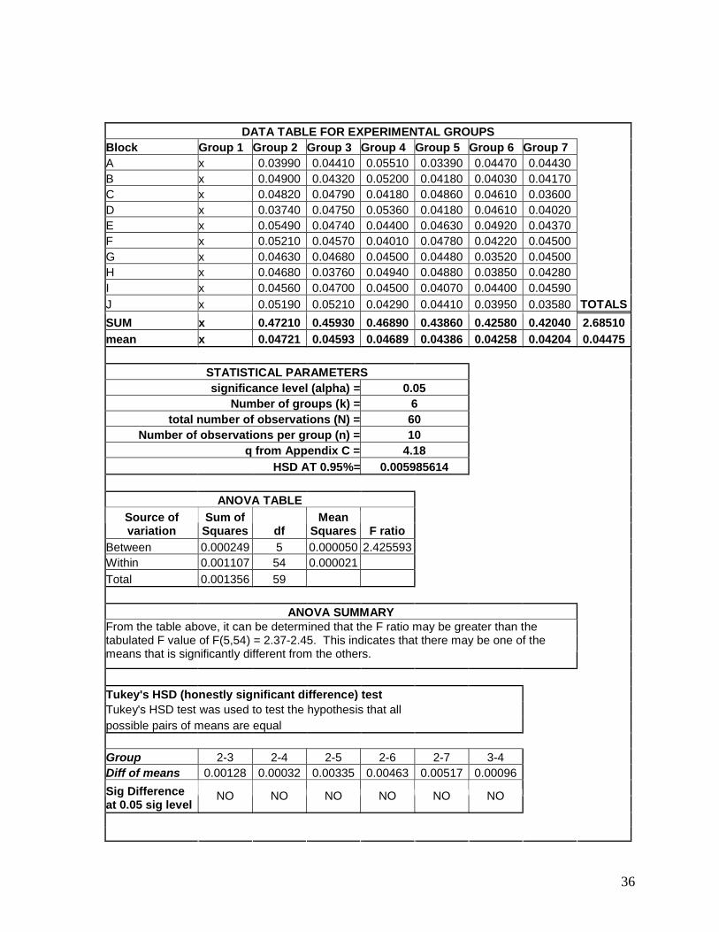
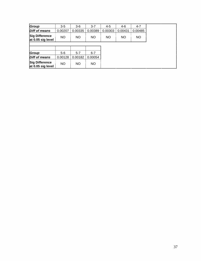
Tukey's HSD (honestly significant difference) testTukey's HSD test was used to test the hypothesis that all possible pairs of means are equal
Group 2-3 2-4 2-5 2-6 2-7 3-4Diff of means 0.00128 0.00032 0.00335 0.00463 0.00517 0.00096
Sig Difference at 0.05 sig level
NO NO NO NO NO NO
37
Group 3-5 3-6 3-7 4-5 4-6 4-7Diff of means 0.00207 0.00335 0.00389 0.00303 0.00431 0.00485
Sig Difference at 0.05 sig level
NO NO NO NO NO NO
Group 5-6 5-7 6-7Diff of means 0.00128 0.00182 0.00054
Sig Difference at 0.05 sig level
NO NO NO
38
CURRICULMN VITAE
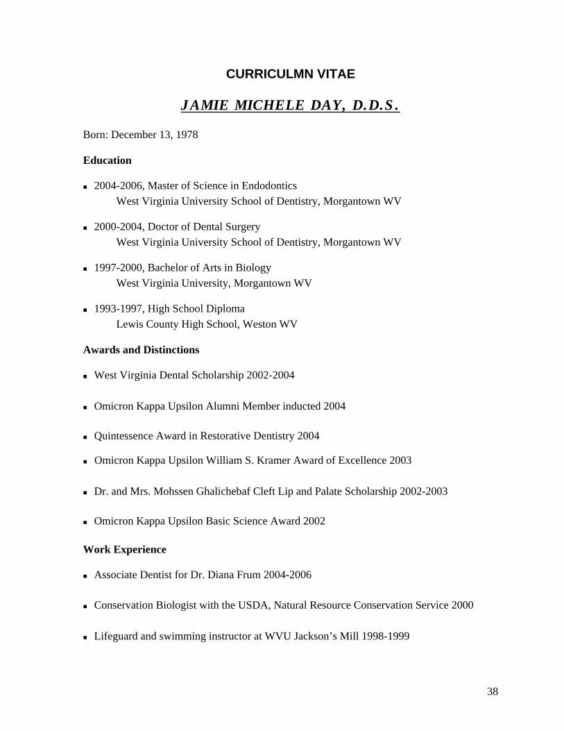
JAMIE MICHELE DAY, D.D.S.
Born: December 13, 1978
Education
2004-2006, Master of Science in Endodontics
West Virginia University School of Dentistry, Morgantown WV
2000-2004, Doctor of Dental Surgery
West Virginia University School of Dentistry, Morgantown WV
1997-2000, Bachelor of Arts in Biology
West Virginia University, Morgantown WV
1993-1997, High School Diploma
Lewis County High School, Weston WV
Awards and Distinctions
West Virginia Dental Scholarship 2002-2004
Omicron Kappa Upsilon Alumni Member inducted 2004
Quintessence Award in Restorative Dentistry 2004
Omicron Kappa Upsilon William S. Kramer Award of Excellence 2003
Dr. and Mrs. Mohssen Ghalichebaf Cleft Lip and Palate Scholarship 2002-2003
Omicron Kappa Upsilon Basic Science Award 2002
Work Experience
Associate Dentist for Dr. Diana Frum 2004-2006
Conservation Biologist with the USDA, Natural Resource Conservation Service 2000
Lifeguard and swimming instructor at WVU Jackson’s Mill 1998-1999
Related Documents