IJSS Journal of Surgery | November-December 2016 | Volume 2 | Issue 6 31 Comparison of the Incidence of Post-operative Wound Infection between Skin Staples and Conventional Sutures in Abdominal Skin Closures Srikantaiah Hiremath 1 , Kiran C Kailas 2 , B M Vinay 3 1 Associate Professor, Department of General Surgery, M. S. Ramaiah Medical College and Hospitals, Bengaluru, Karnataka, India, 2 Senior Resident, Department of General Surgery, M. S. Ramaiah Medical College and Hospitals, Bengaluru, Karnataka, India, 3 Post-graduate Student, Department of General Surgery, M. S. Ramaiah Medical College and Hospitals, Bengaluru, Karnataka, India Abstract Introduction: The type of suture material for skin closure is also reported to influence post-operative wound complications. However, several other studies have failed to demonstrate significant differences between different types of suture material. Materials and Methods: This study was conducted at M. S. Ramaiah hospitals, Bengaluru - 560 054, Karnataka, India, where 100 patients underwent abdominal skin closure with either staples or conventional vertical mattress suturing with Ethilon. Results: The present 1 year observational study was conducted in the Department of Surgery, M. S. Ramaiah Hospitals, Bengaluru - 560 054, Karnataka, during the year December 2014-November 2015. Data obtained was tabulated and analyzed in tables. Conclusion: The use of skin staples in low tension incision is easy, associated with low incidence of wound complications, provides good cosmetic outcome and also takes considerably less time for skin closure and thus recommend its use more frequently especially for closure of long and multiple incisions. Keywords: Surgical Incision, Sutures and Skin staplers INTRODUCTION S urgery is derived from the earlier name chirurgery, which means handwork. It is a science and art that shows the manner in which to work on man’s body exercising all manual operations necessary to heal or as much as possible using most expedient medicines or techniques. 1-3 The goal of surgery is to achieve healing by such means with minimal edema, no serous discharge or infection, without separation of the wound edges and with minimal scar formation. After any surgical procedure (assuming there are no tension and a good blood supply) careful approximation of the tissues will allow healing by primary intention. 4-7 Precise approximation of skin incisions and lacerations with wound closure devices is critical for a favorable cosmetic and functional surgical result. Principles of wound closure focus on relieving tension on the wound and bringing the skin edges together in an everted orientation. If sutures are tied too tight or left in too long, they may leave permanent suture tracts if sutures are removed before adequate healing, the lack of wound tensile strength may result in wound dehiscence or a widened scar. 8,9 Wound closure includes ensuring a clean wound with satisfactory vascularity and hemostasis and apposition without wound tension. Principles of wound closure focus on relieving tension on the wound and bringing the skin edges together in an everted orientation. Corresponding Author: Dr. Srikantaiah Hiremath, Department of General Surgery, Door No. 72, 16 th Cross, 6 th Main Road, Malleswaram, Bengaluru - 560 055, Karnataka, India. Phone: +91-9845208352. E-mail: [email protected] Access this article online www.surgeryijss.com Month of Submission : 07-2016 Month of Peer Review : 08-2016 Month of Acceptance : 09-2016 Month of Publishing : 10-2016 Original Article Print ISSN: 2321-6379 Online ISSN: 2395-1893 DOI: 10.17354/SUR/2016/52

Welcome message from author
This document is posted to help you gain knowledge. Please leave a comment to let me know what you think about it! Share it to your friends and learn new things together.
Transcript
IJSS Journal of Surgery | November-December 2016 | Volume 2 | Issue 6 31
Comparison of the Incidence of Post-operative Wound Infection between Skin Staples and Conventional Sutures in Abdominal Skin Closures
Srikantaiah Hiremath1, Kiran C Kailas2, B M Vinay3
1Associate Professor, Department of General Surgery, M. S. Ramaiah Medical College and Hospitals, Bengaluru, Karnataka, India, 2Senior Resident, Department of General Surgery, M. S. Ramaiah Medical College and Hospitals, Bengaluru, Karnataka, India, 3Post-graduate Student, Department of General Surgery, M. S. Ramaiah Medical College and Hospitals, Bengaluru, Karnataka, India
Abstract
Introduction: The type of suture material for skin closure is also reported to influence post-operative wound complications. However, several other studies have failed to demonstrate significant differences between different types of suture material.
Materials and Methods: This study was conducted at M. S. Ramaiah hospitals, Bengaluru - 560 054, Karnataka, India, where 100 patients underwent abdominal skin closure with either staples or conventional vertical mattress suturing with Ethilon.
Results: The present 1 year observational study was conducted in the Department of Surgery, M. S. Ramaiah Hospitals, Bengaluru - 560 054, Karnataka, during the year December 2014-November 2015. Data obtained was tabulated and analyzed in tables.
Conclusion: The use of skin staples in low tension incision is easy, associated with low incidence of wound complications, provides good cosmetic outcome and also takes considerably less time for skin closure and thus recommend its use more frequently especially for closure of long and multiple incisions.
Keywords: Surgical Incision, Sutures and Skin staplers
INTRODUCTION
Surgery is derived from the earlier name chirurgery, which means handwork. It is a science and art that shows the
manner in which to work on man’s body exercising all manual operations necessary to heal or as much as possible using most expedient medicines or techniques.1-3
The goal of surgery is to achieve healing by such means with minimal edema, no serous discharge or infection, without separation of the wound edges and with minimal scar formation.
After any surgical procedure (assuming there are no tension and a good blood supply) careful approximation of the tissues will allow healing by primary intention.4-7
Precise approximation of skin incisions and lacerations with wound closure devices is critical for a favorable cosmetic and functional surgical result. Principles of wound closure focus on relieving tension on the wound and bringing the skin edges together in an everted orientation. If sutures are tied too tight or left in too long, they may leave permanent suture tracts if sutures are removed before adequate healing, the lack of wound tensile strength may result in wound dehiscence or a widened scar.8,9
Wound closure includes ensuring a clean wound with satisfactory vascularity and hemostasis and apposition without wound tension. Principles of wound closure focus on relieving tension on the wound and bringing the skin edges together in an everted orientation.
Corresponding Author: Dr. Srikantaiah Hiremath, Department of General Surgery, Door No. 72, 16th Cross, 6th Main Road, Malleswaram, Bengaluru - 560 055, Karnataka, India. Phone: +91-9845208352. E-mail: [email protected]
Access this article online
www.surgeryijss.com
Month of Submission : 07-2016 Month of Peer Review : 08-2016 Month of Acceptance : 09-2016 Month of Publishing : 10-2016
Original ArticlePrint ISSN: 2321-6379Online ISSN: 2395-1893DOI: 10.17354/SUR/2016/52

Hiremath, et al.: Comparison of the Incidence of Post-operative Wound Infection between Skin Staples and Conventional Sutures in Abdominal Skin Closures
IJSS Journal of Surgery | November-December 2016 | Volume 2 | Issue 632
“Surgery is the first and the highest division of the healing art, pure in itself, perpetual in its applicability, a working product of heaven and sure of fame on earth” SUSHRUTA (400 B.C).
Surgical site infection (SSI) is the most common nosocomial infections reported in the hospital patients. Up to 2.5% of the patients undergoing clean extra abdominal operations and up to 20% of intra-abdominal operations will develop SSI.
SSI remains a complication of surgical procedures resulting in increased morbidity, mortality, and cost.
Infection remains the most significant factor affecting wound healing. A closure that penetrates the epidermis and dermis only serves to auto-inoculate the wound of the patient, driving surface flora deep into the subcutaneous tissue.
Percutaneous suture closure provides an extra source of contamination through the suture canal and results in a thin perisutural cuff of dead epidermis, dermis, and subcutaneous fat. Suture closure also is a potential source of foreign body reaction within the susceptible subcutaneous tissue.
The type of suture material for skin closure is also reported to influence post-operative wound complications. However, several other studies have failed to demonstrate significant differences between different types of suture material.10-17
The surgical scar remains the only visible evidence of the surgeon’s skill and not infrequently, all of his efforts are judged on its final appearance.
Skin staples give a neat scar with good wound eversion and minimal cross-hatching effect. They can be placed faster than sutures and have a lower predisposition to infection because they do not penetrate entirely through the wound and do not produce a complete track from one wound to the other.17-22
MATERIALS AND METHODS
This study was conducted at M. S. Ramaiah hospitals, Bengaluru - 560 054, Karnataka, India, where 100 patients underwent abdominal skin closure with either staples or conventional vertical mattress suturing with Ethilon.
Out of the 100 patients, 50 underwent skin closure with stainless steel skin staples and the remaining 50 with vertical mattress suturing with Ethilon all these patients were allotted to either group according to random number table.
Inclusion Criteria• Allpatientsundergoingelectiveabdominalsurgeries• Patientsgivingconsentforthetrial.
Exclusion Criteria• Patientshavinglaceratedwoundswithskinloss• Patientshavingdiabetesmellitus(DM)• Patientswith immune compromised status like
AIDS/HIVinfection• Patients having severe comorbidities, i.e., shock,
septicemia, failure of other organ systems, recent myocardialinfarction,and/ormalignancy.
Ethical clearance has been obtained from our institution for conducting this study.
Method of Collection of DataA detailed history of each patient was obtained starting with a history of presenting symptoms and any coexisting, comorbid conditions such asDM, hypertension, andjaundice were ruled out.
A thorough general physical examination was done to rule out the presence of pallor, icterus, and cachexia.
Preoperatively all patients underwent following investigations:• Completebloodcount,urineexamination• Bloodsugar,bloodurea,serumcreatinine• Liverfunctiontest(whereverneeded)• ChestX-ray,electrocardiogram(whereverneeded).
All cases were elective surgeries and the mode of anesthesia was either general anesthesia (GA), spinal anesthesia, or short GA. Shaving was done the night before surgery. They all received one mandatory dose of pre-operative parenteral antibiotic 1 h before the incision. Painting was done with 10% povidone iodine solution for all cases.
Closure TechniqueAfter the subcutaneous fat was sutured with 2-0 vicryl:a. Suture group Skin was approximated with vertical mattress
sutures using non-absorbable 2-0 Ethilon at a distance of 1 cm from each other.
b. Stapled group Before inserting staples, it is important to line up the
wound edges with the centerline indicator on the head of the stapler to make sure that the legs of the staple will enter the skin at equal distances on either side of the wound edge. Each edge is typically picked up with a forceps, everted and precisely lined up. The staples are then used to close the wound while
Hiremath, et al.: Comparison of the Incidence of Post-operative Wound Infection between Skin Staples and Conventional Sutures in Abdominal Skin Closures
IJSS Journal of Surgery | November-December 2016 | Volume 2 | Issue 6 33
the first assistant advances the forceps, everting the edges of the wound. They are placed at a distance of 5 mm from one another. This technique is continued until the entire wound is everted and closed with staples.
The time required for skin closure was recorded. For the sutures group, betadine ointment was applied and a packed dressing given. For the stapled group simple dry gauze dressing was given. Postoperatively all patients received a total of 3 days of parenteral and oral antibiotics. The patients who underwent Mesh repair received a total of 5 days of parenteral and oral antibiotics.
On the 3rd post-operative day, the wound was evaluated for inflammation, infection, and wound gape.
Inflammation was defined as excessive redness and tenderness of incision site with induration.
Infection was defined as any persistence of superficial cellulitis or induration with serosanguinous or pus discharge from wound site lasting beyond 7th post-operative day.
The patients were usually discharged after suture removal on the 7-8th day for the sutures group and the stapled group patients after the 1st wound evaluation if there was no wound inflammation/infection. Incase of wound infection or discharge in any group, the discharge was sent for culture and sensitivity. The total hospital stay was noted. Patients were re-evaluated for infection/gape/inflammation during follow-up on15days/1month.
Statistical analysis was performed using the Mann–Whitney test, and the P value was calculated.
At 1 month follow-up:
The wounds were evaluated at 1 month follow-up which were rated for cosmesis on a previously validated cosmesis visual analog score (VAS)which has beendemonstrated to be reliable and valid outcome measure of cosmesis.
The cosmetic VAS is a 100 mm line with worst scar at 0 and best scar at 100. A senior surgeon rated photographs unaware of the method used to close the wound.• Worstscar• Bestscar• 100
Using the line as a continuous entity the surgeon marked the patient’s scar on the line. The score was then measured
inmillimeters from0 to100.ThemeanVAS for eachgroup was calculated.
Statistical evaluation was done after consulting the college statistician. Using the student’s unpaired t-test, the P value was calculated.
OBSERVATIONS AND RESULTS
The present 1 year observational study was conducted intheDepartmentofSurgery,M.S.RamaiahHospitals,Bengaluru - 560 054, Karnataka, during the year December 2014-November 2015. Data obtainedwastabulated and analyzed as Tables 1-7.
Table 1: Age distributionAge distribution (years) Staples Conventional
methodTotal
<20 4 7 1120‑40 15 15 30>40 31 28 59Grand total 50 50 100
Table 2: ResidencePlace Staples Conventional method TotalBengaluru 21 25 46Outside Bengaluru 29 25 54Grand total 50 50 100
Table 3: Socio economic statusSocio economic status Staples Conventional method TotalLow ‑ 15 15Middle 39 31 70High 11 4 15Grand total 50 50 100
Table 4: Literacy of the study populationEducation Staples Conventional method TotalIlliterate 1 1 2Primary education 7 5 12Secondary ‑ 6 6Intermediate 8 16 20Degree+ 34 22 36Grand total 50 50 100
Table 5: Employment statusEmployment status
Staples Conventional method Total
Unemployed 3 5 8Unskilled ‑ 4 4Semiskilled 8 7 15Skilled 31 25 56Professional 8 9 17Grand total 50 50 100
Hiremath, et al.: Comparison of the Incidence of Post-operative Wound Infection between Skin Staples and Conventional Sutures in Abdominal Skin Closures
IJSS Journal of Surgery | November-December 2016 | Volume 2 | Issue 634
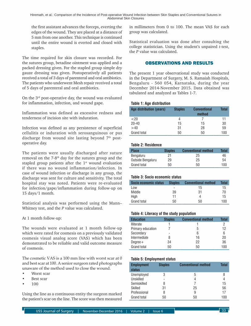
Graph 1a and b shows the age distribution in staples group. The graphs show that the majority were in the 40+ year’s age group.
Graph 2a and b shows the age distribution in conventional suture group. The graphs show that the majority were in the 40+ year’s age group.
Graph 3a and b shows the sex distribution of the staples group. The pie chart shows that 34 of the study population were males and the remaining 16 were females.
Graph 4a and b shows the sex distribution of the conventional sutures group. The pie chart shows that 38 of the study population were males and the remaining 12 were females.
Graph 5a and b shows the residence of the study population. The graph shows that 46% were from Bengaluru and nearby areas whereas the remaining 54% werefromothertowns/citiesinKarnataka.
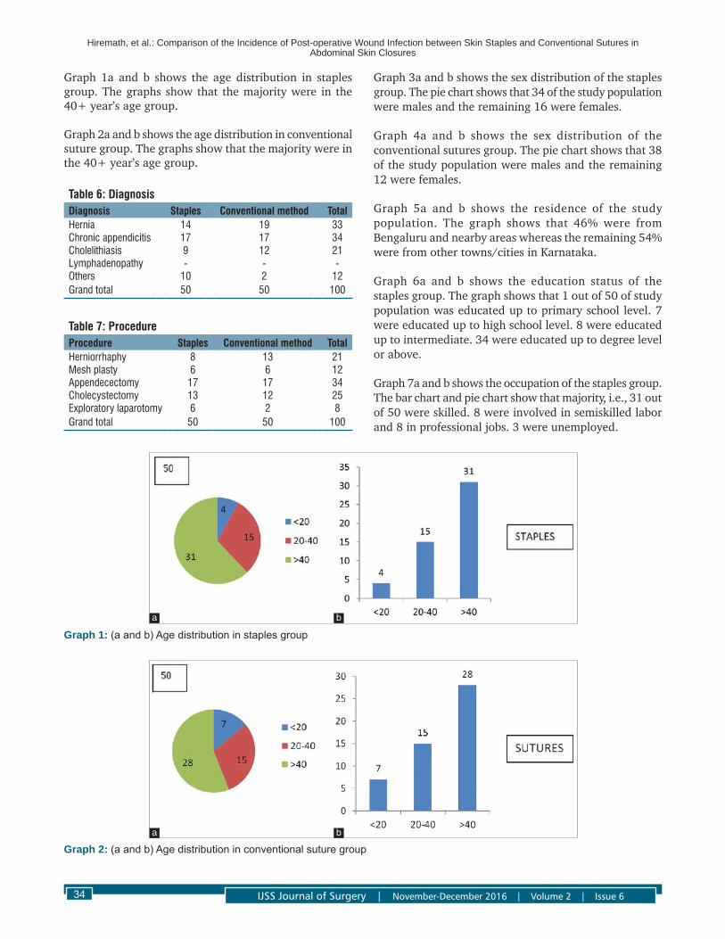
Graph 6a and b shows the education status of the staples group. The graph shows that 1 out of 50 of study population was educated up to primary school level. 7 were educated up to high school level. 8 were educated up to intermediate. 34 were educated up to degree level or above.
Graph 7a and b shows the occupation of the staples group. The bar chart and pie chart show that majority, i.e., 31 out of 50 were skilled. 8 were involved in semiskilled labor and 8 in professional jobs. 3 were unemployed.
Table 6: DiagnosisDiagnosis Staples Conventional method TotalHernia 14 19 33Chronic appendicitis 17 17 34Cholelithiasis 9 12 21Lymphadenopathy ‑ ‑ ‑Others 10 2 12Grand total 50 50 100
Table 7: ProcedureProcedure Staples Conventional method TotalHerniorrhaphy 8 13 21Mesh plasty 6 6 12Appendecectomy 17 17 34Cholecystectomy 13 12 25Exploratory laparotomy 6 2 8Grand total 50 50 100
Graph 1: (a and b) Age distribution in staples group
a b
Graph 2: (a and b) Age distribution in conventional suture groupa b

Hiremath, et al.: Comparison of the Incidence of Post-operative Wound Infection between Skin Staples and Conventional Sutures in Abdominal Skin Closures
IJSS Journal of Surgery | November-December 2016 | Volume 2 | Issue 6 35
Graph 3: (a and b) Sex distribution of the staples groupa b
Graph 4: (a and b) Sex distribution of the conventional sutures group
a b
Graph 5: (a and b) Residence of the study populationa b
Hiremath, et al.: Comparison of the Incidence of Post-operative Wound Infection between Skin Staples and Conventional Sutures in Abdominal Skin Closures
IJSS Journal of Surgery | November-December 2016 | Volume 2 | Issue 636
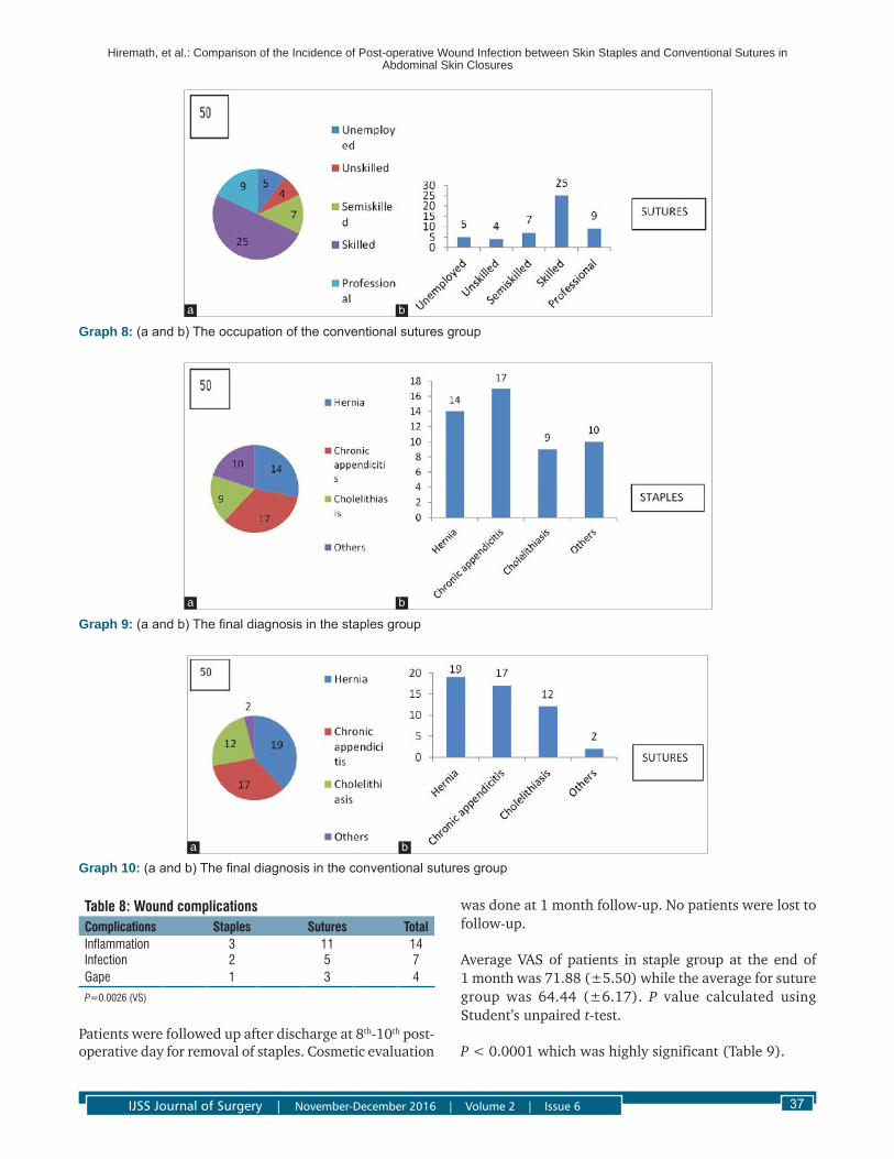
Graph 8a and b shows the occupation of the conventional sutures group. The bar chart and pie chart show that majority i.e., 25 out of 50 were skilled. 7 were involved in semiskilled labor and 9 in professional jobs. 5 were unemployed and 4 were unskilled.
Graph 9a and b shows the final diagnosis in the staples group. The graph shows majority of the patients had chronic appendicitis (17 out of 50) and hernia (14 out of 50). 9 patients had cholelithiasis for which cholecystectomy was done. 10 patients were diagnosed to have other disorders mentioned above.
Graph 10a and b shows the final diagnosis in the conventional sutures group. The graph shows majority of the patients had hernia (19 out of 50) and chronic appendicitis (17 out of 50). 12 patients had cholelithiasis for which cholecystectomy was done. 2 patients were diagnosed to have other disorders mentioned above.
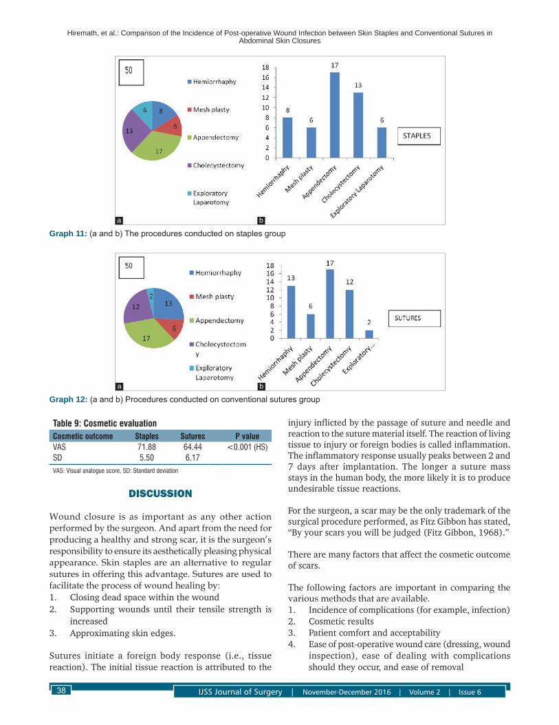
Graphs 11a and b shows the procedures conducted on staples group.17 out of 50 underwent appendicectomy and 13 out of 50 underwent cholecyctectomy. 8 patients
underwent herniorrhaphy and 6 patients each underwent mesh repair and exploratory laparotomy.
Graphs 12a and b shows the procedures conducted on conventional sutures group. 17 out of 50 underwent appendicectomy and 13 out of 50 underwent herniorrhaphy. 12 patients underwent cholecystectomy and 6 patients underwent mesh repair and 2 patients underwent exploratory laparotomy.
Staples GroupAbout 3 patients had post-operative wound inflammation, out of which 2 patients developed wound infection in the form of purulent discharge on the 6th post-operative day. 1 patient went on to develop wound gape.
Conventional Suture GroupAbout 11 patients developed wound inflammation out of which 5 patients developed wound infection and 3 patients had gaping of wound. P value calculated using Mann–Whitney test.
P value was 0.0026 which was very significant (Table 8).
Graph 6: (a and b) The education status of the staples groupa b
Graph 7: (a and b) The occupation of the staples groupa b
Hiremath, et al.: Comparison of the Incidence of Post-operative Wound Infection between Skin Staples and Conventional Sutures in Abdominal Skin Closures
IJSS Journal of Surgery | November-December 2016 | Volume 2 | Issue 6 37
wasdoneat1monthfollow-up.Nopatientswerelosttofollow-up.
AverageVASofpatients in staplegroupat theendof1 month was 71.88 (±5.50) while the average for suture group was 64.44 (±6.17). P value calculated using Student’s unpaired t-test.
P < 0.0001 which was highly significant (Table 9).
Table 8: Wound complicationsComplications Staples Sutures TotalInflammation 3 11 14Infection 2 5 7Gape 1 3 4
P=0.0026 (VS)
Patients were followed up after discharge at 8th-10th post-operative day for removal of staples. Cosmetic evaluation
Graph 8: (a and b) The occupation of the conventional sutures group
a b
Graph 9: (a and b) The final diagnosis in the staples group
a b
Graph 10: (a and b) The final diagnosis in the conventional sutures groupa b
Hiremath, et al.: Comparison of the Incidence of Post-operative Wound Infection between Skin Staples and Conventional Sutures in Abdominal Skin Closures
IJSS Journal of Surgery | November-December 2016 | Volume 2 | Issue 638
DISCUSSION
Wound closure is as important as any other action performed by the surgeon. And apart from the need for producing a healthy and strong scar, it is the surgeon’s responsibility to ensure its aesthetically pleasing physical appearance. Skin staples are an alternative to regular sutures in offering this advantage. Sutures are used to facilitate the process of wound healing by:1. Closing dead space within the wound2. Supporting wounds until their tensile strength is
increased3. Approximating skin edges.
Sutures initiate a foreign body response (i.e., tissue reaction). The initial tissue reaction is attributed to the
injury inflicted by the passage of suture and needle and reaction to the suture material itself. The reaction of living tissue to injury or foreign bodies is called inflammation. The inflammatory response usually peaks between 2 and 7 days after implantation. The longer a suture mass stays in the human body, the more likely it is to produce undesirable tissue reactions.
For the surgeon, a scar may be the only trademark of the surgical procedure performed, as Fitz Gibbon has stated, “By your scars you will be judged (Fitz Gibbon, 1968).”
There are many factors that affect the cosmetic outcome of scars.
The following factors are important in comparing the various methods that are available.1. Incidence of complications (for example, infection)2. Cosmetic results3. Patient comfort and acceptability4. Ease of post-operative wound care (dressing, wound
inspection), ease of dealing with complications should they occur, and ease of removal
Table 9: Cosmetic evaluationCosmetic outcome Staples Sutures P valueVAS 71.88 64.44 <0.001 (HS)SD 5.50 6.17
VAS: Visual analogue score, SD: Standard deviation
Graph 11: (a and b) The procedures conducted on staples groupa b
Graph 12: (a and b) Procedures conducted on conventional sutures groupa b

Hiremath, et al.: Comparison of the Incidence of Post-operative Wound Infection between Skin Staples and Conventional Sutures in Abdominal Skin Closures
IJSS Journal of Surgery | November-December 2016 | Volume 2 | Issue 6 39
5. Time taken to close the wound6. Although sutures are inexpensive, they typically take
longer to place, and there is also risk of needle stick injury to the surgeon and operating staff
7. Needlestickinjuriestothesurgeon8. Stitch abscess may develop9. Injury to the blood vessels in the skin resulting in
hematoma.
In this study, 100 patients underwent abdominal wound closure. Out of the 100, 50 underwent closure of skin with skin staples while the remaining 50 patients had their skin closed with non-absorbable sutures. There was no significant difference between the results of application of staplers or sutures at various anatomic regions. The most common region of the surgical wounds in this study was Mcburney’s 17 in each group and inguinal, 14 in staples and 19 in suture group, the regional distribution of surgical wounds in the staples group was mid line 10, subcostal 9 and among the suture group midline 2 and subcostal 12.
The comparisons of these two groups were done in relation to:1. Post-operative complications2. Cosmetic outcome.
Wound ComplicationsThe primary objective of our study was to measure the incidence of post-operative wound infection.
Wound complications included:1. Inflammation2. Infection3. Wound gape.
Inflammation was defined as the presence of redness and tenderness at the site of incision. In our study, 11 patients had wound inflammation in the suture group as compared to 3 in stapled group. The difference was found to be statistically very significant (P = 0.0026).
Infection of the wound is the presence of serosanguinous discharge or frank pus. It was present in 2 patients of the stapled group and 5 patients of the suture group. Wound gape was seen in 1 patient of the stapled group and 3 of the vertical mattress sutured group which was found to be significant.
Patients with wound infection had their discharge sent for culture and sensitivity and antibiotics were instituted as per the report. Secondary suturing was done in 2 patients of the suture group. In the other 2 patients, 1 each in staple and suture groups, the wounds healed with regular dressings. All of the patients were evaluated 1 month after discharge.
There is a uniform agreement that skin wounds closed by staples exhibit a superior resistance to infection than skin wounds contaminated by the least reactive suture. The superior resistance of stapled wounds to infection as compared with the resistance of sutured wounds was confirmed by the experimental study of Stillman et al., in contaminated wounds in mice, stapled wounds displayed a lower incidence of infection than wounds approximated by either percutaneous suture (4-0 silk, 4-0 monofilament nylon, and 4-0 polyglycolic acid suture) or subcuticular sutures (4-0 polyglycolic).
Nodifferencewasfoundwithregardtowoundinfectionin a randomized trial conducted by Eldrup et al., when they compared stapler with conventional skin closure.
CosmesisAll the patients were followed up 1 month after discharge for evaluation of the scar. A senior surgeon who was blinded to the method of closure evaluated it.
Nopatientswerelosttofollow-up.Cosmeticoutcomefor the remaining patients were evaluated based on VAS.
VASVAS obtained by analysis of month post-operativephotographs, revealed cosmetic results between the two groups - 71.88 (±5.50) for staples and 64.44 (±6.17) for suture group, which was statistically highly significant (P≤0.0001).
George et al. and Macgregor et al. studied wound closure in the accident and emergency department and found that stapled closure promotes wound edge eversion, formation of an incomplete loop with decreased tissue strangulation, and lack of residual cross marks.
In a study comparing staples closure with nylon wound closure in head and neck surgeries by Meiring et al. showed that the cosmetic result of staples is as good as if not better than that with nylon sutures.
Lubowskiet al. compared stapled and sutured abdominal wound closure which resulted in almost equal cosmetic scores for vertical wounds.
No significant difference inwound appearanceswasfound in a study conducted by Bhatia et al. in closure of palmarskinfollowingDuyputren’scontracture.
dos Santos et al. have compared the cosmetic results of staplers with noncontinuous nylon sutures.7 They have observed that the wounds closed with staplers were cosmetically superior in 80% of the cases. There are no studies available in the literature comparing the results

Hiremath, et al.: Comparison of the Incidence of Post-operative Wound Infection between Skin Staples and Conventional Sutures in Abdominal Skin Closures
IJSS Journal of Surgery | November-December 2016 | Volume 2 | Issue 640
of application staplers to various anatomic regions. Although Ranaboldo and Rowe-Jones have compared the results of stapler with subcuticular absorbable sutures for laparotomy wounds and divided them into lower and upper abdominal regions, no mention was made by them regarding the appearance of the scar at various sites.6 There was no significant benefit of staplers over subcuticular sutures in their study.
In this study, the time taken to complete wound closure with the use of staplers as compared to sutures was not done.
In the study by Ranaboldo and Rowe-Jones the rate of woundclosurewas8s/cmwithstaplerand12.7s/cmwith sutures.6
Kanegaye et al., observed that staplers were six times faster than standard sutures.4
Eldrup et al. analyzed 137 patients and concluded that mechanical sutures took one-third of the time taken by conventional sutures.5
Meiring et al. have recorded that there was 80% time saving,whereasHarveyandLoganhavereported66.6%time saving with the use of staplers.8,9
dos Santos et al. found in a prospective trial that the mean skin closure time with staple was 5 min and 25 min with nylon suture.7
In a study done by Wolterbeek et al., they compared various methods of skin closure in infrainguinal bypass surgeries and came to a conclusion that time needed for wound closure is significantly reduced using metallic staples.
Ritchie et al. carried out a prospective double blind randomized study comparing staples versus sutures in the closure of scalp wound and found that stapling was significantly faster and less painful 80.
Cost FactorThe cost of staplers used in this study PROXIMATEPLUSMD (Ethicon Endo-Surgery) is Rs. 695.00 perstapler and reuse is not recommended even after resterilization. Ethilon sutures cost approximately Rs. 120.00 and are 6 times cheaper than the disposable skin stapler.
A prospective study done by dos Santos et al. concluded that even though stapled closure was faster it was found to be costlier.7
Orlinsky et al. did a cost analysis of stapling versus suturing for skin closure and concluded that stapling is less costly than suturing and that the advantage appears to increase as laceration or wound length increases.7,8
Limitations• Samplesizewassmall• Longtermfollow-upofatleast1yearisrequired
for cosmetic evaluation, which was not possible in our study.
Skin staples have several advantages over conventional sutures. They are quick and easy to use. Cosmetically, they produce good wound eversion and have a minimal crosshatch scar. Skin staples are relatively inert and can be left in situ for a longer period of time without any complications and in addition, patient can take a bath in the early post-operative period.22
To summarize, the considerable alteration has taken place from the conventional skin suture technique and switch over to the new era of cosmesis, in the form of skin stapling to achieve a near virgin scarless skin.
CONCLUSION
Several methods of skin closure are available to close the skin incisions in place of sutures such as staples, clips, steristrips, and glue adhesives.
Wound infection is a great hazard in abdominal skin closure as it can lead to disastrous complications.
Cosmesis is essential and important aspect in this day of modern surgical practice. A cosmetic scar gives satisfaction to the patient and also to the surgeon.
Preventing wound infection is necessary as it may lead not only to an ugly scar but also occurrence and recurrence of hernia.
In our study, comparison of abdominal skin closure with staples and vertical mattress sutures was done. We found that:• Incidence of post-operativewound infectionwas
less with skin staples• Skin staples provided better cosmesis than the
vertical mattress skin closure.
Hence, we conclude that the use of skin staples in low tension incision is easy, associated with low incidence of wound complications, provides good cosmetic outcome and also takes considerably less time for skin closure and thus recommend its use more frequently especially for closure of long and multiple incisions.
Hiremath, et al.: Comparison of the Incidence of Post-operative Wound Infection between Skin Staples and Conventional Sutures in Abdominal Skin Closures
IJSS Journal of Surgery | November-December 2016 | Volume 2 | Issue 6 41
REFERENCES
1. DoctorHG.SurgeonsandSutures.2nd ed. USA: Ethicon; 1999.
2. TownsendCMJr,BeauchampDR,EversMB,MattoxKL.The Biological Basis of Modern Surgical Practice. 16th ed. Singapore:HarcourtAsiaPvt.Ltd.;2001.p.260-8.
3. Russel RC, editor. Sutures in Surgery in Recent Advances in Surgery.Vol.12.BerlinHeidelberg:Springer;2008.p.1-15.
4. KanegayeJT,VanceCW,ChanL,SchonfeldN.Comparisonof skin stapling devices and standard sutures for pediatric scalp lacerations: A randomized study of cost and time benefits. J Pediatr 1997;130:808-13.
5. Eldrup J, Wied U, Andersen B. Randomised trial comparing Proximate stapler with conventional skin closure. Acta Chir Scand 1981;147:501-2.
6. RanaboldoCJ, Rowe-JonesDC. Closure of laparotomywounds: Skin staples versus sutures. Br J Surg 1992;79:1172-3.
7. dos Santos LR, Freitas CA,Hojaij FC, Araújo FilhoVJ,CerneaCR,BrandãoLG,et al. Prospective study using skin staplers in head and neck surgery. Am J Surg 1995;170:451-2.
8. MeiringL,CilliersK,BarryR,NelCJ.Acomparisonofadisposable skin stapler and nylon sutures for wound closure. S Afr Med J 1982;62:371-2.
9. HarveyCF,HumeLoganCJ.Aprospectivetrialofskinstaplesand sutures in skin closure. Ir J Med Sci 1986;155:194-6.
10. OrlinskyM,GoldbergRM,ChanL,PuertosA,SlajerHL.Cost analysis of stapling versus suturing for skin closure. Am J Emerg Med 1995;13:77-81.
11. CorsonJD,WilliamsonRC.Surgery.London:Mosby.2000.CherryGW,HughesMA,FergusonMW,LeaperDJ.Woundhealing. In: Morris PJ, Wood WC, editors. Oxford Text Book of Surgery. 2nd ed. Oxford: Oxford University Press; 2001. p. 131-9.
12. RusselRC,NoramanWG.Bailey&Love’sShortPracticeofSurgery. 23rded.ArnoldLondon:ClipsBulstrodeArnoldLondon;2000.p.31-9.
13. Gilbert HW, Everett WG. Clips or sutures for herniorrhaphy wounds? Br J Clin Pract 1990;44:306-8.
14. EdlichRF,BeckerDG,ThackerJG,RodeheaverGT.Scientificbasis for selecting staple and tape skin closures. Clin Plast Surg 1990;17:571-8.
15. BrickmanKR,LambertRW.Evaluationofskinstaplingforwound closure in the emergency department. Ann Emerg Med 1989;18:1122-5.
16. Stockley I, Elson RA. Skin closure using staples and nylon sutures: A comparison of results. Ann R Coll Surg Engl 1987;69:76-8.
17. SwansonNA, Tromovitch TA. Suturematerials, 1980s: Properties,uses,andabuses.IntJDermatol1982;21:373-8.
18. VanWinkleWJr,ThomasN.SalthouseBiologicalResponseto Sutures and Principles of Suture Selection, Post Graduate Surgery,Lectures.Somerville,NJ:Ethicon;1976.
19. Burke JF. Infection. In:Hunt TK, Dunphy JE, editors.Fundamentals of Wound Management. New York:Appleton - Century - Crofts; 1979. p. 170-241.
20. MouzasGL,YeadonA.Doesthechoiceofsuturematerialaffect the incidence of wound infection? A comparison of dexon (polyglycolic acid) sutures with other commonly used sutures in an accident and emergency department. Br J Surg 1975;62:952-5.
21. Johnson A, Rodeheaver GT, Durand LS, EdgertonMT,Edlich RF. Automatic disposable stapling devices for wound closure. Ann Emerg Med 1981;10:631-5.
22. Stillman RM, Bella FJ, Seligman SJ. Skin wound closure. The effect of various wound closure methods on susceptibility to infection. Arch Surg 1980;115:674-5.
How to cite this article: Hiremath S, Kailas KC, Vinay BM. Comparison of the Incidence of Post-operative Wound Infection between Skin Staples and Conventional Sutures in Abdominal Skin Closures. IJSS Journal of Surgery 2016;2(6):31-41.
Source of Support: Nil, Conflict of Interest: None declared.
Related Documents