Surgical treatment of colorectal liver metastases Pushing the frontiers Robbert J. de Haas

Welcome message from author
This document is posted to help you gain knowledge. Please leave a comment to let me know what you think about it! Share it to your friends and learn new things together.
Transcript

Surgical treatment of colorectal liver metastases
Pushing the frontiers
Robbert J. de Haas
Surgical treatment of colorectal liver metastases: pushing the frontiersRobbert J. de Haas
Thesis, Utrecht University, The Netherlands, with a summary in DutchProefschrift, Universiteit Utrecht, met een samenvatting in het Nederlands
ISBN: 978-90-393-5485-8Lay-out: B. Hagoort, Multimedia, UMC Utrecht, The NetherlandsCover: Glaciar Perito Moreno, Parque Nacional Los Glaciares,
Patagonia, ArgentinaPrinted by: Gildeprint Drukkerijen, Enschede, The Netherlands
Copyright © 2011 by Robbert J. de Haas, Utrecht, The Netherlands
No part of this thesis may be reproduced, stored in a database or retrieval system, or transmitted in any form or by any means without prior written permission of the author, or when appropriate, the publishers of the published papers.
Publication of this thesis was financially supported by: Baxter, ChipSoft, Chirurgisch Fonds UMC Utrecht, Covidien, Girard de Mielet van Coehoorn Stichting, J.E. Jurriaanse Stichting, KNMG district Twente, Leonardo da Vinci grant of the European Union, Merck, Novartis Oncology, Olympus Nederland BV, Prins Bernhard Cultuurfonds – Niemans Schootemeijer Fonds, Roche, sanofi aventis, Ziekenhuisgroep Twente

Surgical treatment of colorectal liver metastases
Pushing the frontiers
Chirurgische behandeling van colorectale levermetastasen
Verleggen van de grenzen
(met een samenvatting in het Nederlands)
Proefschrift
ter verkrijging van de graad van doctor aan de Universiteit Utrecht op gezag van de rector magnificus, prof. dr. J.C. Stoof,
ingevolge het besluit van het college voor promoties in het openbaar te verdedigen op
vrijdag 18 februari 2011 des middags te 3.45 uur
door
Robbert Jan de Haasgeboren op 29 januari 1981 te Geldrop

Promotoren: Prof. dr. R. van Hillegersberg Prof. dr. R. Adam Prof. dr. I.H.M. Borel Rinkes

Aan mijn ouders

Contents

Chapter 1 General introduction and outline of the thesis
Part I Factors determining outcome following hepatic resection for colorectal liver metastases
Chapter 2 Comparison of simultaneous or delayed liver surgery for limited synchronous colorectal metastases
British Journal of Surgery 2010; 97:1279-1289
Chapter 3 Impact of expanding criteria for resectability of colorectal metastases on short- and long-term outcomes after hepatic resection
Annals of Surgery; accepted for publication
Chapter 4 Long-term outcomes after hepatic resection for colorectal metastases in young patients
Cancer 2010; 116:647-658
Chapter 5 R1 resection by necessity for colorectal liver metastases: is it still a contraindication to surgery?
Annals of Surgery 2008; 248:626-637
Part II Extrahepatic disease in patients with colorectal liver metastases
Chapter 6 Resection of colorectal liver metastases with extrahepatic disease Digestive Surgery 2008; 25:461-466
Chapter 7 Concomitant extrahepatic disease in patients with colorectal liver metastases: when is there a place for surgery?
Annals of Surgery; accepted for publication
Chapter 8 Is hepatic resection justified after chemotherapy in patients with colorectal liver metastases and lymph node involvement?
Journal of Clinical Oncology 2008; 26:3672-3680
Chapter 9 Long-term outcome in patients with adrenal metastases following resection of colorectal liver metastases
British Journal of Surgery 2009; 96:935-940
Chapter 10 Summary
Chapter 11 General discussion and conclusions
Chapter 12 Nederlandse samenvatting (Summary in Dutch)
Chapter 13 Review committee Dankwoord (Acknowledgements) Curriculum vitae auctoris List of publications
9
25
27
53
81
105
131
133
147
181
205
223
233
245
257259263265


General introduction and outline of the thesis 1
10
Chapter 1
Treatment of colorectal liver metastases
Colorectal cancer remains one of the most common malignancies worldwide, with more than 940 000 new cases annually and nearly 500 000 deaths each year.1 As liver metastases are found in approximately 50% of patients with colorectal cancer, colorectal liver metastases (CLM) concern a major health issue.2 In addition, up to 25% of these patients present with liver metastases at the same time of the primary tumor diagnosis.3 Unfortunately, at the time of diagnosis, only 20% of patients with CLM are directly amenable for surgery.4 The remaining 80% of these patients present with initially unresectable metastases. The main causes for technical unresectability are multinodularity, large metastases, close relationship with major vascular or biliary structures and extensive extrahepatic disease. Patients are considered resectable as long as all (liver) metastases can be macroscopically completely resected while leaving at least 25-30% of remnant liver volume to prevent postoperative liver insufficiency.5 The first nonanatomical resection of the left lobe of the liver was performed in 1899 by W.W. Keen.6 In 1952, J.L. Lortat-Jacob performed the first resection of the right hemiliver.7 Currently, liver surgery is accepted as the standard of care in patients with CLM, as it has proven to be not only life prolonging but also potentially curative.8,9 Due to the increased experience in liver surgery, 5-year survival rates have increased from 20% during the 1980s10,11 to as high as 67% in highly selected patients.12 To enlarge the number of patients with resectable liver disease, various therapeutic approaches have been proposed aiming to convert those considered as having unresectable CLM to a resectable situation. Modern chemotherapy regimens consisting of 5-fluorouracil, leucovorin, oxaliplatin and/or irinotecan enable hepatic resection after tumor downsizing in 13% to 54% of patients with initially unresectable CLM.13-20 In addition, the number of patients switched to resectability can be further increased by the use of monoclonal antibodies like cetuximab and bevacizumab in case of insufficient response to conventional chemotherapy.21-23 In those patients in whom chemotherapy alone is not sufficient to convert their disease to a resectable situation, specific surgical techniques such as portal vein embolization which allows hypertrophy of a small remnant liver volume,24,25 vascular resection and reconstruction techniques,26 two-stage hepatectomy,27-29 and local treatment modalities,30 can be used to increase the number of resectable patients. However, none of these individual therapies can achieve long-term survival comparable to that of radical surgery. Therefore, the modern treatment of CLM should be multidisciplinary, necessitating a close collaboration between surgeons and oncologists, with frequent re-evaluations and adequate timing to optimize therapeutic strategies on an individual basis.

11
General introduction and outline of the thesis
Synchronous colorectal liver metastases
The reported incidence of synchronous CLM (ie, diagnosed before or during resection of the colorectal primary tumor) has ranged from 23% to 46%.31-37 The best treatment strategy in patients with CLM in whom the primary tumor is still in place remains controversial. Effective treatment of this complex clinical situation requires a strategy that addresses both the primary tumor and the metastases, while considering the optimal timing between surgical and medical treatments. In patients in whom the colorectal primary tumor is symptomatic (ie, symptoms of occlusion, perforation, and/or hemorrhage) immediate surgical treatment of the primary tumor is necessary. However, when the primary colorectal tumor is asymptomatic, several treatment strategies are possible. First, it should be determined whether the liver metastases are resectable or not. In case of resectable liver metastases, two important questions arise: (1) should these patients be treated by upfront surgery or by neoadjuvant chemotherapy, and (2) should resection of the liver metastases be combined to resection of the colorectal primary tumor? The value of neoadjuvant chemotherapy in patients with resectable CLM is still not completely elucidated. In a recent randomized controlled trial in which patients with resectable liver metastases were treated by perioperative chemotherapy (FOLFOX4) and liver surgery, or by surgery alone, it was concluded that neoadjuvant chemotherapy increased the progression-free survival rate.38 Furthermore, better overall survival rates have been reported in patients in whom the disease was controlled by neoadjuvant chemotherapy before surgery, compared with those treated by surgery alone.39 Other advantages of neoadjuvant chemotherapy are the ability to test the chemosensitivity of the malignancy, to facilitate surgery, and to select those patients with progressive disease that should not undergo surgery.40 On the other hand, neoadjuvant chemotherapy can induce damage to the remnant liver parenchyma, such as vascular lesions which are described following treatment with oxaliplatin, and steatohepatitis which has been associated with the administration of irinotecan-based chemotherapy.41,42 The second question, ie should liver resection be combined to resection of the colorectal primary tumor, is also still under debate. Obviously, a combined surgical strategy has the advantage of removing all tumoral disease by only one operation, which improves patient comfort and decreases healthcare costs.43 However, the biological behaviour of the tumoral tissue during chemotherapy cannot be determined anymore, thereby compromising selection of best surgical candidates.44 Whether a combined surgical strategy or a delayed liver resection is better in terms of short-term and long-term outcome is still controversial within the literature.43,45-59 In case of unresectable liver metastases, two possibilities remain: resection of the colorectal tumor followed by chemotherapy, or primary chemotherapy. To resect
12
Chapter 1
first the colorectal primary tumor prevents the risk of occlusion, hemorrhage and perforation by the primary tumor.60 Furthermore, it prevents an emergency operation while on chemotherapy and it allows exploration of the abdominal cavity. To treat these patients first with chemotherapy allows global control of the disease and selection of best surgical candidates (those without progressive disease while on chemotherapy61). In addition, as long-term outcome is mainly determined by the liver metastases, primary chemotherapy followed by liver resection, with resection of the primary tumor on a later moment, seems reasonable.62
Expansion of resectability criteria
As mentioned earlier, only 20% of the patients presenting with CLM are immediately amenable to surgery.4 Because surgical resection of CLM remains the best chance for cure, there has been considerable interest in expanding the criteria for resectability. The strategies used to increase the number of patients who could benefit from hepatic resection can be divided into three areas: (1) refinement of prognostic factors which improve patient selection33; (2) better understanding of segmental liver anatomy, the use of intraoperative ultrasound, the use of vascular clamping techniques with low central venous pressure anesthesia, availability of novel devices for parenchymal transection, and the emergence of hepatobiliary surgery as a distinct specialty33,63-66; (3) novel approaches to permit curative hepatic resection such as portal vein embolization,24,25 two-stage hepatectomy,29 vascular resection and reconstruction techniques,26 and the use of modern chemotherapy regimens to downsize metastases.14,15,18-21,67 However, an expansion of resectability criteria allowing liver surgery in a greater number of patients with CLM is only justified if perioperative mortality and morbidity rates and long-term outcomes remain within acceptable limits, which still has to be ascertained.
Age at hepatic resection
Colorectal cancer increasingly affects both older people and the younger population.68,69 Because almost 50% of these patients develop liver metastases, surgeons and oncologists are more often confronted with both elderly patients and younger patients requiring treatment for CLM. This has led to the question whether the age of the patient influences long-term outcome after hepatic resection for CLM. Encouraging results of surgery for CLM in the elderly (mostly >70 years old) have been reported, with 5-year survival rates between 21% and 44%.70-75 In addition,

13
General introduction and outline of the thesis
in a recent prospective multicenter study which included 1624 patients who were at least 70 years old at hepatic resection for CLM, a 3-year overall survival rate of 57% was reported, with an acceptable postoperative mortality rate of 4%.76
Although increasing data have become available concerning long-term outcome after hepatic resection for CLM in older patients, results in younger patients (ie, patients aged <40 years at hepatectomy) are still lacking. Within the literature, in young patients only long-term outcome after resection of the primary colorectal tumor has been reported, with 5-year survival rates ranging between 9% and 55%.77-80 When compared to older patients, long-term outcome after resection of the primary colorectal tumor did not differ between younger and older patients.77,80 However, whether long-term outcome after hepatic resection for colorectal metastases in patients <40 years is also similar to that observed in older patients has never been evaluated.
Surgical margin status
Traditional criteria of unresectability of CLM included the number and size of metastases (>4 nodules, largest diameter >5 cm), bilateral distribution of metastases, surgical margin <1 cm, and the presence of extrahepatic disease. Current criteria of unresectability focus on the liver that remains after resection rather than what is removed. Nowadays, patients are considered resectable as long as all liver metastases can be completely resected with tumor-free margins, while leaving at least 25-30% of normal functioning remnant liver volume to prevent postoperative liver insufficiency.81 Several studies have shown that, due to modern chemotherapy and improvements in surgical techniques, long-term outcome in patients with multiple bilateral metastases and in those with large metastases is similar to that observed in patients with less extensive liver disease.37,82-84 These changes in the definition of unresectability have led to an increasing number of patients with multinodular disease and/or liver metastases located close to important vascular structures (Figure 1) in whom macroscopic complete resection of all metastases sometimes only can be performed with positive surgical margins. An important question arises whether these patients should undergo hepatic resection with microscopically positive surgical margins (R1 resection) or should they be treated with palliative chemotherapy. Surgical margin status has been shown to be an important determinant of long-term outcome after hepatic resection for colorectal metastases. Several studies have reported that a negative surgical margin decreases local recurrence rates and improves survival.32-34,36,85-90 Therefore, the inability to obtain clear surgical margins is generally considered as contraindication to surgery. However, the minimally acceptable negative microscopic surgical margin is still unclear, and cutoff points for the optimal negative surgical margin width varying between 2

14
Chapter 1
and 10 mm have been proposed.11,53,91-95 Although the optimal negative surgical margin width is still unknown, complete macroscopic removal of all metastases with negative surgical margins, irrespective of the margin width, remains the gold standard recommendation in the surgical treatment of CLM. Whether the inability to achieve microscopically free surgical margins (R0 resection) during liver resection in patients with CLM in whom all lesions have been macroscopically completely removed should still be considered a contraindication to surgery, in the era of effective perioperative chemotherapy and the possibility of repeat surgery, still has to be elucidated.
Figure 1. Colorectal liver metastases located close to important vascular structures (arrow). A: before chemotherapy (magnetic resonance imaging); B: after chemotherapy (computed tomography).
Extrahepatic disease
Early reports identified patients in whom besides CLM also extrahepatic disease was present as a group with an especially poor outcome,10,11,96 and therefore, the presence of concomitant extrahepatic disease in these patients has been considered a contraindication to (hepatic) surgery by most surgeons.97 However, during the past 10 years, important improvements in the safety and efficacy of both hepatic resection and systemic chemotherapy have been observed. As a result, the presence of extrahepatic disease is not considered an absolute contraindication to surgery anymore. In 2003, Elias et al reported their results after resection of both hepatic and extrahepatic metastases in 111 patients.98 The 5-year survival rate in patients who underwent resection of both hepatic and
15
General introduction and outline of the thesis
extrahepatic metastases was 20%, which was significantly worse than the 5-year survival rate in patients with liver-only disease (34%; P=0.005). However, survival rates were still favorable compared to that observed when treated by chemotherapy alone (5-year overall survival <5%).16,18 In a later publication of the same researchers, based on 75 patients who underwent an R0 resection (macroscopically and microscopically complete) of hepatic and extrahepatic metastases, it was concluded that not the location of the patients’ metastases was of prognostic value, but that the total number of metastases was.99 Not only the presence of extrahepatic disease in general, but also the presence of regional lymph node involvement in patients with CLM has been associated with poor outcomes after resection, and was therefore considered an absolute contraindication to surgery.11,31,34,36,37,100-105 However, these reports were published in an era of ineffective chemotherapy and contained only a small number of patients. Whether improvements in the efficacy of preoperative chemotherapy regimens, with response rates now increasing to greater than 50% and median survivals exceeding 20 months,106-108 have increased postoperative outcomes in this particular patient group remains unclear. Also, the prognostic significance of the location of involved lymph nodes is still uncertain. In contrast to the poor long-term outcome observed after resection of liver and lymph node metastases of colorectal origin, favorable outcome after resection of limited liver and lung metastases has been reported, similar to that observed in patients with liver-only disease.109,110 Owing to these favorable results, indications for surgery in patients with both liver and lung metastases have increased, enabling long-term survival in well-selected patients. Peritoneal colorectal metastases are generally considered a situation of advanced metastatic disease and therefore a contraindication to resection of CLM. However, improved survival has been reported after cytoreductive surgery plus heated intraperitoneal chemotherapy (HIPEC), with a median survival of 22.2 months, compared with 12.6 months in case of systemic chemotherapy alone (P=0.028).111 But, the published experience with resection in patients with both liver and peritoneal metastases is minimal, and resection is only recommended in some well-selected patients. The place of surgery in patients with CLM and extrahepatic disease located at rare places, such as the adrenal glands, ovaries, bone, or spleen, is unknown, and should be considered on an individual basis.Taken together, based on the available publications, the presence of extrahepatic disease in patients with CLM should no longer be considered an absolute contraindication to surgery, as in highly selected patients long-term survival seems possible, especially when combined with systemic chemotherapy. However, the exact role of preoperative chemotherapy, and the place of surgery in specific extrahepatic disease localizations remain to be determined.
16
Chapter 1
Aim of this thesis
The central theme of this thesis is the multidisciplinary treatment of CLM. Several important clinical aspects in the treatment of patients with CLM are investigated, aiming to improve long-term outcome in this particular patient group. Part I addresses factors which determine outcome following hepatic resection for CLM. In part II the place of surgery in patients with both CLM and extrahepatic disease is discussed.
The specific questions to be answered in this thesis were:
• What is the optimal surgical strategy for patients with colorectal cancer and synchronous liver metastases: simultaneous or delayed liver surgery? (Chapter 2)
• What is the impact of expanding criteria for resectability of colorectal liver metastases on short- and long-term results after hepatic resection? (Chapter 3)
• What is the impact of young age at hepatectomy for colorectal liver metastases on long-term outcome? (Chapter 4)
• Does microscopic margin involvement following hepatic resection for colorectal liver metastases affect long-term outcome in patients treated by an aggressive approach consisting of chemotherapy and repeat surgery? (Chapter 5)
• Does surgery have a place in the treatment of patients with colorectal liver metastases and extrahepatic disease? (Chapters 6-9)
17
General introduction and outline of the thesis
References
1. World Health Organization. Global cancer rates could increase by 50% to 15 million by 2020. World Cancer Report provides clear evidence that action on smoking, diet and infections can prevent one third of cancers, another third can be cured. World Health Organization 2003; Available at: www.who.int/mediacentre/news/releases/2003/pr27/en/print.html.
2. Steele G Jr, Ravikumar TS. Resection of hepatic metastases from colorectal cancer. Biologic perspective. Ann Surg 1989; 210:127-138.
3. Manfredi S, Lepage C, Hatem C et al. Epidemiology and management of liver metastases from colorectal cancer. Ann Surg 2006; 244:254-259.
4. Adam R. Chemotherapy and surgery: new perspectives on the treatment of unresectable liver metastases. Ann Oncol 2003; 14 Suppl 2:ii13-ii16.
5. Abdalla EK, Adam R, Bilchik AJ et al. Improving resectability of hepatic colorectal metastases: expert consensus statement. Ann Surg Oncol 2006; 13:1271-1280.6. Keen WW. Report of a case of resection of the liver for the removal of a neoplasm with a table of seventy-six cases of resection of the liver for hepatic tumors.
Ann Surg 1899; 30:267-283.7. Lortat-Jacob JL, Robert HG, Henry C. Un cas d’hépatectomie droite réglée. Mem Acad Chir (Paris) 1952; 78:244-251.8. Adam R, Wicherts DA, de Haas RJ et al. Patients with initially unresectable colorectal liver metastases: is there a possibility of cure? J Clin Oncol 2009; 27:1829-1835.9. Tomlinson JS, Jarnagin WR, Dematteo RP et al. Actual 10-year survival after resection of colorectal liver metastases defines cure. J Clin Oncol 2007; 25:4575-4580.10. Adson MA, van Heerden JA, Adson MH et al. Resection of hepatic metastases from colorectal cancer. Arch Surg 1984; 119:647-651.11. Ekberg H, Tranberg KG, Andersson R et al. Determinants of survival in liver resection for colorectal secondaries. Br J Surg 1986; 73:727-731.12. Simmonds PC, Primrose JN, Colquitt JL et al. Surgical resection of hepatic metastases from colorectal cancer: a systematic review of published studies. Br J Cancer 2006; 94:982-999.13. Adam R, Delvart V, Pascal G et al. Rescue surgery for unresectable colorectal liver metastases downstaged by chemotherapy: a model to predict long-term survival. Ann Surg
2004; 240:644-657.14. Alberts SR, Horvath WL, Sternfeld WC et al. Oxaliplatin, fluorouracil, and leucovorin for patients with unresectable liver-only metastases from colorectal cancer: a North Central
Cancer Treatment Group phase II study. J Clin Oncol 2005; 23:9243-9249.15. de la Camara J, Rodriguez J, Rotellar F et al. Triplet therapy with oxaliplatin, irinotecan,
5-fluorouracil and folinic acid within a combined modality approach in patients with liver metastases from colorectal cancer. Proc Am Soc Clin Oncol 2004; 23:3593.
16. Giacchetti S, Itzhaki M, Gruia G et al. Long-term survival of patients with unresectable colorectal cancer liver metastases following infusional chemotherapy with 5-fluorouracil,
leucovorin, oxaliplatin and surgery. Ann Oncol 1999; 10:663-669.17. Ho WM, Ma B, Mok T et al. Liver resection after irinotecan, 5-fluorouracil, and folinic acid for patients with unresectable colorectal liver metastases: a multicenter phase II study by the
Cancer Therapeutic Research Group. Med Oncol 2005; 22:303-312.18. Masi G, Cupini S, Marcucci L et al. Treatment with 5-fluorouracil/folinic acid, oxaliplatin, and irinotecan enables surgical resection of metastases in patients with initially unresectable
metastatic colorectal cancer. Ann Surg Oncol 2006; 13:58-65.19. Pozzo C, Basso M, Cassano A et al. Neoadjuvant treatment of unresectable liver disease with irinotecan and 5-fluorouracil plus folinic acid in colorectal cancer patients. Ann Oncol 2004;
15:933-939.
18
Chapter 1
20. Quenet F, Nordlinger B, Rivoire M et al. Resection of previously unresectable liver metastases from colorectal cancer (LMCRC) after chemotherapy (CT) with CPT-11/L-OHP/
LV5FU (Folfirinox): a prospective phase II trial. Proc Am Soc Clin Oncol 2004; 23:3613.21. Adam R, Aloia T, Lévi F et al. Hepatic resection after rescue cetuximab treatment for colorectal liver metastases previously refractory to conventional systemic therapy. J Clin Oncol
2007; 25:4593-4602.22. Cunningham D, Humblet Y, Siena S et al. Cetuximab monotherapy and cetuximab plus irinotecan in irinotecan-refractory metastatic colorectal cancer. N Engl J Med 2004; 351:337-
345.23. Hurwitz H, Fehrenbacher L, Novotny W et al. Bevacizumab plus irinotecan, fluorouracil, and leucovorin for metastatic colorectal cancer. N Engl J Med 2004; 350:2335-2342.24. Azoulay D, Castaing D, Smail A et al. Resection of nonresectable liver metastases from colorectal cancer after percutaneous portal vein embolization. Ann Surg 2000; 231:480-486.25. Wicherts DA, de Haas RJ, Andreani P et al. Impact of portal vein embolization on long-term survival of patients with extensive colorectal liver metastases. Br J Surg 2010; 97:240-250.26. Azoulay D, Andreani P, Maggi U et al. Combined liver resection and reconstruction of the supra-renal vena cava: the Paul Brousse experience. Ann Surg 2006; 244:80-88.27. Adam R, Laurent A, Azoulay D et al. Two-stage hepatectomy: A planned strategy to treat irresectable liver tumors. Ann Surg 2000; 232:777-785.28. Adam R, Miller R, Pitombo M et al. Two-stage hepatectomy approach for initially unresectable colorectal hepatic metastases. Surg Oncol Clin N Am 2007; 16:525-536.29. Wicherts DA, Miller R, de Haas RJ et al. Long-term results of two-stage hepatectomy for irresectable colorectal cancer liver metastases. Ann Surg 2008; 248:994-1005.30. Elias D, Baton O, Sideris L et al. Hepatectomy plus intraoperative radiofrequency ablation and chemotherapy to treat technically unresectable multiple colorectal liver metastases.
J Surg Oncol 2005; 90:36-42.31. Beckurts KT, Holscher AH, Thorban S et al. Significance of lymph node involvement at the hepatic hilum in the resection of colorectal liver metastases. Br J Surg 1997; 84:1081-1084.32. Choti MA, Sitzmann JV, Tiburi MF et al. Trends in long-term survival following liver resection for hepatic colorectal metastases. Ann Surg 2002; 235:759-766.33. Fong Y, Fortner J, Sun RL et al. Clinical score for predicting recurrence after hepatic resection for metastatic colorectal cancer: analysis of 1001 consecutive cases. Ann Surg 1999;
230:309-318.34. Gayowski TJ, Iwatsuki S, Madariaga JR et al. Experience in hepatic resection for metastatic colorectal cancer: analysis of clinical and pathologic risk factors. Surgery 1994; 116:703-710.35. Jaeck D, Bachellier P, Guiguet M et al. Long-term survival following resection of colorectal hepatic metastases. Association Française de Chirurgie. Br J Surg 1997; 84:977-980.36. Jamison RL, Donohue JH, Nagorney DM et al. Hepatic resection for metastatic colorectal cancer results in cure for some patients. Arch Surg 1997; 132:505-510.37. Minagawa M, Makuuchi M, Torzilli G et al. Extension of the frontiers of surgical indications in the treatment of liver metastases from colorectal cancer: long-term results. Ann Surg 2000;
231:487-499.38. Nordlinger B, Sorbye H, Glimelius B et al. Perioperative chemotherapy with FOLFOX4 and surgery versus surgery alone for resectable liver metastases from colorectal cancer (EORTC
Intergroup trial 40983): a randomised controlled trial. Lancet 2008; 371:1007-1016.39. Allen PJ, Kemeny N, Jarnagin W et al. Importance of response to neoadjuvant chemotherapy in patients undergoing resection of synchronous colorectal liver metastases. J Gastrointest Surg
2003; 7:109-115.40. Benoist S, Nordlinger B. The role of preoperative chemotherapy in patients with resectable colorectal liver metastases. Ann Surg Oncol 2009; 16:2385-2390.
19
General introduction and outline of the thesis
41. Rubbia-Brandt L, Audard V, Sartoretti P et al. Severe hepatic sinusoidal obstruction associated with oxaliplatin-based chemotherapy in patients with metastatic colorectal cancer.
Ann Oncol 2004; 15:460-466.42. Vauthey JN, Pawlik TM, Ribero D et al. Chemotherapy regimen predicts steatohepatitis and an increase in 90-day mortality after surgery for hepatic colorectal metastases. J Clin Oncol
2006; 24:2065-2072.43. de Santibañes E, Lassalle FB, McCormack L et al. Simultaneous colorectal and hepatic resections for colorectal cancer: postoperative and longterm outcomes. J Am Coll Surg 2002;
195:196-202.44. Lambert LA, Colacchio TA, Barth RJ Jr. Interval hepatic resection of colorectal metastases improves patient selection. Arch Surg 2000; 135:473-479.45. Capussotti L, Vigano L, Ferrero A et al. Timing of resection of liver metastases synchronous to colorectal tumor: proposal of prognosis-based decisional model. Ann Surg Oncol 2007;
14:1143-1150.46. Chua HK, Sondenaa K, Tsiotos GG et al. Concurrent vs. staged colectomy and hepatectomy for primary colorectal cancer with synchronous hepatic metastases. Dis Colon Rectum 2004;
47:1310-1316.47. Fujita S, Akasu T, Moriya Y. Resection of synchronous liver metastases from colorectal cancer. Jpn J Clin Oncol 2000; 30:7-11.48. Jatzko G, Wette V, Muller M et al. Simultaneous resection of colorectal carcinoma and synchronous liver metastases in a district hospital. Int J Colorectal Dis 1991; 6:111-114.49. Jenkins LT, Millikan KW, Bines SD et al. Hepatic resection for metastatic colorectal cancer. Am Surg 1997; 63:605-610.50. Lyass S, Zamir G, Matot I et al. Combined colon and hepatic resection for synchronous colorectal liver metastases. J Surg Oncol 2001; 78:17-21.51. Martin R, Paty P, Fong Y et al. Simultaneous liver and colorectal resections are safe for synchronous colorectal liver metastasis. J Am Coll Surg 2003; 197:233-241.52. Minagawa M, Yamamoto J, Miwa S et al. Selection criteria for simultaneous resection in patients with synchronous liver metastasis. Arch Surg 2006; 141:1006-1012.53. Nordlinger B, Guiguet M, Vaillant JC et al. Surgical resection of colorectal carcinoma metastases to the liver. A prognostic scoring system to improve case selection, based on 1568
patients. Association Française de Chirurgie. Cancer 1996; 77:1254-1262.54. Tanaka K, Shimada H, Matsuo K et al. Outcome after simultaneous colorectal and hepatic resection for colorectal cancer with synchronous metastases. Surgery 2004; 136:650-659.55. Thelen A, Jonas S, Benckert C et al. Simultaneous versus staged liver resection of synchronous liver metastases from colorectal cancer. Int J Colorectal Dis 2007; 22:1269-1276.56. Vassiliou I, Arkadopoulos N, Theodosopoulos T et al. Surgical approaches of resectable synchronous colorectal liver metastases: timing considerations. World J Gastroenterol 2007;
13:1431-1434.57. Vogt P, Raab R, Ringe B et al. Resection of synchronous liver metastases from colorectal cancer. World J Surg 1991; 15:62-67.58. Weber JC, Bachellier P, Oussoultzoglou E et al. Simultaneous resection of colorectal primary tumour and synchronous liver metastases. Br J Surg 2003; 90:956-962.59. Yan TD, Chu F, Black D et al. Synchronous resection of colorectal primary cancer and liver metastases. World J Surg 2007; 31:1496-1501.60. Nash GM, Saltz LB, Kemeny NE et al. Radical resection of rectal cancer primary tumor provides effective local therapy in patients with stage IV disease.
Ann Surg Oncol 2002; 9:954-960.61. Adam R, Pascal G, Castaing D et al. Tumor progression while on chemotherapy: a contraindication to liver resection for multiple colorectal metastases?
Ann Surg 2004; 240:1052-1061.
20
Chapter 1
62. Mentha G, Majno PE, Andres A et al. Neoadjuvant chemotherapy and resection of advanced synchronous liver metastases before treatment of the colorectal primary.
Br J Surg 2006; 93:872-878.63. Alkozai EM, Lisman T, Porte RJ. Bleeding in liver surgery: prevention and treatment.
Clin Liver Dis 2009; 13:145-154. 64. Belghiti J, Hiramatsu K, Benoist S et al. Seven hundred forty-seven hepatectomies in the 1990s: an update to evaluate the actual risk of liver resection. J Am Coll Surg 2000; 191:38-46.65. Imamura H, Seyama Y, Kokudo N et al. One thousand fifty-six hepatectomies without mortality in 8 years. Arch Surg 2003; 138:1198-1206. 66. Jarnagin WR, Gonen M, Fong Y et al. Improvement in perioperative outcome after hepatic resection: analysis of 1,803 consecutive cases over the past decade. Ann Surg 2002; 236:397-406.67. Adam R, Avisar E, Ariche A et al. Five-year survival following hepatic resection after neoadjuvant therapy for nonresectable colorectal. Ann Surg Oncol 2001; 8:347-353.68. Cooper GS, Yuan Z, Landefeld CS et al. A national population-based study of incidence of colorectal cancer and age. Implications for screening in older Americans.
Cancer 1995; 75:775-781.69. O’Connell JB, Maggard MA, Liu JH et al. Rates of colon and rectal cancers are increasing in young adults. Am Surg 2003; 69:866-872.70. Brunken C, Rogiers X, Malago M et al. Is resection of colorectal liver metastases still justified in very elderly patients? Chirurg 1998; 69:1334-1339.71. Figueras J, Ramos E, Lopez-Ben S et al. Surgical treatment of liver metastases from colorectal carcinoma in elderly patients. When is it worthwhile? Clin Transl Oncol 2007;
9:392-400.72. Fong Y, Blumgart LH, Fortner JG et al. Pancreatic or liver resection for malignancy is safe and effective for the elderly. Ann Surg 1995; 222:426-434.73. Fong Y, Brennan MF, Cohen AM et al. Liver resection in the elderly. Br J Surg 1997; 84:1386-1390.74. Zacharias T, Jaeck D, Oussoultzoglou E et al. First and repeat resection of colorectal liver metastases in elderly patients. Ann Surg 2004; 240:858-865.75. Zieren HU, Muller JM, Zieren J. Resection of colorectal liver metastases in old patients. Hepatogastroenterology 1994; 41:34-37.76. Adam R, Frilling A, Elias D et al. Liver resection of colorectal metastases in elderly patients. Br J Surg 2010; 97:366-376.77. Chung YF, Eu KW, Machin D et al. Young age is not a poor prognostic marker in colorectal cancer. Br J Surg 1998; 85:1255-1259.78. Lin JT, Wang WS, Yen CC et al. Outcome of colorectal carcinoma in patients under 40 years of age. J Gastroenterol Hepatol 2005; 20:900-905.79. Minardi AJ Jr, Sittig KM, Zibari GB et al. Colorectal cancer in the young patient. Am Surg 1998; 64:849-853.80. Turkiewicz D, Miller B, Schache D et al. Young patients with colorectal cancer: how do they fare? ANZ J Surg 2001; 71:707-710.81. Vauthey JN, Chaoui A, Do KA et al. Standardized measurement of the future liver remnant prior to extended liver resection: methodology and clinical associations. Surgery 2000;
127:512-519.82. Hamady ZZ, Malik HZ, Finch R et al. Hepatic resection for colorectal metastasis: impact of tumour size. Ann Surg Oncol 2006; 13:1493-1499.83. Kokudo N, Imamura H, Sugawara Y et al. Surgery for multiple hepatic colorectal metastases. J Hepatobiliary Pancreat Surg 2004; 11:84-91.84. Pawlik TM, Abdalla EK, Ellis LM et al. Debunking dogma: surgery for four or more colorectal liver metastases is justified. J Gastrointest Surg 2006; 10:240-248.
21
General introduction and outline of the thesis
85. Elias D, Cavalcanti A, Sabourin JC et al. Resection of liver metastases from colorectal cancer: the real impact of the surgical margin. Eur J Surg Oncol 1998; 24:174-179.86. Hamady ZZ, Cameron IC, Wyatt J et al. Resection margin in patients undergoing hepatectomy for colorectal liver metastasis: a critical appraisal of the 1cm rule. Eur J Surg Oncol
2006; 32:557-563.87. Pawlik TM, Scoggins CR, Zorzi D et al. Effect of surgical margin status on survival and site of recurrence after hepatic resection for colorectal metastases. Ann Surg 2005; 241:715-722.88. Shirabe K, Takenaka K, Gion T et al. Analysis of prognostic risk factors in hepatic resection for metastatic colorectal carcinoma with special reference to the surgical margin. Br J Surg
1997; 84:1077-1080.89. Welsh FK, Tekkis PP, O’Rourke T et al. Quantification of risk of a positive (R1) resection margin following hepatic resection for metastatic colorectal cancer: an aid to clinical decision-
making. Surg Oncol 2008; 17:3-13.90. Yamamoto J, Shimada K, Kosuge T et al. Factors influencing survival of patients undergoing hepatectomy for colorectal metastases. Br J Surg 1999; 86:332-337.91. Are C, Gonen M, Zazzali K et al. The impact of margins on outcome after hepatic resection for colorectal metastasis. Ann Surg 2007; 246:295-300.92. Cady B, Jenkins RL, Steele GD Jr et al. Surgical margin in hepatic resection for colorectal metastasis: a critical and improvable determinant of outcome. Ann Surg 1998; 227:566-571.93. Kokudo N, Miki Y, Sugai S et al. Genetic and histological assessment of surgical margins in resected liver metastases from colorectal carcinoma: minimum surgical margins for successful
resection. Arch Surg 2002; 137:833-840.94. Ng JK, Urbanski SJ, Mangat N et al. Colorectal liver metastases contract centripetally with a response to chemotherapy: a histomorphologic study. Cancer 2008; 112:362-371.95. Rees M, Plant G, Bygrave S. Late results justify resection for multiple hepatic metastases from colorectal cancer. Br J Surg 1997; 84:1136-1140.96. Hughes KS, Simon R, Songhorabodi S et al. Resection of the liver for colorectal carcinoma metastases: a multi-institutional study of patterns of recurrence. Surgery 1986; 100:278-284.97. Blumgart LH, Fong Y. Surgical options in the treatment of hepatic metastasis from colorectal cancer. Curr Probl Surg 1995; 32:333-421.98. Elias D, Ouellet JF, Bellon N et al. Extrahepatic disease does not contraindicate hepatectomy for colorectal liver metastases. Br J Surg 2003; 90:567-574.99. Elias D, Liberale G, Vernerey D et al. Hepatic and extrahepatic colorectal metastases: when resectable, their localization does not matter, but their total number has a prognostic effect.
Ann Surg Oncol 2005; 12:900-909.100. Chang AE, Schneider PD, Sugarbaker PH et al. A prospective randomized trial of regional versus systemic continuous 5-fluorodeoxyuridine chemotherapy in the treatment of colorectal
liver metastases. Ann Surg 1987; 206:685-693.101. Iwatsuki S, Dvorchik I, Madariaga JR et al. Hepatic resection for metastatic colorectal adenocarcinoma: a proposal of a prognostic scoring system. J Am Coll Surg 1999; 189:291-299.102. Jaeck D, Nakano H, Bachellier P et al. Significance of hepatic pedicle lymph node involvement in patients with colorectal liver metastases: a prospective study. Ann Surg Oncol
2002; 9:430-438.103. Kokudo N, Sato T, Seki M et al. Hepatic lymph node involvement in resected cases of liver metastases from colorectal cancer. Dis Colon Rectum 1999; 42:1285-1290.104. Ohlsson B, Stenram U, Tranberg KG. Resection of colorectal liver metastases: 25-year experience. World J Surg 1998; 22:268-276.105. Rosen CB, Nagorney DM, Taswell HF et al. Perioperative blood transfusion and determinants of survival after liver resection for metastatic colorectal carcinoma. Ann Surg 1992;
216:493-504.
22
Chapter 1
106. Falcone A, Ricci S, Brunetti I et al. Phase III trial of infusional fluorouracil, leucovorin, oxaliplatin, and irinotecan (FOLFOXIRI) compared with infusional fluorouracil, leucovorin,
and irinotecan (FOLFIRI) as first-line treatment for metastatic colorectal cancer: the Gruppo Oncologico Nord Ovest. J Clin Oncol 2007; 25:1670-1676.
107. Köhne CH, van Cutsem E, Wils J et al. Phase III study of weekly high-dose infusional fluorouracil plus folinic acid with or without irinotecan in patients with metastatic colorectal
cancer: European Organisation for Research and Treatment of Cancer Gastrointestinal Group Study 40986. J Clin Oncol 2005; 23:4856-4865.
108. Tournigand C, André T, Achille E et al. FOLFIRI followed by FOLFOX6 or the reverse sequence in advanced colorectal cancer: a randomized GERCOR study. J Clin Oncol 2004;
22:229-237.109. Miller G, Biernacki P, Kemeny NE et al. Outcomes after resection of synchronous or metachronous hepatic and pulmonary colorectal metastases. J Am Coll Surg 2007; 205:231-238.110. Shah SA, Haddad R, Al-Sukhni W et al. Surgical resection of hepatic and pulmonary metastases from colorectal carcinoma. J Am Coll Surg 2006; 202:468-475.111. Verwaal VJ, Bruin S, Boot H et al. 8-year follow-up of randomized trial: cytoreduction and hyperthermic intraperitoneal chemotherapy versus systemic chemotherapy in patients with
peritoneal carcinomatosis of colorectal cancer. Ann Surg Oncol 2008; 15:2426-2432.

23
General introduction and outline of the thesis

Part IFactors determining outcome following hepatic resection for colorectal liver metastases


Robbert J. de Haas1,2
René Adam1
Dennis A. Wicherts1,2
Daniel Azoulay1
Henri Bismuth1
Eric Vibert1
Chady Salloum1
Fabiano Perdigao1
Amine Benkabbou1
Denis Castaing1
1 Centre Hépato-Biliaire, Hôpital Paul Brousse, Villejuif, France2 Department of Surgery, University Medical Center Utrecht, Utrecht, The Netherlands
British Journal of Surgery 2010; 97:1279-1289
Comparison of simultaneous or delayed liver surgery for limited synchronous colorectal metastases
2
28
Chapter 2
Abstract
BackgroundThe optimal surgical strategy for patients with synchronous colorectal liver metastases (CLM) is still unclear. The aim of this study was to compare simultaneous colorectal and hepatic resection with a delayed strategy in patients who had a limited hepatectomy (fewer than three segments).
MethodsAll patients with synchronous CLM who underwent limited hepatectomy between 1990 and 2006 were included retrospectively. Short-term outcome, overall and progression-free survival were compared in patients having simultaneous colorectal and hepatic resection and those treated by delayed hepatectomy.
ResultsOf 228 patients undergoing hepatectomy for synchronous CLM, 55 (24%) had a simultaneous colorectal resection and 173 (76%) had delayed hepatectomy. The mortality rate following hepatectomy was similar in the two groups (0% vs 1% respectively; P=0.56), but cumulative morbidity was significantly lower in the simultaneous group (11% vs 25% in the delayed group; P=0.02). Three-year overall and progression-free survival rates were 74% and 8% respectively in the simultaneous group, compared with 70% and 26% in the delayed group (overall survival: P=0.87; progression-free survival: P=0.005). Significantly more recurrences were observed in the simultaneous group at 3 years (85% vs 64%; P=0.002); a simultaneous strategy was an independent predictor of recurrence.
ConclusionsCombining colorectal resection with a limited hepatectomy is safe in patients with synchronous CLM and associated with less cumulative morbidity than a delayed procedure. However, the combined strategy has a negative impact on progression-free survival.

29
Simultaneous or delayed liver surgery for limited synchronous colorectal metastases
Introduction
Colorectal cancer remains one of the most common malignancies worldwide, with more than 940 000 new cases annually and nearly 500 000 deaths each year.1 More than 50% of these patients will develop colorectal liver metastases (CLM) during the course of the disease, and 15-20% have CLM at the time of diagnosis.2,3 Defining the optimal treatment strategy for patients presenting with CLM remains difficult, especially with regard to synchronous metastases. Complete surgical removal of all liver disease remains the only potentially curative treatment option, with 5-year survival rates of up to 50%.4 However, optimal timing of liver surgery for synchronous metastases remains controversial.5 The classical approach is first to resect the primary colorectal tumor and then to proceed to liver resection after 2-3 months, with chemotherapy in the interim. The advantage of this policy is that it enables selection of best candidates for surgery.6 In contrast, combining hepatic and colorectal surgery has the advantage of a single operation in which all tumoral disease can be resected.7 In addition, important improvements in chemotherapy with higher response rates8-10 enable a combined strategy to be performed even in patients with initially unresectable CLM, when the colorectal primary tumor is asymptomatic. There is no clear recommendation in the literature regarding the optimal surgical strategy in patients with synchronous CLM and an unresected primary (colorectal) tumor. Reported results are still controversial.7,11-25 Five of 16 published series have reported on morbidity outcome; one favored simultaneous surgery16 and four had comparable results for both strategies.11,12,20,23 Nine of these studies reported 5-year overall survival,11,12,17,19,20,22-25 with only one reporting better survival following delayed surgery.25 This might be explained by the fact that, in most series, the simultaneous strategy group contained more patients with less extensive disease, introducing an important methodological drawback. Thus, the oncological value of resecting all tumor burden simultaneously, in comparable patients, is still under debate. In the present study, simultaneous colorectal and liver resection was compared with delayed liver surgery. The study focused on short- and long-term outcome in a single-institution cohort as well as in highly comparable case-matched subgroups.
30
Chapter 2
Methods
To compare simultaneous colorectal and hepatic resection with delayed hepatectomy, all patients with synchronous CLM (diagnosed before or during primary tumor surgery) treated by a limited hepatectomy (resection of fewer than three liver segments26,27) at our institution between 1990 and 2006 were included in this retrospective study. Patients scheduled for a so-called ‘two-stage hepatectomy’ procedure (two sequential hepatectomies for bilateral metastases unresectable by a single resection) were excluded from the study. The institution’s protocol was to avoid the combination of a major hepatectomy (resection of three or more liver segments26,27) with colorectal resection, in order to minimize operative risk; thus, patients who had a major hepatectomy were excluded from the analysis. All patients were identified from a prospective database, and the medical record for each patient was reviewed. Patients were divided into two groups according to the timing of hepatectomy. To obtain highly comparable groups, a one-to-one case-match was performed within the total study population, whereby each patient who had undergone a simultaneous colorectal and hepatic resection was matched with a patient in whom hepatectomy had been delayed. The following matching criteria were used: age, gender, number (categorized as one, two or three, or more than three) and distribution (unilateral or bilateral) of CLM at diagnosis. In addition, institutional results were compared with those of a multicenter cohort of surgically treated patients with CLM (the international registry LiverMetSurvey: http://www.livermetsurvey.org).
Preoperative investigationFor each patient, preoperative investigation consisted of abdominal ultrasonography and computed tomography (CT) to evaluate liver disease, chest radiography and thoracic CT, and colonoscopy. If CLM were initially unresectable (inability to resect all CLM completely while leaving at least 30% of normal functioning parenchyma, and/or unresectable extrahepatic disease sites), preoperative chemotherapy was administered. Furthermore, when at least three nodules were present, patients diagnosed with initially resectable liver disease were often treated by neoadjuvant chemotherapy. Response to chemotherapy was evaluated radiologically every 2 months according to the World Health Organization guidelines28 during the initial study period and to the Response Evaluation Criteria in Solid Tumors29 during the final period. Simultaneous colorectal and liver resection was considered when both the primary tumor and all metastatic disease could be resected curatively, generally in patients with limited liver disease necessitating a limited hepatectomy (fewer than three liver segments26,27). In addition, patients had to be without general contraindications to a combined surgical strategy (such as cardiovascular or pulmonary comorbidity) and with no complications from the primary tumor

31
Simultaneous or delayed liver surgery for limited synchronous colorectal metastases
(bowel obstruction, perforation or hemorrhage). All treatment decisions were taken during a multidisciplinary staff meeting that included surgeons, medical oncologists and radiologists. Surgical techniquesIn all patients, surgical interventions were performed with curative intent. During laparotomy, abdominal exploration and liver ultrasonography were used to determine whether the operation could be curative. If a simultaneous resection strategy was chosen, first the liver resection was performed, representing the noncontaminated part of the procedure, followed by resection of the primary colorectal tumor, which involved a higher risk of septic contamination. If indicated, hepatic resection was combined with radiofrequency ablation and/or cryosurgery. Vascular clamping techniques were only used if needed to decrease intraoperative bleeding. The presence of extrahepatic metastases was not a contraindication to surgery, as long as they were resectable. The timing of resection of extrahepatic metastases depended on their location: if located within the abdomen, resection was performed immediately; if located elsewhere (mostly lung metastases), resection was generally performed 2-3 months after liver surgery, with chemotherapy in the interim to prevent disease progression.
Postoperative follow-upFollow-up in all patients consisted of taking a history, physical examination, estimation of serum tumor markers, liver function parameters, and abdominal ultrasonography, 1 month after surgery and then every 4 months. Every 8 months, abdominal and thoracic CT was performed. To decrease the risk of recurrence, adjuvant chemotherapy was recommended routinely.
Statistical analysisχ2 analysis was used to compare categorical data between groups, and the independent-samples t test to analyze continuous variables. Overall and progression-free survival probabilities following hepatectomy were estimated by the Kaplan-Meier method and compared with the log-rank test. Separate analyses were performed for the total study population and for the case-matched subgroups. To obtain independent predictors of disease recurrence after hepatectomy, a multivariate analysis (logistic regression) was performed including all factors likely to influence the recurrence risk (P<0.10 at univariate analysis). A P value <0.05 was considered statistically significant. All analyses were performed with the statistical program SPSS version 13.0 (SPSS Inc, Chicago, IL, USA).
32
Chapter 2
Results
Between January 1990 and December 2006, 228 patients with synchronous CLM underwent limited hepatectomy with curative intent; 173 (76%) had delayed colorectal and liver resection (delayed group), and 55 patients (24%) had combined colorectal and liver resections (simultaneous group). Patient characteristics did not differ significantly between the groups, although those in the simultaneous group tended to have less extensive metastatic liver involvement, reflected by a higher proportion with solitary lesions, and more often had unilaterally located CLM (Table 1). After case-matching, 26 patients remained in each group, comparable with regard to the number, size, and distribution of CLM (Table 1).
Perioperative characteristicsBefore hepatic resection, a higher proportion of patients in the delayed group received chemotherapy (95% vs 24% in the simultaneous group; P<0.001) (Table 2). The type of hepatic resection, anatomical or nonanatomical, was comparable. However, vascular occlusion techniques were used more frequently in the delayed group (72% vs 9%; P<0.001). Likewise, histopathological examination of the resection specimen revealed a significantly higher mean number of CLM in the delayed group (3 + 2 vs 2 + 2; P=0.002). In addition, more abnormalities in the nontumoral liver parenchyma were observed in the delayed group, especially steatosis and vascular lesions (Table 2). After case-matching, the use of both preoperative chemotherapy and vascular occlusion techniques was still significantly greater in the delayed group (Table 2).
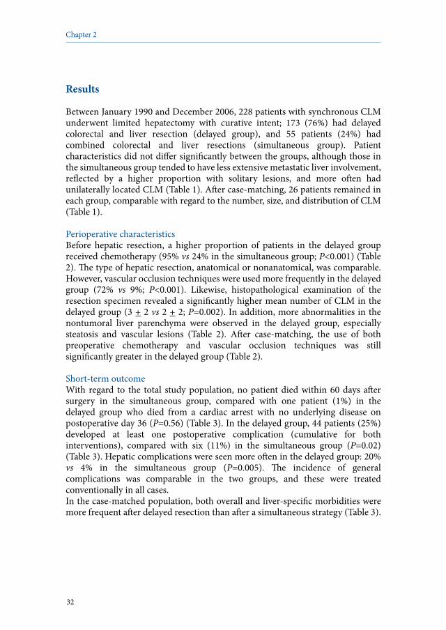
Short-term outcomeWith regard to the total study population, no patient died within 60 days after surgery in the simultaneous group, compared with one patient (1%) in the delayed group who died from a cardiac arrest with no underlying disease on postoperative day 36 (P=0.56) (Table 3). In the delayed group, 44 patients (25%) developed at least one postoperative complication (cumulative for both interventions), compared with six (11%) in the simultaneous group (P=0.02) (Table 3). Hepatic complications were seen more often in the delayed group: 20% vs 4% in the simultaneous group (P=0.005). The incidence of general complications was comparable in the two groups, and these were treated conventionally in all cases. In the case-matched population, both overall and liver-specific morbidities were more frequent after delayed resection than after a simultaneous strategy (Table 3).

33
Simultaneous or delayed liver surgery for limited synchronous colorectal metastases
Table 1.Patient and tumor characteristics.
Variable Total study population N=228 Case-matched groups N=52
SimultaneousN=55
DelayedN=173
P SimultaneousN=26
DelayedN=26
P
Patients Mean age at HR, years + SD 56 + 12 58 + 11 0.32 60 + 8 60 + 8 1.00*Male/Female 28 (51%) /
27 (49%)107 (62%) / 66 (38%)
0.15 17 (65%) / 9 (35%)
17 (65%) / 9 (35%)
1.00*
Primary tumorLocation
Right colonTransverse colonLeft or sigmoid colonRectum
12 (22%)5 (9%)26 (47%)12 (22%)
28 (17%)8 (5%)104 (62%)29 (17%)
0.27 7 (27%)4 (15%)8 (31%)7 (27%)
8 (31%)1 (4%)11 (42%)6 (23%)
0.49
T stage1234
1 (3%)5 (13%)22 (58%)10 (26%)
5 (6%)9 (11%)46 (58%)19 (24%)
0.92 0 (0%)4 (27%)10 (67%)1 (7%)
0 (0%)2 (11%)12 (67%)4 (22%)
0.30
N stage012
14 (37%)10 (26%)14 (37%)
26 (34%)27 (36%)23 (30%)
0.59 5 (33%)6 (40%)4 (27%)
8 (50%)3 (19%)5 (31%)
0.41
Liver metastases at diagnosisNumber of CLM
12–3> 3
28 (54%)14 (27%)10 (19%)
57 (35%)62 (39%)42 (26%)
0.06 15 (58%)7 (27%)4 (15%)
15 (58%)7 (27%)4 (15%)
1.00*
Mean max. size, mm + SD 39 + 37 38 + 30 0.79 38 + 33 41 + 21 0.70Unilateral/Bilateral 37 (69%) /
17 (32%)91 (55%) / 75 (45%)
0.08 19 (73%) / 7 (27%)
19 (73%) / 7 (27%)
1.00*
Initially unresectable 6 (11%) 35 (20%) 0.07 4 (15%) 7 (27%) 0.31Concomitant EHD
NoneLungOther
52 (95%)0 (0%)3 (6%)
146 (84%)12 (7%)15 (10%)
0.13 26 (100%)0 (0%)0 (0%)
21 (81%)2 (8%)3 (12%)
0.06
* matching criteria.Abbreviations: HR, hepatic resection; SD, standard deviation; CLM, colorectal liver metastases; EHD, extrahepatic disease.

34
Chapter 2
Table 2.Hepatic resection characteristics and histopathological findings.
Variable Total study populationN=228
Case-matched groupsN=52
SimultaneousN=55
DelayedN=173
P SimultaneousN=26
DelayedN=26
P
Hepatic resectionChemotherapy before hepatectomy
13 (24%) 165 (95%) < 0.001 8 (31%) 24 (92%) < 0.001
Total no. of lines1> 1
9 (69%)4 (31%)
102 (62%)63 (38%)
0.60 5 (63%)3 (37%)
16 (67%)8 (33%)
0.83
Total no. of cycles< 6 > 6
9 (82%)2 (18%)
27 (25%)80 (75%)
< 0.001 5 (71%)2 (29%)
7 (47%)8 (53%)
0.28
Last preoperative regimen5-FU + LV5-FU + LV + oxaliplatin5-FU + LV + irinotecanOther
6 (60%)2 (20%)1 (10%)1 (10%)
49 (32%)66 (43%)27 (18%)12 (8%)
0.59 3 (60%)1 (20%)1 (20%)0 (0%)
8 (33%)11 (46%)3 (13%)2 (8%)
0.57
Clinical response on CTResponseStabilizationProgression
7 (64%)4 (36%)0 (0%)
87 (57%)62 (40%)5 (3%)
0.78 5 (63%)3 (37%)0 (0%)
9 (43%)11 (52%)1 (5%)
0.57
Median preoperative CEA, ng/ml, range
9 (1-629) 4 (1-568) – 4 (1-629) 8 (1-568) –
Mean no. of CLM + SD 2 + 2 3 + 3 0.11 2 + 2 2 + 2 0.82Median RBC transfusions, units, range
0 (0-6) 0 (0-16) – 0 (0-6) 0 (0-6) –
Resection type AnatomicalNonanatomicalBoth
10 (18%)40 (73%)5 (9%)
52 (30%)106 (61%)15 (9%)
0.22 8 (31%)16 (62%)2 (8%)
10 (39%)14 (54%)2 (8%)
0.84
Vascular occlusionNoSelective inflowTotal pedicularTotal vascular
39 (91%)0 (0%)4 (9%)0 (0%)
39 (28%)5 (4%)81 (59%)13 (9%)
< 0.001 18 (86%)0 (0%)3 (14%)0 (0%)
5 (24%)0 (0%)13 (62%)3 (14%)
< 0.001

35
Simultaneous or delayed liver surgery for limited synchronous colorectal metastases
Table 2.Continued.
Variable Total study population N=228 Case-matched groups N=52
SimultaneousN=55
DelayedN=173
P SimultaneousN=26
DelayedN=26
P
Combined local treatmentRadiofrequency ablationCryosurgeryCombination
0 (0%)0 (0%)0 (0%)
2 (1%)12 (7%)1 (1%)
0.16 0 (0%)0 (0%)0 (0%)
1 (4%)2 (8%)0 (0%)
0.20
HistopathologyMean no. of CLM + SD 2 + 2 3 + 2 0.002 2 + 2 2 + 2 0.30Mean max. size, mm + SD 31 + 29 30 + 26 0.83 32 + 22 33 + 22 0.97Surgical margin status*
R0R1R2
23 (64%)11 (31%)2 (6%)
90 (62%)49 (34%)6 (4%)
0.89 14 (70%)5 (25%)1 (5%)
15 (65%)7 (30%)1 (4%)
0.92
Abnormalities in nontumoral liver 16 (29%)$ 100 (58%)$ < 0.001 8 (31%)$ 14 (54%)$ 0.17Steatosis 6 (11%) 40 (23%) 0.04 4 (15%) 6 (23%) 0.64
Mild (< 30% of hepatocytes)Moderate (30-60% of hepatocytes)Severe (> 60% of hepatocytes)
2 (33%)4 (67%)0 (0%)
21 (52%)16 (40%)3 (8%)
0.43 2 (50%)2 (50%)0 (0%)
5 (83%)1 (17%)0 (0%)
0.26
Fibrosis 9 (16%) 44 (25%) 0.12 5 (19%) 7 (27%) 0.69PortalPorto-portalSeptalCirrhosis
5 (56%)4 (44%)0 (0%)0 (0%)
37 (84%)4 (9%)3 (7%)0 (0%)
0.02 3 (60%)2 (40%)0 (0%)0 (0%)
5 (71%)1 (14%)1 (14%)0 (0%)
0.46
Surgical necrosis 1 (2%) 2 (1%) 0.73 0 (0%) 0 (0%) –Vascular lesions 4 (7%) 48 (28%)$ 0.001 1 (4%) 6 (23%)$ 0.06
Sinusoidal changes#
PeliosisHemorrhagic centrilobular necrosisRegenerative nodular hyperplasia
0 (0%)4 (100%)0 (0%)0 (0%)
23 (40%)24 (42%)2 (4%)8 (14%)
0.17 0 (0%)1 (100%)0 (0%)0 (0%)
2 (29%)2 (29%)1 (14%)2 (29%)
0.59
CASH 0 (0%) 3 (2%) 0.32 0 (0%) 1 (4%) 0.35Postoperative chemotherapy 47 (85%) 125 (72%) 0.53 22 (85%) 20 (77%) 0.67
* R0, complete surgical resection with a negative surgical margin at histopathology; R1, invaded surgical margins according to pathology report; R2, macroscopic tumor remnant seen during surgery; $ several patients had more than one abnormality; # sinusoidal changes include vasodilatation and congestion.Abbreviations: FU, fluorouracil; LV, leucovorin; CT, computed tomography; CLM, colorectal liver metastases; SD, standard deviation; CEA, carcinoembryonic antigen; RBC, red blood cell; CASH, chemotherapy-associated steatohepatitis.
36
Chapter 2
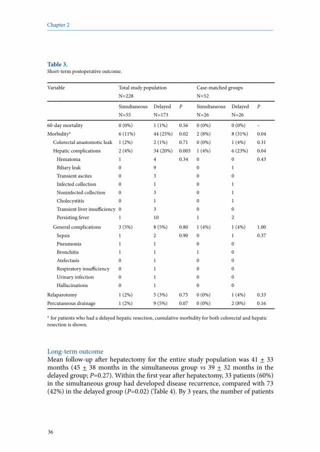
Table 3.Short-term postoperative outcome.
Variable Total study populationN=228
Case-matched groupsN=52
SimultaneousN=55
DelayedN=173
P SimultaneousN=26
DelayedN=26
P
60-day mortality 0 (0%) 1 (1%) 0.56 0 (0%) 0 (0%) –Morbidity* 6 (11%) 44 (25%) 0.02 2 (8%) 8 (31%) 0.04
Colorectal anastomotic leak 1 (2%) 2 (1%) 0.71 0 (0%) 1 (4%) 0.31Hepatic complications 2 (4%) 34 (20%) 0.005 1 (4%) 6 (23%) 0.04
HematomaBiliary leakTransient ascitesInfected collectionNoninfected collectionCholecystitisTransient liver insufficiencyPersisting fever
10000001
493131310
0.34 00000001
01011102
0.43
General complications 3 (5%) 8 (5%) 0.80 1 (4%) 1 (4%) 1.00SepsisPneumoniaBronchitisAtelectasisRespiratory insufficiencyUrinary infectionHallucinations
1110000
2111111
0.90 0010000
1000000
0.37
Relaparotomy 1 (2%) 5 (3%) 0.75 0 (0%) 1 (4%) 0.33Percutaneous drainage 1 (2%) 9 (5%) 0.07 0 (0%) 2 (8%) 0.16
* for patients who had a delayed hepatic resection, cumulative morbidity for both colorectal and hepatic resection is shown.
Long-term outcomeMean follow-up after hepatectomy for the entire study population was 41 + 33 months (45 + 38 months in the simultaneous group vs 39 + 32 months in the delayed group; P=0.27). Within the first year after hepatectomy, 33 patients (60%) in the simultaneous group had developed disease recurrence, compared with 73 (42%) in the delayed group (P=0.02) (Table 4). By 3 years, the number of patients
37
Simultaneous or delayed liver surgery for limited synchronous colorectal metastases
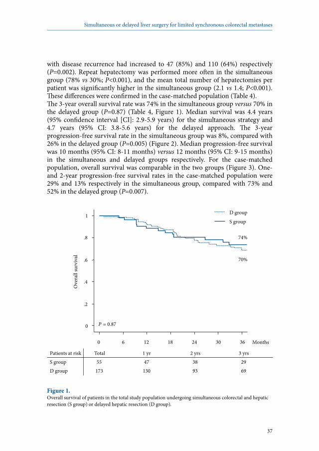
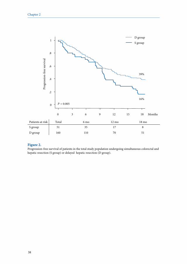
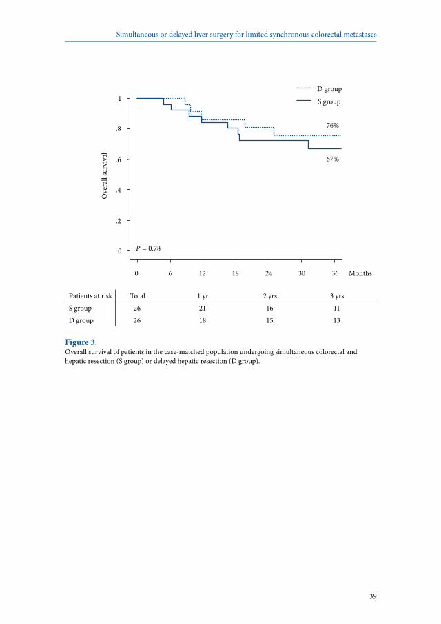
with disease recurrence had increased to 47 (85%) and 110 (64%) respectively (P=0.002). Repeat hepatectomy was performed more often in the simultaneous group (78% vs 30%; P<0.001), and the mean total number of hepatectomies per patient was significantly higher in the simultaneous group (2.1 vs 1.4; P<0.001). These differences were confirmed in the case-matched population (Table 4). The 3-year overall survival rate was 74% in the simultaneous group versus 70% in the delayed group (P=0.87) (Table 4, Figure 1). Median survival was 4.4 years (95% confidence interval [CI]: 2.9-5.9 years) for the simultaneous strategy and 4.7 years (95% CI: 3.8-5.6 years) for the delayed approach. The 3-year progression-free survival rate in the simultaneous group was 8%, compared with 26% in the delayed group (P=0.005) (Figure 2). Median progression-free survival was 10 months (95% CI: 8-11 months) versus 12 months (95% CI: 9-15 months) in the simultaneous and delayed groups respectively. For the case-matched population, overall survival was comparable in the two groups (Figure 3). One- and 2-year progression-free survival rates in the case-matched population were 29% and 13% respectively in the simultaneous group, compared with 73% and 52% in the delayed group (P=0.007).
0 6 12 18 24 30 36
0
.2
.4
.6
.8
1
Ove
rall
surv
ival
Months
P = 0.87
S group
D group
74%
70%
Patients at risk Total 1 yr 2 yrs 3 yrs S group 55 47 38 29 D group 173 130 93 69
Figure 1.Overall survival of patients in the total study population undergoing simultaneous colorectal and hepatic resection (S group) or delayed hepatic resection (D group).
38
Chapter 2
Patients at risk Total 6 mo 12 mo 18 mo S group 51 35 17 8
D group 160 110 70 51
0 3 6 9 12 15 18
0
.2
.4
.6
.8
1 Pr
ogre
ssio
n-fre
e sur
viva
l
Months
P = 0.005
S group
D group
16%
39%
Figure 2.Progression-free survival of patients in the total study population undergoing simultaneous colorectal and hepatic resection (S group) or delayed hepatic resection (D group).
39
Simultaneous or delayed liver surgery for limited synchronous colorectal metastases
0 6 12 18 24 30 36
0
.2
.4
.6
.8
1 O
vera
ll su
rviv
al
Months
P = 0.78
S group
D group
67%
76%
Patients at risk Total 1 yr 2 yrs 3 yrs S group 26 21 16 11 D group 26 18 15 13
Figure 3.Overall survival of patients in the case-matched population undergoing simultaneous colorectal and hepatic resection (S group) or delayed hepatic resection (D group).

40
Chapter 2
Table 4. Long-term postoperative outcome.
Variable Total study populationN=228
Case-matched groupsN=52
SimultaneousN=55
DelayedN=173
P SimultaneousN=26
DelayedN=26
P
Recurrence after 1 year 33 (60%) 73 (42%) 0.02 16 (62%) 5 (19%) 0.002HepaticExtrahepaticBoth
16 (49%)3 (9%)14 (42%)
29 (40%)16 (22%)28 (38%)
0.27 8 (50%)2 (13%)6 (37%)
2 (40%)3 (60%)0 (0%)
0.06
Recurrence after 3 years 47 (85%) 110 (64%) 0.002 21 (81%) 12 (46%) 0.01HepaticExtrahepaticBoth
19 (40%)4 (9%)24 (51%)
37 (34%)24 (22%)49 (44%)
0.14 9 (43%)3 (14%)9 (43%)
3 (25%)5 (42%)4 (33%)
0.20
Total number of hepatectomies
1234
12 (22%)28 (51%)13 (24%)2 (4%)
120 (69%)40 (23%)11 (6%)2 (1%)
< 0.001 7 (27%)11 (42%)8 (31%)0 (0%)
18 (69%)7 (27%)1 (4%)0 (0%)
0.004
Total number of EHD resections
01234
39 (71%)12 (22%)4 (7%)0 (0%)0 (0%)
137 (79%)24 (14%)8 (5%)1 (1%)3 (2%)
0.42 21 (81%)4 (15%)1 (4%)0 (0%)0 (0%)
21 (81%)2 (8%)3 (12%)0 (0%)0 (0%)
0.44
Status at last follow-upAlive without diseaseAlive with diseaseDead
17 (31%)12 (22%)26 (47%)
57 (33%)51 (29%)65 (38%)
0.38 6 (23%)8 (31%)12 (46%)
13 (50%)4 (15%)9 (35%)
0.11
3-year survival rate (%) 74 70 0.87 67 76 0.78
Abbreviation: EHD, extrahepatic disease.

41
Simultaneous or delayed liver surgery for limited synchronous colorectal metastases
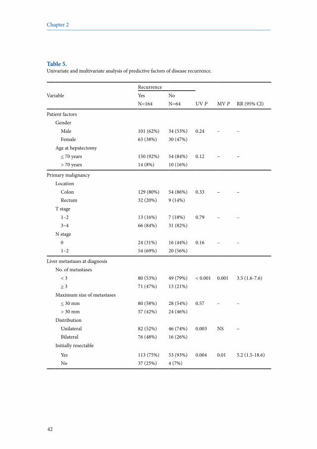
Predictors of disease recurrence after first hepatectomyFor the total study population, multivariate analysis identified three independent predictors of disease recurrence: three or more CLM at diagnosis (RR 3.5, 95% CI 1.6-7.6; P=0.001), initial unresectability of CLM (RR 5.2, 95% CI 1.5-18.6; P=0.01), and simultaneous colorectal and hepatic resection (RR 4.8, 95% CI 1.9-12.0; P=0.001) (Table 5).
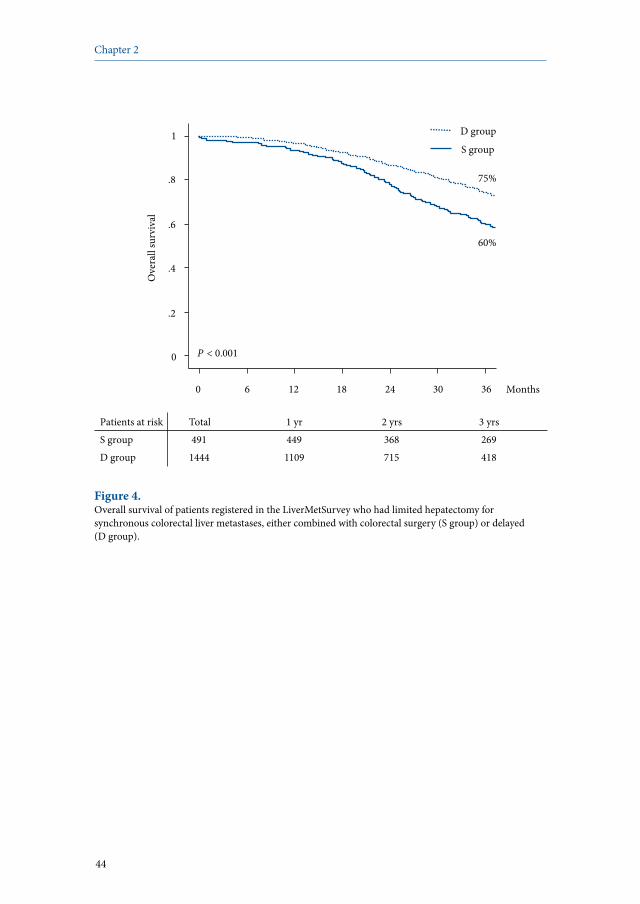
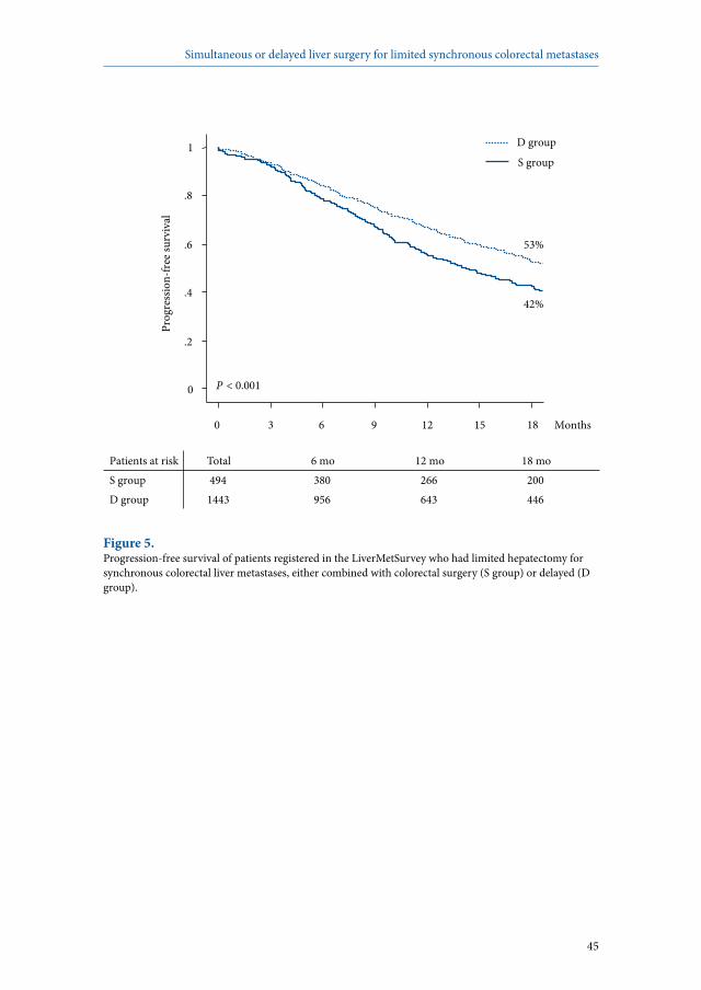
LiverMetSurvey analysisA total of 2001 patients underwent limited hepatectomy for synchronous CLM, of whom 506 had a simultaneous colorectal and hepatic resection, and 1495 a delayed hepatectomy. Apart from a difference in age, there were no other differences in major patient characteristics between the groups (Table 6). A significantly higher recurrence rate was observed after a simultaneous strategy (61% vs 35%; P<0.001). In addition, the 3-year survival rate was significantly lower after a simultaneous compared with a delayed strategy (60% vs 75%, respectively; P<0.001) (Figure 4). Similarly, median progression-free survival was significantly shorter after a simultaneous colorectal and hepatic resection (14 months vs 20 months for the delayed strategy; P<0.001) (Figure 5).
42
Chapter 2
Table 5. Univariate and multivariate analysis of predictive factors of disease recurrence.
RecurrenceVariable Yes
N=164NoN=64 UV P MV P RR (95% CI)
Patient factorsGender
MaleFemale
101 (62%)63 (38%)
34 (53%)30 (47%)
0.24 – –
Age at hepatectomy< 70 years> 70 years
150 (92%)14 (8%)
54 (84%)10 (16%)
0.12 – –
Primary malignancyLocation
ColonRectum
129 (80%)32 (20%)
54 (86%)9 (14%)
0.33 – –
T stage1–23–4
13 (16%)66 (84%)
7 (18%)31 (82%)
0.79 – –
N stage01–2
24 (31%)54 (69%)
16 (44%)20 (56%)
0.16 – –
Liver metastases at diagnosisNo. of metastases
< 3> 3
80 (53%)71 (47%)
49 (79%)13 (21%)
< 0.001 0.001 3.5 (1.6-7.6)
Maximum size of metastases< 30 mm> 30 mm
80 (58%)57 (42%)
28 (54%)24 (46%)
0.57 – –
DistributionUnilateralBilateral
82 (52%)76 (48%)
46 (74%)16 (26%)
0.003 NS –
Initially resectable
YesNo
113 (75%)37 (25%)
53 (93%)4 (7%)
0.004 0.01 5.2 (1.5-18.6)

43
Simultaneous or delayed liver surgery for limited synchronous colorectal metastases
Table 5. Continued.
RecurrenceVariable Yes
N=164NoN=64 UV P MV P RR (95% CI)
Hepatic resectionPreoperative chemotherapy
YesNo
121 (74%)42 (26%)
57 (89%)7 (11%)
0.02 NS –
Timing of hepatic resectionSimultaneous to colorectal surgery Delayed
48 (29%)116 (71%)
7 (11%)57 (89%)
0.004 0.001 4.8 (1.9-12.0)
Concomitant extrahepatic disease
YesNo
28 (17%)136 (83%)
2 (3%)62 (97%)
0.005 NS –
Resection typeAnatomicalNonanatomical
38 (25%)114 (75%)
24 (43%)32 (57%)
0.01 NS –
Vascular occlusionYesNo
66 (53%)59 (47%)
37 (66%)19 (34%)
0.09 NS –
Combined local treatmentYesNo
12 (7%)152 (93%)
3 (5%)61 (95%)
0.47 – –
Surgical margin status*R0R1
69 (60%)46 (40%)
43 (78%)12 (22%)
0.02 NS –
Postoperative chemotherapyYesNo
134 (86%)22 (14%)
38 (73%)14 (27%)
0.03 NS –
* R0, complete surgical resection with a negative surgical margin at histopathology; R1, invaded surgical margins according to pathology report.Abbreviations: UV, univariate; MV, multivariate; RR, risk ratio; CI, confidence interval; NS, not significant.
44
Chapter 2
0 6 12 18 24 30 36
0
.2
.4
.6
.8
1 O
vera
ll su
rviv
al
Months
P < 0.001
S group
D group
60%
75%
Patients at risk Total 1 yr 2 yrs 3 yrs S group 491 449 368 269 D group 1444 1109 715 418
Figure 4.Overall survival of patients registered in the LiverMetSurvey who had limited hepatectomy for synchronous colorectal liver metastases, either combined with colorectal surgery (S group) or delayed (D group).
45
Simultaneous or delayed liver surgery for limited synchronous colorectal metastases
Patients at risk Total 6 mo 12 mo 18 mo S group 494 380 266 200 D group 1443 956 643 446
0 3 6 9 12 15 18
0
.2
.4
.6
.8
1 Pr
ogre
ssio
n-fre
e sur
viva
l
Months
P < 0.001
S group
D group
42%
53%
Figure 5.Progression-free survival of patients registered in the LiverMetSurvey who had limited hepatectomy for synchronous colorectal liver metastases, either combined with colorectal surgery (S group) or delayed (D group).

46
Chapter 2
Table 6. Patient and tumor characteristics LiverMetSurvey analysis.
Variable SimultaneousN=506
DelayedN=1495
P
PatientsMean age at HR, years + SD 58 + 11 61 + 11 < 0.001Male/Female 320 (64%) / 183 (36%) 885 (59%) / 609 (41%) 0.08
Primary tumorColon/Rectum 310 (68%) / 148 (32%) 932 (68%) / 433 (32%) 0.81
Liver metastases at diagnosisNumber of CLM
12–3> 3
134 (38%)128 (36%)91 (26%)
524 (41%)460 (36%)283 (22%)
0.33
Mean maximum size, mm + SD 37 + 33 39 + 33 0.30Unilateral/Bilateral 228 (61%) / 146 (39%) 814 (60%) / 536 (40%) 0.82Preoperative chemotherapy 225 (44%) 622 (42%) 0.20
Abbreviations: HR, hepatic resection; SD, standard deviation; CLM, colorectal liver metastases.

47
Simultaneous or delayed liver surgery for limited synchronous colorectal metastases
Discussion
As the optimal surgical strategy for patients with synchronous CLM is still controversial, the experience of combined colorectal and liver surgery in a tertiary referral center was evaluated by comparison with a delayed surgical strategy. Postoperative mortality was comparable for the two treatment strategies, but the morbidity rate was significantly lower after simultaneous colorectal and hepatic resection. Disease recurrence was observed more often in patients treated by a simultaneous strategy. Three-year overall survival rates did not significantly differ according to the surgical strategy, but progression-free survival was significantly better after delayed hepatic surgery. Three independent predictive factors of disease recurrence were: three or more CLM at diagnosis, initial unresectability of CLM, and simultaneous colorectal and hepatic resection. After case-matching for age, gender, number and location of CLM, the morbidity rate remained lower and the recurrence rate higher in the simultaneous strategy group. Furthermore, progression-free survival was also significantly lower in the simultaneous strategy group. Unfavorable recurrence rates for simultaneous colorectal and hepatic resection were confirmed in the LiverMetSurvey cohort. An important advantage of simultaneous colorectal and liver surgery is that it involves only one operation, thereby lowering the risk of disease dissemination,15 preventing repeated postoperative immunosuppression and thus decreasing tumoral growth,30,31 and establishing a greater reduction of total tumor volume, which might enhance chemotherapeutic efficacy.32 In addition, the need for only one operation improves patient comfort and reduces healthcare costs. However, with this strategy it is not possible to observe the biological behaviour of the metastatic disease following primary tumor resection, thereby probably compromising selection of the best candidates for hepatic resection.6 Importantly, in the present study patients treated by a combined surgical strategy had a significantly lower morbidity rate, mainly as a result of fewer hepatic complications. Interestingly, these patients were less frequently treated by preoperative chemotherapy (24% vs 95% of the delayed group). In addition, abnormalities of the nontumoral liver parenchyma were observed significantly less often. The relation between preoperative chemotherapy and abnormalities of the nontumoral liver parenchyma has been well established, especially for vascular changes and steatohepatitis.33-36 In a recent study, increased postoperative morbidity and a higher incidence of abnormalities of the nontumoral liver parenchyma were observed when patients received preoperative chemotherapy.37 In the present study, the combination of more frequent use of preoperative chemotherapy and higher incidence of abnormalities of the nontumoral liver parenchyma within the delayed strategy group might explain the higher number of postoperative (hepatic) complications. In addition, the higher incidence of
48
Chapter 2
vascular lesions in patients having the delayed strategy could explain the greater need for intraoperative vascular occlusion techniques, as the occurrence of vascular lesions in the nontumoral liver parenchyma at histopathology has been shown to be associated with an increased need for intraoperative red blood cell transfusion.33
The difference in use of chemotherapy before hepatic resection in the two groups may explain the higher recurrence rate with the simultaneous resection strategy, in both the total study population and the case-matched subgroups. However, in multivariate analysis, preoperative chemotherapy was not a predictor of recurrence. In contrast, a simultaneous surgical strategy was found to be an independent predictor of postoperative disease recurrence. This finding was not expected, as the simultaneous strategy group comprised more patients with less extensive liver disease. However, another important factor could be that drop-out in the delayed hepatectomy group of patients with progressive intrahepatic and/or extrahepatic disease after resection of the primary colorectal tumor may have selected a residual group with a more favorable prognosis. Such selection could not, of course, have occurred in patients undergoing a simultaneous strategy. Yan and colleagues24 and Chua et al12 reported similar progression-free survival rates for both surgical strategies. Conversely, Tanaka and co-workers19 observed a shorter progression-free interval for a simultaneous strategy, although the difference was not statistically significant. Of note, the present results were confirmed in a larger multicenter cohort using data from the international registry LiverMetSurvey, both for recurrence and for overall survival. In this study, the combination of major hepatectomy (three or more segments) and colorectal resection was avoided, and the series included only patients who underwent a limited hepatectomy. Only four previous studies19,20,38,39 have considered simultaneous major hepatectomy and colorectal surgery. In three,19,20,39 the simultaneous strategy resulted in higher mortality and/or morbidity rates compared with delayed hepatectomy, but the fourth study38 found similar short-term results, even in the presence of rectal cancer. Colorectal resection can safely be combined with a limited hepatectomy in patients with synchronous CLM with respect to postoperative morbidity. However, the increased postoperative recurrence rate observed after a simultaneous surgical strategy, confirmed in a larger multicenter cohort of LiverMetSurvey, raises questions regarding oncological value and use in clinical practice. Although preferable in the short term with regard to safety and patient comfort, a simultaneous surgical strategy has a negative impact on the long-term outcome.

49
Simultaneous or delayed liver surgery for limited synchronous colorectal metastases
References
1. World Health Organization. Global cancer rates could increase by 50% to 15 million by 2020. World Cancer Report provides clear evidence that action on smoking, diet and infections can prevent one third of cancers, another third can be cured. World Health Organization 2003; Available at: www.who.int/mediacentre/news/releases/2003/pr27/en/print.html.
2. Leporrier J, Maurel J, Chiche L et al. A population-based study of the incidence, management and prognosis of hepatic metastases from colorectal cancer. Br J Surg 2006; 93:465-474.
3. Steele G Jr, Ravikumar TS. Resection of hepatic metastases from colorectal cancer. Biologic perspective. Ann Surg 1989; 210:127-138.
4. Scheele J, Altendorf-Hofmann A. Resection of colorectal liver metastases. Langenbecks Arch Surg 1999; 384:313-327.
5. Adam R. Colorectal cancer with synchronous liver metastases. Br J Surg 2007; 94:129-131.6. Lambert LA, Colacchio TA, Barth RJ Jr. Interval hepatic resection of colorectal metastases
improves patient selection. Arch Surg 2000; 135:473-479.7. de Santibañes E, Lassalle FB, McCormack L et al. Simultaneous colorectal and hepatic
resections for colorectal cancer: postoperative and longterm outcomes. J Am Coll Surg 2002; 195:196-202.
8. Falcone A, Ricci S, Brunetti I et al. Phase III trial of infusional fluorouracil, leucovorin, oxaliplatin, and irinotecan (FOLFOXIRI) compared with infusional fluorouracil, leucovorin, and irinotecan (FOLFIRI) as first-line treatment for metastatic colorectal cancer: the Gruppo Oncologico Nord Ovest. J Clin Oncol 2007; 25:1670-1676.
9. Köhne CH, van Cutsem E, Wils J et al. Phase III study of weekly high-dose infusional fluorouracil plus folinic acid with or without irinotecan in patients with metastatic colorectal cancer: European Organisation for Research and Treatment of Cancer Gastrointestinal Group Study 40986. J Clin Oncol 2005; 23:4856-4865.
10. Tournigand C, André T, Achille E et al. FOLFIRI followed by FOLFOX6 or the reverse sequence in advanced colorectal cancer: a randomized GERCOR study. J Clin Oncol 2004; 22:229-237.
11. Capussotti L, Vigano L, Ferrero A et al. Timing of resection of liver metastases synchronous to colorectal tumor: proposal of prognosis-based decisional model. Ann Surg Oncol 2007; 14:1143-1150.
12. Chua HK, Sondenaa K, Tsiotos GG et al. Concurrent vs staged colectomy and hepatectomy for primary colorectal cancer with synchronous hepatic metastases. Dis Colon Rectum 2004; 47:1310-1316.
13. Fujita S, Akasu T, Moriya Y. Resection of synchronous liver metastases from colorectal cancer. Jpn J Clin Oncol 2000; 30:7-11.
14. Jatzko G, Wette V, Müller M et al. Simultaneous resection of colorectal carcinoma and synchronous liver metastases in a district hospital. Int J Colorectal Dis 1991; 6:111-114.
15. Lyass S, Zamir G, Matot I et al. Combined colon and hepatic resection for synchronous colorectal liver metastases. J Surg Oncol 2001; 78:17-21.
16. Martin R, Paty P, Fong Y et al. Simultaneous liver and colorectal resections are safe for synchronous colorectal liver metastasis. J Am Coll Surg 2003; 197:233-241.
17. Minagawa M, Yamamoto J, Miwa S et al. Selection criteria for simultaneous resection in patients with synchronous liver metastasis. Arch Surg 2006; 141:1006-1012.
18. Nordlinger B, Guiguet M, Vaillant JC et al. Surgical resection of colorectal carcinoma metastases to the liver. A prognostic scoring system to improve case selection, based on 1568 patients. Association Française de Chirurgie. Cancer 1996; 77:1254-1262.
19. Tanaka K, Shimada H, Matsuo K et al. Outcome after simultaneous colorectal and hepatic resection for colorectal cancer with synchronous metastases. Surgery 2004; 136:650-659.

50
Chapter 2
20. Thelen A, Jonas S, Benckert C et al. Simultaneous versus staged liver resection of synchronous liver metastases from colorectal cancer. Int J Colorectal Dis 2007; 22:1269-1276.
21. Vassiliou I, Arkadopoulos N, Theodosopoulos T et al. Surgical approaches of resectable synchronous colorectal liver metastases: timing considerations. World J Gastroenterol 2007; 13:1431-1434.
22. Vogt P, Raab R, Ringe B et al. Resection of synchronous liver metastases from colorectal cancer. World J Surg 1991; 15:62-67.
23. Weber JC, Bachellier P, Oussoultzoglou E et al. Simultaneous resection of colorectal primary tumour and synchronous liver metastases. Br J Surg 2003; 90:956-962.
24. Yan TD, Chu F, Black D et al. Synchronous resection of colorectal primary cancer and liver metastases. World J Surg 2007; 31:1496-1501.
25. Jenkins LT, Millikan KW, Bines SD et al. Hepatic resection for metastatic colorectal cancer. Am Surg 1997; 63:605-610.
26. Bismuth H. Surgical anatomy and anatomical surgery of the liver. World J Surg 1982; 6:3-9.27. Couinaud C. Le foie: études anatomiques et chirurgicales. Paris: Masson et Cie; 1957.28. World Health Organization. Handbook for reporting results of cancer treatment. Geneva: World
Health Organization; 1979.29. Therasse P, Arbuck SG, Eisenhauer EA et al. New guidelines to evaluate the response to
treatment in solid tumors. European Organization for Research and Treatment of Cancer, National Cancer Institute of the United States, National Cancer Institute of Canada. J Natl Cancer Inst 2000; 92:205-216.
30. Eggermont AM, Steller EP, Sugarbaker PH. Laparotomy enhances intraperitoneal tumor growth and abrogates the antitumor effects of interleukin-2 and lymphokine-activated killer cells. Surgery 1987; 102:71-78.
31. Weese JL, Ottery FD, Emoto SE. Do operations facilitate tumor growth? An experimental model in rats. Surgery 1986; 100:273-277.
32. Elias D, Liberale G, Vernerey D et al. Hepatic and extrahepatic colorectal metastases: when resectable, their localization does not matter, but their total number has a prognostic effect. Ann Surg Oncol 2005; 12:900-909.
33. Aloia T, Sebagh M, Plasse M et al. Liver histology and surgical outcomes after preoperative chemotherapy with fluorouracil plus oxaliplatin in colorectal cancer liver metastases. J Clin Oncol 2006; 24:4983-4990.
34. Pawlik TM, Olino K, Gleisner AL et al. Preoperative chemotherapy for colorectal liver metastases: impact on hepatic histology and postoperative outcome. J Gastrointest Surg 2007; 11:860-868.
35. Rubbia-Brandt L, Audard V, Sartoretti P et al. Severe hepatic sinusoidal obstruction associated with oxaliplatin-based chemotherapy in patients with metastatic colorectal cancer. Ann Oncol 2004; 15:460-466.
36. Vauthey JN, Pawlik TM, Ribero D et al. Chemotherapy regimen predicts steatohepatitis and an increase in 90-day mortality after surgery for hepatic colorectal metastases. J Clin Oncol 2006; 24:2065-2072.
37. Karoui M, Penna C, Amin-Hashem M et al. Influence of preoperative chemotherapy on the risk of major hepatectomy for colorectal liver metastases. Ann Surg 2006; 243:1-7.
38. Capussotti L, Ferrero A, Vigano L et al. Major liver resections synchronous with colorectal surgery. Ann Surg Oncol 2007; 14:195-201.
39. Nordlinger B, Jaeck D, Guiguet M et al. Résection chirurgicale des métastases hépatiques. Enquête de l’Association Française de Chirurgie. In: Nordlinger B, Jaeck D (eds). Métastases hépatiques des cancers colo-rectaux. Paris: Springer-Verlag; 1992:141-159.

51
Simultaneous or delayed liver surgery for limited synchronous colorectal metastases


Robbert J. de Haas1,2
Dennis A. Wicherts1,2
Paola Andreani1
Gérard Pascal1
Faouzi Saliba1
Philippe Ichai1
René Adam1
Denis Castaing1
Daniel Azoulay1
1 Centre Hépato-Biliaire, Hôpital Paul Brousse, Villejuif, France2 Department of Surgery, University Medical Center Utrecht, Utrecht,
The Netherlands
Annals of Surgery; accepted for publication
Impact of expanding criteria for resectability of colorectal metastases on short- and long-term outcomes after hepatic resection
3
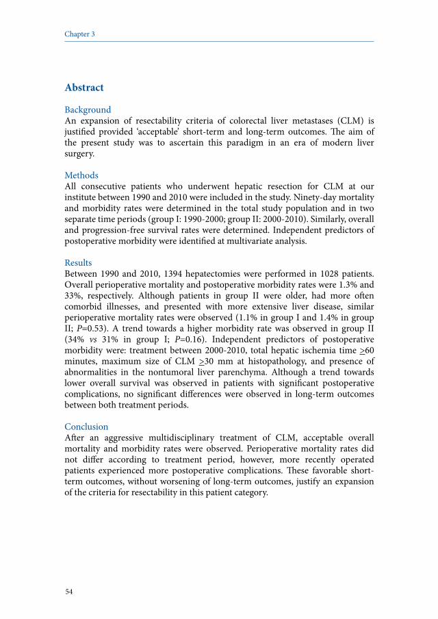
54
Chapter 3
Abstract
BackgroundAn expansion of resectability criteria of colorectal liver metastases (CLM) is justified provided ‘acceptable’ short-term and long-term outcomes. The aim of the present study was to ascertain this paradigm in an era of modern liver surgery.
MethodsAll consecutive patients who underwent hepatic resection for CLM at our institute between 1990 and 2010 were included in the study. Ninety-day mortality and morbidity rates were determined in the total study population and in two separate time periods (group I: 1990-2000; group II: 2000-2010). Similarly, overall and progression-free survival rates were determined. Independent predictors of postoperative morbidity were identified at multivariate analysis.
ResultsBetween 1990 and 2010, 1394 hepatectomies were performed in 1028 patients. Overall perioperative mortality and postoperative morbidity rates were 1.3% and 33%, respectively. Although patients in group II were older, had more often comorbid illnesses, and presented with more extensive liver disease, similar perioperative mortality rates were observed (1.1% in group I and 1.4% in group II; P=0.53). A trend towards a higher morbidity rate was observed in group II (34% vs 31% in group I; P=0.16). Independent predictors of postoperative morbidity were: treatment between 2000-2010, total hepatic ischemia time >60 minutes, maximum size of CLM >30 mm at histopathology, and presence of abnormalities in the nontumoral liver parenchyma. Although a trend towards lower overall survival was observed in patients with significant postoperative complications, no significant differences were observed in long-term outcomes between both treatment periods.
ConclusionAfter an aggressive multidisciplinary treatment of CLM, acceptable overall mortality and morbidity rates were observed. Perioperative mortality rates did not differ according to treatment period, however, more recently operated patients experienced more postoperative complications. These favorable short-term outcomes, without worsening of long-term outcomes, justify an expansion of the criteria for resectability in this patient category.

55
Impact of expanding criteria for resectability of colorectal liver metastases on outcome
Introduction
Hepatic resection is the only treatment option providing long-term survival for patients with colorectal liver metastases (CLM), with 5-year survival rates up to 67% in highly selected patients.1 However, when using traditional criteria, hepatic resection can only be offered to 10-20% of patients presenting with CLM.2 To increase the number of patients amenable for surgery, the criteria for resectability have recently shifted from criteria based on the characteristics of the metastatic disease related to long-term outcome to new criteria which include any patient in whom all disease can be removed with a negative margin and who has an adequate remnant hepatic volume/reserve.3 To achieve resectability, several strategies have been developed. An important increase in resectability rates can be achieved by using modern chemotherapy regimens, which downsize metastatic disease enabling surgical treatment.4-10 If the reduction in tumor size after chemotherapy is not sufficient to allow surgery, several techniques such as portal vein embolization,11,12 two-stage hepatectomy,13 vascular resection and reconstruction,14 and in situ hypothermic liver perfusion15 can be considered, aiming to further enlarge the number of patients with resectable disease. Perioperative mortality rates following hepatic resection in high-volume centers have mostly been reported to be less than 5% and reported postoperative morbidity rates varied between 22% and 45%.16-21 Factors that might have contributed to the increased safety of hepatic resection are the better understanding of segmental liver anatomy, progress in imaging techniques, and refined surgical techniques in controlling hemorrhage, improvements in anesthetic techniques, more stringent patient selection, and the emergence of hepatobiliary surgery as a distinct specialty.16,19,20,22,23 Due to these improvements, hepatic resection is increasingly performed in older patients with more comorbidities,24 and more often, hepatic resection is combined with resection of the primary colorectal tumor.25
Although an expansion of the criteria for resectability of CLM via an aggressive multidisciplinary approach increases the number of patients amenable for surgery, its effects on short-term and long-term outcomes still have to be determined. Therefore, the aim of the present study was to evaluate whether an expansion of the criteria for resectability of CLM could be achieved while maintaining perioperative mortality and morbidity rates as well as long-term survival within acceptable limits. The present study was performed at a single institution, determining short-term and long-term outcomes following hepatic resection for CLM both within the total patient population as well as in separate time periods, enabling to evaluate trends over time.

56
Chapter 3
Patients and methods
Study populationAll consecutive patients treated by hepatic resection for CLM at our institution between January 1990 and January 2010 were included in the study. Patients were identified in our prospectively maintained institutional database, and additional information was obtained by reviewing the medical records. Besides determining outcomes in the total patient population, patients were divided into two groups according to the period of treatment to evaluate trends over time. The two time periods were chosen based on the introduction of important oncosurgical strategies to improve resectability (1990-2000: new chemotherapy agents, portal vein embolization, two-stage hepatectomy; 2000-2010: vascular resection and reconstruction techniques, intraoperative hypothermic liver perfusion, biological agents).26 Also, short-term outcomes were determined following repeat hepatectomy.
Preoperative workupPreoperatively, each patient underwent abdominal ultrasonography and computed tomography (CT) to determine the extent of liver disease, chest radiographs and thoracic CT, colonoscopy, and measurement of serum tumor markers. In case of initially unresectable CLM (ie, a too small remnant liver volume [<30%] after complete resection of all CLM, and/or unresectable extrahepatic disease), portal vein embolization was performed and/or preoperative chemotherapy was administered.11,27 During the latter part of the study, patients with initially resectable CLM were increasingly treated by neoadjuvant chemotherapy, when at least three nodules were present. Clinical response to chemotherapy was determined radiologically every 2 months, initially according to the World Health Organization guidelines,28 but more recently by using the Response Evaluation Criteria in Solid Tumors.29 All treatment decisions were taken during multidisciplinary repeated meetings. Hepatic resectionAll surgical interventions were performed with curative intent, supervised by 3 senior surgeons (DA, DC, RA). After laparotomy, abdominal exploration and intraoperative ultrasound were used to determine the extent of liver disease and to detect extrahepatic disease sites. Resection of three or more anatomic liver segments was considered a major hepatectomy.30,31 In case of multiple bilobar CLM which could not be resected during a single hepatectomy, even when combined with preoperative chemotherapy and specific techniques such as radiofrequency ablation and/or cryosurgery, a two-stage hepatectomy was performed.13,32 Parenchymal transection was performed using an ultrasonic dissector (Cavitron Ultrasonic Surgical Aspirator, Valleylab, Boulder, CO, USA)

57
Impact of expanding criteria for resectability of colorectal liver metastases on outcome
and a fenestrated bipolar forceps. When the hepatocaval confluence and/or the inferior vena cava were involved in the disease process, vascular resection and reconstruction techniques were performed, combined with in situ hypoperfusion of the liver if vascular exclusion was predicted to last beyond 1 hour.14,15 If needed to control intraoperative blood loss, vascular clamping techniques such as the Pringle maneuver or total vascular exclusion were used on the discretion of the surgeon. At the end of the operation, an abdominal drain was placed routinely. In case of intra-abdominal extrahepatic disease, confirmed by frozen-section biopsy, simultaneous resection of CLM and extrahepatic disease was performed only if it was considered complete. When extrahepatic disease was located outside the abdominal cavity (eg, lung metastases), resection was performed 2-3 months after hepatectomy if the disease remained controlled by chemotherapy. Postoperative routine histopathologic examination of all resection specimens was performed.
Short-term outcomesPerioperative mortality was defined as any death that occurred within 90 days of hepatic resection or during the same hospitalization. Postoperative morbidity was defined as any postoperative adverse event which occurred during the same period. Postoperative complications were divided into hepatic complications and general complications. Hepatic complications, which occurred within the field of liver resection, included hemorrhage/hematoma needing reoperation and/or blood transfusion, biliary leakage, transient ascites, infected collection, noninfected collection, cholecystitis, transient liver insufficiency (according to the ‘50-50’ criteria33), and persisting fever. General complications, which occurred distant from the hepatic resection field, included sepsis, pneumonia, bronchitis, atelectasis, respiratory insufficiency, pleural effusion needing puncture or drainage, urinary tract infection, and others such as cardiovascular or renal complications. Severity of postoperative complications was graded retrospectively according to the Dindo-Clavien classification.34
Postoperative follow-upPostoperatively, patients visited our outpatient clinic regularly, starting 1 month after the operation, and then every 4 months. Follow-up consisted of a history, physical examination, serum tumor markers, liver function tests, and hepatic imaging (ultrasonography and CT). In case of disease recurrence, resection was only performed if it could be complete.
58
Chapter 3
Statistical analysisCategorical variables were compared by the χ2 test, and continuous variables were compared using the independent-samples t test. Overall and progression-free survival following hepatic resection were calculated by the Kaplan-Meier method, and compared by the log-rank test. Univariate analysis was performed to identify factors significantly related to postoperative morbidity. To obtain independent predictors of postoperative morbidity, a multivariate analysis (logistic regression) was performed including all factors likely to influence postoperative morbidity rate with an univariate P<0.10. Due to the fact that only a few patients died within 90 days from operation, independent predictors of perioperative mortality could not be determined. A P value <0.05 was considered statistically significant. SPSS version 15.0 (SPSS Inc, Chicago, IL, USA) was used for all statistical analyses.
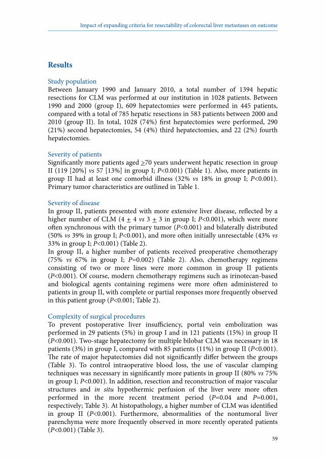
59
Impact of expanding criteria for resectability of colorectal liver metastases on outcome
Results
Study populationBetween January 1990 and January 2010, a total number of 1394 hepatic resections for CLM was performed at our institution in 1028 patients. Between 1990 and 2000 (group I), 609 hepatectomies were performed in 445 patients, compared with a total of 785 hepatic resections in 583 patients between 2000 and 2010 (group II). In total, 1028 (74%) first hepatectomies were performed, 290 (21%) second hepatectomies, 54 (4%) third hepatectomies, and 22 (2%) fourth hepatectomies. Severity of patientsSignificantly more patients aged >70 years underwent hepatic resection in group II (119 [20%] vs 57 [13%] in group I; P<0.001) (Table 1). Also, more patients in group II had at least one comorbid illness (32% vs 18% in group I; P<0.001). Primary tumor characteristics are outlined in Table 1. Severity of diseaseIn group II, patients presented with more extensive liver disease, reflected by a higher number of CLM (4 + 4 vs 3 + 3 in group I; P<0.001), which were more often synchronous with the primary tumor (P<0.001) and bilaterally distributed (50% vs 39% in group I; P<0.001), and more often initially unresectable (43% vs 33% in group I; P<0.001) (Table 2). In group II, a higher number of patients received preoperative chemotherapy (75% vs 67% in group I; P=0.002) (Table 2). Also, chemotherapy regimens consisting of two or more lines were more common in group II patients (P<0.001). Of course, modern chemotherapy regimens such as irinotecan-based and biological agents containing regimens were more often administered to patients in group II, with complete or partial responses more frequently observed in this patient group (P<0.001; Table 2).
Complexity of surgical proceduresTo prevent postoperative liver insufficiency, portal vein embolization was performed in 29 patients (5%) in group I and in 121 patients (15%) in group II (P<0.001). Two-stage hepatectomy for multiple bilobar CLM was necessary in 18 patients (3%) in group I, compared with 85 patients (11%) in group II (P<0.001). The rate of major hepatectomies did not significantly differ between the groups (Table 3). To control intraoperative blood loss, the use of vascular clamping techniques was necessary in significantly more patients in group II (80% vs 75% in group I; P<0.001). In addition, resection and reconstruction of major vascular structures and in situ hypothermic perfusion of the liver were more often performed in the more recent treatment period (P=0.04 and P=0.001, respectively; Table 3). At histopathology, a higher number of CLM was identified in group II (P<0.001). Furthermore, abnormalities of the nontumoral liver parenchyma were more frequently observed in more recently operated patients (P<0.001) (Table 3).
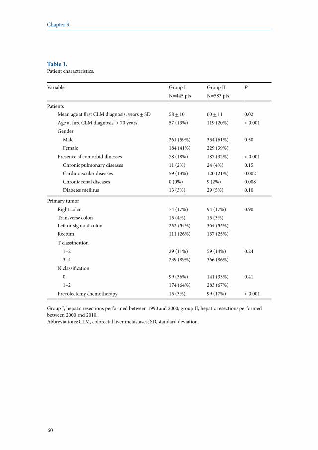
60
Chapter 3
Table 1. Patient characteristics.
Variable Group IN=445 pts
Group IIN=583 pts
P
PatientsMean age at first CLM diagnosis, years + SD 58 + 10 60 + 11 0.02Age at first CLM diagnosis > 70 years 57 (13%) 119 (20%) < 0.001Gender
MaleFemale
261 (59%)184 (41%)
354 (61%)229 (39%)
0.50
Presence of comorbid illnesses 78 (18%) 187 (32%) < 0.001Chronic pulmonary diseasesCardiovascular diseasesChronic renal diseasesDiabetes mellitus
11 (2%)59 (13%)0 (0%)13 (3%)
24 (4%)120 (21%)9 (2%)29 (5%)
0.150.0020.0080.10
Primary tumorRight colonTransverse colonLeft or sigmoid colonRectum
74 (17%)15 (4%)232 (54%)111 (26%)
94 (17%)15 (3%)304 (55%)137 (25%)
0.90
T classification1–23–4
29 (11%)239 (89%)
59 (14%)366 (86%)
0.24
N classification01–2
99 (36%)174 (64%)
141 (33%)283 (67%)
0.41
Precolectomy chemotherapy 15 (3%) 99 (17%) < 0.001
Group I, hepatic resections performed between 1990 and 2000; group II, hepatic resections performed between 2000 and 2010.Abbreviations: CLM, colorectal liver metastases; SD, standard deviation.
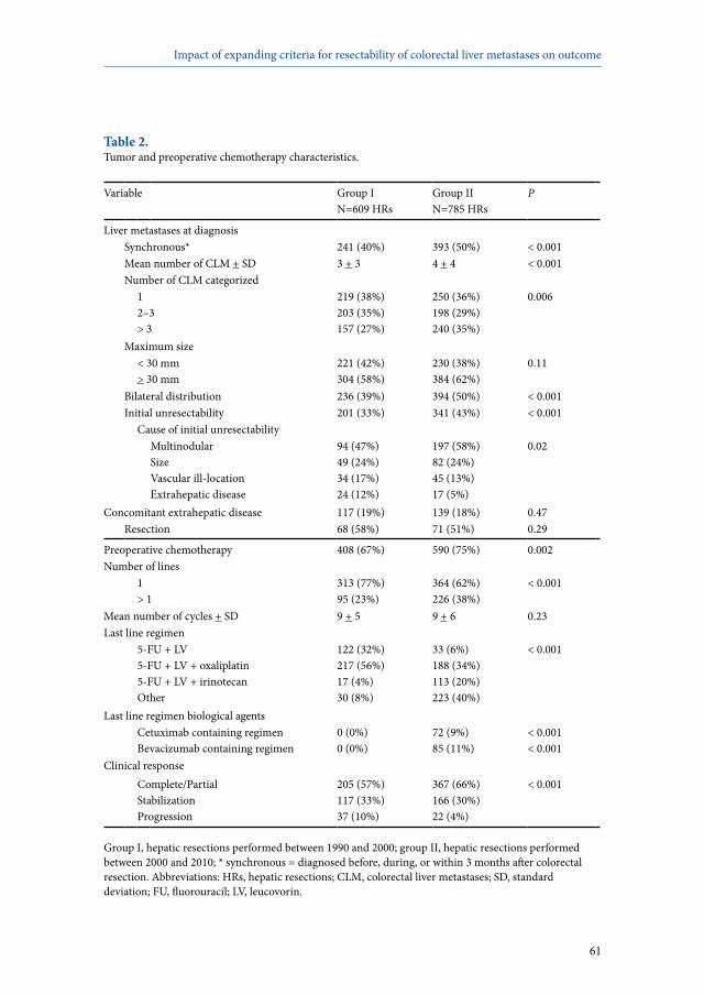
61
Impact of expanding criteria for resectability of colorectal liver metastases on outcome
Table 2. Tumor and preoperative chemotherapy characteristics.
Variable Group I N=609 HRs
Group IIN=785 HRs
P
Liver metastases at diagnosisSynchronous* 241 (40%) 393 (50%) < 0.001Mean number of CLM + SD 3 + 3 4 + 4 < 0.001Number of CLM categorized
12–3> 3
219 (38%)203 (35%)157 (27%)
250 (36%)198 (29%)240 (35%)
0.006
Maximum size< 30 mm> 30 mm
221 (42%)304 (58%)
230 (38%)384 (62%)
0.11
Bilateral distribution 236 (39%) 394 (50%) < 0.001Initial unresectability 201 (33%) 341 (43%) < 0.001
Cause of initial unresectabilityMultinodularSizeVascular ill-locationExtrahepatic disease
94 (47%)49 (24%)34 (17%)24 (12%)
197 (58%)82 (24%)45 (13%)17 (5%)
0.02
Concomitant extrahepatic disease 117 (19%) 139 (18%) 0.47Resection 68 (58%) 71 (51%) 0.29
Preoperative chemotherapy 408 (67%) 590 (75%) 0.002Number of lines
1> 1
313 (77%)95 (23%)
364 (62%)226 (38%)
< 0.001
Mean number of cycles + SD 9 + 5 9 + 6 0.23Last line regimen
5-FU + LV5-FU + LV + oxaliplatin5-FU + LV + irinotecanOther
122 (32%)217 (56%)17 (4%)30 (8%)
33 (6%)188 (34%)113 (20%)223 (40%)
< 0.001
Last line regimen biological agentsCetuximab containing regimenBevacizumab containing regimen
0 (0%)0 (0%)
72 (9%)85 (11%)
< 0.001< 0.001
Clinical responseComplete/PartialStabilizationProgression
205 (57%)117 (33%)37 (10%)
367 (66%)166 (30%)22 (4%)
< 0.001
Group I, hepatic resections performed between 1990 and 2000; group II, hepatic resections performed between 2000 and 2010; * synchronous = diagnosed before, during, or within 3 months after colorectal resection. Abbreviations: HRs, hepatic resections; CLM, colorectal liver metastases; SD, standard deviation; FU, fluorouracil; LV, leucovorin.

62
Chapter 3
Table 3. Operative and histopathological features.
Variable Group IN=609 HRs
Group IIN=785 HRs
P
HepatectomyNumber of hepatectomy
1234
445 (73%)126 (21%)28 (5%)10 (2%)
583 (74%)164 (21%)26 (3%)12 (2%)
0.67
Number of detected metastases12–3> 3
205 (39%)191 (36%)137 (26%)
192 (29%)223 (34%)242 (37%)
< 0.001
PVE 29 (5%) 121 (15%) < 0.001Two-stage resection 18 (3%) 85 (11%) < 0.001Simultaneous colorectal and hepatic resection 29 (5%) 42 (5%) 0.62Major resection (> 3 segments) 198 (33%) 280 (36%) 0.31Type of resection
AnatomicalNonanatomicalBoth
209 (35%)207 (35%)180 (30%)
259 (34%)253 (33%)256 (33%)
0.47
Vascular occlusionNoneSelective Total pedicularVascular exclusion
123 (25%)74 (15%)259 (53%)33 (7%)
138 (20%)56 (8%)402 (58%)102 (15%)
< 0.001
Mean hepatic ischemia time, minutes + SD 40 + 22 43 + 23 0.18Vascular resection and reconstruction 6 (1%) 19 (2%) 0.04Hypothermic liver perfusion 3 (1%) 23 (3%) 0.001Combined local treatment
NoneRFACryotherapy
549 (90%)7 (2%)51 (8%)
726 (93%)52 (7%)7 (1%)
< 0.001
Red blood cell transfusion 166 (27%) 225 (29%) 0.22

63
Impact of expanding criteria for resectability of colorectal liver metastases on outcome
Table 3. Continued.
Variable Group IN=609 HRs
Group IIN=785 HRs
P
HistopathologyNumber of resected metastases
12–3> 3
244 (43%)208 (36%)120 (21%)
214 (37%)173 (30%)192 (33%)
< 0.001
Mean maximum size + SD, mm 39 + 34 37 + 31 0.48Resection margin*
R0R1R2Rx
278 (56%)179 (36%)23 (5%)18 (4%)
332 (53%)199 (32%)9 (1%)85 (14%)
< 0.001
Abnormalities nontumoral liver parenchymaNoneFibrosisSteatosisVascular lesions#
201 (33%)63 (10%)102 (17%)81 (13%)
146 (19%)120 (15%)97 (12%)137 (17%)
< 0.001< 0.0010.200.001
Postoperative chemotherapy 504 (83%) 609 (77%) 0.08
Group I, hepatic resections performed between 1990 and 2000; group II, hepatic resections performed between 2000 and 2010; * R0, complete surgical resection with a negative surgical margin at histopathology; R1, invaded surgical margins according to the pathologist; R2, macroscopic intraoperative tumor remnant; Rx, first step of a two-stage procedure; # vascular lesions include sinusoidal alterations (vasodilatation and congestion), peliosis, hemorrhagic centrilobular necrosis, and regenerative nodular hyperplasia. Abbreviations: HRs, hepatic resections; SD, standard deviation; PVE, portal vein embolization; RFA, radiofrequency ablation.
Short-term outcomesWithin the total study population, overall perioperative mortality and postoperative morbidity rates were 1.3% (18/1394 cases) and 33% (457/1394 cases), respectively (Figures 1 and 2). No significant differences were observed between the first, second, third, and fourth hepatectomy. When comparing the two treatment periods, perioperative mortality rates were 1.1% (7/609 cases) in group I and 1.4% (11/785 cases) in group II (P=0.53). Causes of death were cardiac arrest without underlying disease (N=1), myocardial infarction (N=1), (aspiration) pneumonia (N=4), sepsis (N=7), intra-abdominal bleeding (N=1), ischemic colitis (N=1), and liver insufficiency (N=3). In group I no differences were observed between the first, second, third and fourth hepatic resection, while in group II a trend towards higher perioperative mortality rates after repeat hepatectomy was observed (Figure 1).
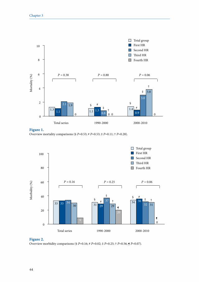
64
Chapter 3
0
2
4
6
8
10
Mor
talit
y (%
)
Total series 1990-2000 2000-2010
1.3 1.1
2.1
0.9 1.1 0.8 1.3 1.4
3.0
P = 0.38 P = 0.88 P = 0.06
‡ §
Total group First HR Second HR �ird HR Fourth HR
1.9
†
†
3.8
0 0 0 0
§
‡
# #
Figure 1. Overview mortality comparisons (§ P=0.53; # P=0.53; ‡ P=0.11; † P=0.28).
Total group
0
20
40
60
80
100
Mor
bidi
ty (%
)
Total series 1990-2000 2000-2010
33 33 34
9
29 37
29 36 34
P = 0.16
† # §
† ‡ #
§
First HR Second HR �ird HR Fourth HR
30
20 ¶
¶
31
0
32 31
P = 0.25 P = 0.06
‡
Figure 2. Overview morbidity comparisons (§ P=0.16; # P=0.02; ‡ P=0.25; † P=0.56; ¶ P=0.07).

65
Impact of expanding criteria for resectability of colorectal liver metastases on outcome
The postoperative morbidity rate tended to be higher in group II, compared with group I (34% vs 31%; P=0.16). Hepatic complications were more often observed in group II (24% vs 20% in group I; P=0.06), while the general complication rate did not differ according to treatment period (P=0.44). Types and severity of postoperative complications are outlined in Table 4. The postoperative morbidity rate following the first hepatic resection was significantly higher in group II, compared with group I (36% vs 29%; P=0.02; Figure 2). There were no significant differences in postoperative morbidity rates after second, third, or fourth hepatectomy between the treatment periods. In addition, when comparing postoperative morbidity rates after first, second, third, and fourth hepatectomy in each treatment period separately, no differences were observed. At multivariate analysis, four independent predictors of significant postoperative morbidity (Dindo-Clavien grade III-V) were observed: hepatic resection performed between 2000 and 2010, total hepatic ischemia time >60 minutes, maximum size of CLM >30 mm at histopathology, and presence of abnormalities in the nontumoral liver parenchyma (Table 5).
Patients at risk Total 1 yr 2 yrs 3 yrs 4 yrs 5 yrs
1990-2000 438 388 294 218 166 131
2000-2010 572 343 190 99 56 28
0
.2
.4
.6
.8
1
Ove
rall
surv
ival
0 1 2 3 4 5 Years
47%
62%
58%
69%
2000-2010
1990-2000
P = 0.10
Figure 3. Overall survival following first hepatectomy comparing patients treated between 1990 and 2000 with patients treated between 2000 and 2010 (patients who died within 90 days from operation were excluded).

66
Chapter 3
Patients at risk Total 1 yr 2 yrs 3 yrs 4 yrs 5 yrs 1990-2000 422 206 107 78 61 53 2000-2010 510 178 72 30 18 10
0
.2
.4
.6
.8
1 Pr
ogre
ssio
n-fre
e sur
viva
l
0 1 2 3 4 5 Years
16% 22%
15% 21%
2000-2010 1990-2000
P = 0.92
Figure 4. Progression-free survival following first hepatectomy comparing patients treated between 1990 and 2000 with patients treated between 2000 and 2010 (patients who died within 90 days from operation were excluded).
Long-term outcomesWithin the total study population, 3-year and 5-year overall survival rates were 65% and 50%, respectively. In group I, 3-year and 5-year overall survival rates were 62% and 47%, respectively, while these were 69% and 58% in group II (P=0.10) (Figure 3). Progression-free survival rates after 3 and 5 years in the total population were 22% and 16%, respectively. In group I, 3-year and 5-year progression-free survival rates were 21% and 16%, not significantly differing from those observed in group II (22% and 15%, respectively; P=0.92) (Figure 4). When comparing patients who experienced significant postoperative morbidity (Dindo-Clavien grade III-IV) to patients without grade III-IV complications, 3-year and 5-year overall survival rates were 60% and 42% in patients with morbidity, while these were 66% and 52% in case of no morbidity (P=0.11) (Figure 5). Progression-free survival rates after 3 and 5 years were 22% and 17% in the absence of grade III-IV postoperative complications, compared with 22% and 16% in case of grade III-IV postoperative complications (P=0.25) (Figure 6).
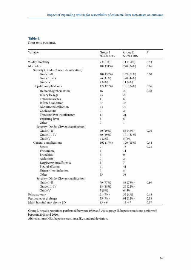
67
Impact of expanding criteria for resectability of colorectal liver metastases on outcome
Table 4. Short-term outcomes.
Variable Group IN=609 HRs
Group IIN=785 HRs
P
90-day mortality 7 (1.1%) 11 (1.4%) 0.53Morbidity 187 (31%) 270 (34%) 0.16
Severity (Dindo-Clavien classification)Grade I–IIGrade III–IVGrade V
104 (56%)76 (41%)7 (4%)
139 (51%)120 (44%)11 (4%)
0.60
Hepatic complications 122 (20%) 191 (24%) 0.06Hemorrhage/hematomaBiliary leakageTransient ascitesInfected collectionNoninfected collectionCholecystitisTransient liver insufficiencyPersisting feverOther
16231273401740
22206357822161
0.08
Severity (Dindo-Clavien classification)Grade I–IIGrade III–IVGrade V
60 (49%)60 (49%)2 (2%)
85 (45%)101 (53%)5 (3%)
0.76
General complications 102 (17%) 120 (15%) 0.44SepsisPneumoniaBronchitisAtelectasisRespiratory insufficiencyPleural effusionUrinary tract infectionOther
9540341733
131102741838
0.25
Severity (Dindo-Clavien classification)Grade I–IIGrade III–IVGrade V
79 (77%)18 (18%)5 (5%)
88 (73%)26 (22%)6 (5%)
0.80
Relaparotomy 21 (3%) 33 (4%) 0.48Percutaneous drainage 55 (9%) 91 (12%) 0.18Mean hospital stay, days + SD 13 + 6 13 + 7 0.57
Group I, hepatic resections performed between 1990 and 2000; group II, hepatic resections performed between 2000 and 2010.Abbreviations: HRs, hepatic resections; SD, standard deviation.
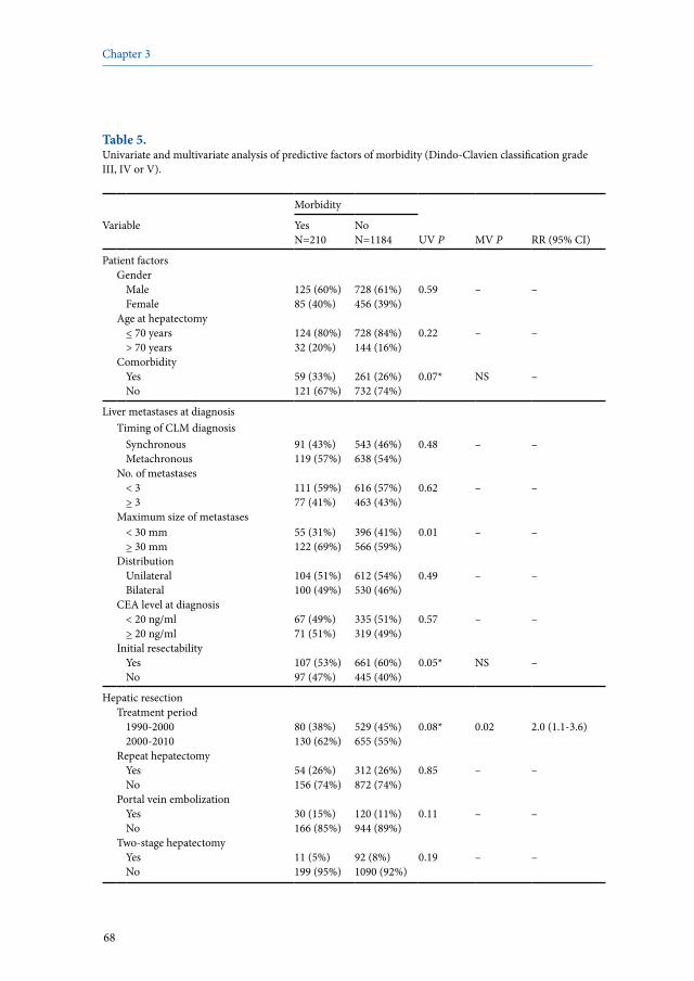
68
Chapter 3
Table 5. Univariate and multivariate analysis of predictive factors of morbidity (Dindo-Clavien classification grade III, IV or V).
Morbidity
Variable YesN=210
NoN=1184 UV P MV P RR (95% CI)
Patient factorsGender
MaleFemale
125 (60%)85 (40%)
728 (61%)456 (39%)
0.59 – –
Age at hepatectomy< 70 years> 70 years
124 (80%)32 (20%)
728 (84%)144 (16%)
0.22 – –
ComorbidityYesNo
59 (33%)121 (67%)
261 (26%)732 (74%)
0.07* NS –
Liver metastases at diagnosisTiming of CLM diagnosis
SynchronousMetachronous
91 (43%)119 (57%)
543 (46%)638 (54%)
0.48 – –
No. of metastases< 3> 3
111 (59%)77 (41%)
616 (57%)463 (43%)
0.62 – –
Maximum size of metastases< 30 mm> 30 mm
55 (31%)122 (69%)
396 (41%)566 (59%)
0.01 – –
DistributionUnilateralBilateral
104 (51%)100 (49%)
612 (54%)530 (46%)
0.49 – –
CEA level at diagnosis< 20 ng/ml> 20 ng/ml
67 (49%)71 (51%)
335 (51%)319 (49%)
0.57 – –
Initial resectabilityYesNo
107 (53%)97 (47%)
661 (60%)445 (40%)
0.05* NS –
Hepatic resectionTreatment period
1990-20002000-2010
80 (38%)130 (62%)
529 (45%)655 (55%)
0.08* 0.02 2.0 (1.1-3.6)
Repeat hepatectomyYesNo
54 (26%)156 (74%)
312 (26%)872 (74%)
0.85 – –
Portal vein embolizationYesNo
30 (15%)166 (85%)
120 (11%)944 (89%)
0.11 – –
Two-stage hepatectomyYesNo
11 (5%)199 (95%)
92 (8%)1090 (92%)
0.19 – –

69
Impact of expanding criteria for resectability of colorectal liver metastases on outcome
Table 5. Continued.
Morbidity
Variable YesN=210
NoN=1184 UV P MV P RR (95% CI)
Preoperative chemotherapyYesNo
154 (73%)56 (27%)
844 (73%)319 (27%)
0.82 – –
Timing of hepatic resectionSimultaneous to colorectal surgeryDelayed hepatectomy
5 (2%)205 (98%)
66 (6%)1118 (94%)
0.05* NS –
Intraoperative RBC transfusionYesNo
84 (46%)97 (54%)
307 (32%)653 (68%)
< 0.001* NS –
Preoperative CEA level< 10 ng/ml> 10 ng/ml
78 (50%)78 (50%)
485 (59%)342 (41%)
0.05* NS –
No. of intraoperative detected CLM< 3> 3
100 (52%)92 (48%)
552 (55%)446 (45%)
0.41 – –
Extent of hepatectomyMinorMajor (> 3 segments)
103 (52%)97 (48%)
716 (65%)381 (35%)
< 0.001* NS –
Concomitant extrahepatic diseaseYesNo
33 (16%)177 (84%)
223 (19%)961 (81%)
0.28 – –
Vascular occlusionYesNo
165 (85%)28 (15%)
761 (77%)233 (23%)
0.006* NS –
Total hepatic ischemia time< 60 minutes> 60 minutes
67 (65%)36 (35%)
342 (83%)68 (17%)
< 0.001* 0.003 2.4 (1.4-4.2)
Vascular resection and reconstruction
YesNo
6 (3%)174 (97%)
19 (2%)971 (98%)
0.23 – –
Hypothermic liver perfusionYesNo
8 (4%)172 (96%)
18 (2%)972 (98%)
0.03* NS –
Combined local treatmentYesNo
12 (6%)198 (94%)
105 (9%)1077 (91%)
0.13 – –
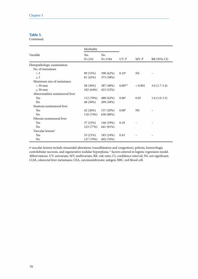
70
Chapter 3
Table 5. Continued.
Morbidity
Variable YesN=210
NoN=1184 UV P MV P RR (95% CI)
Histopathologic examinationNo. of metastases
< 3> 3
99 (55%)81 (45%)
598 (62%)373 (38%)
0.10* NS –
Maximum size of metastases< 30 mm> 30 mm
58 (36%)102 (64%)
387 (48%)423 (52%)
0.007* < 0.001 3.0 (1.7-5.4)
Abnormalities nontumoral liverYesNo
112 (70%)48 (30%)
488 (62%)299 (38%)
0.06* 0.05 1.6 (1.0-5.5)
Steatosis nontumoral liverYesNo
42 (26%)118 (74%)
157 (20%)630 (80%)
0.08* NS –
Fibrosis nontumoral liverYesNo
37 (23%)123 (77%)
146 (19%)641 (81%)
0.18 – –
Vascular lesions#
YesNo
33 (21%)127 (79%)
185 (24%)602 (76%)
0.43 – –
# vascular lesions include sinusoidal alterations (vasodilatation and congestion), peliosis, hemorrhagic centrilobular necrosis, and regenerative nodular hyperplasia; * factors entered in logistic regression model.Abbreviations: UV, univariate; MV, multivariate; RR, risk ratio; CI, confidence interval; NS, not significant; CLM, colorectal liver metastases; CEA, carcinoembryonic antigen; RBC, red blood cell.

71
Impact of expanding criteria for resectability of colorectal liver metastases on outcome
Table 6. Overview of studies specifically mentioning short-term outcomes in patients surgically treated for colorectal liver metastases.
Short-term outcomes
First author Year Institution PeriodNo. ofpatients Mortality (%)* Morbidity (%)*
Fong23 1999 MSKCC, USA 1985-1998 1001 2.8 31
Scheele38 2001 Jena, Germany 1960-1998 654 5.8 16
Choti36 2002 Johns Hopkins, USA 1984-1999 226 0.9 18.6
Imamura19 2003 Tokyo, Japan 1994-2002 164 0 41
Wei39 2006 Toronto, Canada 1992-2002 423 1.6 17
Rees37 2008 Basingstoke, UK 1987-2005 929 1.5 25.9
Present series 2010 Paul Brousse, France 1990-2010 1028 0.71.3 (90-day)
2633 (90-day)
All studies are monocentric and retrospective; * Perioperative mortality and postoperative morbidity rates concern a 30-day period unless indicated otherwise.
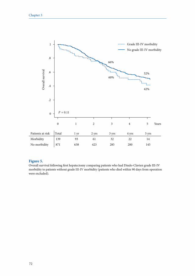
72
Chapter 3
Patients at risk Total 1 yr 2 yrs 3 yrs 4 yrs 5 yrs
Morbidity 139 93 61 32 22 14
No morbidity 871 638 423 285 200 145
0
.2
.4
.6
.8
1 O
vera
ll su
rviv
al
0 1 2 3 4 5 Years
42%
60% 52%
66%
No grade III-IV morbidity
Grade III-IV morbidity
P = 0.11
Figure 5. Overall survival following first hepatectomy comparing patients who had Dindo-Clavien grade III-IV morbidity to patients without grade III-IV morbidity (patients who died within 90 days from operation were excluded).
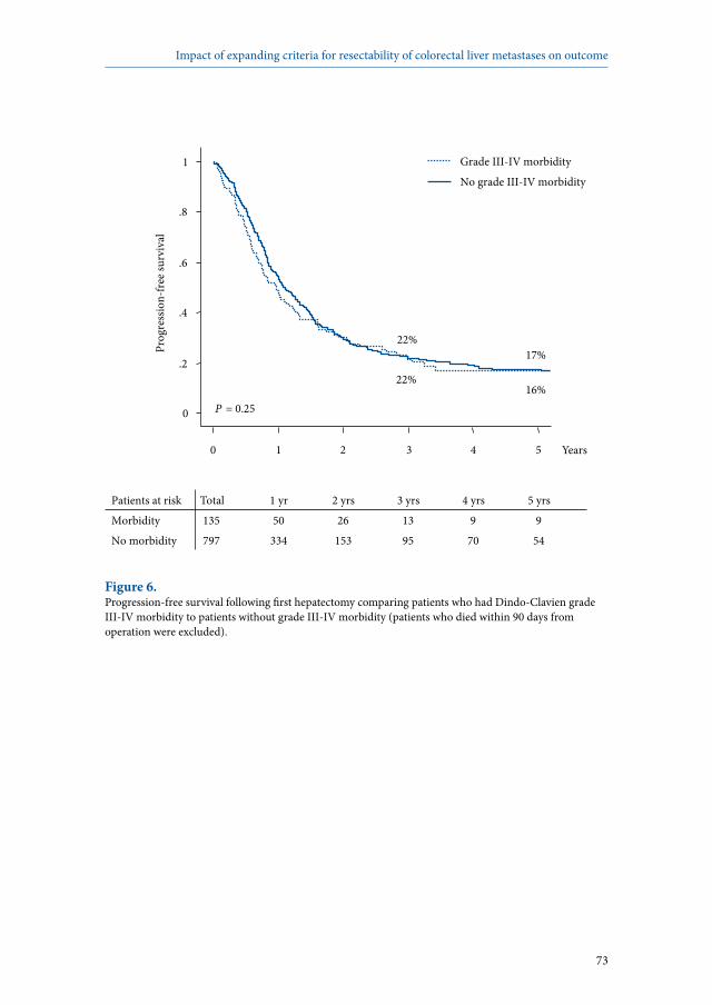
73
Impact of expanding criteria for resectability of colorectal liver metastases on outcome
Patients at risk Total 1 yr 2 yrs 3 yrs 4 yrs 5 yrs
Morbidity 135 50 26 13 9 9
No morbidity 797 334 153 95 70 54
0
.2
.4
.6
.8
1 Pr
ogre
ssio
n-fre
e sur
viva
l
0 1 2 3 4 5 Years
17% 22%
16% 22%
No grade III-IV morbidity
Grade III-IV morbidity
P = 0.25
Figure 6. Progression-free survival following first hepatectomy comparing patients who had Dindo-Clavien grade III-IV morbidity to patients without grade III-IV morbidity (patients who died within 90 days from operation were excluded).

74
Chapter 3
Discussion
As the impact of an expansion of resectability criteria of CLM on outcomes following hepatectomy is still unknown, the aim of the current study was to evaluate whether a more aggressive surgical approach for CLM is related to acceptable short-term and long-term results, and thus to determine whether an expansion of resectability criteria is justified. Within the total study population, overall perioperative mortality and postoperative morbidity rates were 1.3% and 33%, respectively. Although patients who underwent hepatic resection during the latter period (2000-2010) were older, had a higher prevalence of comorbid illnesses, presented with more extensive liver disease, and underwent more complex surgical procedures, perioperative mortality rate was similar to that observed in our initial experience (1990-2000). However, a trend towards a higher postoperative complication rate was observed in the more recently operated cohort. In our more recent experience, the mortality rate following repeat hepatectomy was higher than that observed after first hepatectomy, while the morbidity rate was comparable after first and repeat hepatectomy. Treatment between 2000 and 2010, total hepatic ischemia time >60 minutes, maximum size of liver metastases >30 mm at histopathology, and the presence of abnormalities in the nontumoral liver parenchyma were identified as independent predictors of postoperative morbidity. Although a trend towards lower overall survival was observed in patients who developed grade III-IV postoperative complications, no significant differences were observed in long-term outcomes between both treatment periods. Perioperative mortality rates of 0-5.8% and postoperative morbidity rates ranging between 22% and 45% following hepatic resection have been described in several large series.16-21,35 However, most of these series consisted of a heterogeneous patient population including both benign and malignant disease as indication for liver surgery, thereby comparing different patient profiles. When considering only patients who underwent hepatic resection for CLM, which are currently one of the most important indications for liver surgery, published studies have reported 30-day perioperative mortality and postoperative morbidity rates of 0-5.8% and 16-41%, respectively (Table 6).19,23,36-39 Overall perioperative mortality and postoperative morbidity rates in our series following 1394 hepatic resections for CLM were 1.3% and 33%, respectively. These results compare favorably to the other large published series mentioned earlier. However, our postoperative morbidity rate was slightly higher than that reported in most other series based on surgically treated CLM patients (Table 6). The higher postoperative morbidity rate observed in our series could be related to our more aggressive multimodal treatment approach, as in most patients hepatic resection was preceded by extensive systemic chemotherapy regimens.40 Another important factor is the time-period used to define perioperative mortality and
75
Impact of expanding criteria for resectability of colorectal liver metastases on outcome
postoperative morbidity. In our study, a period of 90 days following hepatic resection was used to define perioperative mortality and postoperative morbidity. However, within the literature a 30-day period is often used, which might explain our slightly higher postoperative morbidity rate. We determined perioperative mortality and morbidity at 90 days from operation as Mullen and colleagues have recently shown an increase in mortality rate of 47% when comparing the 30-day rate with the 90-day rate, and therefore, using a 30-day period might underestimate the true risk of major liver surgery.41 Short-term outcomes following repeat hepatectomy have only been described in few series, with mortality rates varying between 1.6% and 3.5% and morbidity rates of around 30%, to which our results compare favorably.42-44 When comparing the treatment periods, patients treated during the latter period were more often aged >70 years, had a higher prevalence of comorbid illnesses, and presented with more extensive liver disease, necessitating a more extensive treatment approach (ie, more often preoperative chemotherapy, more frequent portal vein embolization, and a higher number of two-stage resections). Despite this, perioperative mortality rates did not differ between the treatment periods. However, the postoperative morbidity rate tended to be higher in our latter experience. Similarly, when considering only first hepatectomies, significantly more postoperative complications were observed in more recently operated patients. A possible explanation for this could be the higher use of preoperative chemotherapy in more recently operated patients. Oxaliplatin-based chemotherapy has been associated with sinusoidal alterations such as sinusoidal dilatation, vascular congestion, perisinusoidal fibrosis, and venous occlusion by fibrous tissue.45 Irinotecan-based chemotherapy has mainly been associated with steatohepatitis.46 Whether these abnormalities of the nontumoral liver parenchyma negatively influence short-term outcomes following hepatic resection for CLM remains uncertain.47 Our group and others have reported a higher need of intraoperative blood transfusions and an increased risk of postoperative morbidity after the prolonged use of oxaliplatin and its associated vascular lesions.40,48-50
In the current study, although the administration of preoperative chemotherapy did not predict the development of postoperative complications, the presence of abnormalities of the nontumoral liver parenchyma at histopathological examination independently predicted the development of postoperative complications. In addition, at univariate analysis the need for an intraoperative blood transfusion was related to the development of significant postoperative complications, possibly caused by the complexity of the surgical procedures and/or by the administration of preoperative chemotherapy, as mentioned earlier. Interestingly, overall survival was significantly poorer in patients who received an intraoperative blood transfusion (5-year survival rate 42% vs 54% in case of no blood transfusion; P=0.007), which is in accordance with other reports.51,52
76
Chapter 3
At multivariate analysis, variables with an independent negative effect on postoperative morbidity were treatment between 2000 and 2010, total hepatic ischemia time >60 minutes, maximum size of liver metastases >30 mm at histopathology, and the presence of abnormalities of the nontumoral liver parenchyma at histopathological examination. The identification of these factors as independent predictors of the development of postoperative complications is not surprising, as all of them are related to the extent of liver surgery and the expansion of the criteria for resectability (ie, higher use of chemotherapy, portal vein embolization, et cetera). In three other studies, similar predictors of postoperative morbidity were found.17,20,21 A limitation of our study could be the retrospective grading of complications according to the Dindo-Clavien classification. However, we believe that grading of complications, even if retrospective, still remains accurate, since it is based on their therapeutic consequences. In addition, to our knowledge the current study is the first describing short-term outcomes following hepatectomy for CLM graded according to the Dindo-Clavien classification and related to changes in resectability criteria. Another limitation of the present study could be the fact that our institute is a tertiary referral center, which might have introduced some referral bias as more complex patients could have been referred to our center. Although the period of treatment did not influence long-term outcomes, a trend to lower overall survival rates was observed in patients who developed grade III-IV postoperative complications, compared with those without significant postoperative complications. Within the literature, several studies have identified the occurrence of postoperative complications as an independent predictor of poor overall survival.53,54 Thus, to improve long-term outcome, the occurrence of significant postoperative complications should be prevented. In addition, long-term outcome can be further improved when the need for intraoperative blood transfusion is kept to a minimum. In summary, an expansion of indications for hepatic resection in patients with CLM has been observed over the years, with more patients being treated by an aggressive approach consisting of extensive perioperative chemotherapy, more complex hepatic resections, and repeat surgery in case of resectable disease recurrence. Acceptable overall mortality and morbidity rates of 1.3% and 33%, respectively, were observed. Perioperative mortality rates did not differ according to treatment period, however, more recently operated patients experienced more postoperative complications. These favorable short-term outcomes, without worsening of long-term outcomes, justify an expansion of the resectability criteria of CLM. Rather than being more selective to improve short-term results, the indications for surgical treatment of CLM can be further expanded provided reasonable perioperative mortality and morbidity rates, as this is the only chance of long-term survival and even cure.
77
Impact of expanding criteria for resectability of colorectal liver metastases on outcome
References
1. Simmonds PC, Primrose JN, Colquitt JL et al. Surgical resection of hepatic metastases from colorectal cancer: a systematic review of published studies. Br J Cancer 2006; 94:982-999.
2. Adam R. Chemotherapy and surgery: new perspectives on the treatment of unresectable liver metastases. Ann Oncol 2003; 14 Suppl 2:ii13-ii16.
3. Pawlik TM, Schulick RD, Choti MA. Expanding criteria for resectability of colorectal liver metastases. Oncologist 2008; 13:51-64.
4. Adam R, Avisar E, Ariche A et al. Five-year survival following hepatic resection after neoadjuvant therapy for nonresectable colorectal. Ann Surg Oncol 2001; 8:347-353.
5. Adam R, Aloia T, Lévi F et al. Hepatic resection after rescue cetuximab treatment for colorectal liver metastases previously refractory to conventional systemic therapy. J Clin Oncol 2007; 25:4593-4602.
6. Alberts SR, Horvath WL, Sternfeld WC et al. Oxaliplatin, fluorouracil, and leucovorin for patients with unresectable liver-only metastases from colorectal cancer: a North Central Cancer Treatment Group phase II study. J Clin Oncol 2005; 23:9243-9249.
7. de la Camara J, Rodriguez J, Rotellar F et al. Triplet therapy with oxaliplatin, irinotecan, 5-fluorouracil and folinic acid within a combined modality approach in patients with liver metastases from colorectal cancer. Proc Am Soc Clin Oncol 2004; 23:3593.
8. Masi G, Cupini S, Marcucci L et al. Treatment with 5-fluorouracil/folinic acid, oxaliplatin, and irinotecan enables surgical resection of metastases in patients with initially unresectable metastatic colorectal cancer. Ann Surg Oncol 2006; 13:58-65.
9. Pozzo C, Basso M, Cassano A et al. Neoadjuvant treatment of unresectable liver disease with irinotecan and 5-fluorouracil plus folinic acid in colorectal cancer patients. Ann Oncol 2004; 15:933-939.
10. Quenet F, Nordlinger B, Rivoire M et al. Resection of previously unresectable liver metastases from colorectal cancer (LMCRC) after chemotherapy (CT) with CPT-11/L-OHP/LV5FU (Folfirinox): a prospective phase II trial. Proc Am Soc Clin Oncol 2004; 23:3613.
11. Azoulay D, Castaing D, Smail A et al. Resection of nonresectable liver metastases from colorectal cancer after percutaneous portal vein embolization. Ann Surg 2000; 231:480-486.
12. Wicherts DA, de Haas RJ, Andreani P et al. Impact of portal vein embolization on long-term survival of patients with primarily unresectable colorectal liver metastases. Br J Surg 2010; 97:240-250.
13. Wicherts DA, Miller R, de Haas RJ et al. Long-term results of two-stage hepatectomy for irresectable colorectal cancer liver metastases. Ann Surg 2008; 248:994-1005.
14. Azoulay D, Andreani P, Maggi U et al. Combined liver resection and reconstruction of the supra-renal vena cava: the Paul Brousse experience. Ann Surg 2006; 244:80-88.
15. Azoulay D, Eshkenazy R, Andreani P et al. In situ hypothermic perfusion of the liver versus standard total vascular exclusion for complex liver resection. Ann Surg 2005; 241:277-285.
16. Belghiti J, Hiramatsu K, Benoist S et al. Seven hundred forty-seven hepatectomies in the 1990s: an update to evaluate the actual risk of liver resection. J Am Coll Surg 2000; 191:38-46.
17. Cescon M, Vetrone G, Grazi GL et al. Trends in perioperative outcome after hepatic resection: analysis of 1500 consecutive unselected cases over 20 years. Ann Surg 2009; 249:995-1002.
18. Fan ST, Lo CM, Liu CL et al. Hepatectomy for hepatocellular carcinoma: toward zero hospital deaths. Ann Surg 1999; 229:322-330.
19. Imamura H, Seyama Y, Kokudo N et al. One thousand fifty-six hepatectomies without mortality in 8 years. Arch Surg 2003; 138:1198-1206.
20. Jarnagin WR, Gonen M, Fong Y et al. Improvement in perioperative outcome after hepatic resection: analysis of 1,803 consecutive cases over the past decade. Ann Surg 2002; 236:397-406.
21. Poon RT, Fan ST, Lo CM et al. Improving perioperative outcome expands the role of hepatectomy in management of benign and malignant hepatobiliary diseases: analysis of 1222 consecutive patients from a prospective database. Ann Surg 2004; 240:698-708.

78
Chapter 3
22. Alkozai EM, Lisman T, Porte RJ. Bleeding in liver surgery: prevention and treatment. Clin Liver Dis 2009; 13:145-154.
23. Fong Y, Fortner J, Sun RL et al. Clinical score for predicting recurrence after hepatic resection for metastatic colorectal cancer: analysis of 1001 consecutive cases. Ann Surg 1999; 230:309-318.
24. Adam R, Frilling A, Elias D et al. Liver resection of colorectal metastases in elderly patients. Br J Surg 2010; 97:366-376.
25. Martin RC 2nd, Augenstein V, Reuter NP et al. Simultaneous versus staged resection for synchronous colorectal cancer liver metastases. J Am Coll Surg 2009; 208:842-850.
26. Gregoire E, Hoti E, Gorden DL et al. Utility of prognostic scoring systems for colorectal liver metastases in an era of advanced multimodal therapy. Eur J Surg Oncol 2010; 36:568-574.
27. Azoulay D, Raccuia JS, Castaing D et al. Right portal vein embolization in preparation for major hepatic resection. J Am Coll Surg 1995; 181:266-269.
28. World Health Organization. Handbook for reporting results of cancer treatment. Geneva: World Health Organization; 1979.
29. Therasse P, Arbuck SG, Eisenhauer EA et al. New guidelines to evaluate the response to treatment in solid tumors. European Organization for Research and Treatment of Cancer, National Cancer Institute of the United States, National Cancer Institute of Canada. J Natl Cancer Inst 2000; 92:205-216.
30. Bismuth H. Surgical anatomy and anatomical surgery of the liver. World J Surg 1982; 6:3-9.31. Couinaud C. Le foie: études anatomiques et chirurgicales. Paris: Masson et Cie; 1957.32. Adam R, Laurent A, Azoulay D et al. Two-stage hepatectomy: a planned strategy to treat
irresectable liver tumors. Ann Surg 2000; 232:777-785. 33. Balzan S, Belghiti J, Farges O et al. The “50-50 criteria” on postoperative day 5: an accurate
predictor of liver failure and death after hepatectomy. Ann Surg 2005; 242:824-828. 34. Dindo D, Demartines N, Clavien PA. Classification of surgical complications: a new proposal
with evaluation in a cohort of 6336 patients and results of a survey. Ann Surg 2004; 240:205-213.
35. Dimick JB, Cowan JA, Knol JA et al. Hepatic resection in the United States: indications, outcomes, and hospital procedural volumes from a nationally representative database. Arch Surg 2003; 138:185-191.
36. Choti MA, Sitzmann JV, Tiburi MF et al. Trends in long-term survival following liver resection for hepatic colorectal metastases. Ann Surg 2002; 235:759-766.
37. Rees M, Tekkis PP, Welsh FK et al. Evaluation of long-term survival after hepatic resection for metastatic colorectal cancer: a multifactorial model of 929 patients. Ann Surg 2008; 247:125-135.
38. Scheele J, Altendorf-Hofmann A, Grube T et al. Resection of colorectal liver metastases. What prognostic factors determine patient selection? Chirurg 2001; 72:547-560.
39. Wei AC, Greig PD, Grant D et al. Survival after hepatic resection for colorectal metastases: a 10-year experience. Ann Surg Oncol 2006; 13:668-676.
40. Karoui M, Penna C, Amin-Hashem M et al. Influence of preoperative chemotherapy on the risk of major hepatectomy for colorectal liver metastases. Ann Surg 2006; 243:1-7.
41. Mullen JT, Ribero D, Reddy SK et al. Hepatic insufficiency and mortality in 1,059 noncirrhotic patients undergoing major hepatectomy. J Am Coll Surg 2007; 204:854-864.
42. Muratore A, Polastri R, Bouzari H et al. Repeat hepatectomy for colorectal liver metastases: a worthwhile operation? J Surg Oncol 2001; 76:127-132.
43. Petrowsky H, Gonen M, Jarnagin W et al. Second liver resections are safe and effective treatment for recurrent hepatic metastases from colorectal cancer: a bi-institutional analysis. Ann Surg 2002; 235:863-871.
44. Suzuki S, Sakaguchi T, Yokoi Y et al. Impact of repeat hepatectomy on recurrent colorectal liver metastases. Surgery 2001; 129:421-428.
79
Impact of expanding criteria for resectability of colorectal liver metastases on outcome
45. Rubbia-Brandt L, Audard V, Sartoretti P et al. Severe hepatic sinusoidal obstruction associated with oxaliplatin-based chemotherapy in patients with metastatic colorectal cancer. Ann Oncol 2004; 15:460-466.
46. Vauthey JN, Pawlik TM, Ribero D et al. Chemotherapy regimen predicts steatohepatitis and an increase in 90-day mortality after surgery for hepatic colorectal metastases. J Clin Oncol 2006; 24:2065-2072.
47. Benoist S, Nordlinger B. The role of preoperative chemotherapy in patients with resectable colorectal liver metastases. Ann Surg Oncol 2009; 16:2385-2390.
48. Aloia T, Sebagh M, Plasse M et al. Liver histology and surgical outcomes after preoperative chemotherapy with fluorouracil plus oxaliplatin in colorectal cancer liver metastases. J Clin Oncol 2006; 24:4983-4990.
49. Mehta NN, Ravikumar R, Coldham CA et al. Effect of preoperative chemotherapy on liver resection for colorectal liver metastases. Eur J Surg Oncol 2008; 34:782-786.
50. Nakano H, Oussoultzoglou E, Rosso E et al. Sinusoidal injury increases morbidity after major hepatectomy in patients with colorectal liver metastases receiving preoperative chemotherapy. Ann Surg 2008; 247:118-124.
51. Jamison RL, Donohue JH, Nagorney DM et al. Hepatic resection for metastatic colorectal cancer results in cure for some patients. Arch Surg 1997; 132:505-510.
52. Ohlsson B, Stenram U, Tranberg KG. Resection of colorectal liver metastases: 25-year experience. World J Surg 1998; 22:268-276.
53. Khuri SF, Henderson WG, DePalma RG et al. Determinants of long-term survival after major surgery and the adverse effect of postoperative complications. Ann Surg 2005; 242:326-343.
54. Rhodes A, Cecconi M, Hamilton M et al. Goal-directed therapy in high-risk surgical patients: a 15-year follow-up study. Intensive Care Med 2010; 36:1327-1332.

Robbert J. de Haas1,2
Dennis A. Wicherts1,2
Chady Salloum1
Paola Andreani1
Dobromir Sotirov1
René Adam1
Denis Castaing1
Daniel Azoulay1
1 Centre Hépato-Biliaire, Hôpital Paul Brousse, Villejuif, France2 Department of Surgery, University Medical Center Utrecht, Utrecht,
The Netherlands
Cancer 2010; 116:647-658
Long-term outcomes after hepatic resection for colorectal metastases in young patients
4

82
Chapter 4
Abstract
BackgroundLong-term outcomes after hepatectomy for colorectal liver metastases (CLM) in relatively young patients are still unknown. The aim of the current study was to evaluate long-term outcomes in patients <40 years old, and to compare them with patients >40 years old.
MethodsAll consecutive patients who underwent hepatectomy for CLM at our hospital between 1990 and 2006 were included in the study. Patients <40 years old were compared with all other patients treated during the same period. Overall survival (OS), progression-free survival (PFS), and disease-free survival (DFS) rates were determined, and prognostic factors were identified.
ResultsIn total, 806 patients underwent hepatectomy for CLM, of whom 56 (7%) were aged <40 years. Among the young patients, more CLM were present at diagnosis, and they were more often diagnosed synchronous with the primary tumor. Five-year OS was 33% in young patients, compared with 51% in older patients (P=0.12). Five-year PFS was 2% in young patients, compared with 16% in older patients (P<0.001). DFS rates were comparable between the groups (17% vs 23%; P=0.10). At multivariate analysis, age <40 years was identified as an independent predictor of poor PFS.
ConclusionIn young patients, CLM seem to be more aggressive with a trend toward lower OS, more disease recurrences, and a significantly shorter PFS after hepatectomy. However, DFS rates were comparable between young and older patients, owing to an aggressive multimodality treatment approach, consisting of chemotherapy and repeat surgery. Therefore, physicians should recognize the poor outcome of CLM in young patients and should consider an aggressive approach to diagnosis and early treatment.
83
Hepatic resection for colorectal metastases in young patients
Introduction
Currently, colorectal cancer is the third most commonly diagnosed cancer and the second leading cause of cancer death in the United States.1 Although a slight decrease in colorectal cancer incidence has been observed in older patients during the past few years, its incidence in patients younger than 40 years is rising.2 As more than 50% of colorectal cancer patients develop liver metastases during their disease course, surgeons and oncologists will be more often confronted with relatively young patients presenting with colorectal liver metastases. Hepatic resection of metastatic colorectal cancer has become the treatment of choice for selected patients, resulting in 5-year survival rates of around 40%,3-5 with postoperative mortality rates of less than 5% in most large series.6-9 However, only 20% of patients with colorectal liver metastases are directly amenable to hepatic surgery.10 Of the remaining patients, up to 13% can be converted to resectability by the administration of modern chemotherapy regimens.11 In addition, specific surgical techniques such as portal vein embolization,12 vascular reconstruction,13 two-stage procedures,14,15 and local ablation can further increase the number of resected patients. Whether these encouraging results differ according to the patient’s age at the time of hepatectomy is still unknown. For elderly patients, mostly defined as being >70 years old at hepatic resection, 5-year survival rates varying between 21% and 44% have been reported after hepatectomy for colorectal liver metastases.16-20 However, long-term outcomes for young patients (ie, patients aged <40 years at hepatectomy) who underwent hepatic resection for colorectal liver metastases have never been evaluated. Nevertheless, a small number of studies have specifically addressed outcomes after resection of the primary colorectal malignancy in patients aged <40 years. Reported 5-year survival rates in recent series ranged between 9% and 54.8%.21-24 In two studies, comparison was made to patients aged >40 years at colorectal resection, and both studies concluded that long-term outcome of the younger patient group was similar to that observed for the older patients.21,24 Whether these results can be extrapolated to patients scheduled for hepatic resection of colorectal metastases is still unknown. However, young patients often have better performance status, with fewer comorbidities than their older counterparts, and therefore can be treated more aggressively with a combination of chemotherapy and repeat surgery, which might influence long-term outcomes. The aim of the current study was to evaluate long-term outcomes after hepatectomy for colorectal liver metastases in young patients treated with an aggressive approach consisting of chemotherapy and repeat surgery, and to investigate whether long-term outcome in young patients differs from that observed in older patients.

84
Chapter 4
Patients and methods
Study populationAll consecutive patients who underwent hepatic resection for colorectal metastases at our hospital between January 1990 and January 2006 were included in the study. All patients were identified in our prospectively maintained institutional database, and additional information was obtained by reviewing each patient’s medical record. The presence of familial colon cancer syndromes was not available in our database. In the current study, patients with an age at hepatectomy <40 years were considered as young patients. Young patients were compared with all other patients who underwent hepatectomy for colorectal metastases during the same period.
Preoperative managementBefore hepatectomy, all patients were evaluated by thoracoabdominal imaging (ultrasonography, computed tomography [CT]), routine blood tests including serum tumor marker levels, and colonoscopy. In case of initially unresectable metastatic disease (ie, a too small estimated future remnant liver or unresectable extrahepatic disease sites), preoperative chemotherapy was administered, with the aim of converting these patients to a resectable situation. In addition, patients with initially resectable metastases were treated with neoadjuvant chemotherapy when it concerned synchronous liver metastases (ie, diagnosed before, during, or within 3 months after colorectal resection), or marginally resectable liver disease (five or more bilateral nodules). Chemotherapy response was evaluated radiologically after every four cycles of treatment, according to the World Health Organization guidelines,25 or more recently, the Response Evaluation Criteria in Solid Tumors.26 When the estimated future remnant liver was judged too small (<30%), preoperative portal vein embolization was performed to induce compensatory hypertrophy of the remaining liver, thereby decreasing the risk of postoperative liver insufficiency.12 The decision for surgery was taken during a multidisciplinary meeting, including surgeons, medical oncologists, and radiologists. Hepatic resectionAll hepatectomies were performed with curative intent. First, thorough exploration of the abdominal cavity was performed to rule out unresectable extrahepatic disease. Then, the extent of liver disease was evaluated by palpation and intraoperative ultrasound examination of the liver. Radiofrequency ablation or cryotherapy was combined with hepatic resection to treat up to three deeply located lesions in the remnant liver. If more than three lesions would remain in the future remnant liver, a two-stage procedure was performed.15,27 When hepatic resection comprised three or more anatomic segments,28,29 it was defined as a
85
Hepatic resection for colorectal metastases in young patients
major hepatectomy. The presence of resectable extrahepatic disease was not considered a contraindication for surgery. When extrahepatic metastases were located in the abdominal cavity, resection was performed immediately. If extrahepatic disease was located outside the abdomen, resection was generally performed 2-3 months after hepatectomy, with systemic chemotherapy in between to reduce the risk of disease progression.
Postoperative follow-upFollow-up after hepatectomy included a history and physical examination, routine blood tests, serum tumor markers, and liver ultrasound 1 month after surgery and then every 4 months. Every 8 months, CT imaging of the chest, abdomen, and pelvis was performed. Postoperative chemotherapy was routinely recommended to reduce the recurrence risk. Recurrences (intra- and/or extrahepatic) were resected only if the overall surgical strategy could be complete.30
Statistical analysisContinuous data were compared using the independent-samples t test, and categorical variables were compared using the χ2 test. Survival probabilities were estimated using the Kaplan-Meier method, with the date of hepatectomy as starting point. Survival curves were compared using the log-rank test. Progression-free survival was defined as the time-interval between hepatectomy and first postoperative recurrence or death. Disease-free survival was defined as the time-interval between hepatectomy and the first postoperative recurrence that could not be curatively treated (resection or ablation) or death. Univariate analysis was performed to determine factors related to overall and progression-free survival using the log-rank test. To identify independent predictors of overall and progression-free survival, all factors with an univariate P <0.15 were entered into a Cox proportional hazard model. All statistical analyses were performed with SPSS version 13.0 (SPSS Inc, Chicago, IL, USA), and statistical significance was determined at P <0.05.
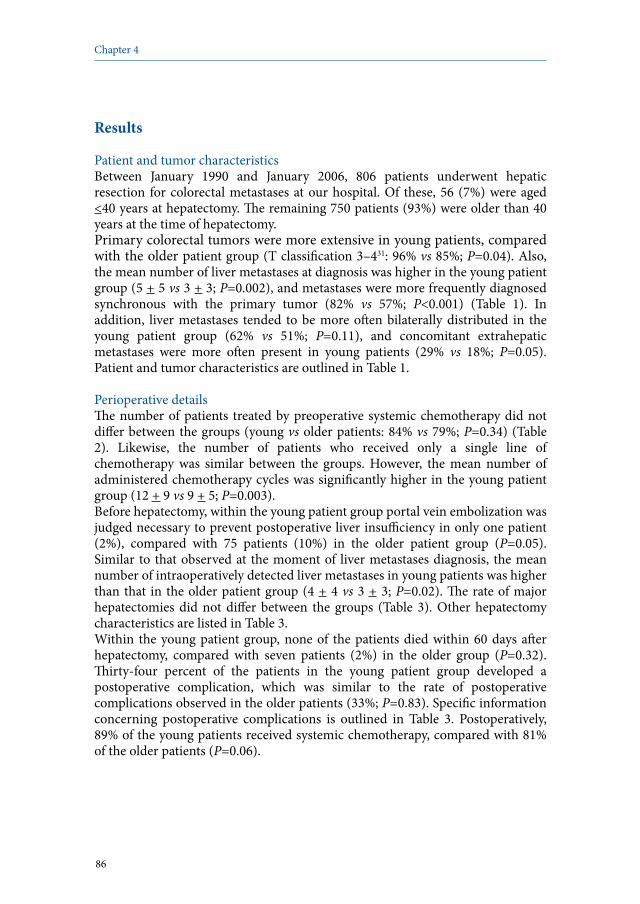
86
Chapter 4
Results
Patient and tumor characteristicsBetween January 1990 and January 2006, 806 patients underwent hepatic resection for colorectal metastases at our hospital. Of these, 56 (7%) were aged <40 years at hepatectomy. The remaining 750 patients (93%) were older than 40 years at the time of hepatectomy. Primary colorectal tumors were more extensive in young patients, compared with the older patient group (T classification 3–431: 96% vs 85%; P=0.04). Also, the mean number of liver metastases at diagnosis was higher in the young patient group (5 + 5 vs 3 + 3; P=0.002), and metastases were more frequently diagnosed synchronous with the primary tumor (82% vs 57%; P<0.001) (Table 1). In addition, liver metastases tended to be more often bilaterally distributed in the young patient group (62% vs 51%; P=0.11), and concomitant extrahepatic metastases were more often present in young patients (29% vs 18%; P=0.05). Patient and tumor characteristics are outlined in Table 1.
Perioperative detailsThe number of patients treated by preoperative systemic chemotherapy did not differ between the groups (young vs older patients: 84% vs 79%; P=0.34) (Table 2). Likewise, the number of patients who received only a single line of chemotherapy was similar between the groups. However, the mean number of administered chemotherapy cycles was significantly higher in the young patient group (12 + 9 vs 9 + 5; P=0.003). Before hepatectomy, within the young patient group portal vein embolization was judged necessary to prevent postoperative liver insufficiency in only one patient (2%), compared with 75 patients (10%) in the older patient group (P=0.05). Similar to that observed at the moment of liver metastases diagnosis, the mean number of intraoperatively detected liver metastases in young patients was higher than that in the older patient group (4 + 4 vs 3 + 3; P=0.02). The rate of major hepatectomies did not differ between the groups (Table 3). Other hepatectomy characteristics are listed in Table 3. Within the young patient group, none of the patients died within 60 days after hepatectomy, compared with seven patients (2%) in the older group (P=0.32). Thirty-four percent of the patients in the young patient group developed a postoperative complication, which was similar to the rate of postoperative complications observed in the older patients (33%; P=0.83). Specific information concerning postoperative complications is outlined in Table 3. Postoperatively, 89% of the young patients received systemic chemotherapy, compared with 81% of the older patients (P=0.06).

87
Hepatic resection for colorectal metastases in young patients
Table 1. Comparison of patient, primary tumor and metastases characteristics.
Variable Age < 40 YrsN=56
Age > 40 YrsN=750
P
Patients
GenderMaleFemale
31 (55%)25 (45%)
441 (59%)309 (41%)
0.61
Primary tumor
SiteRight colonTransverse colonLeft or sigmoid colonRectum
8 (15%)4 (7%)25 (46%)17 (32%)
121 (17%)21 (3%)388 (54%)183 (26%)
0.22
T classification31
1–23–4
2 (4%)46 (96%)
93 (15%)517 (85%)
0.04
N classification31
01–2
11 (22%)38 (78%)
232 (36%)419 (64%)
0.06
Liver metastases at diagnosis
Synchronous* 46 (82%) 427 (57%) < 0.001Mean number of CLM + SD 5 + 5 3 + 3 0.002Number of CLM categorized
12–3> 3
13 (27%)11 (23%)24 (50%)
243 (34%)230 (32%)240 (34%)
0.07
Maximum size< 30 mm> 30 mm
19 (40%)29 (60%)
215 (33%)441 (67%)
0.33
Bilateral 34 (62%) 374 (51%) 0.11Initial unresectability 24 (43%) 345 (46%) 0.84
Cause of initial unresectabilityMultinodularSizeVascular ill-locationExtrahepatic disease
12 (50%)2 (8%)4 (17%)6 (25%)
175 (51%)88 (26%)46 (14%)33 (10%)
0.07
Concomitant extrahepatic disease 16 (29%) 135 (18%) 0.05Resection 12 (75%) 75 (56%) 0.14
* synchronous = diagnosed before, during, or within 3 months after colorectal resection.Abbreviations: CLM, colorectal liver metastases; SD, standard deviation.
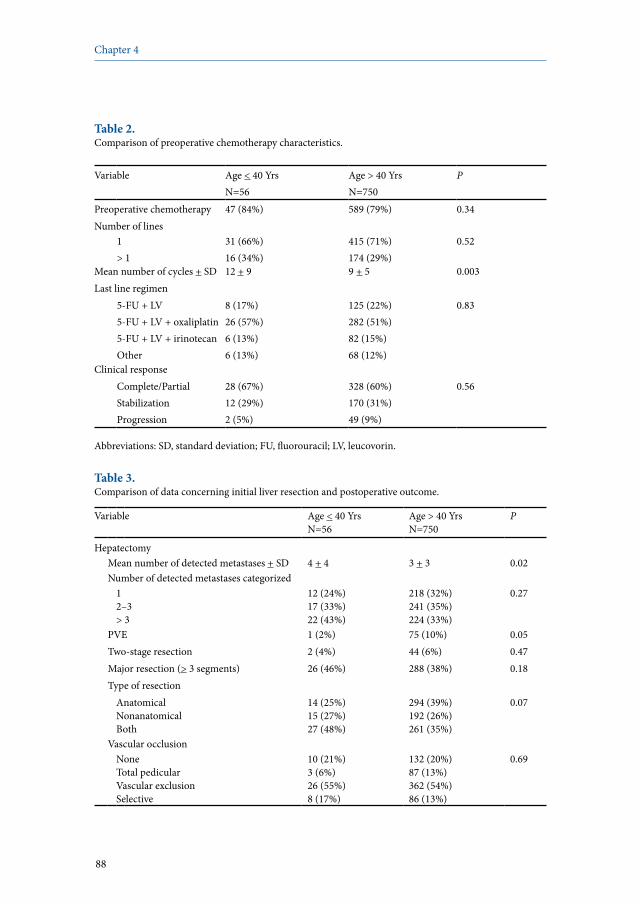
88
Chapter 4
Table 2. Comparison of preoperative chemotherapy characteristics.
Variable Age < 40 YrsN=56
Age > 40 YrsN=750
P
Preoperative chemotherapy 47 (84%) 589 (79%) 0.34Number of lines
1> 1
31 (66%)16 (34%)
415 (71%)174 (29%)
0.52
Mean number of cycles + SD 12 + 9 9 + 5 0.003Last line regimen
5-FU + LV5-FU + LV + oxaliplatin5-FU + LV + irinotecanOther
8 (17%)26 (57%)6 (13%)6 (13%)
125 (22%)282 (51%)82 (15%)68 (12%)
0.83
Clinical responseComplete/PartialStabilizationProgression
28 (67%) 12 (29%)2 (5%)
328 (60%)170 (31%)49 (9%)
0.56
Abbreviations: SD, standard deviation; FU, fluorouracil; LV, leucovorin.
Table 3. Comparison of data concerning initial liver resection and postoperative outcome.
Variable Age < 40 YrsN=56
Age > 40 YrsN=750
P
HepatectomyMean number of detected metastases + SD 4 + 4 3 + 3 0.02Number of detected metastases categorized
12–3> 3
12 (24%)17 (33%)22 (43%)
218 (32%)241 (35%)224 (33%)
0.27
PVE 1 (2%) 75 (10%) 0.05Two-stage resection 2 (4%) 44 (6%) 0.47Major resection (> 3 segments) 26 (46%) 288 (38%) 0.18Type of resection
AnatomicalNonanatomicalBoth
14 (25%)15 (27%)27 (48%)
294 (39%)192 (26%)261 (35%)
0.07
Vascular occlusionNoneTotal pedicularVascular exclusionSelective
10 (21%)3 (6%)26 (55%)8 (17%)
132 (20%)87 (13%)362 (54%)86 (13%)
0.69
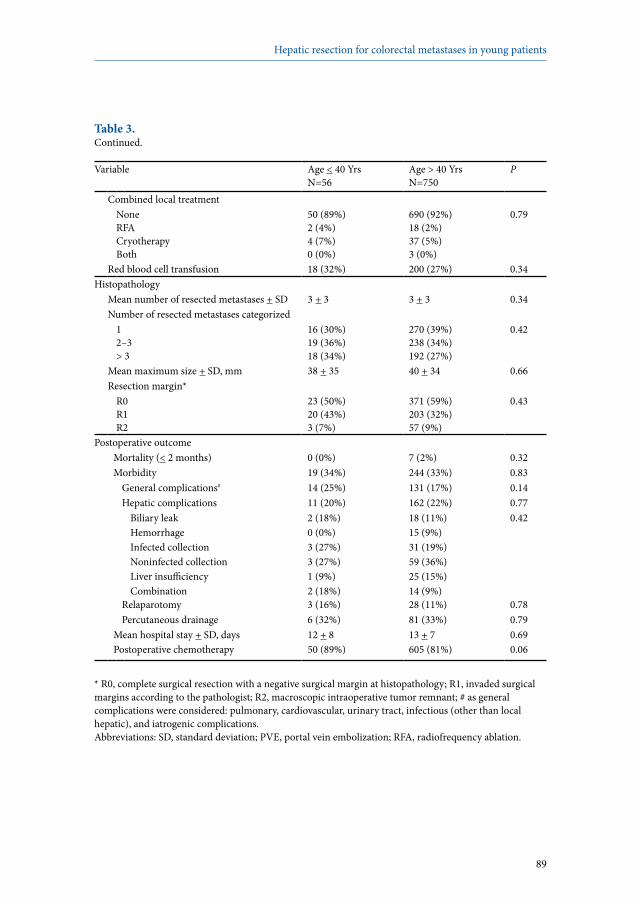
89
Hepatic resection for colorectal metastases in young patients
Table 3. Continued.
Variable Age < 40 YrsN=56
Age > 40 YrsN=750
P
Combined local treatmentNoneRFACryotherapyBoth
50 (89%)2 (4%)4 (7%)0 (0%)
690 (92%)18 (2%)37 (5%)3 (0%)
0.79
Red blood cell transfusion 18 (32%) 200 (27%) 0.34Histopathology
Mean number of resected metastases + SD 3 + 3 3 + 3 0.34Number of resected metastases categorized
12–3> 3
16 (30%)19 (36%)18 (34%)
270 (39%)238 (34%)192 (27%)
0.42
Mean maximum size + SD, mm 38 + 35 40 + 34 0.66Resection margin*
R0R1R2
23 (50%)20 (43%)3 (7%)
371 (59%)203 (32%)57 (9%)
0.43
Postoperative outcomeMortality (< 2 months) 0 (0%) 7 (2%) 0.32Morbidity 19 (34%) 244 (33%) 0.83
General complications# 14 (25%) 131 (17%) 0.14Hepatic complications 11 (20%) 162 (22%) 0.77
Biliary leakHemorrhageInfected collectionNoninfected collectionLiver insufficiencyCombination
2 (18%)0 (0%)3 (27%)3 (27%)1 (9%)2 (18%)
18 (11%)15 (9%)31 (19%)59 (36%)25 (15%)14 (9%)
0.42
Relaparotomy 3 (16%) 28 (11%) 0.78Percutaneous drainage 6 (32%) 81 (33%) 0.79
Mean hospital stay + SD, days Postoperative chemotherapy
12 + 850 (89%)
13 + 7605 (81%)
0.690.06
* R0, complete surgical resection with a negative surgical margin at histopathology; R1, invaded surgical margins according to the pathologist; R2, macroscopic intraoperative tumor remnant; # as general complications were considered: pulmonary, cardiovascular, urinary tract, infectious (other than local hepatic), and iatrogenic complications. Abbreviations: SD, standard deviation; PVE, portal vein embolization; RFA, radiofrequency ablation.
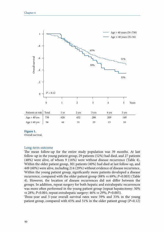
90
Chapter 4
Patients at risk Total 1 yr 2 yrs 3 yrs 4 yrs 5 yrs
Age > 40 yrs 750 626 432 286 209 149
Age ≤ 40 yrs 56 44 31 23 13 10
0
.2
.4
.6
.8
1 O
vera
ll su
rviv
al
0 1 2 3 4 5 Years
P = 0.12
33%
59%
51%
65%
Age ≤ 40 years (N=56) Age > 40 years (N=750)
Figure 1. Overall survival.
Long-term outcomeThe mean follow-up for the entire study population was 39 months. At last follow-up in the young patient group, 29 patients (52%) had died, and 27 patients (48%) were alive, of whom 9 (16%) were without disease recurrence (Table 4). Within the older patient group, 301 patients (40%) had died at last follow-up, and 449 (60%) were alive, including 214 (29%) without evidence of disease recurrence. Within the young patient group, significantly more patients developed a disease recurrence, compared with the older patient group (88% vs 69%; P=0.003) (Table 4). However, the location of disease recurrences did not differ between the groups. In addition, repeat surgery for both hepatic and extrahepatic recurrences was more often performed in the young patient group (repeat hepatectomy: 50% vs 28%; P<0.001; repeat extrahepatic surgery: 46% vs 29%; P=0.005). Three-year and 5-year overall survival rates were 59% and 33% in the young patient group, compared with 65% and 51% in the older patient group (P=0.12)

91
Hepatic resection for colorectal metastases in young patients
(Figure 1). Median overall survival for the younger patients was 43 months, whereas it was 61 months for the older patients. Three-year and 5-year progression-free survival rates were significantly lower in the younger patient group (4% and 2% vs 21% and 16%; P<0.001) (Figure 2). Median progression-free survival was 7 months for the young patients, compared with 13 months in the older patient group. Disease-free survival rates after 3 and 5 years were 25% and 17% in the young patient group, versus 34% and 23% in the older patients (median: 18 months vs 19 months; P=0.10) (Figure 3). The difference between progression-free and disease-free survival rates is illustrated in Figure 4.
Patients at risk Total 1 yr 2 yrs 3 yrs 4 yrs 5 yrs
Age > 40 yrs 701 340 163 102 78 63
Age ≤ 40 yrs 52 13 5 2 1 0
0
.2
.4
.6
.8
1
Prog
ress
ion-
free s
urvi
val
0 1 2 3 4 5 Years
P < 0.001 2% 4%
16% 21%
Age ≤ 40 years (N=56) Age > 40 years (N=750)
Figure 2. Progression-free survival.

92
Chapter 4
Predictive factors of survivalAt multivariate analysis, seven factors emerged as independent predictors of poor overall survival: primary tumor located in the right colon, three or more liver metastases at diagnosis, the presence of concomitant extrahepatic disease, administration of preoperative chemotherapy, preoperative carcinoembryonic antigen (CEA) level >5 ng/ml, preoperative portal vein embolization, and maximum size of liver metastases >30 mm at histopathologic examination (Table 5). For progression-free survival, the following poor predictors were identified: age at hepatectomy <40 years, administration of preoperative chemotherapy, preoperative CEA level >5 ng/ml, three or more liver metastases detected by intraoperative ultrasound, major hepatectomy, nonanatomical hepatic resection, and the administration of postoperative chemotherapy (Table 6).
Table 4. Comparison of long-term outcome.
Variable Age < 40 YrsN=56
Age > 40 YrsN=750
P
Recurrence 49 (88%) 514 (69%) 0.003Location
IntrahepaticExtrahepaticBoth
13 (27%)13 (27%)23 (47%)
171 (33%)160 (31%)183 (36%)
0.27
Repeat hepatectomy 28 (50%) 207 (28%) < 0.001Repeat extrahepatic resection 26 (46%) 214 (29%) 0.005Status at last follow-up
Alive – no evidence of diseaseAlive with diseaseDead
9 (16%)18 (32%)29 (52%)
214 (29%)235 (31%)301 (40%)
0.10
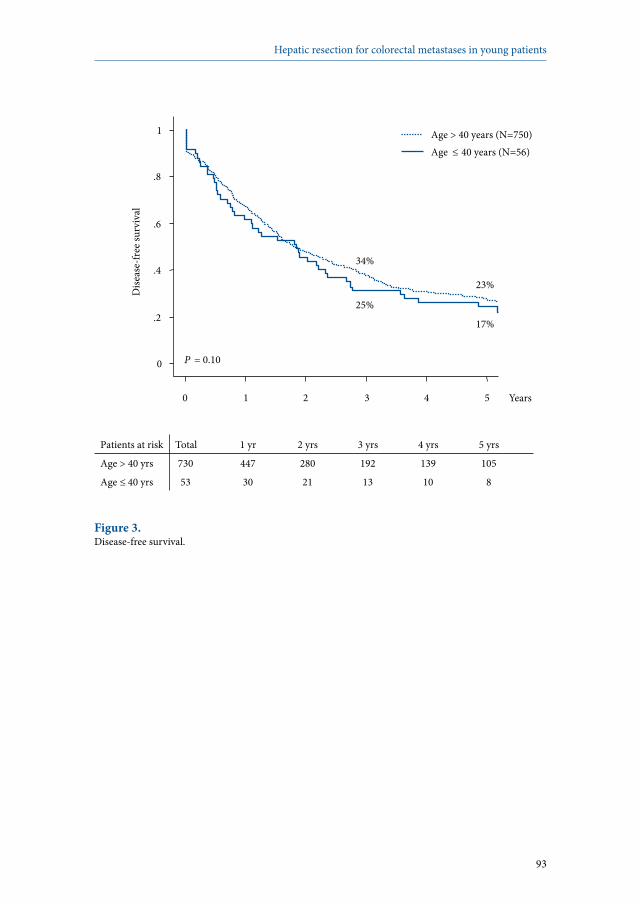
93
Hepatic resection for colorectal metastases in young patients
Patients at risk Total 1 yr 2 yrs 3 yrs 4 yrs 5 yrs
Age > 40 yrs 730 447 280 192 139 105
Age ≤ 40 yrs 53 30 21 13 10 8
0
.2
.4
.6
.8
1 D
iseas
e-fre
e sur
viva
l
0 1 2 3 4 5 Years
P = 0.10
17%
25%
23%
34%
Age ≤ 40 years (N=56) Age > 40 years (N=750)
Figure 3. Disease-free survival.

94
Chapter 4
Patients at risk Total 1 yr 2 yrs 3 yrs 4 yrs 5 yrs
DFS 53 30 21 13 10 8
PFS 52 13 5 2 1 0
0
.2
.4
.6
.8
1 Su
rviv
al
0 1 2 3 4 5 Years
2% 4%
PFS DFS
17% 25%
21% 15%
Figure 4. Difference between disease-free (DFS) and progression-free survival (PFS) in young patients, illustrating the influence of repeat surgery.
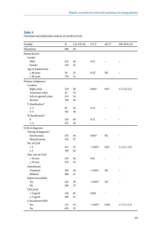
95
Hepatic resection for colorectal metastases in young patients
Table 5. Univariate and multivariate analysis of overall survival.
Variable N 5-yr OS (%) UV P MV P HR (95% CI)
All patients 806 49
Patient factorsGender
MaleFemale
472334
4852
0.25 – –
Age at hepatectomy< 40 years > 40 years
56750
3351
0.12$ NS –
Primary malignancyLocation
Right colonTransverse colonLeft or sigmoid colonRectum
12925413200
36525448
0.001$ 0.03 1.5 (1.0-2.1)
T classification31 1–23–4
95563
4550
0.76 – –
N classification31 01–2
243457
4950
0.72 – –
CLM at diagnosisTiming of diagnosis*
SynchronousMetachronous
473332
4457
0.001$ NS –
No. of CLM< 3> 3
411350
5742
< 0.001$ 0.02 1.4 (1.1-1.9)
Max. size of CLM< 30 mm> 30 mm
234470
5645
0.01 – –
DistributionUnilateralBilateral
384408
5841
< 0.001$ NS –
Initial resectabilityYesNo
422369
5937
< 0.001$ NS –
CEA level< 5 ng/ml> 5 ng/ml
126405
6341
0.001 – –
Concomitant EHDYesNo
151655
3553
< 0.001$ 0.001 1.7 (1.2-2.3)
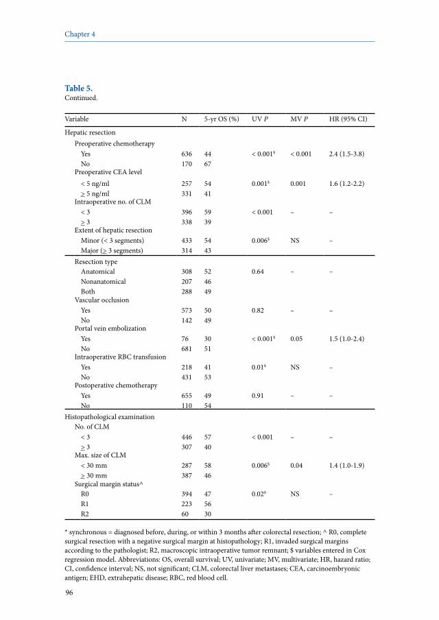
96
Chapter 4
Table 5. Continued.
Variable N 5-yr OS (%) UV P MV P HR (95% CI)
Hepatic resectionPreoperative chemotherapy
YesNo
636170
4467
< 0.001$ < 0.001 2.4 (1.5-3.8)
Preoperative CEA level< 5 ng/ml> 5 ng/ml
257331
5441
0.001$ 0.001 1.6 (1.2-2.2)
Intraoperative no. of CLM< 3> 3
396338
5939
< 0.001 – –
Extent of hepatic resectionMinor (< 3 segments)Major (> 3 segments)
433314
5443
0.006$ NS –
Resection typeAnatomicalNonanatomicalBoth
308207288
524649
0.64 – –
Vascular occlusionYesNo
573142
5049
0.82 – –
Portal vein embolizationYesNo
76681
3051
< 0.001$ 0.05 1.5 (1.0-2.4)
Intraoperative RBC transfusionYesNo
218431
4153
0.01$ NS –
Postoperative chemotherapyYesNo
655110
4954
0.91 – –
Histopathological examinationNo. of CLM
< 3> 3
446307
5740
< 0.001 – –
Max. size of CLM< 30 mm> 30 mm
287387
5846
0.006$ 0.04 1.4 (1.0-1.9)
Surgical margin status^R0R1R2
39422360
475630
0.02$ NS –
* synchronous = diagnosed before, during, or within 3 months after colorectal resection; ^ R0, complete surgical resection with a negative surgical margin at histopathology; R1, invaded surgical margins according to the pathologist; R2, macroscopic intraoperative tumor remnant; $ variables entered in Cox regression model. Abbreviations: OS, overall survival; UV, univariate; MV, multivariate; HR, hazard ratio; CI, confidence interval; NS, not significant; CLM, colorectal liver metastases; CEA, carcinoembryonic antigen; EHD, extrahepatic disease; RBC, red blood cell.

97
Hepatic resection for colorectal metastases in young patients
Table 6. Univariate and multivariate analysis of progression-free survival.
Variable N 5-yr PFS (%) UV P MV P HR (95% CI)
All patients 753 15
Patient factorsGender
MaleFemale
436317
1120
0.07$ NS –
Age at hepatectomy< 40 years > 40 years
52701
216
< 0.001$ < 0.001 2.7 (1.9-4.0)
Primary malignancyLocation
Right colonTransverse colonLeft or sigmoid colonRectum
12022386189
761816
0.009$ NS –
T classification31 1–23–4
89527
2213
0.03$ NS –
N classification31 01–2
230424
1813
0.11$ NS –
CLM at diagnosisTiming of diagnosis*
SynchronousMetachronous
428324
1219
< 0.001$ NS –
No. of CLM< 3> 3
408304
1911
< 0.001 – –
Max. size of CLM< 30 mm> 30 mm
225434
1714
0.90 – –
DistributionUnilateralBilateral
376363
1811
0.001$ NS –
Initial resectabilityYesNo
414324
199
< 0.001$ NS –
CEA level< 5 ng/ml> 5 ng/ml
123371
2015
0.22 – –
Concomitant EHDYesNo
133620
717
0.03$ NS –
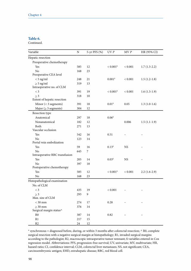
98
Chapter 4
Table 6. Continued.
Variable N 5-yr PFS (%) UV P MV P HR (95% CI)
Hepatic resectionPreoperative chemotherapy
YesNo
585168
1223
< 0.001$ < 0.001 1.7 (1.3-2.2)
Preoperative CEA level< 5 ng/ml> 5 ng/ml
248319
2113
0.001$ < 0.001 1.5 (1.2-1.8)
Intraoperative no. of CLM< 3> 3
391318
1910
< 0.001$ < 0.001 1.6 (1.3-1.9)
Extent of hepatic resectionMinor (< 3 segments)Major (> 3 segments)
391304
1812
0.01$ 0.05 1.3 (1.0-1.6)
Resection typeAnatomicalNonanatomicalBoth
297182271
181213
0.06$
0.006 1.5 (1.1-1.9)
Vascular occlusionYesNo
542123
1614
0.51 – –
Portal vein embolizationYesNo
59645
167
0.13$ NS –
Intraoperative RBC transfusionYesNo
205397
1418
0.03$ NS –
Postoperative chemotherapyYesNo
585168
1223
< 0.001$ < 0.001 2.2 (1.6-2.9)
Histopathological examinationNo. of CLM
< 3> 3
435293
199
< 0.001 – –
Max. size of CLM< 30 mm> 30 mm
274376
1714
0.28 – –
Surgical margin status^R0R1R2
38721724
141512
0.82 – –
* synchronous = diagnosed before, during, or within 3 months after colorectal resection; ^ R0, complete surgical resection with a negative surgical margin at histopathology; R1, invaded surgical margins according to the pathologist; R2, macroscopic intraoperative tumor remnant; $ variables entered in Cox regression model. Abbreviations: PFS, progression-free survival; UV, univariate; MV, multivariate; HR, hazard ratio; CI, confidence interval; CLM, colorectal liver metastases; NS, not significant; CEA, carcinoembryonic antigen; EHD, extrahepatic disease; RBC, red blood cell.

99
Hepatic resection for colorectal metastases in young patients
Discussion
Long-term outcomes after hepatic resection for colorectal liver metastases in young patients are still unknown. This was the impetus for the current study, evaluating long-term outcomes in patients <40 years old, which were compared with patients older than 40 years. Within the current study, young patients presented with more extensive disease, reflected by a higher number of metastases, which were more often diagnosed synchronous with the primary tumor. Furthermore, overall survival rates tended to be lower in the young patient group (5-year overall survival rate: 33% vs 51%; P=0.12). Also, young patients more often developed a disease recurrence (88% vs 69%; P=0.003), and progression-free survival in this patient group was significantly lower than that observed for older patients (5-year progression-free survival rate: 2% vs 16%; P<0.001). However, as recurrences in young patients were often treated by repeat surgery, disease-free survival rates did not differ between the groups. At multivariate analysis, young age independently predicted poor progression-free survival. The potential influence of age on long-term outcomes after hepatic resection for metastatic colorectal cancer has only been described for elderly patients.16-20 Elderly patients are often defined as patients with an age at hepatectomy >70 years, and reported 5-year survival rates for these patients vary between 21% and 44%, which is similar to that observed for patients younger than 70 years.16-20 Although no data are available concerning long-term outcomes after hepatectomy for colorectal metastases in relatively young patients, long-term outcomes after resection of the primary colorectal tumor in patients younger than 40 years have been described by some authors.21-24 Five-year overall survival rates in these studies were highly variable and ranged between 9% and 54.8%.21-24 In two of these studies, long-term outcomes were compared between young and older patients, showing no differences between the groups.21,24 In all of these studies, an age of 40 years was used to define the young patient group, and therefore we decided to use the same cutoff point in the current study. To our knowledge, our study is the first describing long-term outcomes after hepatic resection for colorectal metastases in patients <40 years. The trend to lower overall survival rates after hepatectomy for colorectal metastases in patients aged <40 years was rather surprising. Often, young patients have a better performance status with fewer cardiovascular and pulmonary comorbidities, compared with older patients. As a result, young patients can in general be treated with more aggressive preoperative chemotherapy regimens, and in case of disease recurrence, young patients are more often candidates for repeat surgery. In the current study, before hepatectomy young patients were indeed treated with significantly more cycles of systemic chemotherapy; however, postoperative mortality and morbidity rates were comparable between the
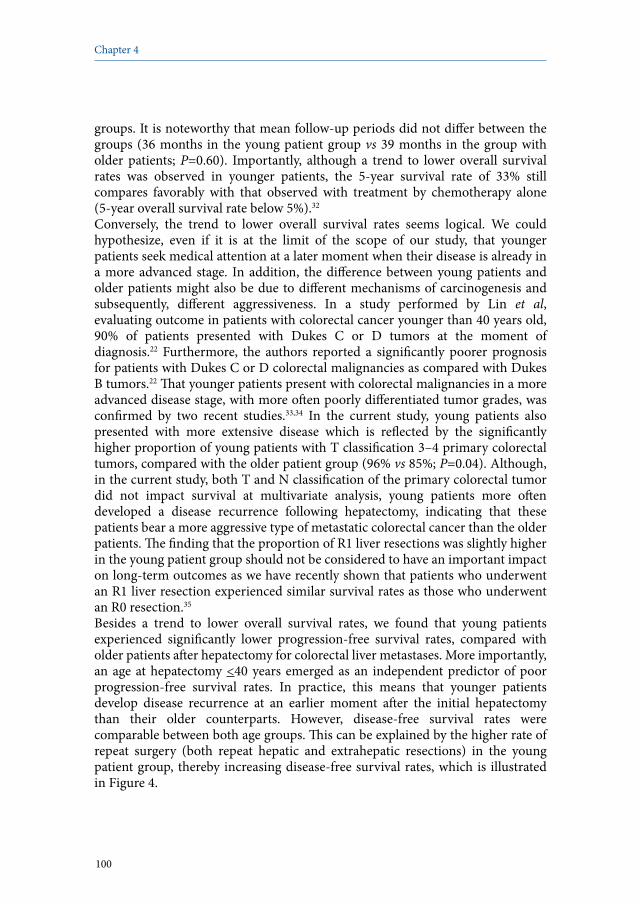
100
Chapter 4
groups. It is noteworthy that mean follow-up periods did not differ between the groups (36 months in the young patient group vs 39 months in the group with older patients; P=0.60). Importantly, although a trend to lower overall survival rates was observed in younger patients, the 5-year survival rate of 33% still compares favorably with that observed with treatment by chemotherapy alone (5-year overall survival rate below 5%).32 Conversely, the trend to lower overall survival rates seems logical. We could hypothesize, even if it is at the limit of the scope of our study, that younger patients seek medical attention at a later moment when their disease is already in a more advanced stage. In addition, the difference between young patients and older patients might also be due to different mechanisms of carcinogenesis and subsequently, different aggressiveness. In a study performed by Lin et al, evaluating outcome in patients with colorectal cancer younger than 40 years old, 90% of patients presented with Dukes C or D tumors at the moment of diagnosis.22 Furthermore, the authors reported a significantly poorer prognosis for patients with Dukes C or D colorectal malignancies as compared with Dukes B tumors.22 That younger patients present with colorectal malignancies in a more advanced disease stage, with more often poorly differentiated tumor grades, was confirmed by two recent studies.33,34 In the current study, young patients also presented with more extensive disease which is reflected by the significantly higher proportion of young patients with T classification 3–4 primary colorectal tumors, compared with the older patient group (96% vs 85%; P=0.04). Although, in the current study, both T and N classification of the primary colorectal tumor did not impact survival at multivariate analysis, young patients more often developed a disease recurrence following hepatectomy, indicating that these patients bear a more aggressive type of metastatic colorectal cancer than the older patients. The finding that the proportion of R1 liver resections was slightly higher in the young patient group should not be considered to have an important impact on long-term outcomes as we have recently shown that patients who underwent an R1 liver resection experienced similar survival rates as those who underwent an R0 resection.35
Besides a trend to lower overall survival rates, we found that young patients experienced significantly lower progression-free survival rates, compared with older patients after hepatectomy for colorectal liver metastases. More importantly, an age at hepatectomy <40 years emerged as an independent predictor of poor progression-free survival rates. In practice, this means that younger patients develop disease recurrence at an earlier moment after the initial hepatectomy than their older counterparts. However, disease-free survival rates were comparable between both age groups. This can be explained by the higher rate of repeat surgery (both repeat hepatic and extrahepatic resections) in the young patient group, thereby increasing disease-free survival rates, which is illustrated in Figure 4.
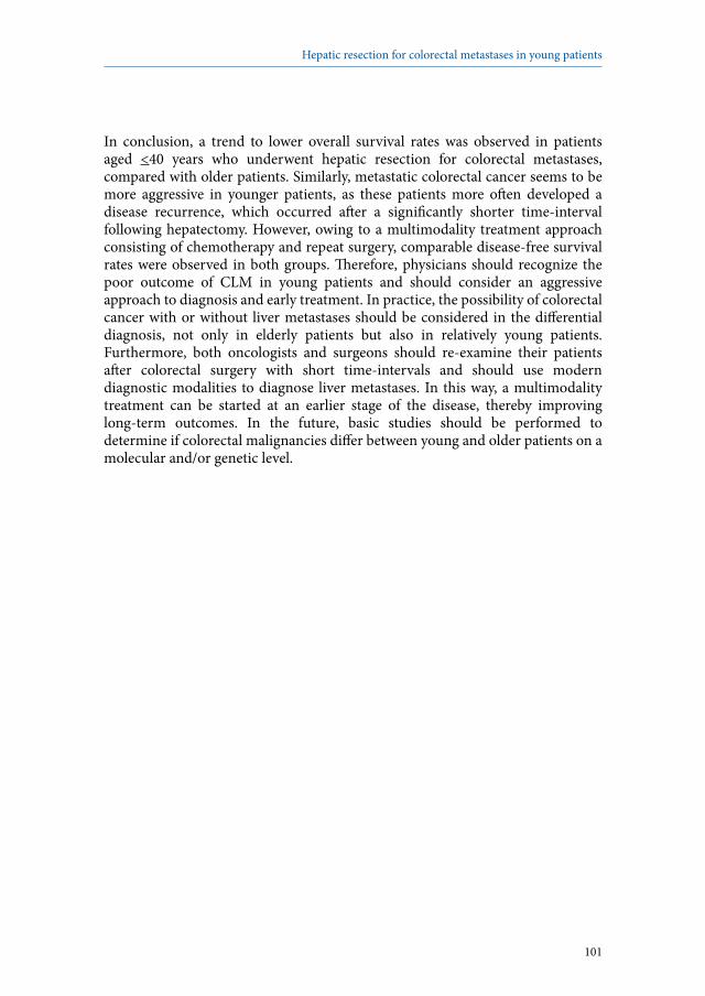
101
Hepatic resection for colorectal metastases in young patients
In conclusion, a trend to lower overall survival rates was observed in patients aged <40 years who underwent hepatic resection for colorectal metastases, compared with older patients. Similarly, metastatic colorectal cancer seems to be more aggressive in younger patients, as these patients more often developed a disease recurrence, which occurred after a significantly shorter time-interval following hepatectomy. However, owing to a multimodality treatment approach consisting of chemotherapy and repeat surgery, comparable disease-free survival rates were observed in both groups. Therefore, physicians should recognize the poor outcome of CLM in young patients and should consider an aggressive approach to diagnosis and early treatment. In practice, the possibility of colorectal cancer with or without liver metastases should be considered in the differential diagnosis, not only in elderly patients but also in relatively young patients. Furthermore, both oncologists and surgeons should re-examine their patients after colorectal surgery with short time-intervals and should use modern diagnostic modalities to diagnose liver metastases. In this way, a multimodality treatment can be started at an earlier stage of the disease, thereby improving long-term outcomes. In the future, basic studies should be performed to determine if colorectal malignancies differ between young and older patients on a molecular and/or genetic level.
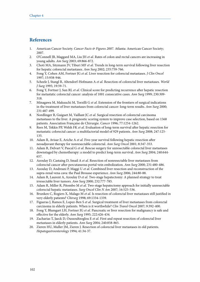
102
Chapter 4
References
1. American Cancer Society. Cancer Facts & Figures 2007. Atlanta: American Cancer Society; 2007.
2. O’Connell JB, Maggard MA, Liu JH et al. Rates of colon and rectal cancers are increasing in young adults. Am Surg 2003; 69:866-872.
3. Choti MA, Sitzmann JV, Tiburi MF et al. Trends in long-term survival following liver resection for hepatic colorectal metastases. Ann Surg 2002; 235:759-766.
4. Fong Y, Cohen AM, Fortner JG et al. Liver resection for colorectal metastases. J Clin Oncol 1997; 15:938-946.
5. Scheele J, Stangl R, Altendorf-Hofmann A et al. Resection of colorectal liver metastases. World J Surg 1995; 19:59-71.
6. Fong Y, Fortner J, Sun RL et al. Clinical score for predicting recurrence after hepatic resection for metastatic colorectal cancer: analysis of 1001 consecutive cases. Ann Surg 1999; 230:309-318.
7. Minagawa M, Makuuchi M, Torzilli G et al. Extension of the frontiers of surgical indications in the treatment of liver metastases from colorectal cancer: long-term results. Ann Surg 2000; 231:487-499.
8. Nordlinger B, Guiguet M, Vaillant JC et al. Surgical resection of colorectal carcinoma metastases to the liver. A prognostic scoring system to improve case selection, based on 1568 patients. Association Française de Chirurgie. Cancer 1996; 77:1254-1262.
9. Rees M, Tekkis PP, Welsh FK et al. Evaluation of long-term survival after hepatic resection for metastatic colorectal cancer: a multifactorial model of 929 patients. Ann Surg 2008; 247:125-135.
10. Adam R, Avisar E, Ariche A et al. Five-year survival following hepatic resection after neoadjuvant therapy for nonresectable colorectal. Ann Surg Oncol 2001; 8:347-353.
11. Adam R, Delvart V, Pascal G et al. Rescue surgery for unresectable colorectal liver metastases downstaged by chemotherapy: a model to predict long-term survival. Ann Surg 2004; 240:644-657.
12. Azoulay D, Castaing D, Smail A et al. Resection of nonresectable liver metastases from colorectal cancer after percutaneous portal vein embolization. Ann Surg 2000; 231:480-486.
13. Azoulay D, Andreani P, Maggi U et al. Combined liver resection and reconstruction of the supra-renal vena cava: the Paul Brousse experience. Ann Surg 2006; 244:80-88.
14. Adam R, Laurent A, Azoulay D et al. Two-stage hepatectomy: A planned strategy to treat irresectable liver tumors. Ann Surg 2000; 232:777-785.
15. Adam R, Miller R, Pitombo M et al. Two-stage hepatectomy approach for initially unresectable colorectal hepatic metastases. Surg Oncol Clin N Am 2007; 16:525-536.
16. Brunken C, Rogiers X, Malago M et al. Is resection of colorectal liver metastases still justified in very elderly patients? Chirurg 1998; 69:1334-1339.
17. Figueras J, Ramos E, Lopez-Ben S et al. Surgical treatment of liver metastases from colorectal carcinoma in elderly patients. When is it worthwhile? Clin Transl Oncol 2007; 9:392-400.
18. Fong Y, Blumgart LH, Fortner JG et al. Pancreatic or liver resection for malignancy is safe and effective for the elderly. Ann Surg 1995; 222:426-434.
19. Zacharias T, Jaeck D, Oussoultzoglou E et al. First and repeat resection of colorectal liver metastases in elderly patients. Ann Surg 2004; 240:858-865.
20. Zieren HU, Muller JM, Zieren J. Resection of colorectal liver metastases in old patients. Hepatogastroenterology 1994; 41:34-37.
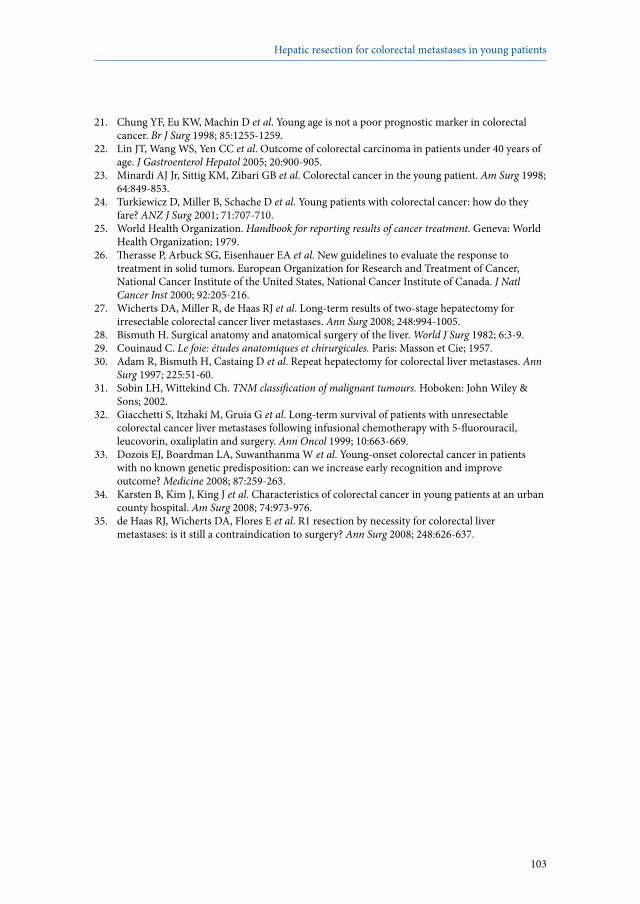
103
Hepatic resection for colorectal metastases in young patients
21. Chung YF, Eu KW, Machin D et al. Young age is not a poor prognostic marker in colorectal cancer. Br J Surg 1998; 85:1255-1259.
22. Lin JT, Wang WS, Yen CC et al. Outcome of colorectal carcinoma in patients under 40 years of age. J Gastroenterol Hepatol 2005; 20:900-905.
23. Minardi AJ Jr, Sittig KM, Zibari GB et al. Colorectal cancer in the young patient. Am Surg 1998; 64:849-853.
24. Turkiewicz D, Miller B, Schache D et al. Young patients with colorectal cancer: how do they fare? ANZ J Surg 2001; 71:707-710.
25. World Health Organization. Handbook for reporting results of cancer treatment. Geneva: World Health Organization; 1979.
26. Therasse P, Arbuck SG, Eisenhauer EA et al. New guidelines to evaluate the response to treatment in solid tumors. European Organization for Research and Treatment of Cancer, National Cancer Institute of the United States, National Cancer Institute of Canada. J Natl Cancer Inst 2000; 92:205-216.
27. Wicherts DA, Miller R, de Haas RJ et al. Long-term results of two-stage hepatectomy for irresectable colorectal cancer liver metastases. Ann Surg 2008; 248:994-1005.
28. Bismuth H. Surgical anatomy and anatomical surgery of the liver. World J Surg 1982; 6:3-9.29. Couinaud C. Le foie: études anatomiques et chirurgicales. Paris: Masson et Cie; 1957.30. Adam R, Bismuth H, Castaing D et al. Repeat hepatectomy for colorectal liver metastases. Ann
Surg 1997; 225:51-60.31. Sobin LH, Wittekind Ch. TNM classification of malignant tumours. Hoboken: John Wiley &
Sons; 2002.32. Giacchetti S, Itzhaki M, Gruia G et al. Long-term survival of patients with unresectable
colorectal cancer liver metastases following infusional chemotherapy with 5-fluorouracil, leucovorin, oxaliplatin and surgery. Ann Oncol 1999; 10:663-669.
33. Dozois EJ, Boardman LA, Suwanthanma W et al. Young-onset colorectal cancer in patients with no known genetic predisposition: can we increase early recognition and improve outcome? Medicine 2008; 87:259-263.
34. Karsten B, Kim J, King J et al. Characteristics of colorectal cancer in young patients at an urban county hospital. Am Surg 2008; 74:973-976.
35. de Haas RJ, Wicherts DA, Flores E et al. R1 resection by necessity for colorectal liver metastases: is it still a contraindication to surgery? Ann Surg 2008; 248:626-637.


Robbert J. de Haas1,2
Dennis A. Wicherts1,2
Eduardo Flores1
Daniel Azoulay1
Denis Castaing1
René Adam1
1 Centre Hépato-Biliaire, Hôpital Paul Brousse, Villejuif, France2 Department of Surgery, University Medical Center Utrecht, Utrecht,
The Netherlands
Annals of Surgery 2008; 248:626-637
R1 resection by necessity for colorectal liver metastases
Is it still a contraindication to surgery?
5

106
Chapter 5
Abstract
ObjectiveTo compare long-term outcome of R0 (negative margins) and R1 (positive margins) liver resections for colorectal liver metastases (CLM) treated by an aggressive approach combining chemotherapy and repeat surgery.
Summary background dataComplete macroscopic resection with negative margins is the gold standard recommendation in the surgical treatment of CLM. However, due to vascular proximity or multinodularity, complete macroscopic resection can sometimes only be performed through R1 resection. Increasingly efficient chemotherapy may have changed long-term outcome after R1 resection.
MethodsAll resected CLM patients (R0 or R1) at our institution between 1990 and 2006 were prospectively evaluated. Exclusion criteria were: macroscopic incomplete (R2) resection, use of local treatment modalities, and presence of extrahepatic disease. We aimed to resect all identified metastases with negative margins. However, when safe margins could not be obtained, resection was still performed provided complete tumor removal. Overall (OS) and disease-free survival (DFS) were compared between groups, and prognostic factors were identified.
ResultsOf 840 patients, 436 (52%) were eligible for the study, 234 (28%) of whom underwent R0 resection, and 202 (24%) underwent R1 resection. Number and size of CLM were higher, and distribution was more often bilateral in the R1 group. After a mean follow-up of 40 months, 5-year OS was 61% and 57% for R0 and R1 patients (P=0.27). Five-year DFS was 29% in the R0 group versus 20% in the R1 group (P=0.12). In the R1 group, intrahepatic (but not surgical margin) recurrences were more often observed (28% vs 17%; P=0.004). Preoperative carcinoembryonic antigen level >10 ng/ml and major hepatectomy, but not R1 resection, were independent predictors of poor OS. Size >30 mm, bilateral distribution, and intraoperative blood transfusions independently predicted positive surgical margins.
ConclusionsDespite a higher recurrence rate, the contraindication of R1 resection should be revisited in the current era of effective chemotherapy because survival is similar to that of R0 resection.

107
R1 resection by necessity for colorectal liver metastases
Introduction
Liver resection is still the only chance of cure for patients with colorectal metastases, resulting in 5-year survival rates up to 67% for highly selected patients.1 Unfortunately, at the time of diagnosis, only one fifth of patients with colorectal liver metastases (CLM) presents with resectable lesions.2 Nowadays, an important amount of patients initially considered as having unresectable CLM, can be converted to resectability by the aid of modern chemotherapy regimens consisting of 5-fluorouracil, leucovorin, oxaliplatin and/or irinotecan.3-6 In addition, chemotherapy efficacy can be further increased by the use of monoclonal antibodies like cetuximab and bevacizumab.7-9 Besides this, surgical improvements, like portal vein embolization which allows hypertrophy of a small remnant liver volume, and vascular resection and reconstruction techniques, enable hepatic resection in an additional number of patients.10,11 However, due to the increased number of patients with multinodular disease and/or CLM close to important vascular structures, macroscopic complete resection of all CLM can sometimes only be performed with positive surgical margins. Within the literature, the inability to achieve clear resection margins is generally considered as contraindication to surgery because it has been associated with significantly reduced survival rates,12-20 and an increased risk of surgical margin recurrence.16,21 Several cutoff points for the optimal negative resection margin width have been proposed, varying between 2 and 10 mm;22-28 however, complete macroscopic removal of all lesions with negative resection margins, irrespective of the width, is currently the gold standard recommendation in the surgical treatment of CLM. At our institute, the inability to obtain a free surgical margin during liver resection for CLM has never been considered a contraindication to surgery, provided complete macroscopic removal of all metastatic lesions. As increasingly effective chemotherapy regimens might have changed the spectrum of this particular situation, we compared long-term outcome in patients with microscopically positive and negative resection margins who were all treated by an aggressive approach consisting of chemotherapy and repeat surgery to assess the validity of this policy.
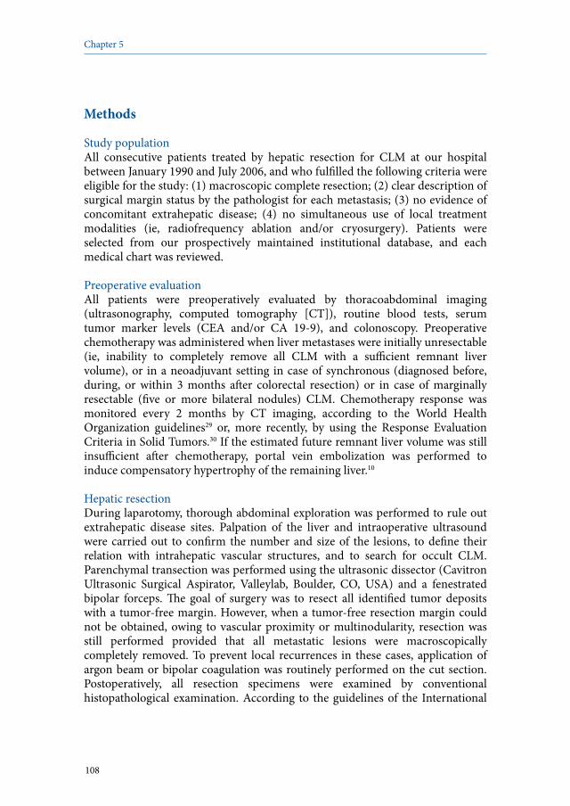
108
Chapter 5
Methods
Study populationAll consecutive patients treated by hepatic resection for CLM at our hospital between January 1990 and July 2006, and who fulfilled the following criteria were eligible for the study: (1) macroscopic complete resection; (2) clear description of surgical margin status by the pathologist for each metastasis; (3) no evidence of concomitant extrahepatic disease; (4) no simultaneous use of local treatment modalities (ie, radiofrequency ablation and/or cryosurgery). Patients were selected from our prospectively maintained institutional database, and each medical chart was reviewed.
Preoperative evaluationAll patients were preoperatively evaluated by thoracoabdominal imaging (ultrasonography, computed tomography [CT]), routine blood tests, serum tumor marker levels (CEA and/or CA 19-9), and colonoscopy. Preoperative chemotherapy was administered when liver metastases were initially unresectable (ie, inability to completely remove all CLM with a sufficient remnant liver volume), or in a neoadjuvant setting in case of synchronous (diagnosed before, during, or within 3 months after colorectal resection) or in case of marginally resectable (five or more bilateral nodules) CLM. Chemotherapy response was monitored every 2 months by CT imaging, according to the World Health Organization guidelines29 or, more recently, by using the Response Evaluation Criteria in Solid Tumors.30 If the estimated future remnant liver volume was still insufficient after chemotherapy, portal vein embolization was performed to induce compensatory hypertrophy of the remaining liver.10
Hepatic resectionDuring laparotomy, thorough abdominal exploration was performed to rule out extrahepatic disease sites. Palpation of the liver and intraoperative ultrasound were carried out to confirm the number and size of the lesions, to define their relation with intrahepatic vascular structures, and to search for occult CLM. Parenchymal transection was performed using the ultrasonic dissector (Cavitron Ultrasonic Surgical Aspirator, Valleylab, Boulder, CO, USA) and a fenestrated bipolar forceps. The goal of surgery was to resect all identified tumor deposits with a tumor-free margin. However, when a tumor-free resection margin could not be obtained, owing to vascular proximity or multinodularity, resection was still performed provided that all metastatic lesions were macroscopically completely removed. To prevent local recurrences in these cases, application of argon beam or bipolar coagulation was routinely performed on the cut section. Postoperatively, all resection specimens were examined by conventional histopathological examination. According to the guidelines of the International

109
R1 resection by necessity for colorectal liver metastases
Union Against Cancer, R0 and R1 resections were defined by the absence (tumor-free margin >1 mm for all detected lesions) or presence (tumor-free margin 0 mm) of microscopic tumor invasion of the resection margins, respectively.31 In case of microscopic tumor invasion, the additional opinion of the operating surgeon with regard to the complete macroscopic resection of all lesions, differentiated R1 from R2 resections.
Postoperative follow-upRegular postoperative follow-up started at 1 month posthepatectomy, and then every 4 months, consisting of routine blood tests, serum tumor marker evaluation (CEA and/or CA 19-9), and hepatic ultrasound. Thoracoabdominal CT imaging was performed every 8 months. After hepatectomy, patients were treated by systemic chemotherapy to decrease the recurrence risk. If an intrahepatic and/or extrahepatic recurrence occurred, repeat surgery was performed if potentially curative.32
Statistical analysisTo assess the impact of R1 resections on long-term outcome, patients were divided according to their surgical margin status into an R0 group and an R1 group. χ2 test was used to compare categorical data, and continuous data were compared using the independent-samples t test. Survival time was calculated from the date of hepatic resection until death or last follow-up. Progression-free survival was defined as the time-interval between hepatectomy and first postoperative recurrence or death, whereas disease-free survival was defined as the time-interval between the hepatectomy and either the first recurrence that could not be curatively treated (resection or ablation) or death. Survival curves were generated by the Kaplan-Meier method, and were compared by the log-rank test. To identify predictors of survival, univariate and multivariate analyses were performed, using the log-rank test and the Cox proportional hazard model, respectively. Furthermore, independent predictors of R1 resection were identified at multivariate analysis (logistic regression), including all factors most likely to influence surgical margin status with a P value <0.10 at univariate analysis. Statistical significance was defined as P<0.05. All statistical analyses were performed by using SPSS version 13.0 (SPSS Inc, Chicago, IL, USA).

110
Chapter 5
Results
PatientsBetween January 1990 and July 2006, 840 patients underwent hepatic resection for CLM at our hospital, 436 (52%) of whom fulfilled the inclusion criteria (Figure 1). Fifty-seven patients (7%) were excluded because of a macroscopic incomplete (R2) liver resection, 128 patients (15%) because a clear description of the microscopic surgical margin status was not available for all metastases, 134 patients (16%) because of concomitant extrahepatic disease, and 85 patients (10%) because of a concomitant local treatment. Among the 436 eligible patients, microscopic tumor invasion at pathologic analysis was present in 202 patients (24%) (R1 group), whereas in 234 patients (28%) no viable tumor cells could be identified at the resection margin (R0 group). Patients in the R1 group presented with more extensive liver disease, reflected by the higher number and larger size of metastases, which were more often bilaterally distributed and initially unresectable (Table 1).
Table 1. Patient and tumor characteristics.
Variable R0 groupN=234
R1 groupN=202
P
PatientsMean age at HR, years + SD 62 + 11 59 + 11 0.01Gender
MaleFemale
140 (60%)94 (40%)
118 (58%)84 (42%)
0.77
Primary tumorSite
ColonRectum
179 (77%)55 (23%)
154 (76%)48 (24%)
0.95
T stage31
1–23–4
21 (12%)159 (88%)
21 (15%)116 (85%)
0.34
N stage31
01–2
70 (38%)116 (62%)
45 (32%)95 (68%)
0.30
Liver metastases at diagnosisSynchronous* 107 (46%) 118 (58%) 0.008Number of CLM
12–3> 3
116 (50%)82 (36%)32 (14%)
61 (32%)61 (32%)69 (36%)
< 0.001
Mean maximum size, mm + SD 39 + 29 56 + 32 < 0.001Bilateral 77 (33%) 118 (58%) < 0.001Mean CEA level, ng/ml + SD 91 + 299 253 + 624 0.003Initial unresectability 72 (31%) 113 (56%) < 0.001
* synchronous = diagnosed before, during, or within 3 months after colorectal resection. Abbreviations: HR, hepatic resection; SD, standard deviation; CLM, colorectal liver metastases; CEA, carcinoembryonic antigen.
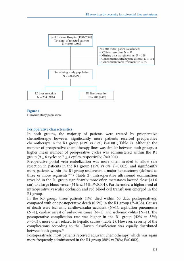
111
R1 resection by necessity for colorectal liver metastases
Paul Brousse Hospital (1990-2006) Total no. of resected patients
N = 840 (100%)
N = 404 (48%) patients excluded: • R2 liver resection: N = 57 • Missing data margin status: N = 128 • Concomitant extrahepatic disease: N = 134• Concomitant local treatment : N = 85
Remaining study population N = 436 (52%)
R0 liver resection N = 234 (28%)
R1 liver resection N = 202 (24%)
Figure 1. Flowchart study population.
Perioperative characteristicsIn both groups, the majority of patients were treated by preoperative chemotherapy; however, significantly more patients received preoperative chemotherapy in the R1 group (81% vs 67%; P=0.001; Table 2). Although the number of preoperative chemotherapy lines was similar between both groups, a higher mean number of preoperative cycles was administered within the R1 group (9 + 6 cycles vs 7 + 4 cycles, respectively; P=0.004). Preoperative portal vein embolization was more often needed to allow safe resection in patients in the R1 group (15% vs 6%; P=0.002), and significantly more patients within the R1 group underwent a major hepatectomy (defined as three or more segments33,34) (Table 2). Intraoperative ultrasound examination revealed in the R1 group significantly more often metastases located close (<1.0 cm) to a large blood vessel (51% vs 35%; P<0.001). Furthermore, a higher need of intraoperative vascular occlusion and red blood cell transfusion emerged in the R1 group. In the R0 group, three patients (1%) died within 60 days postoperatively, compared with one postoperative death (0.5%) in the R1 group (P=0.38). Causes of death were ischemic cardiovascular accident (N=1), aspiration pneumonia (N=1), cardiac arrest of unknown cause (N=1), and ischemic colitis (N=1). The postoperative complication rate was higher in the R1 group (42% vs 32%; P=0.03), more often related to hepatic causes (Table 2). However, severity of the complications according to the Clavien classification was equally distributed between both groups.35 Postoperatively, most patients received adjuvant chemotherapy, which was again more frequently administered in the R1 group (88% vs 78%; P=0.002).
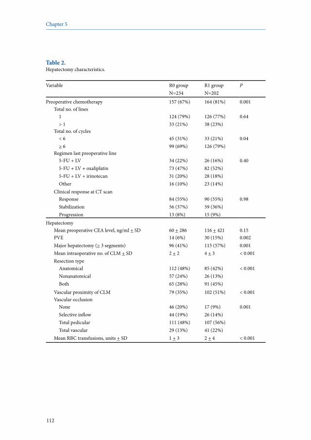
112
Chapter 5
Table 2. Hepatectomy characteristics.
Variable R0 groupN=234
R1 groupN=202
P
Preoperative chemotherapy 157 (67%) 164 (81%) 0.001Total no. of lines
1> 1
124 (79%)33 (21%)
126 (77%)38 (23%)
0.64
Total no. of cycles< 6> 6
45 (31%)99 (69%)
33 (21%)126 (79%)
0.04
Regimen last preoperative line5-FU + LV 5-FU + LV + oxaliplatin5-FU + LV + irinotecanOther
34 (22%)73 (47%)31 (20%)16 (10%)
26 (16%)82 (52%)28 (18%)23 (14%)
0.40
Clinical response at CT scanResponseStabilizationProgression
84 (55%)56 (37%)13 (8%)
90 (55%)59 (36%)15 (9%)
0.98
HepatectomyMean preoperative CEA level, ng/ml + SD 60 + 286 116 + 421 0.15PVE 14 (6%) 30 (15%) 0.002Major hepatectomy (> 3 segments) 96 (41%) 115 (57%) 0.001Mean intraoperative no. of CLM + SD 2 + 2 4 + 3 < 0.001Resection type
AnatomicalNonanatomicalBoth
112 (48%)57 (24%)65 (28%)
85 (42%)26 (13%)91 (45%)
< 0.001
Vascular proximity of CLM 79 (35%) 102 (51%) < 0.001Vascular occlusion
NoneSelective inflowTotal pedicularTotal vascular
46 (20%)44 (19%)111 (48%)29 (13%)
17 (9%)26 (14%)107 (56%)41 (22%)
0.001
Mean RBC transfusions, units + SD 1 + 3 2 + 4 < 0.001

113
R1 resection by necessity for colorectal liver metastases
Table 2. Continued.
Variable R0 groupN=234
R1 groupN=202
P
HistopathologyMean no. of CLM + SD 2 + 2 4 + 3 < 0.001Mean max. size of CLM + SD 34 + 27 54 + 41 < 0.001
Postoperative outcomePostoperative mortality (< 2 months) 3 (1%) 1 (0.5%) 0.38Postoperative complications 75 (32%) 85 (42%) 0.03
Hepatic35* 49 (21%) 70 (35%) 0.001Clavien grade I–IIClavien grade III–IVClavien grade V
36 (73%)13 (27%)0 (0%)
56 (80%)14 (20%)0 (0%)
0.40
General35# 46 (20%) 41 (20%) 0.87Clavien grade I–IIClavien grade III–IVClavien grade V
38 (83%)5 (11%)3 (6%)
29 (71%)11 (27%)1 (2%)
0.12
Postoperative chemotherapy 182 (78%) 178 (88%) 0.002
* as hepatic complications were considered: biliary leak/bilioma, hemorrhage, infected collection, noninfected collection, and transient liver insufficiency; # as general complications were considered: pulmonary, cardiovascular, urinary tract, infectious (other than local hepatic), and iatrogenic complications.Abbreviations: FU, fluorouracil; LV, leucovorin; CT, computed tomography; CEA, carcinoembryonic antigen; SD, standard deviation; PVE, portal vein embolization; CLM, colorectal liver metastases; RBC, red blood cell.
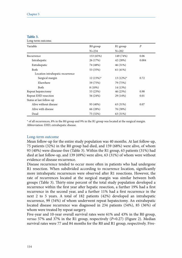
114
Chapter 5
Table 3. Long-term outcome.
Variable R0 groupN=234
R1 groupN=202
P
Recurrence 153 (65%) 149 (74%) 0.06IntrahepaticExtrahepaticBoth
26 (17%)74 (48%)53 (35%)
42 (28%)46 (31%)61 (41%)
0.004
Location intrahepatic recurrenceSurgical marginElsewhereBoth
12 (15%)*59 (75%)8 (10%)
13 (12%)*79 (75%)14 (13%)
0.72
Repeat hepatectomy 53 (23%) 46 (23%) 0.98Repeat EHD resection 56 (24%) 29 (14%) 0.01Status at last follow-up
Alive without diseaseAlive with diseaseDead
93 (40%)66 (28%)75 (32%)
63 (31%)76 (38%)63 (31%)
0.07
* of all recurrences, 8% in the R0 group and 9% in the R1 group was located at the surgical margin.Abbreviation: EHD, extrahepatic disease.
Long-term outcomeMean follow-up for the entire study population was 40 months. At last follow-up, 75 patients (32%) in the R0 group had died, and 159 (68%) were alive, of whom 93 (40%) were disease-free (Table 3). Within the R1 group, 63 patients (31%) had died at last follow-up, and 139 (69%) were alive, 63 (31%) of whom were without evidence of disease recurrence. Disease recurrence tended to occur more often in patients who had undergone R1 resection. When subdivided according to recurrence location, significantly more intrahepatic recurrences were observed after R1 resections. However, the rate of recurrences located at the surgical margin was similar between both groups (Table 3). Thirty-nine percent of the total study population developed a recurrence within the first year after hepatic resection, a further 19% had a first recurrence in the second year, and a further 11% had a first recurrence in the next 2 to 5 years. A total of 182 patients (42%) developed an intrahepatic recurrence, 99 (54%) of whom underwent repeat hepatectomy. An extrahepatic located disease recurrence was diagnosed in 234 patients (54%), 85 (36%) of whom were treated by repeat surgery.Five-year and 10-year overall survival rates were 61% and 43% in the R0 group, versus 57% and 37% in the R1 group, respectively (P=0.27) (Figure 2). Median survival rates were 77 and 84 months for the R0 and R1 group, respectively. Five-
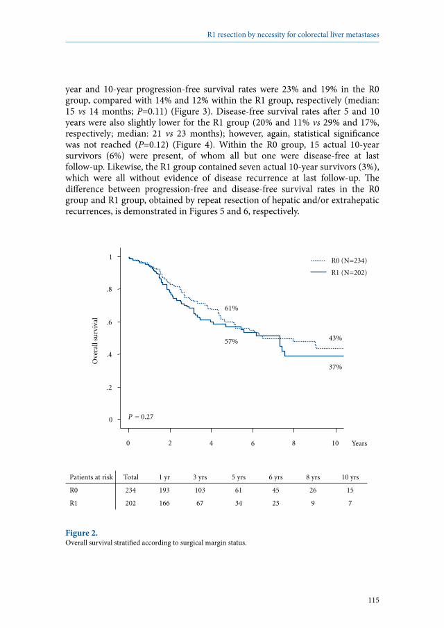
115
R1 resection by necessity for colorectal liver metastases
year and 10-year progression-free survival rates were 23% and 19% in the R0 group, compared with 14% and 12% within the R1 group, respectively (median: 15 vs 14 months; P=0.11) (Figure 3). Disease-free survival rates after 5 and 10 years were also slightly lower for the R1 group (20% and 11% vs 29% and 17%, respectively; median: 21 vs 23 months); however, again, statistical significance was not reached (P=0.12) (Figure 4). Within the R0 group, 15 actual 10-year survivors (6%) were present, of whom all but one were disease-free at last follow-up. Likewise, the R1 group contained seven actual 10-year survivors (3%), which were all without evidence of disease recurrence at last follow-up. The difference between progression-free and disease-free survival rates in the R0 group and R1 group, obtained by repeat resection of hepatic and/or extrahepatic recurrences, is demonstrated in Figures 5 and 6, respectively.
Patients at risk Total 1 yr 3 yrs 5 yrs 6 yrs 8 yrs 10 yrs
R0 234 193 103 61 45 26 15
R1 202 166 67 34 23 9 7
0
.2
.4
.6
.8
1
Ove
rall
surv
ival
Years
P = 0.27
43% 57%
37%
61%
R1 (N=202)
R0 (N=234)
0 2 4 6 8 10
Figure 2. Overall survival stratified according to surgical margin status.
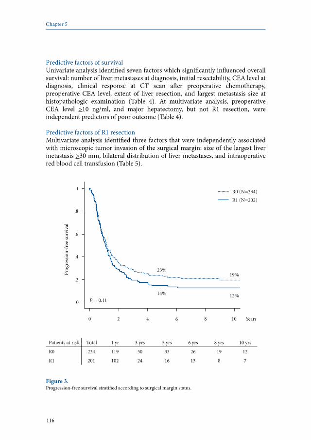
116
Chapter 5
Predictive factors of survivalUnivariate analysis identified seven factors which significantly influenced overall survival: number of liver metastases at diagnosis, initial resectability, CEA level at diagnosis, clinical response at CT scan after preoperative chemotherapy, preoperative CEA level, extent of liver resection, and largest metastasis size at histopathologic examination (Table 4). At multivariate analysis, preoperative CEA level >10 ng/ml, and major hepatectomy, but not R1 resection, were independent predictors of poor outcome (Table 4).
Predictive factors of R1 resectionMultivariate analysis identified three factors that were independently associated with microscopic tumor invasion of the surgical margin: size of the largest liver metastasis >30 mm, bilateral distribution of liver metastases, and intraoperative red blood cell transfusion (Table 5).
Patients at risk Total 1 yr 3 yrs 5 yrs 6 yrs 8 yrs 10 yrs
R0 234 119 50 33 26 19 12
R1 201 102 24 16 13 8 7
0
.2
.4
.6
.8
1
Pro
gres
sion
-fre
e su
rviv
al
0 2 4 Years
P = 0.11
19%
14% 12%
23%
6 8 10
R1 (N=202)
R0 (N=234)
Figure 3. Progression-free survival stratified according to surgical margin status.

117
R1 resection by necessity for colorectal liver metastases
Patients at risk Total 1 yr 3 yrs 5 yrs 6 yrs 8 yrs 10 yrs
R0 232 160 74 46 34 22 14
R1 201 131 47 24 17 9 7
0
.2
.4
.6
.8
1 D
isea
se-f
ree
surv
ival
Years
P = 0.12
17%
20%
11%
29%
R1 (N=202)
R0 (N=234)
0 2 4 6 8 10
Figure 4. Disease-free survival stratified according to surgical margin status.

118
Chapter 5
Patients at risk Total 1 yr 2 yrs 3 yrs 4 yrs 5 yrs
DFS 232 160 98 74 57 46
PFS 234 119 68 50 39 33
0
.2
.4
.6
.8
1 Su
rviv
al
0 1 2 3 4 5 Years
23% 28%
29%
39%
PFS
DFS
11% 6%
57%
76%
19%
Figure 5. Difference between progression-free (PFS) and disease-free survival (DFS) in the R0 group, demonstrating the influence of repeat surgery.
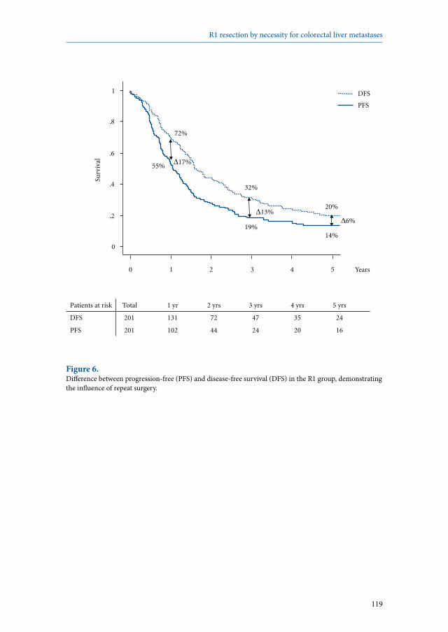
119
R1 resection by necessity for colorectal liver metastases
Patients at risk Total 1 yr 2 yrs 3 yrs 4 yrs 5 yrs
DFS 201 131 72 47 35 24
PFS 201 102 44 24 20 16
0
.2
.4
.6
.8
1 Su
rviv
al
0 1 2 3 4 5 Years
14% 19%
20%
32%
PFS
DFS
13% 6%
17% 55%
72%
Figure 6. Difference between progression-free (PFS) and disease-free survival (DFS) in the R1 group, demonstrating the influence of repeat surgery.
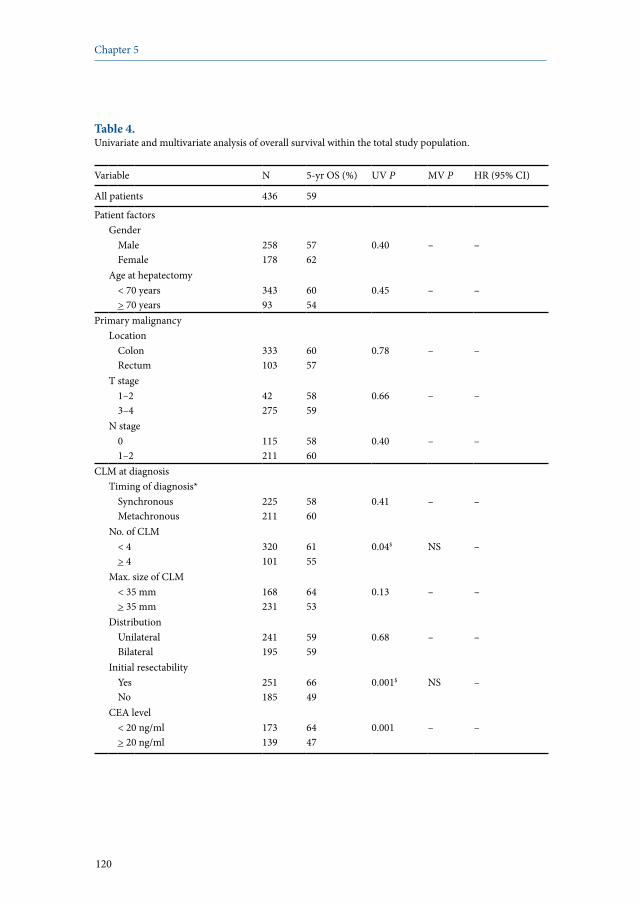
120
Chapter 5
Table 4. Univariate and multivariate analysis of overall survival within the total study population.
Variable N 5-yr OS (%) UV P MV P HR (95% CI)
All patients 436 59
Patient factorsGender
MaleFemale
258178
5762
0.40 – –
Age at hepatectomy< 70 years > 70 years
34393
6054
0.45 – –
Primary malignancyLocation
ColonRectum
333103
6057
0.78 – –
T stage 1–23–4
42275
5859
0.66 – –
N stage 01–2
115211
5860
0.40 – –
CLM at diagnosisTiming of diagnosis*
SynchronousMetachronous
225211
5860
0.41 – –
No. of CLM< 4> 4
320101
6155
0.04$ NS –
Max. size of CLM< 35 mm> 35 mm
168231
6453
0.13 – –
DistributionUnilateralBilateral
241195
5959
0.68 – –
Initial resectabilityYesNo
251185
6649
0.001$ NS –
CEA level< 20 ng/ml> 20 ng/ml
173139
6447
0.001 – –
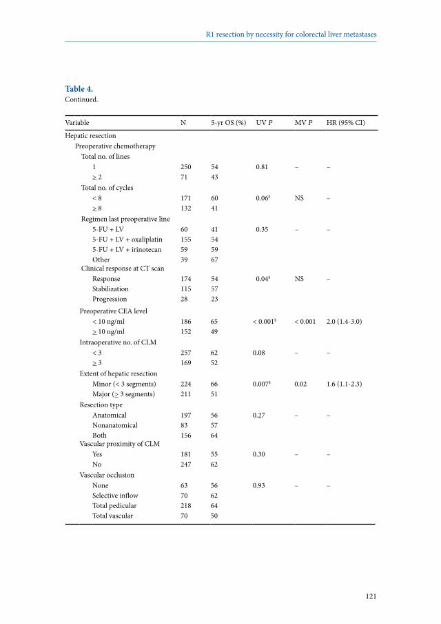
121
R1 resection by necessity for colorectal liver metastases
Table 4. Continued.
Variable N 5-yr OS (%) UV P MV P HR (95% CI)
Hepatic resectionPreoperative chemotherapy
Total no. of lines1> 2
25071
5443
0.81 – –
Total no. of cycles< 8> 8
171132
6041
0.06$ NS –
Regimen last preoperative line5-FU + LV5-FU + LV + oxaliplatin 5-FU + LV + irinotecanOther
601555939
41545967
0.35 – –
Clinical response at CT scanResponseStabilizationProgression
17411528
545723
0.04$ NS –
Preoperative CEA level< 10 ng/ml> 10 ng/ml
186152
6549
< 0.001$ < 0.001 2.0 (1.4-3.0)
Intraoperative no. of CLM< 3> 3
257169
6252
0.08 – –
Extent of hepatic resectionMinor (< 3 segments)Major (> 3 segments)
224211
6651
0.007$ 0.02 1.6 (1.1-2.3)
Resection typeAnatomicalNonanatomicalBoth
19783156
565764
0.27 – –
Vascular proximity of CLMYesNo
181247
5562
0.30 – –
Vascular occlusionNoneSelective inflowTotal pedicularTotal vascular
637021870
56626450
0.93 – –
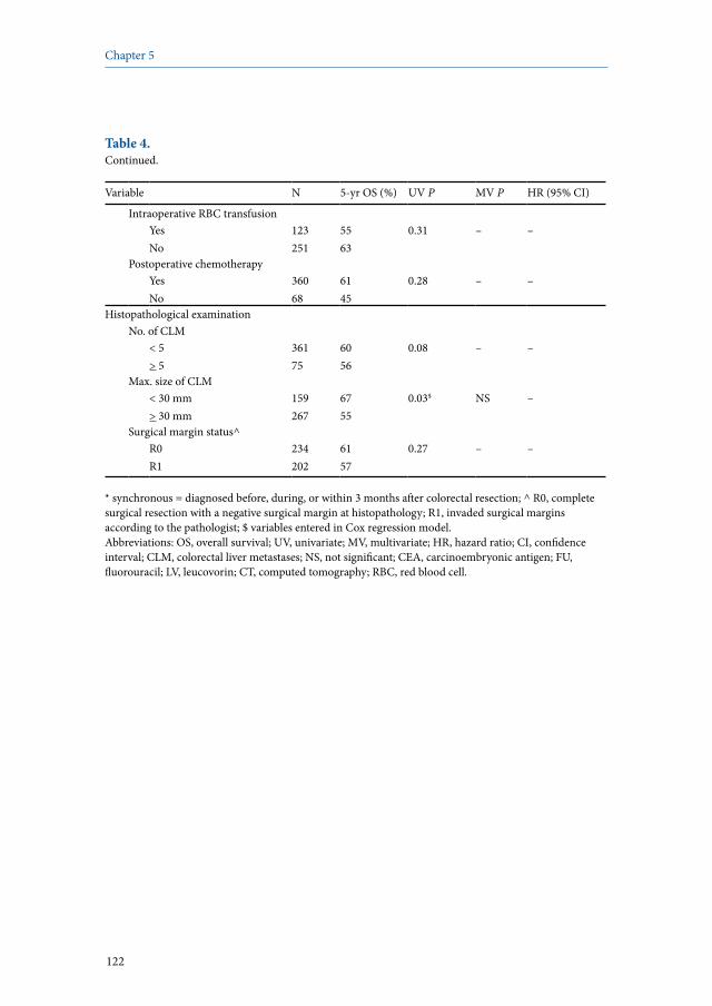
122
Chapter 5
Table 4. Continued.
Variable N 5-yr OS (%) UV P MV P HR (95% CI)
Intraoperative RBC transfusionYesNo
123251
5563
0.31 – –
Postoperative chemotherapyYesNo
36068
6145
0.28 – –
Histopathological examinationNo. of CLM
< 5> 5
36175
6056
0.08 – –
Max. size of CLM< 30 mm> 30 mm
159267
6755
0.03$ NS –
Surgical margin status^R0R1
234202
6157
0.27 – –
* synchronous = diagnosed before, during, or within 3 months after colorectal resection; ^ R0, complete surgical resection with a negative surgical margin at histopathology; R1, invaded surgical margins according to the pathologist; $ variables entered in Cox regression model.Abbreviations: OS, overall survival; UV, univariate; MV, multivariate; HR, hazard ratio; CI, confidence interval; CLM, colorectal liver metastases; NS, not significant; CEA, carcinoembryonic antigen; FU, fluorouracil; LV, leucovorin; CT, computed tomography; RBC, red blood cell.

123
R1 resection by necessity for colorectal liver metastases
Table 5. Univariate and multivariate analysis of predictive factors of R1 resection.
Variable R0 group N=234 R1 group N=202 UV P MV P RR (95% CI)
CLM at diagnosisTiming of diagnosis*
SynchronousMetachronous
107 (46%) 127 (54%)
118 (58%) 84 (42%)
0.008 NS –
No. of CLM1> 2
116 (50%)114 (50%)
61 (32%)130 (68%)
< 0.001 NS –
Max. size of CLM< 30 mm> 30 mm
92 (43%)122 (57%)
30 (16%)155 (84%)
< 0.001 < 0.001 3.6 (2.1-6.3)
DistributionUnilateralBilateral
157 (67%) 77 (33%)
84 (42%)118 (58%)
< 0.001 < 0.001 2.8 (1.7-4.5)
Initial resectabilityYesNo
162 (69%)72 (31%)
89 (44%)113 (56%)
< 0.001 NS –
CEA level< 20 ng/ml> 20 ng/ml
102 (63%)61 (37%)
71 (48%)78 (52%)
0.008 NS –
Hepatic resectionPreoperative chemotherapy
YesNo
157 (67%)77 (33%)
164 (81%)38 (19%)
0.001 NS –
Extent of hepatic resectionMinor (< 3 segments)Major (> 3 segments)
138 (59%)96 (41%)
87 (43%)115 (57%)
0.001 NS –
Resection typeAnatomicalNonanatomical
112 (66%)57 (34%)
85 (77%)26 (23%)
0.07 NS –
Vascular proximity of CLMYesNo
79 (35%)150 (66%)
102 (51%)97 (49%)
< 0.001 NS –
Vascular occlusionYesNo
184 (80%)46 (20%)
174 (91%)17 (9%)
0.001 NS –
Intraoperative RBC transfusionYesNo
45 (21%)165 (79%)
78 (48%)86 (52%)
< 0.001 0.001 2.4 (1.5-4.0)
* synchronous = diagnosed before, during, or within 3 months after colorectal resection.Abbreviations: UV, univariate; MV, multivariate; RR, risk ratio; CI, confidence interval; CLM, colorectal liver metastases; NS, not significant; CEA, carcinoembryonic antigen; RBC, red blood cell.

124
Chapter 5
Discussion
Within the current study, we investigated the influence of surgical margin status (ie, microscopically involved vs clear margins) on long-term outcome in patients with CLM treated by an aggressive approach consisting of chemotherapy and repeat surgery. Five-year overall survival rate was 57% in the R1 group, not significantly different from 61% after R0 resection. Five-year progression-free and disease-free survival rates were slightly better for R0 resections, but did not significantly differ between both groups (23% and 29% after R0 resection vs 14% and 20% after R1 resection, respectively; P=0.11 [PFS] and P=0.12 [DFS]). Although more intrahepatic recurrences were observed in the R1 group, the rate of surgical margin recurrences was comparable between both groups. Maximum diameter of the largest liver metastasis >30 mm, bilateral distribution of liver metastases, and intraoperative red blood cell transfusion independently predicted microscopic involvement of the surgical margins. Overall, a preoperative CEA level >10 ng/ml and a major hepatectomy were identified as independent predictors of poor survival.Microscopic involvement of surgical resection margins emerged in many publications as a significant poor prognostic factor.12-20 Therefore, the inability to obtain clear surgical margins has been considered a contraindication to liver surgery for CLM. However, the optimal negative surgical margin width correlated with best long-term outcome is still unclear. Within the literature, several cutoff points have been proposed, varying between 2 and 10 mm.22-28 Important data concerning the optimal surgical margin width has been provided by Kokudo et al.25 By using a sensitive genetic analysis for detecting K-ras and p53 mutations, micrometastases in the liver parenchyma surrounding CLM were found in only 2% of patients, all within 4 mm of the tumor border.25 In another recent publication, the pattern of chemotherapy response of CLM was evaluated, to define the optimal surgical approach after chemotherapy for liver metastases.26 The response of CLM after chemotherapy was found to consist of tumor shrinkage in a concentric fashion and of random tumor cell death throughout the metastasis.26 Notably, nests of viable tumor cells were never found further than 4 mm from the peripheral edge of the main tumor.26 However, based on the currently available data, complete macroscopic resection with negative surgical margins is the gold standard recommendation in the surgical treatment of CLM. We have questioned for long the inability to achieve microscopically clear surgical margins as an absolute contraindication for surgery in patients with CLM for different reasons. First, the ultrasonic dissector, which is routinely used in hepatic resections, aspires a small rim of hepatic tissue, thereby possibly decreasing the resection margin for 1 to 2 mm. This potential overestimation of the proportion of R1 resections has also been recognized by Pawlik et al.21 Ultrasonic dissectors are

125
R1 resection by necessity for colorectal liver metastases
nowadays used by most centers, and therefore our findings represent current daily practice. Second, the remnant cut section of the liver at the contact of the removed tumor is treated as much as possible with argon beam or bipolar coagulation, which ‘sterilizes’ an extra 1 to 2 mm of hepatic tissue. Although none of the publications in which microscopic surgical margin involvement emerged as a poor prognostic factor described the use of these additional techniques, surgical teams have this possibility.12-20 A third reason is that more effective chemotherapy regimens could reduce the proportion of metastases that develop satellite nodules, thus minimizing residual micrometastatic disease. Within our population, 74% of patients received preoperative chemotherapy, and in 83% of patients surgery was followed by adjuvant chemotherapy. This contrasts with the existing literature, reporting only one third of patients receiving preoperative chemotherapy.19,22 Finally, even in case of local recurrence, repeat hepatectomy may offer a chance of long-term survival,32 whereas chemotherapy alone, if we contraindicate surgery, is merely palliative.5 The value of repeat (extra)hepatic surgery in our population is clearly demonstrated by the difference between progression-free and disease-free survival (Figures 5 and 6). Accordingly, the results of our study demonstrate that not considering the inability to obtain microscopically clear surgical margins as an absolute contraindication for surgery has been a reasonable treatment strategy. Importantly, by excluding patients who underwent R2 resection, or with concomitant local ablative techniques, and those presenting with concomitant extrahepatic disease, the real impact of surgical margin status on survival could be identified. In the recent multicenter study reported by Pawlik et al,21 a positive surgical margin was significantly associated with surgical margin recurrence. Similarly, Hamady et al observed a higher recurrence rate located at the surgical margin in patients with positive resection margins.16 Likewise, Choti et al identified microscopically positive resection margins as an independent factor predicting worse disease-free survival; however, recurrences were not stratified according to their location.12 In addition, Kokudo et al reported an increase in resection margin recurrences in patients with surgical margins narrower than 2 mm.25 In the present study, although more intrahepatic recurrences were observed in patients with positive resection margins, the rate of surgical margin recurrences and intrahepatic recurrences away from the surgical margin were equally balanced between the R0 group and the R1 group. This is in agreement with a recent publication by Bodingbauer et al, in which it was demonstrated that surgical margin status was not associated with an increased hepatic recurrence risk, either at the surgical margin or elsewhere.36 Figueras et al also recently reported that subcentimeter non-positive surgical margins did not influence
126
Chapter 5
hepatic recurrence rates after hepatectomy for CLM.37 That positive surgical margins expose to a higher recurrence risk is obvious, but this should not be the only criterion considered. By stating R1 resection as a contraindication to surgery, what should be the alternative? Even with the increasingly efficient chemotherapy regimens, median survivals reach currently 20 to 24 months.38,39 By combining liver resection (even R1), we quadruple the survival expectancy, since median survival was in our study 84 months after R1 resection. Therefore, no doubt is allowed, provided that the resection is complete. Our multivariate analysis demonstrated that an increased preoperative CEA level and the performance of a major hepatectomy independently predicted poor outcome. In several other publications, an increased preoperative CEA level also emerged as a predictor of worse long-term outcome.12,14,27 By contrast, major hepatectomy by itself is not a widely reported poor prognostic factor; however, it reflects the number, size, and distribution of CLM, which are well-known risk factors for adverse long-term outcome.14,40,41
Not surprisingly, size and distribution of CLM were identified as risk factors for the inability to obtain microscopically tumor-free surgical margins, which reflect tumor load and thereby complexity of achieving a radical clearance. Are et al and Pawlik et al, both, recently reported similar predictors of surgical margin involvement.21,22 Interestingly, in the present study we identified the need for red blood cell transfusion as a significant risk factor for R1 resection. A possible explanation relies on the fact that the need for an intraoperative red blood cell transfusion reflects a more complex operation with higher blood loss, in which it is technically more difficult to obtain free surgical margins. In conclusion, despite a higher intrahepatic recurrence rate, overall, progression-free and disease-free survival rates were almost similar for patients with R0 and R1 resections for CLM, treated by an aggressive strategy consisting of chemotherapy and surgery. As chemotherapy regimens are nowadays increasingly effective, the inability to obtain microscopically free surgical margins as a contraindication to hepatic resection for CLM, provided macroscopic complete removal, should be revisited. As a result, long-term outcome after R1 resections should no longer be considered as poor as that observed after R2 resections, but closer to that observed after R0 resections. The absolute contraindication to hepatic resection for CLM should therefore not include R1 resection anymore.

127
R1 resection by necessity for colorectal liver metastases
References
1. Simmonds PC, Primrose JN, Colquitt JL et al. Surgical resection of hepatic metastases from colorectal cancer: a systematic review of published studies. Br J Cancer 2006; 94:982-999.
2. Adam R. Chemotherapy and surgery: new perspectives on the treatment of unresectable liver metastases. Ann Oncol 2003; 14 Suppl 2:ii13-ii16.
3. Alberts SR, Horvath WL, Sternfeld WC et al. Oxaliplatin, fluorouracil, and leucovorin for patients with unresectable liver-only metastases from colorectal cancer: a North Central Cancer Treatment Group phase II study. J Clin Oncol 2005; 23:9243-9249.
4. de la Camara J, Rodriguez J, Rotellar F et al. Triplet therapy with oxaliplatin, irinotecan, 5-fluorouracil and folinic acid within a combined modality approach in patients with liver metastases from colorectal cancer. Proc Am Soc Clin Oncol 2004; 23:3593.
5. Giacchetti S, Itzhaki M, Gruia G et al. Long-term survival of patients with unresectable colorectal cancer liver metastases following infusional chemotherapy with 5-fluorouracil, leucovorin, oxaliplatin and surgery. Ann Oncol 1999; 10:663-669.
6. Pozzo C, Basso M, Cassano A et al. Neoadjuvant treatment of unresectable liver disease with irinotecan and 5-fluorouracil plus folinic acid in colorectal cancer patients. Ann Oncol 2004; 15:933-939.
7. Adam R, Aloia T, Lévi F et al. Hepatic resection after rescue cetuximab treatment for colorectal liver metastases previously refractory to conventional systemic therapy. J Clin Oncol 2007; 25:4593-4602.
8. Cunningham D, Humblet Y, Siena S et al. Cetuximab monotherapy and cetuximab plus irinotecan in irinotecan-refractory metastatic colorectal cancer. N Engl J Med 2004; 351:337-345.
9. Hurwitz H, Fehrenbacher L, Novotny W et al. Bevacizumab plus irinotecan, fluorouracil, and leucovorin for metastatic colorectal cancer. N Engl J Med 2004; 350:2335-2342.
10. Azoulay D, Castaing D, Smail A et al. Resection of nonresectable liver metastases from colorectal cancer after percutaneous portal vein embolization. Ann Surg 2000; 231:480-486.
11. Azoulay D, Andreani P, Maggi U et al. Combined liver resection and reconstruction of the supra-renal vena cava: the Paul Brousse experience. Ann Surg 2006; 244:80-88.
12. Choti MA, Sitzmann JV, Tiburi MF et al. Trends in long-term survival following liver resection for hepatic colorectal metastases. Ann Surg 2002; 235:759-766.
13. Elias D, Cavalcanti A, Sabourin JC et al. Resection of liver metastases from colorectal cancer: the real impact of the surgical margin. Eur J Surg Oncol 1998; 24:174-179.
14. Fong Y, Fortner J, Sun RL et al. Clinical score for predicting recurrence after hepatic resection for metastatic colorectal cancer: analysis of 1001 consecutive cases. Ann Surg 1999; 230:309-318.
15. Gayowski TJ, Iwatsuki S, Madariaga JR et al. Experience in hepatic resection for metastatic colorectal cancer: analysis of clinical and pathologic risk factors. Surgery 1994; 116:703-710.
16. Hamady ZZ, Cameron IC, Wyatt J et al. Resection margin in patients undergoing hepatectomy for colorectal liver metastasis: a critical appraisal of the 1cm rule. Eur J Surg Oncol 2006; 32:557-563.
17. Jamison RL, Donohue JH, Nagorney DM et al. Hepatic resection for metastatic colorectal cancer results in cure for some patients. Arch Surg 1997; 132:505-510.
18. Shirabe K, Takenaka K, Gion T et al. Analysis of prognostic risk factors in hepatic resection for metastatic colorectal carcinoma with special reference to the surgical margin. Br J Surg 1997; 84:1077-1080.
19. Welsh FK, Tekkis PP, O’Rourke T et al. Quantification of risk of a positive (R1) resection margin following hepatic resection for metastatic colorectal cancer: an aid to clinical decision-making. Surg Oncol 2008; 17:3-13.

128
Chapter 5
20. Yamamoto J, Shimada K, Kosuge T et al. Factors influencing survival of patients undergoing hepatectomy for colorectal metastases. Br J Surg 1999; 86:332-337.
21. Pawlik TM, Scoggins CR, Zorzi D et al. Effect of surgical margin status on survival and site of recurrence after hepatic resection for colorectal metastases. Ann Surg 2005; 241:715-722.
22. Are C, Gonen M, Zazzali K et al. The impact of margins on outcome after hepatic resection for colorectal metastasis. Ann Surg 2007; 246:295-300.
23. Cady B, Jenkins RL, Steele GD Jr et al. Surgical margin in hepatic resection for colorectal metastasis: a critical and improvable determinant of outcome. Ann Surg 1998; 227:566-571.
24. Ekberg H, Tranberg KG, Andersson R et al. Determinants of survival in liver resection for colorectal secondaries. Br J Surg 1986; 73:727-731.
25. Kokudo N, Miki Y, Sugai S et al. Genetic and histological assessment of surgical margins in resected liver metastases from colorectal carcinoma: minimum surgical margins for successful resection. Arch Surg 2002; 137:833-840.
26. Ng JK, Urbanski SJ, Mangat N et al. Colorectal liver metastases contract centripetally with a response to chemotherapy: a histomorphologic study. Cancer 2008; 112:362-371.
27. Nordlinger B, Guiguet M, Vaillant JC et al. Surgical resection of colorectal carcinoma metastases to the liver. A prognostic scoring system to improve case selection, based on 1568 patients. Association Française de Chirurgie. Cancer 1996; 77:1254-1262.
28. Rees M, Plant G, Bygrave S. Late results justify resection for multiple hepatic metastases from colorectal cancer. Br J Surg 1997; 84:1136-1140.
29. World Health Organization. Handbook for reporting results of cancer treatment. Geneva: World Health Organization; 1979.
30. Therasse P, Arbuck SG, Eisenhauer EA et al. New guidelines to evaluate the response to treatment in solid tumors. European Organization for Research and Treatment of Cancer, National Cancer Institute of the United States, National Cancer Institute of Canada. J Natl Cancer Inst 2000; 92:205-216.
31. Sobin LH, Wittekind Ch. TNM classification of malignant tumours. Hoboken: John Wiley & Sons; 2002.
32. Adam R, Bismuth H, Castaing D et al. Repeat hepatectomy for colorectal liver metastases. Ann Surg 1997; 225:51-60.
33. Bismuth H. Surgical anatomy and anatomical surgery of the liver. World J Surg 1982; 6:3-9.34. Couinaud C. Le foie: études anatomiques et chirurgicales. Paris: Masson et Cie; 1957.35. Dindo D, Demartines N, Clavien PA. Classification of surgical complications: a new proposal
with evaluation in a cohort of 6336 patients and results of a survey. Ann Surg 2004; 240:205-213.
36. Bodingbauer M, Tamandl D, Schmid K et al. Size of surgical margin does not influence recurrence rates after curative liver resection for colorectal cancer liver metastases. Br J Surg 2007; 94:1133-1138.
37. Figueras J, Burdio F, Ramos E et al. Effect of subcentimeter nonpositive resection margin on hepatic recurrence in patients undergoing hepatectomy for colorectal liver metastases. Evidences from 663 liver resections. Ann Oncol 2007; 18:1190-1195.
38. Falcone A, Ricci S, Brunetti I et al. Phase III trial of infusional fluorouracil, leucovorin, oxaliplatin, and irinotecan (FOLFOXIRI) compared with infusional fluorouracil, leucovorin, and irinotecan (FOLFIRI) as first-line treatment for metastatic colorectal cancer: the Gruppo Oncologico Nord Ovest. J Clin Oncol 2007; 25:1670-1676.
39. Tournigand C, André T, Achille E et al. FOLFIRI followed by FOLFOX6 or the reverse sequence in advanced colorectal cancer: a randomized GERCOR study. J Clin Oncol 2004; 22:229-237.
40. Minagawa M, Makuuchi M, Torzilli G et al. Extension of the frontiers of surgical indications in the treatment of liver metastases from colorectal cancer: long-term results. Ann Surg 2000; 231:487-499.
41. Scheele J, Stangl R, Altendorf-Hofmann A et al. Resection of colorectal liver metastases. World J Surg 1995; 19:59-71.

129
R1 resection by necessity for colorectal liver metastases

Part IIExtrahepatic disease in patients with colorectal liver metastases

Robbert J. de Haas1,2
Dennis A. Wicherts1,2
René Adam1
1 Centre Hépato-Biliaire, Hôpital Paul Brousse, Villejuif, France2 Department of Surgery, University Medical Center Utrecht, Utrecht,
The Netherlands
Digestive Surgery 2008; 25:461-466
Resection of colorectal liver metastases with extrahepatic disease 6
134
Chapter 6
Abstract
Extrahepatic disease in combination with colorectal liver metastases has long been considered an absolute contraindication for surgery. However, in many reported series, long-term survival is achieved in selected patients with concomitant extrahepatic disease as long as the resection of all metastatic sites is complete. Owing to these results, an increasing number of patients with advanced metastatic colorectal disease are now being referred for surgery.For patients with concomitant liver and lung metastases, sequential resection of both disease sites has proven to be safe, offering a 5-year survival rate of more than 30%. On the contrary, hepatectomy combined with resection of regional lymph node metastases can only provide long-term survival in case of pedicular lymph node involvement. Furthermore, control of the disease by preoperative chemotherapy appears to be crucial. For peritoneal carcinomatosis, aggressive treatment combining cytoreductive surgery and intraperitoneal chemotherapy can offer a chance of prolonged long-term survival to selected patients with limited peritoneal extension.In conclusion, resection of both intra- and extrahepatic colorectal metastases should be considered if resection of all metastatic sites can be complete and the disease is controlled by chemotherapy. Long-term survival in these patients with advanced disease could be achieved if they are managed by experienced multidisciplinary teams.
135
Resection of colorectal liver metastases with extrahepatic disease
Introduction
Complete resection of colorectal liver metastases (CLM) offers a chance of long-term survival to patients having otherwise a very poor prognosis when no resection is performed. Five-year survival rates after liver resection now exceed 50% in selected cases and approximately 20% of patients will survive more than 10 years.1 Owing to more developed multidisciplinary approaches, including systemic perioperative chemotherapy and advanced surgical techniques, an increasing number of patients with advanced metastatic disease are currently offered surgery.2
The presence of extrahepatic disease in combination with liver metastases has long been considered an absolute contraindication for hepatectomy. Although extrahepatic disease is predictive of poor outcome in many reported series, complete resection of all intra- and extrahepatic disease results in a significant proportion of long-term survivors in this patient group.3,4 The technical inability to completely resect all metastatic deposits nowadays remains the only cause of unresectability. Additionally, improved preoperative imaging techniques including MRI and PET-CT have increased the incidence of preoperatively detected extrahepatic disease. For these reasons, hepatobiliary surgeons are increasingly confronted with patients with both CLM as well as extrahepatic disease, discovered either before or during surgery. The aim of this article was to provide an overview of the currently available evidence concerning the indications and contraindications of hepatic resection in patients with CLM and concomitant extrahepatic disease.
Diagnosis of extrahepatic disease
Extrahepatic disease in patients with CLM is usually diagnosed preoperatively by CT imaging of the thorax, abdomen and pelvis, which is performed routinely when liver metastases are detected and an individual treatment strategy needs to be defined. In cases where the presence of extrahepatic disease is doubtful, PET-CT or MRI can be useful to differentiate between a benign and malignant nature. As part of the preoperative workup of patients scheduled for partial liver resection, colonoscopy is usually performed to exclude local colorectal recurrence. The additional value of diagnostic laparoscopy currently remains limited, owing to the relatively high accuracy of preoperatively available imaging modalities.Lung metastases constitute the most frequent site of extrahepatic disease in patients resected for CLM. Of all 791 patients that underwent hepatic resection for CLM at our institute between 1990 and 2005, 186 (24%) had concomitant extrahepatic disease. Of these, the majority (51%) presented with lung metastases

136
Chapter 6
[unpublished data]. Other frequent extrahepatic disease localizations included regional lymph node metastases (26%) and peritoneal metastases (12%).
Lung metastases
Following the liver (33%), the lungs are the second most frequent site of colorectal metastases (22%).5 In addition, approximately 9% of patients who undergo curative resection of the primary colorectal tumor develop lung metastases.6 Surgical resection has increasingly become an acceptable treatment for lung metastases from colorectal cancer and, in selected patients, 5-year survival rates up to 45% can be achieved.7 Owing to these favorable results, indications for surgery have increased, and resection is now not only proposed to patients with isolated lung metastases, but also to selected patients with both hepatic and pulmonary metastatic disease. Within the literature, resection of both liver and lung metastases is proven to be safe with no postoperative mortality and acceptable postoperative morbidity rates ranging between 5% and 13% (Table 1).8-12 Reported 5-year survival rates vary between 9% and 49%.8-16 This wide range of reported 5-year survival rates might partly be explained by differences between series in chosen start date when calculating survival times. Nevertheless, all but two series reported 5-year survival rates exceeding 30%, thereby justifying surgical resection in this particular patient group.9-13,15,16 Furthermore, when patients curatively resected for both metastatic sites are compared to patients in whom one or both metastatic sites remained unresected, observed survival rates were significantly higher for resected patients.12,14
To select the best surgical candidates, prognostic factors were identified in most studies, of which the following factors predicted poor long-term outcome: the presence of multiple lung metastases,13,15,16 appearance of metastases synchronous with the primary colorectal tumor,9 increased preoperative CEA level,8,11 short time interval between diagnosis of the first and the second metastatic site,8,16 bilateral distribution of lung metastases,9 presence of thoracic lymph node metastases,11 and older age (Table 1).16 In two series, several patients were described who underwent repeat lung resection for disease recurrence. Long-term outcome in these patients was not significantly decreased compared to that observed after a first lung resection.8,12

137
Resection of colorectal liver metastases with extrahepatic disease
Table 1. Literature overview of patients resected for both hepatic and pulmonary metastases.
Author Year No. of patients
Postoperative mortality (%)
Postoperative morbidity (%)
5-year OS (%)
Prognostic factors
Okumura13 1996 39 – – 33^ Multiple (>1) lung metastases
Regnard8 1998 43 0* 5* 11^ Preoperative CEA level >5 ng/mlNo. of pulmonary resectionsTime interval HR and PR <36 mo
Murata9 1998 30 0* 10* 43.8§ Synchronous metastasesBilateral pulmonary metastases
Lehnert10 1999 17 0* 12* 39# –
Robinson14 1999 25 – – 9§ –
Kobayashi15 1999 47 – – 31§ Multiple (>1) lung metastases
Headrick11 2001 58 0* 12* 30^ Thoracic lymph node metastasesPreoperative CEA level >5 ng/ml
Shah12 2006 39 0* 13* 74# –
Miller16 2007 131 – – 49# and 31§
Multiple (>1) liver metastasesTime interval 1st to 2nd met <1 yrAge >55 years
Paul Brousse Hospital$
– 88 0* 34* 37^ Synchronous liver metastasesMultiple (>1) lung metastases
* concerning both hepatic and pulmonary resection; ^ following the first pulmonary resection; # following removal of the first metastatic site; § following last organ resection; $ unpublished data.Abbreviations: OS, overall survival; CEA, carcinoembryonic antigen; HR, hepatic resection; PR, pulmonary resection; mo, months; met, metastases; yr, year.
138
Chapter 6
Usually, lung metastases are resected through a posterolateral thoracotomy or a median sternotomy, depending on the anatomic characteristics of the lesion(s) and the preference of the surgeon. If not all pulmonary lesions can be removed in one session, a staged procedure can be performed in selected patients, consisting of two sequential lung resections with chemotherapy in the meantime to prevent disease progression. More recently, video-assisted thoracic surgery (VATS) has been introduced to perform wedge resections for peripheral lung metastases. Although this less-invasive surgery is related to decreased postoperative pain and less functional morbidity, some small palpable lesions might be missed, thereby risking an incomplete resection.17 Importantly, as no regeneration occurs following pulmonary resection, the amount of removed normal lung parenchyma should be limited, to allow for possible repeat resections. At the Paul Brousse Hospital, between January 1984 and December 2000, 88 patients underwent surgical resection of both hepatic and pulmonary colorectal metastases [unpublished data]. Surgical resection was only performed if it could be curative for both metastatic sites. Furthermore, in all patients, liver resection preceded lung resection, and chemotherapy was routinely administered before and after each resection. Sixty patients (68%) presented with less than four liver metastases, and in 38 patients (43%) a major hepatectomy (three or more segments) was performed. More than half of the patients (52%) had only a single lung metastasis, while in 31 patients (35%) lung metastases were bilaterally distributed. Repeat hepatectomy for liver recurrences was performed in 19 patients (22%), and 42 patients (48%) underwent a repeat lung resection for disease recurrence. Following pulmonary resection, a 5-year survival rate of 37% was observed (Figure 1). None of the patients died within 60 days following both surgical interventions, and postoperative morbidity rate was 34%. As independent predictors of poor survival were identified: synchronous CLM, and the presence of more than one lung metastasis.Taken together, resection of hepatic and pulmonary metastases from colorectal cancer is safe and can offer long-term survival to selected patients. Surgery should only be considered if resection of all metastatic sites can be potentially curative. Furthermore, resections should be as limited as possible to allow repeat resections for eventual disease recurrence.
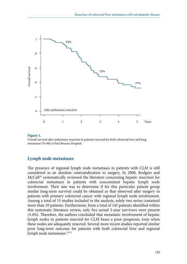
139
Resection of colorectal liver metastases with extrahepatic disease
Figure 1. Overall survival after pulmonary resection in patients resected for both colorectal liver and lung metastases (N=88) at Paul Brousse Hospital.
Lymph node metastases
The presence of regional lymph node metastases in patients with CLM is still considered as an absolute contraindication to surgery. In 2000, Rodgers and McCall18 systematically reviewed the literature concerning hepatic resection for colorectal metastases in patients with concomitant hepatic lymph node involvement. Their aim was to determine if for this particular patient group similar long-term survival could be obtained as that observed after surgery in patients with primary colorectal cancer with regional lymph node involvement. Among a total of 15 studies included in the analysis, solely two series contained more than 10 patients. Furthermore, from a total of 145 patients identified within this systematic literature review, only five actual 5-year survivors were present (3.4%). Therefore, the authors concluded that metastatic involvement of hepatic lymph nodes in patients resected for CLM bears a poor prognosis, even when these nodes are adequately resected. Several more recent studies reported similar poor long-term outcome for patients with both colorectal liver and regional lymph node metastases.19-21
0
.2
.4
.6
.8
1
Ove
rall
surv
ival
0 1 2 3 4 5 Years
A�er pulmonary resection
37%
52%
94%

140
Chapter 6
At our institute, the presence of regional lymph node metastases in patients with CLM has never been considered an absolute contraindication for surgery, provided that response or stabilization during preoperative chemotherapy is observed. To assess the validity of this oncosurgical treatment policy, we recently evaluated our experience.22 Between June 1992 and July 2006, 763 patients underwent liver resection for colorectal metastases, and of these, 47 (6%) underwent simultaneous resection of histologically confirmed regional lymph node involvement. When stratified according to lymph node location, 26 patients (3%) presented with pedicular (distal to the gastroduodenal artery branch), 14 patients (2%) with celiac (overlying the common hepatic artery proximal to the gastroduodenal artery branch), and seven patients (1%) with para-aortic regional lymph node involvement. In 19 patients (40%), lymph node involvement was diagnosed before hepatectomy, and in the remaining 28 patients (60%) lymph node metastases were identified intraoperatively. All patients that were preoperatively diagnosed with regional lymph node involvement responded to or were stabilized by preoperative chemotherapy. When regional lymph node involvement was diagnosed intraoperatively, hepatectomy combined with lymphadenectomy was only performed if all metastatic disease could be resected completely. By combining hepatectomy with lymphadenectomy in patients treated preoperatively with chemotherapy, a 5-year survival rate of 18% could be achieved without any operative mortality. Most favorable outcomes were observed in patients with pedicular lymph node involvement (5-year survival rate 25% vs 0% for patients with celiac and/or para-aortic lymph node metastases), and in patients younger than 40 years (5-year survival rate 45% vs 10% for older patients). In relation to our results and those reported by others, we recommend to combine hepatectomy with lymphadenectomy only for young CLM patients presenting with pedicular lymph node involvement, in the absence of disease progression after preoperative chemotherapy. On the other hand, patients presenting with celiac or para-aortic lymph node involvement should not be subjected to this oncosurgical treatment strategy.
Peritoneal carcinomatosis
As the presence of peritoneal carcinomatosis represents a situation of advanced metastatic disease, it usually is considered a contraindication for hepatic resection of colorectal metastases. With systemic chemotherapy treatment alone, no patients survive more than 5 years, and median survival is limited to 6 months in most cases.23 However, the development of cytoreductive surgery and intraperitoneal chemotherapy has offered a chance of prolonged long-term survival in a proportion of patients with peritoneal metastases. The goal of
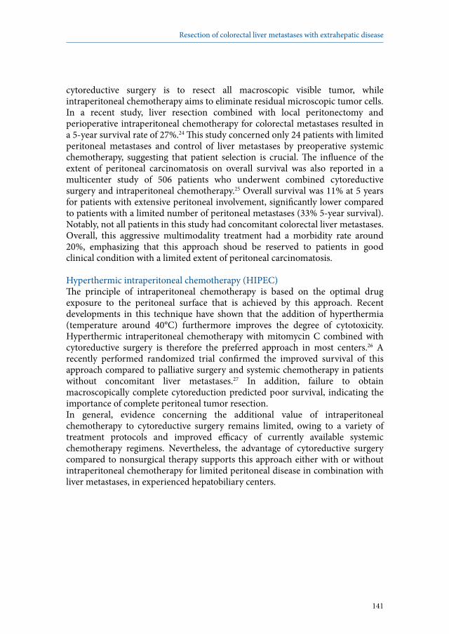
141
Resection of colorectal liver metastases with extrahepatic disease
cytoreductive surgery is to resect all macroscopic visible tumor, while intraperitoneal chemotherapy aims to eliminate residual microscopic tumor cells.In a recent study, liver resection combined with local peritonectomy and perioperative intraperitoneal chemotherapy for colorectal metastases resulted in a 5-year survival rate of 27%.24 This study concerned only 24 patients with limited peritoneal metastases and control of liver metastases by preoperative systemic chemotherapy, suggesting that patient selection is crucial. The influence of the extent of peritoneal carcinomatosis on overall survival was also reported in a multicenter study of 506 patients who underwent combined cytoreductive surgery and intraperitoneal chemotherapy.25 Overall survival was 11% at 5 years for patients with extensive peritoneal involvement, significantly lower compared to patients with a limited number of peritoneal metastases (33% 5-year survival). Notably, not all patients in this study had concomitant colorectal liver metastases. Overall, this aggressive multimodality treatment had a morbidity rate around 20%, emphasizing that this approach shoud be reserved to patients in good clinical condition with a limited extent of peritoneal carcinomatosis. Hyperthermic intraperitoneal chemotherapy (HIPEC)The principle of intraperitoneal chemotherapy is based on the optimal drug exposure to the peritoneal surface that is achieved by this approach. Recent developments in this technique have shown that the addition of hyperthermia (temperature around 40°C) furthermore improves the degree of cytotoxicity. Hyperthermic intraperitoneal chemotherapy with mitomycin C combined with cytoreductive surgery is therefore the preferred approach in most centers.26 A recently performed randomized trial confirmed the improved survival of this approach compared to palliative surgery and systemic chemotherapy in patients without concomitant liver metastases.27 In addition, failure to obtain macroscopically complete cytoreduction predicted poor survival, indicating the importance of complete peritoneal tumor resection. In general, evidence concerning the additional value of intraperitoneal chemotherapy to cytoreductive surgery remains limited, owing to a variety of treatment protocols and improved efficacy of currently available systemic chemotherapy regimens. Nevertheless, the advantage of cytoreductive surgery compared to nonsurgical therapy supports this approach either with or without intraperitoneal chemotherapy for limited peritoneal disease in combination with liver metastases, in experienced hepatobiliary centers.

142
Chapter 6
Rare extrahepatic disease localizations
Metastases to the adrenal glands are a rare finding in patients with colorectal cancer. It has only been described in several anecdotal case-reports, with some of them reporting prolonged survival after adrenalectomy.28,29 Nevertheless, adrenal involvement by colorectal cancer is generally considered as incurable systemic disease, and the place of surgery in these patients is still not elucidated. Similarly, ovarian metastases, bone metastases, and metastatic involvement of the spleen are uncommon extrahepatic disease sites, only described in case-reports or small patient series.30-32 As long-term outcomes after resection of these extrahepatic disease localizations remain unknown, we recommend only performing surgical resection if it can be complete for all known intra- and extrahepatic disease sites.
Conclusion
Although the presence of concomitant extrahepatic disease in patients scheduled for hepatic resection of colorectal metastases is considered by most to be a contraindication for surgery, a multidisciplinary treatment approach can provide long-term survival in selected patients. Encouraging results have been reported after surgical resection of both liver and lung metastases, and to select best surgical candidates, the extent of both liver and pulmonary disease should be carefully evaluated. Patients in whom also regional lymph node involvement is present besides CLM, should first be treated with systemic chemotherapy. Surgery in this subgroup of patients should only be considered in patients with pedicular lymph node metastases and in the absence of disease progression after preoperative chemotherapy. In case of peritoneal carcinomatosis, an aggressive multimodality treatment consisting of cytoreductive surgery and intraperitoneal chemotherapy can offer a chance of prolonged long-term survival to highly selected patients with limited peritoneal disease. In general, patients with both intra- and extrahepatic metastases from colorectal cancer should be managed in hepatobiliary centers experienced in a multidisciplinary treatment approach, consisting of (systemic) chemotherapy and (repeat) surgery. In practice, surgery should only be considered when resection of all metastatic sites can be complete, and it should be combined with perioperative chemotherapy.
143
Resection of colorectal liver metastases with extrahepatic disease
References
1. Choti MA, Sitzmann JV, Tiburi MF et al. Trends in long-term survival following liver resection for hepatic colorectal metastases. Ann Surg 2002; 235:759-766.
2. Wicherts DA, de Haas RJ, Adam R. Bringing unresectable liver disease to resection with curative intent. Eur J Surg Oncol 2007; 33:S42-51.3. Fong Y, Fortner J, Sun RL et al. Clinical score for predicting recurrence after hepatic resection for metastatic colorectal cancer: analysis of 1001 consecutive cases. Ann Surg 1999; 230:309-321.4. Adam R, Delvart V, Pascal G et al. Rescue surgery for unresectable colorectal liver metastases downstaged by chemotherapy: a model to predict long-term survival. Ann Surg 2004; 240:644-657.5. Galandiuk S, Wieand HS, Moertel CG et al. Patterns of recurrence after curative resection of carcinoma of the colon and rectum. Surg Gynecol Obstet 1992; 174:27-32. 6. Pihl E, Hughes ES, McDermott FT et al. Lung recurrence after curative surgery for colorectal cancer. Dis Colon Rectum 1987; 30:417-419.7. Inoue M, Ohta M, Iuchi K et al. Benefits of surgery for patients with pulmonary metastases from colorectal carcinoma. Ann Thorac Surg 2004; 78:238-244. 8. Regnard JF, Grunenwald D, Spaggiari L et al. Surgical treatment of hepatic and pulmonary metastases from colorectal cancers. Ann Thorac Surg 1998; 66:214-219. 9. Murata S, Moriya Y, Akasu T et al. Resection of both hepatic and pulmonary metastases in patients with colorectal carcinoma. Cancer 1998; 83:1086-1093. 10. Lehnert T, Knaebel HP, Dück M et al. Sequential hepatic and pulmonary resections for metastatic colorectal cancer. Br J Surg 1999; 86:241-243. 11. Headrick JR, Miller DL, Nagorney DM et al. Surgical treatment of hepatic and pulmonary metastases from colon cancer. Ann Thorac Surg 2001; 71:975-980. 12. Shah SA, Haddad R, Al-Sukhni W et al. Surgical resection of hepatic and pulmonary metastases from colorectal carcinoma. J Am Coll Surg 2006; 202:468-475.13. Okumura S, Kondo H, Tsuboi M et al. Pulmonary resection for metastatic colorectal cancer: experiences with 159 patients. J Thorac Cardiovasc Surg 1996; 112:867-874. 14. Robinson BJ, Rice TW, Strong SA et al. Is resection of pulmonary and hepatic metastases warranted in patients with colorectal cancer? J Thorac Cardiovasc Surg 1999; 117:66-76. 15. Kobayashi K, Kawamura M, Ishihara T. Surgical treatment for both pulmonary and hepatic metastases from colorectal cancer. J Thorac Cardiovasc Surg 1999; 118:1090-1096.16. Miller G, Biernacki P, Kemeny N et al. Outcomes after resection of synchronous or metachronous hepatic and pulmonary colorectal metastases. J Am Coll Surg 2007; 205:231-238.17. Landreneau RJ, Hazelrigg SR, Mack MJ et al. Postoperative pain-related morbidity: video- assisted thoracic surgery versus thoracotomy. Ann Thorac Surg 1993; 56:1285-1289.18. Rodgers MS, McCall JL. Surgery for colorectal liver metastases with hepatic lymph node involvement: a systematic review. Br J Surg 2000; 87:1142-1155. 19. Minagawa M, Makuuchi M, Torzilli G et al. Extension of the frontiers of surgical indications in the treatment of liver metastases from colorectal cancer: long-term results. Ann Surg 2000; 231:487-499.20. Jaeck D, Nakano H, Bachellier P et al. Significance of hepatic pedicle lymph node involvement in patients with colorectal liver metastases: a prospective study. Ann Surg Oncol 2002; 9:430-438.21. Elias D, Ouellet JF, Bellon N et al. Extrahepatic disease does not contraindicate hepatectomy for colorectal liver metastases. Br J Surg 2003; 90:567-574.22. Adam R, de Haas RJ, Wicherts DA et al. Is hepatic resection justified after chemotherapy in patients with colorectal liver metastases and lymph node involvement? J Clin Oncol 2008;
26:3672-3680.
144
Chapter 6
23. Jayne DG, Fook S, Loi C et al. Peritoneal carcinomatosis from colorectal cancer. Br J Surg 2002; 89:1545-1550.24. Elias D, Benizri E, Pocard M et al. Treatment of synchronous peritoneal carcinomatosis and liver metastases from colorectal cancer. Eur J Surg Oncol 2006; 32:632-636. 25. Glehen O, Kwiatkowski F, Sugarbaker PH et al. Cytoreductive surgery combined with perioperative intraperitoneal chemotherapy for the management of peritoneal carcinomatosis from colorectal cancer: a multi-institutional study. J Clin Oncol 2004; 22:3284-3292.26. Esquivel J, Sticca R, Sugarbaker P et al. Cytoreductive surgery and hyperthermic intraperitoneal chemotherapy in the management of peritoneal surface malignancies of colonic
origin: a consensus statement. Ann Surg Oncol 2007; 14:128-133.27. Verwaal VJ, van Ruth S, de Bree E et al. Randomized trial of cytoreduction and hyperthermic intraperitoneal chemotherapy versus systemic chemotherapy and palliative surgery in patients with peritoneal carcinomatosis of colorectal cancer. J Clin Oncol 2003; 21:3737-3743.28. Kanjo T, Albertini M, Weber S. Long-term disease-free survival after adrenalectomy for
isolated colorectal metastases. Asian J Surg 2006; 29:291-293.29. Nagakura S, Shirai Y, Nomura T et al. Long-term survival after resection of colonic
adenocarcinoma with synchronous metastases to the liver, adrenal gland, and aortic-caval lymph nodes: report of a case. Dis Colon Rectum 2002; 45:1679-1680.
30. Oh YH, Park HC, Kim YS. Atypical bone metastasis and radiation changes in a colon cancer: a case report and a review of the literature. Jpn J Clin Oncol 2001; 31:168-171.
31. Huang PP, Weber TK, Mendoza C et al. Long-term survival in patients with ovarian metastases from colorectal carcinoma. Ann Surg Oncol 1998; 5:695-698.
32. Gencosmanoglu R, Aker F, Kir G et al. Isolated metachronous splenic metastasis from synchronous colon cancer. World J Surg Oncol 2006; 4:42.

145
Resection of colorectal liver metastases with extrahepatic disease


René Adam1*
Robbert J. de Haas1,2*
Dennis A. Wicherts1,2
Eric Vibert1
Chady Salloum1
Daniel Azoulay1
Denis Castaing1
* both authors contributed equally to the manuscript
1 Centre Hépato-Biliaire, Hôpital Paul Brousse, Villejuif, France2 Department of Surgery, University Medical Center Utrecht, Utrecht,
The Netherlands
Annals of Surgery; accepted for publication
Concomitant extrahepatic disease in patients with colorectal liver metastases
When is there a place for surgery?
7
148
Chapter 7
Abstract
ObjectiveTo evaluate the impact of the location of extrahepatic disease (EHD) on survival and to determine patient outcome in a consecutive series of patients with both intrahepatic and extrahepatic colorectal metastases treated by an oncosurgical approach, combining repeat surgery and chemotherapy.
Summary background dataAlthough recognized as poor prognostic factor, concomitant EHD is no more considered an absolute contraindication to surgery in patients with colorectal liver metastases (CLM). However, the impact of the location of EHD on survival and the benefit in patient outcome is still diversely appreciated.
MethodsFrom 840 patients resected for CLM between 1990 and 2006, 186 (22%) also had resectable EHD. Sequential surgery was routinely combined with perioperative chemotherapy. Survival was compared with that of patients without EHD, prognostic factors were identified, and a predictive model was designed to better select surgical candidates. ResultsPatients resected for CLM with concomitant EHD experienced a lower 5-year survival than those without EHD (28% vs 55%; P<0.001). Five poor prognostic factors were identified at multivariate analysis: EHD location other than lung metastases (5-year survival: 23% vs 33%; P=0.02), EHD concomitant to CLM recurrence (14% vs 34%; P<0.001), CEA level >10 ng/ml (16% vs 37%; P=0.02), six or more CLM (9% vs 32%; P=0.02), and right colon cancer (P=0.02). Five-year survival ranged from 64% (0 factors) to 0% (>3 factors). In the EHD group, patients with an EHD recurrence experienced better outcomes when resected than those treated by chemotherapy alone (5-year survival: 38% vs 21%; P=0.05).
ConclusionWhile sequential surgery is warranted for patients with less than six CLM with isolated lung metastases, low CEA levels, and no right colon primary tumor, it should be questioned in the presence of more than three of these prognostic factors.

149
Surgery in patients with colorectal liver metastases and concomitant extrahepatic disease
Introduction
Hepatic resection is still the only potentially curative treatment for patients with colorectal liver metastases (CLM), and nowadays, 5-year survival rates up to 47% can be reached.1 However, in several large patient series, the presence of concomitant extrahepatic disease (EHD) has been reported to significantly reduce survival rates after hepatic resection for colorectal metastases.2-5 Therefore, the presence of concomitant EHD in these patients has been considered a contraindication to (hepatic) surgery by most surgeons. Whether the presence of concomitant EHD in patients with CLM should still contraindicate surgery is currently under debate. In a recent publication, it was concluded that resectable EHD sites in patients with CLM should no longer be considered a contraindication to surgery, as the total number of metastases had a prognostic effect, not their location.6 In addition, several authors have reported prolonged survival rates after staged resection of colorectal metastases to both liver and lung, similar to that observed after hepatic resection in patients without EHD.7-12 Furthermore, many important improvements have been made in the efficacy of chemotherapy regimens, as response rates in patients receiving combinations of 5-fluorouracil, leucovorin, oxaliplatin and/or irinotecan currently exceed 50%.13-15 Therefore, an oncosurgical approach combining effective chemotherapy regimens and surgery might justify an expansion of the indications for surgery in patients with both hepatic and extrahepatic colorectal metastases. At our institute, patients with colorectal metastases are treated by an oncosurgical approach, consisting for most patients of perioperative chemotherapy, resection as much as possible, and repeat surgery in case of new recurrence. In addition, we have never considered the presence of concomitant EHD in patients with CLM an absolute contraindication to surgery, provided that resection of both the liver metastases and EHD sites could be potentially complete. To evaluate if this oncosurgical strategy is justified and to identify which patients are best surgical candidates, the current study was undertaken. We assessed long-term outcome both for the total patient group presenting with concomitant EHD and for each EHD location.
150
Chapter 7
Methods
Study populationAll consecutive patients who underwent hepatic resection for CLM at our institution between January 1990 and July 2006, and who had concomitant EHD sites (ie, diagnosed before or during hepatic resection), were included in the study. Patients were selected from our prospective database, and additional data were obtained by reviewing each patient’s medical record. To evaluate the influence of concomitant EHD on long-term outcomes, comparison was made with all other patients treated in the same period, but who did not have any evidence of EHD.
Preoperative careBefore hepatic resection, all patients were evaluated by thoracoabdominal and pelvic imaging (ultrasonography and computed tomography [CT]), serum tumor marker levels (CEA and/or CA 19-9), bone scintigraphy, and colonoscopy. More recently, selected patients were additionally staged with PET-CT. In case of initially unresectable metastases, preoperative chemotherapy was routinely administered. Besides this, preoperative chemotherapy was administered in a neoadjuvant setting in case of synchronous metastases (diagnosed before, during, or within 3 months following colorectal resection), or in case of marginally resectable CLM (five or more bilateral nodules). Response to chemotherapy was determined on a CT scan after every four cycles of treatment according to the Response Evaluation Criteria in Solid Tumors.16 If the estimated future remnant liver volume was judged too small (<25-30%), portal vein embolization was performed to induce compensatory hypertrophy of the remaining liver.17 All treatment decisions were taken during multidisciplinary meetings, including surgeons, medical oncologists, and radiologists.
Surgical treatmentDuring laparotomy, palpation of the liver and intraoperative ultrasound were carried out to evaluate the metastatic involvement of the liver. Thorough abdominal inspection and palpation were performed to rule out unresectable EHD sites. In case of multiple bilateral CLM, which could not all be resected by a single hepatectomy, even when combined with local ablative techniques, a two-stage procedure was performed.18,19 In case of suspected EHD sites located in the abdominal cavity (either diagnosed before or during laparotomy), frozen-biopsy was performed to confirm malignancy. If positive, simultaneous resection of both the hepatic and extrahepatic metastases was only performed if resection of all disease sites could be complete. When lymph node metastases were present, lymphadenectomy consisted of the involved lymph node station, and in case of celiac and/or para-aortic lymph node metastases also resection of the pedicular

151
Surgery in patients with colorectal liver metastases and concomitant extrahepatic disease
and celiac lymph nodes. In case of peritoneal metastases, resection was followed by immediate intraperitoneal chemotherapy. When located outside the abdomen, concomitant EHD sites were generally resected within 2-3 months after the hepatectomy, with systemic chemotherapy in the meantime, to prevent disease progression. Importantly, in all cases hepatic resection was only performed if the patient could tolerate the required surgical procedures and if the total surgical strategy (for both hepatic and extrahepatic metastases) could be potentially curative.
Postoperative follow-upRegular follow-up at our outpatient clinic started at 1 month postoperatively, and then once every 4 months. It consisted of a history and physical exam, routine blood tests including serum tumor marker levels, abdominal ultrasonography, and thoracoabdominal CT (every 8 months). To decrease the recurrence risk, postoperative chemotherapy was routinely used. In case of intra- and/or extrahepatic disease recurrence, resection was only performed if it could be complete.20 Statistical analysisCategorical data were compared between groups using the χ2 test, and continuous data were compared using the independent-samples t test. Survival curves were made according to the Kaplan-Meier method with the date of hepatectomy as starting date, and were compared using the log-rank test. To identify predictive factors of survival, univariate and multivariate analyses were performed, using the log-rank test and the Cox proportional hazard model, respectively. Subsequently, a predictive model for survival was constructed based on the identified predictive factors at multivariate analysis.21,22 P values <0.05 indicated statistical significance. All statistical analyses were performed with SPSS version 15.0 (SPSS Inc, Chicago, IL, USA).

152
Chapter 7
Results
Patient characteristicsBetween 1990 and 2006, 840 patients underwent hepatic resection for CLM, 186 (22%) of whom had concomitant EHD. Liver metastases in patients in the extrahepatic disease group (EHD group) were less often diagnosed synchronous to the primary tumor, and had a smaller size, compared with patients that did not have EHD (Table 1). In 53 patients (28%) of the EHD group, extrahepatic disease was concomitant to a repeat hepatectomy for recurrent liver metastases. In addition, patients presenting with concomitant EHD were more often considered initially unresectable. Other patient characteristics are outlined in Table 1.
Preoperative chemotherapySignificantly more patients in the EHD group received preoperative systemic chemotherapy, compared with patients without EHD (95% vs 70%; P<0.001) (Table 2). In both groups, most patients received only one line of preoperative chemotherapy (66% in the EHD group vs 73% of patients without EHD; P=0.08). Six or more cycles of chemotherapy were given to 85% of patients in the EHD group, compared with 76% of patients without EHD (P=0.02). Patients within the EHD group treated by one chemotherapy line (N=117) received a median of seven cycles (range: 1-24) of chemotherapy. When two lines of chemotherapy were given (N=41), the median number of cycles was 11 (range: 5-27), and in case of three or more lines of preoperative chemotherapy (N=19), the median number of cycles was 19 (range: 9-37).
Hepatic resectionThe use of portal vein embolization to prevent postoperative liver failure was similar between the EHD group and patients without EHD (11% vs 8%, respectively; P=0.61) (Table 2). The extent of liver disease necessitated a two-stage hepatectomy procedure in 4% of patients in the EHD group versus 5% of patients not having EHD (P=0.31). The rate of major hepatectomies (resection of three or more liver segments) was also similarly distributed between both groups. However, a higher number of liver metastases was detected by intraoperative ultrasound in the EHD group (Table 2). In the EHD group, one patient (0.5%) died within 60 days from hepatectomy, compared with six patients (1%) in the group without concomitant EHD (P=0.56). Postoperative complications were observed in 31% of patients in the EHD group versus 30% of patients without EHD (P=0.57). Subdivision of postoperative complications according to the Clavien classification is outlined in Table 2.23
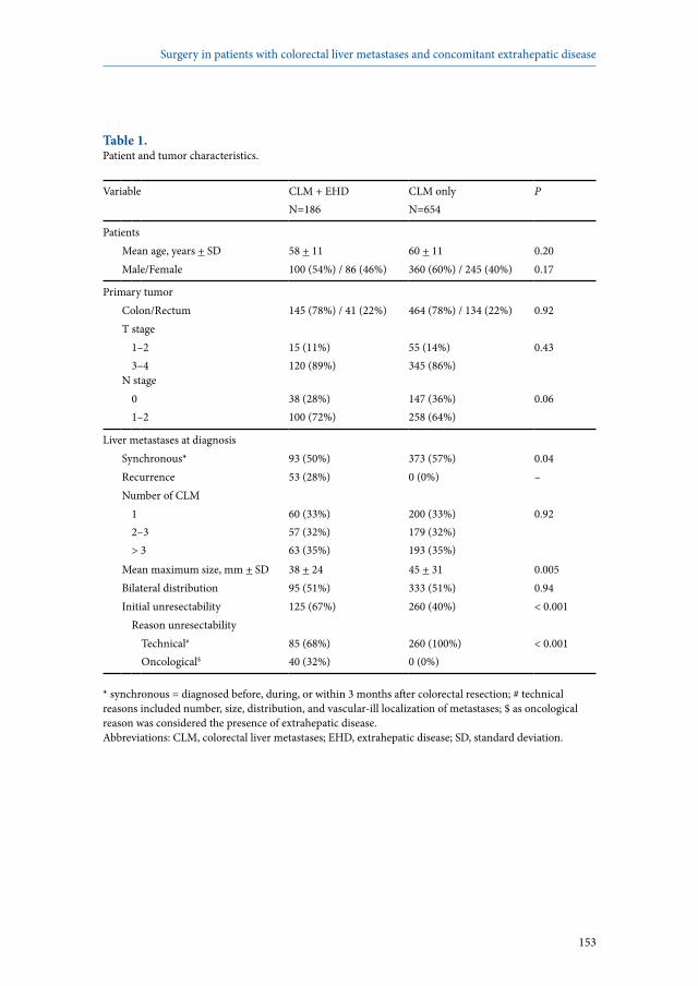
153
Surgery in patients with colorectal liver metastases and concomitant extrahepatic disease
Table 1.Patient and tumor characteristics.
Variable CLM + EHDN=186
CLM onlyN=654
P
PatientsMean age, years + SD 58 + 11 60 + 11 0.20Male/Female 100 (54%) / 86 (46%) 360 (60%) / 245 (40%) 0.17
Primary tumorColon/Rectum 145 (78%) / 41 (22%) 464 (78%) / 134 (22%) 0.92T stage
1–23–4
15 (11%)120 (89%)
55 (14%)345 (86%)
0.43
N stage 01–2
38 (28%)100 (72%)
147 (36%)258 (64%)
0.06
Liver metastases at diagnosisSynchronous* 93 (50%) 373 (57%) 0.04Recurrence 53 (28%) 0 (0%) –Number of CLM
12–3> 3
60 (33%)57 (32%)63 (35%)
200 (33%)179 (32%)193 (35%)
0.92
Mean maximum size, mm + SD 38 + 24 45 + 31 0.005Bilateral distribution 95 (51%) 333 (51%) 0.94Initial unresectability 125 (67%) 260 (40%) < 0.001
Reason unresectabilityTechnical#
Oncological$
85 (68%)40 (32%)
260 (100%)0 (0%)
< 0.001
* synchronous = diagnosed before, during, or within 3 months after colorectal resection; # technical reasons included number, size, distribution, and vascular-ill localization of metastases; $ as oncological reason was considered the presence of extrahepatic disease.Abbreviations: CLM, colorectal liver metastases; EHD, extrahepatic disease; SD, standard deviation.
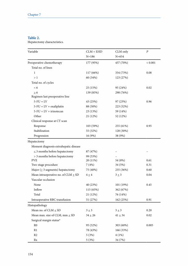
154
Chapter 7
Table 2. Hepatectomy characteristics.
Variable CLM + EHD N=186
CLM only N=654
P
Preoperative chemotherapy 177 (95%) 457 (70%) < 0.001Total no. of lines
1> 1
117 (66%)60 (34%)
334 (73%)123 (27%)
0.08
Total no. of cycles< 6> 6
25 (15%)139 (85%)
95 (24%)298 (76%)
0.02
Regimen last preoperative line5-FU + LV5-FU + LV + oxaliplatin5-FU + LV + irinotecanOther
43 (25%)88 (50%)23 (13%)21 (12%)
97 (23%)223 (52%)59 (14%)52 (12%)
0.96
Clinical response at CT scanResponseStabilizationProgression
103 (59%)55 (32%)16 (9%)
255 (61%)128 (30%)38 (9%)
0.95
HepatectomyMoment diagnosis extrahepatic disease
< 3 months before hepatectomy> 3 months before hepatectomy
87 (47%)99 (53%)
––
–
PVE 20 (11%) 54 (8%) 0.61Two-stage procedure 7 (4%) 34 (5%) 0.31Major (> 3 segments) hepatectomy 75 (40%) 235 (36%) 0.60Mean intraoperative no. of CLM + SD 4 + 4 3 + 3 0.04Vascular occlusion
NoneInflowTotal
40 (23%)115 (65%)21 (12%)
101 (19%)362 (67%)76 (14%)
0.45
Intraoperative RBC transfusion 51 (27%) 162 (25%) 0.91
HistopathologyMean no. of CLM + SD 3 + 3 3 + 3 0.20Mean max. size of CLM, mm + SD 34 + 26 41 + 34 0.02Surgical margin status*
R0R1R2Rx
95 (52%)78 (43%)5 (3%)5 (3%)
303 (60%)166 (33%)4 (1%)34 (7%)
0.005
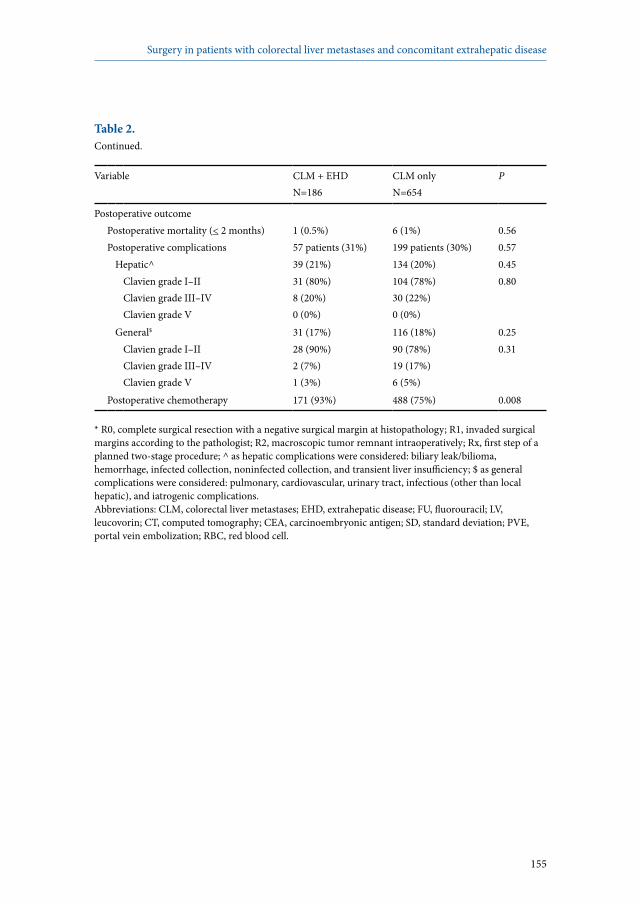
155
Surgery in patients with colorectal liver metastases and concomitant extrahepatic disease
Table 2. Continued.
Variable CLM + EHD N=186
CLM only N=654
P
Postoperative outcomePostoperative mortality (< 2 months) 1 (0.5%) 6 (1%) 0.56Postoperative complications 57 patients (31%) 199 patients (30%) 0.57
Hepatic^ 39 (21%) 134 (20%) 0.45Clavien grade I–IIClavien grade III–IVClavien grade V
31 (80%)8 (20%)0 (0%)
104 (78%)30 (22%)0 (0%)
0.80
General$ 31 (17%) 116 (18%) 0.25Clavien grade I–IIClavien grade III–IVClavien grade V
28 (90%)2 (7%)1 (3%)
90 (78%)19 (17%)6 (5%)
0.31
Postoperative chemotherapy 171 (93%) 488 (75%) 0.008
* R0, complete surgical resection with a negative surgical margin at histopathology; R1, invaded surgical margins according to the pathologist; R2, macroscopic tumor remnant intraoperatively; Rx, first step of a planned two-stage procedure; ^ as hepatic complications were considered: biliary leak/bilioma, hemorrhage, infected collection, noninfected collection, and transient liver insufficiency; $ as general complications were considered: pulmonary, cardiovascular, urinary tract, infectious (other than local hepatic), and iatrogenic complications.Abbreviations: CLM, colorectal liver metastases; EHD, extrahepatic disease; FU, fluorouracil; LV, leucovorin; CT, computed tomography; CEA, carcinoembryonic antigen; SD, standard deviation; PVE, portal vein embolization; RBC, red blood cell.
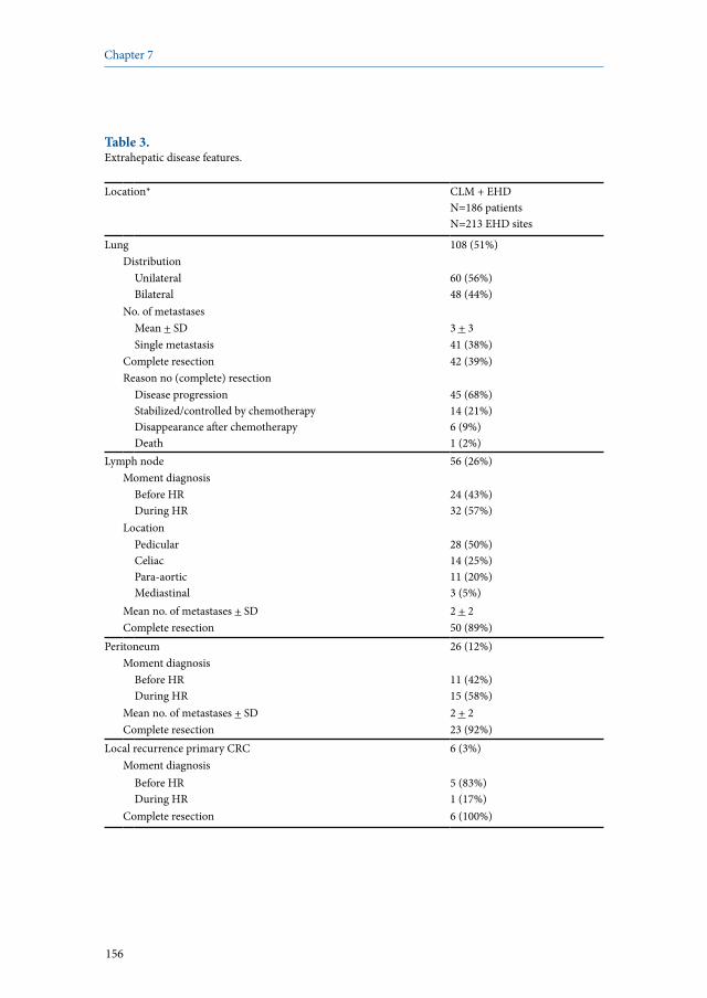
156
Chapter 7
Table 3. Extrahepatic disease features.
Location* CLM + EHDN=186 patientsN=213 EHD sites
Lung 108 (51%)Distribution
UnilateralBilateral
60 (56%)48 (44%)
No. of metastasesMean + SD 3 + 3Single metastasis 41 (38%)
Complete resection 42 (39%)Reason no (complete) resection
Disease progressionStabilized/controlled by chemotherapyDisappearance after chemotherapyDeath
45 (68%)14 (21%)6 (9%)1 (2%)
Lymph node 56 (26%)Moment diagnosis
Before HRDuring HR
24 (43%)32 (57%)
LocationPedicularCeliacPara-aorticMediastinal
28 (50%)14 (25%)11 (20%)3 (5%)
Mean no. of metastases + SD 2 + 2Complete resection 50 (89%)
Peritoneum 26 (12%)Moment diagnosis
Before HRDuring HR
11 (42%)15 (58%)
Mean no. of metastases + SD 2 + 2Complete resection 23 (92%)
Local recurrence primary CRC 6 (3%)Moment diagnosis
Before HRDuring HR
5 (83%)1 (17%)
Complete resection 6 (100%)
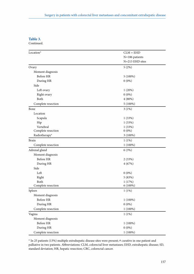
157
Surgery in patients with colorectal liver metastases and concomitant extrahepatic disease
Table 3. Continued.
Location* CLM + EHDN=186 patientsN=213 EHD sites
Ovary 5 (2%)Moment diagnosis
Before HRDuring HR
5 (100%)0 (0%)
SideLeft ovaryRight ovaryBoth
1 (20%)0 (0%)4 (80%)
Complete resection 5 (100%)Bone 3 (1%)
LocationScapulaHipVertebral
1 (33%)1 (33%)1 (33%)
Complete resection 0 (0%)Radiotherapy# 3 (100%)
Brain 1 (1%)Complete resection 1 (100%)
Adrenal gland 6 (3%)Moment diagnosis
Before HRDuring HR
2 (33%)4 (67%)
SideLeftRightBoth
0 (0%)5 (83%)1 (17%)
Complete resection 6 (100%)Spleen 1 (1%)
Moment diagnosisBefore HRDuring HR
1 (100%)0 (0%)
Complete resection 1 (100%)Vagina 1 (1%)
Moment diagnosisBefore HRDuring HR
1 (100%)0 (0%)
Complete resection 1 (100%)
* In 25 patients (13%) multiple extrahepatic disease sites were present; # curative in one patient and palliative in two patients. Abbreviations: CLM, colorectal liver metastases; EHD, extrahepatic disease; SD, standard deviation; HR, hepatic resection; CRC, colorectal cancer.
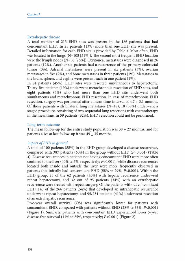
158
Chapter 7
Extrahepatic diseaseA total number of 213 EHD sites was present in the 186 patients that had concomitant EHD. In 25 patients (13%) more than one EHD site was present. Detailed information for each EHD site is provided by Table 3. Most often, EHD was located in the lungs (N=108 [51%]). The second most frequent EHD location were the lymph nodes (N=56 [26%]). Peritoneal metastases were diagnosed in 26 patients (12%). Another six patients had a recurrence of the primary colorectal tumor (3%). Adrenal metastases were present in six patients (3%), ovarian metastases in five (2%), and bone metastases in three patients (1%). Metastases to the brain, spleen, and vagina were present each in one patient (1%). In 84 patients (45%), EHD sites were resected simultaneous to hepatectomy. Thirty-five patients (19%) underwent metachronous resection of EHD sites, and eight patients (4%) who had more than one EHD site underwent both simultaneous and metachronous EHD resection. In case of metachronous EHD resection, surgery was performed after a mean time-interval of 4.7 + 3.1 months. Of those patients with bilateral lung metastases (N=48), 18 (38%) underwent a staged procedure, consisting of two sequential lung resections with chemotherapy in the meantime. In 59 patients (32%), EHD resection could not be performed.
Long-term outcomeThe mean follow-up for the entire study population was 38 + 27 months, and for patients alive at last follow-up it was 49 + 35 months.
Impact of EHD in generalA total of 100 patients (88%) in the EHD group developed a disease recurrence, compared with 387 patients (60%) in the group without EHD (P=0.004) (Table 4). Disease recurrences in patients not having concomitant EHD were more often confined to the liver (40% vs 5%, respectively; P<0.001), while disease recurrences located both inside and outside the liver were more frequently observed in patients that initially had concomitant EHD (58% vs 29%; P<0.001). Within the EHD group, 25 of the 62 patients (40%) with hepatic recurrence underwent repeat hepatectomy, and 32 out of 95 patients (34%) with an extrahepatic recurrence were treated with repeat surgery. Of the patients without concomitant EHD, 143 of the 266 patients (54%) that developed an intrahepatic recurrence underwent repeat hepatectomy, and 95/234 patients (41%) underwent resection of an extrahepatic recurrence. Five-year overall survival (OS) was significantly lower for patients with concomitant EHD, compared with patients without EHD (28% vs 55%; P<0.001) (Figure 1). Similarly, patients with concomitant EHD experienced lower 5-year disease-free survival (11% vs 25%, respectively; P<0.001) (Figure 2).
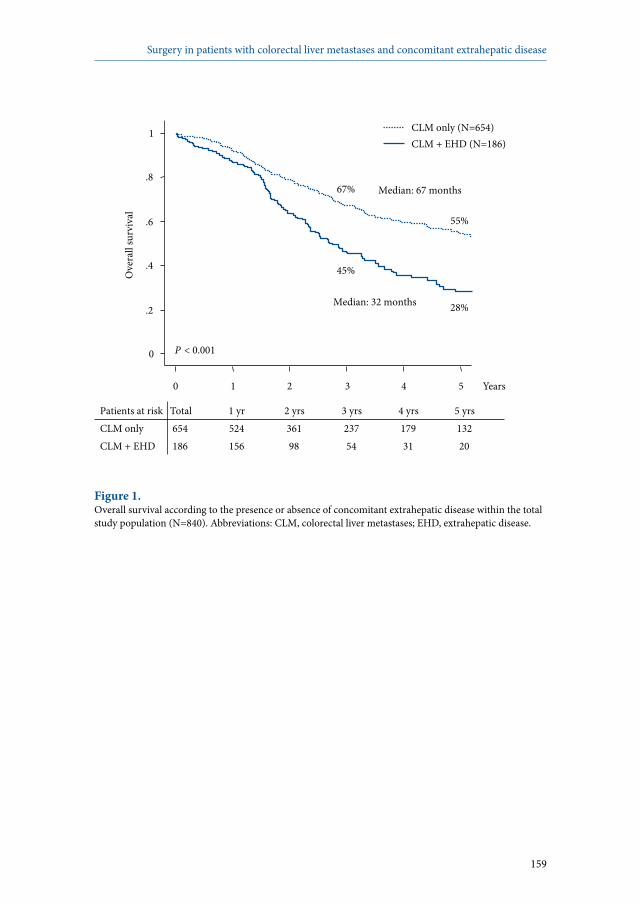
159
Surgery in patients with colorectal liver metastases and concomitant extrahepatic disease
Patients at risk Total 1 yr 2 yrs 3 yrs 4 yrs 5 yrs CLM only 654 524 361 237 179 132 CLM + EHD 186 156 98 54 31 20
0
.2
.4
.6
.8
1 O
vera
ll su
rviv
al
0 1 2 3 4 5 Years
P < 0.001
28%
45%
55%
67%
CLM + EHD (N=186) CLM only (N=654)
Median: 32 months
Median: 67 months
Figure 1. Overall survival according to the presence or absence of concomitant extrahepatic disease within the total study population (N=840). Abbreviations: CLM, colorectal liver metastases; EHD, extrahepatic disease.

160
Chapter 7
Patients at risk Total 1 yr 2 yrs 3 yrs 4 yrs 5 yrs CLM only 612 392 254 168 126 97 CLM + EHD 180 64 36 27 18 15
0
.2
.4
.6
.8
1 D
iseas
e-fre
e sur
viva
l
0 1 2 3 4 5 Years
P < 0.001 11%
17%
25%
37%
CLM + EHD (N=186) CLM only (N=654)
Median: 22 months
Median: 4 months
Figure 2. Disease-free survival according to the presence or absence of concomitant extrahepatic disease within the total study population (N=840). Abbreviations: CLM, colorectal liver metastases; EHD, extrahepatic disease.

161
Surgery in patients with colorectal liver metastases and concomitant extrahepatic disease
Impact of EHD locationWithin the EHD group, patients presenting with isolated lung metastases had a significantly better OS than those not having isolated lung metastases (5-year OS 33% vs 23%, respectively; P=0.02) (Figure 3). When isolated lymph node or peritoneal metastases were present, OS tended to be lower than that observed in the absence of isolated lymph node or peritoneal metastases (5-year OS 20% vs 32%, respectively; P=0.09) (Figure 4). No significant survival difference was observed according to the number of lung metastases (Table 5). If more than one EHD site was present, no 5-year survivors were observed, compared with a 5-year OS of 31% in case of a single EHD site (P=0.02) (Figure 5). Although the total number of EHD metastases did not influence survival rates, a survival difference was observed when CLM and EHD metastases are taken together. Specifically, when the total number of metastases was less than seven, 5-year OS was 33%, compared with 20% if seven or more metastases were present (P=0.05) (Figure 6). When after complete resection of concomitant EHD an EHD recurrence occurred, better OS was observed in case of repeat EHD resection, compared with no resection of the EHD recurrence (5-year OS 38% vs 21%; P=0.05) (Figure 7). Furthermore, patients who underwent repeat surgery for EHD recurrence experienced similar OS as those who did not develop a disease recurrence after complete resection (P=0.67) (Figure 8).

162
Chapter 7
Patients at risk Total 1 yr 2 yrs 3 yrs 4 yrs 5 yrs Isolated lung metastases 90 78 51 32 20 11 No isolated lung metastases 96 78 47 22 11 9
0
.2
.4
.6
.8
1
Ove
rall
surv
ival
0 1 2 3 4 5 Years
P = 0.02
23%
37%
33%
53%
No isolated lung metastases (N=96) Isolated lung metastases (N=90)
Median: 28 months
Median: 40 months
Figure 3. Overall survival according to the presence or absence of isolated lung metastases within the extrahepatic disease group (N=186).

163
Surgery in patients with colorectal liver metastases and concomitant extrahepatic disease
Patients at risk Total 1 yr 2 yrs 3 yrs 4 yrs 5 yrs Lymph node or peritoneal 57 45 27 15 6 4 No lymph node or peritoneal 129 111 71 39 25 16
0
.2
.4
.6
.8
1
Ove
rall
surv
ival
0 1 2 3 4 5 Years
P = 0.09
20%
39% 32%
48%
Isolated lymph node or peritoneal mets (N=57) No isolated lymph node or peritoneal mets (N=129)
Median: 30 months
Median: 36 months
Figure 4. Overall survival according to the presence or absence of isolated lymph node or peritoneal metastases within the extrahepatic disease group (N=186).
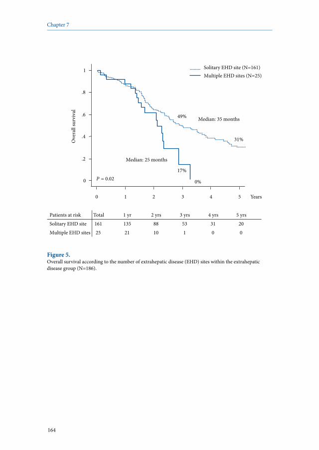
164
Chapter 7
Total 1 yr 2 yrs 3 yrs 4 yrs 5 yrs Solitary EHD site 161 135 88 53 31 20 Multiple EHD sites 25 21 10 1 0 0
0
.2
.4
.6
.8
1 O
vera
ll su
rviv
al
0 1 2 3 4 5 Years
P = 0.02 0%
17%
31%
49%
Multiple EHD sites (N=25) Solitary EHD site (N=161)
Median: 25 months
Median: 35 months
Patients at risk
Figure 5. Overall survival according to the number of extrahepatic disease (EHD) sites within the extrahepatic disease group (N=186).
165
Surgery in patients with colorectal liver metastases and concomitant extrahepatic disease
Impact of timing of EHD diagnosisFive-year OS in patients in whom EHD was diagnosed in the period between 1990 and 1995 was 30%, while this was 23% and 31% in those diagnosed with EHD in the periods 1995-2000 and 2000-2006, respectively (P=0.17) (Table 5). Within the group of patients with isolated lung metastases, 5-year OS was 41%, 25%, and 32%, for those whose lung metastases were diagnosed in the period 1990-1995, 1995-2000, and 2000-2006, respectively (P=0.17). In patients with lymph node and/or peritoneal metastases diagnosed in the period 1990-1995, 1995-2000, and 2000-2006, 5-year OS were 0%, 18%, and 30%, respectively (P=0.18).
Impact of surgical margin statusWhen hepatic and EHD resection in the EHD group were taken together, 5-year OS after R0 resection (ie, complete surgical resection with negative surgical margins at histopathology; N=59) was 36%, compared with 26% and 19% after R1 (no safe surgical margins at histopathology; N=54) or R2 resection (macroscopic incomplete resection or first step of a planned two-stage procedure; N=72), respectively (P=0.04). Considering hepatic resection, 5-year OS was 33% after R0 resection (N=95), 24% after R1 resection (N=78), and 0% after R2 resection (N=10; P=0.10). Considering EHD resection, 5-year OS after complete resection of all EHD sites (N=114) was 83%, compared with 64% for those in whom EHD resection was not complete (N=72; P=0.12).
Prognostic factors of survivalAt univariate analysis, seven factors were identified that significantly influenced OS in the EHD group: location of the primary tumor, CLM recurrence, six or more CLM at diagnosis, preoperative CEA level >10 ng/ml, absence of isolated lung metastases, presence of multiple EHD sites, and a total number of metastases of seven or more (Table 5). After entering all factors with an univariate P <0.10 in the Cox regression model, five independent predictors of poor OS were identified: primary tumor located in the right colon, EHD concomitant to CLM recurrence, six or more CLM at diagnosis, preoperative CEA level >10 ng/ml, and the absence of isolated lung metastases (Table 5). Predictive model for survivalTo predict OS in individual patients scheduled for resection of their CLM in the presence of concomitant EHD, the five independent prognostic factors identified at multivariate analysis were entered in a model (Table 6). When none of the prognostic factors was present, estimated survival was 64%, while in the presence of four or five predictive factors, survival was nil.
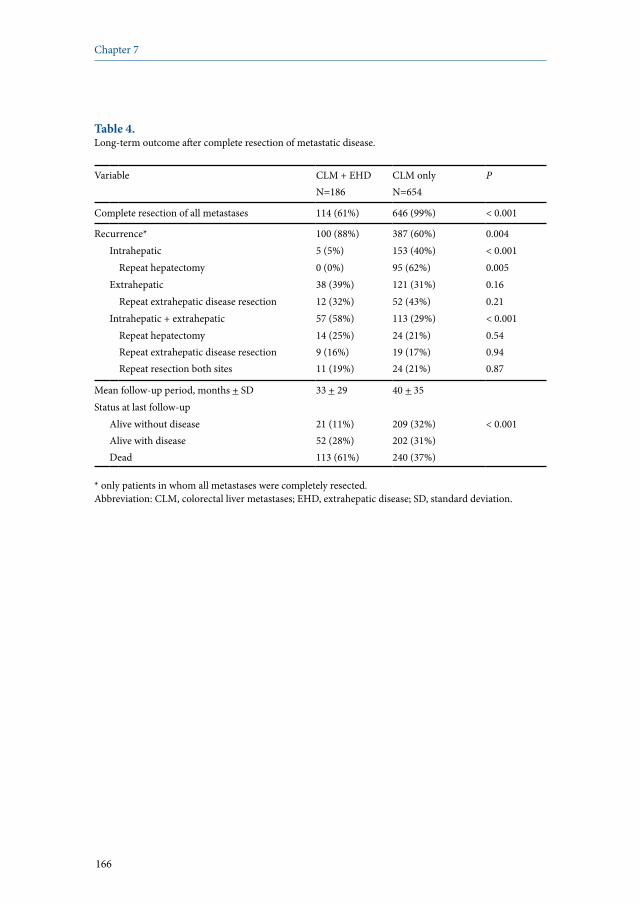
166
Chapter 7
Table 4. Long-term outcome after complete resection of metastatic disease.
Variable CLM + EHD N=186
CLM onlyN=654
P
Complete resection of all metastases 114 (61%) 646 (99%) < 0.001
Recurrence* 100 (88%) 387 (60%) 0.004Intrahepatic 5 (5%) 153 (40%) < 0.001
Repeat hepatectomy 0 (0%) 95 (62%) 0.005Extrahepatic 38 (39%) 121 (31%) 0.16
Repeat extrahepatic disease resection 12 (32%) 52 (43%) 0.21Intrahepatic + extrahepatic 57 (58%) 113 (29%) < 0.001
Repeat hepatectomyRepeat extrahepatic disease resectionRepeat resection both sites
14 (25%)9 (16%)11 (19%)
24 (21%)19 (17%)24 (21%)
0.540.940.87
Mean follow-up period, months + SD 33 + 29 40 + 35Status at last follow-up
Alive without diseaseAlive with diseaseDead
21 (11%)52 (28%)113 (61%)
209 (32%)202 (31%)240 (37%)
< 0.001
* only patients in whom all metastases were completely resected. Abbreviation: CLM, colorectal liver metastases; EHD, extrahepatic disease; SD, standard deviation.
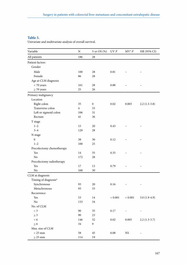
167
Surgery in patients with colorectal liver metastases and concomitant extrahepatic disease
Table 5.Univariate and multivariate analysis of overall survival.
Variable N 5-yr OS (%) UV P MV# P HR (95% CI)
All patients 186 28
Patient factorsGender
MaleFemale
10086
2828
0.81 – –
Age at CLM diagnosis< 70 years > 70 years
16125
2826
0.88 – –
Primary malignancyLocation
Right colonTransverse colonLeft or sigmoid colonRectum
35410641
0333136
0.02 0.003 2.2 (1.3-3.8)
T stage 1–23–4
15120
2028
0.43 – –
N stage 01–2
38100
3025
0.12 – –
Precolectomy chemotherapyYesNo
14172
3528
0.35 – –
Precolectomy radiotherapyYesNo
17168
1330
0.79 – –
CLM at diagnosisTiming of diagnosis*
SynchronousMetachronous
9393
2033
0.16 – –
RecurrenceYesNo
53133
1434
< 0.001 < 0.001 3.0 (1.9-4.9)
No. of CLM< 3> 3
9090
3523
0.17 – –
< 6> 6
14634
329
0.02 0.005 2.2 (1.3-3.7)
Max. size of CLM< 25 mm> 25 mm
58114
4519
0.08 NS –

168
Chapter 7
Table 5. Continued.
Variable N 5-yr OS (%) UV P MV# P HR (95% CI)
DistributionUnilateralBilateral
9095
3324
0.57 – –
CEA level< 10 ng/ml> 10 ng/ml
7298
3716
0.02 0.01 1.8 (1.1-2.9)
Initial resectabilityYesNo
61125
3625
0.08 NS –
Hepatic resectionPreoperative chemotherapy
YesNo
1779
2677
0.11 – –
Regimen last preoperative line5-FU + LV5-FU + LV + oxaliplatin5-FU + LV + irinotecanOther
43882321
22261537
0.69 – –
Response last chemotherapy lineResponse/stabilizationProgression
15816
2720
0.20 – –
Year of diagnosis of extrahepatic disease
1990-19951995-20002000-2006
297681
302331
0.17 – –
Moment diagnosis extrahepatic disease
< 3 months before hepatectomy> 3 months before hepatectomy
8799
2234
0.08 NS –
Concomitant EHD characteristicsIsolated lung metastases
YesNo
9096
3323
0.02 0.007 1.8 (1.2-2.8)
Year of diagnosis of isolated lung metastases
1990-19951995-20002000-2006
163440
412532
0.17 – –

169
Surgery in patients with colorectal liver metastases and concomitant extrahepatic disease
Table 5. Continued.
Variable N 5-yr OS (%) UV P MV# P HR (95% CI)
Isolated lymph node metastasesYesNo
41145
2031
0.12 – –
Isolated peritoneal metastases YesNo
16170
029
0.58 – –
Isolated lymph node or peritoneal metastases
YesNo
57129
2032
0.09 NS –
Year of diagnosis of isolated lymph node or peritoneal metastases
1990-19951995-20002000-2006
62526
01830
0.18 – –
Other isolated metastases sitesYesNo
14172
5625
0.43 – –
Number of lung metastases1> 1
4164
3526
0.26 – –
Number of EHD sites1> 1
16125
310
0.02 NS –
Total number of EHD metastases< 4> 4
13245
3116
0.13 – –
Total number of metastases (CLM + EHD)
< 7> 7
10566
3320
0.05 NS –
# variables with an univariate P<0.10 were entered in the Cox regression model; * synchronous = diagnosed before, during, or within 3 months after colorectal resection.Abbreviations: OS, overall survival; UV, univariate; MV, multivariate; HR, hazard ratio; CI, confidence interval; CLM, colorectal liver metastases; NS, not significant; CEA, carcinoembryonic antigen; FU, fluorouracil; LV, leucovorin; EHD, extrahepatic disease.

170
Chapter 7
Patients at risk Total 1 yr 2 yrs 3 yrs 4 yrs 5 yrs < 7 metastases 105 88 63 35 20 13 ≥ 7 metastases 66 55 29 15 8 6
0
.2
.4
.6
.8
1 O
vera
ll su
rviv
al
0 1 2 3 4 5 Years
P = 0.05
20%
35%
33%
51%
≥ 7 metastases (liver + other; N=66) < 7 metastases (liver + other; N=105)
Median: 28 months
Median: 39 months
Figure 6. Overall survival according to the total number of metastases (liver + extrahepatic metastases) within the extrahepatic disease group (N=186).
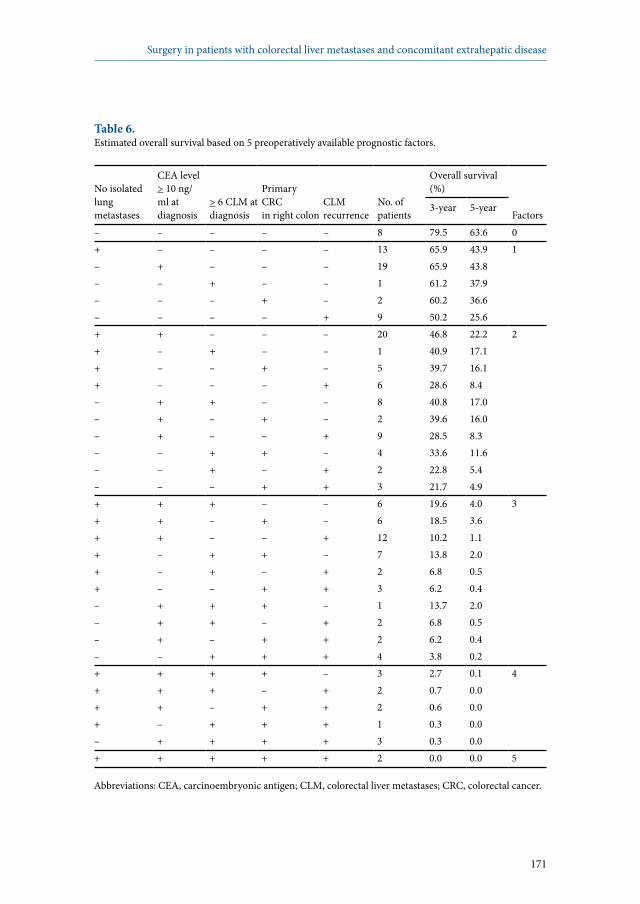
171
Surgery in patients with colorectal liver metastases and concomitant extrahepatic disease
Table 6. Estimated overall survival based on 5 preoperatively available prognostic factors.
No isolated lung metastases
CEA level > 10 ng/ml at diagnosis
> 6 CLM at diagnosis
Primary CRC in right colon
CLM recurrence
No. of patients
Overall survival (%)
Factors3-year 5-year
– – – – – 8 79.5 63.6 0+ – – – – 13 65.9 43.9 1– + – – – 19 65.9 43.8– – + – – 1 61.2 37.9– – – + – 2 60.2 36.6– – – – + 9 50.2 25.6+ + – – – 20 46.8 22.2 2+ – + – – 1 40.9 17.1+ – – + – 5 39.7 16.1+ – – – + 6 28.6 8.4– + + – – 8 40.8 17.0– + – + – 2 39.6 16.0– + – – + 9 28.5 8.3– – + + – 4 33.6 11.6– – + – + 2 22.8 5.4– – – + + 3 21.7 4.9+ + + – – 6 19.6 4.0 3+ + – + – 6 18.5 3.6+ + – – + 12 10.2 1.1+ – + + – 7 13.8 2.0+ – + – + 2 6.8 0.5+ – – + + 3 6.2 0.4– + + + – 1 13.7 2.0– + + – + 2 6.8 0.5– + – + + 2 6.2 0.4– – + + + 4 3.8 0.2+ + + + – 3 2.7 0.1 4+ + + – + 2 0.7 0.0+ + – + + 2 0.6 0.0+ – + + + 1 0.3 0.0– + + + + 3 0.3 0.0+ + + + + 2 0.0 0.0 5
Abbreviations: CEA, carcinoembryonic antigen; CLM, colorectal liver metastases; CRC, colorectal cancer.

172
Chapter 7
Patients at risk Total 1 yr 2 yrs 3 yrs 4 yrs 5 yrs Repeat EHD resection 32 29 21 15 8 6 No repeat EHD resection 63 56 37 15 8 6
0
.2
.4
.6
.8
1
Ove
rall
surv
ival
0 1 2 3 4 5 Years
P = 0.05
38%
60%
21%
37%
No repeat EHD resection (N=63) Repeat EHD resection (N=32)
Median: 30 months
Median: 54 months
Figure 7. Overall survival after complete resection of concomitant extrahepatic disease (EHD) in patients in whom an EHD recurrence occurred (N=95): resection of the EHD recurrence versus no surgical treatment.

173
Surgery in patients with colorectal liver metastases and concomitant extrahepatic disease
Patients at risk Total 1 yr 2 yrs 3 yrs 4 yrs 5 yrs Repeat EHD resection 32 29 21 15 8 6 No recurrence 23 18 12 10 8 6
0
.2
.4
.6
.8
1
Ove
rall
surv
ival
0 1 2 3 4 5 Years
P = 0.67
38%
60%
46% 54%
No recurrence (N=23) Repeat EHD resection (N=32)
Median: 58 months
Median: 54 months
Figure 8. Overall survival after complete resection of concomitant extrahepatic disease (EHD) (N=114) comparing patients who underwent a repeat resection of an extrahepatic disease recurrence with patients without an extrahepatic disease recurrence.
174
Chapter 7
Discussion
The presence of concomitant EHD in patients with CLM is still considered a contraindication to surgery by most clinicians. The fact that at our institution the presence of EHD in patients with CLM has never been considered a contraindication to surgery when there was a possibility of complete resection, now enables us to assess whether this policy was justified. Of importance to note is that in our setting all patients were treated by an aggressive oncosurgical approach, consisting of repeat surgery and perioperative chemotherapy. Patients resected for CLM who also presented with EHD experienced a lower OS than those without concomitant EHD. However, within the EHD group, significantly better OS was observed in patients presenting with isolated lung metastases, compared with those not having lung-only metastases. In addition, survival was negatively influenced if more than one EHD site was present, and if the total number of metastases (irrespective of the site) was seven or more. Not surprisingly, better OS was observed in case of EHD recurrence for those patients who underwent repeat EHD resection, compared with those without repeat surgery. But, survival following repeat surgery for an EHD recurrence was similar to that observed when no recurrence occurred after complete resection of the initial disease. At multivariate analysis, five prognostic factors were identified in the EHD group: absence of isolated lung metastases, CEA level >10 ng/ml, six or more CLM at diagnosis, primary tumor located in the right colon, and EHD concomitant to CLM recurrence. In the absence of the aforementioned prognostic factors, OS was 64%, while no survivors were observed in the presence of four or five factors. In several recent large patient series, the presence of EHD in patients who underwent hepatectomy for CLM emerged as an independent poor prognostic factor.2-5 This is in contrast with a recent publication of Elias et al, concluding that resectable EHD in patients with CLM scheduled for hepatectomy should no longer be considered a contraindication to surgery, as the total number of metastases, irrespective of their location, had a stronger prognostic effect than their site.6 However, the statistical methods used in the latter study have been questioned, and consequently, interpretation of the results remains difficult.24 Therefore, the place of surgery in patients with both CLM and concomitant EHD remains unclear. To answer this question, we reviewed our experience in treating this particular patient group. This concerns one of the largest series of patients who underwent hepatectomy for CLM with concomitant EHD (N=186), with the specificity that all were treated by an aggressive oncosurgical approach, consisting of perioperative chemotherapy, resection of all metastatic sites, and repeat surgery in case of new resectable disease recurrence. Although OS was significantly lower in the EHD group compared with patients without EHD, our oncosurgical approach for patients with both CLM and
175
Surgery in patients with colorectal liver metastases and concomitant extrahepatic disease
concomitant EHD was associated with a 5-year OS of 28%, which compares favorably with rates much less than 5% when treated by chemotherapy alone.25,26 Within the EHD group, best survival rates were observed in patients with isolated lung metastases (5-year OS 33% vs 23% in the absence of isolated lung metastases; P=0.02). This is similar to the existing literature, reporting 5-year OS rates varying between 30% and 44% after resection of both CLM and lung metastases.7-12 In addition, a tendency to lower survival was observed in patients having isolated lymph node or peritoneal metastases (5-year OS 20% vs 32% in the absence of isolated lymph node or peritoneal metastases; P=0.09). Recently, we reported our experience in resecting patients with both hepatic and regional lymph node metastases of colorectal origin.27 Our main finding was a 5-year OS of 25% when only pedicular lymph nodes contained metastatic disease, while no 5-year survivors were observed in case of celiac or para-aortic lymph node involvement.27 In the current study, survival was also lower if more than one EHD site was present, and if the total number of metastases was seven or more. However, of all factors related to the number or location of colorectal metastases, only the absence of isolated lung metastases emerged as a poor prognostic factor at multivariate analysis. Therefore, our data does not support the key conclusion of Elias et al, that the total number of colorectal metastases, irrespective of their location, had a stronger prognostic effect than their site.6 As a result, in patients in whom resection of all metastatic disease can be potentially complete, the clinical decision should also be based on the location of the metastatic sites, not solely on the total number of metastases. Another important factor is the completeness of the surgical treatment. Accordingly, best survival rates were observed when resection of all metastatic sites was macroscopically and microscopically complete (R0). However, owing to our multimodal treatment approach, still acceptable survival rates were observed after R1 resections. Besides the absence of isolated lung metastases, we identified four other independent poor prognostic factors, which were all related to the primary tumor or the CLM: CEA level >10 ng/ml, six or more CLM at diagnosis, EHD concomitant to CLM recurrence, and location of the primary tumor in the right colon. The CEA level and the number of CLM are well-known risk factors, reflecting less favorable tumor biology.2,28-30 That patients in whom EHD was concomitant to a repeat hepatectomy for CLM experienced worse survival, could appear in contrast with our earlier publication as well as with the recent publication of Rees et al.4,20 However, this discrepancy can be explained by the fact that both our earlier study and the publication of Rees et al reported on the total population treated by hepatectomy for CLM, which contrasts with the current study based on a subset of patients with both CLM and concomitant EHD.4,20 The adverse effect of right colon cancer on long-term outcome can be explained by its relatively long asymptomatic period, and therefore, at the moment of diagnosis, patients often present with more advanced disease. The
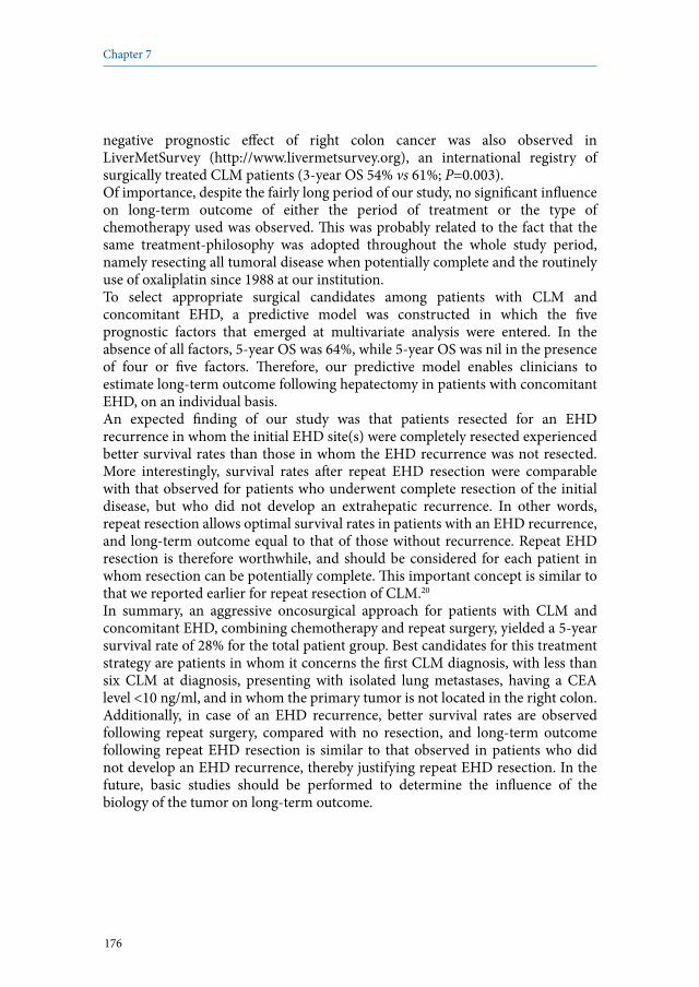
176
Chapter 7
negative prognostic effect of right colon cancer was also observed in LiverMetSurvey (http://www.livermetsurvey.org), an international registry of surgically treated CLM patients (3-year OS 54% vs 61%; P=0.003). Of importance, despite the fairly long period of our study, no significant influence on long-term outcome of either the period of treatment or the type of chemotherapy used was observed. This was probably related to the fact that the same treatment-philosophy was adopted throughout the whole study period, namely resecting all tumoral disease when potentially complete and the routinely use of oxaliplatin since 1988 at our institution.To select appropriate surgical candidates among patients with CLM and concomitant EHD, a predictive model was constructed in which the five prognostic factors that emerged at multivariate analysis were entered. In the absence of all factors, 5-year OS was 64%, while 5-year OS was nil in the presence of four or five factors. Therefore, our predictive model enables clinicians to estimate long-term outcome following hepatectomy in patients with concomitant EHD, on an individual basis. An expected finding of our study was that patients resected for an EHD recurrence in whom the initial EHD site(s) were completely resected experienced better survival rates than those in whom the EHD recurrence was not resected. More interestingly, survival rates after repeat EHD resection were comparable with that observed for patients who underwent complete resection of the initial disease, but who did not develop an extrahepatic recurrence. In other words, repeat resection allows optimal survival rates in patients with an EHD recurrence, and long-term outcome equal to that of those without recurrence. Repeat EHD resection is therefore worthwhile, and should be considered for each patient in whom resection can be potentially complete. This important concept is similar to that we reported earlier for repeat resection of CLM.20 In summary, an aggressive oncosurgical approach for patients with CLM and concomitant EHD, combining chemotherapy and repeat surgery, yielded a 5-year survival rate of 28% for the total patient group. Best candidates for this treatment strategy are patients in whom it concerns the first CLM diagnosis, with less than six CLM at diagnosis, presenting with isolated lung metastases, having a CEA level <10 ng/ml, and in whom the primary tumor is not located in the right colon. Additionally, in case of an EHD recurrence, better survival rates are observed following repeat surgery, compared with no resection, and long-term outcome following repeat EHD resection is similar to that observed in patients who did not develop an EHD recurrence, thereby justifying repeat EHD resection. In the future, basic studies should be performed to determine the influence of the biology of the tumor on long-term outcome.

177
Surgery in patients with colorectal liver metastases and concomitant extrahepatic disease
References
1. Wei AC, Greig PD, Grant D et al. Survival after hepatic resection for colorectal metastases: a 10-year experience. Ann Surg Oncol 2006; 13:668-676.
2. Fong Y, Fortner J, Sun RL et al. Clinical score for predicting recurrence after hepatic resection for metastatic colorectal cancer: analysis of 1001 consecutive cases. Ann Surg 1999; 230:309-318.
3. Kato T, Yasui K, Hirai T et al. Therapeutic results for hepatic metastasis of colorectal cancer with special reference to effectiveness of hepatectomy: analysis of prognostic factors for 763 cases recorded at 18 institutions. Dis Colon Rectum 2003; 46:S22-S31.
4. Rees M, Tekkis PP, Welsh FK et al. Evaluation of long-term survival after hepatic resection for metastatic colorectal cancer: a multifactorial model of 929 patients. Ann Surg 2008; 247:125-135.
5. Scheele J, Altendorf-Hofmann A, Grube T et al. Resection of colorectal liver metastases. What prognostic factors determine patient selection? Chirurg 2001; 72:547-560.
6. Elias D, Liberale G, Vernerey D et al. Hepatic and extrahepatic colorectal metastases: when resectable, their localization does not matter, but their total number has a prognostic effect. Ann Surg Oncol 2005; 12:900-909.
7. Headrick JR, Miller DL, Nagorney DM et al. Surgical treatment of hepatic and pulmonary metastases from colon cancer. Ann Thorac Surg 2001; 71:975-979.
8. Kobayashi K, Kawamura M, Ishihara T. Surgical treatment for both pulmonary and hepatic metastases from colorectal cancer. J Thorac Cardiovasc Surg 1999; 118:1090-1096.
9. Lehnert T, Knaebel HP, Duck M et al. Sequential hepatic and pulmonary resections for metastatic colorectal cancer. Br J Surg 1999; 86:241-243.
10. Miller G, Biernacki P, Kemeny NE et al. Outcomes after resection of synchronous or metachronous hepatic and pulmonary colorectal metastases. J Am Coll Surg 2007; 205:231-238.
11. Murata S, Moriya Y, Akasu T et al. Resection of both hepatic and pulmonary metastases in patients with colorectal carcinoma. Cancer 1998; 83:1086-1093.
12. Okumura S, Kondo H, Tsuboi M et al. Pulmonary resection for metastatic colorectal cancer: experiences with 159 patients. J Thorac Cardiovasc Surg 1996; 112:867-874.
13. Falcone A, Ricci S, Brunetti I et al. Phase III trial of infusional fluorouracil, leucovorin, oxaliplatin, and irinotecan (FOLFOXIRI) compared with infusional fluorouracil, leucovorin, and irinotecan (FOLFIRI) as first-line treatment for metastatic colorectal cancer: the Gruppo Oncologico Nord Ovest. J Clin Oncol 2007; 25:1670-1676.
14. Köhne CH, van Cutsem E, Wils J et al. Phase III study of weekly high-dose infusional fluorouracil plus folinic acid with or without irinotecan in patients with metastatic colorectal cancer: European Organisation for Research and Treatment of Cancer Gastrointestinal Group Study 40986. J Clin Oncol 2005; 23:4856-4865.
15. Tournigand C, André T, Achille E et al. FOLFIRI followed by FOLFOX6 or the reverse sequence in advanced colorectal cancer: a randomized GERCOR study. J Clin Oncol 2004; 22:229-237.
16. Therasse P, Arbuck SG, Eisenhauer EA et al. New guidelines to evaluate the response to treatment in solid tumors. European Organization for Research and Treatment of Cancer, National Cancer Institute of the United States, National Cancer Institute of Canada. J Natl Cancer Inst 2000; 92:205-216.
17. Azoulay D, Castaing D, Smail A et al. Resection of nonresectable liver metastases from colorectal cancer after percutaneous portal vein embolization. Ann Surg 2000; 231:480-486.
18. Adam R, Miller R, Pitombo M et al. Two-stage hepatectomy approach for initially unresectable colorectal hepatic metastases. Surg Oncol Clin N Am 2007; 16:525-536.
178
Chapter 7
19. Wicherts DA, Miller R, de Haas RJ et al. Long-term results of two-stage hepatectomy for irresectable colorectal cancer liver metastases. Ann Surg 2008; 248:994-1005.
20. Adam R, Bismuth H, Castaing D et al. Repeat hepatectomy for colorectal liver metastases. Ann Surg 1997; 225:51-60.
21. Breslow N. Covariance analysis of censored survival data. Biometrics 1974; 30:89-99.22. Cox DR. Regression models and life tables (with discussion). J R Stat Soc B 1972; 34:187-202.23. Dindo D, Demartines N, Clavien PA. Classification of surgical complications: a new proposal
with evaluation in a cohort of 6336 patients and results of a survey. Ann Surg 2004; 240:205-213.
24. Strasberg S. Re: Hepatic and extrahepatic colorectal metastases: when resectable, their localization does not matter, but their total number has a prognostic effect. Ann Surg Oncol 2007; 14:1517-1518.
25. Giacchetti S, Itzhaki M, Gruia G et al. Long-term survival of patients with unresectable colorectal cancer liver metastases following infusional chemotherapy with 5-fluorouracil, leucovorin, oxaliplatin and surgery. Ann Oncol 1999; 10:663-669.
26. Masi G, Cupini S, Marcucci L et al. Treatment with 5-fluorouracil/folinic acid, oxaliplatin, and irinotecan enables surgical resection of metastases in patients with initially unresectable metastatic colorectal cancer. Ann Surg Oncol 2006; 13:58-65.
27. Adam R, de Haas RJ, Wicherts DA et al. Is hepatic resection justified after chemotherapy in patients with colorectal liver metastases and lymph node involvement? J Clin Oncol 2008; 26:3672-3680.
28. Minagawa M, Makuuchi M, Torzilli G et al. Extension of the frontiers of surgical indications in the treatment of liver metastases from colorectal cancer: long-term results. Ann Surg 2000; 231:487-499.
29. Nordlinger B, Guiguet M, Vaillant JC et al. Surgical resection of colorectal carcinoma metastases to the liver. A prognostic scoring system to improve case selection, based on 1568 patients. Association Française de Chirurgie. Cancer 1996; 77:1254-1262.
30. Scheele J, Stangl R, Altendorf-Hofmann A et al. Resection of colorectal liver metastases. World J Surg 1995; 19:59-71.

179
Surgery in patients with colorectal liver metastases and concomitant extrahepatic disease


René Adam1*
Robbert J. de Haas1,2*
Dennis A. Wicherts1,2
Thomas A. Aloia1
Valérie Delvart1
Daniel Azoulay1
Henri Bismuth1
Denis Castaing1
* both authors contributed equally to the manuscript
1 Centre Hépato-Biliaire, Hôpital Paul Brousse, Villejuif, France2 Department of Surgery, University Medical Center Utrecht, Utrecht,
The Netherlands
Journal of Clinical Oncology 2008; 26:3672-3680
Is hepatic resection justified after chemotherapy in patients with colorectal liver metastases and lymph node involvement?
8
182
Chapter 8
Abstract
PurposeFor patients with colorectal liver metastases (CLM), regional lymph node (RLN) involvement is one of the worst prognostic factors. The objective of this study was to evaluate the ability of a multidisciplinary approach, including preoperative chemotherapy and hepatectomy, to improve patient outcomes.
Patients and methodsOutcomes for a consecutively treated group of patients with CLM and simultaneous RLN involvement were compared to a cohort without RLN involvement. Univariate and multivariate analysis of clinical variables was used to identify prognostic factors in this high-risk group.
ResultsOf the 763 patients resected at our institution for CLM between 1992 and 2006, 47 patients (6%) were treated with hepatectomy and simultaneous lymphadenectomy. All patients had received preoperative chemotherapy. Five-year overall survival (OS) for patients with and without RLN involvement were 18% and 53%, respectively (P<0.001). Five-year disease-free survival rates were 11% and 23%, respectively (P=0.004). When diagnosed preoperatively, RLN involvement had an increased 5-year OS compared to intraoperative detection, although the difference was not significant (35% vs 10%; P=0.18). Location of metastatic RLN strongly influenced survival, with observed 5-year OS of 25% for pedicular, 0% for celiac, and 0% for para-aortic RLN (P=0.001). At multivariate analysis, celiac RLN involvement and age >40 years were identified as independent poor prognostic factors.
ConclusionCombined liver resection and pedicular lymphadenectomy is justified when RLN metastases respond to or are stabilized by preoperative chemotherapy, particularly in young patients. In contrast, this approach does not benefit patients with celiac and/or para-aortic RLN involvement, even when patients’ disease is responding to preoperative chemotherapy.

183
Hepatic resection in patients with colorectal liver metastases and lymph node involvement
Introduction
For patients with colorectal liver metastases (CLM) hepatic resection remains the best treatment option, with reported 5-year survivals between 28% and 41%.1-5 These favorable outcomes have prompted an expansion in the indications for hepatic surgery for CLM. Technical improvements, including two-stage hepatectomy and vascular exclusion and reconstruction techniques, have made more patients with CLM eligible for surgery.6-9 In addition, preoperative chemotherapy has been shown to convert a substantial number of cases that were initially unresectable to a resectable situation.10,11 Despite this progress, the presence of regional lymph node (RLN) involvement in patients with CLM is still considered to be one of the worst prognostic factors and, for most oncologists, continues to be an absolute contraindication to surgery. Accordingly, a review of 16 reported series of patients with both CLM and RLN involvement consistently identifies poor 5-year survivals, including 11 series reporting no 5-year survivors (Table 1).3,12-26 Given these data, the expert consensus-based therapeutic decision model OncoSurge rated metastatic RLN involvement as a poor prognostic factor, and celiac or para-aortic RLN involvement as absolute contraindications to liver surgery.27 More recently however, several studies have concluded that the presence of extrahepatic disease is not an absolute contraindication to hepatic surgery.16,28,29 Given the limited number of patients studied and the apparent conflicts in the literature, the role of surgery in these patients remains highly controversial. For patients with CLM with extrahepatic disease, improved outcomes over time may be related to the development of more effective chemotherapy regimens, with response rates now increasing to greater than 50% and median survivals exceeding 20 months.30-32 The addition of oxaliplatin and irinotecan to 5-fluorouracil and leucovorin has significantly improved response rates and survival.33-37 By downsizing the intra- and extrahepatic tumor burden, these therapies have allowed an increased number of patients with initially inoperable disease to become candidates for resection, significantly changing the prognosis. The combined use of targeted molecular therapies, such as cetuximab or bevacizumab, is further increasing chemotherapy efficacy and concomitantly broadening the opportunity for curative resections.38,39 During our experience treating patients with CLM, we have not considered the presence of RLN metastases as an absolute contraindication to surgery, provided that response or stabilization during preoperative chemotherapy was observed. This experience gives us an unique opportunity to determine the impact of preoperative chemotherapy on postoperative outcomes for patients with liver and RLN involvement. The purpose of the present study was to define the role of surgery in this category of patients by systematically examining long-term outcomes and prognostic factors.
184
Chapter 8
Patients and methods
PatientsPatients who fulfilled the following criteria were included in this study: hepatectomy for CLM, presence of RLN metastases simultaneous to hepatectomy (ie, diagnosed before or during hepatectomy), preoperative chemotherapy, and resection of histologically proven RLN metastases. All study data were present in our Human Subject Committee-approved prospective database. For outcomes analysis, patients were grouped according to the location of RLN metastases: pedicular (distal to the gastroduodenal artery branch), celiac (overlying the common hepatic artery proximal to the gastroduodenal artery branch), and para-aortic RLN metastases. To determine the influence of RLN metastases on patient outcomes, additional comparisons were made to a simultaneously treated group of patients undergoing hepatectomy in the absence of RLN involvement.
Preoperative evaluationBefore hepatectomy, all patients were radiologically staged with abdominal ultrasound and abdominopelvic and chest computed tomography (CT). In our more recent experience, selected patients were additionally staged with positron emission tomography (PET) CT. If the diagnosis of RLN involvement was made preoperatively (based on evolution of size and round shape on CT and/or hypermetabolic appearance on PET-CT), patients were initially treated with systemic chemotherapy. Furthermore, preoperative chemotherapy was considered in patients with initially unresectable disease or who presented with recurrent hepatic metastases within 1 year after the previous operation. The Response Evaluation Criteria in Solid Tumors were applied to serial imaging studies obtained every 2 months during preoperative therapy to determine chemotherapy response.40 In cases of RLN involvement identified before surgery, hepatectomy combined with lymphadenectomy was only performed in patients with RLN involvement responding to or stabilized by chemotherapy.
OperationAll patients underwent hepatectomy with curative intent, preserving as much normal liver parenchyma as possible. Resection of three or more segments was considered a major hepatectomy.41,42 If the estimated future remnant liver volume was <30% (<40% in case of prolonged chemotherapy), portal vein embolization was performed.43 When up to three lesions were deeply located in the future remnant liver, resection was combined to radiofrequency ablation and/or cryosurgery. If not all liver lesions could be completely resected in a single procedure, even when combined to radiofrequency ablation and/or cryosurgery, a two-stage procedure was performed.6 In patients with preoperatively identified RLN involvement, intraoperative confirmation of RLN disease by frozen-section
185
Hepatic resection in patients with colorectal liver metastases and lymph node involvement
biopsy preceded lymphadenectomy. In cases of no preoperative suspicion of RLN involvement, the intraoperative finding of macroscopically enlarged and firm nodes prompted immediate frozen-section analysis. If this was positive, hepatectomy combined with lymphadenectomy was performed when resection of all disease sites was deemed both feasible and safe. Postoperative histopathologic examination of all resection specimens was routine.
Postoperative managementAll patients were observed regularly, starting 1 month postoperatively and then every 4 months. These evaluations consisted of a history and physical examination, serum tumor markers, liver function parameters, and abdominal ultrasonography. Every 8 months, thoracoabdominal CT scans were performed. When concomitant resectable extrahepatic disease sites (except RLN metastases) were present, a resection was generally performed 2 to 3 months after hepatectomy, with intercurrent systemic chemotherapy delivered to prevent disease progression. To decrease the risk of recurrence, especially in patients with initially unresectable disease or who presented with a recurrence before, adjuvant chemotherapy was routinely recommended.
Statistical analysisCategorical and continuous study variables were compared between groups using the χ2 test, the independent-samples t test, or the one-way analysis of variance test, as appropriate. Overall and disease-free survival probabilities were estimated using the Kaplan-Meier method and were compared using log-rank tests. The log-rank tests were also used to identify variables associated with survival in univariate analysis. For multivariate analysis, all variables with a univariate P value <0.10 were entered in a Cox regression model to identify variables independently associated with survival. A P value <0.05 was considered statistically significant. Statistical analysis was performed using SPSS version 13.0 (SPSS Inc, Chicago, IL, USA).
186
Chapter 8
Results
Patient and tumor characteristicsBetween June 1992 and July 2006, 763 patients with CLM underwent hepatectomy at our institution. Among them, 53 patients (7%) had clinical evidence of RLN metastases simultaneous to hepatectomy. Six patients were excluded from the study because their suspected RLN disease could not be confirmed intraoperatively, leaving 47 patients (6%) available for analysis. Of these 47 study patients, 26 patients (3%) presented with pedicular, 14 patients (2%) presented with celiac, and seven patients (1%) presented with para-aortic RLN involvement. The group of patients who underwent resection and who were without simultaneous RLN metastases consisted of 710 patients (93%). Patient characteristics and liver metastases data are outlined in Table 2. The RLN group consisted of younger patients, with more advanced primary tumors (T4),44 a higher frequency of metachronous CLM, and a higher incidence of initially unresectable CLM.
Perioperative dataWithin the RLN group, all 47 patients (100%) received preoperative chemotherapy, compared with 582 patients (82%) in the group with liver disease only. Significantly more lines and cycles of preoperative chemotherapy were administered in the RLN group (Table 3). Within the RLN group, RLN involvement was related to first hepatectomy in 36 patients (77%), second hepatectomy in nine patients (19%), and third hepatectomy in two patients (4%). Portal vein embolization and major hepatectomies were performed more often in the RLN group (Table 3). Number and maximum size of resected CLM at histopathologic examination were similar between both groups. Other perioperative and pathologic characteristics are listed in Table 3.
Lymph node metastasesLymph node involvement was diagnosed before hepatectomy in 19 (40%) of 47 patients and intraoperatively in the remaining 28 patients (60%). Preoperatively, PET-CT was performed in six (13%) of the 47 patients, identifying RLN metastases in four patients. All patients with RLN disease diagnosed before surgery were treated with preoperative systemic chemotherapy and experienced either a response or stabilization of RLN disease. Of the patients with intraoperative diagnosis of RLN involvement, all had received preoperative chemotherapy. In all but four of these cases, liver disease responded to or was stabilized by preoperative chemotherapy (Table 4). Within the pedicular and celiac groups, all patients underwent a complete lymphadenectomy with no residual RLN disease, compared with 86% (six of seven patients) in the para-aortic group. Type of lymphadenectomy with the resected RLN areas are indicated in Table 3. Lymph node features are listed in Table 5.
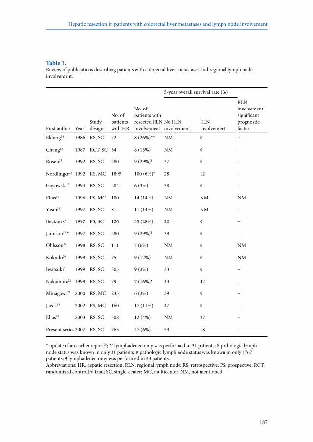
187
Hepatic resection in patients with colorectal liver metastases and lymph node involvement
Table 1. Review of publications describing patients with colorectal liver metastases and regional lymph node involvement.
5-year overall survival rate (%)
First author YearStudydesign
No. ofpatientswith HR
No. of patients with resected RLN involvement
No RLN involvement
RLN involvement
RLN involvement significant prognostic factor
Ekberg14 1986 RS, SC 72 8 (26%)** NM 0 +
Chang13 1987 RCT, SC 64 8 (13%) NM 0 +
Rosen25 1992 RS, SC 280 9 (29%)$ 37 0 +
Nordlinger23 1992 RS, MC 1895 100 (6%)# 28 12 +
Gayowski17 1994 RS, SC 204 6 (3%) 38 0 +
Elias15 1996 PS, MC 100 14 (14%) NM NM NM
Yasui26 1997 RS, SC 81 11 (14%) NM NM +
Beckurts12 1997 PS, SC 126 35 (28%) 22 0 +
Jamison19 * 1997 RS, SC 280 9 (29%)$ 39 0 +
Ohlsson24 1998 RS, SC 111 7 (6%) NM 0 NM
Kokudo20 1999 RS, SC 75 9 (12%) NM 0 NM
Iwatsuki3 1999 RS, SC 305 9 (3%) 33 0 +
Nakamura22 1999 RS, SC 79 7 (16%)¶ 43 42 –
Minagawa21 2000 RS, MC 235 6 (3%) 39 0 +
Jaeck18 2002 PS, MC 160 17 (11%) 47 0 +
Elias16 2003 RS, SC 308 12 (4%) NM 27 –
Present series 2007 RS, SC 763 47 (6%) 53 18 +
* update of an earlier report25; ** lymphadenectomy was performed in 31 patients; $ pathologic lymph node status was known in only 31 patients; # pathologic lymph node status was known in only 1767 patients; ¶ lymphadenectomy was performed in 43 patients. Abbreviations: HR, hepatic resection; RLN, regional lymph node; RS, retrospective; PS, prospective; RCT, randomized controlled trial; SC, single-center; MC, multicenter; NM, not mentioned.
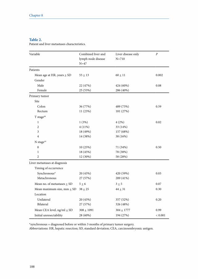
188
Chapter 8
Table 2. Patient and liver metastases characteristics.
Variable Combined liver and lymph node diseaseN=47
Liver disease onlyN=710
P
PatientsMean age at HR, years + SD 55 + 13 60 + 11 0.002Gender
MaleFemale
22 (47%)25 (53%)
424 (60%)286 (40%)
0.08
Primary tumorSite
ColonRectum
36 (77%)11 (23%)
489 (73%)181 (27%)
0.59
T stage44
1234
1 (3%)4 (11%)18 (49%)14 (38%)
4 (2%)33 (14%)157 (68%)38 (16%)
0.02
N stage44
012
10 (25%)18 (45%)12 (30%)
71 (34%)78 (38%)58 (28%)
0.50
Liver metastases at diagnosisTiming of occurrence
Synchronous*Metachronous
20 (43%)27 (57%)
420 (59%)289 (41%)
0.03
Mean no. of metastases + SD 5 + 6 3 + 5 0.07Mean maximum size, mm + SD 38 + 25 44 + 31 0.30Location
UnilateralBilateral
20 (43%)27 (57%)
357 (52%)326 (48%)
0.20
Mean CEA level, ng/ml + SD 308 + 1091 304 + 1777 0.99Initial unresectability 28 (60%) 194 (27%) < 0.001
*synchronous = diagnosed before or within 3 months of primary tumor surgery.Abbreviations: HR, hepatic resection; SD, standard deviation; CEA, carcinoembryonic antigen.

189
Hepatic resection in patients with colorectal liver metastases and lymph node involvement
Table 3. Hepatic resection and histopathologic examination.
Variable Combined liver and lymph node disease N=47
Liver disease onlyN=710
P
Preoperative chemotherapy 47 patients (100%) 582 patients (82%) 0.001Mean no. of lines + SD 2 + 1 1 + 1 < 0.001Mean no. of cycles + SD 15 + 9 10 + 6 < 0.001Indication
Conversion therapy*Neoadjuvant therapy**
28 (60%)19 (40%)
194 (33%)388 (67%)
< 0.001
Presence of extrahepatic diseaseSynchronous CLM#
CLM recurrenceMultiple bilateral CLM
8 (42%)4 (21%)5 (26%)2 (11%)
45 (12%)222 (57%)0 (0%)121 (31%)
< 0.001
Hepatic resectionPreoperative PVE 7 (15%) 34 (5%) 0.02Two-stage resection 3 (6%) 50 (7%) 0.85Mean preoperative CEA level, ng/ml + SD 49 + 114 98 + 418 0.43Mean RBC transfusions, units + SD 1 + 2 1 + 3 0.62Mean intraoperative no. of metastases + SD 5 + 5 3 + 3 0.002Extent of resection
Major (> 3 segments)Minor
19 (40%)28 (60%)
98 (23%)324 (77%)
0.01
Resection typeAnatomicalNonanatomicalBoth
18 (38%)15 (32%)14 (30%)
238 (34%)190 (27%)274 (39%)
0.45
Vascular occlusionNoneSelective inflow Total pedicularTotal vascular
10 (21%)4 (9%)28 (60%)5 (11%)
111 (21%)16 (3%)325 (63%)68 (13%)
0.28
Combined local treatmentRFACryosurgeryCombined RFA and cryosurgery
1 (2%)2 (4%)0 (0%)
17 (2%)52 (7%)3 (0%)
0.83
Type of lymphadenectomyPedicular + celiacPara-aorticPedicular + celiac + para-aortic
40 (85%)5 (11%)2 (4%)
–––
–

190
Chapter 8
Table 3. Continued.
Variable Combined liver and lymph node disease N=47
Liver disease onlyN=710
P
HistopathologyMean no. of resected metastases + SD 3 + 3 3 + 3 0.70Mean maximum size, mm + SD 39 + 36 42 + 37 0.57Surgical margin status^
R0R1R2
23 (50%)19 (41%)4 (9%)$
315 (58%)217 (40%)8 (2%)
0.004
Complete tumor necrosisYesNo
2 (4%)44 (96%)
17 (4%)394 (96%)
0.95
Postoperative chemotherapy 41 patients (87%) 498 patients (70%) 0.21Mean no. of lines + SD 1 + 1 1 + 1 0.56Mean no. of cycles + SD 9 + 6 9 + 6 0.68
* conversion of unresectable CLM to resectable CLM; ** resectable CLM; # diagnosed before or within 3 months of primary tumor surgery; ^ R0, complete surgical resection with a negative surgical margin at histopathology; R1, invaded surgical margins according to the pathologist; R2, macroscopic tumor remnant intraoperatively; $ in 3 patients R2 designation reflects disease status after the first step of a planned two-stage procedure. Abbreviations: SD, standard deviation; CLM, colorectal liver metastases; PVE, portal vein embolization; CEA, carcinoembryonic antigen; RBC, red blood cell; RFA, radiofrequency ablation.
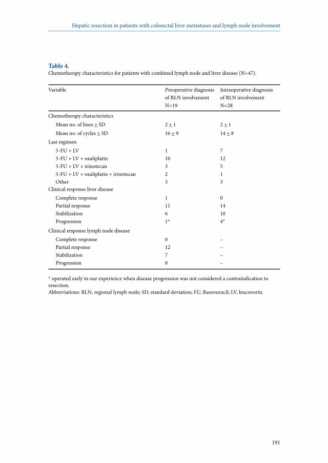
191
Hepatic resection in patients with colorectal liver metastases and lymph node involvement
Table 4. Chemotherapy characteristics for patients with combined lymph node and liver disease (N=47).
Variable Preoperative diagnosis of RLN involvementN=19
Intraoperative diagnosis of RLN involvementN=28
Chemotherapy characteristicsMean no. of lines + SD 2 + 1 2 + 1Mean no. of cycles + SD 16 + 9 14 + 8
Last regimen5-FU + LV5-FU + LV + oxaliplatin5-FU + LV + irinotecan5-FU + LV + oxaliplatin + irinotecanOther
110323
712513
Clinical response liver diseaseComplete responsePartial responseStabilizationProgression
11161*
014104*
Clinical response lymph node diseaseComplete responsePartial responseStabilizationProgression
01270
––––
* operated early in our experience when disease progression was not considered a contraindication to resection. Abbreviations: RLN, regional lymph node; SD, standard deviation; FU, fluorouracil; LV, leucovorin.
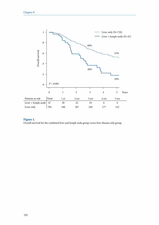
192
Chapter 8
0
.2
.4
.6
.8
1
Ove
rall
surv
ival
0 1 2 3 4 5 Years
Liver + lymph node (N=47) Liver only (N=710)
P < 0.001
18%
38%
53%
68%
Patients at risk Total 1 yr 2 yrs 3 yrs 4 yrs 5 yrs Liver + lymph node 47 38 22 10 6 4 Liver only 704 540 367 249 177 122
Figure 1. Overall survival for the combined liver and lymph node group versus liver disease only group.
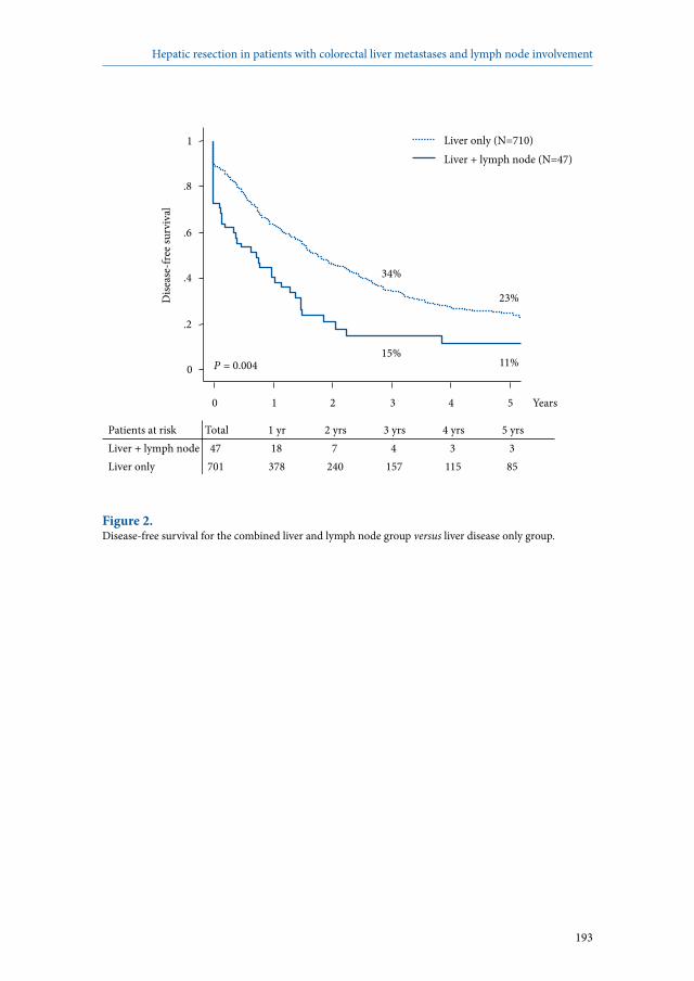
193
Hepatic resection in patients with colorectal liver metastases and lymph node involvement
Patients at risk Total 1 yr 2 yrs 3 yrs 4 yrs 5 yrs Liver + lymph node 47 18 7 4 3 3 Liver only 701 378 240 157 115 85
0
.2
.4
.6
.8
1
Dise
ase-
free s
urvi
val
0 1 2 3 4 5 Years
P = 0.004 11% 15%
23%
34%
Liver + lymph node (N=47) Liver only (N=710)
Figure 2. Disease-free survival for the combined liver and lymph node group versus liver disease only group.
194
Chapter 8
Outcomes In the RLN group, no patients died within 60 days postoperatively, compared with a 60-day mortality rate of 1% (six of 710 patients) in the group without RLN metastases. Morbidity rates were 21% (10/47 patients) and 18% (127/710 patients), respectively. For the RLN group, morbidity consisted of biliary leakage (N=1), hemorrhage (N=1), abdominal infected fluid collection (N=3), abdominal noninfected fluid collection (N=4), and transient liver insufficiency (N=1). The mean follow-up interval for the RLN group was 29 months, and for patients alive at last follow-up, the mean interval was 30 months. Only two patients were lost to follow-up. At the time of last follow-up, 32 patients (68%) had died of disease progression, and 15 patients were alive (32%), seven of whom were disease-free (15%). Within the RLN group, 19 patients (40%) underwent a repeat hepatectomy, compared with 223 patients (31%) in the liver disease only group (P=0.20). Three- and 5-year overall survival rates in the RLN group and the group without RLN involvement were 38% and 18% versus 68% and 53%, respectively (P<0.001) (Figure 1). Median survivals were 28 months (95% CI: 21-34 months) and 65 months (95% CI: 55-75 months) from hepatectomy and 34 months (95% CI: 26-43 months) and 75 months (95% CI: 61-89 months) from CLM diagnosis, respectively. Disease-free survival rates after 3 and 5 years were significantly lower in the RLN group compared with the liver disease only group (15% and 11% vs 34% and 23%; median 9 months [95% CI: 2-16 months] vs 20 months [95% CI: 18-23 months]; P=0.004) (Figure 2). Five-year overall survival for patients preoperatively diagnosed with RLN involvement was 35%, compared with 10% when the diagnosis was intraoperative (median survival: 43 months [95% CI: 21-64 months] vs 27 months [95% CI: 15-38 months]; P=0.18). When liver disease responded to chemotherapy (N=26), 5-year survival was 26%, compared with 17% in case of stabilization or progression (N=21) (P=0.08). Survival was similar whether RLN disease responded to chemotherapy (N=12) or was only stabilized (N=7) (5-year survival rate 32% vs 35%, respectively; P=0.94). According to the location of RLN metastases, 5-year overall survival rates after hepatectomy were 25% in the pedicular RLN group vs 0% in both the celiac and para-aortic RLN groups (P=0.001). In the pedicular RLN group, median survival was 30 months (95% CI: 22-39 months), compared with 20 months (95% CI: 5-35 months) for the combined celiac and para-aortic RLN groups (P=0.53).
Prognostic factorsUnivariate analysis of potential prognostic factors within the RLN group identified three variables associated with poor outcome: age at hepatectomy (P=0.04), initial carcinoembryonic antigen level (P=0.02), and location of RLN metastases (P=0.001) (Table 6). In multivariate analysis, two variables were identified as independent poor prognostic factors: celiac location of RLN metastases (hazard ratio 4.7; P<0.001) and age at hepatectomy >40 years (hazard ratio 5.2; P=0.004).

195
Hepatic resection in patients with colorectal liver metastases and lymph node involvement
Table 5. Regional lymph node metastases characteristics.
Variable Pedicular RLN involvement N=26
Celiac RLN involvement N=14
Para-aortic RLN involvementN=7
P
RLN metastasesDiagnosis
Before hepatic resectionDuring hepatic resection
7 (27%)19 (73%)
6 (43%)8 (57%)
6 (86%)1 (14%)
0.02
Complete lymphadenectomy 26 (100%) 14 (100%) 6 (86%) 0.06Histologic lymph node involvement
MassiveModerateMicroscopicNecrosis
14 (56%)9 (36%)2 (8%)0 (0%)
9 (69%)3 (23%)1 (8%)0 (0%)
3 (43%)2 (29%)0 (0%)1 (14%)
0.27
Extrahepatic disease, other
NoneLungPeritoneumMultiple sites
16 (62%)8 (31%)1 (4%)1 (4%)
9 (64%)5 (36%)0 (0%)0 (0%)
6 (86%)1 (14%)0 (0%)0 (0%)
0.83
Recurrence* 21 (81%) 9 (64%) 5 (71%) 0.51HepaticPulmonaryLymphaticBoneSpleenOvaryPeritoneal
15832011
7420100
4120000
* combined recurrence patterns are included.Abbreviation: RLN, regional lymph node.
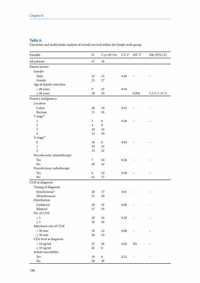
196
Chapter 8
Table 6. Univariate and multivariate analysis of overall survival within the lymph node group.
Variable N 5-yr OS (%) UV P MV P HR (95% CI)
All patients 47 18Patient factors
GenderMaleFemale
2225
1527
0.89 – –
Age at hepatic resection< 40 years> 40 years
938
4510
0.040.004 5.2 (1.7-15.7)
Primary malignancyLocation
Colon Rectum
3611
1916
0.45 – –
T stage44
1234
141814
001630
0.20 – –
N stage44
012
101812
02322
0.84 – –
Precolectomy chemotherapyYesNo
740
4416
0.46 – –
Precolectomy radiotherapyYesNo
641
2417
0.58 – –
CLM at diagnosisTiming of diagnosis
Synchronous*Metachronous
2027
1720
0.61 – –
DistributionUnilateralBilateral
2027
1619
0.80 – –
No. of CLM< 3> 3
2026
1620
0.29 – –
Maximum size of CLM< 30 mm> 30 mm
1626
1419
0.86 – –
CEA level at diagnosis< 10 ng/ml> 10 ng/ml
2120
260
0.02 NS –
Initial resectabilityYesNo
1928
828
0.22 – –
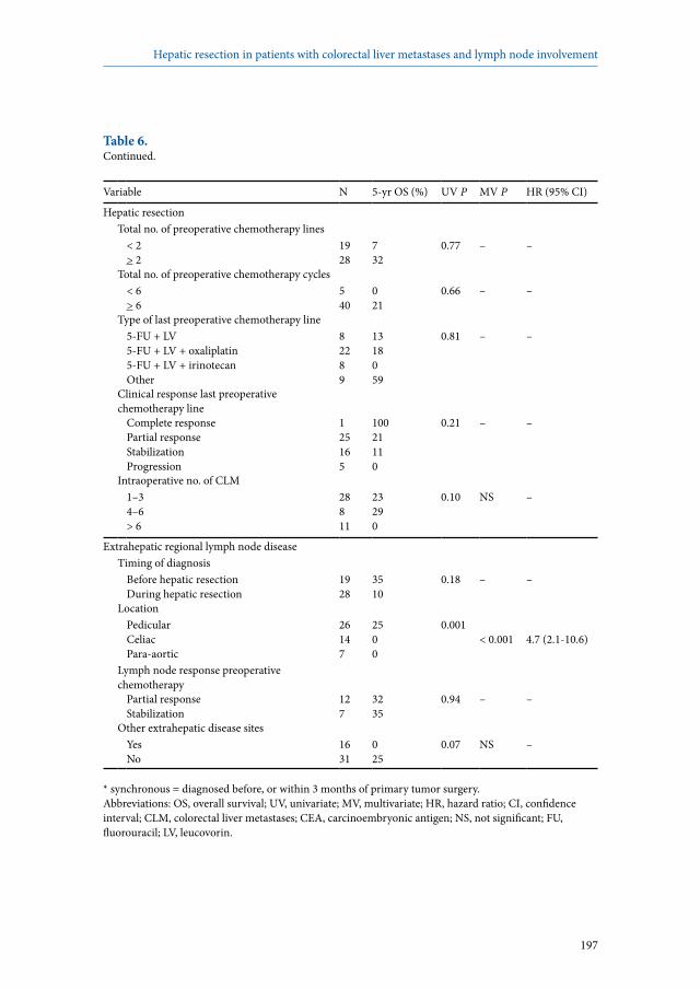
197
Hepatic resection in patients with colorectal liver metastases and lymph node involvement
Table 6. Continued.
Variable N 5-yr OS (%) UV P MV P HR (95% CI)
Hepatic resectionTotal no. of preoperative chemotherapy lines
< 2> 2
1928
732
0.77 – –
Total no. of preoperative chemotherapy cycles< 6> 6
540
021
0.66 – –
Type of last preoperative chemotherapy line5-FU + LV5-FU + LV + oxaliplatin5-FU + LV + irinotecanOther
82289
1318059
0.81 – –
Clinical response last preoperative chemotherapy line
Complete responsePartial responseStabilizationProgression
125165
10021110
0.21 – –
Intraoperative no. of CLM1–34–6> 6
28811
23290
0.10 NS –
Extrahepatic regional lymph node diseaseTiming of diagnosis
Before hepatic resectionDuring hepatic resection
1928
3510
0.18 – –
LocationPedicularCeliacPara-aortic
26147
2500
0.001< 0.001 4.7 (2.1-10.6)
Lymph node response preoperative chemotherapy
Partial responseStabilization
127
3235
0.94 – –
Other extrahepatic disease sitesYesNo
1631
025
0.07 NS –
* synchronous = diagnosed before, or within 3 months of primary tumor surgery. Abbreviations: OS, overall survival; UV, univariate; MV, multivariate; HR, hazard ratio; CI, confidence interval; CLM, colorectal liver metastases; CEA, carcinoembryonic antigen; NS, not significant; FU, fluorouracil; LV, leucovorin.
198
Chapter 8
Discussion
Although RLN involvement remains a generally poor prognostic factor, selected patients with RLN involvement seem to benefit from a multimodality treatment strategy that includes chemotherapy followed by surgical resection of both CLM and RLN disease. Specifically, the present study indicates that hepatectomy combined with lymphadenectomy could offer patients with simultaneous RLN involvement a 5-year survival rate of 18% with no operative mortality. Patients presenting with pedicular RLN involvement experienced more favorable postoperative outcomes (5-year survival rate of 25%) than those with celiac and/or para-aortic RLN involvement (no 5-year survivors). Furthermore, patients younger than 40 years experienced better long-term survival than older patients (5-year survival rate of 45% vs 10%, respectively). These results make an important contribution to the still controversial topic of hepatectomy combined with lymphadenectomy in patients with CLM. Only three of the 16 series directly addressing outcomes after combined hepatectomy and lymphadenectomy report any 5-year survivorship.3,12-26 On the basis of this experience, RLN involvement has generally been considered a contraindication to surgery. In contrast, our institution has maintained the indication for combined hepatectomy with lymphadenectomy. As a result of the dramatic improvements in the effectiveness of chemotherapy both in terms of response and survival,30-39 we have continued to offer surgery to these patients, provided that no progression of RLN disease was observed and that resection was potentially curative. To our knowledge, the present study reports the largest single-institution series of patients with resected RLN and is the first one reporting on a cohort of patients all treated with preoperative chemotherapy.As with any aggressive treatment approach, the results of this study should be assessed with regards both to the risk and to the long-term benefit of this combined approach. In regard to the risks, no patients died within 60 days of combined hepatectomy with regional lymphadenectomy. Although these results are in accordance with previous reports,3,15,17,21 our cohort contains more patients undergoing lymphadenectomy for para-aortic RLN involvement. In our series, lymphadenectomy for para-aortic RLN disease was performed in seven patients who represented 15% of all patients with RLN involvement. On the basis of this experience with extensive lymphadenectomy in the absence of perioperative mortality, we conclude that combined hepatectomy with lymphadenectomy can be done safely. Regarding the potential benefit of this combined surgical procedure, a substantial 5-year survival rate of 18% was obtained for the total RLN group. As expected, when compared with patients without RLN involvement, patients with CLM and

199
Hepatic resection in patients with colorectal liver metastases and lymph node involvement
simultaneous RLN involvement shared other poor prognostic features including more advanced T stage of the primary tumor, and higher incidence of initially unresectable CLM. Accordingly, the 5-year survival rate for patients in the RLN group was lower compared with that of the control group (18% vs 53%; P<0.001). Despite these survival differences, the 18% 5-year survival rate observed in our series compares favorably with the outcomes reported in the majority of studies addressing this topic. Possible explanations for these better results are the systematic use of preoperative chemotherapy and the selection of surgical candidates based on the absence of progression of RLN disease before surgery. The site of RLN metastases was strongly associated with long-term outcomes. For patients with pedicular nodal involvement, the 5-year overall survival rate was 25%, compared with 0% for either celiac or para-aortic RLN involvement. To date, the prognostic distinction between pedicular and celiac RLN involvement has only been made by Jaeck et al,18 who reported 17 patients, eight of whom presented with pedicular RLN metastases and the remaining nine patients with celiac RLN metastases. In this series, reported 3-year survival rates for the pedicular and celiac RLN groups were 38% and 0%, respectively. Although limited in the number of patients and in the duration of follow-up, these results agree with our present findings. Combined, these data suggest that although chemotherapy may have changed the spectrum of disease for patients with pedicular involvement, the prognosis continues to be poor for more distant RLN disease.The timing of diagnosis of RLN involvement also seems to impact the efficacy of this approach. When RLN metastases were diagnosed intraoperatively, the 5-year survival rate was only 10%. In contrast, patients with preoperative identification of suspicious regional lymphadenopathy experienced a 5-year survival rate of 35% (P=0.18). The difference between preoperative and intraoperative identification of RLN metastases has, to our knowledge, never been explored. In practice, surgeons are frequently confronted with intraoperative discovery of metastatic RLN. As was the case in 60% of our study patients, these involved nodes are indeed often underdiagnosed on preoperative imaging for a variety of reasons. The proportion of patients with intraoperatively discovered RLN involvement in our experience may be explained by the limited use of [18F]fluorodeoxyglucose (FDG) PET scanning at our institution. In a recent meta-analysis, both the sensitivity and specificity of FDG-PET imaging for the detection of extrahepatic lesions exceeded 90%, compared with only 60.9% and 91.1%, respectively, for CT scanning.45 Furthermore, a systematic review of FDG-PET imaging results in patients with CLM has determined that FDG-PET may detect extrahepatic disease not identified by other modalities in 10% to 32% of patients.46 When diagnosed intraoperatively, there is no consensus regarding whether or not to proceed with hepatectomy. In our practice, when the diagnosis of RLN
200
Chapter 8
involvement was made intraoperatively, we used the intrahepatic tumoral response to preoperative chemotherapy as a guide. In all but four of these patients, CLM had been stabilized or responded to preoperative chemotherapy, and we proceeded with combined hepatectomy and lymphadenectomy. When RLN involvement was diagnosed preoperatively, lymphadenectomy and hepatectomy were only performed in patients with chemotherapy-induced response or stabilization of RLN disease. Regardless of diagnostic timing, only those patients in whom both the hepatectomy and lymphadenectomy could be curative underwent resection. That patients with RLN involvement younger than 40 years experienced better outcomes after surgery was an unexpected finding of our study. Only 10% of the 38 patients with RLN involvement who were older than 40 years survived 5 years, whereas the nine patients younger than 40 years experienced a 45% 5-year survival rate. Obviously, this survival difference was not related to a decreased operative mortality, because the operative mortality in the RLN involved group was nil. Additionally, surgical procedures did not differ between both age groups and the rate of postoperative chemotherapy was similarly distributed (89% of patients <40 years vs 87% of patients >40 years). A slightly higher proportion of older patients had celiac or para-aortic RLN involvement (47% vs 33%), which may have contributed to the observed survival difference. In addition, these outcomes data may also reflect a younger patient’s ability to tolerate an aggressive multimodality regimen. In summary, the perioperative mortality-free 5-year survival rate of 25% in patients with CLM and isolated pedicular RLN involvement justifies combined hepatectomy with lymphadenectomy. This strategy is best suited to younger patients and those with preoperatively diagnosed RLN involvement that responds to systemic chemotherapy. In contrast, it is now clear that patients with either celiac or para-aortic RLN metastases, even when responding to preoperative chemotherapy, do not benefit from this approach.

201
Hepatic resection in patients with colorectal liver metastases and lymph node involvement
References
1. Adam R, Pascal G, Azoulay D et al. Liver resection for colorectal metastases: the third hepatectomy. Ann Surg 2003; 238:871-883.
2. Fong Y, Cohen AM, Fortner JG et al. Liver resection for colorectal metastases. J Clin Oncol 1997; 15:938-946.
3. Iwatsuki S, Dvorchik I, Madariaga JR et al. Hepatic resection for metastatic colorectal adenocarcinoma: a proposal of a prognostic scoring system. J Am Coll Surg 1999; 189:291-299.
4. Nordlinger B, Guiguet M, Vaillant JC et al. Surgical resection of colorectal carcinoma metastases to the liver. A prognostic scoring system to improve case selection, based on 1568 patients. Association Française de Chirurgie. Cancer 1996; 77:1254-1262.
5. Scheele J, Stangl R, Altendorf-Hofmann A et al. Resection of colorectal liver metastases. World J Surg 1995; 19:59-71.
6. Adam R, Laurent A, Azoulay D et al. Two-stage hepatectomy: A planned strategy to treat irresectable liver tumors. Ann Surg 2000; 232:777-785.
7. Azoulay D, Eshkenazy R, Andreani P et al. In situ hypothermic perfusion of the liver versus standard total vascular exclusion for complex liver resection. Ann Surg 2005; 241:277-285.
8. Azoulay D, Andreani P, Maggi U et al. Combined liver resection and reconstruction of the supra-renal vena cava: the Paul Brousse experience. Ann Surg 2006; 244:80-88.
9. Hemming AW, Reed AI, Langham MR Jr et al. Combined resection of the liver and inferior vena cava for hepatic malignancy. Ann Surg 2004; 239:712-719.
10. Adam R, Delvart V, Pascal G et al. Rescue surgery for unresectable colorectal liver metastases downstaged by chemotherapy: a model to predict long-term survival. Ann Surg 2004; 240:644-657.
11. Giacchetti S, Itzhaki M, Gruia G et al. Long-term survival of patients with unresectable colorectal cancer liver metastases following infusional chemotherapy with 5-fluorouracil, leucovorin, oxaliplatin and surgery. Ann Oncol 1999; 10:663-669.
12. Beckurts KT, Hölscher AH, Thorban S et al. Significance of lymph node involvement at the hepatic hilum in the resection of colorectal liver metastases. Br J Surg 1997; 84:1081-1084.
13. Chang AE, Schneider PD, Sugarbaker PH et al. A prospective randomized trial of regional versus systemic continuous 5-fluorodeoxyuridine chemotherapy in the treatment of colorectal liver metastases. Ann Surg 1987; 206:685-693.
14. Ekberg H, Tranberg KG, Andersson R et al. Determinants of survival in liver resection for colorectal secondaries. Br J Surg 1986; 73:727-731.
15. Elias D, Saric J, Jaeck D et al. Prospective study of microscopic lymph node involvement of the hepatic pedicle during curative hepatectomy for colorectal metastases. Br J Surg 1996; 83:942-945.
16. Elias D, Ouellet JF, Bellon N et al. Extrahepatic disease does not contraindicate hepatectomy for colorectal liver metastases. Br J Surg 2003; 90:567-574.
17. Gayowski TJ, Iwatsuki S, Madariaga JR et al. Experience in hepatic resection for metastatic colorectal cancer: analysis of clinical and pathologic risk factors. Surgery 1994; 116:703-710.
18. Jaeck D, Nakano H, Bachellier P et al. Significance of hepatic pedicle lymph node involvement in patients with colorectal liver metastases: a prospective study. Ann Surg Oncol 2002; 9:430-438.
19. Jamison RL, Donohue JH, Nagorney DM et al. Hepatic resection for metastatic colorectal cancer results in cure for some patients. Arch Surg 1997; 132:505-510.
20. Kokudo N, Sato T, Seki M et al. Hepatic lymph node involvement in resected cases of liver metastases from colorectal cancer. Dis Colon Rectum 1999; 42:1285-1290.
21. Minagawa M, Makuuchi M, Torzilli G et al. Extension of the frontiers of surgical indications in the treatment of liver metastases from colorectal cancer: long-term results. Ann Surg 2000; 231:487-499.

202
Chapter 8
22. Nakamura S, Suzuki S, Konno H. Resection of hepatic metastases of colorectal carcinoma: 20 years’ experience. J Hepatobiliary Pancreat Surg 1999; 6:16-22.
23. Nordlinger B, Jaeck D, Guiguet M et al. Résection chirurgicale des métastases hépatiques. Enquête de l’Association Française de Chirurgie. In: Nordlinger B, Jaeck D (eds). Métastases hépatiques des cancers colo-rectaux. Paris: Springer-Verlag; 1992:141-159.
24. Ohlsson B, Stenram U, Tranberg KG. Resection of colorectal liver metastases: 25-year experience. World J Surg 1998; 22:268-276.
25. Rosen CB, Nagorney DM, Taswell HF et al. Perioperative blood transfusion and determinants of survival after liver resection for metastatic colorectal carcinoma. Ann Surg 1992; 216:493-504.
26. Yasui K, Hirai T, Kato T et al. A new macroscopic classification predicts prognosis for patient with liver metastases from colorectal cancer. Ann Surg 1997; 226:582-586.
27. Poston GJ, Adam R, Alberts S et al. OncoSurge: a strategy for improving resectability with curative intent in metastatic colorectal cancer. J Clin Oncol 2005; 23:7125-7134.
28. Elias D, Sideris L, Pocard M et al. Results of R0 resection for colorectal liver metastases associated with extrahepatic disease. Ann Surg Oncol 2004; 11:274-280.
29. Elias D, Liberale G, Vernerey D et al. Hepatic and extrahepatic colorectal metastases: when resectable, their localization does not matter, but their total number has a prognostic effect. Ann Surg Oncol 2005; 12:900-909.
30. Falcone A, Masi G, Allegrini G et al. Biweekly chemotherapy with oxaliplatin, irinotecan, infusional fluorouracil, and leucovorin: a pilot study in patients with metastatic colorectal cancer. J Clin Oncol 2002; 20:4006-4014.
31. Köhne CH, van Cutsem E, Wils J et al. Phase III study of weekly high-dose infusional fluorouracil plus folinic acid with or without irinotecan in patients with metastatic colorectal cancer: European Organisation for Research and Treatment of Cancer Gastrointestinal Group Study 40986. J Clin Oncol 2005; 23:4856-4865.
32. Tournigand C, André T, Achille E et al. FOLFIRI followed by FOLFOX6 or the reverse sequence in advanced colorectal cancer: a randomized GERCOR study. J Clin Oncol 2004; 22:229-237.
33. Alberts SR, Horvath WL, Sternfeld WC et al. Oxaliplatin, fluorouracil, and leucovorin for patients with unresectable liver-only metastases from colorectal cancer: a North Central Cancer Treatment Group phase II study. J Clin Oncol 2005; 23:9243-9249.
34. de Gramont A, Figer A, Seymour M et al. Leucovorin and fluorouracil with or without oxaliplatin as first-line treatment in advanced colorectal cancer. J Clin Oncol 2000; 18:2938-2947.
35. Douillard JY, Cunningham D, Roth AD et al. Irinotecan combined with fluorouracil compared with fluorouracil alone as first-line treatment for metastatic colorectal cancer: a multicentre randomised trial. Lancet 2000; 355:1041-1047.
36. Giacchetti S, Perpoint B, Zidani R et al. Phase III multicenter randomized trial of oxaliplatin added to chronomodulated fluorouracil-leucovorin as first-line treatment of metastatic colorectal cancer. J Clin Oncol 2000; 18:136-147.
37. Saltz LB, Cox JV, Blanke C et al. Irinotecan plus fluorouracil and leucovorin for metastatic colorectal cancer. Irinotecan Study Group. N Engl J Med 2000; 343:905-914.
38. Cunningham D, Humblet Y, Siena S et al. Cetuximab monotherapy and cetuximab plus irinotecan in irinotecan-refractory metastatic colorectal cancer. N Engl J Med 2004; 351:337-345.
39. Hurwitz H, Fehrenbacher L, Novotny W et al. Bevacizumab plus irinotecan, fluorouracil, and leucovorin for metastatic colorectal cancer. N Engl J Med 2004; 350:2335-2342.
40. Therasse P, Arbuck SG, Eisenhauer EA et al. New guidelines to evaluate the response to treatment in solid tumors. European Organization for Research and Treatment of Cancer, National Cancer Institute of the United States, National Cancer Institute of Canada. J Natl Cancer Inst 2000; 92:205-216.

203
Hepatic resection in patients with colorectal liver metastases and lymph node involvement
41. Bismuth H. Surgical anatomy and anatomical surgery of the liver. World J Surg 1982; 6:3-9.42. Couinaud C. Le foie: études anatomiques et chirurgicales. Paris: Masson et Cie; 1957.43. Azoulay D, Castaing D, Smail A et al. Resection of nonresectable liver metastases from
colorectal cancer after percutaneous portal vein embolization. Ann Surg 2000; 231:480-486.44. Sobin LH, Wittekind Ch. TNM classification of malignant tumours. Hoboken: John Wiley &
Sons; 2002.45. Wiering B, Krabbe PF, Jager GJ et al. The impact of fluor-18-deoxyglucose-positron emission
tomography in the management of colorectal liver metastases. Cancer 2005; 104:2658-2670.46. Yang YY, Fleshman JW, Strasberg SM. Detection and management of extrahepatic colorectal
cancer in patients with resectable liver metastases. J Gastrointest Surg 2007; 11:929-944.

Robbert J. de Haas1,2
Aida C. Rahy Martin1
Dennis A. Wicherts1,2
Daniel Azoulay1
Denis Castaing1
René Adam1
1 Centre Hépato-Biliaire, Hôpital Paul Brousse, Villejuif, France2 Department of Surgery, University Medical Center Utrecht, Utrecht,
The Netherlands
British Journal of Surgery 2009; 96:935-940
Long-term outcome in patients with adrenal metastases following resection of colorectal liver metastases
9
206
Chapter 9
Abstract
BackgroundThe prognostic significance of adrenal metastases (AM) in patients with colorectal liver metastases (CLM) remains unknown. The aim of this study was to determine the influence of AM on long-term outcome and the role of adrenalectomy in patients with CLM.
MethodsAll patients resected for CLM who developed AM at a single institution between 1992 and 2006 were included in the study. Their long-term outcome was compared with that of all other patients resected for CLM but without AM.
ResultsHepatectomy was performed in 796 patients, of whom 14 (2%) developed AM, a median of 28 months after initial diagnosis of CLM; the remaining 782 patients (98%) had no AM. All 14 patients had chemotherapy, and ten went on to adrenalectomy. Median survival after diagnosis of CLM was 50 months in patients with AM versus 68 months in those without (P=0.02). After diagnosis of AM, median survival was 23 months, whether or not adrenalectomy was performed.
Conclusion The development of AM after liver resection for colorectal cancer deposits carries a poor prognosis, and adrenalectomy is probably not warranted.
207
Adrenal metastases following resection of colorectal liver metastases
Introduction
Adrenal metastases are often found at autopsy,1 but it is uncommon for them to be identified in patients living with or recovering from colorectal cancer (Table 1).2-15 Interestingly, only two cases have been described in which extra-adrenal metastasis was present at identification of adrenal metastases.10,15 Adrenalectomy is well described for metastatic disease, but any additional benefit over chemotherapy alone is questionable in patients with colorectal cancer.6,7,11,12,14,16-18 Increased detection of extrahepatic colorectal metastases by modern imaging, including positron emission tomography (PET),19 results in a probable increase in the number of patients identified with adrenal metastases. As the optimal treatment strategy for colorectal adrenal metastases is still unclear, this study was undertaken to evaluate whether it is justifiable to resect adrenal metastases following liver resection.

208
Chapter 9
Patients and methods
Study designAll patients who underwent hepatectomy for colorectal liver metastases at the Paul Brousse Hospital between January 1992 and December 2006, and who developed adrenal metastases were included in this study. To evaluate the impact of adrenal metastases on long-term outcome, comparison was made with all other patients who had surgery for colorectal liver metastases during the same period. Patients were identified from our prospective database. Data were collected on patient demographics, primary colorectal tumor, liver metastases at initial diagnosis, first hepatectomy, adrenal metastases, and long-term outcome.
Hepatic metastasesPreoperative evaluation consisted of abdominal ultrasonography and computed tomography (CT), thoracic CT and colonoscopy. Preoperative chemotherapy was given when lesions were initially unresectable, when the estimated remnant liver was too small and/or when synchronous extrahepatic disease sites were present. More recently, chemotherapy has increasingly been given in a neoadjuvant setting, in patients with initially resectable liver metastases. All hepatectomies were performed with curative intent. Intraoperative ultrasonography was performed in all patients to determine the extent of liver disease. The extent of hepatectomy was classified as major (three or more liver segments) or minor (fewer than three liver segments), according to Couinaud’s classification.20,21 Resectable, concomitant extrahepatic disease was not considered a contraindication to surgery. If located within the abdominal cavity, extrahepatic metastases were resected at the hepatectomy. If outside the abdomen, resection of extrahepatic disease was postponed for 2-3 months, with chemotherapy in the interim, to prevent disease progression. At 1 month after surgery, and then every 4 months, follow-up included the history, physical examination, serum tumor markers, liver function parameters and abdominal ultrasonography. Abdominal and thoracic CT was performed every 8 months. Most patients received adjuvant chemotherapy to reduce the risk of recurrence.
Adrenal metastasesAdrenal metastases were diagnosed by CT during preoperative investigation for hepatectomy, at operation, or during postoperative surveillance. If suspected, resection of adrenal metastases was combined with hepatectomy, following histologic confirmation of their malignant nature, provided that complete resection of both colorectal liver metastases and adrenal metastases could be performed. If diagnosed during follow-up after hepatectomy, adrenal metastases were resected in a separate operation, again after histologic confirmation of the
209
Adrenal metastases following resection of colorectal liver metastases
diagnosis. Adrenalectomy was in all patients combined with systemic chemotherapy. When surgical resection could not be performed with curative intent, adrenal metastases were not removed and systemic chemotherapy alone was given.
Statistical analysisCategorical variables were compared using the χ2 test, and continuous data with the independent-samples t test. The Kaplan-Meier method was used to estimate survival probabilities, which were compared with the log-rank test. A P value <0.05 was considered statistically significant. All statistical calculations were performed with SPSS version 13.0 (SPSS Inc, Chicago, IL, USA).
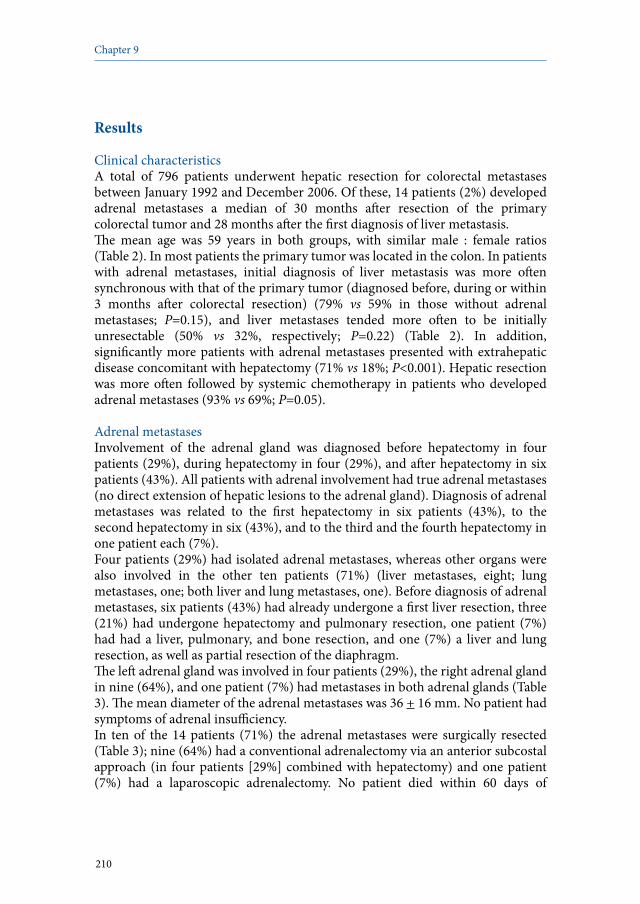
210
Chapter 9
Results
Clinical characteristicsA total of 796 patients underwent hepatic resection for colorectal metastases between January 1992 and December 2006. Of these, 14 patients (2%) developed adrenal metastases a median of 30 months after resection of the primary colorectal tumor and 28 months after the first diagnosis of liver metastasis. The mean age was 59 years in both groups, with similar male : female ratios (Table 2). In most patients the primary tumor was located in the colon. In patients with adrenal metastases, initial diagnosis of liver metastasis was more often synchronous with that of the primary tumor (diagnosed before, during or within 3 months after colorectal resection) (79% vs 59% in those without adrenal metastases; P=0.15), and liver metastases tended more often to be initially unresectable (50% vs 32%, respectively; P=0.22) (Table 2). In addition, significantly more patients with adrenal metastases presented with extrahepatic disease concomitant with hepatectomy (71% vs 18%; P<0.001). Hepatic resection was more often followed by systemic chemotherapy in patients who developed adrenal metastases (93% vs 69%; P=0.05).
Adrenal metastasesInvolvement of the adrenal gland was diagnosed before hepatectomy in four patients (29%), during hepatectomy in four (29%), and after hepatectomy in six patients (43%). All patients with adrenal involvement had true adrenal metastases (no direct extension of hepatic lesions to the adrenal gland). Diagnosis of adrenal metastases was related to the first hepatectomy in six patients (43%), to the second hepatectomy in six (43%), and to the third and the fourth hepatectomy in one patient each (7%). Four patients (29%) had isolated adrenal metastases, whereas other organs were also involved in the other ten patients (71%) (liver metastases, eight; lung metastases, one; both liver and lung metastases, one). Before diagnosis of adrenal metastases, six patients (43%) had already undergone a first liver resection, three (21%) had undergone hepatectomy and pulmonary resection, one patient (7%) had had a liver, pulmonary, and bone resection, and one (7%) a liver and lung resection, as well as partial resection of the diaphragm. The left adrenal gland was involved in four patients (29%), the right adrenal gland in nine (64%), and one patient (7%) had metastases in both adrenal glands (Table 3). The mean diameter of the adrenal metastases was 36 + 16 mm. No patient had symptoms of adrenal insufficiency. In ten of the 14 patients (71%) the adrenal metastases were surgically resected (Table 3); nine (64%) had a conventional adrenalectomy via an anterior subcostal approach (in four patients [29%] combined with hepatectomy) and one patient (7%) had a laparoscopic adrenalectomy. No patient died within 60 days of
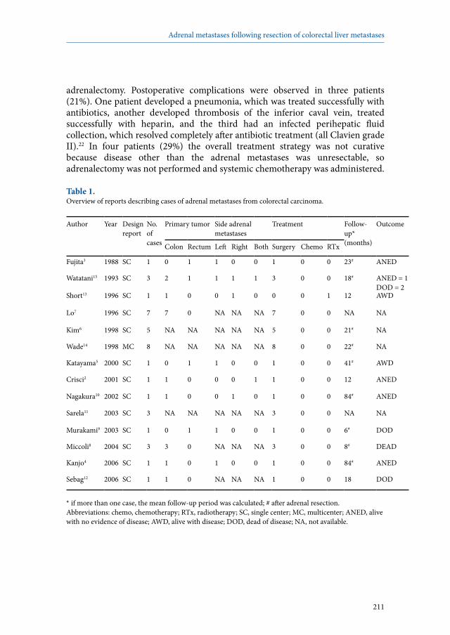
211
Adrenal metastases following resection of colorectal liver metastases
adrenalectomy. Postoperative complications were observed in three patients (21%). One patient developed a pneumonia, which was treated successfully with antibiotics, another developed thrombosis of the inferior caval vein, treated successfully with heparin, and the third had an infected perihepatic fluid collection, which resolved completely after antibiotic treatment (all Clavien grade II).22 In four patients (29%) the overall treatment strategy was not curative because disease other than the adrenal metastases was unresectable, so adrenalectomy was not performed and systemic chemotherapy was administered.
Table 1. Overview of reports describing cases of adrenal metastases from colorectal carcinoma.
Author Year Design report
No. of cases
Primary tumor Side adrenal metastases
Treatment Follow-up*(months)
Outcome
Colon Rectum Left Right Both Surgery Chemo RTx
Fujita3 1988 SC 1 0 1 1 0 0 1 0 0 23# ANED
Watatani15 1993 SC 3 2 1 1 1 1 3 0 0 18# ANED = 1DOD = 2
Short13 1996 SC 1 1 0 0 1 0 0 0 1 12 AWD
Lo7 1996 SC 7 7 0 NA NA NA 7 0 0 NA NA
Kim6 1998 SC 5 NA NA NA NA NA 5 0 0 21# NA
Wade14 1998 MC 8 NA NA NA NA NA 8 0 0 22# NA
Katayama5 2000 SC 1 0 1 1 0 0 1 0 0 41# AWD
Crisci2 2001 SC 1 1 0 0 0 1 1 0 0 12 ANED
Nagakura10 2002 SC 1 1 0 0 1 0 1 0 0 84# ANED
Sarela11 2003 SC 3 NA NA NA NA NA 3 0 0 NA NA
Murakami9 2003 SC 1 0 1 1 0 0 1 0 0 6# DOD
Miccoli8 2004 SC 3 3 0 NA NA NA 3 0 0 8# DEAD
Kanjo4 2006 SC 1 1 0 1 0 0 1 0 0 84# ANED
Sebag12 2006 SC 1 1 0 NA NA NA 1 0 0 18 DOD
* if more than one case, the mean follow-up period was calculated; # after adrenal resection.Abbreviations: chemo, chemotherapy; RTx, radiotherapy; SC, single center; MC, multicenter; ANED, alive with no evidence of disease; AWD, alive with disease; DOD, dead of disease; NA, not available.
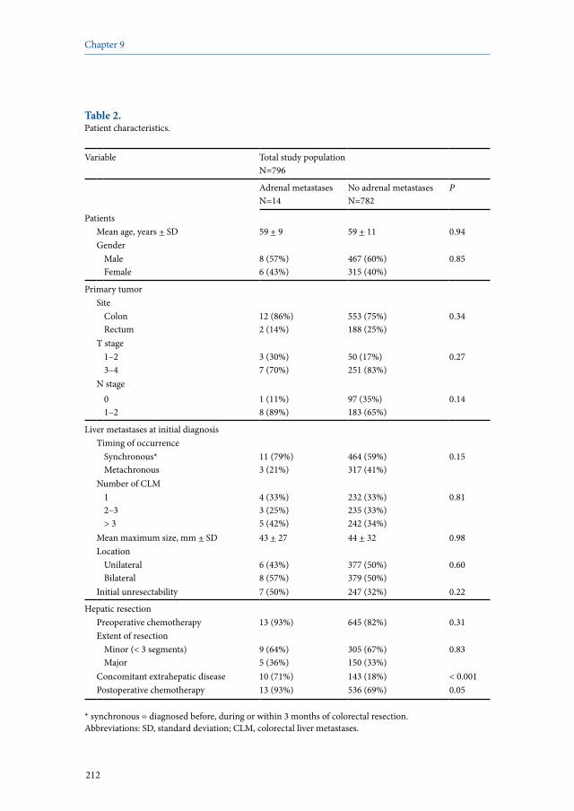
212
Chapter 9
Table 2. Patient characteristics.
Variable Total study populationN=796
Adrenal metastasesN=14
No adrenal metastasesN=782
P
PatientsMean age, years + SD 59 + 9 59 + 11 0.94Gender
MaleFemale
8 (57%)6 (43%)
467 (60%)315 (40%)
0.85
Primary tumorSite
ColonRectum
12 (86%)2 (14%)
553 (75%)188 (25%)
0.34
T stage1–23–4
3 (30%)7 (70%)
50 (17%)251 (83%)
0.27
N stage01–2
1 (11%)8 (89%)
97 (35%)183 (65%)
0.14
Liver metastases at initial diagnosisTiming of occurrence
Synchronous*Metachronous
11 (79%)3 (21%)
464 (59%)317 (41%)
0.15
Number of CLM 12–3> 3
4 (33%)3 (25%)5 (42%)
232 (33%)235 (33%)242 (34%)
0.81
Mean maximum size, mm + SD 43 + 27 44 + 32 0.98Location
UnilateralBilateral
6 (43%)8 (57%)
377 (50%)379 (50%)
0.60
Initial unresectability 7 (50%) 247 (32%) 0.22
Hepatic resectionPreoperative chemotherapy 13 (93%) 645 (82%) 0.31Extent of resection
Minor (< 3 segments)Major
9 (64%)5 (36%)
305 (67%)150 (33%)
0.83
Concomitant extrahepatic disease 10 (71%) 143 (18%) < 0.001Postoperative chemotherapy 13 (93%) 536 (69%) 0.05
* synchronous = diagnosed before, during or within 3 months of colorectal resection.Abbreviations: SD, standard deviation; CLM, colorectal liver metastases.
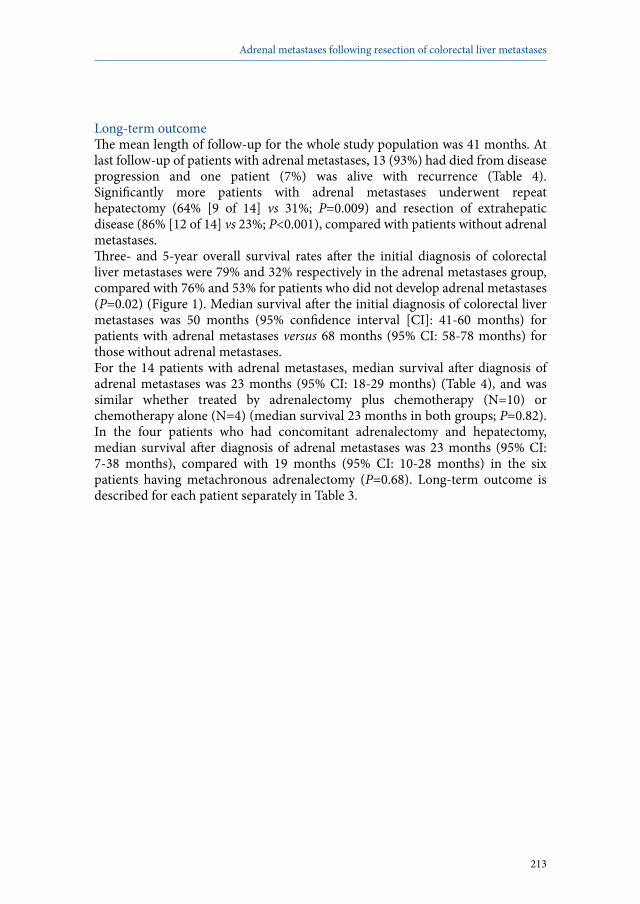
213
Adrenal metastases following resection of colorectal liver metastases
Long-term outcome The mean length of follow-up for the whole study population was 41 months. At last follow-up of patients with adrenal metastases, 13 (93%) had died from disease progression and one patient (7%) was alive with recurrence (Table 4). Significantly more patients with adrenal metastases underwent repeat hepatectomy (64% [9 of 14] vs 31%; P=0.009) and resection of extrahepatic disease (86% [12 of 14] vs 23%; P<0.001), compared with patients without adrenal metastases. Three- and 5-year overall survival rates after the initial diagnosis of colorectal liver metastases were 79% and 32% respectively in the adrenal metastases group, compared with 76% and 53% for patients who did not develop adrenal metastases (P=0.02) (Figure 1). Median survival after the initial diagnosis of colorectal liver metastases was 50 months (95% confidence interval [CI]: 41-60 months) for patients with adrenal metastases versus 68 months (95% CI: 58-78 months) for those without adrenal metastases. For the 14 patients with adrenal metastases, median survival after diagnosis of adrenal metastases was 23 months (95% CI: 18-29 months) (Table 4), and was similar whether treated by adrenalectomy plus chemotherapy (N=10) or chemotherapy alone (N=4) (median survival 23 months in both groups; P=0.82). In the four patients who had concomitant adrenalectomy and hepatectomy, median survival after diagnosis of adrenal metastases was 23 months (95% CI: 7-38 months), compared with 19 months (95% CI: 10-28 months) in the six patients having metachronous adrenalectomy (P=0.68). Long-term outcome is described for each patient separately in Table 3.
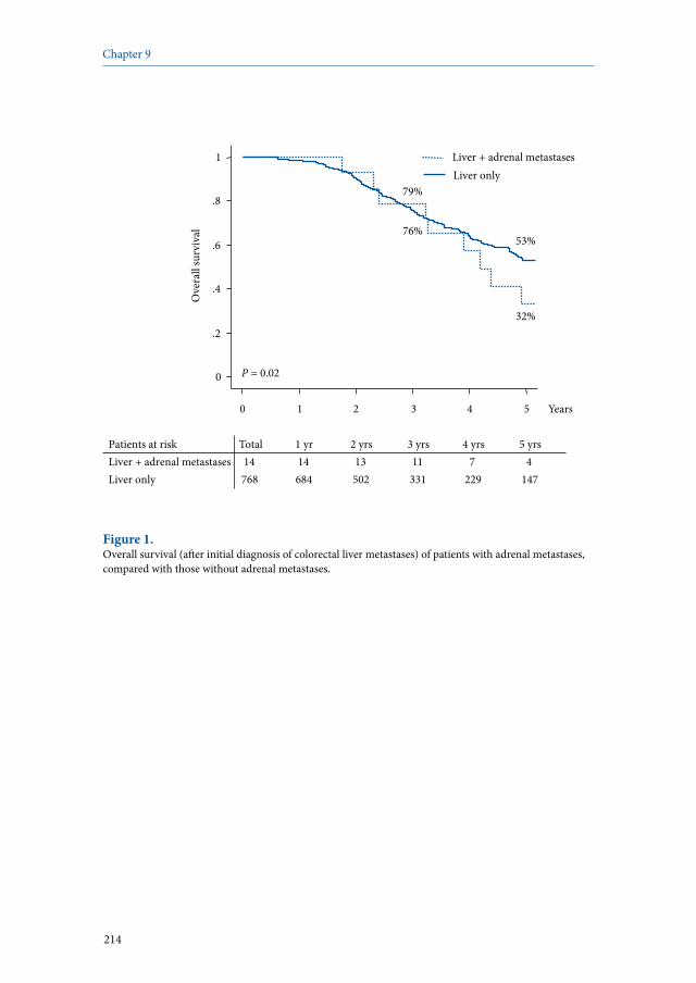
214
Chapter 9
Patients at risk Total 1 yr 2 yrs 3 yrs 4 yrs 5 yrs Liver + adrenal metastases 14 14 13 11 7 4 Liver only 768 684 502 331 229 147
0
.2
.4
.6
.8
1
Ove
rall
surv
ival
0 1 2 3 4 5 Years
P = 0.02
32%
79%
53% 76%
Liver only Liver + adrenal metastases
Figure 1. Overall survival (after initial diagnosis of colorectal liver metastases) of patients with adrenal metastases, compared with those without adrenal metastases.
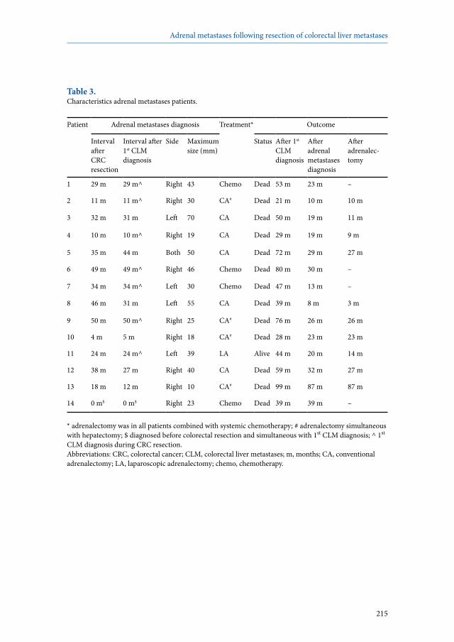
215
Adrenal metastases following resection of colorectal liver metastases
Table 3. Characteristics adrenal metastases patients.
Patient Adrenal metastases diagnosis Treatment* Outcome
Interval after CRC resection
Interval after 1st CLM diagnosis
Side Maximum size (mm)
Status After 1st CLM diagnosis
After adrenal metastases diagnosis
After adrenalec-tomy
1 29 m 29 m^ Right 43 Chemo Dead 53 m 23 m –
2 11 m 11 m^ Right 30 CA# Dead 21 m 10 m 10 m
3 32 m 31 m Left 70 CA Dead 50 m 19 m 11 m
4 10 m 10 m^ Right 19 CA Dead 29 m 19 m 9 m
5 35 m 44 m Both 50 CA Dead 72 m 29 m 27 m
6 49 m 49 m^ Right 46 Chemo Dead 80 m 30 m –
7 34 m 34 m^ Left 30 Chemo Dead 47 m 13 m –
8 46 m 31 m Left 55 CA Dead 39 m 8 m 3 m
9 50 m 50 m^ Right 25 CA# Dead 76 m 26 m 26 m
10 4 m 5 m Right 18 CA# Dead 28 m 23 m 23 m
11 24 m 24 m^ Left 39 LA Alive 44 m 20 m 14 m
12 38 m 27 m Right 40 CA Dead 59 m 32 m 27 m
13 18 m 12 m Right 10 CA# Dead 99 m 87 m 87 m
14 0 m$ 0 m$ Right 23 Chemo Dead 39 m 39 m –
* adrenalectomy was in all patients combined with systemic chemotherapy; # adrenalectomy simultaneous with hepatectomy; $ diagnosed before colorectal resection and simultaneous with 1st CLM diagnosis; ^ 1st
CLM diagnosis during CRC resection. Abbreviations: CRC, colorectal cancer; CLM, colorectal liver metastases; m, months; CA, conventional adrenalectomy; LA, laparoscopic adrenalectomy; chemo, chemotherapy.

216
Chapter 9
Table 4. Long-term outcome comparisons.
Variable Total study populationN=796
Adrenal metastasesN=14
No adrenal metastasesN=782
P
Recurrence 14 (100%) 527 (67%) 0.01HepaticExtrahepaticBoth
1 (7%)2 (14%)11 (79%)
136 (26%)159 (30%)232 (44%)
0.04
Total number of hepatectomies12> 3
5 (36%)6 (43%)3 (21%)
537 (69%)187 (24%)58 (7%)
0.02
Total number of EHD resections012> 3
2 (14%)6 (43%)4 (29%)2 (14%)
600 (77%)119 (15%)42 (5%)21 (3%)
< 0.001
Status at last follow-upAlive without diseaseAlive with diseaseDead
0 (0%)1 (7%)13 (93%)
285 (36%)245 (31%)252 (32%)
< 0.001
Median survival, months (95% CI)After diagnosis of first CLM 50 (41-60) 68 (58-78) 0.02After diagnosis of AM (N=14) 23 (18-29) –
Treated by adrenalectomy (N=10)* 23 (14-31)# –Adrenalectomy combined with HR (N=4)*Metachronous adrenalectomy (N=6)*
23 (7-38)^19 (10-28)^
––
Treated by chemotherapy (N=4)* 23 (7-40)# –
* survival calculated from the date of diagnosis of adrenal metastasis; # treated by adrenalectomy vs treated by chemotherapy: P=0.82; ^ adrenalectomy combined with HR vs metachronous adrenalectomy: P=0.68.Abbreviations: EHD, extrahepatic disease; CI, confidence interval; CLM, colorectal liver metastases; AM, adrenal metastases; HR, hepatic resection.
217
Adrenal metastases following resection of colorectal liver metastases
Discussion
As the prognostic significance of adrenal metastases in patients with colorectal liver metastases remains unknown, the principal aims of the present study were to determine their influence on long-term outcome after liver resection and the value of adrenal resection. Patients with adrenal metastases had a significantly lower 5-year survival rate than those without adrenal metastases (32% vs 53%; P=0.02). After diagnosis of adrenal metastases, median survival was 23 months, and was not influenced by the type of treatment (chemotherapy plus adrenalectomy, or chemotherapy alone). At last follow-up, of the patients with adrenal metastases all but one patient (93%) had died, compared with 36% of those without adrenal metastases (P<0.001). Within the English literature, only 14 case reports and small series of patients with adrenal metastases from colorectal cancer have been published, some of them reporting prolonged survival (Table 1).2-15 Notably, all but two case reports concerned isolated adrenal metastases,10,15 in contrast to the present study where other organs were also involved in ten of the 14 patients (71%). As a result of the improving diagnostic accuracy of CT in evaluating adrenal masses, as well as the introduction of PET, surgeons and medical oncologists will be faced more often with patients with adrenal metastases of colorectal origin.23-27 In addition, the significant increase in survival and follow-up observed in recent years favors the emergence of late metastatic sites, such as adrenal metastases. Therefore, it is important to know whether or not these patients correspond to a specific patient profile. In our experience, patients treated for colorectal liver metastases who also developed adrenal metastases tended more often to have initially unresectable liver metastases synchronous with the primary colorectal tumor, compared with patients without adrenal metastases. Furthermore, patients with adrenal metastases presented more often with extrahepatic disease concomitant with hepatectomy. They also had a significantly lower 5-year survival rate, although 3-year survival rates in patients with and without adrenal metastases were comparable. This finding emphasizes the adverse prognostic influence of the development of adrenal metastases, which occurred after a median period of 28 months after the initial diagnosis of liver metastases.To determine whether the present results might be considered representative of daily practice, the international registry LiverMetSurvey (http://www.livermetsurvey.org), a multicenter online registry of patients undergoing surgery for colorectal liver metastases, was consulted. Of a total of 7302 patients in the database, adrenal metastases had been found in only 32 patients (0.4%), thereby confirming the rarity of this particular patient group. Of these, 21 patients (66%) underwent adrenalectomy. Median survival after the initial diagnosis of liver metastasis was 53 months (95% CI: 41-64 months), that after diagnosis of adrenal metastases was 23 months (95% CI: 14-33 months), and the subgroup treated by
218
Chapter 9
adrenalectomy had a median survival of 25 months (95% CI: 14-33 months). Taken together, these multicenter results are in accord with the present findings. At our hospital, the treatment policy for patients presenting with intrahepatic and/or extrahepatic metastases of colorectal origin has always been surgical, provided that resection of all disease sites can be complete. Using this approach, in patients treated for colorectal liver metastases who presented with adrenal metastases, a median survival of 23 months after diagnosis of adrenal metastases was achieved whether treatment was with combined chemotherapy and adrenalectomy, or chemotherapy alone. However, one patient survived for more than 7 years following the combined treatment, indicating that long-term remission may be possible in highly selected patients. Therefore, we recommend to perform adrenalectomy only when patients are also scheduled for hepatectomy, as a combined procedure can be performed safely. When diagnosed during follow-up after hepatic resection for colorectal metastases, patients with adrenal metastases could be treated by chemotherapy alone, reserving adrenal resection for highly selected patients, as surgery does not appear to offer any benefit over chemotherapy alone.
219
Adrenal metastases following resection of colorectal liver metastases
References
1. Cedermark BJ, Blumenson LE, Pickren JW et al. The significance of metastases to the adrenal glands in adenocarcinoma of the colon and rectum. Surg Gynecol Obstet 1977; 144:537-546.
2. Crisci A, Cartei G, De Antoni P et al. Surgical management of isolated bilateral adrenal metastases from colon carcinoma causing adrenal insufficiency. Urol Int 2001; 67:113-116.
3. Fujita K, Kameyama S, Kawamura M. Surgically removed adrenal metastasis from cancer of the rectum. Report of a case. Dis Colon Rectum 1988; 31:141-143.
4. Kanjo T, Albertini M, Weber S. Long-term disease-free survival after adrenalectomy for isolated colorectal metastases. Asian J Surg 2006; 29:291-293.
5. Katayama A, Mafune K, Makuuchi M. Adrenalectomy for solitary adrenal metastasis from colorectal carcinoma. Jpn J Clin Oncol 2000; 30:414-416.
6. Kim SH, Brennan MF, Russo P et al. The role of surgery in the treatment of clinically isolated adrenal metastasis. Cancer 1998; 82:389-394.
7. Lo CY, van Heerden JA, Soreide JA et al. Adrenalectomy for metastatic disease to the adrenal glands. Br J Surg 1996; 83:528-531.
8. Miccoli P, Materazzi G, Mussi A et al. A reappraisal of the indications for laparoscopic treatment of adrenal metastases. J Laparoendosc Adv Surg Tech A 2004; 14:139-145.
9. Murakami S, Terakado M, Hashimoto T et al. Adrenal metastasis from rectal cancer: report of a case. Surg Today 2003; 33:126-130.
10. Nagakura S, Shirai Y, Nomura T et al. Long-term survival after resection of colonic adenocarcinoma with synchronous metastases to the liver, adrenal gland, and aortic-caval lymph nodes: report of a case. Dis Colon Rectum 2002; 45:1679-1680.
11. Sarela AI, Murphy I, Coit DG et al. Metastasis to the adrenal gland: the emerging role of laparoscopic surgery. Ann Surg Oncol 2003; 10:1191-1196.
12. Sebag F, Calzolari F, Harding J et al. Isolated adrenal metastasis: the role of laparoscopic surgery. World J Surg 2006; 30:888-892.
13. Short S, Chaturvedi A, Leslie MD. Palliation of symptomatic adrenal gland metastases by radiotherapy. Clin Oncol (R Coll Radiol) 1996; 8:387-389.
14. Wade TP, Longo WE, Virgo KS et al. A comparison of adrenalectomy with other resections for metastatic cancers. Am J Surg 1998; 175:183-186.
15. Watatani M, Ooshima M, Wada T et al. Adrenal metastasis from carcinoma of the colon and rectum: a report of three cases. Surg Today 1993; 23:444-448.
16. Falcone A, Ricci S, Brunetti I et al. Phase III trial of infusional fluorouracil, leucovorin, oxaliplatin, and irinotecan (FOLFOXIRI) compared with infusional fluorouracil, leucovorin, and irinotecan (FOLFIRI) as first-line treatment for metastatic colorectal cancer: the Gruppo Oncologico Nord Ovest. J Clin Oncol 2007; 25:1670-1676.
17. Köhne CH, van Cutsem E, Wils J et al. Phase III study of weekly high-dose infusional fluorouracil plus folinic acid with or without irinotecan in patients with metastatic colorectal cancer: European Organisation for Research and Treatment of Cancer Gastrointestinal Group Study 40986. J Clin Oncol 2005; 23:4856-4865.
18. Tournigand C, André T, Achille E et al. FOLFIRI followed by FOLFOX6 or the reverse sequence in advanced colorectal cancer: a randomized GERCOR study. J Clin Oncol 2004; 22:229-237.
19. Yang YY, Fleshman JW, Strasberg SM. Detection and management of extrahepatic colorectal cancer in patients with resectable liver metastases. J Gastrointest Surg 2007; 11:929-944.
20. Bismuth H. Surgical anatomy and anatomical surgery of the liver. World J Surg 1982; 6:3-9.21. Couinaud C. Le foie: études anatomiques et chirurgicales. Paris: Masson et Cie; 1957.22. Dindo D, Demartines N, Clavien PA. Classification of surgical complications: a new proposal
with evaluation in a cohort of 6336 patients and results of a survey. Ann Surg 2004; 240:205-213.
220
Chapter 9
23. Boland GW, Hahn PF, Peña C et al. Adrenal masses: characterization with delayed contrast-enhanced CT. Radiology 1997; 202:693-696.
24. Caoili EM, Korobkin M, Francis IR et al. Adrenal masses: characterization with combined unenhanced and delayed enhanced CT. Radiology 2002; 222:629-633.
25. Erasmus JJ, Patz EF Jr, McAdams HP et al. Evaluation of adrenal masses in patients with bronchogenic carcinoma using 18F-fluorodeoxyglucose positron emission tomography. AJR Am J Roentgenol 1997; 168:1357-1360.
26. Mayo-Smith WW, Boland GW, Noto RB et al. State-of-the-art adrenal imaging. Radiographics 2001; 21:995-1012.
27. Yun M, Kim W, Alnafisi N et al. 18F-FDG PET in characterizing adrenal lesions detected on CT or MRI. J Nucl Med 2001; 42:1795-1799.

221
Adrenal metastases following resection of colorectal liver metastases


Summary 10
224
Chapter 10
Background
Colorectal cancer remains one of the most common malignancies worldwide, with liver metastases being found in approximately 50% of these patients.1,2 Therefore, colorectal liver metastases (CLM) concern a major health issue. Unfortunately, at the time of diagnosis, only 20% of patients with CLM are directly amenable for surgery.3 The remaining 80% of these patients present with initially unresectable metastases, mainly caused by multinodularity, large size, close relationship with major vascular or biliary structures and extensive extrahepatic disease. Patients are considered resectable as long as all (liver) metastases can be macroscopically completely resected while preserving at least 25-30% of remnant liver with adequate vascular supply and biliary drainage.4 As surgery is still the only potentially curative treatment option for these patients, many strategies have been developed to increase the number of patients with resectable liver disease. Modern chemotherapy regimens are nowadays widely used to convert initially unresectable CLM to a resectable situation.5 If chemotherapy alone is not sufficient to allow surgery, specific surgical techniques such as portal vein embolization which allows hypertrophy of a small remnant liver volume,6 vascular resection and reconstruction techniques,7 two-stage hepatectomy,8 and local treatment modalities,9 can be used to increase the number of resectable patients. Taken together, the modern treatment of CLM should be multidisciplinary, necessitating a close collaboration between surgeons and oncologists, with frequent re-evaluations and adequate timing to optimize therapeutic strategies on an individual basis. This multidisciplinary treatment of CLM is the central theme of this thesis. In part I, factors determining outcome following hepatic resection for CLM are investigated, while in part II the place of surgery in patients with both CLM and extrahepatic disease is discussed.

225
Summary
Part I Factors determining outcome following hepatic resection for colorectal liver metastases Timing of liver surgery for synchronous colorectal liver metastasesThe optimal timing of liver surgery for synchronous CLM remains controversial. Should the primary colorectal tumor be resected first and the liver resection be delayed for 2-3 months with chemotherapy in the interim, enabling selection of best candidates for surgery? Or should hepatic and colorectal surgery be combined with the advantage of a single operation in which all tumoral disease can be removed? To answer this question, we compared in chapter 2 simultaneous colorectal and liver resection with delayed liver surgery in patients with limited liver disease, focusing on short- and long-term outcomes both in the total study population as well as in highly comparable case-matched subgroups. Although postoperative mortality was comparable for the two treatment strategies, the morbidity rate was significantly lower after simultaneous colorectal and hepatic resection. In addition, disease recurrence was observed more often in patients who underwent a simultaneous strategy, and at multivariate analysis, a simultaneous strategy was identified as an independent predictor of disease recurrence. Overall survival rates were comparable for both surgical strategies, but progression-free survival was significantly better after delayed hepatic surgery. After case-matching for age, gender, number and location of CLM, similar results were found. Thus, colorectal resection can safely be combined with a limited hepatectomy in patients with synchronous CLM with respect to postoperative morbidity. However, the combined strategy has a negative impact on progression-free survival and therefore raises questions regarding oncological value and use in clinical practice.
Expansion of criteria for resectabilityAs surgical resection is still the only potentially curative treatment option for patients with CLM, many efforts have been made during the past decade to increase the number of patients with resectable disease. As mentioned earlier, strategies used to increase the number of CLM patients amenable for surgery are the administration of systemic chemotherapy, portal vein embolization, vascular resection and reconstruction techniques, two-stage hepatectomy, and the use of local treatment modalities. As a result, resectability is no longer defined by what is removed, but decisions concerning resectability now center around what will remain after resection. Such an expansion of the criteria for resectability is only justified when perioperative mortality and morbidity rates remain within acceptable limits together with reasonable long-term outcomes. As this is still to be ascertained in an era of modern liver surgery, we decided to determine short-term and long-term outcomes following hepatic resection for CLM both within the total patient population as well as in separate time periods (1990-2000 vs
226
Chapter 10
2000-2010) which enabled us to evaluate trends over time (chapter 3). Within the total study population, overall perioperative mortality and postoperative morbidity rates were 1.3% and 33%, respectively. Although patients who underwent hepatic resection during the latter period (2000-2010) were older, had a higher prevalence of comorbid illnesses, and presented with more extensive liver disease necessitating more complex surgical procedures, the perioperative mortality rate was similar to that observed in our initial experience (1990-2000). However, a trend towards a higher postoperative complication rate was observed in the more recently operated cohort. In the more recent experience, the mortality rate following repeat hepatectomy was higher than that observed after first hepatectomy, while the morbidity rate was comparable. Four independent predictors of postoperative morbidity were identified: treatment between 2000 and 2010, total hepatic ischemia time >60 minutes, maximum size of liver metastases >30 mm at histopathology, and the presence of abnormalities in the nontumoral liver parenchyma. Although a trend towards lower overall survival was observed in patients who developed significant postoperative complications (Dindo-Clavien grade III-IV), no differences were observed in long-term outcomes between both treatment periods. Because short-term outcomes observed in our study remained within acceptable limits, without worsening of long-term outcomes, an expansion of the resectability criteria of CLM is justified as this is the only chance of long-term survival and even cure.
Impact of young ageAlthough it becomes more and more clear that hepatic resection for CLM can be safely performed with favorable long-term results in elderly patients,10 outcomes after hepatic resection in young patients have never been evaluated. Therefore, in chapter 4 we compared long-term outcomes in patients aged <40 years with patients older than 40 years. Young patients were found to have more extensive liver disease and a trend towards lower overall survival was observed compared with patients older than 40 years. In addition, young patients more often developed a disease recurrence, and progression-free survival in this patient group was significantly lower than that observed for older patients. However, as recurrences in young patients were often treated by repeat surgery, disease-free survival rates did not differ between the groups. At multivariate analysis, young age independently predicted poor progression-free survival. These findings underscore the importance of recognizing the poor outcome of CLM in young patients and the need to consider an aggressive approach to diagnosis and early treatment.
227
Summary
Surgical margin statusMost clinicians consider the inability to achieve clear resection margins as contraindication to surgery, as it has been associated with significantly reduced survival rates and an increased risk of surgical margin recurrence.11,12 Currently, owing to the increased number of patients with multinodular disease and/or CLM close to important vascular structures, macroscopic complete resection of all CLM can sometimes only be performed with positive surgical margins (R1 resection). And although complete macroscopic removal of all lesions with negative resection margins is still the gold standard recommendation in the surgical treatment of CLM, increasingly efficient chemotherapy may have changed long-term outcome after R1 resection. Therefore, we compared long-term outcome of R0 (negative margins) and R1 (positive margins) liver resections for CLM treated by an aggressive approach combining chemotherapy and repeat surgery (chapter 5). Five-year overall survival rates did not significantly differ after an R0 and an R1 liver resection. Although 5-year progression-free and disease-free survival rates were slightly better after an R0 resection, these differences were not significant. Intrahepatic recurrences were more often observed in the R1 group, but the rate of surgical margin recurrences was comparable between both groups. The following independent predictors of microscopic surgical margin involvement were identified: maximum diameter of the largest liver metastasis >30 mm, bilateral distribution of liver metastases, and intraoperative red blood cell transfusion. These data indicate that, when efficient chemotherapy and surgery are combined, the inability to obtain microscopically free surgical margins as a contraindication to hepatic resection for CLM, provided macroscopic complete removal, should be revisited.
Part II Extrahepatic disease in patients with colorectal liver metastases
The presence of extrahepatic disease (EHD) in patients with CLM has long been considered an absolute contraindication for hepatectomy. However, more evidence has become available showing that complete resection of all intra- and extrahepatic disease results in a significant proportion of long-term survivors in this patient group.5,13 The technical inability to completely resect all metastatic deposits nowadays remains the only cause of unresectability. Additionally, improved preoperative imaging techniques have increased the incidence of preoperatively detected EHD. For these reasons, hepatobiliary surgeons are increasingly confronted with patients with both CLM and EHD, discovered either before or during surgery. Chapter 6 provides an overview of the currently available evidence concerning the indications and contraindications of hepatic resection in patients with CLM and concomitant EHD.

228
Chapter 10
In chapter 7 we evaluated the impact of the location of EHD on survival and we determined patient outcome in a consecutive series of patients with both intrahepatic and extrahepatic colorectal metastases treated by an oncosurgical approach, combining repeat surgery and chemotherapy. A total number of 186 patients with both CLM and concomitant EHD was compared with 654 CLM patients who did not have EHD. Patients who underwent surgical treatment of both CLM and concomitant EHD experienced lower 5-year overall survival rates than those without concomitant EHD (28% vs 55%; P<0.001). Among the patients with EHD, overall survival was significantly better when it concerned isolated lung metastases. In addition, survival was negatively influenced if more than one EHD site was present, and if the total number of metastases (irrespective of the site) was seven or more. In case of an EHD recurrence, survival rates were better for those who underwent repeat EHD resection, and interestingly, their survival was similar to that observed in patients in whom no recurrence occurred after complete resection of the initial disease. Five independent predictors of poor overall survival were identified in the patient group with concomitant EHD: absence of isolated lung metastases, CEA level >10 ng/ml, six or more CLM at diagnosis, primary tumor located in the right colon, and EHD concomitant with CLM recurrence. These factors were used to construct a predictive model which enables clinicians to estimate long-term outcome following hepatectomy in patients with concomitant EHD on an individual basis. Due to these results, we consider surgery justified in patients with up to five CLM with isolated lung metastases, low CEA levels, and no right colon primary tumor, while it should be questioned in the presence of more than three of these prognostic factors.Besides determining the indications for surgery in the total cohort of patients with both CLM and extrahepatic disease, we evaluated the place of surgery in patients with CLM and regional lymph node (RLN) involvement (chapter 8) and in those who had both CLM and adrenal metastases (chapter 9). The presence of RLN metastases in patients with CLM is generally considered as one of the worst prognostic factors. However, the use of more efficient chemotherapy regimens might have changed postoperative long-term outcome in patients with both CLM and RLN involvement. To define the role of surgery in patients with both CLM and RLN metastases responding to or stabilized by preoperative chemotherapy, we compared patient outcomes in this particular patient group with a control group consisting of patients who underwent hepatectomy for CLM in the absence of RLN involvement. In patients with CLM and simultaneous RLN involvement who underwent hepatectomy combined with lymphadenectomy a 5-year survival rate of 18% with no operative mortality was observed, compared with a 5-year survival rate of 53% in the control group (P<0.001). Among patients with both CLM and RLN involvement, best survival rates were observed in those presenting with pedicular lymph node metastases and in patients younger than 40 years. These results show that combined liver
229
Summary
resection and pedicular lymphadenectomy is justified when RLN metastases respond to or are stabilized by preoperative chemotherapy, particularly in young patients. In patients with celiac and/or para-aortic RLN involvement, even when the disease is responding to preoperative chemotherapy, surgery should not be performed. As the prognostic significance of adrenal metastases in patients with CLM was still unknown, we aimed to determine the influence of adrenal metastases on long-term outcome and the role of adrenalectomy in patients with CLM. Therefore, long-term outcome of patients resected for CLM and who developed adrenal metastases (N=14) was compared with that of patients resected for CLM but who did not have adrenal metastases (N=782). Of the 14 patients with adrenal metastases, ten received systemic chemotherapy followed by adrenalectomy, while the remaining four were treated by chemotherapy alone. Patients with adrenal metastases had a significantly lower 5-year survival rate than those without adrenal metastases (32% vs 53%; P=0.02). After diagnosis of adrenal metastases, median survival was 23 months, which was not influenced by the type of treatment. At last follow-up, of the patients with adrenal metastases all but one patient (93%) had died, compared with 36% of those without adrenal metastases (P<0.001). Our data show that the development of adrenal metastases after hepatic resection for CLM carries a poor prognosis, and surgery does not appear to offer any benefit over chemotherapy alone.
230
Chapter 10
References
1. World Health Organization. Global cancer rates could increase by 50% to 15 million by 2020. World Cancer Report provides clear evidence that action on smoking, diet and infections can prevent one third of cancers, another third can be cured. World Health Organization 2003; Available at: www.who.int/mediacentre/news/releases/2003/pr27/en/print.html.
2. Steele G Jr, Ravikumar TS. Resection of hepatic metastases from colorectal cancer. Biologic perspective. Ann Surg 1989; 210:127-138.3. Adam R. Chemotherapy and surgery: new perspectives on the treatment of unresectable liver
metastases. Ann Oncol 2003; 14 Suppl 2:ii13-ii16. 4. Abdalla EK, Adam R, Bilchik AJ et al. Improving resectability of hepatic colorectal
metastases: expert consensus statement. Ann Surg Oncol 2006; 13:1271-1280.5. Adam R, Delvart V, Pascal G et al. Rescue surgery for unresectable colorectal liver metastases
downstaged by chemotherapy: a model to predict long-term survival. Ann Surg 2004; 240:644-657.
6. Wicherts DA, de Haas RJ, Andreani P et al. Impact of portal vein embolization on long-term survival of patients with extensive colorectal liver metastases. Br J Surg 2010; 97:240-250.
7. Azoulay D, Andreani P, Maggi U et al. Combined liver resection and reconstruction of the supra-renal vena cava: the Paul Brousse experience. Ann Surg 2006; 244:80-88.
8. Wicherts DA, Miller R, de Haas RJ et al. Long-term results of two-stage hepatectomy for irresectable colorectal cancer liver metastases. Ann Surg 2008; 248:994-1005.
9. Elias D, Baton O, Sideris L et al. Hepatectomy plus intraoperative radiofrequency ablation and chemotherapy to treat technically unresectable multiple colorectal liver metastases. J Surg Oncol 2005; 90:36-42.
10. Adam R, Frilling A, Elias D et al. Liver resection of colorectal metastases in elderly patients. Br J Surg 2010; 97:366-376.
11. Pawlik TM, Scoggins CR, Zorzi D et al. Effect of surgical margin status on survival and site of recurrence after hepatic resection for colorectal metastases. Ann Surg 2005; 241:715-722.
12. Welsh FK, Tekkis PP, O’Rourke T et al. Quantification of risk of a positive (R1) resection margin following hepatic resection for metastatic colorectal cancer: An aid to clinical decision-making. Surg Oncol 2008; 17:3-13.
13. Fong Y, Fortner J, Sun RL et al. Clinical score for predicting recurrence after hepatic resection for metastatic colorectal cancer: analysis of 1001 consecutive cases. Ann Surg 1999; 230:309-318.

231
Summary


General discussion and conclusions 11

234
Chapter 11
The results of the studies described in this thesis are discussed in this chapter, guided by the central questions which were formulated in chapter 1 (General introduction).
What is the optimal surgical strategy for patients with colorectal cancer and synchronous liver metastases: simultaneous or delayed liver surgery?
After comparing short- and long-term outcomes in patients who underwent simultaneous colorectal and liver surgery for limited synchronous colorectal liver metastases (CLM) with patients treated by delayed liver surgery, a significantly lower morbidity rate was observed after a simultaneous strategy, but the postoperative recurrence rate was significantly higher following a simultaneous colorectal and hepatic resection. Overall survival rates did not differ according to the surgical strategy, but progression-free survival was significantly better after a delayed strategy. The lower morbidity rate observed in our study following simultaneous colorectal and liver surgery could possibly be related to the less frequent use of preoperative chemotherapy in this patient group, compared with those who underwent a delayed liver resection. Also, abnormalities of the nontumoral liver parenchyma were less often observed after a simultaneous strategy. Currently, the relation between preoperative systemic chemotherapy and abnormalities of the nontumoral liver parenchyma is well-known.1-4 In addition, an increased postoperative morbidity rate and a higher incidence of abnormalities of the nontumoral liver parenchyma have been described after the administration of preoperative chemotherapy.5 Thus, the difference in morbidity rate between both treatment strategies seems to be related to the difference in use of preoperative chemotherapy. However, the real impact of preoperative chemotherapy on postoperative morbidity should be the aim of future research. Another important observation in our study was the higher recurrence rate observed after a simultaneous treatment strategy. Although this could be related to the less frequent use of preoperative chemotherapy in this patient group, preoperative chemotherapy was not found to be an independent predictor of disease recurrence. Interestingly, a simultaneous treatment strategy itself was identified as an independent predictor of disease recurrence. The higher recurrence rate following a simultaneous strategy was confirmed by our case-matched analysis and in the LiverMetSurvey cohort.
235
General discussion and conclusions
Within the patient group treated by a delayed hepatic resection, some selection bias might have occurred, due to a drop-out of patients with progressive intrahepatic and/or extrahepatic disease after resection of the primary tumor. Thereby, in this group patients could have been selected with a more favorable prognosis. However, when comparing patient and tumor characteristics, there were no statistically significant differences in the extent of liver disease between both groups. Furthermore, after case-matching for patient and disease characteristics, long-term outcome remained worse after a simultaneous strategy, which was confirmed by the LiverMetSurvey analysis. Thus, a drop-out of patients with progressive disease in the delayed strategy group might be of importance, but it is not the only explanation for a worse long-term outcome in the simultaneous strategy group. The present study shows that a simultaneous colorectal and liver resection can be safely performed in patients with limited synchronous CLM. On the other hand, the higher postoperative recurrence rate observed after a simultaneous treatment strategy renders its oncological value questionable. To define the best surgical strategy in patients with synchronous CLM, our results should be confirmed by randomized controlled trials. These studies should also further investigate the role of preoperative chemotherapy in this patient category.
What is the impact of expanding criteria for resectability of colorectal liver metastases on short- and long-term results after hepatic resection?
The impact of an expansion of the criteria for resectability of CLM on postoperative outcomes is still unclear. However, after comparing our early experience with our recently treated patients who were all treated by an aggressive multidisciplinary approach, we can conclude that both short-term and long-term outcomes remained within acceptable limits, despite an expansion of the resectability criteria. Increasingly effective chemotherapy regimens and the use of biological agents can be considered as the most important determinant in the expansion of the criteria for resection of CLM.6 However, their effects on short-term results after hepatic resection are still not completely determined. Oxaliplatin- and irinotecan-based chemotherapy regimens both have been associated with various histological patterns of liver injury, but their effects on short-term outcomes are not well established.7 In our study, although the administration of preoperative chemotherapy itself did not predict the development of postoperative complications, the presence of chemotherapy-induced liver injury independently predicted postoperative morbidity. In addition, the need for an intraoperative blood transfusion, which has been reported to be increased after prolonged use of oxaliplatin and its associated vascular lesions,1,8 was found to be related to a
236
Chapter 11
higher incidence of significant postoperative complications. Besides, patients who needed an intraoperative blood transfusion experienced worse overall survival rates. Thus, although systemic chemotherapy and novel biological agents are an important part of the modern multidisciplinary treatment of CLM, they can result in damage to the liver that may affect short-term outcomes after hepatic resection. Therefore, the duration of preoperative chemotherapy should be carefully considered, and resection should be performed as soon as CLM become technically resectable. The real impact of chemotherapy-induced liver injury on postoperative outcome remains to be determined. Although postoperative mortality rates were not influenced by the period of treatment (early vs recent experience), treatment during the more recent period was an independent predictor of significant postoperative morbidity. This is most probably related to the more extensive surgical procedures and the increasing use of more intensive chemotherapy regimens as part of the expansion of the criteria for resectability of CLM. However, our short-term results still compare favorably to those reported by others. Importantly, long-term results did not differ between both treatment periods. As our institute is a tertiary referral center with large experience in treating patients with hepatobiliary diseases, some referral bias might have been introduced in our study, because more complex patients could have been referred to our center. Therefore, our results should be confirmed by other centers in the near future.An expansion of the criteria for resectability of CLM is related to acceptable perioperative mortality and postoperative morbidity rates, without worsening of long-term outcomes. Therefore, an aggressive multidisciplinary treatment of CLM, which increases the number of patients eligible for potentially curative resection, seems justified.
What is the impact of young age at hepatectomy for colorectal liver metastases on long-term outcome?
In our patient series, young patients (<40 years old) who underwent hepatic resection for CLM had worse long-term outcome than those aged >40 years. More specifically, a trend towards lower overall survival rates and a higher rate of disease recurrence which occurred after a significantly shorter time-interval following hepatectomy were observed in younger patients. Due to repeat hepatic and extrahepatic surgery, disease-free survival rates did not differ between younger and older patients. The study presented in this thesis is the first describing long-term outcomes after hepatic resection for CLM in patients aged <40 years.
237
General discussion and conclusions
A possible explanation for the worse long-term results after surgical treatment of CLM in younger patients could be the fact that younger patients present themselves at a later moment when their disease is already in a more advanced stage. Besides, colorectal cancer with liver metastases is a diagnosis which might be considered less often by physicians in their differential diagnosis in younger patients, compared with elderly patients. This could result in younger patients having more extensive liver disease, and thereby, worse long-term outcome. In our series, younger patients presented more often with T3 and T4 primary colorectal tumors, and their liver metastases were more numerous and more often synchronous with the primary colorectal tumor compared with older patients. Although it was not the aim of our study, another important explanation for the worse long-term results in younger patients could be found on a molecular and/or genetic level. It can be hypothesized that colorectal cancer is more aggressive in younger patients due to genetic and/or molecular alterations which are not present in the elderly. To optimize treatment strategies in younger patients with CLM, potential differences on a molecular and/or genetic level should be explored, thereby improving long-term outcomes in this particular patient group. In general, patients of younger age often have a better performance status with less comorbidities than elderly patients. Therefore, younger patients with CLM can be treated more aggressively. In our study, younger patients received more intensive preoperative chemotherapy and, in case of disease recurrence, these patients were more often treated by repeat hepatic and/or extrahepatic surgery. Although overall survival tended to be lower in younger patients, it compared still favorably with that observed when treated by chemotherapy alone. In addition, despite a higher recurrence rate in younger patients, repeat surgery resulted in a similar disease-free survival rate compared with the older patient cohort.In our series, we observed worse long-term results after surgery for CLM in the younger patient cohort, compared with patients >40 years. However, an aggressive treatment approach consisting of intensive chemotherapy and repeat surgery still resulted in acceptable long-term outcomes in young patients. In the future, our results should be confirmed by other large patient series. In addition, future research should focus on molecular and/or genetic changes of the primary colorectal tumor in young patients which might explain the more aggressive disease course observed in these patients. In clinical practice, physicians should be aware of the poor long-term outcomes of CLM in young patients and they should use modern diagnostic techniques and offer these patients a multidisciplinary treatment approach.
238
Chapter 11
Does microscopic margin involvement following hepatic resection for colorectal liver metastases affect long-term outcome in patients treated by an aggressive approach consisting of chemotherapy and repeat surgery?
When comparing patients who underwent an R0 liver resection (microscopically clear surgical margins) with those who underwent an R1 resection (microscopically involved surgical margins) for CLM, all treated by an aggressive treatment consisting of perioperative chemotherapy and repeat surgery, no significant differences in overall, progression-free and disease-free survival rates were observed. Although many surgeons consider the inability to obtain microscopic clear surgical margins as contraindication for surgical treatment of CLM, this was not true for our hospital as these patients underwent surgery provided complete macroscopic removal of all metastases was possible. To evaluate whether this policy is justified, we compared patients who underwent an R1 liver resection with those who underwent an R0 resection. In order to obtain reliable results, strict inclusion criteria were used, namely macroscopic complete resection, clear description of surgical margin status by the pathologist for each metastasis, absence of extrahepatic disease and no simultaneous use of local treatment modalities. Furthermore, strict definitions of an R0 and an R1 resection were used; according to the International Union Against Cancer guidelines,9 an R0 resection was defined as a tumor-free margin >1 mm for all detected lesions, while an R1 resection was defined as having a tumor-free margin of 0 mm (microscopic tumor invasion).The favorable results observed after an R1 liver resection for CLM in our analysis could be related both to the surgical techniques and to the relatively aggressive multimodal treatment approach. The modern surgical techniques which might be of importance are the use of an ultrasonic dissector and the use of argon beam or bipolar coagulation. When using the ultrasonic dissector, an additional small rim of hepatic tissue is aspired, resulting in a possible overestimation of the proportion of R1 resections. However, nowadays this is a widely used technique, and therefore, our results reflect current daily practice. With argon beam or bipolar coagulation the remnant cut section of the liver can be treated, thereby ‘sterilizing’ an extra 1-2 mm of hepatic tissue. Currently, several new techniques have been developed which include thermal devices for pretransection ablation, but long-term results after using these devices are still unknown. The aggressive multimodal treatment approach which consists of modern perioperative chemotherapy regimens and repeat surgery in case of disease recurrence further improves long-term outcome. Administration of perioperative chemotherapy facilitates surgery, minimizes residual micrometastatic disease, and decreases the risk of recurrence. Thus, the combination of (repeat) surgery and modern perioperative chemotherapy in patients who underwent an R1 liver resection for
239
General discussion and conclusions
CLM can offer these patients long-term results which are close to that observed after an R0 resection. Among patients who underwent an R1 liver resection, more intrahepatic disease recurrences were observed. Interestingly, the rate of surgical margin recurrences did not differ between both groups. This could be related to the presence in the R0 resection group of patients with safe resection margins of only 1-2 mm. These patients are known to be at higher risk of recurrence than patients with larger tumor-free surgical margins. This further underlines that long-term outcome following R1 resections is close to that observed after R0 resections. Although an R0 liver resection for CLM should remain the gold standard recommendation, an R1 resection should be considered when tumor-free surgical margins cannot be obtained. When these patients are treated by an aggressive oncosurgical approach, consisting of effective chemotherapy and repeat surgery, long-term outcome is close to that observed after an R0 resection. Therefore, an R1 resection should no longer be considered an absolute contraindication to hepatic resection. Improvements in chemotherapies and the use of targeted molecular therapies could even further increase survival rates after an R1 resection, thereby narrowing the gap with an R0 resection.
Does surgery have a place in the treatment of patients with colorectal liver metastases and extrahepatic disease?
The results of our studies in which we investigated the outcomes after surgical treatment of patients with both CLM and extrahepatic disease (EHD) show that well-selected patients can undergo resection with reasonable expectations of long-term survival. More specifically, patients most likely to benefit from resection of their CLM and EHD are those with isolated lung metastases and limited liver disease. In addition, in case of lymph node metastases, best surgical candidates are young patients with pedicular lymph node involvement responding to or stabilized by preoperative chemotherapy. In patients with CLM and adrenal metastases, surgical treatment does not result in better long-term outcome compared with chemotherapy alone. The favorable long-term results after resection of both CLM and EHD observed in our studies should not only be attributed to the surgical treatment. All patients were treated by an aggressive oncosurgical approach consisting of repeat surgery and perioperative chemotherapy. Such a multimodal treatment approach resulted in reasonable long-term outcomes, which compare favorably with 5-year survival rates of less than 5% when treated by chemotherapy alone.10,11 Scientifically, to exactly define the influence of surgery in patients with both CLM and EHD, these patients should be compared with similar patients not treated by surgery. However, it would appear ethically questionable not to offer surgery to this
240
Chapter 11
patient category, given the poor survival observed with chemotherapy alone. In our studies we focused our analyses on the complete oncosurgical treatment strategy, which might have introduced some heterogeneity in the patient population. However, all patients underwent hepatic resection, all were treated by the same surgical team, and all were treated by an aggressive multimodal treatment approach. In addition, the treatment strategy at our hospital consisting of macroscopic complete resection of all tumoral disease and routine use of modern chemotherapy has been the same throughout the whole study period, thereby explaining the fact that there was no significant influence on long-term outcome of either the period of treatment or the type of chemotherapy used. To select best surgical candidates among patients with both CLM and EHD, a predictive model was constructed containing five prognostic factors: primary tumor located in the right colon, EHD concomitant with CLM recurrence, six or more CLM at diagnosis, preoperative CEA level >10 ng/ml, and the absence of isolated lung metastases. Five-year survival rates ranged from 64% in the absence of these prognostic factors, while no survivors were observed in the presence of four or five factors. Among patients with CLM and regional lymph node involvement, best survival rates were observed in patients younger than 40 years with pedicular lymph node metastases. Carpizo et al recently published their analysis of 127 patients who underwent resection of both CLM and EHD, and concluded that survival was worse in case of a clinical risk score >3, intra-operative detection of EHD, incomplete EHD resection, and administration of neoadjuvant chemotherapy.12 Currently, the best patients to consider for an operative approach are young, fit patients with limited and favorable CLM, limited resectable EHD, and responsive tumors on systemic chemotherapy. In summary, there is a role for surgery in highly selected patients with both CLM and EHD, although expectations should be different than those of patients with liver-only metastases. In addition, surgery should be combined with effective systemic chemotherapy to guide patient selection and to decrease the risk of recurrence. Further studies are necessary to better define the patient group best suited for resection of liver metastases and EHD.
241
General discussion and conclusions
Conclusions
The following conclusions can be drawn from the studies described in this thesis:
• Colorectal resection can safely be combined with a limited hepatectomy in patients with synchronous CLM with respect to postoperative morbidity, however, it has a negative impact on long-term outcome
• The favorable short-term outcomes observed after an aggressive multidisciplinary treatment approach, without worsening of long-term outcomes, justify an expansion of the resectability criteria of CLM
• Long-term outcomes after surgery for CLM in young patients are worse, compared with patients >40 years
• Overall, progression-free and disease-free survival rates after R0 and R1 liver resections for CLM are almost similar, and therefore, the inability to obtain microscopically free surgical margins as a contraindication to liver surgery for CLM, provided macroscopic complete removal, should be revisited
• Well-selected patients with CLM and concomitant extrahepatic disease can undergo resection with reasonable expectations of long-term survival
242
Chapter 11
References
1. Aloia T, Sebagh M, Plasse M et al. Liver histology and surgical outcomes after preoperative chemotherapy with fluorouracil plus oxaliplatin in colorectal cancer liver metastases. J Clin Oncol 2006; 24:4983-4990.
2. Pawlik TM, Olino K, Gleisner AL et al. Preoperative chemotherapy for colorectal liver metastases: impact on hepatic histology and postoperative outcome. J Gastrointest Surg 2007; 11:860-868.
3. Rubbia-Brandt L, Audard V, Sartoretti P et al. Severe hepatic sinusoidal obstruction associated with oxaliplatin-based chemotherapy in patients with metastatic colorectal cancer. Ann Oncol 2004; 15:460-466.
4. Vauthey JN, Pawlik TM, Ribero D et al. Chemotherapy regimen predicts steatohepatitis and an increase in 90-day mortality after surgery for hepatic colorectal metastases. J Clin Oncol 2006; 24:2065-2072.
5. Karoui M, Penna C, Amin-Hashem M et al. Influence of preoperative chemotherapy on the risk of major hepatectomy for colorectal liver metastases. Ann Surg 2006; 243:1-7.
6. Abdalla EK, Adam R, Bilchik AJ et al. Improving resectability of hepatic colorectal metastases: expert consensus statement. Ann Surg Oncol 2006; 13:1271-1280.
7. Bartlett DL, Berlin J, Lauwers GY et al. Chemotherapy and regional therapy of hepatic colorectal metastases: expert consensus statement. Ann Surg Oncol 2006; 13:1284-1292.
8. Nakano H, Oussoultzoglou E, Rosso E et al. Sinusoidal injury increases morbidity after major hepatectomy in patients with colorectal liver metastases receiving preoperative chemotherapy. Ann Surg 2008; 247:118-124.
9. Sobin LH, Wittekind Ch. TNM classification of malignant tumours. Hoboken: John Wiley & Sons; 2002.
10. Giacchetti S, Itzhaki M, Gruia G et al. Long-term survival of patients with unresectable colorectal cancer liver metastases following infusional chemotherapy with 5-fluorouracil, leucovorin, oxaliplatin and surgery. Ann Oncol 1999; 10:663-669.
11. Masi G, Cupini S, Marcucci L et al. Treatment with 5-fluorouracil/folinic acid, oxaliplatin, and irinotecan enables surgical resection of metastases in patients with initially unresectable metastatic colorectal cancer. Ann Surg Oncol 2006; 13:58-65.
12. Carpizo DR, Are C, Jarnagin W et al. Liver resection for metastatic colorectal cancer in patients with concurrent extrahepatic disease: results in 127 patients treated at a single center. Ann Surg Oncol 2009; 16:2138-2146.

243
General discussion and conclusions


Nederlandse samenvatting (Summary in Dutch) 12
246
Chapter 12
Achtergrond
Kanker van de dikkedarm is een van de meest voorkomende vormen van kanker wereldwijd en bij ongeveer de helft van de patiënten worden uitzaaiingen naar de lever gevonden.1,2 Leveruitzaaiingen van dikkedarmkanker vormen dus een belangrijk gezondheidsprobleem. Helaas komt op het moment van het stellen van de diagnose maar 20% van de patiënten direct in aanmerking voor operatieve behandeling van de leveruitzaaiingen.3 De overige 80% van de patiënten presenteert zich met leveruitzaaiingen die in eerste instantie niet geschikt zijn voor operatie, wat met name wordt veroorzaakt door het grote aantal uitzaaiingen, een te grote diameter van de uitzaaiingen, lokalisatie van de uitzaaiingen dichtbij belangrijke vaat- en galwegstructuren en de aanwezigheid van uitgebreide ziekte buiten de lever (extrahepatische ziekte). Momenteel worden patiënten met leveruitzaaiingen van dikkedarmkanker geschikt bevonden voor operatie indien alle (lever-) uitzaaiingen compleet kunnen worden verwijderd waarbij minimaal 25-30% goed functionerend leverweefsel resteert.4 Chirurgische behandeling van leveruitzaaiingen van dikkedarmkanker biedt tot op heden de enige kans op genezing en daarom zijn er inmiddels vele strategieën ontwikkeld waarmee het aantal patiënten dat in aanmerking komt voor een operatie kan worden vergroot. Zo worden moderne vormen van chemotherapie tegenwoordig vaak gebruikt om leveruitzaaiingen die in eerste instantie niet voor operatie in aanmerking komen te verkleinen waardoor een operatie wel mogelijk wordt.5 Daarnaast zijn er specifieke chirurgische technieken ontwikkeld waarmee het aantal patiënten dat in aanmerking komt voor een leveroperatie kan worden vergroot. Voorbeelden hiervan zijn vena portae embolisatie waardoor het gedeelte van de lever dat zal resteren na de operatie kan worden vergroot,6 het verwijderen en reconstrueren van grote bloedvaten,7 twee-stappen operatie8 en de toepassing van lokale behandelingstechnieken zoals radiofrequente ablatie waarbij de uitzaaiing met behulp van hitte wordt vernietigd.9 Kortom, de moderne behandeling van leveruitzaaiingen van dikkedarmkanker moet in teamverband worden uitgevoerd (multidisciplinair), waarbij er een nauwe samenwerking moet bestaan tussen chirurgen en medisch oncologen, met frequente herbeoordelingen en het optimaal kiezen van de juiste behandeling op een individuele basis. Deze multidisciplinaire behandeling van leveruitzaaiingen van dikkedarmkanker vormt het centrale thema van dit proefschrift. In deel I worden factoren onderzocht die van invloed zijn op de uitkomsten na een leveroperatie voor leveruitzaaiingen van dikkedarmkanker. In deel II wordt onderzocht of er een plaats is voor operatieve behandeling bij patiënten met uitzaaiingen van dikkedarmkanker die zowel in de lever als buiten de lever gelokaliseerd zijn.

247
Nederlandse samenvatting (Summary in Dutch)
Deel I Factoren die van invloed zijn op de uitkomsten na een leveroperatie voor uitzaaiingen van dikkedarmkanker
Het tijdstip van een leveroperatie bij patiënten met synchrone leveruitzaaiingen van dikkedarmkankerSynchrone leveruitzaaiingen van dikkedarmkanker zijn uitzaaiingen die ontdekt worden voorafgaand aan of tijdens de operatie van de dikkedarmkanker. Het optimale tijdstip van een leveroperatie voor synchrone leveruitzaaiingen is nog onbekend. Het eerst verwijderen van de dikkedarmkanker en pas na 2-3 maanden het operatief verwijderen van de leveruitzaaiingen met in de tussentijd chemotherapie biedt als voordeel dat de beste kandidaten voor een leveroperatie worden geselecteerd (patiënten zonder toename van de ziekte tijdens chemotherapie). Aan de andere kant heeft het combineren van de operatieve verwijdering van zowel de dikkedarmkanker als de leveruitzaaiingen het voordeel dat dan in één operatie alle kankercellen worden verwijderd. In hoofdstuk 2 van dit proefschrift hebben we patiënten die een gecombineerde operatie van de dikkedarmkanker en beperkte leveruitzaaiingen ondergingen vergeleken met patiënten die een uitgestelde leveroperatie ondergingen, waarbij specifiek werd gekeken naar zowel de uitkomsten op korte termijn als op lange termijn in de totale studiegroep en in een ‘case-matched’ subgroep. Hoewel het aantal patiënten dat binnen 2 maanden na de operatie was overleden gelijk was tussen beide groepen, werden er minder complicaties gezien na een gecombineerde darm- en leveroperatie. Daarentegen was het aantal patiënten dat opnieuw leveruitzaaiingen kreeg na de leveroperatie groter in de groep die een gecombineerde darm- en leveroperatie had ondergaan. Tevens bleek de gecombineerde behandelingsstrategie een onafhankelijke voorspeller te zijn voor het opnieuw ontwikkelen van leveruitzaaiingen na de operatie. De algehele overleving verschilde niet tussen beide behandelingsstrategieën, maar de progressievrije overleving was beduidend beter na een uitgestelde leveroperatie. Na combineren van patiënten tussen beide groepen qua leeftijd, geslacht, aantal en lokalisatie van de leveruitzaaiingen (case-matching), werden dezelfde resultaten gevonden. Ondanks dat een operatie voor dikkedarmkanker veilig gecombineerd kan worden met een leveroperatie bij patiënten met beperkte synchrone leveruitzaaiingen, blijkt het aantal patiënten dat opnieuw leveruitzaaiingen krijgt groter te zijn na een gecombineerde operatie, wat de toepassing ervan ter discussie stelt.
248
Chapter 12
Uitbreiding van de indicaties voor leveroperatieOmdat het operatief verwijderen van de leveruitzaaiingen van dikkedarmkanker momenteel de enige kans biedt op genezing, zijn, zoals al eerder genoemd, vele strategieën ontwikkeld om het aantal patiënten dat in aanmerking komt voor een operatie te vergroten. Dit heeft ertoe geleid dat bij het inschatten van de operatieve mogelijkheden niet meer primair wordt gekeken naar hoeveel van de lever moet worden verwijderd, maar naar hoeveel van de lever na de operatie zal resteren. Deze verschuiving van de indicaties voor een leveroperatie bij patiënten met leveruitzaaiingen van dikkedarmkanker is alleen geoorloofd wanneer zowel de korte termijn als de lange termijn resultaten binnen acceptabele grenzen blijven. Omdat dit in het huidige tijdperk van de moderne leverchirurgie nog niet goed bekend is, hebben we de korte termijn en lange termijn resultaten na een leveroperatie voor uitzaaiingen van dikkedarmkanker onderzocht in de gehele studiepopulatie en in twee afzonderlijke tijdsperiodes (1990-2000 vs 2000-2010) om ontwikkelingen in de tijd te evalueren (hoofdstuk 3). Het percentage overleden patiënten (mortaliteit) en het postoperatieve complicatiegehalte waren in de totale studiepopulatie respectievelijk 1.3% en 33%. Ondanks dat patiënten die geopereerd werden in de meest recente periode (2000-2010) ouder waren, vaker bijkomende ziekten hadden en er vaker sprake was van uitgebreidere leveruitzaaiingen waarvoor complexere operaties nodig waren, was de mortaliteit gelijk aan die werd gezien in de groep die geopereerd werd tussen 1990 en 2000. Daarentegen werden in de recenter geopereerde patiëntengroep meer postoperatieve complicaties gezien. Tevens was in deze groep na een herhaalde leveroperatie de mortaliteit hoger dan na de eerste leveroperatie, terwijl het complicatiegehalte vergelijkbaar was. Vier onafhankelijke voorspellers voor het ontwikkelen van postoperatieve complicaties werden gevonden: behandeling in de periode 2000-2010, totale tijdsduur van afklemming van de bloedtoevoer naar de lever >60 minuten, maximale diameter van de leveruitzaaiingen >30 mm bij histopathologisch onderzoek en de aanwezigheid van histologische afwijkingen in het normale leverweefsel. Bij patiënten die evidente postoperatieve complicaties ontwikkelden (Dindo-Clavien graad III-IV) werd een trend naar een lagere algehele overleving waargenomen, maar wanneer beide behandelingsperiodes met elkaar werden vergeleken was er geen sprake van een verschil in algehele overleving. Omdat de korte termijn resultaten in onze studie binnen acceptabele grenzen bleven, zonder verslechtering van de lange termijn resultaten, vinden wij dat een uitbreiding van de indicaties voor operatieve behandeling van leveruitzaaiingen van dikkedarmkanker geoorloofd is, aangezien dit de enige kans op genezing biedt voor deze patiënten.
249
Nederlandse samenvatting (Summary in Dutch)
Invloed van jonge leeftijd Gedurende de afgelopen jaren is het steeds duidelijker geworden dat operatieve behandeling van leveruitzaaiingen van dikkedarmkanker bij oudere patiënten veilig is en gunstige resultaten op lange termijn heeft.10 Echter, of dit ook geldt voor jongere patiënten is nog onduidelijk. Daarom hebben we in hoofdstuk 4 de lange termijn resultaten van patiënten <40 jaar oud vergeleken met die van patiënten ouder dan 40 jaar. Jongere patiënten bleken vaker uitgebreidere leveruitzaaiingen te hebben dan oudere patiënten en de algehele overleving was lager in de jongere patiëntengroep. Daarnaast ontstonden bij jongere patiënten na de leveroperatie vaker opnieuw leveruitzaaiingen, met als gevolg een lagere progressievrije overleving dan bij oudere patiënten. Maar omdat jongere patiënten vaker een herhaalde leveroperatie ondergingen was de ziektevrije overleving niet verschillend tussen beide groepen. Een jongere leeftijd bleek een onafhankelijke voorspeller te zijn voor een slechtere progressievrije overleving. Samengevat kan worden gezegd dat na een leveroperatie in verband met uitzaaiingen van dikkedarmkanker bij jongere patiënten de lange termijn resultaten slechter zijn dan bij oudere patiënten en daarom adviseren wij een agressievere aanpak met betrekking tot diagnostiek en behandeling bij deze patiëntengroep.
Status van de chirurgische snijrandenHet niet kunnen verkrijgen van chirurgische snijranden die vrij zijn van tumorcellen wordt door de meeste chirurgen als een contra-indicatie voor een operatie beschouwd, aangezien dit in de vakliteratuur is gerelateerd aan een verlaagde algehele overleving en een verhoogd risico op het terugkomen van de ziekte (recidief).11,12 Echter, tegenwoordig presenteren patiënten zich steeds vaker bij de chirurg met een groter aantal leveruitzaaiingen welke regelmatig dichtbij belangrijke vaatstructuren gelegen zijn, met als gevolg dat een ‘op het oog’ complete verwijdering van alle uitzaaiingen vaker alleen mogelijk is met achterlaten van microscopische tumorcellen in de snijranden (R1 operatie). En ondanks dat een ‘op het oog’ complete verwijdering van alle leveruitzaaiingen met onder de microscoop schone snijranden (R0 operatie) nog steeds de gouden standaard is binnen de leverchirurgie, zouden de lange termijn resultaten van patiënten die een R1 operatie hebben ondergaan toch verbeterd kunnen zijn door de ontwikkeling van efficiëntere chemotherapie. Daarom hebben wij besloten om de lange termijn resultaten van patiënten die een R0 operatie (negatieve snijranden) hebben ondergaan te vergelijken met die van patiënten die een R1 operatie (positieve snijranden) hebben ondergaan, waarbij alle patiënten werden behandeld middels een agressieve strategie bestaande uit chemotherapie en frequente operaties (hoofdstuk 5).

250
Chapter 12
In onze studie konden we geen verschil aantonen in de 5-jaarsoverleving na een R0 en een R1 leveroperatie. Ook werden geen significante verschillen gezien tussen een R0 en een R1 leveroperatie met betrekking tot de progressievrije en ziektevrije overleving. Hoewel er meer recidiefziekte in de lever werd gezien bij patiënten die een R1 operatie hadden ondergaan, was het aantal recidieven dat gelokaliseerd was ter hoogte van de snijranden niet verschillend tussen beide groepen. Als voorspellers voor de aanwezigheid van tumorcellen in de snijranden werden de volgende factoren geïdentificeerd: maximale diameter van de leveruitzaaiingen >30 mm, verspreiding van de leveruitzaaiingen in beide leverkwabben en de noodzaak van een bloedtransfusie tijdens de operatie. Het blijkt dus dat wanneer efficiënte chemotherapie gecombineerd wordt met operatieve behandeling bij patiënten met leveruitzaaiingen van dikkedarmkanker, het niet kunnen verkrijgen van microscopisch schone snijranden niet meer gezien zou moeten worden als een contra-indicatie voor een operatie indien de operatie ‘op het oog’ compleet kan zijn.
Deel II Extrahepatische ziekte bij patiënten met leveruitzaaiingen van dikkedarmkanker
De aanwezigheid van uitzaaiingen van dikkedarmkanker buiten de lever bij patiënten met leveruitzaaiingen is lange tijd beschouwd als een absolute contra-indicatie voor een leveroperatie. Echter, in de vakliteratuur wordt het steeds duidelijker dat een complete verwijdering van alle uitzaaiingen zowel in de lever als daarbuiten resulteert in een significante groep patiënten met acceptabele lange termijn resultaten.5,13 Omdat tegenwoordig het technisch niet volledig kunnen verwijderen van alle uitzaaiingen de enige absolute contra-indicatie is voor een operatie bij patiënten met uitgezaaide dikkedarmkanker en door de verbeterde beeldvormende technieken, worden oncologisch chirurgen steeds vaker geconfronteerd met patiënten die zowel uitzaaiingen ín de lever als buiten de lever hebben.In hoofdstuk 6 wordt een overzicht gepresenteerd van de huidige vakliteratuur waarin de indicaties en contra-indicaties worden besproken van een leveroperatie bij patiënten met leveruitzaaiingen van dikkedarmkanker met tegelijkertijd de aanwezigheid van extrahepatische ziekte.Vervolgens hebben we in hoofdstuk 7 gekeken naar de overleving van patiënten met zowel leveruitzaaiingen als extrahepatische ziekte en naar de invloed van de lokalisatie van de extrahepatische ziekte op de lange termijn resultaten, allen behandeld middels een combinatie van chemotherapie en chirurgie. Een totaal aantal van 186 patiënten met zowel leveruitzaaiingen als extrahepatische ziekte werd vergeleken met 654 patiënten die alleen leveruitzaaiingen hadden. De
251
Nederlandse samenvatting (Summary in Dutch)
5-jaarsoverleving van patiënten met leveruitzaaiingen en extrahepatische ziekte was beduidend lager dan die van patiënten met alleen leveruitzaaiingen (28% vs 55%; P<0.001). Binnen de patiëntengroep met extrahepatische ziekte werd de beste overleving gezien bij patiënten met geïsoleerde uitzaaiingen naar de longen. Wanneer de extrahepatische ziekte in meer dan één orgaan was gelokaliseerd, of wanneer het totaal aantal uitzaaiingen (onafhankelijk van de lokalisatie) zeven of meer was, werd een slechtere overleving gezien. Wanneer er sprake was van een terugkeer van de extrahepatische ziekte na een eerdere operatie, dan werd een betere overleving gezien bij patiënten die opnieuw geopereerd werden, waarbij deze zelfs gelijk was aan de overleving van patiënten die geen terugkeer hadden van de extrahepatische ziekte na de eerste operatie. Daarnaast konden we vijf onafhankelijke voorspellers voor een slechtere overleving identificeren in de patiëntengroep met extrahepatische ziekte: afwezigheid van geïsoleerde uitzaaiingen in de longen, CEA gehalte >10 ng/ml, zes of meer uitzaaiingen ten tijde van de diagnose, lokalisatie van de darmkanker in het rechter deel van de dikkedarm en het samengaan van de extrahepatische ziekte met een recidief van de leveruitzaaiingen. Met behulp van deze factoren waren we in staat om een voorspellend model te maken waarmee artsen per patiënt een voorspelling kunnen doen over de lange termijn resultaten na een leveroperatie indien er sprake is van leveruitzaaiingen en simultane extrahepatische ziekte. Naar aanleiding van onze resultaten kunnen we concluderen dat een operatie bij patiënten met leveruitzaaiingen en simultane extrahepatische ziekte gerechtvaardigd is indien er maximaal vijf leveruitzaaiingen zijn met geïsoleerde uitzaaiingen naar de longen, het CEA gehalte laag is en wanneer de dikkedarmkanker niet in het rechter gedeelte van de dikkedarm gelokaliseerd is. Naast het onderzoeken van de indicaties voor een operatie bij patiënten met leveruitzaaiingen van dikkedarmkanker en extrahepatische ziekte in de totale studiepopulatie, hebben we ook de indicaties voor een operatie bij patiënten met leveruitzaaiingen en regionale lymfeklieruitzaaiingen (hoofdstuk 8) en bij patiënten met uitzaaiingen naar de lever en bijnier (hoofdstuk 9) onderzocht.De aanwezigheid van regionale lymfeklieruitzaaiingen bij patiënten met leveruitzaaiingen van dikkedarmkanker wordt algemeen beschouwd als één van de slechtste prognostische factoren. Echter, de toepassing van efficiëntere chemotherapie zou de postoperatieve lange termijn resultaten in deze patiëntengroep wellicht veranderd kunnen hebben. Om de plaats van chirurgie binnen de patiëntengroep met zowel lever- als lymfeklieruitzaaiingen van dikkedarmkanker te bepalen bij diegenen bij wie er sprake is van een respons of stabilisatie van ziekte na preoperatieve chemotherapie hebben we deze groep vergeleken met een controlegroep bestaande uit patiënten die een leveroperatie ondergingen vanwege leveruitzaaiingen van dikkedarmkanker maar bij wie geen sprake was van regionale lymfeklieruitzaaiingen. Bij patiënten die een gecombineerde lever- en lymfeklieroperatie ondergingen in verband met lever-

252
Chapter 12
en lymfeklieruitzaaiingen van dikkedarmkanker was de 5-jaarsoverleving 18% zonder mortaliteit, terwijl een 5-jaarsoverleving van 53% werd gezien in de controlegroep (P<0.001). Bij patiënten met lever- en lymfeklieruitzaaiingen werd de meest gunstige overleving gezien indien er sprake was van lymfeklieruitzaaiingen in de leverhilus en indien de patiënt jonger dan 40 jaar oud was. Derhalve kunnen we concluderen dat een gecombineerde leveroperatie en hilaire lymfeklierdissectie gerechtvaardigd is indien de regionale lymfeklieruitzaaiingen reageren op of gestabiliseerd zijn door preoperatieve chemotherapie, vooral wanneer het een jonge patiënt betreft. Bij patiënten met lymfeklieruitzaaiingen die zich bevinden rondom de truncus coeliacus of de aorta zou geen operatie moeten plaatsvinden, zelfs niet wanneer de ziekte reageert op preoperatieve chemotherapie. Omdat de prognostische waarde van bijnieruitzaaiingen bij patiënten met leveruitzaaiingen van dikkedarmkanker nog onbekend was, besloten we de invloed van bijnieruitzaaiingen op de lange termijn resultaten te bepalen en de rol van chirurgische verwijdering van de bijnier bij deze patiënten te definiëren. Om dit te onderzoeken hebben we de lange termijn resultaten van patiënten die een leveroperatie ondergingen in verband met leveruitzaaiingen van dikkedarmkanker en bij wie tevens sprake was van bijnieruitzaaiingen (N=14) vergeleken met de lange termijn resultaten van patiënten die een leveroperatie ondergingen vanwege leveruitzaaiingen maar bij wie geen sprake was van bijnieruitzaaiingen (N=782). Van de 14 patiënten met bijnieruitzaaiingen werden er tien behandeld met systemische chemotherapie gevolgd door het operatief verwijderen van de bijnier, terwijl de overige vier patiënten alleen met chemotherapie werden behandeld. De 5-jaarsoverleving van patiënten met bijnieruitzaaiingen was significant lager dan die van patiënten zonder bijnieruitzaaiingen (32% vs 53%; P=0.02). Na het diagnosticeren van de bijnieruitzaaiingen was de mediane overleving 23 maanden, ongeacht het type behandeling. Ten tijde van de laatste follow-up, waren 13 van de 14 patiënten (93%) met bijnieruitzaaiingen overleden, terwijl dit 36% van de patiënten betrof in de groep zonder bijnieruitzaaiingen (P<0.001). Onze resultaten laten zien dat de ontwikkeling van bijnieruitzaaiingen na een leveroperatie vanwege leveruitzaaiingen van dikkedarmkanker een slechte prognose heeft en dat operatieve behandeling niet beter lijkt te zijn dan de behandeling met alleen chemotherapie.
253
Nederlandse samenvatting (Summary in Dutch)
Conclusies
Naar aanleiding van de studies beschreven in dit proefschrift kunnen de volgende conclusies worden getrokken:
• Een darmoperatie kan veilig worden gecombineerd met een beperkte leveroperatie bij patiënten met synchrone leveruitzaaiingen van dikkedarmkanker, maar een gecombineerde behandelingsstrategie heeft een negatief effect op de lange termijn uitkomsten
• De gunstige korte termijn resultaten die werden gezien bij patiënten die een agressieve multidisciplinaire behandeling ondergingen voor leveruitzaaiingen van dikkedarmkanker, zonder verslechtering van de lange termijn uitkomsten, rechtvaardigen een uitbreiding van de criteria voor een leveroperatie bij deze patiëntengroep
• De lange termijn resultaten na operatieve behandeling van leveruitzaaiingen van dikkedarmkanker bij jonge patiënten zijn slechter dan die bij patiënten ouder dan 40 jaar
• De algehele, progressievrije en ziektevrije overleving na een R0 en een R1 leveroperatie vanwege leveruitzaaiingen van dikkedarmkanker zijn vrijwel gelijk en daarom zou het niet kunnen verkrijgen van microscopisch schone snijranden als contra-indicatie voor een leveroperatie, indien de operatie ‘op het oog’ compleet is, moeten worden herzien
• Chirurgische behandeling van goed geselecteerde patiënten met leveruitzaaiingen van dikkedarmkanker en simultane extrahepatische ziekte leidt tot acceptabele lange termijn resultaten

254
Chapter 12
Referenties
1. World Health Organization. Global cancer rates could increase by 50% to 15 million by 2020. World Cancer Report provides clear evidence that action on smoking, diet and infections can prevent one third of cancers, another third can be cured. World Health Organization 2003; Available at: www.who.int/mediacentre/news/releases/2003/pr27/en/print.html.
2. Steele G Jr, Ravikumar TS. Resection of hepatic metastases from colorectal cancer. Biologic perspective. Ann Surg 1989; 210:127-138.
3. Adam R. Chemotherapy and surgery: new perspectives on the treatment of unresectable liver metastases. Ann Oncol 2003; 14 Suppl 2:ii13-ii16.
4. Abdalla EK, Adam R, Bilchik AJ et al. Improving resectability of hepatic colorectal metastases: expert consensus statement. Ann Surg Oncol 2006; 13:1271-1280.
5. Adam R, Delvart V, Pascal G et al. Rescue surgery for unresectable colorectal liver metastases downstaged by chemotherapy: a model to predict long-term survival. Ann Surg 2004; 240:644-657.
6. Wicherts DA, de Haas RJ, Andreani P et al. Impact of portal vein embolization on long-term survival of patients with extensive colorectal liver metastases. Br J Surg 2010; 97:240-250.
7. Azoulay D, Andreani P, Maggi U et al. Combined liver resection and reconstruction of the supra-renal vena cava: the Paul Brousse experience. Ann Surg 2006; 244:80-88.
8. Wicherts DA, Miller R, de Haas RJ et al. Long-term results of two-stage hepatectomy for irresectable colorectal cancer liver metastases. Ann Surg 2008; 248:994-1005.
9. Elias D, Baton O, Sideris L et al. Hepatectomy plus intraoperative radiofrequency ablation and chemotherapy to treat technically unresectable multiple colorectal liver metastases. J Surg Oncol 2005; 90:36-42.
10. Adam R, Frilling A, Elias D et al. Liver resection of colorectal metastases in elderly patients. Br J Surg 2010; 97:366-376.
11. Pawlik TM, Scoggins CR, Zorzi D et al. Effect of surgical margin status on survival and site of recurrence after hepatic resection for colorectal metastases. Ann Surg 2005; 241:715-722.
12. Welsh FK, Tekkis PP, O’Rourke T et al. Quantification of risk of a positive (R1) resection margin following hepatic resection for metastatic colorectal cancer: An aid to clinical decision-making. Surg Oncol 2008; 17:3-13.
13. Fong Y, Fortner J, Sun RL et al. Clinical score for predicting recurrence after hepatic resection for metastatic colorectal cancer: analysis of 1001 consecutive cases. Ann Surg 1999; 230:309-318.

255
Nederlandse samenvatting (Summary in Dutch)


Review committee Dankwoord (Acknowledgements) Curriculum vitae auctoris List of publications
13
258
Chapter 13
Review committee
Prof. dr. C.H.C. DejongDepartment of SurgeryUniversity Medical Center Maastricht Prof. dr. T.M. van GulikDepartment of SurgeryAcademic Medical Center Amsterdam
Prof. dr. R.J. PorteDepartment of SurgeryUniversity Medical Center Groningen
Prof. dr. E.E. VoestDepartment of Medical OncologyUniversity Medical Center Utrecht

259
Dankwoord (Acknowledgements)
Dankwoord (Acknowledgements)
Het proefschrift is af en ik kan terugkijken op een mooie periode. Uiteraard is het schrijven van een proefschrift niet de prestatie van één persoon. Ik wil daarom iedereen bedanken die op een of andere manier heeft bijgedragen aan de totstandkoming van dit proefschrift. Een aantal mensen wil ik in het bijzonder bedanken.
Prof. dr. R. van Hillegersberg, geachte promotor, beste Richard, toen ik als student bij je kwam met de vraag of jij een onderzoeksproject voor me had, had ik niet gedacht dat dat zou leiden tot zo’n succesvolle periode als onderzoeker in het buitenland met dit proefschrift als resultaat. Jij hebt me vanaf het allereerste moment enthousiast gemaakt voor het wetenschappelijk onderzoek. Jouw idee om naar Parijs te gaan en daar als onderzoeker in het centrum van professor Adam te gaan werken bleek de gouden tip te zijn en daar ben ik je erg dankbaar voor. Daarnaast heb je me de leuke kanten van het doen van wetenschappelijk onderzoek laten zien: de buitenlandse congressen! Ik denk nog vaak terug aan de mooie momenten in Los Angeles, Edinburgh en Mumbai. Ik weet zeker dat er nog velen zullen volgen. Ik hoop in de toekomst nog veel van je te leren en met je te kunnen samenwerken, niet alleen op wetenschappelijk gebied, maar ook op klinisch gebied.
Prof. dr. R. Adam, dear professor, your ambition and writing skills are very inspiring for me. The period I spend in Paris has been a very pleasant and productive period. From the first day we met you trusted me with all the patient data you gathered throughout the years and I would like to thank you for that. You learned me how to write an article and to re-evaluate the draft from the point of view of the reviewer, which resulted in so many nice publications in high-impact journals. You also taught me how to present the data at an international congress, and I am sure that, in the future, I will have great benefit of these skills. Although the work for this thesis is now completed, I am sure that this will not be the end of our fantastic collaboration. Fortunately, The Netherlands and France are only 3 hours away from each other…
Prof. dr. I.H.M. Borel Rinkes, geachte promotor, beste Inne, via jou ben ik in contact gekomen met Richard wat uiteindelijk heeft geleid tot dit proefschrift. Ik herinner me nog goed de eerste keer dat je samen met Richard langs kwam in Parijs, eten in zo’n echt Frans restaurantje en daarna samen met Richard een club in, wat een top avond was dat! Ik ben je nog altijd dankbaar voor het advies om nog een jaar langer te blijven in Parijs. Daardoor was het onderzoek vrijwel afgerond bij terugkomst in Nederland. Ik hoop in de toekomst nog veel van je te leren.
260
Chapter 13
Prof. dr. D. Azoulay, dear Daniel, many thanks for the nice time in Paris. I also really enjoyed the moments in Mumbai at the IHPBA congress and the trip to Elephanta Island. It is a pity that we could not meet in Buenos Aires due to the ash cloud from the volcano in Iceland, but hopefully we can have dinner this year in Cape Town at the EHPBA congress!
De leden van de beoordelingscommissie, prof. dr. C.H.C. Dejong, prof. dr. T.M. van Gulik, prof. dr. R.J. Porte en prof. dr. E.E. Voest, ben ik bijzonder erkentelijk voor hun bereidheid het proefschrift op zijn wetenschappelijke waarde te beoordelen. De overige leden wil ik danken voor hun bereidheid zitting te nemen in de promotiecommissie.
Tous les professeurs, les chefs de clinique, les internes, les stagiaires, et les secrétaires au Centre Hépato-Biliaire de l’Hôpital Paul Brousse, vous m’avez donné une période inoubliable! J’ai appris vraiment beaucoup de choses et j’ai eu une période très agréable à Paris. Merci beaucoup!
Valérie et David Delvart, vous m’avez aidé vraiment beaucoup avec les calculs statistiques et avec les données de LiverMetSurvey. Sans cette aide, ma thèse n’aurait pas été comme elle est maintenant. Je suis sûr que, à l’avenir, je vais en profiter beaucoup de ces compétences. Je suis très reconnaissant à vous.
Aida Rahy Martin and Eduardo Flores, many thanks for your great help in collecting the data for the studies.
Dr. M.H.A. Bemelmans, beste Marc, dank voor je adviezen voorafgaand aan mijn vertrek naar Parijs. Het 5e arrondissement bleek inderdaad één van de mooiste plekken van Parijs om te wonen!
Drs. N. Snoeren, beste Nikol, dank voor de gezellige werkbezoeken aan Parijs en natuurlijk voor de magnetron frikandellen! Succes met het afronden van je eigen proefschrift!
Romy Liesdek en Mariëlle Hoefakker, veel dank voor het plannen van de afspraken bij de professoren en voor de secretariële ondersteuning.
Maatschap Chirurgie van de ZGT Almelo. Veel dank voor jullie vertrouwen de afgelopen jaren en voor het prettige opleidingsklimaat wat jullie me bieden. Ik ben blij dat ik me nu volledig op de opleiding kan richten.

261
Dankwoord (Acknowledgements)
Collega arts-assistenten van de ZGT Almelo. Het werken in zo’n fantastische groep maakt de combinatie van het afronden van een proefschrift en het werken als arts-assistent een stuk gemakkelijker. Dank voor jullie collegialiteit en jullie interesse in mijn onderzoek.
Huub en Annemiek Wicherts, dank voor de vele gezellige weekenden, zowel in Parijs als in Nederland. Ik kijk nu al weer uit naar de volgende oudejaarsavond in Soest!
Mijn paranimfen, dr. E.J.B. Furnée en drs. M.A. van der Lingen. Beste Edgar, we kennen elkaar al vanaf de eerste dag dat we Geneeskunde gingen studeren, nu al weer ruim 11 jaar geleden. Inmiddels zijn we vele skivakanties verder en beiden assistent Chirurgie. Jouw promotie was een heel goed voorbeeld voor de mijne. Dank voor de vele tips wat voor mij de afronding van mijn proefschrift een stuk gemakkelijker maakte. Het geeft een goed gevoel dat jij naast me staat de 18e. Beste Michel, ook al waren we niet de beste hockeyers van het team, we hebben wel veel gezellige zondagmiddagen gehad op de clubs in en rondom Utrecht! En gelukkig heeft zich dat voortgezet in Parijs waar jij regelmatig even in je Golf voor een weekendje naartoe kwam. Ik ben blij dat jij naast me staat op deze voor mij zo belangrijke dag.
Drs. D.A. Wicherts, beste Dennis, wat een fantastische tijd hebben we in Parijs gehad! Ik herinner me nog goed dat we allebei met een weekendtas de Thalys instapten, zonder dat we wisten hoe het uiteindelijk zou lopen. Twee jaar later moest de ‘rode bus’ komen om alle spullen vanuit ons ‘IKEA-appartement’ op één van de mooiste plekken van Parijs weer terug te verhuizen naar Nederland. Inmiddels hebben we een lange lijst met publicaties, beiden een plek als AIOS Chirurgie, beiden een afgerond proefschrift, en daarnaast hebben we inmiddels de halve wereld rond gereisd. Zonder jouw hulp was dit alles nooit gelukt. Veel dank hiervoor!
Lieve pap, mam en Sanne, wat ben ik blij dat ik zo’n fantastische familie heb. Zonder jullie steun had ik nooit kunnen bereiken wat ik inmiddels bereikt heb. Ik vind het nog altijd fijn om een weekend bij jullie in Brabant te zijn en te genieten van de gezelligheid en het lekkere eten. Heel veel dank voor alles wat jullie voor mij hebben gedaan en nog altijd doen! Jullie zijn heel belangrijk voor me!

262
Chapter 13

263
Curriculum vitae auctoris
Curriculum vitae auctoris
Robbert de Haas was born on January 29th, 1981 in Geldrop, The Netherlands. In July 1999 he graduated from the Varendonck College in Asten. In September of the same year he started to study medicine at the University of Utrecht. In 2004, the author conducted his first prospective study at the Department of Surgery at the University Medical Center Utrecht under the supervision of prof. dr. R. van Hillegersberg. Within this study, he investigated the feasibility and safety of the sentinel node procedure in patients with colon cancer using radiocolloid as a single tracer. Furthermore, he did an additional internship at the Department of Surgery of the Academic Hospital of Paramaribo, Republic of Surinam. In January 2006, the author obtained his medical degree Cum Laude. Between May 2006 and May 2008, the author lived in Paris, France, and worked as a research fellow at the Centre Hépato-Biliaire of the Paul Brousse Hospital in Villejuif (supervisors: prof. dr. R. Adam, prof. dr. R. van Hillegersberg, prof. dr. I.H.M. Borel Rinkes). The studies performed during this period were focused on the treatment of patients with colorectal liver metastases and are presented in this thesis. Besides, his work was presented at several international conferences. After returning to The Netherlands, the author started in June 2008 as a surgical resident at the Meander Medical Center in Amersfoort (dr. A.J. van Overbeeke). In December 2008, he started his surgical training at the ZGT Hospital in Almelo (dr. J.G. van Baal), which will be continued in 2013 at the University Medical Center Utrecht (prof. dr. I.H.M. Borel Rinkes).

264
Chapter 13

265
List of publications
List of publications
1. Wicherts DA, de Haas RJ, Sebagh M, Saenz Corrales E, Gorden DL, Lévi F, Paule B, Azoulay D, Castaing D, Adam R. Impact of bevacizumab on functional recovery and histology of the liver after resection of colorectal metastases. Br J Surg; accepted for publication.
2. de Haas RJ, Kruik J, van Golde AE, Fraatz TF, Mulder B, Veneman TF. A patient with the Austrian syndrome. Neth J Crit Care; accepted for publication.
3. de Haas RJ, Wicherts DA, Andreani P, Pascal G, Saliba F, Ichai P, Adam R, Castaing D, Azoulay D. Impact of expanding criteria for resectability of colorectal metastases on short- and long-term outcomes after hepatic resection. Ann Surg; accepted for publication.
4. Wicherts DA, de Haas RJ, Sebagh M, Ciacio O, Lévi F, Paule B, Giacchetti S, Guettier C, Azoulay D, Castaing D, Adam R. Regenerative nodular hyperplasia of the liver related to chemotherapy: impact on outcome of liver surgery for colorectal metastases. Ann Surg Oncol; accepted for publication.
5. Adam R, de Haas RJ, Wicherts DA, Vibert E, Salloum C, Azoulay D, Castaing D. Concomitant extrahepatic disease in patients with colorectal liver metastases: when is there a place for surgery? Ann Surg; accepted for publication.
6. de Haas RJ, Adam R, Wicherts DA, Azoulay D, Bismuth H, Vibert E, Salloum C, Perdigao F, Benkabbou A, Castaing D. Comparison of simultaneous or delayed liver surgery for limited synchronous colorectal metastases. Br J Surg 2010; 97:1279-1289.
7. Wicherts DA, de Haas RJ, Azoulay D. Authors’ reply: Impact of portal vein embolization on long-term survival of patients with primarily unresecta-ble colorectal liver metastases (Br J Surg 2010; 97:240-250). Br J Surg 2010; 97:958.
8. Adam R, Hoti E, Wicherts DA, de Haas RJ. Extended hepatectomy for primary and metastatic liver lesions. In: Audisio RA (ed). Atlas of procedures in surgical oncology with critical, evidence-based commentary notes. Singapore: World Scientific Publishing; 2010:149-155.
266
Chapter 13
9. de Haas RJ, Wicherts DA, Salloum C, Andreani P, Sotirov D, Adam R, Castaing D, Azoulay D. Long-term outcomes after hepatic resection for colorectal metastases in young patients. Cancer 2010; 116:647-658.
10. de Haas RJ, Wicherts DA, Flores E, Ducreux M, Lévi F, Paule B, Azoulay D, Castaing D, Lemoine A, Adam R. Tumor marker evolution: comparison with imaging for assessment of response to chemotherapy in patients with colorectal liver metastases. Ann Surg Oncol 2010; 17:1010-1023.
11. de Haas RJ, Wicherts DA, Adam R. Authors’ reply: Long-term outcome in patients with adrenal metastases following resection of colorectal liver metastases (Br J Surg 2009; 96:935-940). Br J Surg 2010; 97:458.
12. Adam R, Frilling A, Elias D, Laurent C, Ramos E, Capussotti L, Poston GJ, Wicherts DA, de Haas RJ, and the LiverMetSurvey Centers. Liver resection of colorectal metastases in elderly patients. Br J Surg 2010; 97:366-376.
13. Wicherts DA, de Haas RJ, Andreani P, Sotirov D, Salloum C, Castaing D, Adam R, Azoulay D. Impact of portal vein embolization on long-term survival of patients with primarily unresectable colorectal liver metastases. Br J Surg 2010; 97:240-250.
14. Wicherts DA, de Haas RJ, van Kessel CS, Bisschops RHC, Takahara T, van Hillegersberg R, Bipat S, Borel Rinkes IHM, van Leeuwen MS. Incremental value of arterial and equilibrium phase compared to hepatic venous phase CT in the preoperative staging of colorectal liver metastases: an evaluation with different reference standards. Eur J Radiol; accepted for publication.
15. de Haas RJ, Rahy Martin AC, Wicherts DA, Azoulay D, Castaing D, Adam R. Long-term outcome in patients with adrenal metastases following resection of colorectal liver metastases. Br J Surg 2009; 96:935-940.
16. de Haas RJ, Wicherts DA, Adam R. Oncosurgical strategies for irresectable liver metastases. In: Vauthey JN, Hoff PM, Audisio RA, Poston GJ (eds). Liver metastases. London: Springer-Verlag; 2009:69-81.
17. Adam R, Wicherts DA, de Haas RJ, Ciacio O, Lévi F, Paule B, Ducreux M, Azoulay D, Bismuth H, Castaing D. Patients with initially unresectable colorectal liver metastases: is there a possibility of cure? J Clin Oncol 2009; 27:1829-1835.

267
List of publications
18. de Haas RJ, Wicherts DA, Borel Rinkes IHM, Voest EE, Adam R, van Hillegersberg R. Langetermijnoverleving door een combinatie van chirurgie en chemotherapie bij multipele irresectabele colorectale levermetastasen. Oncollectie 2009; 1:9-15.
19. de Haas RJ, Wicherts DA, Adam R. Resection of colorectal liver metastases with extrahepatic disease. Dig Surg 2008; 25:461-466.
20. Deelstra N, de Haas RJ, Wicherts DA, van Diest PJ, Borel Rinkes IHM, van Hillegersberg R. The current status of sentinel lymph node staging in rectal cancer. Current Colorectal Cancer Reports 2008; 4:218-223.
21. Wicherts DA, Miller R, de Haas RJ, Bitsakou G, Vibert E, Veilhan LA, Azoulay D, Bismuth H, Castaing D, Adam R. Long-term results of two-stage hepatectomy for irresectable colorectal cancer liver metastases. Ann Surg 2008; 248:994-1005.
22. de Haas RJ, Wicherts DA, Flores E, Azoulay D, Castaing D, Adam R. R1 resection by necessity for colorectal liver metastases: is it still a contraindication to surgery? Ann Surg 2008; 248:626-637.
23. Adam R, de Haas RJ, Wicherts DA, Aloia TA, Delvart V, Azoulay D, Bismuth H, Castaing D. Is hepatic resection justified after chemotherapy in patients with colorectal liver metastases and lymph node involvement? J Clin Oncol 2008; 26:3672-3680.
24. Adam R, Wicherts DA, de Haas RJ, Aloia T, Lévi F, Paule B, Guettier C, Kunstlinger F, Delvart V, Azoulay D, Castaing D. Complete pathologic response after preoperative chemotherapy for colorectal liver metastases: myth or reality? J Clin Oncol 2008; 26:1635-1641.
25. Fioole B, de Haas RJ, Wicherts DA, Elias SG, Scheffers JM, van Hillegersberg R, van Leeuwen MS, Borel Rinkes IHM. Additional value of contrast enhanced intraoperative ultrasound for colorectal liver metastases. Eur J Radiol 2008; 67:169-176.
26. Wicherts DA, de Haas RJ, Adam R. Bringing unresectable liver disease to resection with curative intent. Eur J Surg Oncol 2007; 33 Suppl 2:S42-51.
27. Adam R, Aloia T, Lévi F, Wicherts DA, de Haas RJ, Paule B, Bralet MP, Bouchahda M, Machover D, Ducreux M, Castagne V, Azoulay D, Castaing D. Hepatic resection following rescue cetuximab treatment for colorectal liver metastases previously refractory to conventional systemic therapy. J Clin Oncol 2007; 25:4593-4602.
268
Chapter 13
28. Adam R, Miller R, Pitombo M, Wicherts DA, de Haas RJ, Bitsakou G, Aloia T. Two-stage hepatectomy approach for initially unresectable colorectal hepatic metastases. Surg Oncol Clin N Am 2007; 16:525-536.
29. Aloia TA, de Haas RJ, Wicherts DA, Sebagh M, Lévi F, Adam R. Case study: hepatic chemotoxicity following systemic chemotherapy in a 65-year-old male with colorectal liver metastases. American Journal of Hematology/Oncology 2007; 6:223-226.
30. de Haas RJ, Wicherts DA, Hobbelink MGG, Borel Rinkes IHM, Schipper MEI, van der Zee JA, van Hillegersberg R. Sentinel lymph node mapping in colon cancer: current status. Ann Surg Oncol 2007; 14:1070-1080.
31. Wicherts DA, de Haas RJ, Borel Rinkes IHM, Voest EE, van Hillegersberg R. Betere behandelingsmogelijkheden voor patiënten met colorectale levermetastasen. Ned Tijdschr Geneeskd 2006; 150:345-351.
32. de Haas RJ, Wicherts DA, Hobbelink MGG, Schwartz MP, van Hillegersberg R. Bestaat de schildwachtklier bij het coloncarcinoom? Oncologie Magazine 2005; 1:8-10.
33. de Haas RJ. Een neonaat met congenitale hydronefrose en afwezige mictie. Ned Tijdschr Geneeskd Stud Ed 2004; 7:53-55.
Related Documents