Comparison of primary breast carciomas and multiple corresponding distant metastases Synopsis of Ph.D. Thesis Dr. Borbála Székely Semmelweis University School of Doctoral Studies Supervisor: Janina Kulka M.D., Ph.D. Critical examiners: Nóra Bittner M.D., Ph.D. Zoltán Sápi M.D., D.Sc. President of the University Examination Committee: György Bodoky, M.D., Ph.D. Members of the University Examination Committee: Péter Nagy, M.D., D.Sc. András Lászik, M.D., Ph.D. Budapest 2018

Welcome message from author
This document is posted to help you gain knowledge. Please leave a comment to let me know what you think about it! Share it to your friends and learn new things together.
Transcript
Comparison of primary breast carciomas and multiple
corresponding distant metastases
Synopsis of Ph.D. Thesis
Dr. Borbála Székely
Semmelweis University
School of Doctoral Studies
Supervisor: Janina Kulka M.D., Ph.D.
Critical examiners:
Nóra Bittner M.D., Ph.D.
Zoltán Sápi M.D., D.Sc.
President of the University Examination Committee:
György Bodoky, M.D., Ph.D.
Members of the University Examination Committee:
Péter Nagy, M.D., D.Sc.
András Lászik, M.D., Ph.D.
Budapest
2018
1
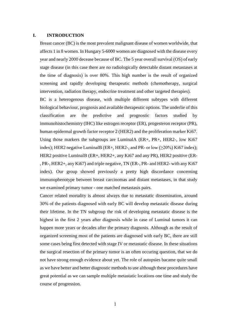
I. INTRODUCTION
Breast cancer (BC) is the most prevalent malignant disease of women worldwide, that
affects 1 in 8 women. In Hungary 5-6000 women are diagnosed with the disease every
year and nearly 2000 decease because of BC. The 5 year overall survival (OS) of early
stage disease (in this case there are no radiologically detectable distant metastases at
the time of diagnosis) is over 80%. This high number is the result of organized
screening and rapidly developing therapeutic methods (chemotherapy, surgical
intervention, radiation therapy, endocrine treatment and other targeted therapies).
BC is a heterogenous disease, with multiple different subtypes with different
biological behaviour, prognosis and available therapeutic options. The underlie of this
classification are the predictive and prognostic factors studied by
immunohistochemistry (IHC) like estrogen receptor (ER), progesteron receptor (PR),
human epidermal growth factor receptor 2 (HER2) and the proliferation marker Ki67.
Using those markers the subgroups are LuminalA (ER+, PR+, HER2-, low Ki67
index); HER2 negative LuminalB (ER+, HER2-, and PR- or low (≥20%) Ki67 index);
HER2 positive LuminalB (ER+, HER2+, any Ki67 and any PR), HER2 positive (ER-
, PR-, HER2+, any Ki67) and triple negative, TN (ER-, PR- and HER2- with any Ki67
index). Our group showed previously a pretty high discordance concerning
immunophenotype between breast carcinomas and distant metastases, in that study
we examined primary tumor - one matched metastasis pairs.
Cancer related mortality is almost always due to metastatic dissemination, around
30% of the patients diagnosed with early BC will develop metastatic disease during
their lifetime. In the TN subgroup the risk of developing metastatic disease is the
highest in the first 2 years after diagnosis while in case of Luminal tumors it can
happen more years or decades after the primary diagnosis. Although as the result of
organized screening most of the patients are diagnosed with early BC, there are still
some cases being first detected with stage IV or metastatic disease. In these situations
the surgical resection of the primary tumor is an often occuring question, that we do
not have strong enough evidence about yet. The role of autopsies bacame quite small
as we have better and better diagnostic methods to use although these procedures have
great potential as we can sample multiple metastatic locations one time and study the
course of progression.

2
While research continues to unravel the molecular underpinnings of the metastatic
cascade, it is increasingly recognized that profiling of advanced disease could help
elucidate such biological phenomena as distant recurrence and the emergence of de
novo resistance to therapy. A handful of studies using genome-wide molecular
techniques have begun to explore the clonal relationships between primary and
matched metastatic tumours in diverse types of neoplasia including pancreatic, clear-
cell renal cell, high-grade serous ovarian and prostate cancer. Despite the small cohort
sizes and, too often, a limited number of matched metastases for each patient, these
pioneering efforts brought forth thoughtprovoking findings such as the first
quantitative model of cancer progression from onset of the founder mutation to
metastatic dissemination, the occurrence of organ specific lineages, monoclonal, as
well as its counterpart, polyclonal seeding, horizontal cross-seeding between distant
metastases and finally homing of metastatic cells to the primary tumour bed. The use
of phylogenetic techniques on data generated using whole-exome sequencing
studying single nucleotide variants (SNVs) and copy number profiling to identify
copy number aberrations (CNAs) can lead us to gain knowledge about the progression
and the evolution of the disease. In the past couple of years lots of research focuses
on circulating tumor cells and tumor DNA that is a much less invasive way to closely
monitor the progression of the disease. While yet other studies continue to highlight
the potential of genomic analyses from small cohort sizes to decipher the origins of
intra-tumour heterogeneity and its contribution to metastatic dissemination in-depth
knowledge is currently lacking for breast cancer. The question is critically important
from a clinical and a public health point of view seeing the high number of patients.

3
II. AIMS AND OBJECTIVES
1. Characterisation of the primary tumors of metastatic patients; histological type, grade,
immunophenotype. Is there a correlation between the immunophenotype of the
primary and the localisation of the metastatic lesions?
2. Did the metastatic lesions keep the characteristics of the corresponding primaries?
3. Is there a correlation between the localisation of the metastases and the change in
immunophenotype?
4. Are the metastases of the same patient similar to each other (regarding histological
characteristics, immunophenotype and genetic parameters)?
5. Is there a correlation between DFS and the change in immunophenotype of the
metastases?
6. Monoclonal or polyclonal seeding is more common in the studied patient population?
7. Is there a correlation between OS and the number of genomic alterations in the
metastases?
8. Is there horizontal cross-seeding in the any of the studied patients?
9. Is there a clonal relationship between primary tumors and the metachronous
contralateral breast tumors?
4
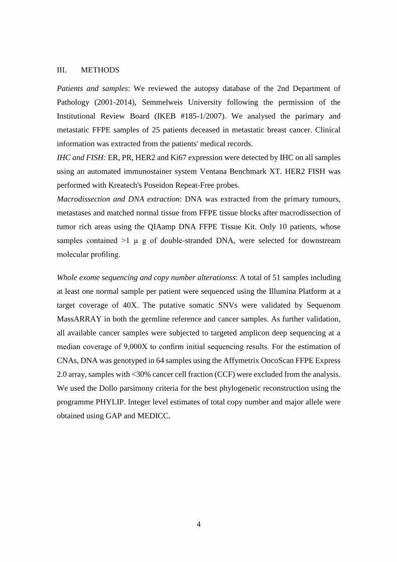
III. METHODS
Patients and samples: We reviewed the autopsy database of the 2nd Department of
Pathology (2001-2014), Semmelweis University following the permission of the
Institutional Review Board (IKEB #185-1/2007). We analysed the parimary and
metastatic FFPE samples of 25 patients deceased in metastatic breast cancer. Clinical
information was extracted from the patients' medical records.
IHC and FISH: ER, PR, HER2 and Ki67 expression were detected by IHC on all samples
using an automated immunostainer system Ventana Benchmark XT. HER2 FISH was
performed with Kreatech's Poseidon Repeat-Free probes.
Macrodissection and DNA extraction: DNA was extracted from the primary tumours,
metastases and matched normal tissue from FFPE tissue blocks after macrodissection of
tumor rich areas using the QIAamp DNA FFPE Tissue Kit. Only 10 patients, whose
samples contained >1 µ g of double-stranded DNA, were selected for downstream
molecular profiling.
Whole exome sequencing and copy number alterationss: A total of 51 samples including
at least one normal sample per patient were sequenced using the Illumina Platform at a
target coverage of 40X. The putative somatic SNVs were validated by Sequenom
MassARRAY in both the germline reference and cancer samples. As further validation,
all available cancer samples were subjected to targeted amplicon deep sequencing at a
median coverage of 9,000X to confirm initial sequencing results. For the estimation of
CNAs, DNA was genotyped in 64 samples using the Affymetrix OncoScan FFPE Express
2.0 array, samples with <30% cancer cell fraction (CCF) were excluded from the analysis.
We used the Dollo parsimony criteria for the best phylogenetic reconstruction using the
programme PHYLIP. Integer level estimates of total copy number and major allele were
obtained using GAP and MEDICC.
5
IV. RESULTS
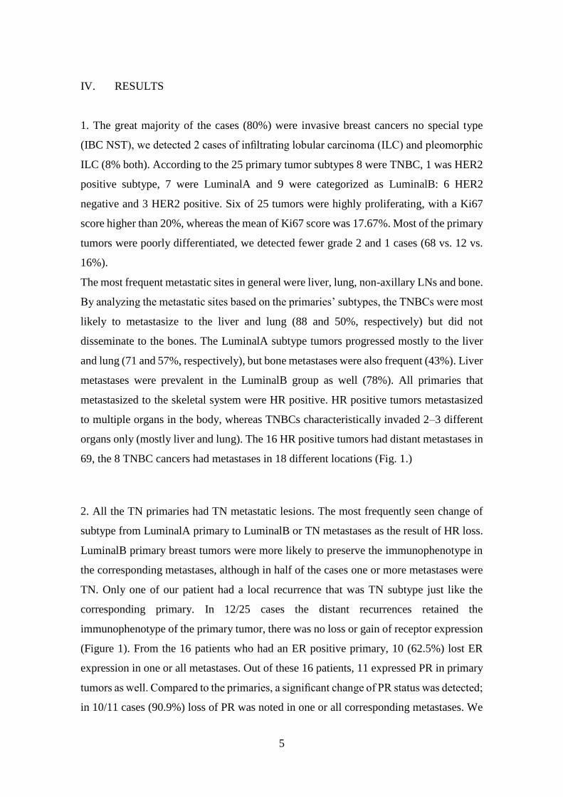
1. The great majority of the cases (80%) were invasive breast cancers no special type
(IBC NST), we detected 2 cases of infiltrating lobular carcinoma (ILC) and pleomorphic
ILC (8% both). According to the 25 primary tumor subtypes 8 were TNBC, 1 was HER2
positive subtype, 7 were LuminalA and 9 were categorized as LuminalB: 6 HER2
negative and 3 HER2 positive. Six of 25 tumors were highly proliferating, with a Ki67
score higher than 20%, whereas the mean of Ki67 score was 17.67%. Most of the primary
tumors were poorly differentiated, we detected fewer grade 2 and 1 cases (68 vs. 12 vs.
16%).
The most frequent metastatic sites in general were liver, lung, non-axillary LNs and bone.
By analyzing the metastatic sites based on the primaries’ subtypes, the TNBCs were most
likely to metastasize to the liver and lung (88 and 50%, respectively) but did not
disseminate to the bones. The LuminalA subtype tumors progressed mostly to the liver
and lung (71 and 57%, respectively), but bone metastases were also frequent (43%). Liver
metastases were prevalent in the LuminalB group as well (78%). All primaries that
metastasized to the skeletal system were HR positive. HR positive tumors metastasized
to multiple organs in the body, whereas TNBCs characteristically invaded 2–3 different
organs only (mostly liver and lung). The 16 HR positive tumors had distant metastases in
69, the 8 TNBC cancers had metastases in 18 different locations (Fig. 1.)
2. All the TN primaries had TN metastatic lesions. The most frequently seen change of
subtype from LuminalA primary to LuminalB or TN metastases as the result of HR loss.
LuminalB primary breast tumors were more likely to preserve the immunophenotype in
the corresponding metastases, although in half of the cases one or more metastases were
TN. Only one of our patient had a local recurrence that was TN subtype just like the
corresponding primary. In 12/25 cases the distant recurrences retained the
immunophenotype of the primary tumor, there was no loss or gain of receptor expression
(Figure 1). From the 16 patients who had an ER positive primary, 10 (62.5%) lost ER
expression in one or all metastases. Out of these 16 patients, 11 expressed PR in primary
tumors as well. Compared to the primaries, a significant change of PR status was detected;
in 10/11 cases (90.9%) loss of PR was noted in one or all corresponding metastases. We
6
had 4 patients with HER2 positive primary disease, 2 of which lost HER2 expression in
distant recurrences in this group. Gain of receptor (HR or HER2) was not seen in distant
recurrences of TNBCs.
Figure 1. Subtype of primary tumors, local recurrences and distant
metastases.
7
3. The metastases of the same primary tumor were more similar to each other compared
to their corresponding primary breast cancer. Based on immunophenotype there were no
differences between the groups of metastases compared to each other at any of the steps
mentioned above, meaning that the distant recurrences based on localization (regions,
organ systems, organs) are very similar to each other. (Table 1.) Metastases were clonally
related and originate from cells disseminated at various stages of the disease. Thus, they
inherit varying fractions of genomic alterations from their parental lineage, followed by
acquisition of private alterations.
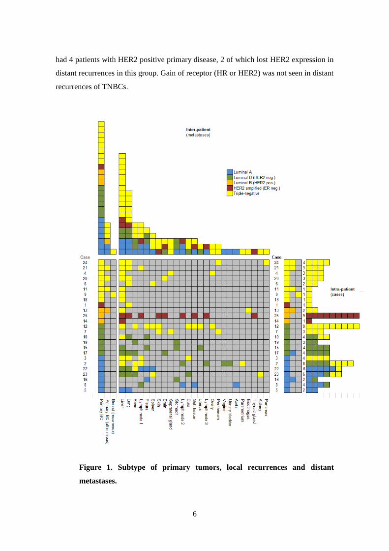
4. While analyzing the various metastases in 7/25 cases (in all of them the primary tumor
was of luminal subtype) the metastases were heterogeneous; some were discordant while
others were concordant with the corresponding primary tumor. Three of 7 patients had
the diagnosis of breast cancer at autopsy rendering the clinical history and progression of
the disease obscure. In 4/7 patients we were able to follow the course of the disease by
retrospectively analyzing the clinical data. In these cases we found a subgroup/time
pattern of the metastases: the later the metastasis was discovered the more likely it was
to have a differing IHC profile compared to the primary tumor (Fig. 2).
Figure 2. Cases with metastases of heterogeneous subtype.

8
5. To analyze the distant metastases in more detail we formed a 3-step grouping system
based on the localization of the metastases. First we grouped the metastases in five major
regional groups: central nervous system (CNS), thorax, abdomen, bone and skin. Second,
we divided the metastases to various organ systems, and third, we analyzed the metastases
in each organ individually (Table 1).
i. When we studied the diversities between primaries and regions of metastases, we found
that tumors were most likely to lose ER expression while metastasizing to the abdomen.
Loss of PR expression was detected in thoracic and abdominal metastases also.
ii. Regarding organ systems, loss of ER and PR expression was detected in metastases of
the gastrointestinal system. In the respiratory system only the loss of PR expression was
significant.
iii. While comparing the primary tumors with their corresponding distant metastases by
each organ individually, we observed that the primary tumors were likely to lose HR
expression in the liver and lung. Statistically significant change in Ki67 expression was
seen only in bone metastases; HER2 status remained unchanged in the above mentioned
locations.

9
Table 1. Difference of ER, PR and Ki67 status in the regions, organ systems and
organs compared to the primary tumor.

10
6. The phylogenies of patients with early BC confirmed that distant metastases probably
arose via a seeding event to an initial ‘metastatic precursor’ from the primary tumour and
in absence of the latter, removed at surgery, the source of further dissemination to
additional organs occurred by metastasis-to-metastasis disseminations. Our observation
suggests that for breast cancer patients diagnosed at an early stage and undergoing
curative intent surgery, who represent the majority of patients, cascading disseminations
from metastases appears to be a major route of tumor progression (Figure 3a). A
contrasting clinical and biological condition to the dissemination via a ‘metastatic
precursor’ is illustrated by the case of patients diagnosed with stage IV. disease, here we
identified independent seeding events (Figure 3b).
Figure 3. Combined phylogenies representing metastatic progression across eight
patients.

11
7. The normalized phylogenetic branch length, which is the ratio of the path from the common
ancestor to the given lesion relative to the common trunk, represents the extent of genomic
alterations that accumulated since the first metastasizing event took place irrespective of the
mode of progression. If this distance is short, it means that the bulk of evolutionary changes
occurred ‘early’ in the trunk of the phylogenetic tree, in these cases we saw a shorter OS.
Figure 4. shows the correlation of the average normalized phylogenetic branch lengths with
overall survival. Although the number of patients is small, we observed a positive correlation
for both CNAs and SNVs.
Figure 4. Dynamics of genomic alterations during metastatic progression.
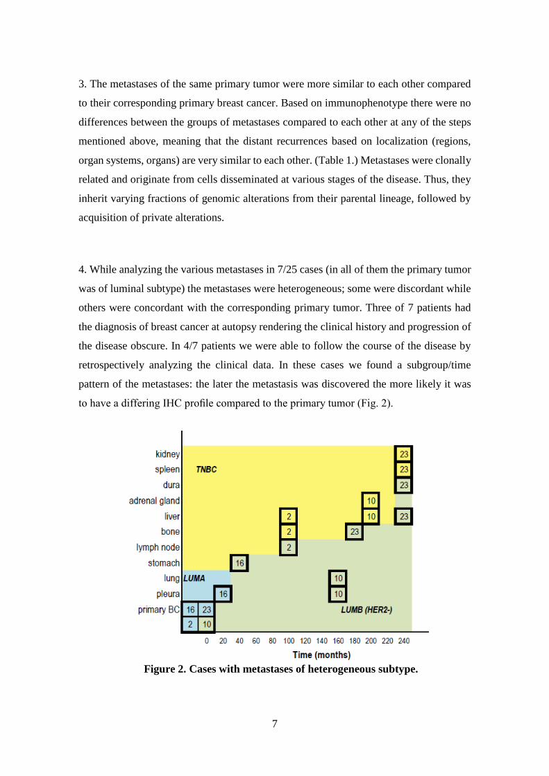
8. In one of our patients we identified two independent seeding events; the phylogenetic
analysis of the metastasis to the adrenal gland (M2) revealed that this lesion originated
12
from a precursor shared with the liver metastasis. However, the adrenal gland lesion
displayed both SNVs acquired ‘late’ in the evolutionary history of the clade composed of
the primary tumour and liver metastasis as well as SNVs private to the ovarian metastasis.
Pairwise comparisons of the SNVs showed that those private to the primary tumor and
liver metastasis clade were also present at full clonal frequencies in the adrenal gland
metastasis in agreement with the phylogeny inferred from the CNA profiles. The ‘late’
SNVs private to the ovarian metastasis were observed at subclonal frequencies in the
adrenal gland metastasis. Our results imply that circulating metastatic cells, disseminated
by the ovarian metastasis, horizontally cross-seeded the already metastatic adrenal gland.
(Figure 5.)
Figure 5. Horizontal reseeding from metastasis to metastasis
9. Two patients from our series were diagnosed with a metachronous contralateral breast
tumour. Patient 20, 1 year and Pt14, 10 years after initial diagnosis. In patient 20, the
phylogenetic reconstruction showed that the contralateral left breast tumour (M3) was the
earliest branching of the tree. In the case of patient 14 the contralateral left tumour (M1)
originated from a daughter lesion shared with the liver metastases (M2 and M3). Both cases
confirm the clonal relatedness of the contralateral tumour with the initially diagnosed breast
cancer. (Figure 6.)

13
Figure 6. Metachronous contralateral breast tumors as metastatic deposits.
14
V. CONCLUSIONS
The aim of our study was to gain a better understanding of breast cancer progression by
examining primary tumors and multiple corresponding distant metastases.
Based on our results the metastases of the same primary were more similar to each other
and more often differed from the primary lesion. On protein expression level the most
common change was the loss of PR, less frequent the loss of ER. It is well known that
ER+ PR- tumors have a worse prognosis compared to ER+ PR+ lesions while TN breast
tumors have the least favorable prognosis. Based on that the change from Luminal A to
Luminal B to TN is a logical way of progression, that might happen in one patient too as
the result of disease progression. We detected significant loss of HR in the liver and lung
metastases but it is important to notice that these were the locations where we had the
highest number of samples.
We observed that the HR positive tumors metastasized to more organs than TNBCs, the
usually longer disease history might be a reason behind this phenomenon. The broader
metastatic spectrum with more unusual locations is a known feature of HR positive breast
cancers.
We had patients with heterogeneous metastases at HR level. Although this occurred only
in a minority of the patients, this may be important, since the result of a biopsy from a
metastatic site has a crucial role in making decisions about the next line of therapy. Based
on our results the longer period of time elapsed between the diagnosis of the primary
disease and the appearance of the metastasis, the more likely it was that the metastasis
differed from the corresponding primary tumor.
We also applied phylogenetic techniques to infer the evolutionary history of breast cancer
progression, compared to previous studies the availability of a larger number of patients
with matched primary and multiple metastatic samples was critical to our study for
deciphering the routes of dissemination underlying metastatic progression. We observed
two possible scenarios. The most frequent implied a single successful seeding event from
the primary tumour followed by metastasis-to-metastasis cascading disseminations,
whereas the second involved multiple seeding events from the primary tumour alongside
daughter metastasis- to metastasis disseminations. This dichotomy coincides with the
clinical history where descent from a common metastatic origin was observed in patients
diagnosed with early stage breast cancer, whereas multiple seeding events from the
15
primary tumour occurred in patients diagnosed with advanced stage disease. The role of
primary tumour resection in de novo metastatic breast cancer patients is unclear, and there
is currently no consensus whether this procedure confers a survival benefit. Previous
clinical trials observed an increased progression free survival for primary tumour
resection in HER2- de novo metastatic patients with solitary bone metastases. Thus, our
observations suggest that surgical excision of the primary tumour might reduce metastatic
dissemination in selected cases hence providing a potential biological rationale for this
practice. Similarly, there is no strong recommendation showing overall survival benefit
from surgical resection of oligo-metastases in breast cancer. From our analyses,
metastatic lesions constitute an additional source of seeding and heterogeneity in
advanced breast cancer.
Our results imply the first time in human breast cancer samples, that circulating metastatic
cells, disseminated by a metastasis, horizontally cross-seeded another, already metastatic
spot. That confirms the previous observations in ovarian and prostate cancers further
lending support to the hypothesis of tumour self-seeding.
Our results together with recent reports call into question the current practice of
considering metachronous contralateral tumours as second primary cancers. Since
treatment strategies offered to patients differ widely between early and advanced stage
breast cancers, it is imperative to determine in practice whether contralateral tumours
represent a metastatic deposit of the primary tumour.
Our results have some immediate consequences to the clinical practice in treating breast
cancer patients, it is important:
i. to sample metastatic lesions (at the diagnosis of stage IV. disease, after
longer progression free intervals),
ii. to determine whether contralateral tumours represent a metastatic
deposit of the primary tumour,
iii. to resect the primary lesion when the patient is diagnosed with stage IV.
in a well defined group of patients.

16
VI. PUBLICATIONS
PUBLICATIONS RELATED TO THE DISSERTATION
1. Szekely B, Nagy Z I, Farago Z, Kiss O, Lotz G, Kovacs A, Udvarhelyi N, Dank M,
Szentmartoni Gy, Baranyai Z, Tokes AM, Szasz AM, Kulka J (2017). Comparison of
immunophenotypes of primary breast carcinomas and corresponding multiple distant
metastases – an autopsy study of 25 patients.
Clin Exp Metastasis, 34:103-113 (IF:3.144)
2. Brown D*, Smeets D*, Szekely B*, Larsimont D, Szász AM, Adnet PY, Rothe F,
Rouas G, Nagy ZI, Farago Z, Tokes AM, Dank M, Szentmartoni G, Udvarhelyi N,
Zoppoli G, Pusztai L, Piccart M, Kulka J, Lambrechts D, Sotiriou C, Desmedt C
(2017). Phylogenetic analysis of metastatic progression in breast cancer using somatic
mutations and copy number aberrations.
Nat Commun, 8:14944 (IF:12.124)
PUBLICATIONS NOT RELATED TO THE DISSERTATION
1. Kulka J, Tokes AM, Toth AI, Szasz AM, Farkas A, Borka K, Jaray B, Szekely E,
Harsanyi L, Rusz Z, Laszlo Zs, Istok R, Lotz G, Madaras L, Korompay A, Harsanyi
L, Laszlo Z, Rusz Z, Molnar BA, Molnar IA, Kenessey I, Szentmartoni G, Szekely
B, Dank M (2009). Immunohistochemical phenotype of breast carcinomas predicts
the effectiveness of primary systemic therapy. Magy Onkol, 53:335–343.
2. Szekely B, Madaras L, Szentmartoni G, Szasz AM, Baranyak Z, Szittya L, Torgyik
L, Zergenyi E, Borbenyi E, Kenessey I, Korompay A, Langmar Z, Banhidy F, Kulka
J, Dank M (2010). Comparison of breast cancer in young and old women based on
clinicopathological features. Magy Onkol, 54:19-26.
3. Dank M, Szentmartoni Gy, Szekely B, Langmar Z (2010). Pain management in cancer
patients. Hippocrates, RGD:73088/HU/2010.07.12.
4. Szekely B, Langmar Z, Somlai K, Szentmartoni Gy, Szalay K, Korompay A, Szasz
AM, Kulka J, Banhidy F, Dank M (2010). Treatment of pregnancy associated breast
cancer. Orv Hetil, 151:1299-303.
5. Szasz AM, Szendroi A, Szucs M, Idan R, Tokes AM, Kardos M, Szekely B, Szabo
Gy, Kulka J, Szendroi M, Romics I, Timar J (2010). Role of hypoxia on gene

17
expression and their prognostic power in renal cell carcinoma. Uroonkológia, 7:74-
81.
6. Szekely B, Pusztai L (2011). The value of genomic analysis of breast cancer in drug
development. J Natl Cancer Inst Monogr, 43:1–3.
7. Szekely B, Szentmartoni G, Kulka J, Szasz AM, Langmar Z, Dank M (2011). Primary
systemic therapy in breast cancer--an update for gynecologic oncologists. Eur J
Gynaecol Oncol, 32:636-41. (IF: 0.474)
8. Stoddard FR II, Szasz AM, Szekely B, Tokes AM, Kulka J (2011). Molecular genetic
tests in the prediction of the prognosis of breast cancer. MEMO, 4:158-162.
9. Madaras L, Szasz AM, Baranyak Zs, Tokes AM, Szittya L, Lotz G, Szekely B,
Szentmartoni Gy, Dank M, Baranyai Zs, Kulka J (2012). Morphological and
immunophenotypical heterogeneity in breast cancers of young and elderly women.
Magy Onkol, 56:75-8.
10. Tokes T, Somlai K, Szekely B, Kulka J, Szentmartoni G, Torgyik L, Galgoczy H,
Lengyel Z, Gyorke T, Dank M (2012). The role of FDG-PET-CT in the evaluation of
primary systemic therapy in breast cancer: links between metabolic and pathological
remission. Orv Hetil, 153:1958-64.
11. Szasz AM, Li Q, Eklund AC, Sztupinszki Z, Rowan A, Tokes AM, Szekely B, Kiss
A, Szendroi M, Gyorffy B, Szallasi Z, Swanton C, Kulka J (2013). The CIN4
chromosomal instability qPCR classifier defines tumor aneuploidy and stratifies
outcome in grade 2 breast cancer. PLoS ONE, 8:e56707. (IF=3.534)
12. Szasz AM, Acs B, Agoston E, Sztupinszki Z, Tokes AM, Szittya L, Szekely B,
Szendroi M, Li Q, Harsanyi L, Timar J, Szallasi Z, Swanton C, Gyorffy B, Kulka J
(2013). Simplified, low-cost gene expression profiling for the prediction of outcome
in breast cancer based on routine histologic specimens. Orv Hetil, 154:627-32.
13. Szekely B, Iwamoto T, Szasz AM, Qi Y, Szallasi Z, Matsuoka J, Symmans WF,.
Tokes AM, Kulka J, Swanton C, Pusztai L (2013). A 3-gene proliferation score (TOP-
FOX-67) can re-classify histological grade 2, ER-positive breast cancers into low and
high risk prognostic categories. Breast Cancer Res Treat, 138:691-8. (IF=4.198)
14. Madaras L, Baranyák Z, Kulka J, Szasz AM, Kovacs KA, Phan Huong Van, Szekely
B, Dank M, Nagy T, Kiss O, Harsanyi L, Barbay T, Kenessey I, Tokes AM (2013).
Retrospective analysis of clinicopathological characteristics and family history data

18
of early-onset breast cancer: a single-institutional study of Hungarian patients. Pathol
Oncol Res, 19:723-9. (IF=1.806)
15. Madaras L, Kovacs KA, Szasz AM, Kenessey I, Tokes AM, Szekely B, Baranyak Z,
Kiss O, Dank M, Kulka J (2013). Clinicopathological features and prognosis of
pregnancy associated breast cancer - a matched case control study. Pathol Oncol Res,
20:581-90. (IF=1.855)
16. Selmeci T, Tokes AM, Rona A, Molnar BA, Kenessey I, Szekely B, Madaras L, Szasz
AM, Kulka J (2015). Prognostic impact of progesterone receptor expression in HER2-
negative luminal-B breast cancer. J Surg Mol Path, 1:41-49.
17. Kiss O, Tokes AM, Spisak S, Szilagyi A, Lippai N, Szekely B, Szasz AM, Kulka J
(2015). Breast- and salivary gland-derived adenoid cystic carcinomas: potential post-
transcriptional divergencies. A pilot study based on miRNA expression profiling of
four cases and review of the potential relevance of the findings. Pathol Oncol Res,
21:29-44. (IF: 1.94)
18. Selmeci T, Tokes AM, Rona A, Molnár BA, Kenessey I, Szekely B, Szasz AM, Kulka
J (2014). A progeszteronreceptor kifejeződésének kórjóslati értéke a HER2-negatív
Luminalis -B altípusú emlődaganatokban. Nőgyógy Onk, 19:45-49.
19. Kulka J, Tokes AM, Madaras L, Kovacs A, Acs B, Illyes I, Kiss O, Szekely B, Lotz
G, Szasz MA (2015). Clinico-pathologically focused breast cancer research.
Magy Onkol, 59:286-91.
20. Pusztai L, Ladanyi A, Szekely B, Dank M (2016). Immunotherapy opportunities in
breast cancer. Magy Onkol, 60:34-40.
21. Kulka J, Szekely B, Lukacs L, Madaras L, Tokes AM, Lotz G, Kas J, Harsanyi L,
Baranyai Z, Fillinger J, Soltesz I, Hanzely Z, Balint K, Arato G, Szendroi M, Szasz
AM (2016). Comparison of predictive immunohistochemical marker expression of
primary breast cancer and paired distant metastasis using surgical material: a practice-
based study.
J Histochem Cytochem, 64:256-67. (IF:2.511)
22. Santarpia L, Bottai G, Kelly CM, Gyorffy B, Szekely B, Pusztai L (2016).
Deciphering and targeting oncogenic mutations and pathways in breast cancer.
Oncologist, 21:1063-78.
19
23. Bottai G, Raschioni C, Szekely B, Di Tommaso L, Szasz A, Losurdo A, Gyorffy B,
Acs B, Torrisi R, Karachaliou N, Tokes T, Caruso M, Kulka J, Roncalli M, Santoro
A, Mantovani A, Rosell R, Reis-Filho JS, Santarpia L (2016). AXL associated tumor
inflammation as a poor prognostic signature in chemotherapy-treated triple-negative
breast cancer patients. NPJ Breast Cancer, 2:16033.
24. Molnar IA, Molnar BA, Vizkeleti L, Fekete K, Tamas J, Deak P, Szundi C, Szekely
B, Moldvay J, Vari-Kakas S, Szasz MA, Acs B, Kulka J, Tokas AM (2017). Breast
carcinoma subtypes show different patterns of metastatic behavior. Virchows Arch,
470:275-83. (IF:2.848)
25. Szekely B, Silber AL, Pusztai L (2017). New therapeutic strategies for triple-negative
breast cancer. Oncology (Williston Park NY), 31:221108.
26. Wei W, Kurita T, Hess KR, Sanft T, Szekely B, Hatzis C, Pusztai L (2018).
Comparison of residual risk-based eligibility vs tumor size and nodal status for power
estimates in adjuvant trials of breast cancer therapies.
JAMA Oncol, 25:175092. (IF:16.559)
27. Pelekanou V, Barlow WE, Nahleh Z, Wasserman B, Lo YC, von Wahlde MK,
Hayes DF, Hortobagyi GN, Gralow JR, Tripathy D, Porter P, Szekely B, Hatzis C,
Rimm DL, Pusztai L (2018). Tumor infiltrating lymphocytes and PD-L1 expression
in pre- and post-treatment breast cancers in the SWOG S0800 Phase II neoadjuvant
chemotherapy trial.
Mol Cancer Ther, 17:1324-1331. (IF:5.764)
Cumulative IF: 56,757
Related Documents