Journal of Biochemistry Rapid Communication JB 06-11-0359 revised form Field: Biochemistry Topic: Analytical Biochemistry COMPARISON BETWEEN TOTAL ENDOTHELIAL PROGENITOR CELL ISOLATION VERSUS ENRICHED CD133 + CULTURE Amelia Casamassimi* , †, Maria Luisa Balestrieri** , †, Carmela Fiorito*, Concetta Schiano*, Ciro Maione*, Raffaele Rossiello**, Vincenzo Grimaldi*, Vincenzo Del Giudice*, Ciro Balestrieri**, Bartolomeo Farzati*, Vincenzo Sica*, and Claudio Napoli* , ° *Department of General Pathology, Division of Clinical Pathology and Excellence Research Center on Cardiovascular Diseases, and **Department of Chemical Biology and Physics, 1 st School of Medicine, II University of Naples, Italy. †Contributed equally to this study Running title: Endothelial progenitor cell isolation: a methodologic comparison. °Correspondence to: Prof. Claudio Napoli, Department of General Pathology, Division of Clinical Pathology and Excellence Research Center on Cardiovascular Diseases, Complesso S. Andrea delle Dame, 1 st School of Medicine, II University of Naples, Naples 80138 Italy- e-mail: [email protected] Tel/fax +39-081- 293399 Key words: CD133, Endothelial progenitor cells, glucose, TNF © 2007 The Japanese Biochemical Society 1 Journal of Biochemistry Advance Access published February 18, 2007

Welcome message from author
This document is posted to help you gain knowledge. Please leave a comment to let me know what you think about it! Share it to your friends and learn new things together.
Transcript
Journal of Biochemistry Rapid Communication JB 06-11-0359 revised form
Field: Biochemistry
Topic: Analytical Biochemistry
COMPARISON BETWEEN TOTAL ENDOTHELIAL PROGENITOR CELL ISOLATION
VERSUS ENRICHED CD133+ CULTURE
Amelia Casamassimi*,†, Maria Luisa Balestrieri**,†, Carmela Fiorito*, Concetta Schiano*, Ciro
Maione*, Raffaele Rossiello**, Vincenzo Grimaldi*, Vincenzo Del Giudice*, Ciro Balestrieri**,
Bartolomeo Farzati*, Vincenzo Sica*, and Claudio Napoli*,°
*Department of General Pathology, Division of Clinical Pathology and Excellence Research Center on
Cardiovascular Diseases, and **Department of Chemical Biology and Physics, 1st School of Medicine, II
University of Naples, Italy.
†Contributed equally to this study
Running title: Endothelial progenitor cell isolation: a methodologic comparison.
°Correspondence to: Prof. Claudio Napoli, Department of General Pathology, Division of Clinical
Pathology and Excellence Research Center on Cardiovascular Diseases, Complesso S. Andrea delle Dame,
1st School of Medicine, II University of Naples, Naples 80138 Italy- e-mail: [email protected] Tel/fax +39-081-
293399
Key words: CD133, Endothelial progenitor cells, glucose, TNF
© 2007 The Japanese Biochemical Society
1
Journal of Biochemistry Advance Access published February 18, 2007
ABSTRACT
Endothelial progenitor cells (EPCs) play a role in endogenous neovascularization of ischemic tissues.
Isolation and characterization of EPCs from circulating mononuclear cells is important for developing
targeted cellular therapies and reproducibility of data is the major scientific goals. Here we compared two
currently employed isolation methods, i.e. from total peripheral blood mononuclear cells (PBMCs) and from
enriched CD133+ cells, by defining the cell morphology and functional activities. We show that EPCs from
cultured PBMCs resulted in an adherent population of 23%±4% merged cells positive for Dil-Ac-LDL and
lectin, whereas the percentage of double positive cells in cultured CD133+ enriched cells was 50%±7%
(P<0.01). These data were obtained through a novel an more complete method of analysis of cell
calculations (specifically by dividing each microscope field into 120 sub-fields). When stimulated with TNF-
α and glucose, cell number was reduced in EPCs from total PBMCs and, more consistently, in CD133+
enriched cells. However, both cultured total PBMCs and CD133+ enriched cells respond similarly to TNF-α
or glucose-induced p38-phosphorylation.
EPCs from both procedures show similar results in terms of phenotype and response to modulators of their
functional activities. However, when the cell phenotype of CD133+ enrichment-derived cells was compared
with that of cells from the total PBMC, a significant increase in CD133+ expression was observed (P<0.01)
This may have relevance during intervention studies using cultured EPCs.
2

INTRODUCTION
Isolation, differentiation, and expansion of endothelial progenitor cells (EPCs) from peripheral blood have
potential applicability in areas of therapeutic neovascularization, vascular repair, and tissue engineering (1-
5). The postulated rationale of this action is due to EPC homing at the sites of vascular damage (6,7). Some
published studies used EPCs from total peripheral blood mononuclear cells (PBMCs) as standard method to
explore their pathophysiologic characteristics in the context of immunological markers typical of these cells
(7). However, some methodological concerns are still raised in order to select the gold and immunogenic
standard properties of EPCs, for example, adult cultured EPCs maintained monocytoid function throughout
cell culture (9).
Although EPCs are usually derived from PBMCs cultured in presence of vascular endothelial growth factor
(VEGF) and identified as a population of adherent cells with both 1,1'-dioctadecyl-3,3,3',3'-
tetramethylindocarbocyanine–labeled acetylated LDL (Dil-Ac-LDL) uptake and lectin binding, several
methods of isolation are currently described (9,10-16). To generate putative EPCs, short-term culture of
PBMCs over 4–7 days leads to adherence and differentiation of putative EPCs defined by the expression of
VEGF-receptor-2 (VEGFR-2), vWF, VE-cadherin and CD31 (14). Other methods involve selection through
surface markers such as CD133 (10,11,15), VEGFR-2 (17), and CD34, another so-called stem cell marker
expressed at a very early developmental stage (12,13). Although CD34+ or CD133+ progenitors purified by
the immunomagnetic technique represent a very small subset of PBMCs, they can generate endothelial cells
and exhibit revascularization properties in vivo (18). A major source of circulating EPCs has been described
as a subset of a double positive CD14+CD34low (19). Conventional cytofluorimetric techniques of PBMC-
derived EPCs have shown that these cells consist of a population mainly derived from the
monocyte/macrophages-containing CD34- mononuclear cell population (8) and only in part from the
hematopoietic stem cell-containing CD34+ mononuclear cell population (20,21). At the same time,
adherence-related selection of cultured PBMCs allows for the recovery of a number of EPCs sufficient for
therapeutic treatments (3,6,7,18,22) suggesting that EPCs can also originate from circulating populations
other than CD34+ or CD133+ progenitors. Thus, since the exact origin of EPCs is still not clear the culture of
3

pre-selected PBMCs could preclude certain type of EPCs. The procedures currently used allow the isolation
of two main types of EPCs: EPCs that coexpress monocyte/macrophage and EC markers or EPCs that
express only typical EC markers and show high proliferating activity (23,24).
To date, published studies were not homogeneous in terms of methodology employed and reproducibility.
Thus, the aim of the present study was to compare the two principal methods for isolation and culture of
EPCs and to elucidate whether differences in the evaluation of their role and function under
pathophysiologic conditions can vary upon the isolation method used. For this purpose we compared the
short-term culture of unprocessed PBMCs and CD133+ enriched cells.
METHODS
Regular EPC isolation and cultivation. EPCs were isolated from total PBMCs as previously described
(25). Briefly, PBMCs were isolated by density gradient centrifugation (400 x g for 40 min at 4°C) of 15 ml
of leucocyte-rich buffy coat of healthy human donor on 20 ml of Histopaque-10771 (1.077 g/ml, Sigma).
After centrifugation the interface cells were carefully removed and transferred to a new conical tube. Cells
were washed twice with Pipes (1x), centrifuged at 300 x g for 10 min at 4°C and then suspended in 9 ml of
H2O, 3 ml KCl 0.6 M to a final volume of 50 ml of Pipes (1x). After centrifugation at 300 x g for 10 min at
4°C the pellet was suspended in an appropriate volume of Pipes (1x) and cells were counted. Isolated
PBMCs (~200 x 106 cells) were plated on culture dishes (5 x 10⁶ cells/ml medium) coated with human
fibronectin and maintained in endothelial basal medium (EBM; Cell Systems) supplemented with 1µg/ml
hydrocortisone, 12 µg/ml bovine brain extract, 50 µg/ml gentamycin, 50 ng/ml amphotericin B, 10 ng/ml
epidermal growth factor, and 20% FCS (25). Cells were cultured at 37 °C with 5% CO2 in a humidified
atmosphere for 3 days. After 3 days of culture a low percentage of cells was attached (about 10% of the total
plated PBMCs). The nonadherent cells were removed by washing with PBS and adherent cells were used for
further analysis.
4

Enriched procedure of CD133+ EPC isolation and cultivation. PBMCs were used to isolate EPCs with
immunomagnetic CD133-bound microbeads (Miltenyi Biotec) following the manufacturer’s protocol (100 x
106 cells PBMCs/column). Briefly, the magnetically labelled the CD133+ cells were retained in a LS column
and subsequently eluted as the positively selected cell fraction after removal of the column from the
magnetic field. Isolated CD133+ enriched cells (2 x 10⁶ cells) were plated on culture dishes coated with
human fibronectin (0.25 x 10⁶ cells/ml medium) and maintained in complete EBM (25). Cells were cultured
at 37 °C with 5% CO2 in a humidified atmosphere. After 3 days of culture, at the first media change, all
seeded cells were adherent.
Dil-Ac-LDL/Lectin staining and cell calculation method. Total PBMCs (5 x 10⁶ cells/ml medium) or
enriched CD133+ cells (0.25 x 10⁶ cells/ml medium) cells were grown on microscope fibronectin coated
glasses in 24-multiwell plates for 3 days.
After 3 days of culture, nonadherent cells from total PBMC preparation were removed by washing with PBS.
EPCs isolated from total PBMCs and CD133+ enriched cells were incubated with 2 µg/ml 1,1'-dioctadecyl-
3,3,3',3'-tetramethylindocarbocyanine–labeled acetylated LDL (Dil-Ac-LDL) (Biomedical Technologies
Inc.) for 3 hours at 37°C as previously described (25,9). Cells were fixed in 4% paraformaldehyde and
counterstained with 50 µg/ml FITC-labeled lectin from Ulex europaeus (Sigma) for 1 hour at 37°C in the
dark. Then, 3 to 5 power fields were randomly counted using a computer-based program (Leica FW4000).
Nuclear staining was performed by Hoechst 33258 (4 µg/ml) (Sigma). Cell counting was performed by using
Photoshop software, in which the cells from the each field to be counted can be marked (9), and, more
accurately, by dividing each microscope field image in 120 sub-fields by an array. Total number of double
positive Dil-Ac-LDL/Lectin cells was calculated by counting cells in each sub-field. EPC number was
expressed as percentage of cells positive for Dil-Ac-LDL/Lectin dual staining. EPC distribution in the
microscope field was monitored by dividing total double positive Dil-Ac-LDL/Lectin cells for the number of
sub-fields, excluding empty sub-fields.
5

Flow cytometry analysis. Flow cytometry analysis (FACS) was performed on freshly isolated cells at (day
0) and on the cells after 3 days of culture (day 3). Briefly, the freshly isolated cells, 106 total PBMCs and 105
enriched CD133+ cells, were washed with PBS, resuspended with PBS/BSA 0.1% and then incubated at 4 °C
in the dark for 1h with directly conjugated mouse monoclonal antibodies to CD34-phycoerythrin (PE) or
VE-cadherin-PE (Santa Cruz) or for 10 min with directly conjugated mouse monoclonal antibodies to
CD133-PE (Miltenyi Biotec). A PE isotype-matched antibody was used as negative control. The cells were
then washed twice with PBS/BSA and fixed with PBS/FBS-2%/PFA-2% for 10 min at room temperature and
analysed in PBS/BSA. Quantitative fluorescence analysis was performed with a FACS-CANTO instrument
(BD Biosciences). Each analysis included 10.000 events.
Cell treatments. EPCs from total PBMCs or CD133+ enriched cells were incubated with TNF-α (10 ng/ml)
or glucose (15 mmol/l) for 3 days (day 0-3) without changing the medium or at day 3 (day 3) for 10 min as
described (25). After 3 days of culture, cell morphology and TNF-α or glucose induced-reduction of EPC
number and -activation of p38 MAP kinase were determined (25).
Western Blot Analysis. Total cell extracts (20 to 50 µg/lane) were loaded onto SDS-polyacrylamide gels
and blotted onto polyvinylidene difluoride membranes. Western blots were performed by use of antibodies
directed against phospho-p38 (Thr180/Tyr182)(3D7) (1:1000; Cell Signaling), total p38 (c-20) (1:1500;
Santa Cruz Biotechnology), tubulin (GTU-88) (1:10000; Sigma). Secondary antibodies were anti-rabbit
(1:4000; Santa Cruz Biotechnology) and anti-mouse antibody (1:5000; Santa Cruz Biotechnology).
Enhanced chemiluminescence was performed according to the instructions of the manufacturer (Amersham).
The autoradiographs were scanned and semi-quantitatively analyzed. The protein ratio was calculated by
LKB analyzer.
Statistical Analysis. Data are given as mean ±SD. Differences were assessed by T-test and a P value less
than 0.05 was considered to be significant.
6

RESULTS
EPC morphology. Culturing human total PBMCs (5 x 10⁶ cells/ml medium) for 3 days under standard
conditions (25) resulted in an adherent population consisting of about 10% of the total plated PBMCs (about
0.5 x 10⁶ cells/ml medium). Phase control fluorescent microscope counting indicated that 23%±4% of these
cells were double positive for Dil-Ac-LDL (red) and lectin (green) (Fig. 1A), thus matching the previously
described EPC phenotype (21). When EPCs were isolated from total PBMCs (100 x 106 cells
PBMCs/column) by immunomagnetic CD133+ selection the yield was about 1-2%. However, fluorescence
microscopy cell counting indicated that the percentage of Dil-Ac-LDL/Lectin positive cells in cultured
CD133+ enriched cells (Fig.1B) was consistently higher than that obtained with total PBMC procedure
(50%±7% of positive cells compared to 23%±4% double positive cells from total PBMCs). Cell counting
was performed by dividing microscope field image in 120 sub-fields (Fig. 1 C and D) and total number of
double positive Dil-Ac-LDL/Lectin cells was calculated by counting cells in each sub-field (Fig. 1 C). EPC
number was expressed as percentage of cells positive for Dil-Ac-LDL/Lectin dual staining. Moreover, EPC
distribution in the microscope field was monitored by dividing total EPC number for the number of sub-
fields, excluding empty sub-fields (Fig. 1D). After 3 days most of the cells appeared elongated and became
spindle-shaped in CD133+ enriched preparation whereas round cells were still present in the cell population
from total PBMCs.
Surface marker expression of PBMC and CD133+ enriched cells. When we isolated PBMCs from
leukocyte-rich buffy coat, we found that cells were positive for stem/progenitor cell surface markers CD34
and CD133, which means that freshly isolated PBMCs contain the cells from which the EPCs originated.
This was true also for the expression of endothelial markers, VE-cadherin (VE-cad) (Fig. 2A-B). As
indicated by the forward and sideward scatter, two main populations were gated, corresponding to the
lymphocyte gate (single arrow) and to the monocyte gate (double arrow). In freshly isolated cells (day 0) the
percentage of lymphocyte gated cells expressing CD34 and VE-cad did not significantly differ between total
PBMCs and CD133+ enriched cells (Fig. 3A). However, as expected, significant difference in the percentage
of cells expressing CD133 was observed (1.3%±0.6% in total PBMCs compared to 45.6±2.3% in CD133+
7

enriched cells) (P<<0.01). The monocyte gated population of freshly isolated total PBMCs (day 0) showed a
higher percentage of cells expressing CD34 and VE-cad compared to CD133+ enriched cells (Fig. 3B).
Similarly to the lymphocyte gated population, the monocyte gated population of CD133+ enriched cells
showed a higher percentage of CD133 positivity compared to total PBMCs.
When the phenotype of total PBMCs and CD133+ enriched cells was analyzed after 3 days of culture
significant changes were observed in the two gated populations. The lymphocyte gated population of both
total PBMCs and CD133+ enriched cells showed a significant decrease of the CD34 and CD133 positive
cells (P<0.05 or <0.01). In contrast, the cell phenotype of the monocyte gated population of both total
PBMCs and CD133+ enriched cells showed a significant decrease only of the CD133 positive cells
percentage (P<0.05 or <0.01). A significant decrease of the percentage of CD34 positive cells was observed
in total PBMCs (P<0.05) but not CD133+ enriched cells. Finally, both EPCs from total PBMCs and CD133+
enriched procedure exhibited a constant expression of VE-cad during short-term culture.
Modulation of EPC number derived from total PBMCs and CD133+ enriched cells. Stimulation with
TNF-α and glucose impairs EPCs functional activity reducing cell number (25). Both forms of EPCs were
incubated with TNF-α (10 ng/ml) or glucose (15 mmol/l) for 3 days (day 0-3) accordingly to previous
experiments (25). Adherent cells, that were double positive for both lectin and Di-LDL uptake, were counted
at day 4. As shown in Fig. 4 (panels A-D), the inhibitory effect of TNF-α and glucose on EPC number was
observed both in EPCs from PBMCs and from CD133+ enriched cells, although it was more evident on
CD133+ enriched cells (with a P value in the latter case <0.01 vs. a P value ≈0.05).
p38 MAP kinase activation in EPCs derived from total PBMCs and CD133+ enriched cells. The
mechanism underlying the effect of TNF-α or glucose on EPC number involves p38 MAP kinase (25). An
increased p38-phosphorylation was observed in TNF-α - or glucose- treated EPCs (Fig. 5). Densitometry
analysis revealed that total p38 was uniformly expressed in EPCs derived from total PBMCs (Fig. 5A) and
CD133+ enriched cells (Fig. 5B) and that no significant differences in p38 phosphorylation were observed
8

between treated-EPCs obtained by both methods (P<0.01 after TNF-α treatment and P<0.05 after glucose
treatment vs. either untreated cells from total PBMCs and untreated CD133+ enriched cells).
DISCUSSION
We show that EPCs isolated from previously described isolation protocols, unprocessed total PBMCs and
CD133+ enrichment, show similar phenotype and morphologic characteristics, and respond similarly to
modulators of their functional activities. EPCs from both total PBMCs and CD133+ enrichment showed the
attached spindle-shaped cells that have been regarded as EPCs (22). These cells did not develop in cluster
and reach confluence during the short-term culture.
EPCs obtained by unprocessed total PBMCs culture show morphological signs of endothelial markers
starting between day 3 and 7 of culture (22,25). At the same time, CD133+ progenitor cells from peripheral
blood differentiate into adherent endothelial progenitors with both hematopoietic and endothelial character
(10). In the attempt to define whether cellular response might differ among EPCs isolated with different
methods, we have compared previously described protocols for short-term culture of total PBMCs and
CD133+ enriched cells. When the phenotype of cells derived from CD133+ enrichment was compared with
that of cells from the total PBMC, we found a significant increase in CD133+ expression (45.6% of CD133
positive cells compared to 1.3% in total PBMCs, P<0.01) and a 2-fold increase of the percentage of double
positive cells for Dil-Ac-LDL uptake and lectin binding.
Double positive cells for Dil-Ac-LDL uptake and lectin binding were analyzed by fluorescence microscopy
counting (9). An exact quantification of the double-positive cells is also commonly performed by FACS
analysis (26).
Phenotypic analysis of total PBMCs and CD133+ enriched cells from leukocyte-rich buffy coat
showed two main homogeneous populations corresponding to the lymphocyte and monocyte gates (Fig. 2),
both matching the previously described early EPC phenotype (9,20,25). In freshly isolated cells, as expected,
a significant difference between the two compared methods was observed only in the highest percentage of
9

cells expressing CD133 in lymphocyte gated population of CD133+ enriched cells. Cells obtained by both
methods coexpress endothelial and monocyte markers. Therefore, it is plausible that EPCs obtained by in
vitro culture of total PBMCs or CD133+ enriched cells are not only monocyte (9) but also lymphocyte
derived from CD34+ and CD133+ hematopoietic cells, which can express some endothelial characteristic.
During short-term culture a significant decrease was observed only in percentage of cells expressing CD34
and CD133. The expression of VE-cad was maintained constant in EPCs from total PBMCs and CD133+
enriched procedure (Fig. 3 and Table 1) consistently with the fact that EPCs maintain the progenitor cell
characteristic during short-term culture. A significant increase in VE-cad expression, and other EC markers,
i.e. KDR and von Willebrand factor, has been described during long-term culture of EPCs isolated from total
PBMCs (9). However, although the surface markers expression allows the identification of cell type, their
expression may change during culture in the presence of growth factors. This can explain why different
groups have reported different surface markers profiles of EPCs (9,15). Thus, along with the surface markers
expression, EPC characterization by functional test can be more appropriate.
To test the functional activity of EPCs from these two different isolation methods, we used TNF-α and
glucose, known to impair EPC number via p38 mitogen-activated protein kinase phosphorylation (25). We
found the inhibitory effect of TNF-α and glucose on EPC number isolated from total PBMCs was
comparable to that already described (25). However, when CD133+ enriched cells were used, the reduction
of EPC number in response to TNF-α and glucose was more significant. Moreover, accordingly to the
previous literature (25), an increased p38-phosphorylation was observed in TNF-α - and glucose- treated
EPCs, but no significant differences were observed between EPCs obtained by procedures used in this study.
Differences in the significance might be related to the method used for microscopy counts. Indeed, although
the same number of experiments was performed for both microscopy counts and Western blotting, the
number of fields and cells counted in each sub-fields (n=120) were used to calculate statistical significance
(Figure 1). Several studies have shown the possible application of EPC as a therapeutic strategy for
myocardial neovascularization but also for endothelial regeneration, in-stent restenosis (2,3). Recent small-
scale trials have provided preliminary evidence of feasibility, safety, and efficacy in patients with myocardial
and critical limb ischemia (2). Some studies have also shown that age and cardiovascular disease risk factors
10

reduce the availability of EPCs and impair their function to various degrees (25,26). The relative scarcity of
EPCs limits the ability to expand these cells in sufficient numbers for some therapeutic applications (27-31).
In this context, the development of strategies to enhance the number and improve the function of circulating
EPCs are still a priority.
When we compared the yield of Dil-Ac-LDL/Lectin double positive cells that can be obtained from both
procedures starting from the same number of PBMCs, results indicated that in total PBMC preparation only
about 10% of the cells attached to the plate and about 23% of these cells were Dil-Ac-LDL/Lectin double
positive cells, whereas following the CD133+ enriched isolation procedure all seeded cells attached to the
plate and about 50% of these cells were Dil-Ac-LDL/Lectin double positive cells. Thus, the yield of Dil-Ac-
LDL/Lectin double positive cells among these two procedures is not particularly consistent (about 2% from
total PBMCs vs. about 1% from CD133+ enriched).
Previous studies demonstrate the applicability of CD133+ selection with Miltenyi’s immunomagnetic beads
suggesting that depletion of T cells may be adequate for prevention of graft-vs-host disease (28, 29). Infusion
of CD133+ positive stem cells to patients (from 2.6 x 10⁴ to 1.1 x 10⁵ cells/Kg ) has been shown to be a
useful method for safe transplantation with haploidentically mismatched stem cell allografts while avoiding
lethal acute and chronic graft-vs-host disease (29). However, studies for a complete evaluation of the number
of blood derived CD133+ cells for clinical use are still lacking. Here we confirm that the CD133+ enrichment
allows the isolation of a homogenous progenitor population and, based reported studies (2,29), we estimated
that the recovery of a consistent cell number may require about 75 ml of leucocyte-rich buffy coat (350-450
ml of peripheral blood are needed to obtain about 30 ml of leucocyte-rich buffy coat). Additional clinical
trials are required to investigate the feasibility of this method to isolate CD133+ cells sufficient to cure one
patient.
The present study suggests that the two compared isolation procedures are equivalent since EPCs showed
similar morphology and response to modulators of their functional activities. Despite such similarity the
CD133+ enrichment, although more expensive in terms of cost and time-consuming, allows the isolation of a
11

progenitor population more suitable for supplying EPCs for clinical application. Indeed, some pilot clinical
trials, still in progress, utilize EPCs for treatment of several diseases, such as myocardial infarction and
chronic ischemic cardiomyopathy (2,27-31).
12
REFERENCES
1. Ribatti, D. (2006) The discovery of endothelial progenitor cells An historical review. Leuk Res. Nov 17;
[Epub ahead of print] PMID: 17113640.
2. Dzau, V.J., Gnecchi, M., Pachori, A.S., Morello, F., and Melo, L.G. (2005) Therapeutic potential of
endothelial progenitor cells in cardiovascular diseases. Hypertension 46, 7-18
3. Hristov, M., and Weber, P.C. (2006) The therapeutic potential of progenitor cells in ischemic heart
disease-Past, present and future. Basic Res. Cardiol. 101, 1-7
4. Luttun, A., Carmeliet, G., and Carmeliet, P. (2002) Vascular progenitors: from biology to treatment.
Trends Cardiovasc. Med. 2, 88-96
5. Lyden, D., Hattori, K., Dias, S., Costa, C., Blaikie, P., Butros, L., Chadburn, A., Heissig, B., Marks, W.,
Witte. L., Wu, Y., Hicklin, D., Zhu, Z., Hackett, N.R., Crystal, R.G., Moore, M.A., Hajjar, K.A., Manova,
K., Benezra, R., and Rafii, S. (2001) Impaired recruitment of bone-marrow-derived endothelial and
hematopoietic precursor cells blocks tumor angiogenesis and growth. Nat. Med. 7, 1194-1201
6. Rafii, S., and Lyden, D. (2003) Therapeutic stem and progenitor cell transplantation for organ
vascularization and regeneration. Nat. Med. 9, 702-712
7. Szmitko, P.E., Fedak, P.W.M., Weisel, R.D., de Almeida, J.R., Anderson, T.J., and Verma, S. (2003)
Endothelial progenitor cells: new hope for a broken heart. Circulation 107, 3093-3100
8. Harraz, M., Jiao, C., Hanlon, H.D., Hartley, R.S., and Schatteman, G.C. (2001) CD34−blood-derived
human endothelial cell progenitors. Stem Cells 19, 304-312
13
9. Zhang, S.J., Zhang, H., Wei, Y.J., Su, W.J., Liao, Z.K., Hou, M., Zhou, J.Y., and Hu, S.S. (2006) Adult
endothelial progenitor cells from human peripheral blood maintain monocyte/macrophage function
throughout in vitro culture. Cell Res. 16, 577-584
10. Gehling, U.M., Ergun, S., Schumacher, U., Wagener, C., Pantel, K., Otte, M., Schuch, G., Schafhausen,
P., Mende, T., Kilic, N., Kluge, K., Schafer, B., Hossfeld, D.K., and Fiedler, W. (2000) In vitro
differentiation of endothelial cells from AC133-positive progenitor cells. Blood 95, 3106-3112
11.Miraglia, S., Godfrey, W., Yin, A.H., Atkins, K., Warnke, R., Holden, J.T., Bray, R.A., Waller, E.K., and
Buck, D.W. (1997) A novel five-transmembrane hematopoietic stem cell antigen: isolation, characterization,
and molecular cloning. Blood 90, 5013-5021
12.Peichev, M., Naiyer, A.J., Pereira, D., Zhu, Z., Lane, W.J., Williams, M., Oz, M.C., Hicklin, D.J., Witte,
L., Moore, M.A., and Rafii, S., (2000) Expression of VEGFR-2 and AC133 by circulating human CD34(+)
cells identifies a population of functional endothelial precursors. Blood 95, 952-958
13. Roookmaaker, M.B., Verhaar, M.C., Loomans, C.J., Verloop, R., Peters, E., Westerweel, P.E.,
Murohara, T., Staal, F.J., van Zonneveld, A.J., Koolwijk, P., Rabelink, T.J., and van Hinsbergh, V.W. (2005)
CD34+ cells home, proliferate, and participate in capillary formation, and in combination with CD34- cells
enhance tube formation in a 3-dimensional matrix. Arterioscler. Thromb. Vasc. Biol. 25, 1843-1850
14.Vasa, M., Fichtlscherer, S., Aicher, A., Adler, K., Urbich, C., Martin, H., Zeiher, A.M., and Dimmeler, S.
(2001) Number and migratory activity of circulating endothelial progenitor cells inversely correlate with risk
factors for coronary artery disease. Circ. Res. 89, E1-7
15.Walenta, W., Friedrich, E.B., Sehnert, F., Werner, N., Nickenig, G. (2005) In vitro differentiation
characteristics of cultured human mononuclear cells-implications for endothelial progenitor cell biology.
Biochem. Biophys. Res. Commun. 333, 476-482
14

16. Yin, A.H., Miraglia, S., Zanjani, E.D., Almeida-Porada, G., Ogawa, M., Leary, A.G., Olweus, J.,
Kearney, J., and Buck, D.W. (1997) AC133, a novel marker for human hematopoietic stem and progenitor
cells. Blood 90, 5002-5012
17. Ziegler, B.L., Valtieri, M., Porada, G.A., De Maria, R., Muller, R., Masella, B., Gabbianelli, M., Casella,
I., Pelosi, E., Bock, T., Zanjani, E.D., and Peschle, C. (1999) KDR receptor: a key marker defining
hematopoietic stem cells. Science 285, 1553-1558
18. Kalka, C., Masuda, H., Takahashi, T., Kalka-Moll, W.M., Silver, M., Kearney, M., Li, T., Isner, J.M.,
and Asahara, T. (2000) Transplantation of ex-vivo expanded endothelial progenitor cells for therapeutic
neovascularization. Proc Natl Acad Sci U S A 97, 3422-3427
19.Romagnani, P., Annunziato, F., Lotta, F., Lazzeri, E., Mazzinghi, B., Frosali, F., Cosmi, L., Maggi, L.,
Lasagni, L., Scheffold, A., Kruger, M., Dimmeler, S., Marra, F., Genuini. G., Maggi, E., and Romagnani, S.
(2005) CD14+CD34low cells with stem cell phenotypic and functional features are the major source of
circulating endothelial progenitors. Circ Res. 97, 314-322
20. Nakul-Aquaronne, D., Bayle, J., and Frelin, C. (2003) Coexpression of endothelial markers and CD14 by
cytokine mobilized CD34+ cells under angiogenic stimulation. Cardiovasc. Res. 57, 816-823
21. Rookmaaker, M.B., Vergeer, M., van, Zonneveld, A.J., Rabelink, T.J., and Verhaar, M.C. (2003)
Endothelial Progenitor Cells: mainly derived from the monocyte/ macrophage–containing CD34-
mononuclear cell population and only in part from the hematopoietic stem cell– containing CD34+
mononuclear cell population. Circulation 108, e150
22. Dimmeler, S., Zeiher, A.M., and Schneider, M.D. (2005) Unchain my heart: the scientific foundations of
cardiac repair. J. Clin. Invest. 115, 572-583
15

23. Gulati, R., Jevremovic. D., Peterson, T.E., Chatterjee, S., Shah, V., Vile, R.G., and Simari, R.D. (2003)
Diverse origin and function of cells with endothelial phenotype obtained from adult human blood. Circ Res.
93, 1023-1025
24. Hur, J., Yoon, C.H., Kim, H.S., Choi, J.H., Kang, H.J., Hwang, K.K., Oh, B.H., Lee, M.M., and Park,
Y.B. (2004) Characterization of two types of endothelial progenitor cells and their different contributions to
neovasculogenesis. Arterioscler. Thromb., Vasc Biol. 24, 288-293
25. Seeger, F.H., Haendeler, J., Walter, D.H., Rochwalsky, U., Reinhold. J., Urbich, C., Rossig, L., Corbaz,
A., Chvatchko, Y., Zeiher, A.M., and Dimmeler, S. (2005) p38 mitogen-activated protein kinase
downregulates endothelial progenitor cells. Circulation 111, 1184-1191
26. Hristov, M., Fach, C., Becker, C., Heussen, N., Liehn, E.A., Blindt, R., Hanrath, P., and Weber, C.
(2006) Reduced numbers of circulating endothelial progenitor cells in patients with coronary artery disease
associated with long-term statin treatment. Atherosclerosis [Epub ahead of print] PMID: 16837000
27. Bartunek, J., Vanderheyden, M., Vandekerckhove, B., Mansour, S., De Bruyne, B., De Bondt, P., Van
Haute, I., Lootens, N., Heyndrickx, G., and Wijns, W. (2005) Intracoronary injection of CD133-positive
enriched bone marrow progenitor cells promotes cardiac recovery after recent myocardial infarction:
feasibility and safety. Circulation 112, I178-1183
28. Gordon, P.R., Leimig, T., Babarin-Dorner, A.,, Houston, J., Holladay, M., Mueller, I., Geiger, T., and
Handgretinger, R. (2003) Large-scale isolation of CD133+ progenitor cells from G-CSF mobilized
peripheral blood stem cells. Bone Marrow Transplant. 31, 17-22
29. Bitan, M., Shapira, M.Y., Resnick, I.B., Zilberman, I., Miron, S., Samuel, S., Ackerstein, A., Elad, S.,
Israel, S., Amar, A., Fibach, E., Or, R., and Slavin, S. (2005) Successful transplantation of haploidentically
mismatched peripheral blood stem cells using CD133+-purified stem cells. Exp. Hematol. 33, 713-718
16

30. Goussetis, E., Manginas, A., Koutelou, M., Peristeri, I., Theodosaki, M., Kollaros, N., Leontiadis, E.,
Theodorakos, A., Paterakis, G., Karatasakis, G., Cokkinos, D.V., and Graphakos, S. (2006) Intracoronary
infusion (ICI) of CD133+ and CD133-CD34+ selected autologous bone marrow (BM) progenitor cells in
patients with chronic ischemic cardiomyopathy (CIC): Cell isolation, adherence to the infarcted area and
body distribution. Stem Cells 24, 2279-2283
31. Suuronen, E.J., Veinot, J.P., Wong, S., Kapila, V., Price, J., Griffith, M., Mesata, T.G., and Ruel, M.
(2006) Tissue-engineered injectable collagen-based matrices for improved cell delivery and vascularization
of ischemic tissue using CD133+ progenitors expanded from the peripheral blood. Circulation 114, I138-144
17
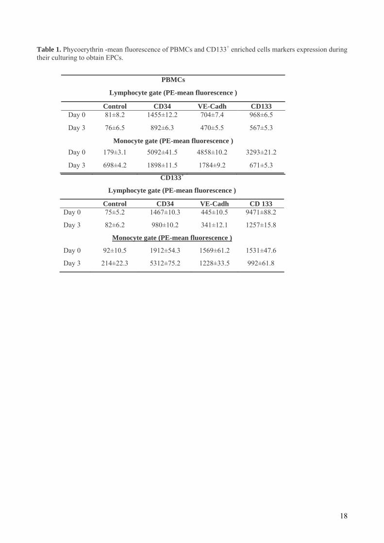
Table 1. Phycoerythrin -mean fluorescence of PBMCs and CD133+ enriched cells markers expression during their culturing to obtain EPCs.
PBMCs
Lymphocyte gate (PE-mean fluorescence )
Control CD34 VE-Cadh CD133 Day 0 81±8.2 1455±12.2 704±7.4 968±6.5
Day 3 76±6.5 892±6.3 470±5.5 567±5.3
Monocyte gate (PE-mean fluorescence ) Day 0 179±3.1 5092±41.5 4858±10.2 3293±21.2
Day 3 698±4.2 1898±11.5 1784±9.2 671±5.3
CD133+
Lymphocyte gate (PE-mean fluorescence )
Control CD34 VE-Cadh CD 133 Day 0 75±5.2 1467±10.3 445±10.5 9471±88.2
Day 3 82±6.2 980±10.2 341±12.1 1257±15.8
Monocyte gate (PE-mean fluorescence )
Day 0 92±10.5 1912±54.3 1569±61.2 1531±47.6
Day 3 214±22.3 5312±75.2 1228±33.5 992±61.8
18

Legend to Figures
Fig. 1. Characterization of EPCs. Dil-Ac-LDL uptake and lectin binding of isolated EPCs were determined
by fluorescence microscopy (A) from PBMCs and (B) from CD133+ enriched cells. Hoechst positive nuclei
from PBMCs and CD133+ enriched cells were 453±86 cells/field and 94±21 cells/field, respectively. Overlay
images are shown in right panel. Cell counting was performed by dividing microscope field images in 120
sub-fields by an array (C, D). (C) Total number of double positive Dil-Ac-LDL/Lectin cells was calculated
by counting cells in each sub-field and was expressed as percentage of EPC positive for merged Dil-Ac-
LDL/Lectin dual staining. (D) EPC distribution was monitored by dividing total double positive Dil-Ac-
LDL/Lectin cells for the number of sub-fields, excluding empty sub-fields. Two different experiments are
shown, one as main image and another as insert. Data are expressed as mean ±SD. Images are representative
of 3 different experiments in duplicate.
Fig. 2. Flow cytometry analysis of PBMCs and CD133+ enriched cells. (A) PBMCs were obtained by
density gradient centrifugation of leukocyte-rich buffy coat on Histopaque-1077 as described. Expression of
surface markers was performed on PBMCs freshly isolated (day 0) and after 3 days of culture (day 3); (B)
CD133+ enriched cells were separated from PBMCs as described and analyzed for expression of surface
markers on cells freshly isolated (day 0) and after 3 days of culture (day 3). Controls were corresponding to
negative isotope controls. Results are representative of 3 separate experiments in duplicate. Data are
expressed as mean ±SD.
Fig. 3. Phenotype of PBMCs and CD133+ enriched cells during culture. Phenotype of total PBMCs and
CD133+ enriched cells was analyzed during short-term culture in (A) lymphocyte gated population and (B)
monocyte gated population. Data are expressed as mean ±SD (n=3). *P<0.05 vs. day 0. **P<0.01 vs. day 0.
¶P<0.05 of CD133+ enriched cells at day 0 vs. total PBMCs at day 0.
Fig. 4. Effect of TNF-α and glucose on EPC number isolated by two compared methods. Dil-Ac-LDL
uptake and lectin binding staining of isolated EPCs were determined by fluorescence microscopy (A) from
PBMCs (regular) untreated and treated with TNF-α and glucose; (B) from CD133+ enriched cells (enriched)
19

untreated and treated with TNF-α and glucose; (C) bar graphs of corresponding EPC numbers from PBMCs
(regular) and (D) CD133+ enriched cells (enriched) (untreated and treated with TNF-α and glucose),
respectively. Treatments with TNF-α and glucose were performed as described. Hoechst positive nuclei in
control, TNF-α, and glucose samples prepared from total PBMCs were 148±15 cells/field, 104±18
cells/field, and 140±25 cells/field, respectively. In samples prepared from CD133+ enrichment nuclear
Hoechst positive cells in control, TNF-α, and glucose were 12±3 cells/field, 7±1.5 cells/field, and 8±2
cells/field, respectively. Results are expressed as mean ±SD and are representative of 3 different experiments
in duplicate. Differences were assessed by T-test. *P<0.01 vs. untreated; **P<0.05 vs. untreated; #P>0.05
vs. untreated.
Fig. 5. Detection of p38 phosphorylation. (A) PBMCs and (B) CD133+ enriched cells were treated with or
without TNF-α (10 ng/ml) or glucose (15 mmol/L glucose) from day 0 to day 3 or at day 4 as a stimulation
for 10 minutes as described. γ-tubulin and total p38 served as loading controls. Lanes 1 are control cells;
lanes 2: cells treated with glucose for 72 hours; lanes 3: cells treated with glucose for 10 minutes; lanes 4 are
cells treated with TNF-α for 72 hours; lanes 5 are cells treated with TNF-α for 10 minutes. Data are mean
±SD and are representative of 3 different experiments, *P<0.05 vs. untreated, **P<0.01 vs. untreated.
Immunoblotting images are representative of 3 Western blotting experiments. Densitometric analysis is for
pP38 lanes.
20

Figure 1
21

Figure 2
22

Figure 3
23

Figure 4
24

Figure 5
25
Related Documents


![Viability, proliferation and adhesion of smooth muscle ... filerecruitment of adjacent endothelial cells (EC) and endothelial progenitor cells (EPC) [13]. This requires a selective](https://static.cupdf.com/doc/110x72/5e20b942a0730f09e1657e0b/viability-proliferation-and-adhesion-of-smooth-muscle-of-adjacent-endothelial.jpg)