J Periodontol • July 2002 Comparative Histologic Analysis of Coronally Advanced Flap With and Without Collagen Membrane for Root Coverage Eun-Ju Lee,* Stephen J. Meraw, † Tae-Ju Oh, † William V. Giannobile, † and Hom-Lay Wang † 779 Background: Guided tissue regeneration (GTR)-based root coverage has been utilized to correct gingival recession defects with promising results. However, limited histologic information is available. Therefore, the aims of this study were to clinically and histologically evaluate the efficacy of GTR-based root cov- erage using collagen membrane (GTRC) and to compare the healing response to that of coronally advanced flaps (CAF). Methods: Standardized gingival recession defects were sur- gically created on the labial surfaces of the maxillary cuspids of 8 mongrel dogs. Plaque was allowed to accumulate for 8 weeks to develop a plaque-infected recession defect. Full-mouth scaling and root planing was then performed coincident with 4 weeks of oral hygiene. Defects were randomly assigned to receive either GTRC or CAF surgery. Four dogs each were sac- rificed at 4 and 16 weeks post-treatment. Clinical measurements included: percent root coverage, the amount of keratinized gin- giva (KG), and probing depth (PD). Sulcular depth, junctional epithelium and connective tissue attachment, new cementum formation, and new bone formation were evaluated histomor- phometrically. Results: Clinically, both treatments (CAF and GTRC) achieved statistically significant (P <0.05) root coverage com- pared to baseline. KG was significantly increased in CAF-treated sites at 16 weeks, while no significant differences were found for other clinical parameters between treatments. Histometrically, GTRC showed a statistically significant increase of new attach- ment and newly formed connective tissue when compared to CAF at 16 weeks. Conclusion: Within the limits of this preclinical study, both GTRC and CAF can be successfully used for the treatment of gingival recession defects. J Periodontol 2002;73:779-788. KEY WORDS Collagen/therapeutic use; comparison studies; gingival recession/surgery; gingival recession/therapy; membranes, barrier; membranes, bioabsorbable; guided tissue regeneration; surgical flaps; wound healing. * Previously, Department of Periodontics/Prevention/Geriatrics, School of Dentistry, University of Michigan, Ann Arbor, MI; currently, private practice, Seoul, Korea. † Department of Periodontics/Prevention/Geriatrics, School of Dentistry, University of Michigan. G ingival recession (soft tissue or marginal) is defined as displace- ment of the gingival margin api- cal to the cemento-enamel junction (CEJ). 1 Common causes of recession include: traumatic tooth brushing tech- nique, periodontal disease, 2 high muscle attachment and frenal pull, tooth posi- tion, 3 alveolar bone dehiscence, 2 and iatrogenic factors. 4 Consequences of gin- gival recession include problems associ- ated with esthetics, root sensitivity, and/or root caries. 2 A variety of periodontal plastic proce- dures have been developed and have shown promising results in correcting gin- gival recession defects. 5-7 Traditional approaches, such as the coronally ad- vanced flap (CAF) and its modifications have been used in periodontics for many years. 6,8 CAF offers several advantages compared to other traditional root cover- age procedures, which include no need for a donor site and a better color and contour match. However, CAF is often limited by the height and thickness of the gingiva apical to the recession and can only be applied to recession with an ade- quate width and thickness of keratinized gingiva (KG) tissue. In addition, recession treated with traditional approaches such as the lateral sliding flap mainly healed by a long junctional epithelium with a limited amount of bone and cementum forma- tion. 9 GTR-based root coverage has emerged as an alternative treatment because it may not only achieve similar clinical results to those of traditional root coverage procedures, but also demon-

Welcome message from author
This document is posted to help you gain knowledge. Please leave a comment to let me know what you think about it! Share it to your friends and learn new things together.
Transcript
J Periodontol • July 2002
Comparative Histologic Analysis ofCoronally Advanced Flap With and WithoutCollagen Membrane for Root CoverageEun-Ju Lee,* Stephen J. Meraw,† Tae-Ju Oh,† William V. Giannobile,† and Hom-Lay Wang†
779
Background: Guided tissue regeneration (GTR)-based rootcoverage has been utilized to correct gingival recession defectswith promising results. However, limited histologic informationis available. Therefore, the aims of this study were to clinicallyand histologically evaluate the efficacy of GTR-based root cov-erage using collagen membrane (GTRC) and to compare thehealing response to that of coronally advanced flaps (CAF).
Methods: Standardized gingival recession defects were sur-gically created on the labial surfaces of the maxillary cuspidsof 8 mongrel dogs. Plaque was allowed to accumulate for 8weeks to develop a plaque-infected recession defect. Full-mouthscaling and root planing was then performed coincident with 4weeks of oral hygiene. Defects were randomly assigned toreceive either GTRC or CAF surgery. Four dogs each were sac-rificed at 4 and 16 weeks post-treatment. Clinical measurementsincluded: percent root coverage, the amount of keratinized gin-giva (KG), and probing depth (PD). Sulcular depth, junctionalepithelium and connective tissue attachment, new cementumformation, and new bone formation were evaluated histomor-phometrically.
Results: Clinically, both treatments (CAF and GTRC)achieved statistically significant (P <0.05) root coverage com-pared to baseline. KG was significantly increased in CAF-treatedsites at 16 weeks, while no significant differences were found forother clinical parameters between treatments. Histometrically,GTRC showed a statistically significant increase of new attach-ment and newly formed connective tissue when compared toCAF at 16 weeks.
Conclusion: Within the limits of this preclinical study, bothGTRC and CAF can be successfully used for the treatment ofgingival recession defects. J Periodontol 2002;73:779-788.
KEY WORDS
Collagen/therapeutic use; comparison studies; gingivalrecession/surgery; gingival recession/therapy; membranes,barrier; membranes, bioabsorbable; guided tissueregeneration; surgical flaps; wound healing.
* Previously, Department of Periodontics/Prevention/Geriatrics, School of Dentistry,University of Michigan, Ann Arbor, MI; currently, private practice, Seoul, Korea.
† Department of Periodontics/Prevention/Geriatrics, School of Dentistry, University ofMichigan.
Gingival recession (soft tissue ormarginal) is defined as displace-ment of the gingival margin api-
cal to the cemento-enamel junction(CEJ).1 Common causes of recessioninclude: traumatic tooth brushing tech-nique, periodontal disease,2 high muscleattachment and frenal pull, tooth posi-tion,3 alveolar bone dehiscence,2 andiatrogenic factors.4 Consequences of gin-gival recession include problems associ-ated with esthetics, root sensitivity, and/orroot caries.2
A variety of periodontal plastic proce-dures have been developed and haveshown promising results in correcting gin-gival recession defects.5-7 Traditionalapproaches, such as the coronally ad-vanced flap (CAF) and its modificationshave been used in periodontics for manyyears.6,8 CAF offers several advantagescompared to other traditional root cover-age procedures, which include no needfor a donor site and a better color andcontour match. However, CAF is oftenlimited by the height and thickness of thegingiva apical to the recession and canonly be applied to recession with an ade-quate width and thickness of keratinizedgingiva (KG) tissue. In addition, recessiontreated with traditional approaches suchas the lateral sliding flap mainly healed bya long junctional epithelium with a limitedamount of bone and cementum forma-tion.9 GTR-based root coverage hasemerged as an alternative treatmentbecause it may not only achieve similarclinical results to those of traditional rootcoverage procedures, but also demon-
Coronally Advanced Flap With and Without Collagen Membrane for Root Coverage Volume 73 • Number 7
strate histologically new attachmentformation.10-13 Root coverageusing GTR offers advantages suchas no need for donor tissue, read-ily commercial available materials,and esthetic outcomes as compa-rable to traditional approaches.14
Tinti et al.12 used non-ab-sorbable expanded polytetrafluo-roethylene (ePTFE) to treat humanbuccal recession in 12 patients,and obtained average recessionreduction of 2.50 mm and attach-ment gain of 2.84 mm. Cortellini etal.11,15 further confirmed that sitesreceiving GTR demonstrated for-mation of new bone, cementum,and periodontal ligament. None-theless, GTR-based root coverageutilizing non-absorbable mem-branes has several drawbacks,including membrane removal and high membraneexposure rates.16 To overcome these, bioabsorbablemembranes, such as collagen and polylactide or poly-glycolide, were developed and have achieved similarresults to those obtained by non-absorbable barri-ers.17,18
Collagen, a major component of the periodontium,has shown promising results for the treatment of gin-gival recession.18 The rationale of using collagen mem-branes for root coverage include: 1) proven barrierfunction;19 2) bioabsorbable;20 3) chemotactic func-tion;21 and 4) hemostatic properties.22 Wang et al.14
compared GTRC to subepithelial connective tissue graft,and reported similar enhancement of root coverage.Ozcan et al.23 found similar amounts of root coveragebetween GTRC and CAF. However, GTRC-treated sitesrevealed clinical attachment gain as compared to CAF.
Most of these studies have reported clinical out-comes, and limited information is available regardingthe regenerated tissue at the microscopic level. There-fore, the aims of this study were: 1) to clinically andhistologically evaluate the efficacy of GTRC and 2) tocompare healing response between GTRC and CAF.
MATERIALS AND METHODS
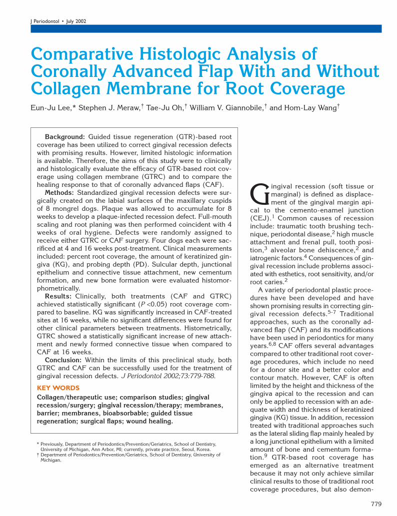
This study was approved by University Committee onUse and Care of Animals at University of Michigan.Eight 2- to 3-year-old healthy male mongrel dogs wereused in this study. Using a computer-generated ran-domization program, the animals were divided into 2groups of 4 each. Using a split-mouth design, 2 max-illary cuspids in each animal were randomly assignedby a coin-flip to receive either CAF or GTRC. Theexperimental timeline is shown in Figure 1. Procedureswere performed in a standard sterile operating room
fashion under general anesthesia as follows: intra-muscular injections of butorphanol (0.2 mg/kg), ace-promazine (0.05 mg/kg), and glycopyrolate (0.01mg/kg) were given to the animals 20 to 30 minutesprior to surgery. An IV catheter was placed into thecephalic vein, and anesthesia was induced with 12mg/kg of Na thiopental intravenously. In addition, anendotracheal tube was placed to ensure a patent air-way and for administration of 1% to 2% of isofluranemixed with 22 to 44 ml/kg/minute of oxygen duringthe surgery. Anesthesia depth was monitored by heartrate, respiration rate, jaw tone, corneal reflex, and footwithdrawal reflex. Additionally, IV fluids at a rate of 10ml/kg/hour were administrated. For local anesthesiaand hemostasis of the surgical areas, 2% xylocainewith 1:100,000 epinephrine was employed. To preventpostsurgical infection, antibiotics (amoxicillin 500 mgPO BID) were administered for 2 weeks followingsurgery. In addition, a long acting opioid, buprenor-phine (0.015 mg/kg, IM) was given to the animalsimmediately after surgery and postsurgically every 12hours for up to 96 hours as needed. After 2 weeks ofpretreatment adjustment, all animals received fullmouth scaling using ultrasonic and hand instrumen-tation. Then, the standardized labial gingival recessiondefects (5 mm from CEJ and 5 mm mesial to distal)were surgically created on both maxillary cupids byusing chisels and sterile water-cooled carbide and dia-mond burs. The root surface was then hand instru-mented for cementum removal. No oral hygiene mea-sures were employed for 8 weeks to facilitate plaqueaccumulation. After this period, all animals received full-mouth scaling and root planing. Areas were maintainedwith oral hygiene, daily tooth brushing and applica-tion of 0.12% chlorhexidine by cotton swab for 4 weeks
780
Figure 1.Study timeline.

J Periodontol • July 2002 Lee, Meraw, Oh, Giannobile, Wang
to the surgical treatment area. Clinical baseline datawere then recorded using the UNC-15 probe‡ by onecalibrated examiner with gentle manual probing, withcare to avoid the exact midfacial point where histologicsections would be made. Measurements were roundedup to the nearest millimeter for the following: 1) clin-ical gingival recession (GR): measured from CEJ togingival margin; 2) width of KG: recorded from gingi-val margin to mucogingival junction; and 3) PD:assessed from free gingival margin to the depth ofprobing depth.
After adequate anesthesia, defects were restandard-ized to specified dimensions (5 mm from CEJ and 5mm mesial to distal) with slight gingivectomy due tominimal overgrowth during initial healing. A flap offull- and partial-thickness with oblique vertical inci-sions beyond the mucogingival junction was made.The root surface was thoroughly scaled and rootplaned. A coronal reference notch was marked alongthe CEJ, and an apical notch was made at the levelof alveolar crest using a 1/2 round bur. The distancebetween 2 notches was standardized to 7 mm using theUNC-15 probe.‡ Sites were then randomly assignedby flipping a coin to receive CAF at one site and GTRCat the contralateral site. The flap and tooth prepara-tion was the same at all sites except that in the GTRCgroup, a collagen membrane (bovine type I collagen§)was used in addition to the CAF. The membranes weretrimmed and extended beyond defects 3 to 4 mm allthe way around. The membrane was then secured tothe tooth via a bioabsorbable 4-0 suture� with a slingtechnique. The flap was repositioned and sutured at alevel coronal to the pretreatment position, attemptingto completely cover the defect and/or barrier mem-brane. No periodontal dressings were used.
Specimen Retrieval
After 4 and 16 weeks of healing, clinical parameterswere recorded avoiding the exact midfacial aspectwhere histologic sections were to be made. Animalswere then euthanized with an overdose of pentobarbi-tal. Block sections of the cuspid and surrounding tis-sue including bone were obtained. Blocks were imme-diately fixed and stored in 4% paraformaldehyde, andsubsequently dehydrated in step gradients of alcohol,infiltrated, and embedded in methyl methacrylate byroutine histological methods. Serial sections of 4 to 5µm were made in the bucco-lingual plane at 0.1 mmintervals and stained by toluidine blue. Three repre-sentative sections per specimen located at the centralregion of the gingival recession defects were made,which identified both coronal and apical referencenotches. Histologic and histomorphometric analyseswere performed using a microscope fitted with a com-puter programmed with measurement software.¶ Themeasurements of 3 sections per each site were aver-
aged and used for histomorphometric analyses. Datawere recorded by a masked examiner unaware of thetreatment variable. The intra-examiner calibration errorwas revealed to be less than 5% for all parametersassessed in the study. Histologic observations includedthe presence or absence of inflammatory infiltrate andankylotic bone to the tooth surface. Figure 2 shows thehistomorphometric parameters analyzed: sulcular depth,length of junctional epithelium (JE), connective tissueattachment (CTA), height of new bone, area of newbone, and length of new cementum.
Statistical Analysis
Data were presented as mean and standard deviation(SD). A paired t test was utilized to analyze treatmenteffect (between treatments) as well as time effect(baseline versus 4 or 16 weeks) within and betweengroups, both clinically and histologically. Significancewas reported at the 95% confidence level (P <0.05).
781
‡ Hu-Friedy Mfg. Co. Inc., Chicago, IL.§ BioMend Regular, Sulzer Dental, Inc., Carlsbad, CA.� Vicryl, Ethicon Inc., Johnson & Johnson Company, Somerville, NJ.¶ Image-Pro Plus, The Imaging Express, Media Cybernetics, Silver Spring,
MD.
Figure 2.Histomorphometric parameters. a) sulcular epithelium; b) junctionalepithelium; c) connective tissue attachment; d) new bone formation;e) coronal notch; and f) apical notch.

Coronally Advanced Flap With and Without Collagen Membrane for Root Coverage Volume 73 • Number 7
RESULTS
No clinical adverse effects were observed for eithertreatment. There was no collagen membrane expo-sure noted.
Clinical Measurements
Table 1 lists clinical results obtained from this study.There were no statistical differences in GR, KG, and PDat baseline.
Four-Week Results
At 4 weeks, both treatment procedures resulted in sta-tistically significant reduction (P <0.05) of GR whencompared to baseline. This translates to 92% and 72%of root coverage for CAF and GTRC, respectively(Table 1). However, no statistically significant differencewas noted between treatments. It was noted that onesite receiving GTRC obtained only 11% root coverage(Fig. 3). If this site is considered a treatment failure anddeleted from the analysis, the average percent of rootcoverage for GTRC increases to 92%.
Both groups showed a gain of KG when comparedto baseline, 1.2 mm (from 3.8 to 5.0 mm) for CAF and0.4 mm (from 3.9 to 4.3 mm) for GTRC. No statisti-cally significant difference was found between treat-ments and between baseline and 4 weeks.
Sixteen-Week Results
At 16 weeks, both CAF and GTRC displayed signifi-cant reduction (P <0.05) of GR when compared tobaseline. The average root coverage for CAF andGTRC was 56% and 66%, respectively. Again, no sta-tistically significant difference was found between treat-ments. Figure 3 shows the percentage root coverageand the corresponding number of sites. Three sites (2GTRC and 1 CAF) had root coverage ≤60%. The amountof root coverage was decreased in CAF (from 92% at4 weeks to 56% at 16 weeks), while remaining rela-tively stable in GTRC sites (from 72% at 4 weeks to66% at 16 weeks).
At 16 weeks, CAF-treated sites demonstrated a sta-tistically significant increase (2.1 mm) in KG whencompared to baseline, while there was only a small(0.9 mm) gain of KG in GTRC-treated sites. Again, nostatistically significant difference was found betweenCAF and GTRC.
An increase in PD was noted at 16 weeks: 1.9 mmand 1.3 mm for CAF and GTRC, respectively. In addi-tion, no statistically significant difference was foundbetween treatments.
782
Table 1.
Comparison of Clinical ParametersBetween CAF and GTRC at 4 and 16Weeks Following Surgery
CAF GTRC
Clinical Parameter (N = 4) (N = 4)
Gingival recession (%)
Baseline –4.5 ± 0.0 –4.4 ± 0.2
4 weeks –0.4 ± 0.2* –1.3 ± 2.8*
% root coverage 91.7 ± 16.6 72.2 ± 61.2
Baseline –4.3 ± 0.2 –4.5 ± 0.4
16 weeks –1.9 ± 1.4* –1.5 ± 2.6*
% root coverage 55.8 ± 29.8 66.3 ± 56.8
Keratinized gingiva (mm)
Baseline 3.8 ± 3.0 3.9 ± 3.0
4 weeks 5.0 ± 4.0 4.3 ± 3.8
Gain of KG 1.2 ± 1.4 0.4 ± 1.6
Baseline 2.0 ± 1.2 2.7 ± 2.0
16 weeks 4.1 ± 1.2* 3.6 ± 0.8
Gain of KG 2.1 ± 1.0* 0.9 ± 2.2
Probing depth (mm)
Baseline 1.5 ± 0.0 1.5 ± 0.0
4 weeks 4.3 ± 1.6* 3.8 ± 1.8*
Change in PD 2.8 ± 1.6 2.3 ± 1.8
Baseline 1.5 ± 0.0 1.5 ± 0.0
16 weeks 3.4 ± 1.6* 2.8 ± 0.8*
Change in PD 1.9 ± 1.6 1.3 ± 0.8
Values are expressed as mean ± standard deviation (mm).* Significantly different from baseline within each treatment (paired t test,
P <0.05).
Figure 3.Root coverage expressed as a percentage of the total defect and thecorresponding number of sites for each treatment at 4 and 16 weeks.% root coverage = (original recession depth – recession depth aftertreatment)/original recession depth × 100.

J Periodontol • July 2002 Lee, Meraw, Oh, Giannobile, Wang
Histologic Observations
New connective tissue and new bone formation wasnoted in both treatments at 4 weeks; however, theamount was minimal (Figs. 4 and 5). No new cemen-tum formation was identified at this time. In someGTRC sites, remnants of membrane were found, andthey were surrounded by numerous inflammatory cells(Fig. 5).
At 16 weeks, new bone, new cementum, and newconnective tissue formation was evident in both treat-ments (Figs. 6 and 7). GTRC-treated sites demon-strated higher frequency of these findings than CAF.
Histomorphometric Analysis
Table 2 lists histomorphometric results for both treat-ments. At 4 weeks, similar histological healing was
noted between CAF and GTRC. Statistically, no dif-ference was noted between treatments. New bone for-mation was minimal for both treatment groups and nocementogenesis was found.
At 16 weeks, GTRC showed statistically significantlyhigher amounts of connective tissue (P <0.05) com-pared to CAF. In addition, GTRC showed a trend ofmore new bone and new cementum formation thanCAF-treated sites. However, this difference was notstatistically significant. For new connective tissue for-mation, CAF-sites decreased significantly from 2.48mm (4 weeks) to 1.10 mm (16 weeks), while thechange of connective tissue was minimal in GTRC(from 2.65 to 2.45 mm). The length of JE wasincreased in the CAF group, whereas it was reducedin GTRC-treated teeth from 4 weeks to 16 weeks.
783
Figure 4.Healing response at 4 weeks following CAF. A. New bone formation inthe apical notch can be observed (original magnification ×10, toluidineblue stain). B. Higher magnification view of the area in A.This area isfilled with connective tissue with inflammatory cell infiltration devoid ofnew cementum formation (original magnification ×40, toluidine bluestain).
Figure 5.Healing response at 4 weeks following GTRC. A. Reveals new boneformation along the root surface coronal to the apical notch. No newcementum formation can be observed (original magnification ×10,toluidine blue stain). B. Higher magnification view of the area in A,demonstrating remnants of the collagen membrane (arrow) withinflammatory cells infiltration (arrowheads) (original magnification×40, toluidine blue stain).

Coronally Advanced Flap With and Without Collagen Membrane for Root Coverage Volume 73 • Number 7
There was no significant change in the other para-meters evaluated (new bone height, new bone area,and new cementum) when both treatment- and time-effects were examined.
DISCUSSION
The present study was designed to investigate histo-logic and histometric differences between CAF andGTRC for the treatment of gingival recession defects.Previous studies have demonstrated several limitationsin the use of CAF, including the height and thicknessof the gingiva apical to the recession, the presence orabsence of keratinized gingiva, and limited/non-idealregeneration of lost periodontium.24 Most of the tradi-tional root coverage procedures including CAF havebeen reported to heal by long junctional epitheliumwith minimal amount of new attachment.25,26 On the
other hand, GTR-based root coverage procedures havebeen shown to result in new attachment formation whileproducing comparable clinical outcomes to thoseobtained by traditional approaches.10,12,13,27 In partic-ular, bioabsorbable collagen membrane has gained pop-ularity due to its properties. It has been demonstratedthat collagen stimulates platelet attachment, enhancesfibrin linkage, and is chemotactic for fibroblasts.21 Italso inhibits apical migration of epithelium,19 stabilizesthe wound, and augments tissue thickness via enzy-matic degradation.20 These characteristics have madecollagen an attractive material for GTR-based root cov-erage. The rationale of this preclinical in vivo study wasto demonstrate the nature of healing response in cre-ated recession defects between GTRC and CAF and toassess histologic, histometric, and clinical parameters.
784
Figure 6.Healing response at 16 weeks following CAF. A. Note a long junctionalepithelium below the apical reference notch. No new bone formationcan be seen (original magnification ×10, toluidine blue stain). B. Adetailed view of junctional epithelium of the area in A.Apicaldowngrowth of the epithelium and presence of cementum (arrow) areclearly visible (original magnification ×40, toluidine blue stain).
Figure 7.Healing response at 16 weeks following GTRC A. New bone andcementum formation can be observed above the apical referencenotch (original magnification ×10, toluidine blue stain). B. Highermagnification view of the area in A.Arrow and arrow heads representnew bone and new cementum formation, respectively.The areabetween bone and cementum is filled with richly vascularizedconnective tissue (original magnification ×40, toluidine blue stain).

J Periodontol • July 2002 Lee, Meraw, Oh, Giannobile, Wang
Results from this animal study indicated that rootcoverage utilizing a bioabsorbable collagen membraneresulted in higher amount of connective tissue, and atrend toward higher amounts of bone and cementumwhen compared to traditional CAF. This may beexplained by the fact that collagen membranes canact as a GTR barrier excluding epithelial cells and allowundifferentiated mesenchymal cells to repopulate thespace and to promote tissue regeneration. In addition,collagen membrane not only can increase tissue thick-ness via membrane integration with the flap but alsoprotect initial attachment gain.27 Other properties suchas hemostatic and chemotactic properties may alsoplay an important role in this type of treatment. Thisis in agreement with Cortellini et al.,10 who reportedsignificantly higher amounts of new connective tissuewith GTR than with a laterally positioned pedicle graftfor the treatment of gingival recession defects in dogs.Similar results were also reported by Weng et al.28 Inthis study, the sites treated with GTR procedure resultedin higher amounts of new bone formation than in theconnective tissue graft group; however, they were notstatistically significant. This is somehow contradictoryto the results reported by Casati et al.29 They com-pared bioabsorbable polylactic acid membranes to
CAF for the treatment of gingival recession. No sta-tistical differences were found between the 2 treat-ments in any of the evaluated histological parameters.The different results observed between the Casati et al.study and ours could be explained by the differentmembranes used: a hydrophobic barrier material madefrom polylactic acid combined with a citric acid esterversus collagen. In a 14-week human biopsy speci-men, Tatakis and Trombelli30 showed remnants ofmembrane along with a typical foreign body reaction,which were surrounded with numerous multinucleatedgiant cells and foamy macrophages. Hence, it couldbe speculated that the foreign body reaction and itsbyproducts during degradation might have affectedthe outcomes. However, the small sample size in bothstudies may limit accurate comparisons.
CAF had a significantly higher amount of newattachment formation at 4 weeks than 16 weeks (Table2). This implies that the initial gain of attachment wasdue to advancement of flap, and this may eventuallydisappear as demonstrated in the present study. On theother hand, this phenomenon was not seen in GTRC,where the gain of attachment increased over time,however, not significantly. This may be attributed tomembrane ability in creating space for PDL/bone cellsto promote tissue regeneration. A similar observationwas also reported by da Silva Pereira et al.,31 whoreported a superior length of new bone and cementumin sites treated by GTR with bioabsorbable polylacticacid when compared to open flap debridement for thetreatment of dehiscence-type gingival recessiondefects.
In addition, the degradation process of collagenmembrane may explain why new attachment forma-tion was less at 4 weeks than at 16 weeks. Since thecollagen membrane absorption requires foreign bodyreaction, which may, in turn, cause initial delayedwound healing. A similar phenomenon was reported byCortellini et al.,10 who thought that the membranemight act as a foreign body and a plaque-retentivedevice, thus causing osteoclastic activation during theearly healing phase. This is further supported by ourhistological observation, where numerous multinucle-ated cells and neutrophils were commonly found inthe connective tissue along the surface of the residualmembrane observed at 4 weeks. With a longer heal-ing period, the space occupied by membrane and multi-nucleated/leukocyte cells was slowly replaced by newattachment consisting of connective tissue, bone, andcementum. Hence, a longer healing time may beneeded for GTRC to achieve optimal results. This con-firms our clinical observations. CAF-treated sites hadhigher amounts of root coverage at 4 weeks but it wasreduced at 16 weeks (from 92% to 56%). On the con-trary, the amounts of root coverage in GTRC remainedstable over time (from 72% to 66%).
785
Table 2.
Histomorphometric Comparison BetweenCAF and GTRC at 4 and 16 WeeksPostsurgery
CAF GTRC
Histomorphometric Parameters (n = 4) (n = 4)
Sulcus depth (mm)
4 weeks 1.30 ± 0.32 1.07 ± 0.18
16 weeks 0.93 ± 0.40 1.15 ± 1.20
Junctional epithelium (mm)
4 weeks 0.63 ± 0.22 1.48 ± 1.92
16 weeks 1.08 ± 0.56 1.22 ± 0.70
Connective tissue (mm)
4 weeks 2.48 ± 1.80 2.65 ± 2.60
16 weeks 1.10 ± 1.38 2.45 ± 1.74*
New bone height (mm)
4 weeks 0.62 ± 0.70 0.21 ± 0.62
16 weeks 0.54 ± 1.62 1.68 ± 1.70
New bone area (mm2)
4 weeks 0.50 ± 0.92 0.01 ± 0.04
16 weeks 0.09 ± 0.28 0.34 ± 0.34
New cementum (mm)
4 weeks 0.00 ± 0.00 0.00 ± 0.00
16 weeks 0.30 ± 0.90 0.41 ± 1.22
Values are expressed as mean ± standard deviation.* Significantly different from CAF-treated sites (t test, P <0.05).

Coronally Advanced Flap With and Without Collagen Membrane for Root Coverage Volume 73 • Number 7
Clinically, in the present study, a significant reduc-tion of gingival recession was noted in both treatmentsat 4 and 16 weeks. CAF resulted in root coverageranging from 56% to 92%. This is in agreement withprevious human clinical trials,32,33 where more than95% of root coverage was reported. On the other hand,the mean root coverage of GTRC-treated sites rangedfrom 66% to 72%. This compares well with a previoushuman clinical trial,14 in which the same collagenmembrane was used to treat gingival recession in 16patients, and 73% root coverage was reported. Theamount of root coverage reported in this study is some-how less than others reported in the literature.12,16 Theaverage 56% to 66% of root coverage after 4 months inthis study is particularly notable. This might have beenin part due to 2 outliers, 1 GTRC and 1 CAF site, whichshowed ≤40% of root coverage (Fig. 3). The unfavor-able outcomes found in the outliers could be consid-ered failures to root coverage (25% failure rate in eachgroup). It suggests that root coverage procedures aretechnique sensitive, and success of root coverage maybe influenced by the condition of surgical sites, suchas soft tissue thickness.34 In addition, differences ofroot coverage outcomes between studies may be partlyexplained by type of membrane used (polylactide ver-sus collagen), size of defects, observation periodlength, and measuring methods employed. Also thesmall sample size may add more variation in the inter-pretation of the results. Hence, the ability to directlycompare the percent of root coverage between thisstudy and those previous clinical human trials is lim-ited.
Several factors may influence clinical outcomesobtained by GTR. These include types of membrane,the shape or size of defect, tissue thickness, spacemaking, and the presence or absence of membraneexposure.
The collagen membranes used in our study are com-posed of bovine type I collagen, heavily cross-linkedwith a retention time of 6 to 7 weeks. This retentiontime is considered to be adequate for tissue regener-ation in infrabony35 and furcation19 defects. However,the longevity of collagen membranes, utilized for thetreatment of gingival recession, has not been deter-mined. Future study in this area is needed.
The size of defect has been shown to influence theoutcome of GTR-based root coverage procedures.36,37
Pini Prato et al.36 reported that GTR produced a greateramount of root coverage when the recession was ≥4.98mm. In our study, the defect was created deep enoughto obtain benefit from GTR-based root coverage (5mm from CEJ) at pretreatment phase. Nonethelessthe overgrown gingival tissue had to be excised atbaseline to standardize the defect size, resulting in sig-nificant reduction in PD at baseline and thus significantdiscrepancy in PD between baseline and 4 or 16 weeks.
It was suggested that the increase in PD during heal-ing resulted from reformation of sulcular epithelium,junctional epithelium, and some part of connective tis-sue attachment. At 16 weeks, GTRC had shallower PDthan CAF, corresponding to more histologic gain ofnew attachment in sites treated with GTRC than CAF.
Exposure of membranes has been a common com-plication of GTR-based root coverage.38 Postoperativemembrane exposure has been shown negatively relatedto the amount of root coverage achieved.39 In ourstudy, none of GTRC-treated sites had membraneexposure due to the efforts made to relieve the inher-ent tissue tension, obtaining primary coverage withoutany tension.
Increased amounts of KG at 4 and 16 weeks com-pared to baseline in both treatments might have beendue to advancement of flap via coronally positionedflap. Since the mucogingival line has a tendency toregain its genetically defined position, increase of gin-gival tissue can be advocated by coronally positionedflaps.40 Pini Prato et al.41 showed more gain of KG inGTR if a longer healing period was allowed. This issupported by the data reported in the literature.37,40,42
GTR-based root coverage gained its KG via new tis-sue regeneration from periodontal ligament cells andmucogingival junction migrated apically overtime.
Limitations of the present study include a small sam-ple size, a short study period, and use of an animalmodel. Further studies with a longer healing periodand a larger sample size are needed. Also, it would bebeneficial to use naturally occurring gingival recessiondefects instead of surgically created ones.
In summary, GTRC produced similar clinical resultsto those obtained by CAF. The results achieved byGTRC remained constant over time as compared toCAF during this short-term observation interval. Futurestudy is needed to better understand the kinetics oftissue repair using collagen barriers for root coverage.
ACKNOWLEDGMENTS
The authors acknowledge the expert histologic assis-tance of Ms. Susan Ryan. This study was supported byThe University of Michigan, Periodontal Graduate Stu-dent Research Fund. BioMend collagen membraneswere donated by Sulzer Dental Inc., Carlsbad, Califor-nia.
REFERENCES
1. The American Academy of Periodontology. Glossary ofPeriodontal Terms, 4th ed. Chicago: The American Acad-emy of Periodontology; 2001:44.
2. Löe H, Anerud A, Boysen H. The natural history of peri-odontal disease in man: Prevalence, severity, and extentof gingival recession. J Periodontol 1992;63:489-495.
3. Modeer T, Odenrick L. Post-treatment periodontal statusof labially erupted maxillary canines. Acta Odontol Scand1980;38:253-256.
786

J Periodontol • July 2002 Lee, Meraw, Oh, Giannobile, Wang
J Dent Res 1992;71(Spec. Issue):298(Abstr. 1538).23. Ozcan G, Kurtis B, Balos K. Combined use of root con-
ditioning, fibrin-fibronectin system and a collagen mem-brane to treat a localized gingival recession: A 10-casereport. J Marmara Univ Dental Faculty 1997;2:588-598.
24. Matter J. Free gingival graft and coronally repositionedflap. A 2-year follow-up report. J Clin Periodontol 1979;6:437-442.
25. Listgarten MA, Rosenberg MM. Histological study ofrepair following new attachment procedures in humanperiodontal lesions. J Periodontol 1979;50:333-344.
26. Guiha R, Khodeiry SE, Mota L, Caffesse RG. Histologi-cal evaluation of healing and revascularization of thesubepithelial connective tissue graft. J Periodontol 2001;72:470-478.
27. Pitaru S, Tal H, Soldinger M, Azar AO, Noff M. Collagenmembranes prevent the apical migration of epitheliumduring periodontal wound healing. J Periodont Res 1987;22:331-333.
28. Weng D, Hurzeler MB, Quinones CR, Pechstadt B, MotaL, Caffesse RG. Healing patterns in recession defectstreated with ePTFE membranes and with free connec-tive tissue grafts. A histologic and histometric study inthe beagle dog. J Clin Periodontol 1998;25:238-245.
29. Casati MZ, Sallum EA, Caffesse RG, Nociti FH Jr, Sal-lum AW, Pereira SL. Guided tissue regeneration with abioabsorbable polylactic acid membrane in gingivalrecessions. A histometric study in dogs. J Periodontol2000;71:238-248.
30. Tatakis DN, Trombelli L. Adverse effects associated witha bioabsorbable guided tissue regeneration device in thetreatment of human gingival recession defects. A clini-copathologic case report. J Periodontol 1999;70:542-547.
31. da Silva Pereira SL, Sallum AW, Casati MZ, Caffesse RG,Nociti FH Jr, Sallum EA. Comparison of bioabsorbableand non-resorbable membranes in the treatment ofdehiscence-type defects. A histomorphometric study indogs. J Periodontol 2000;71:1306-1314.
32. Allen EP, Miller PD. Coronal positioning of existing gin-giva/short-term results in the treatment of shallow mar-ginal tissue recession. J Periodontol 1989;60:316-319.
33. Harris RJ, Harris AW. The coronally positioned pediclegraft with inlaid margins: A predictable method of obtain-ing root coverage of shallow defects. Int J PeriodonticsRestorative Dent 1994;14:228-241.
34. Harris RJ. A comparative study of root coverageobtained with guided tissue regeneration utilizing a bioab-sorbable membrane versus the connective tissue withpartial-thickness double pedicle graft. J Periodontol1997;68:779-790.
35. Chen CC, Wang HL, Smith F, Glickman GN, Shyr Y,O’Neal RB. Evaluation of a collagen membrane with andwithout bone grafts in treating periodontal intrabonydefects. J Periodontol 1995;66:838-847.
36. Pini Prato G, Tinti C, Cortellini P, Magnani C, Clauser C.Periodontal regenerative therapy with coverage of pre-viously restored root surfaces: Case reports. Int J Peri-odontics Restorative Dent 1992;12:450-461.
37. Borghetti A, Glise JM, Monnet-Corti V, Dejou J. Com-parative clinical study of a bioabsorbable membrane andsubepithelial connective tissue graft in the treatment ofhuman gingival recession. J Periodontol 1999;70:123-130.
38. Trombelli L, Schincaglia G, Checchi L, Calura G. Com-bined guided tissue regeneration, root conditioning, andfibrin-fibronectin system application in the treatment of
787
4. Lindhe J, Socransky SS, Nyman S, Westfelt E. Dimen-sional alteration of the periodontal tissues following ther-apy. Int J Periodontics Restorative Dent 1987;7(2):9-22.
5. Grupe J, Warren R. Repair of gingival defects by a slid-ing flap operation. J Periodontol 1956;27:92-99.
6. Tarnow DP. Semilunar coronally repositioned flap. J ClinPeriodontol 1986;13:182-185.
7. Langer B, Langer L. Subepithelial connective tissue grafttechnique for root coverage. J Periodontol 1985;56:715-720.
8. Allen EP, Miller PD Jr. Coronal positioning of existinggingiva: Short term results in the treatment of shallowmarginal tissue recession. J Periodontol 1989;60:316-319.
9. Caffesse RG, Kon S, Castelli WA, Nasjleti CE. Revascu-larization following the lateral sliding flap procedure. JPeriodontol 1984;55:352-358.
10. Cortellini P, DeSanctis M, Pini Prato G, Baldi C, ClauserC. Guided tissue regeneration procedure using a fibrin-fibronectin system in surgically induced recession indogs. Int J Periodontics Restorative Dent 1991;11:150-163.
11. Cortellini P, Clauser C, Pini Prato G. Histologic assess-ment of new attachment following the treatment of ahuman buccal recession by means of a guided tissueregeneration procedure. J Periodontol 1993;64:387-391.
12. Tinti C, Vincenzi G, Cortellini P, Pini Prato G, Clauser C.Guided tissue regeneration in the treatment of humanfacial recession. A 12-case report. J Periodontol 1992;63:554-560.
13. Vincenzi G, De Chiesa A, Trisi P. Guided tissue regen-eration using a resorbable membrane in gingival reces-sion-type defects: A histologic case report in humans.Int J Periodontics Restorative Dent 1998;18:24-33.
14. Wang HL, Bunyaratavej P, Labadie MA, Shyr Y, MacNeilRL. Comparison of two clinical techniques for treatmentof gingival recession. J Periodontol 2001;72:1301-1311.
15. Cortellini P, Pini Prato GP, DeSanctis M, Baldi C, ClauserC. Guided tissue regeneration procedure in the treat-ment of a bone dehiscence associated with a gingivalrecession: A case report. Int J Periodontics RestorativeDent 1991;11:460-467.
16. Roccuzzo M, Buser D. Treatment of buccal gingivalrecessions with e-PTFE membranes and miniscrews:Surgical procedure and results of 12 cases. Int J Peri-odontics Restorative Dent 1996;16:356-365.
17. Roccuzzo M, Lungo M, Corrente G, Gandolfo S. Com-parative study of a bioresorbable and a non-resorbablemembrane in the treatment of human buccal gingivalrecessions. J Periodontol 1996;67:7-14.
18. Shieh AT, Wang HL, O’Neal R, Glickman GN, MacNeilRL. Development and clinical evaluation of a root cov-erage procedure using a collagen barrier membrane. JPeriodontol 1997;68:770-778.
19. Wang HL, O’Neal RB, Thomas CL, Shyr Y, MacNeil RL.Evaluation of an absorbable collagen membrane in treat-ing Class II furcation defects. J Periodontol 1994;65:1029-1036.
20. Blumenthal NM. A clinical comparison of collagen mem-branes with e-PTFE membranes in the treatment ofhuman mandibular buccal class II furcation defects. JPeriodontol 1993;64:925-333.
21. Sableman E. Biology, Biotechnology and Biocompati-bility of Collagen. Biocompatibility of Tissue Analogs.Boca Raton, FL: CRC Press; 1985:27.
22. Johns LP, Merritt K, Agarwal S. Immunogenicity of abovine collagen membrane in guided tissue regeneration.
Coronally Advanced Flap With and Without Collagen Membrane for Root Coverage Volume 73 • Number 7
gingival recession. A 15-case report. J Periodontol 1994;65:796-803.
39. Jepsen K, Heinz B, Halben JH, Jepsen S. Treatment ofgingival recession with titanium reinforced barrier mem-branes versus connective tissue grafts. J Periodontol1998;69:383-391.
40. Zucchelli G, Clauser C, De Sanctis M, Calandriello M.Mucogingival versus guided tissue regeneration proce-dures in the treatment of deep recession type defects.J Periodontol 1998;69:138-145.
41. Pini Prato G, Clauser C, Cortellini P, Tinti C, Vincenzi G,Pagliaro U. Guided tissue regeneration versus mucogin-gival surgery in the treatment of human buccal reces-sions. A 4-year follow-up study. J Periodontol 1996;67:1216-1223.
42. Harris RJ. A comparison of 2 root coverage techniques:Guided tissue regeneration with a bioabsorbable matrixstyle membrane versus a connective tissue graft com-bined with a coronally positioned pedicle graft withoutvertical incisions. Results of a series of consecutivecases. J Periodontol 1998;69:1426-1434.
Correspondence: Dr. Hom-Lay Wang, Department of Peri-odontics/Prevention/Geriatrics, The University of Michi-gan, School of Dentistry, 1011 North University Avenue,Ann Arbor, MI 48109-1078. Fax: 734/936-0374; e-mail:[email protected].
Accepted for publication February 15, 2002.
788
Related Documents