RESEARCH ARTICLE Open Access Comparative genomic and proteomic analyses of two Mycoplasma agalactiae strains: clues to the macro- and micro-events that are shaping mycoplasma diversity Laurent X Nouvel 1,2† , Pascal Sirand-Pugnet 3,4† , Marc S Marenda 1,2,8† , Eveline Sagné 1,2 , Valérie Barbe 5 , Sophie Mangenot 5 , Chantal Schenowitz 5 , Daniel Jacob 6 , Aurélien Barré 6 , Stéphane Claverol 7 , Alain Blanchard 3,4 , Christine Citti 2,1* Abstract Background: While the genomic era is accumulating a tremendous amount of data, the question of how genomics can describe a bacterial species remains to be fully addressed. The recent sequencing of the genome of the Mycoplasma agalactiae type strain has challenged our general view on mycoplasmas by suggesting that these simple bacteria are able to exchange significant amount of genetic material via horizontal gene transfer. Yet, events that are shaping mycoplasma genomes and that are underlining diversity within this species have to be fully evaluated. For this purpose, we compared two strains that are representative of the genetic spectrum encountered in this species: the type strain PG2 which genome is already available and a field strain, 5632, which was fully sequenced and annotated in this study. Results: The two genomes differ by ca. 130 kbp with that of 5632 being the largest (1006 kbp). The make up of this additional genetic material mainly corresponds (i) to mobile genetic elements and (ii) to expanded repertoire of gene families that encode putative surface proteins and display features of highly-variable systems. More specifically, three entire copies of a previously described integrative conjugative element are found in 5632 that accounts for ca. 80 kbp. Other mobile genetic elements, found in 5632 but not in PG2, are the more classical insertion sequences which are related to those found in two other ruminant pathogens, M. bovis and M. mycoides subsp. mycoides SC. In 5632, repertoires of gene families encoding surface proteins are larger due to gene duplication. Comparative proteomic analyses of the two strains indicate that the additional coding capacity of 5632 affects the overall architecture of the surface and suggests the occurrence of new phase variable systems based on single nucleotide polymorphisms. Conclusion: Overall, comparative analyses of two M. agalactiae strains revealed a very dynamic genome which structure has been shaped by gene flow among ruminant mycoplasmas and expansion-reduction of gene repertoires encoding surface proteins, the expression of which is driven by localized genetic micro-events. * Correspondence: [email protected] † Contributed equally 2 INRA, UMR 1225 Interactions Hôtes - Agents Pathogènes, 31076 Toulouse, France Nouvel et al. BMC Genomics 2010, 11:86 http://www.biomedcentral.com/1471-2164/11/86 © 2010 Nouvel et al; licensee BioMed Central Ltd. This is an Open Access article distributed under the terms of the Creative Commons Attribution License (http://creativecommons.org/licenses/by/2.0), which permits unrestricted use, distribution, and reproduction in any medium, provided the original work is properly cited.

Welcome message from author
This document is posted to help you gain knowledge. Please leave a comment to let me know what you think about it! Share it to your friends and learn new things together.
Transcript
RESEARCH ARTICLE Open Access
Comparative genomic and proteomic analyses oftwo Mycoplasma agalactiae strains: clues to themacro- and micro-events that are shapingmycoplasma diversityLaurent X Nouvel1,2†, Pascal Sirand-Pugnet3,4†, Marc S Marenda1,2,8†, Eveline Sagné1,2, Valérie Barbe5,Sophie Mangenot5, Chantal Schenowitz5, Daniel Jacob6, Aurélien Barré6, Stéphane Claverol7, Alain Blanchard3,4,Christine Citti2,1*
Abstract
Background: While the genomic era is accumulating a tremendous amount of data, the question of howgenomics can describe a bacterial species remains to be fully addressed. The recent sequencing of the genome ofthe Mycoplasma agalactiae type strain has challenged our general view on mycoplasmas by suggesting that thesesimple bacteria are able to exchange significant amount of genetic material via horizontal gene transfer. Yet,events that are shaping mycoplasma genomes and that are underlining diversity within this species have to befully evaluated. For this purpose, we compared two strains that are representative of the genetic spectrumencountered in this species: the type strain PG2 which genome is already available and a field strain, 5632, whichwas fully sequenced and annotated in this study.
Results: The two genomes differ by ca. 130 kbp with that of 5632 being the largest (1006 kbp). The make up ofthis additional genetic material mainly corresponds (i) to mobile genetic elements and (ii) to expanded repertoireof gene families that encode putative surface proteins and display features of highly-variable systems. Morespecifically, three entire copies of a previously described integrative conjugative element are found in 5632 thataccounts for ca. 80 kbp. Other mobile genetic elements, found in 5632 but not in PG2, are the more classicalinsertion sequences which are related to those found in two other ruminant pathogens, M. bovis and M. mycoidessubsp. mycoides SC. In 5632, repertoires of gene families encoding surface proteins are larger due to geneduplication. Comparative proteomic analyses of the two strains indicate that the additional coding capacity of 5632affects the overall architecture of the surface and suggests the occurrence of new phase variable systems based onsingle nucleotide polymorphisms.
Conclusion: Overall, comparative analyses of two M. agalactiae strains revealed a very dynamic genome whichstructure has been shaped by gene flow among ruminant mycoplasmas and expansion-reduction of generepertoires encoding surface proteins, the expression of which is driven by localized genetic micro-events.
* Correspondence: [email protected]† Contributed equally2INRA, UMR 1225 Interactions Hôtes - Agents Pathogènes, 31076 Toulouse,France
Nouvel et al. BMC Genomics 2010, 11:86http://www.biomedcentral.com/1471-2164/11/86
© 2010 Nouvel et al; licensee BioMed Central Ltd. This is an Open Access article distributed under the terms of the Creative CommonsAttribution License (http://creativecommons.org/licenses/by/2.0), which permits unrestricted use, distribution, and reproduction inany medium, provided the original work is properly cited.
BackgroundOver the last decade, it has become clear that a singlebacterial strain is not always representative of the wholespecies. Moreover, the range of physiological and viru-lence properties of a given bacterial pathogen mostoften relies on a particular subset of genes which areresponsible for strain-specific lifestyles and may not beequally distributed within the species [1]. Comparativegenomics provide a powerful approach to understandingwhat makes a pathogen but the question of how it candescribe a bacterial species is still debated [2]. Within asingle bacterial species, mathematical models are pre-dicting the discovery of new genes even after sequencinghundreds of different genomes [3].The genus Mycoplasma includes the smallest self-
replicative bacterium, M. genitalium, which genome wasamong the first sequenced [4]. It belongs to the classMollicutes which regressive evolution from Gram-posi-tive ancestors has been marked by drastic genomedownsizing. As a result, contemporary mycoplasmashave limited metabolic capacities and are among themost evolved prokaryotes as they localised on some ofthe longest branch of the phylogenetic tree of fullysequenced organisms [5]. While our genomic era isaccumulating a tremendous amount of data with morethan 900 microbial genomes currently available in publicdatabases (Microbial Genome Resource, NCIB), only 15other mycoplasma genomes have been completed [6-8],including 3 strains of the M. hyopneumoniae species[9,10]. This number is surprising low owing the smallsize of mycoplasma genomes and the several speciesthat are relevant for public and animal health becausethey are known as pathogenic for man or for a widerange of animals [11].Recently, genome sequencing of the M. agalactiae
type strain has shown that a significant portion of itsgenome (ca. 18%) has undergone horizontal gene trans-fer (HGT) with members of the phylogenetically distant“mycoides” cluster [12]. This cluster includes a numberof mycoplasma species which are, like M. agalactiae,important ruminant pathogens and the nature of theexchanged genes suggests that some may play a role inmycoplasma-host interactions. While this first evidencefor large HGT in mycoplasmas is offering possible newmeans for host-adaptation, it has changed our view onthe evolution of these minimal bacteria, which is notonly driven by gene loss but also by gene flow betweenorganisms sharing a same host [6]. Based on previousstudies on M. agalactiae genetic diversity, the speciesappears to be fairly homogeneous with little intra-spe-cies genetic variation and most of the isolates resem-bling the type strain PG2 [13-15]. One of these studieshowever pointed toward a subset of strains having
particular genetic features also found in M. bovis, a cat-tle pathogen closely related to the ovine-caprine M. aga-lactiae, but not in PG2 or in PG2-like strains [14]. Oneof these particular strains, namely 5632, turned out toharbour (i) a putative Integrative Conjugative Element,ICE, of 27 kpb which occurrence is low in the M. aga-lactiae species but high in M. bovis [16], (ii) a differentrepertoire of genes encoding surface lipoproteins knownas the Vpmas [17], and (iii) other genetic elements yetto be characterized [14,18]. While the 5632 and PG2strains have both been isolated from Spain, data accu-mulated so far tend to indicate that each stands at oneend of the genetic spectrum encountered in the M. aga-lactiae species.Inter-strain whole genome comparison within a
Mycoplasma species has been carried once for animportant pathogen of swine, M. hyopneumoniae. Thisstudy has provided evidence of intraspecific rearrange-ments resulting in strain-specific gene clusters as wellas clues to factors related to pathogenicity [10]. Tofurther comprehend the genome plasticity and themechanisms responsible in mycoplasmas for intra-spe-cies genetic diversity, the genome of M. agalactiaestrain 5632 was fully sequenced and compared in thisstudy with that of the PG2 type strain [12]. AlthoughM. agalactiae is an important pathogen of small rumi-nants [19,20], little is known regarding its virulence orpathogenicity factors. Since all mycoplasmas lack a cellwall, the surface of their membrane acts as the primaryinterface in the interaction with the host and theenvironment. For instance, a number of M. agalactiaesurface components has been shown to stimulate thehost humoral response and includes lipoproteins suchas the P80 [21], P40 [22], P48 [23], P30 [24] and theVpma family [25]. Except for P80, all displayed a cer-tain degree of variability in expression either in clonalpopulation as for the phase-variable Vpmas [17,25,26]or among strains as shown for the P30 which promo-ter is mutated in the P30-negative 5632 strain [24]. Inthis study, high-throughput identification of proteinsexpressed under laboratory conditions in M. agalactiaestrain PG2 and 5632 was performed by a shotgunapproach based on 1D SDS-PAGE protein fractiona-tion followed by proteolyses and nanoLC-MS/MS.These proteomic data sets were used to validate gen-ome annotation and, by comparative analyses, tofurther detect rare events that may be responsible forsurface diversification. The combination of compara-tive genomics with comparative proteomics revealedthat both large and localized events are shaping the M.agalactiae population structure which one might bemuch more dynamic than first expected from theirreduced genomes.
Nouvel et al. BMC Genomics 2010, 11:86http://www.biomedcentral.com/1471-2164/11/86
Page 2 of 19

ResultsWhole genome and proteome comparisonWhole genome sequencing of the M. agalactiae strain5632 revealed that it is composed of 1,006.7 kbp andthus, is ca.130 kpb larger than the genome of the PG2type strain [12] (see Table 1 for general features). Theannotated genome of 5632 displays a total of 826 CDSfor only 752 in PG2 and whole proteome analyses ofboth strains identified a global set of 507 as beingexpressed under laboratory conditions in complex, axe-nic media (see additional file 1: Table S1). Of theseexpressed CDS, 184 were detected in only one strain(140 in 5632 and 44 in PG2) and 313 in both. Amongthese, 139 were annotated as hypothetical, 41 wererelated to hypothetical ABC transporter while most ofthe remaining corresponded to house keeping genes.These data indicate that ca. 60% of the M. agalactiaepredicted CDS products were confirmed by the globalproteomic approach. In a recent study by Demina et al.
[27], a same proportion of the M. gallisepticum anno-tated proteins was found to be expressed using similarapproaches. Whether the remaining annotated CDS ofM. agalactiae would be expressed or detected under dif-ferent conditions is not known but it is unlikely thatthey all correspond to false ORF. Comparison of thetwo genomes using the MolliGen dot plot, the VISTAand the ACT softwares revealed an almost perfect syn-teny with no major genome rearrangement but with anumber of regions being prominently different (Figure1). These correspond to (i) mobile genetic elements, (ii)restriction modification systems, and (iii) families ofgene encoding surface proteins. As described below,these regions account for most of the difference in CDScontent observed in between PG2 and 5632.
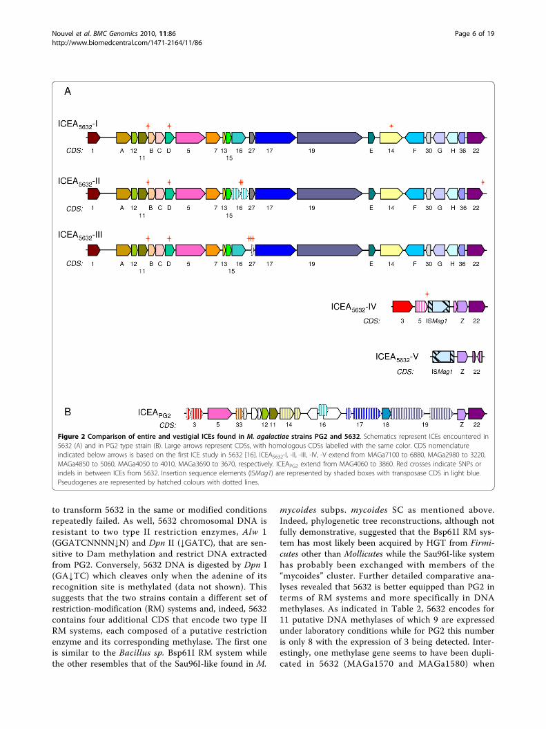
Role of the mobilome in M. agalactiae genetic diversityand genome plasticityAnalyses of the 5632 genome revealed the presence ofthree large regions (ca. 27 kb) that correspond to an ICEelement previously identified in this strain [16]. Thethree ICE copies were designated ICEA5632-I, -II and -III,with ICE-I corresponding to that previously published,and represented a total of about 80 kbp. In addition, twosmaller regions designated ICEA5632-IV and -V weredetected that relate to the degenerated, single ICE formfound in strain PG2 [12] and that appear to be ICEs ves-tiges as suggested by their reduced size and the presenceof insertion sequences and pseudogenes. Predicted pro-teins encoded by these vestiges were designated accord-ing to our previous nomenclature (Figure 2).Interestingly, while phylogenetic and BLAST analysesindicate that ICEA5632-I to-III are related to the ICEF ofMycoplasma fermentans strain PG18 [28], the ICEA5632-IV and -V vestiges and the degenerated ICEAPG2 aresomewhat similar to the ICEC of M. capricolum; in parti-cular, they all contain a CDS with no predicted function,CDSZ, that is not found in ICEA5632-I to -III nor inICEF. ICEA5632-IV also contains a CDS, CDS3, which ispresent as a pseudogene in both the ICEAPG2 and ICECbut is absent from the large copies ICEA5632-I to III. TheICEA copies I to IV also contain two CDS widely con-served in mycoplasma ICEs, CDS22 and CDS5 (the copy-V appear to have a degenerated version of CDS22).ICEA left and right borders of the ICEA have beenexperimentally defined as GGAA-[ICEA]-TTCC forcopy-I [16] and an identical inverted repeat also flankedcopies-II and -III. The high level of sequence conserva-tion between the two M. agalactiae genomes alloweddefining the insertion points of the 3 large ICE copies inthe 5632 chromosome which correspond to intergenicregions in PG2. Although ICE insertions do not result inapparent gene disruption, the targeted regions seem tobe prone to instability: the copy -I is located next to a
Table 1 General properties of M. agalactiae PG2 and 5632strains
PG2 5632
Date of isolation 1952 <1991
Country Spain Spain
Source nk articulation
Host caprine caprine
Genome size (bp) 877,438 1,006,702
G+C (%) 29.70 29.62
Gene density (%) 88.5 88.7
Total number of CDS 752 826
HP (Hypothetical Protein) 138 148
CHP (Conserved HP) 186 150
CDS with predicted function 404 505
Pseudogenes 45 23
rRNAs sets 2 2
tRNAs 34 34
GenBank accession number CU179680 FP671138
ICE number (1 vestigial) 3 (+2 vestigial)
Transposase number 1a 15
(+2pseudogenes)
(+2pseudogenes)
Genomic DNA digested by:
Dpn I or Alw I (sens. to Dammethylation)
Yes No
Dpn II (Dam resistant) Yes Yes
Relative colony sizeb 100% 180%
Data were extracted from the MolliGen database http://cbi.labri.fr/outils/molligen/.a One CDS, MAG3410, was annotated as transposase and was detected byproteomics analysis in this study but no inverted repeat sequences could befound.bRelative colony sizes as defined on agar medium with PG2 as reference.Repeatedly colonies of 5632 were found to be approximately 1.8 times largerthan those of PG2.nk, not known
Nouvel et al. BMC Genomics 2010, 11:86http://www.biomedcentral.com/1471-2164/11/86
Page 3 of 19
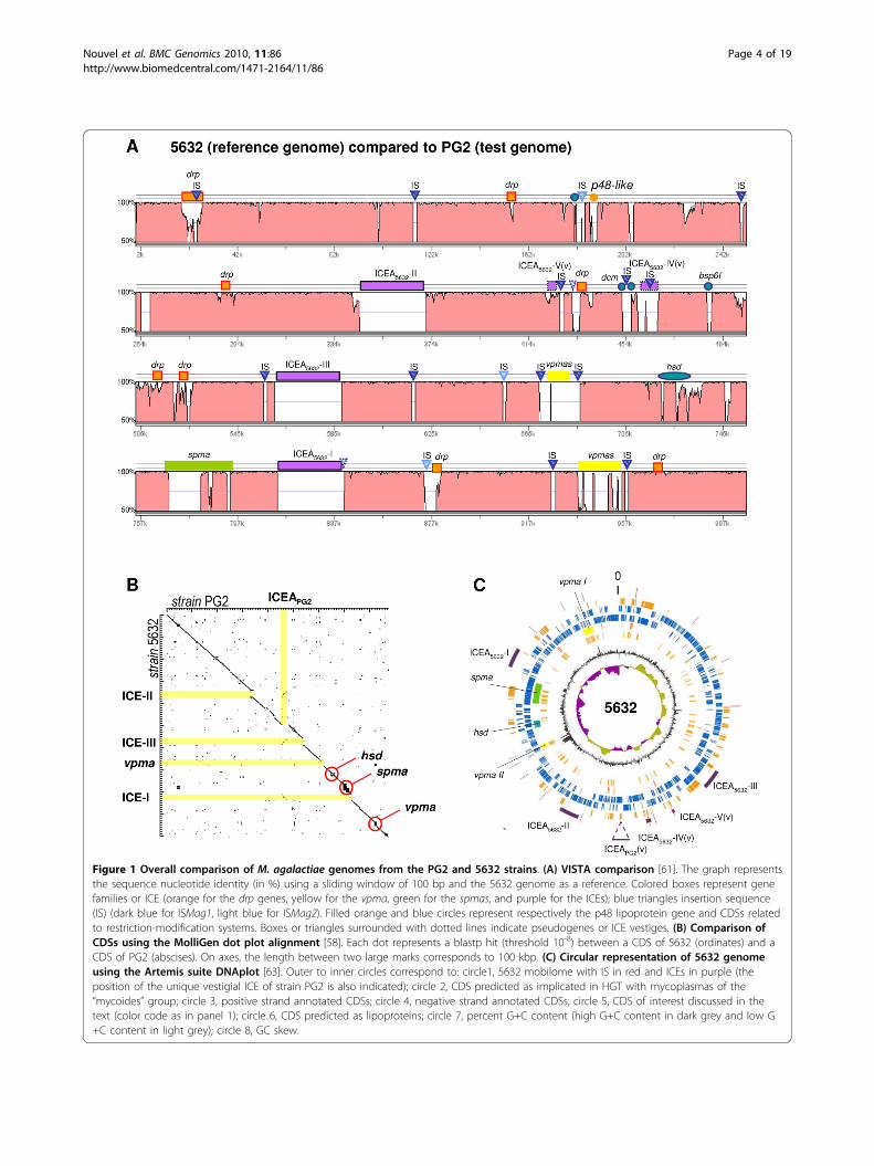
Figure 1 Overall comparison of M. agalactiae genomes from the PG2 and 5632 strains. (A) VISTA comparison [61]. The graph representsthe sequence nucleotide identity (in %) using a sliding window of 100 bp and the 5632 genome as a reference. Colored boxes represent genefamilies or ICE (orange for the drp genes, yellow for the vpma, green for the spmas, and purple for the ICEs); blue triangles insertion sequence(IS) (dark blue for ISMag1, light blue for ISMag2). Filled orange and blue circles represent respectively the p48 lipoprotein gene and CDSs relatedto restriction-modification systems. Boxes or triangles surrounded with dotted lines indicate pseudogenes or ICE vestiges. (B) Comparison ofCDSs using the MolliGen dot plot alignment [58]. Each dot represents a blastp hit (threshold 10-8) between a CDS of 5632 (ordinates) and aCDS of PG2 (abscises). On axes, the length between two large marks corresponds to 100 kbp. (C) Circular representation of 5632 genomeusing the Artemis suite DNAplot [63]. Outer to inner circles correspond to: circle1, 5632 mobilome with IS in red and ICEs in purple (theposition of the unique vestigial ICE of strain PG2 is also indicated); circle 2, CDS predicted as implicated in HGT with mycoplasmas of the“mycoides” group; circle 3, positive strand annotated CDSs; circle 4, negative strand annotated CDSs; circle 5, CDS of interest discussed in thetext (color code as in panel 1); circle 6, CDS predicted as lipoproteins; circle 7, percent G+C content (high G+C content in dark grey and low G+C content in light grey); circle 8, GC skew.
Nouvel et al. BMC Genomics 2010, 11:86http://www.biomedcentral.com/1471-2164/11/86
Page 4 of 19
conserved insertion sequence (IS) present as a pseudo-gene in both strains but showing some sequence diver-gence; the copy -III is also in the vicinity of an IS presentonly in strain 5632 and which is inserted next to a con-served tRNA gene. The copy -II is inserted next to a pre-dicted lipoprotein gene (MAG2840 or MAGa2970)showing clear sequence divergence as the two predictedproteins have only 66.4% identities. The three ICE copiesI-III are flanked by an almost perfect 9 bp direct repeatwhich is most likely generated during the integrationprocess. Alignment of ICEA-I, -II and -III DNAsequences from the first G of the GGAA repeat to thelast C of the TTCC repeat showed that there are highlysimilar, presenting only 7 to 8 SNPs. This suggests thatthe copies originated from subsequent excision and inte-gration events, possibly during chromosomal replicationor by exchange within the population. SNPs resulted ingenerating (i) two pseudogenes corresponding to CDS16of copy II and CDS27 of copy III, (ii) truncation ofCDS22 in copy II and (iii) insertion of an Asn codon in astretch of repeated AAT (poly Asn) in CDS14 of copy I.Overall, 5632 ICEAs account for 21 different CDS thatare not present in PG2, with the products of two detectedby MS/MS in the Triton-X114 fraction of 5632 (CDS14and 17).Other mobile elements were found in the 5632 genome
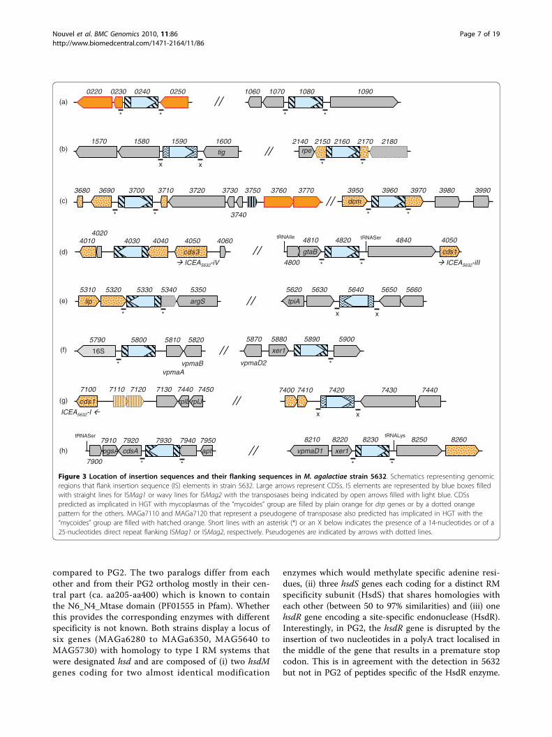
but not in PG2 and correspond to multiple copies of twoIS elements that both belong to the IS30 family. Thelocation of these elements relative to their flanking CDSis shown in Figure 3. The IS element, ISMag1, has pre-viously been described in some M. agalactiae strains [29]and an isoform was also described in M. bovis (namedISMbov1) [30]. In 5632, this element occurs in 12 copieswith 10 that localized either next to genomic islandsencoding a repertoire of variable surface lipoproteins (seeMAGa5890, MAGa5800 and MAGa8230) or to regionsassociated with HGT (see circles 1 and 2 in Figure 1C).The second type of IS, ISMag2, resembles ISMbov6recently described [31] and is found only in three copies,none of which seems to truncate or disrupt a CDS.In silico analyses further indicate that ISMag1 occur-
rence most likely disrupts gene expression in only threecases. In the first case, the insertion has taken place inbetween a dcm gene encoding a Cytosine-specific DNA-methyltransferase Sau96I (MAGa3950) and a Type IIspecific deoxyribonuclease sau96I-like gene(MAGa3970). Both genes are absent from PG2 but arefound next to each other in M. mycoides subsp.mycoides SC in which they most likely occur as anoperon. These genes are highly similar to those found in5632 with ca. 85% and 78% similarity, respectively, withhigher divergence in the N-terminal of 5632 sau96I-likegene. Interestingly, a global proteomic approach (seebelow and additional file 1: Table S1) detected several
specific peptides of the cytosine-specific methyl transfer-ase encoded by MAGa3950 but none corresponding tothe sau96I-like gene supporting the hypothesis that theIS occurrence at its 5’end may affect its expression. Inthe second case, IS insertion at the 3’ end of MAGa4040would result in truncating the product by more than50% when compared to the situation found in PG2. Thethird case relates to MAGa5320 and MAGa5350 whichare separated by an IS and which have been annotatedas two distinct pseudogenes because they are highlysimilar to either the N- or the C-terminal part of theMycoplasma capricolum subsp. capricolum glycosyltransferase (MCAP0063). In PG2, homologs toMAGa5320 and MAGa5350 also exist as pseudogenesalthough no IS is involved.Finally, two vestiges of transposase having similarities
with that of ISMmy1 of M. mycoides subsp. mycoides SCwere detected in the 5632 genome; one located next toan ICE element while the other was found next to atruncated hypothetical protein that displays a DUF285motif (see below) and that is predicted to have under-gone HGT with member of the “mycoides” clusterspecies.In most cases, IS elements were flanked by direct
repeated sequences of 14 nt for ISMag1 and of 25 ntfor ISMag2 that indicated a single IS insertion event.Exceptions were found for IS elements (MAGa5800and MAGa5890) located next to the vpma gene familyas previously described [17] and was also observedhere for the IS insertion located at the 3’ end ofMAGa4040 suggesting that further genomic rearrange-ments have occurred in this area. Indeed, this regionhas been described above as a putative vestige of ICEintegration. Finally, a single transposase gene whichproduct was detected by proteomic analyses(MAG3410; see also Figure 4) is found in PG2 but notin 5632. This transposase has some similarities (46.7%)with an ISMmy1 transposase of M. mycoides subsp.mycoides SC, but no flanking repeated sequence couldbe readily identified.These data indicate that ca. 76% of the additional
genomic material of 5632 is composed of mobile geneticelements when compared to PG2. This represents 10%of the genome, yet these do not lead to major genomerearrangement. Overall, 5632 has 95 additional CDS, 72of which correspond to CDS of ICE or transposase.Among the remaining, 8 relate to the spma family (seebelow), 11 to hypothetical products (including 4detected by LC-MS/MS) and 4 to restriction modifica-tion systems (RM) (Table 2).
Protection from DNA degradation and invasionWhile genetic transformation of strain PG2 [26,32] hasbecome a standard protocol in our laboratory, attempts
Nouvel et al. BMC Genomics 2010, 11:86http://www.biomedcentral.com/1471-2164/11/86
Page 5 of 19
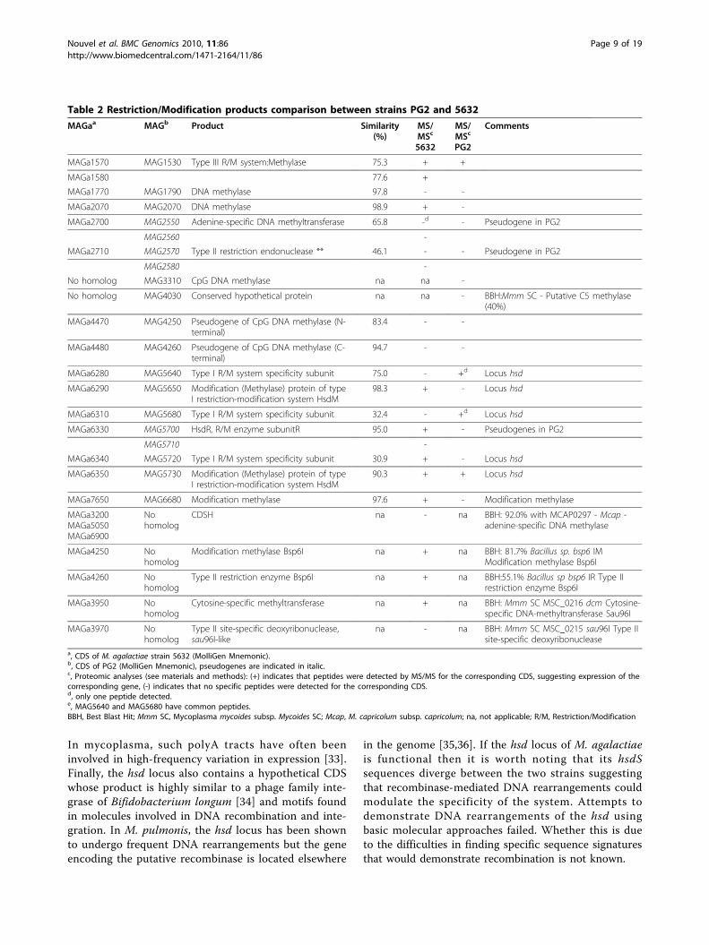
to transform 5632 in the same or modified conditionsrepeatedly failed. As well, 5632 chromosomal DNA isresistant to two type II restriction enzymes, Alw 1(GGATCNNNN↓N) and Dpn II (↓GATC), that are sen-sitive to Dam methylation and restrict DNA extractedfrom PG2. Conversely, 5632 DNA is digested by Dpn I(GA↓TC) which cleaves only when the adenine of itsrecognition site is methylated (data not shown). Thissuggests that the two strains contain a different set ofrestriction-modification (RM) systems and, indeed, 5632contains four additional CDS that encode two type IIRM systems, each composed of a putative restrictionenzyme and its corresponding methylase. The first oneis similar to the Bacillus sp. Bsp61I RM system whilethe other resembles that of the Sau96I-like found in M.
mycoides subps. mycoides SC as mentioned above.Indeed, phylogenetic tree reconstructions, although notfully demonstrative, suggested that the Bsp61I RM sys-tem has most likely been acquired by HGT from Firmi-cutes other than Mollicutes while the Sau96I-like systemhas probably been exchanged with members of the“mycoides” cluster. Further detailed comparative ana-lyses revealed that 5632 is better equipped than PG2 interms of RM systems and more specifically in DNAmethylases. As indicated in Table 2, 5632 encodes for11 putative DNA methylases of which 9 are expressedunder laboratory conditions while for PG2 this numberis only 8 with the expression of 3 being detected. Inter-estingly, one methylase gene seems to have been dupli-cated in 5632 (MAGa1570 and MAGa1580) when
Figure 2 Comparison of entire and vestigial ICEs found in M. agalactiae strains PG2 and 5632. Schematics represent ICEs encountered in5632 (A) and in PG2 type strain (B). Large arrows represent CDSs, with homologous CDSs labelled with the same color. CDS nomenclatureindicated below arrows is based on the first ICE study in 5632 [16]. ICEA5632-I, -II, -III, -IV, -V extend from MAGa7100 to 6880, MAGa2980 to 3220,MAGa4850 to 5060, MAGa4050 to 4010, MAGa3690 to 3670, respectively. ICEAPG2 extend from MAG4060 to 3860. Red crosses indicate SNPs orindels in between ICEs from 5632. Insertion sequence elements (ISMag1) are represented by shaded boxes with transposase CDS in light blue.Pseudogenes are represented by hatched colours with dotted lines.
Nouvel et al. BMC Genomics 2010, 11:86http://www.biomedcentral.com/1471-2164/11/86
Page 6 of 19
compared to PG2. The two paralogs differ from eachother and from their PG2 ortholog mostly in their cen-tral part (ca. aa205-aa400) which is known to containthe N6_N4_Mtase domain (PF01555 in Pfam). Whetherthis provides the corresponding enzymes with differentspecificity is not known. Both strains display a locus ofsix genes (MAGa6280 to MAGa6350, MAG5640 toMAG5730) with homology to type I RM systems thatwere designated hsd and are composed of (i) two hsdMgenes coding for two almost identical modification
enzymes which would methylate specific adenine resi-dues, (ii) three hsdS genes each coding for a distinct RMspecificity subunit (HsdS) that shares homologies witheach other (between 50 to 97% similarities) and (iii) onehsdR gene encoding a site-specific endonuclease (HsdR).Interestingly, in PG2, the hsdR gene is disrupted by theinsertion of two nucleotides in a polyA tract localised inthe middle of the gene that results in a premature stopcodon. This is in agreement with the detection in 5632but not in PG2 of peptides specific of the HsdR enzyme.
vpmaB
16S
*
58105800 58205790
vpmaA
xer1
*
59005870
vpmaD2
58905880
**
02400230 02500220
**
1080 090107011060
**
21602150 2170 2180rpe
2140
x x
tig
1600159015801570
40304020
4040 4050 40604010
cds3
**
4820
cds1
050404840184
4800
gtaB
tRNAIle tRNASer
x x
5650564056305620
tpiA
5660
**
53305320 5340 53505310
argSlip
7130 7440712071107100
cds1 rplL rplJ
7450
x x
7430 7440742074107400
**
79307920 7940 79507910
cdsApgsA apt
tRNASer
7900
xer1
**
tRNALys8220
vpmaD1
8230 8250 82608210
3760 377037503730
3740**
37003690 3710 02730863
**
39603950 3970 3980 3990
dcm
(a)
(b)
(c)
(d)
(e)
(f)
(g)
(h)
ICEA5632-I
ICEA5632-iV ICEA5632-iII
Figure 3 Location of insertion sequences and their flanking sequences in M. agalactiae strain 5632. Schematics representing genomicregions that flank insertion sequence (IS) elements in strain 5632. Large arrows represent CDSs. IS elements are represented by blue boxes filledwith straight lines for ISMag1 or wavy lines for ISMag2 with the transposases being indicated by open arrows filled with light blue. CDSspredicted as implicated in HGT with mycoplasmas of the “mycoides” group are filled by plain orange for drp genes or by a dotted orangepattern for the others. MAGa7110 and MAGa7120 that represent a pseudogene of transposase also predicted has implicated in HGT with the“mycoides” group are filled with hatched orange. Short lines with an asterisk (*) or an X below indicates the presence of a 14-nucleotides or of a25-nucleotides direct repeat flanking ISMag1 or ISMag2, respectively. Pseudogenes are indicated by arrows with dotted lines.
Nouvel et al. BMC Genomics 2010, 11:86http://www.biomedcentral.com/1471-2164/11/86
Page 7 of 19
2430 2440 2450 246024202410
nox
nox
2600 2610 2620 263025902580
0190 0200 0210 0220 0230 0240 0250 0260 0270
dgk dgk
1330 1340 1350 136013201350
thi I
1370 1380 1390 1400
thi I
4530 4540 455045204510 4560
adtH adtH
7270 728072607250
8400 8410 842083908380
dnaJcmk
dnaJcmk
43004310
432043304290 04340824
adtHadtH
0190 0200 0210 0220 0230 0240 0250 026001800170
dgk dgk
4210 4220 4230 424042004190 4250
4450 446044404430 4470
3720 376037103700 3770 37803740
37503730
gcp
36603680
3630 3640 3650 3670 3690
3250 3260 3270 328032403230 3290 3300
gcp
3310 33203330
33403350
33603370
3380 3390 3400 3410 3420
*
*
*
64706480
6490 6500646064506510
6520
7430 7440 7450 746074207410 7470 7480*
7490
MAGa:
MAG:
(a)
(b)(c)
(d)
(e) (f)
(g)
*
*
*
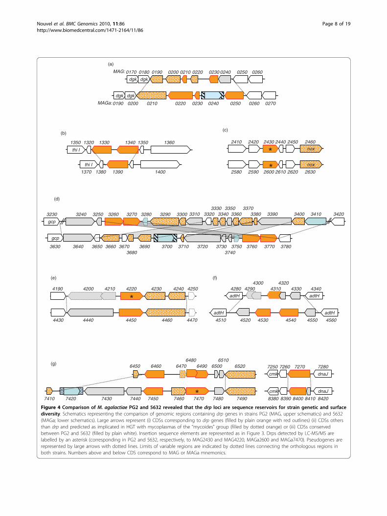
Figure 4 Comparison of M. agalactiae PG2 and 5632 revealed that the drp loci are sequence reservoirs for strain genetic and surfacediversity. Schematics representing the comparison of genomic regions containing drp genes in strains PG2 (MAG, upper schematics) and 5632(MAGa; lower schematics). Large arrows represent (i) CDSs corresponding to drp genes (filled by plain orange with red outlines) (ii) CDSs othersthan drp and predicted as implicated in HGT with mycoplasmas of the “mycoides” group (filled by dotted orange) or (iii) CDSs conservedbetween PG2 and 5632 (filled by plain white). Insertion sequence elements are represented as in Figure 3. Drps detected by LC-MS/MS arelabelled by an asterisk (corresponding in PG2 and 5632, respectively, to MAG2430 and MAG4220, MAGa2600 and MAGa7470). Pseudogenes arerepresented by large arrows with dotted lines. Limits of variable regions are indicated by dotted lines connecting the orthologous regions inboth strains. Numbers above and below CDS correspond to MAG or MAGa mnemonics.
Nouvel et al. BMC Genomics 2010, 11:86http://www.biomedcentral.com/1471-2164/11/86
Page 8 of 19
In mycoplasma, such polyA tracts have often beeninvolved in high-frequency variation in expression [33].Finally, the hsd locus also contains a hypothetical CDSwhose product is highly similar to a phage family inte-grase of Bifidobacterium longum [34] and motifs foundin molecules involved in DNA recombination and inte-gration. In M. pulmonis, the hsd locus has been shownto undergo frequent DNA rearrangements but the geneencoding the putative recombinase is located elsewhere
in the genome [35,36]. If the hsd locus of M. agalactiaeis functional then it is worth noting that its hsdSsequences diverge between the two strains suggestingthat recombinase-mediated DNA rearrangements couldmodulate the specificity of the system. Attempts todemonstrate DNA rearrangements of the hsd usingbasic molecular approaches failed. Whether this is dueto the difficulties in finding specific sequence signaturesthat would demonstrate recombination is not known.
Table 2 Restriction/Modification products comparison between strains PG2 and 5632
MAGaa MAGb Product Similarity(%)
MS/MSc
5632
MS/MSc
PG2
Comments
MAGa1570 MAG1530 Type III R/M system:Methylase 75.3 + +
MAGa1580 77.6 +
MAGa1770 MAG1790 DNA methylase 97.8 - -
MAGa2070 MAG2070 DNA methylase 98.9 + -
MAGa2700 MAG2550 Adenine-specific DNA methyltransferase 65.8 -d - Pseudogene in PG2
MAG2560 -
MAGa2710 MAG2570 Type II restriction endonuclease ** 46.1 - - Pseudogene in PG2
MAG2580 -
No homolog MAG3310 CpG DNA methylase na na -
No homolog MAG4030 Conserved hypothetical protein na na - BBH:Mmm SC - Putative C5 methylase(40%)
MAGa4470 MAG4250 Pseudogene of CpG DNA methylase (N-terminal)
83.4 - -
MAGa4480 MAG4260 Pseudogene of CpG DNA methylase (C-terminal)
94.7 - -
MAGa6280 MAG5640 Type I R/M system specificity subunit 75.0 - +d Locus hsd
MAGa6290 MAG5650 Modification (Methylase) protein of typeI restriction-modification system HsdM
98.3 + - Locus hsd
MAGa6310 MAG5680 Type I R/M system specificity subunit 32.4 - +d Locus hsd
MAGa6330 MAG5700 HsdR, R/M enzyme subunitR 95.0 + - Pseudogenes in PG2
MAG5710 -
MAGa6340 MAG5720 Type I R/M system specificity subunit 30.9 + - Locus hsd
MAGa6350 MAG5730 Modification (Methylase) protein of typeI restriction-modification system HsdM
90.3 + + Locus hsd
MAGa7650 MAG6680 Modification methylase 97.6 + - Modification methylase
MAGa3200MAGa5050MAGa6900
Nohomolog
CDSH na - na BBH: 92.0% with MCAP0297 - Mcap -adenine-specific DNA methylase
MAGa4250 Nohomolog
Modification methylase Bsp6I na + na BBH: 81.7% Bacillus sp. bsp6 IMModification methylase Bsp6I
MAGa4260 Nohomolog
Type II restriction enzyme Bsp6I na + na BBH:55.1% Bacillus sp bsp6 IR Type IIrestriction enzyme Bsp6I
MAGa3950 Nohomolog
Cytosine-specific methyltransferase na + na BBH: Mmm SC MSC_0216 dcm Cytosine-specific DNA-methyltransferase Sau96I
MAGa3970 Nohomolog
Type II site-specific deoxyribonuclease,sau96I-like
na - na BBH: Mmm SC MSC_0215 sau96I Type IIsite-specific deoxyribonuclease
a, CDS of M. agalactiae strain 5632 (MolliGen Mnemonic).b, CDS of PG2 (MolliGen Mnemonic), pseudogenes are indicated in italic.c, Proteomic analyses (see materials and methods): (+) indicates that peptides were detected by MS/MS for the corresponding CDS, suggesting expression of thecorresponding gene, (-) indicates that no specific peptides were detected for the corresponding CDS.d, only one peptide detected.e, MAG5640 and MAG5680 have common peptides.BBH, Best Blast Hit; Mmm SC, Mycoplasma mycoides subsp. Mycoides SC; Mcap, M. capricolum subsp. capricolum; na, not applicable; R/M, Restriction/Modification
Nouvel et al. BMC Genomics 2010, 11:86http://www.biomedcentral.com/1471-2164/11/86
Page 9 of 19
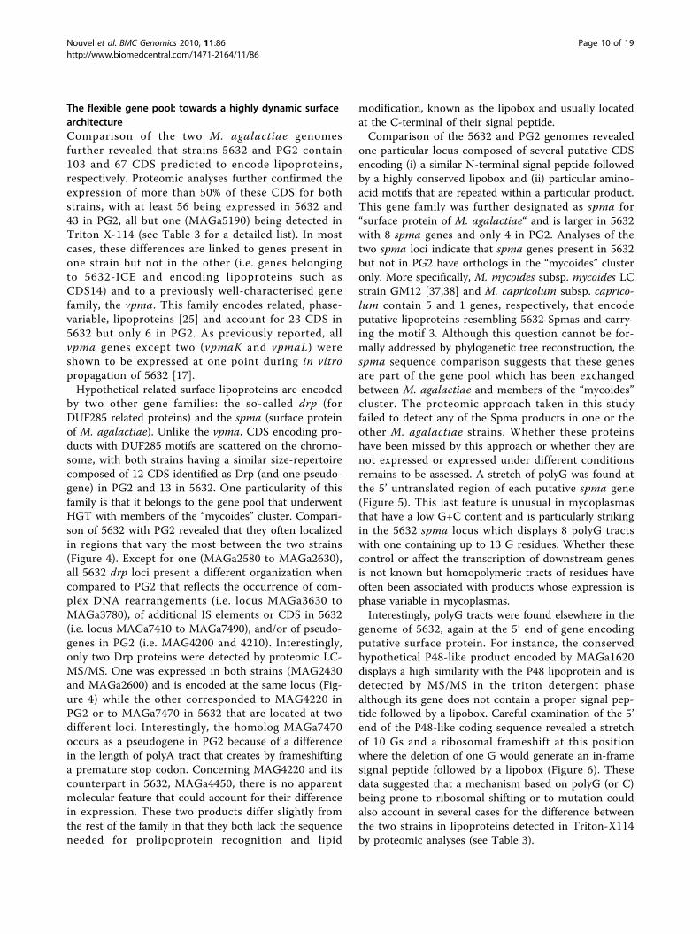
The flexible gene pool: towards a highly dynamic surfacearchitectureComparison of the two M. agalactiae genomesfurther revealed that strains 5632 and PG2 contain103 and 67 CDS predicted to encode lipoproteins,respectively. Proteomic analyses further confirmed theexpression of more than 50% of these CDS for bothstrains, with at least 56 being expressed in 5632 and43 in PG2, all but one (MAGa5190) being detected inTriton X-114 (see Table 3 for a detailed list). In mostcases, these differences are linked to genes present inone strain but not in the other (i.e. genes belongingto 5632-ICE and encoding lipoproteins such asCDS14) and to a previously well-characterised genefamily, the vpma. This family encodes related, phase-variable, lipoproteins [25] and account for 23 CDS in5632 but only 6 in PG2. As previously reported, allvpma genes except two (vpmaK and vpmaL) wereshown to be expressed at one point during in vitropropagation of 5632 [17].Hypothetical related surface lipoproteins are encoded
by two other gene families: the so-called drp (forDUF285 related proteins) and the spma (surface proteinof M. agalactiae). Unlike the vpma, CDS encoding pro-ducts with DUF285 motifs are scattered on the chromo-some, with both strains having a similar size-repertoirecomposed of 12 CDS identified as Drp (and one pseudo-gene) in PG2 and 13 in 5632. One particularity of thisfamily is that it belongs to the gene pool that underwentHGT with members of the “mycoides” cluster. Compari-son of 5632 with PG2 revealed that they often localizedin regions that vary the most between the two strains(Figure 4). Except for one (MAGa2580 to MAGa2630),all 5632 drp loci present a different organization whencompared to PG2 that reflects the occurrence of com-plex DNA rearrangements (i.e. locus MAGa3630 toMAGa3780), of additional IS elements or CDS in 5632(i.e. locus MAGa7410 to MAGa7490), and/or of pseudo-genes in PG2 (i.e. MAG4200 and 4210). Interestingly,only two Drp proteins were detected by proteomic LC-MS/MS. One was expressed in both strains (MAG2430and MAGa2600) and is encoded at the same locus (Fig-ure 4) while the other corresponded to MAG4220 inPG2 or to MAGa7470 in 5632 that are located at twodifferent loci. Interestingly, the homolog MAGa7470occurs as a pseudogene in PG2 because of a differencein the length of polyA tract that creates by frameshiftinga premature stop codon. Concerning MAG4220 and itscounterpart in 5632, MAGa4450, there is no apparentmolecular feature that could account for their differencein expression. These two products differ slightly fromthe rest of the family in that they both lack the sequenceneeded for prolipoprotein recognition and lipid
modification, known as the lipobox and usually locatedat the C-terminal of their signal peptide.Comparison of the 5632 and PG2 genomes revealed
one particular locus composed of several putative CDSencoding (i) a similar N-terminal signal peptide followedby a highly conserved lipobox and (ii) particular amino-acid motifs that are repeated within a particular product.This gene family was further designated as spma for“surface protein of M. agalactiae“ and is larger in 5632with 8 spma genes and only 4 in PG2. Analyses of thetwo spma loci indicate that spma genes present in 5632but not in PG2 have orthologs in the “mycoides” clusteronly. More specifically, M. mycoides subsp. mycoides LCstrain GM12 [37,38] and M. capricolum subsp. caprico-lum contain 5 and 1 genes, respectively, that encodeputative lipoproteins resembling 5632-Spmas and carry-ing the motif 3. Although this question cannot be for-mally addressed by phylogenetic tree reconstruction, thespma sequence comparison suggests that these genesare part of the gene pool which has been exchangedbetween M. agalactiae and members of the “mycoides”cluster. The proteomic approach taken in this studyfailed to detect any of the Spma products in one or theother M. agalactiae strains. Whether these proteinshave been missed by this approach or whether they arenot expressed or expressed under different conditionsremains to be assessed. A stretch of polyG was found atthe 5’ untranslated region of each putative spma gene(Figure 5). This last feature is unusual in mycoplasmasthat have a low G+C content and is particularly strikingin the 5632 spma locus which displays 8 polyG tractswith one containing up to 13 G residues. Whether thesecontrol or affect the transcription of downstream genesis not known but homopolymeric tracts of residues haveoften been associated with products whose expression isphase variable in mycoplasmas.Interestingly, polyG tracts were found elsewhere in the
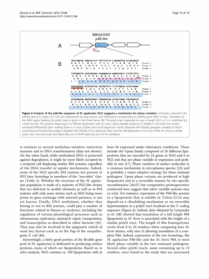
genome of 5632, again at the 5’ end of gene encodingputative surface protein. For instance, the conservedhypothetical P48-like product encoded by MAGa1620displays a high similarity with the P48 lipoprotein and isdetected by MS/MS in the triton detergent phasealthough its gene does not contain a proper signal pep-tide followed by a lipobox. Careful examination of the 5’end of the P48-like coding sequence revealed a stretchof 10 Gs and a ribosomal frameshift at this positionwhere the deletion of one G would generate an in-framesignal peptide followed by a lipobox (Figure 6). Thesedata suggested that a mechanism based on polyG (or C)being prone to ribosomal shifting or to mutation couldalso account in several cases for the difference betweenthe two strains in lipoproteins detected in Triton-X114by proteomic analyses (see Table 3).
Nouvel et al. BMC Genomics 2010, 11:86http://www.biomedcentral.com/1471-2164/11/86
Page 10 of 19
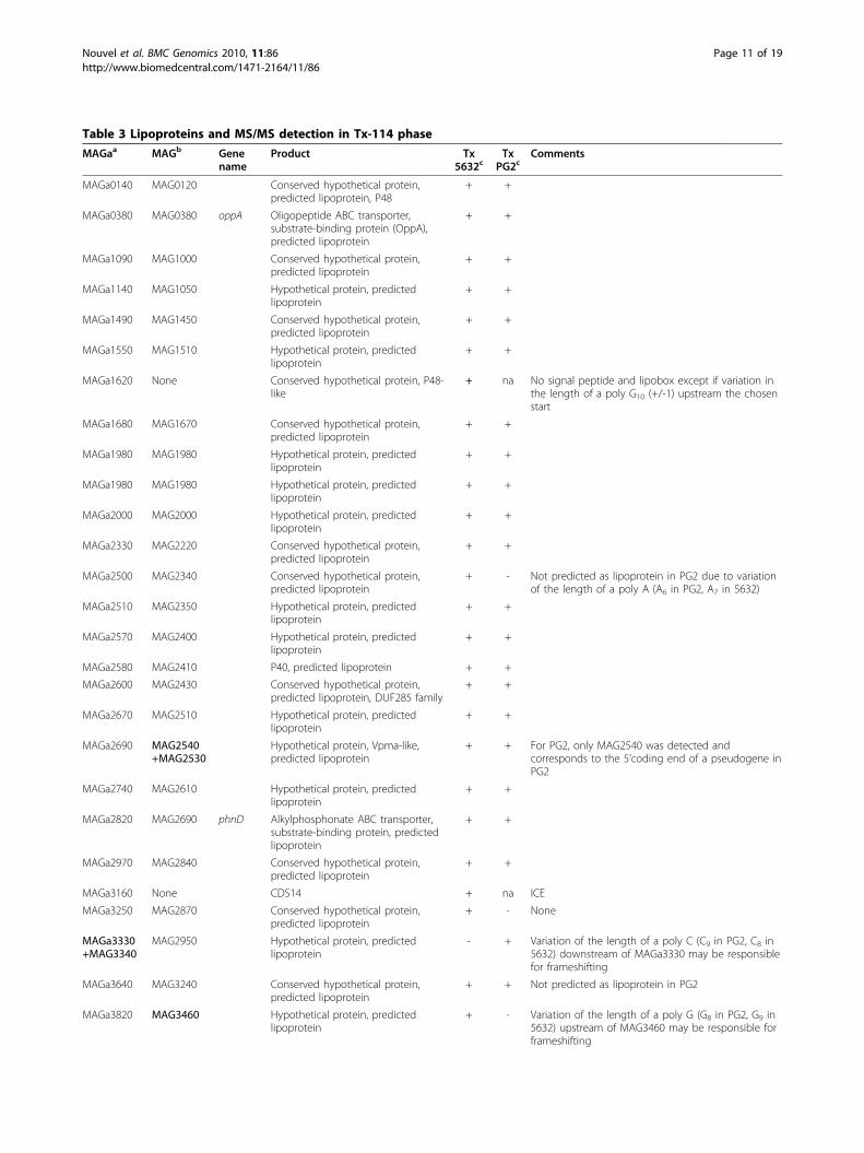
Table 3 Lipoproteins and MS/MS detection in Tx-114 phase
MAGaa MAGb Genename
Product Tx5632c
TxPG2c
Comments
MAGa0140 MAG0120 Conserved hypothetical protein,predicted lipoprotein, P48
+ +
MAGa0380 MAG0380 oppA Oligopeptide ABC transporter,substrate-binding protein (OppA),predicted lipoprotein
+ +
MAGa1090 MAG1000 Conserved hypothetical protein,predicted lipoprotein
+ +
MAGa1140 MAG1050 Hypothetical protein, predictedlipoprotein
+ +
MAGa1490 MAG1450 Conserved hypothetical protein,predicted lipoprotein
+ +
MAGa1550 MAG1510 Hypothetical protein, predictedlipoprotein
+ +
MAGa1620 None Conserved hypothetical protein, P48-like
+ na No signal peptide and lipobox except if variation inthe length of a poly G10 (+/-1) upstream the chosenstart
MAGa1680 MAG1670 Conserved hypothetical protein,predicted lipoprotein
+ +
MAGa1980 MAG1980 Hypothetical protein, predictedlipoprotein
+ +
MAGa1980 MAG1980 Hypothetical protein, predictedlipoprotein
+ +
MAGa2000 MAG2000 Hypothetical protein, predictedlipoprotein
+ +
MAGa2330 MAG2220 Conserved hypothetical protein,predicted lipoprotein
+ +
MAGa2500 MAG2340 Conserved hypothetical protein,predicted lipoprotein
+ - Not predicted as lipoprotein in PG2 due to variationof the length of a poly A (A6 in PG2, A7 in 5632)
MAGa2510 MAG2350 Hypothetical protein, predictedlipoprotein
+ +
MAGa2570 MAG2400 Hypothetical protein, predictedlipoprotein
+ +
MAGa2580 MAG2410 P40, predicted lipoprotein + +
MAGa2600 MAG2430 Conserved hypothetical protein,predicted lipoprotein, DUF285 family
+ +
MAGa2670 MAG2510 Hypothetical protein, predictedlipoprotein
+ +
MAGa2690 MAG2540+MAG2530
Hypothetical protein, Vpma-like,predicted lipoprotein
+ + For PG2, only MAG2540 was detected andcorresponds to the 5’coding end of a pseudogene inPG2
MAGa2740 MAG2610 Hypothetical protein, predictedlipoprotein
+ +
MAGa2820 MAG2690 phnD Alkylphosphonate ABC transporter,substrate-binding protein, predictedlipoprotein
+ +
MAGa2970 MAG2840 Conserved hypothetical protein,predicted lipoprotein
+ +
MAGa3160 None CDS14 + na ICE
MAGa3250 MAG2870 Conserved hypothetical protein,predicted lipoprotein
+ - None
MAGa3330+MAG3340
MAG2950 Hypothetical protein, predictedlipoprotein
- + Variation of the length of a poly C (C9 in PG2, C8 in5632) downstream of MAGa3330 may be responsiblefor frameshifting
MAGa3640 MAG3240 Conserved hypothetical protein,predicted lipoprotein
+ + Not predicted as lipoprotein in PG2
MAGa3820 MAG3460 Hypothetical protein, predictedlipoprotein
+ - Variation of the length of a poly G (G8 in PG2, G9 in5632) upstream of MAG3460 may be responsible forframeshifting
Nouvel et al. BMC Genomics 2010, 11:86http://www.biomedcentral.com/1471-2164/11/86
Page 11 of 19
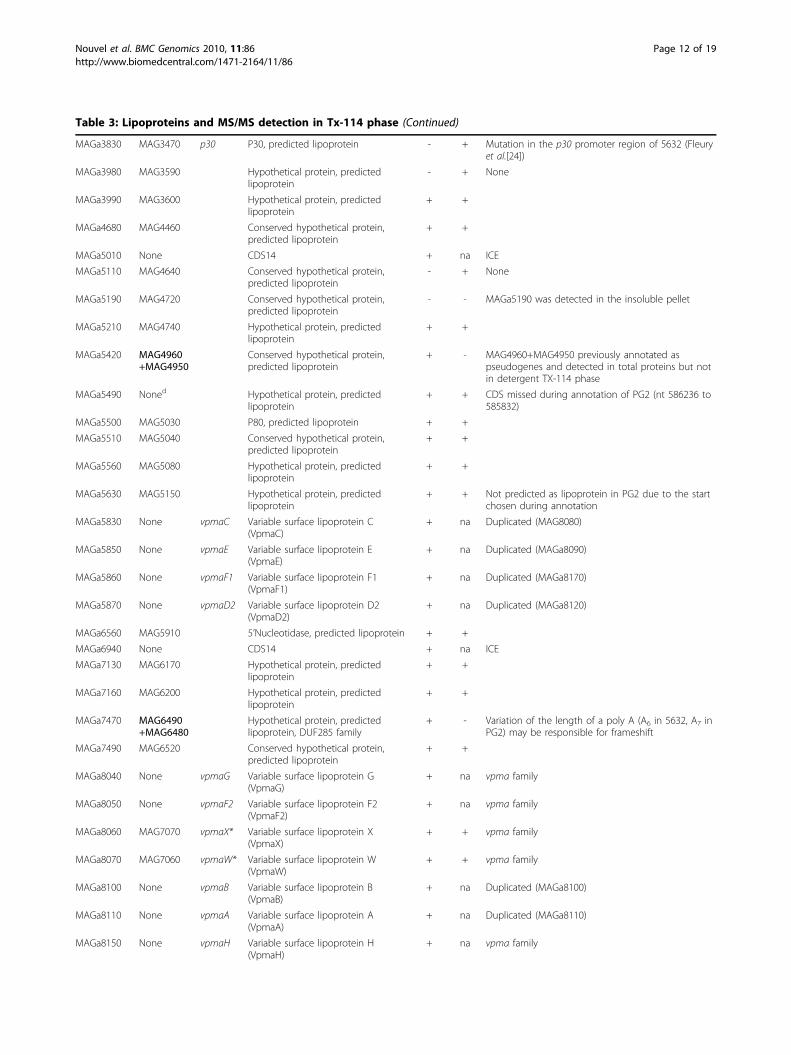
Table 3: Lipoproteins and MS/MS detection in Tx-114 phase (Continued)
MAGa3830 MAG3470 p30 P30, predicted lipoprotein - + Mutation in the p30 promoter region of 5632 (Fleuryet al.[24])
MAGa3980 MAG3590 Hypothetical protein, predictedlipoprotein
- + None
MAGa3990 MAG3600 Hypothetical protein, predictedlipoprotein
+ +
MAGa4680 MAG4460 Conserved hypothetical protein,predicted lipoprotein
+ +
MAGa5010 None CDS14 + na ICE
MAGa5110 MAG4640 Conserved hypothetical protein,predicted lipoprotein
- + None
MAGa5190 MAG4720 Conserved hypothetical protein,predicted lipoprotein
- - MAGa5190 was detected in the insoluble pellet
MAGa5210 MAG4740 Hypothetical protein, predictedlipoprotein
+ +
MAGa5420 MAG4960+MAG4950
Conserved hypothetical protein,predicted lipoprotein
+ - MAG4960+MAG4950 previously annotated aspseudogenes and detected in total proteins but notin detergent TX-114 phase
MAGa5490 Noned Hypothetical protein, predictedlipoprotein
+ + CDS missed during annotation of PG2 (nt 586236 to585832)
MAGa5500 MAG5030 P80, predicted lipoprotein + +
MAGa5510 MAG5040 Conserved hypothetical protein,predicted lipoprotein
+ +
MAGa5560 MAG5080 Hypothetical protein, predictedlipoprotein
+ +
MAGa5630 MAG5150 Hypothetical protein, predictedlipoprotein
+ + Not predicted as lipoprotein in PG2 due to the startchosen during annotation
MAGa5830 None vpmaC Variable surface lipoprotein C(VpmaC)
+ na Duplicated (MAG8080)
MAGa5850 None vpmaE Variable surface lipoprotein E(VpmaE)
+ na Duplicated (MAGa8090)
MAGa5860 None vpmaF1 Variable surface lipoprotein F1(VpmaF1)
+ na Duplicated (MAGa8170)
MAGa5870 None vpmaD2 Variable surface lipoprotein D2(VpmaD2)
+ na Duplicated (MAGa8120)
MAGa6560 MAG5910 5’Nucleotidase, predicted lipoprotein + +
MAGa6940 None CDS14 + na ICE
MAGa7130 MAG6170 Hypothetical protein, predictedlipoprotein
+ +
MAGa7160 MAG6200 Hypothetical protein, predictedlipoprotein
+ +
MAGa7470 MAG6490+MAG6480
Hypothetical protein, predictedlipoprotein, DUF285 family
+ - Variation of the length of a poly A (A6 in 5632, A7 inPG2) may be responsible for frameshift
MAGa7490 MAG6520 Conserved hypothetical protein,predicted lipoprotein
+ +
MAGa8040 None vpmaG Variable surface lipoprotein G(VpmaG)
+ na vpma family
MAGa8050 None vpmaF2 Variable surface lipoprotein F2(VpmaF2)
+ na vpma family
MAGa8060 MAG7070 vpmaX* Variable surface lipoprotein X(VpmaX)
+ + vpma family
MAGa8070 MAG7060 vpmaW* Variable surface lipoprotein W(VpmaW)
+ + vpma family
MAGa8100 None vpmaB Variable surface lipoprotein B(VpmaB)
+ na Duplicated (MAGa8100)
MAGa8110 None vpmaA Variable surface lipoprotein A(VpmaA)
+ na Duplicated (MAGa8110)
MAGa8150 None vpmaH Variable surface lipoprotein H(VpmaH)
+ na vpma family
Nouvel et al. BMC Genomics 2010, 11:86http://www.biomedcentral.com/1471-2164/11/86
Page 12 of 19
DiscussionWhole genome sequencing of M. agalactiae strain 5632revealed that it contains an additional 95 genes repre-senting an extra-130 kbps when compared to the PG2type strain. For organisms that have a small genome sizesuch as mycoplasmas, this is a rather significant feature.The additional material is mostly composed of repeatedelements, so that our knowledge of the M. agalactiaepan-genome has been enriched by 39 new genes. Alarge portion of those, more specifically 23, is present inICEs or corresponds to IS. Recent mathematical modelsby Tettelin et al. [39] show that the pan-genome of themollicute Ureaplasma urealyticum is limited, based onthe draft sequences of nine strains. This implies that thesequencing of additional strains might not significantlyincrease our knowledge of this species unless it is target-ing a specific biological question [40]. Although U. urea-lyticum is a human pathogen and has a genome slightlysmaller (ca. 750 kbp), the same observation may applyto the M. agalactiae species as indicated by the lownumber of new genes discovered in our study. Thus,sequencing additional M. agalactiae strains might bringlittle more information on the global coding capacity ofthis organism.Overall, data obtained here and elsewhere indicate
that about 10% of the 5632 genome is highly dynamicin that large regions corresponding to ICE can excise[16] and, theoretically, relocate elsewhere or be trans-ferred to a recipient cell during conjugation, if suchevent is further shown to occur in this species. The two
ICE’s vestiges, ICE IV and V, represent scars of pastICE insertions followed by a progressive decay. Interest-ingly, these more resemble the larger ICE vestige ofPG2 or the ICE of M. capricolum subsp. capricolumthan the three entire ICE copies of 5632 suggesting thatthis strain may have, at one point, hosted two types ofICEs. These data indicate that the circulation of ICEs insome strains might not be such a rare event. The pre-sence of ICE circular forms in 5632 [16] together withthe low number of SNPs detected between the threecopies indicate that multiple ICE insertions are recent.The mechanisms underlying ICE insertion, excision andputative transfer in mycoplasmas have yet to be investi-gated, but recent studies on ICE elements in Gram-positive bacteria suggest that these events can be underthe control of sophisticated regulation systems inresponse to changing environmental conditions such asstress or population density [41]. The finding of ICE inM. agalactiae and members of the “mycoides” clustertogether with evidence of HGT in between these speciesfurther raised the prospect that these simple bacteriacould conjugate. So far, a single report has supportedthe occurrence of conjugation in mycoplasmas by show-ing the exchange of genetic material in between M. pul-monis cells via a mechanism resistant to DNAse [42].The idea that this phenomenon might be more com-mon among mycoplasmas than first expected is veryexciting because, if occurring, it would change the waywe see the evolution of these so called “minimalorganisms”.
Table 3: Lipoproteins and MS/MS detection in Tx-114 phase (Continued)
MAGa8160 None vpmaI Variable surface lipoprotein I (VpmaI) + na vpma family
MAGa8180 None vpmaJ Variable surface lipoprotein J(VpmaJ)
+ na vpma family
MAGa8210 None vpmaD1 Variable surface lipoprotein D1(VpmaD1)
+ na Duplicated (MAGa5840)
MAGa8260 MAG7130 Hypothetical protein, predictedlipoprotein
+ - Not predicted as lipoprotein in PG2 due to a pointmutation:TAA (ochre) ↔TCA (serine))
None MAG1570 Hypothetical protein - + No signal peptide and lipobox except if variation ofthe length of a poly G9 (+/-1) next to the chosenstart
None MAG7050 vpmaV Variable surface lipoprotein V(VpmaV)
na + vpma family
None MAG7080 vpmaY Variable surface lipoprotein Y(VpmaY)
na + vpma family
None MAG7090 vpmaU Variable surface lipoprotein U(VpmaU)
na + vpma family
None MAG7100 vpmaZ Variable surface lipoprotein Z(VpmaZ)
na + vpma family
a CDS of M. agalactiae strain 5632 (Molligen Mnemonic).b CDS of PG2 (Molligen Mnemonic), pseudogenes are indicated in italic and bold.c Peptides detected by MS/MS in the Triton-X114 phase (Tx) (see the Methods section): (+) indicates that peptides corresponding to CDS were detected,suggesting expression of the corresponding gene, (-) indicates that no peptides corresponding to CDS were detected.d CDS detected in proteomic but for which no Mnemonic was defined because it was missed during the annotation of the PG2 genome [12].na, not applicable.
Nouvel et al. BMC Genomics 2010, 11:86http://www.biomedcentral.com/1471-2164/11/86
Page 13 of 19
Although smaller in size than ICE, IS elements as awhole represent a dynamic potential for the genomebecause of their copy number. In other bacteria, theircontribution to genome plasticity and dynamics is wellknown [43]. Here, no major DNA inversion or rearran-gement was detected between the two M. agalactiaegenomes that could be associated to IS except for twocases. As previously shown by our group, the first onerefers to the duplication in 5632 of the single vpmacluster of PG2 that has been most likely driven by ISelements and that resulted in 5632 having extended pos-sibilities for surface diversification when compared toPG2 [17]. The second case refers to a region whichorganization significantly differs in between PG2 and5632 (see Figure 4d) and which contains several ISrelated elements (i.e. IS, transposases or pseudogenes oftransposase). Events underlying rearrangements in this
region cannot be exactly retraced but most likely theyare ancient and have resulted in duplication of the ptsGgene in PG2 (MAG3250 and MAG3320). Interestingly,this region, like many others associated with IS, containsseveral genes or pseudogenes that have undergone HGTsuggesting that IS may directly contribute to this phe-nomenon as suggested for other bacteria [43]. Finally,we showed that IS insertions may have an impact ongene expression, thus modifying some of the strainproperties such as those associated with restriction-modification in 5632.Compared to the PG2 type strain, 5632 seems better
equipped for DNA exchange. Besides harbouring animpressive “mobilome”, some of which may be tailoredfor conjugative transfer, it contains a number of operat-ing RM systems. On one hand, these may act as a bar-rier to DNA invasion [44] and explain why 5632 DNA
MAGa6820
MAGa6790
MAGa6780
MAGa6770
MAGa6760
MAGa6730
MAGa6720
MAGa6670
MAG6090
MAG6060
MAG6010
MAG6030
S
S
S
MAGa6790MAGa6780
MAGa6770
MAGa6760
MAGa6720
MAGa6820MAGa6670
MAGa6730
CHP ABC transporter
G10 G11 G10G4T2G4
G7 G13G10
G9
MAGa6830MAGa6660
S S S S S S S S
G2TG2TG3G10 G9 G10
MAG6000MAG6090
CHP
MAG6100
ABC transporter
MAG6080
MAG6060
MAG6040
SSSMAG6050
MAG6030
MAG6020
MAG6010
MAGa6810
S
S
S
S
S
S
S
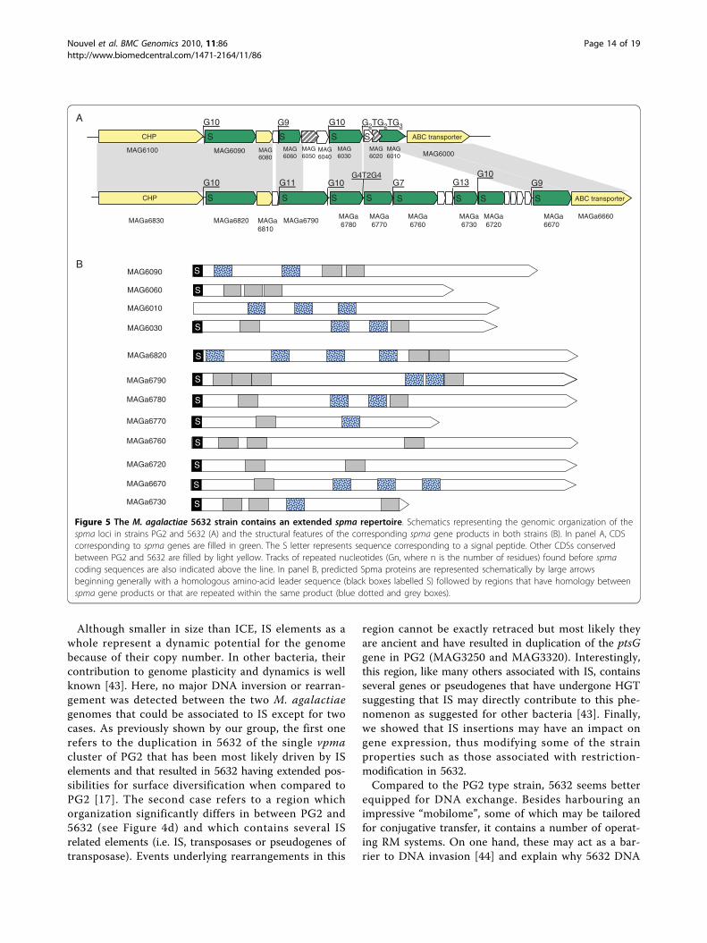
S
S
A
B
Figure 5 The M. agalactiae 5632 strain contains an extended spma repertoire. Schematics representing the genomic organization of thespma loci in strains PG2 and 5632 (A) and the structural features of the corresponding spma gene products in both strains (B). In panel A, CDScorresponding to spma genes are filled in green. The S letter represents sequence corresponding to a signal peptide. Other CDSs conservedbetween PG2 and 5632 are filled by light yellow. Tracks of repeated nucleotides (Gn, where n is the number of residues) found before spmacoding sequences are also indicated above the line. In panel B, predicted Spma proteins are represented schematically by large arrowsbeginning generally with a homologous amino-acid leader sequence (black boxes labelled S) followed by regions that have homology betweenspma gene products or that are repeated within the same product (blue dotted and grey boxes).
Nouvel et al. BMC Genomics 2010, 11:86http://www.biomedcentral.com/1471-2164/11/86
Page 14 of 19
is resistant to several methylase-sensitive restrictionenzymes and to DNA transformation (data not shown).On the other hand, while methylated DNA is protectedagainst degradation, it might be more likely accepted bya recipient cell displaying similar RM systems, regardlessof the DNA transfer or uptake mechanisms. Indeed,some of the 5632 specific RM systems not present inPG2 have homologs in members of the “mycoides” clus-ter (Table 2). Whether the structure of the M. agalac-tiae population is made of a majority of PG2-like strainsthat are deficient in mobile elements as well as in RMsystems with only some strains such as 5632 being moreprone to gene exchange with selected partners, is notyet known. Finally, DNA methylases, whether theybelong or not to RM systems, could play a number offunctions related to fitness or virulence, including theregulation of various physiological processes such aschromosome replication, mismatch repair, transposition,and transcription as described in other bacteria [45].They may also be involved in the epigenetic switch ofsome key factors such as in the Pap of the uropatho-genic E. coli [46].Interestingly, a fairly good portion of the flexible gene
pool of M. agalactiae is dedicated to producing surfaceproteins, many of which are lipoproteins. Based on insilico analysis, 5632 contains ca. 100 lipoproteins with at
least 56 expressed under laboratory conditions. Theseinclude the Vpma family composed of 16 different lipo-proteins that are encoded by 23 genes in 5632 and 6 inPG2 and that are phase variable in expression and prob-ably in size [17]. Phase variation of surface molecules isa common mechanism in mycoplasma species [33] andis probably a major adaptive strategy for these minimalpathogens. Vpma phase variants are produced at highfrequencies and in a reversible manner by site-specificrecombination [26,47] but comparative proteogenomicsconducted here suggest that other variable systems mayco-exist. For instance, expression of the P48-like proteinas a lipoprotein that is soluble in Triton-X114 maydepend on a riboshifting mechanism or on reversiblehypermutation in a polyG tract localised at the 5’ codingsequence (Figure 6). Indeed, data obtained by Lynyanskyet al. [48] showed that translation of a full length P68lipoprotein in M. bovis is associated with the length of asimilar polyG tract. The length of this homopolymervaries from 8 to 10 residues when comparing four M.bovis strains, with nine G allowing translation of a com-plete P68. Indeed, expression of the two homologs, theM. agalactiae P48-like and the M. bovis P68, is mostlikely phase variable in the two ruminant pathogens.Several other polyG tracts, some containing up to 13residues, were found in the study that are associated
Figure 6 Analysis of the p48-like sequence of M. agalactiae 5632 suggests a mechanism for phase variation. Schematic represents thep48-like genomic region (A). CDSs are represented by large arrows with MAGa1620 corresponding to p48-like gene filled in blue. Translation ofthe DNA region flanking the polyG track is given in the three frames (B). The polyG tract suspected to vary in length (G10 +/-1) is underlined bya bold red bar. The putative beginning of a P48-like lipoprotein with an entire signal peptide sequence is shaded in red while the currentannotated MAGa1620 open reading frame is in blue. Global amino-acid alignment results obtained with Needle (program available at http://www.ebi.ac.uk/Tools/emboss/align/) between the P48-like of M. agalactiae 5632 and the P68 lipoprotein of M. bovis PG45 for which a similarpolyG tract was previously described [48], are of 89.3% (identity) and 92.1% (similarity).
Nouvel et al. BMC Genomics 2010, 11:86http://www.biomedcentral.com/1471-2164/11/86
Page 15 of 19
with the 5’ end of genes encoding surface lipoproteinssuggesting that this may be a common slippage mechan-ism in M. agalactiae. Finally, the drp family involvesgenes that circulate by HGT between M. agalactiae andmembers of the “mycoides” cluster. Based on compara-tive proteogenomics, 5632 and PG2 have a same sizerepertoire each composed of a different set with only 2out of 12 or 13 drp products being expressed, one com-mon to the two strains and one specific. Whether thisreflects a mechanism of phase variation is unlikely, butsilent drp genes may act as a sequence reservoir for theemergence of new Drp expression patterns. Takentogether these results suggest that the two M. agalactiaestrains might display very different surface architectureswith highly dynamic compositions during clonalpropagation.The strain 5632 was initially chosen because of its
particular genetic features, several of which were foundin its close relative M. bovis. This was further confirmedin this study which shows that 5632, unlike PG2, pos-sesses (i) mobile elements such ICE and IS in multiplecopies, (ii) a P48-like gene that is expressed, and (iii)two genes related to phage immunity that are also pre-sent in M. bovis PG45 [17]. The ovine/caprine pathogenM. agalactiae and the cattle pathogen M. bovis werefirst classified as the same species and our findings indi-cate that a continuum of strains might exist in betweenthese two species. The genome sequence of M. bovis hasbeen achieved (Craig Venter Institute, unpublished dataand [31]) and its analysis may unravel even more com-mon traits as well as some specificities that may explaintheir respective host-specificity.
ConclusionsMultiple genome sequencing of closely related bacterialspecies can address various significant issues whichrange from a better understanding of forces drivingmicrobial evolution to the design of novel vaccines.Recent pan-genome studies using genome [3] or gene
centred [49] approaches, strongly suggest that microbialgenomes are continuously sampling and/or shufflingtheir genetic information rather than undergoing slow,progressive changes. By introducing the means for varia-bility in the population, this dynamic process increasesthe chances for rapid adaptation and survival to chan-ging environments.Mycoplasma species have limited coding capacity yet
our comparative study shows that two strains of thesame species may display significant differences in thesize of their mobile gene set, which one is marginal inthe type strain but may represent up to 13% of a fieldstrain. As observed for E. coli, these genes that relate toIS, phages or plasmids, are often associated with anaccessory gene pool, usually represented by ORFans or
genes present only in limited number of genomes acrossbacteria. For minimal genomes, this mobile gene setmay provide a vehicle for the accessory as well as forthe character genes to disseminate throughout popula-tion. Moreover, large mobile elements such as ICE mayexpand the genomic space, facilitating the emergence ofnew genes. This dynamic genome scheme may be cru-cial for mycoplasmas to counterbalance their reductiveevolution so far marked by genome downsizing.Ultra-high throughput genome sequencing is becom-
ing more and more accessible so that wide and afford-able studies will soon expand our knowledge of themycoplasma pan-genome. Because several mycoplasmaspecies are of importance for the medical and veterinaryfields as well as excellent models for studying the mini-mal cell concept, this research area will undoubtly havea beneficial impact for both applied and theoreticalmycoplasmology as well as for general microbiology.
MethodsBacterial strains, culture conditions and DNA isolationM. agalactiae type strain PG2, clone 55.5 [47] and strain5632, clonal variant C1 [18] used in this study have beenpreviously described. These strains have been indepen-dently isolated from goat in Spain. Experiments reportedin this manuscript have all been performed with theseclonal variants but for simplicity, we will further refer tothem as PG2 and 5632. Mycoplasmas were propagatedin complex Aluotto [50] or SP4 liquid medium [51] at37°C and genomic DNA was extracted as described else-where [52,53].
M. agalactiae strain 5632 sequencing and annotationWhole genome sequencing of strain 5632 was performedas follows. A library of 3 kb inserts (A) was generated bymechanical shearing of the DNA followed by cloning offragments into the pcDNA2.1 (Invitrogen) E. coli vector.Two libraries of 25 kb (B) and 80 kb (C) inserts also weregenerated by Hin dIII partial digestion and cloning of theresulting DNA fragments into the pBeloBAC11 (CAL-TECH) modified E. coli vector. The plasmid inserts of10752, 3072 and 768 clones picked from the A, B and Clibraries respectively were end-sequenced by dye-termi-nator chemistry on ABI3730 sequencer. The PHRED/PHRAP/CONSED software package was used forsequence assemblies. Gap closure and quality assessmentwere made according to the Bermuda rules with 10307additional sequences. Annotation was performed as pre-viously described using the CAAT-Box platform [54]with an automatic pre-annotation for CDS having a highsimilarity to PG2 followed by expert validation. Criteriaused for the automatic pre-annotation step were: CDSconsidered as Probable IF %similarity >= 60 OR (IF %similary >= 35 AND START at identical position);
Nouvel et al. BMC Genomics 2010, 11:86http://www.biomedcentral.com/1471-2164/11/86
Page 16 of 19
Putative IF NOT Probable AND IF %similarity >= 35.Annotation fields were transposed IF (status = ProbableOR Putative). The BLAST program suite was used forhomology searches in non-redundant databases http://www.ncbi.nlm.nih.gov/blast/blast.cgi. In order to deter-mine the extent of sequence similarity, alignmentsbetween sequences were performed using the Needle(Needleman-Wunsch global alignment algorithm) or theWater (Smith-Waterman local alignment algorithm)http://www.ebi.ac.uk/Tools/emboss/align/ software.Lipoproteins were detected as previously described
[12] based on the presence (i) of the PROSITE Prokar-yotic membrane lipoprotein lipid attachment site motif(PROKAR_LIPOPROTEIN, Acc. Numb. PS00013)[DERK](6)-[LIVMFWSTAG](2)-[LIVMFYSTAGCQ]-[AGS]-C and/or (ii) of two motifs previously defined byMEME-MAST that correspond to a charged N-terminalfollowed by a specific lipobox. After manual confirma-tion, a total of 105 CDSs were annotated as predictedlipoproteins.The tRNA genes were located on the chromosome
using the tRNAscan-SE software [55]; the rRNA genesand the rnpB gene from the RNAseP system weresearched using BLASTN by homology with M. agalac-tiae strain PG2 [12]; the tmRNA involved in transla-tional surveillance and ribosome rescue was predictedusing the ARAGORN software http://130.235.46.10/ARAGORN/[56].Phylogenetic analyses were performed using MEGA
4.0 [57] and the Neighbor-Joining tree method. Thereliability of the tree nodes was tested by performing500 bootstrap replicates.
Comparative genome analysisComparative genomic analyses involving Mollicutes gen-omes were performed using a combination of toolsavailable in the MolliGen public database [58] afterincorporating the 5632 genome into a private section.Other bioinformatics softwares were used that include(i) Artemis [59], (ii) Artemis Comparison Tool (ACT)[60], (iii) mVISTA [61].
Proteomic analysesAfter propagation of the two M. agalactiae strainsrespectively grown in the complex Aluotto media at 37°C, the cells were collected by centrifugation, washedthree times in PBS before being re-suspended in thisbuffer. One aliquot was used for defining the total pro-tein content (PG2) while the remaining was subjected toprotein partitioning using Triton-Tx114 as previouslydescribed [62]. Partitioning resulted in three fractionscorresponding to hydrophobic (suspended in Triton-TX114), hydrophilic and insoluble proteins that werefurther subjected to 1D SDS-PAGE as such except for
the hydrophobic fraction which was first precipitatedovernight at -70°C after addition of 9 volumes of coldMeOH and centrifugated 10 min at 12,000 × g andresuspended in loading buffer. The gel was sliced intoabout 15 sections which were subjected to trypsin diges-tion. Peptides were further analyzed by nano liquidchromatography coupled to a MS/MS ion-trap massspectrometer (LC-MS/MS).Peptides were identified with SEQUEST through the
Bioworks 3.3.1 interface (Thermo-Finnigan, Torrence,CA, USA) against a database consisting of both directand reverse sense Mycoplasma agalactiae strain PG2 or5632 entries. Using the following criteria (DeltaCN ≥0.1, Xcorr vs Charge State ≥ 1.5 (+1), 2.0 (+2), 2.5 (+3),Peptide Probability ≤ 0.001 and Number of DifferentPeptides ≥ 2) as validation filters, the False Positive rateis null. Additional file 1: Table S1 summarizes the CDSwhich at least two specific peptides were detected in atleast one of the fractions.
Database submission and web-accessible databaseThe genome sequence from M. agalactiae strain 5632,as well as related features were submitted to the EMBL/GenBank/DDBJ databases under accession numberFP671138. All data were also loaded into the MolliGendatabase http://cbi.labri.fr/outils/molligen/[58].
Additional file 1: Table S1: Products of 5632 for which more thanone specific peptide were detected by LC MS/MS after 1D SDS-PAGE. List of the CDSs which expression was confirmed by proteomicdata in 5632 and/or in PG2.Click here for file[ http://www.biomedcentral.com/content/supplementary/1471-2164-11-86-S1.PDF ]
AcknowledgementsThis research was supported by grants from the Institute National de laRecherche Agronomique (AgroBI, INRA), the University Victor SégalenBordeaux 2, and the Ecole Nationale Vétérinaire de Toulouse.
Author details1Université de Toulouse, ENVT, UMR 1225 Interactions Hôtes - AgentsPathogènes, 31076 Toulouse, France. 2INRA, UMR 1225 Interactions Hôtes -Agents Pathogènes, 31076 Toulouse, France. 3Université de Bordeaux, UMR1090 Génomique Diversité Pouvoir Pathogène, 33076 Bordeaux, France.4INRA, UMR 1090 Génomique Diversité Pouvoir Pathogène, 33883 Villenaved’Ornon, France. 5CEA-IG, Genoscope, 91057 Evry Cedex, France. 6Centre deBioinformatique de Bordeaux, Université de Bordeaux, 33076 Bordeaux,France. 7Pôle Protéomique, Centre de Génomique Fonctionnelle Bordeaux,Université de Bordeaux, 33076 Bordeaux, France. 8Current address: School ofVeterinary Science, 250 Princes Highway, Werribee, Victoria 3030, Australia.
Authors’ contributionsLXN, PSP, MSM and CC carried out the genetic analyses, participated inmanual expert annotation and drafted the manuscript. ES carried outmycoplasma cultures, genomic DNA extraction and proteins enrichment. SCcarried out LC-MS/MS whole proteomic sequencing, and CC analysed theproteomic data. VB, CS and SM produced the DNA libraries and carried outgenome sequencing, finishing and assembly. DJ and AB adapted thebioinformatics server for pre-annotation and analyses. MSM, PSP, AB and CC
Nouvel et al. BMC Genomics 2010, 11:86http://www.biomedcentral.com/1471-2164/11/86
Page 17 of 19
conceived and participated in the design of the study which wascoordinated by CC. All authors read and approved the final manuscript.
Received: 12 November 2009Accepted: 2 February 2010 Published: 2 February 2010
References1. Dobrindt U, Hacker J: Whole genome plasticity in pathogenic bacteria.
Curr Opin Microbiol 2001, 4(5):550-557.2. Medini D, Donati C, Tettelin H, Masignani V, Rappuoli R: The microbial pan-
genome. Curr Opin Genet Dev 2005, 15(6):589-594.3. Tettelin H, Masignani V, Cieslewicz MJ, Donati C, Medini D, Ward NL,
Angiuoli SV, Crabtree J, Jones AL, Durkin AS, et al: Genome analysis ofmultiple pathogenic isolates of Streptococcus agalactiae: implications forthe microbial “pan-genome”. Proc Natl Acad Sci USA 2005,102(39):13950-13955.
4. Fraser CMGocayne JD, White O, Adams MD, Clayton RA, Fleischmann RD,Bult CJ, Kerlavage AR, Sutton G, Kelley JM, et al: The minimal genecomplement of Mycoplasma genitalium. Science 1995, 270(5235):397-403.
5. Ciccarelli FD, Doerks T, von Mering C, Creevey CJ, Snel B, Bork P: Towardautomatic reconstruction of a highly resolved tree of life. Science 2006,311(5765):1283-1287.
6. Sirand-Pugnet P, Citti C, Barre A, Blanchard A: Evolution of mollicutes:down a bumpy road with twists and turns. Res Microbiol 2007,158(10):754-766.
7. Dybvig K, Zuhua C, Lao P, Jordan DS, French CT, Tu AH, Loraine AE:Genome of Mycoplasma arthritidis. Infect Immun 2008, 76(9):4000-4008.
8. Calderon-Copete SP, Wigger G, Wunderlin C, Schmidheini T, Frey J,Quail MA, Falquet L: The Mycoplasma conjunctivae genome sequencing,annotation and analysis. BMC Bioinformatics 2009, 10(Suppl6):S7.
9. Minion FC, Lefkowitz EJ, Madsen ML, Cleary BJ, Swartzell SM, Mahairas GG:The genome sequence of Mycoplasma hyopneumoniae strain 232, theagent of swine mycoplasmosis. J Bacteriol 2004, 186(21):7123-7133.
10. Vasconcelos AT, Ferreira HB, Bizarro CV, Bonatto SL, Carvalho MO, Pinto PM,Almeida DF, Almeida LG, Almeida R, Alves-Filho L, et al: Swine and poultrypathogens: the complete genome sequences of two strains ofMycoplasma hyopneumoniae and a strain of Mycoplasma synoviae. JBacteriol 2005, 187(16):5568-5577.
11. Rosengarten R, Citti C, Much P, Spergser J, Droesse M, Hewicker-Trautwein M: The changing image of mycoplasmas: from innocentbystanders to emerging and reemerging pathogens in human andanimal diseases. Contrib Microbiol 2001, 8:166-185.
12. Sirand-Pugnet P, Lartigue C, Marenda M, Jacob D, Barre A, Barbe V,Schenowitz C, Mangenot S, Couloux A, Segurens B, et al: Beingpathogenic, plastic, and sexual while living with a nearly minimalbacterial genome. PLoS Genet 2007, 3(5):e75.
13. Solsona M, Lambert M, Poumarat F: Genomic, protein homogeneity andantigenic variability of Mycoplasma agalactiae. Vet Microbiol 1996, 50(1-2):45-58.
14. Marenda MS, Sagne E, Poumarat F, Citti C: Suppression subtractivehybridization as a basis to assess Mycoplasma agalactiae andMycoplasma bovis genomic diversity and species-specific sequences.Microbiology 2005, 151(Pt 2).
15. McAuliffe L, Churchward CP, Lawes JR, Loria G, Ayling RD, Nicholas RA:VNTR analysis reveals unexpected genetic diversity within Mycoplasmaagalactiae, the main causative agent of contagious agalactia. BMCMicrobiol 2008, 8(193).
16. Marenda M, Barbe V, Gourgues G, Mangenot S, Sagne E, Citti C: A newintegrative conjugative element occurs in Mycoplasma agalactiae aschromosomal and free circular forms. J Bacteriol 2006, 188(11):4137-4141.
17. Nouvel LX, Marenda M, Sirand-Pugnet P, Sagne E, Glew M, Mangenot S,Barbe V, Barre A, Claverol S, Citti C: Occurrence, plasticity, and evolutionof the vpma gene family, a genetic system devoted to high-frequencysurface variation in Mycoplasma agalactiae. J Bacteriol 2009,191(13):4111-4121.
18. Marenda MS, Vilei EM, Poumarat F, Frey J, Berthelot X: Validation of thesuppressive subtractive hybridization method in Mycoplasma agalactiaespecies by the comparison of a field strain with the type strain PG2. VetRes 2004, 35(2):199-212.
19. Frey J: Mycoplasmas of Animals. Molecular biology and pathogenicity ofMycoplasmas New York (USA): Kluwer Academic/Plenum PublishersRazin S,Herrmann R 2002, 73-90.
20. Corrales JC, Esnal A, De la Fe C, Sanchez A, Assuncao P, Poveda JB,Contreras A: Contagious agalactia in small ruminants. Small Ruminant Res2007, 68(1-2):154-166.
21. Tola S, Crobeddu S, Chessa G, Uzzau S, Idini G, Ibba B, Rocca S: Sequence,cloning, expression and characterisation of the 81-kDa surfacemembrane protein (P80) of Mycoplasma agalactiae. FEMS Microbiol Lett2001, 202(1):45-50.
22. Fleury B, Bergonier D, Berthelot X, Peterhans E, Frey J, Vilei EM:Characterization of P40, a cytadhesin of Mycoplasma agalactiae. InfectImmun 2002, 70(10):5612-5621.
23. Rosati S, Robino P, Fadda M, Pozzi S, Mannelli A, Pittau M: Expression andantigenic characterization of recombinant Mycoplasma agalactiae P48major surface protein. Vet Microbiol 2000, 71(3-4):201-210.
24. Fleury B, Bergonier D, Berthelot X, Schlatter Y, Frey J, Vilei EM:Characterization and analysis of a stable serotype-associated membraneprotein (P30) of Mycoplasma agalactiae. J Clin Microbiol 2001,39(8):2814-2822.
25. Glew MD, Papazisi L, Poumarat F, Bergonier D, Rosengarten R, Citti C:Characterization of a multigene family undergoing high-frequency DNArearrangements and coding for abundant variable surface proteins inMycoplasma agalactiae. Infect Immun 2000, 68(8):4539-4548.
26. Chopra-Dewasthaly R, Citti C, Glew MD, Zimmermann M, Rosengarten R,Jechlinger W: Phase-locked mutants of Mycoplasma agalactiae: definingthe molecular switch of high-frequency Vpma antigenic variation. MolMicrobiol 2008, 67(6):1196-1210.
27. Demina IA, Serebryakova MV, Ladygina VG, Rogova MA, Zgoda VG,Korzhenevskyi DA, Govorun VM: Proteome of the bacterium Mycoplasmagallisepticum. Biochemistry (Mosc) 2009, 74(2):165-174.
28. Calcutt MJ, Lewis MS, Wise KS: Molecular genetic analysis of ICEF, anintegrative conjugal element that is present as a repetitive sequence inthe chromosome of Mycoplasma fermentans PG18. J Bacteriol 2002,184(24):6929-6941.
29. Pilo P, Fleury B, Marenda M, Frey J, Vilei EM: Prevalence and distribution ofthe insertion element ISMag1 in Mycoplasma agalactiae. Vet Microbiol2003, 92(1-2):37-48.
30. Thomas A, Linden A, Mainil J, Bischof DF, Frey J, Vilei EM: Mycoplasmabovis shares insertion sequences with Mycoplasma agalactiae andMycoplasma mycoides subsp. mycoides SC: Evolutionary anddevelopmental aspects. FEMS Microbiol Lett 2005, 245(2):249-255.
31. Lysnyansky I, Calcutt MJ, Ben-Barak I, Ron Y, Levisohn S, Methe BA,Yogev D: Molecular characterization of newly identified IS3, IS4 and IS30insertion sequence-like elements in Mycoplasma bovis and their possibleroles in genome plasticity. 2009, 294(2):172-182.
32. Breton M, Sagne E, Duret S, Beven L, Citti C, Renaudin J: First report of atetracycline-inducible gene expression system for mollicutes.Microbiology 2010, 156(1):198-205.
33. Citti C, Browning GF, Rosengarten R: Phenotypic diversity and cellinvasion in host subversion by pathogenic mycoplasmas. Mycoplasmas:Molecular biology, pathogenicity and strategies for control Norfolk (UnitedKingdom): Horizon BioscienceBlanchard A, Browning GF 2005, 439-484.
34. Schell MA, Karmirantzou M, Snel B, Vilanova D, Berger B, Pessi G,Zwahlen MC, Desiere F, Bork P, Delley M: The genome sequence ofBifidobacterium longum reflects its adaptation to the humangastrointestinal tract. Proc Natl Acad Sci USA 2002, 99(22):14422-14427.
35. Chambaud I, Heilig R, Ferris S, Barbe V, Samson D, Galisson F, Moszer I,Dybvig K, Wroblewski H, Viari A, et al: The complete genome sequence ofthe murine respiratory pathogen Mycoplasma pulmonis. Nucleic Acids Res2001, 29(10):2145-2153.
36. Sitaraman R, Denison AM, Dybvig K: A unique bifunctional site-specificDNA recombinase from Mycoplasma pulmonis. Mol Microbiol 2002,46(4):1033-1040.
37. Lartigue C, Glass JI, Alperovich N, Pieper R, Parmar PP, Hutchison CA,Smith HO, Venter JC: Genome transplantation in bacteria: changing onespecies to another. Science 2007, 317(5838):632-638.
38. Lartigue C, Vashee S, Algire MA, Chuang RY, Benders GA, Ma L, Noskov VN,Denisova EA, Gibson DG, Assad-Garcia N: Creating Bacterial Strains from
Nouvel et al. BMC Genomics 2010, 11:86http://www.biomedcentral.com/1471-2164/11/86
Page 18 of 19
Genomes That Have Been Cloned and Engineered in Yeast. Science 2009,325(5948):1693-1696.
39. Tettelin H, Riley D, Cattuto C, Medini D: Comparative genomics: thebacterial pan-genome. Curr Opin Microbiol 2008, 11(5):472-477.
40. Whitworth DE: Genomes and knowledge - a questionable relationship?.Trends Microbiol 2008, 16(11):512-519.
41. Churchward G: Back to the future: the new ICE age. Mol Microbiol 2008,70(3):554-556.
42. Teachman AM, French CT, Yu H, Simmons WL, Dybvig K: Gene transfer inMycoplasma pulmonis. J Bacteriol 2002, 184(4):947-951.
43. Mahillon J, Chandler M: Insertion sequences. Microbiol Mol Biol Rev 1998,62(3):725-774.
44. Thomas CM, Nielsen KM: Mechanisms of, and barriers to, horizontal genetransfer between bacteria. Nat Rev Microbiol 2005, 3(9):711-721.
45. Heusipp G, Falker S, Schmidt MA: DNA adenine methylation and bacterialpathogenesis. Int J Med Microbiol 2007, 297(1):1-7.
46. Hernday AD, Braaten BA, Low DA: The mechanism by which DNA adeninemethylase and PapI activate the pap epigenetic switch. Mol Cell 2003,12(4):947-957.
47. Glew MD, Marenda M, Rosengarten R, Citti C: Surface diversity inMycoplasma agalactiae is driven by site-specific DNA inversions withinthe vpma multigene locus. J Bacteriol 2002, 184(21):5987-5998.
48. Lysnyansky I, Yogev D, Levisohn S: Molecular characterization of theMycoplasma bovis p68 gene, encoding a basic membrane protein withhomology to P48 of Mycoplasma agalactiae. FEMS Microbiol Lett 2008,279(2):234-242.
49. Lapierre P, Gogarten JP: Estimating the size of the bacterial pan-genome.Trends Genet 2009, 25(3):107-110.
50. Aluotto BB, Wittler RG, Williams CO, Faber JE: Standardized bacteriologictechniques for the characterization of mycoplasma species. Int J SystBacteriol 1970, 20:35-58.
51. Tully JG: Culture medium formulation for primary isolation andmaintenance of mollicutes. Molecular and Diagnostic Procedures inMycoplasmology: Molecular Characterization San Diego: Academic PressTullyJG 1995, 33-39.
52. Sambrook J, Fritsch EF, Maniatis T: Molecular cloning: a laboratory manual.N.Y.: Cold Spring Harbor Laboratory Press, 2 1989.
53. Chen WP, Kuo TT: A simple and rapid method for the preparation ofgram-negative bacterial genomic DNA. Nucleic Acids Res 1993, 21(9):2260.
54. Frangeul L, Glaser P, Rusniok C, Buchrieser C, Duchaud E, Dehoux P,Kunst F: CAAT-Box, Contigs-Assembly and Annotation Tool-Box forgenome sequencing projects. Bioinformatics 2004, 20(5):790-797.
55. Lowe TM, Eddy SR: tRNAscan-SE: a program for improved detection oftransfer RNA genes in genomic sequence. Nucleic Acids Res 1997,25(5):955-964.
56. Laslett D, Canback B: ARAGORN, a program to detect tRNA genes andtmRNA genes in nucleotide sequences. Nucleic Acids Res 2004, 32(1):11-16.
57. Tamura K, Dudley J, Nei M, Kumar S: MEGA4: Molecular EvolutionaryGenetics Analysis (MEGA) software version 4.0. Mol Biol Evol 2007,24(8):1596-1599.
58. Barre A, de Daruvar A, Blanchard A: MolliGen, a database dedicated to thecomparative genomics of Mollicutes. Nucleic Acids Res 2004, , 32 Database:D307-310.
59. Rutherford K, Parkhill J, Crook J, Horsnell T, Rice P, Rajandream MA, Barrell B:Artemis: sequence visualization and annotation. Bioinformatics 2000,16(10):944-945.
60. Carver TJ, Rutherford KM, Berriman M, Rajandream MA, Barrell BG, Parkhill J:ACT: the Artemis Comparison Tool. Bioinformatics 2005, 21(16):3422-3423.
61. Frazer KA, Pachter L, Poliakov A, Rubin EM, Dubchak I: VISTA:computational tools for comparative genomics. Nucleic Acids Res 2004, ,32 Web Server: W273-279.
62. Bordier C: Phase separation of integral membrane proteins in Triton X-114 solution. J Biol Chem 1981, 256(4):1604-1607.
63. Carver T, Thomson N, Bleasby A, Berriman M, Parkhill J: DNAPlotter: circularand linear interactive genome visualization. Bioinformatics 2009,25(1):119-120.
doi:10.1186/1471-2164-11-86Cite this article as: Nouvel et al.: Comparative genomic and proteomicanalyses of two Mycoplasma agalactiae strains: clues to the macro- andmicro-events that are shaping mycoplasma diversity. BMC Genomics2010 11:86.
Submit your next manuscript to BioMed Centraland take full advantage of:
• Convenient online submission
• Thorough peer review
• No space constraints or color figure charges
• Immediate publication on acceptance
• Inclusion in PubMed, CAS, Scopus and Google Scholar
• Research which is freely available for redistribution
Submit your manuscript at www.biomedcentral.com/submit
Nouvel et al. BMC Genomics 2010, 11:86http://www.biomedcentral.com/1471-2164/11/86
Page 19 of 19
Related Documents


