RESEARCH ARTICLE Comparative Genome Analyses Reveal Distinct Structure in the Saltwater Crocodile MHC Weerachai Jaratlerdsiri 1 , Janine Deakin 2,3 , Ricardo Godinez M. 4,13 , Xueyan Shan 5 , Daniel G. Peterson 6 , Sylvain Marthey 7 , Eric Lyons 8 , Fiona M. McCarthy 9 , Sally R. Isberg 1,10 , Damien P. Higgins 1 , Amanda Y. Chong 1 , John St John 11 , Travis C. Glenn 12 , David A. Ray 5,6¤ , Jaime Gongora 1 * 1. Faculty of Veterinary Science, University of Sydney, Sydney, New South Wales 2006, Australia, 2. Evolution Ecology and Genetics, Research School of Biology, Australian National University, Canberra, Australian Capital Territory 2601, Australia, 3. Institute for Applied Ecology, University of Canberra, Canberra, Australian Capital Territory 2601, Australia, 4. Department of Organismic and Evolutionary Biology, Harvard University, Cambridge, Massachusetts 02138, United States of America, 5. Department of Biochemistry, Molecular Biology, Entomology and Plant Pathology, Mississippi State University, Mississippi State, Mississippi 39762, United States of America, 6. Institute for Genomics, Biocomputing and Biotechnology (IGBB), Mississippi State University, Mississippi State, Mississippi 39762, United States of America, 7. Animal Genetics and Integrative Biology, INRA, UMR 1313 Jouy-en-Josas 78352, France, 8. School of Plant Science, University of Arizona, Tucson, Arizona 85721, United States of America, 9. School of Animal and Comparative Biomedical Sciences, University of Arizona, Tucson, Arizona 85721, United States of America, 10. Center for Crocodile Research, P.O. Box 329, Noonamah, Northern Territory 0837, Australia, 11. Department of Biomolecular Engineering, University of California Santa Cruz, Santa Cruz, California 95064, United States of America, 12. Department of Environmental Health Science, University of Georgia, Athens, Georgia 30602, United States of America, 13. Department of Genetics, Harvard Medical School, 77 Louis Pasteur Ave., Boston, Massachusetts 02115, United States of America * [email protected] ¤ Current address: Department of Biological Sciences, Texas Tech University, Lubbock, Texas 79409, United States of America. Abstract The major histocompatibility complex (MHC) is a dynamic genome region with an essential role in the adaptive immunity of vertebrates, especially antigen presentation. The MHC is generally divided into subregions (classes I, II and III) containing genes of similar function across species, but with different gene number and organisation. Crocodylia (crocodilians) are widely distributed and represent an evolutionary distinct group among higher vertebrates, but the genomic organisation of MHC within this lineage has been largely unexplored. Here, we studied the MHC region of the saltwater crocodile (Crocodylus porosus) and compared it with that of other taxa. We characterised genomic clusters encompassing MHC class I and class II genes in the saltwater crocodile based on sequencing of bacterial artificial chromosomes. Six gene clusters spanning ,452 kb were identified to contain nine MHC class I genes, six MHC class II genes, three TAP genes, and a TRIM gene. OPEN ACCESS Shan X, Peterson DG, et al. (2014) Comparative Genome Analyses Reveal Distinct Structure in the Saltwater Crocodile MHC. PLoS ONE 9(12): e114631. doi:10.1371/journal.pone.0114631 Editor: Michael Schubert, Laboratoire de Biologie du De ´veloppement de Villefranche-sur-Mer, France Received: June 30, 2014 Accepted: November 11, 2014 Published: December 11, 2014 Copyright: ß 2014 Jaratlerdsiri et al. This is an open-access article distributed under the terms of the Creative Commons Attribution License, which permits unrestricted use, distribution, and repro- duction in any medium, provided the original author and source are credited. Data Availability: The authors confirm that all data underlying the findings are fully available without restriction. All sequence files are available from the NCBI ftp site ( ftp://ftp.crocgenomes.org/pub/), GenBank accession number (KP036996), or CoGe database ( http://genomevolution.org/CoGe/). Funding: This project was partially supported by a Rural Industries Research and Development Corporation grant ( http://www.rirdc.gov.au/; PRJ- 002461 to SRI JG). Funding for the C. porosus, A. mississippiensis, and G. gangeticus genome drafts was provided, in part, by the US National Science Foundation ( http://www.nsf.gov/; MCB-1052500 and MCB-0841821 to DAR). The funders had no role in study design, data collection and analysis, decision to publish, or preparation of the manu- script. Competing Interests: The authors have declared that no competing interests exist. PLOS ONE | DOI:10.1371/journal.pone.0114631 December 11, 2014 1 / 33 Citation: Jaratlerdsiri W, Deakin J, Godinez MR,

Welcome message from author
This document is posted to help you gain knowledge. Please leave a comment to let me know what you think about it! Share it to your friends and learn new things together.
Transcript
RESEARCH ARTICLE
Comparative Genome Analyses RevealDistinct Structure in the SaltwaterCrocodile MHCWeerachai Jaratlerdsiri1, Janine Deakin2,3, Ricardo Godinez M.4,13, Xueyan Shan5,Daniel G. Peterson6, Sylvain Marthey7, Eric Lyons8, Fiona M. McCarthy9,Sally R. Isberg1,10, Damien P. Higgins1, Amanda Y. Chong1, John St John11,Travis C. Glenn12, David A. Ray5,6¤, Jaime Gongora1*
1. Faculty of Veterinary Science, University of Sydney, Sydney, New South Wales 2006, Australia, 2.Evolution Ecology and Genetics, Research School of Biology, Australian National University, Canberra,Australian Capital Territory 2601, Australia, 3. Institute for Applied Ecology, University of Canberra, Canberra,Australian Capital Territory 2601, Australia, 4. Department of Organismic and Evolutionary Biology, HarvardUniversity, Cambridge, Massachusetts 02138, United States of America, 5. Department of Biochemistry,Molecular Biology, Entomology and Plant Pathology, Mississippi State University, Mississippi State,Mississippi 39762, United States of America, 6. Institute for Genomics, Biocomputing and Biotechnology(IGBB), Mississippi State University, Mississippi State, Mississippi 39762, United States of America, 7. AnimalGenetics and Integrative Biology, INRA, UMR 1313 Jouy-en-Josas 78352, France, 8. School of PlantScience, University of Arizona, Tucson, Arizona 85721, United States of America, 9. School of Animal andComparative Biomedical Sciences, University of Arizona, Tucson, Arizona 85721, United States of America,10. Center for Crocodile Research, P.O. Box 329, Noonamah, Northern Territory 0837, Australia, 11.Department of Biomolecular Engineering, University of California Santa Cruz, Santa Cruz, California 95064,United States of America, 12. Department of Environmental Health Science, University of Georgia, Athens,Georgia 30602, United States of America, 13. Department of Genetics, Harvard Medical School, 77 LouisPasteur Ave., Boston, Massachusetts 02115, United States of America
¤ Current address: Department of Biological Sciences, Texas Tech University, Lubbock, Texas 79409, UnitedStates of America.
Abstract
The major histocompatibility complex (MHC) is a dynamic genome region with an
essential role in the adaptive immunity of vertebrates, especially antigen
presentation. The MHC is generally divided into subregions (classes I, II and III)
containing genes of similar function across species, but with different gene number
and organisation. Crocodylia (crocodilians) are widely distributed and represent an
evolutionary distinct group among higher vertebrates, but the genomic organisation
of MHC within this lineage has been largely unexplored. Here, we studied the MHC
region of the saltwater crocodile (Crocodylus porosus) and compared it with that of
other taxa. We characterised genomic clusters encompassing MHC class I and
class II genes in the saltwater crocodile based on sequencing of bacterial artificial
chromosomes. Six gene clusters spanning ,452 kb were identified to contain nine
MHC class I genes, six MHC class II genes, three TAP genes, and a TRIM gene.
OPEN ACCESS
Shan X, Peterson DG, et al. (2014) ComparativeGenome Analyses Reveal Distinct Structure in theSaltwater Crocodile MHC. PLoS ONE 9(12):e114631. doi:10.1371/journal.pone.0114631
Editor: Michael Schubert, Laboratoire de Biologiedu Developpement de Villefranche-sur-Mer, France
Received: June 30, 2014
Accepted: November 11, 2014
Published: December 11, 2014
Copyright: � 2014 Jaratlerdsiri et al. This is anopen-access article distributed under the terms ofthe Creative Commons Attribution License, whichpermits unrestricted use, distribution, and repro-duction in any medium, provided the original authorand source are credited.
Data Availability: The authors confirm that all dataunderlying the findings are fully available withoutrestriction. All sequence files are available from theNCBI ftp site (ftp://ftp.crocgenomes.org/pub/),GenBank accession number (KP036996), orCoGe database (http://genomevolution.org/CoGe/).
Funding: This project was partially supported by aRural Industries Research and DevelopmentCorporation grant (http://www.rirdc.gov.au/; PRJ-002461 to SRI JG). Funding for the C. porosus, A.mississippiensis, and G. gangeticus genome draftswas provided, in part, by the US National ScienceFoundation (http://www.nsf.gov/; MCB-1052500and MCB-0841821 to DAR). The funders had norole in study design, data collection and analysis,decision to publish, or preparation of the manu-script.
Competing Interests: The authors have declaredthat no competing interests exist.
PLOS ONE | DOI:10.1371/journal.pone.0114631 December 11, 2014 1 / 33
Citation: Jaratlerdsiri W, Deakin J, Godinez MR,
These MHC class I and class II genes were in separate scaffold regions and were
greater in length (2–6 times longer) than their counterparts in well-studied fowl B
loci, suggesting that the compaction of avian MHC occurred after the crocodilian-
avian split. Comparative analyses between the saltwater crocodile MHC and that
from the alligator and gharial showed large syntenic areas (.80% identity) with
similar gene order. Comparisons with other vertebrates showed that the saltwater
crocodile had MHC class I genes located along with TAP, consistent with birds
studied. Linkage between MHC class I and TRIM39 observed in the saltwater
crocodile resembled MHC in eutherians compared, but absent in avian MHC,
suggesting that the saltwater crocodile MHC appears to have gene organisation
intermediate between these two lineages. These observations suggest that the
structure of the saltwater crocodile MHC, and other crocodilians, can help
determine the MHC that was present in the ancestors of archosaurs.
Introduction
The major histocompatibility complex (MHC) is one of the most dynamic
genomic regions involved in self/non-self recognition and defense against
pathogens in jawed vertebrates [1, 2]. The MHC typically consists of three regions:
class I, class II, and class III [3]. The MHC class I region is occupied by genes
predominantly encoding for proteins that present intracytoplasmic (e.g. viral)
antigens for the activation of cytotoxic T-cells (CD8+), whereas the MHC class II
region consists of genes responsible for presentation of phagocytosed antigens
from extracellular pathogens to helper T-cells (CD4+). The MHC class III region
contains genes with a functional role in innate (non-specific) immunity,
inflammation, and regulation of the immune response [4]. Gene content and
orientation of MHC regions has diverged throughout vertebrate evolution as
species evolved and adapted to pathogenic pressures in their environment [5]. To
date, MHC has been well-characterised in more than 20 species and four major
vertebrate groups, including fish [6, 7], amphibians [8], birds [9–13], and
mammals [5, 14, 15]. Assessment of non-avian reptile MHC is needed for
comparisons of gene gain and loss, as well as gene rearrangement (i.e. insertions,
deletions, inversion, and gene conversion) between this lineage and others, as it
will be crucial to understand species adaption to environmental challenges and
provide background information on evolutionary potentials of the lineage to fight
against pathogens [16–18].
MHC gene content and organisation varies among vertebrate groups. In teleost
fish, MHC genes are known to spread across multiple chromosomes [19, 20]. For
instance, zebrafish MHC class II genes have been reported to split into two or
more unlinked loci [21, 22]. Some fish, such as the Atlantic cod (Gadus morhua)
and pipefish (Syngnathus typhle), are found to have lost MHC class II genes
The Saltwater Crocodile MHC
PLOS ONE | DOI:10.1371/journal.pone.0114631 December 11, 2014 2 / 33

[23, 24]. In contrast, MHC in most eutherian mammals consists of syntenic
clusters of class I, II and III regions, which includes the large gene-dense region
with MHC class III genes positioned in-between class I and class II. Mammals are
found to have complex and varying numbers of MHC class I and II genes [5, 15].
For instance, the human MHC (human leukocyte antigen, HLA) consists of six
class I genes (HLA-A, -B, -C, -E, -F and -G), and five class II gene groups (DP,
DO, DM, DQ and DR), with different numbers of genes per group within an
individual [25]. In addition, eutherians have framework genes within the MHC
class I region, which are thought to play a role in duplication and insertion events
[26], and antigen processing genes within the MHC class II region, which are
responsible for peptide loading into MHC molecules [27].
Birds are the closest phylogenetic group to Crocodylia, together they form a
group known as archosaurs [28]. Complete sequences of avian MHC have been
documented to have exceptional gene content and organisation. The chicken
(Gallus gallus) has one of the smallest core MHC regions (B locus) spanning 92 kb
in length and containing only 19 genes (from B-BTN1 to CRP21 gene), most of
which have a human MHC counterpart [9]. Although Shiina et al. [29] have
revealed an additional 25 genes beyond the 92-kb core (in total, 46 genes within
242 kb), most have uncertain functions in immunity, such as tripartite motif
(TRIM; framework gene), C-type lectin (Blec) and immunoglobulin superfamily
type (BG) genes. The non-core regions do not contain antigen presentation genes
(MHC class I and II). The B locus is thought to be the main functional MHC
region, with a small number of MHC genes necessary for adaptive immune
responses (known as ‘minimum essential MHC’). The B locus consists of two
antigen presentation genes of the MHC class I region (BF1 and BF2), and two of
the MHC class II region (BLB1 and BLB2) [9]. This MHC structure results from
an enhanced degree of gene loss and size reduction in the chicken genome [30].
The chicken MHC differs from the eutherian mammal MHC, not only in its size,
but also in the orientation of its three MHC regions, whereby the class I region is
surrounded by class II and III [29]. Other differences are the position of the
antigen processing genes (TAP1 and TAP2) that are found in the MHC class I
region of the chicken B locus [9, 15], and the presence of putative natural killer
cell receptor-related genes in the chicken [9]. It remains uncertain as to what
extent this simple MHC structure is generalised among other related species. The
black grouse (Tetrao tetrix) and golden pheasant (Chrysolophus pictus) show
similar MHC simplicity to the 92-kb B locus in the chicken, with their MHC cores
that contain 19 genes (88 kb) [31] and 20 genes (97 kb) [13], respectively.
However, genomic studies in other birds, including the quail (Coturnix japonica)
and turkey (Meleagris gallopavo), have not indicated restriction to a minimum
essential MHC in their genomes, despite their consistency in gene organisation
with the chicken 92-kb MHC core [10, 12]. The turkey has 34 genes within a 197-
kb B locus [12] and the quail has 41 genes within a 180-kb B locus [10]. All of
these avian species belong to a single order (Galliformes), so the role of gene loss
versus duplication within the MHC of these lineages and their role in adaptation
to environmental challenges [32] requires additional work.
The Saltwater Crocodile MHC
PLOS ONE | DOI:10.1371/journal.pone.0114631 December 11, 2014 3 / 33
Characterisation of the MHC among distantly related species provides a
valuable resource to investigate the evolution of MHC among vertebrates. Recent
comparative genomic analyses of fish, amphibians, birds, and mammals have
provided a model of MHC evolution, illustrating structural transition of the MHC
from a simple ancestral form to a complex and interactive form [1, 5, 15]. The
simple MHC structure, whereby class I and class II genes are located together at
one end of the region along with antigen processing genes, is suggested to be an
ancestral form of the MHC. This structure is found commonly in non-
mammalian species, such as nurse sharks (Ginglymostoma cirratum) [33, 34], frogs
(Xenopus tropicalis) [8], and chicken [9], as well as in marsupials, such as
opossums (Monodelphis domestica) [15] and tammar wallabies (Macropus eugenii)
[35]. Furthermore, large and complex MHC regions have been observed thus far
in mammals [5] and the amphibian Xenopus [8]. The MHC class I–III–II structure
described above in eutherian mammals appears to have evolved relatively recently;
MHC class I genes must have relocated across the MHC class III region on the
opposite side of the MHC class II region and interspersed between the framework
genes. It is estimated that this eutherian MHC structure occurred after the
divergence of marsupials and eutherians [15], which is estimated to be 161–185
million years ago (MYA) [36].
Non-avian reptiles have emerged since the lines leading to birds and mammals
diverged more than 300 MYA [36] and consist of diverse orders with several
thousands of species, making them ideal for comparative MHC studies. To date,
knowledge of MHC in non-avian reptiles is limited, creating difficulties in making
MHC comparisons with other vertebrates. Only one genomic comparative study
has been performed in non-avian reptiles (Godinez et al., manuscript in revision),
which is used as the framework for comparative analyses of the current analysis.
The large and complex MHC in the green anole (Anolis carolinensis) contains
more than 300 genes and it is shown to have a MHC class III region closely linked
to a framework class I region and a cluster of MHC class I genes. The green anole
MHC also shows close linkage between the MHC class I and an antigen processing
gene, as well as close proximity between MHC class I and II genes (Godinez et al.,
manuscript in revision). Most recently, genomic resources of three species of
Crocodylia (the saltwater crocodile, Crocodylus porosus; the American alligator,
Alligator mississippiensis; and the Indian gharial, Gavialis gangeticus) have been
developed by the International Crocodilian Genomes Working Group (ICGWG).
These resources include bacterial artificial chromosome (BAC) libraries [37] and
whole genome sequencing and gene annotation efforts [28, 38], which have
allowed assembly and annotation of the MHC in the three species. Wan et al. [39]
have recently reported the 2.3-Gb genome of the Chinese alligator (Alligator
sinensis), but the core MHC region was not described in that study. Although a
number of MHC class I genes have been predicted in the Chinese alligator genome
[39], the MHC sequences were not made publicly available at the time of
publication. The Order Crocodylia is the best extant outgroup for comparison
with avian genomes [28]. As such, the MHC in Crocodylia allows us to assess
whether the reduction in size and gradual gene loss in avian genomes that is
The Saltwater Crocodile MHC
PLOS ONE | DOI:10.1371/journal.pone.0114631 December 11, 2014 4 / 33
thought to have resulted in the minimalist B locus and its variation in fowl also
applies to crocodilian genomes. Alternatively, we assess whether a combination of
this genome size reduction and independent evolutionary forces have resulted in
differences of MHC structure in the Archosauria. Here, we use available genomic
resources [40–42] to identify BAC clones [37] that contain MHC sequences and
assess regions of MHC class I and II based on construction of BAC contigs/
scaffolds in the saltwater crocodile.
Results
BAC library screening
In total, 103 BACs were identified as positive for the MHC regions, based on
results of both primary and secondary screening (S1 Table). Nine BACs that
showed very strong signal during the BAC screening, clear PCR bands, and
sequences consistent with MHC genes of interest, were then chosen for
sequencing and downstream analyses.
Characterisation of MHC gene clusters in the saltwater crocodile
genome
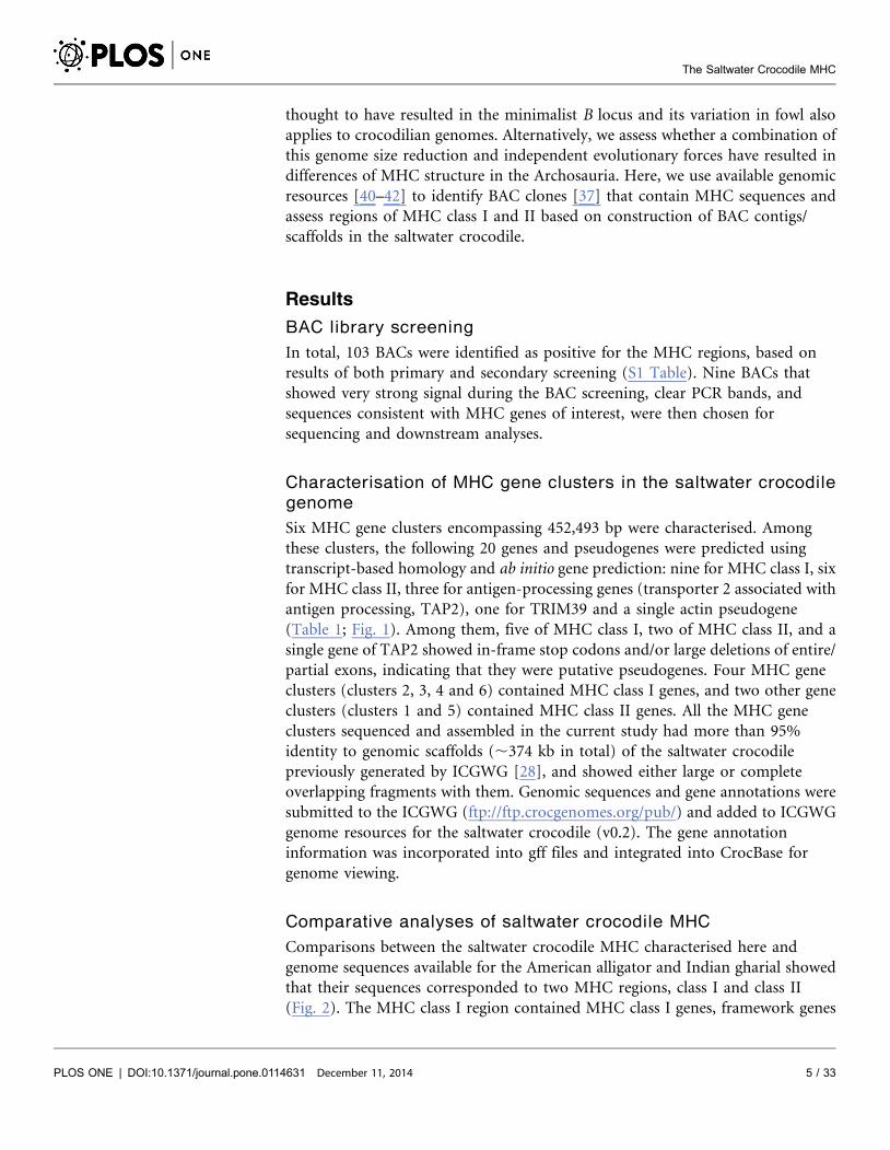
Six MHC gene clusters encompassing 452,493 bp were characterised. Among
these clusters, the following 20 genes and pseudogenes were predicted using
transcript-based homology and ab initio gene prediction: nine for MHC class I, six
for MHC class II, three for antigen-processing genes (transporter 2 associated with
antigen processing, TAP2), one for TRIM39 and a single actin pseudogene
(Table 1; Fig. 1). Among them, five of MHC class I, two of MHC class II, and a
single gene of TAP2 showed in-frame stop codons and/or large deletions of entire/
partial exons, indicating that they were putative pseudogenes. Four MHC gene
clusters (clusters 2, 3, 4 and 6) contained MHC class I genes, and two other gene
clusters (clusters 1 and 5) contained MHC class II genes. All the MHC gene
clusters sequenced and assembled in the current study had more than 95%
identity to genomic scaffolds (,374 kb in total) of the saltwater crocodile
previously generated by ICGWG [28], and showed either large or complete
overlapping fragments with them. Genomic sequences and gene annotations were
submitted to the ICGWG (ftp://ftp.crocgenomes.org/pub/) and added to ICGWG
genome resources for the saltwater crocodile (v0.2). The gene annotation
information was incorporated into gff files and integrated into CrocBase for
genome viewing.
Comparative analyses of saltwater crocodile MHC
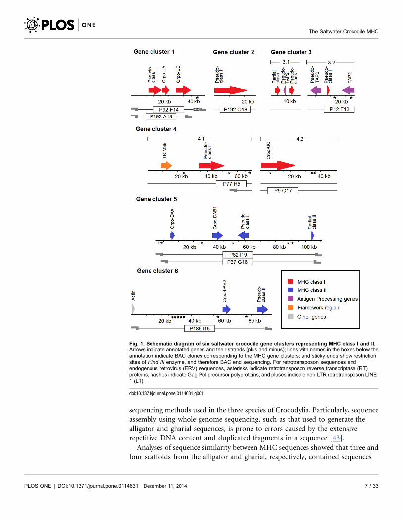
Comparisons between the saltwater crocodile MHC characterised here and
genome sequences available for the American alligator and Indian gharial showed
that their sequences corresponded to two MHC regions, class I and class II
(Fig. 2). The MHC class I region contained MHC class I genes, framework genes
The Saltwater Crocodile MHC
PLOS ONE | DOI:10.1371/journal.pone.0114631 December 11, 2014 5 / 33

(TRIM39) and antigen processing genes (TAP), while the MHC class II region
contained MHC class II genes and, in the alligator only, a bromodomain
containing 2 (BRD2). Twenty of these genes/pseudogenes were characterised in
the saltwater crocodile MHC regions, but only eight and ten of them accounting
for 40% and 50% of the saltwater crocodile genes identified in this analysis were
observed in the gharial and alligator scaffolds, respectively (S2 Table). In addition,
three genes/pseudogenes within the MHC class I region of the alligator and four
from the gharial homologous region showed small and large regions of ambiguity.
These ambiguous nucleotide sites and genes could reflect differences in
Table 1. List of annotated MHC gene clusters of saltwater crocodile genome from a total of nine BACs.
Genecluster Length (bp)
Scaffolda/accessionno. BAC clone IDb Startc Endd Strand Exon Description
1 49565 scaffold-266 P92 F14 (P) 11961 20285 + 8 Truncated MHC class I antigen, pseudo-class I
(41015 bp); P193 A19 (C) 21047 26101 + 9 MHC class I antigen, Crpo-UA
KP118845 30592 .39846 + .4 MHC class I antigen, Crpo-UB
2 28338 scaffold-11053 P192 O18 (P) 26 22403 + 8 MHC class I antigen, pseudo-class I
(15897 bp);
KP118842
3.1 18621 scaffold-10298 P12 F13(P) ,2746 6082 + .7 MHC class I antigen, partial class I
(11851 bp); 9743 8351 2 7 Truncated antigen peptide transporter 2,pseudo-TAP2
KP118844 12699 14600 + 4 Truncated MHC class I antigen, pseudo-class I
3.2 31246 scaffold-7467 P12 F13 (P) 8733 2687 2 12 Antigen peptide transporter 2 (TAP2) pseudo-gene,
(31246 bp); pseudo-TAP2
KP118843 13758 14041 + 1 Truncated MHC class I antigen, pseudo-class I
31195 22602 2 11 Antigen peptide transporter 2 (TAP2)
4.1 69185 scaffold-19650 P9 O17 (P) 8913 15882 + 5 Tripartite motif-containing protein 39-like,TRIM39
(69203 bp); P77 H5(P) 34457 50813 + 6 Truncated MHC class I antigen, pseudo-class I
KP118847
4.2 50603 KP036996 P9 O17 (P) 352 22617 + 7 MHC class I antigen, Crpo-UC
P77 H5 (P)
5 111194 scaffold-2257 P82 I19 (C) 9848 12052 + 4 MHC class II alpha chain, Crpo-DAA
(111218 bp); P67 G16 (C) 37889 43833 + 6 MHC class II beta chain, Crpo-DAB1
KP118841 61987 55155 2 4 MHC class II beta chain, pseudo-class II
109568 .110298 + .2 MHC class II beta chain, partial class II
6 93741 scaffold-16192 P186 I16 (C) 1136 1739 + 2 Truncated actin, cytoskeletal 1A-like
(93750 bp); 61439 66139 + 6 MHC class II beta chain, Crpo-DAB2
KP118846 84792 91595 + 3 Truncated MHC class II beta chain, pseudo-class II
aGenetic scaffolds from whole genome sequencing project of the saltwater crocodile (DA Ray laboratory unmasked v0.2; ftp://ftp.crocgenomes.org/pub/).bBAC clones from the saltwater crocodile genomic library [37]. Completely and partially sequenced BAC clones are abbreviated as C and P in bracket,respectively.c,dSequence range (,or.) is provided if entire length of a gene is uncharacterised.
doi:10.1371/journal.pone.0114631.t001
The Saltwater Crocodile MHC
PLOS ONE | DOI:10.1371/journal.pone.0114631 December 11, 2014 6 / 33
sequencing methods used in the three species of Crocodylia. Particularly, sequence
assembly using whole genome sequencing, such as that used to generate the
alligator and gharial sequences, is prone to errors caused by the extensive
repetitive DNA content and duplicated fragments in a sequence [43].
Analyses of sequence similarity between MHC sequences showed that three and
four scaffolds from the alligator and gharial, respectively, contained sequences
Fig. 1. Schematic diagram of six saltwater crocodile gene clusters representing MHC class I and II.Arrows indicate annotated genes and their strands (plus and minus); lines with names in the boxes below theannotation indicate BAC clones corresponding to the MHC gene clusters; and sticky ends show restrictionsites of Hind III enzyme, and therefore BAC end sequencing. For retrotransposon sequences andendogenous retrovirus (ERV) sequences, asterisks indicate retrotransposon reverse transcriptase (RT)proteins; hashes indicate Gag-Pol precursor polyproteins; and pluses indicate non-LTR retrotransposon LINE-1 (L1).
doi:10.1371/journal.pone.0114631.g001
The Saltwater Crocodile MHC
PLOS ONE | DOI:10.1371/journal.pone.0114631 December 11, 2014 7 / 33
conserved with the saltwater crocodile MHC gene clusters (E-value 50.0). These
scaffolds consisted of the same gene number and order as observed in the
saltwater crocodile (Fig. 2; S1 and S2 Figures). For the saltwater crocodile MHC
class I, gene cluster 4.2 appeared to occupy the region between gene clusters 3.1
and 4.1 from 59 to 39 direction when compared to the arrangement of the alligator
scaffold-14097, showing a large MHC class I region consisting of a framework
gene, MHC class I genes and antigen processing genes.
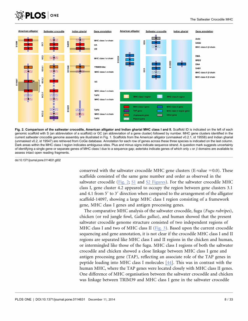
The comparative MHC analysis of the saltwater crocodile, fugu (Fugu rubripes),
chicken (or red jungle fowl, Gallus gallus), and human showed that the present
saltwater crocodile genome structure consisted of two independent regions of
MHC class I and two of MHC class II (Fig. 3). Based upon the current crocodile
sequencing and gene annotation, it is not clear if the crocodile MHC class I and II
regions are separated like MHC class I and II regions in the chicken and human,
or intermingled like those of the fugu. MHC class I regions of both the saltwater
crocodile and chicken showed a close linkage between MHC class I gene and
antigen processing gene (TAP), reflecting an associate role of the TAP genes in
peptide loading into MHC class I molecules [44]. This was in contrast with the
human MHC, where the TAP genes were located closely with MHC class II genes.
One difference of MHC organisation between the saltwater crocodile and chicken
was linkage between TRIM39 and MHC class I gene in the saltwater crocodile
Fig. 2. Comparison of the saltwater crocodile, American alligator and Indian gharial MHC class I and II. Scaffold ID is indicated on the left of eachgenomic scaffold with S (an abbreviation of a scaffold) or GC (an abbreviation of a gene cluster) followed by number. MHC gene clusters identified in thecurrent saltwater crocodile genome assembly are illustrated in Fig. 1. Scaffolds from the American alligator (unmasked v0.2.1, id 19558) and Indian gharial(unmasked v0.2, id 19547) are retrieved from CoGe database. Annotation for each row of genes across these three species is indicated on the last column.Dark areas within the MHC class I region indicates ambiguous sites. Plus and minus signs indicate sequence strand. A question mark suggests uncertaintyof identifying a single gene or separate genes of MHC class I due to a sequence gap; asterisks indicate genes of which only a or b domains are available toassess intact open reading fragments.
doi:10.1371/journal.pone.0114631.g002
The Saltwater Crocodile MHC
PLOS ONE | DOI:10.1371/journal.pone.0114631 December 11, 2014 8 / 33
(18,575 bp distant between TRIM39 and MHC class I pseudogene in gene cluster
4.1). In the chicken, it was reported that framework genes, such as TRIM genes,
were located 41 kb upstream of the core B locus with TRIM-class II-class I-class
III orientation [29]. Identifying collinear sets of MHC regions/genes of sequence
similarity to infer synteny between the saltwater crocodile and the chicken (plus
the human), using SynMap [45], did not show significant large conserved
Fig. 3. Comparative MHC organisations of the fugu, chicken, saltwater crocodile and human. MHC mapping in the fugu, chicken, and human isgenerated using data from Clark et al. [87], AL023516 (plus Shiina et al. [29] for a framework region), and NT_007592, respectively. Gene cluster 2 of thesaltwater crocodile, where a gene model of coding sequences is absent, is omitted in this figure. Graphics in the first row of each vertebrate represent genesin the MHC based on schematic representation in GEvo [88], where all graphics are automatically created if applicable. Unlinked MHC genes and regions inthe fugu and saltwater crocodile presented by the absence of line connection indicate that their order is arbitrary and is not based on the current data. Grayarrows indicate gene models; green arrows indicate protein coding sequences (CDS); blue arrows (on top of gray genes) indicate mRNA; and yellow arrowsindicate approximately 50% GC content in codon wobble positions. Scales above the graphics show different sizes of MHC regions in kilo base pairs (K) ormega base pairs (M).
doi:10.1371/journal.pone.0114631.g003
The Saltwater Crocodile MHC
PLOS ONE | DOI:10.1371/journal.pone.0114631 December 11, 2014 9 / 33

sequences among these comparisons, even though the two species are closely
related within Archosauria. It is likely, because the saltwater crocodile compared
had much larger MHC regions (452,493 bp in total) and the greater number of
MHC class I and II genes/pseudogenes than the fowl B locus (92 Kbp). Only a
single gene, the saltwater crocodile TAP2, was shown to present a synteny to TAP2
coding sequences of the fowl B locus (S3 Figure), suggesting their orthologous
relationships between the two species.
MHC class I genes
Gene structure and content
A total of nine MHC class I loci were identified from gene cluster 1 to gene cluster
4 (S4 Figure). Three of these loci were named Crpo-UA, UB and UC, which had
intact and complete open reading fragments for all a domains responsible for
peptide loading. These genes contained between four and nine exons and encoded
for proteins that were 408, 293, and 350 amino acids (aa), respectively. The first
four exons represented a leader peptide, and three extracellular domains (a1, a2
and a3) while the remaining exons encoded for a transmembrane domain and
cytoplasmic tail. All the three genes contained conserved amino acid positions
that are well-known for their biological roles in salt bridge-forming, disulfide
bridge-forming, N-glycosylation, and CD8+ binding, as well as likely peptide-
binding of antigen N and C termini. Although it is unclear whether these genes are
functional or not, homology searches with transcriptome data from the American
alligator showed that the alligator cDNA sequence, Class I transcript had
significant matches to UA (E-value 52e–107, aa identity 554%, query coverage
580%), UB (4e–153, 70%, 98%), and UC genes (2e–167, 80%, 80%). This
alligator cDNA sequence was found to be expressed in testes, spinal cord,
thalamus, and liver. Another MHC class I gene locus in gene cluster 3.1 was
partially sequenced from the a2 domain to cytoplasmic tail. It was suggested to be
putatively functional, because it matched significantly with Class I transcript (E-
value 51e–124, aa identity 581%, query coverage 596%) and shared high aa
identity to UB (88.6%) and UC genes (90.2%). The remaining five loci of MHC
class I showed truncated features in their open reading fragments, suggesting that
they were pseudogenes: one in gene cluster 1 contained deletions in the a2
domain; one in gene cluster 2 contained a stop codon in the a3 domain; one in
gene cluster 3.1 contained a stop codon in the a2 domain with an a1 domain
absent; one in gene cluster 3.2 contained only an a3 domain; and one in gene
cluster 4.1 contained an additional a3 domain with the absence of an a1 domain.
All the nine loci had high GC contents ranging from 58.1% to 64.3%.
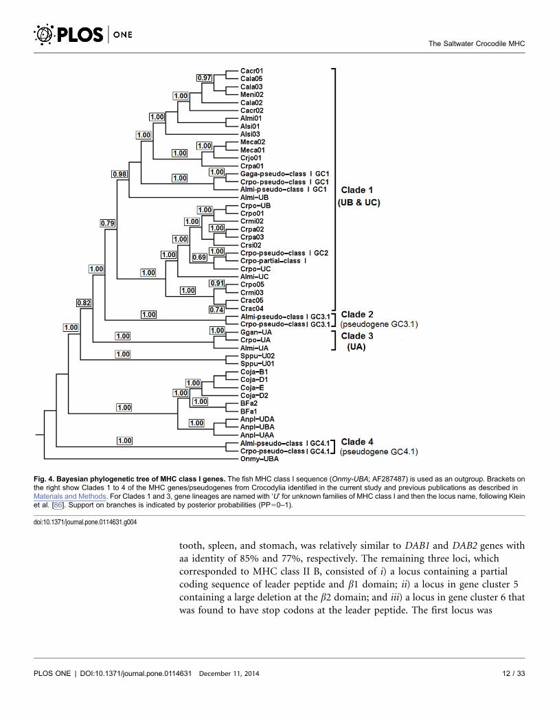
Phylogenetic inference
Bayesian inference of MHC class I genes from the current study and those from
the previous study described in S3 Table showed that they clustered into four
clades (Clades 1–4; PP50.79–1.00) when a fish sequence (Oncorhynchus mykiss)
was used as an outgroup (Fig. 4). All these clades contained MHC genes/
The Saltwater Crocodile MHC
PLOS ONE | DOI:10.1371/journal.pone.0114631 December 11, 2014 10 / 33

pseudogenes from the saltwater crocodile, American alligator and/or Indian
gharial identified herein. Clades 2–4 each clustered MHC genes/pseudogenes
(PP51.00) that were assigned to the same MHC locus in the three crocodilian
taxa studied. Therefore, orthologous relationships could be suggested for these
three clades corresponding to the cluster 3.2 pseudogene, UA gene and cluster 4.1
pseudogene respectively, with between-clade pairwise distance ranging from 0.466
to 0.703 showing great divergence between them. Cluster 4.1 pseudogenes from
the saltwater crocodile and American alligator within Clade 4 showed dramatic
divergence from the rest of MHC genes in Crocodylia analysed here with pairwise
genetic distance of exons ranging from 0.564 to 0.744. These MHC class I
pseudogenes formed a paraphyletic clade with currently analysed MHC genes
from crocodilians, fowl, and the tuatara (Sphenodon punctatus), indicating that
they are expected to predate the divergence of these vertebrate groups. However,
Clade 1 clustered five loci of saltwater crocodile MHC class I identified here (the
UB gene, UC gene, cluster 1 pseudogene, cluster 2 pseudogene and partial MHC
class I gene) and all previously sequenced variants (PP50.79). This clustering
indicated that the Clade 1 variants corresponded to UB and UC gene lineages,
where their loci were detected at different sites on the saltwater crocodile genome.
Since the topology in Clade 1 did not allow a clear subdivision of these two genes,
we proposed this clade as a representation of UB and UC gene lineages. The
saltwater crocodile UB gene clustered well (PP51.00) with other five MHC class I
sequences from four species of crocodiles (the saltwater crocodile, mugger
crocodile, Philippine crocodile and Siamese crocodile), suggesting that they may
represent orthologs to the UB gene in the saltwater crocodile.
MHC class II genes
Gene structure and content
Six MHC class II loci were identified within gene clusters 5 and 6 (S5 Figure).
Three of these loci contained complete coding sequences, including two encoding
for b chains (II B assigned as ‘DAB1 and DAB2’) and another encoding for an a
chain (II A assigned as ‘DAA’). These MHC class II A and B genes encoded for
proteins that were 268 and 257 aa and consisted of four to six exons,: exon 1
encoding the leader peptide, exons 2 and 3 encoding the two extracellular
domains (a1/a2 or b1/b2 domains) and exons 4–6 encoding the transmembrane
domain and cytoplasmic tail. They contained 100% conserved aa positions for
disulfide bridge-forming (C–C), peptide-binding of antigen N and C termini and/
or CD4+ binding, suggesting that they were functional. Homology searches with
American alligator cDNA sequences appeared to support immunological function
of these genes with significant matches between alligator class II A transcript and
DAA (E-value 50.0, aa identity 598%, query coverage 5100%), as well as
between alligator class II B transcript and DAB (DAB152e–175, 85%, 100%; and
DAB254e–150, 77%, 100%). DAA gene showed high similarity to the class II A
transcript (aa identity 598%) that was found to express in various organs, such as
testes, thalamus, spleen, ovary, liver and kidney. The class II B transcript found in
The Saltwater Crocodile MHC
PLOS ONE | DOI:10.1371/journal.pone.0114631 December 11, 2014 11 / 33
tooth, spleen, and stomach, was relatively similar to DAB1 and DAB2 genes with
aa identity of 85% and 77%, respectively. The remaining three loci, which
corresponded to MHC class II B, consisted of i) a locus containing a partial
coding sequence of leader peptide and b1 domain; ii) a locus in gene cluster 5
containing a large deletion at the b2 domain; and iii) a locus in gene cluster 6 that
was found to have stop codons at the leader peptide. The first locus was
Fig. 4. Bayesian phylogenetic tree of MHC class I genes. The fish MHC class I sequence (Onmy-UBA; AF287487) is used as an outgroup. Brackets onthe right show Clades 1 to 4 of the MHC genes/pseudogenes from Crocodylia identified in the current study and previous publications as described inMaterials and Methods. For Clades 1 and 3, gene lineages are named with ‘U’ for unknown families of MHC class I and then the locus name, following Kleinet al. [86]. Support on branches is indicated by posterior probabilities (PP50–1).
doi:10.1371/journal.pone.0114631.g004
The Saltwater Crocodile MHC
PLOS ONE | DOI:10.1371/journal.pone.0114631 December 11, 2014 12 / 33

considered to be putatively functional with aa identity of 75% and 52% to DAB1
and DAB2, respectively; the last two loci were pseudogenes.
Phylogenetic inference
Bayesian inference of MHC class II A genes identified among the saltwater
crocodile, American alligator and Indian gharial studied here and those from
other 16 species of Crocodylia in the previous study (S3 Table) showed that they
formed a monophyletic clade (PP51.0) without clear subdivision in the clade
when the fish sequence was used as an outgroup (Fig. 5). All the genes, including
Crpo-DAA, Almi-DAA and Gaga-DAA showed high aa identity with an overall
genetic distance of 0.004, indicating that they are orthologous to each other. The
analysis of introns 1 to 3 among the full-length DAA genes identified in the
current study was consistent showing little pairwise genetic distance ranging from
0.034–0.057. Using this phylogeny, the DAA locus identified on the saltwater
crocodile genome enabled us to assign the previously sequenced variants from the
other 16 species of Crocodylia to this locus. In addition, all the genes from
Crocodylia clustered as a sister clade to mallard and chicken MHC class II A,
Anpl-DRA and BLA, respectively, suggesting that these avian genes are orthologs
to DAA genes among crocodilians.
Bayesian inference of MHC class II B genes identified in this study and those
from the previous study described in S3 Table showed two clades (clades 1 and 2)
using the fish sequence as an outgroup (PP50.85–1.0; Fig. 6). In Clade 1, the tree
was found to have six subclades (1A–1F) with low branch support. Subclades 1A
and 1B provided two separate clusterings of the saltwater crocodile DAB1 and
DAB2 genes, respectively. Subclade 1A clustered the saltwater crocodile DAB1
gene with MHC variants from six other species of Crocodilidae (crocodiles) and a
single species of Alligatoridae (alligators and caimans) (pairwise genetic distance,
0.0–0.074), while Subclade 1B clustered the saltwater crocodile DAB2 gene with
variants from seven other species of Crocodilidae (pairwise genetic distance, 0.0–
0.070). These clusterings appear to suggest orthologous relationships of two gene
lineages (DAB1 and DAB2) to which the variants correspond. The remaining
subclades (1C–1F) contained MHC variants from different species of Crocodylia:
three (1C, 1E and 1F) clustered variants from different species of Alligatoridae and
Crocodilidae; and one (1D) clustered variants from different species of
Alligatoridae. However, collapsing low branch support (PP,0.50) caused
Subclades 1A and 1B to cluster together (PP50.51) and the others disappeared,
except for Subclade 1E (PP50.92), suggesting high identity between DAB1 and
DAB2 genes analysed. In addition, Clade 2 consisted of only a putative
pseudogene identified in gene cluster 6, and revealed large divergence to other
variants compared, with pairwise genetic distances ranging from 0.425 to 0.507.
This could suggest that this pseudogene corresponds to a different locus from the
other genes from Crocodylia and may have been selected against in the past
resulting in the pseudogenisation of the gene.
The Saltwater Crocodile MHC
PLOS ONE | DOI:10.1371/journal.pone.0114631 December 11, 2014 13 / 33

Other genes, retrotransposons and endogenous retrovirus (ERV)
sequences
Three novel antigen processing genes (TAP2) were observed near MHC class I
genes/pseudogenes within gene cluster 3. A single TAP2 gene (TAP2) provided an
intact open reading fragment of 604 aa, and was shown to be expressed due to
high sequence identity to unpublished cDNA sequences from the American
alligator (TAP2 transcripts 1 and 2; E-value 50.0, aa identity 587–90%, query
coverage 590–92%) (S6 Figure). This gene consisted of five exons representing
the ABC transporter transmembrane region, and five other exons representing the
P-loop containing Nucleoside Triphosphate Hydrolases, and shared significant
homology with the reference TAP2 gene (XP_001496065; E-value 50.0, aa
identity 563%, query coverage 590%) in the horse (Equus caballus). The other
Fig. 5. Bayesian phylogenetic tree of MHC class II A genes. The fish MHC class II A sequence (Onmy-DAA; AFP94173) is used as an outgroup. Abracket on the right shows the DAA gene lineage of the MHC genes from Crocodylia identified in the current study and previous publications as described inMaterials and Methods. This gene lineage is named with ‘DAA’ (an abbreviation for MHC class II A), following Klein et al. [86]. Support on branches isindicated by posterior probabilities (PP50–1).
doi:10.1371/journal.pone.0114631.g005
The Saltwater Crocodile MHC
PLOS ONE | DOI:10.1371/journal.pone.0114631 December 11, 2014 14 / 33

The Saltwater Crocodile MHC
PLOS ONE | DOI:10.1371/journal.pone.0114631 December 11, 2014 15 / 33

two TAP genes were strongly similar to the intact TAP2-like gene in the green
anole (XP_003229695; E-value 58e–174 for pseudo-TAP2 GC3.1, and 4e–25 for
pseudo-TAP2 GC3.2). However, they contained in-frame stop codons and
deletions, suggesting that they were pseudogenes. The pseudo-TAP2 in gene
cluster 3.2 encompassed 12 exons and had relatively high aa identity (71%) to
TAP2 in contrast to the pseudo-TAP2 in gene cluster 3.1, where the ABC
transporter transmembrane region were absent and 44% aa identity to the intact
TAP2 was observed using seven exon sequences available for the identity analysis.
This difference was also true when TAP2 intron sequences were compared with
those of pseudo-TAP2 genes in gene clusters 3.1 (identity 571.6%) and 3.2
(76.5%), suggesting differences in pseudogenisation between TAP2 genes. In
addition, a framework gene TRIM39 was found near the MHC class I pseudogene
in gene cluster 4.1. This gene spanned 462 aa and had significant homology to the
chicken TRIM39 (NP_001006196; E-value 51e–112, aa identity 543%, query
coverage 597%) using BLAST search. The phylogenetic analysis of this homology
is shown in S7 Figure. The TRIM gene contained four main domains consistent
with the chicken TRIM39 (S8 Figure): RING- zinc finger (aa site 15–61), B-Box-
type zinc finger (aa site 91–129), PRY (aa site 288–336), and SPRY (aa site 339–
459). This gene was found to be functional due to a match with the cDNA
sequence from the American alligator, TRIM transcript (E-value 58e–148, aa
identity 548%, query coverage 599%).
A non-MHC gene, actin was also found among the MHC class II genes in gene
cluster 6. This gene has functions involved in muscle contraction, cell motility,
and cytokinesis [46]. The actin gene identified herein was considered a
pseudogene because only a nucleotide binding domain (NBD) was present with a
197-aa region distal to the NBD absent when compared to the unpublished actin
cDNA sequence from the American alligator, Actin transcript (S9 Figure).
Seventeen following genes corresponding to retrotransposons and ERV were
distributed across the MHC gene clusters identified here: 13 retrotransposon
reverse transcriptase genes (RT), three Gag-Pol precursor genes, and one non-LTR
retrotransposon LINE-1 (Fig. 1). Two retrotransposon RT genes were identified
within UC and TAP2 genes.
Discussion
Size of the saltwater crocodile MHC
The current study shows that MHC in the saltwater crocodile is larger and more
complex than other extant archosaurs, (i.e., representative species from the three
Fig. 6. Bayesian phylogenetic analysis of MHC class II B sequences. The fish MHC class II B sequence(Onmy-DAB; FR688148) is used as an outgroup. Brackets on the right show Clades 1 and 2 of the MHCvariants from Crocodylia, and six subclades (A–F) for Clade 1. For Subclades A and B, gene lineages arenamed with ‘DAB’ (an abbreviation for MHC class II B) and then the identification number, following Klein et al.[86]. Support on branches is indicated by posterior probabilities (PP50–1).
doi:10.1371/journal.pone.0114631.g006
The Saltwater Crocodile MHC
PLOS ONE | DOI:10.1371/journal.pone.0114631 December 11, 2014 16 / 33
major groups of birds; S4 and S5 Tables). Firstly in terms of the number of MHC
genes, the saltwater crocodile is shown to have multiple copies of MHC class I and
II genes/pseudogenes (n59 and 6, respectively) outnumbering those, especially
MHC class I genes, in the minimum essential MHC (n52 and 5–6, respectively)
observed in the Galloansera, such as chickens [9], black grouses [31], and golden
pheasants [13]. Similar to the present study, Jaratlerdsiri et al. [40] have
demonstrated a large number of MHC class I genes (at least four loci) in the
saltwater crocodile. Other galliform species, turkeys and quails, have more
elaborate B loci than the minimum essential MHC, but still have fewer MHC class
I genes than saltwater crocodiles [10, 12], while entire number of MHC class II
genes in the crocodile is required for comparisons with that in these two taxa.
Considering the number of MHC genes identified in high coverage genome drafts
from the remaining two major groups of birds, the Neoaves and Palaeognathae
(S5 Table), the complexity of the saltwater crocodile MHC is more evident. The
budgerigar (Melopsittacus undulatus) [47], peregrine falcon (Falco peregrinus)
[48], and ostrich (Struthio camelus) [49] have fewer MHC class I and II genes
(n51–3 and 1–4, respectively) than the saltwater crocodile. However, there are
some passerine birds that also have complex MHC structure with a number of
potentially duplicated genes in their genomes [50–55]. Supporting this, a genome
draft in the zebra finch (Taeniopygia guttata; accession number
GCA_000151805.2) [11] has 22 and 16 genes in a single chromosome
corresponding to MHC class I and II, respectively (S5 Table).
Secondly, the saltwater crocodile MHC genes are longer. It is estimated that the
mean length of UA, UB and UC genes identified is about five to six times greater
in saltwater crocodiles than that of MHC class I genes in the fowl species described
above (,2 kb on average). Additionally, saltwater crocodile DAA and DAB genes
are approximately two to three times larger than the fowl MHC class II B gene
(,1.8 kb on average) [9, 10, 12, 13, 31]. The differences in gene length seem to be
the result of greater intron size observed among the saltwater crocodile MHC
genes, as these MHC genes have similar length of coding sequences to published
fowl sequences [10, 12, 13, 31]. These findings provide the first evidence of MHC
organisation in the saltwater crocodile, contrasting with the compact MHC
structure known in the Galloanseres. The Chinese alligator has also shown a well-
developed immune defense system with 22 MHC class I genes identified [39].
Evolutionary mechanisms of the differences in MHC size between the saltwater
crocodile and birds are discussed below.
Organisation of the saltwater crocodile MHC
The saltwater crocodile MHC (evolution model; Fig. 7A) appears to have a gene
organisation intermediate between the fowl B locus and the eutherian MHC. The
saltwater crocodile shows the same linkage between TAP and MHC class I genes as
the fowl (but not eutherians), whilst in contrast TRIM is placed near MHC class I
genes in the saltwater crocodile and eutherians (but not fowl). Inclusion of the
recently generated green anole MHC in this comparison shows that the gene
The Saltwater Crocodile MHC
PLOS ONE | DOI:10.1371/journal.pone.0114631 December 11, 2014 17 / 33
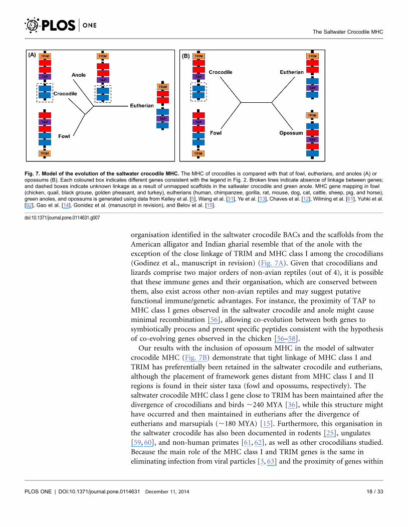
organisation identified in the saltwater crocodile BACs and the scaffolds from the
American alligator and Indian gharial resemble that of the anole with the
exception of the close linkage of TRIM and MHC class I among the crocodilians
(Godinez et al., manuscript in revision) (Fig. 7A). Given that crocodilians and
lizards comprise two major orders of non-avian reptiles (out of 4), it is possible
that these immune genes and their organisation, which are conserved between
them, also exist across other non-avian reptiles and may suggest putative
functional immune/genetic advantages. For instance, the proximity of TAP to
MHC class I genes observed in the saltwater crocodile and anole might cause
minimal recombination [56], allowing co-evolution between both genes to
symbiotically process and present specific peptides consistent with the hypothesis
of co-evolving genes observed in the chicken [56–58].
Our results with the inclusion of opossum MHC in the model of saltwater
crocodile MHC (Fig. 7B) demonstrate that tight linkage of MHC class I and
TRIM has preferentially been retained in the saltwater crocodile and eutherians,
although the placement of framework genes distant from MHC class I and II
regions is found in their sister taxa (fowl and opossums, respectively). The
saltwater crocodile MHC class I gene close to TRIM has been maintained after the
divergence of crocodilians and birds ,240 MYA [36], while this structure might
have occurred and then maintained in eutherians after the divergence of
eutherians and marsupials (,180 MYA) [15]. Furthermore, this organisation in
the saltwater crocodile has also been documented in rodents [25], ungulates
[59, 60], and non-human primates [61, 62], as well as other crocodilians studied.
Because the main role of the MHC class I and TRIM genes is the same in
eliminating infection from viral particles [3, 63] and the proximity of genes within
Fig. 7. Model of the evolution of the saltwater crocodile MHC. The MHC of crocodiles is compared with that of fowl, eutherians, and anoles (A) oropossums (B). Each coloured box indicates different genes consistent with the legend in Fig. 2. Broken lines indicate absence of linkage between genes;and dashed boxes indicate unknown linkage as a result of unmapped scaffolds in the saltwater crocodile and green anole. MHC gene mapping in fowl(chicken, quail, black grouse, golden pheasant, and turkey), eutherians (human, chimpanzee, gorilla, rat, mouse, dog, cat, cattle, sheep, pig, and horse),green anoles, and opossums is generated using data from Kelley et al. [5], Wang et al. [31], Ye et al. [13], Chaves et al. [12], Wilming et al. [61], Yuhki et al.[92], Gao et al. [14], Gonidez et al. (manuscript in revision), and Belov et al. [15].
doi:10.1371/journal.pone.0114631.g007
The Saltwater Crocodile MHC
PLOS ONE | DOI:10.1371/journal.pone.0114631 December 11, 2014 18 / 33

a MHC region has been suggested to allow co-evolution between the genes
[56, 57], our finding in organisation between MHC class I and TRIM genes may
indicate an advantageous role in their immune function in vertebrates, especially
in antiviral immune responses. It could be suggested that TRIM and MHC class I
genes in the saltwater crocodile collaborate with each other to eliminate given viral
invasion through innate [64] and adaptive immune mechanisms [3], respectively.
Based on a study in humans, TRIM proteins can directly bind to certain viral
proteins or act as regulators in interferon-induced innate immunity, and thus
inhibiting viral maturity and release [65]; some of them are found to significantly
interact with DNA-binding proteins to regulate expression of genes within a MHC
class I region [66].
Evolutionary mechanisms of the saltwater crocodile MHC
The additional number of MHC genes and pseudogenes identified in the saltwater
crocodile relative to the avian MHC compared above could be a result of the
evolutionary mechanisms of gene duplication and loss. Given the multiple copies
of MHC genes observed in the saltwater crocodile, and their clustering into
various clades and subclades rather than all together as species, it could be
suggested that such a mechanism (also known as ‘the birth-and-death process of
evolution’) plays a role in complexity of the saltwater crocodile MHC. The effect
of gene duplication and loss has previously been documented in mammals and
owls to explain orthologous MHC gene relationships [67, 68]. Also, phylogenetic
clusterings of these duplicated genes into different clades and subclades suggest
that these loci might have evolved independently to each other, allowing them to
respond to different environmental pressures or perform novel immune
functions. At the same time, the duplicated genes may have been maintained, with
similar selective pressures, along with their orthologs from other species of
Crocodylia; that is, they all might represent the same biological functions among
crocodilians and, therefore, similarity in coding sequences [69]. This is likely to
explain why some of the MHC genes from different species are more similar to
each other than they are to the duplicated genes within a species. Based on gene
loss, some copies of the MHC genes identified in this study have experienced a
different fate of evolution, where they were selected against [70] allowing
mutations, pseudogenisation, or even deletion from the saltwater crocodile
genome. For example, the current clustering of three genomic loci corresponding
to MHC class I and II pseudogenes in the saltwater crocodile (ones in gene
clusters 3, 4 and 6) into separate clades suggest that they are evolutionarily
divergent from their intact gene counterparts.
The identification of retroelements suggests that other mechanisms may have
also played a role in creating the additional number of MHC genes in the saltwater
crocodile. Kulski et al. [71] suggest that these elements act as insertion and
deletion sources on the genome. Long interspersed nuclear element (LINE)
sequences identified in the saltwater crocodile, for example, could trigger
duplication of genome fragments [72] that contain MHC genes. These duplicated
The Saltwater Crocodile MHC
PLOS ONE | DOI:10.1371/journal.pone.0114631 December 11, 2014 19 / 33
genome fragments then could insert themselves into the saltwater crocodile MHC,
leading to expansions of the number of MHC genes and their functions in
immunity. This is consistent with that observed in the human MHC, where LINE-
mediated duplications have resulted in multiple MHC class I genes, with differing
levels of expression, and different expression profiles across tissues [73]. Wan et al.
[39] have also shown remarkable abundance of LINE sequences within a genome
from Crocodylia (the Chinese alligator genome), and these elements have been
suggested to trigger segmental duplications on the genome. In addition, ERV
fragments identified might play some role in arrangement of the present-day
MHC in the saltwater crocodile consistent with the potential role of Kangaroo
endogenous retrovirus (KERV) in the movement of MHC class I genes on the
macropod genome [35, 74, 75]. However, the greater gene number observed at the
saltwater crocodile MHC relative to the avian MHC may be a result of massive
gene loss and subsequent reduction of genome size in birds [30, 76]. Future work
investigating complete and contiguous MHC core in the saltwater crocodile is
required to refine the size of MHC in this species, as it will provide an opportunity
to identify large or small duplicated genome blocks using retroelements as a
molecular clock to distinguish those blocks based on differences in their
generation times [73].
Conclusions
Here, we present six MHC gene clusters, representatives of MHC class I and class
II regions in the saltwater crocodile genome, providing insights into gene
organisation within the two regions. Our comparative analysis shows that the
MHC of the saltwater crocodile does not conform to the simple structure found in
many birds, and appears to have a distinct organisation to that of avian MHC in
relation to the proximity of antigen presentation and framework genes. To extend
this knowledge, sequencing of additional positive BACs for MHC markers
generated in the current study would be a starting point for full characterisation of
the MHC core in the saltwater crocodile genome. Construction and sequencing of
cDNA libraries is also needed in this species for accurate gene annotation of the
MHC core sequence, as it will be useful for further characterisation of MHC
expression and comparative genetics across all vertebrate groups enabling
inference of their adaptive immune functions.
Materials and Methods
Overgo design
In order to screen the saltwater crocodile genomic BAC library described below,
four overgo pairs (forward and reverse) were designed (Table 2) using saltwater
crocodile sequences of MHC class I and II from previous studies [40, 42]. The
overgos were designed using OligoSpawn software, with a GC content of 50–60%
The Saltwater Crocodile MHC
PLOS ONE | DOI:10.1371/journal.pone.0114631 December 11, 2014 20 / 33

and 36 bp in length (8-bp overlapping) [77]. The specificity of the overgos was
checked against vertebrate sequences using the basic local alignment search tool
(BLAST; http://www.ncbi.nlm.nih.gov/).
BAC library screening
The saltwater crocodile BAC library, Mississippi State University CP_Eba,
contained 3.76 genome coverage with 101760 clones [37] that was prepared from
a single individual named ‘Errol’ (the University of Sydney Animal Ethics permit
number N00/8-2005/3/4177). Errol was a wild-caught adult male housed at
Darwin Crocodile Farm near Darwin, Australia. He was housed individually in a
purpose-built facility in the tourism section of the farm where trained and
experienced staff monitored the animal daily and was fed the equivalent of two
whole chickens weekly. During relocation by specialised contractors, blood
samples for genome sequencing [38] and BAC library preparation were collected
from the cervical sinus as described by Lloyd and Morris [78]. To identify BAC
clones with positive MHC inserts, high-density filters containing all BACs in the
library were screened with a pool of radiolabeled MHC overgos at the Australian
National University (ANU). Each pair of overgos were radioactively labeled with32P-dATP and 32P-dCTP (GE Healthcare Life Sciences, Rydalmere NSW)
following the BACPAC hybridisation protocol (http://bacpac.chori.org/
overgohyb.htm). Unincorporated nucleotides (32P-dNTP) were removed using
Illustra ProbeQuant G-50 Micro Columns (GE Healthcare Life Sciences,
Rydalmere NSW) with the manufacturer’s protocol. These labeled overgos were
hybridised onto BAC filters, and washed using the BACPAC hybridisation
protocol (http://bacpac.chori.org/overgohyb.htm). Washed filters were exposed to
x-ray films, Hyperfilm (GE Healthcare Life Sciences, Rydalmere NSW) for up to
ten days at 280 C. The films were developed and positive BAC clones were
isolated.
Secondary screening of MHC-associated BAC clones
The positive BACs were subject to secondary screening using dot blot and each
pair of the overgos described above to identify the class of MHC within the BACs
and to eliminate false positives. All the BAC clones were cultured at 37 C
overnight with Luria Broth (LB) supplemented with 12 mg/ml chloramphenicol.
Cultured clones (2 ml) were then applied to gridded Hybond N+ membranes (GE
Healthcare Life Sciences, Rydalmere NSW), which were placed on LB agar plates
containing 12 mg/ml chloramphenicol, and incubated overnight at 37 C. The
membranes were removed from the agar plates, bacterial colonies were lysed, and
the DNA was denatured, neutralised and fixed as described by Deakin et al. [79].
The membranes were washed in 66 SSC to eliminate bacterial debris prior to
hybridisation. The membranes were screened at 60 C overnight using individual
overgo pairs that were radiolabeled, hybridised and washed as described above,
and then exposed to Hyperfilm for up to seven days at 280 C. BAC clones
The Saltwater Crocodile MHC
PLOS ONE | DOI:10.1371/journal.pone.0114631 December 11, 2014 21 / 33

positive from the secondary screening were also verified by PCR and direct
sequencing, using the appropriate PCR primers and conditions described in the
previous publications [40–42].
Library preparation and next-generation sequencing
A subset of BACs (n59) that showed i) very strong signal during all the steps of
the BAC screening, and ii) clear PCR amplification products and sequences
consistent with MHC were selected for next-generation sequencing. Cell culture of
BAC clones was performed twice: one at the University of Sydney, Australia where
nine positive and verified BACs were incubated overnight at 37 C using LB
supplemented with chloramphenicol, and another at Lucigen Corporation
(Wisconsin, USA) to which these BACs were submitted for high yields of purified
DNA (10 mg). DNA library preparations and 454 sequencing were performed
according to internal protocol at Georgia Genomics Facility in Georgia of the USA
(http://dna.uga.edu/) using the Roche FLX Genome Sequencer platform, where
the GS FLX Titanium Emulsion PCR protocol for the 454 sequencing system
(http://www.454.com/) was conducted.
Data analyses
Data control and cleaning
Quality control of the raw 454 reads from each BAC was performed using the
following three steps: i) removal of duplicated reads (PyroCleaner), ii) trimming
of adapter and low quality sequences (sff_extract), and iii) filtering by read size.
The PyroCleaner was used to remove duplicated reads generated by the Roche 454
platform, and to filter sequence data using different criteria, such as length,
complexity, and number of undetermined bases [80]. Sff_extract was used to
extract information related to read quality and sequences from the sff file and clip
multiplex identifier adaptor (MID) at the 59 end and low quality sequences at the
39 end of each read (http://bioinf.comav.upv.es/sff_extract/index.html). The last
step of data cleaning selected reads from 100 to 800 bp in length. The number of
Table 2. Four pairs of forward and reverse overgos used for BAC library screening of MHC-associated BACs.
Gene region Overgo (59R39)
MHC class I exon 3 F - GAGAAGACTACATCAGCTATGA
R – TGTGCGTGCTCGGGTCATAGCT
MHC class II A exon 2 F - TGACTTCGGCAAGTTCACCAGC
R – CCCTGTGCCTCAAAGCTGGTGA
MHC class II A exon 3 F - GAGGACAACGCTTTCCGCAAGT
R – GGGCAGGTAGGTGAACTTGCGG
MHC class II B exon 3 F - CGGGATCGAGGTGAAATGGTTG
R – TCCTGCCCGTTCCTCAACCATT
doi:10.1371/journal.pone.0114631.t002
The Saltwater Crocodile MHC
PLOS ONE | DOI:10.1371/journal.pone.0114631 December 11, 2014 22 / 33
reads per BAC remaining after the quality control and cleaning is shown in S6
Table.
Genome assembly and gene annotation
Sequence assembly of the 454 reads cleaned above within each BAC was
performed using the following two methods: i) de novo genome assembly and ii)
reference genome mapping. Newbler v2.5.3 program, which is designed
specifically for de novo genome assembly of 454 sequence datasets [81], was used
to produce BAC contigs for use in downstream analyses (S7 Table). The reference
genome mapping, where the cleaned reads were mapped with reference MHC
sequences, was performed using BWA-SW v0.6.1-r104 [82] and BLAT v34 [83]
with default parameters. The reference MHC sequences used for this genome
mapping consisted of the turkey B locus (DQ993255), quail MHC (AB078884),
chicken B locus (AL023516), and chicken Y locus (NC_006103), as they
correspond to completely annotated MHC in birds that are sister taxa to
crocodilians. Although a number of BAC contigs were constructed using Newbler,
mapping the reads against these reference sequences was unsuccessful owing to
the fact that small numbers of alignment matches were shown between them (S8
Table).
Contigs within each BAC clone sequenced were subjected to the NCBI genome
assembly and annotation pipeline [84]. Based on the NCBI genome assembly, all
the contigs within each BAC clone were checked and then trimmed for
pIndigoBAC-5 Hind III vector sequences used for construction of saltwater
crocodile BAC library, using VecScreen (http://www.ncbi.nlm.nih.gov/VecScreen/
VecScreen.html). The BAC contigs that contained contaminant sequences from
other species were screened using genomic BLAST against genome sequences
available in the BLAST database (http://www.ncbi.nlm.nih.gov/sutils/genom_
table.cgi). None of these contigs were found to be contaminated in the current
study. The resultant BAC contigs without any vector or contaminated sequences
were aligned and then merged for construction of larger BAC sequences/scaffolds
using MegaBLAST (http://blast.ncbi.nlm.nih.gov/Blast.cgi), where only sequence
pairs with more than 80% identity were detected.
The NCBI annotation pipeline was used to annotate our BAC scaffolds using
the following two methods: i) ab initio gene prediction, and ii) transcript-based
gene modeling [84]. The gene prediction for each scaffold was run using
GENSCAN for prediction of coding sequence positions across the BAC sequence
[85]. Transcript-based gene models within a BAC scaffold were generated from
the best identity hits of nucleotide and amino acid alignments between scaffold
and sequences from the RefSeq RNA, RefSeq proteins and American alligator
RNA database (http://crocgenomes.org/), and were conducted using standalone
NCBI BLAST 2.2.26+ (http://www.ncbi.nlm.nih.gov/books/NBK1762/). Gene
annotation from the two methods was merged with preference given to
annotation models based on RefSeq RNA sequences or mRNA sequences. Genes
predicted by the annotation process were named after the best match to
eukaryotic proteins with the exceptions of MHC class I and II genes, which were
The Saltwater Crocodile MHC
PLOS ONE | DOI:10.1371/journal.pone.0114631 December 11, 2014 23 / 33
assigned species-specific nomenclature proposed by Klein et al. [86]. ERV
sequences were annotated in the BAC scaffolds using BLAST search against the
conserved domain database (http://www.ncbi.nlm.nih.gov/cdd).
Comparative analyses of MHC
To conduct comparative MHC analyses between different vertebrate genomes,
BAC scaffolds constructed in the saltwater crocodile were compared with
reference genomes using CoGe Accelerating Comparative Genomics (http://
genomevolution.org/CoGe/). SynMap is a CoGe tool allowing generation of a
syntenic dotplot between two genomes and identification of their syntenic
regions, collinear sets of genes or regions of sequence similarity [45]. This analysis
can also help to link BAC scaffolds into larger fragments using reference genomes
from closely related organisms. Reference genomes used for the MHC comparison
included those from the American alligator (ID 19558, v0.2.1, unmasked and
coding sequence/CDS), Indian gharial (ID 19547, v0.2, unmasked and CDS), red
jungle fowl (ID 2731, v2, unmasked and CDS) and human (ID 12142, NCBI
v37.2, unmasked and CDS). The reference genomes from two species of
Crocodylia were generated using a whole genome sequencing approach, which is
different from the BAC-based sequencing conducted in the current study. In
addition, organisation of the present saltwater crocodile MHC was compared with
genomic regions corresponding to MHC in previous publications from three
vertebrate species, including the fugu [87], chicken (AL023516; Shiina et al. [29])
and human (NT_007592). The MHC regions of these species are well-
characterised and show unique differences in organisation.
Another CoGe tool, called ‘CoGeBlast’ was used to compare the current BAC
scaffolds with the reference genomes at the small scale or gene level for assessment
of sequence similarity and gene annotation. This analysis conducts BLAST search,
where a query sequence can compare any number of genomes in the CoGe
database, and allows visualisation of individual matches in their genomic context
[88]. The latest versions of the American alligator (v0.2.1) and Indian gharial
reference genomes (v0.2) retrieved from the CoGe database have been poorly
sequenced and annotated at the MHC. The tool was used to find conserved genes
within MHC regions among the saltwater crocodile and the two other species of
Crocodylia and facilitate connection between our neighbouring BAC scaffolds
when compared to scaffolds from the two other crocodilians. Novel MHC genes
that showed high sequence similarity among the three species of Crocodylia using
the BLAST search were annotated with the same names following MHC
nomenclature of Klein et al [86]. CoGeBlast results comparing MHC organisa-
tions of these species of Crocodylia were displayed using Genome Evolution
Analysis (GEvo). This tool helps to compare multiple genomic regions from any
number of organisms using a variety of different sequence comparison algorithms
in order to quickly identify patterns of genome evolution [88].
The Saltwater Crocodile MHC
PLOS ONE | DOI:10.1371/journal.pone.0114631 December 11, 2014 24 / 33
Phylogenetic and sequence analyses
In order to identify evolutionary lineages within the saltwater crocodile MHC
genes, Bayesian inference was conducted using BEAST version 1.5.4 [89]. BEAST
performs Markov chain Monte Carlo (MCMC) sampling to estimate posterior
probability distributions of parameters involved in substitution, site and clock
models as well as tree topology. The best-fitting model of amino acid substitutions
was selected using the Bayesian Information Criterion (BIC) and Akaike
Information Criterion (AIC) in ModelGenerator version 0.85 [90]. Samples from
the posterior probability distribution were drawn every 105 steps over a total of
108 MCMC steps, with the first 103 samples discarded as burn-in. Pairwise genetic
distance (d) compared the number of nucleotide substitutions or amino acid
substitutions in each pair of MHC genes using p-distance and pairwise deletion
options in MEGA 5.0 [91].
Bayesian inference described above were performed separately on the three
following amino acid sequence datasets that consisted of MHC variants identified
within the current BAC sequences and those from Crocodylia and other
vertebrates analysed previously [41, 42]: i) the MHC class I exons 3 and 4 dataset
consisted of eight sequences from the study BACs, 24 from Crocodylia, two from
other Reptilia, and nine from Aves, and a single variant from Osteichthyes
(outgroup). This dataset had the JTT model with gamma distribution as a best-fit
model; ii) the MHC class II A exon 2 dataset consisted of a single sequence from
the study BACs, 17 from Crocodylia, two from Aves, 12 from Mammalia and a
single variant from Osteichthyes (outgroup). This dataset was run with the WAG
model with a gamma distribution based on BIC and AIC criteria; and iii) the
MHC class II B exon 3 dataset consisted of five sequences from the study BACs, 53
from Crocodylia, three from other Reptilia, 11 from Aves, and a single variant
from Osteichthyes (outgroup). This dataset fitted the JTT model with gamma
distribution. The list of MHC variants and their GenBank accession numbers and
species of origin used for phylogenetic comparisons with the current BAC
sequences is displayed in S3 Table.
Supporting Information
S1 Figure. Dot plot analyses across length of saltwater crocodile MHC gene
clusters (X axis) and American alligator scaffolds v0.2.1 (Y axis). Thick lines in
the graphs indicate syntenic regions of the two sequences compared with more
than 80% identity.
doi:10.1371/journal.pone.0114631.s001 (DOCX)
S2 Figure. Dot plot analyses across length of saltwater crocodile MHC gene
clusters (X axis) and Indian gharial scaffolds v0.2 (Y axis). Thick lines in the
graphs indicate syntenic regions of the two sequences compared with more than
80% identity.
doi:10.1371/journal.pone.0114631.s002 (DOCX)
The Saltwater Crocodile MHC
PLOS ONE | DOI:10.1371/journal.pone.0114631 December 11, 2014 25 / 33

S3 Figure. Syntenic areas (red) between saltwater crocodile TAP2 (MHC gene
cluster 3.2 or scaffold-7467 described in Table 1) and jungle fowl TAP2 (ID
2731, v2, unmasked and CDS). Gray arrows indicate gene models; green arrows
indicate protein coding sequences (CDS); blue arrows (on top of gray genes)
indicate mRNA; and yellow arrows indicate approximately 50% GC content in
codon wobble positions. Areas highlighted in orange show ambiguous positions.
doi:10.1371/journal.pone.0114631.s003 (DOCX)
S4 Figure. Amino acid alignment of saltwater crocodile MHC class I genes/
pseudogenes identified in the current study and a MHC class I transcript from
the American alligator. Variable positions are relative to sequence variants on the
top stretching from leader peptide to cytoplasmic domain. Dots represent
identical amino acids to the top variant; asterisks represent stop codons; and
numbers above the alignments represent the order of amino acid positions. Sites
in boxes indicate amino acid positions that are conserved or have expected
functions. These amino acid positions also contain the following additional labels:
asterisks, conserved peptide-binding residues of antigen N and C termini, as
described in Kaufman et al. [93]; diamonds, salt bridge-forming residues; circles,
disulfide bridge-forming cysteines; squares: N-glycosylation site; CD8+, the
expected CD8+ binding site. Background colours in the alignments indicate
degrees of amino acid identity: 100% in blue; 65-100% in yellow; and below 65%
in white.
doi:10.1371/journal.pone.0114631.s004 (DOCX)
S5 Figure. Amino acid alignments of (A) MHC class II A and (B) MHC class II
B identified in the current study. The saltwater crocodile MHC class II genes and
pseudogenes were aligned with transcript sequences of MHC class II A and B from
the American alligator. Variable positions are relative to sequence variants on the
top stretching from leader peptide to cytoplasmic domain. Dots represent
identical amino acids to the top variant; asterisks represent stop codons; and
numbers above the alignments represent the order of amino acid positions. Sites
in boxes indicate amino acid positions that are conserved or have expected
functions. These amino acid positions also contain the following additional labels:
asterisks, conserved peptide-binding residues of antigen N and C termini, as
described in Kaufman et al. [93]; circles, disulfide bridge-forming cysteines; CD4+,
the expected CD4+ binding site. Background colours in the alignments indicate
degrees of amino acid identity: 100% in blue; 65–100% in yellow; and below 65%
in white.
doi:10.1371/journal.pone.0114631.s005 (DOCX)
S6 Figure. Amino acid alignment of the saltwater crocodile TAP2 gene/
pseudogenes identified in the current study and the TAP2 transcripts (1 and 2)
observed in the American alligator. Variable positions are relative to sequence
variants on the top. Dots represent identical amino acids to the top variant;
asterisks represent stop codons; and numbers above the alignments represent the
order of amino acid positions. Background colours in the alignments indicate
The Saltwater Crocodile MHC
PLOS ONE | DOI:10.1371/journal.pone.0114631 December 11, 2014 26 / 33
degrees of amino acid identity: 100% in blue; 65–100% in yellow; and below 65%
in white.
doi:10.1371/journal.pone.0114631.s006 (TIF)
S7 Figure. Bayesian phylogenetic tree of TRIM genes from the saltwater crocodile,
American alligator and Indian gharial identified in the current study using the
human TRIM31 sequence as an outgroup. Other TRIM genes used for this analysis
are from the chicken (AB268588), turkey (DQ993255) and human (NT_007592) as
they have MHC cores comprehensively sequenced and annotated. Akaike and
Bayesian Information Criteria are used in the ModelGenerator version 0.85 [90] to
determine a best-fit substitution model for this phylogenetic analysis. The JTT model
with a gamma distribution parameter (alpha), proportion of invariable sites and
number of rate categories equivalent to five is selected as the best-fit model.
Topological support for the Bayesian Inference is assessed with 16107 MCMC steps
(sampling every 1,000 steps and 1000 burn-in steps) and is indicated by posterior
probabilities (PP50–1) on branches. Bayesian analysis shows that the TRIM genes
from Crocodylia form a monophyletic clade to each other and cluster as sister taxa to
TRIM39.1 from the chicken and turkey. It is reasonable to draw a conclusion that
these genes identified in the current study are as TRIM39-like genes
doi:10.1371/journal.pone.0114631.s007 (DOCX)
S8 Figure. Amino acid alignment of the saltwater crocodile TRIM39 gene
identified in the current study and the TRIM transcript observed in the
American alligator. Variable positions are relative to sequence variants on the
top. Dots represent identical amino acids to the top variant; asterisks represent
stop codons; and numbers above the alignments represent the order of amino acid
positions. Background colours in the alignments indicate degrees of amino acid
identity: 100% in blue; 65–100% in yellow; and below 65% in white.
doi:10.1371/journal.pone.0114631.s008 (TIF)
S9 Figure. Amino acid alignment of the saltwater crocodile actin gene
identified in the current study and the actin transcript observed in the
American alligator. Variable positions are relative to sequence variants on the
top. Dots represent identical amino acids to the top variant; asterisks represent
stop codons; and numbers above the alignments represent the order of amino acid
positions. Background colours in the alignments indicate degrees of amino acid
identity: 100% in blue; 65–100% in yellow; and below 65% in white.
doi:10.1371/journal.pone.0114631.s009 (TIF)
S1 Table. List of 103 BAC clones isolated using dot blot approach for each pair
of overgos at the secondary screening of MHC-associated BAC clones and nine
BAC clones subjected to BAC-based sequencing.
doi:10.1371/journal.pone.0114631.s010 (DOCX)
S2 Table. Table. List of novel genes and pseudogenes identified in the saltwater
crocodile MHC gene clusters (1–6) and scaffolds from the American alligator
and Indian gharial compared in the current study.
doi:10.1371/journal.pone.0114631.s011 (DOCX)
The Saltwater Crocodile MHC
PLOS ONE | DOI:10.1371/journal.pone.0114631 December 11, 2014 27 / 33
S3 Table. Table. List of GenBank sequences (MHC class I and II variants) used
for the current phylogenetic analyses.
doi:10.1371/journal.pone.0114631.s012 (DOCX)
S4 Table. Gene copy number of MHC in different bird species (especially
Galloanseres/fowl), where their B loci have been fully characterised to date and
were used for comparative analyses with the present saltwater crocodile MHC.
doi:10.1371/journal.pone.0114631.s013 (DOCX)
S5 Table. Predicted MHC class I and II genes within four genome drafts
corresponding to two major bird lineages, Neoaves and Palaeognathae.
doi:10.1371/journal.pone.0114631.s014 (DOCX)
S6 Table. Number of reads validated after three steps of data cleaning among
nine BAC clones.
doi:10.1371/journal.pone.0114631.s015 (DOCX)
S7 Table. Number of BAC contigs among nine BAC clones constructed using
Newbler results.
doi:10.1371/journal.pone.0114631.s016 (DOCX)
S8 Table. Number of aligned sequences and number of hits between read and
reference analysed using BWA-SW and BLAT, respectively.
doi:10.1371/journal.pone.0114631.s017 (DOCX)
Acknowledgments
We thank Simon Ho, who kindly provided invaluable comments on phylogenetic
analyses performed in the current study. We are also grateful to Guojie Zhang,
Tom Gilbert, and Erich D. Jarvis for earlier access to the bird genomes of the
Avian Phylogenomics Consortium compared in this study, as well as Erich D.
Jarvis for making some comments on the manuscript.
Author ContributionsConceived and designed the experiments: WJ SRI JG. Performed the experiments:
WJ JD TCG. Analyzed the data: WJ RGM SM EL FMM AYC JSJ JG. Contributed
reagents/materials/analysis tools: WJ JD XS DGP EL FMM JSJ TCG DAR JG.
Wrote the paper: WJ JD RGM XS DGP SM EL FMM SRI DPH AYC JSJ TCG DAR
JG. Contributed to the experimental design, undertook primary experiments,
performed data analyses and led the writing up of this paper: WJ. Initiated,
directed and supervised this work, contributed with the interpretation of the data
and preparation of the manuscript and provided extensive feedback: JG. Provided
assistance in BAC library screening: JD. Contributed to data of the green anole
MHC used in this paper: RGM. Provided saltwater crocodile BAC library,
prepared BAC clones containing MHC genes in the USA, and shipped them to
Australia: XS DGP. Provided invaluable assistance on data analyses and offered
useful suggestions on the manuscript: SM EL FMM. Contributed to supervision of
The Saltwater Crocodile MHC
PLOS ONE | DOI:10.1371/journal.pone.0114631 December 11, 2014 28 / 33
the project and discussions concerning the project plan and manuscript
preparation: SRI DPH. Provided assistance in the lab, performed the second run
of de novo BAC assemblies for cross-referencing purposes, and contributed useful
comments on retroelements: AYC. Offered advice on techniques and analyses of
BAC-based sequencing, and provided genome and transcriptome resources of the
saltwater crocodile, American alligator and Indian gharial used in the data
analyses: JSJ TCG DAR. Read, commented on and approved the drafts of the
manuscript: WJ JD RGM XS DGP SM EL FMM SRI DPH AYC JSJ TCG DAR JG.
References
1. Kulski JK, Shiina T, Anzai T, Kohara S, Inoko H (2002) Comparative genomic analysis of the MHC: theevolution of class I duplication blocks, diversity and complexity from shark to man. Immunol Rev 190:95–122.
2. Snell GD (1992) The Nobel Lectures in Immunology. Lecture for the Nobel Prize for Physiology orMedicine, 1980: Studies in histocompatibilit. Scand J Immunol 36: 513–526.
3. Murphy KP (2012) Janeway’s Immunobiology. New York: Garland Science.
4. Gruen JR, Weissman SM (2001) Human MHC class III and IV genes and disease associations. FrontBiosci 6: D960–972.
5. Kelley J, Walter L, Trowsdale J (2005) Comparative genomics of major histocompatibility complexes.Immunogenetics 56: 683–695.
6. Sambrook JG, Figueroa F, Beck S (2005) A genome-wide survey of Major Histocompatibility Complex(MHC) genes and their paralogues in zebrafish. BMC Genomics 6: 152.
7. Lukacs MF, Harstad H, Grimholt U, Beetz-Sargent M, Cooper GA, et al. (2007) Genomic organizationof duplicated major histocompatibility complex class I regions in Atlantic salmon (Salmo salar). BMCGenomics 8: 251.
8. Ohta Y, Goetz W, Hossain MZ, Nonaka M, Flajnik MF (2006) Ancestral organization of the MHCrevealed in the amphibian Xenopus. J Immunol 176: 3674–3685.
9. Kaufman J, Milne S, Gobel TW, Walker BA, Jacob JP, et al. (1999) The chicken B locus is a minimalessential major histocompatibility complex. Nature 401: 923–925.
10. Shiina T, Shimizu S, Hosomichi K, Kohara S, Watanabe S, et al. (2004) Comparative genomicanalysis of two avian (quail and chicken) MHC regions. J Immunol 172: 6751–6763.
11. Balakrishnan CN, Ekblom R, Volker M, Westerdahl H, Godinez R, et al. (2010) Gene duplication andfragmentation in the zebra finch major histocompatibility complex. BMC Biol 8: 29.
12. Chaves LD, Krueth SB, Reed KM (2009) Defining the turkey MHC: sequence and genes of the B locus.J Immunol 183: 6530–6537.
13. Ye Q, He K, Wu SY, Wan QH (2012) Isolation of a 97-kb minimal essential MHC B locus from a newreverse-4D BAC library of the golden pheasant. PLOS ONE 7: e32154.
14. Gao J, Liu K, Liu H, Blair HT, Li G, et al. (2010) A complete DNA sequence map of the ovine MajorHistocompatibility Complex. BMC Genomics 11: 466–473.
15. Belov K, Deakin JE, Papenfuss AT, Baker ML, Melman SD, et al. (2006) Reconstructing an ancestralmammalian immune supercomplex from a marsupial Major Histocompatibility Complex. PLOS Biol 4:317–328.
16. Bailey JA, Eichler EE (2006) Primate segmental duplications: crucibles of evolution, diversity anddisease. Nature Rev Genet 7: 552–564.
17. Edwards SV, Hedrick PW (1998) Evolution and ecology of MHC molecules: from genomics to sexualselection. Trends Ecol Evol 13: 305–311.
The Saltwater Crocodile MHC
PLOS ONE | DOI:10.1371/journal.pone.0114631 December 11, 2014 29 / 33
18. Sommer S (2005) The importance of immune gene variability (MHC) in evolutionary ecology andconservation. Front Zool 2: 16–33.
19. Kuroda N, Figueroa F, O’hUigin C, Klein J (2002) Evidence that the separation of MHC class II fromclass I loci in the zebrafish, Danio rerio, occurred by translocation. Immunogenetics 54: 418–430.
20. Phillips RB, Zimmerman A, Noakes MA, Palti Y, Morasch MR, et al. (2003) Physical and geneticmapping of the rainbow trout major histocompatibility regions: evidence for duplication of the class Iregions. Immunogenetics 55: 561–569.
21. Sato A, Figueroa F, Murray BW, Malaga-Trillo E, Zaleska-Rutczynska Z, et al. (2000) Nonlinkage ofmajor histocompatibility complex class I and class II loci in bony fishes. Immunogenetics 51: 108–116.
22. Bingulac-Popovic J, Figueroa F, Sato A, Talbot WS, Johnson SL, et al. (1997) Mapping of mhc classI and class II regions to different linkage groups in the zebrafish, Danio rerio. Immunogenetics 46: 129–134.
23. Star B, Nederbragt AJ, Jentoft S, Grimholt U, Malmstrøm M, et al. (2011) The genome sequence ofAtlantic cod reveals a unique immune system. Nature 477: 207–210.
24. Haase D, Roth O, Kalbe M, Schmiedeskamp G, Scharsack JP, et al. (2013) Absence of majorhistocompatibility complex class II mediated immunity in pipefish, Syngnathus typhle: evidence fromdeep transcriptome sequencing. Biol Lett 9: 20130044.
25. Kumanovics A, Takada T, Lindahl KF (2003) Genomic organization of the mammalian MHC. Annu RevImmunol 21: 629–657.
26. Amadou C (1999) Evolution of the MHC class I region: the framework hypothesis. Immunogenetics 49:362–367.
27. Amadou C, Younger RM, Sims S, Matthews LH, Rogers J, et al. (2003) Co-duplication of olfactoryreceptor and MHC class I genes in the mouse major histocompatibility complex. Hum Mol Genet 12:3025–3040.
28. St John JA, Braun EL, Isberg SR, Miles LG, Chong AY, et al. (2012) Sequencing three crocodiliangenomes to illuminate the evolution of archosaurs and amniotes. Genome Biol 13: 415.
29. Shiina T, Briles WE, Goto RM, Hosomichi K, Yanagiya K, et al. (2007) Extended gene map revealstripartite motif, C-type lectin, and Ig superfamily type genes within a subregion of the chicken MHC-Baffecting infectious disease. J Immunol 178: 7162–7172.
30. Hughes AL, Friedman R (2008) Genome size reduction in the chicken has involved massive loss ofancestral protein-coding genes. Mol Biol Evol 25: 2681–2688.
31. Wang B, Ekblom R, Strand TM, Portela-Bens S, Hoglund J (2012) Sequencing of the core MHCregion of black grouse (Tetrao tetrix) and comparative genomics of the galliform MHC. BMC Genomics13: 553.
32. Hosomichi K, Shiina T, Suzuki S, Tanaka M, Shimiza S, et al. (2006) The major histocompatibilitycomplex (Mhc) class IIB region has greater genomic structural flexibility and diversity in the quail than thechicken. BMC Genomics 7: 322–334.
33. Kasahara M, McKinney EC, Flajnik MF, Ishibashi T (1993) The evolutionary origin of the majorhistocompatibility complex: polymorphism of class II a chain genes in the cartilaginous fish.Eur J Immunol 23: 2160–2165.
34. Bartl S, Weissman IL (1994) Isolation and characterization of major histocompatibility complex class IIBgenes from the nurse shark. Proc Natl Acad Sci USA 91: 262–266.
35. Siddle HV, Deakin JE, Coggill P, Whilming LG, Harrow J, et al. (2011) The tammar wallaby majorhistocompatibility complex shows evidence of past genomic instability. BMC Genomics 19: 421.
36. Kumar S, Hedges B (1998) A molecular timescale for vertebrate evolution. Nature 392: 917–920.
37. Shan X, Ray DA, Bunge JA, Peterson DG (2009) A bacterial artificial chromosome library for theAustralian saltwater crocodile (Crocodylus porosus) and its utilization in gene isolation and genomecharacterization. BMC Genomics 10: 59.
38. Green RE, Braun EL, Armstrong J, Earl D, Nguyen N, et al. In press) Three crocodilian genomesreveal ancestral patterns of evolution among archosaurs. Science.
The Saltwater Crocodile MHC
PLOS ONE | DOI:10.1371/journal.pone.0114631 December 11, 2014 30 / 33
39. Wan QH, Pan SK, Hu L, Zhu Y, Xu PW, et al. (2013) Genome analysis and signature discovery fordiving and sensory properties of the endangered Chinese alligator. Cell Res 23: 1091–1105.
40. Jaratlerdsiri W, Isberg SR, Higgins DP, Gongora J (2012) MHC class I of saltwater crocodiles(Crocodylus porosus): polymorphism and balancing selection. Immunogenetics 64: 825–838.
41. Jaratlerdsiri W, Isberg SR, Higgins DP, Ho SY, Salomonsen J, et al. (2014) Evolution of MHC class Iin the Order Crocodylia. Immunogenetics 66: 53–65.
42. Jaratlerdsiri W, Isberg SR, Higgins DP, Miles LG, Gongora J (2014) Selection and trans-speciespolymorphism of Major Histocompatibility Complex class II genes in the Order Crocodylia. PLOS ONE 9:e87534.
43. Brown TA (2002) Chapter 6: Sequencing Genomes. Genomes. 2nd edition ed. Oxford: Wiley-Liss.
44. Bouvier M (2003) Accessory proteins and the assembly of human class I MHC molecules: a molecularand structural perspective. Mol Immunol 39: 697–706.
45. Lyons E, Pedersen B, Kane J, Freeling M (2008) The value of nonmodel genomes and an exampleusing SynMap within CoGe to dissect the hexaploidy that predates rosids. Tropical Plant Biol 1: 181–190.
46. Doherty GJ, McMahon HT (2008) Mediation, Modulation and Consequences of Membrane-Cytoskeleton Interactions. Annu Rev Biophys 37: 65–95.
47. Ganapathy G, Howard JT, Ward JM, Li J, Li B, et al. (2014) High-coverage sequencing and annotatedassemblies of the budgerigar genome. Gigascience 3: 11.
48. Zhan X, Pan S, Wang J, Dixon A, He J, et al. (2014) Genomic data from the peregrine falcon (Falcoperegrinus). Gigascience. doi: 10.5524/101006.
49. Zhang G, Li B, Li C, Gilbert MTP, Ryder O, et al. (2014) Genomic data of the Ostrich (Struthio camelusaustralis). Gigascience. doi: 10.5524/101013.
50. Westerdahl H, Wittzel H, Von Schantz T, Bensch S (2004) MHC class I typing in a songbird withnumerous loci and high polymorphism using motif-specific PCR and DGGE. Heredity 92: 534–542.
51. Loiseau C, Richard M, Garnier S, Chastel O, Julliard R, et al. (2009) Diversifying selection on MHCclass I in the house sparrow (Passer domesticus). Mol Ecol 18: 1331–1340.
52. Promerova M, Allbrecht T, Bryja J (2009) Extremely high MHC class I variation in a population of along-distance migrant, the Scarlet Rosefinch (Carpodacus erythrinus). Immunogenetics 61: 451–461.
53. Zagalska-Neubauer M, Babik W, Stuglik M, Gustafsson L, Cichon M, et al. (2010) 454 sequencingreveals extreme complexity of the class II Major Histocompatibility Complex in the collared flycatcher.BMC Evol Biol 10: 395–409.
54. Schut E, Rivero-de Aguilar J, Merino S, Magrath MJL, Komdeur J, et al. (2011) Characterization ofMHC-I in the blue tit (Cyanistes caeruleus) reveals low levels of genetic diversity and trans-populationevolution across European populations. Immunogenetics 63: 531–542.
55. Sato A, Tichy H, Grant PR, Grant BR, Sato T, et al. (2011) Spectrum of MHC class II variability inDarwin Finches and Their Close Relatives. Mol Biol Evol 28: 1943–1956.
56. Kaufman J (2013) Antigen processing and presentation: Evolution from a bird’s eye view. Mol Immunol55: 159–161.
57. Walker BA, van Hateren A, Milne S, Beck S, Kaufman J (2005) Chicken TAP genes differ from theirhuman orthologues in locus organisation, size, sequence features and polymorphism. Immunogenetics57: 232–247.
58. Kaufman J (1999) Co-evolving genes in MHC haplotypes: the ‘rule’ for nonmammalian vertebrates?Immunogenetics 50: 228–236.
59. Zimin AV, Delcher AL, Florea L, Kelley DR, Schatz MC, et al. (2009) A whole-genome assembly of thedomestic cow, Bos taurus. Genome Biol 10: R42.
60. Renard C, Hart E, Sehra H, Beasley H, Coggill P, et al. (2006) The genomic sequence and analysis ofthe swine major histocompatibility complex. Genomics 88: 96–110.
61. Wilming LG, Hart EA, Coggill PC, Horton R, Gilbert JG, et al. (2013) Sequencing and comparativeanalysis of the gorilla MHC genomic sequence. Database (Oxford) 2013: bat011.
The Saltwater Crocodile MHC
PLOS ONE | DOI:10.1371/journal.pone.0114631 December 11, 2014 31 / 33
62. Anzai T, Shiina T, Kimura N, Yanagiya K, Kohara S, et al. (2003) Comparative sequencing of humanand chimpanzee MHC class I regions unveils insertions/deletions as the major path to genomicdivergence. Proc Natl Acad Sci USA 100: 7708–7713.
63. McNab FW, Rajsbaum R, Stoye JP, O’Garra A (2011) Tripartite-motif proteins and innate immuneregulation. Curr Opin Immunol 23: 46–56.
64. Yap MW, Stoye JP (2012) TRIM proteins and the innate immune response to viruses. In: Meroni G,editor.TRIM/RBCC proteins. Austin, Texas: Landes Bioscience.
65. Randall RE, Goodbourn S (2008) Interferons and viruses: an interplay between induction, signalling,antiviral responses and virus countermeasures. J Gen Virol 89: 1–47.
66. Kumar PP, Bischof O, Purbey PK, Notani D, Urlaub H, et al. (2007) Functional interaction betweenPML and SATB1 regulates chromatin-loop architecture and transcription of the MHC class I locus. NatCell Biol 9: 45–56.
67. Nei M, Gu X, Sitnikova T (1997) Evolution by the birth-and-death process in multigene families of thevertebrate immune system. Proc Natl Acad Sci USA 94: 7799–7806.
68. Burri R, Hirzel HN, Salamin N, Roulin A, Fumagalli L (2008) Evolutionary patterns of MHC class II B inowls and their implications for the understanding of avian MHC evolution. Mol Biol Evol 25: 1180–1191.
69. Tatusov RL, Koonin EV, Lipman DJ (1997) A genomic perspective on protein families. Science 278:631–637.
70. Francino MP (2005) An adaptive radiation model for the origin of new gene functions. Nature Genet 37:573–577.
71. Kulski JK, Gaudieri S, Martin A, Dawkins RL (1999) Coevolution of PERB11 (MIC) and HLA class Igenes with HERV-16 and retroelements by extended genomic duplication. J Mol Evol 49: 84–97.
72. Fukami-Kobayashi K, Shiina T, Anzai T, Sano K, Yamazaki M, et al. (2005) Genomic evolution ofMHC class I region in primates. Proc Natl Acad Sci USA 102: 9230–9234.
73. Shiina T, Tamiya G, Oka A, Takishima N, Yamagata T, et al. (1999) Molecular dynamics of MHCgenesis unraveled by sequence analysis of the 1,796,938-bp HLA class I region. Proc Natl Acad SciUSA 96: 13282–13287.
74. Ferreri GC, Marzelli M, Rens W, O’Neill RJ (2004) A centromere-specific retroviral element associatedwith breaks of synteny in macropodine marsupials. Cytogenet Genome Res 107: 115–118.
75. Siddle HV, Deakin JE, Coggill P, Hart E, Cheng Y, et al. (2009) MHC-linked and un-linked class Igenes in the wallaby. BMC Genomics 10: 310.
76. Organ CL, Shedlock AM, Meade A, Pagel M, Edwards SV (2007) Origin of avian genome size andstructure in non-avian dinosaurs. Nature 446: 180–184.
77. Zheng J, Svensson JT, Madishetty K, Close TJ, Jiang T, et al. (2006) OligoSpawn: a software tool forthe design of overgo probes from large unigene datasets. BMC Bioinformatics 7: 7.
78. Lloyd M, Morris PJ (1999) Phlebotomy techniques in crocodilians. Bull Assoc Rep Amphib Vet 93: 12–14.
79. Deakin JE, Koina E, Waters PD, Doherty R, Patel VS, et al. (2008) Physical map of two tammarwallaby chromosomes: a strategy for mapping in non-model mammals. Chromosome Res 16: 1159–1175.
80. Mariette J, Noirot C, Klopp C (2011) Assessment of replicate bias in 454 pyrosequencing and a multi-purpose read-filtering tool. BMC Res Notes 4: 149.
81. Margulies M, Egholm M, Altman WE, Attiya S, Bader JS, et al. (2005) Genome sequencing inmicrofabricated highdensity picolitre reactors. Nature 437: 376–380.
82. Li H, Durbin R (2010) Fast and accurate long-read alignment with Burrows-Wheeler transform.Bioinformatics 26: 589–595.
83. Kent WJ (2002) BLAT–the BLAST-like alignment tool. Genome Res 12: 656–664.
84. Kitts P (2002) Genome Assembly and Annotation Process. In: McEntyre J, Ostell Jeditors.The NCBIHandbook. Bethesda: National Center for Biotechnology Information.
The Saltwater Crocodile MHC
PLOS ONE | DOI:10.1371/journal.pone.0114631 December 11, 2014 32 / 33

85. Burge C, Karlin S (1997) Prediction of complete gene structures in human genomic DNA. J Mol Biol268: 78–94.
86. Klein J, Bontrop RE, Dawkins RL, Erlich HA, Gyllensten UB, et al. (1990) Nomenclature for the majorhistocompatibility complexes of different species: a proposal. Immunogenetics 31: 217–219.
87. Clark MS, Shaw L, Kelly A, Snell P, Elgar G (2001) Characterization of the MHC class I region of theJapanese puffer fish (Fugu rubripes). Immunogenetics 52: 174–185.
88. Lyons E, Freeling M (2008) How to usefully compare homologous plant genes and chromosomes asDNA sequences. Plant J 53: 661–673.
89. Drummond AJ, Rambaut A (2007) BEAST: Bayesian evolutionary analysis by sampling trees. BMCEvol Biol 7: 214.
90. Keane TM, Creevey CJ, Pentony MM, Naughton TJ, O McInerney J (2006) Assessment of methodsfor amino acid matrix selection and their use on empirical data shows that ad hoc assumptions for choiceof matrix are not justified. BMC Evol Biol 6: 29–45.
91. Tamura K, Dudley J, Nei M, Kumar S (2007) MEGA4: molecular evolutionary genetics analysis(MEGA) software version 4.0. Mol Biol Evol 24: 1596–1599.
92. Yuhki N, Beck T, Stephens R, Neelam B, O’Brien SJ (2007) Comparative genomic structure of human,dog, and cat MHC: HLA, DLA, and FLA. J Hered 98: 390–399.
93. Kaufman J, Salomonsen J, Flajnik MF (1994) Evolutionary conservation of MHC class I and class IImolecules-different yet the same. Semin Immunol 6: 411–424.
The Saltwater Crocodile MHC
PLOS ONE | DOI:10.1371/journal.pone.0114631 December 11, 2014 33 / 33
Related Documents