Comparative Anatomy Comparative Anatomy Digestive System Digestive System Note Set 11 Note Set 11 Chapter 13 Chapter 13

Comparative Anatomy Digestive System Note Set 11 Chapter 13.
Dec 11, 2015
Welcome message from author
This document is posted to help you gain knowledge. Please leave a comment to let me know what you think about it! Share it to your friends and learn new things together.
Transcript

Comparative Comparative AnatomyAnatomy
Digestive SystemDigestive System
Note Set 11Note Set 11
Chapter 13Chapter 13
Digestive SystemDigestive System
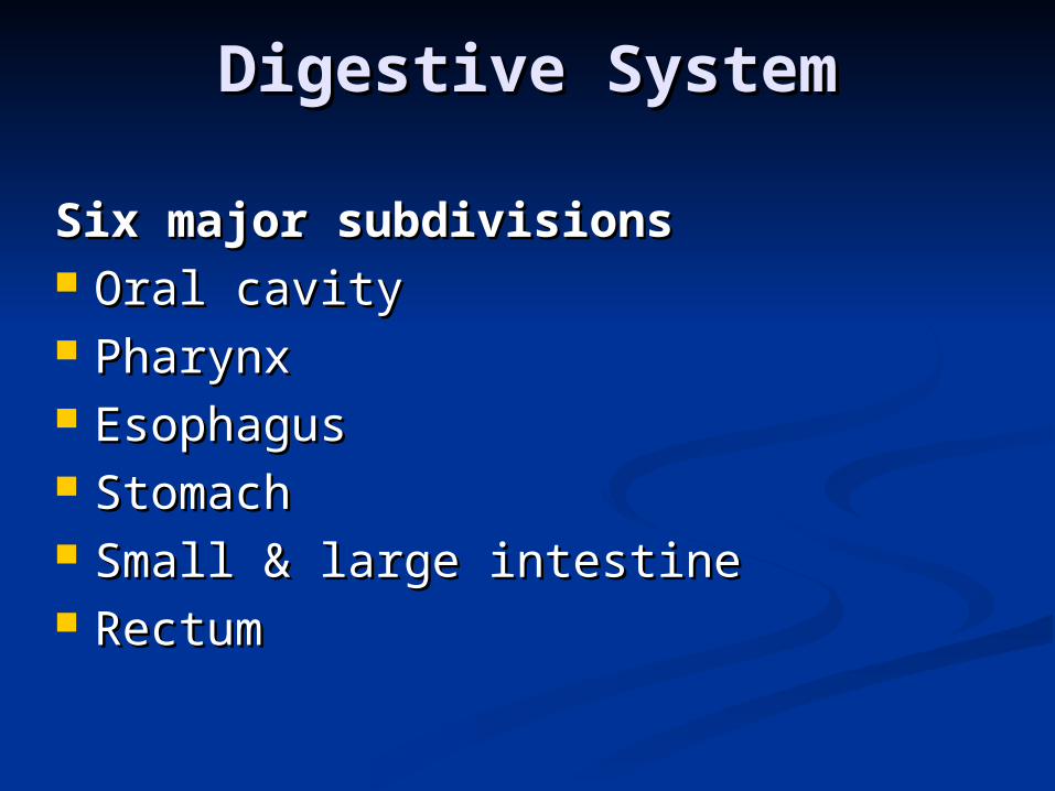
Six major subdivisionsSix major subdivisions Oral cavityOral cavity PharynxPharynx EsophagusEsophagus StomachStomach Small & large intestineSmall & large intestine Rectum Rectum

Digestive SystemDigestive System
Agnatha - straight Agnatha - straight digestive tubedigestive tube Coiled tube evolved Coiled tube evolved
with lengthening of with lengthening of tracttract
Figure 11.1: Simple to complex digestive systems.
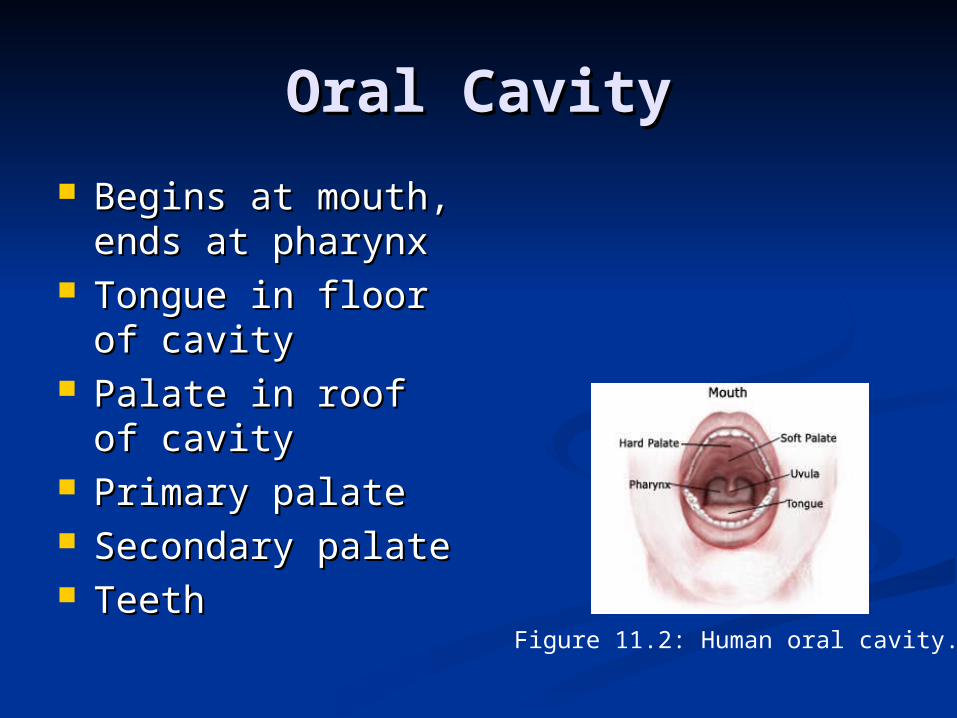
Oral CavityOral Cavity
Begins at mouth, Begins at mouth, ends at pharynxends at pharynx
Tongue in floor of Tongue in floor of cavitycavity
Palate in roof of Palate in roof of cavitycavity
Primary palate Primary palate Secondary palateSecondary palate Teeth Teeth
Figure 11.2: Human oral cavity.

PalatesPalates
Primary palate in Primary palate in anamniotes- nasal anamniotes- nasal passageways empty passageways empty into oral cavityinto oral cavity Ex: SalamanderEx: Salamander
Secondary palate of Secondary palate of amniotes- extends to amniotes- extends to pharyngeal cavity pharyngeal cavity Internal naresInternal nares
Figure 11.3: Oral cavity of amphibian (a) and mammal (b).

TeethTeeth On jaws normallyOn jaws normally Cheeks in mammals form pocketCheeks in mammals form pocket Acrodont teeth- fish and snakesAcrodont teeth- fish and snakes Bicuspid- amphibiansBicuspid- amphibians Tricuspid- lizardsTricuspid- lizards Pleurodont teeth- snakesPleurodont teeth- snakes Thecodont teeth- Thecodont teeth-
crocodilianscrocodilians
Figure 11.4- Cross section of jaw.
Figure 11.5- Types of cusps.

Jaw Teeth and CheekJaw Teeth and Cheek
Used for storage- rodents and Used for storage- rodents and squirrelssquirrels
Modified placoid scales- sharksModified placoid scales- sharks Polyhyodont- permanent Polyhyodont- permanent
replacement of teethreplacement of teeth Diphyodont- two sets of teethDiphyodont- two sets of teeth Monophyodont- one set of teethMonophyodont- one set of teeth
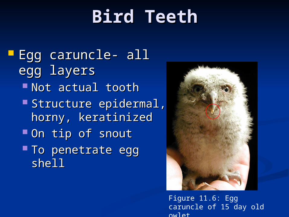
Bird TeethBird Teeth
Egg caruncle- all egg Egg caruncle- all egg layerslayers Not actual toothNot actual tooth Structure epidermal, Structure epidermal,
horny, keratinizedhorny, keratinized On tip of snoutOn tip of snout To penetrate egg shellTo penetrate egg shell
Figure 11.6: Egg caruncle of 15 day old owlet.

Reptilian TeethReptilian Teeth
Egg tooth- lizards and snakesEgg tooth- lizards and snakes Actual toothActual tooth Upper jawUpper jaw To penetrate egg shellTo penetrate egg shell
Figure 11.7: Monitor egg tooth..

Aglyphous- no Aglyphous- no modifications for venom modifications for venom deliverydelivery
Solenoglyphous- Solenoglyphous- retractable teeth, fangsretractable teeth, fangs
Proteroglyphous- fangs Proteroglyphous- fangs in front of mouthin front of mouth
Opisthoglyphous- fangs Opisthoglyphous- fangs in back of mouthin back of mouth
Modifications of Snake Modifications of Snake TeethTeeth
Figure 11.8: Position, cross and longitudinal sections of aglyphous (1), opisthoglyphous (2), and solenoglyphous (3) fangs.
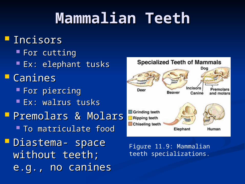
IncisorsIncisors For cuttingFor cutting Ex: elephant tusksEx: elephant tusks
CaninesCanines For piercingFor piercing Ex: walrus tusksEx: walrus tusks
Premolars & MolarsPremolars & Molars To matriculate foodTo matriculate food
Diastema- space Diastema- space without teeth; e.g., without teeth; e.g., no caninesno canines
Figure 11.9: Mammalian teeth specializations.
Mammalian TeethMammalian Teeth
Mammalian TeethMammalian Teeth
Heterodont dentitionHeterodont dentition Other varietiesOther varieties
Homodont- all teeth the sameHomodont- all teeth the same Bunodont- all teeth on single plainBunodont- all teeth on single plain Sectorial teeth – carnassials; e.g., upper Sectorial teeth – carnassials; e.g., upper
premolar and lower molar in carnivorespremolar and lower molar in carnivores
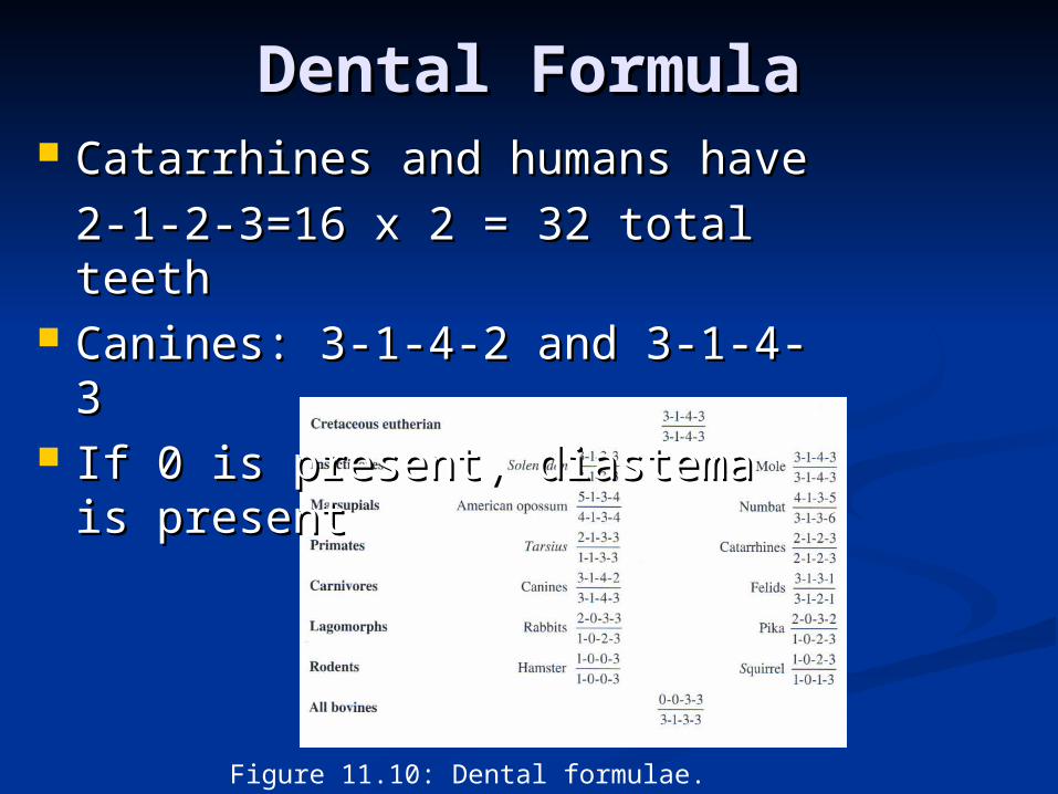
Dental FormulaDental Formula
Figure 11.10: Dental formulae.
Catarrhines and humans have Catarrhines and humans have
2-1-2-3=16 x 2 = 32 total 2-1-2-3=16 x 2 = 32 total teethteeth
Canines: 3-1-4-2 and 3-1-4-3Canines: 3-1-4-2 and 3-1-4-3 If 0 is present, diastema is If 0 is present, diastema is
presentpresent

TongueTongue Immobile in jawed fishImmobile in jawed fish Fleshy in higher Fleshy in higher
vertebratesvertebrates Frog- tongue shoots out and Frog- tongue shoots out and
draws backdraws back Glandular field secretes Glandular field secretes
sticky fluidsticky fluid Immobile tongue- turtles, Immobile tongue- turtles,
crocs, and some birdscrocs, and some birds Flexible tongue- nectar Flexible tongue- nectar
feeding bats and snakesfeeding bats and snakes Forked tongue of snakeForked tongue of snake
Figure 11.11: Jacobson’s organ (sensing apparatus) of snake and forked tongue.

Oral GlandsOral Glands Named based on locationNamed based on location
Labial- near the lipsLabial- near the lips Palatal- near palatePalatal- near palate InternasalInternasal Sublingual- releases Sublingual- releases
venomvenom Parotid- salivary glandParotid- salivary gland SubmaxillarySubmaxillary
Birds have few oral Birds have few oral glandsglands SwiftsSwifts
Figure 11.12: Swift and nest.
PharynxPharynx
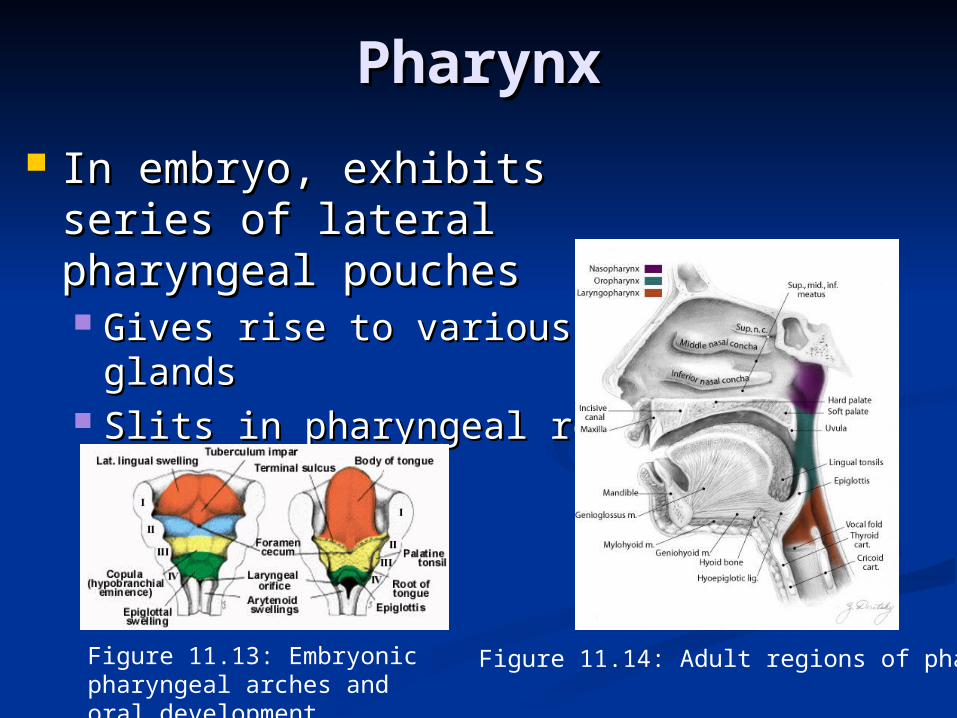
In embryo, exhibits series of In embryo, exhibits series of lateral pharyngeal poucheslateral pharyngeal pouches Gives rise to various glandsGives rise to various glands Slits in pharyngeal regionSlits in pharyngeal region
Figure 11.13: Embryonic pharyngeal arches and oral development.
Figure 11.14: Adult regions of pharynx.
Constant Features in TetrapodsConstant Features in Tetrapods Glottis-slit to larynxGlottis-slit to larynx
Covered by epiglottis Covered by epiglottis Eustachian tube- openingEustachian tube- opening Esophagus- openingEsophagus- opening
Pharynx further subdivided for food Pharynx further subdivided for food and air passageand air passage
Foramen cecum- groove on back of Foramen cecum- groove on back of tonguetongue Vestigial structure the leads to embryonic Vestigial structure the leads to embryonic
thyroid glandthyroid gland
PharynxPharynx
PharynxPharynx
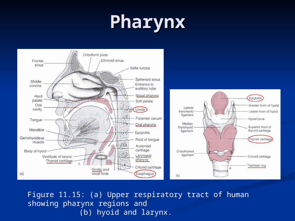
Figure 11.15: (a) Upper respiratory tract of human showing pharynx regions and
(b) hyoid and larynx.

EsophagusEsophagus
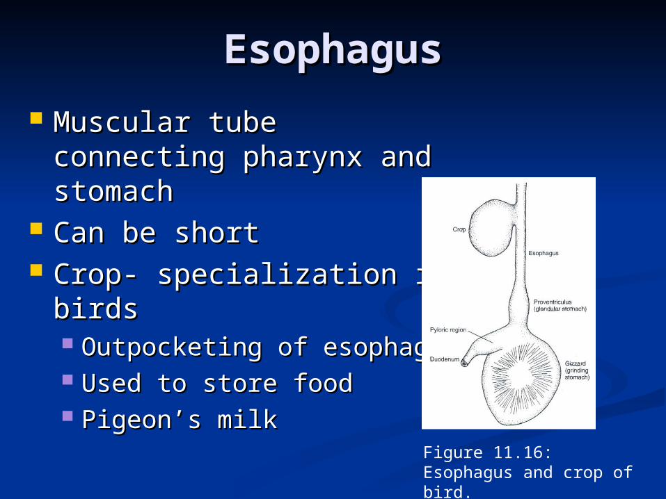
Muscular tube connecting Muscular tube connecting pharynx and stomachpharynx and stomach
Can be shortCan be short Crop- specialization in Crop- specialization in
birdsbirds Outpocketing of esophagusOutpocketing of esophagus Used to store foodUsed to store food Pigeon’s milkPigeon’s milk
Figure 11.16: Esophagus and crop of bird.

StomachStomach Muscular chamberMuscular chamber Secretes gastric juicesSecretes gastric juices Different lining of stomachsDifferent lining of stomachs
Esophageal-like epitheliaEsophageal-like epithelia Glandular epitheliaGlandular epithelia
Ruminant stomachRuminant stomach 4 chambers: rumen, 4 chambers: rumen,
reticulum, omasum, reticulum, omasum, abomasumabomasum
Human stomachHuman stomach Cardiac sphincter- esophagus Cardiac sphincter- esophagus
meets stomachmeets stomach Mostly lined with gastric Mostly lined with gastric
epitheliumepithelium
Figure 11.17: Stomach of mammals with esophageal-like epithelia in gray and glandular epithelia in red.

Stomach StructureStomach Structure
Greater and lesser curvatureGreater and lesser curvature MessentariesMessentaries
Greater omentum – attaches along greater Greater omentum – attaches along greater curvaturecurvature
Lesser omentum – attaches along lesser Lesser omentum – attaches along lesser curvaturecurvature
Cecum- increases surface areaCecum- increases surface area 2 parts in bird and crocodile stomach2 parts in bird and crocodile stomach
Proventiculus-glandularProventiculus-glandular Gizzard- grinding mill (gastroliths)Gizzard- grinding mill (gastroliths)

4-Chambered Stomachs4-Chambered Stomachs
Figure 11.18: Stomach of calf.
Rumen- food entersRumen- food enters Bacterial actionBacterial action
Reticulum- forms a bolusReticulum- forms a bolus Omasum- reswallowed Omasum- reswallowed
grassgrass Salivary actionSalivary action
Abomasum- food worked Abomasum- food worked out by gastric glandsout by gastric glands

Small IntestineSmall Intestine
Duodenum- 1Duodenum- 1stst segment segment Bile and pancreatic Bile and pancreatic
ductsducts Jejunum and Ileum Jejunum and Ileum
subdivisionssubdivisions
Figure 11.19: Digestive tract showing regions of small intestine.
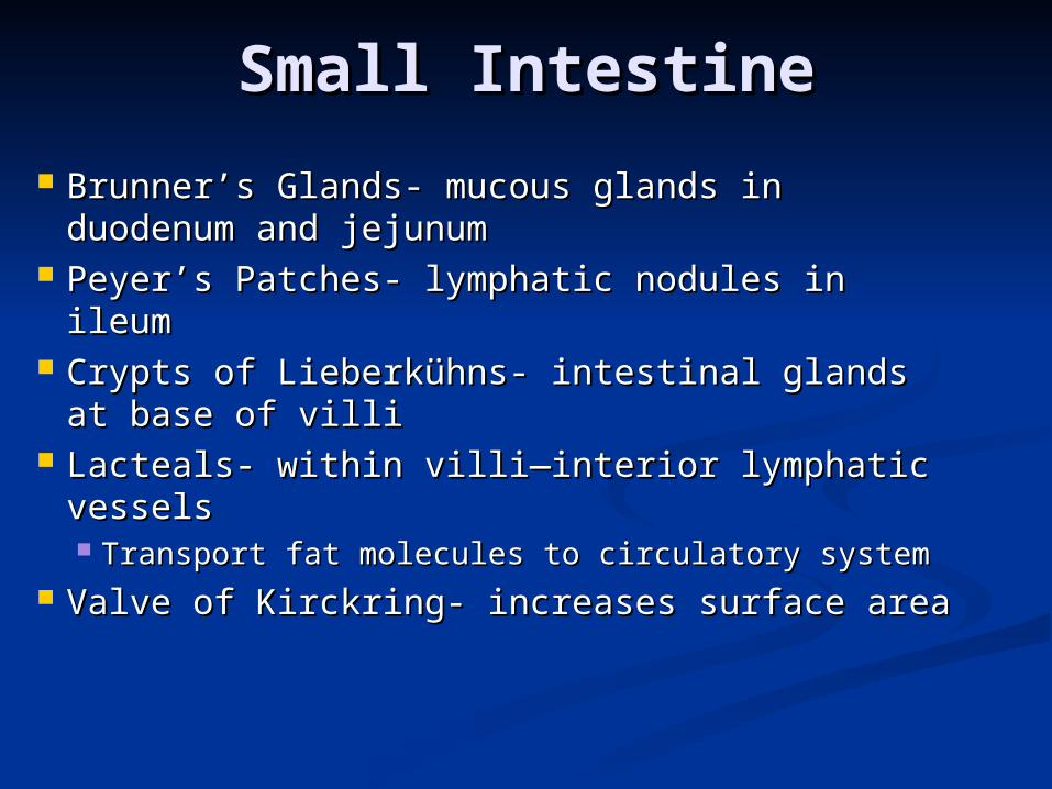
Brunner’s Glands- mucous glands in Brunner’s Glands- mucous glands in duodenum and jejunum duodenum and jejunum
Peyer’s Patches- lymphatic nodules in Peyer’s Patches- lymphatic nodules in ileumileum
Crypts of Lieberkühns- intestinal glands at Crypts of Lieberkühns- intestinal glands at base of villibase of villi
Lacteals- within villi—interior lymphatic Lacteals- within villi—interior lymphatic vesselsvessels Transport fat molecules to circulatory systemTransport fat molecules to circulatory system
Valve of Kirckring- increases surface areaValve of Kirckring- increases surface area
Small IntestineSmall Intestine
Small IntestineSmall Intestine
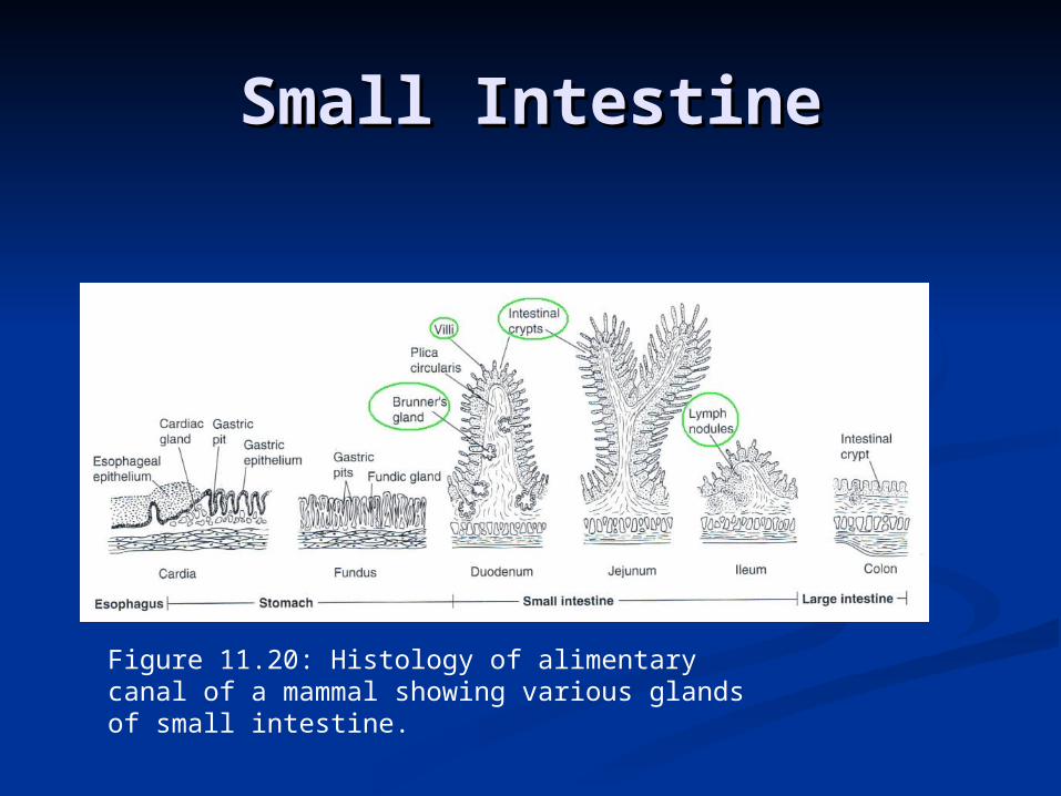
Figure 11.20: Histology of alimentary canal of a mammal showing various glands of small intestine.

Large IntestineLarge Intestine Fish and amphibians - straight and Fish and amphibians - straight and
shortshort Amniotes- divided into colon and rectumAmniotes- divided into colon and rectum
Ileocecal valve- allows passage from Ileocecal valve- allows passage from small intestine into largesmall intestine into large
Sigmoid flexure- S-shaped regionSigmoid flexure- S-shaped region at rectumat rectum Cecum- aids in absorptionCecum- aids in absorption
Terminates at vermiform appendixTerminates at vermiform appendix Cloaca- Cloaca- common chamber for common chamber for
digestive, urinary, and reproductive digestive, urinary, and reproductive products to empty (includes monotremes)products to empty (includes monotremes)Figure 11.21: Large intestine of human.
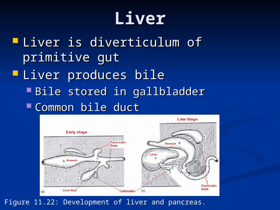
LiverLiver Liver is diverticulum of Liver is diverticulum of
primitive gutprimitive gut Liver produces bileLiver produces bile
Bile stored in gallbladderBile stored in gallbladder Common bile ductCommon bile duct
Ampulla of Vater- terminal portionAmpulla of Vater- terminal portion
Figure 11.22: Development of liver and pancreas.

PancreasPancreas
Pancreas – diverticulum of gutPancreas – diverticulum of gut Duct of Santorini- small, dorsal Duct of Santorini- small, dorsal
pancreaspancreas Duct of Wirsung- large, ventral Duct of Wirsung- large, ventral
pancreaspancreas Accessory duct- large duct after small, Accessory duct- large duct after small,
dorsal duct disappearsdorsal duct disappears Exocrine and endocrine glandsExocrine and endocrine glands
Islets of Langerhans- endocrine glandsIslets of Langerhans- endocrine glands
Rectal gland- diverticulum in sharks Rectal gland- diverticulum in sharks
Figure 11.1, 11.3, 11.4, 11.5, 11.10, 11.15, 11.16, 11.17, 11.18 & 11.22- Kent, Figure 11.1, 11.3, 11.4, 11.5, 11.10, 11.15, 11.16, 11.17, 11.18 & 11.22- Kent, George C. and Robert K. Carr. Comparative Anatomy of the Vertebrates. 9th George C. and Robert K. Carr. Comparative Anatomy of the Vertebrates. 9th ed. McGraw-Hill, 2001.ed. McGraw-Hill, 2001.
Figure Figure 11.2- http://www.mouth-cancer-symptoms.com/.2- http://www.mouth-cancer-symptoms.com/Figure Figure 11.6- http://gargravarr.cc.utexas.edu/owl/2002/.6- http://gargravarr.cc.utexas.edu/owl/2002/Figure Figure 11.7- http://www.proexotics.com/collection_nonPE9.html.7- http://www.proexotics.com/collection_nonPE9.htmlFigure Figure 11.8- .8-
http://www.kingsnake.com/reptilia-italia/My_HomePage_file/snakesgeneral.hthttp://www.kingsnake.com/reptilia-italia/My_HomePage_file/snakesgeneral.htmm
Figure Figure 11.9- http://www.okc.cc.ok.us/biologylabs/Documents/zoology/22.9- http://www.okc.cc.ok.us/biologylabs/Documents/zoology/22Figure Figure 11.11- .11-
http://www2.worldbook.com/features/reptiles/html/body_senorg.htmlhttp://www2.worldbook.com/features/reptiles/html/body_senorg.htmlFigure Figure 11.12- .12-
http://www.rspb.org.uk/birds/whatyoucando/attracthousemartins/index.asphttp://www.rspb.org.uk/birds/whatyoucando/attracthousemartins/index.aspFigure Figure 11.13- http://people.eku.edu/ritchisong/342notes7.html.13- http://people.eku.edu/ritchisong/342notes7.htmlFigure Figure 11.14- http://www.cortexity.com:8080/nicksblog/.14- http://www.cortexity.com:8080/nicksblog/Figure Figure 11.19- http://www.yoursurgery.com/ProcedureDetails.cfm?.19- http://www.yoursurgery.com/ProcedureDetails.cfm?
BR=1&Proc=49BR=1&Proc=49Figure Figure 11.20- Kardong, K. Vertebrates: Comparative Anatomy, Function, .20- Kardong, K. Vertebrates: Comparative Anatomy, Function,
Evolution. McGraw Hill, 2002. Evolution. McGraw Hill, 2002. Figure Figure 11.21- http://www.becomehealthynow.com/popups/lrg_intest.htm- http://www.becomehealthynow.com/popups/lrg_intest.htm
Literature CitedLiterature Cited
Related Documents