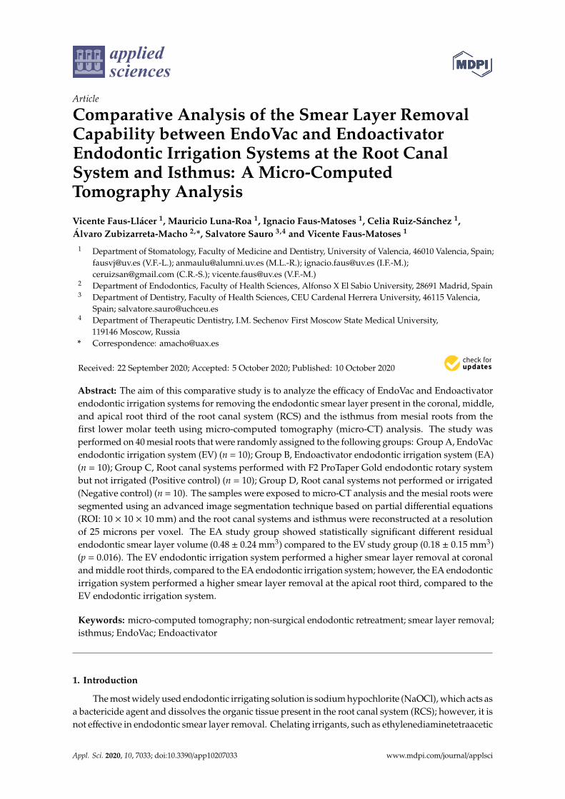
applied sciences Article Comparative Analysis of the Smear Layer Removal Capability between EndoVac and Endoactivator Endodontic Irrigation Systems at the Root Canal System and Isthmus: A Micro-Computed Tomography Analysis Vicente Faus-Llácer 1 , Mauricio Luna-Roa 1 , Ignacio Faus-Matoses 1 , Celia Ruiz-Sánchez 1 , Álvaro Zubizarreta-Macho 2, *, Salvatore Sauro 3,4 and Vicente Faus-Matoses 1 1 Department of Stomatology, Faculty of Medicine and Dentistry, University of Valencia, 46010 Valencia, Spain; [email protected] (V.F.-L.); [email protected] (M.L.-R.); [email protected] (I.F.-M.); [email protected] (C.R.-S.); [email protected] (V.F.-M.) 2 Department of Endodontics, Faculty of Health Sciences, Alfonso X El Sabio University, 28691 Madrid, Spain 3 Department of Dentistry, Faculty of Health Sciences, CEU Cardenal Herrera University, 46115 Valencia, Spain; [email protected] 4 Department of Therapeutic Dentistry, I.M. Sechenov First Moscow State Medical University, 119146 Moscow, Russia * Correspondence: [email protected] Received: 22 September 2020; Accepted: 5 October 2020; Published: 10 October 2020 Abstract: The aim of this comparative study is to analyze the efficacy of EndoVac and Endoactivator endodontic irrigation systems for removing the endodontic smear layer present in the coronal, middle, and apical root third of the root canal system (RCS) and the isthmus from mesial roots from the first lower molar teeth using micro-computed tomography (micro-CT) analysis. The study was performed on 40 mesial roots that were randomly assigned to the following groups: Group A, EndoVac endodontic irrigation system (EV) (n = 10); Group B, Endoactivator endodontic irrigation system (EA) (n = 10); Group C, Root canal systems performed with F2 ProTaper Gold endodontic rotary system but not irrigated (Positive control) (n = 10); Group D, Root canal systems not performed or irrigated (Negative control) (n = 10). The samples were exposed to micro-CT analysis and the mesial roots were segmented using an advanced image segmentation technique based on partial differential equations (ROI: 10 × 10 × 10 mm) and the root canal systems and isthmus were reconstructed at a resolution of 25 microns per voxel. The EA study group showed statistically significant different residual endodontic smear layer volume (0.48 ± 0.24 mm 3 ) compared to the EV study group (0.18 ± 0.15 mm 3 ) (p = 0.016). The EV endodontic irrigation system performed a higher smear layer removal at coronal and middle root thirds, compared to the EA endodontic irrigation system; however, the EA endodontic irrigation system performed a higher smear layer removal at the apical root third, compared to the EV endodontic irrigation system. Keywords: micro-computed tomography; non-surgical endodontic retreatment; smear layer removal; isthmus; EndoVac; Endoactivator 1. Introduction The most widely used endodontic irrigating solution is sodium hypochlorite (NaOCl), which acts as a bactericide agent and dissolves the organic tissue present in the root canal system (RCS); however, it is not effective in endodontic smear layer removal. Chelating irrigants, such as ethylenediaminetetraacetic Appl. Sci. 2020, 10, 7033; doi:10.3390/app10207033 www.mdpi.com/journal/applsci

Welcome message from author
This document is posted to help you gain knowledge. Please leave a comment to let me know what you think about it! Share it to your friends and learn new things together.
Transcript
applied sciences
Article
Comparative Analysis of the Smear Layer RemovalCapability between EndoVac and EndoactivatorEndodontic Irrigation Systems at the Root CanalSystem and Isthmus: A Micro-ComputedTomography Analysis
Vicente Faus-Llácer 1, Mauricio Luna-Roa 1, Ignacio Faus-Matoses 1, Celia Ruiz-Sánchez 1,Álvaro Zubizarreta-Macho 2,*, Salvatore Sauro 3,4 and Vicente Faus-Matoses 1
1 Department of Stomatology, Faculty of Medicine and Dentistry, University of Valencia, 46010 Valencia, Spain;[email protected] (V.F.-L.); [email protected] (M.L.-R.); [email protected] (I.F.-M.);[email protected] (C.R.-S.); [email protected] (V.F.-M.)
2 Department of Endodontics, Faculty of Health Sciences, Alfonso X El Sabio University, 28691 Madrid, Spain3 Department of Dentistry, Faculty of Health Sciences, CEU Cardenal Herrera University, 46115 Valencia,
Spain; [email protected] Department of Therapeutic Dentistry, I.M. Sechenov First Moscow State Medical University,
119146 Moscow, Russia* Correspondence: [email protected]
Received: 22 September 2020; Accepted: 5 October 2020; Published: 10 October 2020�����������������
Abstract: The aim of this comparative study is to analyze the efficacy of EndoVac and Endoactivatorendodontic irrigation systems for removing the endodontic smear layer present in the coronal, middle,and apical root third of the root canal system (RCS) and the isthmus from mesial roots from thefirst lower molar teeth using micro-computed tomography (micro-CT) analysis. The study wasperformed on 40 mesial roots that were randomly assigned to the following groups: Group A, EndoVacendodontic irrigation system (EV) (n = 10); Group B, Endoactivator endodontic irrigation system (EA)(n = 10); Group C, Root canal systems performed with F2 ProTaper Gold endodontic rotary systembut not irrigated (Positive control) (n = 10); Group D, Root canal systems not performed or irrigated(Negative control) (n = 10). The samples were exposed to micro-CT analysis and the mesial roots weresegmented using an advanced image segmentation technique based on partial differential equations(ROI: 10 × 10 × 10 mm) and the root canal systems and isthmus were reconstructed at a resolutionof 25 microns per voxel. The EA study group showed statistically significant different residualendodontic smear layer volume (0.48 ± 0.24 mm3) compared to the EV study group (0.18 ± 0.15 mm3)(p = 0.016). The EV endodontic irrigation system performed a higher smear layer removal at coronaland middle root thirds, compared to the EA endodontic irrigation system; however, the EA endodonticirrigation system performed a higher smear layer removal at the apical root third, compared to theEV endodontic irrigation system.
Keywords: micro-computed tomography; non-surgical endodontic retreatment; smear layer removal;isthmus; EndoVac; Endoactivator
1. Introduction
The most widely used endodontic irrigating solution is sodium hypochlorite (NaOCl), which acts asa bactericide agent and dissolves the organic tissue present in the root canal system (RCS); however, it isnot effective in endodontic smear layer removal. Chelating irrigants, such as ethylenediaminetetraacetic
Appl. Sci. 2020, 10, 7033; doi:10.3390/app10207033 www.mdpi.com/journal/applsci
Appl. Sci. 2020, 10, 7033 2 of 12
acid (EDTA), have shown their effectiveness in endodontic smear layer removal and improving RCSdisinfection [1,2]. During root canal surgery, the application of endodontic instruments onto thedentinal root canal walls produces hard tissue debris deposition within the RCS, which may containbacterial cells [1]. It can also interfere with the endodontic irrigation agents and root canal fillingmaterials of the RCS, preventing RCS disinfection and sealing, especially in anatomically complexareas, such as the isthmus areas in mesial roots of lower molar teeth [2,3]. Isthmus areas are consideredan important anatomical feature because they can contain smear layer remains of dental pulp tissue,necrotic tissues, and microorganisms, which can promote root canal re-infection after treatment [4].Tahmasbi et al. reported a prevalence of isthmus areas in the mesial root from the first lower molarteeth of 65.7% [5], whereas Kim et al. reported a frequency range from 20% to 70% [6]. The isthmus,according to its anatomical characteristics, is classified into five categories: Type I—There are twoseparate root canals, but union does not appear. Type II—An isthmus between the two separate rootcanals is observed. Type III—There are three root canals connected by an isthmus. Type IV—There aretwo elongated root canals connected in the center. Type V—There is a single, very wide and elongatedroot canal [4,7].
Traditionally, disinfection of the RCS has been performed by means of a passive irrigation procedurethrough endodontic irrigation syringes. However, this disinfection approach has demonstrated limitedefficacy to clean the apical third of the RCS and the anatomical irregularities of the RCS, such asthe isthmus. Therefore, new approaches have been developed to improve the cleaning capability oftraditional disinfection procedures. The Endoactivator (Dentsply Maillefer, Ballaigues, Switzerland)endodontic irrigation system can promote the penetration of the irrigant agent within the dentinaltubules of the RCS by means of continuous sonic movement [8]. The EndoVac (Dentsply Maillefer,Ballaigues, Switzerland) endodontic irrigation system can also safely reduce the bacterial load presentin the RCS, ensuring the presence of the irrigant agent in the apical third of the RCS and also preventingvapor lock formation, which avoids disinfection of the irrigant agent in the apical portion of theRCS [9,10]. The device allows for constantly renewed NaOCl irrigation throughout the root canalsystem by means of a negative apical pressure system. Two cannulas allow this flow of NaOCl: a macrocannula that introduces the NaOCl irrigation agent at the access cavity, and a micro cannula located inthe apical constriction that aspirates the NaOCl irrigation agent and evacuates it from the RCS.
The aim of the present study is to compare the efficacy of the EndoVac and Endoactivatorendodontic irrigation systems for removing the endodontic smear layer present in the coronal, middle,and apical root third of the RCS and the isthmus from mesial roots from first lower molar teeth usingmicro-CT analysis, with a null hypothesis (H0). This hypothesis determines that there would existno difference between the EndoVac and Endoactivator endodontic irrigation systems for removingthe endodontic smear layer present in the coronal, middle, and apical root third of the RCS and theisthmus from mesial roots from first lower molar teeth using micro-CT analysis.
2. Experimental Section
2.1. Study Design
A total of 40 first lower molar teeth with two canals and isthmus in the mesial root andextracted for periodontal reasons, with mature roots, absence of previous root canal treatment,calcium metamorphosis, and root resorptions were selected. This study was performed at theDepartment of Stomatology of the University of Valencia (Valencia, Spain), between February andJuly 2019. A randomized controlled experimental trial was performed attending to the German EthicsCommittee′s statement for the use of organic tissues in medical research, which was approved by theUniversity of Valencia Ethics Committee (process no. 12151). All of the patients were informed andconsented to transfer their teeth for the study.
Appl. Sci. 2020, 10, 7033 3 of 12
2.2. Experimental Procedure
The crowns of the teeth were removed between 1 mm and 3 mm under the cement–enamel junctionto standardize dental samples to 10 mm in length. The teeth were submitted to digital radiographyin both buccolingual and mesiodistal directions to assess the RCS anatomy. The working length(WL) of the RCS was determined using an operative microscope at 12 ×magnification (Zeiss DentalMicroscope, Oberkochen, Germany) until the emergence of a size 8 K-file (Dentsply Maillefer, Ballaigues,Switzerland) was visible at the apical foramen of the RCS. Then, the samples were randomly placed(Epidat 4.1, Galicia, Spain) into the following study groups: Group A, EndoVac endodontic irrigationsystem (Dentsply Maillefer, Baillagues, Switzerland) (EV) (n = 10); Group B, Endoactivator endodonticirrigation system (Dentsply Maillefer, Ballaigues, Switzerland) (EA) (n = 10); Group C, RCS performedwith F2 ProTaper Gold endodontic rotary system (Dentsply Maillefer, Baillagues, Switzerland) but notirrigated (Positive control) (n = 10); Group D, RCS not performed or irrigated (Negative control) (n =
10).The RCSs were randomly assigned to the above-mentioned study and control groups. Surgery for
the EV and EA study groups and Positive control group was performed by using the ProTaper Goldendodontic rotary system up to an F2 endodontic rotary file (Dentsply Maillefer, Ballaigues, Switzerland),using a 6:1 reduction handpiece (X-Smart plus, Dentsply Maillefer, Ballaigues, Switzerland) at 300 rpmand 2 N/cm torque, according to the manufacturer’s recommendations. The RCSs randomly assignedto the EV study group were irrigated with 5 mL of 5.25% NaOCl (Clorox, Oakland, CA, USA) with a0.3-mm-diameter endodontic needle (Miraject Endo Luer, Hager and Werken, Duisburg, Germany)inserted 1 mm into the WL. Furthermore, the disinfection of the RCS was improved by using theEndoactivator endodontic irrigation system (Dentsply Maillefer, Ballaigues, Switzerland) betweeneach endodontic file of the sequence. The final irrigation procedure was performed with 5 mL of 17%ethylenediaminetetraacetic acid (EDTA) (SmearClear, SybronEndo, CA, USA), 5 mL of 5.25% NaOCl,and 5 mL of sterile saline solution (Braun®, Melsungen, Germany). However, the RCSs randomlyassigned to the EA study group were irrigated with 5 mL of 5.25% NaOCl (Clorox, Oakland, CA,USA) with a 0.3-mm-diameter endodontic needle (Miraject Endo Luer, Hager and Werken, Duisburg,Germany) inserted 1 mm into the WL. Furthermore, the disinfection of the RCS was improved byusing the EndoVac endodontic irrigation system (Dentsply Maillefer, Ballaigues, Switzerland) betweeneach endodontic file of the sequence. The final irrigation procedure was performed with 5 mL of 17%EDTA (SmearClear, SybronEndo, CA, USA), left in the canal for 1 min with 5 mL of 5.25% activatedNaOCl, and 5 mL of sterile saline solution (Braun®, Melsungen, Germany). Then, the RCSs randomlyassigned to the EV and EA study groups were dried with sterile paper points (Dentsply Maillefer,Ballaigues, Switzerland) and the teeth were then stored in an incubator (mco-18aic, Sanyo, Moriguchi,Osaka, Japan) (37 ◦C, 100% relative humidity). The RCSs randomly assigned to the Negative controlgroup were neither shaped nor cleaned with irrigation solutions. All of the root canal treatments wereperformed by a single clinician.
2.3. Micro-CT Scanning
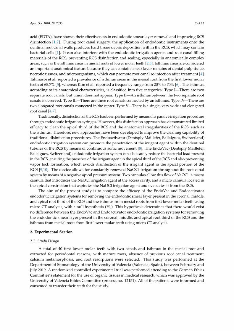
A micro-CT scan (Micro-CAT II, Siemens Preclinical Solutions, Knoxville, TN, USA) was performedto the RCS to analyze the residual smear layer volume present in the coronal, middle, and apical thirdof the RCS and the isthmus, the RCS volume, and the relationship between them at coronal, middle,and apical third of the RCS and at the interradicular isthmus with the following exposure parameters:90 kV, 88 µA, isotropic resolution of 50 µm, and 360◦ rotation. The tomographic three-dimensionalimages containing the entire tooth had a total of 512 slices, with isotropic 50 microns voxel size anda resolution of 512 × 512 pixels per slice. The isthmus presence was analyzed on the micro-CT scan(Micro-CAT II, Siemens Preclinical Solutions, Knoxville, TN, USA) at coronal, middle, and apical thirdof the root canal system (Figure 1).
Appl. Sci. 2020, 10, 7033 4 of 12Appl. Sci. 2020, 10, x FOR PEER REVIEW 4 of 12
Figure 1. Analysis of the isthmus presence on the transversal cross sections of the coronal, middle,
and apical third of the RCS of (A) Negative control group, (B) Positive control group, (C) EndoVac
endodontic irrigation system (EV) study group, and (D) Endoactivator endodontic irrigation system
(EA) study group.
2.4. Measurement Procedure
The analysis of the endodontic smear layer present in the coronal, middle, and apical third of
the RCS and the isthmus was performed through image processing software (ImageJ, National
Institutes of Health, Bethesda, MD, USA) after defining and segmenting the mesial root (ROI: 10 × 10
× 10 mm) of the dental samples. Next, the segmented mesial roots were reconstructed at a resolution
of 25 microns per voxel (Quantum 3.0, San Jose, CA, USA). Then, the RCSs forming on the segmented
mesial roots were divided using an advanced image segmentation technique based on partial
differential equations (Level Sets, National Institutes of Health, Bethesda, MD, USA) which allow
progressive differentiation between neighboring pixels and define the anatomy of the RCS (Figure
1). The algorithm is manually initialized in the first slice of the axial view of the volume, where the
user draws a contour outside, but close to, the channel. The segmentation technique method then
deforms the contour towards the inside, until convergence is reached (i.e., the channel is segmented
for the first slice). Then, the calculated contour is expanded by a fixed number of pixels (i.e., 6 pixels)
as initialization for the next slice, where segmentation technique is applied again. This process is
iteratively applied for every axial slice of the volume, until the whole channel is segmented in 3D.
Once the channel has been retrieved, the tooth debris inside the channel is segmented by thresholding
the volume of interest (i.e., interior of the channel). Small particles are eliminated from the debris’
segmentation mask as a final denoising step. The resulting segmentation mask may be manually
edited, by adding or deleting objects, in order to correct for possible inaccuracies in the automatic
segmentation. Finally, tooth debris volume, total channel volume, and the ratio between both are
calculated for apical, medial and coronal sections of the tooth.
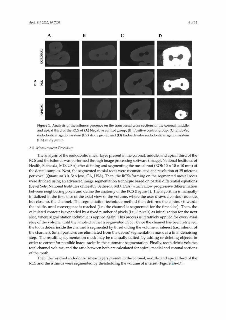
Then, the residual endodontic smear layers present in the coronal, middle, and apical third of
the RCS and the isthmus were segmented by thresholding the volume of interest (Figure 2A–D).
CO
RO
NA
L
MID
DL
E
AP
ICA
L
A B C D
Figure 1. Analysis of the isthmus presence on the transversal cross sections of the coronal, middle,and apical third of the RCS of (A) Negative control group, (B) Positive control group, (C) EndoVacendodontic irrigation system (EV) study group, and (D) Endoactivator endodontic irrigation system(EA) study group.
2.4. Measurement Procedure
The analysis of the endodontic smear layer present in the coronal, middle, and apical third of theRCS and the isthmus was performed through image processing software (ImageJ, National Institutes ofHealth, Bethesda, MD, USA) after defining and segmenting the mesial root (ROI: 10 × 10 × 10 mm) ofthe dental samples. Next, the segmented mesial roots were reconstructed at a resolution of 25 micronsper voxel (Quantum 3.0, San Jose, CA, USA). Then, the RCSs forming on the segmented mesial rootswere divided using an advanced image segmentation technique based on partial differential equations(Level Sets, National Institutes of Health, Bethesda, MD, USA) which allow progressive differentiationbetween neighboring pixels and define the anatomy of the RCS (Figure 1). The algorithm is manuallyinitialized in the first slice of the axial view of the volume, where the user draws a contour outside,but close to, the channel. The segmentation technique method then deforms the contour towardsthe inside, until convergence is reached (i.e., the channel is segmented for the first slice). Then, thecalculated contour is expanded by a fixed number of pixels (i.e., 6 pixels) as initialization for the nextslice, where segmentation technique is applied again. This process is iteratively applied for every axialslice of the volume, until the whole channel is segmented in 3D. Once the channel has been retrieved,the tooth debris inside the channel is segmented by thresholding the volume of interest (i.e., interior ofthe channel). Small particles are eliminated from the debris’ segmentation mask as a final denoisingstep. The resulting segmentation mask may be manually edited, by adding or deleting objects, inorder to correct for possible inaccuracies in the automatic segmentation. Finally, tooth debris volume,total channel volume, and the ratio between both are calculated for apical, medial and coronal sectionsof the tooth.
Then, the residual endodontic smear layers present in the coronal, middle, and apical third of theRCS and the isthmus were segmented by thresholding the volume of interest (Figure 2A–D).
Appl. Sci. 2020, 10, 7033 5 of 12Appl. Sci. 2020, 10, x FOR PEER REVIEW 5 of 12
Figure 2. Reconstructed three-dimensional micro-CT images of (A) Negative control group, (B)
Positive control group, (C) EV study group, and (D) EA study group. Residual smear layer volume
present in the coronal, middle, and apical third of the root canal system (RCS) and the isthmus (red).
Finally, the residual smear layer volume present in the coronal, middle, and apical third of the
root canal system and at the interradicular isthmus, the root canal system volume, and the
relationship between them at the coronal, middle, and apical third of the root canal system and the
isthmus were analyzed and compared by a blinded examiner (Amira 5.2 software, Thermo Fisher
Scientific, Bilbao, Spain).
2.5. Statistical Tests
The variables of interest were registered for statistical analysis (SPSS 22.00, Microsoft Inc,
Redmond, WA, USA). The mean, median, and standard deviation (SD) were used to perform the
descriptive statistical analysis for the quantitative variables. A comparative analysis was performed
by comparing the median residual endodontic smear layer volume present in the coronal, middle,
and apical third of the RCS and the isthmus (mm3), the root canal system volume (mm3), and the
relationship between them at coronal, middle, and apical third of the RCS and the isthmus (%)
between the EndoVac and Endoactivator endodontic irrigation systems and Positive and Negative
control groups using ANOVA and Kruskal–Wallis tests—p < 0.05—was considered statistically
significant.
3. Results
The mean, median, and SD values of the residual endodontic smear layer volume present in the
coronal, middle, and apical third of the RCS and the isthmus (mm3) are presented in Table 1 and
Figure 3A–C.
B
A C
D
Figure 2. Reconstructed three-dimensional micro-CT images of (A) Negative control group, (B) Positivecontrol group, (C) EV study group, and (D) EA study group. Residual smear layer volume present inthe coronal, middle, and apical third of the root canal system (RCS) and the isthmus (red).
Finally, the residual smear layer volume present in the coronal, middle, and apical third of theroot canal system and at the interradicular isthmus, the root canal system volume, and the relationshipbetween them at the coronal, middle, and apical third of the root canal system and the isthmuswere analyzed and compared by a blinded examiner (Amira 5.2 software, Thermo Fisher Scientific,Bilbao, Spain).
2.5. Statistical Tests
The variables of interest were registered for statistical analysis (SPSS 22.00, Microsoft Inc, Redmond,WA, USA). The mean, median, and standard deviation (SD) were used to perform the descriptivestatistical analysis for the quantitative variables. A comparative analysis was performed by comparingthe median residual endodontic smear layer volume present in the coronal, middle, and apical third ofthe RCS and the isthmus (mm3), the root canal system volume (mm3), and the relationship betweenthem at coronal, middle, and apical third of the RCS and the isthmus (%) between the EndoVac andEndoactivator endodontic irrigation systems and Positive and Negative control groups using ANOVAand Kruskal–Wallis tests—p < 0.05—was considered statistically significant.
3. Results
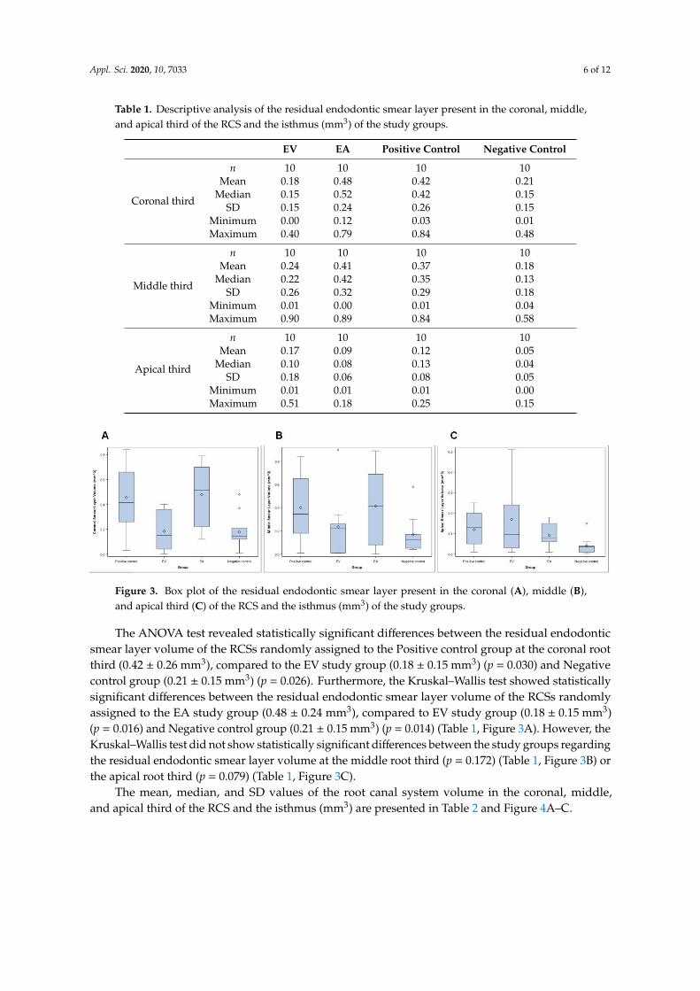
The mean, median, and SD values of the residual endodontic smear layer volume present inthe coronal, middle, and apical third of the RCS and the isthmus (mm3) are presented in Table 1 andFigure 3A–C.
Appl. Sci. 2020, 10, 7033 6 of 12
Table 1. Descriptive analysis of the residual endodontic smear layer present in the coronal, middle,and apical third of the RCS and the isthmus (mm3) of the study groups.
EV EA Positive Control Negative Control
Coronal third
n 10 10 10 10Mean 0.18 0.48 0.42 0.21
Median 0.15 0.52 0.42 0.15SD 0.15 0.24 0.26 0.15
Minimum 0.00 0.12 0.03 0.01Maximum 0.40 0.79 0.84 0.48
Middle third
n 10 10 10 10Mean 0.24 0.41 0.37 0.18
Median 0.22 0.42 0.35 0.13SD 0.26 0.32 0.29 0.18
Minimum 0.01 0.00 0.01 0.04Maximum 0.90 0.89 0.84 0.58
Apical third
n 10 10 10 10Mean 0.17 0.09 0.12 0.05
Median 0.10 0.08 0.13 0.04SD 0.18 0.06 0.08 0.05
Minimum 0.01 0.01 0.01 0.00Maximum 0.51 0.18 0.25 0.15
Appl. Sci. 2020, 10, x FOR PEER REVIEW 6 of 12
Table 1. Descriptive analysis of the residual endodontic smear layer present in the coronal, middle,
and apical third of the RCS and the isthmus (mm3) of the study groups.
EV EA Positive Control Negative Control
Coronal third
n 10 10 10 10
Mean 0.18 0.48 0.42 0.21
Median 0.15 0.52 0.42 0.15
SD 0.15 0.24 0.26 0.15
Minimum 0.00 0.12 0.03 0.01
Maximum 0.40 0.79 0.84 0.48
Middle third
n 10 10 10 10
Mean 0.24 0.41 0.37 0.18
Median 0.22 0.42 0.35 0.13
SD 0.26 0.32 0.29 0.18
Minimum 0.01 0.00 0.01 0.04
Maximum 0.90 0.89 0.84 0.58
Apical third
n 10 10 10 10
Mean 0.17 0.09 0.12 0.05
Median 0.10 0.08 0.13 0.04
SD 0.18 0.06 0.08 0.05
Minimum 0.01 0.01 0.01 0.00
Maximum 0.51 0.18 0.25 0.15
The ANOVA test revealed statistically significant differences between the residual endodontic
smear layer volume of the RCSs randomly assigned to the Positive control group at the coronal root
third (0.42 ± 0.26 mm3), compared to the EV study group (0.18 ± 0.15 mm3) (p = 0.030) and Negative
control group (0.21 ± 0.15 mm3) (p = 0.026). Furthermore, the Kruskal–Wallis test showed statistically
significant differences between the residual endodontic smear layer volume of the RCSs randomly
assigned to the EA study group (0.48 ± 0.24 mm3), compared to EV study group (0.18 ± 0.15 mm3) (p
= 0.016) and Negative control group (0.21 ± 0.15 mm3) (p = 0.014) (Table 1, Figure 3A). However, the
Kruskal–Wallis test did not show statistically significant differences between the study groups
regarding the residual endodontic smear layer volume at the middle root third (p = 0.172) (Table 1,
Figure 3B) or the apical root third (p = 0.079) (Table 1, Figure 3C).
Figure 3. Box plot of the residual endodontic smear layer present in the coronal (A), middle (B), and
apical third (C) of the RCS and the isthmus (mm3) of the study groups.
The mean, median, and SD values of the root canal system volume in the coronal, middle, and
apical third of the RCS and the isthmus (mm3) are presented in Table 2 and Figure 4A–C.
Figure 3. Box plot of the residual endodontic smear layer present in the coronal (A), middle (B),and apical third (C) of the RCS and the isthmus (mm3) of the study groups.
The ANOVA test revealed statistically significant differences between the residual endodonticsmear layer volume of the RCSs randomly assigned to the Positive control group at the coronal rootthird (0.42 ± 0.26 mm3), compared to the EV study group (0.18 ± 0.15 mm3) (p = 0.030) and Negativecontrol group (0.21 ± 0.15 mm3) (p = 0.026). Furthermore, the Kruskal–Wallis test showed statisticallysignificant differences between the residual endodontic smear layer volume of the RCSs randomlyassigned to the EA study group (0.48 ± 0.24 mm3), compared to EV study group (0.18 ± 0.15 mm3)(p = 0.016) and Negative control group (0.21 ± 0.15 mm3) (p = 0.014) (Table 1, Figure 3A). However, theKruskal–Wallis test did not show statistically significant differences between the study groups regardingthe residual endodontic smear layer volume at the middle root third (p = 0.172) (Table 1, Figure 3B) orthe apical root third (p = 0.079) (Table 1, Figure 3C).
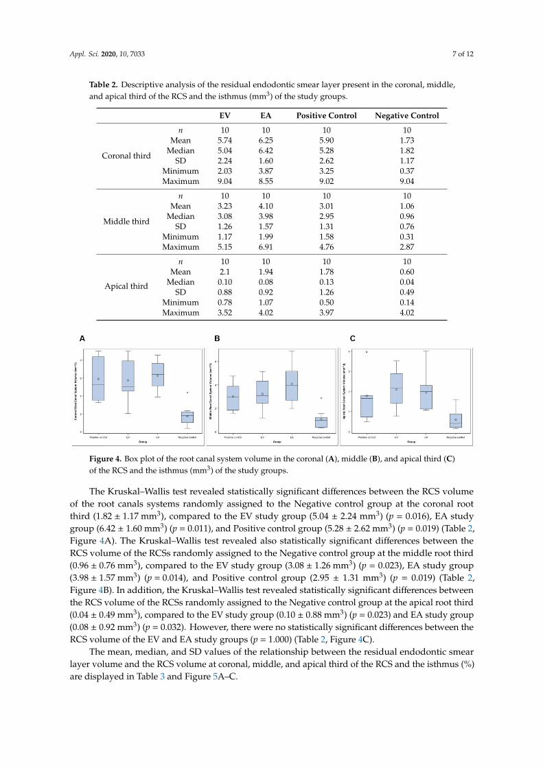
The mean, median, and SD values of the root canal system volume in the coronal, middle,and apical third of the RCS and the isthmus (mm3) are presented in Table 2 and Figure 4A–C.
Appl. Sci. 2020, 10, 7033 7 of 12
Table 2. Descriptive analysis of the residual endodontic smear layer present in the coronal, middle,and apical third of the RCS and the isthmus (mm3) of the study groups.
EV EA Positive Control Negative Control
Coronal third
n 10 10 10 10Mean 5.74 6.25 5.90 1.73
Median 5.04 6.42 5.28 1.82SD 2.24 1.60 2.62 1.17
Minimum 2.03 3.87 3.25 0.37Maximum 9.04 8.55 9.02 9.04
Middle third
n 10 10 10 10Mean 3.23 4.10 3.01 1.06
Median 3.08 3.98 2.95 0.96SD 1.26 1.57 1.31 0.76
Minimum 1.17 1.99 1.58 0.31Maximum 5.15 6.91 4.76 2.87
Apical third
n 10 10 10 10Mean 2.1 1.94 1.78 0.60
Median 0.10 0.08 0.13 0.04SD 0.88 0.92 1.26 0.49
Minimum 0.78 1.07 0.50 0.14Maximum 3.52 4.02 3.97 4.02
Appl. Sci. 2020, 10, x FOR PEER REVIEW 7 of 12
Table 2. Descriptive analysis of the residual endodontic smear layer present in the coronal, middle,
and apical third of the RCS and the isthmus (mm3) of the study groups.
EV EA Positive Control Negative Control
Coronal third
n 10 10 10 10
Mean 5.74 6.25 5.90 1.73
Median 5.04 6.42 5.28 1.82
SD 2.24 1.60 2.62 1.17
Minimum 2.03 3.87 3.25 0.37
Maximum 9.04 8.55 9.02 9.04
Middle third
n 10 10 10 10
Mean 3.23 4.10 3.01 1.06
Median 3.08 3.98 2.95 0.96
SD 1.26 1.57 1.31 0.76
Minimum 1.17 1.99 1.58 0.31
Maximum 5.15 6.91 4.76 2.87
Apical third
n 10 10 10 10
Mean 2.1 1.94 1.78 0.60
Median 0.10 0.08 0.13 0.04
SD 0.88 0.92 1.26 0.49
Minimum 0.78 1.07 0.50 0.14
Maximum 3.52 4.02 3.97 4.02
The Kruskal–Wallis test revealed statistically significant differences between the RCS volume of
the root canals systems randomly assigned to the Negative control group at the coronal root third
(1.82 ± 1.17 mm3), compared to the EV study group (5.04 ± 2.24 mm3) (p = 0.016), EA study group (6.42
± 1.60 mm3) (p = 0.011), and Positive control group (5.28 ± 2.62 mm3) (p = 0.019) (Table 2, Figure 4A).
The Kruskal–Wallis test revealed also statistically significant differences between the RCS volume of
the RCSs randomly assigned to the Negative control group at the middle root third (0.96 ± 0.76 mm3),
compared to the EV study group (3.08 ± 1.26 mm3) (p = 0.023), EA study group (3.98 ± 1.57 mm3) (p =
0.014), and Positive control group (2.95 ± 1.31 mm3) (p = 0.019) (Table 2, Figure 4B). In addition, the
Kruskal–Wallis test revealed statistically significant differences between the RCS volume of the RCSs
randomly assigned to the Negative control group at the apical root third (0.04 ± 0.49 mm3), compared
to the EV study group (0.10 ± 0.88 mm3) (p = 0.023) and EA study group (0.08 ± 0.92 mm3) (p = 0.032).
However, there were no statistically significant differences between the RCS volume of the EV and
EA study groups (p = 1.000) (Table 2, Figure 4C).
Figure 4. Box plot of the root canal system volume in the coronal (A), middle (B), and apical third (C)
of the RCS and the isthmus (mm3) of the study groups.
The mean, median, and SD values of the relationship between the residual endodontic smear
layer volume and the RCS volume at coronal, middle, and apical third of the RCS and the isthmus
(%) are displayed in Table 3 and Figure 5A–C.
Figure 4. Box plot of the root canal system volume in the coronal (A), middle (B), and apical third (C)of the RCS and the isthmus (mm3) of the study groups.
The Kruskal–Wallis test revealed statistically significant differences between the RCS volumeof the root canals systems randomly assigned to the Negative control group at the coronal rootthird (1.82 ± 1.17 mm3), compared to the EV study group (5.04 ± 2.24 mm3) (p = 0.016), EA studygroup (6.42 ± 1.60 mm3) (p = 0.011), and Positive control group (5.28 ± 2.62 mm3) (p = 0.019) (Table 2,Figure 4A). The Kruskal–Wallis test revealed also statistically significant differences between theRCS volume of the RCSs randomly assigned to the Negative control group at the middle root third(0.96 ± 0.76 mm3), compared to the EV study group (3.08 ± 1.26 mm3) (p = 0.023), EA study group(3.98 ± 1.57 mm3) (p = 0.014), and Positive control group (2.95 ± 1.31 mm3) (p = 0.019) (Table 2,Figure 4B). In addition, the Kruskal–Wallis test revealed statistically significant differences betweenthe RCS volume of the RCSs randomly assigned to the Negative control group at the apical root third(0.04 ± 0.49 mm3), compared to the EV study group (0.10 ± 0.88 mm3) (p = 0.023) and EA study group(0.08 ± 0.92 mm3) (p = 0.032). However, there were no statistically significant differences between theRCS volume of the EV and EA study groups (p = 1.000) (Table 2, Figure 4C).
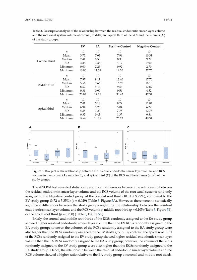
The mean, median, and SD values of the relationship between the residual endodontic smearlayer volume and the RCS volume at coronal, middle, and apical third of the RCS and the isthmus (%)are displayed in Table 3 and Figure 5A–C.
Appl. Sci. 2020, 10, 7033 8 of 12
Table 3. Descriptive analysis of the relationship between the residual endodontic smear layer volumeand the root canal system volume at coronal, middle, and apical third of the RCS and the isthmus (%)of the study groups.
EV EA Positive Control Negative Control
Coronal third
n 10 10 10 10Mean 3.72 7.63 7.94 10.31
Median 2.41 8.50 8.30 9.22SD 3.35 3.38 4.17 7.90
Minimum 0.00 2.23 0.92 2.70Maximum 10.06 11.59 14.20 27.75
Middle third
n 10 10 10 10Mean 7.97 9.11 13.40 17.70
Median 5.56 9.66 16.97 16.13SD 8.62 5.44 9.56 12.89
Minimum 0.31 0.00 0.54 4.52Maximum 23.87 17.21 30.43 47.54
Apical third
n 10 10 10 10Mean 7.41 5.18 8.29 11.84
Median 6.94 5.26 5.04 6.22SD 5.55 3.23 7.78 12.78
Minimum 0.35 0.43 1.37 0.34Maximum 14.49 10.28 26.23 40.54
Appl. Sci. 2020, 10, x FOR PEER REVIEW 8 of 12
Table 3. Descriptive analysis of the relationship between the residual endodontic smear layer volume
and the root canal system volume at coronal, middle, and apical third of the RCS and the isthmus (%)
of the study groups.
EV EA Positive Control Negative Control
Coronal third
n 10 10 10 10
Mean 3.72 7.63 7.94 10.31
Median 2.41 8.50 8.30 9.22
SD 3.35 3.38 4.17 7.90
Minimum 0.00 2.23 0.92 2.70
Maximum 10.06 11.59 14.20 27.75
Middle third
n 10 10 10 10
Mean 7.97 9.11 13.40 17.70
Median 5.56 9.66 16.97 16.13
SD 8.62 5.44 9.56 12.89
Minimum 0.31 0.00 0.54 4.52
Maximum 23.87 17.21 30.43 47.54
Apical third
n 10 10 10 10
Mean 7.41 5.18 8.29 11.84
Median 6.94 5.26 5.04 6.22
SD 5.55 3.23 7.78 12.78
Minimum 0.35 0.43 1.37 0.34
Maximum 14.49 10.28 26.23 40.54
The ANOVA test revealed statistically significant differences between the relationship between
the residual endodontic smear layer volume and the RCS volume of the root canal systems randomly
assigned to the Negative control group at the coronal root third (10.31 ± 9.22%), compared to the EV
study group (3.72 ± 3.35%) (p = 0.029) (Table 3, Figure 5A). However, there were no statistically
significant differences between the study groups regarding the relationship between the residual
endodontic smear layer volume and the RCS volume at middle root third (p = 0.105) (Table 3, Figure
5B), or the apical root third (p = 0.780) (Table 3, Figure 5C).
Figure 5. Box plot of the relationship between the residual endodontic smear layer volume and RCS
volume in the coronal (A), middle (B), and apical third (C) of the RCS and the isthmus (mm3) of the
study groups.
Briefly, the coronal and middle root thirds of the RCSs randomly assigned to the EA study group
showed higher residual endodontic smear layer volume than the EV RCSs randomly assigned to the
EA study group; however, the volumes of the RCSs randomly assigned to the EA study group were
also higher than the RCSs randomly assigned to the EV study group. By contrast, the apical root third
of the RCSs randomly assigned to the EV study group showed higher residual endodontic smear
layer volume than the EA RCSs randomly assigned to the EA study group; however, the volume of
the RCSs randomly assigned to the EV study group were also higher than the RCSs randomly
assigned to the EA study group. Hence, the relationship between the residual endodontic smear layer
volume and the RCS volume showed a higher ratio relative to the EA study group at coronal and
Figure 5. Box plot of the relationship between the residual endodontic smear layer volume and RCSvolume in the coronal (A), middle (B), and apical third (C) of the RCS and the isthmus (mm3) of thestudy groups.
The ANOVA test revealed statistically significant differences between the relationship betweenthe residual endodontic smear layer volume and the RCS volume of the root canal systems randomlyassigned to the Negative control group at the coronal root third (10.31 ± 9.22%), compared to theEV study group (3.72 ± 3.35%) (p = 0.029) (Table 3, Figure 5A). However, there were no statisticallysignificant differences between the study groups regarding the relationship between the residualendodontic smear layer volume and the RCS volume at middle root third (p = 0.105) (Table 3, Figure 5B),or the apical root third (p = 0.780) (Table 3, Figure 5C).
Briefly, the coronal and middle root thirds of the RCSs randomly assigned to the EA study groupshowed higher residual endodontic smear layer volume than the EV RCSs randomly assigned to theEA study group; however, the volumes of the RCSs randomly assigned to the EA study group werealso higher than the RCSs randomly assigned to the EV study group. By contrast, the apical root thirdof the RCSs randomly assigned to the EV study group showed higher residual endodontic smear layervolume than the EA RCSs randomly assigned to the EA study group; however, the volume of the RCSsrandomly assigned to the EV study group were also higher than the RCSs randomly assigned to theEA study group. Hence, the relationship between the residual endodontic smear layer volume and theRCS volume showed a higher ratio relative to the EA study group at coronal and middle root thirds,
Appl. Sci. 2020, 10, 7033 9 of 12
but a higher ratio related to the EA study group at the middle third. In conclusion, the EV endodonticirrigation system provided higher smear layer removal at coronal and middle root thirds, comparedto EA endodontic irrigation system; however, the EA endodontic irrigation system provided highersmear layer removal at the apical root third, compared to EV endodontic irrigation system.
4. Discussion
The results presented in this study reject the null hypothesis (H0) that determines that there wouldbe no differences between the EndoVac and Endoactivator endodontic irrigation systems for removingthe endodontic smear layer present in the coronal, middle, and apical third of the RCS and the isthmus.
This study analyzed the endodontic smear layer removal capacity of two endodontic irrigationsystems in the RCS and the isthmus from mesial roots of first lower molar teeth by means of micro-CTanalysis [9]. The endodontic smear layer removal has been previously analyzed by Scanning ElectronMicroscope [11,12] and Confocal Laser Scanning Microscopy [13]; however, these measurementprocedures require the longitudinal section of the dental root, only a limited region of the RCS canbe analyzed, and does not allow for the measurement of the endodontic smear layer volume [14].Micro-CT analysis allows an accurate 3D measurement technology to quantify the endodontic smearlayer volume in the RCS; however, micro-CT cannot detect the remaining soft tissue and requires toothextraction as well as the previously described measurement procedures [15,16]. Few articles havereported the use of micro-CT technology to analyze the endodontic smear layer removal in the RCS ofmesial roots from lower molar teeth and especially containing isthmus [11,17].
It has been reported that the smear layer can contain inorganic dentin debris and organic cells,such as parts of the odontoblastic process, bacterial cells, and necrotic pulp tissue; therefore, it is veryimportant to analyze the ability to remove smear layer from irrigation systems to prevent secondaryendodontic infections and facilitate sealing of root canal systems. [18]. It has been reported that 130 µmof NaOCl can be introduced into the dentinal tubules [19], while Enterococcus faecalis can penetrate upto a depth of 151 µm and attach to collagen fibers present [20], leaving bacteria harbored in deeperlayers, isthmus, accessory canals, anastomoses, and fins [21,22]. Furthermore, endodontic smear layerformation during RCS surgery can cause blockage of dentinal tubules and isthmus, avoiding theeffect of NaOCl inside the RCS [23]. Sjögren et al. reported that negative microbiological culturesobtained from the RCS show a root canal treatment success rate close to 94%. However, positivemicrobiological cultures reduce endodontic success rate to 68% [24]. Hence, the elimination of thepathogenic microflora from the RCS is a determinant factor in the outcome of the endodontic therapy.Chelating solutions have created soluble calcium chelates in contact with calcium ions from rootdentin to remove the endodontic smear layer, improving the efficacy of NaOCl inside the RCS [25].Furthermore, supplemental irrigation procedures after root canal preparation with chelating agents,passive ultrasonic irrigation [13,26], sonic irrigation [27], and apical negative-pressure procedures [13]have been shown to improve the endodontic smear layer removal of the canal walls and also increaseendodontic sealer penetration into the dentinal tubules and isthmus. However, few reports have beenperformed comparing sonic irrigation and apical negative pressure. Overall, in this study the meanendodontic smear layer removal of the EA and EV study groups was reduced from the coronal to theapical root thirds, perhaps because the dentinal tubules reduce its number and diameter toward theapical root third [11] and due to the sclerotic dentin present at the apical root third, which shows a higherresistance to irrigant solutions to remove the smear layer and also to endodontic sealer penetration [9].This agrees with previous studies, which showed that irrigating solutions are less effective in theapical root third [13]. Although the endodontic irrigation systems showed a statistically significantdecrease in the endodontic smear layer volume of the RCS and the isthmus from the mesial root of firstlower molar teeth (p < 0.05), none of them were able to remove completely the endodontic smear layerpresent in the coronal, middle, and apical root third of the RCS and the isthmus. These findings areconsistent with previous studies [10,15,28], which highlighted the relevance of the endodontic irrigantsystems to remove endodontic smear layer formation during RCS preparation, especially in areas of
Appl. Sci. 2020, 10, 7033 10 of 12
anatomical irregularities that cannot be removed with current techniques available [16]. Therefore,this study proposes the development of new endodontic smear layer removal protocols suitable foroptimizing and improving the cleaning of the most irregular regions of the RCS and the isthmus fromthe mesial roots of first lower molar teeth.
The EndoVac endodontic irrigation system comprises an alternative irrigation regimen thatinvolves apical negative pressure and uses a master tip to deliver the irrigant agent into the pulpchamber, while microcannulas are used throughout the entire RCS [29]. Zeliha et al. reported thatthe EndoVac endodontic irrigation system improved the apical root third disinfection of the RCSbecause the microcannula allows for the presence of the irrigating solution at the apical region [30].However, the Endoactivator endodontic irrigation system has been demonstrated to improve coronaland middle root third disinfection of the RCS due to the movement of the Endoactivator tip at coronaland middle root third of the RCS that allows for the penetration of the irrigating solution in thedentinal tubules [20,31,32]. In addition, the EndoVac endodontic irrigation system has reported a meanreduction rate of the smear layer volume in the RCS and isthmus of the mesial roots of first lowermolar teeth of 2.12% [8] and 3.4% [33].
5. Conclusions
Within the limitations of this study, the results show that the endodontic smear layer removaldepends on the endodontic irrigation system used and the location in the RCS and the isthmus frommesial roots of first lower molar teeth. The Endoactivator endodontic irrigation system achieves ahigher endodontic smear layer removal in the coronal and middle root thirds of the RCS and theisthmus from mesial roots of first lower molar teeth compared to the EndoVac endodontic irrigationsystem, which allows for a similar endodontic smear layer removal in the coronal, middle, and apicalroot thirds of the RCS and the isthmus from mesial roots of first lower molar teeth.
Author Contributions: All of the authors contributed to the investigation, supervision, writing, review, andediting of the study. Conceptualization, M.L.-R., V.F.-L., Á.Z.-M., and S.S.; data curation, I.F.-M.; formal analysis,V.F.-M.; visualization, C.R.-S. All authors have read and agreed to the published version of the manuscript.
Funding: This research received no external funding.
Conflicts of Interest: The authors declare no conflict of interest.
References
1. Arias-Moliz, M.T.; Morago, A.; Ordinola-Zapata, R.; Ferrer-Luque, C.M.; Ruiz-Linares, M.; Baca, P. Effects ofDentin Debris on the Antimicrobial Properties of Sodium Hypochlorite and Etidronic Acid. J. Endod. 2016,42, 771–775. [CrossRef] [PubMed]
2. de Gregorio, C.; Estevez, R.; Cisneros, R.; Paranjpe, A.; Cohenca, N. Efficacy of different irrigation andactivation systems on the penetration of sodium hypochlorite into simulated lateral canals and up to workinglength: An in vitro study. J. Endod. 2010, 36, 1216–1221. [CrossRef] [PubMed]
3. Adcock, J.M.; Sidow, S.J.; Looney, S.W.; Liu, Y.; McNally, K.; Lindsey, K.; Tay, F.R. Histologic evaluation ofcanal and isthmus debridement efficacies of two different irrigant delivery techniques in a closed system.J. Endod. 2011, 37, 544–548. [CrossRef]
4. Silva, E.J.N.L.; Carvalho, C.R.; Belladonna, F.G.; Prado, M.C.; Lopes, R.T.; De-Deus, G.; Moreira, E.J.L.Micro-CT evaluation of different final irrigation protocols on the removal of hard-tissue debris fromisthmus-containing mesial root of mandibular molars. Clin. Oral Investig. 2019, 23, 681–687. [CrossRef]
5. Tahmasbi, M.; Jalali, P.; Nair, M.K.; Barghan, S.; Nair, U.P. Prevalence of Middle Mesial Canals and Isthmi inthe Mesial Root of Mandibular Molars: An In Vivo Cone-beam Computed Tomographic Study. J. Endod.2017, 43, 1080–1083. [CrossRef]
6. Kim, S.; Jung, H.; Kim, S.; Shin, S.J.; Kim, E. The Influence of an Isthmus on the Outcomes of SurgicallyTreated Molars: A Retrospective Study. J. Endod. 2016, 42, 1029–1034. [CrossRef]
7. Cleghorn, B.M.; Christie, W.H.; Dong, C.C. Root and root canal morphology of the human permanentmaxillary first molar: A literature review. J. Endod. 2006, 32, 813–821. [CrossRef]
Appl. Sci. 2020, 10, 7033 11 of 12
8. Rius, L.; Arias, A.; Aranguren, J.M.; Romero, M.; de Gregorio, C. Analysis of the smear layer generated bydifferent activation systems: An in vitro study. Clin. Oral Investig. 2020, 10, 1–8. [CrossRef]
9. Adorno, C.G.; Fretes, V.R.; Ortiz, C.P.; Mereles, R.; Sosa, V.; Yubero, M.F.; Escobar, P.M.; Heilborn, C.Comparison of two negative pressure systems and syringe irrigation for root canal irrigation: An ex vivostudy. Int. Endod. J. 2016, 49, 174–183. [CrossRef]
10. Thomas, A.R.; Velmurugan, N.; Smita, S.; Jothilatha, S. Comparative evaluation of canal isthmus debridementefficacy of modified EndoVac technique with different irrigation systems. J. Endod. 2014, 40, 1676–1680.[CrossRef]
11. Alakshar, A.; Saleh, A.R.M.; Gorduysus, M.O. Debris and Smear Layer Removal from Oval Root CanalsComparing XP-Endo Finisher, EndoActivator, and Manual Irrigation: A SEM Evaluation. Eur. J. Dent.2020, 10. [CrossRef] [PubMed]
12. Turkyilmaz, A.; Erdemir, A. Comparison of dentin penetration ability of different root canal sealers usedwith different obturation methods. Microsc. Res. Tech. 2020, 10. [CrossRef] [PubMed]
13. Matos, F.S.; da Silva, F.R.; Paranhos, L.R.; Moura, C.C.G.; Bresciani, E.; Valera, M.C. The effect of 17% EDTAand QMiX ultrasonic activation on smear layer removal and sealer penetration: Ex vivo study. Sci. Rep.2020, 10, 10311. [CrossRef]
14. van der Sluis, L.W.; Versluis, M.; Wu, M.K.; Wesselink, P.R. Passive ultrasonic irrigation of the root canal:A review of the literature. Int. Endod. J. 2007, 40, 415–426. [CrossRef] [PubMed]
15. De-Deus, G.; Marins, J.; Neves, A.D.A.; Reis, C.; Fidel, S.; Versiani, M.A.; Alves, H.; Lopes, R.T.; Paciornik, S.Assessing accumulated hard-tissue debris using micro-computed tomography and free software for imageprocessing and analysis. J. Endod. 2014, 40, 271–276. [CrossRef]
16. Paqué, F.; Boessler, C.; Zehnder, M. Accumulated hard tissue debris levels in mesial roots of mandibularmolars after sequential irrigation steps. Int. Endod. J. 2011, 44, 148–153. [CrossRef]
17. Estrela, C.; Rabelo, L.E.; de Souza, J.B.; Alencar, A.H.G.; Estrela, C.R.A.; Sousa Neto, M.D.; Pécora, J.S.Frequency of Root Canal Isthmi in Human Permanent Teeth Determined by Cone-beam Computed Tomoghy.J. Endod. 2015, 41, 1535–1539. [CrossRef]
18. Violich, D.R.; Chandler, N.P. The smear layer in endodontics—A review. Int. Endod. J. 2010, 43, 2–15.[CrossRef]
19. Cheng, X.; Guan, S.; Lu, H.; Zhao, C.; Chen, X.; Li, N.; Bai, Q.; Tian, Y.; Yu, Q. Evaluation of the bactericidaleffect of Nd:YAG, Er:YAG, Er,Cr:YSGG laser radiation, and antimicrobial photodynamic therapy (aPDT) inexperimentally infected root canals. Lasers Surg. Med. 2012, 44, 824–831. [CrossRef]
20. Harrison, A.J.; Chivatxaranukul, P.; Parashos, P.; Messer, H.H. The effect of ultrasonically activated irrigationon reduction of Enterococcus faecalis in experimentally infected root canals. Int. Endod. J. 2010, 43, 968–977.[CrossRef]
21. Cheng, X.; Qu, T.; Ma, C.; Xiang, D.; Yu, Q.; Liu, X. Bioactive mono-dispersed nanospheres with long-termantibacterial effects for endodontic sealing. J. Mater. Chem. B. 2017, 5, 1195–1204. [CrossRef] [PubMed]
22. Dioguardi, M.; Di Gioia, G.; Illuzzi, G.; Arena, C.; Caponio, V.C.A.; Caloro, G.A.; Zhurakivska, K.; Adipietro, I.;Troiano, G.; Lo Muzio, L. Inspection of the Microbiota in Endodontic Lesions. Dent. J. 2019, 7, 47. [CrossRef]
23. Machado, R.; Ferrari, C.H.; Back, E.; Comparin, D.; Tomazinho, L.F.; Vansan, L.P. The Impact of ApicalPatency in the Success of Endodontic Treatment of Necrotic Teeth with Apical Periodontitis: A Brief Review.Iran. Endod. J. 2016, 11, 63–66. [PubMed]
24. Sjögren, U.; Figdor, D.; Persson, S.; Sundqvist, G. Influence of infection at the time of root filling on theoutcome of endodontic treatment of teeth with apical periodontitis. Int. Endod. J. 1997, 30, 297–306.[CrossRef] [PubMed]
25. Souza, M.A.; Hoffmann, I.P.; Menchik, V.H.S.; Zandoná, J.; Dias, C.T.; Palhano, H.S.; Bertol, C.H.;Rossato-Grando, L.G. Influence of ultrasonic activation using different final irrigants on antimicrobialactivity, smear layer removal and bond strength of filling material. Aust. Endod. J. 2019, 45, 209–215.[CrossRef] [PubMed]
26. Dioguardi, M.; Di Gioia, G.; Illuzzi, G.; Ciavarella, D.; Laneve, E.; Troiano, G.; Lo Muzio, L. Passive UltrasonicIrrigation Efficacy in the Vapor Lock Removal: Systematic Review and Meta-Analysis. Sci. World J. 2019,12, 6765349.
27. Johnson, M.; Sidow, S.J.; Looney, S.W.; Lindsey, K.; Niu, L.N.; Tay, F.R. Canal and isthmus debridementefficacy using a sonic irrigation technique in a closed-canal system. J. Endod. 2012, 38, 1265–1268. [CrossRef]
Appl. Sci. 2020, 10, 7033 12 of 12
28. Paqué, F.; Laib, A.; Gautschi, H.; Zehnder, M. Hard-tissue debris accumulation analysis by high-resolutioncomputed tomography scans. J. Endod. 2009, 35, 1044–1047. [CrossRef]
29. Mozo, S.; Llena, C.; Chieffi, N.; Forner, L.; Ferrari, M. Effectiveness of passive ultrasonic irrigation inimproving elimination of smear layer and opening dentinal tubules. J. Clin. Exp. Dent. 2014, 6, e47–e52.[CrossRef]
30. Ugur, A.Z.; Erdönmez, D.; Ates, M.O.; Dogan, T. Efficacy of different irrigation activation systems on bacterialextrusion. Aust. Endod. J. 2020, 10. [CrossRef]
31. Leoni, G.B.; Versiani, M.A.; Silva-Sousa, Y.T.; Bruniera, J.F.; Pécora, J.D.; Sousa-Neto, M.D. Ex vivo evaluationof four final irrigation protocols on the removal of hard-tissue debris from the mesial root canal system ofmandibular first molars. Int. Endod. J. 2017, 50, 398–406. [CrossRef] [PubMed]
32. Siqueira, J.F., Jr.; Alves, F.R.; Versiani, M.A.; Rôças, I.N.; Almeida, B.M.; Neves, M.A.; Sousa-Neto, M.D.Correlative bacteriologic and micro-computed tomographic analysis of mandibular molar mesial canalsprepared by self-adjusting file, reciproc, and twisted file systems. J. Endod. 2013, 39, 1044–1050. [CrossRef][PubMed]
33. Freire, L.G.; Iglecias, E.F.; Cunha, R.S.; Dos Santos, M.; Gavini, G. Micro-Computed Tomographic Evaluationof Hard Tissue Debris Removal after Different Irrigation Methods and Its Influence on the Filling of CurvedCanals. J. Endod. 2015, 41, 1660–1666. [CrossRef] [PubMed]
© 2020 by the authors. Licensee MDPI, Basel, Switzerland. This article is an open accessarticle distributed under the terms and conditions of the Creative Commons Attribution(CC BY) license (http://creativecommons.org/licenses/by/4.0/).
Related Documents