Comparative analysis of the integument of different tree frog species from Ololygon and Scinax genera (Anura: Hylidae) Henrique Alencar Meira da Silva 1 , Thiago Silva-Soares 2 , Lycia de Brito-Gitirana 1 1 Laboratório de Histologia Integrativa, Programa de Pesquisa em Glicobiologia, Instituto de Ciências Biomédicas, Universidade Federal do Rio de Janeiro. Avenida Carlos Chagas Filho 373, Bloco B1-019, 21941-902 Rio de Janeiro, RJ, Brazil. 2 Laboratório de Zoologia, Museu de Biologia Prof. Mello Leitão, Instituto Nacional da Mata Atlântica. Avenida José Ruschi, 29650-000 Santa Teresa, ES, Brazil. Corresponding author: Lycia de Brito-Gitirana ([email protected]) http://zoobank.org/94B1A213-D259-4366-8EB4-86E2A8E05443 ABSTRACT. The integuments of ten treefrog species of two genera from Scinaxnae – O. angrensis (Lutz, 1973), O. flavoguttata (Lutz & Lutz, 1939), O. humilis (Lutz & Lutz, 1954), O. perpusilla (Lutz & Lutz, 1939), O. v-signata (Lutz, 1968), Scinax hayii (Barbour, 1909), S. similis (Cochran, 1952), O. trapicheroi (Lutz & Lutz, 1954) and S. x-signatus (Spix, 1824) – were investi- gated using conventional and histochemical techniques of light microscopy, and polarized light microscopy. All integuments showed the basic structure of the anuran integument. Moreover, the secretory portions of exocrine glands, such as serous merocrine and apocrine glands, were found to be restricted to the spongious dermis. Lipid content occurred together with the heterogeneous secretory material of the glands with an apocrine secretion mechanism. In addition, clusters of these apocrine glands were present in the ventrolateral integument of some species. Melanophores were also visualized in all examined hylids. However, the occurrence of iridophores, detected through polarized light microscopy, varied according to the species. The Eberth-Katschenko layer occurred in the dorsal integument from both genera, but it was only present in the ventral integument of O. albicans, O. angrensis, O. flavoguttata, O. perpusilla and O. v-signata. Although the integument of all treefrogs showed the same basic structure, some characteristics were genus-specific; however, these features alone may not be used to distinguish both genera. KEY WORDS. Brazilian Atlantic forest, histochemistry, hylids, treefrog. INTRODUCTION Many challenges confront biologists studying amphibians, since human activities have been prejudicial to natural biota. Hylidae is a large anuran family, and one of the most abundant and prominent groups of frogs in the Neotropics. Overall, 345 species of Hylidae are known to existin Brazil (SBH 2016). The integument of anurans performs several functions, such as protection against diverse environmental circumstances (Elkan 1968, Fox 1986a, b, Greven et al. 1995, de Brito-Gitirana and Azevedo 2005, Azevedo et al. 2006, de Brito-Gitirana and Azevedo 2005), mechanical protection (Fox 1986a, b, Azevedo et al. 2006), chemical defense (Delfino et al. 1995, Daly 1995, Jekel et al. 2015), sensory perception (Mearow and Diamond 1988, Koyama et al. 2001), ionic transport and water absorp- tion (Sullivan et al. 2000, Azevedo et al. 2006), and respiration (Duellman and Trueb 1994). In adult anurans, the integument consists of two firmly attached layers: the epidermis and the dermis, which is located just beneath the epidermis. The epidermis, formed by a stratified squamous epithelium, overlies the dermis of connective tissue, which is subdivided into two layers: the spongious dermis and the compact dermis. The spongious dermis is formed by loose connective tissue with pigment cells, such as melanophores (melanin producing cells) and iridophores (with reflective or iridescent structures) located just beneath the basal lamina (Bag- nara et al. 1968). In addition to the pigment cells, blood vessels, alveolar mucous, serous, mixed and granular glands also occur. The compact dermis is composed of a series of alternating layers of collagenous fiber bundles arranged in a crisscross manner (Fox RESEARCH ARTICLE ZOOLOGIA 34: e20176 | DOI: 10.3897/zoologia.34.e20176 | September 18, 2017 1/17 ZOOLOGIA 34: e20176 ISSN 1984-4689 (online) zoologia.pensoft.net

Welcome message from author
This document is posted to help you gain knowledge. Please leave a comment to let me know what you think about it! Share it to your friends and learn new things together.
Transcript

Comparative analysis of the integument of different tree frog species from Ololygon and Scinax genera (Anura: Hylidae)
Henrique Alencar Meira da Silva1, Thiago Silva-Soares2, Lycia de Brito-Gitirana1
1Laboratório de Histologia Integrativa, Programa de Pesquisa em Glicobiologia, Instituto de Ciências Biomédicas, Universidade Federal do Rio de Janeiro. Avenida Carlos Chagas Filho 373, Bloco B1-019, 21941-902 Rio de Janeiro, RJ, Brazil.
2Laboratório de Zoologia, Museu de Biologia Prof. Mello Leitão, Instituto Nacional da Mata Atlântica. Avenida José Ruschi, 29650-000 Santa Teresa, ES, Brazil.
Corresponding author: Lycia de Brito-Gitirana ([email protected])
http://zoobank.org/94B1A213-D259-4366-8EB4-86E2A8E05443
ABSTRACT. The integuments of ten treefrog species of two genera from Scinaxnae – O. angrensis (Lutz, 1973), O. flavoguttata
(Lutz & Lutz, 1939), O. humilis (Lutz & Lutz, 1954), O. perpusilla (Lutz & Lutz, 1939), O. v-signata (Lutz, 1968), Scinax hayii
(Barbour, 1909), S. similis (Cochran, 1952), O. trapicheroi (Lutz & Lutz, 1954) and S. x-signatus (Spix, 1824) – were investi-
gated using conventional and histochemical techniques of light microscopy, and polarized light microscopy. All integuments
showed the basic structure of the anuran integument. Moreover, the secretory portions of exocrine glands, such as serous
merocrine and apocrine glands, were found to be restricted to the spongious dermis. Lipid content occurred together with
the heterogeneous secretory material of the glands with an apocrine secretion mechanism. In addition, clusters of these
apocrine glands were present in the ventrolateral integument of some species. Melanophores were also visualized in all
examined hylids. However, the occurrence of iridophores, detected through polarized light microscopy, varied according to
the species. The Eberth-Katschenko layer occurred in the dorsal integument from both genera, but it was only present in the
ventral integument of O. albicans, O. angrensis, O. flavoguttata, O. perpusilla and O. v-signata. Although the integument of
all treefrogs showed the same basic structure, some characteristics were genus-specific; however, these features alone may
not be used to distinguish both genera.
KEY WORDS. Brazilian Atlantic forest, histochemistry, hylids, treefrog.
INTRODUCTION
Many challenges confront biologists studying amphibians, since human activities have been prejudicial to natural biota. Hylidae is a large anuran family, and one of the most abundant and prominent groups of frogs in the Neotropics. Overall, 345 species of Hylidae are known to existin Brazil (SBH 2016).
The integument of anurans performs several functions, such as protection against diverse environmental circumstances (Elkan 1968, Fox 1986a, b, Greven et al. 1995, de Brito-Gitirana and Azevedo 2005, Azevedo et al. 2006, de Brito-Gitirana and Azevedo 2005), mechanical protection (Fox 1986a, b, Azevedo et al. 2006), chemical defense (Delfino et al. 1995, Daly 1995, Jekel et al. 2015), sensory perception (Mearow and Diamond 1988, Koyama et al. 2001), ionic transport and water absorp-
tion (Sullivan et al. 2000, Azevedo et al. 2006), and respiration (Duellman and Trueb 1994).
In adult anurans, the integument consists of two firmly attached layers: the epidermis and the dermis, which is located just beneath the epidermis. The epidermis, formed by a stratified squamous epithelium, overlies the dermis of connective tissue, which is subdivided into two layers: the spongious dermis and the compact dermis. The spongious dermis is formed by loose connective tissue with pigment cells, such as melanophores (melanin producing cells) and iridophores (with reflective or iridescent structures) located just beneath the basal lamina (Bag-nara et al. 1968). In addition to the pigment cells, blood vessels, alveolar mucous, serous, mixed and granular glands also occur. The compact dermis is composed of a series of alternating layers of collagenous fiber bundles arranged in a crisscross manner (Fox
RESEARCH ARTICLE
ZOOLOGIA 34: e20176 | DOI: 10.3897/zoologia.34.e20176 | September 18, 2017 1 / 17
ZOOLOGIA 34: e20176ISSN 1984-4689 (online)
zoologia.pensoft.net

1986a, b, Azevedo et al. 2005, de Brito-Gitirana and Azevedo 2005). Nevertheless, integument structure varies according to the body region (de Brito-Gitirana and Azevedo 2005, Felsemburgh et al. 2007). In addition, different types of exocrine glands occur in the anuran integument, withtheir secretory portions restricted to the spongious dermis (Duellmann and Trueb 1994, Brizzi et al. 2002).
In general, anurans show a wide array of colors related to specialized cells named chromatophores. Color and reflectivity are important mechanisms that allow anurans to change their in-tegument color, enabling the maintenance of body temperature and avoiding detection by predators (Duellman and Trueb 1994). Three types of chromatophores are important for amphibian coloration: melanophores (with a black or brownish pigment named melanin), xantophores (with yellow colored pigments), and iridophores (with reflective or iridescent structures). These pigment cells are located in the spongious dermis just beneath the basal lamina, and their arrangement was called the Dermal Chromatophore Unit (DCU) (Bagnara et al. 1968). However, the exact mechanism of DCU organization is still unknown.
Some anurans, like arboreal frogs, exhibit diverse mor-phology in their typical integument outline, showing cutane-ous adaptations to avoid evaporative water loss (Blaylock et al. 1976, Warburg et al. 2000, Barbeau and Lillywhite 2005). Such adaptations include exocrine glands secreting lipids, which are spread over the body surface, aided by an elaborate series of be-havioral movements to form a useful barrier against water loss. In addition, the histological aspects of the anuran integument can contribute to the differentiation of anuran species from Proceratophrys Miranda-Ribeiro, 1920 (Felsemburgh et al. 2007).
The abundant fauna of amphibians of Brazil (SBH 2016) contrast with the present loss and degradation of natural habitats and the scarce knowledge on the morphological, taxonomical, biological, and geographical distribution of most species. The aim of the present work was to investigate the morphological features of the integuments of some hylids in order to characterize this tissue and determine whetherthere are species-specific structures, contributing to the knowledge of the integument biology.
MATERIAL AND METHODS
Nine species of male adult tree frogs belonging to Sci-naxinae from both Ololygon Fitzinger, 1843 and Scinax Wagler, 1830 genera were collected in Serra dos Órgãos National Park (PARNASO, 22°29’31”S, 42°59’11.48”W), in the municipality of Teresópolis, during two years (from March 2007 to November 2008). Ololygon angrensis (Lutz, 1973) was collected in Rio de Ja-neiro state at the Lídice district (22°46’53.82”S, 44°13’55.96”W), municipality of Rio Claro (from March 2009 to March 2010) (Table 1). Tree frogs were collected under license permit number 15396-1 and 219/2011, issued by the Instituto Chico Mendes de Conservação da Biodiversidade (ICMBio).
Samplings were carried out at night from 7 to 10 pm and all individuals collected (Figs 1–10) were carefully placed in
small humid plastic bags, transported from the collectionsite to the field laboratory with a walk of no longer than one hour. They were kept in captivity until the next morning, when the fixation procedures were carried out. Individuals were euthanized with 0.5% xylocaine according to regulations of the Federal Council of Veterinary Medicine (Law 5.517/68 article 16, “f”). The animals were fixed in 10% formaldehyde and subsequently maintained in 70% (v/v) alcohol. At the end of field expedition, the treefrogs were taken to the laboratory, to undergo histological analysis.
For light microscopic (LM) analysis, 3-5 mm thick sec-tions from the ventral, ventrolateraland dorsal regions of the integument were processed according to standard histological techniques for paraffin embedding before sectioning, i.e., the sections were quickly washed in water, dehydrated (70%, 90%, twice in 100% ethanol; 30 minutes each), clarified twice in xylene (30 minutes each), infiltrated and embedded in paraffin. The 5-µm thick serial histological sections were stained with hema-toxylin-eosin (HE) (Lillie and Fulmer 1976), which is the standard stain for histological examination of animal tissues, staining thenuclei and cytoplasm in blue with the extracellular matrix in pale pink. Mallory’s trichrome (Lillie and Fulmer 1976) was used since it is a good stain for distinguishing cellular from extracellular elements and is especially suitable for studying connective tissue, staining the collagenous fibers in blue, red blood cells in orange, and nuclei in red. Staining with 1% Alcian blue (AB) 8GX at pH 2.5 (Mowry 1963, Kiernan 1990) was utilized to detect sulfated and carboxylated glycoconjugates (in light blue). In addition, HE-stained slices were observed under a light microscopeusing polarized light in order to detect iridophores (an iridescent cell), since these cell types possess reflective structures (Bagnara et al. 1968) that can be easily recognized under polarized light. Sections were analyzed using a Leica DM750 microscope, and the images were captured using a Leica DFC452 digital camera.
RESULTS
In general, the integuments of all species showed the basic morphological structure of the anuran integument, essentially
Table 1. List of individuals used in this study.
Number of individuals Common name (Frost 2016)
Ololygon albicans (Bokermann, 1967) 10 Teresopolis Snouted Treefrog
Ololygon angrensis (Lutz, 1973) 5Serra da Bocaina Snouted
Treefrog
Ololygon flavoguttata (Lutz & Lutz, 1939) 5 Yellowbelly Snouted Treefrog
Ololygon humilis (Lutz & Lutz, 1954) 3 Rio Babi Snouted Treefrog
Ololygon perpusilla (Lutz & Lutz, 1939) 3 Bandeirantes Snouted Treefrog
Ololygon trapicheroi (Lutz & Lutz, 1954) 5 Three-lined Snouted Treefrog
Ololygon v-signata (Lutz, 1968) 2 Forest Snouted Treefrog
Scinax similis (Cochran, 1952) 3 Cochran’s Snouted Treefrog
Scinax hayii (Barbour, 1909) 3 Hay’s Snouted Treefrog
Scinax x-signatus (Spix, 1824) 3 Venezuela Snouted Treefrog
H.A.M. da Silva et al.
ZOOLOGIA 34: e20176 | DOI: 10.3897/zoologia.34.e20176 | September 18, 20172 / 17
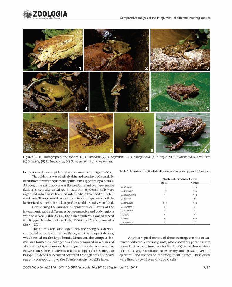
Table 2. Number of epithelial cell alyers of Oloygon spp. and Scinax spp.
Number of epithelial cell layers
Dorsal Ventral
O. albicans 4 4–5
O. angrensis 4 4–5
O. flavoguttata 4 4–5
O. humilis 4 8
O. perpusilla 3–4 4–5
O. trapicheroi 5 5
O. v-signata 4 4
S. similis 4 4
S. hayii 4 4–5
S. x-signatus 5 7
Figures 1–10. Photograph of the species: (1) O. albicans; (2) O. angrensis; (3) O. flavoguttata; (4) S. hayii; (5) O. humilis; (6) O. perpusilla; (6) S. similis; (8) O. trapicheroi; (9) O. v-signata; (10) S. x-signatus.
being formed by an epidermal and dermal layer (Figs 11–55).The epidermis was relatively thin and consisted of a partially
keratinized stratified squamous epithelium supported by a dermis. Although the keratinocyte was the predominant cell type, native flask cells were also visualized. In addition, epidermal cells were organized into a basal layer, an intermediate layer and an outer-most layer. The epidermal cells of the outermost layer were partially keratinized, since their nuclear profiles could be easily visualized.
Considering the number of epidermal cell layers of the integument, subtle differences betweenspecies and body regions were observed (Table 2), i.e., the ticker epidermis was observed in Ololygon humilis (Lutz & Lutz, 1954) and Scinax x-signatus (Spix, 1824).
The dermis was subdivided into the spongious dermis, composed of loose connective tissue, and the compact dermis, which rested on the hypodermis. Moreover, the compact der-mis was formed by collagenous fibers organized in a series of alternating layers, compactly arranged in a crisscross manner. Between the spongious dermis and the compact dermis, irregular basophilic deposits occurred scattered through this boundary region, corresponding to the Eberth-Katschenko (EK) layer.
Another typical feature of these treefrogs was the occur-rence of different exocrine glands, whose secretory portions were housed in the spongious dermis (Figs 11–55). From the secretory portion, a single unbranched excretory duct passed over the epidermis and opened on the integument surface. These ducts were lined by two layers of cuboid cells.
Comparative analysis of the integument of different tree frog species
ZOOLOGIA 34: e20176 | DOI: 10.3897/zoologia.34.e20176 | September 18, 2017 3 / 17

Figures 11–13. Light micrograph of the integument of O. albicans: (11) Dorsal region (HE-staining); (12) Ventrolateral region (HE-stain-ing); (13) Ventral region (AB-method). In all integument regions, the epidermis (E) rests on the dermis, which is subdivided into the spongious dermis (SD) and the compact dermis (CD). Iridophores (→) occur in the dorsal region; however, they are absent in both ventrolateral and ventral regions. Melanophores (_) in the spongious dermis. Both serous (Ø) and apocrine glands (¬) occur in the spongious dermis. No glandular cell reacts to AB-method, suggesting that secretory units is made up of serous cells. The EK-layer (Æ) exhibits its typical basophilic staining. Note clusters of apocrine glands (¬) with heterogeneous content in the ventrolateral integument. The EK-layer () exhibits typical alcianophilic reaction of its glycoconjugate content. Large blood vessels occur in the hypodermis.
H.A.M. da Silva et al.
ZOOLOGIA 34: e20176 | DOI: 10.3897/zoologia.34.e20176 | September 18, 20174 / 17

Figures 14–17. Light micrograph of the integument of O. angrensis: (14) Dorsal region (AB-staining); (15) Ventrolateral region (HE-stain-ing), inset (Mallory´s trichrome staining); (16) Ventral region (HE-staining); (17) Ventral region (AB-method). The melanophores (_) are numerous and located just beneath of the epidermis as well as around de glandular secretory units. They occur also in the hypodermis, but absent in the ventral integument. Iridophores (→) occur in the spongious dermis of the dorsal region just beneath the epidermis. No iridophore is visualized in the ventral region of the integument. Clusters of apocrine glands (¬) with heterogeneous intake predominate at ventrolateral integument; inset of Fig. 15: Observe the granular content with dense stained core () intermingled with cytoplasm material (*). The EK-layer () is continuous in the ventral region, but discontinuous in the ventral integument. Serous glands (Ø) occur in all body regions. Note blood vessels (Æ) in the spongious dermis. CD = compact dermis.
Depending on the dye affinity to their secretion, the merocrine glands of the serous and mixed types were visualized. Serous glands are formed by only serous-secreting cells, which have spherical nuclei and an acidophilic cytoplasm. Mixed secretory units are made up of mucous cells (nuclei generally flattened and displaced to the basal portion of the secretory cells and basophilic cytoplasm) and serous cells. It is noteworthy that mucous glands essentially formed by mucous cells did not occur in the integuments of all tree frogs (Table 3 and Figs 11–55).
While serous glands were visualized in all species, mixed glands occurred in Ololygon albicans (Bokermann, 1967), O. ang-rensis, O. flavoguttata (Lutz & Lutz, 1939), O. humilis, O. trapicheroi (Lutz & Lutz, 1954), O. v-signata (Lutz, 1968), Scinax similis (Co-chran, 1952) and S. x-signatus, but they did not occur in S. hayii (Barbour, 1909) and O. perpusilla (Lutz & Lutz, 1939) (Table 3).
Considering the apocrine glands, their secretory portion consisted of syncytial units, varying according to their content and dye affinity. Their secretory products were made up of small
Comparative analysis of the integument of different tree frog species
ZOOLOGIA 34: e20176 | DOI: 10.3897/zoologia.34.e20176 | September 18, 2017 5 / 17
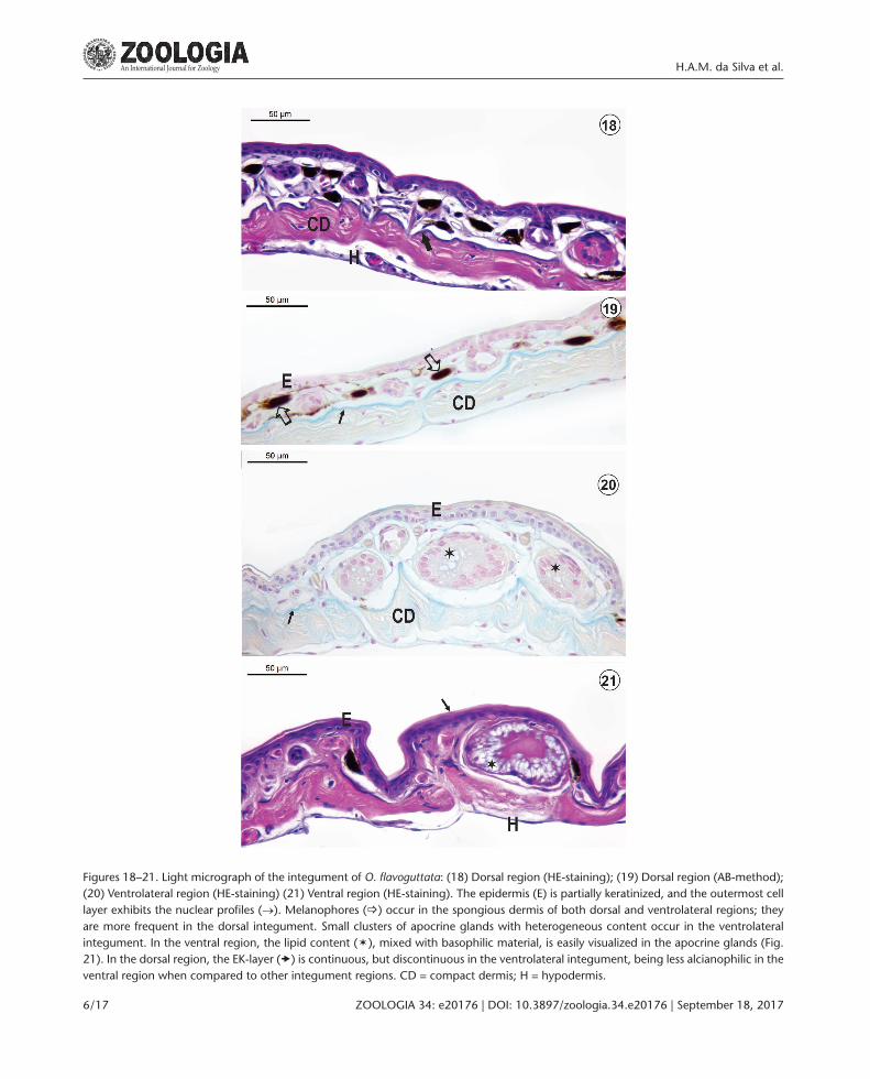
Figures 18–21. Light micrograph of the integument of O. flavoguttata: (18) Dorsal region (HE-staining); (19) Dorsal region (AB-method); (20) Ventrolateral region (HE-staining) (21) Ventral region (HE-staining). The epidermis (E) is partially keratinized, and the outermost cell layer exhibits the nuclear profiles (→). Melanophores (_) occur in the spongious dermis of both dorsal and ventrolateral regions; they are more frequent in the dorsal integument. Small clusters of apocrine glands with heterogeneous content occur in the ventrolateral integument. In the ventral region, the lipid content (ë), mixed with basophilic material, is easily visualized in the apocrine glands (Fig. 21). In the dorsal region, the EK-layer () is continuous, but discontinuous in the ventrolateral integument, being less alcianophilic in the ventral region when compared to other integument regions. CD = compact dermis; H = hypodermis.
H.A.M. da Silva et al.
ZOOLOGIA 34: e20176 | DOI: 10.3897/zoologia.34.e20176 | September 18, 20176 / 17

Figures 22–26. Light micrograph of the integument of S. hayii: (22) Dorsal region (Mallory´s trichrome staining); (23) Dorsal region (AB-method); (24) Ventrolateral region (HE-staining) (25) Ventral region (AB-method); (26) Ventral region (HE-staining). In the dorsal integument, exocrine glands are more frequent, mainly the serous glands (Ø). Melanophores (_) occur in the spongious dermis, even around the secretory portion of glands. Note clusters of apocrine glands with heterogeneous content (¬)in the spongious dermis of the ventrolateral integument. The EK-layer (Æ) is continuous in the dorsal integument, but absent in the ventral region. Slight cutaneous elevations (á) in the ventral integument are formed by the epidermis and the dermis, mainly the spongious dermis. They are separated by groves (). CD = compact dermis.
Comparative analysis of the integument of different tree frog species
ZOOLOGIA 34: e20176 | DOI: 10.3897/zoologia.34.e20176 | September 18, 2017 7 / 17

Table 4. Occurence of apocrine glands in the integument of Oloygon spp. and Scinax spp.
Heterogeneous content
Basophilic Lipid
Dorsal Ventrolateral Ventral Dorsal Ventrolateral Ventral
O. albicans + +/× + + + +
O. angrensis + – – + + –
O. flavoguttata + +/× + – + +
O. humilis + + + + + +
O. perpusilla + + – + + –
O. trapicheroi + + + + – –
O. v-signata + + + + + +
S. similis – – – + + –
S. hayii + +/× – + + +
S. x-signatus – + – + + –
(+) Present, (−) absent, (×) occurrence in clusters.
Table 3. Occurence of exocrine merocrine glands in the integument of Oloygon spp. and Scinax spp.
Merocrine glands
Mucous gland Serous gland Mixed glands
Dorsal Ventrolateral Ventral Dorsal Ventrolateral Ventral Dorsal Ventrolateral Ventral
O. albicans – – – + + + – – +
O. angrensis – – – + + + – + –
O. flavoguttata – – – + + + – + –
O. humilis – – – + + + + + +
O. perpusilla – – – + + – – – –
O. trapicheroi – – – + + + + + +
O. v-signata – – – + – – + + +
S. similis – – – + + + + + +
S. hayii – – – + + + – – –
S. x-signatus – – – + – – + + +
(+) Present, (–) absent.
acidophilic granules, seemingly due to their protein content. Nevertheless, some apocrine glands revealed that this secretion is a mixture of basophilic and lipid contents, intermingled with cytoplasmic material; their rounded nuclei were displaced to the cell basal domain. In some glands, the secretory product was constituted of acidophilic cytoplasmic material mixed with het-erogeneous material, which revealed slight basophilic reaction usually associated with lipid material (Table 4).
In O. angrensis, the secretory product of the apocrine glands exhibited basophilic granules with an acidophil core after staining with Mallory´s trichrome (Figs 14–17). Further-more, these secretory granules were also observed in S. similis (Figs 36–40) and contained lipids associated with basophilic material. We suggest that the basophilic staining was due to carbohydrates of glycolipids, since they showed no alcianophilic reaction typical of acid polysaccharides.
Table 5. Occurence of pigment cells in the integument of Oloygon spp. and Scinax spp.
Melanophores Iridophores
Dorsal Ventral Dorsal Ventral
O. albicans –/+ –/+ + +
O. angrensis + –/+ –/+ –
O. flavoguttata + –/+ – –
O. humilis + –/+ + –
O. perpusilla + – – –
O. trapicheroi –/+ – + –
O. v-signata + – – –
S. similis + –/+ + +
S. hayii + –/+ – –
S. x-signatus + – – –
(+) Widespread, (–/+) occasional, (–) not visualized.
H.A.M. da Silva et al.
ZOOLOGIA 34: e20176 | DOI: 10.3897/zoologia.34.e20176 | September 18, 20178 / 17

Figures 27–31. Light micrograph of the integument of O. humilis: (27) Dorsal region (HE-staining); (28) Dorsal region (AB-method); (29) Ventrolateral region (HE-staining) (30) Ventral region (Mallory´s trichrome staining); (31) Ventral region (AB-method). In the dorsal region, the spongious dermis is poorly developed. Melanophores (_) are visualized in all integument regions; however, iridophores (→) are visualized only in both dorsal and ventrolateral integument. Both pigment cells are located just beneath the epidermis. Alcianophilic reaction is observed in cytoplasm of iridophores as well as in the EK-layer (Æ) of the dorsal integument. The EK-layer is absent in the ventral integument. Apocrine glands with heterogeneous content (¬) occur in both ventrolateral and ventral integument. In S. humilis, mixed glands (Ú) are visualized in the ventral region, being formed by serous and mucous cells. Mucous cells exhibit alcianophilic reaction. E = epidermis; CD = compact dermis.
In O. albicans, O. angrensis, O. flavoguttata and S. hayii, apo-crine glands with heterogeneous content were more frequent in the ventrolateral integument, occurring as small clusters (Figs 11–26).
Pigment cells, such as melanophores and iridophores, were identified in the integument, occurring in the spongious dermis, just beneath the basal lamina. While melanophores were identi-fied through the typical brownish color of their melanin granules under light microscopy, iridophores were visualized by polarized light microscopy through their reflective or iridescent pigments (Figs 11–55). Melanophores also occurred in the hypodermis of O. angrensis, O. v-signata and S. similis (Figs 4–11, 36–40, 52–55),
while iridophores were only detected in the hypodermis of S. similis. The occurrence of both pigment cells varied according to the species and integument region (Table 5).
The Eberth-Katschenko (EK) layer occurred between the spongious and compact dermis and was recognized through its typical basophilic and alcianophilic reaction after using the HE- and AB-methods, respectively (Table 6; Figs 11–55). The EK-layer was visualized in the dorsal region of the integument of all Oloygon species, but absent in all species of Scinax.
Cutaneous elevations occurred in the ventral integu-ment of O. angrensis, O. flavoguttata, O. perpusilla, S. similis, O.
Comparative analysis of the integument of different tree frog species
ZOOLOGIA 34: e20176 | DOI: 10.3897/zoologia.34.e20176 | September 18, 2017 9 / 17

Figures 32–35. Light micrograph of the integument of O. perpusilla: (32) Dorsal region (HE-staining); (33) Dorsal region (AB-method); (34) Ventrolateral region (HE-staining) (35) Ventral region (AB-method). The spongious dermis houses both apocrine glands with hetero-geneous content (¬) and serous glands (Ø). Melanophores (_) occur in the dorsal integument, but they are not identified in either the ventrolateral or ventral region. Iridophores did not occur in all integument regions. The EK-layer (Æ) is a well defined continuous layer occurs as irregular deposits between the spongious and compact dermis of the ventral region. Cutaneous elevations (â) are separated by grooves (Ú) in the ventral region. CD = compact dermis.
trapicheroi and S. x-signatus (Figs 14–21, 32-40, 52–55). These slight elevations were formed by the epidermis, followed by the dermis, being separated by grooves.
DISCUSSION
Although some studies of the anuran integument are available, such as those of bufonids (de Brito-Gitirana and Aze-vedo 2005, de Brito-Gitirana et al. 2007, Almeida et al. 2007, Felsemburgh et al. 2009), ranids (Azevedo et al. 2006, Pelli et al. 2010), and leptodactylids (Goniakowska-Witalinska and Kubiczek 1998, Warburg et al. 2000, Nosiet al. 2002, Barbeau and Lillywhite 2005, Felsemburgh et al. 2007, Gonçalves and de
Brito-Gitirana 2008, Rigolo et al. 2008), information about the integument structure of hylids is still insufficient.
In this study, the integument showed the basic structure as already described for other anurans, i.e., the epidermis rests on a dermis, which is divided into a spongious and a compact dermis. The majority of anurans display this structural pattern of the integument (Elkan 1968, Goniakowska-Witalinska and Kubiczek 1998, Warburg et al. 2000, de Brito-Gitirana and Azevedo 2005, Delfino et al. 2006, Felsemburgh et al. 2007, Gonçalves and de Brito-Gitirana 2008, Felsemburgh et al. 2009).
In general, the epidermis varied from 4-5 cell layers. How-ever, in O. humilis and S. x-signatus the ventral epidermis was thicker, and it may be related to species habitat. Nevertheless,
H.A.M. da Silva et al.
ZOOLOGIA 34: e20176 | DOI: 10.3897/zoologia.34.e20176 | September 18, 201710 / 17

Figures 36–40. Light micrograph of the integument of S. similis: (36) Dorsal region (Mallory´s trichrome staining); (37) Dorsal region (AB-method); (38) Ventrolateral region (HE-staining); (39) Ventral region (Mallory´s trichrome staining); (40) Ventral region (AB-method). Melanophores (_) and iridophores (→) organized as chromatophore units occur in both dorsal and ventrolateral integument. Iridophores exhibit alcianophilic reaction. Note serous glands (Ø) and apocrine glands with granular content (¬) in the spongious dermis. In the ventral region, cutaneous elevations (¬) are separated by prominent grooves (Ú). The EK-layer (Æ) occur in the dorsal integument but not in the ventral integument. Moreover, the epidermis (E) of the ventral region is more developed than other integument regions. E = epidermis; CD = compact dermis; H = hypodermis.
we did not find detailed behavioral data in the literature to sup-port this explanation. In mammals, in some body areas, friction and other forces dictate the thickness of the lining epithelium, since the number of epithelial cell layers is related to epithelial resistance (Ham 1977, de Brito-Gitirana 2015).
Anuran glands have received significant attention. They have been described as being of different types, like mucous, serous, lipid (or wax), and mixed (seromucous) glands. How-ever, some authors have named the granular glands as poison or serous glands (Mills and Prum 1984, Duellmann and Trueb 1994, Brizzi et al. 2002).
In this study, well-established histological criteria to cat-egorize the cutaneous gland of mammals (Ham 1977, Kierzen-baum 2004) were used in order to adopt a coherent histological classification, especially in reference to anuran glands, since they exhibit variable morphology. Thus, on the basis of how their secretory products are released to the external environment, the exocrine glands can be classified as holocrine, apocrine or merocrine. Holocrine glands release both secretions and entire cells, while apocrine glands release the secretory product and cytoplasmic matrix of the apical portion of the cell. In mero-crine cells, no cytoplasm is lost and the gland uses exocytosis
Comparative analysis of the integument of different tree frog species
ZOOLOGIA 34: e20176 | DOI: 10.3897/zoologia.34.e20176 | September 18, 2017 11 / 17
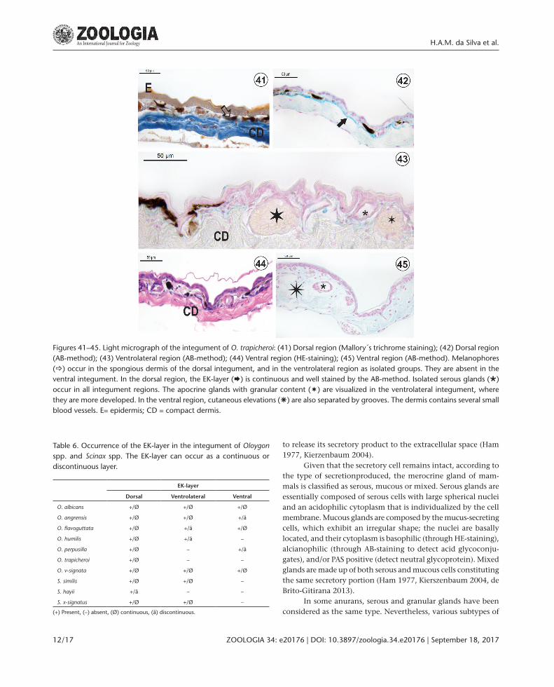
Figures 41–45. Light micrograph of the integument of O. trapicheroi: (41) Dorsal region (Mallory´s trichrome staining); (42) Dorsal region (AB-method); (43) Ventrolateral region (AB-method); (44) Ventral region (HE-staining); (45) Ventral region (AB-method). Melanophores (_) occur in the spongious dermis of the dorsal integument, and in the ventrolateral region as isolated groups. They are absent in the ventral integument. In the dorsal region, the EK-layer (Æ) is continuous and well stained by the AB-method. Isolated serous glands (Ø) occur in all integument regions. The apocrine glands with granular content (¬) are visualized in the ventrolateral integument, where they are more developed. In the ventral region, cutaneous elevations (â) are also separated by grooves. The dermis contains several small blood vessels. E= epidermis; CD = compact dermis.
Table 6. Occurrence of the EK-layer in the integument of Oloygon spp. and Scinax spp. The EK-layer can occur as a continuous or discontinuous layer.
EK-layer
Dorsal Ventrolateral Ventral
O. albicans +/Ø +/Ø +/Ø
O. angrensis +/Ø +/Ø +/â
O. flavoguttata +/Ø +/â +/Ø
O. humilis +/Ø +/â –
O. perpusilla +/Ø – +/â
O. trapicheroi +/Ø – –
O. v-signata +/Ø +/Ø +/Ø
S. similis +/Ø +/Ø –
S. hayii +/â – –
S. x-signatus +/Ø +/Ø –
(+) Present, (–) absent, (Ø) continuous, (â) discontinuous.
to release its secretory product to the extracellular space (Ham 1977, Kierzenbaum 2004).
Given that the secretory cell remains intact, according to the type of secretionproduced, the merocrine gland of mam-mals is classified as serous, mucous or mixed. Serous glands are essentially composed of serous cells with large spherical nuclei and an acidophilic cytoplasm that is individualized by the cell membrane. Mucous glands are composed by the mucus-secreting cells, which exhibit an irregular shape; the nuclei are basally located, and their cytoplasm is basophilic (through HE-staining), alcianophilic (through AB-staining to detect acid glycoconju-gates), and/or PAS positive (detect neutral glycoprotein). Mixed glands are made up of both serous and mucous cells constituting the same secretory portion (Ham 1977, Kierszenbaum 2004, de Brito-Gitirana 2013).
In some anurans, serous and granular glands have been considered as the same type. Nevertheless, various subtypes of
H.A.M. da Silva et al.
ZOOLOGIA 34: e20176 | DOI: 10.3897/zoologia.34.e20176 | September 18, 201712 / 17
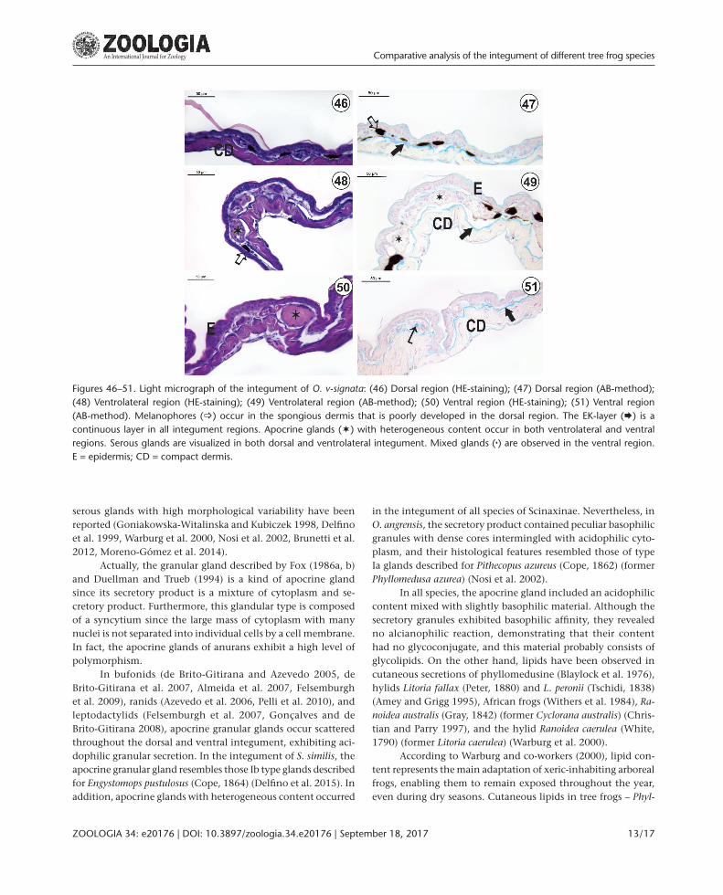
Figures 46–51. Light micrograph of the integument of O. v-signata: (46) Dorsal region (HE-staining); (47) Dorsal region (AB-method); (48) Ventrolateral region (HE-staining); (49) Ventrolateral region (AB-method); (50) Ventral region (HE-staining); (51) Ventral region (AB-method). Melanophores (_) occur in the spongious dermis that is poorly developed in the dorsal region. The EK-layer (Æ) is a continuous layer in all integument regions. Apocrine glands (¬) with heterogeneous content occur in both ventrolateral and ventral regions. Serous glands are visualized in both dorsal and ventrolateral integument. Mixed glands () are observed in the ventral region. E = epidermis; CD = compact dermis.
serous glands with high morphological variability have been reported (Goniakowska-Witalinska and Kubiczek 1998, Delfino et al. 1999, Warburg et al. 2000, Nosi et al. 2002, Brunetti et al. 2012, Moreno-Gómez et al. 2014).
Actually, the granular gland described by Fox (1986a, b) and Duellman and Trueb (1994) is a kind of apocrine gland since its secretory product is a mixture of cytoplasm and se-cretory product. Furthermore, this glandular type is composed of a syncytium since the large mass of cytoplasm with many nuclei is not separated into individual cells by a cell membrane. In fact, the apocrine glands of anurans exhibit a high level of polymorphism.
In bufonids (de Brito-Gitirana and Azevedo 2005, de Brito-Gitirana et al. 2007, Almeida et al. 2007, Felsemburgh et al. 2009), ranids (Azevedo et al. 2006, Pelli et al. 2010), and leptodactylids (Felsemburgh et al. 2007, Gonçalves and de Brito-Gitirana 2008), apocrine granular glands occur scattered throughout the dorsal and ventral integument, exhibiting aci-dophilic granular secretion. In the integument of S. similis, the apocrine granular gland resembles those Ib type glands described for Engystomops pustulosus (Cope, 1864) (Delfino et al. 2015). In addition, apocrine glands with heterogeneous content occurred
in the integument of all species of Scinaxinae. Nevertheless, in O. angrensis, the secretory product contained peculiar basophilic granules with dense cores intermingled with acidophilic cyto-plasm, and their histological features resembled those of type Ia glands described for Pithecopus azureus (Cope, 1862) (former Phyllomedusa azurea) (Nosi et al. 2002).
In all species, the apocrine gland included an acidophilic content mixed with slightly basophilic material. Although the secretory granules exhibited basophilic affinity, they revealed no alcianophilic reaction, demonstrating that their content had no glycoconjugate, and this material probably consists of glycolipids. On the other hand, lipids have been observed in cutaneous secretions of phyllomedusine (Blaylock et al. 1976), hylids Litoria fallax (Peter, 1880) and L. peronii (Tschidi, 1838) (Amey and Grigg 1995), African frogs (Withers et al. 1984), Ra-noidea australis (Gray, 1842) (former Cyclorana australis) (Chris-tian and Parry 1997), and the hylid Ranoidea caerulea (White, 1790) (former Litoria caerulea) (Warburg et al. 2000).
According to Warburg and co-workers (2000), lipid con-tent represents the main adaptation of xeric-inhabiting arboreal frogs, enabling them to remain exposed throughout the year, even during dry seasons. Cutaneous lipids in tree frogs – Phyl-
Comparative analysis of the integument of different tree frog species
ZOOLOGIA 34: e20176 | DOI: 10.3897/zoologia.34.e20176 | September 18, 2017 13 / 17

Figures 52–55. Light micrograph of the integument of S. x-signatus: (52) Dorsal region (HE-staining); (53) Dorsal region (AB-method); (54) Ventrolateral region (Mallory´s trichrome staining); (55) Ventral region (Mallory´s trichrome staining). The epidermis (E) is slightly ticker when compared to those of other hylids, as in the compact dermis (CD). Melanophores (_) are visulized in both dorsal and ventrolateral integument just beneath the epidermis. Serous glands are present in all regions; however, some of them show slightly alcianophilic con-tent (→) in both ventrolateral and ventral regions. The apocrine glands (¬) with granular content occur in both dorsal and ventrolateral regions. The EK-layer (Æ) is visualized in both dorsal and ventrolateral integument but is absent in the ventral integument.
lomedusa sauvagei Boulenger, 1882, P. iherengii Boulenger, 1885, P. boliviana Boulenger, 1902, Ranoid gracilenta (Peters, 1869), R. caerulea (White, 1790), Polypedates maculatus (Gray, 1830) – are a specialized adaptation to reduce dehydration in arid environ-ments (Amey and Grigg 1995, Lillywhite 2004, Barbeau and Lillywhite 2005, Gomez et al. 2006). These lipids are spread over the body by complex self-wiping behavior to form an
effective barrier that reduces evaporative water loss (Barbeau and Lillywhite 2005).
In this study, clusters of glands were observed in the ventrolateral integument of O. albicans, O. angrensis, O. flavo-guttata, S. hayii. These glandular accumulations are probably present in a specialized region of the integument that provides special functions.
H.A.M. da Silva et al.
ZOOLOGIA 34: e20176 | DOI: 10.3897/zoologia.34.e20176 | September 18, 201714 / 17

Clusters of tubuloalveolar alveoli in the ventral integ-ument occurs in Cycloramphus fuliginosus (Gonçalves and de Brito-Gitirana 2008), being related to parental care. In Rhinella icterica (de Brito-Gitirana et al. 2007, Almeida et al. 2007) and in R. ornata (Felsemburgh et al. 2009), glandular aggregates consti-tute the parotoid gland, whose secretion is related to chemical defense against predators and parasites (Croce et al. 1973, Clarke 1997, Sakate and Lucas de Oliveira 2000).
In all hylids, examined in this study, melanophores oc-curred in the dorsal integument, but they did not always occur in the ventral integument. In contrast to melanophores, irido-phores were visualized only in the dorsal region of O. albicans, O. angrensis, O humilis, and S. similis, while they occurred only in the ventral integument of S. albicans.
In all examined species, at least in the dorsal region of the integument, the Eberth-Katschenko (EK) layer was visualized as an acellular layer that was restricted to a region between the spongious and compact dermis. Moreover, the EK-layer was usually continuous in the dorsal integument, showing its typical basophilic and alcianophilic stainings, which were due to the glycoconjugate content. In R. icterica and L. catesbeianus, the EK-layer contained both dermatan sulfate and calcium, and occurred as scattered aggregates throughout the spongious dermis (Pelli et al. 2007, 2010). These mineral consists of cal-cium phosphate deposits (Katchburian et al. 2001). Moreover, calcium of the EK-layer was more concentrated in the dorsal integument of male toads, but no significant difference was detected in the integument of females (Azevedo et al. 2005). Elkan (1968) suggested that the absence of the EK layer in some anuran species may be correlated with the fixative type and storage time. Nevertheless, in this study, all hylids were fixed in the same manner, using the same fixative, and the presence or absence of the EK-layer varied according to the specimen and integument region. In addition, the EK-layer was absent in the ventral region of the integument of the species of Scinax, probably being a genus-specific feature. For some authors (Katchburian et al. 2001, Mangione et al. 2011), the EK-layer might be a remnant of an ancestral dermal skeleton. Toledo and Jared (1993) suggested that the calcium located in the EK layer participates in hydric balance, affecting the hydric absorption and retention. On the other hand, Azevedo and et al. (2007) demonstrated that hyaluronic acid (HA) occurs in the spongious dermis, suggesting that the entire spongious dermis acts as a hydric reservoir since HA, an important component of connective tissue matrices, is involved in promoting matrix assembly, tissue hydration and viscosity of some fluids (Laurent et al. 1996). However, the functional significance of the presence of calcium in the EK-layer remains unclear.
In O. angrensis, O. flavoguttata, O. perpusilla, O. trapicheroi, S. similis and S. x-signatus, cutaneous elevations were evident in the ventral integument, and were separated by a network of grooves. Cutaneous elevations were also noted in the ventral integument of Hyla arborea (Linnaeus, 1758) (Goniakowska-Wi-
talinska and Kubiczek 1998). In R. icterica and in Proceratophrys boiei (Wied-Neuwied, 1824), Proceratophrys laticeps Izecksohn & Peixoto, 1981, Proceratophrys appendiculata (Günther, 1873) and Odontophrynus americanus (Duméril & Bibron, 1841), these elevations were separated by a network of grooves that probably acts as a distribution system ofwater from the ventral to dorsal surface of the integument (Azevedo and de Brito-Gitirana 2005, Felsemburgh et al. 2007). According to Lillywhite and Licht (1974), grooves can work as water distribution channels by a capillarity mechanism from one integument surface to another. Several authors have proposed that water distribution keeps the integument moist, protecting the animal against desiccation (Parakkal and Matoltsy 1964, Machin 1969, Duellman and Trueb 1994, de Brito-Gitirana and Azevedo 2005).
Although the usual patterns observed in the Ololygon and Scinax species, their integuments revealed histological charac-teristics. Thus, histological methods can be efficient to help characterize and differentiate of anuran integuments, thereby improving their taxonomy.
ACKNOWLEDGMENTS
We thank all colleagues for the help during fieldwork at Serra dos Órgãos National Park and the park staff for logistic support and permission for fieldwork. We also thank Conselho Nacional de Desenvolvimento Científico e Tecnológico (CNPq) and Fundação Carlos Chagas Filho de Amparo à Pesquisa do Estado do Rio de Janeiro (FAPERJ). TSS thanks support from CNPq (process 304374/2016-4).
LITERATURE CITED
Almeida PG, Felsemburgh FA, Azevedo RA, de Brito-Gitirana L (2007) Morphological re-evaluation of the parotoid glands of Bufo ictericus (Amphibia, Anura, Bufonidae). Contributions to Zoology 763: 145–152.
Amey AP, Grigg GC (1995) Lipid-reduced evaporative water loss in two arboreal hylid frogs. Comparative Biochemistry and Physiology 111: 283–291. https://doi.org/10.1016/0300-9629(94)00213-D
Azevedo RA, Pelli AA, Ferreira-Pereira A, Santana ASJ, Felsem-burgh F, de Brito-Gitirana L (2005) Structural aspects of the Eberth-Katschenko layer of Bufo ictericus integument: histo-chemical characterization and biochemical analysis of the cu-taneous calcium (Amphibian, Bufonidae). Micron 361: 61–65. https://doi.org/10.1016/j.micron.2004.06.004
Azevedo RA, Santana ASJ, de Brito-Gitirana L (2006) Dermal col-lagen organization in Bufo ictericus and in Rana catesbeiana integument (Anuran, Amphibian) under the evaluation of la-ser confocal microscopy. Micron 37: 223–228. https://doi.org/10.1016/j.micron.2005.11.001
Bagnara JT, Taylor JD, Hadleu ME (1968) The dermal chromato-phore unit. The Journal of Cell Biology 38: 67–79. https://doi.org/10.1083/jcb.38.1.67
Comparative analysis of the integument of different tree frog species
ZOOLOGIA 34: e20176 | DOI: 10.3897/zoologia.34.e20176 | September 18, 2017 15 / 17

Barbeau TR, Lillywhite HB (2005) Body wiping behaviors associated with cutaneous lipids in hylid tree frogs of Florida. The Journal of Cell Biology 208: 2147–2156. https://doi.org/10.1242/jeb.01623
Blaylock LA, Ruibal R, Platt-Aloia K (1976) Skin structure and wiping behavior of Phyllomedusine frogs. Copeia 2: 283–295. https://doi.org/10.1242/jeb.01623
Brizzi R, Delfino G, Pellegrini R (2002) Specialized mucous glands and their possible adaptive role in the males of some species of Rana (Amphibia, Anura). Journal of Morphology 254: 328–341. https://doi.org/10.1002/jmor.10039
Brizzi R, Delfino G, Pellegrini R (2002) Specialized mucous glands and their possible adaptive role in the males of some species of Rana (Amphibia, Anura). Journal of Morphology 254: 328–341. https://doi.org/10.1002/jmor.10039
Brunetti AE, Faivovich J, Hermida GN (2012) New insights into sexually dimorphic skin glands of anurans: the structure and ultrastructure of the mental and lateral lands in Hypsiboas punc-tatus (Amphibia: Anura: Hylidae). Journal of Morphology 273: 1257–1271. https://doi.org/10.1002/jmor.20056
Christian K, Parry D (1997) Reduced rates of water loss and chem-ical properties of skin secretions of the frog Litoria caerulea and Cyclorana australis. Australian Journal of Zoology 45: 13–20. https://doi.org/10.1071/ZO96046
Clarke BT (1997) The natural history of amphibian skin secretions, their normal functioning and potential medical applications. Biology Reviews 72: 365–379. https://doi.org/10.1111/j.1469-185X.1997.tb00018.x
Croce G, Giglioli N, Bolognani L (1973) Antimicrobial activity in the skin of Bombina variegata pachypus. Toxicon 11: 99–100. https://doi.org/10.1016/0041-0101(73)90159-1
Daly JW (1995) The chemistry of poisons in amphibian skin. Proceedings of the National Academy of Sciences 92: 9–13. https://doi.org/10.1073/pnas.92.1.9
de Brito-Gitirana L, Azevedo RA (2005) Morphology of Bufo ictericus integument. Micron 364: 532–538. https://doi.org/10.1016/j.micron.2005.03.013
de Brito-Gitirana L, Azevedo RA, Pelli AA (2007) Expression pattern of glycoconjugates in the integument of Bufo ictericus (Anura, Bufonidae): Biochemical and histochemical (lectin) profiles. Tissue and Cell 39: 415–421. https://doi.org/10.1016/j.tice.2007.08.002
de Brito-Gitirana L (2013) Coleção Conhecendo. Histologia dos Tecidos. Publit Soluções Editoriais, Rio de Janeiro, 252 pp.
de Brito-Gitirana L (2015) Coleção Conhecendo. Histologia: Sistema Tegumentar. Publit Soluções Editoriais, Rio de Janeiro, 59 pp.
Delfino G, Brizzi R, Feri L (1995) Chemical skin defense in Bufo bufo: an ultrastructural study during ontogenesis. Zoologische Anzeiger 234: 101–111.
Delfino G, Brizzi R, Alvarez BB, Gentili M (1999) Granular cuta-neous glands in the frog Physalaemus biligonigerus (Anura, Leptodactylidae): comparison between ordinary serous and “inguinal” glands. Tissue and Cell 316: 576–586. https://doi.org/10.1054/tice.1999.0071
Delfino G, Drews RC, Magherini S, Malentacchi C, Nosi D, Terreni A (2006) Serous cutaneous glands of the Pacific tree-frog Hyla regilla (Anura, Hylidae): patterns of secretory release induced by nor-epinephrine. Tissue and Cell 38: 65–77. https://doi.org/10.1016/j.tice.2005.11.002
Delfino G, Giach F, Malentacchi C, Nosi D (2015) Ultrastrucutiral evidence of serous gland polymorphism in the skin of the tungara frog Engystomops pustulosus (Anura Leptodactylidae) Anatom-ical Record 298: 1659–1667. https://doi.org/10.1002/ar.23189
Duellman WR, Trueb L (1994) Biology of Amphibians. Johns Hop-kins University Press, Baltimore.
Elkan E (1968) Mucopolysaccharides in the anuran defense against desiccation. Journal of Zoology 155: 19–53. https://doi.org/10.1111/j.1469-7998.1968.tb03028.x
Felsemburgh FA, Carvalho-e-Silva SP, de Brito-Gitirana L (2007) Morphological characterization of the anuran integument of the Proceratophrys and Odontophrynus genera (Amphibia, Anuran, Leptodactylidae). Micron 38: 439–445. https://doi.org/10.1016/j.micron.2006.06.015
Felsemburgh FA, Almeida PG, Carvalho-e-Silva SP, de Brito-Gitira-na L (2009) Microscopical methods promote the understand-ing of the integument biology of Rhinella ornata. Micron 402: 198–205. https://doi.org/10.1016/j.micron.2008.09.003
Fox H (1986a) Epidermis. In: Bereiter-Hahn J, Matoltsy AG, Richards S (Eds) Biology of the Integument 2: Vertebrates. Springer-Verlag, Berlin, 78–110. https://doi.org/10.1007/978-3-662-00989-5_5
Fox H (1986b) Dermis. In: Bereiter-Hahn J, Matoltsy AG, Richards S (Eds) Biology of the Integument 2: Vertebrates. Springer-Verlag, Berlin, 111–149. https://doi.org/10.1007/978-3-662-00989-5_6
Frost DR (2016) Amphibian Species of the World: an Online Refer-ence. American Museum of Natural History, New York. http://research.amnh.org/vz/herpetology/amphibia/index.php//Am-phibia/Anura/Hylidae [Accessed: 04/05/2016]
Gomez NA, Acosta M, Zaidan F, Lillywhite HB (2006) Wiping be-havior, skin resistance, and the Metabolic Response to dehydra-tion in the Arboreal Frog Phyllomedusa hypochondrialis. Physio-logical and Biochemical Zoology 796: 1058–1068. https://doi.org/10.1086/507659
Gonçalves VF, de Brito-Gitirana L (2008) Structure of the sexu-ally dimorphic gland of Cycloramphus fuliginosus (Amphib-ia, Anura, Cycloramphidae). Micron 39: 32–39. https://doi.org/10.1016/j.micron.2007.08.005
Goniakowska-Witalinska L, Kubiczek U (1998) The structure of the skin of the tree frog (Hyla arborea L.). Annals of Anatomy 180: 237–246. https://doi.org/10.1016/S0940-9602(98)80080-0
Greven H, Zanger K, Schwinger G (1995) Mechanical properties of the skin of Xenopus laevis (Anura, Amphibia). Journal of Morpho-logy 224: 15–22. https://doi.org/10.1002/jmor.1052240103
Ham AW (1977) Histologia. Guanabara Koogan, Rio de Janeiro, 159 pp.
Jeckel AM, Saporito RA, Grant T (2015) The relationship between poi-son frog chemical defenses and age, body size, and sex. Frontiers in Zoology 12: 27. https://doi.org/10.1186/s12983-015-0120-2
H.A.M. da Silva et al.
ZOOLOGIA 34: e20176 | DOI: 10.3897/zoologia.34.e20176 | September 18, 201716 / 17

Katchburian E, Antoniazzi MM, Jared C, Faria FP, Souza Santos H, Freymüller E (2001) Mineralized dermal layer of the Brazilian tree frog Corythomantis greeningi. Journal of Morphology 248: 56–63. https://doi.org/10.1002/jmor.1020
Kiernan JA (1990) Histological & Histochemical Methods: Theory and Practice. Pergamon Press, Frankfurt.
Kierszenbaum AL (2004) Histology and Cell Biology: An Introduc-tion to Pathology. Mosby, New York.
Koyama H, Nagai T, Takeuchi H, Hillyard SD (2001) The spinal nerves innervate putative chemosensory cells in the ventral skin of desert toads, Bufo alvarius. Cell and Tissue 30: 185–192. https://doi.org/10.1007/s004410100370
Laurent TC, Laurent UBG, Fraser JRE (1996) The structure and function of hyaluronan: An over view. Immunology and Cell Biology 74: A1–A7. https://doi.org/10.1038/icb.1996.32
Lillie RD, Fullmer HM (1976) Histopathologic Technique and Prac-tical Histochemistry. MacGraw-Hill Book Co, New York.
Lillywhite HB (2004) Plasticity of the water barrier in vertebrate integument. International Congress Series 1275: 283–290. https://doi.org/10.1016/j.ics.2004.08.088
Lillywhite HB, Licht PA (1974) Movement of water over toad skin: functional role of epidermal sculpturing. Copeia 1: 165–171. https://doi.org/10.2307/1443019
Machin J (1969) Passive water movements through skin of the toad Bufo marinus in air and in water. American Journal of Phys-iology 216: 1562–1568.
Mangione S, Garcia G, Cardozo OM (2011) The Eberth-Katsch-enko layer in three species of Ceratophryines anurans (Anu-ra: Ceratophrydae). Acta Zoologica 921: 21–26. https://doi.org/10.1111/j.1463-6395.2009.00442.x
Mearow KS, Diamond J (1988) Merkel cells and the mechano-sensitivity of normal and regenerating nerves in Xenopus skin. Neuroscience 26: 695–708. https://doi.org/10.1016/0306-4522(88)90175-3
Mills JW, Prum BE (1984) Morphology of the exocrine glands of the frog skin. American Journal of Anatomy 1711: 91–106. https://doi.org/10.1002/aja.1001710108
Moreno-Gómez F, Duque T, Fierro L, Arango J, Peckham X, Asen-cio-Santofimio H (2014) Histological Description of the Skin Glands of Phyllobates bicolor (Anura: Dendrobatidae) Using Three Staining Techniques. International Journal of Mor-phology 323: 882–888. https://doi.org/10.4067/S0717-95022014000300022
Mowry RW (1963) The special value of methods that color both acidic and vicinal hydroxyl groups in the histochemical study of mucins. With revised directions for the colloidal iron stain, the use of Alcian Blue G8X and their combinations with the periodic acid-Schiff reaction. Annals of the New York Academy of Sciences 106: 402–423. https://doi.org/10.1111/j.1749-6632.1963.tb16654.x
Nosi D, Terreni A, Alvarez BB, Delfino G (2002) Serous gland poly-morphism in the skin of Phyllomedusa hypochondrialis azurea (Anura, Hylidae): response by different gland types to norepi-
nephrine stimulation. Zoomorphology 121: 139–148. https://doi.org/10.1007/s004350100051
Parakkal PF, Matoltsy AG (1964) A study of the fine structure of the epidermis of Rana pipiens. The Journal of Cell Biology 20: 85–94. https://doi.org/10.1083/jcb.20.1.85
Pelli AA, Azevedo RA, Cinelli LP, Mourão PA, de Brito-Gitirana L (2007) Dematan sulfate is the major metachromatic glycosaminoglycan in the integument of the anuran Bufo ictericus. Comparative Bio-chemistry and Physiology Part B, Biochemistry Molecular Biology 146: 160–165. https://doi.org/10.1016/j.cbpb.2006.10.098
Pelli AA, Cinelli LP, Mourão PA, de Brito-Gitirana L (2010) Glycos-aminoglycans and glycoconjugates in the adult anuran integ-ument (Lithobates catesbeianus). Micron 41: 660–665. https://doi.org/10.1016/j.micron.2010.03.001
Rigolo JR, Almeida JA, Ananias F (2008) Histochemistry of skin glands of Trachycephalus aff. venulosus Laurenti, 1768 (Anura, Hylidae). Micron 391: 56–60. https://doi.org/10.1016/j.mi-cron.2007.08.006
Sakate M, Oliveira LPC (2000) Toad envenoming in dogs: effects and treatment. Journal of Venomous Animals and Toxins 6: 46–58. https://doi.org/10.1590/S0104-79302000000100003
SBH (2016) Brazilian Amphibians: List of Species. Sociedade Bra-sileira de Herpetologia, available on line at: http://www.sbher-petologia.org.br/images/LISTAS/Lista_Anfibios 2016.pdf [Ac-cessed: 30/06/2017]
Sullivan PA, Hoff KVS, Hillyard SD (2000) Effects of anion substitu-tion on hydration behavior and water uptake of the red-spot-ted toad, Bufo punctatus: is there an anion paradox in amphibi-an skin? Chemical Senses 25: 167–172. https://doi.org/10.1111/j.1748-1716.2010.02200
Toledo RC, Jared C (1993) Cutaneous adaptations to water bal-ance in amphibians. Comparative Biochemistry and Physiology 105A: 593–608.
Warburg MR, Rosenberg M, Roberts JR, Heatwole H (2000) Cuta-neous glands in the Australian hylid Litoria caerulea (Amphibia, Hylidae). The Italian Journal of Anatomy and Embryology 201: 341–348. https://doi.org/10.1007/s004290050323
Withers PC, Hillman SS, Drewes RC (1984) Evaporative water loss and skin lipids of anuran amphibians. Journal of Experimental Biology 232: 11–17. https://doi.org/10.1002/jez.1402320103
Submitted: 4 May 2016 Received in revised form: 28 November 2016 Accepted: 22 December 2016 Editorial responsibility: Carolina Arruda Freire
Author Contributions: HMA, LBG and TSS participated equally in the preparation of this article.Competing Interests: The authors have declared that no competing interests exist.
Comparative analysis of the integument of different tree frog species
ZOOLOGIA 34: e20176 | DOI: 10.3897/zoologia.34.e20176 | September 18, 2017 17 / 17
Related Documents