Comparative Analysis of Apicomplexa and Genomic Diversity in Eukaryotes Thomas J. Templeton, 1,7,8 Lakshminarayan M. Iyer, 2,7 Vivek Anantharaman, 2 Shinichiro Enomoto, 3 Juan E. Abrahante, 3 G.M. Subramanian, 5 Stephen L. Hoffman, 6 Mitchell S. Abrahamsen, 3,4 and L. Aravind 2,8 1 Department of Microbiology and Immunology, Weill Medical College and the Program in Immunology and Microbial Pathogenesis, Weill Graduate School of Medical Sciences of Cornell University, New York, New York 10021, USA; 2 National Center for Biotechnology Information, National Library of Medicine, National Institutes of Health, Bethesda, Maryland 20894, USA; 3 Department of Veterinary Pathobiology and 4 Biomedical Genomics Center, University of Minnesota, St. Paul, Minnesota 55108, USA; 5 Human Genome Sciences, Rockville, Maryland 20850, USA; 6 Sanaria Inc., Rockville, Maryland 20852, USA The apicomplexans Plasmodium and Cryptosporidium have developed distinctive adaptations via lineage-specific gene loss and gene innovation in the process of diverging from a common parasitic ancestor. The two lineages have acquired distinct but overlapping sets of surface protein adhesion domains typical of animal proteins, but in no case do they share multidomain architectures identical to animals. Cryptosporidium, but not Plasmodium, possesses an animal-type O-linked glycosylation pathway, along with >30 predicted surface proteins having mucin-like segments. The two parasites have notable qualitative differences in conserved protein architectures associated with chromatin dynamics and transcription. Cryptosporidium shows considerable reduction in the number of introns and a concomitant loss of spliceosomal machinery components. We also describe additional molecular characteristics distinguishing Apicomplexa from other eukaryotes for which complete genome sequences are available. [Supplemental material is available online at www.genome.org.] The availability of two apicomplexan complete genome se- quences, Plasmodium (Gardner et al. 2002) and Cryptosporidium (Abrahamsen et al. 2004), provides a unique opportunity to un- derstand the genome-scale trends accompanying adaptation to parasitic niches in the eukaryotes. All members of the apicom- plexan clade are parasitic and share specific features related to parasitism, most notably a unique apical secretory structure me- diating locomotion and cellular invasion. Despite general shared features, the apicomplexans have greatly diverged in many re- spects, including host specificities, tissue tropisms, and the re- quirement of multiple hosts. Hemosporidians, such as Plasmo- dium and the piroplasms Theileria and Babesia, infect blood cells and are transmitted to vertebrates by hematophagous arthropod definitive hosts. Most hemosporidians show multiple tissue tro- phisms and transformation through multiple developmental stages, such as the hepatocyte invasion and intrahepatocytic schizogony that is observed in Plasmodium. In contrast, Crypto- sporidium and the gregarines have a relatively simple parasitic strategy involving a single host and invasion of a single cell type, primarily intestinal epithelial cells. The current phylogenetic analysis of the characterized Api- complexa suggest a basal position for Cryptosporidium and the gregarines with respect to a poorly defined “crown group” com- posed of hemosporidians and coccidians (Carreno et al. 1999; Zhu et al. 2000). Hence, comparative genome analysis is likely to yield a picture of both ancestral adaptations common to the characterized Apicomplexa, and also reveal the extent of diver- sification accompanying the two distant branches of the clade. Furthermore, comparisons with other eukaryotes will provide in- sights into the affinities of the apicomplexans, and the mode and relative tempo of the evolution of key eukaryotic cellular com- ponents. Toward these goals, we present here a comparative analysis of Plasmodium and Cryptosporidium parvum, with empha- sis on comparison of these apicomplexans with eukaryotic lin- eages with complete genome sequences. RESULTS AND DISCUSSION The Relationship of Apicomplexans to Other Eukaryotes and the Degree of Relatedness of the Apicomplexan Proteomes To obtain a robust phylogenetic model for the relationship of apicomplexans with other eukaryotes having complete genome sequences, we prepared a concatenated multiple alignment (see Supplemental data 1) of >30 conserved C. parvum proteins, such as ribosomal proteins, DNA and RNA polymerases, translation factors, and tRNA synthetases having orthologs in Plasmodium (Apicomplexa); Arabidopsis (plants); Caenorhabditis, Drosophila and Homo (animals); Neurospora, Saccharomyces, and Schizosaccha- romyces (fungi); Giardia (Parabasalids); and Aeropyrum and Ar- chaeoglobus (Archaea). This multiple alignment, spanning >4000 aligned positions, was used to compute maximum likelihood, maximum parsimony, neighbor joining, and least squares trees, all rooted using the archaeal sequences. These methods uni- formly yield a tree topology with Plasmodium and Cryptospo- ridium forming a monophyletic lineage lying outside of a strongly supported “crown group” composed of animals, fungi, and plants (Fig. 1A). Giardia occupies a basal position amidst the eukaryotes included in this analysis. This topology is also sup- 7 These authors contributed equally to this work. 8 Corresponding authors. E-MAIL [email protected]; FAX (301) 435-7794. E-MAIL [email protected]; FAX (212) 746-4028. Article and publication are at http://www.genome.org/cgi/doi/10.1101/ gr.2615304. Letter 1686 Genome Research 14:1686–1695 ©2004 by Cold Spring Harbor Laboratory Press ISSN 1088-9051/04; www.genome.org www.genome.org

Welcome message from author
This document is posted to help you gain knowledge. Please leave a comment to let me know what you think about it! Share it to your friends and learn new things together.
Transcript

Comparative Analysis of Apicomplexa and GenomicDiversity in EukaryotesThomas J. Templeton,1,7,8 Lakshminarayan M. Iyer,2,7 Vivek Anantharaman,2
Shinichiro Enomoto,3 Juan E. Abrahante,3 G.M. Subramanian,5 Stephen L. Hoffman,6
Mitchell S. Abrahamsen,3,4 and L. Aravind2,8
1Department of Microbiology and Immunology, Weill Medical College and the Program in Immunology and MicrobialPathogenesis, Weill Graduate School of Medical Sciences of Cornell University, New York, New York 10021, USA; 2NationalCenter for Biotechnology Information, National Library of Medicine, National Institutes of Health, Bethesda, Maryland 20894,USA; 3Department of Veterinary Pathobiology and 4Biomedical Genomics Center, University of Minnesota, St. Paul, Minnesota55108, USA; 5Human Genome Sciences, Rockville, Maryland 20850, USA; 6Sanaria Inc., Rockville, Maryland 20852, USA
The apicomplexans Plasmodium and Cryptosporidium have developed distinctive adaptations via lineage-specific gene lossand gene innovation in the process of diverging from a common parasitic ancestor. The two lineages have acquireddistinct but overlapping sets of surface protein adhesion domains typical of animal proteins, but in no case do theyshare multidomain architectures identical to animals. Cryptosporidium, but not Plasmodium, possesses an animal-typeO-linked glycosylation pathway, along with >30 predicted surface proteins having mucin-like segments. The twoparasites have notable qualitative differences in conserved protein architectures associated with chromatin dynamicsand transcription. Cryptosporidium shows considerable reduction in the number of introns and a concomitant loss ofspliceosomal machinery components. We also describe additional molecular characteristics distinguishingApicomplexa from other eukaryotes for which complete genome sequences are available.
[Supplemental material is available online at www.genome.org.]
The availability of two apicomplexan complete genome se-quences, Plasmodium (Gardner et al. 2002) and Cryptosporidium(Abrahamsen et al. 2004), provides a unique opportunity to un-derstand the genome-scale trends accompanying adaptation toparasitic niches in the eukaryotes. All members of the apicom-plexan clade are parasitic and share specific features related toparasitism, most notably a unique apical secretory structure me-diating locomotion and cellular invasion. Despite general sharedfeatures, the apicomplexans have greatly diverged in many re-spects, including host specificities, tissue tropisms, and the re-quirement of multiple hosts. Hemosporidians, such as Plasmo-dium and the piroplasms Theileria and Babesia, infect blood cellsand are transmitted to vertebrates by hematophagous arthropoddefinitive hosts. Most hemosporidians show multiple tissue tro-phisms and transformation through multiple developmentalstages, such as the hepatocyte invasion and intrahepatocyticschizogony that is observed in Plasmodium. In contrast, Crypto-sporidium and the gregarines have a relatively simple parasiticstrategy involving a single host and invasion of a single cell type,primarily intestinal epithelial cells.
The current phylogenetic analysis of the characterized Api-complexa suggest a basal position for Cryptosporidium and thegregarines with respect to a poorly defined “crown group” com-posed of hemosporidians and coccidians (Carreno et al. 1999;Zhu et al. 2000). Hence, comparative genome analysis is likely toyield a picture of both ancestral adaptations common to thecharacterized Apicomplexa, and also reveal the extent of diver-
sification accompanying the two distant branches of the clade.Furthermore, comparisons with other eukaryotes will provide in-sights into the affinities of the apicomplexans, and the mode andrelative tempo of the evolution of key eukaryotic cellular com-ponents. Toward these goals, we present here a comparativeanalysis of Plasmodium and Cryptosporidium parvum, with empha-sis on comparison of these apicomplexans with eukaryotic lin-eages with complete genome sequences.
RESULTS AND DISCUSSION
The Relationship of Apicomplexansto Other Eukaryotes and the Degreeof Relatedness of the Apicomplexan ProteomesTo obtain a robust phylogenetic model for the relationship ofapicomplexans with other eukaryotes having complete genomesequences, we prepared a concatenated multiple alignment (seeSupplemental data 1) of >30 conserved C. parvum proteins, suchas ribosomal proteins, DNA and RNA polymerases, translationfactors, and tRNA synthetases having orthologs in Plasmodium(Apicomplexa); Arabidopsis (plants); Caenorhabditis, Drosophilaand Homo (animals); Neurospora, Saccharomyces, and Schizosaccha-romyces (fungi); Giardia (Parabasalids); and Aeropyrum and Ar-chaeoglobus (Archaea). This multiple alignment, spanning >4000aligned positions, was used to compute maximum likelihood,maximum parsimony, neighbor joining, and least squares trees,all rooted using the archaeal sequences. These methods uni-formly yield a tree topology with Plasmodium and Cryptospo-ridium forming a monophyletic lineage lying outside of astrongly supported “crown group” composed of animals, fungi,and plants (Fig. 1A). Giardia occupies a basal position amidst theeukaryotes included in this analysis. This topology is also sup-
7These authors contributed equally to this work.8Corresponding authors.E-MAIL [email protected]; FAX (301) 435-7794.E-MAIL [email protected]; FAX (212) 746-4028.Article and publication are at http://www.genome.org/cgi/doi/10.1101/gr.2615304.
Letter
1686 Genome Research 14:1686–1695 ©2004 by Cold Spring Harbor Laboratory Press ISSN 1088-9051/04; www.genome.orgwww.genome.org

ported by domain architecture analysis of ∼400 proteins belong-ing to different functional categories as discrete characters. Pre-vious reports propose a weak association between the “plantclade” (comprised of green plants, rhodophytes, and glaucocys-tophytes) and a large assemblage of eukaryotes that include Stra-menopiles and Alveolates (including Apicomplexa; Baldauf et al.2000). We did not find significant support for a grouping be-tween the alveolates and the plants with the above data set. Thissuggests that, against the vertical relationship of these eukaryotictaxa, apicomplexan proteins showing specific affinities to plantsare likely contributions of the rhodophyte apicoplast progenitor(Fig. 1B). Additionally, several proteins with clear phylogeneticaffinity to bacterial homologs were observed (Fig. 1C). These pro-teins appear to be derived from several distinct bacterial lineages,but the sources of lateral transfers were not always assignable.
To determine the relatedness of the apicomplexan pro-teome, and to provide a quantitative measurement of proteome
similarity and divergence across protein functional categories, weused a simple measure termed the orthology coefficient (OC). Fora set of proteins from two compared organisms, the OC repre-sents the fraction occurring in orthologous groups. Thus, anOC = 1 indicates that all the proteins within a compared set havean orthologous relationship. Likewise, if only a fraction of theproteins in the set form orthologous groups, then the OC wouldfall between 1 and 0. Plasmodium and C. parvum shared ∼2000orthologous groups, with an overall orthology coefficient of 0.41(Fig. 2A). This suggests that both parasites possess a significantcomplement of genes that do not have any orthologous repre-sentatives in the other. We further defined OCs for protein setsclassified into functional categories (Fig. 2A), revealing that or-thology coefficients span a striking range from ∼0.2 to 0.4 forproteins related to extracellular adhesion and surface protein gly-cosylation, and 0.8 to 0.85 for core cellular functions such astranslation, RNA processing, ubiquitination, DNA repair/replica-
Figure 1 (A) Higher-order relationships between eukaryotes (having complete genome sequence information) rooted with archaeal orthologs, asinferred from a concatenated alignment of 30 highly conserved proteins. The circles indicate bootstrap supports >85% (or Bayesian posterior prob-ability > 0.9) obtained by the full ML (Proml), Puzzle ML, weighted neighbor-joining, parsimony, and minimum evolution methods. Bacterial andarchaeal branches are in gray and eukaryotic branches are in black. (B) Plant affinities of apicomplexan proteins, glucose-6-phosphate isomerase, andphosphoenolpyruvate carboxylase. (C) Bacterial affinities of apicomplexan proteins, SBMA and thymidine kinase. (D) Animal affinities of apicomplexanproteins, SCP and MAM-domain-containing proteins. In these cases, the circles indicate boostrap support >85% by ML distance analysis (with Puzzle),RellBP, and neighbor-joining methods. Proteins are represented by their gene names and specific names. Some are abbreviated for convenience. Speciesabbreviations are: (Afu) Archaeoglobus fulgidus; (Ape) Aeropyrum pernix; (Ath) Arabidopsis thaliana; (Bb) Borrelia burgdorferi; (Ce) Caenorhabditis elegans;(Cpa) Cryptosporidium parvum; (Dm) Drosophila melanogaster; (Dr) Deinococcus radiodurans; (Gila) Giardia lambia; (Hs) Homo sapiens; (Pa) Pseudomonasaeruginosa; (Pfa) Plasmodium falciparum; (Sce) Saccharomyces cerevisiae; (Spo) Schizosaccharomyces pombe.
Comparison of Apicomplexa
Genome Research 1687www.genome.org

tion, and chromatin dynamics. This suggests that evolutionarydivergence of the two parasites has differentially affected variousfunctional classes (Fig. 2A).
Many differences in ortholog distribution could be attrib-uted to gene loss accompanying mitochondrion or apicoplastorganellar degradation, or elimination of metabolic pathways inC. parvum. For example, versions of the tRNA synthetases andDNA repair enzymes, present in Plasmodium but lost in Crypto-sporidium, likely represent forms with mitochondrion- and api-
coplast-specific functions in Plasmodium. This prediction, basedon patterns of gene loss, is corroborated by the presence of longN-terminal extensions mediating organellar targeting only in thePlasmodium versions of these proteins. Qualitative differences be-tween the apicomplexan lineages occur even in the high-OC-value protein sets corresponding to core cellular processes. Whenthe demography of the conserved domains in the two apicom-plexan proteomes were compared to each other and against Sac-charomyces cerevisiae, a unicellular eukaryote with a roughly com-
Figure 2 (A) Orthology coefficients across different protein functional classes. The overall OC refers to comparison of all proteins within the twoapicomplexan proteomes. Surface proteins (or extracellular secreted proteins) are defined as those proteins that contain a predicted signal peptidesequence, lack of ER retention signals, and in many instances, contain transmembrane regions, globular cysteine-rich domains, or known surface proteindomains. Note that smaller OC values are observed in the Apicomplexa for functional classes such as chromatin dynamics and splicing. Comparison indifferent eukaryotes of the number of domains per protein (B) and number of types of domains per protein (C). (Cpa) Cryptosporidium parvum; (Pfa)Plasmodium falciparum; (Sc) Saccharomyces cerevisiae; (At) Arabidopsis thaliana; (Hs) Homo sapiens. Comparison of the demography of the most prevalentconserved domains Plasmodium versus Cryptosporidium (D) and Cryptosporidium/Plasmodium versus Yeast (E) by means of scatterplots. The number ofproteins containing an occurrence of 190 commonly found regulatory protein domains were determined in each of the proteome using a library ofPSI-BLAST profiles of these domains. The number was then plotted as a scatterplot with each organism being compared representing one of the axes.In each graph, the equivalence lines, which have a slope equal to the ratio of the two proteomes being compared, are shown. Points below theequivalence are overrepresented in the organism on the x-axis, whereas points above the equivalence line are overrepresented in the organism on they-axis.
Templeton et al.
1688 Genome Researchwww.genome.org

parable number of protein-coding genes, certain interestinglarge-scale trends were observed (Fig. 2B,C). The two apicom-plexans show independent lineage-specific expansions of en-tirely different protease families, and C. parvum does not sharethe prominent lineage-specific expansion of RESA-type DnaJ do-mains that is encountered in Plasmodium falciparum (Fig. 2B,C).Relative to yeast, Cryptosporidium, like Plasmodium (Aravind et al.2003), also shows a remarkable expansion of the calcium-bindingEF hand domains, suggesting that a well-developed calcium-dependent signaling apparatus is likely to have been present inthe ancestral apicomplexan. These differences involve both dif-ferential gene loss as well as lineage-specific innovations and arediscussed further below in the context of specific functional sys-tems. In general, proteins related to functional categories, such aschromatin structure and RNA processing, show fewer domainsper protein in the apicomplexan with respect to animals. How-ever, at least in the case of Cryptosporidium, the number of do-mains per protein in modular surface proteins is closer to those inanimals (Fig. 2D). A case-by-case examination of these functionalcategories indicated more pronounced qualitative similaritiesand differences with protein architectures in other eukaryotesthat are potentially correlated with the divergent adaptation ofvarious eukaryotes.
Functional Categories With Low OC Values:Surface Proteins and the Glycosylation MachineryPrevious studies on P. falciparum and other apicomplexans haveimplicated many surface proteins in the recognition of host cells,extracellular matrices, and hemo-lymphatic fluids. Sequenceanalysis of these molecules has shown that they are distinguish-able into two principal classes: (1) those with surface proteindomains that are either restricted to a single apicomplexan genusor a few genera of the apicomplexan clade; and (2) those thatcontain conserved domains widespread across a broad range oforganisms in evolution. The former class of proteins includesvariant surface antigens (vars; Baruch et al. 1995; Peterson et al.1995; Smith et al. 1995; Su et al. 1995) and the Rifin/Stevor fam-ily (Cheng et al. 1998; Gardner et al. 1998) from Plasmodium, andoocyst wall proteins (OWPs, also present in Toxoplasma; Temple-ton et al. 2004) and the newly described lineage-specific surfacemolecules (Abrahamsen et al. 2004) of Cryptosporidium. Theseproteins are characterized by predominant �-helical composi-tions, or cysteine-rich modules stabilized by disulfide bridges,suggesting that they have emerged rather late in evolution inparticular lineages of apicomplexans. Extensive proliferationand divergence of these proteins are likely caused by selectiveforces such as host immune pressure driving antigenic diversity.Exemplifying the second class of proteins are MSP1, P25/28,CSP1, and TRAP from P. falciparum and the Toxoplasma gondiiMIC micronemal proteins. These proteins contain conserved ad-hesion domains, such as the EGF domain, Thrombospondin type1 (TSP1) domain, the von Willebrand Factor A (vWA) domain,and the APPLE domain, that are typically abundant in animalsurface proteins but are either absent or rarely present in surfaceadhesion molecules in other eukaryotic lineages examined todate.
We systematically investigated the affinities of the surfaceprotein domains by searching the C. parvum proteome with acomprehensive library of PSI-BLAST-derived position-specificscore matrices and hidden Markov models for surface proteindomains. These profiles were previously used to detect such do-mains in adhesion proteins of Caenorhabditis elegans, Homo sapi-ens, and P. falciparum (Aravind and Subramanian 1999; Landeret al. 2001; Chervitz et al. 1998). As a result of these analyses, wehave identified 32 widely conserved surface domains distributed
in 51 proteins (Supplemental Table 1), including 24 noncatalyticprotein- or carbohydrate-interacting domains and seven catalyticdomains (see domain architectures in Fig. 3A). Most strikingly,10 of these domains, namely, TSP1, Sushi/CCP, Notch/Lin1 (NL1),NEC (Neurexin-Collagen domain), Fibronectin type 2 (FN2), Pen-traxin, MAM, ephrin receptor EGF-like domain, the animal sig-naling protein hedgehog-type HINT domain, and the Scavengerreceptor domain, have thus far been found only in the surfaceproteins of animals other than apicomplexans. The remainingdomains, such as the EGF, LCCL, Kazal-type protease inhibitor,Kringle, ToxI, PR1/SCP, and Fibronectin type 3 (FN3) domains,are seen in other eukaryotes, but their extracellular forms arepredominantly found only in animals (Supplemental Table 1;Fig. 1D). Additional sets of domains from the apicomplexan sur-face proteins, namely, the clostripain protease domain (a raredivergent protease domain of the caspase-hemoglobinase fold),levanase-associated lectin-, the discoidin-, Archaeoglobus proteaseassociated cysteine rich-, and the anthrax toxin subunit N-term-inal domains (Supplemental Table 1; Fig. 3A), show clear pro-karyotic affinities.
The surface protein adhesion domains in the apicomplexanproteomes can be attributed to multiple distinct heritages: thoseoriginally derived from bacteria and animals and laterally trans-ferred to Apicomplexa, and those “invented” within the Apicom-plexa, typically in a lineage-specific manner. In principle, it ispossible that the surface protein domains shared by animals andapicomplexans were present in the ancestral eukaryotes andsecondarily lost in other lineages. Although gene loss occursfrequently in eukaryotes, most of these domains shared by boththese lineages are often undetectable in the (nearly) complete-ly sequenced genomes of multicellular (filamentous) fungi,plants, and other unicellular eukaryotes such as trypanosomesand parabasalids. Thus, if multiple gene losses were to be in-voked, it would imply that the common ancestor of these lin-eages was probably more diverse in its protein complement thanmost of the descendents, and this is not consistent with the de-mography of the protein families encoded by eukaryotic ge-nomes (Lespinet et al. 2002). Furthermore, in phylogeneticanalysis, specific affinities between apicomplexan and animalversions were recovered (Fig. 1D; Pradel et al. 2004). The animalaffinities of domains are also highly overrepresented in the cat-egory of surface proteins, as against intracellular proteins belong-ing to other functional categories. Taken together, these obser-vations make lateral transfer from animals, followed by selectiveretention of functionally relevant protein domains involved inadhesion, as the most parsimonious explanation for these obser-vations.
The plausibility of horizontal transfer from an animal sourceis also supported by the intracellular location of apicomplexanparasites for most of their life cycle and, in the case of Plasmo-dium, there is facile uptake and expression of DNA constructsintroduced to the erythrocyte cytoplasm prior to parasite infec-tion (Deitsch et al. 2001). The bacterial component of the lateraltransfer could be attributed, in part, to the original cyanobacte-rial origin of the apicoplast progenitor. Additionally, it is possiblethat the apicomplexans acquired additional bacterial genesthrough contact with bacteria cohabitating their ecologicalniches, such as animal guts and intracellular niches. Interest-ingly, the domain architectures of these apicomplexan surfaceproteins are more similar to those present in the multicellularorganisms like animals, in terms of the numbers and diversity ofdomains, than proteins present in bacteria or unicellular eukary-otes, such as yeasts and microsporidians. This, taken togetherwith the observation that bacterial parasites have acquired farfewer animal surface proteins (Ponting et al. 1999; Subramanianet al. 2000), suggests that the presence of eukaryotic secretory
Comparison of Apicomplexa
Genome Research 1689www.genome.org

and glycosylation systems in the apicomplexans facilitated utili-zation of laterally acquired domains of animal provenance.
For P. falciparum and C. parvum surface proteins, the OC isno more than 0.2 for proteins having conserved “animal-type”and “bacterial-type” domains, with few conserved architecturesshared by the two lineages. Furthermore, the set of surface do-mains in these proteins is overlapping but not identical in theapicomplexans. For example, the MAC/perforin-type domain(Aravind et al. 2003) is found only in Plasmodium whereas theanimal Hedgehog-related HINT domain (Hall et al. 1997) isfound only in Cryptosporidium. This suggests that, although somelateral transfer events may have occurred early in the evolutionof the apicomplexan clade, extensive lineage-specific domain ac-quisition, gene loss, and domain shuffling occurred during spe-ciation. In most animals, these surface protein domains are en-coded by distinct exons and the architectural diversity arisesthrough exon shuffling. In contrast, the majority of C. parvummultidomain proteins are encoded within a single exon, whereasin Plasmodium the multiexon genes show no clear correlationwith the structure of the domain architecture of the protein. Thissuggests that the process of domain shuffling in the Apicomplexais likely unrelated to the exon shuffling process of animal mul-tidomain surface proteins.
Comparison of the surface protein glycosylation apparatusreveals dramatic divergence between these two apicomplexans
(Fig. 3B). Both possess a well-developed GPI anchor synthesisapparatus that is largely similar to the corresponding pathway inother eukaryotes. Unlike Plasmodium, Cryptosporidium lacks thecanonical N-acetylglucosaminylphosphatidylinositol deacetylasethat catalyzes the second step in the GPI anchor biosyntheticpathway. However, sequence analysis revealed the presence of anunrelated bacterial-type sugar deacetylase (cgd1_3060) that islikely to catalyze the same reaction. Whereas there is only a ru-dimentary N-linked glycosylation pathway present in Plasmo-dium, a more developed pathway is predicted in Cryptosporidium.N-linked glycosylation has been widely detected in other eukary-otes, including Toxoplasma (Odenthal-Schnittler et al. 1993), andit is likely that Cryptosporidium retains the primitive state whereasmost of the apparatus has degenerated in Plasmodium. In starkcontrast to Plasmodium, we detected at least seven enzymes of thecanonical O-linked glycosylation pathway in C. parvum (Fig. 3B).The core enzymes for this pathway have previously only beenobserved in the animals (Varki 1999). It is therefore possible thatthe Cryptosporidium lineage acquired this capacity from an ani-mal host at some point during its evolution. Interestingly, one ofthe galactosyl transferases of this pathway, which is homologousto the animal Fringe protein, contains an additional C-terminaldomain related to the bacterial WcaK-like glycosyltransferase do-mains (Reeves et al. 1996). Cryptosporidium also possesses a sec-ond standalone version of the WcaK-like glycosyltransferase,
Figure 3 (Continued on next page)
Templeton et al.
1690 Genome Researchwww.genome.org

which may decorate the core O-linked oligosaccharides withsugar moieties unique to the parasite. Consistent with the pres-ence of an O-linked glycosylation pathway, we detected >30 mu-cin-like surface proteins in C. parvum having stretches of serinesand threonines in their extracellular regions, possibly function-ing to mediate adhesive interactions with the host cell surface.Remarkable among the mucins is an 11,696-amino-acid protein(cgd3_720; gi: 46228293) having an architecture largely com-posed of 17 repeats of an ∼600-residue-long C. parvum-specificall-�-strand globular 12-cysteine domain (Fig. 3A; an alignmentof the repeated domain is shown in Supplemental Fig. 1). The 14C-terminal-most domains each have a predicted internal loop con-taining a Thr/Ser stretch that is likely a target for glycosylation,including one domain containing 360 consecutive Ser/Thr resi-dues (domain 11, indicated in Fig. 3A and Supplemental Fig. 1).
Low OC of Metabolic Pathway ComponentsSuggest Life-Cycle-Specific AdaptationsThe fairly low OC value for the metabolic machinery is under-standable given that the C. parvum metabolism is greatly stream-lined in comparison to Plasmodium (Abrahamsen et al. 2004). Innoted contrast, C. parvum possesses specialized pathways absentin Plasmodium, such as the presence of at least nine enzymesrelated to the metabolism of high-molecular-weight polysaccha-rides, glycogen or amylopectin. These include biosynthetic en-zymes such as glycogen phosphorylase, storage proteins such asamylopectin/starch-binding proteins, and catabolic enzymes in-cluding amylases and debranching enzymes. Interestingly, wealso detected an ortholog of the plant starch-associated proteinR1, an �-glucan, water dikinase (Ritte et al. 2002). This enzymehas otherwise been detected only in the plant lineage, and was
Figure 3 (A) Domain organizations of a representative set of surface proteins from Cryptosporidium parvum (top panel) and orthologs common toPlasmodium falciparum (bottom panel). All proteins shown here have a signal peptide sequence represented by a yellow rectangular box at the beginningof the architectures. The domains are labeled as in Supplemental Table 1. Those not shown in Supplemental Table 1 are (Ank) ankyrin repeat; (CYS)cysteine-rich repeats found in Archaeoglobus proteases; (M) mucophorin domain (with Thr/Ser stretches indicated by gray boxes; see SupplementalFig. 1); and (TM) membrane-spanning region. (B) Schematic representation of the reconstructed glycosylation pathways in Apicomplexa. The enzymesare shown in boxes along with the protein names of the respective yeast homologs. The reconstructed oligosaccharide chain is shown using abbre-viations for the various sugars. (Glc) Glucose; (Gal) galactose; (Man) mannose; (GlcNAC) N-acetylglucosamine; (GalNAC) N-acetylgalactosamine; (Dol)dolichol; and (Ino) inositol. (X?) The uncharacterized sugar added by the WcaK-like glycosyltransferases. Wherever Cryptosporidium contains an enzymeof the pathway, it is indicated with a C in red, and Plasmodium is indicated with a P in black.
Comparison of Apicomplexa
Genome Research 1691www.genome.org

therefore probably acquired from the genome of the rhodophyteapicoplast progenitor. The presence of glycogen/amylopectin ina range of protists, including ciliates and dinoflagellates (Lay-bourn-Parry 1984), suggests that polysaccharide synthesis is anancestral adaptation related to food-storage accompanying cystformation. Loss of this primitive polysaccharide metabolismpathway in the hemosporidians may have occurred following theemergence of an insect vector, along with the elimination ofexternal cyst stages.
The stem of the glycolytic pathway represents the mosthighly conserved metabolic pathway between Cryptosporidiumand Plasmodium. However, unlike Plasmodium, Cryptosporidiumalso possesses enzymes for the terminal metabolism of pyruvatesuch as pyruvate:ferredoxin oxidoreductase, pyruvate decarbox-ylase, and malate dehydrogenase. Phylogenetic analysis of path-way components reveals a mosaic of strong affinities to enzymeversions of plants and bacteria. For example, the apicomplexanphosphoglucomutase, phosphofructokinase, and enolase en-zymes grouped with the plant versions, whereas fructose bisphos-phate phosphatase and phosphoglucomutase showed bacterialaffinities. These affinities suggest displacements of the ancestraleukaryotic enzymes by versions derived from the apicoplast pre-cursor and bacterial sources. Nevertheless, the current state ofthe data precludes us from determining the temporal point inalveolate evolution at which these displacements occurred. In-terestingly, similar to the parasite Leishmania, C. parvum pos-sesses a plant-type 2-phosphoglycerate kinase implicated in ar-chaea in the synthesis of the possible denaturation protectant,2–3 cyclic phosphoglyceric acid (Matussek et al. 1998).
Differences Between Cryptosporidium and Plasmodiumin Functional Classes With Moderate to High OCs,and Comparisons With Other EukaryotesDespite having moderate to high orthology coefficients, func-tional classes such as RNA processing/splicing and chromatindynamics provide a picture of how even well-conserved func-tions can be affected by the divergence of two lineages from acommon ancestor. Striking numerical difference is seen in thecomplements of two RNA-binding domains, Sm and RRM, be-tween P. falciparum (17 and 71 domains, respectively) and C.parvum (9 and 51 domains; Fig. 2B,C). In particular, C. parvumhas lost genes encoding Sm domain proteins associated with theU4/U6 and U4/U6 · U5 snRNPs spliceosomal particle, suggestingthat the particle activity has degenerated in this organism. Thereduction in the number of RRMs also results, in part, from theloss of conserved proteins belonging to the spliceosomal machin-ery (Fig. 4). Consistent with this loss, the number of predictedintrons in Cryptosporidium (<10% of genes harbor introns) isvastly lower than those seen in Plasmodium (>50% of genes har-bor introns; Gardner et al. 2002). This situation is reminiscent ofthe similar degeneration of the splicing machinery in S. cerevisiaeversus Schizosaccharomyces pombe (Aravind et al. 2000), suggest-ing that on multiple occasions in eukaryotic evolution, the lossof introns has triggered degeneration of the splicing machinery.
The ratio of the total number of proteins in the proteome tothe predicted specific transcription factors in Cryptosporidium andPlasmodium, 340 and 800, respectively, is in great contrast to theratio of 29 in S. cerevisiae. The decreased ratio in C. parvum rela-tive to Plasmodium is caused by a greater absolute number ofspecific transcription factors possessing a variety of conservedDNA-binding domains, such as E2F/DP1, bZip, and GATA DNA-binding domains, in conjunction with a lower overall gene count(Fig. 2B,C). Nevertheless, the numbers of specific transcriptionfactors are far fewer than those encountered in yeast and othereukaryotes, suggesting major differences in the mechanisms
of apicomplexan gene regulation. Recent microarray studies onP. falciparum indicate a continuous cascade of gene expression inthe course of its intraerythrocytic stage cycle, in which groups offunctionally related genes are coexpressed, with those involvedin generalized functions being expressed first followed by thosefor increasingly specialized, lineage-specific functions (Bozdechet al. 2003; Le Roch et al. 2003). The relative paucity of transcrip-tion factors in both the apicomplexans suggests that the regula-tion of such transcriptional cascades may be dependent on thewell-developed chromatin-remodeling apparatus (Fig. 4), whichmight work in conjunction with the small set of specific DNA-binding proteins detected in these genomes. In this context, it isof interest to note that both of these apicomplexans containorthologs of the DNA cytosine methyltransferase DNMT2 (theP. falciparum, gi: 23612639, and C. parvum orthologs were recov-ered with e-values of 10�15 and 10�26, respectively, in searches ofthe nonredundant protein database). These apicomplexanDNMT2 orthologs showed a full-length alignment with the ver-sions from other eukaryotes (see alignment, Supplemental Fig. 2),and the sequence similarity encompassed both the N-terminalcatalytic Ado-Met binding domain and a C-terminal domainunique to the DNMT2 family of methylases (Tang et al. 2003).They also show absolute conservation of the active-site residuesimplicated in Ado-Met binding and catalysis, suggesting thatthey are active enzymes. Consistent with this, cytosine methyl-ation has been previously reported in Plasmodium (Pollack et al.1991); however, it remains to be determined if they participatein an epigenetic control mechanism related to transcriptionalcascading.
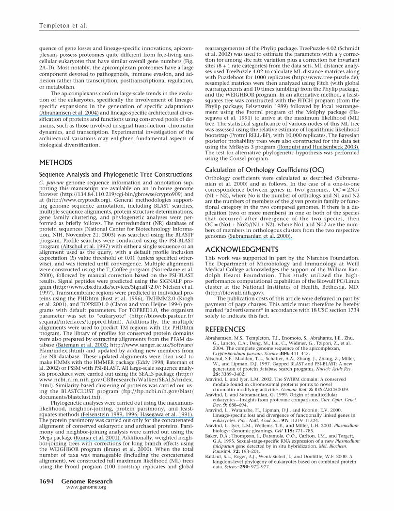
Although there is a much higher correspondence betweenthe two apicomplexans in chromatin proteins than specific tran-scription factors, interesting differences are found in both abso-lute numbers and architectures of these proteins (Fig. 4). C. par-vum has 14 chromatin-remodeling SNF2/SWI2 ATPases, whereasPlasmodium has just 11. Comparisons of the apicomplexan Swi2/Snf2 ATPases with the other eukaryotes suggests that Plasmodiumhas lost the Rad26 and Swr1 orthologs, whereas Cryptosporidiumappears to have lost one of the Rad16/Rad5-like Ring finger-containing forms and the version fused to a C-terminal Endo-nuclease VII domain. Cryptosporidium possesses a version of theSwi2/Snf2 ATPase, with a unique architecture containing twoN-terminal chromo domains and one bromo domain. Likewise,Plasmodium possesses a unique predicted chromatin-associatedprotein, having an amine oxidase domain fused to a C-terminalPHD finger that is predicted to function as a novel enzyme thatmight modify histone amino groups or chromatin basic amines(Fig. 4). It may represent a case of an independently derivedchromatin-associated oxidase, parallel to the amine oxidasefused to the SWIRM domain that is seen in the crown groupeukaryotes (Aravind and Iyer 2002). The two apicomplexans alsoshare several chromatin proteins with domain architecturesunique from any of the crown group eukaryotes. Examples ofthese include an ISWI-related Swi2/Snf2 ATPase with five PHDfingers, a protein that combines bromo domains with N-terminalankyrin repeats, and four distinct SET domain methylases joinedto a variety of other protein- and nucleic-acid-interacting do-mains (Fig. 4). An exploration of apicomplexan nuclear proteinshaving novel architectures may shine light on epigenetic regula-tory mechanisms unique to this lineage.
The pellicle in several protozoans is supported by a distinc-tive fibrous cytoskeletal structure predominantly composed oflow complexity, proline- and valine-rich proteins called articu-lins (Mann and Beckers 2001). We identified 10 distinct articu-lins in Plasmodium and six in Cryptosporidium having composi-tion and sequence similarity to the articulins from other alveo-lates such as the ciliate, Pseudomicrothorax. This suggests the
Templeton et al.
1692 Genome Researchwww.genome.org

retention of this ancient cytoskeletal feature of the alveolateclade despite the dramatic parasitic adaptations of the apicom-plexans. Our current analysis also identifies as an articulin thePlasmodium gametocyte-expressed protein, Pfs77 (Baker et al.1995), suggesting involvement of articulins in the maintenanceof stage-specific cellular shapes.
ConclusionsComparison of the Plasmodium and Cryptosporidium complete ge-nome sequences reveals that the ancestral apicomplexan en-coded at least 145 shared “apicomplexan” proteins with no ob-vious orthologs in other organisms (see Supplemental data 2).This apicomplexan set includes ∼30 membrane proteins and fivesecreted proteins. These surface proteins, unique to the apicom-plexan lineage, possibly participate in the formation of surfacestructures related to interactions with eukaryotic host cells andthe biogenesis of the apical complex. The unique intracellularapicomplexan proteins, which are typically enriched in low-complexity segments, are also likely to be internal structuralcomponents of lineage-specific organelles such as dense gran-ules, micronemes, rhoptries, and the apical complex. In contrastto other eukaryotic parasites such as the microsporidians, kineto-
plastids, and Giardia, the apicomplexans show a wide array ofsurface proteins with domains that are typically prevalent in ani-mal surface proteins. At least five multidomain proteins (Fig. 3A,lower panel) can be traced back to the common ancestor of thetwo apicomplexan lineages, suggesting that the ancestor had al-ready acquired a set of domains from a host belonging to theanimal lineage.
However, beyond the core set of genes, the evolution ofparasitism has involved lineage-specific adaptation occurringthrough gene loss, additional lateral transfers, and lineage-specific expansions. Considerable lineage-specific gene loss is in-dicated by the absence of widespread eukaryotic proteins specifi-cally in either one of the apicomplexan lineages (Fig. 4), suggest-ing that the common ancestral apicomplexan possessed a morecomplex genome encoding a greater repertoire of biochemicalactivities. The streamlining appears to correlate with increasingpropensity of the parasite to use its host(s) for most of its meta-bolic requirements. Some of the losses are not easily explained:for example, in Cryptosporidium, the apparent elimination ofSkp1p and Skp2p-like proteins, despite the presence of cullins,suggests potential differences in the cell cycle related ubiquitina-tion complexes relative to other eukaryotes. Thus, as a conse-
Figure 4 Eukaryotic tree showing select points of derivation and loss of various architectures. The proteins are designated by either Plasmodium orCryptosporidium gene names shown below a cartoon of their architecture. Gray boxes indicate globular domains that are not detected elsewhere. Yellowboxes indicate transmembrane segments or signal peptides. The domains found in chromatin proteins are (Ch) chromo domain; (Br) bromo domain;(PHD) PHD finger; (SET) SET protein methyltransferase domain; (CCC) cysteine cluster associated with SET domains; (AT) AT hook domain; (HTH)helix–turn–helix domain; (MYB) MYB-type HTH domain; (TFIIB) TFIIB-like HTH domain; (OB) oligomer-binding domain; (SWI2/SNF2) ATPase moduleof chromatin-remodeling proteins; (HAS) domain found in SWI2/SNF2 ATPases. The signaling domains are MYND and MIZ-Zn-finger domains; (R) ringfinger domain; (ANK) ankyrin domain; (WD) WD40 �-propeller domain; (Kinase) protein kinase domain; (SAM) sterile �-motif domain; (Sec7) ARFGTPase exchange factor domain; (TBC) GTPase-activating domain; (MORN) a �-hairpin repeat motif; (POZ) pox virus zinc finger domain; (MATH)meprin-A5-TRAF homology domain; (EF) EF-hand domain. RNA-binding domains are (G-patch) glycine-containing RNA-binding domain; (SWAP)suppressor of white apricot domain; (RRM) RNA recognition motif domain. Other domains are (YbeY) predicted metal-dependent lecithinase domain;(ACP) acyl carrier protein domain. cgd8_2430 is a predicted MAP kinase, cgd5_4390 kinase shows a lineage-specific expansion in Plasmodium, andcgd3_2010 is predicted to be a novel signaling receptor with intracellular calcium-binding EF-hand domains.
Comparison of Apicomplexa
Genome Research 1693www.genome.org
quence of gene losses and lineage-specific innovations, apicom-plexans possess proteomes quite different from free-living uni-cellular eukaryotes that have similar overall gene numbers (Fig.2A–D). Most notably, the apicomplexan proteomes have a largecomponent devoted to pathogenesis, immune evasion, and ad-hesion rather than transcription, posttranscriptional regulation,or metabolism.
The apicomplexans confirm large-scale trends in the evolu-tion of the eukaryotes, specifically the involvement of lineage-specific expansions in the generation of specific adaptations(Abrahamsen et al. 2004) and lineage-specific architectural diver-sification of proteins and functions using conserved pools of do-mains, such as those involved in signal transduction, chromatindynamics, and transcription. Experimental investigation of thearchitectural variations may enlighten fundamental aspects ofbiological diversification.
METHODS
Sequence Analysis and Phylogenetic Tree ConstructionsC. parvum genome sequence information and annotation sup-porting this manuscript are available on an in-house genomebrowser (http://134.84.110.219/cgi-bin/gbrowse/crypto909) andat (http://www.cryptodb.org). General methodologies support-ing genome sequence annotation, including BLAST searches,multiple sequence alignments, protein structure determinations,gene family clustering, and phylogenetic analyses were per-formed as briefly follows. The nonredundant (NR) database ofprotein sequences (National Center for Biotechnology Informa-tion, NIH, November 21, 2003) was searched using the BLASTPprogram. Profile searches were conducted using the PSI-BLASTprogram (Altschul et al. 1997) with either a single sequence or analignment used as the query, with a default profile inclusionexpectation (E) value threshold of 0.01 (unless specified other-wise), and was iterated until convergence. Multiple alignmentswere constructed using the T_Coffee program (Notredame et al.2000), followed by manual correction based on the PSI-BLASTresults. Signal peptides were predicted using the SIGNALP pro-gram (http://www.cbs.dtu.dk/services/SignalP-2.0/; Nielsen et al.1997). Transmembrane regions were predicted in individual pro-teins using the PHDhtm (Rost et al. 1996), TMHMM2.0 (Kroghet al. 2001), and TOPRED1.0 (Claros and von Heijne 1994) pro-grams with default parameters. For TOPRED1.0, the organismparameter was set to “eukaryote” (http://bioweb.pasteur.fr/seqanal/interfaces/toppred.html). Additionally, the multiplealignments were used to predict TM regions with the PHDhtmprogram. The library of profiles for conserved protein domainswere also prepared by extracting alignments from the PFAM da-tabase (Bateman et al. 2002; http://www.sanger.ac.uk/Software/Pfam/index.shtml) and updated by adding new members fromthe NR database. These updated alignments were then used tomake HMMs with the HMMER package (Eddy 1998; Bateman etal. 2002) or PSSM with PSI-BLAST. All large-scale sequence analy-sis procedures were carried out using the SEALS package (http://www.ncbi.nlm.nih.gov/CBBresearch/Walker/SEALS/index.html). Similarity-based clustering of proteins was carried out us-ing the BLASTCLUST program (ftp://ftp.ncbi.nih.gov/blast/documents/blastclust.txt).
Phylogenetic analyses were carried out using the maximum-likelihood, neighbor-joining, protein parsimony, and least-squares methods (Felsenstein 1989, 1996; Hasegawa et al. 1991).The protein parsimony was carried out only for the concatenatedalignment of conserved eukaryotic and archaeal proteins. Parsi-mony and neighbor-joining analysis were carried out using theMega package (Kumar et al. 2001). Additionally, weighted neigh-bor-joining trees with corrections for long branch effects usingthe WEIGHBOR program (Bruno et al. 2000). When the totalnumber of taxa was manageable (including the concatenatedalignment), we constructed full maximum likelihood (ML) treesusing the Proml program (100 bootstrap replicates and global
rearrangements) of the Phylip package. TreePuzzle 4.02 (Schmidtet al. 2002) was used to estimate the parameters with a � correc-tion for among site rate variation plus a correction for invariantsites (8 + 1 rate categories) from the data sets. ML distance analy-ses used TreePuzzle 4.02 to calculate ML distance matrices alongwith Puzzleboot for 1000 replicates (http://www.tree-puzzle.de);resampled matrices were then analyzed using Fitch (with globalrearrangements and 10 times jumbling) from the Phylip package,and the WEIGHBOR program. In an alternative method, a least-squares tree was constructed with the FITCH program (from thePhylip package; Felsenstein 1989) followed by local rearrange-ment using the Protml program of the Molphy package (Ha-segawa et al. 1991) to arrive at the maximum likelihood (ML)tree. The statistical significance of various nodes of this ML treewas assessed using the relative estimate of logarithmic likelihoodbootstrap (Protml RELL-BP), with 10,000 replicates. The Bayesianposterior probability trees were also constructed for the data setusing the MrBayes 3 program (Ronquist and Huelsenbeck 2003).The test for alternative phylogenetic hypothesis was performedusing the Consel program.
Calculation of Orthology Coefficients (OC)Orthology coefficients were calculated as described (Subrama-nian et al. 2000) and as follows. In the case of a one-to-onecorrespondence between genes in two genomes, OC = 2No/(N1 + N2), where No is the number of orthologs and N1 and N2are the numbers of members of the given protein family or func-tional category in the two compared genomes. If there is a du-plication (two or more members) in one or both of the speciesthat occurred after divergence of the two species, thenOC = (No1 + No2)/(N1 + N2), where No1 and No2 are the num-bers of members in orthologous clusters from the two respectivegenomes (Subramanian et al. 2000).
ACKNOWLEDGMENTSThis work was supported in part by the Niarchos Foundation.The Department of Microbiology and Immunology at WeillMedical College acknowledges the support of the William Ran-dolph Hearst Foundation. This study utilized the high-performance computational capabilities of the Biowulf PC/Linuxcluster at the National Institutes of Health, Bethesda, MD.(http://biowulf.nih.gov).
The publication costs of this article were defrayed in part bypayment of page charges. This article must therefore be herebymarked “advertisement” in accordance with 18 USC section 1734solely to indicate this fact.
REFERENCESAbrahamsen, M.S., Templeton, T.J., Enomoto, S., Abrahante, J.E., Zhu,
G., Lancto, C.A., Deng, M., Liu, C., Widmer, G., Tzipori, Z., et al.2004. The complete genome sequence of the apicomplexan,Cryptosporidium parvum. Science 304: 441–445.
Altschul, S.F., Madden, T.L., Schaffer, A.A., Zhang, J., Zhang, Z., Miller,W., and Lipman, D.J. 1997. Gapped BLAST and PSI-BLAST: A newgeneration of protein database search programs. Nucleic Acids Res.25: 3389–3402.
Aravind, L. and Iyer, L.M. 2002. The SWIRM domain: A conservedmodule found in chromosomal proteins points to novelchromatin-modifying activities. Genome Biol. 3: RESEARCH0039.
Aravind, L. and Subramanian, G. 1999. Origin of multicellulareukaryotes—Insights from proteome comparisons. Curr. Opin. Genet.Dev. 9: 688–694.
Aravind, L., Watanabe, H., Lipman, D.J., and Koonin, E.V. 2000.Lineage-specific loss and divergence of functionally linked genes ineukaryotes. Proc. Natl. Acad. Sci. 97: 11319–11324.
Aravind, L., Iyer, L.M., Wellems, T.E., and Miller, L.H. 2003. Plasmodiumbiology: Genomic gleanings. Cell 115: 771–785.
Baker, D.A., Thompson, J., Daramola, O.O., Carlton, J.M., and Targett,G.A. 1995. Sexual-stage-specific RNA expression of a new Plasmodiumfalciparum gene detected by in situ hybridization. Mol. Biochem.Parasitol. 72: 193–201.
Baldauf, S.L., Roger, A.J., Wenk-Siefert, I., and Doolittle, W.F. 2000. Akingdom-level phylogeny of eukaryotes based on combined proteindata. Science 290: 972–977.
Templeton et al.
1694 Genome Researchwww.genome.org

Baruch, D.I., Pasloske, B.L., Singh, H.B., Bi, X., Ma, X.C., Feldman, M.,Taraschi, T.F., and Howard, R.J. 1995. Cloning the P. falciparum geneencoding PfEMP1, a malarial antigen and adherence receptor on thesurface of parasitized human erythrocytes. Cell 82: 77–87.
Bateman, A., Birney, E., Cerruti, L., Durbin, R., Etwiller, L., Eddy, S.R.,Griffiths-Jones, S., Howe, K.L., Marshall, M., and Sonnhammer, E.L.2002. The Pfam protein families database. Nucleic Acids Res. 30:2762–2800.
Bozdech, Z., Llinas, M., Pulliam, B.L., Wong, E.D., Zhu, J., and DeRisi,J.L. 2003. The transcriptome of the intraerythrocytic developmentalcycle of Plasmodium falciparum. PLoS Biol. 1: E5.
Bruno, W.J., Socci, N.D., and Halpern, A.L. 2000. Weighted neighborjoining: A likelihood-based approach to distance-based phylogenyreconstruction. Mol. Biol. Evol. 17: 189–197.
Carreno, R.A., Martin, D.S., and Barta, J.R. 1999. Cryptosporidium is moreclosely related to the gregarines than to coccidia as shown byphylogenetic analysis of Apicomplexan parasites inferred using small-subunit ribosomal RNA gene sequences. Parasitol. Res. 85: 899–904.
Cheng, Q., Cloonan, N., Fischer, K., Thompson, J., Waine, G., Lanzer,M., and Saul, A. 1998. stevor and rif are Plasmodium falciparummulticopy gene families which potentially encode variant antigens.Mol. Biochem. Parasitol. 97: 161–176.
Chervitz, S.A., Aravind, L., Sherlock, G., Ball, C.A., Koonin, E.V.,Dwight, S.S., Harris, M.A., Dolinski, K., Mohr, S., Smith, T., et al.1998. Comparison of the complete protein sets of worm and yeast:Orthology and divergence. Science 282: 2022–2028.
Claros, M.G. and von Heijne, G. 1994. TopPred II: An improvedsoftware for membrane protein structure predictions. Comput. Appl.Biosci. 10: 685–686.
Deitsch, K., Driskill, C., and Wellems, T. 2001. Transformation ofmalaria parasites by the spontaneous uptake and expression of DNAfrom human erythrocytes. Nucleic Acids Res. 29: 850–853.
Eddy, S.R. 1998. Profile hidden Markov models. Bioinformatics 14: 755–763.Felsenstein, J. 1989. PHYLIP—Phylogeny inference package (Version
3.2). Cladistics 5: 164–166.———. 1996. Inferring phylogenies from protein sequences by
parsimony, distance, and likelihood methods. Methods Enzymol.266: 418–427.
Gardner, M.J., Tettlin, H., Carucci, D.J., Cummings, L.M., Aravind, L.,Koonin, E.V., Shallom, S., Mason, T., Yu, K., Fujii, C., et al. 1998.Chromosome 2 sequence of the human malaria parasite Plasmodiumfalciparum. Science 282: 1126–1132.
Gardner, M.J., Hall, N., Fung, E., White, O., Berriman, M., Hyman, R.W.,Carlton, J.M., Pain, A., Nelson, K.E., Bowman, S., et al. 2002.Genome sequence of the human malaria parasite Plasmodiumfalciparum. Nature 419: 498–511.
Hall, T.M., Porter, J.A., Young, K.E., Koonin, E.V., Beachy, P.A., andLeahy, D.J. 1997. Crystal structure of a Hedgehog autoprocessingdomain: Homology between Hedgehog and self-splicing proteins.Cell 91: 85–97.
Hasegawa, M., Kishino, H., and Saitou, N. 1991. On the maximum likeli-hood method in molecular phylogenetics. J. Mol. Evol. 32: 443–445.
Krogh, A., Larsson, B., von Heijne, G., and Sonnhammer, E. L. 2001.Predicting transmembrane protein topology with a hidden Markovmodel: Application to complete genomes. J. Mol. Biol. 305: 567–580.
Kumar, S., Tamura, K., Jakobsen, I.B., and Nei, M. 2001. MEGA2:Molecular evolutionary genetics analysis software. Bioinformatics 17:1244–1245.
Lander, E.S., Linton, L.M., Birren, B., Nusbaum, C., Zody, M.C.,Baldwin, J., Devon, K., Dewar, K., Doyle, M., Fitzhugh, W., et al.2001. Initial sequencing and analysis of the human genome. Nature409: 860–921.
Laybourn-Parry, J. 1984. A functional biology of the free-living protozoa.University of California Press, Berkeley, CA.
Le Roch, K.G., Zhou, Y., Blair, P.L., Grainger, M., Moch, J.K., Haynes,J.D., De La Vega, P., Holder, A.A., Batalov, S., Carucci, D.J., et al.2003. Discovery of gene function by expression profiling of themalaria parasite life cycle. Science 301: 1503–1508.
Lespinet, O., Wolf, Y.I., Koonin, E.V., and Aravind, L. 2002. The role oflineage-specific gene family expansion in the evolution ofeukaryotes. Genome Res. 12: 1048–1059.
Mann, T. and Beckers, C. 2001. Characterization of the subpellicularnetwork, a filamentous membrane skeletal component in theparasite Toxoplasma gondii. Mol. Biochem. Parasitol. 115: 257–268.
Matussek, K., Moritz, P., Brunner, N., Eckerskorn, C., and Hensel, R.1998. Cloning, sequencing, and expression of the gene encodingcyclic 2,3-diphosphoglycerate synthetase, the key enzyme of cyclic2,3-diphosphoglycerate metabolism in Methanothermus fervidus. J.Bacteriol. 180: 5997–6004.
Nielsen, H., Engelbrecht, J., Brunak, S., and von Heijne, G. 1997.Identification of prokaryotic and eukaryotic signal peptides andprediction of their cleavage sites. Protein Eng. 10: 1–6.
Notredame, C., Higgins, D.G., and Heringa, J. 2000. T-Coffee: A novelmethod for fast and accurate multiple sequence alignment. J. Mol.Biol. 302: 205–217.
Odenthal-Schnittler, M., Tomavo, S., Becker, D., Dubremetz, J.F., andSchwarz, R.T. 1993. Evidence of N-linked glycosylation inToxoplasma gondii. Biochem. J. 291: 713–721.
Peterson, D.S., Miller, L.H., and Wellems, T.E. 1995. Isolation ofmultiple sequences from the Plasmodium falciparum genome thatencode conserved domains homologous to those inerythrocyte-binding proteins. Proc. Natl. Acad. Sci. 92: 7100–7104.
Pollack, Y., Kogan, N., and Golenser, J. 1991. Plasmodium falciparum:Evidence for a DNA methylation pattern. Exp. Parasitol. 72: 339–344.
Ponting, C.P., Aravind, L., Schultz, J., Bork, P., and Koonin, E.V. 1999.Eukaryotic signalling domain homologues in archaea and bacteria.Ancient ancestry and horizontal gene transfer. J. Mol. Biol. 289:729–745.
Pradel, G., Hayton, K., Aravind, L., Iyer, L.M., Abrahamsen, M.S.,Bonawitz, A., Mejia, C., and Templeton, T.J. 2004. A multidomainadhesion protein family expressed in Plasmodium falciparum isessential for transmission to the mosquito. J. Exp. Med.199: 1533–1544.
Reeves, P.R., Hobbs, M., Valvano, M.A., Skurnik, M., Whitfield, C.,Coplin, D., Kido, N., Klena, J., Maskell, D., Raetz, C.R., et al. 1996.Bacterial polysaccharide synthesis and gene nomenclature. TrendsMicrobiol. 4: 495–503.
Ritte, G., Lloyd, J.R., Eckermann, N., Rottmann, A., Kossmann, J., andSteup, M. 2002. The starch-related R1 protein is an �-glucan, waterdikinase. Proc. Natl. Acad. Sci. 99: 7166–7171.
Ronquist, F. and Huelsenbeck, J.P. 2003. MrBayes 3: Bayesian phylogeneticinference under mixed models. Bioinformatics 19: 1572–1574.
Rost, B., Fariselli, P., and Casadio, R. 1996. Topology prediction forhelical transmembrane proteins at 86% accuracy. Protein Sci. 5:1704–1718.
Schmidt, H.A., Strimmer, K., Vingron, M., and von Haeseler, A. 2002.TREE-PUZZLE: Maximum likelihood phylogenetic analysis usingquartets and parallel computing. Bioinformatics 18: 502–504.
Smith, J.D., Chitnis, C.E., Craig, A.G., Roberts, D.J., Hudson-Taylor,D.E., Peterson, D.S., Pinches, R., Newbold, C.I., and Miller, L.H.1995. Switches in expression of Plasmodium falciparum var genescorrelate with changes in antigenic and cytoadherent phenotypes ofinfected erythrocytes. Cell 82: 101–110.
Su, X.Z., Heatwole, V.M., Wertheimer, S.P., Guinet, F., Herrfeldt, J.A.,Peterson, D.S., Ravetch, J.A., and Wellems, T.E. 1995. The largediverse gene family var encodes proteins involved in cytoadherenceand antigenic variation of Plasmodium falciparum-infectederythrocytes. Cell 82: 89–100.
Subramanian, G., Koonin, E.V., and Aravind, L. 2000. Comparativegenome analysis of the pathogenic spirochetes Borrelia burgdorferiand Treponema pallidum. Infect. Immun. 68: 1633–1648.
Tang, L.Y., Reddy, M.N., Rasheva, V., Lee, T.L., Lin, M.J., Hung, M.S.,and Shen, C.K. 2003. The eukaryotic DNMT2 genes encode a newclass of cytosine-5 DNA methyltransferases. J. Biol. Chem. 278:33613–33616.
Templeton, T.J., Lancto, C.A., Vigdorovich, V., Liu, C., London, N.R.,Hadsell, K.Z., and Abrahamsen, M.S. 2004. The Cryptosporidiumoocyst wall protein is a member of a multigene family and has ahomolog in Toxoplasma. Infect. Immun. 72: 980–987.
Varki, A., Cummings, R., Esko, J., Freeze, H., Hart, G., and Marth, J.1999. Essentials of glycobiology. Cold Spring Harbor Laboratory Press,Cold Spring Harbor, NY.
Zhu, G., Keithly, J.S., and Philippe, H. 2000. What is the phylogeneticposition of Cryptosporidium? Int. J. Syst. Evol. Microbiol.50: 1673–1681.
WEB SITE REFERENCESftp://ftp.ncbi.nih.gov/blast/documents/blastclust.txt; BLASTCLUST.http://134.84.110.219/cgi-bin/gbrowse/crypto909; C. parvum genome
sequence information and annotation.http://bioweb.pasteur.fr/seqanal/interfaces/toppred.html; TOPRED1.0.http://www.cbs.dtu.dk/services/SignalP-2.0/; SIGNALP.http://www.cryptodb.org; C. parvum genome sequence information and
annotation.http://www.ncbi.nlm.nih.gov/CBBresearch/Walker/SEALS/index.html;
SEALS.http://www.sanger.ac.uk/Software/Pfam/index.shtml; PFAM database.http://www.tree-puzzle.de; TreePuzzle 4.02.http://biowulf.nih.gov; Biowulf processing system at the National
Institutes of Health.
Received March 23, 2004; accepted in revised form June 14, 2004.
Comparison of Apicomplexa
Genome Research 1695www.genome.org
Related Documents