Welcome message from author
This document is posted to help you gain knowledge. Please leave a comment to let me know what you think about it! Share it to your friends and learn new things together.
Transcript
�

Biological diversity (L. diversitas—variety) is the occurrence of varied types of life differing inexternal appearance, size, colour pattern, internal structure, mode of living, habitats and nutrition.Currently 1·7—1·8 million species of animals and plants have been given scientific names, 1·25 millionanimals and 0·55 million plants. Insects constitute the single largest group of animals with about 1·025million species. Scientists believe that the total number of species of living beings may be anywherebetween 5—30 million because every year about 15,000 new species are discovered while a large area oftropical rain forests and under water reef formations remains unexplored. Both are rich in variety oforganisms.
Present day living beings are ‘‘islands in the sea of death.’’ During the past more than 3·5 billionyears a very large number of organisms came into existence, fluorished and then faded away. Accordingto one estimate, the extinct species outnumber existing species by 50—100 times.
Living forms range from microscopic creatures of less than 0·15 μm to large animals (30 m inWhale) and trees (114 m in Eucalyptus). They have a variety of habits (e.g., vine, herb, shrub, tree,free living, parasite or symbiont) and habitats like terrestrial and aquatic, plankton (free floating) nekton(active swimmers), benthon (bottom dwellers), epiphytes arboreal, cave dwellers, fussorial or burrowing,aerial or volant and scansorial or wall climbers.
Nutritionally, living beings are distinguishable into autotrophs and heterotrophs. Autotrophs areable to manufacture their own food from inorganic raw materials. They are of two types, photoautotrophsand chemoautotrophs. Heterotrophs obtain organic food from outside. They include parasites, saprotrophs,detrivores and predators. Intake of solid food particles is called ingestive or holozoic nutrition. Incontrast, plant nutrition is called holophytic nutrition where inorganic nutrients are taken from outsidemedium as solution. Saprotrophic and parasitic nutrition are together called absorptive nutrition.
Diversity of living beings is not only present in their form, size, habit, habitat and nutrition but alsostructures build by them, e.g., shells, cocoons, webs, nests, hives, etc.
Diversity develops due to organic evolution and development of adaptations in order to overcomecompetition. Competition occurs because resources are limited while living beings have a high reproductivepotential. Living beings require energy and matter for their body building and survival. Energy entersthe biosphere as solar energy. It is trapped by photoautotrophs. Photoautotrophs absorb inorganic matterfrom their external medium and convert the same into organic matter in the process of photosynthesis.During photosynthesis trapped solar enregy is changed into chemical energy of organic matter.Photoautotrophs are also called producers as they produce organic matter not only for themselves butalso for other living beings. The latter are of two types, consumers and decomposers. There is acompetition for inorganic matter at the level of photoautotrophs and organic matter at the level ofconsumers. Decomposers feed on organic remains and release the inorganic matter. Adaptations developin organisms for obtaining optimum resources in various types of environments and habitats. It producesdiversity.
In order to study such a large number of organisms, it is important to evolve a common system ofnomenclature and classification. There are certain names by which common man recognises plants andanimals like roses, oaks, teaks, cats, dogs, cattle, etc. However, many names are misleading. Severalorganisms have no common names. Further, common names differ from language to language and placeto place. As a result, a universal scientific nomenclature had to be evolved. Further, all organismscannot be studied individually. It is better to classify them into ordered and ranked groups on the basisof their similarities and differences. Fruitful attempts on both classification and nomenclature begansome 300 years ago. A number of comprehensive works have appeared since then. Better understandingof living beings will result in better system of classification. Likewise, a good system of classificationhelps us to understand diversity better.
3
1.1. WHAT IS LIVING
A dictionary will define life to be property that separates living beings from non-living objects. Itleaves the meaning of life unclear even to a common man. However, even a common man can recognisea living organism (e.g., plants, animals, bacteria, fungi) and differentiate it from non-living (e.g., stone,rock, brick, nail). Problem arises when we go deeper into this differentiation. Viruses are neither plantsnor animals, neither a living being, nor a non-living one. It is lifeless like a crystal outside the body of aliving being. However, inside a living cell, it becomes active, takes over the living machinery of the celland multiplies to form a large number of viruses. Therefore, scientists prefer to define life with anumber of its basic characteristics. Life is unique, complex cellular organisation of molecules and thecells themselves that shows various types of chemical reactions which lead to availability of energy,growth, development, responsiveness, adaptation and reproduction. This definition, though nearest totruth, does not give either a simple or a composite property of life. Hence, it is better to recognise lifeby a few unified and basic characteristics of living beings which are as follows :
Characteristics of Living Beings
1. Individuality. Each living being has a distinct individuality. It cannot be broken into two or moreindependent parts.
2. Definite Shape and Size. Every living being has a definite shape and size by which we canrecognise a Peepal tree from Mango tree or Cow from Buffalo. Non-living things are often shapeless oramorphous as compared to morphous (with shape) nature of living beings.
3. Organisation. Living beings have an internal hierarchy of parts where smaller parts cooperate toform larger one, the larger ones a still larger part while all the still larger parts are organised to form theindividual. At each level, organisation provides certain characteristics not found in its components.Biomolecules are organised to form a cell organelle that has its own characteristics. Cell organelles aresimilarly organised to form cell that has its specific properties. Cells are organised into tissues andtissues into organs and so on. Cell Organelles ��� Cells ��� Tissues ��� Organs ���Organs Systems ��� Individual. Because of this organisation, living beings are also called organisms.
4. Homeostasis (=Homoeostasis). It is the ability to maintain a favourable internal environmentdespite changes in the external environment. Living beings have developed an internal environment thatfavours optimum functioning. The internal environment is under control of a self regulated system. Itbrings about changes in the internal environment that bring about adjustments to variations in externalenvironment.
5. Protoplasm (Gk. protos—first, plasma—form). It is semitransparent jelly-like viscous semifluidcomplex organic material present in all living components of organisms. All body functions andproperties of life are actually due to protoplasm, viz., irritability, nutrition, growth, respiration, excretion,reproduction. Protoplasm is also called living matter.
4
����������� ��
������ ��
����������� �� 5
• Huxley (1868). Protoplasm is the physical basis of life.
• Lamarck (1809). No body can have life if its constituent parts are not cellular.
6. Cellular Structure. The body of a living being is made of one or more cells. Rather, cells areconsidered to be structural and functional units of organisms. Cellular organisation of the body is thedefining feature of all life forms. A cell is a mass of protoplasm covered by cell membrane and inplants an additional cell wall. Organisms are called acellular or unicellular if their body cannot bedivided into two or more cells, e.g., protista, many monerans. Metazoans and metaphytes are multicellularformed of innumerable cells, e.g., 100 trillion in adult human body (Guyton and Hall, 1996).
7. Genetic Material. All organisms possess genetic or hereditary material in the form of DNA(genes and chromosomes). It is concerned with expression of traits in the individuals, transmission oftraits from parents to offspring and to maintain continuity from one generation to the next.
8. Variations. Genetic material undergoes mutations and reshuffling of genes. This causes variations.Variations are so abundant that no two individuals of the same race are exactly similar.
9. Struggle for Existence. There is competition amongst individuals of a species, amongst individualsof different species and between individuals and their environment. The struggle or competition is forfood, shelter, reproduction and survival.
10. Adaptations. They are variations which help organisms to modify themselves according tochanges in environment and specific requirement of their surroundings. For example, birds havepneumatic bones and wings for flight while aquatic animals have stream-lined body to reduce friction.
11. Evolution. It is formation of new species from the pre-existing ones. Evolution occurs due toaccumulation of variations.
12. Reproduction. Life comes from pre-existing life. Young-ones grow and become mature. Theydevelop the faculty to produce young-ones of their own type. Some organisms reproduce asexuallythrough fission, buds and spores. Others produce gametes or sex cells which fuse to form the young-ones. However, reproduction is not a must for all living beings. Infertile couples live but do notreproduce. Mule is sterile. Worker honey bees seldom reproduce.
13. Growth. Young individuals grow in size. The growth is due to internal addition of protoplasmicmaterials by which cells enlarge and divide. The phenomenon is called intussusception (internalgrowth). This growth is seen in the development of a large sized tree from a small-sized seed or an adulthuman from a small baby. Non-living object may also increase in size but it occurs due to deposition ofsimilar material over the surface. The process is called accretion.
Growth occurs when anabolism exceeds catabolism. Two types of substances are formed forgrowth. They are protoplasmic and apoplasmic substances. Protoplasmic substances are constituents ofliving matter. They bring about increase in bulk of protoplasm. Apoplasmic substances are non-livingsubstances, e.g., cell wall, matrix and fibres of connective tissue. Formation of the two types ofsubstances is followed by cell division and cell enlargement.
14. Death. After a period of time each living being dies. Death occurs naturally due to wear and tear.
15. Life Cycle. Each individual passes through a definite life cycle of birth, growth, maturation,reproduction, ageing and death. The life cycle is completed in a definite life span ranging from one day (e.g.,May Fly) to several years (80—100 years in humans, 200 years in Tortoise, more than 2000 years in Peepal).
16. Self Regulation. A system of controls operates in each living being. It helps in regulation ofbody functioning including metabolism, excretion, growth, development and reproduction.
��������Companion Biology–XI6
17. Movements. It is of two types, visible and invisible. Invisible movements occur at the molecularlevel and are found in all living organisms. Visible movement can be of parts or whole body calledlocomotion. Animals perform both the types of movements while plants show only movement of parts(e.g., opening and closing of stomata, coiling of a tendril). Some movements occur due to purelyinternal forces. They are called autonomic (= spontaneous) movements. Others occur in response to anexternal stimulus. They are known as paratonic (= induced) movements.
18. Coordination. Different parts of the body of a living being cooperate with one another for thefunctioning of the whole. For example, heart pumps blood to all parts of the body, lungs provideoxygen, digestive system—nutrients, nerves and hormones—information, muscles—movements, bones—support, etc.
19. Metabolism. A large number of chemical reactions are going on in the body of a living being.They can also be carried out in vitro or cell free system. Therefore, the metabolic reactions are alsocalled biological reactions. The sum of all the chemical reactions occurring in an organism is calledmetabolism (Gk. metabole—change). Metabolism has two main types, catabolism and anabolism.Catabolism (= katabolism, Gk. kata—down, bole—throw) is destructive metabolism. It involvesbreakdown of complex organic materials into simpler ones, e.g., respiration. Anabolism (Gk. ana—up,bole—throw) is constructive metabolism which involves building up of complex organic substancesfrom simpler ones, e.g., photosynthesis, assimilation. Metabolism is a defining property of livingbeings.
20. Energy. It is essential for carrying out various life activities not only by the individual but alsoby each and every cell. Energy is obtained from food which is either manufactured by the cells or gotfrom outside.
21. Water Intake. Water is essential for maintaining optimum metabolic state of protoplasm andinternal circulation of materials. All living beings require water. The same is taken from outside eitherdirectly or as part of food.
22. Excretion. Metabolism produces a number of by-products which are useless to the body. Thesame are either expelled out of the body (animals) or stored inside ageing tissues (in plants).
23. Consciousness and Irritability. Consciousness is the awareness of one's environment, actionsand intentions. It is present only in living organisms. Therefore, consciousness is the defining propertyof living organisms. It enables an organism to handle substances entering the body and respond tononliving and living entities present in the medium. Human beings additionally possess self consciousnessor awareness of self. Higher animals have sense organs for scanning the external environment. Otherorganisms do so without the presence of sense organs. For example, both plants and animals can sensephotoperiods for their breeding though plants do not have sense organs.
Irritability or sensitivity is the faculty of responding to a stimulus. It is the property of protoplasmand is shown by all organisms, both plants and animals. Animals show quick response to a stimulusbecause they additionally possess nervous system, e.g., pain on being pricked, running away from thesight of predator. Plants show slow response, e.g., bending of shoot towards light. However, SensitivePlant (Mimosa pudica, vern. Lajwanti, Chhui-Mui) shows almost immediate response on being touchedby folding and bending of its leaves. However, consciousness and irritability cannot distinguish a livingbeing from a nonliving being. A person in coma fails to respond to any stimulus. Even then the personcannot be called dead or non-living.
24. Healing. Small wounds heal automatically due to the body defence system present in allorganisms.
25. Regeneration (L. re—again, generare—to produce). Regeneration is the ability to form lostparts. A small fragment can regenerate the whole organism in plants and primitive animals (e.g., Hydra,Planaria, Earthworm). It is limited to certain organs in higher animals.
����������� �� 7
• Trembley (1742). First to study regeneration (in Hydra).
• T.H. Morgan. Studied the mechanism of animal regeneration.
• Cybernetics. Science of communication, coordination and control.
1.2. DIVERSITY IN THE LIVING WORLD
Earth has 1.7–1.8 million known organisms. Each organism represents a distinct species. All thespecies differ from one another. Variability present among organisms, their individuals, communities andecosystems is called biodiversity. It is not fixed. New organisms are being continuously discovered. Sucha large number of organisms cannot be studied without distinct names and a proper system of classification.
1.2.1. Nomenclature
Nomenclature (L. nomen—name, calare—to call) is making and giving proper, specific and distinguishingname to each and every organism. It is of two types, common names and scientific names. The mode ofgiving scientific names is binomial nomenclature as each of them consists of two words.
Common or Vernacular Names
They are local names which are given to organisms in a particular region and language by localpersons. Group names are also given on the basis of similarities among the organisms, e.g., dog, cattle,tree, shrub, vine. However, each region, tribe and language has its own specific names for organismsknown in the area so that they can be properly identified. For example, Rose in English is Gulab inHindi, Golap in Bangla and Rojapo in Tamil. Similarly, Butterfly in English is Titli in Hindi, Prajapatiin Bangla and Vannathu Poochi in Tamil. Because common names differ from region to region andlanguage to language they cannot be used by biologists.
Scientific Names
They are specific and distinguishing names assigned to organisms by the scientists so as to removeambiguity and make the names universally understood by other biologists. Scientific names must be basedon (i) agreed principles and criteria (ii) acceptable all over the world (iii) should help the scientists to arriveat the same name in any part of the world (iv) should be distinct or different for each species (v) new andnot used earlier for some other organism. Scientific naming of organisms is called binomial nomenclature.
Binomial Nomenclature
It is a system of providing distinct and proper scientific names to organisms, each consisting of twowords, first generic and second specific. Though two-word names were first used by Cato (about 200B.C.), they were not based on any scientific system. Binomial nomenclature (L. bis—twice, nomen—name, calare—to call) for scientific naming of organisms was developed by Carolus Linnaeus in 1751(Philosophia Botanica, 1751). All valid names for animals under binomial nomenclature are the onesgiven by Linnaeus in the tenth edition of his book Systema Naturae published in 1758. All valid namesfor plants are the ones given by Linnaeus in his book Species Plantarum published in 1753.
The scientific or technical name of a species consists of two words in Latin, e.g., Mangifera indica(Mango), Homo sapiens (Human), Apis mellifera (Honey Bee). The first word is the generic name orgenus to which the species belongs. It is like a noun. Its first letter is always written in capital. Thesecond word is the specific epithet which identifies the species itself. It is like an adjective. Its first letteris small except occasionally when it represents a very important or sacred place or personality. Thename of the discoverer is appended at the end of two word name either in full or in abbreviation, e.g.,Mangifera indica Linn, Homo sapiens L. (Homo sapiens Linnaeus).
Name of the species cannot be written by using only the specific name. It has to follow the generic
��������Companion Biology–XI8
name, i.e., tigris alone is wrong but accompanied by Panthera (or Panthera tigris) is correct. The twoword names are similar to our name having a common name and a surname (e.g., Rita Gupta).However, the title or surname is reversed in case of scientific naming of organisms.
Trinomial Nomenclature. In certain cases the name of subspecies or variety is also written after thetwo-word name. Such an organism comes to have three word name or trinomial nomenclature, e.g.,Corvus splendens insolens (Burmese Crow), Ascaris lumbricoides humanis (Common Human Roundworm),Homo sapiens sapiens, Acacia nilotica indica.
Rules for Binomial Nomenclature
They were initially framed by Linnaeus. Standard references are Species Plantarum (1753) andSystema Naturae (1758). The rules were revised in nineteenth and twentieth centuries through InternationalCode of Botanical Nomenclature (ICBN), International Code of Zoological Nomenclature (ICZN),International Code of Bacteriological Nomenclature (ICBacN or ICBN), International Code of ViralNomenclature (ICVN) and International Code of Nomenclature for Cultivated Plants (ICNCP).
Important Rules
1. Each organism has a distinct scientific name having two words, generic and specific.
2. The generic name is written first. It is like a noun. Its first letter is always capital.
3. The specific word or epithet is written after the generic name.It starts with a smaller letter butexceptions can be made in case it represents a very important or sacred place or personality.
4. The names are derived from Latin language, e.g., Homo (L. homo—man), Ficus (L. ficus—fig).When words are used from other languages, they are latinised with suitable ending, e.g., Mangiferaindica. The gender of specific name is changed according to the gender of generic name, e.g.,Mangifera indica, Tamarindus indicus.
5. Both the words of scientific name are printed in italics to indicate their latin origin. They areunderlined separately in hand written name. Exceptions are made when the name is used as title of abook, chapter or para. The name of the discoverer is appended in roman script without any break orcomma, e.g., Homo sapiens Linn.
Other Rules
6. The generic and specific words should not have less than three letters or more than twelve letters.
7. The scientific name retains its original spellings. It is because the same is derived from Latinlanguage which is dead. The spellings are, therefore, not liable to change. However, misprints and otherobvious errors can be rectified.
8. When the name of a species is changed or revised, the name of original discoverer is retained inbrackets. The name of the new worker is appended after the brackets, e.g., Albizzia lebbeck (Linn.)Benth.
9. When an organism has been given different scientific names by different workers, the earlier onegiven validly (not prior to 1.5.1753 for plants or 1.8.1758 for animals) is recognised. This is called lawof priority.
10. Families and sub-families should be named after some prominent genus, e.g., Asteraceae(=Compositae, after Aster), Felidae (after Felis).
11. The name of categories higher than the rank of genus are not printed in italics. Bold letters can,however, be used. The first letter of the name is capital, e.g., Spermatophyta, Mammalia.
12. A new valid name is the one which is (i) Given according to binomial nomenclature. (ii) Typespecimen is placed in recognised herbarium or museum. (iii) Type specimen is described inLatin. (iv) Name, description and report of the discovery is published in a reputed scientific journal.(v) All the previous work connected with the nomenclature of the organism is mentioned.
����������� �� 9
• Type Specimen. 1. Holotype. Type specimen which is described in Latin and on which thenomenclature of the organism is based. Also called nomenclature type. 2. Isotype. Duplicate ofholotype, e.g., another branch of same tree as of holotype. 3. Paratype. A specimen describedalongwith holotype. 4. Syntype. Any of the specimens quoted by discoverer when there is noholotype. 5. Lectotype. Specimen taken from the original collection for nomenclature in the
absence of a holotype. 6. Neotype. New nomenclature type when the original material is lost.
Advantages of Binomial Nomenclature
1. The names are universally recognised. They remain the same in all the languages.
2. An organism has been given a single distinct and specific name of two (or occasionally three) words.
3. All the organisms known to science have been given scientific names irrespective of their size orimportance.
4. The names are not cumbersome. They are small and comprehensive.
5. There is a mechanism to provide a scientific name to every newly discovered organism.
6. Scientific names are often based on some characteristic of the organism.
7. They indicate relationship with other species present in the same genus.
8. There is no chance of change in spellings of a scientific name as the same has been obtained froma dead language like Latin.
9. An inappropriate or incorrect name can be easily corrected.
Classifying A Group of Species or Revision of Group
Revision of group is the delimitation of species, finding out the common characters amongst speciesand placing them successively into higher taxonomic categories on the basis of their resemblances andevolutionary relationships. It begins with first selecting the criteria for delimitation of species. Insexually reproducing organisms, reproductive isolation from others and free interbreeding amongstmembers under natural conditions are the best criteria. Evidences from morphology, anatomy, embryology,cytology, chromosomes and molecular biology are used in support of the same. In organisms reproducingasexually, morphotaxonomy, cytotaxonomy, karytaxonomy, chemotaxonomy and biochemical taxonomyare employed. After delimitation of species, the resemblances amongst the species are measured andcorrelated characters selected. Correlated characters are common similar and related features whichoccur in all members of a group and are used for delimiting the same.
Each group has some primitive or ancestral characters found in all members. It also possessessome conservative characters which do not change much. The group is distinguished from other groupsby the presence of derived characters which have evolved in the group. Homology indicates how anancestral character has been modified in the group, e.g., fore limbs in birds.
1.2.2. Taxonomy (Gk. taxis—arrangement, nomos —law)
It is defined as (i) The science dealing with identification, nomenclature and classification oforganisms. (ii) The study of rules, principles and practice of classification identification and nomenclatureof organisms (Simpson, 1961). The term was coined by de Candolle in 1813. Carolus Linnaeus isconsiderded as father of taxonomy. Taxonomic studies are based on comparative morphology (externalstructure and anatomy), cell structure (cytology), biochemicals and secondary metabolites (chemotaxonomyand biochemical taxonomy), development (embryology), fossils, serology, molecular biology, ecologicalrelationships, comparative study of behaviour and use of computers for their evaluation. They provideinformation as to their similarities, dissimilarities and evolutionary relationships. Phylogeny (Gk. phyle—
��������Companion Biology–XI10
tribe or race, gen—to produce ; Lamarck, 1801) is the evolutionary history or lineage of one or more groups oforganisms. It is based on the study of fossils, comparative anatomy and other fields of study.
Systematics (Gk. systema—whole made of parts) is defined as (i) The science that brings out uniqueproperties at each level of grouping of organisms. (ii) The science connected with identification,nomenclature, description and classification of organisms based on unique properties of every speciesand groups of species at every level of classification. (iii) Simpson (1961) has defined systematics as thestudy of diversity of organisms and all their comparative and evolutionary relationships derived fromcomparative anatomy, comparative ecology, comparative physiology and comparative biochemistry. Theterm systematics was coined by Linnaeus (1751). It is often used interchangeably with taxonomy.
Differences Between Taxonomy and Systematics
S. No. Taxonomy Systematics
1. It is science of identification, nomenclature, Systematics is the study of diversity, comparative
and classification. and evolutionary relationships.
2. It deals with rules and principles of It brings out unique properties at every level
classification. of classification.
Processes Basic to Taxonomy
Four processes are basic to taxonomy—characterisation, classification, identification and nomenclature.Any organism can be picked up for taxonomic study.
1. All the morphological and other characteristics of the organism are described.
2. On the basis of characteristics described, the placement of the organism in various taxa is studied.
3. Taxa connected with the characterisation of the organism are then arranged according to a systemof classification. A new group or taxon can be raised if the ogranism is different from the alreadyknown taxa.
4. After placing the organism in various taxa of a system of classification, the correct name of theorganism or nomenclature is found out. If the organism is new to taxonomy and has not been providedwith a name, it is given a new name after following the standard rules and conventions.
Identification
Identification is determining the correct place in a system of classification and finding out thecorrect name of the organism. It is just like locating a title in a library on the basis of knowledge of itssubject, title and name of the author. Identification is carried out with the help of keys after describingthe organism and knowing its distinctive features.
All possible characteristic features of the different parts (e.g., leaf, stem, flower of plant) of theorganisms are studied. They are compared with the features of known species by means of keys.Identification not only assigns the organism to a particular group, locates its correct name but alsoprovides information if the organism is new to systematics and requires being given a new name.
Classification
Biological classification is the scientific arrangement of organisms in a hierarchical series of groups andsubgroups on the basis of similarities and differences in their traits so as to bring out their relationships. Itis just like arranging books in a library subject-wise, title-wise and author-wise in a proper sequence.
Need for Classification
1. It is not possible to identify an organism without any system of classification.2. A number of new organisms are discovered every year. They require a system of classification as
to find out their correct affinity.3. Study of fossils requires a proper system of classification.
����������� �� 11
4. Modern-day breeders are in search of better traits from wild relatives of domesticated organisms.The relatives can be located only through a system of classification.
5. It helps in building evolutionary pathways.6. Classification is useful in fortelling missing or connecting links through which evolution of one
group has occurred from another.7. It is required to know organisms of other localities.8. Because of similarities of traits in a group, it is easy to know about the whole group by studying
one or two organisms of the group.Objectives and Functions of Classification
1. Recognition and complete description of different species.2. Development of a system for easy identification of species, both known and unknown.
3. Bringing out similar or correlated characters of various levels.
4. Formulation of a scheme of hierarchical grouping of species on the basis of their correlatedcharacters.
5. Bringing out natural relationships and preparation of a phylogenetic system of classification onthe basis of resemblances and relationships of organisms.
Uses of Taxonomy
1. Diversity. The spectrum of diversity found in the living world can be known only through thestudy of taxonomy.
2. Study of Organisms. Systematics divides organisms into groups on the basis of their resemblancesor common correlated characters. A detailed study of one or two organisms of a group providesinformation about the essential features of other members of the group.
3. New Organisms. They can be identified on the basis of taxonomy.
4. Fossils. Identification and study of fossils are possible only through taxonomy. Study of fossils isessential for bringing out evolutionary relationships among the organisms.
5. Identification of Pests and Pathogens. We can become aware of pests and pathogens only withthe help of taxonomy.
6. Identification of Useful Organisms. Taxonomy enables us to identify food fishes, ediblemushrooms, edible fruits, medicinal plants, etc.
7. Indicator Organisms. Organisms which can provide an important information are calledbioindicators. Reduction in lichen population indicates SO2 pollution. Presence of Colpidium, Escherichiacoli and Daphnia in a water body indicates excessive pollution by sewage. In arid areas growth ofcertain plants indicates the presence of ground water, e.g., Prosopis, Alhagi.
8. Quarantine Control. Knowledge of taxonomy helps in identification of weeds, pests anddiseased organisms at the quarantine centres.
9. Identification of Weeds. The weeds must be identified and destroyed prior to their spread in anarea. Some weeds have spread far and wide destroying horticulture, agriculture and forests, e.g.,Opuntia in Australia, Eupatorium in East India, Lantana in U.P. and South India, Parthenium in NorthIndia, and Eichhornia in water bodies.
10. Study of Ecology. In can be done only through sound knowledge of plant, animal and microbialtaxonomy.
11. Breeding. Improvement of useful plants and animals can be made through identifying variousvarieties, sub-species and related species and utilising them in breeding programmes.
12. Other Biological Fields. Knowledge of taxonomy is required in all fields of biological study asthey involve study of particular organisms which have to be identified.
13. Forestry. It requires knowledge of all types of plants, animals, pests, pathogens, pollinators,disseminators and food chains found in the forests.
��������Companion Biology–XI12
14. Natural Resources. Taxonomy is useful in knowing our natural bioresources, their evolutionand diversity for direct use in agriculture and forestry.
15. Evolution. Taxonomy brings out natural relationships and evolutionary tendencies in variousgroups of organisms.
1.3. TAXONOMIC CATEGORIES
Category is a unit of grouping in a system. Taxonomic category is a unit of grouping of any levelused in taxonomy or classification of organisms. Usually there are seven obligate and a few intermediatecategories used in taxonomy.
Hierarchy of Categories
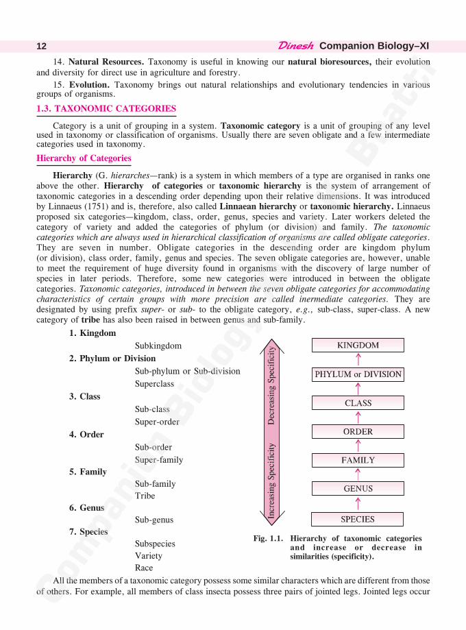
Hierarchy (G. hierarches—rank) is a system in which members of a type are organised in ranks oneabove the other. Hierarchy of categories or taxonomic hierarchy is the system of arrangement oftaxonomic categories in a descending order depending upon their relative dimensions. It was introducedby Linnaeus (1751) and is, therefore, also called Linnaean hierarchy or taxonomic hierarchy. Linnaeusproposed six categories—kingdom, class, order, genus, species and variety. Later workers deleted thecategory of variety and added the categories of phylum (or division) and family. The taxonomiccategories which are always used in hierarchical classification of organisms are called obligate categories.They are seven in number. Obligate categories in the descending order are kingdom phylum(or division), class order, family, genus and species. The seven obligate categories are, however, unableto meet the requirement of huge diversity found in organisms with the discovery of large number ofspecies in later periods. Therefore, some new categories were introduced in between the obligatecategories. Taxonomic categories, introduced in between the seven obligate categories for accommodatingcharacteristics of certain groups with more precision are called inermediate categories. They aredesignated by using prefix super- or sub- to the obligate category, e.g., sub-class, super-class. A newcategory of tribe has also been raised in between genus and sub-family.
1. Kingdom
Subkingdom
2. Phylum or Division
Sub-phylum or Sub-division
Superclass
3. Class
Sub-class
Super-order
4. Order
Sub-order
Super-family
5. Family
Sub-family
Tribe
6. Genus
Sub-genus
7. Species
Subspecies
Variety
Race
All the members of a taxonomic category possess some similar characters which are different from thoseof others. For example, all members of class insecta possess three pairs of jointed legs. Jointed legs occur
Fig. 1.1. Hierarchy of taxonomic categoriesand increase or decrease insimilarities (specificity).
����������� �� 13
in other classes of phylum arthropoda as well but their number is different. The placement of individuals ororganisms in species, genus, family, order, class and phylum is determined by their specific similarcharacters and relationships. Maximum similarity occurs in species which is also the lowest category in thehierarchy of categories. Similarity of characters decreases with the ascent in hierarchy. (Fig. 1.1)
• Cohort. A taxonomic category used variously by different workers like (a) Rank below sub-genus(b) Rank between order and class (c) Rank between super-order and class.
• Domain. (i) The highest level of biological classification as super kingdom eukarya. (ii) Globularportion of a protein or looped part of chromatin.
1.3.1. Species (John Ray)
It is the lowest or basic taxonomic category which consists of one or more natural populations ofindividuals that resemble one another more closely than individuals of other species, interbreed freely,have a distinct genetic set up and are reproductively isolated from others, e.g., Panthera leo (Lion),Mangifera indica (Mango), Solanum tuberosum (Potato). Thus dog can breed with only bitch and notmember of another species, say cat. In organisms lacking sexual reproduction, the morphological,anatomical, physiological, cytological and biochemical resemblances are taken into consideration.
Some species cannot be divided into races varieties and subspecies. They are called monotypicspecies. Others are polytypic having two or more subspecies varieties or races. The category ofsubspecies is more commonly used by zoologists while botanists instead recognise the category ofvariety.
1.3.2. Genus (John Ray)
It is the first higher category above the level of species. As per rules of binomial nomenclature, aspecies can be named only if it is assigned to a genus. A genus may have one to several species. Agenus having a single species is called monotypic. Currently, the genus Homo is monotypic with asingle species of Homo sapiens. A genus having two or more species is called polytypic. The genusPanthera is polytypic. Some species of this genus are P. leo (Lion), P. tigris (Tiger), P. onca (Jaguar)and P. pardus (Leopard). Similarly the genus Canis contains C. familiaris (Dog), C. lupus (Wolf) andC. aureus (Jackal). Solanum contains S. tuberosum (Potato), S. melongena (Brinjal) and S. lycopersicum(Tomato). All the species of a genus have a number of common features called correlated characters.Some of them are small and inconspicuous like number and position of spines on insect wings. The closeresemblance indicates a common ancestry for all the species of a genus.
1.3.3. Family (John Ray)
The category is of higher rank than that of genus and is formed of one to numerous related generawhich are more similar to one another than to genera of other families, e.g., members of felidae (cats,tiger, lion, leopard) and canidae (dogs, wolf, fox). All the genera of a family resemble one another incertain correlated characters indicating a common ancestry. For example, family canidae (of dogs)contains four genera of Canis, Vulpus, Lycaon and Dusicyan. Family felidae includes Panthera andFelis. Family solanaceae of dicots has genera like Solanum, Petunia, Datura and Atropa.
There is some difference in the suffix used for families. A plant family ends in a suffix-aceae andsubfamily in -oideae. An animal family has generally a suffix of -idae, a subfamily -inae, a tribe -iniwhile a superfamily has the suffix of -oidea.
1.3.4. Order (Linnaeus, 1735)
It is a taxonomic category having one or more related families that possess some similar correlated
characters which are lesser in number as compared to correlated characters of genera of a family. For
example, in animals order carnivora contains related families of canidae (dogs, wolf, fox), felidae (cat,
��������Companion Biology–XI14
leopard, tiger, lion), ursidae (bear) and hyaenidae (hyaena). All of them are carnivores with large
canines, carnassial teeth, powerful jaws and claws. Similarly in plants the order polemoniales has five
families including solanaceae, convolvutaceae, polymoniaceae, hydrophyllaceae and boraginaceae, all of
which have regular corolla with stamens equal to the number of petals. In plants the order ends in a
suffix -ales. Different suffixes are used in case of animals, e.g., carnivora, cetacea, primata, artiodactyla,
lagomorpha (all of class mammalia). Order and higher taxonomic categories are distinguished on the
basis of aggregate of characters.
1.3.5. Class (Linnaeus, 1735)
Class is a major category made of one or more related orders that possess certain similar correlated
characters. For example, in animals the class mammlia has a number of orders like carnivora, rodentia,
lagomorpha, insectivora, primata, etc., all of which possess mammary glands, external ears and hair. In
plants the suffix used for class is -phyceae, -opsida or -ae. The suffix is not fixed in case of animals,
e.g., Amphibia, Aves, Mammalia, Cyclostomata.
1.3.6. Phylum or Division (Cuvier, 1829, Eichler 1883)
It is a taxonomic category higher than class and lower in rank to kingdom. The term phylum
(coined by George Cuvier) is used for animals while the term division (coined by Eichler) is employed
for plants. A phylum or division consists of one to several related classes having a few similar correlated
characters. Classes pisces, amphibia, reptilia, aves and mammalia are included in phylum chordata. All
of them possess four common characters of presence of notochord, hollow dorsal nerve cord, pharyngeal
gill slits and postanal tail. A division ends in suffix -phyta, a subdivision in -phytina while there is no
fixed suffix for animals, e.g., Porifera, Annelida, Aschelminthes, Cnidaria.
1.3.7. Kingdom (Linnaeus 1751, ICBN)
It is the highest category in taxonomy where all the organisms included in it share a set of
distinguishing characters, e.g., all plants in plant kingdom or all animals in animal kingdom. R.H.
Whittaker (1969) has recognised five kingdoms of organisms—monera, protista, fungi, plantae (metaphyta)
and animalia (metazoa).
It is observed that the number of common or correlated characters is maximum in lower rank
categories. The number progressively decreases with the rise in the rank of category. Species of genus
Panthera have several similar traits. The different genera of family felidae have less number of similar
traits, the different families of order carnivora still smaller number while orders of class mammalia are
related to each other by fewer common traits. As a result diverse animals like bat, whale, elephant and
man with several different features are put together. Therefore, higher the category, greater is the
difficulty in evaluating relationship of its components to other taxa of the same level.
Advantages of Hierarchical System of Classification
1. It gives information about the relationships of an organism with others.
2. All the major traits present in an organism can be studied by noting the traits of various
categories in which the organism is classified.
3. It helps in quick identification of a taxon.
4. It reduces the volume of description of traits by non-repetition of correlated traits of a high rank
category in the lower rank category. In the lower rank category only the additional correlated characters of
that category are mentioned. Taxonomic categories of some common organisms are given in table 1.1.
����������� �� 15
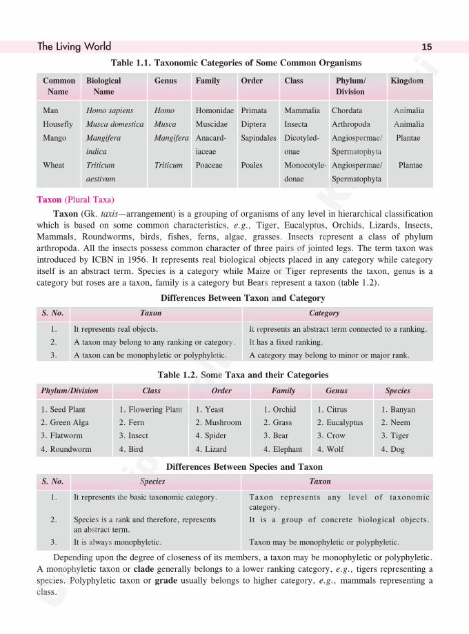
Table 1.1. Taxonomic Categories of Some Common Organisms
Common Biological Genus Family Order Class Phylum/ Kingdom
Name Name Division
Man Homo sapiens Homo Homonidae Primata Mammalia Chordata Animalia
Housefly Musca domestica Musca Muscidae Diptera Insecta Arthropoda Animalia
Mango Mangifera Mangifera Anacard- Sapindales Dicotyled- Angiospermae/ Plantae
indica iaceae onae Spermatophyta
Wheat Triticum Triticum Poaceae Poales Monocotyle- Angiospermae/ Plantae
aestivum donae Spermatophyta
Taxon (Plural Taxa)
Taxon (Gk. taxis—arrangement) is a grouping of organisms of any level in hierarchical classificationwhich is based on some common characteristics, e.g., Tiger, Eucalyptus, Orchids, Lizards, Insects,
Mammals, Roundworms, birds, fishes, ferns, algae, grasses. Insects represent a class of phylumarthropoda. All the insects possess common character of three pairs of jointed legs. The term taxon wasintroduced by ICBN in 1956. It represents real biological objects placed in any category while category
itself is an abstract term. Species is a category while Maize or Tiger represents the taxon, genus is acategory but roses are a taxon, family is a category but Bears represent a taxon (table 1.2).
Differences Between Taxon and Category
S. No. Taxon Category
1. It represents real objects. It represents an abstract term connected to a ranking.
2. A taxon may belong to any ranking or category. It has a fixed ranking.
3. A taxon can be monophyletic or polyphyletic. A category may belong to minor or major rank.
Table 1.2. Some Taxa and their Categories
Phylum/Division Class Order Family Genus Species
1. Seed Plant 1. Flowering Plant 1. Yeast 1. Orchid 1. Citrus 1. Banyan
2. Green Alga 2. Fern 2. Mushroom 2. Grass 2. Eucalyptus 2. Neem
3. Flatworm 3. Insect 4. Spider 3. Bear 3. Crow 3. Tiger
4. Roundworm 4. Bird 4. Lizard 4. Elephant 4. Wolf 4. Dog
Differences Between Species and Taxon
S. No. Species Taxon
1. It represents the basic taxonomic category. Taxon represents any level of taxonomiccategory.
2. Species is a rank and therefore, represents It is a group of concrete biological objects.an abstract term.
3. It is always monophyletic. Taxon may be monophyletic or polyphyletic.
Depending upon the degree of closeness of its members, a taxon may be monophyletic or polyphyletic.
A monophyletic taxon or clade generally belongs to a lower ranking category, e.g., tigers representing aspecies. Polyphyletic taxon or grade usually belongs to higher category, e.g., mammals representing aclass.
��������Companion Biology–XI16
1.4. TAXONOMIC AIDS/TOOLS
Both laboratory and field studies are required for study and correct identification of species. Theinformation once gathered should also be stored for future reference. Specimens should also be collected,preserved and stored for later verification. Biologists have standardised pocedure and techniques to storeinformation and specimens. Storehouses of information and specimens which can help in identificationand classification of organisms are called taxonomic aids. The important components are botanicalgardens, herbaria, museums and zoological parks.
1.4.1. Herbarium
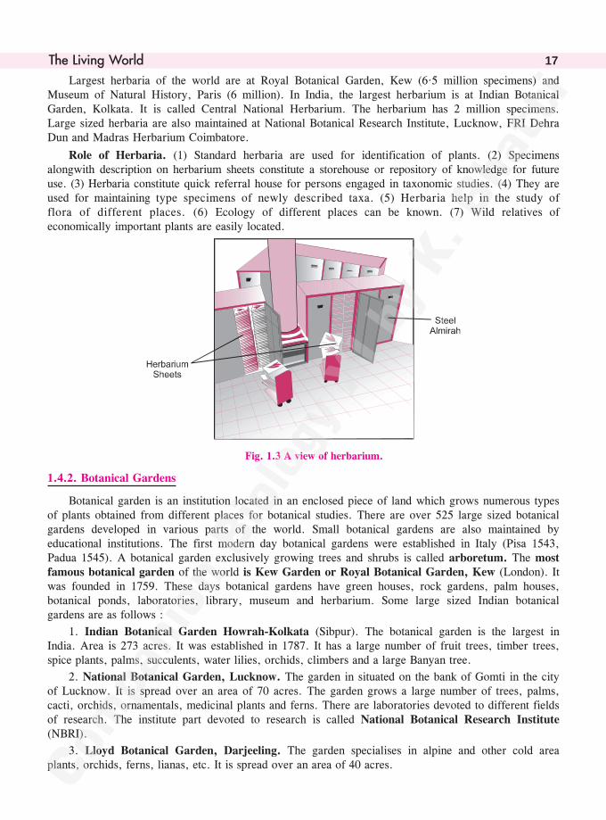
Herbarium is a collection of pressed mounted or preserved plant specimens arranged systematicallyaccording to commonly accepted system of classification so as to provide first hand information of plantmaterials of various types. All institutes dealing with botanical studies maintain their own herbarium.Students are trained to collect and identify herbarium specimens of local and distant places. For this thestudents are taken on excursions.
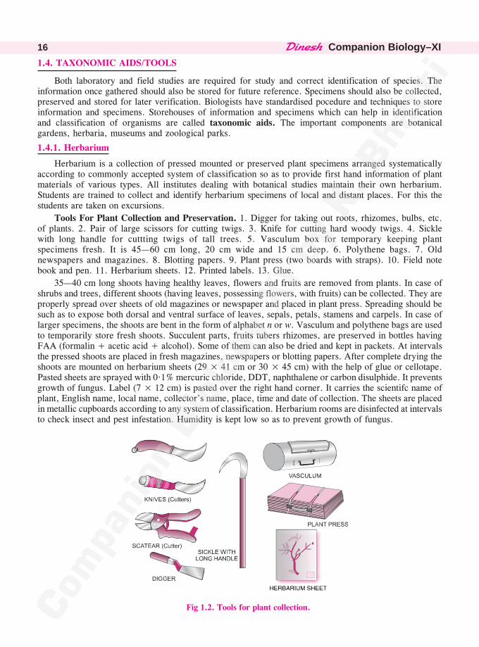
Tools For Plant Collection and Preservation. 1. Digger for taking out roots, rhizomes, bulbs, etc.of plants. 2. Pair of large scissors for cutting twigs. 3. Knife for cutting hard woody twigs. 4. Sicklewith long handle for cuttting twigs of tall trees. 5. Vasculum box for temporary keeping plantspecimens fresh. It is 45—60 cm long, 20 cm wide and 15 cm deep. 6. Polythene bags. 7. Oldnewspapers and magazines. 8. Blotting papers. 9. Plant press (two boards with straps). 10. Field notebook and pen. 11. Herbarium sheets. 12. Printed labels. 13. Glue.
35—40 cm long shoots having healthy leaves, flowers and fruits are removed from plants. In case ofshrubs and trees, different shoots (having leaves, possessing flowers, with fruits) can be collected. They areproperly spread over sheets of old magazines or newspaper and placed in plant press. Spreading should besuch as to expose both dorsal and ventral surface of leaves, sepals, petals, stamens and carpels. In case oflarger specimens, the shoots are bent in the form of alphabet n or w. Vasculum and polythene bags are usedto temporarily store fresh shoots. Succulent parts, fruits tubers rhizomes, are preserved in bottles havingFAA (formalin + acetic acid + alcohol). Some of them can also be dried and kept in packets. At intervalsthe pressed shoots are placed in fresh magazines, newspapers or blotting papers. After complete drying theshoots are mounted on herbarium sheets (29 × 41 cm or 30 × 45 cm) with the help of glue or cellotape.Pasted sheets are sprayed with 0·1% mercuric chloride, DDT, naphthalene or carbon disulphide. It preventsgrowth of fungus. Label (7 × 12 cm) is pasted over the right hand corner. It carries the scientifc name ofplant, English name, local name, collector’s name, place, time and date of collection. The sheets are placedin metallic cupboards according to any system of classification. Herbarium rooms are disinfected at intervalsto check insect and pest infestation. Humidity is kept low so as to prevent growth of fungus.
Fig 1.2. Tools for plant collection.
����������� �� 17
Largest herbaria of the world are at Royal Botanical Garden, Kew (6·5 million specimens) andMuseum of Natural History, Paris (6 million). In India, the largest herbarium is at Indian BotanicalGarden, Kolkata. It is called Central National Herbarium. The herbarium has 2 million specimens.Large sized herbaria are also maintained at National Botanical Research Institute, Lucknow, FRI DehraDun and Madras Herbarium Coimbatore.
Role of Herbaria. (1) Standard herbaria are used for identification of plants. (2) Specimensalongwith description on herbarium sheets constitute a storehouse or repository of knowledge for futureuse. (3) Herbaria constitute quick referral house for persons engaged in taxonomic studies. (4) They areused for maintaining type specimens of newly described taxa. (5) Herbaria help in the study offlora of different places. (6) Ecology of different places can be known. (7) Wild relatives ofeconomically important plants are easily located.
Fig. 1.3 A view of herbarium.
1.4.2. Botanical Gardens
Botanical garden is an institution located in an enclosed piece of land which grows numerous typesof plants obtained from different places for botanical studies. There are over 525 large sized botanicalgardens developed in various parts of the world. Small botanical gardens are also maintained byeducational institutions. The first modern day botanical gardens were established in Italy (Pisa 1543,Padua 1545). A botanical garden exclusively growing trees and shrubs is called arboretum. The mostfamous botanical garden of the world is Kew Garden or Royal Botanical Garden, Kew (London). Itwas founded in 1759. These days botanical gardens have green houses, rock gardens, palm houses,botanical ponds, laboratories, library, museum and herbarium. Some large sized Indian botanicalgardens are as follows :
1. Indian Botanical Garden Howrah-Kolkata (Sibpur). The botanical garden is the largest inIndia. Area is 273 acres. It was established in 1787. It has a large number of fruit trees, timber trees,spice plants, palms, succulents, water lilies, orchids, climbers and a large Banyan tree.
2. National Botanical Garden, Lucknow. The garden in situated on the bank of Gomti in the cityof Lucknow. It is spread over an area of 70 acres. The garden grows a large number of trees, palms,cacti, orchids, ornamentals, medicinal plants and ferns. There are laboratories devoted to different fieldsof research. The institute part devoted to research is called National Botanical Research Institute(NBRI).
3. Lloyd Botanical Garden, Darjeeling. The garden specialises in alpine and other cold areaplants, orchids, ferns, lianas, etc. It is spread over an area of 40 acres.
��������Companion Biology–XI18
Other Indian botanical gardens of importance are Botanical Garden, Ootacamund; Lalbagh Gardens,Bangalore; Botanical Garden of Indian Agriculture Research Institute, New Delhi; Botanical Garden ofForest Research Institute, Dehra Dun.
Role of Botanical Gardens. 1. Providing plant material for comparative taxonomic studies.2. Functioning as acclimitisation centres for exotic plants of economic importance. 3. Growing andmaintaining records of local flora. 4. Growing plants of various types for research. 5. Providing seedsand living materials to different research centres. 6. Coordinating research through International Associationof Botanical Gardens. 7. Ex situ conservation of endangered plant species.
• ���������� ��������������������������� �����������������������������• Hanging Gardens of Babylon. They are considered to be one of the seven wonders of Ancient
World by Greek scholars.
1.4.3. Museums
Role of Museums. The roles are similar to those of herbaria. (i) Standard museums have collectionsof plants and animals of various areas. (ii) Museums provide information not only about the local faunaand flora but also of other areas. (iii) They are used to deposit type specimens whenever new taxa aredescribed. (iv) They are important centres for taxonomic studies like important members of various taxa,their important characteristics, study and identification of various organisms.
1.4.4. Zoological Parks
Zoological parks or zoological gardens are enclosed areas or park lands where animals are kept inopen enclosures instead of cages (in zoos). Zoological parks provide more natural environment toanimals. Therefore, most zoos are being converted into zoological parks. All countries of the worldmaintain zoological parks. In India, there are 200 zoos and zoological parks. A Central Zoo Authoritylooks after their management in India. An international body also coordinates the activities of zoologicalparks in different countries. Besides large zoological parks, some states maintain large aquaria (forvarieties of fishes, e.g., Mumbai), aviaries (for birds) and serpentaria (for snakes, e.g., Chennai).
Role of Zoological Parks. Zoological parks are prized assets. They are useful for (i) Familiarisingpublic, especially children, with wild animals. (ii) Study of live animal types by students. (iii) Sources oftourist attraction. (iv) Ex situ conservation through captive breeding of endangered animals. Because ofcaptive breeding, Californian Candor (Gymnogyps californicanus) and Black Footed Ferret (Mustelanigripes) have been saved from extinction.
Museum is a place used for storing preservation and exhibition of objects of natural history (bothplants and animals), art and objects of antiquities. All educational institutes and universities maintainmuseums in their botany and zoology departments. Museum of natural history has collection of preservedplants and animals. Only those plants are preserved in museums which cannot be kept in herbaria e.g.,algae, fungi, mosses, ferns, parts of gymnosperms, fruits, underground storage organs and othermaterials of interest. Preservative solution consists of alcohol and formalin. Many animal specimens(e.g., worms, insects, fishes, reptiles) can also be kept in preservative solution in jars. Insects can bedried out and mounted in boxes. Larger animals are preserved in stuffed and skeleton forms. However,these days catching of live animals (for killing and preservation) is discouraged. Biology students areasked to collect and preserve only dead animals. Some important museums are (i) American Museum ofNatural History, New York, U.S.A. (ii) State Museum of Natural History, Stuttgaut, Germany.(iii) Museum of Natural History, Basel, Switzerland. (iv) Bird Collection Museum of Natural History,Vienna, Austria. (v) National Museum of Natural History, Paris. (vi) National Museum of NaturalHistory (Barakhamba Road), New Delhi. (vii) Museum of Mumbai Natural History Society (HornbillHouse, Shahid Bhagat Singh Road), Mumbai. (viii) Museum of Arthropoda (Shaniwar Petu), Pune.
����������� �� 19
Monograph
It gives detailed information of various aspectsof a taxon.
Monograph deals with worldwide distributionand distinguishing features of various membersof the taxon.
Differences Between Museum and Zoological Park/Botanical Garden
S.No. Museum Zoological Park/Botanical Garden
1. It deals with preserved objects and materials. It deals with living objects.
2. It has no role in conservation. It is connected with ex situ conservation.3. It has collection of items of local and other It has organisms of local area as well as climatically
areas. similar areas.
Floras, Manuals, Monographs and Catalogues
Floras are compilations describing habitat, environmental conditions, seasonal changes and descriptionof plants alongwith their identification keys. They are available for ready reference e.g., Flora of BritishIndia, Flora of Delhi, Flora Simlensis, etc. Manuals are books of instructions having information foridentification of names of species occurring in particular areas. Monographs are publications containingexhaustive treatment of all the members and aspects of biology of taxonomic groups as far as they relateto their identification, nomenclature and classification. Catalogue is a book containing list of systematicallyarranged plants or animals found in an area alongwith their identification.
Differences Between Flora and Monograph
S. No. Flora
1. It provides information as to all the plants growingin an area.
2. Flora describes the habitat, environment andtaxonomic features of plants in an area.
1.4.5. Taxonomic Key
(Key for Identification)
It is an important taxonomic aid. Key is a set of alternate characters or couplets of different typesarranged sequence-wise in such a fashion that by selection and elimination one can quickly find out thename of the organism. Depending upon the category, a key may be class key, order key, family key,genus key and species key.
Taxonomic keys are of two types—bracketed and indented. In a bracketed key the alternatecharacters are given numbers in brackets. In indented or yolked key there is a sequence of two or morealternate characters from which selection and elimination are carried out. Three examples are as follows :
Example 1. Six vertebrates are to be identified—fish, frog, snake, birds, bat and cat. Distinguishingcharacters are recorded for each group—presence or absence of ears (mammalian or non-mammaliancharacter), ability or inability to fly (flight is trait of both birds and bats), presence and absence of limbs(tetrapods except snakes, and nontetrapods like fishes), presence and absence of gills (character offishes, absent in adults of others).
Indented Key Bracketed Key
1. External ears present (1) External ears present.....(2)
2. Wings present.......Bat (1) External ears absent.....(3)
2. Wings absent.......Cat (2) Wings present.....Bat
1. External ears absent......... (2) Wings absent.....Cat
2. Wings present.....Bird (3) Wings present.....Bird
2. Wing absent (3) Wing absent.....(4)
3. Limbs Present.....Frog (4) Limbs present.....Frog
3. Limbs absent (4) Limbs absent.....(5)
4. Gills present.....Fish (5) Gills present.....Fish
4. Gills absent.....Snake (5) Gills absent.....Snake
��������Companion Biology–XI20
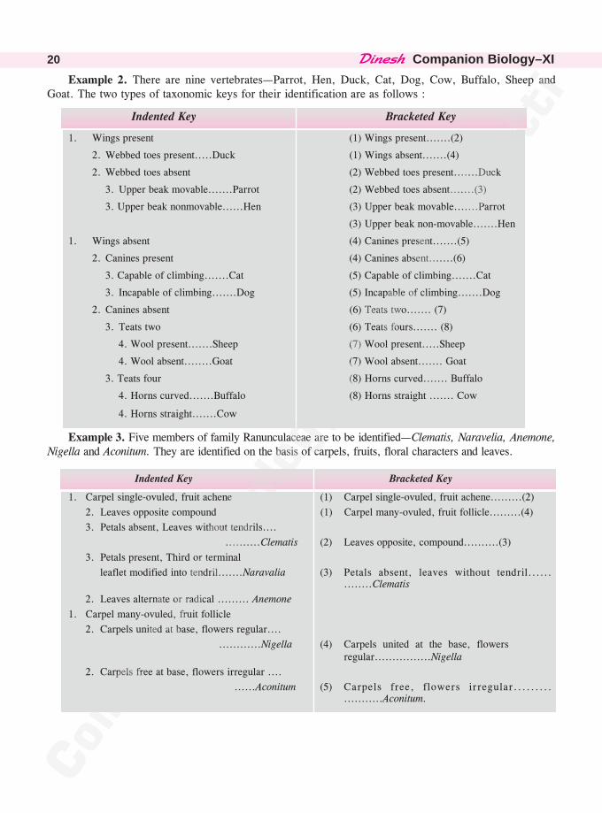
Example 2. There are nine vertebrates—Parrot, Hen, Duck, Cat, Dog, Cow, Buffalo, Sheep andGoat. The two types of taxonomic keys for their identification are as follows :
Indented Key Bracketed Key
1. Wings present (1) Wings present.......(2)
2. Webbed toes present.....Duck (1) Wings absent.......(4)
2. Webbed toes absent (2) Webbed toes present.......Duck
3. Upper beak movable.......Parrot (2) Webbed toes absent.......(3)
3. Upper beak nonmovable......Hen (3) Upper beak movable.......Parrot
(3) Upper beak non-movable.......Hen
1. Wings absent (4) Canines present.......(5)
2. Canines present (4) Canines absent.......(6)
3. Capable of climbing.......Cat (5) Capable of climbing.......Cat
3. Incapable of climbing.......Dog (5) Incapable of climbing.......Dog
2. Canines absent (6) Teats two....... (7)
3. Teats two (6) Teats fours....... (8)
4. Wool present.......Sheep (7) Wool present.....Sheep
4. Wool absent........Goat (7) Wool absent....... Goat
3. Teats four (8) Horns curved....... Buffalo
4. Horns curved.......Buffalo (8) Horns straight ....... Cow
4. Horns straight.......Cow
Example 3. Five members of family Ranunculaceae are to be identified—Clematis, Naravelia, Anemone,Nigella and Aconitum. They are identified on the basis of carpels, fruits, floral characters and leaves.
Indented Key Bracketed Key
1. Carpel single-ovuled, fruit achene (1) Carpel single-ovuled, fruit achene.........(2)
2. Leaves opposite compound (1) Carpel many-ovuled, fruit follicle.........(4)
3. Petals absent, Leaves without tendrils....
..........Clematis (2) Leaves opposite, compound..........(3)
3. Petals present, Third or terminal
leaflet modified into tendril.......Naravalia (3) Petals absent, leaves without tendril..............Clematis
2. Leaves alternate or radical ......... Anemone
1. Carpel many-ovuled, fruit follicle
2. Carpels united at base, flowers regular....
............Nigella (4) Carpels united at the base, flowers regular................Nigella
2. Carpels free at base, flowers irregular ....
......Aconitum (5) Carpels free, flowers irregular....................Aconitum.
����������� �� 21
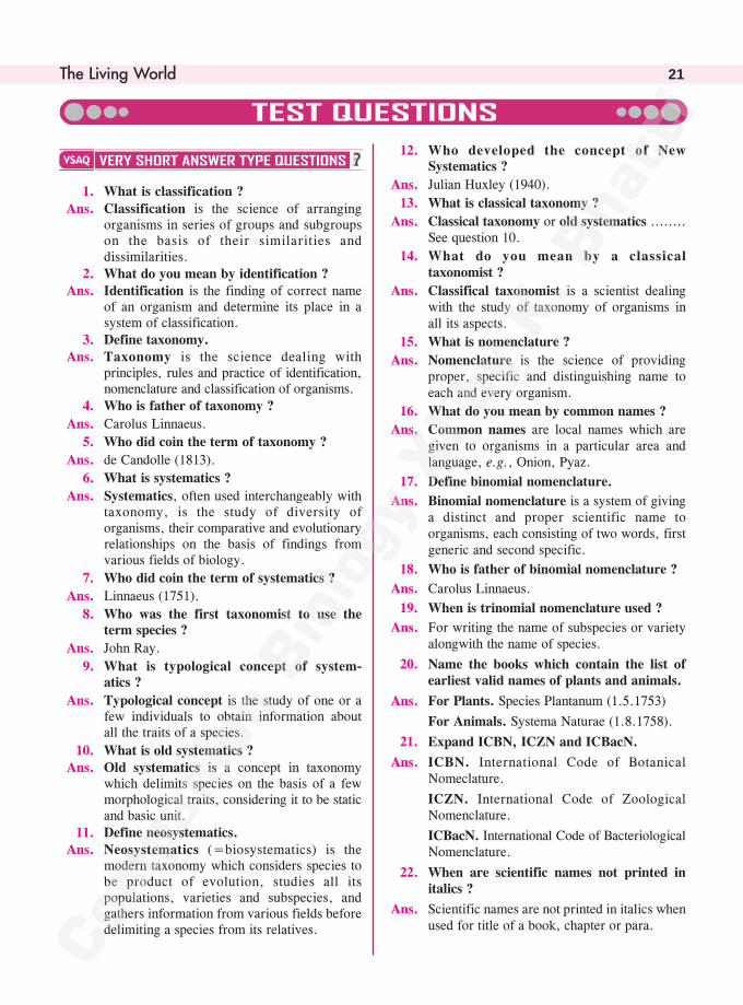
12. Who developed the concept of NewSystematics ?
Ans. Julian Huxley (1940).
13. What is classical taxonomy ?
Ans. Classical taxonomy or old systematics ........See question 10.
14. What do you mean by a classicaltaxonomist ?
Ans. Classifical taxonomist is a scientist dealingwith the study of taxonomy of organisms inall its aspects.
15. What is nomenclature ?
Ans. Nomenclature is the science of providingproper, specific and distinguishing name toeach and every organism.
16. What do you mean by common names ?
Ans. Common names are local names which aregiven to organisms in a particular area andlanguage, e.g., Onion, Pyaz.
17. Define binomial nomenclature.
Ans. Binomial nomenclature is a system of givinga distinct and proper scientific name toorganisms, each consisting of two words, firstgeneric and second specific.
18. Who is father of binomial nomenclature ?
Ans. Carolus Linnaeus.
19. When is trinomial nomenclature used ?
Ans. For writing the name of subspecies or varietyalongwith the name of species.
20. Name the books which contain the list ofearliest valid names of plants and animals.
Ans. For Plants. Species Plantanum (1.5.1753)
For Animals. Systema Naturae (1.8.1758).
21. Expand ICBN, ICZN and ICBacN.
Ans. ICBN. International Code of BotanicalNomeclature.
ICZN. International Code of ZoologicalNomenclature.
ICBacN. International Code of BacteriologicalNomenclature.
22. When are scientific names not printed initalics ?
Ans. Scientific names are not printed in italics whenused for title of a book, chapter or para.
1. What is classification ?
Ans. Classification is the science of arrangingorganisms in series of groups and subgroupson the basis of their similarities anddissimilarities.
2. What do you mean by identification ?Ans. Identification is the finding of correct name
of an organism and determine its place in asystem of classification.
3. Define taxonomy.Ans. Taxonomy is the science dealing with
principles, rules and practice of identification,nomenclature and classification of organisms.
4. Who is father of taxonomy ?
Ans. Carolus Linnaeus.
5. Who did coin the term of taxonomy ?
Ans. de Candolle (1813).
6. What is systematics ?
Ans. Systematics, often used interchangeably withtaxonomy, is the study of diversity oforganisms, their comparative and evolutionaryrelationships on the basis of findings fromvarious fields of biology.
7. Who did coin the term of systematics ?
Ans. Linnaeus (1751).
8. Who was the first taxonomist to use theterm species ?
Ans. John Ray.
9. What is typological concept of system-atics ?
Ans. Typological concept is the study of one or afew individuals to obtain information aboutall the traits of a species.
10. What is old systematics ?Ans. Old systematics is a concept in taxonomy
which delimits species on the basis of a fewmorphological traits, considering it to be staticand basic unit.
11. Define neosystematics.Ans. Neosystematics (=biosystematics) is the
modern taxonomy which considers species tobe product of evolution, studies all itspopulations, varieties and subspecies, andgathers information from various fields beforedelimiting a species from its relatives.
��������Companion Biology–XI22
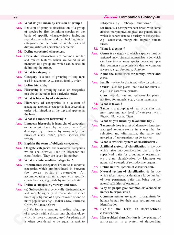
23. What do you mean by revision of group ?
Ans. Revision of group is classification of a groupof species by first delimiting species on thebasis of specific characteristics includingreproductive isolation and then building highercategories on the basis of similarities anddissimilarities of correlated characters.
24. Define correlated characters.
Ans. Correlated characters are common similarand related features which are found in allmembers of a group and which can be used indelimiting the group.
25. What is category ?
Ans. Category is a unit of grouping of any rankused in taxonomy, e.g., genus, family, order.
26. Define hierarchy.
Ans. Hierarchy is arranging ranks or categoriesone above the other in a particular order.
27. What is hierarchy of categories ?
Ans. Hierarchy of categories is a system ofarranging taxonomic categories in a descendingorder with kingdom at the top and species atthe base.
28. What is Linnaean hierarchy ?
Ans. Linnaean hierarchy is hierarchy of categoriesor taxonomic hierarchy because it was firstdeveloped by Linnaeus by using only fiveranks of class, order, genus, species andvariety.
29. Explain the term of obligate categories.
Ans. Obligate categories are taxonomic categorieswhich are always used in hierarchicalclassification. They are seven in number.
30. What are intermediate categories ?
Ans. Intermediate categories are those taxonomiccategories which are introduced in betweenthe seven obligate categories foraccommodating certain groups with specificcharacteristics, e.g., subphylum vertebrata.
31. Define a subspecies, variety and race.
Ans. (a) Subspecies is a genetically distinguishableand morphologically differentiable distinctbreeding subgroup of a species made of one ormore populations,e.g., Indian Crow, BurmeseCrow, SriLankan Crow.
(b) Variety is a separate breeding subgroupof a species with a distinct morphophysiologywhich is more commonly used for plants andis often considered to be equal in rank to
subspecies, e.g., Cabbage, Cauliflower.
(c) Race is a near permanent breed with somedistinct morphophysiological and genetic traitswhich is subordinate to a variety or subspecies,e.g., caucasoid, mongoloid, negroid humanraces.
32. What is a genus ?
Ans. Genus is a category to which a species must beassigned under binomial nomenclature but whichcan have two or more species depending upontheir common characteristics due to commonancestry, e.g., Panthera, Solanum.
33. Name the suffix used for family, order andclass.
Ans. Family. –aceae for plants and –idae for animals.
Order. –ales for plants, not fixed for animals,e.g., –a in carnivora, primata.
Class. –opsida, –ae and –phyceae for plants,not fixed for animals, e.g. , –ia in mammalia.
34. What is taxon ?
Ans. Taxon is a grouping of real organisms thatmay represent any level of category, e.g.,Pigeon, Flatworm, Tiger.
35. What do you mean by taxonomic key ?
Ans. Taxonomic key is a set of alternate charactersarranged sequence-wise in a way that byselection and elimination, the name andgrouping of an organism can be known.
36. What is artificial system of classification ?
Ans. Artificial system of classification is the onewhich takes into consideration one or a fewsuperficial traits for grouping of organisms,e.g., plant classification by Linnaeus onnumerical strength of reproductive organs.
37. Define natural system of classification.
Ans. Natural system of classification is the onewhich takes into consideration a large numberof near permanent traits that bring out thenatural affinities of organisms.
38. Why do people give common or vernacularnames to organisms ?
Ans. Common names are given to organisms byhuman beings for their easy recognition andidentification.
39. Explain the term of hierarchicalclassification.
Ans. Hierarchical classification is the placing ofan organism in a system of descending
����������� �� 23
taxonomic categories which bring out specificand correlated characters as well asrelationships with other organisms.
40. Which of the following cover the greaternumber of organisms
(a) Phylum or Genus (b) Family or Phylum(c) Family or Order (d) Class or Phylum ?
Ans. Greater Number of Organisms. (a) Phylum(b) Phylum (c) Order (d) Phylum.
41. Which of the following have morecharacters in common
(a) Phylum or Family (b) Species or Genus(c) Class or Order ?
Ans. More Common Characters. (a) Family(b) Species (c) Order.
42. Mark the odd one in the series
(a) Family, Class, Taxon, Phylum
(b) Indica, Ficus, Mangifera, Tamarindus
(c) Plantae, Chordata, Tracheophyta,Eucalyptus.
Ans. Odd One. (a) Taxon (b) Indica (c) Chordata.
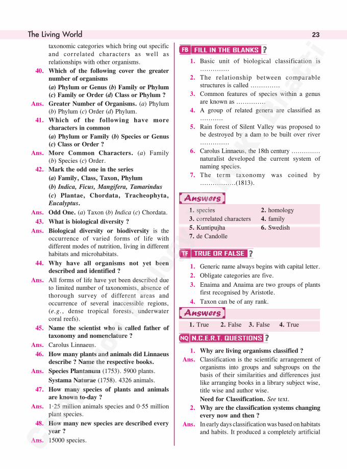
43. What is biological diversity ?
Ans. Biological diversity or biodiversity is theoccurrence of varied forms of life withdifferent modes of nutrition, living in differenthabitats and microhabitats.
44. Why have all organisms not yet beendescribed and identified ?
Ans. All forms of life have yet been described dueto limited number of taxonomists, absence ofthorough survey of different areas andoccurrence of several inaccessible regions,(e.g., dense tropical forests, underwatercoral reefs).
45. Name the scientist who is called father oftaxonomy and nomenclature ?
Ans. Carolus Linnaeus.
46. How many plants and animals did Linnaeusdescribe ? Name the respective books.
Ans. Species Plantanum (1753). 5900 plants.
Systama Naturae (1758). 4326 animals.
47. How many species of plants and animalsare known to-day ?
Ans. 1·25 million animals species and 0·55 millionplant species.
48. How many new species are described everyyear ?
Ans. 15000 species.
1. Basic unit of biological classification is..............
2. The relationship between comparablestructures is called ..............
3. Common features of species within a genusare known as ..............
4. A group of related genera are classified as...........
5. Rain forest of Silent Valley was proposed tobe destroyed by a dam to be built over river..............
6. Carolus Linnaeus, the 18th century ..............naturalist developed the current system ofnaming species.
7. The term taxonomy was coined by.................(1813).
1. species 2. homology
3. correlated characters 4. family
5. Kuntipujha 6. Swedish
7. de Candolle
1. Generic name always begins with capital letter.
2. Obligate categories are five.
3. Enaima and Anaima are two groups of plantsfirst recognised by Aristotle.
4. Taxon can be of any rank.
1. True 2. False 3. False 4. True
1. Why are living organisms classified ?
Ans. Classification is the scientific arrangement oforganisms into groups and subgroups on thebasis of their similarities and differences justlike arranging books in a library subject wise,title wise and author wise.
Need for Classification. See text.
2. Why are the classification systems changingevery now and then ?
Ans. In early days classification was based on habitatsand habits. It produced a completely artificial
��������Companion Biology–XI24
classification. Then external morphology wasadded. It refined artificial systems. This wasfollowed by addition of anatomy or internalmorphology. Embryology provided informationto affinities of some organisms. Natural systemsof classification came into existence. Latercytology, genetics, molecular biology and fossilstudies started adding information about theaffinities of orgnisms. They paved the way forphylogenetic systems of classification.However, knowledge is seldom final. Newdiscoveries are always being made because ofthe refinement of tools and techniques.Therefore, classification systems which aremeant for bringing out true relationshipsamongst organisms are also changing.
3. What different criteria would you chooseto classify people that you meet often ?
Ans. (i) Sex (ii) Age
(iii) Height (iv) Weight
(v) Classmates (vi) Family members
(vii) Family friends (viii) Team mates.
(ix) School mates, etc.
4. What do we learn from identification ofindividuals and populations ?
Ans. (a) Identification of Individuals. Eachindividual possesses certain specificvariations in traits not found in otherindividuals of the same family or group.
(b) Identification of Populations. (i) Eachpopulation comprises individuals whichresemble one another more closely thanindividuals of other species.(ii) Membersof a population interbreed freely.(iii) Members of a population share thesame gene pool and have a distinct geneticset up.(iv) Members of a population arereproductively isolated from individuals ofother species/ populations found in thearea.
5. Given below is the scientific name of Mango.Identify the correctly written name.
Mangifera Indica
Mangifera indica.
Ans. (ii) Mangifera indica.
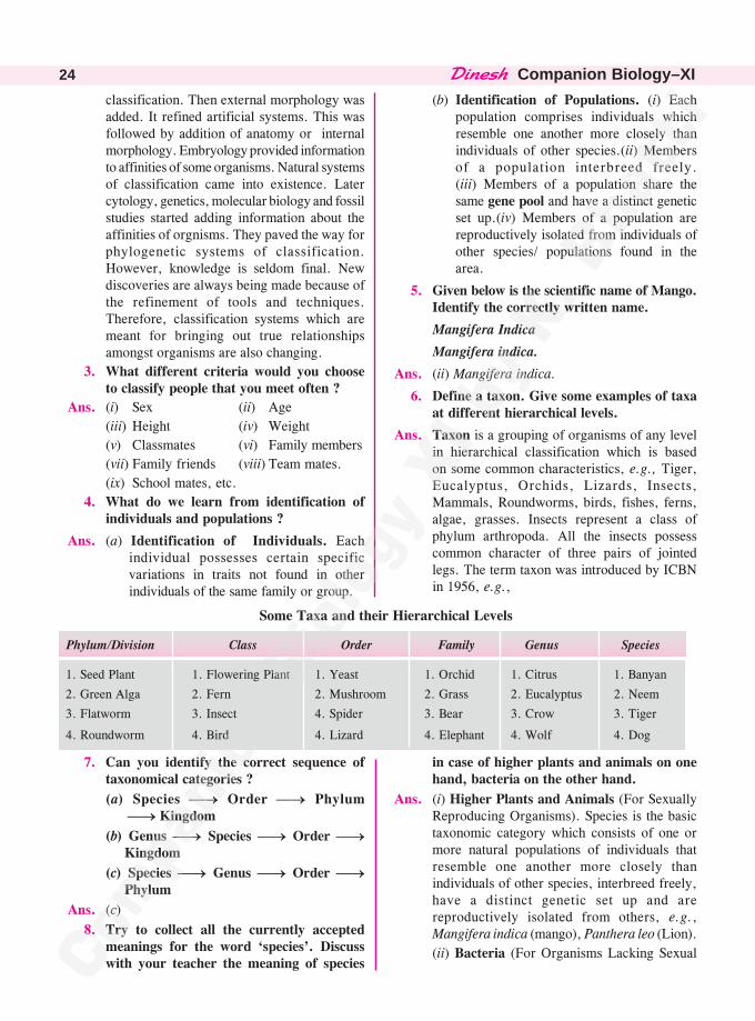
6. Define a taxon. Give some examples of taxaat different hierarchical levels.
Ans. Taxon is a grouping of organisms of any levelin hierarchical classification which is basedon some common characteristics, e.g., Tiger,Eucalyptus, Orchids, Lizards, Insects,Mammals, Roundworms, birds, fishes, ferns,algae, grasses. Insects represent a class ofphylum arthropoda. All the insects possesscommon character of three pairs of jointedlegs. The term taxon was introduced by ICBNin 1956, e.g.,
Some Taxa and their Hierarchical Levels
Phylum/Division Class Order Family Genus Species
1. Seed Plant 1. Flowering Plant 1. Yeast 1. Orchid 1. Citrus 1. Banyan
2. Green Alga 2. Fern 2. Mushroom 2. Grass 2. Eucalyptus 2. Neem
3. Flatworm 3. Insect 4. Spider 3. Bear 3. Crow 3. Tiger
4. Roundworm 4. Bird 4. Lizard 4. Elephant 4. Wolf 4. Dog
7. Can you identify the correct sequence oftaxonomical categories ?
(a) Species �� Order �� Phylum���������Kingdom
(b) Genus �� Species �� Order ������ Kingdom
(c) Species �� Genus �� Order ������ Phylum
Ans. (c)
8. Try to collect all the currently acceptedmeanings for the word ‘species’. Discusswith your teacher the meaning of species
in case of higher plants and animals on onehand, bacteria on the other hand.
Ans. (i) Higher Plants and Animals (For SexuallyReproducing Organisms). Species is the basictaxonomic category which consists of one ormore natural populations of individuals thatresemble one another more closely thanindividuals of other species, interbreed freely,have a distinct genetic set up and arereproductively isolated from others, e.g.,Mangifera indica (mango), Panthera leo (Lion).
(ii) Bacteria (For Organisms Lacking Sexual
����������� �� 25
Reproduction). It is the basic taxonomicgrouping of individuals that resemble oneanother in their morphology, cytology,biochemistry, physiology and modes ofmultiplication, perennation and dispersal.
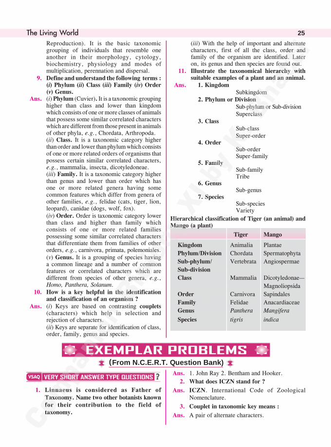
9. Define and understand the following terms :(i) Phylum (ii) Class (iii) Family (iv) Order(v) Genus.
Ans. (i) Phylum (Cuvier). It is a taxonomic groupinghigher than class and lower than kingdomwhich consists of one or more classes of animalsthat possess some similar correlated characterswhich are different from those present in animalsof other phyla, e.g., Chordata, Arthropoda.(ii) Class. It is a taxonomic category higherthan order and lower than phylum which consistsof one or more related orders of organisms thatpossess certain similar correlated characters,e.g., mammalia, insecta, dicotyledoneae.(iii) Family. It is a taxonomic category higherthan genus and lower than order which hasone or more related genera having somecommon features which differ from genera ofother families, e.g., felidae (cats, tiger, lion,leopard), canidae (dogs, wolf, fox).(iv) Order. Order is taxonomic category lowerthan class and higher than family whichconsists of one or more related familiespossessing some similar correlated charactersthat differentiate them from families of otherorders, e.g., carnivora, primata, polemoniales.(v) Genus. It is a grouping of species havinga common lineage and a number of commonfeatures or correlated characters which aredifferent from species of other genera, e.g.,Homo, Panthera, Solanum.
10. How is a key helpful in the identificationand classification of an organism ?
Ans. (i) Keys are based on contrasting couplets(characters) which help in selection andrejection of characters.(ii) Keys are separate for identification of class,order, family, genus and species.
(iii) With the help of important and alternatecharacters, first of all the class, order andfamily of the organism are identified. Lateron, its genus and then species are found out.
11. Illustrate the taxonomical hierarchy withsuitable examples of a plant and an animal.
Ans. 1. KingdomSubkingdom
2. Phylum or DivisionSub-phylum or Sub-divisionSuperclass
3. ClassSub-classSuper-order
4. OrderSub-orderSuper-family
5. FamilySub-familyTribe
6. GenusSub-genus
7. SpeciesSub-speciesVariety
Hierarchical classification of Tiger (an animal) andMango (a plant)
Tiger Mango
Kingdom Animalia Plantae
Phylum/Division Chordata Spermatophyta
Sub-phylum/ Vertebrata Angiospermae
Sub-division
Class Mammalia Dicotyledonae—
Magnoliopsida
Order Carnivora Sapindales
Family Felidae Anacardiaceae
Genus Panthera Mangifera
Species tigris indica
1. Linnaeus is considered as Father ofTaxonomy. Name two other botanists knownfor their contribution to the field oftaxonomy.
Ans. 1. John Ray 2. Bentham and Hooker.
2. What does ICZN stand for ?
Ans. ICZN. International Code of ZoologicalNomenclature.
3. Couplet in taxonomic key means :
Ans. A pair of alternate characters.
(From N.C.E.R.T. Question Bank)
��������Companion Biology–XI26
classifying plants and mention differentsuffixes used for the units.
Ans. Category Suffix
1. Kingdom No fixed suffix, e.g.,Monera, Protista, Fungi
2. Division – Phyta
3. Class – phyceae or opsida
4. Order – ales
5. Family – aceae
6. Genus – Not fixed
7. Species – Not fixed.
4. A plant species shows several morphologicalvariations in response to altitudinalgradients. When grown under similarconditions of growth, the morphologicalvariations disappear and all the variantshave common morphology. What are thesevariants called ?
Ans. Ecads. They are habitat or environmentvariants which have not yet been geneticallyfixed. In a common habitat or environment allof them will have similar morphology.
5. How do you prepare your own herbariumsheets ? What are different tools you carrywith you while collecting plants for thepreparation of a herbarium ? Whatinformation should be preserved plantmaterial or herbarium sheet provide fortaxonomical studies ?
Ans. Herbarium Sheets. (i) 29 × 41 cm or30 × 45 cm sheets cut from chart papers.(ii) Dried shoots with well spread leaves andflowers are mounted on the sheets with thehelp of glue or cellotape. A label (7 × 12 cm)is pasted over the right hand corner.
Tools For Plant Collection. Digger, sickle,knife, scissors, vasculum, polythene bags, oldnewspapers and magazines, blotting papers,plant press, field notebook and pen, magnifyingglass.
Information on Herbarium Sheets. Scientificname of plant, family, common English name,local name, collector’s name, place, time anddate of collection.
6. What is the difference between flora, faunaand vegetation ? Eichhornia crassipes iscalled an exotic species while Rauwolfiaserpentina is an endemic species in India.What do these terms exotic and endemicrefer to :
4. What is monograph ?
Ans. Monograph is an exhaustive treatment of asingle taxon dealing with identification,nomenclature, classification and biology of allits members.
5. Amoeba multiplies by mitotic cell division.Is this phenomenon growth or repro-duction ? Explain.
Ans. Reproduction. In unicellular organisms like
Amoeba cell division is a means of
multiplication while in multicellular organisms,
it is a means of growth.
6. Define metabolism.
Ans. Metabolism is the sum total of all biochemical
reactions that supports life in a living organism.
7. Which is the largest botanical garden in the
world ? Name a few well known botanical
gardens in India.
Ans. Largest Botanical Garden. Royal Botanical
Garden, Kew (London).
Botanical Gardens In India. (i) Indian
Botanical Garden Howrah, Kolkata (Sibpur).
(ii) National Botanical Garden, Lucknow.
(iii) Lloyd Botanical Garden, Darjeeling.
1. A ball of snow when rolled over snowincreases in mass, volume and size. Is thiscomparable to growth as seen in livingorganisms ? Why ?
Ans. No, increase in size of ball of snow is due toacretion or addition of similar material on theoutside. Growth in living organisms is due tointussusception or increase of body materialinternally from other substances.
2. In a given habitat we have 20 plant speciesand 20 animal species. Should we call thisas ‘‘diversity’’ or ‘‘biodiversity’’. Justifyyour answer.
Ans. It is called biodiversity or biological diversitywhich means variability among livingorganisms from all sources. Diversity isecologically variety of niches, habitats andother regions.
3. International Code of Botanical Nome-nclautre (ICBN) has provided a code forclassificatioon of plants. Give hierarchy ofunits of classification botanists follow while
����������� �� 27
Vegetation
It is plant cover of the area withoutany reference to detailed taxonomyof the plants.It describes the types of plantscomprising vegetation of an area.
It gives information aboutmorphophysiological traits of plantsof the area.
Ex. Grassland, Forest.
Fauna
It represents animals particular toa region, habitat or area.
It describes habitat, environ-mentand taxonomic features of animalsof an area.
It may deal with one or all thegroups of animals
Ex. Avian fauna of Andamans.
Exotic. It is an organism belonging to a foreignarea which has not been naturalised oracclimitised.Endemic. It is a natural inhabitantthat is restricted to a particular region.
7. A plant may have different names indifferent regions of the country or world.How do botanists solve this problem ?
Ans. A plant may have different local names indifferent parts of the country and world. Botanistsof one region cannot communicate with botanistsof another region if one is using local names.Therefore, a universal code of nomenclaturehas been adapted by botanists. It is calledbinomial nomenclature. Here name of theplant remains the same in all the languages.
8. Brinjal and Potato belong to the same genusSolanum but to two different species. Whatdefines them as separate species ?
Ans. Brinjal and Potato belong to the same genusas they possess similar correlated characters.They belong to different species because of(i) Different morphology (i) Inability tointerbreed (reproductive isolation).
9. Properties of cell organelles are not alwaysfound in the molecular constituents of cellorganelles. Justify.
Ans. Organisation produces a new functional entitywhich has its own properties not found in itscomponents. Therefore, a cell organelle willhave its own properties not found in themolecular constituents.
10. The number and kinds of organisms arenot constant. How do you explain thisstatement ?
Ans. The number and kinds of organisms are neverconstant at a place as development and
reproduction are influenced by seasons andavailability of resources. For example, numberof houseflies is very large in warm rainy season.It is rare to see a housefly in winter.
1. What is meant by living ? Give any fourdefining features of life form.
Ans. Living is a set of traits found in an individualthat enable it to maintain itself, respond tostimuli, obtain energy for various bodyfunctions, grow and undergo development.Features. 1. Organisation. See text.
2. Cellular structure. See text.3. Growth. See text.4. Reproduction. See text.
2. A scientist has come across a plant whichhe feels is a new species. How will be goabout its identification, classification andnomenclature ?
Ans. 1. With the help of family key, the scientistwill first identify family to which the plantbelongs.2. Using genus key, the genus is identified.3. Using species key, identification is carriedout. If the description of the plant does nottally with any known species, it is no doubt anew plant. More specimens of the plant arecollected.4. One of the specimens called holotype, isdescribed in latin. It is then given a new name.As the genus is known, the species is provideda new latinised name as per guidelines.
5. Type specimen or holotype is placed in arecognised herbarium or museum.
6. Name, description and report of thediscovery is published in a reputed scientificjournal.
Ans. Differences between Flora, Fauna and Vegetation
Flora
1. It represents plants particularto a region, habitat or area.
2. It describes habitat, environ-ment and taxonomic featuresof plants of an area.
3. It may deal with one or allthe groups of plants.
Ex. Flora of Delhi
��������Companion Biology–XI28
3. Brassica campestris Linn. (a) Give thecommon name of the plant (b) What do thefirst two parts of the name denote ?(c) Why are they written in italics. (d) Whatis the meaning of Linn written at the end ofthe name ?
Ans. (a) Common Name. Mustard.
(b) Parts of the Name. First part representsthe genus or generic name while the secondpart represents the specific name.
(c) Italics. In order to highlight their latin origin.
(d) Linn. It is abbreviation of the name ofscientist Linnaeus, who gave the name to theplant.
4. What are taxonomic aids ? Give theimportance of herbaria and museums. Howare botanical gardens and zoological parksuseful in conserving biodiversity ?
Ans. Taxonomic Aids. They are store houses ofinformation and specimens which can help inidentification and classification of organisms,e.g., botanical garden, herbarium, museum,zoological park.
Importance of Herbaria. See text.
Importance of Museums. See text.
Role of Botanical Garden. See text.
Role of Zoological Park. See text.
5. Define a taxon. What is meant by taxonomichierarchy ? Give a flow diagram from thelowest to highest category for a plant and ananimal. What happens to the number ofindividuals and number of shared charactersas we go up the taxonomic hierarchy ?
Ans. (a) Taxon. See question 34 V.S.A. (TestQuestions).
(b) Taxonomic Hierarchy. It is a system ofarranging taxonomic categories in a descendingorder with kingdom at the top and species atthe base.
(c) Flow Chart. See NCERT question 11.
(d) Number of Individuals. Increases as wego up in hierarchy.
(e) Correlated Characters. Decreases as wego up in hierarchy.
6. A student of taxonomy was puzzled whentold by his professor to look for a key toidentify a plant. He went to his friend toclarify what ‘‘key’’ the professor was
refering to ? What would the friend explainto him ?
Ans. Key. See text (1.4.5).
7. Metabolism is a defining feature of all livingorganisms without exception. Isolatedmetabolic reactions in vitro are not livingthings but surely living reactions. Comment.
Ans. Metabolism is a defining feature of life asliving depends upon the occurrence of a largenumber of biochemical reactions. Thesereactions are vital function of life but can beperformed in vitro as well. Because theyinvolve biochemicals, they are calledbiochemical or living reactions. However, ourefforts have not yet succeeded in building acircuitary of biochemical reactions that occurin the living matter.
8. Do yo consider a person in coma, living ordead ?
Ans. Living. A person in coma is not conscious tothe external environment nor does that personrespond to external stimuli. However, all othersystems of the body are functioning. Heart ispumping blood. Respiratory system is takingpart in breathing and release of energy.Excretory system is removing wastes of thebody and so on.
9. What is similarity and dissimilarity between‘‘whole moong daal’’ and ‘‘broken moongdaal’’ in terms of respiration and growth ?Based on these parameters classify them intoliving and non-living ?
Ans. Whole Moong Daal. In consists of dormantseeds which are respiring at a very slow rate.When provided with moisture, the seeds showa spurt in respiration and then growth intoseedlings.
Broken Moong Daal. It consists of brokenparts of seeds. When provided with moisturethe broken seeds become soft but do not showspurt in respiration. There is no growth intoseedlings.
Classification. Whole Moong Daal is madeof living seeds while broken Moong Daal ismade of dead or non-living seeds.
10. Some of the properties of tissues are not theconstituents of its cells. Give three examplesto support the statement.
Ans. In organisation, the larger component comesto possess certain characteristics not found inits sub-components. Tissues develop
����������� �� 29
(ii) Bone. It is a hard tissue that forms theframework of the body. The individual bonecells do not have this property.
(iii) Collenchyma. It is able to provide strengthand flexibility to the organ only by thearrangement of its cells in the organ.
characteristics not found in their individualcells.
(i) Aerenchyma. The tissue develops airstoring cavities by the arrangement of itsconstituent cells. However, its individual cellscannot store air.
1. As we go from species to kingdom in ataxonomic hierarchy, the number of commoncharacteristics
(A) Will decrease
(B) Will increase
(C) Remain same
(D) May increase or decrease.
2. Which of the following ‘‘suffixes’’ used forunits of classification in plants indicates ataxonomic category of ‘family’ ?
1. Even a common man can distinguish a living from
a nonliving. However, scientists are unable to
define the same till now. They recognise a living
being by some defining features.
(a) What are defining features of living beings ?
(b) What does the basic unit of life contain that
controls and ensures inheritance of traits ?
(c) What characteristic does the living beings
possess which ensures continuity of life
forms ?
Ans. (a) Defining features are characteristics found in
all living beings and are absent in the
nonliving, e.g., cellular nature, metabolism,
consciousness.
(b) The basic unit of life is cell. It contains genetic
material that controls its activity as well as
ensures inheritance of traits.
(c) Reproduction is an important characteristic
of living beings that ensures continuity of life
forms despite senescence and death of
individuals.
2. Biologists do not use common names. Instead they
have their own system of naming organisms.
(a) Why do biologists use a different set of names
for organisms ?
(b) Who introduced the scientific system of
naming ?
(c) What is the peculiarity of scientific names ?
Ans. (a) Biologists use a system of naming of
organisms which is accepted and used in all
languages and countries so that they can
coordinate their work. Common names are
different in different languages and areas.
(b) Carlous Linnaeus (1751).
(c) Each scientific name consists of two words,
a genus and specific epithet. It is called
binomial nomenclature.
3. On a visit to herbarium, we observe so many
different plants pasted over sheets. Seeing so many
plants preserved in the herbarium one gets
confused.
(a) How can one search a particular plant in the
herbarium ?
(b) If per chance you have collected a plant how
can you know that it is an already discovered
one or a new one ?
(c) What is the importance of keeping her-
barium ?
Ans. (a) By studying the characteristic of class, order,
family and genus. All the plants of one genus
are kept in nearby cupboards. This helps you
to identify your specimens.
(b) Describing the traits of the plant and
comparing it with traits of class, order, family,
genus and various known specie. If it does
not match with any known species, it is a
newly discovered plant.
(c) Herbarium, being a collection of plant
specimens, gives information of flora of
different places, ecological conditions of the
areas and finding of their relatives.
������������� �����������
��������������� ��
��������Companion Biology–XI30
(A) —Ales (B) —Onae
(C) —Aceae (D) —Ae.
3. The term ‘systematics’ refers to :
(A) Identification and classification of plantsand animals
(B) Nomenclature and identification of plantsand animals
(C) Diversity of kinds of organisms and theirrelationship
(D) Different kinds of organisms and theirclassification.
4. Genus represents
(A) An individual plant or animal
(B) A collection of plants or animals
(C) Group of closely related species of plantsor animals
(D) None of these.
5. The taxonomic unit ‘Phylum’ in theclassification of animals is equivalent to whichhierarchial level in classification of plants ?
(A) Class (B) Order
(C) Division (D) Family.
6. Botanical gardens and zoological parks have
(A) Collection of endemic living species only
(B) Collection of exotic living species only
(C) Collection of endemic and exotic livingspecies
(D) Collection of only local plants andanimals.
7. Taxonomic key is one of the taxonomic tools inthe identification and classification of plants andanimals. It is used in the preparation of
(A) Monographs (B) Flora
(C) Both A and B (D) None of these.
8. All living organisms are linked to one anotherbecause
(A) They have common genetic material of the same type
(B) They share common genetic material butto varying degrees
(C) All have common cellular organization
(D) All of above.
9. Which of the following is a definingcharacteristic of living organisms ?
(A) Growth
(B) Ability to make sound
(C) Reproduction
(D) Response to external stimuli.
10. Match the following and choose the correctoption :
a Family i. tuberosum
b Kingdom ii. Polymoniales
c Order iii. Solanum
d Species iv. Plantae
e Genus v. Solanaceae
Options
(A) i-d, ii-c, iii-e, iv-b, v-a
(B) i-e, ii-d, iii-b, iv-a, v-c
(C) i-d, ii-e, iii-b, iv-a, v-c
(D) i-e, ii-c, iii-b, iv-e, v-d.
11. Interbreeding between two populations canoccur if they belong to the same
(A) Order (B) Family
(C) Species (D) Genus.
(A.M.U. 2000)
12. Descending arrangement of categories is called
(A) Key (B) Hierarchy
(C) Taxonomy (D) Classification.
(M.H.C.E.T. 2001)
13. Species as unit of classification was firstemployed by
(A) Huxley (B) De Candolle
(C) John Ray (D) Linnaeus.
(B.V. 2002)
14. ICBN is
(A) International Code of BotanicalNomenclature
(B) International Class of BiologicalNomenclature
(C) International Code of BiologicalNaming
(D) International Classification of BiologicalNomenclature. (D.P.M.T. 2003)
15. Phenetic classification is based on
(A) Ancestral lineage
(B) Observable characteristics of existingorganisms
(C) Sexual characteristics
(D) Dendrograms of DNA characteristics.
(C.B.S.E. 2004)
����������� �� 31
16. Botanical gardens provide
(A) Natural habitat to wildlife
(B) Beautiful area for recreation
(C) Ex situ conservation of germplasm
(D) Repository of tropical plants.
(C.B.S.E. 2005)
17. Which one has a real existence ?
(A) Kingdom (B) Phylum
(B) Genus (D) Species.
(K.C.E.T. 2006)
18. The living organisms can be unexceptionallydistinguished from the non-living things on thebasis of their ability for
(A) Reproduction
(B) Growth and movement
(C) Responsiveness to touch
(D) Interaction with environment andprogressive evolution. (C.B.S.E. 2007)
19. New systematics introduced by Sir JulianHuxley is also called
(A) Biosystematics
(B) Phenetics
(C) Cladistics
(D) Numerical taxonomy. (Kerala 2008)
20. ‘‘Systema Naturae’’ has been written by
(A) Bentham and Hooker
(B) Lamarck
(C) Linnaeus
(D) De Candolle. (D.P.M.T. 2008)
21. Binomial nomenclature was proposed by
(A) Bauhin (B) Linnaeus
(C) Huxley (D) de Vries.
(C.P.M.T. 2008, B.H.U. 2008, A.F.M.C. 2010)
22. Royal Botanical Garden, Kew is located in
(A) Germany (B) France
(C) England (D) India.
(A.M.U. 2009)
23. Which taxonomic aid gives comprehensiveinformation about a taxon
(A) Taxonomic key (B) Herbarium
(C) Flora (D) Monograph
(E) Dictionary. (Kerala 2009)
24. The basic unit of taxonomy is
(A) Class (B) Order
(C) Genus (D) Species.
(M.P.P.M.T. 2010)
25. Which is correct hierarchical sequence ?
(A) Phylum, class, order, family
(B) Phylum, division, family, class
(C) Genus, species, order, family
(D) Division, order, class, genus.
(Odisha 2010)
26. A group of related genera represent
(A) Species (B) Taxa
(B) Order (D) Family.
(CET.Chd 2011)
27. Scientific study of diversity and evolutionaryrelationships of organisms is called(A) Systematics (B) Anatomy(B) Taxonomy (D) Morphology.
(J.K.C.E.T.2011)28. A condition in which internal environment of
the body remains constant is(A) Hematoma (B) Haemopoiesis(B) Homeostasis (D) Hemostasis.
(Chd. C.E.T. 2012)29. Which one is taxonomic aid for identification
of plants and animals based on similarities anddissimilarities(A) Flora (B) Keys(B) Monographs (D) Catalogues.(E) Manuals. (Kerala 2012)
30. nigrum is one species of genus(A) Mangifera (B) Solanum(B) Triticum (D) Pisum.
(A.M.U. 2013)31. Which is not correct
(A) Herbarium houses dried, pressed andpreserved plant specimens
(B) Botanical gardens have collection of livingplants for reference
(C) Museum has collection of photographs ofplants and animals
(D) Key is taxonomic aid for identification ofspecimens. (N.E.E.T. 2013)
32. Taxon is a unit of(A) Order (B) Genus(B) Species (D) Taxonomy.
(J.K.C.E.T 2013)33. Largest herbarium of India is at
(A) Lloyd Botanical Garden, Darjeeling(B) Indian Botanical Garden, Sibpur(C) National Botanical Garden, Lucknow(D) Forest Research Institute, Dehradun.
(M.H.C.E.T. 2014)34. Who gave the nomenclature according to which
humans are called Homo sapiens(A) Darwin (B) Mendel(B) Aristotle (D) Linnaeus.
(Bih. 2015)
��������Companion Biology–XI32
35. Plant preservation centre in which the collectedplants are preserved as dry specimens,according to any recognised system ofclassification, is called
(A) Flora (B) Herbarium
(B) Museum (D) Botanical garden.
(Uttarakhand 2015)
36. The taxonomic unit phylum in the classificationof animals is equivalent to which hierarchicallevel in the classification of plants
(A) Class (B) Order
(B) Division (D) Family.
(C.P.M.T. 2016)
37. Match the columns and find the correct optionfor housefly classification
I II
(a) Family (i) Diptera
(b) Order (ii) Arthropoda
(c) Class (iii) Muscidae
(d) Phylum (iv) Insecta
(A) a-iv, b-ii, c-i, d-iii
����� ���!����!������������������!�����!���������"��� ��!������!��������� ��� � ��������
38. Which is the correct scientific name of wheatderived by binomial nomenclature
(A) Triticum Vulgare (B) Triticum aestivum
(B) Oryza sativa (D) Zea mays.
(A.I.I.M.S. 2016)
39. Match the columns and find the correct options I II
(a) Herbarium (i) It is a place havingcollection of preservedplants and animals.
(b) Key (ii) A list that enumeratesmethodically all thespecies found in anarea with briefdescription aidingidentification
(c) Museum (iii) A place where driedand pressed plantspecimens mounted onsheets are kept
(d) Catalogue (iv) A booklet containing alist of characters andtheir alternates whichare helpful in identifi-cation of various taxa
(A) a–ii, b–iv, c–iii, d–i(B) a–iii, b–ii, c–i, d–iv(C) a–i, b–iv, c–iii, d–ii(D) a–iii, b–iv, c–i, d–ii (N.E.E.T. 2018)
40. Select the correctly written scientific name ofmango which was first described by CarolusLinnaeus(A) Mangifera indica Car. Linn.(B) Mangifera indica Linn.(C) Mangifera indica(D) Mangifera Indica. (N.E.E.T. 2019)
1. (A) 2. (C) 3. (C) 4. (C) 5. (C)
6. (C) 7. (C) 8. (D) 9. (D) 10. (A)
11. (C) 12. (B) 13. (C) 14. (A) 15. (B)
16. (C) 17. (D) 18. (D) 19. (A) 20. (C)
21. (B) 22. (C) 23. (D) 24. (D) 25. (A)
26. (D) 27. (A) 28. (C) 29. (B) 30. (B)
31. (C) 32. (D) 33. (B) 34. (D) 35. (B)
36. (C) 37. (B) 38. (B) 39. (D) 40. (B)
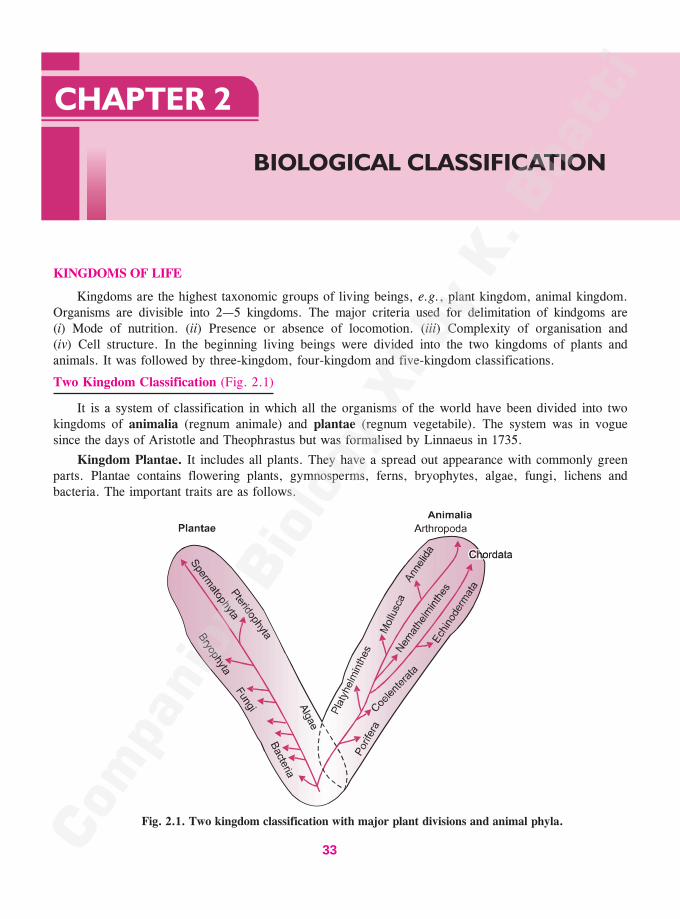
KINGDOMS OF LIFE
Kingdoms are the highest taxonomic groups of living beings, e.g., plant kingdom, animal kingdom.Organisms are divisible into 2—5 kingdoms. The major criteria used for delimitation of kindgoms are(i) Mode of nutrition. (ii) Presence or absence of locomotion. (iii) Complexity of organisation and(iv) Cell structure. In the beginning living beings were divided into the two kingdoms of plants andanimals. It was followed by three-kingdom, four-kingdom and five-kingdom classifications.
Two Kingdom Classification (Fig. 2.1)
It is a system of classification in which all the organisms of the world have been divided into twokingdoms of animalia (regnum animale) and plantae (regnum vegetabile). The system was in voguesince the days of Aristotle and Theophrastus but was formalised by Linnaeus in 1735.
Kingdom Plantae. It includes all plants. They have a spread out appearance with commonly greenparts. Plantae contains flowering plants, gymnosperms, ferns, bryophytes, algae, fungi, lichens andbacteria. The important traits are as follows.
Fig. 2.1. Two kingdom classification with major plant divisions and animal phyla.
33
���������������������
��������
������� �Companion Biology–XI34
1. Form. Plants are generally irregular in outline. A symmetry is generally absent. It is because of thepresence of branches. Organs may have a regular form, e.g., flower, leaf.
2. External Organs. The various organs of the body are external, e.g., leaves, flowers, fruits.
3. Growth. It continues throughout the life. Growth is generally unlimited and is due to the presence ofdefinite growing points called meristems. New organs also continue to develop till death.
4. Locomotion. Plants are fixed, except for some microscopic forms. Individual parts may showmovements but they are slow. It is due to absence of muscles.
5. Surface Area. Plants have a spread-out configuration and large surface area for absorbingmaximum sunlight, water and proper gaseous exchange.
6. Photosynthesis. Plants are autotrophic. They have the ability to manufacture organic food frominorganic raw materials with the help of solar energy trapped by green pigment called chlorophyll. Theprocess is called photosynthesis. Autotrophic nutrition of plants is also called holophytic nutrition. Itinvolves absorption of inorganic nutrients from outside.
7. Reserve Food. It is starch and oil.
8. Sense Organs. Sense organs and nervous system are absent.
9. Irritability. Response to external stimuli is slow except in a few cases.
10. Excretory Organs. They are absent.
11. Cell Structure. (i) Plant cells are bounded by a rigid cell wall. (ii) A central sap vacuole occursin mature cells. This pushes the cytoplasm to the periphery. (iii) A centrosome is generally absent.(iv) The cells contain plastids. (v) Inorganic crystals are often present.
12. Asexual Reproduction. It occurs widely through vegetative propagules and spores.
13. Sexual Reproduction. Sexual reproduction requires the assistance of external agencies likewind, water, insects, etc.
Kingdom Animalia. The kingdom contains all the animals. Animals are generally characterised byingestive type of nutrition and locomotion. Members of the kingdom animalia are protozoans, sponges,jelly fishes, worms, insects and other arthropods, molluscs, star fishes, frogs, reptiles, birds andmammals. The important traits of animals are :
1. Form. Animals have a regular form and a definite symmetry.
2. Organs. They are internal.
3. Growth. It is limited and does not occur after reaching a certain stage. Growth is diffused.Growing points are absent.
4. Locomotion. Animals move about bodily from one place to another in search of food and othernecessities of life. Their body parts also show quick movements. It is due to the presence of muscles.
5. Surface Area. Animals have a compact body with small surface area.
6. Nutrition. Animals obtain organic food from outside. Nutrition is holozoic or of ingestive type.Elimination of undigested food or defaecation occurs.
7. Reserve Food. It is glycogen and fat.
8. Sensory Organs. Sensory organs and nervous system are present. They make the animals awareof their surroundings.
9. Irritability. Animals are quick to respond to various stimuli.
10. Excretory Organs. They are present for collection and elimination of waste products, mostly ofnitrogen metabolism.
11. Cell Structure. (i) A cell wall is absent. (ii) Small vacuoles occur instead of a central vacuole.(iii) Centrosome or central apparatus occurs. (iv) Plastids are absent. (v) Inorganic crystals do not occur.
12. Asexual Reproduction. It is absent in higher forms.
13. Sexual Reproduction. An external agency is not required.
14. Parental Care. It occurs in animals for feeding and protecting the young ones.
���������� ��������� ��� 35
Objections. Two-kingdom classification was proposed in the period when details of cell structure were
unknown. Therefore, a number of shortcomings occur in the system.
1. Viruses. They have been placed in kingdom plantae of two-kingdom classification. However, viruses
are neither plants nor animals. They do not have a cellular structure or a metabolic machinery of their own.
Viruses lie on the border-line between living and the non-living.
2. Procaryotes. They do not have sap vacuoles, membrane bound organelles, true nucleus, spindle
apparatus and sexual reproduction. Procaryotes cannot be related to plants which are eucaryotic.
3. Motile Algae. Chlamydomonas and a number of other unicellular algae possess cilia or flagella for
locomotion in the aquatic habitat.
4. Dual Organisms. Organisms like Euglena have features of both plants and animals. They are
unicellular, motile flagellate organisms with chloroplasts. They perform photosynthesis in the presence of
light. In dark, they lose chloroplasts and become saprotrophic or holozoic. Organisms like Euglena are
studied by both botanists and zoologists.
5. Slime Moulds. They have no cell wall in the vegetative phase. Nutrition is holozoic like animals.
Cell walls develop in the reproductive stage.
6. Fungi. They are included amongst plants though they are quite different in structure, physiology,
reproduction, food reserve and absence of chlorophyll.
7. Lichens. They are included amongst plants despite being dual organisms where there is a symbiotic
type of association between a fungus and a green (eucaryotic) or blue-green (procaryotic) alga.
8. Sponges and Corals. They are fixed like plants and superficially resemble them in being irregular
and slow to react.
9. Tunicates and Plants. Tunicates resemble plants in having cellulose, branching and anchorage.
10. Unicellular and Multicellular Organisms. The two kingdom classification includes both unicellular
and multicellular organisms though they possess quite different types of organisation.
11. Unicellular Organisms. Protozoans have little resemblance to animals. Many of them have green,
brown or red colouration.
Procaryotes and Eucaryotes (Chatton 1925)
Study of bacteria, cyanobacteria, mycoplasma, and rickettsia under the electron microscope has
revealed that their cells are quite different from those of others. They have a single compartment. A true
nucleus is absent. DNA is without association with histone. It may, however, get coiled to form a compact
structure called nucleoid. Chatton (1925) designated such organisms as procaryotes (Gk. pro—before,
karyon—nucleus). Their cells are called procaryotic cells. The cells of other organisms have two
compartments, outer plasma membrane contained entire cell and inner nucleus containing genetic material.
They have been named as eucaryotes (Gk. eu—true, karyon—nucleus) by Chatton (1925). Their cells are
called eucaryotic cells. Procaryotic cells are smaller in size with a volume of 0·2—10·0 �m3. Eucaryotic
cells are larger with a volume of 1000—10,000 �m3. In comparison the viruses have a volume of only 10–6—
10–3 �m3. Very large cells occur in eggs. The largest eucaryotic cell is Ostrich egg on land and Whale
Shark in sea. However, the large size of egg cells is due to stored food. The size of their nucleus is similar
to that of other eucaryotic cells.
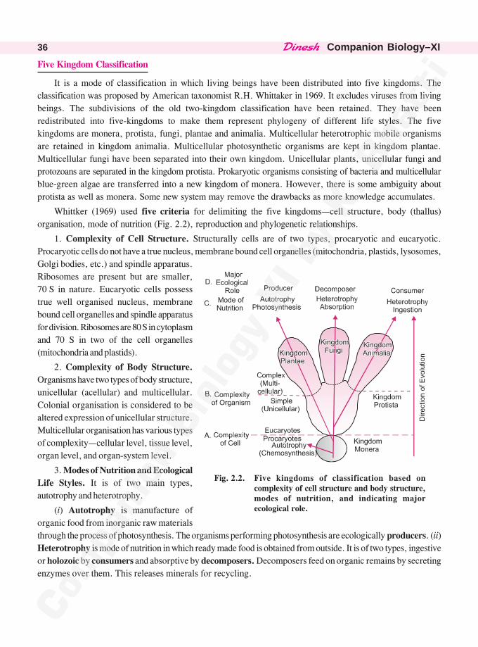
������� �Companion Biology–XI36
Five Kingdom Classification
It is a mode of classification in which living beings have been distributed into five kingdoms. The
classification was proposed by American taxonomist R.H. Whittaker in 1969. It excludes viruses from living
beings. The subdivisions of the old two-kingdom classification have been retained. They have been
redistributed into five-kingdoms to make them represent phylogeny of different life styles. The five
kingdoms are monera, protista, fungi, plantae and animalia. Multicellular heterotrophic mobile organisms
are retained in kingdom animalia. Multicellular photosynthetic organisms are kept in kingdom plantae.
Multicellular fungi have been separated into their own kingdom. Unicellular plants, unicellular fungi and
protozoans are separated in the kingdom protista. Prokaryotic organisms consisting of bacteria and multicellular
blue-green algae are transferred into a new kingdom of monera. However, there is some ambiguity about
protista as well as monera. Some new system may remove the drawbacks as more knowledge accumulates.
Whittker (1969) used five criteria for delimiting the five kingdoms—cell structure, body (thallus)
organisation, mode of nutrition (Fig. 2.2), reproduction and phylogenetic relationships.
1. Complexity of Cell Structure. Structurally cells are of two types, procaryotic and eucaryotic.
Procaryotic cells do not have a true nucleus, membrane bound cell organelles (mitochondria, plastids, lysosomes,
Golgi bodies, etc.) and spindle apparatus.
Ribosomes are present but are smaller,
70 S in nature. Eucaryotic cells possess
true well organised nucleus, membrane
bound cell organelles and spindle apparatus
for division. Ribosomes are 80 S in cytoplasm
and 70 S in two of the cell organelles
(mitochondria and plastids).
2. Complexity of Body Structure.
Organisms have two types of body structure,
unicellular (acellular) and multicellular.
Colonial organisation is considered to be
altered expression of unicellular structure.
Multicellular organisation has various types
of complexity—cellular level, tissue level,
organ level, and organ-system level.
3. Modes of Nutrition and Ecological
Life Styles. It is of two main types,
autotrophy and heterotrophy.
(i) Autotrophy is manufacture of
organic food from inorganic raw materials
through the process of photosynthesis. The organisms performing photosynthesis are ecologically producers. (ii)
Heterotrophy is mode of nutrition in which ready made food is obtained from outside. It is of two types, ingestive
or holozoic by consumers and absorptive by decomposers. Decomposers feed on organic remains by secreting
enzymes over them. This releases minerals for recycling.
Fig. 2.2. Five kingdoms of classification based oncomplexity of cell structure and body structure,modes of nutrition, and indicating majorecological role.
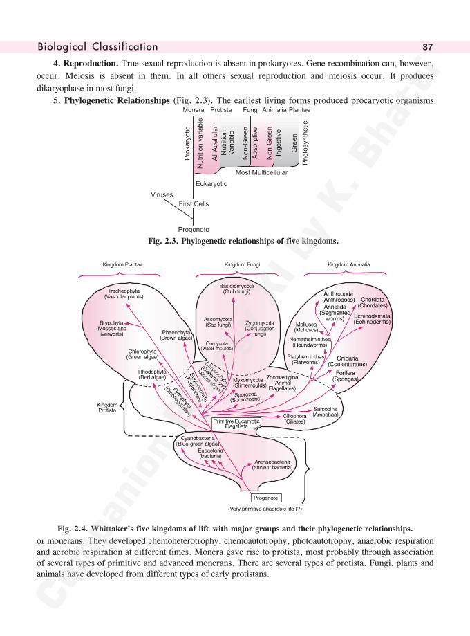
���������� ��������� ��� 37
4. Reproduction. True sexual reproduction is absent in prokaryotes. Gene recombination can, however,
occur. Meiosis is absent in them. In all others sexual reproduction and meiosis occur. It produces
dikaryophase in most fungi.
5. Phylogenetic Relationships (Fig. 2.3). The earliest living forms produced procaryotic organisms
Fig. 2.3. Phylogenetic relationships of five kingdoms.
Fig. 2.4. Whittaker’s five kingdoms of life with major groups and their phylogenetic relationships.
or monerans. They developed chemoheterotrophy, chemoautotrophy, photoautotrophy, anaerobic respirationand aerobic respiration at different times. Monera gave rise to protista, most probably through associationof several types of primitive and advanced monerans. There are several types of protista. Fungi, plants andanimals have developed from different types of early protistans.
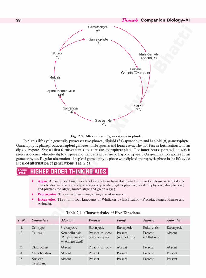
������� �Companion Biology–XI38
Fig. 2.5. Alternation of generations in plants.
In plants life cycle generally possesses two phases, diploid (2n) sporophyte and haploid (n) gametophyte.Gametophytic phase produces haploid gametes, male sperms and female ova. The two fuse in fertilization to formdiploid zygote. Zygote first forms embryo and then the sporophyte plant. The latter bears sporangia in whichmeiosis occurs whereby diploid spore mother cells give rise to haploid spores. On germination spores formgametophytes. Regular alternation of haploid gametophytic phase with diploid sporophytic phase in the life cycleis called alternation of generations (Fig. 2.5).
• Algae. Algae of two kingdom classification have been distributed in three kingdoms in Whittaker’sclassification—monera (blue-green algae), protista (euglenophyceae, bacillariophyceae, dinophyceae)and plantae (red algae, brown algae and green algae).
• Procaryotes. They constitute a single kingdom of monera.
• Eucaryotes. They form four kingdoms of Whittaker’s classification—Protista, Fungi, Plantae andAnimalia.
Table 2.1. Characteristics of Five Kingdoms
S. No. Characters Monera Protista Fungi Plantae Animalia
1. Cell type Prokaryotic Eukaryotic Eukaryotic Eukaryotic Eukaryotic
2. Cell wall Non-cellulosic Present in some Present Present Absent(Polysaccharide (various type) (with chitin) (Cellulose)+ Amino acid)
3. Chloroplast Absent Present in some Absent Present Absent
4. Mitochondria Absent Present Present Present Present
5. Nuclear Absent Present Present Present Presentmembrane
���������� ��������� ��� 39
6. Tissue or Multi- Absent Absent Present but Present in Present incellularity limited all forms all forms
7. Body Organi- Cellular Cellular Multicellular/ Tissue/Organ Tissue/sation loose tissue Organ/ Organ
system
8. Motility Bacterial Cilia, Flagella, Cilia and Cilia and Cilia andflagella, amoeboid, Flagella in Flagella in Flagella,gliding or contractile some, none lower forms, contractilenon-motile fibrils in most of absent in fibrils
the forms. most of theforms
9. Mode of Autotrophic, Phosynthesis Heteroprophic, Autotrophic Heterotrophicnutrition chemosyn- and Saprophytic, Photosynth- (Holozoic
thetic and heterotrophic Parasitic esis saprophytic)photosynthetic, absorptionheterotrophic(saprophyteand parasite)
10. Reproduction/ Conjugation Syngamy and Fertilization Fertilization Fertilizationmeans of Transduction meiosis, and meiosis, and meiosis and meiosisgenetic Transforma- conjugation or dikaryosisrecombination tion or none none or none
11. Nervous system Absent Primitive for Absent Absent Present,conducting oftenstimuli complex
Advantages of Five Kingdom Classification
1. It brings out phylogeny in the living world.
2. The system is based on levels of organisation and modes of nutrition. The latter became established
very early during evolution.
3. Kingdom animalia has become more homogeneous with the exclusion of protozoa. Evolutionary
tendencies and pathways are more clearly defined.
4. Kingdom plantae has become more coherent after exclusion of bacteria, fungi and some unicellular
algal forms. Evolutionary steps from green algae to top of embryophytes can be studied clearly.
5. Separation of fungi from plants as well as slime moulds is a wise step.
6. Early eucaryotic forms which have flexible modes of nutrition and life styles have been separated
and placed in kingdom protista.
7. Creation of kingdom monera for procaryotes is fully justified because they have their own level of
structural and biochemical organisation.
Disadvantages of Five Kingdom Classification
1. Kingdom protista is highly heterogeneous group with several lines of evolution.
2. It is very difficult to make distinction of unicellular and multicellular forms of algae. Placing algae
in three kingdoms seems to be unrealistic.
3. Slime Moulds do not fit into kingdom protista.
4. Viruses do not find any place in this system of classification.
5. Red and Brown algae are not related to other members of kingdom plantae.
6. Archaebacteria are quite different from other prokaryotes.
������� �Companion Biology–XI40
Three Domains of Life
Woese (1994) has divided living beings into three super kingdoms or domains of Archaea, Bacteriaand Eukarya.
Domain Archaea contains a single kingdom of Archaebacteria. Archaebacteria differ from botheubacteria and eukaryotes. (i) The cell wall does not contain cellulose or peptidoglycan. (ii) Cellmembrane possess a single layer of branched chain lipid having ether bond (–O·CH2) instead of esterbonds (O·CO). (iii) Histone-like proteins are present. (iv) 16S RNA has many nucleotides different fromother organisms.
Domain Bacteria contains a single kingdom of eubacteria having monera minus archaebacteria.Domain Eukarya has four kingdoms of protista, fungi, plantae and animalia. However, some
workers have raised their number to five (Keeling et al 2005) and six (Adl et al 2005)In this way, Woese (1994) has proposed a six kingdom classification.
VIRUSES (Akaryota)
Virus is an ultramicroscopic nucleoprotein entity which becomes active only inside a living cell using thelatter’s machinery for multiplication through separate synthesis of its parts. Scientific work on virus started withMayer (1886) studying Tobacco mosaic disease which could pass through bacterial filters. Dmitri Ivanowski(1892) found that the disease was due to a agent present in the extract of diseased plant. Ivanowski (1892) is alsocredited with the discovery and naming of virus (L. virus—poison). Beijerinck (1896) called the extract ofdiseased plant to be contagium vivum fluidum or living infectious fluid, virus. At the same time, Loeffer andFrosch (1898) investigated that the causative agent of foot and mouth disease was also an agent that could passthough bacterial filter. Reed (1902) discovered that the agent of Yellow Fever which could pass through bacterialfilter was transmitted by bite of infected mosquitoes. Twort (1915) and d’Herelle (1917) discoveredbacteriophages because of the ability to cause lysis of bacteria (Twort—d’Herelle phenomenon). Virus waspurified by Schleissinger (1933) while Stanley (1935) was able to crystallise Tobacco Mosaic Virus (TMV).Bawden and Pirie (1937) found TMV to be nucleoprotein entity which contains RNA (ribonucleic acid). Takahashiand Rawlins (1933) observed virus under light microscope while Stanley (1946) saw the virus under electronmicroscope. Hershey and Chase (1952) found that genetic material of T2 bacteriophage is DNA. The geneticmaterial of TMV was found out to be RNA by Conrat (1953) and confirmed by Gierere and Schramm (1956).Single stranded DNA was found to represent genetic material in bacteriophase ��× 174 (Sinsheimer, 1959).Retroviruses were discovered by Temin and Baltimore (1970). Holmes (1948) classified viruses on the basis oforganisms they parasitise, viz., phaginae, phytophaginae and zoophaginae.
Virus is obligate parasite. It cannot be grown on artificial culture medium. Living cells are essential. Virusis inert outside host cell. The inert particle of virus, outside its host, is called virion. On the basis of host, avirus is called zoophage (animal virus), phytophage (plant virus), bacteriophage (bacterial virus), mycophage(fungal virus), phycophage (algal virus), cyanophage (blue-green algal virus), coliphage (bacteriophage of colonbacterium Escherichia coli), etc. Volume ranges from 10–6—10–3 �m3. Size of virus ranges from about 10 nm(Foot and Mouth Virus of Cattle), 17 nm (Alfalfa Mosaic Virus) to 1250 × 40 nm (Beet Yellow Virus). TMVis 300 × 18·0 nm. Very large viruses have been recently discovered—Mimivirus, Megavirus, Pandovirus. Theirgenomes carry 900—2500 genes (Claverie and Abergel 2013). Many of these genes are metabolic genes for sugar,lipid and amino acid metabolism.
Architecturally virus is of three types—helical (cylindrical, elongated), cuboidal (rounded or polyhedral)and binal (cuboidal head and helical tail). Binal virus bacteriophage T2 or T4 has an end plate with tail fibres(Fig. 2.8).
Structurally a virus has four parts : (i) Envelope. It is the outer loose covering present in certainviruses (e.g., HIV or Human Immunodeficiency Virus) and made of protein of viral origin, lipid andcarbohydrate of host. Outgrowths called spikes may or may not be present. Envelope protein has subunits calledpeplomers. A virus without envelope is naked virus. (ii) Capsid. It is protein covering around the geneticmaterial. Capsid has protein subunits called capsomeres. TMV has 2130 capsomeres. They arearranged helically or in geometric forms. Capsid has antigenic properties. (iii) Nucleoid. It represents genetic
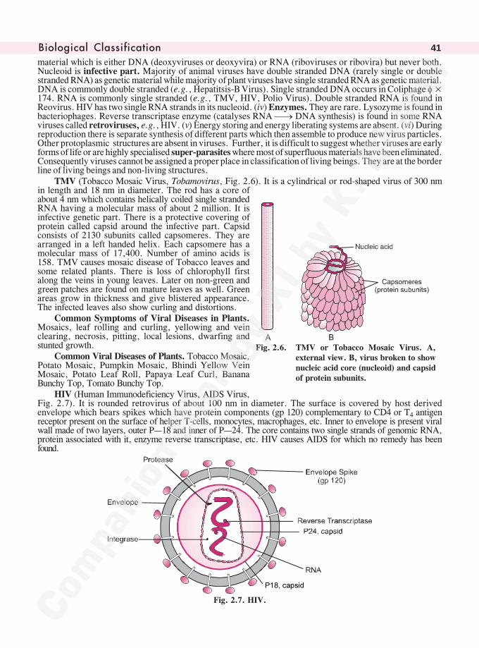
���������� ��������� ��� 41
material which is either DNA (deoxyviruses or deoxyvira) or RNA (riboviruses or ribovira) but never both.Nucleoid is infective part. Majority of animal viruses have double stranded DNA (rarely single or doublestranded RNA) as genetic material while majority of plant viruses have single stranded RNA as genetic material.DNA is commonly double stranded (e.g., Hepatitsis-B Virus). Single stranded DNA occurs in Coliphage ��×174. RNA is commonly single stranded (e.g., TMV, HIV, Polio Virus). Double stranded RNA is found inReovirus. HIV has two single RNA strands in its nucleoid. (iv) Enzymes. They are rare. Lysozyme is found inbacteriophages. Reverse transcriptase enzyme (catalyses RNA �� DNA synthesis) is found in some RNAviruses called retroviruses, e.g., HIV. (v) Energy storing and energy liberating systems are absent. (vi) Duringreproduction there is separate synthesis of different parts which then assemble to produce new virus particles.Other protoplasmic structures are absent in viruses. Further, it is difficult to suggest whether viruses are earlyforms of life or are highly specialised super-parasites where most of superfluous materials have been eliminated.Consequently viruses cannot be assigned a proper place in classification of living beings. They are at the borderline of living beings and non-living structures.
TMV (Tobacco Mosaic Virus, Tobamovirus, Fig. 2.6). It is a cylindrical or rod-shaped virus of 300 nmin length and 18 nm in diameter. The rod has a core ofabout 4 nm which contains helically coiled single strandedRNA having a molecular mass of about 2 million. It isinfective genetic part. There is a protective covering ofprotein called capsid around the infective part. Capsidconsists of 2130 subunits called capsomeres. They arearranged in a left handed helix. Each capsomere has amolecular mass of 17,400. Number of amino acids is158. TMV causes mosaic disease of Tobacco leaves andsome related plants. There is loss of chlorophyll firstalong the veins in young leaves. Later on non-green andgreen patches are found on mature leaves as well. Greenareas grow in thickness and give blistered appearance.The infected leaves also show curling and distortions.
Common Symptoms of Viral Diseases in Plants.Mosaics, leaf rolling and curling, yellowing and veinclearing, necrosis, pitting, local lesions, dwarfing andstunted growth.
Common Viral Diseases of Plants. Tobacco Mosaic,Potato Mosaic, Pumpkin Mosaic, Bhindi Yellow VeinMosaic, Potato Leaf Roll, Papaya Leaf Curl, BananaBunchy Top, Tomato Bunchy Top.
HIV (Human Immunodeficiency Virus, AIDS Virus,Fig. 2.7). It is rounded retrovirus of about 100 nm in diameter. The surface is covered by host derivedenvelope which bears spikes which have protein components (gp 120) complementary to CD4 or T4 antigenreceptor present on the surface of helper T-cells, monocytes, macrophages, etc. Inner to envelope is present viralwall made of two layers, outer P—18 and inner of P—24. The core contains two single strands of genomic RNA,protein associated with it, enzyme reverse transcriptase, etc. HIV causes AIDS for which no remedy has beenfound.
���������
Fig. 2.7. HIV.
Fig. 2.6. TMV or Tobacco Mosaic Virus. A,external view. B, virus broken to shownucleic acid core (nucleoid) and capsidof protein subunits.
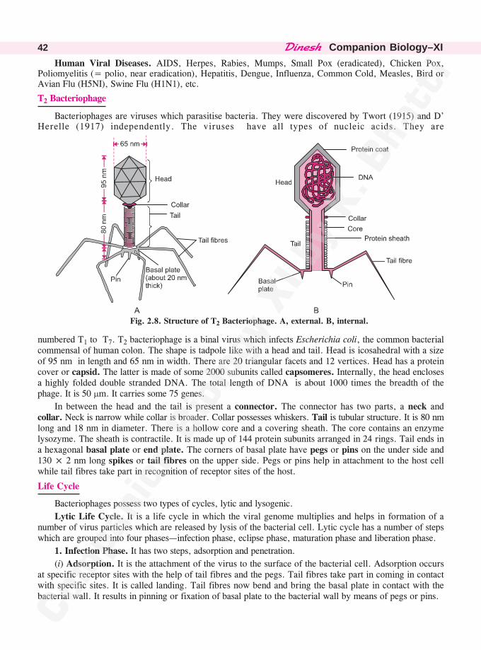
������� �Companion Biology–XI42
Human Viral Diseases. AIDS, Herpes, Rabies, Mumps, Small Pox (eradicated), Chicken Pox,Poliomyelitis (= polio, near eradication), Hepatitis, Dengue, Influenza, Common Cold, Measles, Bird orAvian Flu (H5NI), Swine Flu (H1N1), etc.
T2 Bacteriophage
Bacteriophages are viruses which parasitise bacteria. They were discovered by Twort (1915) and D’Herelle (1917) independently. The viruses have all types of nucleic acids. They are
Fig. 2.8. Structure of T2 Bacteriophage. A, external. B, internal.
numbered T1 to T7. T2 bacteriophage is a binal virus which infects Escherichia coli, the common bacterialcommensal of human colon. The shape is tadpole like with a head and tail. Head is icosahedral with a sizeof 95 nm in length and 65 nm in width. There are 20 triangular facets and 12 vertices. Head has a proteincover or capsid. The latter is made of some 2000 subunits called capsomeres. Internally, the head enclosesa highly folded double stranded DNA. The total length of DNA is about 1000 times the breadth of thephage. It is 50 �m. It carries some 75 genes.
In between the head and the tail is present a connector. The connector has two parts, a neck andcollar. Neck is narrow while collar is broader. Collar possesses whiskers. Tail is tubular structure. It is 80 nmlong and 18 nm in diameter. There is a hollow core and a covering sheath. The core contains an enzymelysozyme. The sheath is contractile. It is made up of 144 protein subunits arranged in 24 rings. Tail ends ina hexagonal basal plate or end plate. The corners of basal plate have pegs or pins on the under side and130 × 2 nm long spikes or tail fibres on the upper side. Pegs or pins help in attachment to the host cellwhile tail fibres take part in recognition of receptor sites of the host.
Life Cycle
Bacteriophages possess two types of cycles, lytic and lysogenic.
Lytic Life Cycle. It is a life cycle in which the viral genome multiplies and helps in formation of anumber of virus particles which are released by lysis of the bacterial cell. Lytic cycle has a number of stepswhich are grouped into four phases—infection phase, eclipse phase, maturation phase and liberation phase.
1. Infection Phase. It has two steps, adsorption and penetration.
(i) Adsorption. It is the attachment of the virus to the surface of the bacterial cell. Adsorption occursat specific receptor sites with the help of tail fibres and the pegs. Tail fibres take part in coming in contactwith specific sites. It is called landing. Tail fibres now bend and bring the basal plate in contact with thebacterial wall. It results in pinning or fixation of basal plate to the bacterial wall by means of pegs or pins.
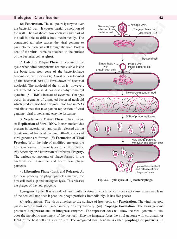
���������� ��������� ��� 43
(ii) Penetration. The tail pours lysozyme over
the bacterial wall. It causes partial dissolution of
the wall. The tail sheath now contracts and part of
the tail is able to drill a hole mechanically. The
contracted tail also causes the viral genome to
pass into the bacterial cell through the hole. Protein
coat of the virus remains attached to the surface
of the bacterial cell as ghost.
2. Latent or Eclipse Phase. It is phase of life
cycle when viral components are not visible inside
the bacterium. dna gene of the bacteriophage
becomes active. It causes (i) Arrest of development
of the bacterial host.(ii) Breakdown of bacterial
nucleoid. The nucleoid of the virus is, however,
not affected because it possesses 5-hydromethyl
cytosine (5—HMC) instead of cytosine. Changes
occur in segments of disrupted bacterial nucleoid
which produce modified enzymes, modified mRNAs
and ribosomes that take part in replication of viral
genome, viral proteins and enzyme lysozyme.
3. Vegetative or Mature Phase. It has 3 steps.
(i) Replication of Viral DNA. It uses nucleotides
present in bacterial cell and partly released during
breakdown of bacterial nucleoid. 40—80 copies of
viral genome are formed. (ii) Formation of Viral
Proteins. With the help of modified enzymes the
host synthesises different types of viral proteins.
(iii) Assembly or Maturation of Infective Progeny.
The various components of phage formed in the
bacterial cell assemble and form new phage
particles.
4. Liberation Phase (Lysis and Release). As
the new progeny of phage particles mature, the
host cell swells up and undergoes lysis. This releases
the phages of the new progeny.
Lysogenic Cycle. It is a mode of viral multiplication in which the virus does not cause immediate lysis
of the host cell nor does it produce phage particles immediately. It has five phases
(i) Adsorption. The virus attaches to the surface of host cell. (ii) Penetration. The viral nucleoid
passes into the host cell, mechanically or enzymatically. (iii) Prophage Formation. The virus genome
produces a repressor and an integrase enzymes. The repressor does not allow the viral genome to take
over the metabolic machinery of the host cell. Enzyme integrase fuses the viral genome with chromatin or
DNA of the host cell at a specific site. The integrated viral genome is called prophage or provirus. In
Fig. 2.9. Lytic cycle of T2 Bacteriophage.
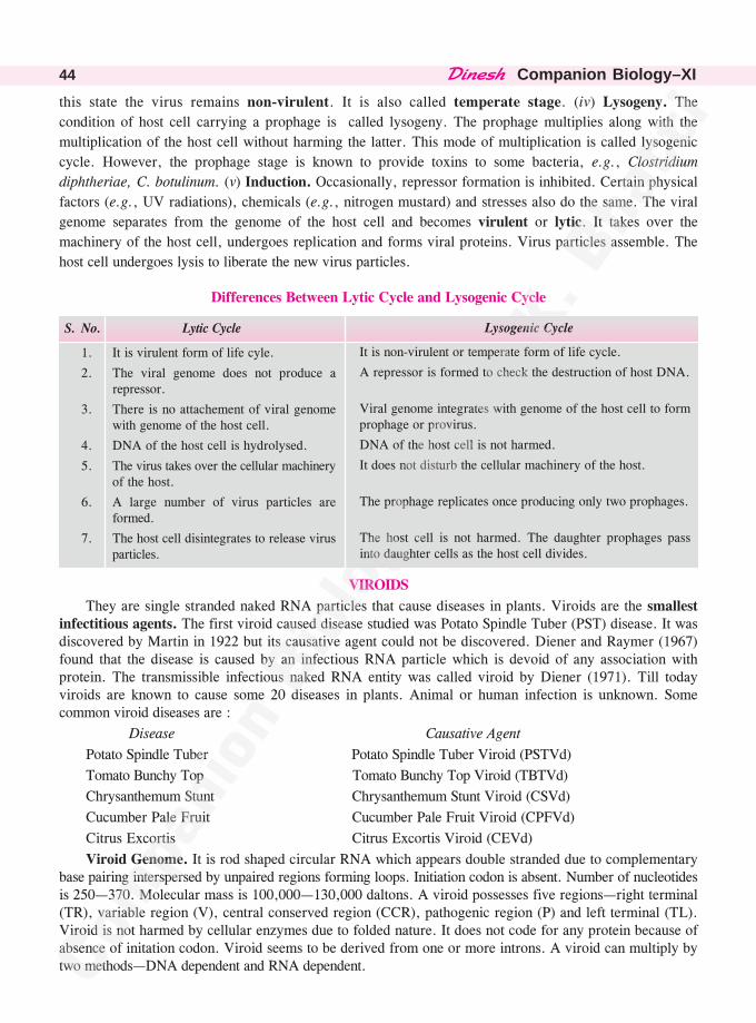
������� �Companion Biology–XI44
Lysogenic Cycle
It is non-virulent or temperate form of life cycle.
A repressor is formed to check the destruction of host DNA.
Viral genome integrates with genome of the host cell to formprophage or provirus.
DNA of the host cell is not harmed.
It does not disturb the cellular machinery of the host.
The prophage replicates once producing only two prophages.
The host cell is not harmed. The daughter prophages passinto daughter cells as the host cell divides.
this state the virus remains non-virulent. It is also called temperate stage. (iv) Lysogeny. The
condition of host cell carrying a prophage is called lysogeny. The prophage multiplies along with the
multiplication of the host cell without harming the latter. This mode of multiplication is called lysogenic
cycle. However, the prophage stage is known to provide toxins to some bacteria, e.g., Clostridium
diphtheriae, C. botulinum. (v) Induction. Occasionally, repressor formation is inhibited. Certain physical
factors (e.g., UV radiations), chemicals (e.g., nitrogen mustard) and stresses also do the same. The viral
genome separates from the genome of the host cell and becomes virulent or lytic. It takes over the
machinery of the host cell, undergoes replication and forms viral proteins. Virus particles assemble. The
host cell undergoes lysis to liberate the new virus particles.
Differences Between Lytic Cycle and Lysogenic Cycle
S. No. Lytic Cycle
1. It is virulent form of life cyle.
2. The viral genome does not produce arepressor.
3. There is no attachement of viral genomewith genome of the host cell.
4. DNA of the host cell is hydrolysed.
5. The virus takes over the cellular machineryof the host.
6. A large number of virus particles areformed.
7. The host cell disintegrates to release virusparticles.
VIROIDS
They are single stranded naked RNA particles that cause diseases in plants. Viroids are the smallestinfectitious agents. The first viroid caused disease studied was Potato Spindle Tuber (PST) disease. It wasdiscovered by Martin in 1922 but its causative agent could not be discovered. Diener and Raymer (1967)found that the disease is caused by an infectious RNA particle which is devoid of any association withprotein. The transmissible infectious naked RNA entity was called viroid by Diener (1971). Till todayviroids are known to cause some 20 diseases in plants. Animal or human infection is unknown. Somecommon viroid diseases are :
Disease Causative Agent
Potato Spindle Tuber Potato Spindle Tuber Viroid (PSTVd)
Tomato Bunchy Top Tomato Bunchy Top Viroid (TBTVd)
Chrysanthemum Stunt Chrysanthemum Stunt Viroid (CSVd)
Cucumber Pale Fruit Cucumber Pale Fruit Viroid (CPFVd)
Citrus Excortis Citrus Excortis Viroid (CEVd)
Viroid Genome. It is rod shaped circular RNA which appears double stranded due to complementarybase pairing interspersed by unpaired regions forming loops. Initiation codon is absent. Number of nucleotidesis 250—370. Molecular mass is 100,000—130,000 daltons. A viroid possesses five regions—right terminal(TR), variable region (V), central conserved region (CCR), pathogenic region (P) and left terminal (TL).Viroid is not harmed by cellular enzymes due to folded nature. It does not code for any protein because ofabsence of initation codon. Viroid seems to be derived from one or more introns. A viroid can multiply bytwo methods—DNA dependent and RNA dependent.
���������� ��������� ��� 45
Fig. 2.10. Regions of a Viroid.
(i) DNA Dependent Replication. (a) Viroids can develop directly from one or more segments of DNA.(b) With the help of enzyme reverse transcriptase, a viroid forms its copy DNA which develops itscomplentary strand. The new DNA synthesises a number of copies of viroid RNA.
Viroid RNA ����������������
���� cDNA ��� DNA ����
��������
�� Viroid RNAs.
(ii) RNA Dependent Replication. Branch and Robertson (1984) have found infected plant cell topossess RNA polymerase that can allow RNA replication over RNA.
Viroid RNA ��������������� ��
������������� Viroid RNAs
Differences between Viruses and Viroids
S.No Viruses Viroids
1. Larger than viroids Smaller than viruses.
2. They have a protein coat. They lack protein coat.
3. Genome consists of DNA or RNA. They are RNA particles.4. Their RNA has relatively high molecular Their RNA has low molecular weight.
weight.5. They parasitise all types of organisms. They parasitise only plants.
Potato Spindle Tuber Disease
It is found in all potato growing areas of the world including India (Owens et al, 1992). The diseasedplants show spindly appearance. They remain small and become erect. The leaflets become greyish green.They may show rolling and twisting. Tuber yield is reduced by 25% or more. Tubers remain small spindle-like elongated with tapering ends. Eyes are numerous and prominent with enlarged bud scales. Cracks mayappear at places. Such tubers are not worth marketing.
The viroid causing Potato Spindle Tuber disease has a length of 50 nm and 359 nucleotides with amolecular mass of about 1,00,000 daltons. The viroid spreads by a number of methods—(i) Seed tubers.(ii) Knives. (iii) Agricultural implements. (iv) Pollen (Singh et al, 1992). (v) Insects.
Control Measures. (i) Use of only healthy seed tubers. (ii) Uprooting of diseased plants and theirburning. (iii) Burning of debris of harvested crop. (iv) Use of sterilised implements. (v) Soil sterilisation.
Position of Viruses
Resemblance with Living Beings. (i) Presence of genetic material. (ii) Occurrence of variations due tomutations. (iii) Reproduction. (iv) Daughter viruses are genetically similar to parent virus. (v) Viruses are killedon exposure to ultraviolet radiations. (vi) They are killed by autoclaving. (vii) Viruses are obligate parasites.(viii) They have host specificity. (ix) A virus takes over cellular machinery of host cell. (x) It has antigenicproperty. (xi) Virus is made of organic molecules. (xii) Occasional presence of enzymes.
• ������������ ����������������� ����������������• Prions (Prusiner 1983, Nobel Prize 1997). They are protein particles which function as obligate
parasites causing scrapie in Sheep and Goats, Mad Cow disease. Prions are rods or fibrils which areresistant to protease action.
• Virology. Study of viruses.
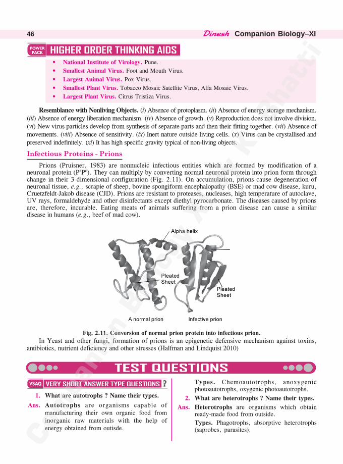
������� �Companion Biology–XI46
• National Institute of Virology. Pune.
• Smallest Animal Virus. Foot and Mouth Virus.
• Largest Animal Virus. Pox Virus.
• Smallest Plant Virus. Tobacco Mosaic Satellite Virus, Alfa Mosaic Virus.
• Largest Plant Virus. Citrus Tristiza Virus.
Resemblance with Nonliving Objects. (i) Absence of protoplasm. (ii) Absence of energy storage mechanism.(iii) Absence of energy liberation mechanism. (iv) Absence of growth. (v) Reproduction does not involve division.(vi) New virus particles develop from synthesis of separate parts and then their fitting together. (vii) Absence ofmovements. (viii) Absence of sensitivity. (ix) Inert nature outside living cells. (x) Virus can be crystallised andpreserved indefinitely. (xi) It has high specific gravity typical of non-living objects.
��������� ��������� ����Prions (Pruisner, 1983) are nonnucleic infectious entities which are formed by modification of a
neuronal protein (PrPc). They can multiply by converting normal neuronal protein into prion form throughchange in their 3-dimensional configuration (Fig. 2.11). On accumulation, prions cause degeneration ofneuronal tissue, e.g., scrapie of sheep, bovine spongiform encephalopathy (BSE) or mad cow disease, kuru,Cruetzfeldt-Jakob disease (CJD). Prions are resistant to proteases, nucleases, high temperature of autoclave,UV rays, formaldehyde and other disinfectants except diethyl pyrocarbonate. The diseases caused by prionsare, therefore, incurable. Eating meats of animals suffering from a prion disease can cause a similardisease in humans (e.g., beef of mad cow).
Fig. 2.11. Conversion of normal prion protein into infectious prion.
In Yeast and other fungi, formation of prions is an epigenetic defensive mechanism against toxins,antibiotics, nutrient deficiency and other stresses (Halfman and Lindquist 2010)
�����������
������ �
�����������
���������� �� ������ ����� ��
1. What are autotrophs ? Name their types.
Ans. Autotrophs are organisms capable ofmanufacturing their own organic food frominorganic raw materials with the help ofenergy obtained from outisde.
Types. Chemoautotrophs, anoxygenicphotoautotrophs, oxygenic photoautotrophs.
2. What are heterotrophs ? Name their types.
Ans. Heterotrophs are organisms which obtainready-made food from outside.
Types. Phagotrophs, absorptive heterotrophs(saprobes, parasites).
���������� ��������� ��� 47
3. Define holozoic nutrition.
Ans. Holozoic, ingestive or phagotrophic nutritionis a mode of heterotrophic nutrition found inanimals in which solid or particulate food istaken into the body for internal digestion andassimilation.
4. What is holophytic nutrition ?
Ans. Holophytic nutrition is autotrophic nutritionas found in plants in which inorganic rawmaterials are obtained from outside forconversion into organic state with the help ofsolar energy.
5. What do you mean by absorptivenutrition ?
Ans. Absorptive nutrition is a type of nutrition inwhich soluble organic food is obtained fromoutside.
6. Name the types of organisms which haveabsorptive nutrition.
Ans. Absorptive nutrition occurs in saprobes andparasites e.g., bacteria and fungi.
7. Name the scientist connected with two-kingdom classification.
Ans. Linnaeus (1758).
8. Who first of all proposed the kingdommonera ?
Ans. Monera or mychota by Copeland (1956).
9. Who first created the kingdom protista ?
Ans. Protista by Ernst Haeckel (1866).
10. Name the scientist who proposed the termsof procaryotes and eucaryotes.
Ans. Chatton (1925).
11. Who proposed five kingdom classification ?
Ans. Whittaker (1969).
12. To which kingdom do archaebacteriabelong ?
Ans. Archaebacteria belong to kingdom monera ofWhittaker 1969).
13. What is the kingdom of procaryotes ?
Ans. Monera.
14. Name the kingdom of unicellular eucaryotes.
Ans. Protista.
15. What is the kingdom of multicellulardecomposers ?
Ans. Fungi.
16. Name the kingdom of multicellularproducers.
Ans. Plantae.
17. What is the kingdom of multicellularconsumers ?
Ans. Animalia.
18. Name the structures not included in fivekingdom classification of Whittaker.
Ans. Viruses.
1. Ecologically animals are ..............
2. Animal kingdom exceeds all other kingdomsin ..............
3. Yeasts are a group of exceptional..............involved in fermentation.
4. Certain members of Euglena group of............. can lead .............. life.
5. ..............can tolerate high salt content, hightemperature (80°C), acidic pH and absenceof oxygen.
1. consumers 2. diversity 3. fungi
4. (i) protists (ii) dual 5. Archaebacteria
1. Procaryotes are organisms without a truenucleus.
2. All autotrophs are monerans.
3. Phytoplantktons perform 80% of thephotosynthetic activity.
1. True 2. False 3. True
1. Column I Column II
A Cyanobacteria a Symbiosis
B Diatom b Phytoplankton
C Protozoa c Fungi
D Lichen d Blue Green Algae
e Holozoic Nutrition
1. A—d, B—b, C—e, D—a.
������� �Companion Biology–XI48
1. How are viroids different from viruses ?
Ans. See text.
2. Give a brief account of viruses with respect totheir structure and nature of genetic material.Also name four common viral diseases.
Ans. Structure. Virus is a small nucleoprotein entitywhich can have three forms—helical or cylindrical,(e.g., TMV), cuboidal (rounded or polyhedral,e.g., HIV) and binal (cuboidal head and helicaltail, e.g., bacteriphage T2). A bacteriphage likeT2 or T4 has a basal end plate with tail fibres forsticking to its host.
Four types of structures are found in a virus —envelope, capsid, nucleoid and enzymes.
(i) Envelope. It is the outer loose covering presentin certain viruses (e.g., HIV or HumanImmunodeficiency Virus) and made of proteinof viral origin, lipid and carbohydrate of host.Outgrowths called spikes may or may not bepresent. Envelope protein has subunits calledpeplomers. A virus without envelope is nakedvirus. (ii) Capsid. It is protein covering aroundthe genetic material. Capsid has protein subunitscalled capsomeres. TMV has 2130 capsomeres.
They are arranged helically or in geometric forms.Capsid has antigenic properties. (iii) Nucleoid.It represents genetic as well as infective part ofvirus. Nucleoid consists of coiled DNA or RNAbut not both. (iv) Enzymes. They are rare.Lysozyme is found in bacteriophages. Reversetranscriptase enzyme (catalyses RNA �� DNAsynthesis) is found in some RNA viruses likeHIV.
Genetic Material. It is DNA in deoxyvira andRNA in ribovira. Majority of animal viruseshave double stranded DNA (rarely single ordouble stranded RNA) as genetic material whilemajority of plant viruses have single strandedRNA as genetic material. DNA is commonlydouble stranded (e.g., Hepatitis-B Virus). Singlestranded DNA occurs in Coliphage �� × 174.RNA is commonly single stranded (e.g., TMV,HIV, Polio Virus). Double stranded RNA isfound in Reovirus. HIV has two single RNAstrands in its nucleoid.
Common Viral Diseases. Potato Mosaic, BananaBunchy top, AIDS, Dengue, Poliomyelitis.
3. Organise a discussion in your class on thetopic—Are viruses living or non-living ?
Ans. Resemblance with Living Beings. See text.
Resemblance with Non-living Objects. See text.
1. The common name of Pea is simpler thanits botanical (scientific) name of Pisumsativum. Why is then the simpler commonname not used instead of the complexscientific/botanical name in biology ?
Ans. Common name of Pea is limited to Englishlanguage. It is known by different names inother languages. Scientific name of Pea,Pisum sativum, is universal for all thelanguages and regions.
2. In the five kingdom system of Whittaker,how many kingdoms are eukaryotes ?
Ans. Four.
1. How is the five kingdom classificationadvantageous over the two kingdomclassification ?
Ans. See text.
2. A virus is considered as a living organismand an obligate parasite when inside a hostcell. But virus is not classified alongwithbacteria or fungi. What are the charactersof virus that are similar to non-livingobjects ?
Ans. See text.
1. Biological classification is a dynamic andever evolving phenomenon which keepschanging with our understanding of lifeforms. Justify the statement taking any twoexamples.
Ans. Biological classification has never been static.It has been undergoing change throughout thehistory of taxanomy as and when new factsand understanding of life forms becameavailable. Initially there were artificial systems
(From N.C.E.R.T. Question Bank)
���������� ��������� ��� 49
of classification where a few characters werepicked up arbitrarily, e.g., Aristotle,Theopharastrus, Pliny, John Ray, Linnaeus.Despite his vast knowledge, Linnaeous dividedplants on the basis of sex organs alone.
As understanding of life forms increased withthe availability of new tools and techniques,more and more characters became available fordelimitation of taxa. This paved the way fornatural systems of classification. Changes haveoccurred in them from time to time.
Two kingdom classification was replaced bythree kingdom classification when Haeckel(1866) proposed the kingdom of protista. It wasconverted into four kingdom classification byCopeland (1955) when he raised the kingdom ofmonera. Whittakar (1969) proposed five kingdomclassification. As information about biochemistryof archaebacteria became known, Woese hasraised another kingdom of Archaea.
(See also NCERT question 1 of chapter 3)
1. Whittaker (1969) was pioneer taxonomist whowas able to arrange living organisms accordingto their natural relationships.(a) How many groups of organisms were created
by Whittaker ?(b) What criteria were used by Whittaker for
creating groups of organisms ?(c) What are the advantages of Whittaker’s
classification ?Ans. (a) Five kingdoms of monera, protista, fungi,
plantae and animalia.(b) Five— (i) Compexity of cell structure.
(ii)Complexity of body structure(iii) Mode of nutrition. (iv) Reproduction.(v) Phylogenetic relationships.
(c) (i) Separation of protista, monera and fungihas made classification of organisms morereal and without confusion which wasprevailing earlier. (ii) Both kingdoms ofplantae and animalia are now morehomogeneous. (iii) It has brought outphylogeny of different organisms.
2. Viruses have two types of life cycles, lytic andlysogenic. In both cases, the virus first attachesitself to the surface of the host cell. Then itsends it genome into the host cell. The viralgenome takes over the metabolic machinery ofthe host cell. It creates more viral genomes andthen manufactures the viral covers to enclosethe genomes. The new virus particles come outof the host cell which undergoes lysis. Inlysogenic cycle, the virus remains nonvirulentfor some time when it is called prophage orprovirus. It divides with the division of the cell.However, ultimately prophage separates fromthe genome of the host cell and becomes virulentby taking over metabolic machinery of the hostcell.
(a) Why do viruses require metabolic machineryof the host cell for their multiplication ?
(b) What happens in lysogenic cycle of thevirus ?
(c) How is virus able to form prophage ?Ans. (a) Viruses do not have cellular apparatus so
that they cannot produce biochemicals andobtain energy for their formation. The hostcell metabolic machinery is used by virusfor its multiplication.
(b) In lysogenic cycle, the virus remains non-virulent or temperate for variable period oftime but ultimately becomes virulent andbegins to multiply.
(c) After entry in host cell, the lysogenic virusproduces an integrase and repressor. Withthe help of integrase, the viral genomeattaches to the genome of the host cell toform prophage or provirus. The repressordoes not allow the virus to multiply.
3. Viroids are infectious RNA particles. Theycause diseases in plants, e.g., Potato SpindleTuber. A protein covering is absent. Viroid issingle stranded RNA which appears doublestranded due to complementary pairing of somebases. It has a specific pathogenic region whichis not attacked by cellular enzymes of the hostdue to its folded nature.(a) Who discovered viroids ?(b) How do viroids spread ?(c) How do viroids multiply ?
Ans. (a) Diener (1971).(b) Viroids spread through agricultural
implements, insects and other means ofcontact with the infected plants.
(c) Viroids multiply either directly with the helpof RNA polymerase or through formamtionof DNA and its transcription.
��������������� ��
������� �Companion Biology–XI50
1. All eukaryotic unicellular organisms belong to
(A) Monera (B) Protista
(C) Fungi (D) Bacteria.
2. The five kingdom classification was proposedby
(A) R.H. Whittaker (B) C. Linnaeus
(C) A. Roxberg (D) Virchow.
(C.P.M.T. 2010)
3. Contagium vivum fluidum was proposed by
(A) D.J. Ivanowsky (B) M.W. Beijerinck
(C) Stanley (D) Robert Hook.
4. Difference between Virus and Viroid is
(A) Absence of protein coat in viroid butpresent in virus
(B) Presence of low molecular weight RNAin virus but absent in viroid
(C) Both A and B
(D) None of the above.
5. Viruses are non-cellular organisms butreplicate themselves once they infect the hostcell. To which of the following kingdom doviruses belong to ?
(A) Monera (B) Protista
(C) Fungi (D) None of the above.
6. Number of capsomeres in TMV is
(A) 1200 (B) 2130
(C) 2310 (D) 3102.
(K.C.E.T. 2000)
7. Five kingdom classification is based on
(A) Cell wall
(B) Genetic material
(C) Types of organelles
(D) Mode of nutrition. (Manipal 2001)
8. Genetic material of virus is
(A) DNA
(B) RNA
(C) Either DNA or RNA
(D) Both DNA and RNA. (M.P.P.M.T. 2002)
9. Which is correct about viruses ?
(A) They are obligate parasites
(B) Nucleic acid is present in capsid
(C) They possess both DNA and RNA
(D) They have their own metabolic system.
(C.B.S.E. 2003)
10. Viruses causing rupturing of bacteria are
(A) Lysogenic (B) Lytic
(C) Lipolytic (D) Lysozymes.
(C.B.S.E. 2004)
11. Viral genome that has been integrated withgenome of the host is called
(A) DNA (B) Prophage
(C) RNA (D) Viroid.
(A.F.M.C. 2005, B.H.U. 2006, Odisha 2010)
12. Genetic material of a retrovirus is
(A) DNA (B) RNA
(C) DNA and RNA
(D) Either DNA or RNA (M.P.P.M.T. 2007)
13. SARS is
(A) Acute form of Asthma
(B) Caused by Pneumococcus
(C) Caused by Corona virus
(D) More common in non-vegetarians.(A.I.I.M.S. 2008)
14. Virus having ssRNA that acts as templatefor DNA synthesis is(A) Retrovirus (B) Adenovirus(C) Pox virus (D) Polio virus.
(Odisha 2008)15. Virus multiplies in
(A) Water (B) Soil(C) Dead tissue (D) Living tissue.
(B.H.U. 2008)16. A kingdom having unicellular plants and
animals is(A) Monera (B) Plantae(C) Fungi (D) Protista.
(D.P.M.T. 2009)17. T.O. Diener discovered
(A) Bacteriophage(B) Infectious protein(C) Free infectious DNA(D) Free infectious RNA. (C.B.S.E. 2009)
18. Infectious proteins are present in(A) Prions (B) Viroids(C) Satellite viruses (D) Gemini viruses.
(C.B.S.E. 2010)
������������� �����������
���������� ��������� ��� 51
19. Virus envelope is known as(A) Virion (B) Nucleoprotein(C) Core (D) Capsid. (C.B.S.E. 2010)
20. Virus differs from bacterium in having(A) Cytosol(B) DNA as genetic material(C) Cell wall(D) DNA or RNA with no ribosomes.
(J.K.C.E.T. 2011)21. Virus was first crystallised by
(A) Beijerink (B) Stanley(C) Ivanowsky (D) Leeuvenhoek.
(H.P.P.M.T. 2012)22. Correct the sequence of events in viral
replication(i) Eclipse (ii) Maturation (iii) Adsorption(iv) Assembly (v) Penetration (vi) Lysis.(A) i�ii�iii�iv�v�vi(B) ii�i�iii�iv�v�vi(C) iii�v�i�ii�iv�vi(D) iii�v�i�iv�ii�vi. (W.B. 2012)
23. Which originated first(A) Prokaryotic cell (B) Eukaryotic cell(C) Green algae (D) None of these.
(M.P.P.M.T. 2013)24. A virus can be considered living as it
(A) Respires
(B) Responds to touch
(C) Reproduces inside host
(D) Can cause disease. (J.K.C.E.T. 2013)
25. Which one has coiled RNA strand andcapsomeres
(A) Polio virus (B) Retro virus
(C) Tobacco mosaic virus
(D) Measles virus. (C.B.S.E. 2014)
26. Enzymes are not found in
(A) Fungi (B) Cyanobacteria
(C) Virus (D) Algae.(Uttarakhand 2015)
27. Select the wrong statement
(A) Viroids were discovered by D.J.Ivanowski
(B) W.M. Stanley showed that viruses couldbe crystallised
(C) The term 'contagium vivum fluidum' wascoined by M.W. Beijerinck
(D) Mosaic disease of tobacco and AIDS inhuman beings are caused by viruses.
(C.B.S.E. 2015)
28. Which of the following is true about prions
(A) Useful polysaccharide of galactose
(B) High molecular weight infectious proteins
(C) Small nucleic acid fragments capable ofreplicaion
(D) Low molecular weight infectious proteins.
(Pb. P.M.T. 2016)
29. Which of the following statements is wrongfor viroids
(A) RNA is of high molecular weight
(B) They lack a protein coat
(C) They are smaller than viruses
(D) They cause infections. (N.E.E.T. 2016)
30. Viroids differ from viruses in having
(A) DNA molecules with protein coat
(B) DNA molecules without protein coat
(C) RNA molecules with protein coat
(D) RNA molecules without protein coat.(N.E.E.T. 2017)
31. An infectious agent, smaller than virus, wasdiscovered by Diener. What is incorrect aboutit(A) It is without protein coat(B) It is free infectious RNA(C) It produces potato spindle tuber disease(D) It causes plant and animal diseases.
(A.I.I.M.S. 2019)
1. (B) 2. (A) 3. (B) 4. (A) 5. (D)
6. (B) 7. (D) 8. (C) 9. (A) 10. (B)
11. (B) 12. (B) 13. (C) 14. (A) 15. (D)
16. (D) 17. (D) 18. (A) 19. (D) 20. (D)
21. (B) 22. (C) 23. (A) 24. (C) 25. (C)
26. (C) 27. (A) 28. (D) 29. (A) 30. (D)31. (D)
Kingdom monera (Gk. monos—single) is the kingdom of all procaryotes. The kingdom was
proposed by Dougherty and Allen (1960). It contains the most primitive and ancient organisms thatdeveloped from the earliest living stock or progenote.
Characteristics of Kingdom Monera—The Kingdom of Procaryotes
1. Monera is the kingdom of all procaryotes. It is, therefore, also called procaryota. The importantmonerans are bacteria, mycoplasma, actinomycetes and cyanobacteria (or blue-green algae).
2. The organisms are unicellular, colonial, mycelial (e.g., actinomycetes) and filamentous (e.g.,many cyanobacteria like Anabaena).
3. Monerans occur in almost all types of habitats including snow, salt pans, sulphur springs, hot
springs, etc. Archaebacteria, a group of ancient bacteria, seem to live under conditions when livingbeings originated, viz., high temperature (upto 80°C), high salt content, acidic pH, anaerobic conditions.
4. Cell size is small, from 1 �m to a few microns in length. The cell volume lies in the range of0·2—10·0 �m3.
5. Genetic material is not organised into true nucleus. Instead, naked DNA (without histone
association) lies coiled directly inside the cytoplasm. It is called nucleoid. The same is also calledprochromosome because it is equivalent to a single chromosome but without the organisation of the latter.
6. A cell wall is often present. Cellulose is, however, absent. The cell wall contains peptidoglycanand non-cellulosic polysaccharides.
7. Structural organisation is very simple. Even cells have simple organisation. It is of one envelope
type. Intracellular compartmentalisation is absent as membrane-bound sub-cellular structures or organelles(E.R., mitochondria, plastids, lysosomes, peroxisomes, Golgi apparatus, etc.) are lacking.
8. Thylakoids, if present, occur freely in the cytoplasm.
9. Ribosomes are present but they are of 70 S nature.
10. Sap vacuoles are absent but gas vacuoles can occur.
11. Flagella may be present but they are commonly unistranded and made of protein flagellin.
12. A spindle apparatus is not formed during cell division. Typical mitosis is absent. Meiosis does
not occur.
13. Sexual reproduction is absent. Gene recombination can, however, occur.
14. Reproduction is asexual through fission, fragmentation, budding and spores.
15. Nutrition is varied. Both autotrophy and heterotropy are present. Chemoautotrophs use chemicalenergy for synthesis of food. The process is called chemosynthesis. Photoautotrophs employ solar
��
������������
����������� 53
radiations for food synthesis. The process is called photosynthesis. It is anoxygenic (without liberationof oxygen) in photoautotrophic bacteria and oxygenic (with liberation of oxygen) in cyanobacteria (blue-green bacteria or algae). Heterotrophy is obtaining organic food from outside. In monera, heterotrophyis of absorptive type. It is saprotrophic and parasitic. Saprotrophic monera are important decomposersand mineralisers of biosphere alongwith the fungi.
16. Biological nitrogen fixation is restricted to this group. It is carried out by both free inving andsymbiotic monerans.
Monera has two major groups, archaebacteria and eubacteria. Eubacteria has several phyla orsubgroups—bacteria, mollicutes (mycoplasmas), rickettsiae, chlamydiae, actinomycetes, spirochaetes,myxobacteria, cyanobacteria, etc.
• ���������������������������������� ���� ��������������� �� �������������� ��������������� ������������������ �������������� ������� ���
• Microorganisms. They are small-sized microscopic organisms with a size of 1 mm or less. Theycannot be observed with the naked eye. Microorganisms belong to several taxonomic groups—viruses, bacteria, many algae and fungi, protistans and some metazoans.
• Microbiologist. The scientist connected with the study of various aspects of microorganisms iscalled microbiologist.
• Father of Microbiology. Louis Pasteur (1822—1895). Leeuwenhoek (1632—1723), the amateurmicroscopist who first sketched microorganisms, is also considered to be founder of microbiology
by some workers.
BACTERIA
(Gk. bakterion—small rod)
Bacteria are moneran procaryotic microorganisms that are basically unicellular, devoid of chlorophylla, possess nucleoid attached to mesosome, and food reserve in the form of glycogen. They were firstseen by Dutch merchant and amateur microscopist Leeuwenhoek (1676) in stored rain water and tartarscraped from teeth. He named them animalcules (small animals). Linnaeus (1758) called them vermes.The term bacteria was coined by Ehrenberg in 1828.
Occurrence
Bacteria are ubiquitous in their distribution. They are found in all types of habitats and parts of theplanet wherever organic matter is found—in air, mountain tops, ocean bottoms, hot springs, oil spills,acidic and alkaline waters, all types of soils, surface and inside all types of other organisms. They arethe most abundant organisms found on earth.
• �� ����������� ���� ���������� ��������������� !������������������������• Bacteriology. The science connected with study of various aspects of bacteria is called bacteriology.
• Father of Bacteriology. Robert Koch (1843—1910).
Size
Bacteria are microscopic organisms. They are commonly unicellular with a size of
1·1—1·5 × 2·0—2·6 �m. The smallest bacterium is Dialister pneumosintes with a size of 0·15 �m. A
size of 0·1—0·15 �m is also found in mollicutes or mycoplasmas. The maximum size is 10—30 �m for
Borrelia recurrentis. Multicellular and filamentous bacteria are large-sized, e.g., Beggiotoa mirabilis.
Marine bacterium Thiomargarita ramibiensis reaches a size of 750 �m. Another large sized bacterium is
Epulopscium fishelsoni (600 × 80 �m, found in intestine of brown Sturgeon fish).
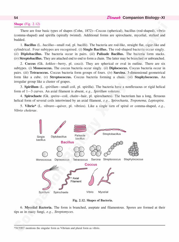
��������Companion Biology–XI54
Shape (Fig. 2.12)
There are four basic types of shapes (Cohn, 1872)—Coccus (spherical), bacillus (rod-shaped), vibrio
(comma-shaped) and spirilla (spirally twisted). Additional forms are spirochaete, mycelial, stalked and
budded.
1. Bacillus (L. bacillus—small rod, pl. bacilli). The bacteria are rod-like, straight flat, cigar-like and
cylindrical. Four subtypes are recognised. (i) Single Bacillus. The rod-shaped bacteria occur singly.
(ii) Diplobacillus. The bacteria occur in pairs. (iii) Palisade Bacillus. The bacteria form stacks.
(iv) Streptobacillus. They are attached end to end to form a chain. The latter may be branched or unbranched.
2. Coccus (Gk. kokkos—berry, pl. cocci). They are spherical or oval in outline. There are six
subtypes. (i) Monococcus. The coccus bacteria occur singly. (ii) Diplococcus. Coccus bacteria occur in
pairs. (iii) Tetracoccus. Coccus bacteria form groups of fours. (iv) Sarcina. 3-dimensional geometrical
form like a cube. (v) Streptococcus. Coccus bacteria forming a chain. (vi) Staphylococcus. An
irregular group like a cluster of grapes.
3. Spirillum (L. spirillum—small coil, pl. spirilla). The bacteria have a nonflexuous or rigid helical
form of 1—3 curves. An axial filament is absent, e.g., Spirillum volutans.
4. Spirochaete (Gk. speira—coil, chaite—hair, pl. spirochaetes). The bacterium has a long, flexuous
helical form of several coils intertwined by an axial filament, e.g., Spirochaeta, Treponema, Leptospira.
5. Vibrio* (L. vibrare—quiver, pl. vibrios). Like a single turn of spiral or comma-shaped, e.g.,
Vibrio cholerae.
�����������
Fig. 2.12. Shapes of Bacteria.
6. Mycelial Bacteria. The form is branched, aseptate and filamentous. Spores are formed at theirtips as in many fungi, e.g., Streptomyces.
*NCERT mentions the singular form as Vibrium and plural form as vibrio.
����������� 55
• Coccobacillus. Cylindrical bacterium with length approximately equal to breadth, e.g.,Brucella.
• Actinomycetes. Formerly Ray Fungi. Mycetial bacteria.• Pleomorphism (Gk.pleon—more, morphe—form). It is the phenomenon of having more
than one form or shape by the same individual or structure. Pleomorphism occurs insome bacteria, e.g., Azotobacter, Rhizobium. Bacterium Rhizobium has three forms—atrichous rods, peritrichous oval swarmers and atrichous irregular bacteriods.
Gram Staining
Christian Gram (1884) developed a technique of staining bacteria called Gram staining. Dependingupon reaction to Gram staining, bacteria have been divided into two groups, Gram positive and Gramnegative. A bacterial smear is heat fixed on a glass slide. The latter is dipped in solution of crystal violet orgentian violet for one minute. All types of bacteria pick up a purple colouration. The stain is fixed bydipping the slide in 0·5% iodine solution for one minute. The slide is now kept in a polar organic solventlike alcohol or acetone for 10—30 seconds. Some bacteria retain a blue or purple stain. They are calledGram positive (G +ve). Others lose the stain in the organic solvent. The latter are designated as Gramnegative (G –ve). The slide is counter-stained with safranin for 20—30 seconds. Gram negative bacteriapick up the pink counter-stain while Gram positive bacteria come to have both purple and pink stains.
The difference between Gram +ve and Gram –ve bacteria is due to difference in lipid content oftheir walls. In Gram +ve bacteria the wall has very little lipid content. Therefore, very little stain leaksout of their walls in organic solvent. In Gram –ve bacteria the wall has high lipid content. The samedissolves in organic solvent taking out the stain alongwith.
Gram Positive Bacteria. Pneumococcus, Streptococcus, Staphylococcus, Bacillus, Clostridium,Mycobacterium, Streptomyces.
Gram Negative Bacteria. Salmonella, Pseudomonas, Escherichia, Haemophilus, Helicobacter,Vibrio, Rhizobium.
Differences Between Gram Positive and Gram Negative Bacteria
S. No. Gram Positive Bacteria Gram Negative Bacteria
1. They retain the Gram stain even after Washing in organic solvent drains away thewashing in organic solvent. stain.
2. Cell wall is thick, 20—80 nm. Cell wall is comparatively thin, 7·5—12 nm.
3. Cell wall is single layered. It is two layered.
4. Porins do not occur in the cell wall. Porins occur in the outer layer of cell wall.
5. Cell wall is smooth. Cell wall is wavy.
6. A periplasmic space or gel is less conspi- A periplasmic space or gel lies between cellcuous. wall and plasma membrane.
7. Lipid content is low, 1—5%. Lipid content is high, 20—30%.
8. Peptidoglycan content of wall is 70—80%. Peptidoglycan content of wall is 10—20%.
9. The wall contains teichoic acids. Teichoic acids are absent.
10. Flagella possess single pair of rings in the Flagella have two pairs of rings in the area ofregion of basal body. their basal body.
11. Mesosomes are well developed. Mesosomes are less prominent.
12. Ratio of DNA to RNA is 1 : 8. Ratio of DNA to RNA is 1 : 1.
13. Pathogenic forms are fewer. Pathogenic forms are more abundant.
14. They are highly susceptible to antibiotics. They are less susceptible to antibiotics.
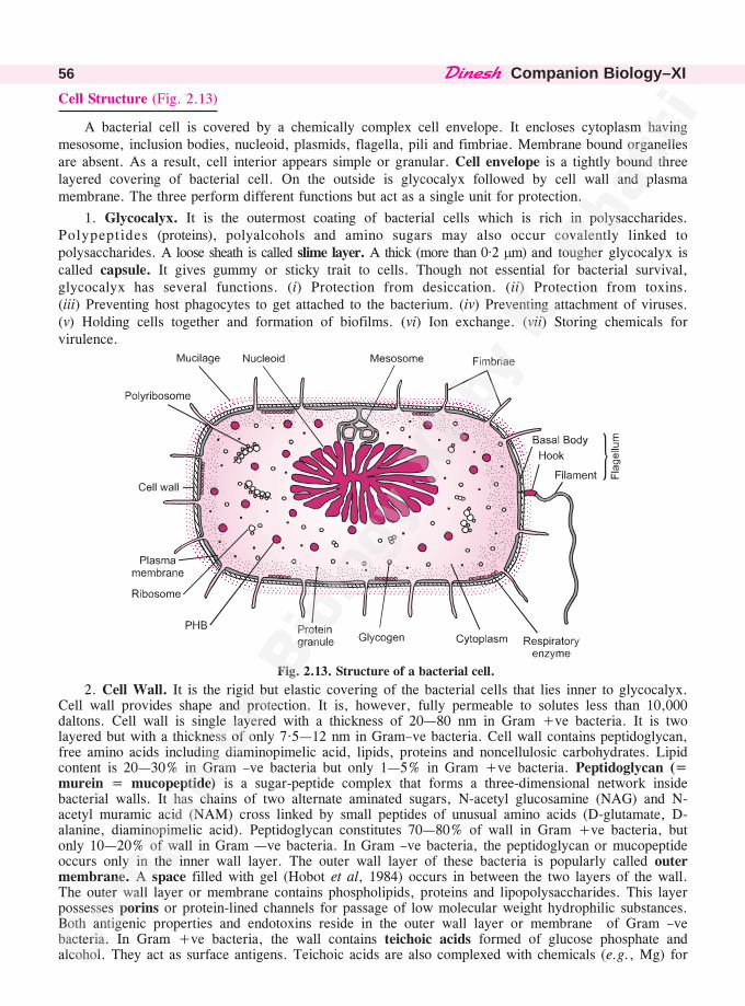
��������Companion Biology–XI56
Cell Structure (Fig. 2.13)
A bacterial cell is covered by a chemically complex cell envelope. It encloses cytoplasm havingmesosome, inclusion bodies, nucleoid, plasmids, flagella, pili and fimbriae. Membrane bound organellesare absent. As a result, cell interior appears simple or granular. Cell envelope is a tightly bound threelayered covering of bacterial cell. On the outside is glycocalyx followed by cell wall and plasmamembrane. The three perform different functions but act as a single unit for protection.
1. Glycocalyx. It is the outermost coating of bacterial cells which is rich in polysaccharides.Polypeptides (proteins), polyalcohols and amino sugars may also occur covalently linked topolysaccharides. A loose sheath is called slime layer. A thick (more than 0·2 �m) and tougher glycocalyx iscalled capsule. It gives gummy or sticky trait to cells. Though not essential for bacterial survival,glycocalyx has several functions. (i) Protection from desiccation. (ii) Protection from toxins.(iii) Preventing host phagocytes to get attached to the bacterium. (iv) Preventing attachment of viruses.(v) Holding cells together and formation of biofilms. (vi) Ion exchange. (vii) Storing chemicals forvirulence.
Fig. 2.13. Structure of a bacterial cell.
2. Cell Wall. It is the rigid but elastic covering of the bacterial cells that lies inner to glycocalyx.Cell wall provides shape and protection. It is, however, fully permeable to solutes less than 10,000daltons. Cell wall is single layered with a thickness of 20—80 nm in Gram +ve bacteria. It is twolayered but with a thickness of only 7·5—12 nm in Gram–ve bacteria. Cell wall contains peptidoglycan,free amino acids including diaminopimelic acid, lipids, proteins and noncellulosic carbohydrates. Lipidcontent is 20—30% in Gram –ve bacteria but only 1—5% in Gram +ve bacteria. Peptidoglycan (=murein = mucopeptide) is a sugar-peptide complex that forms a three-dimensional network insidebacterial walls. It has chains of two alternate aminated sugars, N-acetyl glucosamine (NAG) and N-acetyl muramic acid (NAM) cross linked by small peptides of unusual amino acids (D-glutamate, D-alanine, diaminopimelic acid). Peptidoglycan constitutes 70—80% of wall in Gram +ve bacteria, butonly 10—20% of wall in Gram —ve bacteria. In Gram –ve bacteria, the peptidoglycan or mucopeptideoccurs only in the inner wall layer. The outer wall layer of these bacteria is popularly called outermembrane. A space filled with gel (Hobot et al, 1984) occurs in between the two layers of the wall.The outer wall layer or membrane contains phospholipids, proteins and lipopolysaccharides. This layerpossesses porins or protein-lined channels for passage of low molecular weight hydrophilic substances.Both antigenic properties and endotoxins reside in the outer wall layer or membrane of Gram –vebacteria. In Gram +ve bacteria, the wall contains teichoic acids formed of glucose phosphate andalcohol. They act as surface antigens. Teichoic acids are also complexed with chemicals (e.g., Mg) for
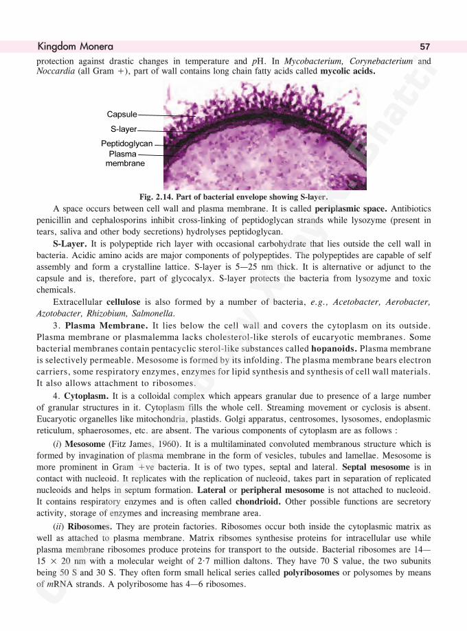
����������� 57
protection against drastic changes in temperature and pH. In Mycobacterium, Corynebacterium andNoccardia (all Gram +), part of wall contains long chain fatty acids called mycolic acids.
�������
�����
������������ �������������
Fig. 2.14. Part of bacterial envelope showing S-layer.
A space occurs between cell wall and plasma membrane. It is called periplasmic space. Antibiotics
penicillin and cephalosporins inhibit cross-linking of peptidoglycan strands while lysozyme (present in
tears, saliva and other body secretions) hydrolyses peptidoglycan.
S-Layer. It is polypeptide rich layer with occasional carbohydrate that lies outside the cell wall in
bacteria. Acidic amino acids are major components of polypeptides. The polypeptides are capable of self
assembly and form a crystalline lattice. S-layer is 5—25 nm thick. It is alternative or adjunct to the
capsule and is, therefore, part of glycocalyx. S-layer protects the bacteria from lysozyme and toxic
chemicals.
Extracellular cellulose is also formed by a number of bacteria, e.g., Acetobacter, Aerobacter,
Azotobacter, Rhizobium, Salmonella.
3. Plasma Membrane. It lies below the cell wall and covers the cytoplasm on its outside.
Plasma membrane or plasmalemma lacks cholesterol-like sterols of eucaryotic membranes. Some
bacterial membranes contain pentacyclic sterol-like substances called hopanoids. Plasma membrane
is selectively permeable. Mesosome is formed by its infolding. The plasma membrane bears electron
carriers, some respiratory enzymes, enzymes for lipid synthesis and synthesis of cell wall materials.
It also allows attachment to ribosomes.
4. Cytoplasm. It is a colloidal complex which appears granular due to presence of a large number
of granular structures in it. Cytoplasm fills the whole cell. Streaming movement or cyclosis is absent.
Eucaryotic organelles like mitochondria, plastids. Golgi apparatus, centrosomes, lysosomes, endoplasmic
reticulum, sphaerosomes, etc. are absent. The various components of cytoplasm are as follows :
(i) Mesosome (Fitz James, 1960). It is a multilaminated convoluted membranous structure which is
formed by invagination of plasma membrane in the form of vesicles, tubules and lamellae. Mesosome is
more prominent in Gram +ve bacteria. It is of two types, septal and lateral. Septal mesosome is in
contact with nucleoid. It replicates with the replication of nucleoid, takes part in separation of replicated
nucleoids and helps in septum formation. Lateral or peripheral mesosome is not attached to nucleoid.
It contains respiratory enzymes and is often called chondrioid. Other possible functions are secretory
activity, storage of enzymes and increasing membrane area.
(ii) Ribosomes. They are protein factories. Ribosomes occur both inside the cytoplasmic matrix as
well as attached to plasma membrane. Matrix ribsomes synthesise proteins for intracellular use while
plasma membrane ribosomes produce proteins for transport to the outside. Bacterial ribosomes are 14—
15 × 20 nm with a molecular weight of 2·7 million daltons. They have 70 S value, the two subunits
being 50 S and 30 S. They often form small helical series called polyribosomes or polysomes by means
of mRNA strands. A polyribosome has 4—6 ribosomes.
��������Companion Biology–XI58
(iii) Inclusion Bodies. They are nonliving nonstructural materials of bacterial cells which may liefree in the cytoplasm (e.g., volutin granules, glycogen granules, cyanophycean granules) or covered bya single nonunit 2—4 nm thick membrane (e.g., gas vacuoles, PHB, sulphur granules, carboxysomes).They are of three types—food reserve, inorganic inclusions and gas vacuoles.
(a) Food Reserve. It consists of glycogen and protein granules. Neutral fats are absent. Manybacteria possess refractile granules of poly �-hydroxybutyric acid (PHB), e.g., Azotobacter, Bacillus,Pseudomonas species. They are intermediates of fatty acid synthesis. A biodegradable plastic can beformed from PHB.
(b) Inorganic Granules. They are also called metachromatic granules as they take up differentcolours with basic dyes. Volutin or polymetaphosphate granules contain reserve phosphate. Sulphurgranules are quite common in sulphur bacteria or bacteria using H2S as hydrogen donor. They may liein cytoplasm or periplasmic space. Magnetite occurs in bacterium Aquaspirillum magnetotacticum. It isfound in small vesicles called magnetosomes (Balkwill et al 1980). Magnetosomes help the bacteria toorientate along geomagnetic lines. All granules are covered by nonunit or single-layered membrane.
(c) Gas Vacuoles. They store metabolic gases. Gas vacuoles occur in aquatic forms of cyanobacteria,photosynthetic purple and green eubacteria and a number of other plankonic species. The vacuoles areuseful in buoyancy regulation and protection against harmful radiations. Each gas vacuole is made of alarge number of extremely small cylindrical hexagonal units called gas vesicles. A gas vesicle is coveredby banded protein membrane of about 2 nm thickness. It is permeable to gases but not to water.
(iv) Photosynthetic Pigments. They are bacteriochlorophylls, bacteriophaeophytins and carotenoids.In purple bacteria the pigments are associated with thylakoid like membranes that are formed byinvagination of plasmalemma. In green bacteria the photosynthetic pigments are not associated withmembranes but occur inside small sacs called chlorosomes. Membrane covering of a chlorosome is non-unit membrane. Chlorosomes are, however, joined to plasmalemma through base plates. Both bacterialphotosynthetic thylakoids and chlorosomes are collectively called chromatophores.
Nitrifying bacteria also possess membrane bound flat, spherical or tubular vesicles.
5. Nucleoid or Nuclear Body (= Genophore). Typical nucleus is absent in bacteria as they areprocaryotic. The genetic material is a single molecule of double stranded DNA. DNA is circular as itstwo ends are covalently joined. In a few cases, where it is linear, the ends are joined by a proteinbridge, e.g., Streptomyces. It is naked or without association of histone proteins. DNA is extensivelylooped and coiled variously with the help of RNA and at times polyamines to form a spherical,cylindrical, oval or dumb-bell shaped compact structure called genophore, nuclear body or nucleoid(Gk. nucleus—kernel, eidos—resembling). In Escherichia coli, 1100 �m long DNA duplex with about3·3 × 106 base pairs is coiled to form 1 �m3 nucleoid. Nucleoid is attached to plasma membranedirectly or by means of mesosome. Bacterial nucleoid formed of a single molecule of DNA is equivalentto what is contained in a single chromosome of eucaryotes but without the organisation of the latter. Itis, therefore, often called prochromosome.
6. Plasmids (Gk. plasma—form; Hayes and Lederberg, 1952). They are small dispensable, self-replicating extrachromosomal or extra-nuclear circular segments of DNA present in the cytoplasm ofbacterial cells. They endow some extra properties to bacteria which are often useful but not essential fortheir living. Many plasmids can be transferred to other bacteria. Three transferable plasmids areF-plasmids (having fertility factor), R-plasmids (having resistance to antibiotics like streptomycin,sulphonamide, tetracycline, chloramphanicol, etc., heavy metals) and col-plasmids (producing colicinsor bacteriocins that kill related bacteria). Others are antibiotic producing plasmids, pigment synthesisingplasmids, nodule forming plasmids, etc. Some plasmids can temporarily integrate with nuclear body.They are then called episomes (Gk. epi—addition, soma—body). Formation of episome depends uponchromosome memory. Jumping genes or transposons also occur in some plasmids. Plasmids are importanttools in genetic engineering and biotechnology.
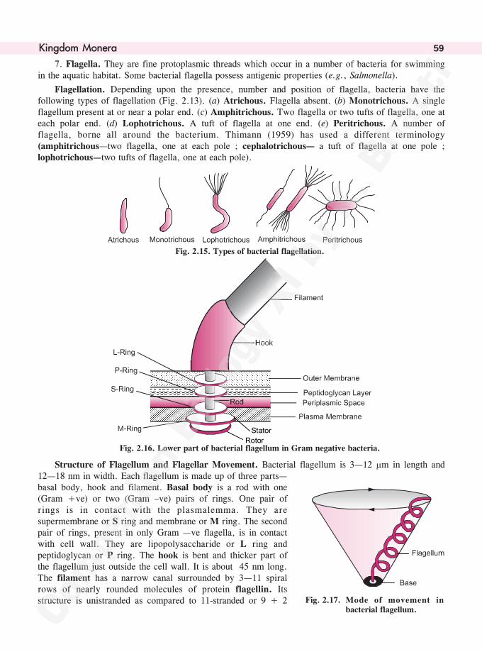
����������� 59
7. Flagella. They are fine protoplasmic threads which occur in a number of bacteria for swimmingin the aquatic habitat. Some bacterial flagella possess antigenic properties (e.g., Salmonella).
Flagellation. Depending upon the presence, number and position of flagella, bacteria have thefollowing types of flagellation (Fig. 2.13). (a) Atrichous. Flagella absent. (b) Monotrichous. A singleflagellum present at or near a polar end. (c) Amphitrichous. Two flagella or two tufts of flagella, one ateach polar end. (d) Lophotrichous. A tuft of flagella at one end. (e) Peritrichous. A number offlagella, borne all around the bacterium. Thimann (1959) has used a different terminology(amphitrichous—two flagella, one at each pole ; cephalotrichous— a tuft of flagella at one pole ;lophotrichous—two tufts of flagella, one at each pole).
Fig. 2.15. Types of bacterial flagellation.
�����������
Fig. 2.16. Lower part of bacterial flagellum in Gram negative bacteria.
Structure of Flagellum and Flagellar Movement. Bacterial flagellum is 3—12 �m in length and12—18 nm in width. Each flagellum is made up of three parts—basal body, hook and filament. Basal body is a rod with one(Gram +ve) or two (Gram –ve) pairs of rings. One pair ofrings is in contact with the plasmalemma. They aresupermembrane or S ring and membrane or M ring. The secondpair of rings, present in only Gram —ve flagella, is in contactwith cell wall. They are lipopolysaccharide or L ring andpeptidoglycan or P ring. The hook is bent and thicker part ofthe flagellum just outside the cell wall. It is about 45 nm long.The filament has a narrow canal surrounded by 3—11 spiralrows of nearly rounded molecules of protein flagellin. Itsstructure is unistranded as compared to 11-stranded or 9 + 2 Fig. 2.17. Mode of movement in
bacterial flagellum.
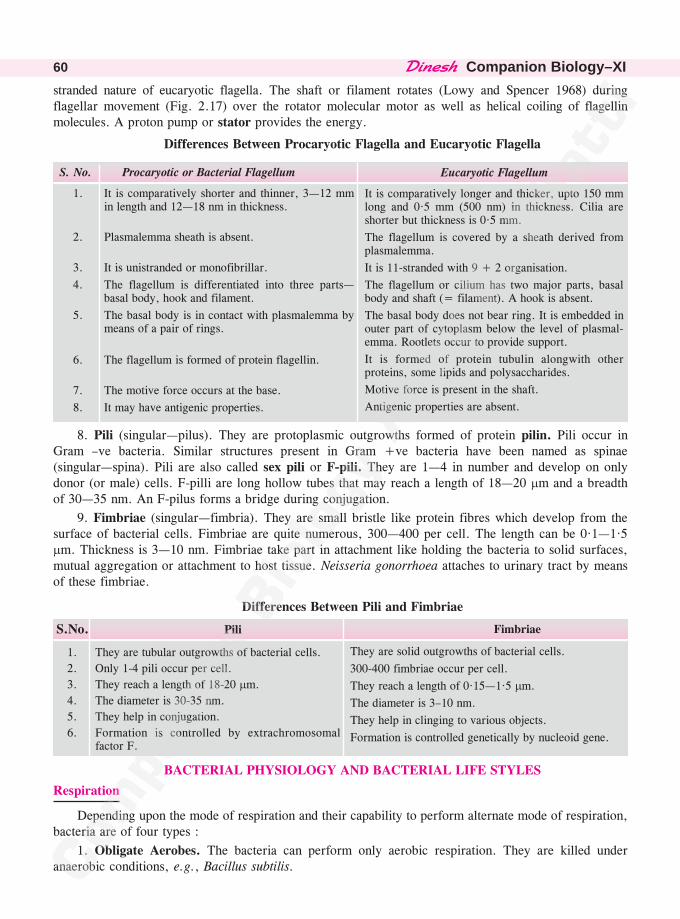
��������Companion Biology–XI60
Fimbriae
They are solid outgrowths of bacterial cells.
300-400 fimbriae occur per cell.
They reach a length of 0·15—1·5 �m.
The diameter is 3–10 nm.
They help in clinging to various objects.
Formation is controlled genetically by nucleoid gene.
stranded nature of eucaryotic flagella. The shaft or filament rotates (Lowy and Spencer 1968) duringflagellar movement (Fig. 2.17) over the rotator molecular motor as well as helical coiling of flagellinmolecules. A proton pump or stator provides the energy.
Differences Between Procaryotic Flagella and Eucaryotic Flagella
Eucaryotic Flagellum
It is comparatively longer and thicker, upto 150 mmlong and 0·5 mm (500 nm) in thickness. Cilia areshorter but thickness is 0·5 mm.
The flagellum is covered by a sheath derived fromplasmalemma.
It is 11-stranded with 9 + 2 organisation.
The flagellum or cilium has two major parts, basalbody and shaft (= filament). A hook is absent.
The basal body does not bear ring. It is embedded inouter part of cytoplasm below the level of plasmal-emma. Rootlets occur to provide support.
It is formed of protein tubulin alongwith otherproteins, some lipids and polysaccharides.
Motive force is present in the shaft.
Antigenic properties are absent.
S. No. Procaryotic or Bacterial Flagellum
1. It is comparatively shorter and thinner, 3—12 mmin length and 12—18 nm in thickness.
2. Plasmalemma sheath is absent.
3. It is unistranded or monofibrillar.
4. The flagellum is differentiated into three parts—basal body, hook and filament.
5. The basal body is in contact with plasmalemma bymeans of a pair of rings.
6. The flagellum is formed of protein flagellin.
7. The motive force occurs at the base.
8. It may have antigenic properties.
Differences Between Pili and Fimbriae
S.No. Pili
1. They are tubular outgrowths of bacterial cells.
2. Only 1-4 pili occur per cell.
3. They reach a length of 18-20 �m.
4. The diameter is 30-35 nm.
5. They help in conjugation.
6. Formation is controlled by extrachromosomalfactor F.
BACTERIAL PHYSIOLOGY AND BACTERIAL LIFE STYLES
Respiration
Depending upon the mode of respiration and their capability to perform alternate mode of respiration,bacteria are of four types :
1. Obligate Aerobes. The bacteria can perform only aerobic respiration. They are killed underanaerobic conditions, e.g., Bacillus subtilis.
8. Pili (singular—pilus). They are protoplasmic outgrowths formed of protein pilin. Pili occur inGram –ve bacteria. Similar structures present in Gram +ve bacteria have been named as spinae(singular—spina). Pili are also called sex pili or F-pili. They are 1—4 in number and develop on onlydonor (or male) cells. F-pilli are long hollow tubes that may reach a length of 18—20 �m and a breadthof 30—35 nm. An F-pilus forms a bridge during conjugation.
9. Fimbriae (singular—fimbria). They are small bristle like protein fibres which develop from thesurface of bacterial cells. Fimbriae are quite numerous, 300—400 per cell. The length can be 0·1—1·5�m. Thickness is 3—10 nm. Fimbriae take part in attachment like holding the bacteria to solid surfaces,mutual aggregation or attachment to host tissue. Neisseria gonorrhoea attaches to urinary tract by meansof these fimbriae.
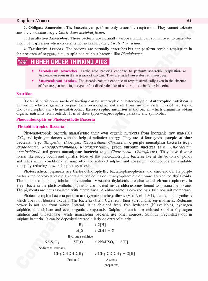
����������� 61
2. Obligate Anaerobes. The bacteria can perform only anaerobic respiration. They cannot tolerateaerobic conditions, e.g., Clostridium acetobutylicum.
3. Facultative Anaerobes. These bacteria are normally aerobes which can switch over to anaerobicmode of respiration when oxygen is not available, e.g., Clostridium tetani.
4. Facultative Aerobes. The bacteria are normally anaerobes but can perform aerobic respiration inthe presence of oxygen, e.g., purple non sulphur bacteria like Rhodobacter.
• Aerotolerant Anaerobes. Lactic acid bacteria continue to perform anaerobic respiration orfermentation even in the presence of oxygen. They are called aerotolerant anaerobes.
• Anaerotolerant Aerobes. The aerobic bacteria continue to respire aerobically even in the absence
of free oxygen by using oxygen of oxidised salts like nitrate, e.g., denitrifying bacteria.
Nutrition
Bacterial nutrition or mode of feeding can be autotrophic or heterotrophic. Autotrophic nutrition isthe one in which organisms prepare their own organic nutrients from raw materials. It is of two types,photoautotrophic and chemoautotrophic. Heterotrophic nutrition is the one in which organisms obtainorganic nutrients from outside. It is of three types—saprotrophic, parasitic and symbiotic.
Photoautotrophic or Photosynthetic Bacteria
(Photolithotrophic Bacteria)
Photoautotrophic bacteria manufacture their own organic nutrients from inorganic raw materials(CO2 and hydrogen donor) with the help of radiation energy. They are of four types—purple sulphurbacteria (e.g., Thiopedia, Thiocapsa, Thiospirillum, Chromatium), purple nonsulphur bacteria (e.g.,Rhodobacter, Rhodopseudomonas, Rhodospirillum), green sulphur bacteria (e.g., Chlorobium,Ancalochloris) and green nonsulphur bacteria (e.g., Chloronema, Chloroflexus). They have diverseforms like cocci, bacilli and spirilla. Most of the photoautotrophic bacteria live at the bottom of pondsand lakes where conditions are anaerobic and reduced sulphur and nonsulphur compounds are availableto supply reducing power for photosynthesis.
Photosynthetic pigments are bacteriochlorophylls, bacteriophaeophytins and carotenoids. In purplebacteria the photosynthetic pigments are located inside intracytoplasmic membrane sacs called thylakoids.The latter are lamellar, tubular or vesicular. Vesicular thylakoids are also called chromatophores. Ingreen bacteria the photosynthetic pigments are located inside chlorosomes bound to plasma membrane.The pigments are not associated with membranes. A chlorosome is covered by a thin nonunit membrane.
Photoautotrophic bacteria perform anoxygenic photosynthesis (Van Niel, 1931), that is, photosynthesiswhich does not liberate oxygen. The bacteria obtain CO2 from their surrounding environment. Reducingpower is not got from water. Instead, it is obtained from free hydrogen (if available), hydrogensulphide, thiosulphate and even organic compounds. Sulphur bacteria use reduced sulphur (hydrogensulphide and thiosulphate) while nonsulphur bacteria use other sources. Sulphur precipitates out insulphur bacteria. It can be deposited intracellularly or extracellularly.
H2 ��� 2[H]
H2S ��� 2[H] + S
Hydrogen sulphide
Na2S2O3 + 5H2O ��� 2NaHSO4 + 8[H]
Sodium thiosulphate
CH3.CHOH.CH3 ��� CH3.CO.CH3 + 2[H]
Propanol Acetone
(propanone)
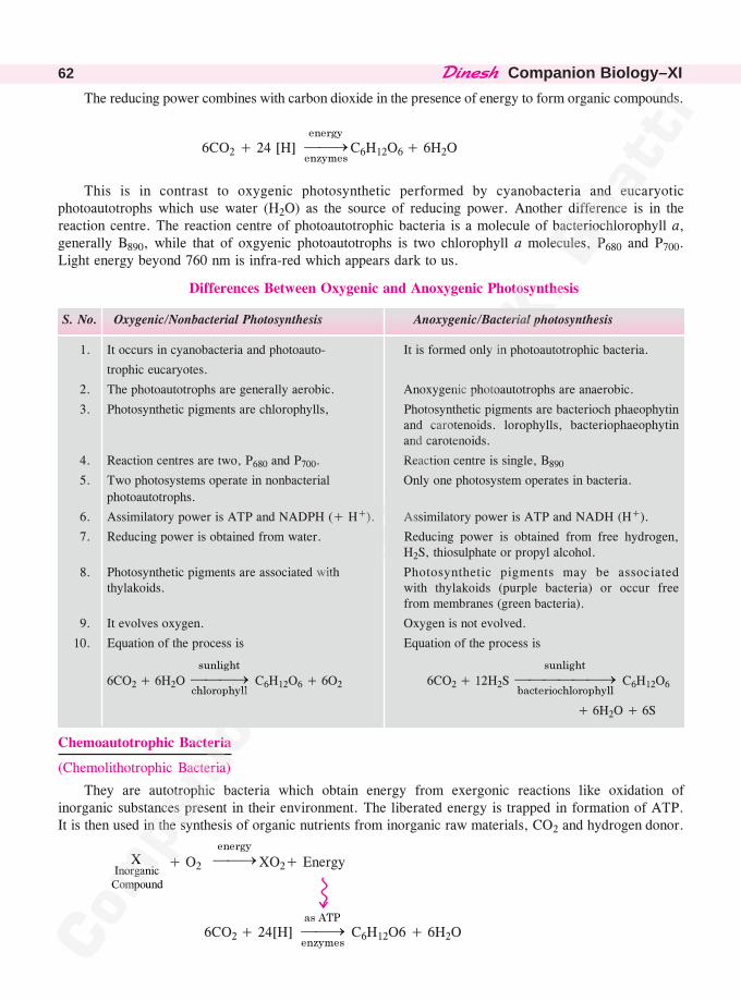
��������Companion Biology–XI62
The reducing power combines with carbon dioxide in the presence of energy to form organic compounds.
6CO2 + 24 [H] ����������
�����C6H12O6 + 6H2O
This is in contrast to oxygenic photosynthetic performed by cyanobacteria and eucaryoticphotoautotrophs which use water (H2O) as the source of reducing power. Another difference is in thereaction centre. The reaction centre of photoautotrophic bacteria is a molecule of bacteriochlorophyll a,generally B890, while that of oxgyenic photoautotrophs is two chlorophyll a molecules, P680 and P700.Light energy beyond 760 nm is infra-red which appears dark to us.
Differences Between Oxygenic and Anoxygenic Photosynthesis
S. No. Oxygenic/Nonbacterial Photosynthesis Anoxygenic/Bacterial photosynthesis
1. It occurs in cyanobacteria and photoauto- It is formed only in photoautotrophic bacteria.
trophic eucaryotes.
2. The photoautotrophs are generally aerobic. Anoxygenic photoautotrophs are anaerobic.
3. Photosynthetic pigments are chlorophylls, Photosynthetic pigments are bacterioch phaeophytinand carotenoids. lorophylls, bacteriophaeophytinand carotenoids.
4. Reaction centres are two, P680 and P700. Reaction centre is single, B890
5. Two photosystems operate in nonbacterial Only one photosystem operates in bacteria.
photoautotrophs.
6. Assimilatory power is ATP and NADPH (+ H+). Assimilatory power is ATP and NADH (H+).
7. Reducing power is obtained from water. Reducing power is obtained from free hydrogen,H2S, thiosulphate or propyl alcohol.
8. Photosynthetic pigments are associated with Photosynthetic pigments may be associatedthylakoids. with thylakoids (purple bacteria) or occur free
from membranes (green bacteria).
9. It evolves oxygen. Oxygen is not evolved.
10. Equation of the process is Equation of the process is
6CO2 + 6H2O ���������������
��� �� C6H12O6 + 6O2 6CO2 + 12H2S �������
����������� ��
������� C6H12O6
+ 6H2O + 6S
Chemoautotrophic Bacteria
(Chemolithotrophic Bacteria)
They are autotrophic bacteria which obtain energy from exergonic reactions like oxidation ofinorganic substances present in their environment. The liberated energy is trapped in formation of ATP.It is then used in the synthesis of organic nutrients from inorganic raw materials, CO2 and hydrogen donor.
XInorganicCompound
+ O2 ���������
XO2+ Energy
6CO2 + 24[H] ����������
���� C6H12O6 + 6H2O
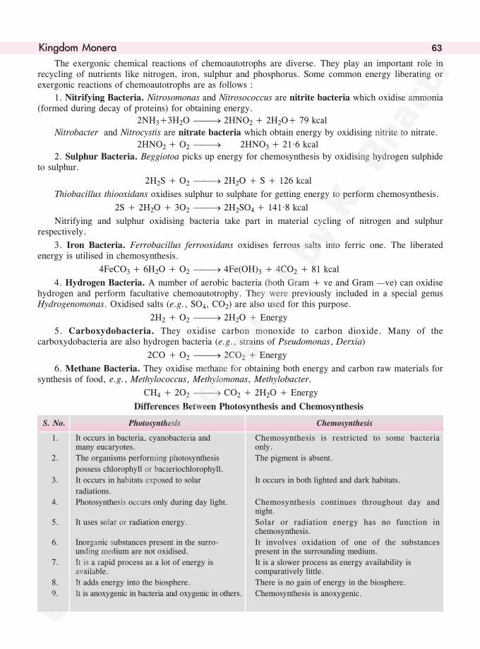
����������� 63
The exergonic chemical reactions of chemoautotrophs are diverse. They play an important role inrecycling of nutrients like nitrogen, iron, sulphur and phosphorus. Some common energy liberating orexergonic reactions of chemoautotrophs are as follows :
1. Nitrifying Bacteria. Nitrosomonas and Nitrosococcus are nitrite bacteria which oxidise ammonia(formed during decay of proteins) for obtaining energy.
2NH3+3H2O ��� 2HNO2 + 2H2O+ 79 kcal
Nitrobacter and Nitrocystis are nitrate bacteria which obtain energy by oxidising nitrite to nitrate.
2HNO2 + O2 ��� 2HNO3 + 21·6 kcal
2. Sulphur Bacteria. Beggiotoa picks up energy for chemosynthesis by oxidising hydrogen sulphideto sulphur.
2H2S + O2 ��� 2H2O + S + 126 kcal
Thiobacillus thiooxidans oxidises sulphur to sulphate for getting energy to perform chemosynthesis.
2S + 2H2O + 3O2 ��� 2H2SO4 + 141·8 kcal
Nitrifying and sulphur oxidising bacteria take part in material cycling of nitrogen and sulphurrespectively.
3. Iron Bacteria. Ferrobacillus ferrooxidans oxidises ferrous salts into ferric one. The liberatedenergy is utilised in chemosynthesis.
4FeCO3 + 6H2O + O2 ��� 4Fe(OH)3 + 4CO2 + 81 kcal
4. Hydrogen Bacteria. A number of aerobic bacteria (both Gram + ve and Gram —ve) can oxidisehydrogen and perform facultative chemoautotrophy. They were previously included in a special genusHydrogenomonas. Oxidised salts (e.g., SO4, CO2) are also used for this purpose.
2H2 + O2 ��� 2H2O + Energy
5. Carboxydobacteria. They oxidise carbon monoxide to carbon dioxide. Many of thecarboxydobacteria are also hydrogen bacteria (e.g., strains of Pseudomonas, Derxia)
2CO + O2 ��� 2CO2 + Energy
6. Methane Bacteria. They oxidise methane for obtaining both energy and carbon raw materials forsynthesis of food, e.g., Methylococcus, Methylomonas, Methylobacter.
CH4 + 2O2 ��� CO2 + 2H2O + Energy
Differences Between Photosynthesis and Chemosynthesis
S. No. Photosynthesis Chemosynthesis
1. It occurs in bacteria, cyanobacteria and Chemosynthesis is restricted to some bacteriamany eucaryotes. only.
2. The organisms performing photosynthesis The pigment is absent.
possess chlorophyll or bacteriochlorophyll.
3. It occurs in habitats exposed to solar It occurs in both lighted and dark habitats.
radiations.
4. Photosynthesis occurs only during day light. Chemosynthesis continues throughout day andnight.
5. It uses solar or radiation energy. Solar or radiation energy has no function inchemosynthesis.
6. Inorganic substances present in the surro- It involves oxidation of one of the substancesunding medium are not oxidised. present in the surrounding medium.
7. It is a rapid process as a lot of energy is It is a slower process as energy availability isavailable. comparatively little.
8. It adds energy into the biosphere. There is no gain of energy in the biosphere.
9. It is anoxygenic in bacteria and oxygenic in others. Chemosynthesis is anoxygenic.
��������Companion Biology–XI64
Heterotrophic Bacteria
(Chemoheterotrophic or Chemi-organotrophic Bacteria)
They are bacteria which perform heterotrophic nutrition, that is, obtain organic nutrients fromoutside for both body building and as source of energy. Chemoheterotrophic bacteria are of threetypes—saprotrophic, symbiotic and parasitic.
1. Saprotrophic Bacteria. They are free-living bacteria which obtain nourishment from organicexcretions and organic remains like fruits, vegetables, corpses, faeces, fallen leaves, human foods, etc.The saprotrophic bacteria pour enzymes over them for their solubilisation. The solubilised substancesare absorbed and assimitated by saprotrophs. The organisms which absorb their organic nourishment inthe solution form are also called osmotrophs. Some of the solubilised substances are left in the externalmedium. The most important of these are inorganic substances. Liberation of inorganic substances fromorganic materials is called mineralisation. This is an important step for material cycling. Organismswhich bring about physical and chemical breakdown of dead organisms and their remains are calleddecomposers. Gram –ve Pseudomonas is capable of decomposing a wide variety of organic compounds,both natural and man-made, as well as petroleum wastes. Three special terms are used for saprotrophicbreakdown of organic materials. (i) Decay. Aerobic breakdown of organic remains. (ii) Putrefaction.Anaerobic breakdown of proteins. It is characterised by release of offensive odour. (iii) Fermentation.Anaerobic breakdown of carbohydrates. It produces a number of alcohols and organic acids.
Saprotrophic bacteria are used by human beings in disposal of garbage, sewage, preparation ofmanure, retting of fibres, cleaning of hides, preparation of ensilage, curd, yoghurt, alcohols, organicacids, curing of Tea, Tobacco, Coffee and Cocoa, etc.
• Facultative Saprotrophs. Organisms capable of living as saprotrophs. Their normal mode ofnutrition is different like autotrophic or parasitic.
• Facultative Parasites. Organisms capable of living on or inside a host. Normal nutrition is
different, like saprotrophic.
2. Symbiotic Bacteria. The bacteria live in mutually beneficial association with other organisms.Examples. (i) Escherichia coli (Escherich, 1885) is a symbiont (considered commensal by some) in theintestine of human beings and many other mammals feeding on human wastes. The bacterium checks thegrowth of putrefying bacteria. It also secretes vitamins B and K. Presence of E. coli in water indicatescontamination by sewage. (ii) Rhizobium is a symbiont in the root nodules of legumes. Different species ofRhizobium are associated with different types of legumes, e.g., R. leguminosarum, R. lupini, R. trifoli, R.meliloti, R. phaseoli. The bacteria perform nitrogen fixation. They obtain nourishment and shelter from thehost and provide a part of fixed nitrogen to the host. A pink-red pigment called leghaemoglobin is presentaround the bacteria inside host cells for oxygen scavenging. (iii) Aerorhizobium is nitrogen fixing symbiontin the stem nodules of Sesbania rostrata. (iv) Frankia is symbiont nitrogen fixing mycelial bacterium thatoccurs in root nodules of Casuarina, Alnus (Alder), Myrica, Rubus, etc. (v) Xanthomonas is symbiontnitrogen fixing bacterium that occurs in the leaf cavities (leaf nodules) of Ardisia.
3. Parasitic Bacteria. The bacteria obtain nourishment from other living organisms called hosts.Alongwith food, the parasitic forms often become dependent upon the host for special organic compoundsrequired for growth. The latter are called growth factors. The parasitic bacteria may be harmless to the host,feeding on only insignificant quantity of food. A number of bacteria live on and inside human body withoutcausing any harm, e.g., Treponema denticola, T. orale, Staphylococcus aureus. The parasitic forms whichcause diseases are known as pathogens. The pathogenic bacteria are able to overcome host defences withthe help of biochemicals like (i) Aggressins (L. aggressus—attacked). Toxins that inhibit defence reactionsof host, cause breakdown of connective tissues. (ii) Cellulase. Digests cellulose of plant cell walls.
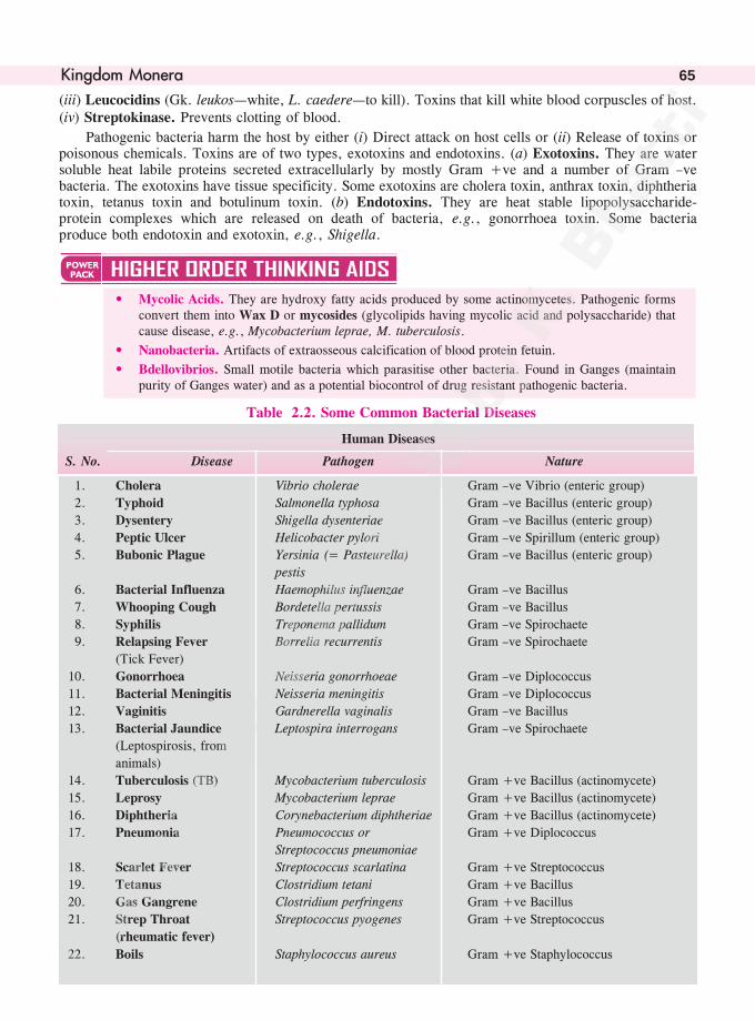
����������� 65
(iii) Leucocidins (Gk. leukos—white, L. caedere—to kill). Toxins that kill white blood corpuscles of host.(iv) Streptokinase. Prevents clotting of blood.
Pathogenic bacteria harm the host by either (i) Direct attack on host cells or (ii) Release of toxins orpoisonous chemicals. Toxins are of two types, exotoxins and endotoxins. (a) Exotoxins. They are watersoluble heat labile proteins secreted extracellularly by mostly Gram +ve and a number of Gram –vebacteria. The exotoxins have tissue specificity. Some exotoxins are cholera toxin, anthrax toxin, diphtheriatoxin, tetanus toxin and botulinum toxin. (b) Endotoxins. They are heat stable lipopolysaccharide-protein complexes which are released on death of bacteria, e.g., gonorrhoea toxin. Some bacteriaproduce both endotoxin and exotoxin, e.g., Shigella.
• Mycolic Acids. They are hydroxy fatty acids produced by some actinomycetes. Pathogenic formsconvert them into Wax D or mycosides (glycolipids having mycolic acid and polysaccharide) thatcause disease, e.g., Mycobacterium leprae, M. tuberculosis.
• Nanobacteria. Artifacts of extraosseous calcification of blood protein fetuin.
• Bdellovibrios. Small motile bacteria which parasitise other bacteria. Found in Ganges (maintainpurity of Ganges water) and as a potential biocontrol of drug resistant pathogenic bacteria.
Table 2.2. Some Common Bacterial Diseases
Human Diseases
S. No. Disease Pathogen Nature
1. Cholera Vibrio cholerae Gram –ve Vibrio (enteric group)
2. Typhoid Salmonella typhosa Gram –ve Bacillus (enteric group)
3. Dysentery Shigella dysenteriae Gram –ve Bacillus (enteric group)
4. Peptic Ulcer Helicobacter pylori Gram –ve Spirillum (enteric group)
5. Bubonic Plague Yersinia (= Pasteurella) Gram –ve Bacillus (enteric group)
pestis
6. Bacterial Influenza Haemophilus influenzae Gram –ve Bacillus
7. Whooping Cough Bordetella pertussis Gram –ve Bacillus
8. Syphilis Treponema pallidum Gram –ve Spirochaete
9. Relapsing Fever Borrelia recurrentis Gram –ve Spirochaete
(Tick Fever)
10. Gonorrhoea Neisseria gonorrhoeae Gram –ve Diplococcus
11. Bacterial Meningitis Neisseria meningitis Gram –ve Diplococcus
12. Vaginitis Gardnerella vaginalis Gram –ve Bacillus
13. Bacterial Jaundice Leptospira interrogans Gram –ve Spirochaete
(Leptospirosis, from
animals)
14. Tuberculosis (TB) Mycobacterium tuberculosis Gram +ve Bacillus (actinomycete)
15. Leprosy Mycobacterium leprae Gram +ve Bacillus (actinomycete)
16. Diphtheria Corynebacterium diphtheriae Gram +ve Bacillus (actinomycete)
17. Pneumonia Pneumococcus or Gram +ve Diplococcus
Streptococcus pneumoniae
18. Scarlet Fever Streptococcus scarlatina Gram +ve Streptococcus
19. Tetanus Clostridium tetani Gram +ve Bacillus
20. Gas Gangrene Clostridium perfringens Gram +ve Bacillus
21. Strep Throat Streptococcus pyogenes Gram +ve Streptococcus
(rheumatic fever)
22. Boils Staphylococcus aureus Gram +ve Staphylococcus
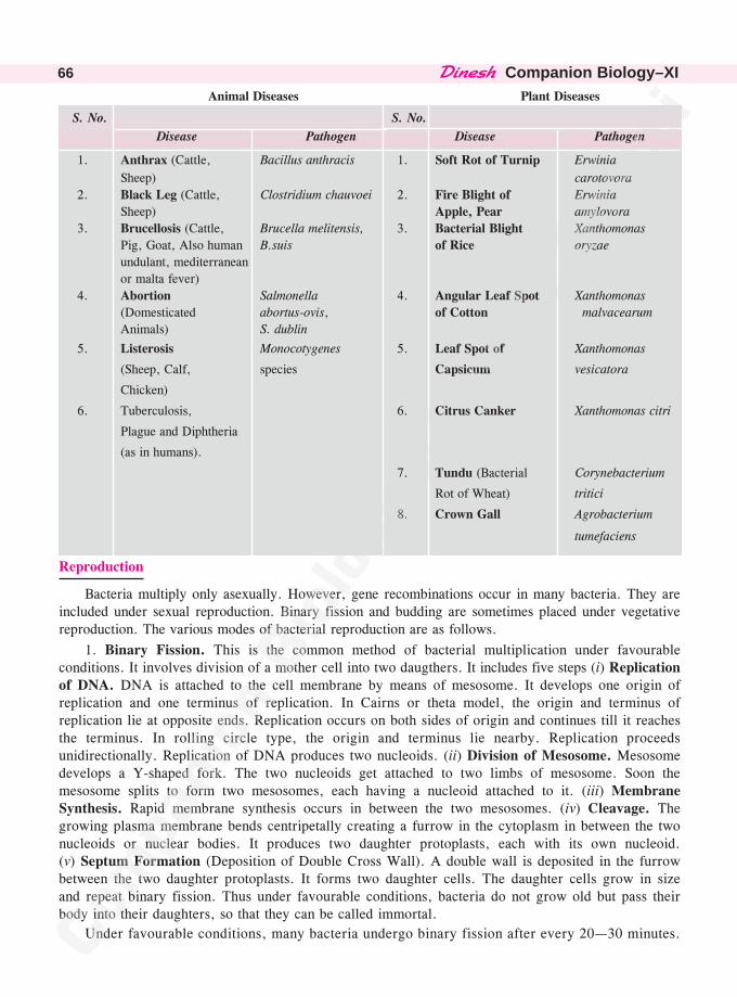
��������Companion Biology–XI66
Animal Diseases Plant Diseases
S. No. S. No.
Disease Pathogen Disease Pathogen
1. Anthrax (Cattle, Bacillus anthracis 1. Soft Rot of Turnip Erwinia
Sheep) carotovora
2. Black Leg (Cattle, Clostridium chauvoei 2. Fire Blight of Erwinia
Sheep) Apple, Pear amylovora
3. Brucellosis (Cattle, Brucella melitensis, 3. Bacterial Blight Xanthomonas
Pig, Goat, Also human B.suis of Rice oryzae
undulant, mediterranean
or malta fever)
4. Abortion Salmonella 4. Angular Leaf Spot Xanthomonas
(Domesticated abortus-ovis, of Cotton malvacearum
Animals) S. dublin
5. Listerosis Monocotygenes 5. Leaf Spot of Xanthomonas
(Sheep, Calf, species Capsicum vesicatora
Chicken)
6. Tuberculosis, 6. Citrus Canker Xanthomonas citri
Plague and Diphtheria
(as in humans).
7. Tundu (Bacterial Corynebacterium
Rot of Wheat) tritici
8. Crown Gall Agrobacterium
tumefaciens
Reproduction
Bacteria multiply only asexually. However, gene recombinations occur in many bacteria. They areincluded under sexual reproduction. Binary fission and budding are sometimes placed under vegetativereproduction. The various modes of bacterial reproduction are as follows.
1. Binary Fission. This is the common method of bacterial multiplication under favourableconditions. It involves division of a mother cell into two daugthers. It includes five steps (i) Replicationof DNA. DNA is attached to the cell membrane by means of mesosome. It develops one origin ofreplication and one terminus of replication. In Cairns or theta model, the origin and terminus ofreplication lie at opposite ends. Replication occurs on both sides of origin and continues till it reachesthe terminus. In rolling circle type, the origin and terminus lie nearby. Replication proceedsunidirectionally. Replication of DNA produces two nucleoids. (ii) Division of Mesosome. Mesosomedevelops a Y-shaped fork. The two nucleoids get attached to two limbs of mesosome. Soon themesosome splits to form two mesosomes, each having a nucleoid attached to it. (iii) MembraneSynthesis. Rapid membrane synthesis occurs in between the two mesosomes. (iv) Cleavage. Thegrowing plasma membrane bends centripetally creating a furrow in the cytoplasm in between the twonucleoids or nuclear bodies. It produces two daughter protoplasts, each with its own nucleoid.(v) Septum Formation (Deposition of Double Cross Wall). A double wall is deposited in the furrowbetween the two daughter protoplasts. It forms two daughter cells. The daughter cells grow in sizeand repeat binary fission. Thus under favourable conditions, bacteria do not grow old but pass theirbody into their daughters, so that they can be called immortal.
Under favourable conditions, many bacteria undergo binary fission after every 20—30 minutes.
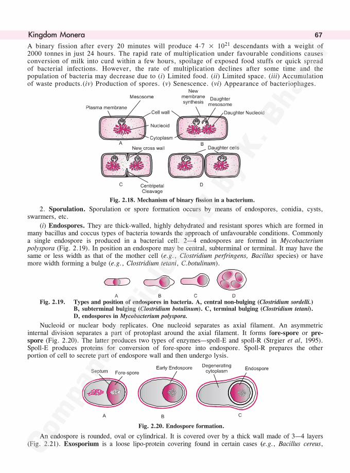
����������� 67
A binary fission after every 20 minutes will produce 4.7 × 10²¹ descendants with a weight of2000 tonnes in just 24 hours. The rapid rate of multiplication under favourable conditions causesconversion of milk into curd within a few hours, spoilage of exposed food stuffs or quick spreadof bacterial infections. However, the rate of multiplication declines after some time and thepopulation of bacteria may decrease due to (i) Limited food. (ii) Limited space. (iii) Accumulationof waste products.(iv) Production of spores. (v) Senescence. (vi) Appearance of bacteriophages.
Fig. 2.18. Mechanism of binary fission in a bacterium.
2. Sporulation. Sporulation or spore formation occurs by means of endospores, conidia, cysts,swarmers, etc.
(i) Endospores. They are thick-walled, highly dehydrated and resistant spores which are formed inmany bacillus and coccus types of bacteria towards the approach of unfavourable conditions. Commonlya single endospore is produced in a bacterial cell. 2—4 endospores are formed in Mycobacteriumpolyspora (Fig. 2.19). In position an endospore may be central, subterminal or terminal. It may have thesame or less width as that of the mother cell (e.g., Clostridium perfringens, Bacillus species) or havemore width forming a bulge (e.g., Clostridium tetani, C.botulinum).
Fig. 2.19. Types and position of endospores in bacteria. A, central non-bulging (Clostridium sordelli.)B, subterminal bulging (Clostridium botulinum). C, terminal bulging (Clostridium tetani).D, endospores in Mycobacterium polyspora.
Nucleoid or nuclear body replicates. One nucleoid separates as axial filament. An asymmetricinternal division separates a part of protoplast around the axial filament. It forms fore-spore or pre-spore (Fig. 2.20). The latter produces two types of enzymes—spoll-E and spoll-R (Strgier et al, 1995).Spoll-E produces proteins for conversion of fore-spore into endospore. Spoll-R prepares the otherportion of cell to secrete part of endospore wall and then undergo lysis.
Fig. 2.20. Endospore formation.
An endospore is rounded, oval or cylindrical. It is covered over by a thick wall made of 3—4 layers(Fig. 2.21). Exosporium is a loose lipo-protein covering found in certain cases (e.g., Bacillus cereus,
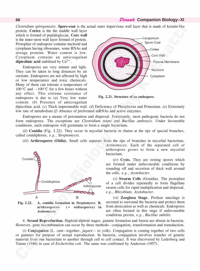
��������Companion Biology–XI68
Clostridium spirogenesis). Spore-coat is the actual outer impervious wall layer that is made of keratin-likeprotein. Cortex is the the middle wall layerwhich is formed of peptidoglycan. Core wallis the inner-most wall layer formed of protein.Protoplast of endospore contains nucleoid andcytoplasm having ribosomes, some RNAs andstorage proteins. Water content is low.Cytoplasm contains an anticoagulantdipicolinic acid stabilised by Ca2+.
Endospores are very minute and light.They can be taken to long distances by aircurrents. Endospores are not affected by highor low temperature and toxic chemicals.Many of them can tolerate a temperature of100°C and —100°C for a few hours withoutany effect. This extreme resistance ofendospores is due to (a) Very low watercontent. (b) Presence of anticoagulantdipicolinic acid. (c) Thick impermeable wall. (d) Deficiency of Phosphorus and Potassium. (e) Extremelylow rate of metabolism (f) Absence of preformed mRNAs and active enzymes.
Endospores are a means of perennation and dispersal. Fortunately, most pathogenic bacteria do notform endospores. The exceptions are Clostridium tetani and Bacillus anthracis. Under favourableconditions, each endospore will germinate to form a single bacterium.
(ii) Conidia (Fig. 2.22). They occur in mycelial bacteria in chains at the tips of special branches,called conidiphores, e.g., Streptomyces.
(iii) Arthrospores (Oidia). Small cells separate from the tips of branches in mycelial bacterium,Actinomyces. Each of the separated cell orarthrospore grows to form a new mycelialbacterium.
(iv) Cysts. They are resting spores whichare formed under unfavourable conditions byrounding off and secretion of thick wall aroundthe cells, e.g., Azotobacter.
(v) Swarm Cells (Gonidia). The protoplastof a cell divides repeatedly to form flagellateswarm cells for rapid multiplication and dispersal,e.g., Rhizobium, Azotobacter.
(vi) Zoogloea Stage. Profuse mucilage issecreted to surround the bacteria and protect themfrom desiccation as well as chemicals. Endosporesare often formed in this stage if unfavourableconditions persist, e.g., Bacillus subtilis.
4. Sexual Reproduction. Haploid-diploid stages, gamete formation and fusion are absent in bacteria.However, gene recombination can occur by three methods—conjugation, transformation and transduction.
(i) Conjugation (L. cum—together, jugare— to yolk). Conjugation is coming together of two cellsor gametes for purpose of sexual reproduction. In bacteria, conjugation involves transfer of geneticmaterial from one bacterium to another through cell to cell contact. It was discovered by Lederberg andTatum (1946) in case of Escherichia coli. The same was confirmed by Anderson (1957).
Fig. 2.21. Structure of an endospore.
Fig. 2.22. A, conidia formation in Streptomyces. B,arthrospores (= oidiospores) inActinomyces.
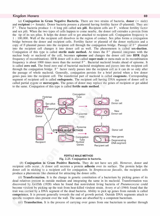
����������� 69
(a) Conjugation in Gram Negative Bacteria. There are two strains of bacteria, donor (= male)and recipient (= female). Donor bacteria possess a plasmid having fertility factor (F-plasmid). They areF+. These bacteria produce 1—4 long pili called sex pili. Recipient cells are F–, without fertility factorand sex pili. When the two types of cells happen to come nearby, the donor cell extrudes a protein fromthe tip of its sex pilus. It helps the donor cell to get attached to recipient cell. Conjugation frequency is1 : 100,000. Wall of the recipient cell dissolves in the region of contact. Sex pilus forms a conjugationbridge between the donor and recipient cells. Fertility factor or plasmid of the donor replicates and acopy of F-plasmid passes into the recipient cell through the conjugation bridge. Passage of F+ plasmidinto the recipient cell changes it into donor cell as well. The phenomenon is called sex-duction.Conjugation of this type is called sterile male method. At times the F+ plasmid integrates with thenuclear body or nucleoid of the cell, becomes episome and changes the donor cell into HFR (highfrequency of recombination). HFR donor cell is also called super-male or meta-male as its recombinationfrequency is about 1000 times more than the normal F+. Bacterial nucleoid breaks ahead of episome. Itis called zero end. The freed zero end of bacterial nucleoid straightens and passes into the recipient cellthrough the conjugation bridge. F+ factor rarely passes into the recipient cell as it can do so only afterthe passage of whole nucleoid. Generally, conjugation persists for a brief period when a few donorgenes pass into the recipient cell. The transferred part of nucleoid is called exogenote. Correspondingsegment of recipient cell is called endogenote. The recipient cell having DNA segment of donor cell iscalled partial zygote or merozygote. The genes of donor may replace the genes of recipient or get addedto the same. Conjugation of this type is called fertile male method.
Fig. 2.23. Conjugation in bacteria.
(b) Conjugation in Gram Positive Bacteria. They do not have sex pili. However, donor andrecipient cells occur. A donor cell secretes a protein adhesin over its surface. The protein helps thedonor cell in sticking to a recipient cell for conjugation. In Streptococcus faecalis, the recipient cellsproduce a pheromone like chemical for attracting the donor cells.
(ii) Transformation. It is the change in genetic constitution of a bacterium by picking genes of itsdead relatives present in outside medium and integrating the same in its nucleoid. Transformation wasdiscovered by Griffith (1928) when he found that nonvirulent living bacteria of Pneumococcus couldbecome virulent by picking up the trait from heat-killed virulent strain. Avery et al (1944) found that thetrait was carried by a DNA segment of the dead bacteria. Ability to pick up genes from outside is calledcompetence. It is present generally towards the end of growth period. Outside DNA segments attach tospecific receptors sites present over the wall. The same are absorbed by a competent bacterium.
(iii) Transduction. It is the process of carrying over genes from one bacterium to another through
STERILE MALE METHOD
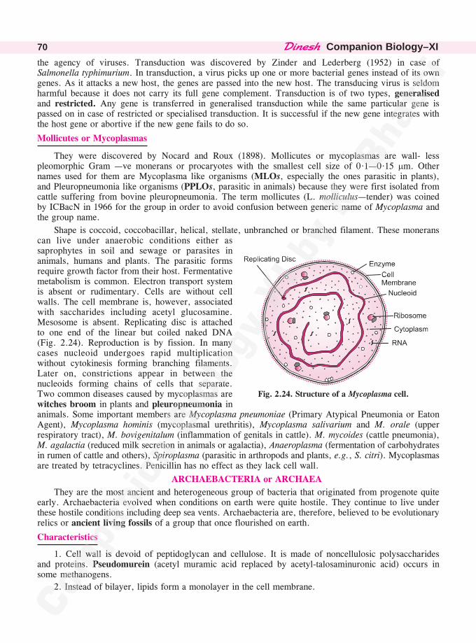
��������Companion Biology–XI70
the agency of viruses. Transduction was discovered by Zinder and Lederberg (1952) in case ofSalmonella typhimurium. In transduction, a virus picks up one or more bacterial genes instead of its owngenes. As it attacks a new host, the genes are passed into the new host. The transducing virus is seldomharmful because it does not carry its full gene complement. Transduction is of two types, generalisedand restricted. Any gene is transferred in generalised transduction while the same particular gene ispassed on in case of restricted or specialised transduction. It is successful if the new gene integrates withthe host gene or abortive if the new gene fails to do so.
Mollicutes or Mycoplasmas
They were discovered by Nocard and Roux (1898). Mollicutes or mycoplasmas are wall- lesspleomorphic Gram —ve monerans or procaryotes with the smallest cell size of 0·1—0·15 �m. Othernames used for them are Mycoplasma like organisms (MLOs, especially the ones parasitic in plants),and Pleuropneumonia like organisms (PPLOs, parasitic in animals) because they were first isolated fromcattle suffering from bovine pleuropneumonia. The term mollicutes (L. molliculus—tender) was coinedby ICBacN in 1966 for the group in order to avoid confusion between generic name of Mycoplasma andthe group name.
Shape is coccoid, coccobacillar, helical, stellate, unbranched or branched filament. These moneranscan live under anaerobic conditions either assaprophytes in soil and sewage or parasites inanimals, humans and plants. The parasitic formsrequire growth factor from their host. Fermentativemetabolism is common. Electron transport systemis absent or rudimentary. Cells are without cellwalls. The cell membrane is, however, associatedwith saccharides including acetyl glucosamine.Mesosome is absent. Replicating disc is attachedto one end of the linear but coiled naked DNA(Fig. 2.24). Reproduction is by fission. In manycases nucleoid undergoes rapid multiplicationwithout cytokinesis forming branching filaments.Later on, constrictions appear in between thenucleoids forming chains of cells that separate.Two common diseases caused by mycoplasmas arewitches broom in plants and pleuropneumonia inanimals. Some important members are Mycoplasma pneumoniae (Primary Atypical Pneumonia or EatonAgent), Mycoplasma hominis (mycoplasmal urethritis), Mycoplasma salivarium and M. orale (upperrespiratory tract), M. bovigenitalum (inflammation of genitals in cattle). M. mycoides (cattle pneumonia),M. agalactia (reduced milk secretion in animals or agalactia), Anaeroplasma (fermentation of carbohydratesin rumen of cattle and others), Spiroplasma (parasitic in arthropods and plants, e.g., S. citri). Mycoplasmasare treated by tetracyclines. Penicillin has no effect as they lack cell wall.
ARCHAEBACTERIA or ARCHAEA
They are the most ancient and heterogeneous group of bacteria that originated from progenote quiteearly. Archaebacteria evolved when conditions on earth were quite hostile. They continue to live underthese hostile conditions including deep sea vents. Archaebacteria are, therefore, believed to be evolutionaryrelics or ancient living fossils of a group that once flourished on earth.
Characteristics
1. Cell wall is devoid of peptidoglycan and cellulose. It is made of noncellulosic polysaccharidesand proteins. Pseudomurein (acetyl muramic acid replaced by acetyl-talosaminuronic acid) occurs insome methanogens.
2. Instead of bilayer, lipids form a monolayer in the cell membrane.
�Fig. 2.24. Structure of a Mycoplasma cell.
����������� 71
3. In cell membrane, the fatty acids are attached to glycerol by ether bonds (—O·CH2—) instead ofester bond (—O.CO—).
4. Fatty acids present in the cell membrane are branched. Branched chain lipids decrease membranefluidity and increase tolerance to extremes of heat as well as pH.
5. Core histones occur. Operons are monocistronic. DNA replication, transcription and translationresemble eukaryotes more than prokaryotes.
6. Ribonucleotides of 16S rRNA are different from those of other organisms. RNA polymerase hasalso a different constitution.
Groups of Archaebacteria
Archaebacteria have been divided arbitrarily on the basis of their metabolic and ecological properties.(i) Obligate anaerobic autotrophs which get killed in presence of oxygen and produce methane as end productof metabolism. They are called methanogens. (ii) Aerobic heterotrophs which live in brine and are calledhalophiles. (iii) Facultative or obligate anaerobes that chemoheterotrophically or facultativechemoautotrophically live in hot sulphur springs or similar areas. They are called thermoacidophiles.(iv) Spang et al (2015) have discovered a new group of archaebacteria named lokiarchaeota after LokiCastle, a hydrothermal vent. It has a number of genes similar to eukaryotes. The group is believed to be alink between prokaryotes and eukaryotes.
Methanogens
They are obligate anaerobic archaebacteria which produce methane as end product of their metabolism.They live in swamps, lake sediments, anaerobic sewage digesters and other similar places where organicmatter is undergoing anaerobic decomposition. Methanopappus kandleri is an extremely thermophilichalotolerant archaebacterium growing in temperature of upto 110°C (Kurr et al 1992). Some methanogensare endosymbionts in anaerobic protozoa and digestive tract of certain higher animals. Methanogens donot take part in decomposition of organic matter but metabolise end products of the same like H2, CO2,formate, methanol, methylamine acetate, etc. They are autotrophic which obtain energy as well ascarbon for assimilation from end products of decomposition. Typical methods of carbon assimilation areabsent. However, acetate is an important precursor.
Methanogens are employed in production of biogas or gobar gas from formic acid and carbondioxide produced during fermentation of organic matter. The important examples are Methanobacteriumand Methanococcus. Rumen of cattle which is the seat of cellulose fermentation also contain methanogenslike Methanobacterium ruminantium.
• Methophiles. Eubacteria capable of obtaining carbon and energy from either onecarbon compound or two and more carbon compounds where carbon atoms are notdirectly linked. There are two groups, methanotrophs (e.g., Methylococcus) andmethylotrophs (e.g., Methylophilus).
Halophiles
They live in habitats having high salinity and high light intensity. The minimum salt concentrationrequired for growth of halophiles is 2—2·5 M while optimum is 4—5 M. Halophiles are able to livecomfortably in this high salt concentration because their intracellular salt concentration is equally high.It is made of Na+, Cl� and K+. The enzymes and ribosomes of halophiles also function efficiently onlyat high intracellular salt concentration. Halophiles possess red carotenoids in their cell membrane. Theyprotect the archaebacteria from intense harmful radiations.
In shape the halophiles may be cocci (e.g., Halococcus) or bacilli with polar flagellum (e.g.,Halobacterium). Nutrition is chemoheterotrophic (saprotrophic). The bacteria are aerobes though solubilityof O2 in brine is low. Oxygen deficiency is environmental stress faced by halophiles. Under anaerobicconditions, Halobacterium develops purple membrane having photoreceptor pigment bacteriorhodopsin.In light, it acts as a proton pump and helps synthesise ATP. Formation of ATP is a survival mechanismbecause under anaerobic conditions, Halobacterium is unable to metabolise properly.
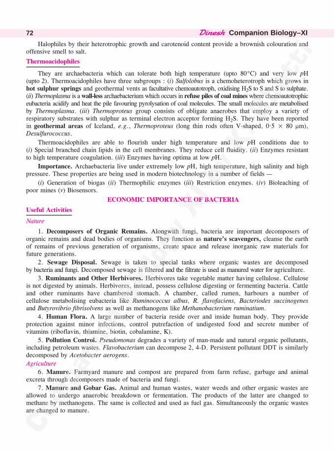
��������Companion Biology–XI72
Halophiles by their heterotrophic growth and carotenoid content provide a brownish colouration andoffensive smell to salt.
Thermoacidophiles
They are archaebacteria which can tolerate both high temperature (upto 80°C) and very low pH(upto 2). Thermoacidophiles have three subgroups : (i) Sulfolobus is a chemoheterotroph which grows inhot sulphur springs and geothermal vents as facultative chemoautotroph, oxidising H2S to S and S to sulphate.(ii) Thermoplasma is a wall-less archaebacterium which occurs in refuse piles of coal mines where chemoautotrophiceubacteria acidify and heat the pile favouring pyrolysation of coal molecules. The small molecules are metabolisedby Thermoplasma. (iii) Thermoproteus group consists of obligate anaerobes that employ a variety ofrespiratory substrates with sulphur as terminal electron acceptor forming H2S. They have been reportedin geothermal areas of Iceland, e.g., Thermoproteus (long thin rods often V-shaped, 0·5 × 80 �m),Desulfurococcus.
Thermoacidophiles are able to flourish under high temperature and low pH conditions due to(i) Special branched chain lipids in the cell membranes. They reduce cell fluidity. (ii) Enzymes resistantto high temperature coagulation. (iii) Enzymes having optima at low pH.
Importance. Archaebacteria live under extremely low pH, high temperature, high salinity and highpressure. These properties are being used in modern biotechnology in a number of fields —
(i) Generation of biogas (ii) Thermophilic enzymes (iii) Restriction enzymes. (iv) Bioleaching ofpoor mines (v) Biosensors.
ECONOMIC IMPORTANCE OF BACTERIA
Useful Activities
Nature
1. Decomposers of Organic Remains. Alongwith fungi, bacteria are important decomposers oforganic remains and dead bodies of organisms. They function as nature’s scavengers, cleanse the earthof remains of previous generation of organisms, create space and release inorganic raw materials forfuture generations.
2. Sewage Disposal. Sewage is taken to special tanks where organic wastes are decomposedby bacteria and fungi. Decomposed sewage is filtered and the filtrate is used as manured water for agriculture.
3. Ruminants and Other Herbivores. Herbivores take vegetable matter having cellulose. Celluloseis not digested by animals. Herbivores, instead, possess cellulose digesting or fermenting bacteria. Cattleand other ruminants have chambered stomach. A chamber, called rumen, harbours a number ofcellulose metabolising eubacteria like Ruminococcus albus, R. flavofaciens, Bacteriodes succinogenesand Butyrovibrio fibrisolvens as well as methanogens like Methanobacterium ruminatium.
4. Human Flora. A large number of bacteria reside over and inside human body. They provideprotection against minor infections, control putrefaction of undigested food and secrete number ofvitamins (riboflavin, thiamine, biotin, cobalamine, K).
5. Pollution Control. Pseudomonas degrades a variety of man-made and natural organic pollutants,including petroleum wastes. Flavobacterium can decompose 2, 4-D. Persistent pollutant DDT is similarlydecomposed by Acetobacter aerogens.
Agriculture
6. Manure. Farmyard manure and compost are prepared from farm refuse, garbage and animalexcreta through decomposers made of bacteria and fungi.
7. Manure and Gobar Gas. Animal and human wastes, water weeds and other organic wastes areallowed to undergo anaerobic breakdown or fermentation. The products of the latter are changed tomethane by methanogens. The same is collected and used as fuel gas. Simultaneously the organic wastesare changed to manure.
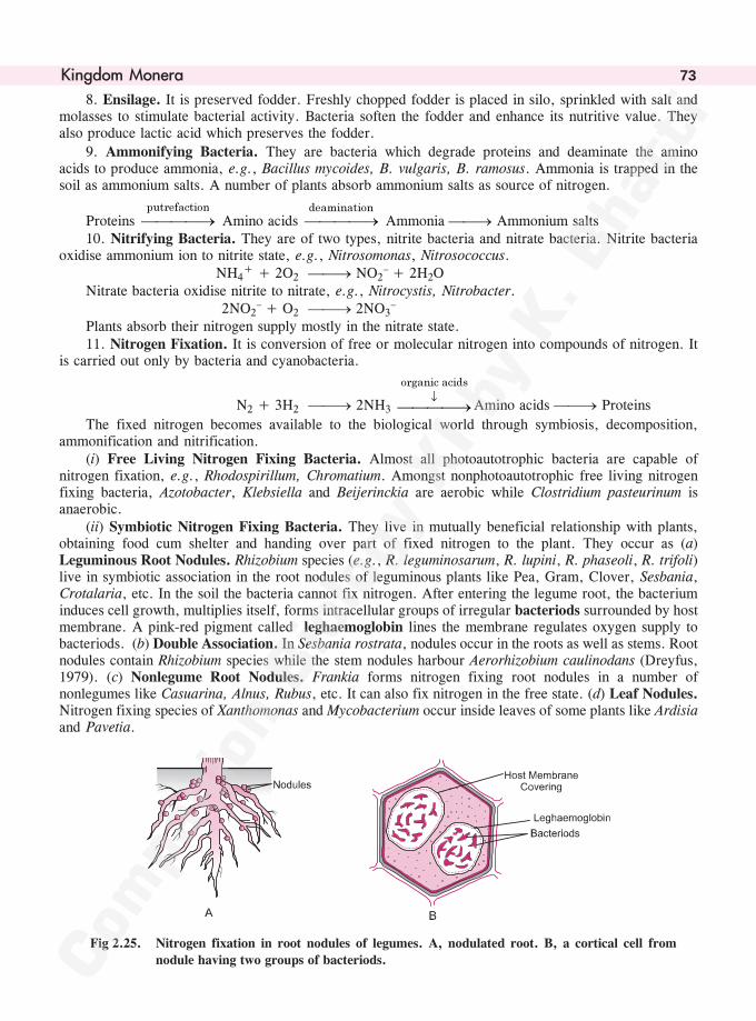
����������� 73
8. Ensilage. It is preserved fodder. Freshly chopped fodder is placed in silo, sprinkled with salt andmolasses to stimulate bacterial activity. Bacteria soften the fodder and enhance its nutritive value. Theyalso produce lactic acid which preserves the fodder.
9. Ammonifying Bacteria. They are bacteria which degrade proteins and deaminate the aminoacids to produce ammonia, e.g., Bacillus mycoides, B. vulgaris, B. ramosus. Ammonia is trapped in thesoil as ammonium salts. A number of plants absorb ammonium salts as source of nitrogen.
Proteins ���������������
Amino acids ���������������
Ammonia ��� Ammonium salts10. Nitrifying Bacteria. They are of two types, nitrite bacteria and nitrate bacteria. Nitrite bacteria
oxidise ammonium ion to nitrite state, e.g., Nitrosomonas, Nitrosococcus. NH4
+ + 2O2 ��� NO2– + 2H2O
Nitrate bacteria oxidise nitrite to nitrate, e.g., Nitrocystis, Nitrobacter.2NO2
– + O2 ��� 2NO3–
Plants absorb their nitrogen supply mostly in the nitrate state.11. Nitrogen Fixation. It is conversion of free or molecular nitrogen into compounds of nitrogen. It
is carried out only by bacteria and cyanobacteria.
N2 + 3H2 ��� 2NH3 ������
�����������
Amino acids ��� Proteins
The fixed nitrogen becomes available to the biological world through symbiosis, decomposition,ammonification and nitrification.
(i) Free Living Nitrogen Fixing Bacteria. Almost all photoautotrophic bacteria are capable ofnitrogen fixation, e.g., Rhodospirillum, Chromatium. Amongst nonphotoautotrophic free living nitrogenfixing bacteria, Azotobacter, Klebsiella and Beijerinckia are aerobic while Clostridium pasteurinum isanaerobic.
(ii) Symbiotic Nitrogen Fixing Bacteria. They live in mutually beneficial relationship with plants,obtaining food cum shelter and handing over part of fixed nitrogen to the plant. They occur as (a)Leguminous Root Nodules. Rhizobium species (e.g., R. leguminosarum, R. lupini, R. phaseoli, R. trifoli)live in symbiotic association in the root nodules of leguminous plants like Pea, Gram, Clover, Sesbania,Crotalaria, etc. In the soil the bacteria cannot fix nitrogen. After entering the legume root, the bacteriuminduces cell growth, multiplies itself, forms intracellular groups of irregular bacteriods surrounded by hostmembrane. A pink-red pigment called leghaemoglobin lines the membrane regulates oxygen supply tobacteriods. (b) Double Association. In Sesbania rostrata, nodules occur in the roots as well as stems. Rootnodules contain Rhizobium species while the stem nodules harbour Aerorhizobium caulinodans (Dreyfus,1979). (c) Nonlegume Root Nodules. Frankia forms nitrogen fixing root nodules in a number ofnonlegumes like Casuarina, Alnus, Rubus, etc. It can also fix nitrogen in the free state. (d) Leaf Nodules.Nitrogen fixing species of Xanthomonas and Mycobacterium occur inside leaves of some plants like Ardisiaand Pavetia.
Fig 2.25. Nitrogen fixation in root nodules of legumes. A, nodulated root. B, a cortical cell from
nodule having two groups of bacteriods.
��������Companion Biology–XI74
12. Sulphur Bacteria. Putrefaction of proteins releases hydrogen sulphide or H2S. The same isoxidised by sulphur bacteria to produce first sulphur and then sulphate. This helps in cycling of sulphuras sulphate is picked up by plants for their metabolism (formation of amino acids and proteins).
13. Biopesticides. Bacillus popilliae reduces population of Japanese Beetle (Popilia) which is aserious pest of vegetables and fruits. B. sphericus similarly controls population of mosquito Anopheles.Bacillus thuringiensis yields an insecticide called thurioside. The bacterium is also sprayed as spores(e.g., sporeine) to control insect pests.
Industries14. Dairy Industry. Lactic acid bacteria like Streptococcus lactis and S. cremoris are able to
convert milk sugar lactose into lactic acid.C12H22O11 + H2O ��� 4C3H6O3
lactose lactic acidLactic acid coagulates milk protein casein. This converts milk into curd. Another bacterium
Leuconostoc citrovorus provides characteristic flavour to curd and its product butter. Bacteria involvedin formation of Yoghurt or fermented milk are Lactobacillus bulgaricus and Streptococcus thermophilus.During cheese formation Streptococcus and Lactobacillus are involved in formation of curd andPropionibacterium for flavour.
15. Retting of Fibres. Plant fibres (e.g., Jute, Flax, Hemp) are separated from softer tissues by twotypes of retting processes : (i) Anaerobic Retting (submersion in water). Butyric acid bacteria (Clostridiumbutylicum). (ii) Dew Retting (periodic sprinkling of water). Pseudomonas fluorescens.
16. Cleaning of Raw Hides. Raw hides contain a lot of tissues, fat and hair. They are immersed inwater when bacterial action removes the extra materials.
17. Acetone and Butanol. It was the first commercial process for production of industrial solvents,acetone and butanol, from molasses through the activity of bacteria, Clostridium acetobutylicum. Nowthe same are obtained from petroleum.
18. Lactic Acid. Lactobacillus delbruckii is used in fermentation of lactic acid from ammoniatedsugar in closed container kept at 50°C. Lactic acid is used in preservation, tanning and plastics.
19. Vinegar. Fermented molasses (containing ethyl alcohol) is inoculated with bacteria, Acetobacteraceti, under aerobic conditions. It yields vinegar or dilute acetic acid.
20. Curing. Tea, Tobacco, Coffee and Cocoa are removed of their bitterness and provided withcharacteristic taste and aroma by bacterial action, e.g., Micrococcus candicans, Bacillus megatherium.
21. Biodegradable Plastic. Poly �-hydroxybutyrate (PHB) is found in several bacteria as reservefood. It can be used to produce biodegradable plastic.Medicinal
22. Cellulose. There are many bacteria which synthesise cellulose, e.g., Acetobacter, Aerobacter,Azotobacter. Being pure, the same can be put to several uses.
23. Vitamins. Riboflavin was once manufactured with the help of Clostridium butylicum. Aceticacid bacteria are employed in certain steps of vitamin C synthesis. Bacillus megatherium and Streptomycesolivaceous are used for production of B12 (cobalamine).
• Rhizobium. It is aerobic bacterium. Nitrogenase present in bacteriods requires protection from
oxygen.
• Bdellovibrio. It is a very small (0·3—0·4 × 0·8—1·2 �m) monotrichous bacterium which feeds onother bacteria. Bdellovibrio attaches to host bacterium, passes into it, multiplies and causes lysis ofhost cells. Ganges contains Bdellovibrio bacteriovorus that maintains purity of its water alongwithbacteriophages.
24. Antibiotics. Antibiotics (Waksman, 1942) are biochemicals produced by microorganisms whichin low concentration can inhibit the growth of pathogenic microorganisms without affecting the host.Penicillin was the first commercial antibiotic. A number of antibiotics are produced by bacteria, many ofthem by actinomycetal genus Streptomyces.
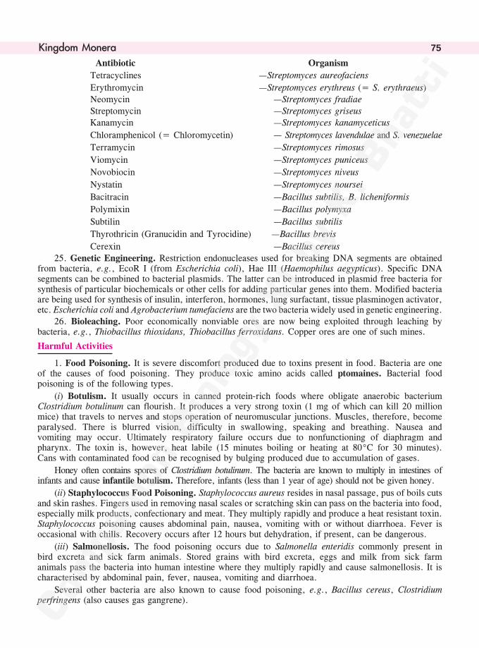
����������� 75
Antibiotic Organism
Tetracyclines —Streptomyces aureofaciens
Erythromycin —Streptomyces erythreus (= S. erythraeus)Neomycin —Streptomyces fradiaeStreptomycin —Streptomyces griseusKanamycin —Streptomyces kanamyceticus
Chloramphenicol (= Chloromycetin) — Streptomyces lavendulae and S. venezuelae
Terramycin —Streptomyces rimosus
Viomycin —Streptomyces puniceus
Novobiocin —Streptomyces niveus
Nystatin —Streptomyces noursei
Bacitracin —Bacillus subtilis, B. licheniformis
Polymixin —Bacillus polymyxa
Subtilin —Bacillus subtilis
Thyrothricin (Granucidin and Tyrocidine) —Bacillus brevis
Cerexin —Bacillus cereus25. Genetic Engineering. Restriction endonucleases used for breaking DNA segments are obtained
from bacteria, e.g., EcoR I (from Escherichia coli), Hae III (Haemophilus aegypticus). Specific DNAsegments can be combined to bacterial plasmids. The latter can be introduced in plasmid free bacteria forsynthesis of particular biochemicals or other cells for adding particular genes into them. Modified bacteriaare being used for synthesis of insulin, interferon, hormones, lung surfactant, tissue plasminogen activator,etc. Escherichia coli and Agrobacterium tumefaciens are the two bacteria widely used in genetic engineering.
26. Bioleaching. Poor economically nonviable ores are now being exploited through leaching bybacteria, e.g., Thiobacillus thioxidans, Thiobacillus ferroxidans. Copper ores are one of such mines.
Harmful Activities
1. Food Poisoning. It is severe discomfort produced due to toxins present in food. Bacteria are oneof the causes of food poisoning. They produce toxic amino acids called ptomaines. Bacterial foodpoisoning is of the following types.
(i) Botulism. It usually occurs in canned protein-rich foods where obligate anaerobic bacteriumClostridium botulinum can flourish. It produces a very strong toxin (1 mg of which can kill 20 millionmice) that travels to nerves and stops operation of neuromuscular junctions. Muscles, therefore, becomeparalysed. There is blurred vision, difficulty in swallowing, speaking and breathing. Nausea andvomiting may occur. Ultimately respiratory failure occurs due to nonfunctioning of diaphragm andpharynx. The toxin is, however, heat labile (15 minutes boiling or heating at 80°C for 30 minutes).Cans with contaminated food can be recognised by bulging produced due to accumulation of gases.
Honey often contains spores of Clostridium botulinum. The bacteria are known to multiply in intestines ofinfants and cause infantile botulism. Therefore, infants (less than 1 year of age) should not be given honey.
(ii) Staphylococcus Food Poisoning. Staphylococcus aureus resides in nasal passage, pus of boils cutsand skin rashes. Fingers used in removing nasal scales or scratching skin can pass on the bacteria into food,especially milk products, confectionary and meat. They multiply rapidly and produce a heat resistant toxin.Staphylococcus poisoning causes abdominal pain, nausea, vomiting with or without diarrhoea. Fever isoccasional with chills. Recovery occurs after 12 hours but dehydration, if present, can be dangerous.
(iii) Salmonellosis. The food poisoning occurs due to Salmonella enteridis commonly present inbird excreta and sick farm animals. Stored grains with bird excreta, eggs and milk from sick farmanimals pass the bacteria into human intestine where they multiply rapidly and cause salmonellosis. It ischaracterised by abdominal pain, fever, nausea, vomiting and diarrhoea.
Several other bacteria are also known to cause food poisoning, e.g., Bacillus cereus, Clostridiumperfringens (also causes gas gangrene).
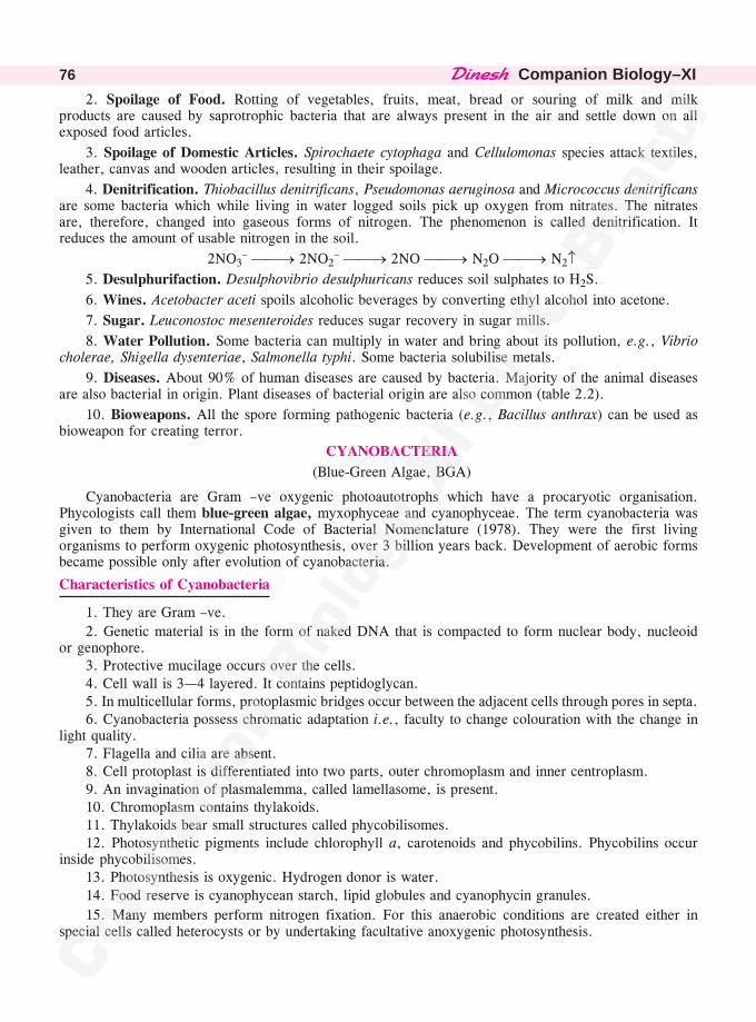
��������Companion Biology–XI76
2. Spoilage of Food. Rotting of vegetables, fruits, meat, bread or souring of milk and milkproducts are caused by saprotrophic bacteria that are always present in the air and settle down on allexposed food articles.
3. Spoilage of Domestic Articles. Spirochaete cytophaga and Cellulomonas species attack textiles,leather, canvas and wooden articles, resulting in their spoilage.
4. Denitrification. Thiobacillus denitrificans, Pseudomonas aeruginosa and Micrococcus denitrificansare some bacteria which while living in water logged soils pick up oxygen from nitrates. The nitratesare, therefore, changed into gaseous forms of nitrogen. The phenomenon is called denitrification. Itreduces the amount of usable nitrogen in the soil.
2NO3– ��� 2NO2
– ��� 2NO ��� N2O ��� N2�5. Desulphurifaction. Desulphovibrio desulphuricans reduces soil sulphates to H2S.
6. Wines. Acetobacter aceti spoils alcoholic beverages by converting ethyl alcohol into acetone.
7. Sugar. Leuconostoc mesenteroides reduces sugar recovery in sugar mills.
8. Water Pollution. Some bacteria can multiply in water and bring about its pollution, e.g., Vibriocholerae, Shigella dysenteriae, Salmonella typhi. Some bacteria solubilise metals.
9. Diseases. About 90% of human diseases are caused by bacteria. Majority of the animal diseasesare also bacterial in origin. Plant diseases of bacterial origin are also common (table 2.2).
10. Bioweapons. All the spore forming pathogenic bacteria (e.g., Bacillus anthrax) can be used asbioweapon for creating terror.
CYANOBACTERIA
(Blue-Green Algae, BGA)
Cyanobacteria are Gram –ve oxygenic photoautotrophs which have a procaryotic organisation.Phycologists call them blue-green algae, myxophyceae and cyanophyceae. The term cyanobacteria wasgiven to them by International Code of Bacterial Nomenclature (1978). They were the first livingorganisms to perform oxygenic photosynthesis, over 3 billion years back. Development of aerobic formsbecame possible only after evolution of cyanobacteria.
Characteristics of Cyanobacteria
1. They are Gram –ve.
2. Genetic material is in the form of naked DNA that is compacted to form nuclear body, nucleoidor genophore.
3. Protective mucilage occurs over the cells.4. Cell wall is 3—4 layered. It contains peptidoglycan.5. In multicellular forms, protoplasmic bridges occur between the adjacent cells through pores in septa.6. Cyanobacteria possess chromatic adaptation i.e., faculty to change colouration with the change in
light quality.7. Flagella and cilia are absent.8. Cell protoplast is differentiated into two parts, outer chromoplasm and inner centroplasm.9. An invagination of plasmalemma, called lamellasome, is present.10. Chromoplasm contains thylakoids.11. Thylakoids bear small structures called phycobilisomes.12. Photosynthetic pigments include chlorophyll a, carotenoids and phycobilins. Phycobilins occur
inside phycobilisomes.13. Photosynthesis is oxygenic. Hydrogen donor is water.14. Food reserve is cyanophycean starch, lipid globules and cyanophycin granules.
15. Many members perform nitrogen fixation. For this anaerobic conditions are created either inspecial cells called heterocysts or by undertaking facultative anoxygenic photosynthesis.
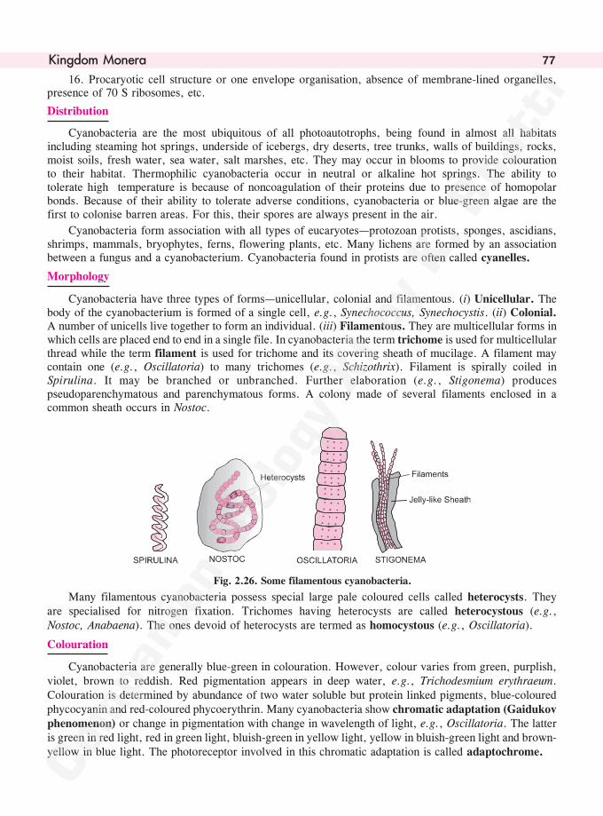
����������� 77
16. Procaryotic cell structure or one envelope organisation, absence of membrane-lined organelles,presence of 70 S ribosomes, etc.
Distribution
Cyanobacteria are the most ubiquitous of all photoautotrophs, being found in almost all habitatsincluding steaming hot springs, underside of icebergs, dry deserts, tree trunks, walls of buildings, rocks,moist soils, fresh water, sea water, salt marshes, etc. They may occur in blooms to provide colourationto their habitat. Thermophilic cyanobacteria occur in neutral or alkaline hot springs. The ability totolerate high temperature is because of noncoagulation of their proteins due to presence of homopolarbonds. Because of their ability to tolerate adverse conditions, cyanobacteria or blue-green algae are thefirst to colonise barren areas. For this, their spores are always present in the air.
Cyanobacteria form association with all types of eucaryotes—protozoan protists, sponges, ascidians,shrimps, mammals, bryophytes, ferns, flowering plants, etc. Many lichens are formed by an associationbetween a fungus and a cyanobacterium. Cyanobacteria found in protists are often called cyanelles.
Morphology
Cyanobacteria have three types of forms—unicellular, colonial and filamentous. (i) Unicellular. Thebody of the cyanobacterium is formed of a single cell, e.g., Synechococcus, Synechocystis. (ii) Colonial.A number of unicells live together to form an individual. (iii) Filamentous. They are multicellular forms inwhich cells are placed end to end in a single file. In cyanobacteria the term trichome is used for multicellularthread while the term filament is used for trichome and its covering sheath of mucilage. A filament maycontain one (e.g., Oscillatoria) to many trichomes (e.g., Schizothrix). Filament is spirally coiled inSpirulina. It may be branched or unbranched. Further elaboration (e.g., Stigonema) producespseudoparenchymatous and parenchymatous forms. A colony made of several filaments enclosed in acommon sheath occurs in Nostoc.
Fig. 2.26. Some filamentous cyanobacteria.
Many filamentous cyanobacteria possess special large pale coloured cells called heterocysts. Theyare specialised for nitrogen fixation. Trichomes having heterocysts are called heterocystous (e.g.,Nostoc, Anabaena). The ones devoid of heterocysts are termed as homocystous (e.g., Oscillatoria).
Colouration
Cyanobacteria are generally blue-green in colouration. However, colour varies from green, purplish,violet, brown to reddish. Red pigmentation appears in deep water, e.g., Trichodesmium erythraeum.Colouration is determined by abundance of two water soluble but protein linked pigments, blue-colouredphycocyanin and red-coloured phycoerythrin. Many cyanobacteria show chromatic adaptation (Gaidukovphenomenon) or change in pigmentation with change in wavelength of light, e.g., Oscillatoria. The latteris green in red light, red in green light, bluish-green in yellow light, yellow in bluish-green light and brown-yellow in blue light. The photoreceptor involved in this chromatic adaptation is called adaptochrome.
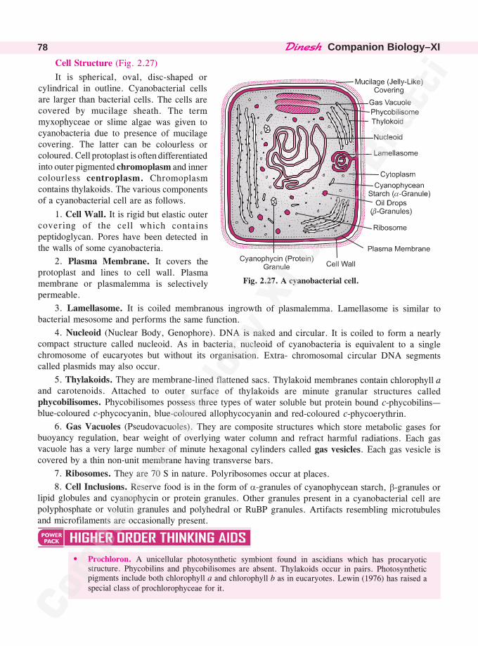
��������Companion Biology–XI78
Cell Structure (Fig. 2.27)
It is spherical, oval, disc-shaped orcylindrical in outline. Cyanobacterial cellsare larger than bacterial cells. The cells arecovered by mucilage sheath. The termmyxophyceae or slime algae was given tocyanobacteria due to presence of mucilagecovering. The latter can be colourless orcoloured. Cell protoplast is often differentiatedinto outer pigmented chromoplasm and innercolourless centroplasm. Chromoplasmcontains thylakoids. The various componentsof a cyanobacterial cell are as follows.
1. Cell Wall. It is rigid but elastic outercovering of the cell which containspeptidoglycan. Pores have been detected inthe walls of some cyanobacteria.
2. Plasma Membrane. It covers theprotoplast and lines to cell wall. Plasmamembrane or plasmalemma is selectivelypermeable.
3. Lamellasome. It is coiled membranous ingrowth of plasmalemma. Lamellasome is similar tobacterial mesosome and performs the same function.
4. Nucleoid (Nuclear Body, Genophore). DNA is naked and circular. It is coiled to form a nearlycompact structure called nucleoid. As in bacteria, nucleoid of cyanobacteria is equivalent to a singlechromosome of eucaryotes but without its organisation. Extra- chromosomal circular DNA segmentscalled plasmids may also occur.
5. Thylakoids. They are membrane-lined flattened sacs. Thylakoid membranes contain chlorophyll aand carotenoids. Attached to outer surface of thylakoids are minute granular structures calledphycobilisomes. Phycobilisomes possess three types of water soluble but protein bound c-phycobilins—blue-coloured c-phycocyanin, blue-coloured allophycocyanin and red-coloured c-phycoerythrin.
6. Gas Vacuoles (Pseudovacuoles). They are composite structures which store metabolic gases forbuoyancy regulation, bear weight of overlying water column and refract harmful radiations. Each gasvacuole has a very large number of minute hexagonal cylinders called gas vesicles. Each gas vesicle iscovered by a thin non-unit membrane having transverse bars.
7. Ribosomes. They are 70 S in nature. Polyribosomes occur at places.
8. Cell Inclusions. Reserve food is in the form of �-granules of cyanophycean starch, �-granules orlipid globules and cyanophycin or protein granules. Other granules present in a cyanobacterial cell arepolyphosphate or volutin granules and polyhedral or RuBP granules. Artifacts resembling microtubulesand microfilaments are occasionally present.
• Prochloron. A unicellular photosynthetic symbiont found in ascidians which has procaryoticstructure. Phycobilins and phycobilisomes are absent. Thylakoids occur in pairs. Photosyntheticpigments include both chlorophyll a and chlorophyll b as in eucaryotes. Lewin (1976) has raised aspecial class of prochlorophyceae for it.
�Fig. 2.27. A cyanobacterial cell.
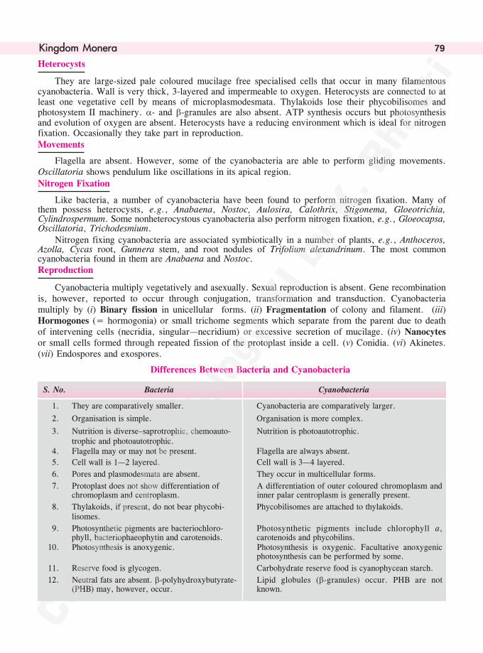
����������� 79
Heterocysts
They are large-sized pale coloured mucilage free specialised cells that occur in many filamentouscyanobacteria. Wall is very thick, 3-layered and impermeable to oxygen. Heterocysts are connected to atleast one vegetative cell by means of microplasmodesmata. Thylakoids lose their phycobilisomes andphotosystem II machinery. �- and �-granules are also absent. ATP synthesis occurs but photosynthesisand evolution of oxygen are absent. Heterocysts have a reducing environment which is ideal for nitrogenfixation. Occasionally they take part in reproduction.Movements
Flagella are absent. However, some of the cyanobacteria are able to perform gliding movements.Oscillatoria shows pendulum like oscillations in its apical region.Nitrogen Fixation
Like bacteria, a number of cyanobacteria have been found to perform nitrogen fixation. Many ofthem possess heterocysts, e.g., Anabaena, Nostoc, Aulosira, Calothrix, Stigonema, Gloeotrichia,Cylindrospermum. Some nonheterocystous cyanobacteria also perform nitrogen fixation, e.g., Gloeocapsa,Oscillatoria, Trichodesmium.
Nitrogen fixing cyanobacteria are associated symbiotically in a number of plants, e.g., Anthoceros,Azolla, Cycas root, Gunnera stem, and root nodules of Trifolium alexandrinum. The most commoncyanobacteria found in them are Anabaena and Nostoc.Reproduction
Cyanobacteria multiply vegetatively and asexually. Sexual reproduction is absent. Gene recombinationis, however, reported to occur through conjugation, transformation and transduction. Cyanobacteriamultiply by (i) Binary fission in unicellular forms. (ii) Fragmentation of colony and filament. (iii)Hormogones (= hormogonia) or small trichome segments which separate from the parent due to deathof intervening cells (necridia, singular—necridium) or excessive secretion of mucilage. (iv) Nanocytesor small cells formed through repeated fission of the protoplast inside a cell. (v) Conidia. (vi) Akinetes.(vii) Endospores and exospores.
Differences Between Bacteria and Cyanobacteria
S. No. Bacteria Cyanobacteria
1. They are comparatively smaller. Cyanobacteria are comparatively larger.
2. Organisation is simple. Organisation is more complex.
3. Nutrition is diverse–saprotrophic, chemoauto- Nutrition is photoautotrophic.trophic and photoautotrophic.
4. Flagella may or may not be present. Flagella are always absent.
5. Cell wall is 1—2 layered. Cell wall is 3—4 layered.
6. Pores and plasmodesmata are absent. They occur in multicellular forms.
7. Protoplast does not show differentiation of A differentiation of outer coloured chromoplasm andchromoplasm and centroplasm. inner palar centroplasm is generally present.
8. Thylakoids, if present, do not bear phycobi- Phycobilisomes are attached to thylakoids.lisomes.
9. Photosynthetic pigments are bacteriochloro- Photosynthetic pigments include chlorophyll a,phyll, bacteriophaeophytin and carotenoids. carotenoids and phycobilins.
10. Photosynthesis is anoxygenic. Photosynthesis is oxygenic. Facultative anoxygenicphotosynthesis can be performed by some.
11. Reserve food is glycogen. Carbohydrate reserve food is cyanophycean starch.
12. Neutral fats are absent. �-polyhydroxybutyrate- Lipid globules (�-granules) occur. PHB are not(PHB) may, however, occur. known.
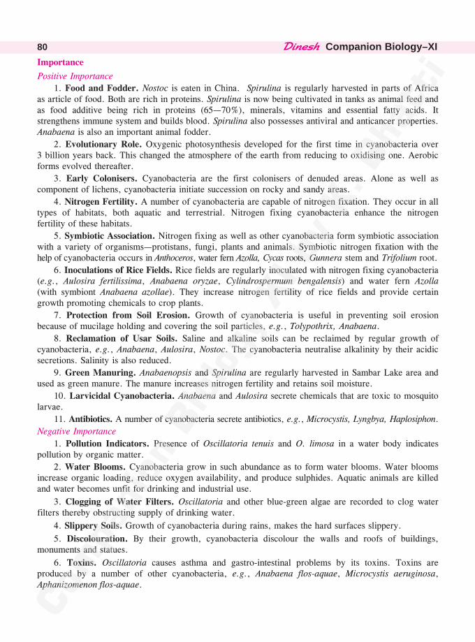
��������Companion Biology–XI80
Importance
Positive Importance
1. Food and Fodder. Nostoc is eaten in China. Spirulina is regularly harvested in parts of Africaas article of food. Both are rich in proteins. Spirulina is now being cultivated in tanks as animal feed andas food additive being rich in proteins (65—70%), minerals, vitamins and essential fatty acids. Itstrengthens immune system and builds blood. Spirulina also possesses antiviral and anticancer properties.Anabaena is also an important animal fodder.
2. Evolutionary Role. Oxygenic photosynthesis developed for the first time in cyanobacteria over3 billion years back. This changed the atmosphere of the earth from reducing to oxidising one. Aerobicforms evolved thereafter.
3. Early Colonisers. Cyanobacteria are the first colonisers of denuded areas. Alone as well ascomponent of lichens, cyanobacteria initiate succession on rocky and sandy areas.
4. Nitrogen Fertility. A number of cyanobacteria are capable of nitrogen fixation. They occur in alltypes of habitats, both aquatic and terrestrial. Nitrogen fixing cyanobacteria enhance the nitrogenfertility of these habitats.
5. Symbiotic Association. Nitrogen fixing as well as other cyanobacteria form symbiotic associationwith a variety of organisms—protistans, fungi, plants and animals. Symbiotic nitrogen fixation with thehelp of cyanobacteria occurs in Anthoceros, water fern Azolla, Cycas roots, Gunnera stem and Trifolium root.
6. Inoculations of Rice Fields. Rice fields are regularly inoculated with nitrogen fixing cyanobacteria(e.g., Aulosira fertilissima, Anabaena oryzae, Cylindrospermum bengalensis) and water fern Azolla(with symbiont Anabaena azollae). They increase nitrogen fertility of rice fields and provide certaingrowth promoting chemicals to crop plants.
7. Protection from Soil Erosion. Growth of cyanobacteria is useful in preventing soil erosionbecause of mucilage holding and covering the soil particles, e.g., Tolypothrix, Anabaena.
8. Reclamation of Usar Soils. Saline and alkaline soils can be reclaimed by regular growth ofcyanobacteria, e.g., Anabaena, Aulosira, Nostoc. The cyanobacteria neutralise alkalinity by their acidicsecretions. Salinity is also reduced.
9. Green Manuring. Anabaenopsis and Spirulina are regularly harvested in Sambar Lake area andused as green manure. The manure increases nitrogen fertility and retains soil moisture.
10. Larvicidal Cyanobacteria. Anabaena and Aulosira secrete chemicals that are toxic to mosquitolarvae.
11. Antibiotics. A number of cyanobacteria secrete antibiotics, e.g., Microcystis, Lyngbya, Haplosiphon.
Negative Importance
1. Pollution Indicators. Presence of Oscillatoria tenuis and O. limosa in a water body indicatespollution by organic matter.
2. Water Blooms. Cyanobacteria grow in such abundance as to form water blooms. Water bloomsincrease organic loading, reduce oxygen availability, and produce sulphides. Aquatic animals are killedand water becomes unfit for drinking and industrial use.
3. Clogging of Water Filters. Oscillatoria and other blue-green algae are recorded to clog waterfilters thereby obstructing supply of drinking water.
4. Slippery Soils. Growth of cyanobacteria during rains, makes the hard surfaces slippery.
5. Discolouration. By their growth, cyanobacteria discolour the walls and roofs of buildings,monuments and statues.
6. Toxins. Oscillatoria causes asthma and gastro-intestinal problems by its toxins. Toxins areproduced by a number of other cyanobacteria, e.g., Anabaena flos-aquae, Microcystis aeruginosa,Aphanizomenon flos-aquae.
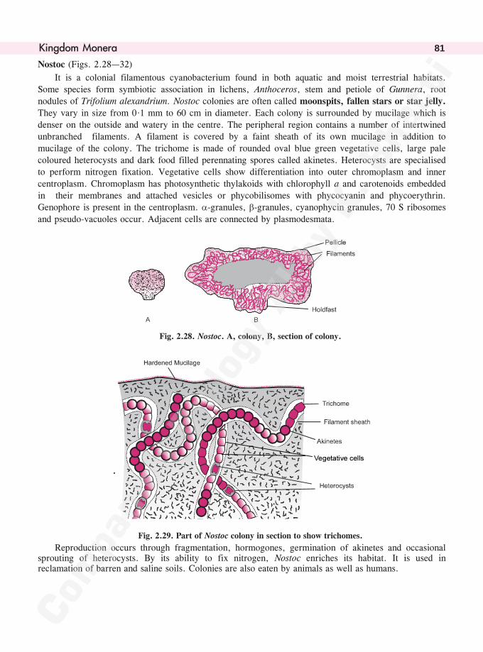
����������� 81
Nostoc (Figs. 2.28—32)
It is a colonial filamentous cyanobacterium found in both aquatic and moist terrestrial habitats.Some species form symbiotic association in lichens, Anthoceros, stem and petiole of Gunnera, rootnodules of Trifolium alexandrium. Nostoc colonies are often called moonspits, fallen stars or star jelly.
They vary in size from 0·1 mm to 60 cm in diameter. Each colony is surrounded by mucilage which isdenser on the outside and watery in the centre. The peripheral region contains a number of intertwinedunbranched filaments. A filament is covered by a faint sheath of its own mucilage in addition to
mucilage of the colony. The trichome is made of rounded oval blue green vegetative cells, large palecoloured heterocysts and dark food filled perennating spores called akinetes. Heterocysts are specialisedto perform nitrogen fixation. Vegetative cells show differentiation into outer chromoplasm and inner
centroplasm. Chromoplasm has photosynthetic thylakoids with chlorophyll a and carotenoids embeddedin their membranes and attached vesicles or phycobilisomes with phycocyanin and phycoerythrin.Genophore is present in the centroplasm. �-granules, �-granules, cyanophycin granules, 70 S ribosomes
and pseudo-vacuoles occur. Adjacent cells are connected by plasmodesmata.
Fig. 2.28. Nostoc. A, colony, B, section of colony.
.
��������������
Fig. 2.29. Part of Nostoc colony in section to show trichomes.
Reproduction occurs through fragmentation, hormogones, germination of akinetes and occasionalsprouting of heterocysts. By its ability to fix nitrogen, Nostoc enriches its habitat. It is used inreclamation of barren and saline soils. Colonies are also eaten by animals as well as humans.
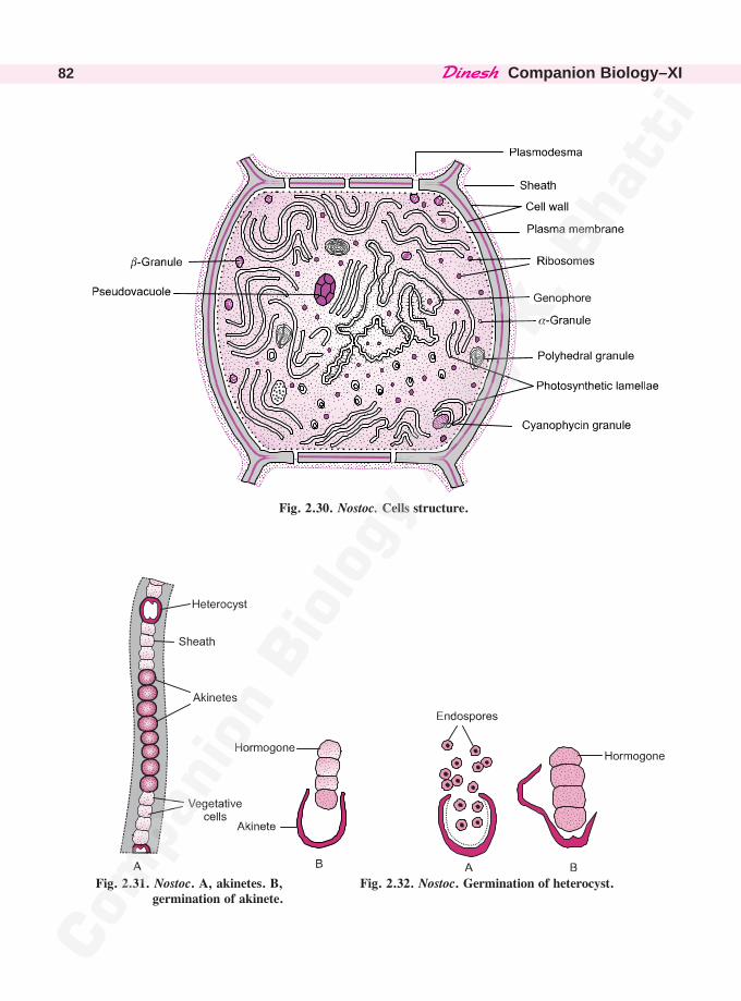
��������Companion Biology–XI82
���������
Fig. 2.30. Nostoc. Cells structure.
Fig. 2.31. Nostoc. A, akinetes. B, germination of akinete.
Fig. 2.32. Nostoc. Germination of heterocyst.
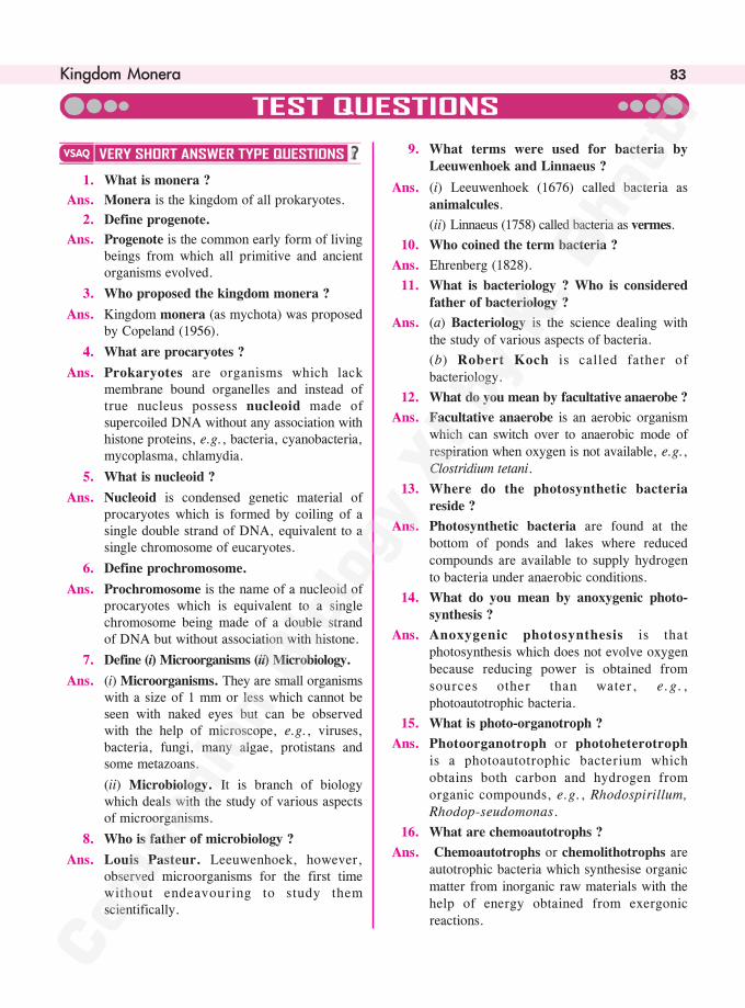
����������� 83
1. What is monera ?
Ans. Monera is the kingdom of all prokaryotes.
2. Define progenote.
Ans. Progenote is the common early form of livingbeings from which all primitive and ancientorganisms evolved.
3. Who proposed the kingdom monera ?
Ans. Kingdom monera (as mychota) was proposedby Copeland (1956).
4. What are procaryotes ?
Ans. Prokaryotes are organisms which lackmembrane bound organelles and instead oftrue nucleus possess nucleoid made ofsupercoiled DNA without any association withhistone proteins, e.g., bacteria, cyanobacteria,mycoplasma, chlamydia.
5. What is nucleoid ?
Ans. Nucleoid is condensed genetic material ofprocaryotes which is formed by coiling of asingle double strand of DNA, equivalent to asingle chromosome of eucaryotes.
6. Define prochromosome.
Ans. Prochromosome is the name of a nucleoid ofprocaryotes which is equivalent to a singlechromosome being made of a double strandof DNA but without association with histone.
7. Define (i) Microorganisms (ii) Microbiology.
Ans. (i) Microorganisms. They are small organismswith a size of 1 mm or less which cannot beseen with naked eyes but can be observedwith the help of microscope, e.g., viruses,bacteria, fungi, many algae, protistans andsome metazoans.
(ii) Microbiology. It is branch of biologywhich deals with the study of various aspectsof microorganisms.
8. Who is father of microbiology ?
Ans. Louis Pasteur. Leeuwenhoek, however,observed microorganisms for the first timewithout endeavouring to study themscientifically.
9. What terms were used for bacteria byLeeuwenhoek and Linnaeus ?
Ans. (i) Leeuwenhoek (1676) called bacteria asanimalcules.
(ii) Linnaeus (1758) called bacteria as vermes.
10. Who coined the term bacteria ?
Ans. Ehrenberg (1828).
11. What is bacteriology ? Who is consideredfather of bacteriology ?
Ans. (a) Bacteriology is the science dealing withthe study of various aspects of bacteria.
(b) Robert Koch is called father ofbacteriology.
12. What do you mean by facultative anaerobe ?
Ans. Facultative anaerobe is an aerobic organismwhich can switch over to anaerobic mode ofrespiration when oxygen is not available, e.g.,Clostridium tetani.
13. Where do the photosynthetic bacteriareside ?
Ans. Photosynthetic bacteria are found at thebottom of ponds and lakes where reducedcompounds are available to supply hydrogento bacteria under anaerobic conditions.
14. What do you mean by anoxygenic photo-synthesis ?
Ans. Anoxygenic photosynthesis is thatphotosynthesis which does not evolve oxygenbecause reducing power is obtained fromsources other than water, e.g.,photoautotrophic bacteria.
15. What is photo-organotroph ?
Ans. Photoorganotroph or photoheterotrophis a photoautotrophic bacterium whichobtains both carbon and hydrogen fromorganic compounds, e.g., Rhodospirillum,Rhodop-seudomonas.
16. What are chemoautotrophs ?
Ans. Chemoautotrophs or chemolithotrophs areautotrophic bacteria which synthesise organicmatter from inorganic raw materials with thehelp of energy obtained from exergonicreactions.
��������Companion Biology–XI84
26. Time interval between two binary fissionsis 30 minutes. How many bacteria will beformed from a single individual after(i) 12 hours and (ii) 24 hours.
Ans. (i) 223 (ii) 247.
27. What is anticoagulant present in bacterialendospores ?
Ans. Dipicolinic acid.
28. Who discovered conjugation in bacteria ?
Ans. Lederberg and Tatum (1946).
29. What is HFR ?
Ans. HFR (High Frequency of Recombination) isconversion of a donor bacterial cell into supermale which develops a recombinationfrequency of 1000 times higher than a normalF+ due to conversion of F+ plasmid intoepisome by integration with nuclear body ofthe donor cell.
30. What are endogenote, exogenote andmerozygote ?
Ans. (a) Endogenote. DNA segment of recipientbacterium which corresponds to the DNAsegment of donor cell that has entered it.
(b) Exogenote. DNA segment of donorbacterium that enters the recipient cell.
(c) Merozygote. It is partial zygote formeddue to the presence of DNA segment of donorcell in the recipient cell.
31. Name the structure involved in(i) Conjugation in Gram –ve bacteria(ii) Conjugation in Gram +ve bacteria.
Ans. (a) Gram –ve Bacteria. Sex pilus.
(b) Gram +ve Bacteria. Adhesin.
32. Name the scientists who discovered(i) Transformation (ii) Transduction inbacteria.
Ans. (a) Transformation in Bacteria. Griffith(1928).
(b) Transduction in Bacteria. Zinder andLederberg (1952).
33. What is transformation in bacteria ?
Ans. Transformation is the change in their geneticconstitution due to picking up of genes of thedead relatives and integrating the same in theirnucleoids.
34. Define transduction.
Ans. Transduction is the transfer of genes from
17. Define chemosynthesis.
Ans. Chemosynthesis is the synthesis of organicmatter from inorganic raw materials with thehelp of energy obtained from oxidisingchemicals in the external medium.
18. Give example of a chemolithotrophicbacterium.
Ans. Chemolithotrophic bacterium, e.g.,Nitrosomonas, Nitrobacter.
19. Give example of (i) Sulphur bacterium(ii) Iron bacterium (iii) Nitrifying bacterium.
Ans. (a) Sulphur bacterium, e.g., Beggiotoa.
(b) Iron bacterium, e.g., Ferrobacillus.
(c) Nitrifying bacterium, e.g., Nitrosomonas.
20. What do you mean by chemi-organotrophicbacteria ?
Ans. Chemiorganotrophic bacteria areheterotrophic bacteria which absorb organicnutrients from outside for both body buildingand as source of energy.
21. What is a facultative saprotroph ?
Ans. Facultative saprotroph is an autotrophic orparasitic organism which under certainsituations can behave as saprotroph.
22. Name the symbiotic nitrogen fixingbacterium found in leguminous roots.
Ans. Rhizobium leguminosarum.
23. Define endotoxin and exotoxin.
Ans. (i) Endotoxin. It is poisonous chemicalreleased on the death of the pathogen, e.g.,gonorrhoea toxin.
(ii) Exotoxin. It is poisonous chemical secretedextracellularly by pathogens, e.g., choleratoxin, anthrax toxin.
24. Name the bacteria responsible for thefollowing diseases : Typhoid, Tetanus andCholera.
Ans. Typhoid. Salmonella typhosa.
Tetanus. Clostridium tetani.
Cholera. Vibrio cholerae.
25. Name the bacterium causing (i) TB(ii) Syphilis (iii) Plague (iv) Leprosy(v) Whooping cough.
Ans. TB. Mycobacterium tuberculosis.
Syphilis. Treponema pallidum.
Plague. Yersinia (=Pasteurella) pestis.
Leprosy. Mycobacterium leprae.
Whooping Cough. Bordetella pertussis.
����������� 85
one bacterium to another through the agencyof virus.
35. What are mollicutes ?
Ans. Mollicutes is the term coined by ICBacN formycoplasmas.
36. What are mycoplasmas ?
Ans. Mycoplasmas are wall-less Gram –veprocaryotes of small size (0·1–0·15 �m) withlinear naked DNA and fermentation type ofmetabolism due to absence of electrontransport system.
37. Name the common disease caused bymycoplasma in (i) Plants (ii) Animals.
Ans. Witches broom in plants and pleuropneumoniain animals.
38. What are archaebacteria ?
Ans. Archaebacteria are heterogeneous group ofprimitive bacteria found in hostileenvironment, which lack peptidoglycan in cellwall and possess a monolayer of branchedfatty acids attached to glycerol by ether bondsin their cell membranes.
39. Why are archaebacteria called livingfossils ?
Ans. Archaebacteria are called living fossilsbecause they are not only evolution relics butalso continue to live under hostile conditionssimilar to the ones present on early earth atthe time of their development.
40. Expand MLO and PPLO.
Ans. (i) MLO. Mycoplasma like organisms.
(ii) PPLO. Pleuropneumonia like organisms.
41. What are methanogens ?
Ans. Methanogens are obligate anaerobicarchaebacteria which produce methane as endproduct of metabolism.
42. Define halophiles.
Ans. Halophiles are archaebacteria that occur inhabitats having high salinity and high lightintensity.
43. What are thermo-acidophiles ?
Ans. Thermoacidophiles are archaebacteria whichlive in habitats having low pH (upto 2) andhigh temperature (upto 80°C).
44. Name the pigment present in a halophilethat can synthesise ATP.
Ans. Bacteriorhodopsin.
45. Name two cellulose digesting bacteriapresent in the rumen of cattle.
Ans. Methanobacterium, Rumenococcus.
46. Name a methanogen present in rumen ofcattle.
Ans. Methanobacterium ruminantium.
47. What is salt concentration inside the cellsof halophiles ?
Ans. 4—5 M.
48. Name a bacterium which can degrade(i) Petroleum waste (ii) 2,4-D (iii) DDT.
Ans. (a) Pseudomonas (b) Flavobacterium
(c) Acetobacter.
49. Name a bacterium that yields insecticide.
Ans. Bacillus thuringiensis.
50. What is the function of bacteriumStreptococcus lactis in producing variousmilk products.
Ans. Formation of curd from milk.
51. What is retting of fibres ? Name abacterium involved in it.
Ans. Separation of fibres from soft tissues is calledretting of fibres, e.g., Jute, Flax. A commonbacterium is Pseudomonas fluorescens.
52. What is antibiotic ?
Ans. Antibiotic is a biochemical produced by amicroorganism which in low concentrationinhibits the growth of pathogenicmicroorganisms without harming the host.
53. Name the organism that yieldstetracyclines.
Ans. Streptomyces aureofaciens.
54. Name the microorganism that yieldsstreptomycin.
Ans. Streptomyces griseus.
55. What is the bacterial source ofchloramphenicol ?
Ans. Streptomyces lavendulae, S.venezuelae.
56. Name the bacterial product that is used inbreaking DNA at specific points.
Ans. Restriction endonucleases.
57. What is food poisoning ? Name a bacteriumcausing it.
Ans. Food poisoning is severe discomfort producedby toxic amino acids or ptomaines formedduring microbial contamination of food, e.g.,Streptomyces aureus.
��������Companion Biology–XI86
58. What is denitrification ? Name a bacteriumcausing it.
Ans. Denitrification is the reduction of soil nitrateinto gaseous forms of nitrogen that escape intoatmosphere, e.g., Thiobacillus denitrificans.
59. What are cyanobacteria ?
Ans. Cyanobacteria are Gram –ve oxygenicphotoautotrophic procaryotes, previouslyknown as blue-green algae.
60. What is trichome ?
Ans. Multicellular thread of a cyanobacterium orfilament without mucilage cover is calledtrichome.
61. What is chromatic adaptation ? Give anexample.
Ans. Chromatic adaptation or Gaidukovphenomenon is change in pigmentation of acyanobacterium in response to change inwavelength of light received by it. Oscillatoriais green in red light, red in green light,yellowish in bluish-green light and bluish-green in yellow light.
62. Define lamellasome.
Ans. Lamellasome is membranous ingrowth ofplasma membrane found in cyanobacteria.
63. What is phycobilisome ?
Ans. Phycobilisome is a phycobilin containinggranular structure that is attached to thylakoidsurface in cyanobacteria and red algae.
64. What are phycobilins ?
Ans. Phycobilins are water soluble, protein boundaccessory photosynthetic pigments found incyanobacteria and red algae, viz., phycocyanin(blue), allophycocyanin (blue), phycoerythrin(red).
65. Define heterocyst.
Ans. Heterocyst is large sized pale coloured,mucilage free cell of filamentouscyanobacteria that is specialised to performnitrogen fixation with occasionalreproduction.
66. What type of photosynthesis is performedby cyanobacteria ?
Ans. Oxygenic photosynthesis.
67. What is hormogone ?
Ans. Hormogone is a small trichome segmentwhich functions as propagule in filamentouscyanobacteria.
68. Name a nitrogen fixing cyanobacterium ofrice fields.
Ans. Aulosira fertilissima.
69. Define nitrogen fixation.
Ans. Nitrogen fixation is conversion of gaseousnitrogen into compounds of nitrogen.
1. Studies in molecular homology favours creationof three super kingdoms of.........., eubacteriaand .......... .
2. Besides morphology, bacterial classificationuses .........., physiological and ecologicalcharacteristics.
3. The group of eubacteria having mycelial formis ...........
4. When dry, endospores can withstand heat at.......... for several hours.
5. Depending upon colour, photoautotrophicbacteria are .......... or purple.
6. .......... oxidising bacteria convert H2S, S orpartially oxidised forms of sulphur to sulphate.
7. Mycoplasmas are the .......... knownprocaryotes that also lack cell .......... .
8. In cyanobacteria, the photosynthetic pigmentsinclude chlorophyll a and ...........
1. (i) Archaebacteria (ii) Eucaryotes
2. biochemical 3. actinomycetes
4. 100° 5. green
6. Sulphur 7. (i) smallest (ii) wall
8. phycobilins
1. Kingdom monera has two major groups,archaebacteria and eubacteria.
2. Endotoxins are water soluble heat labileproteins.
3. Methanogens are facultative anaerobes.
4. Purple membrane occurs in some halophiles.
5. Cyanobacteria residing in hot springs prefersulphur springs with acidic pH.
6. Spirulina is a component of traditional foodin some parts of Africa.
7. Anaerobic breakdown of proteins is calleddecay.
����������� 87
1. A—(iii), B—(i), C—(iv), D—(ii)
1. State two economically important uses of :
(a) Heterotrophic bacteria
(b) Archaebacteria.
Ans. (a) Heterotrophic bacteria.
(i) Dairy Industry. See text.
(ii) Retting of Fibres. See text.
(b) Archaebacteria.
(i) Biogas. See text.
(ii) Enzymes. Archaebacteria are being employed
to produce thermophilic enzymes and restriction
enzymes required in biotechnology.
1. True 2. False 3. False
4. True 5. False 6. True7. False
1. Column I Column II
A. Cocci (i) True bacteria
B. Eubacteria (ii) Archaebacteria
C. Chemoautotrophs (iii) Spherical shape
D. Halophile (iv) Iron bacteria
(v) Rod-shaped.
1. What is the principle underlying the use ofcyanobacteria in agricultural fields for cropimprovement ?
Ans. Cyanobacteria improve nitrogen fertility of thecrop fields as many of them are capable ofnitrogen fixation. They also reduce soil alkalinityand improve soil structure.
2. Polluted water bodies have usually highabundance of plants like Nostoc andOscillatoria. Give reasons.
Ans. Cyanobacteria (e.g., Nostoc, Oscillatoria) cantolerate adverse conditions includingpollutants, better than other aquatic plants andother algae due to absence of sap vacuoles,presence of mucilage covering and resistantproteins. Their spores are almost alwayspresent in air.
3. Are chemosynthetic bacteria autotrophic orheterotrophic ?
Ans. Autotrophic, as they are able to synthesize
their organic food from inorganic rawmaterials (with the help of energy obtainedfrom chemical reactions).
1. Cyanobacteria and heterotrophic bacteria
have been clubbed together in Eubacteriaof Kingdom Monera as per the ‘‘FiveKingdom classification’’ even though the
two are vastly different from each other. Isthis grouping of the two types of taxa in thesame kingdom justified ? If so, why ?
Ans. Yes, placing cyanobacteria alongwith
heterotrophic bacteria in Monera is fullyjustified as the two have prokaryotic nature,possess cell wall with peptidoglycan, 70 S
ribosomes, without sap vacuoles andmembrane covered organelles. Members ofmonera have diverse mode of nutrition as they
were the earliest living beings–anoxygenic
photosynthesis, oxygenic photosynthesis,chemosynthesis, heterotrophic nutrition, etc.
(From N .C .E.R .T. Question Bank)
1. Bacterial flagellum is peculiar in several ways. Itis unistranded hollow structure with rings forattachement and a basal rotor for movement.(a) Name the chemical which forms the bacterial
flagellum.
(b) Besides flagella, what other outgrowths occurin bacteria ?
(c) Name the rings present in the basal body ofthe flagellum.
��������������� ��
��������Companion Biology–XI88
6. Chlorophyll a is absent in photosynthetic
(A) Bacteria (B) Cyanobacteria
(C) Red algae (D) None of the above.
(D.P.M.T. 2003)
7. Pseudomonas is highly useful bacterium as itcan
(A) Transfer genes from one plant to another
(B) Decompose a variety of organic substances
(C) Fix atmospheric nitrogen
(D) Produce several antibiotics.
(A.I.I.M.S. 2004)
8. Fermenting microbe used for retting of jute is
(A) Methanophilic bacterium
(B) Streptococcus lactin
(C) Helicobacter pylori
(D) Butyric acid bacterium. (C.B.S.E. 2005)
9. F-factor is located in
(A) Cell wall (B) Cosmid
(C) Plasmid (D) Prochromosome.
(Odisha 2006)
10. Bacterial cell membrane is infolded to form
(A) Episome (B) Mesosome
(C) Acrosome (D) Sphaerosome.
(R.P.M.T. 2006)
1. Organisms living in salty areas are called as
(A) Methanogens (B) Halophiles(C) Heliophytes (D) Thermoacidophiles.
2. Cyanobacteria are classified under(A) Protista (B) Plantae(C) Monera (D) Algae.
3. A bacterial cell dividing every minute fills up acup in 60 mintues. What time would it take tofill half the cup
(A) 29 minutes (B) 30 minutes
(C) 45 mintues (D) 59 mintues.
(A.I.I.M.S. 2000)
4. Archaebacteria are
(A) All halophytes
(B) All chemoheterotrophs
(C) Earliest known organisms
(D) Fossil forms. (C.B.S.E. 2001)
5. Bacterial transformation was first observed in
(A) Streptococcus
(B) Diplococcus pneumoniae
(C) Escherichia coli
(D) Salmonella typhimurium. (C.B.S.E. 2002)
Ans. (a) Protein flagellin.(b) Fimbriae and pili.(c) S and M in all bacteria. Additionally L and P
in Gram (–) bacteria.2. The common method of bacterial reproduction is
binary fission. Here mesosome plays an importantrole in replication of DNA. It itself divides intotwo. The cell elongates between the daughtermesosomes having replicated nucleoids.(a) What is the peculiarity of DNA replication in
bacteria ?(b) What are the products of DNA replication ?(c) How is the division of bacterial body occur ?
Ans. (a) DNA replication in bacteria is unidirectionalfrom origin of replication to the terminus ofreplication.
(b) Two nucleoids.(c) The membrane elongates, cytoplasm
undergoes cleavage followed by double cellwall formation in the furrow.
3. Though bacteria do not reproduce sexually, theydo undergo conjugation for transfer of genes fromone bacterium to another. In Gram (–) bacteria,
one of the pili grows and forms a conjugationtube between the donor (F+) and recipient (F–)bacteria. In Gram (+) bacteria, the donor cellsticks to the recipient cell by means of adhesiveprotein and forms an intervening bridge for genetransfer.(a) What is sexduction ? How does it occur ?(b) What is fertile male method of conjugation ?(c) What is HFR ?
Ans. (a) It is the transfer of a copy of F+ plasmidfrom a donor cell to a recipient cell. As aresult the recipient bacterial cell also becomesdonor. The phenomenon is called sexduction.
(b) In fertile male method of bacterial conjugation,the bacterial genome opens and some genes ofthe donor cell pass into the recipient cell. Thegenes of donor cell may replace the genes ofthe recipient or get added to its genome.
(c) HFR is high frequency of recombination. Itoccurs when F+ plasmid of donor cellintegrates with its genome and makes it supermale or tendency to send mucleoid genes intothe recipient cell.
������������� �����������
����������� 89
11. Which one of the following statements aboutmycoplasmas is wrong.
(A) They are sensitive to penicillin
(B) They cause diseases in plants
(C) They are also called PPLOs
(D) They are pleomorphic. (C.B.S.E. 2007)
12. Which is not characteristic of Gram positivebacteria ?
(A) Smooth cell wall
(B) Outer membrane
(C) Prominent mesosomes
(D) Basal body of flagellum has two rings.
(Kerala 2008)
13. Gram negative bacteria detect and respond tochemicals by
(A) Porins
(B) Muramic acid
(C) Lipopolysaccharide
(D) Volutin granules. (W.B. 2008)
14. Streptomycin is extracted from
(A) Streptomyces griseus
(B) S.ramosus
(C) S.venezuelae
(D) S.aureofaciens. (C.P.M.T. 2008)
15. Oxygenic photosynthesis is found in
(A) Oscillatoria (B) Chlorobium
(C) Rhodospirillum (D) Chromatium.
(K.C.E.T. 2009)
16. Teichoic acid occurs in
(A) Gram(–) bacteria (B) Gram (+) bacteria
(C) Cyanobacteria (D) Mycoplasma.
(C.P.M.T. 2009)
17. Botulism is due to contamination by(A) Escherichia coli (B) Salmonella(C) Clostridium (D) Pseudomonas.
(C.E.T. Chd. 2010)18. Citrus canker is caused by
(A) Xanthomonas (B) Diplococcus(C) Streptococcus (D) Micrococcus.
(M.P.P.M.T. 2010)19. Specialised cells called heterocysts occur in
(A) Archaebacteria (B) Cyanobacteria(C) Chrysophytes (D) Euglenoids(E) Dinoflagellates. (Kerala 2010)
20. Organisms called methanogens are abundant in(A) Cattle yard (B) Hot spring(C) Sulphur rock (D) Polluted stream
(C.B.S.E. 2011)
21. Milk bacterium is(A) Acetobacter (B) Lactobacillus(C) Diplococcus (D) Streptobacillus.
(M.P.P.M.T. 2011)22. Which is not true of Nostoc
(A) It is filamentous (B) It is autotrophic(C) It is prokaryotic (D) It is macroscopic.
(K.C.E.T. 2012)
23. Maximum nutritional diversity is found in
(A) Animalia (B) Plantae
(C) Monera (D) Fungi.
(C.B.S.E. 2012)
24. The term episome is used for a type of
(A) Plasmid (B) Gene
(C) Membrane (D) Cell wall.
(Odisha 2013)
25. Which is likely to be present in deep sea water
(A) Saprophytic fungi
(B) Archaebacteria
(C) Eubacteria
(D) Blue-green algae. (N.E.E.T. 2013)
26. Archaebacteria differ from eubacteria in
(A) Cell shape
(B) Mode of reproduction
(C) Mode of nutrition
(D) Cell membrane structure. (C.B.S.E. 2014)
27. The organisms which lack a cell wall and canlive without oxygen are
(A) Thermoacidophiles
(B) Methanogens
(C) Archaebacteria
(D) Mycoplasmas. (K.C.E.T. 2015)
28. The structures that help some bacteria to attachare
(A) Rhizoids (B) Fimbriae
(C) Mesosomes (D) Holdfast.
(C.B.S.E. 2015)
29. The primitive prokaryotes responsible forproduction of biogas from dung of ruminantanimals include
(A) Eubacteria (B) Halophiles
(C) Thermoacidophiles
(D) Methanogens. (N.E.E.T. 2016)
30. Which of the following sets of diseases is causedby bacteria
(A) Herpes and influenza
(B) Cholera and tetanus
��������Companion Biology–XI90
(C) Typhoid and small pox
(D) Tetanus and mumps. (N.E.E.T. 2016)
31. Which of the following are the smallest liv-ing cells known without a definite cell wall,pathogenic to plants as well as animals andcan survive without oxygen.
(A) Bacillus (B) Pseudomonas
(C) Mycoplasma (D) Nostoc.
(N.E.E.T. 2017)
32. DNA replication in bacteria occurs
(A) During S-phase
(B) Within nucleolus
(C) Prior to fission
(D) Just before transcription.(N.E.E.T. 2017)
33. Which of the following are nitrogen fixing al-gae
(A) Nostoc, Oscillatoria, Anabaena
(B) Oscillatoria, Azolla, Anabaena
(C) Azolla, Anabaena, Azotobacter
(D) Oscillatoria, Azolla, Nostoc.
(A.I.I.M.S. 2018)
34. Oxygen is not produced during photosynthesisby
(A) Cycas (B) Nostoc
(C) Green sulphur (D) Chara.
bacteria
(N.E.E.T. 2018)
1. (B) 2. (C) 3. (D) 4. (C) 5. (B)
6. (A) 7. (B) 8. (D) 9. (C) 10. (B)
11. (A) 12. (B) 13. (C) 14. (A) 15. (A)
16. (B) 17. (C) 18. (A) 19. (B) 20. (A)
21. (B) 22. (D) 23. (C) 24. (A) 25. (B)
26. (D) 27. (D) 28. (B) 29. (D) 30. (B)
31. (C) 32. (C) 33. (A) 34. (C)
Kingdom protista (Gk. protistos—first of all) or protoctista is the kingdom of all unicellular and
colonial eukaryotes except some unicellular members of algae and fungi. The kingdom was created byHaeckel (1866) to include all those microorganisms which do not show differentiation of tissues (bacteria,algae, fungi and protozoa). Later on the kingdom protista was reserved for only unicellular eukaryotes.
Protists were the first eucaryotes to evolve on this earth some 1600 million years ago. All othereukaryotes developed from them.
Characteristics
1. It is the kingdom of all unicellular and colonial eucaryotes. The important members are diatoms,dinoflagellates, euglenoids (= flagellates), slime moulds and protozoans (sarcodines, ciliates and sporozoans).
2. Most of them are aquatic and constitute plankton.
3. Being unicellular and colonial, protistans have a simple organisation. However, cell structure shows
high degree of organisation—double envelope system, presence of true nucleus, vacuoles, membrane boundcell organelles, 80 S ribosomes in cytoplasm and 70 S ribosomes in mitochondria and plastids if present,cytoplasmic streaming, etc.
4. Locomotion is often present. It takes place with the help of flagella, cilia and pseudopodia. Flagella
and cilia have 11 stranded (9 + 2) constitution. The fibrils are made of tubulin.
5. In many protistans the unicell is covered by a cell wall of cellulose. In others a cell coat is found.Depositions may also occur over them.
6. Nutrition is diverse—photosynthetic, ingestive (phagotrophic or holozoic) and absorptive (both
saprotrophic and parasitic). Ingestive or holozoic type of nutrition occurs in only wall-less protistans.
7. Food reserve is diverse—starch, paramylum, glycogen, fat, etc.
8. Phytoplankton or photosynthetic plankton mostly belong to protista. They are the major producers ofaquatic system. Rather, 80% of the total photosynthesis in the biosphere is carried out by them. Zooplankton
are mostly wall-less protistans which feed on phytoplankton.
9. Saprotrophic protistans are able to break down all types of organic compounds including cellulose.Wood-eating termites and wood eating cockroaches depend upon symbiontic protistans (viz. Trichonymphaand Lophomonas respectively) for digesting cellulose. Some protistans are regular inhabitants of digestive
tract or gut of animals. Many are parasites.
10. A few protists have two or more types of nutrition. Euglena and its relatives perform photosynthesisin the presence of light. In dark and in the presence of organic matter, the protists become saprotrophic and
holozoic. They thus possess mixotrophic nutrition.
91
������� �Companion Biology–XI92
11. Slime Moulds have a flexible life cycle. They are intermediate between wall-less and walledprotistans. In the feeding state, the body is a wall-less amoeboid plasmodium. Nutrition is of ingestive orholozoic type. During reproductive state, Slime Moulds develop walls and become multicellular.
12. Asexual reproduction is quite common.
13. Sexual reproduction is present but an embryo stage is absent.
Drawbacks. The kingdom protista is not a natural one because of the following reasons : (i) Dinoflagellateshave a different type of nuclear matter which is devoid of histone. Here, a spindle apparatus is not formedduring nuclear division. (ii) Slime Moulds do not fit in protista. (iii) It is very difficult to find differencesbetween protistan algae and algae of kingdom plantae. (iv) There is a great deal of diversity in form,structure and modes of life. (v) Protists seem to be polyphyletic.
Differences between Monera and Protista
S.No. Monera Protista
1. It contains unicellular, colonial, mycelial Protista contains only unicellular and colonialand filamentous organisms. organisms.
2. Monerans are prokaryotic. Protistans are eukaryotic.
3. Genetic material is not organised into a Genetic material is present inside a membranenucleus. bound special structure called nucleus.
4. Genetic material is not associated with Genetic material is associated with histonehistone proteins. proteins to form chromatin.
5. Genetic material is equal to a single Genetic material consists of two to severalchromosome. chromosomes.
6. Cell wall contains mucopeptide or peptido- Cell wall, if present, contains cellulose.
glycans.
7. Cell size is small. Cell size is large.
8. There is one envelope organisation. There is two-envelope organisation.
9. Sap vacuoles are absent. Gas vacuoles may Sap vacuoles are present.
occur.
10. Membrane bound cell organelles are absent. Membrane bound cell organelles are present.
11. Flagella are unistranded. They are made of Flagella are 11-stranded. They are formed ofprotein flagellin. protein tubulin.
12. Ribosomes are 70 S. Ribosomes are 80 S in cytoplasm and 70 S inmitochondria and plastids.
13. A spindle is not formed during cell division. A spindle is formed during cell division.
14. Reproduction is only asexual. Sexual repro- Reproduction occurs through both asexual and
duction is absent. sexual methods.
Occurrence
Protists live in aquatic and moist habitats like sea, ponds, pools, lakes, moist soils, etc. In aquatichabitats, protists constitute 95% of the plankton or surface water biota. Photosynthetic members of planktonare known as phytoplankton. The latter performs 80% of the photosynthetic activity on this planet. 50% ofthe benthic biota is also formed by them. Moist soil is rich in a number of protists. Slime moulds are foundon decaying plant matter in all the places. Several protists live as parasites in humans, animals and someplants.
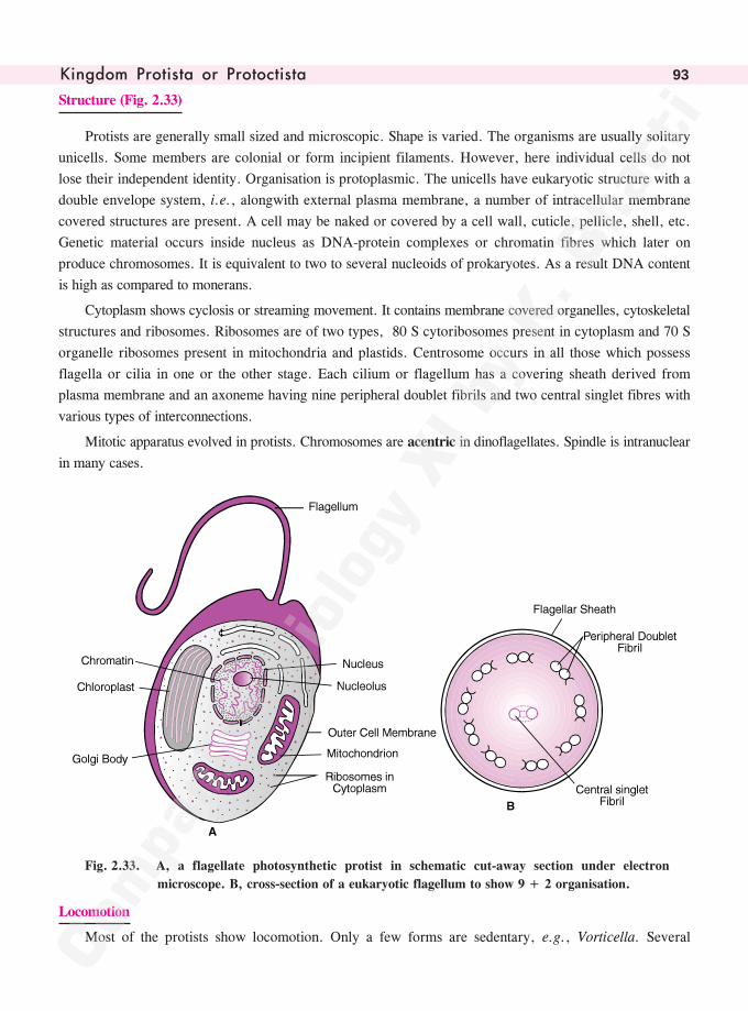
������� ���� �� �� ������� �� 93
Structure (Fig. 2.33)
Protists are generally small sized and microscopic. Shape is varied. The organisms are usually solitary
unicells. Some members are colonial or form incipient filaments. However, here individual cells do not
lose their independent identity. Organisation is protoplasmic. The unicells have eukaryotic structure with a
double envelope system, i.e., alongwith external plasma membrane, a number of intracellular membrane
covered structures are present. A cell may be naked or covered by a cell wall, cuticle, pellicle, shell, etc.
Genetic material occurs inside nucleus as DNA-protein complexes or chromatin fibres which later on
produce chromosomes. It is equivalent to two to several nucleoids of prokaryotes. As a result DNA content
is high as compared to monerans.
Cytoplasm shows cyclosis or streaming movement. It contains membrane covered organelles, cytoskeletal
structures and ribosomes. Ribosomes are of two types, 80 S cytoribosomes present in cytoplasm and 70 S
organelle ribosomes present in mitochondria and plastids. Centrosome occurs in all those which possess
flagella or cilia in one or the other stage. Each cilium or flagellum has a covering sheath derived from
plasma membrane and an axoneme having nine peripheral doublet fibrils and two central singlet fibres with
various types of interconnections.
Mitotic apparatus evolved in protists. Chromosomes are acentric in dinoflagellates. Spindle is intranuclear
in many cases.
Fig. 2.33. A, a flagellate photosynthetic protist in schematic cut-away section under electron
microscope. B, cross-section of a eukaryotic flagellum to show 9 + 2 organisation.
Locomotion
Most of the protists show locomotion. Only a few forms are sedentary, e.g., Vorticella. Several
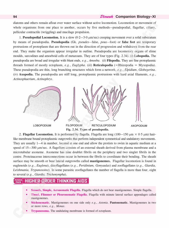
������� �Companion Biology–XI94
diatoms and others remain afloat over water surface without active locomotion. Locomotion or movement of
whole organisms from one place to another, occurs by five methods—pseudopodial, flagellar, ciliary,
pellicular contractile (wriggling) and mucilage propulsion.
1. Pseudopodial Locomotion. It is a slow (0·2—3·0 �m/sec) creeping movement over a solid substratum
by means of pseudopodia. Pseudopodia (Gk. pseudes—false, pous—foot) or false feet are temporary
protrusions of protoplasm that are thrown out in the direction of progression and withdrawn from the rear
end. They make the organism appear irregular in outline. Pseudopodia are locomotory organs of slime
moulds, sarcodines and amoeboid cells of metazoans. They are of four types (Fig. 2.34). (i) Lobopodia. The
pseudopodia are broad and irregular with blunt ends, e.g., Amoeba. (ii) Filopodia. They are fine protoplasmic
threads formed of mostly ectoplasm, e.g., Euglypha. (iii) Reticulopodia (=Rhizopodia = Myxopodia).
These pseudopodia are thin, long branching structures which form a network, e.g., Elpidium, Globegerina.
(iv) Axopodia. The pseudopodia are stiff long, protoplasmic protrusions with hard axial filaments, e.g.,
Actinosphaerium, Actinophrys.
Fig. 2.34. Types of pseudopodia.
2. Flagellar Locomotion. It is performed by flagella. Flagella are long (100—150 �m × 0·5 �m) hair-
like membrane bound protoplasmic outgrowths that perform independent symmetrical and undulatory movements.
They are usually 1—4 in number, located at one end and allow the protists to swim in aquatic medium at a
speed of 15—300 �m/sec. A flagellum consists of an external sheath derived from plasma membrane and a
microtubular axoneme. Axoneme has nine doublet fibrils on the periphery and two singlet fibrils in the
centre. Proteinaceous interconnections occur in between the fibrils to coordinate their bending. The sheath
surface may be smooth or bear lateral outgrowths called mastigonemes. Flagellar locomotion is found in
euglenoids (e.g., Euglena), dinoflagellates (e.g., Peridinium, Gonyaulax) and zooflagellates (e.g., Giardia,
Leishmania, Trypanosoma). In some parasitic zooflagellates the number of flagella is more than four, eight
to several (e.g., Giardia, Trichonympha).
• Smooth, Simple, Acronematic Flagella. Flagella which do not bear mastigonemes. Simple flagella.
• Tinsel, Flimmer or Pleuronematic Flagella. Flagella with minute lateral surface appendages calledmastigonemes.
• Stichonematic. Mastigonemes on one side only e.g., Astonia. Pantonematic. Mastigonemes in twoor more rows, e.g., Monas.
• Trypanosoma. The undulating membrane is formed of ectoplasm.
������� ���� �� �� ������� �� 95
3. Ciliary Locomotion. It is locomotion performed by cilia. Cilia are similar to flagella in structurebut are always smooth. They are shorter (5—10 �m) but the width is equal to that of flagella (0·5 �m). Ciliaare numerous, from a few hundred to a few thousand. They generally occur all over the body of the protistbut can also be restricted to certain areas. By their coalescence, cilia may form undulating membranes,membranelles and cirri. Cilia perform highly coordinated fast asymmetrical sweeping or rowing type ofmovement. The speed of locomotion is 0·4—2·0 mm/sec. All the cilia of an individual or group performeither isochronic (=synchronous, simultaneous) or metachronic (one after the other) movements. Ciliarylocomotion occurs in group ciliata, e.g., Paramecium.
4. Pellicular Contractile or Wriggling Locomotion. A wave of contraction and expansion passesthrough the body by means of special contractile elastic structures popularly called myoneme fibrils, e.g.,Monocystis. They are formed of pellicle plates in Euglena.
5. Mucilage Propulsion. It occurs in diatoms. Diatoms secrete mucilage which is passed out in onedirection while the unicells are propelled in the opposite direction.
Respiration
Most of the free living protists and protistan parasites of body fluids perform aerobic respiration. Theprotists living at the bottom of aquatic habitats and alimentary canal parasites respire anaerobically.
Nutrition
Nutrition or mode of nourishment is diverse in protista. Some are autotrophic. They performphotosynthesis. Others are heterotrophic, obtaining food from outside through ingestion (phagotrophic,holozoic), absorption from organic remains (saprotrophic) or living host (parasitic). A few have two or moretypes of nutrition (mixotrophic).
1. Photosynthetic or Holophytic Nutrition. It occurs in protistan algae. Important members aredinoflagellates (e.g., Gonyaulax, Peridinium), diatoms (e.g., Pinnularia, Triceratium) and euglenoids (e.g.,Euglena).
2. Ingestive or Holozoic Nutrition (= Phagotrophic or Zootrophic Nutrition). It occurs in most freeliving protozoan protists, some dinoflagellates and a few euglenoids. Ingestive or holozoic nutrition involvescapturing of small preys or microorganisms and small fragments of larger organisms. Internally the ingestedfood forms a phagosome which combines with lysosome to produce digestive vacuole. Digestion occurs inthe digestive vacuoles. The digested food passed into the surrounding cytoplasm. The undigested matterpresent in residual vacuole is thrown out through a specific site or anywhere from the surface in the processof ephagy.
3. Saprotrophic Nutrition. It is absorptive type of nutrition where food is obtained from organicremains. Saprotrophs secrete enzymes over the organic remains. This converts complex organic compoundsinto simpler and soluble form. The same are absorbed and assimilated. Saprotrophs are also calledosmotrophs and their mode of nutrition as osmotrophy. It occurs in many slime moulds, Mastigamoeba,Chilomonas, etc.
4. Pinocytosis. Soluble organic substances and salts in solution form are known to be absorbed inAmoeba and others through pinocytosis.
5. Parasitic Nutrition. Parasites are organisms that obtain nourishment from a living organism calledhost. A number of them ingest food through phagotrophy, e.g., Trichomonas, Balantidium, Entamoeba.Others absorb liquid food through their surface, e.g., Trypanosoma, Plasmodium.
6. Mixotrophic Nourishment. It is a mode of nourishment in which part of food is obtained fromoutside source. The individuals are photosynthetic, that is, holophytic. Euglena shows mixotrophic nourishment.It obtains nourishment mainly from photosynthesis and a part through saprotrophy.
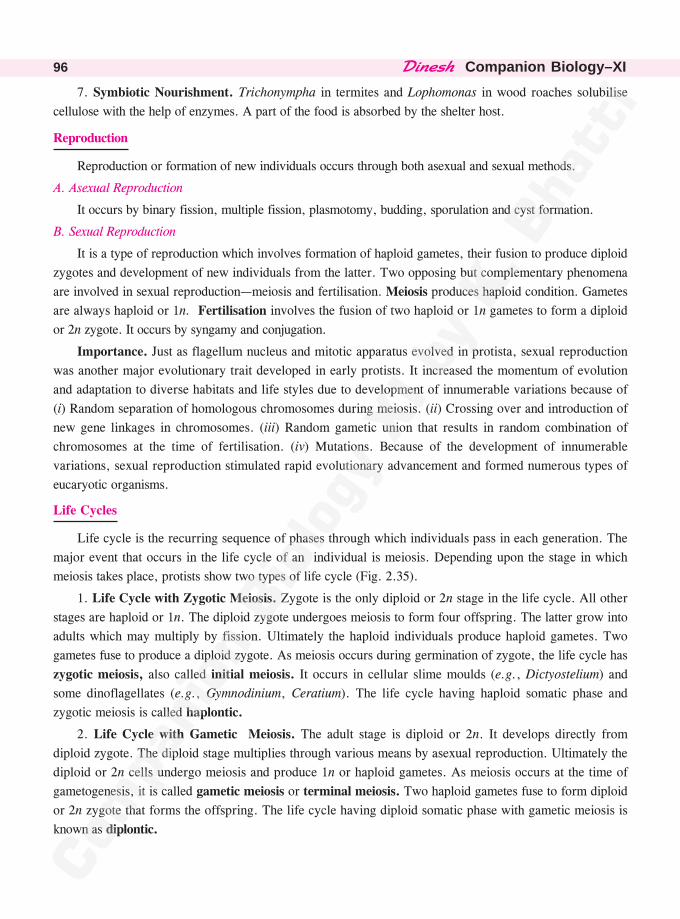
������� �Companion Biology–XI96
7. Symbiotic Nourishment. Trichonympha in termites and Lophomonas in wood roaches solubilise
cellulose with the help of enzymes. A part of the food is absorbed by the shelter host.
Reproduction
Reproduction or formation of new individuals occurs through both asexual and sexual methods.
A. Asexual Reproduction
It occurs by binary fission, multiple fission, plasmotomy, budding, sporulation and cyst formation.
B. Sexual Reproduction
It is a type of reproduction which involves formation of haploid gametes, their fusion to produce diploid
zygotes and development of new individuals from the latter. Two opposing but complementary phenomena
are involved in sexual reproduction—meiosis and fertilisation. Meiosis produces haploid condition. Gametes
are always haploid or 1n. Fertilisation involves the fusion of two haploid or 1n gametes to form a diploid
or 2n zygote. It occurs by syngamy and conjugation.
Importance. Just as flagellum nucleus and mitotic apparatus evolved in protista, sexual reproduction
was another major evolutionary trait developed in early protists. It increased the momentum of evolution
and adaptation to diverse habitats and life styles due to development of innumerable variations because of
(i) Random separation of homologous chromosomes during meiosis. (ii) Crossing over and introduction of
new gene linkages in chromosomes. (iii) Random gametic union that results in random combination of
chromosomes at the time of fertilisation. (iv) Mutations. Because of the development of innumerable
variations, sexual reproduction stimulated rapid evolutionary advancement and formed numerous types of
eucaryotic organisms.
Life Cycles
Life cycle is the recurring sequence of phases through which individuals pass in each generation. The
major event that occurs in the life cycle of an individual is meiosis. Depending upon the stage in which
meiosis takes place, protists show two types of life cycle (Fig. 2.35).
1. Life Cycle with Zygotic Meiosis. Zygote is the only diploid or 2n stage in the life cycle. All other
stages are haploid or 1n. The diploid zygote undergoes meiosis to form four offspring. The latter grow into
adults which may multiply by fission. Ultimately the haploid individuals produce haploid gametes. Two
gametes fuse to produce a diploid zygote. As meiosis occurs during germination of zygote, the life cycle has
zygotic meiosis, also called initial meiosis. It occurs in cellular slime moulds (e.g., Dictyostelium) and
some dinoflagellates (e.g., Gymnodinium, Ceratium). The life cycle having haploid somatic phase and
zygotic meiosis is called haplontic.
2. Life Cycle with Gametic Meiosis. The adult stage is diploid or 2n. It develops directly from
diploid zygote. The diploid stage multiplies through various means by asexual reproduction. Ultimately the
diploid or 2n cells undergo meiosis and produce 1n or haploid gametes. As meiosis occurs at the time of
gametogenesis, it is called gametic meiosis or terminal meiosis. Two haploid gametes fuse to form diploid
or 2n zygote that forms the offspring. The life cycle having diploid somatic phase with gametic meiosis is
known as diplontic.
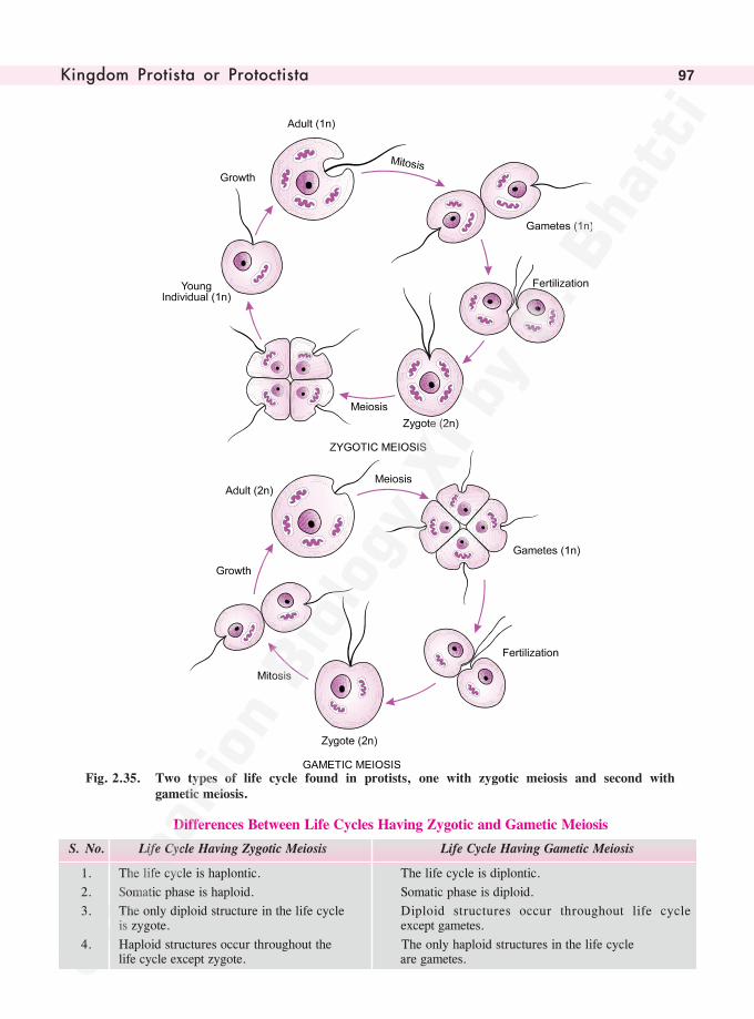
������� ���� �� �� ������� �� 97
Fig. 2.35. Two types of life cycle found in protists, one with zygotic meiosis and second withgametic meiosis.
Differences Between Life Cycles Having Zygotic and Gametic Meiosis
S. No. Life Cycle Having Zygotic Meiosis Life Cycle Having Gametic Meiosis
1. The life cycle is haplontic. The life cycle is diplontic.
2. Somatic phase is haploid. Somatic phase is diploid.
3. The only diploid structure in the life cycle Diploid structures occur throughout life cycleis zygote. except gametes.
4. Haploid structures occur throughout the The only haploid structures in the life cyclelife cycle except zygote. are gametes.
������� �Companion Biology–XI98
5. Gametes are produced through mitosis of Gametes are produced through meiosis of
parent cells. parent cells.
6. Meiosis is zygotic or initial. Meiosis is gametic or terminal.
Examples. Dictyostelium, Gymnodinium, Examples. Physarum, Paramecium.
Ceratium.
Groups of Protists
Kingdom protista has three types of unicellular eukaryotic organisms—photosynthetic protistan algae,slime moulds and protozoan protists.
PHOTOSYNTHETIC PROTISTS
The Protistan Algae
Algae are a heterogeneous aggregation of photosynthetic organisms that are characterised by(i) Nonvascular somatic body. (ii) Presence of chlorophyll a and other photosynthetic pigments.(iii) Ability to perform photosynthesis. (iv) Presence of nonjacketed sex organs where each cell isfunctional. (v) Absence of embryo stage. (vi) Aquatic or semi-aquatic habitat. (vii) Abundance of asexualreproduction. Like gametangia, all the cells of sporangia are functional.
Algae have been distributed in three kingdoms of monera, protista and plantae. Monera containsprocaryotic blue-green algae or cyanobacteria. Kingdom plantae contains predominantly multicellular algaeand their unicellular relatives, viz., chlorophyceae (green algae), phaeophyceae (brown algae) and rhodophyceae(red algae). Protista has unicellular or multinucleate acellular groups of algae, viz., dinoflagellates,diatoms, euglenoids, yellow-green algae. Protistan algae comprise more than 95% of phytoplankton andperform about 80% of the total photosynthetic activity on earth.
Dinoflagellates
They belong to class dinophyceae (Gk. dinein—to whorl, phycos—alga) of division pyrrhophyta (Gk.pyre—fire, phycos—alga). Dinoflagellates are biflagellate unicellular golden brown photosynthetic protistscomprising over 1000 species. All of them are aquatic, mostly marine with a few being fresh water aquatic.
1. Colour. It is commonly golden brown but yellow, brown, red, green, blue and colourless (heterotrophic)forms also occur.
2. Form. They are unicellular and biflagellate. However, amoeboid (e.g., Dinamoebidium), colonialand filamentous types also occur.
3. Plankton. They are important component of phytoplankton. At times they grow in such abundance asto form blooms. A litre of water may have 1—20 million of them.
4. Red Tides. Bloom forming dinoflagellates spread rapidly over several kilometre area of sea. Theyare yellowish brown to red in colour. Therefore, dinoflagellate blooms are also called red tides, e.g.,Gonyaulax, Gymnodinium, Ceratium.
5. Nutrition. It is photosynthetic or holophytic. Some members are saprotrophic, parasitic and symbionts.A few colourless dinoflagellates (e.g., Noctiluca) are phagotrophs or holozoic. Parasites (e.g., Haplozoon)are few. Ceratium is both photosynthetic and phagotrophic.
• Zooxanthella. Golden brown, reddish or yellowish symbiont protistan algal cell in other organisms.
• Zoochlorella. Green algal cell (kingdom plantae) in other organisms.
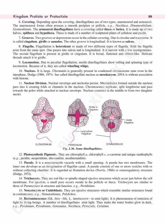
������� ���� �� �� ������� �� 99
6. Covering. Depending upon the covering, dinoflagellates are of two types, unarmoured and armoured.The unarmoured forms often possess a smooth periplast or pellicle, e.g., Noctiluca, Dinamoebidium,Gymnodinium. The armoured dinoflagellates have a covering called theca or lorica. It is made up of twohalves, epitheca and hypotheca. Theca is made of a number of sculptured plates of cellulose and pectin.
7. Grooves. Two grooves or depressions occur in the cellular covering. One is circular and transverse. Itis called cingulum, girdle or annulus. The other groove is longitudinal. It is known as sulcus.
8. Flagella. Flagellation is heterokont or made of two different types of flagella. Both the flagellaarise from the same spot. One passes into sulcus and is longitudinal. It is narrow with a few mastigonemes.The second flagellum is present in girdle or cingulum. It is broad, flattened and ribbon-like. Delicatethreads attach it to girdle.
9. Locomotion. Due to peculiar flagellation, motile dinoflagellates show rolling and spinning type oflocomotion. Because of it, they are called whorling whips.
10. Nucleus. It is large. Chromatin is generally present in condensed chromosome state even in theinterphase. Dodge (1966, 1971) has called dinoflagellate nucleus as mesokaryon. DNA is without associationwith histone.
11. Nuclear Division. Nuclear envelope and nucleolus persist. Microtubules formed outside the nucleuspass into it creating folds or channels in the nucleus. Chromosomes replicate, split lengthwise and passtowards the poles while attached to nuclear envelope. Nucleus constrics in the middle to form two daughternuclei.
Fig. 2.36. Some dinoflagellates.
12. Photosynthetic Pigments. They are chlorophyll a, chlorophyll c, �-carotene and unique xanthophylls(e.g., peridin, neoperidinin, dinoxanthin, neodinoxanthin).
13. Pusule. It is a noncontractile vacuole with a small opening. A pusule has two membranes. Theinner one develops as an invagination of flagellar canal. A pusule consists of one or more vesicles with orwithout collecting chamber. It is regarded as floatation device (Norris, 1966) or osmoregulatory structure(Dodge, 1972).
14. Trichocysts. They are rod-like or spindle-shaped ejective structures which occur just below the cellmembrane. For ejection, a small pore occurs ouside in the pellicle or theca. Trichocysts are similar tothose of Paramecium in structure and function, e.g., Peridinium.
15. Nematocysts or Cnidoblasts. They are ejective structures which resemble similar structures foundin coelenterates, e.g., Nematodinium, Polykrikos.
16. Bioluminescence (Gk. bios—life, L. luminescere—to emit light). It is phenomenon of emission oflight by living beings. A number of dinoflagellates emit light. They make the water bodies glow in dark,e.g., Peridinium, Pyrodinium, Gonyaulax, Noctiluca, Pyrocystis, Ceratium.
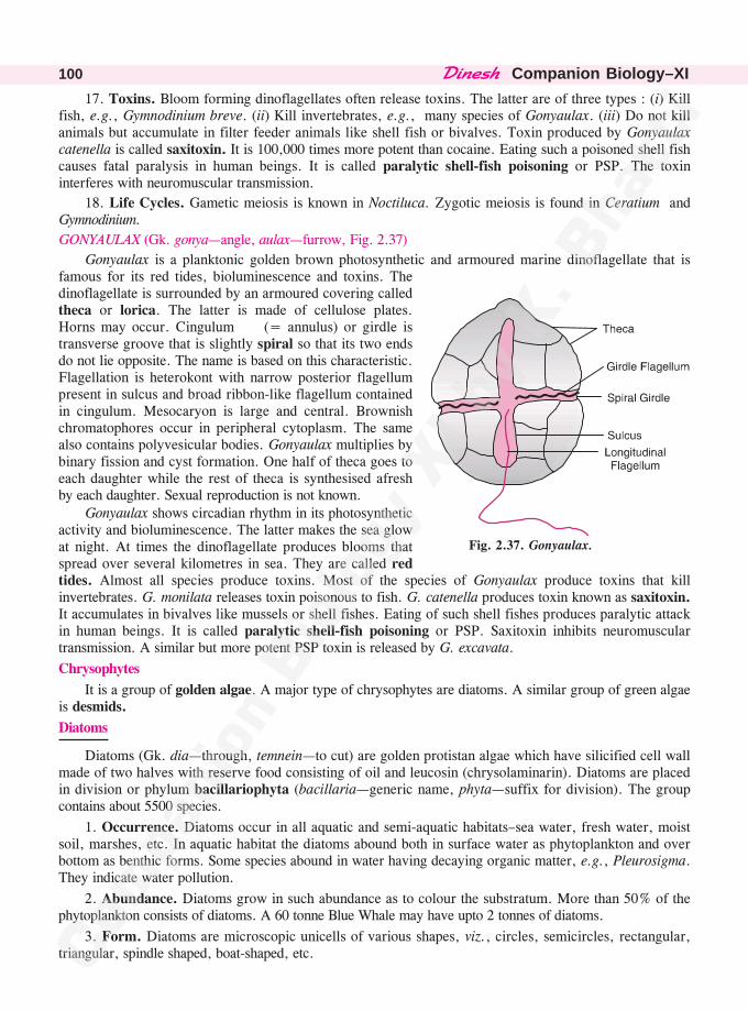
������� �Companion Biology–XI100
17. Toxins. Bloom forming dinoflagellates often release toxins. The latter are of three types : (i) Killfish, e.g., Gymnodinium breve. (ii) Kill invertebrates, e.g., many species of Gonyaulax. (iii) Do not killanimals but accumulate in filter feeder animals like shell fish or bivalves. Toxin produced by Gonyaulaxcatenella is called saxitoxin. It is 100,000 times more potent than cocaine. Eating such a poisoned shell fishcauses fatal paralysis in human beings. It is called paralytic shell-fish poisoning or PSP. The toxininterferes with neuromuscular transmission.
18. Life Cycles. Gametic meiosis is known in Noctiluca. Zygotic meiosis is found in Ceratium andGymnodinium.
GONYAULAX (Gk. gonya—angle, aulax—furrow, Fig. 2.37)
Gonyaulax is a planktonic golden brown photosynthetic and armoured marine dinoflagellate that isfamous for its red tides, bioluminescence and toxins. Thedinoflagellate is surrounded by an armoured covering calledtheca or lorica. The latter is made of cellulose plates.Horns may occur. Cingulum (= annulus) or girdle istransverse groove that is slightly spiral so that its two endsdo not lie opposite. The name is based on this characteristic.Flagellation is heterokont with narrow posterior flagellumpresent in sulcus and broad ribbon-like flagellum containedin cingulum. Mesocaryon is large and central. Brownishchromatophores occur in peripheral cytoplasm. The samealso contains polyvesicular bodies. Gonyaulax multiplies bybinary fission and cyst formation. One half of theca goes toeach daughter while the rest of theca is synthesised afreshby each daughter. Sexual reproduction is not known.
Gonyaulax shows circadian rhythm in its photosyntheticactivity and bioluminescence. The latter makes the sea glowat night. At times the dinoflagellate produces blooms thatspread over several kilometres in sea. They are called redtides. Almost all species produce toxins. Most of the species of Gonyaulax produce toxins that killinvertebrates. G. monilata releases toxin poisonous to fish. G. catenella produces toxin known as saxitoxin.It accumulates in bivalves like mussels or shell fishes. Eating of such shell fishes produces paralytic attackin human beings. It is called paralytic shell-fish poisoning or PSP. Saxitoxin inhibits neuromusculartransmission. A similar but more potent PSP toxin is released by G. excavata.
Chrysophytes
It is a group of golden algae. A major type of chrysophytes are diatoms. A similar group of green algaeis desmids.
Diatoms
Diatoms (Gk. dia—through, temnein—to cut) are golden protistan algae which have silicified cell wallmade of two halves with reserve food consisting of oil and leucosin (chrysolaminarin). Diatoms are placedin division or phylum bacillariophyta (bacillaria—generic name, phyta—suffix for division). The groupcontains about 5500 species.
1. Occurrence. Diatoms occur in all aquatic and semi-aquatic habitats–sea water, fresh water, moistsoil, marshes, etc. In aquatic habitat the diatoms abound both in surface water as phytoplankton and overbottom as benthic forms. Some species abound in water having decaying organic matter, e.g., Pleurosigma.They indicate water pollution.
2. Abundance. Diatoms grow in such abundance as to colour the substratum. More than 50% of thephytoplankton consists of diatoms. A 60 tonne Blue Whale may have upto 2 tonnes of diatoms.
3. Form. Diatoms are microscopic unicells of various shapes, viz., circles, semicircles, rectangular,triangular, spindle shaped, boat-shaped, etc.
Fig. 2.37. Gonyaulax.
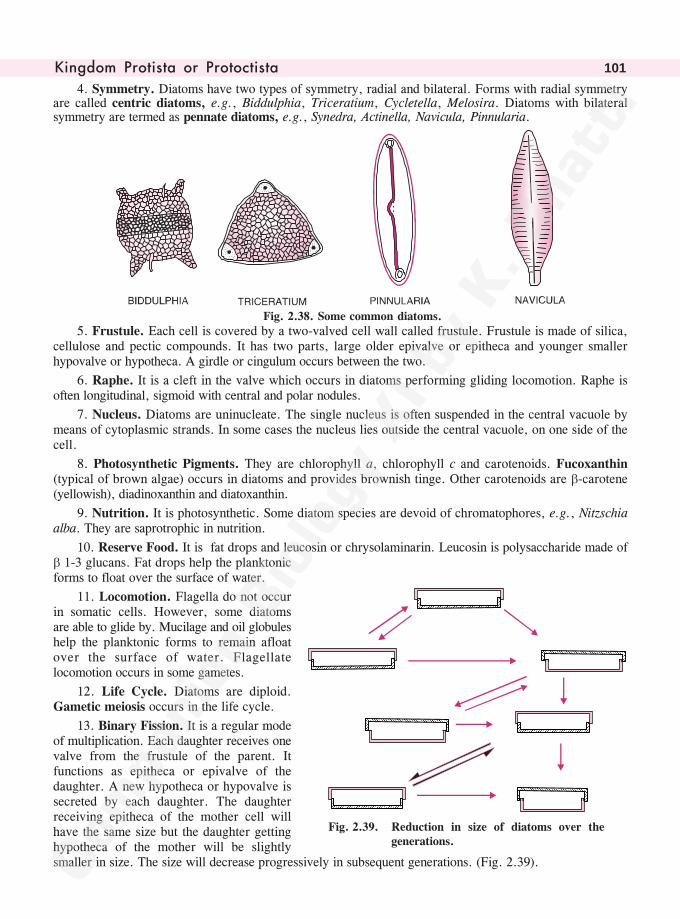
������� ���� �� �� ������� �� 101
4. Symmetry. Diatoms have two types of symmetry, radial and bilateral. Forms with radial symmetryare called centric diatoms, e.g., Biddulphia, Triceratium, Cycletella, Melosira. Diatoms with bilateralsymmetry are termed as pennate diatoms, e.g., Synedra, Actinella, Navicula, Pinnularia.
Fig. 2.38. Some common diatoms.
5. Frustule. Each cell is covered by a two-valved cell wall called frustule. Frustule is made of silica,cellulose and pectic compounds. It has two parts, large older epivalve or epitheca and younger smallerhypovalve or hypotheca. A girdle or cingulum occurs between the two.
6. Raphe. It is a cleft in the valve which occurs in diatoms performing gliding locomotion. Raphe isoften longitudinal, sigmoid with central and polar nodules.
7. Nucleus. Diatoms are uninucleate. The single nucleus is often suspended in the central vacuole bymeans of cytoplasmic strands. In some cases the nucleus lies outside the central vacuole, on one side of thecell.
8. Photosynthetic Pigments. They are chlorophyll a, chlorophyll c and carotenoids. Fucoxanthin(typical of brown algae) occurs in diatoms and provides brownish tinge. Other carotenoids are �-carotene(yellowish), diadinoxanthin and diatoxanthin.
9. Nutrition. It is photosynthetic. Some diatom species are devoid of chromatophores, e.g., Nitzschiaalba. They are saprotrophic in nutrition.
10. Reserve Food. It is fat drops and leucosin or chrysolaminarin. Leucosin is polysaccharide made of� 1-3 glucans. Fat drops help the planktonicforms to float over the surface of water.
11. Locomotion. Flagella do not occurin somatic cells. However, some diatomsare able to glide by. Mucilage and oil globuleshelp the planktonic forms to remain afloatover the surface of water. Flagellatelocomotion occurs in some gametes.
12. Life Cycle. Diatoms are diploid.Gametic meiosis occurs in the life cycle.
13. Binary Fission. It is a regular modeof multiplication. Each daughter receives onevalve from the frustule of the parent. Itfunctions as epitheca or epivalve of thedaughter. A new hypotheca or hypovalve issecreted by each daughter. The daughterreceiving epitheca of the mother cell willhave the same size but the daughter gettinghypotheca of the mother will be slightlysmaller in size. The size will decrease progressively in subsequent generations. (Fig. 2.39).
Fig. 2.39. Reduction in size of diatoms over thegenerations.
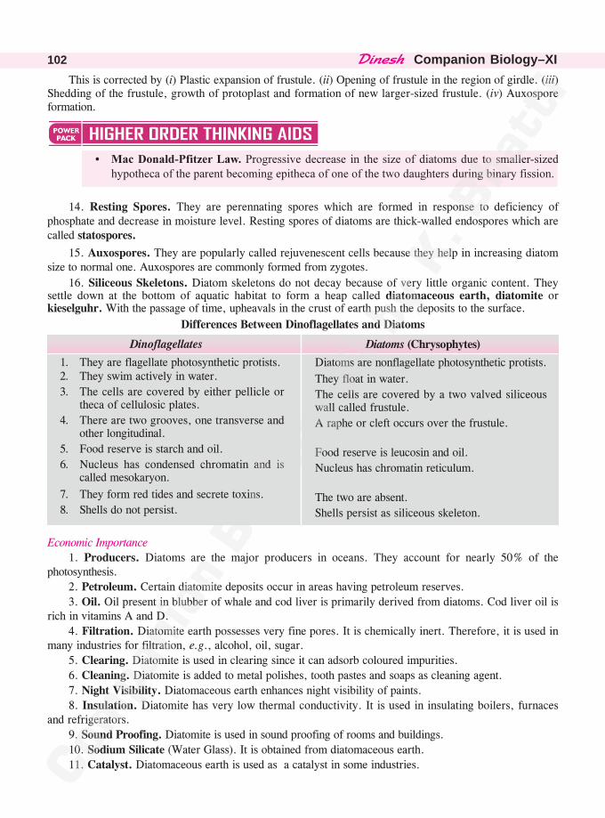
������� �Companion Biology–XI102
This is corrected by (i) Plastic expansion of frustule. (ii) Opening of frustule in the region of girdle. (iii)Shedding of the frustule, growth of protoplast and formation of new larger-sized frustule. (iv) Auxosporeformation.
� ��������� ��������������������������� ��� �� ������������ ��������� ����� ����������������� ��������� ������������������� �������������������� ���������������� ���������
14. Resting Spores. They are perennating spores which are formed in response to deficiency ofphosphate and decrease in moisture level. Resting spores of diatoms are thick-walled endospores which arecalled statospores.
15. Auxospores. They are popularly called rejuvenescent cells because they help in increasing diatomsize to normal one. Auxospores are commonly formed from zygotes.
16. Siliceous Skeletons. Diatom skeletons do not decay because of very little organic content. Theysettle down at the bottom of aquatic habitat to form a heap called diatomaceous earth, diatomite orkieselguhr. With the passage of time, upheavals in the crust of earth push the deposits to the surface.
Differences Between Dinoflagellates and Diatoms
Economic Importance
1. Producers. Diatoms are the major producers in oceans. They account for nearly 50% of thephotosynthesis.
2. Petroleum. Certain diatomite deposits occur in areas having petroleum reserves.
3. Oil. Oil present in blubber of whale and cod liver is primarily derived from diatoms. Cod liver oil isrich in vitamins A and D.
4. Filtration. Diatomite earth possesses very fine pores. It is chemically inert. Therefore, it is used inmany industries for filtration, e.g., alcohol, oil, sugar.
5. Clearing. Diatomite is used in clearing since it can adsorb coloured impurities.
6. Cleaning. Diatomite is added to metal polishes, tooth pastes and soaps as cleaning agent.
7. Night Visibility. Diatomaceous earth enhances night visibility of paints.
8. Insulation. Diatomite has very low thermal conductivity. It is used in insulating boilers, furnacesand refrigerators.
9. Sound Proofing. Diatomite is used in sound proofing of rooms and buildings.
10. Sodium Silicate (Water Glass). It is obtained from diatomaceous earth.
11. Catalyst. Diatomaceous earth is used as a catalyst in some industries.
Dinoflagellates
1. They are flagellate photosynthetic protists.2. They swim actively in water.
3. The cells are covered by either pellicle ortheca of cellulosic plates.
4. There are two grooves, one transverse andother longitudinal.
5. Food reserve is starch and oil.
6. Nucleus has condensed chromatin and iscalled mesokaryon.
7. They form red tides and secrete toxins.
8. Shells do not persist.
Diatoms (Chrysophytes)
Diatoms are nonflagellate photosynthetic protists.
They float in water.
The cells are covered by a two valved siliceouswall called frustule.
A raphe or cleft occurs over the frustule.
Food reserve is leucosin and oil.
Nucleus has chromatin reticulum.
The two are absent.
Shells persist as siliceous skeleton.
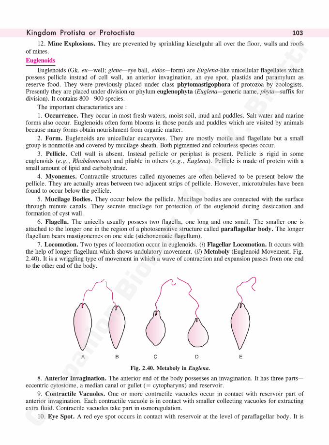
������� ���� �� �� ������� �� 103
12. Mine Explosions. They are prevented by sprinkling kieselguhr all over the floor, walls and roofsof mines.
Euglenoids
Euglenoids (Gk. eu—well; glene—eye ball, eidos—form) are Euglena-like unicellular flagellates whichpossess pellicle instead of cell wall, an anterior invagination, an eye spot, plastids and paramylum asreserve food. They were previously placed under class phytomastigophora of protozoa by zoologists.Presently they are placed under division or phylum euglenophyta (Euglena—generic name, phyta—suffix fordivision). It contains 800—900 species.
The important characteristics are :
1. Occurrence. They occur in most fresh waters, moist soil, mud and puddles. Salt water and marineforms also occur. Euglenoids often form blooms in those ponds and puddles which are visited by animalsbecause many forms obtain nourishment from organic matter.
2. Form. Euglenoids are unicellular eucaryotes. They are mostly motile and flagellate but a smallgroup is nonmotile and covered by mucilage sheath. Both pigmented and colourless species occur.
3. Pellicle. Cell wall is absent. Instead pellicle or periplast is present. Pellicle is rigid in someeuglenoids (e.g., Rhabdomonas) and pliable in others (e.g., Euglena). Pellicle is made of protein with asmall amount of lipid and carbohydrate.
4. Myonemes. Contractile structures called myonemes are often believed to be present below thepellicle. They are actually areas between two adjacent strips of pellicle. However, microtubules have beenfound to occur below the pellicle.
5. Mucilage Bodies. They occur below the pellicle. Mucilage bodies are connected with the surfacethrough minute canals. They secrete mucilage for protection of the euglenoid during desiccation andformation of cyst wall.
6. Flagella. The unicells usually possess two flagella, one long and one small. The smaller one isattached to the longer one in the region of a photosensitive structure called paraflagellar body. The longerflagellum bears mastigonemes on one side (stichonematic flagellum).
7. Locomotion. Two types of locomotion occur in euglenoids. (i) Flagellar Locomotion. It occurs withthe help of longer flagellum which shows undulatory movement. (ii) Metaboly (Euglenoid Movement, Fig.2.40). It is a wriggling type of movement in which a wave of contraction and expansion passes from one endto the other end of the body.
Fig. 2.40. Metaboly in Euglena.
8. Anterior Invagination. The anterior end of the body possesses an invagination. It has three parts—eccentric cytostome, a median canal or gullet (= cytopharynx) and reservoir.
9. Contractile Vacuoles. One or more contractile vacuoles occur in contact with reservoir part ofanterior invagination. Each contractile vacuole is in contact with smaller collecting vacuoles for extractingextra fluid. Contractile vacuoles take part in osmoregulation.
10. Eye Spot. A red eye spot occurs in contact with reservoir at the level of paraflagellar body. It is
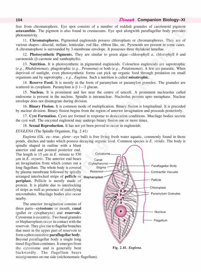
������� �Companion Biology–XI104
free from chromatophore. Eye spot consists of a number of reddish granules of carotenoid pigmentastaxanthin. The pigment is also found in crustaceans. Eye spot alongwith paraflagellar body providesphotosensivity.
11. Chromatophores. Pigmented euglenoids possess chloroplasts or chromatophores. They are ofvarious shapes—discoid, stellate, lenticular, rod like, ribbon like, etc. Pyrenoids are present in some cases.A chromatophore is surrounded by 3-membrane envelope. It possesses three thylakoid lamellae.
12. Photosynthetic Pigments. They are similar to green algae—chlorophyll a, chlorophyll b andcarotenoids (�-carotene and xanthophylls).
13. Nutrition. It is photosynthetic in pigmented euglenoids. Colourless euglenoids are saprotrophic(e.g., Rhabdomonas), phagotrophic (e.g., Peranema) or both (e.g., Petalomonas). A few are parasitic. Whendeprived of sunlight, even photosynthetic forms can pick up organic food through predation on smallorganisms and by saprotrophy., e.g., Euglena. Such a nutrition is called mixotrophic.
14. Reserve Food. It is mostly in the form of paramylum or paramylon granules. The granules arescattered in cytoplasm. Paramylum is � 1—3 glucan.
15. Nucleus. It is prominent and lies near the centre of unicell. A prominent nucleuolus calledendosome is present in the nucleus. Spindle is intranuclear. Nucleolus persists upto metaphase. Nuclearenvelope does not disintegrate during division.
16. Binary Fission. It is common mode of multiplication. Binary fission is longitudinal. It is precededby nuclear division. Binary fission begins from the region of anterior invagination and proceeds posteriorly.
17. Cyst Formation. Cysts are formed in response to desiccation conditions. Mucilage bodies secretethe cyst wall. The encysted euglenoid may undergo binary fission one or more times.
18. Sexual Reproduction. It has not yet been proved to occur in euglenoids.
EUGLENA (The Spindle Organism, Fig. 2.41)
Euglena (Gk. eu—true, glene—eye ball) is free living fresh water aquatic, commonly found in thoseponds, ditches and tanks which possess decaying organic food. Common species is E. viridis. The body isspindle shaped in outline with a bluntanterior end and pointed posterior end.The length is 15 �m in E. minuta to 530�m in E. oxyuris. The anterior end bearsan invagination from which comes out along flagellum. The whole body is coveredby plasma membrane followed by spirallyarranged interlocked strips of pellicle orperiplast. Pellicle is mostly made ofprotein. It is pliable due to interlockingof strips as well as presence of underlyingmicrotubules. Mucilage bodies also occurnearby.
The anterior invagination consists ofthree parts—cytostome or mouth, canal(gullet or cytopharynx) and reservoir.Cytostome is eccentric. Two basal granulesor blepharoplasts occur in contact with thereservoir. They give rise to flagellar branchesthat meet in the upper part of reservoir toform a photosensitive paraflagellar body.Beyond paraflagellar body a single longtinsel flagellum continues. It emerges fromthe cytostome and is generally bentbackwardly. The flagellum bearsmastigonemes on one side (stichonematic flagellum).
Fig. 2.41. Euglena.
�����������
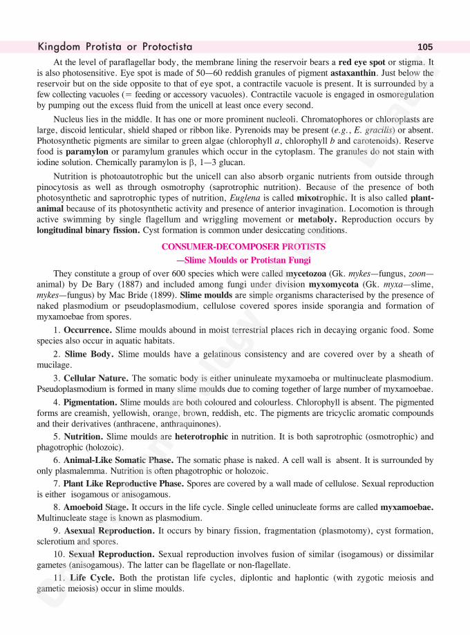
������� ���� �� �� ������� �� 105
At the level of paraflagellar body, the membrane lining the reservoir bears a red eye spot or stigma. Itis also photosensitive. Eye spot is made of 50—60 reddish granules of pigment astaxanthin. Just below thereservoir but on the side opposite to that of eye spot, a contractile vacuole is present. It is surrounded by afew collecting vacuoles (= feeding or accessory vacuoles). Contractile vacuole is engaged in osmoregulationby pumping out the excess fluid from the unicell at least once every second.
Nucleus lies in the middle. It has one or more prominent nucleoli. Chromatophores or chloroplasts arelarge, discoid lenticular, shield shaped or ribbon like. Pyrenoids may be present (e.g., E. gracilis) or absent.Photosynthetic pigments are similar to green algae (chlorophyll a, chlorophyll b and carotenoids). Reservefood is paramylon or paramylum granules which occur in the cytoplasm. The granules do not stain withiodine solution. Chemically paramylon is �, 1—3 glucan.
Nutrition is photoautotrophic but the unicell can also absorb organic nutrients from outside throughpinocytosis as well as through osmotrophy (saprotrophic nutrition). Because of the presence of bothphotosynthetic and saprotrophic types of nutrition, Euglena is called mixotrophic. It is also called plant-animal because of its photosynthetic activity and presence of anterior invagination. Locomotion is throughactive swimming by single flagellum and wriggling movement or metaboly. Reproduction occurs bylongitudinal binary fission. Cyst formation is common under desiccating conditions.
CONSUMER-DECOMPOSER PROTISTS
—Slime Moulds or Protistan Fungi
They constitute a group of over 600 species which were called mycetozoa (Gk. mykes—fungus, zoon—animal) by De Bary (1887) and included among fungi under division myxomycota (Gk. myxa—slime,mykes—fungus) by Mac Bride (1899). Slime moulds are simple organisms characterised by the presence ofnaked plasmodium or pseudoplasmodium, cellulose covered spores inside sporangia and formation ofmyxamoebae from spores.
1. Occurrence. Slime moulds abound in moist terrestrial places rich in decaying organic food. Somespecies also occur in aquatic habitats.
2. Slime Body. Slime moulds have a gelatinous consistency and are covered over by a sheath ofmucilage.
3. Cellular Nature. The somatic body is either uninuleate myxamoeba or multinucleate plasmodium.Pseudoplasmodium is formed in many slime moulds due to coming together of large number of myxamoebae.
4. Pigmentation. Slime moulds are both coloured and colourless. Chlorophyll is absent. The pigmentedforms are creamish, yellowish, orange, brown, reddish, etc. The pigments are tricyclic aromatic compoundsand their derivatives (anthracene, anthraquinones).
5. Nutrition. Slime moulds are heterotrophic in nutrition. It is both saprotrophic (osmotrophic) andphagotrophic (holozoic).
6. Animal-Like Somatic Phase. The somatic phase is naked. A cell wall is absent. It is surrounded byonly plasmalemma. Nutrition is often phagotrophic or holozoic.
7. Plant Like Reproductive Phase. Spores are covered by a wall made of cellulose. Sexual reproductionis either isogamous or anisogamous.
8. Amoeboid Stage. It occurs in the life cycle. Single celled uninucleate forms are called myxamoebae.Multinucleate stage is known as plasmodium.
9. Asexual Reproduction. It occurs by binary fission, fragmentation (plasmotomy), cyst formation,sclerotium and spores.
10. Sexual Reproduction. Sexual reproduction involves fusion of similar (isogamous) or dissimilargametes (anisogamous). The latter can be flagellate or non-flagellate.
11. Life Cycle. Both the protistan life cycles, diplontic and haplontic (with zygotic meiosis andgametic meiosis) occur in slime moulds.
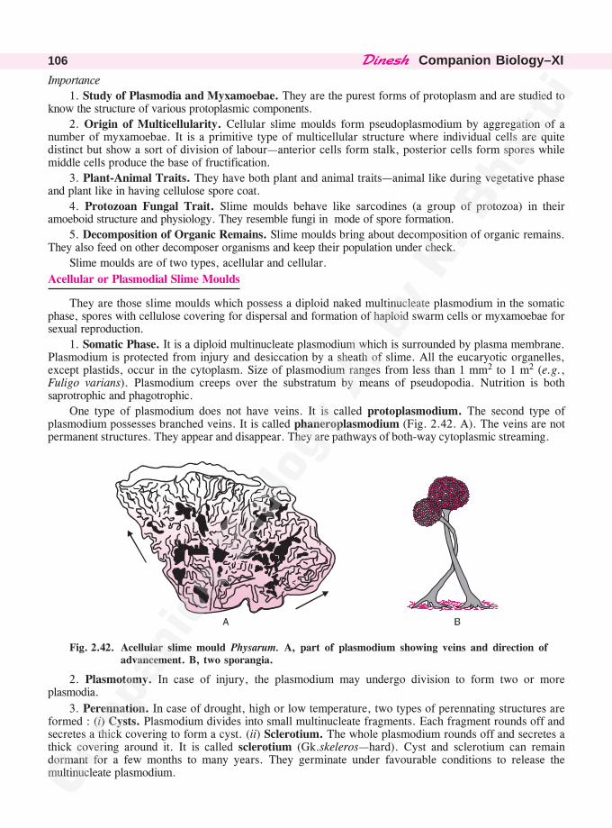
������� �Companion Biology–XI106
Importance
1. Study of Plasmodia and Myxamoebae. They are the purest forms of protoplasm and are studied toknow the structure of various protoplasmic components.
2. Origin of Multicellularity. Cellular slime moulds form pseudoplasmodium by aggregation of anumber of myxamoebae. It is a primitive type of multicellular structure where individual cells are quitedistinct but show a sort of division of labour—anterior cells form stalk, posterior cells form spores whilemiddle cells produce the base of fructification.
3. Plant-Animal Traits. They have both plant and animal traits—animal like during vegetative phaseand plant like in having cellulose spore coat.
4. Protozoan Fungal Trait. Slime moulds behave like sarcodines (a group of protozoa) in theiramoeboid structure and physiology. They resemble fungi in mode of spore formation.
5. Decomposition of Organic Remains. Slime moulds bring about decomposition of organic remains.They also feed on other decomposer organisms and keep their population under check.
Slime moulds are of two types, acellular and cellular.
Acellular or Plasmodial Slime Moulds
They are those slime moulds which possess a diploid naked multinucleate plasmodium in the somaticphase, spores with cellulose covering for dispersal and formation of haploid swarm cells or myxamoebae forsexual reproduction.
1. Somatic Phase. It is a diploid multinucleate plasmodium which is surrounded by plasma membrane.Plasmodium is protected from injury and desiccation by a sheath of slime. All the eucaryotic organelles,except plastids, occur in the cytoplasm. Size of plasmodium ranges from less than 1 mm2 to 1 m2 (e.g.,Fuligo varians). Plasmodium creeps over the substratum by means of pseudopodia. Nutrition is bothsaprotrophic and phagotrophic.
One type of plasmodium does not have veins. It is called protoplasmodium. The second type ofplasmodium possesses branched veins. It is called phaneroplasmodium (Fig. 2.42. A). The veins are notpermanent structures. They appear and disappear. They are pathways of both-way cytoplasmic streaming.
Fig. 2.42. Acellular slime mould Physarum. A, part of plasmodium showing veins and direction ofadvancement. B, two sporangia.
2. Plasmotomy. In case of injury, the plasmodium may undergo division to form two or moreplasmodia.
3. Perennation. In case of drought, high or low temperature, two types of perennating structures areformed : (i) Cysts. Plasmodium divides into small multinucleate fragments. Each fragment rounds off andsecretes a thick covering to form a cyst. (ii) Sclerotium. The whole plasmodium rounds off and secretes athick covering around it. It is called sclerotium (Gk.skeleros—hard). Cyst and sclerotium can remaindormant for a few months to many years. They germinate under favourable conditions to release themultinucleate plasmodium.
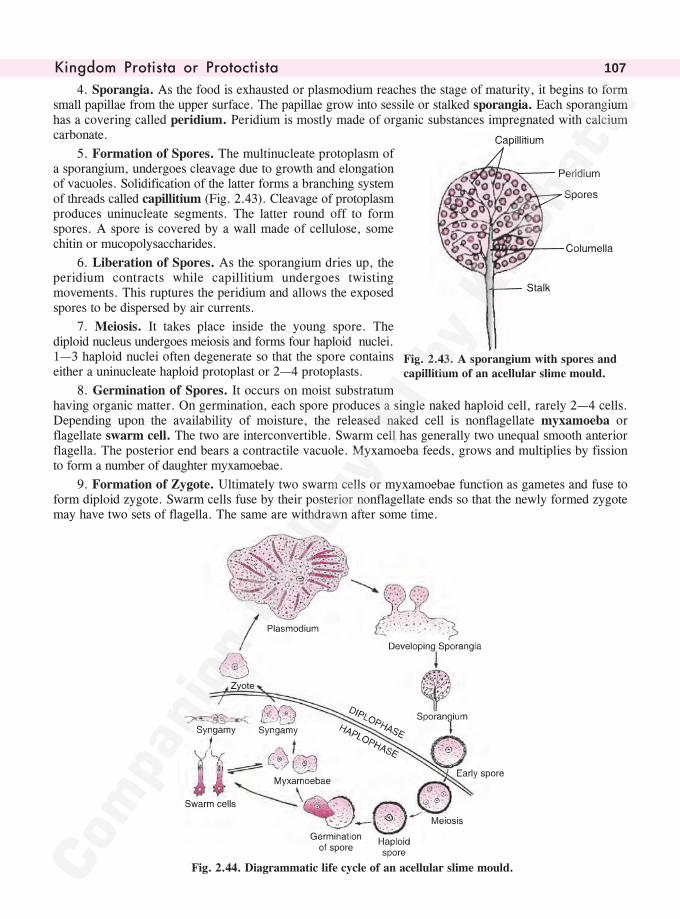
������� ���� �� �� ������� �� 107
4. Sporangia. As the food is exhausted or plasmodium reaches the stage of maturity, it begins to formsmall papillae from the upper surface. The papillae grow into sessile or stalked sporangia. Each sporangiumhas a covering called peridium. Peridium is mostly made of organic substances impregnated with calciumcarbonate.
5. Formation of Spores. The multinucleate protoplasm ofa sporangium, undergoes cleavage due to growth and elongationof vacuoles. Solidification of the latter forms a branching systemof threads called capillitium (Fig. 2.43). Cleavage of protoplasmproduces uninucleate segments. The latter round off to formspores. A spore is covered by a wall made of cellulose, somechitin or mucopolysaccharides.
6. Liberation of Spores. As the sporangium dries up, theperidium contracts while capillitium undergoes twistingmovements. This ruptures the peridium and allows the exposedspores to be dispersed by air currents.
7. Meiosis. It takes place inside the young spore. Thediploid nucleus undergoes meiosis and forms four haploid nuclei.1—3 haploid nuclei often degenerate so that the spore containseither a uninucleate haploid protoplast or 2—4 protoplasts.
8. Germination of Spores. It occurs on moist substratumhaving organic matter. On germination, each spore produces a single naked haploid cell, rarely 2—4 cells.Depending upon the availability of moisture, the released naked cell is nonflagellate myxamoeba orflagellate swarm cell. The two are interconvertible. Swarm cell has generally two unequal smooth anteriorflagella. The posterior end bears a contractile vacuole. Myxamoeba feeds, grows and multiplies by fissionto form a number of daughter myxamoebae.
9. Formation of Zygote. Ultimately two swarm cells or myxamoebae function as gametes and fuse toform diploid zygote. Swarm cells fuse by their posterior nonflagellate ends so that the newly formed zygotemay have two sets of flagella. The same are withdrawn after some time.
Fig. 2.44. Diagrammatic life cycle of an acellular slime mould.
Fig. 2.43. A sporangium with spores andcapillitium of an acellular slime mould.
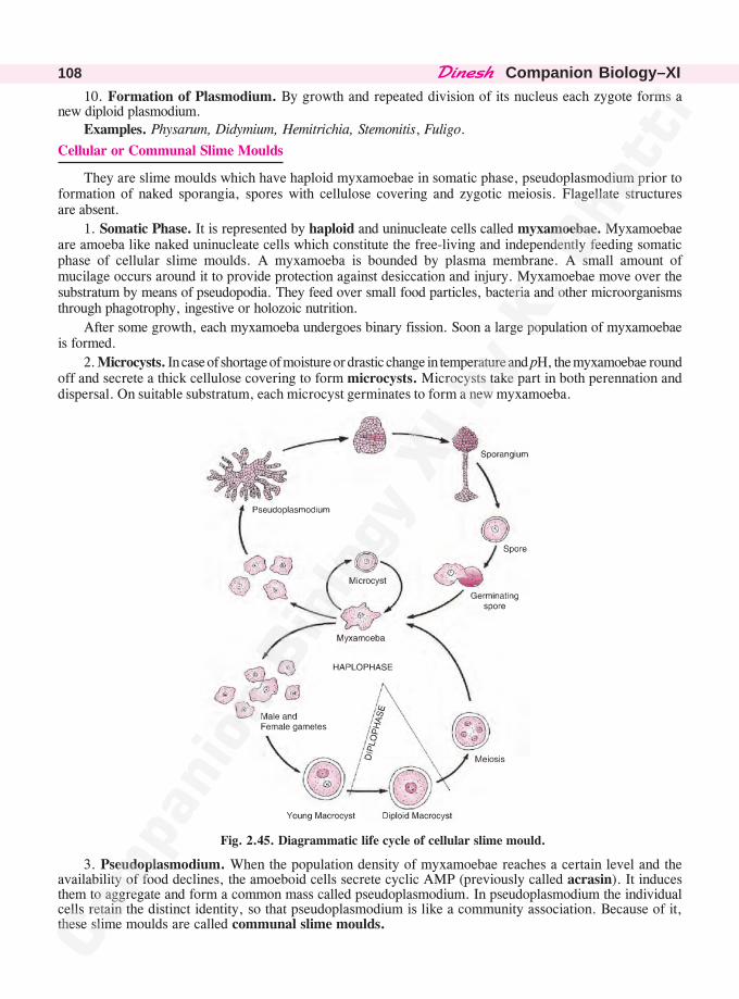
������� �Companion Biology–XI108
10. Formation of Plasmodium. By growth and repeated division of its nucleus each zygote forms anew diploid plasmodium.
Examples. Physarum, Didymium, Hemitrichia, Stemonitis, Fuligo.
Cellular or Communal Slime Moulds
They are slime moulds which have haploid myxamoebae in somatic phase, pseudoplasmodium prior toformation of naked sporangia, spores with cellulose covering and zygotic meiosis. Flagellate structuresare absent.
1. Somatic Phase. It is represented by haploid and uninucleate cells called myxamoebae. Myxamoebaeare amoeba like naked uninucleate cells which constitute the free-living and independently feeding somaticphase of cellular slime moulds. A myxamoeba is bounded by plasma membrane. A small amount ofmucilage occurs around it to provide protection against desiccation and injury. Myxamoebae move over thesubstratum by means of pseudopodia. They feed over small food particles, bacteria and other microorganismsthrough phagotrophy, ingestive or holozoic nutrition.
After some growth, each myxamoeba undergoes binary fission. Soon a large population of myxamoebaeis formed.
2. Microcysts. In case of shortage of moisture or drastic change in temperature and pH, the myxamoebae roundoff and secrete a thick cellulose covering to form microcysts. Microcysts take part in both perennation anddispersal. On suitable substratum, each microcyst germinates to form a new myxamoeba.
Fig. 2.45. Diagrammatic life cycle of cellular slime mould.
3. Pseudoplasmodium. When the population density of myxamoebae reaches a certain level and theavailability of food declines, the amoeboid cells secrete cyclic AMP (previously called acrasin). It inducesthem to aggregate and form a common mass called pseudoplasmodium. In pseudoplasmodium the individualcells retain the distinct identity, so that pseudoplasmodium is like a community association. Because of it,these slime moulds are called communal slime moulds.
������� ���� �� �� ������� �� 109
Differences Between Plasmodium and Pseudoplasmodium
S. No. Plasmodium Pseudoplasmodium
1. It is unicellular or acellular structure. It is a multicellular structure.
2. Plasmodium is a single highly coordinated Pseudoplasmodium is an incipient unit structureprotoplasmic unit. where individual cells are independent.
3. It is a multinucleate structure. It is a multicellular structure where individual cellsare uninucleate.
4. Plasmodium is found in acellular slime Pseudoplasmodium occurs in cellular or communamoulds. slime moulds.
4. Sporangia. Pseudoplasmodium gives rise to stalked sporangia. A covering or peridium is absentaround the sporangium. Different parts of pseudoplasmodium produce different structures—middle cells
remain undifferentiated and form the base, anterior amoebae the stalk cells while the posterior amoebaeform the spores.
5. Spores. Amoebae present in the naked sporangium round off and secrete cellulose covering around them.They separate and function as spores. The spores are haploid like the myxamoebae forming them.
6. Germination of Spore. After falling on a suitable substratum, each spore produces a nakedmyxamoeba.
7. Macrocyst Formation. It is a product of sexual reproduction. A myxamoeba grows in size andfunctions as female gamete. It attracts a number of smaller myxamoebae but only one functions as male
gamete and fuses with the female gamete. Fusion product or zygote secretes a thick wall and is calledmacrocyst. The two nuclei fuse in the macrocyst forming single diploid nucleus.
8. Meiosis. It is zygotic and occurs inside the macrocyst. A single diploid nucleus forms four haploid nuclei.
The number of the latter may increase due to mitosis. This is followed by the cleavage of the protoplast.
9. Germination of Macrocyst. Under favourable conditions, the covering wall of macrocyst breaks anduninucleate haploid myxamoebae are released.
Examples. Dictyostelium, Polysphondylium, Acytostelium
Differences between Acellular and Cellular Slime Moulds
S. No. Acellular Slime Moulds Cellular Slime Moulds
1. Somatic phase is diploid. Somatic phase is haploid.
2. Somatic phase is represented by large sized It is represented by uninucleate amoeboid cellsmultinucleate plasmodium. called myxamoebae.
3. No aggregation occurs prior to formation of Myxamoebae aggregate to produce a pseudop-sporangia. lasmodium prior to formation of sporangia.
4. Sporangium is covered by peridium. Sporangium is naked.
5. Capillitium is present inside the sporangium. Capillitium is absent.
6. Meiosis occurs inside spores. The spores are already haploid.
7. On germination, a spore may produce 1—4 On germination each spore produces a singlemyxamoebae or swarm cells. myxamoeba.
8. Flagellate swarmers may occur in the life Flagellate stage is usually absent.
cycle.
9. Sexual reproduction is isogamous. Sexual reproduction is anisogamous.
10. Sexual reproduction does not produce Sexual reproduction leads to macrocyst formation.macrocyst.
11. Life cycle shows gametic meiosis. Life cycle shows zygotic meiosis.
������� �Companion Biology–XI110
PROTOZOAN PROTISTS
(Gk. protos—first, zoon—animal)
They are microscopic unicellular (acellular) and colonial nonphotosynthetic organisms which werepreviously placed in phylum protozoa of kingdom animalia. They possess some animal traits in nutrition,locomotion and absence of cell wall. The group contains about 15,000 species.
1. Occurrence. Protozoan protists occur in all aquatic habitats (marine, salt water and fresh water)and moist terrestrial places. A number of them are commensals and parasites.
2. Nature. They are unicellular or colonial. In colonial species, the individual cells are alike andindependent.
3. Organisation. It is acellular and protoplasmic.
4. Shape. It is of various types—globular, oval, spindle-shaped, bell-shaped, slipper like or irregular.
5. Symmetry. It can be spherical, radial, bilateral or irregular.
6. Covering. The unicells are naked (e.g., Amoeba) or covered by a nonrigid noncellulosic pellicle(e.g., Paramoecium) and various types of exoskeleton (e.g., foraminiferans). Endoskeleton also occurs insome.
7. Locomotion. It is through flagella (mastigophora), cilia (ciliata), pseudopodia (sarcodina), wrigglingor none (e.g., sporozoa). The latter possess neurofibrils and contractile myofibrils below their cell surface.
8. Nucleus. Protozoan protists are commonly uninucleate but a number of them contain more than onenucleus (e.g., Pelomyxa, Paramoecium). The latter may be monomorphic (one type) or dimorphic (e.g.,Paramoecium).
9. Contractile Vacuoles. They occur in fresh water forms for osmoregulation as well as excretion.
10. Nutrition. The common mode of nutrition is phagotrophic, ingestive or holozoic. Holozoic protozoafeed on bacteria, microscopic algae, rotifers and other protozoa. Other modes of nutrition are saprotrophicand parasitic (e.g., Monocystis). Euglena and other holophytic organisms are also included under protozoaby zoologists due to the absence of cell wall covering over them.
11. Reserve Food. It is commonly glycogen. In euglenoids it is paramylum.
12. Excretion. Waste materials are thrown out of the body through general surface as well as along thecontents of contractile vacuoles.
13. Ephagy. Undigested food materials are thrown out in phagotrophic forms.
14. Gaseous Exchange. Respiratory gases are exchanged between the organism and its environmentthrough general surface of the body.
15. Asexual Reproduction. It occurs by binary fission, multiple fission, budding and sporulation.
16. Cyst Formation. A number of protozoan protists develop cysts for perennation and dispersal.
17. Sexual Reproduction. It is performed through fusion of gametes (syngamy) and conjugation (wheregamete formation is absent).
Classification
It is based on locomotory organelles. There are four classes—mastigophora, flagellata or zooflagellata(locomotion through flagella), sarcodina ( or rhizopoda, locomotion through pseudopodia), sporozoa (lacklocomotory organelles) and ciliata (= infusoria, locomotion through cilia).
I. Class : Mastigophora or Zooflagellata—Zooflagellates, Flagellated Protozoans
(Gk. mastix—whip, pherein—to bear)
They are flagella bearing protozoans with a pellicle covering, binary fission type of asexual reproduction,sexual reproduction rare, with diverse nutrition.
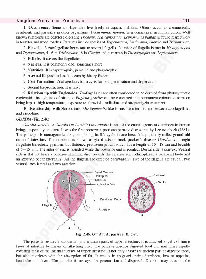
������� ���� �� �� ������� �� 111
1. Occurrence. Some zooflagellates live freely in aquatic habitats. Others occur as commensals,symbionts and parasites in other organisms. Trichomonas hominis is a commensal in human colon. Wellknown symbionts are cellulose digesting Trichonympha campanula, Lophomonas blattarum found respectivelyin termites and wood roaches. Parasites include species of Trypanosoma, Leishmania, Giardia and Trichomonas.
2. Flagella. A zooflagellate bears one to several flagella. Number of flagella is one in Mastigamoebaand Trypanosoma, 4—6 in Trichomonas, 8 in Giardia and numerous in Trichonympha and Lophomonas.
3. Pellicle. It covers the flagellates.
4. Nucleus. It is commonly one, sometimes more.
5. Nutrition. It is saprotrophic, parasitic and phagotrophic.
6. Asexual Reproduction. It occurs by binary fission.
7. Cyst Formation. Zooflagellates form cysts for both perennation and dispersal.
8. Sexual Reproduction. It is rare.
9. Relationship with Euglenoids. Zooflagellates are often considered to be derived from photosyntheticeuglenoids through loss of plastids. Euglena gracilis can be converted into permanent colourless form onbeing kept at high temperature, exposure to ultraviolet radiations and streptomycin treatment.
10. Relationship with Sarcodines. Mastigamoeba like forms are intermediate between zooflagellatesand sacrodines.
GIARDIA (Fig. 2.46)
Giardia lamblia or Giardia (= Lamblia) intestinalis is one of the causal agents of diarrhoea in humanbeings, especially children. It was the first protozoan protistan parasite discovered by Leeuwenhoek (1681).The pathogen is monogenetic, i.e., completing its life cycle in one host. It is popularly called grand oldman of intestine. The infection is known as giardiasis or back packer’s disease Giardia is an eightflagellate binucleate pyriform but flattened protozoan protist which has a length of 10—18 �m and breadthof 6—15 �m. The anterior end is rounded while the posterior end is pointed. Dorsal side is convex. Ventralside is flat but bears a concave attaching disc towards the anterior end. Rhizoplasts, a parabasal body andan axostyle occur internally. All the flagella are directed backwardly. Two of the flagella are caudal, twoventral, two lateral and two anterior.
Fig. 2.46. Giardia. A, parasite. B, cyst.
The parasite resides in duodenum and jejunum parts of upper intestine. It is attached to cells of lininglayer of intestine by means of attaching disc. The parasite absorbs digested food and multiplies rapidlycovering most of the internal surface of upper intestine. It not only absorbs sufficient part of digested food,but also interferes with the absorption of fat. It results in epigastric pain, diarrhoea, loss of appetite,headache and fever. The parasite forms cyst for perennation and dispersal. Division may occur in the
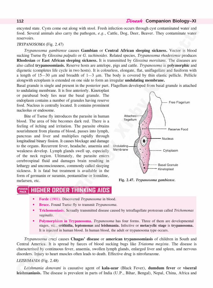
������� �Companion Biology–XI112
encysted state. Cysts come out along with stool. Fresh infection occurs through cyst contaminated water andfood. Several animals also carry the pathogen, e.g., Cattle, Dog, Deer, Beaver. They contaminate waterreservoirs.
TRYPANOSOMA (Fig. 2.47)
Trypanosoma gambiense causes Gambian or Central African sleeping sickness. Vector is bloodsucking Tsetse fly Glossina palpalis or G. tachinoides. Related species, Trypanosoma rhodesiense producesRhodesian or East African sleeping sickness. It is transmitted by Glossina morsitans. The diseases arealso called trypanosomiasis. Reserve hosts are antelope, pigs and cattle. Trypanosoma is polymorphic anddigenetic (completes life cycle in two hosts). It is colourless, elongate, flat, uniflagellate and fusiform witha length of 15—30 �m and breadth of 1—3 �m. The body is covered by thin elastic pellicle. Pelliclealongwith ectoplasm is extended on one side to form an irregular undulating membrane.
Basal granule is single and present in the posterior part. Flagellum developed from basal granule is attachedto undulating membrane. It is free anteriorly. Kinetoplastor parabasal body lies near the basal granule. Theendoplasm contains a number of granules having reservefood. Nucleus is centrally located. It contains prominentnucleolus or endosome.
Bite of Tsetse fly introduces the parasite in humanblood. The area of bite becomes dark red. There is afeeling of itching and irritation. The parasite obtainsnourishment from plasma of blood, passes into lymph,pancreas and liver and multiplies rapidly throughlongitudinal binary fission. It causes blockage and damageto the organs. Recurrent fever, headache, anaemia andweakness develop. Lymph glands swell up, especiallyof the neck region. Ultimately, the parasite enterscerebrospinal fluid and damages brain resulting inlethargy and unconsciousness, commonly called sleepingsickness. It is fatal but treatment is available in theform of germanin or suramin, pentamidine or lomidine,melarsen, etc.
• Forde (1901). Discovered Trypanosoma in blood.
• Bruce. Found Tsetse fly to transmit Trypanosoma.
• Trichomoniasis. Sexually transmitted disease caused by tetraflagellate protozoan called Trichomonas
vaginalis.
• Polymorphism in Trypanosoma. Trypanosoma has four forms. Three of them are developmentalstages, viz., crithidia, leptomonas and leishmania. Infective or metacyclic stage is trypanosoma.
It is injected in human blood. In human blood, the adult or trypanosoma type occurs.
Trypanosoma cruzi causes Chagas’ disease or american trypanosomiasis of children in South andCentral America. It is spread by faeces of blood sucking bugs like Triatoma megista. The disease ischaracterised by continuous fever, anaemia, swollen lymph glands, enlarged liver and spleen, and nervousdisorders. Injury to heart muscles often leads to death. Effective drug is nitrofurazone.
LEISHMANIA (Fig. 2.48)
Leishmania donovani is causative agent of kala-azar (Black Fever), dumdum fever or visceralleishmaniasis. The disease is prevalent in parts of India (U.P., Bihar, Bengal), Nepal, China, Africa and
Fig. 2.47. Trypanosoma gambiense.
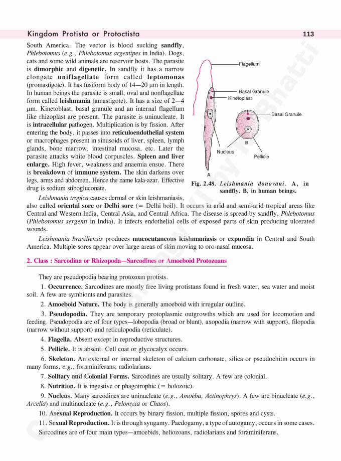
������� ���� �� �� ������� �� 113
South America. The vector is blood sucking sandfly,Phlebotomus (e.g., Phlebotomus argentipes in India). Dogs,cats and some wild animals are reservoir hosts. The parasiteis dimorphic and digenetic. In sandfly it has a narrowelongate uniflagellate form called leptomonas(promastigote). It has fusiform body of 14—20 �m in length.In human beings the parasite is small, oval and nonflagellateform called leishmania (amastigote). It has a size of 2—4�m. Kinetoblast, basal granule and an internal flagellumlike rhizoplast are present. The parasite is uninucleate. Itis intracellular pathogen. Multiplication is by fission. Afterentering the body, it passes into reticuloendothelial systemor macrophages present in sinusoids of liver, spleen, lymphglands, bone marrow, intestinal mucosa, etc. Later theparasite attacks white blood corpuscles. Spleen and liverenlarge. High fever, weakness and anaemia ensue. Thereis breakdown of immune system. The skin darkens overlegs, arms and abdomen. Hence the name kala-azar. Effectivedrug is sodium stibogluconate.
Leishmania tropica causes dermal or skin leishmaniasis,also called oriental sore or Delhi sore (= Delhi boil). It occurs in arid and semi-arid tropical areas likeCentral and Western India, Central Asia, and Central Africa. The disease is spread by sandfly, Phlebotomus(Phlebotomus sergenti in India). It infects endothelial cells of exposed parts of skin producing ulceratedwounds.
Leishmania brasiliensis produces mucocutaneous leishmaniasis or expundia in Central and SouthAmerica. Multiple sores appear over large areas of skin moving to oro-nasal mucosa.
2. Class : Sarcodina or Rhizopoda—Sarcodines or Amoeboid Protozoans
They are pseudopodia bearing protozoan protists.
1. Occurrence. Sarcodines are mostly free living protistans found in fresh water, sea water and moistsoil. A few are symbionts and parasites.
2. Amoeboid Nature. The body is generally amoeboid with irregular outline.
3. Pseudopodia. They are temporary protoplasmic outgrowths which are used for locomotion andfeeding. Pseudopodia are of four types—lobopodia (broad or blunt), axopodia (narrow with support), filopodia(narrow without support) and reticulopodia (reticulate).
4. Flagella. Absent except in reproductive structures.
5. Pellicle. It is absent. Cell coat or glycocalyx occurs.
6. Skeleton. An external or internal skeleton of calcium carbonate, silica or pseudochitin occurs inmany forms, e.g., foraminiferans, radiolarians.
7. Solitary and Colonial Forms. Sarcodines are usually solitary. A few are colonial.
8. Nutrition. It is ingestive or phagotrophic (= holozoic).
9. Nucleus. Many sarcodines are uninucleate (e.g., Amoeba, Actinophrys). A few are binucleate (e.g.,Arcella) and multinucleate (e.g., Pelomyxa or Chaos).
10. Asexual Reproduction. It occurs by binary fission, multiple fission, spores and cysts.
11. Sexual Reproduction. It is through syngamy. Paedogamy, a type of autogamy, occurs in some cases.
Sarcodines are of four main types—amoebids, heliozoans, radiolarians and foraminiferans.
Fig. 2.48. Leishmania donovani. A, insandfly. B, in human beings.
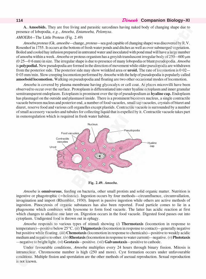
������� �Companion Biology–XI114
A. Amoebids. They are free living and parasitic sarcodines having naked body of changing shape due topresence of lobopodia, e.g., Amoeba, Entamoeba, Pelomyxa.
AMOEBA—The Little Proteus (Fig. 2.49)
Amoeba proteus (Gk. amoeibe—change, proteus—sea god capable of changing shape) was discovered by R.V.Rosenhof in 1755. It occurs at the bottom of fresh water ponds and ditches as well as over submerged vegetation.Boiled and cooled hay infusion prepared in untreated water and inoculated with pond mud will have a large numberof amoeba within a week. Amoeba or proteus organism has a greyish transluscent irregular body of 250—600 �m(0·25—0·6 mm) in size. The irregular shape is due to presence of many lobopodia or blunt pseudopodia. Amoebais polypodial. New pseudopodia are formed in the direction of movement while older pseudopodia are withdrawnfrom the posterior side. The posterior side may show wrinkled area or uroid. The rate of locomotion is 0·02—0·03 mm/min. Slow creeping locomotion performed by Amoeba with the help of pseudopodia is popularly calledamoeboid locomotion. Walking on pseudopodia and floating are two other occasional modes of locomotion.
Amoeba is covered by plasma membrane having glycocalyx or cell coat. At places microvilli have beenobserved to occur over the surface. Protoplasm is differentiated into outer hyaline ectoplasm and inner granularsemitransparent endoplasm. Ectoplasm is prominent over the tip of pseudopodium as hyaline cap. Endoplasmhas plasmagel on the outside and plasmasol inside. There is a prominent biconvex nucleus, a single contractilevacuole between nucleus and posterior end, a number of food vacuoles, small sap vacuoles, crystals of biuret anddiuret, reserve food and various cell organelles except plastids. Contractile vacuole is surrounded by a numberof small accessory vacuoles and tubules for collecting liquid that is expelled by it. Contractile vacuole takes partin osmoregulation which is required in fresh water habitat.
Fig. 2.49. Amoeba.
Amoeba is omnivorous, feeding on bacteria, other small protists and solid organic matter. Nutrition isingestive or phagotrophic (=holozoic). Ingestion occurs by four methods—circumfluence, circumvallation,invagination and import (Rhumbler, 1930). Import is passive ingestion while others are active methods ofingestion. Pinocytosis of organic substances has also been reported. Food particle comes to lie in aphagosome which combines with lysosome to form food vacuole. The latter has acidic reaction at firstwhich changes to alkaline one later on. Digestion occurs in the food vacuole. Digested food passes out intocytoplasm. Undigested food is thrown out in ephagy.
Amoeba responds to various types of stimuli showing (i) Thermotaxis (locomotion in response totemperature)—positive below 25°C. (ii) Thigmotaxis (locomotion in response to contact)—generally negativebut positive while floating. (iii) Chemotaxis (locomotion in response to chemicals)—positive to weakly acidicmedium and negative to other. (iv) Rheotaxis (locomotion in response to water current)—negative. (v) Phototaxis—negative to bright light. (vi) Geotaxis—positive. (vii) Galvanotaxis—positive to cathode.
Under favourable conditions, Amoeba multiplies every 24 hours through binary fission. Mitosis isintranuclear. Chromosome number is high (250 and more). Cyst formation occurs under unfavourableconditions. Multiple fission and sporulation are the other methods of asexual reproduction. Sexual reproductionis not known.
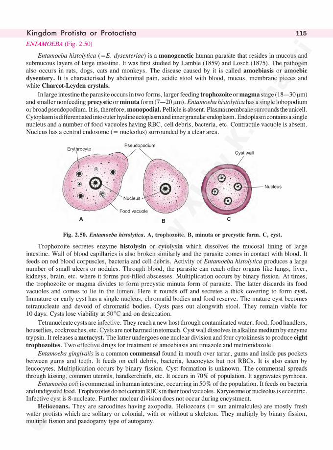
������� ���� �� �� ������� �� 115
ENTAMOEBA (Fig. 2.50)
Entamoeba histolytica (=E. dysenteriae) is a monogenetic human parasite that resides in mucous andsubmucous layers of large intestine. It was first studied by Lamble (1859) and Losch (1875). The pathogenalso occurs in rats, dogs, cats and monkeys. The disease caused by it is called amoebiasis or amoebicdysentery. It is characterised by abdominal pain, acidic stool with blood, mucus, membrane pieces andwhite Charcot-Leyden crystals.
In large intestine the parasite occurs in two forms, larger feeding trophozoite or magma stage (18—30 �m)and smaller nonfeeding precystic or minuta form (7—20 �m). Entamoeba histolytica has a single lobopodiumor broad pseudopodium. It is, therefore, monopodial. Pellicle is absent. Plasma membrane surrounds the unicell.Cytoplasm is differentiated into outer hyaline ectoplasm and inner granular endoplasm. Endoplasm contains a singlenucleus and a number of food vacuoles having RBC, cell debris, bacteria, etc. Contractile vacuole is absent.Nucleus has a central endosome (= nucleolus) surrounded by a clear area.
Fig. 2.50. Entamoeba histolytica. A, trophozoite. B, minuta or precystic form. C, cyst.
Trophozoite secretes enzyme histolysin or cytolysin which dissolves the mucosal lining of largeintestine. Wall of blood capillaries is also broken similarly and the parasite comes in contact with blood. Itfeeds on red blood corpuscles, bacteria and cell debris. Activity of Entamoeba histolytica produces a largenumber of small ulcers or nodules. Through blood, the parasite can reach other organs like lungs, liver,kidneys, brain, etc. where it forms pus-filled abscesses. Multiplication occurs by binary fission. At times,the trophozoite or magma divides to form precystic minuta form of parasite. The latter discards its foodvacuoles and comes to lie in the lumen. Here it rounds off and secretes a thick covering to form cyst.Immature or early cyst has a single nucleus, chromatid bodies and food reserve. The mature cyst becomestetranucleate and devoid of chromatid bodies. Cysts pass out alongwith stool. They remain viable for10 days. Cysts lose viability at 50°C and on desiccation.
Tetranucleate cysts are infective. They reach a new host through contaminated water, food, food handlers,houseflies, cockroaches, etc. Cysts are not harmed in stomach. Cyst wall dissolves in alkaline medium by enzymetrypsin. It releases a metacyst. The latter undergoes one nuclear division and four cytokinesis to produce eighttrophozoites. Two effective drugs for treatment of amoebiasis are tiniazole and metronidazole.
Entamoeba gingivalis is a common commensal found in mouth over tartar, gums and inside pus pocketsbetween gums and teeth. It feeds on cell debris, bacteria, leucocytes but not RBCs. It is also eaten byleucocytes. Multiplication occurs by binary fission. Cyst formation is unknown. The commensal spreadsthrough kissing, common utensils, handkerchiefs, etc. It occurs in 70% of population. It aggravates pyrrhoea.
Entamoeba coli is commensal in human intestine, occurring in 50% of the population. It feeds on bacteriaand undigested food. Trophozoites do not contain RBCs in their food vacuoles. Karyosome or nucleolus is eccentric.Infective cyst is 8-nucleate. Further nuclear division does not occur during encystment.
Heliozoans. They are sarcodines having axopodia. Heliozoans (= sun animalcules) are mostly freshwater protists which are solitary or colonial, with or without a skeleton. They multiply by binary fission,multiple fission and paedogamy type of autogamy.
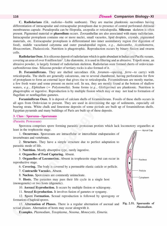
������� �Companion Biology–XI116
C. Radiolarians (Gk. radiolus—feeble sunbeam). They are marine planktonic sacrodines havingdifferentiation of intracapsular and extracapsular protoplasm due to presence of central perforated chitinoidmembranous capsule. Pseudopodia can be filopodia, axopodia or reticulopodia. Siliceous skeleton is oftenpresent. Pigmented material or phaeodium occurs. Zooxanthellae are also associated with many radiolarians.Intracapsular protoplasm contains one or more nuclei, small vacuoles, lipid droplets, crystals, pigmentedmaterials, etc. Extracapsular protoplasm is differentiated into inner assimilatory region (for digestion offood), middle vacuolated calymma and outer pseudopodial region, e.g., Aulocantha, Acanthometra,Hexacontium, Thalassicola. Nutrition is phagotrophic. Reproduction occurs by binary fission and swarmcells.
Radiolarian Ooze. It is siliceous deposit of radiolarians which is quite abundant in Indian and Pacific oceans,covering an area of over 8 million km2. Like diatomite, it is used in filtering and as abrasive. Tripoli stone, anabrasive powder, is largely formed of radiolarium skeleton. Radiolarian ooze formed cherts of ordovician-carboniferous time. Siliceous powder of tertiary rocks is also formed of it.
D. Foraminiferans. They are shelled sarcodines (Gk. foramen—opening, ferre—to carry) withreticulopodia. The shells are generally calcareous, one to several chambered, having perforations for flowof protoplasm to form an external layer that gives rise to reticulopodia. Foraminiferans are mostly marine,a few fresh water and some present on moist soil. In sea, they are mostly found at the bottom of shallowwaters, e.g., Elphidium (= Polystomella). Some forms (e.g., Globigerina) are planktonic. Nutrition isphagotrophic or ingestive. Reproduction is by multiple fission which may or may not lead to formation offlagellate or nonflagellate gametes.
Foraminiferan Ooze. It is deposit of calcium shells of foraminiferans. Fossils of these shells occurs inall ages from Ordovician to present. They are used in determining the age of sediments, especially oilbearing strata. White chalk and limestone deposits of some periods are built up of foraminiferan shells.Egyptian pyramids and many buildings of Paris are built up of these deposits.
3. Class : Sporozoa—Sporozoans
(Parasitic Protozoans)
Sporozoa comprises spore forming parasitic protozoan protists which lack locomotory organelles atleast in the trophozoite stage.
1. Occurrence. Sporozoans are intracellular or intercellular endoparasites ofinvertebrates and vertebrates.
2. Structure. They have a simple structure due to perfect adaptation toparasitic mode of life.
3. Nutrition. Mostly absorptive type, rarely ingestive.
4. Organelles of Food Capturing. Absent.
5. Organelles of Locomotion. Absent in trophozoite stage but can occur inreproductive stage.
6. Covering. The body is covered by a permeable elastic cuticle or pellicle.
7. Contractile Vacuoles. Absent.
8. Nucleus. Sporozoans are commonly uninucleate.
9. Hosts. The parasites may pass their life cycle in a single host(monogenetic) or two hosts (digenetic).
10. Asexual Reproduction. It occurs by multiple fission or schizogony.
11. Sexual Reproduction. It involves fusion of gametes or syngamy.
12. Spore Formation. Sexual reproduction is followed by sporogony orformation of haploid spores.
13. Alternation of Phases. There is a regular alternation of asexual andsexual phases. Alternation of hosts may occur alongwith it.
Examples. Plasmodium, Toxoplasma, Nosema, Monocystis, Eimeria.
Fig. 2.51. Sporozoite ofPlasmodium.
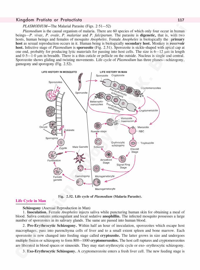
������� ���� �� �� ������� �� 117
PLASMODIUM—The Malarial Parasite (Figs. 2·51—52)
Plasmodium is the causal organism of malaria. There are 60 species of which only four occur in humanbeings—P. vivax, P. ovale, P. malariae and P. falciparum. The parasite is digenetic, that is, with twohosts, human beings and females of mosquito Anopheles. Female Anopheles is biologically the primaryhost as sexual reproduction occurs in it. Human being is biologically secondary host. Monkey is reservoirhost. Infective stage of Plasmodium is sporozoite (Fig. 2.51). Sporozoite is sickle-shaped with apical cap atone end, probably for producing lytic materials for passing into host cells. The size is 6—12 �m in lengthand 0.5—1.0 �m in breadth. There is a thin cuticle or pellicle on the outside. Nucleus is single and central.Sporozoite shows gliding and twisting movements. Life cycle of Plasmodium has three phases—schizogony,gamogony and sporogony (Fig. 2.52).
Fig. 2.52. Life cycle of Plasmodium (Malaria Parasite).
Life Cycle in Man
Schizogony (Asexual Reproduction in Man)1. Inoculation. Female Anopheles injects saliva while puncturing human skin for obtaining a meal of
blood. Saliva contains anticoagulant and local sedative anophillin. The infected mosquito possesses a largenumber of sporozoites in its salivary glands. The same are passed into human blood.
2. Pre-Erythrocytic Schizogony. Within half an hour of inoculation, sporozoites which escape host
macrophages, pass into parenchyma cells of liver and to a small extent spleen and bone marrow. Each
sporozoite is now changed into feeding stage called cryptozoite. The latter grows in size and undergoes
multiple fission or schizogony to form 800—1000 cryptomerozoites. The host cell ruptures and cryptomerozoites
are liberated in blood spaces or sinusoids. They may start erythrocytic cycle or exo- erythrocytic schizogony.
3. Exo-Erythrocytic Schizogony. A cryptomerozoite enters a fresh liver cell. The new feeding stage is
������� �Companion Biology–XI118
called metacryptozoite. It grows in size, undergoes schizogony and forms about 64 larger
macrometacryptomerozoites and 800—1000 smaller micrometacryptomerozoites. Macrometa-
cryptomerozoites enter new liver cells to repeat exoerythrocytic schizogony. Micrometacryptomerozoites
enter erythrocytes.
Shelter in Spleen. Chotivanich (2002) has reported Plasmodium to take shelter inside the dendritic
cells of spleen where they are protected from drugs and immune responses of the body.
4. Erythrocytic Schizogony. A merozoite (cryptomerozoite or micrometa- cryptomerozoite) enters a
red blood corpuscle and produces a feeding stage called trophozoite. Trophozoite feeds on cytoplasm of red
blood corpuscle, protein part of haemoglobin and some materials absorbed from blood plasma. (i) Discoid
Stage. The young feeding parasite grows in size and becomes discoid. (ii) Signet Ring Stage. A noncontractile
vacuole appears in the centre of trophozoite. Peripheral cytoplasm shows a bulge in the region of nucleus
and therefore, appears like a signet ring. The stage is absent in Plasmodium falciparum. The latter may
have more than one parasite in a single erythrocyte. Signet ring stage increases absorptive area and helps in
quick growth. (iii) Amoeboid Stage. Noncontractile central vacuoles disappears. The trophozoite now
develops pseudopodia like outgrowths for increasing absorptive area. It is called amoeboid stage. At this
time the cytoplasm of RBC may have granules. A type of minute granules called Schuffner’s dots or
granules occur in P. ovale and P. vivax. They are replaced by irregular Maurer's dots in case of P.
falciparum and Ziemann’s dots in P. malariae. The central part of trophozoite also possesses brown to
black pigment granules of haemozoin, haematin or melanin. They are derived from haematin part of
haemoglobin. RBC enlarges in P. vivax. (iv) Nuclear Divisions. Nucleus of trophozoite undergoes repeated
division to form 6—24 daughter nuclei. (v) Rosette Stage. Trophozoite with 6—24 nuclei is called erythrocytic
schizont. Cytoplasm gathers around each nucleus. It produces 6—24 merozoites or schizozoites on the
periphery of central cytoplasm having haemozoin granules. (vi) Liberation of Merozoites. Infected red
blood corpuscles rupture. Merozoites and haemozoin granules are liberated in blood. They are attacked by
macrophages. Haemozoin granules are toxic and bring about fever. Surviving merozoites enter fresh red
blood corpuscles to repeat the cycle. Symptoms of malaria appear at intervals synchronising with completion
of erythrocytic cycles. Cell ghosts are eliminated by spleen. Spleen enlarges (splenomegaly).
Table 2.3. Different species of Plasmodium infecting human beings
1. P. vivax Tropical and temperate areas. Benign tertiary fever (recurs after every 48 hours).
Incubation period is 8—10 days.
2. P. malariae Tropical and temperate areas. Quartan fever (recurs after every 72 hours). Incubation
period is 20—30 days. Causes subclinical malaria.
3. P. ovale West Africa and Sourth America. Benign tertiary fever (recurs after every 48 hours).
Incubation period is 10—15 days.
4. P. falciparum Tropical areas. Malignant tertian,, quotadian (daily) or irregular, aestivo-autumnal (early
summer to autumn). Incubation period is 6—12 days. May lead to cerebral malaria
due to synthesis of protein PFEMP—1 that makes erythrocytes sticky blocking small
blood vessels.
Pre-Patent Period. It is the period between inoculation and first appearance of parasite in RBCs.
Incubation Period. It is the period of time between inoculation of sporozoite and the appearance ofsymptoms of malaria.
Symptoms of Malaria. (i) Anaemia. (ii) Enlarged spleen (iii) Pigmentation of skin. (iv) Pigmentationof urine. It may lead to black water fever in P. falciparum due to excessive intravascular haemolysis.(v) Intermittent fever preceded by chills. Actual attack of malaria or paroxysm has three stages :
(a) Rigor Stage. Period of intense chill and shivering. Pulse is fast. It lasts for about an hour. (b) Febrile
������� ���� �� �� ������� �� 119
Stage. Rise in body temperature to 104°—106° F (40.5°—41.6°C). It continues for a few hours. (c) DefervescentStage. A lot of sweating occurs. This decreases the body temperature to almost normal.
5. Post-Erythrocytic Schizogony. In face of natural or induced resistance, some of the freshly releasedmerozoites enter liver cells. Here they remain inactive for a long time. The inactive parasite is calledhypnozoite. As the immunity of the host falls, hypnozoites become active, grow and perform schizogony.Relapse of malaria is due to it. Post-erythrocytic shizogony is absent in P. falciparum. Some workers callit exo-erythrocytic schizogony.
6. Formation of Gametocytes. After a few generations of erythrocytic schizogony, some of themerozoites after entering RBCs, grow into gametocytes. Gametocytes or gamonts are commonly roundedbut in P. falciparum they are crescent shaped. They are of two types, smaller male microgametocyte withcentral large nucleus and larger female macrogametocyte with reserve food and eccentric smaller nucleus.Further growth occurs inside the midgut of female Anopheles as it requires a temperature of about 25°C.
Life Cycle in Female Anopheles
1. Gamogony (Gametogenesis). In the midgut (often referred as stomach) of Mosquito, all stages exceptgametocytes are digested. Gametocytes lose their RBC covering and undergo maturation to form gametes.The female gametocyte develops a cone of reception and gets converted into female gamete. Themicrogametocyte shows nuclear divisions followed by 6—8 elongated male gametes from its peripherythrough a process called exflagellation.
2. Fertilisation. A male gamete enters a female gamete through the cone of reception. Their fusionproduces a diploid zygote.
3. Ookinete. Zygote elongates, becomes worm like and is called ookinete. Ookinete in contact with gutwall penetrates the same enzymatically and comes to lie sub-epidermally on the outer side of stomach.Ookinete becomes round. A permeable cyst wall is formed around it jointly by it and the stomach wall. Thisconverts ookinete into oocyst.
4. Sporogony. 50—2000 oocysts may appear on the stomach wall. They absorb nourishment fromhaemocoel and grow in size. Each oocyst now functions as sporont. It becomes vacuolated. The diploidnucleus divides first meiotically (Bano, 1959) and then mitotically to form a large number of haploid nuclei.Cytoplasm gathers around each nucleus and forms a sporozoite. A mature oocyst or sporont produces upto10,000 sporozoites. The cyst wall ruptures and the sporozoites pass into haemocoel. From here manysporozoites enter salivary glands for passage to new hosts.
• Macculoch (1827). Coined the term malaria (Gk. mala—bad, aria—air) after its predominance inlocalities having marshy areas.
• Parasitemia. Presence of parasites in blood.
• Laveran (1880). First to observe malaria parasite in erythrocytes of patient.
• Golgi (1885). Studied erythrocyte cycle of Plasmodium.
• Ronald Ross (1897). Found oocysts on stomach of female Anopheles. Studied the role of femaleAnopheles in transmission of malaria. Got Nobel Prize in 1902.
• Grassi et al (1898). Studied life cycle of Plasmodium in female mosquito.
• Fairley (1945). Within 30 minutes of inoculation, the malaria parasite passes into liver cells.
• Shortt (1948). Studied pre-erythrocytic schizogony.
• Rudzinska et al (1965). Studied ultrastructure of sporozoite.
• Mosquito Day. August 29. This day in 1897, Ronald Ross found oocyst on the stomach offemale Anopheles.
• Malaria Day. August 20.
������� �Companion Biology–XI120
• Malaria Research Institute. New Delhi.
• National Malaria Eradication Programme (NMEP). In operation in India since 1953.
• Best Time to Observe Malaria Parasite in Blood. In the middle of febrile stage.
Control of Malaria
Three types of measures are undertaken to control malaria—elimination of mosquito, prophylaxis(prevention of infection) and treatment.
Elimination of Mosquito. (i) Spraying human habitations, cattle sheds and surroundings with insecticideslike BHC and malathion. (ii) Filling small ditches. (iii) Covering water drains. (iv) Proper drainage forquick transfer of waste water into streams. (v) Covering surface of stagnant waters with oil to suffocatelarvae and pupae due to nonavailability of air. (vi) Release of ducks, fish Stickleback, Minnow, Trout,Mosquito Fish (Gambusia affinis), Guppy Fish (Poecilia reticulata), Water Bug (Diplonychus indicus) andDragon Fly naiads into water bodies. They all feed on mosquito larvae. (viii) Growth of insectivorous orcarnivorous aquatic plants like Utricularia and Aldrovanda. (ix) Inoculation of water bodies with cyanobacteria,Aulosira and Anabaena. They release chemicals toxic to larvae. (x) Introduction of Bacillus sphericus inwater bodies. (xi) Release of sterile male Anopheles.
Prophylaxis (Prevention of Infection). (i) Fitting doors, windows and ventilators with wire gauge.(ii) Sleeping under mosquito nets. (iii) Use of mosquito repellents like odomos, good night, casper, all-out,etc. (iv) Prophylactic dose of antimalarial medicine during summer autumn period.
Treatment. Quinine obtained from bark of Cinchona tree has been the first and most widely used tocombat malaria. Later drugs were chloroquine, camoquine, primaquine, nivaquine, daraprim, etc. Thelatest one is artemesinin, which besides killing all stages of malaria parasite, is also antibacterial andantifungal. It is obtained from small plant, Artemesia annua.
Differences between Plasmodium and Plasmodium
S. No. Plasmodium Plasmodium
1. It is the generic name of malaria parasite. It is the name of somatic body of acellular slimemoulds.
2. It is parasitic. It is free living.
3. Plasmodium is uninucleate. It is multinucleate.
4. Nutrition is absorptive. Nutrition is ingestive or phagotrophic.
4. Ciliata—Ciliates, Ciliated Protozoans
They are protistan protozoans which possess cilia as organelles of locomotion and food capturing.
1. Occurrence. Ciliates are mostly free living fresh water protistans. A few are marine. Some areparasites.
2. Motility. Free living forms are generally motile. A few are sedentary (e.g., Vorticella).
3. Solitary and Colonial. Mostly solitary. A few are colonial.
4. Structure. Though unicellular, ciliates have the highest degree of structural and physiologicalcomplexity due to specialisation of organelles for skin, muscles, sense organs, kidney, ingestion, egestion,etc.
5. Evolutionary Status. Ciliates have reached the pinacle of unicellular specialization beyond whichevolution can occur only in multicellular organisms. They have, therefore, reached the dead end of protistanevolution.
6. Polarity. Presence of definite anterior and posterior ends provide polarity to ciliates.
7. Food Collecting Apparatus. It consists of an oral groove, vestibule, buccal cavity, cytostome andcytopharynx. A cytopyge or cytoproct is also formed for egestion.
8. Pellicle. A flexible pellicle covers the organism on its ouside.

������� ���� �� �� ������� �� 121
9. Cilia. Numerous cilia occur over the body. They are used for locomotion. The speed is 1—2 mm/sec, the highest for any protist.
10. Trichocysts. They are ejective organelles which are used for attachment and defence.
11. Nuclear Dimorphism. Two types of nuclei are found, larger metabolism controlling polyploidmacronucleus and smaller heredity controlling diploid micronucleus.
12. Contractile Vacuoles. They occur in fresh water forms for osmoregulation.
13. Nutrition. It is of ingestive or phagotrophic type in free living forms. The parasites have absorptivetype of nutrition.
14. Asexual Reproduction. It occurs by binary fission, budding and cyst formation.
15. Sexual Reproduction. It is by conjugation. Autogamy also occurs.
Examples. Paramecium, Tetrahymena, Vorticella, Balantidium. Tetrahymena is used in biologicalresearch. Balantidium coli causes balantidial or ciliary dysentery in humans. Infection comes from faecalcontamination of pig.
PARAMECIUM—Slipper Organism or Animalcule (Fig. 2.53)
Paramecium (= Paramoecium; Hill,1752) is cylindrical, cigar-shaped or spindleshaped free living ciliate protozoan protistwhich is popularly called slipper organism.It lives in fresh water ponds, ditches, pools,lakes and streams rich in decaying organicmatter. Length is 80—350 �m while breadthis 40—70 �m. The anterior end is roundedand blunt. The posterior end is pointed. Theventral surface bears a shallow obliquedepression called oral groove. Oral grooveends in a funnel-shaped depression calledvestibule. Vestibule leads into S-shapedbuccal cavity, a mouth or cytostome and acytopharynx in contact with endoplasm. Acell anus, cytopyge or cytoproct appearsbehind the vestibule at the time of egestionor ephagy.
The whole body bears longitudinalparallel rows of cilia which are slightlyoblique on the ventral side. Number of ciliais 2500—14000. Cilia of posterior end areslightly longer. They constitute the caudaltuft. On the region of vestibule and buccalcavity, the cilia form (i) Undulating or endoral membrane by a row of cilia. (ii) Three membranelles bymore than one row of cilia each (commonly four rows)—ventral peniculus, dorsal peniculus and quadrulus.Locomotion occurs by metachronous beating of cilia. Paramecium can swim forward as well as backward. Itrotates during locomotion so that the path of locomotion is spiral. Creeping with the help of cilia andmetaboly can also occur.
A thin firm but elastic pellicle occurs on the outside. It has pores for cilia and trichocysts. Trichocystsare spindle-shaped structures of 4 �m length which are discharged on stimulation to form long threads ofupto 40 �m. Contents of trichocysts help in adhesion, offence and defence against predators. Parameciumshows nuclear dimorphism. There are two types of nuclei—a large bean-shaped polyploid vegetativenucleus called macronucleus (=meganucleus) and a small rounded diploid reproductive nucleus known asmicronucleus. Paramecium caudatum has one macronucleus and one micronucleus. P. aurelia has onemacronucleus and two micronuclei. P. multimicronucleatum has a number of micronuclei. Two contractilevacuoles occur for osmoregulation. One is anterior while the other is posterior. The two work alternately
�������������������
������� �Companion Biology–XI122
but the posterior contractile vacuole pulsates a bit faster. Each contractile vacuole is fed by 6—12 feedingcanals which in turn are connected to a network of tubules (= nephridial tubules) for collection of extrawater. A number of food vacuoles are present in the protist. They circulate with the streaming ofprotoplasm, undertaking a path similar to figure of 8. Cytoplasm contains all other eucaryotic organelles.
Differences Between Cyst and Trichocyst
S. No. Cyst Trichocyst
1. It is a thick-walled resting cell. Trichocyst is an ejective organelle.
2. Cyst is meant for perennation. It is meant for offence and defence.
3. It often takes part in dispersal. It helps in attaching.
Nutrition is ingestive (phagotrophic or holozoic). Paramecium feeds on bacteria, other protistans andbits of organic matter. They are pushed into buccal cavity by beating of cilia of oral groove and membranelles.From buccal cavity they pass into cytopharynx. Phagosome is formed at its tip. The same fuses withlysosome to produce food vacuole. Paramecium bursaria is green due to presence of zoochlorellae. Itobtains nourishment from outside as well as zoochlorellae.
Asexual reproduction takes place by transverse binary fission. Macronucleus divides by amitosis whilemicronucleus undergoes intranuclear mitosis. Both the daughters develop structures not present in theirhalves. Sexual reproduction occurs by (i) Conjugation. Two paramecia come in contact in the region oftheir oral grooves and establish protoplasmic bridge. Macronuclei degenerate. Micronuclei divide meioticallyand mitotically in each. Out of them two persist in each conjugant as pronuclei, a stationary female and amigratory male. Migratory pronucleus passes into the other conjugant and fuses with its stationary pronucleusto form diploid synkaryon. Conjugants now separate. Synkaryon of each exconjugant divides a few timesmitotically to form nuclei which differentiate into macronuclei and micronuclei. (ii) Autogamy. Macronucleusdegenerates. Micronucleus undergoes meiosis, forms pronuclei which fuse to produce synkaryon. Synkaryonthen divides, producing both macronuclei and micronuclei. (iii) Cytogamy. It is intermediate betweenconjugation and autogamy. Here two paramecia come in contact without establishing a protoplasmic bridge.Contact provides stimulus to both for performing autogamy. (iv) Endomixis. Macronucleus degenerates.Micronucleus divides forming daughter nuclei that differentiate into macro- and micro- nuclei. (v) Hemixis.Macronucleus reconstructs itself by losing extra parts and elaborating the deficient ones.
Killer Particles. Sonneborn (1943) discovered kappa particles in certain strains of Paramecium whichby releasing a toxin paramecin kill the strains deficient in these particles. Maintenance of kappa particlesis controlled by a dominant nuclear gene. Conjugation can occur between killer and sensitive strains whentransfer of gene and particles may take place. Nontoxic mutants of kappa particles are pi particles. Othertoxic particles found in Paramecium are m� and lambda particles. Conjugant having m� particles kills theother one deficient in them. Lambda particles bring about lysis of sensitive paramecia.
1. What type of organisms are included underkingdom protista ?
Ans. Unicellular and colonial eukaryotes.
2. Who coined the term protista ?
Ans. Haeckel (1866).
3. Name the life styles represented in protista.
Ans. (i) Photosynthetic or holophytic (ii) Ingestiveor holozoic (iii) Saprotrophic (iv) Parasitic(v) Mixotrophic (vi) Symbiotic.
4. In which group of organisms did the mitoticapparatus evolve ?
Ans. Protista.
5. What are mastigonemes ?
Ans. Mastigonemes are minute lateral appendagesfound on the surface of flagella.
6. What do you mean by isochronic andmetachronic movements of cilia ?
Ans. Isochronic is a ciliary movement where allthe cilia beat simultaneously. Metachronicis a ciliary movement where cilia move oneafter the other.
������� ���� �� �� ������� �� 123
7. Name the two types of nutrition that occurin a parasite ?
Ans. Phagotrophic and absorptive.
8. What is the reason for large population ofprotists ?
Ans. Rapid asexual reproduction.
9. Define plasmotomy.
Ans. Plasmotomy is division of a multinucleateprotistan or plasmodium into two to severalmultinucleate daughters, e.g., Opalina.
10. What for is cyst formed ?
Ans. To tide over unfavourable conditions.
11. Define (i) Binary Fission (ii) MultipleFission.
Ans. (i) Binary fission is a method of asexualreproduction in which a mature individualdivides into two equal and similar daughters.
(ii) Multiple fission is a division of a matureindividual into a number of daughters byrepeated division of nucleus followed bycleavage.
12. Define isogamy, anisogamy and oogamy.
Ans. (i) Isogamy. It is a mode of sexual reproductionwhich involves fusion of two morphologicallyand physiologically similar gametes.
(ii) Anisogamy. It is sexual reproductioninvolving fusion of different sized butotherwise similar gametes.
(iii) Oogamy. It is a mode of sexualreproduction in which a nonmotile foodladen female gamete or ovum fuses with amotile small sized male gamete or sperm.
13. Name three groups representing protistanalgae.
Ans. Dinoflagellates, diatoms and euglenoids.
14. What are red tides ?
Ans. Red tides are occasional blooms ofdinoflagellates that spread over severalkilometres of sea, e.g., Gonyaulax.
15. Name an armoured and an unarmoureddinoflagellate.
Ans. Unarmoured. Noctiluca. Armoured.Gonyaulax.
16. Define mesokaryon.
Ans. Mesokaryon is the name of dinoflagellatenucleus given by Dodge (1966, 1971) becauseof the occurrence of condensed chromatin orchromosomes even in the interphase state.
17. Name two unique xanthophylls present indinoflagellates.
Ans. Dinoxanthin, Neodinoxanthin.
18. Name a colourless dinoflagellate.
Ans. Noctiluca.
19. Name a dinoflagellate bearing (i)Trichocysts (ii) Cnidoblasts.
Ans. Trichocysts occur in Peridinium whilecnidoblasts are found in Nematodinium.
20. What is bioluminescence ? Name abioluminescent protistan alga.
Ans. Bioluminescence is the emission of light byliving beings which, therefore, glow in night,e.g., Peridinium, Gonyaulax.
21. Name the grooves present in dinoflagellates.
Ans. Longitudinal sulcus and circular cingulum(girdle, annulus).
22. What types of flagella occur indinoflagellates ?
Ans. Heterokont with one narrow flagellum havingmastigonemes and the second flattenedribbon-like but smooth present in girdle orcingulum.
23. What is PSP ?
Ans. PSP is paralytic shell fish poisoning.
24. What is saxitoxin ?
Ans. Saxitoxin is a strong toxin (100,000 timesstronger than cocaine) produced by Gonyaulaxcatenella which is the source of paralyticshell fish poisoning.
25. Name a dinoflagellate in which phagotrophysupplements photosynthesis.
Ans. Ceratium.
26. What are diatoms ?
Ans. Diatoms are golden coloured protistan algaewhich are characterised by presence of two-valved silicified cell wall.
27. What are dinoflagellates ?
Ans. Dinoflagellates are unicellular goldenprotistan algae which have two heterokontflagella and two grooves, cingulum and sulcus.
28. Name a diatom that indicates pollution.
Ans. Pleurosigma.
29. What types of symmetry are found indiatoms ?
Ans. (i) Radial symmetry—Centric diatoms, e.g.,Biddulphia, Triceratium.
������� �Companion Biology–XI124
(ii) Bilateral symmetry—Pennate diatoms,e.g., Pinnularia.
30. Name the pigment that provides brownishcolouration to diatoms ?
Ans. Fucoxanthin.
31. What is frustule ?
Ans. Frustule is a two-valved siliceous cell wallof diatoms consisting of larger epitheca anda slightly smaller hypotheca.
32. What is Mac Donald-Pfitzer law ?
Ans. Mac Donald-Pfitzer law is the progressivedecrease in the size of diatoms during repeatedbinary fission due to smaller sized hypothecaof the parent becoming epitheca of one of thedaughters.
33. Define auxospores.
Ans. Auxospores are rejuvenescent cells of diatomsthat help in increasing their size to normal.
34. What is diatomite ?
Ans. Diatomite or kieselguhr is deposit of siliceousskeletons of diatoms.
35. How many flagella are present in Euglena.
Ans. Apparently one but actually two with smallerone reaching only upto paraflagellar body.
36. Name the photosensitive organelles ofeuglenoids.
Ans. Paraflagellar body and eye spot.
37. What is mixotrophic nutrition ?
Ans. Mixotrophic nutrition is dual nutrition withability to perform photosynthesis in light andobtain nourishment from organic matter indark, e.g., Euglena.
38. Define metaboly.
Ans. Metaboly is wriggling type of movement inwhich a wave of contraction and expansionpasses through the body from anterior toposterior end, e.g., Euglena.
39. Name the carbohydrate reserve food ofeuglenoids.
Ans. Paramylum, a � 1, 3-glucan.
40. Which protist is called plant-animal ?
Ans. Euglena.
41. What is the other name of consumerdecomposer protists ?
Ans. Slime Moulds.
42. Define slime moulds.
Ans. Slime moulds are consumer-decomposer
protists having a naked body of plasmodiumor pseudoplasmodium, cellulose coveredspores and myxamoebae.
43. Define : (i) Plasmodium (ii) Myxamoeba(iii) Pseudoplasmodium.
Ans. (a) Plasmodium is a free-living wall-less ornaked multinucleate mass of protoplasm asfound in acellular slime moulds.
(b) Myxamoeba is amoeba-like nakeduninucleate cell of slime moulds that iscapable of independent feeding, growth andmultiplication.
(c) Pseudoplasmodium is a multicellular
structure formed by coming together of a
large number of myxamoebae in cellular slime
moulds.
44. What type of pigments are found in slime
moulds ?
Ans. Pigments found in slime moulds are non-
photosynthetic, e.g., anthracene, authraquinone.
45. What are acellular slime moulds ?
Ans. Acellular slime moulds are those consumer
decomposer protists which have a diploid
somatic phase represented by plasmodium,
cellulose covered spores for dispersal and
haploid swarm cells or myxamoebae for
sexual reproduction.
46. What is capillitium ?
Ans. Capillitium is a branching system of thread-
like structures formed inside a sporangium
of acellular slime moulds during formation
of spores.
47. Define communal slime moulds.
Ans. Communal slime moulds is the name of
cellular slime moulds because of the coming
together of numerous haploid myxamoebae to
form pseudoplasmodium prior to production
of sporangia.
48. What is macrocyst ?
Ans. Macrocyst is encysted fusion product of a
large female gamete and a small male gamete
in the life cycle of cellular slime moulds.
49. What is cyst ?
Ans. Cyst is a metabolically resting structure
surrounded by a thick impremeable wall which
takes part in perennation, dispersal and
������� ���� �� �� ������� �� 125
occasionally multiplication in protists, e.g.,
Entamoeba, Amoeba.
50. What are protozoan protists ?
Ans. Protozoan protists are unicellular or colonial
nonphotosynetic organisms which show animal-
like nutrition, locomotion and absence of cell
wall.
51. Name the various groups of protozoan protists.
Ans. Protozoan protists are of four types—flagellates,
sarcodines, sporozoans and ciliates.
52. What are zooflagellates ? Name a commensal,symbiont and parasitic zooflagellate.
Ans. Zooflagellates or mastigophores are flagellabearing protozoan protistans that have a pellicleon the outside and binary fission as commonmode of multiplication. Commensal.Trichomonas hominis in humans. Symbiont.Trichonympha is termites. Parasite.Trypanosoma.
53. Name an organism intermediate betweenzooflagellata and sarcodina.
Ans. Mastigoamoeba.
54. What is backpacker’s disease ?
Ans. Giardiasis.
55. Name the grand oldman of intestine.
Ans. Grand old man of intestine. Giardia lambia(G.intestinalis).
56. How many nuclei and flagella are present inGiardia ?
Ans. Giardia. Nuclei. Two. Flagella. Eight.
57. Name the causal organism of African SleepingSickness.
Ans. Trypanosoma gambiense.
58. Name the vector of trypanosomiasis.
Ans. Tsetse Fly (Glossina palpalis or G.techinoides).
59. What is Chagas’ disease ?
Ans. Chagas’ disease (American trypanosomiasis)is a disease of children caused byTrypanosoma cruzi spread through faeces ofblood sucking bug like Triatoma megista.
60. What is the causal agent of dumdumfever ?
Ans. Causal agent of Dumdum fever or Kala-azar–Leishmania donovani.
61. Which vector spreads kala-azar ?
Ans. Kala-azar is spread by blood sucking sandfly Phlebotomus (e.g., P.argentipes).
62. Name the causal agent and vector of orientalsore.
Ans. Oriental (Delhi) Sore. Causal agent isLeishmania tropica. Vector is SandflyPhlebotomus (e.g., P.sergenti).
63. What are sarcodines ?
Ans. Sarcodines are amoeboid protozoans thatdevelop pseudopodia for locomotion andfeeding.
64. What are the symptoms of amoebicdysentery ?
Ans. Symptoms of Amoebic Dysentery :Abdominal pain and acidic stool with blood,mucus, membrane pieces and white Charcot-Leyden crystals.
65. What is the infective stage of Entamoebahistolytica ?
Ans. Infective stage of Entamoeba histolytica.Tetranucleate cyst.
66. Where does Amoeba live ?
Ans. Amoeba lives inside fresh water ponds andditches at the bottom and over submergedvegetation.
67. What are heliozoans ?
Ans. Helizonas (sun animalcules) are axopodiacontaing sarcodines.
68. What are radiolarians ?
Ans. Radiolarians are marine planktonicsarcodines having intracapsular andextracapsular protoplasm due to presence of acentral perforated chitinised membranouscapsule and siliceous skeleton.
69. What do you mean by radiolarian ooze ?
Ans. Radiolarian ooze is siliceous deposit ofradiolarians.
70. What are foraminiferans ?
Ans. Foraminiferans are shelled sarcodines havingreticulopodia.
71. What is foraminiferan ooze ?
Ans. Foraminiferan ooze is a calcareous depositof foraminiferans.
72. What are sporozoa ?
Ans. Sporozoa is a group of spore producingparasitic protozoan protists that lacklocomotory organelles at least in trophozoitestage.
73. Which species causes cerebral malaria ?
Ans. Cerebral Malaria. Plasmodium falciparum.
������� �Companion Biology–XI126
74. What is black water fever ?
Ans. Black water fever is malarial fever causedby Plasmodium falciparum.
75. Name the species of malaria parasite foundin human beings.
Ans. (i) Plasmodium vivax (ii) P.malariae(iii) P.ovule (iv) P. falciparum.
76. Which is the primary host of malariaparasite?
Ans. Primary host is the one where the parasiteperforms its sexual reproduction. Biologicallythe primary host of malaria parasite is femaleAnopheles.
77. What is the name of infective stage ofmalaria parasite ?
Ans. Infective Stage of Malaria Parasite.Sporozoite.
78. Define pre-patent and incubation periods.
Ans. (i) Prepatent Period. Period betweeninoculation and first appearance of theparasite in host tissues (e.g., RBCs in caseof malaria).
(ii) Incubation Period. Period betweeninoculation and appearance of symptoms inhost.
79. Where is exoerythrocytic schizogonyaccomplished ?
Ans. Liver cells.
80. What is exflagellation ?
Ans. Exflagellation is the formation and releaseof mature male gametes from the surface ofmicrogametocyte as in Plasmodium.
81. Where are haemozoin and Schuffner’sgranules found ?
Ans. (a) Haemozoin. Brown to black pigmentgranules formed from haematin part ofhaemoglobin in the central part of trophozoiteand released into blood at the time of liberationof merozoites.
(b) Schuffner's Granules or Dots. They areminute granules found in the cytoplasm ofRBCs during infection of Plasmodium ovaleand P.vivax.
82. How many nuclei are present in Parameciumcaudatum and P. aurelia ?
Ans. Paramecium caudatum. Two, one macronucleusand one micronucleus.
Paramecium aurelia. Three, one macronucleusand two micronuclei.
83. What are trichocysts ?
Ans. Trichocysts are ejective organelles which aredischarged on stimulation to form long threadsfor adhesion, offence and defence.
84. Expand NMEP.
Ans. NMEP. National Malaria EradicationProgramme.
85. Who coined the term malaria ?
Ans. Macculoch (1827).
86. Define Syngamy, Diatomaceous Earth,Plasmodium, Trichocysts and Micronuclei.
Ans. (a) Syngamy. It is complete fusion of twogametes to form a diploid zygote.
(b) Diatomaceous Earth. See question 34.
(c) Plasmodium. See question 43(a).
(d) Trichocysts. See question 83.
(e) Micronuclei. They are reproductive nucleias found in ciliates (which also have trophicnuclei called macronuclei).
87. What do you understand by the terms 1n and2n ?
Ans. 1n. Haploid. 2n. Diploid.
88. Define zygote.
Ans. Zygote. It is diploid cell formed by fusion oftwo haploid gametes.
89. What is zygotic meiosis ?
Ans. Zygotic Meiosis (Initial Meiosis). It is meiosiswhich occurs during the germination of zygoteto form four haploid structures.
90. Define gametic meiosis.
Ans. Gametic Meiosis (Terminal Meiosis). Meiosisoccurs only at the time of formation of gametesso that they are the only haploid structures. Allothers are diploid.
1. In guts of termites and wood roaches, certain
zooflagellates help in breakdown of .......
2. Macronucleus has many sets of ...........
3. Because of ........... impregnation, the walls of
diatoms are nearly indestructible.
4. Armour plates of dinoflagellates are made of
...........
5. Besides human beings, the alternate host of
malaria parasite is ........... Anopheles.
������� ���� �� �� ������� �� 127
6. Acellular slime moulds have a ........... mass
of multinucleate protoplasm.
7. In kala-azar there is a severe ........... infection.
8. The latest drug for treatment of resistant forms
of malaria parasites is ........
9. Mosquito day is ........... August.
1. Cellulose 2. Chromosomes
3. Silica 4. Cellulose
5. Female 6. wall-less/naked
7. liver 8. artemesinin
9. 29
1. The protists form trichocysts to tide overadverse situations.
2. Diatoms have silica and cellulose in theirwalls.
3. Eye spots of euglenoids are able to sensemovement as they lie close to the base offlagella.
4. Plasmodium is a slime mould.5. Both micronuclei and macronuclei are found in
ciliates.6. Malaria can be controlled by destroying the
alternate host.7. Paramecium is spindle organism.8. Amoeba is omnivorous.9. Protists constitute connecting link between
monerans and other kingdoms.10. Eye spot of Euglena is formed of carotenoid
granules.
1. False 2. True 3. False
4. False 5. True 6. True
7. False 8. True 9. True
10. True
1. What is the nature of cell walls in diatoms ?
Ans. Cell wall or frustule of diatoms is a two-valved soap dish like, basically made of silicawith smaller amounts of cellulose and pecticcompounds. There is a larger epivalve or epithecaand a smaller hypovalve or hypotheca with acingulum or girdle between the two.
2. Find out what do the terms ‘algal bloom’and ‘red-tides’, signify.
Ans. (a) Algal Bloom. It is exceptionally rich growthof algae, especially cyanobacteria over the surfaceof water bodies due to highly favourable climaticconditions or inflow of excess nutrients. Algalbloom often kills other aquatic forms due todepletion of oxygen, secretion of toxic chemicalsand organic loading. The water becomes unfitfor use.
(b) Red Tides. They are blooms of reddinoflagellates which spread for severalkilometres of sea making it appear red, e.g.,Gonyaulax, Gymnodinium, Ceratium. Thedinoflagellates produce toxins that kill the fishand poison the shell fish. The latter is thesource of paralytic shell fish poisoning orPSP in human beings.
3. Describe briefly the four major groups ofProtozoa.
Ans. The four groups of Protozoa are mastigophora,sarcodina, sporozoa and ciliata.
Mastigophora or Flagellata. See text.
Sarcodina. See text.
Sporozoa. See text.Ciliata. See text.
4. What are the characteristic features ofEuglenoids ?
Ans. See text.
(From N.C.E.R.T. Question Bank)
1. Suppose you accidentally find an oldpreserved permanent slide without a label.In your effort to identify it, you place theslide under microscope and observe thefollowing features :
(a) Unicellular
(b) Well defined nucleus
(c) Biflagellate, one flagellum lyinglongitudinally and other transversely.
What would you identify it as ? Can youname the kingdom it belongs to ?
Ans. Dinoflagellate, kingdom Protista.
������� �Companion Biology–XI128
1. Diatoms are called as ‘‘pearls of ocean’’.Why ? What is diatomaceous earth ?
Ans. Pearls of Ocean. Diatoms have a shiningsilicified two valved covering or frustule.
Diatomaceous Earth. It is pile of siliceous
skeletons of diatoms which accumulates dueto being resistant to decay.
2. What observable features in Trypanosomawould make you classify it under kingdomProtista ?
Ans. (i) Unicellular structure (ii) Eukaryotic naturewith well developed nucleus.
(iii) Flagellum with basal granules.
1. Dinoflagellates are golden brown photosyntheticprotists which are important component of marinephytoplankton. They sometimes grow in suchabundance as to form red tides. An armoured orunarmoured theca of cellulose occurs around. Itbears two grooves. Both of them possess flagella,broader in circular groove and narrow comingout of longitudinal groove. The two flagella causerolling and spinning movements. Because of thispeculiar mode of locomotion, dinoflagellates arecalled whorling whips.
(a) Where are dinoflagellates found and howdo they form red tides ?
(b) Why do dinoflagellates perform spinningmovements ?
(c) Which type of wall is present overdinoflagellates ?
Ans. (a) Most of the dinoflagellates live as planktonin sea. In large abundance they cover thesea water over large area. The same is calledred tide.
(b) Dinoflagellates have two flagella arrangedin two different directions. Their activitymakes the dinoflagellates spin in water.
(c) The wall is made of cellulose plates whichprovide an armoured or nonarmoured coverof theca.
2. Mastigophora are flagella bearing protozoanswith a pellicle covering. They are both freeliving aquatic and parasites. Giardia,Trypanosoma, Leishmania and Trichomons areparasitic in human beings. Giardia is transmittedthrough cyst containing food and water.Trichomonas is sexually transmitted.Trypanosma and Leishmania are spread by vectorflies, Tsetse and sandfly.
(a) What is the number of flagella present inGiardia ? What harm does it cause ?
(b) Which disease is caused by Trypanosomagambiense ?
(c) Kala-azar, the disease caused by Leishmaniadonovani is named so due to which type ofsymptoms ?
Ans. (a) Eight – two anterior, two lateral, two ventraland two caudal. The pathogen resides induodenum and jejunum. It multiplies fast andcovers the lining of gut, causing various typesof disorders including epigastric pain,diarrhoea,, loss of appetite, headache andfever.
(b) Sleeping sickness.
(c) The skin darkens over legs, arms andabdomen.
3. Paramecium is a member of class ciliata ofprotozoa. Whole of its body is covered over bycilia. Shape of the body is like a slipper.Therefore, Paramecium is also called slipperanimalcule. There are two nuclei (nucleardimorphism), larger macronucleus and smallermicronucleus. Micronucleus is reproductivenucleus while macronucleus is bean-shapedvegetative nucleus. Pellicle has trichocysts. Acilia–lined groove leads to cytostome.
(a) Where is Paramecium found ?(b) What is the peculiarity of macronucleus ?(c) What is the role of trichocysts ?
Ans. (a) Fresh water.(b) Macronucleus is large polyploid nucleus
which controls the functioning of theorganism. It is formed afresh afterreproduction.
(c) Trichocysts are protrusible organelles thathelp in attachment, offence and defence.
��������������� ��
������� ���� �� �� ������� �� 129
1. Naked cytoplasm, multinucleated andsaprophytic are the characteristics of(A) Monera (B) Protista(C) Fungi (D) Slime moulds.
2. Fusion of two gametes which are dissimilar insize is termed as(A) Oogamy (B) Isogamy(C) Anisogamy (D) Zoogamy.
3. Black water fever is due to(A) Plasmodium falciparum(B) P. ovale(C) P. vivax(D) P. malariae. (C.P.M.T. 2001)
4. Trypanosoma gambiense produces(A) Filaria (B) Malaria(C) Sleeping sickness (D) Kala-azar.
(A.F.M.C. 2003)5. Contractile vacuole does not occur in
(A) Slime Moulds (B) Sporozoa(C) Sarcodina (D) Zooflagellata.
(A.F.M.C. 2004)6. Slipper Animalcule is the name of
(A) Paramecium (B) Plasmodium(C) Chlamydomones (D) Vorticella.
(Manipal 2005)7. Bioluminescence is found in
(A) Chlorella (B) Ceratium(C) Hirudinaria (D) Chlamydomonas.
(Odisha 2006)8. Thalloid body of Slime Mould is
(A) Mycelium (B) Protonema(C) Plasmodium (D) Fruiting body.
(C.B.S.E. 2006, A.F.M.C. 2008)9. Which one of the following is a slime mould
(A) Thiobacillus (B) Anabanea(C) Rhizopus (D) Physarum.
(C.B.S.E. 2007)10. The vector for kala-azar is
(A) Anopheles (B) Phlebotomus(C) Culex (D) Aedes. (W.B. 2008)
11. Some stages of Plasmodium vivax may remaindormant in liver for long time. On reactivation,the parasite undergoes(A) Erythrocytic schizogony(B) Gametogony(C) Cycle of Ross(D) Exoerthrocytic schizogony.
(E.A.M.C.E.T. 2008)
12. What is not true for Paramecium ?(A) Forms cysts under unfavourable conditions(B) Cilia over body(C) Contractile vacuoles for osmoregulation(D) Uses pseudopodia for capturing prey.
(Odisha 2009)13. Slime Moulds are
(A) Parasitic, plasmodium, with walls, sporesdispersed by air
(B) Saprophytic, plasmodium, without walls,spores dispersed by water
(C) Saprophytic, plasmodium, without walls,spores dispersed by air
(D) Parasitic, plasmodium, without walls,spores dispersed by water.
(D.P.M.T. 2009)14. Which of the following is a flagellate protozoan
(A) Amoeba (B) Entamoeba(C) Trypanosoma (D) Paramecium(E) Plasmodium (Kerala 2010)
15. The basis of protozoan classification is(A) Shape (B) Locomotion(C) Measurement (D) Number of nuclei.
(M.P.P.M.T. 2010)16. Trypanosoma is
(A) Sporozoan(B) Amoeboid protozoan(C) Ciliated protozoan(D) Flagellated protozoan. (H.P.P.M.T. 2011)
17. Sporozoites of malarial parasite will be foundin(A) Saliva of infected female Anopheles(B) Salivary glands of freshly moulted female
Anopheles(C) Spleen of infected human(D) RBCs of person suffering from malaria.
(C.B.S.E. 2011)18. Unicellular eukaryotic organisms are placed
in kingdom(A) Fungi (B) Protista(C) Monera (D) Animalia.
(H.P.P.M.T. 2012)19. Diatoms are placed under
(A) Chrysophtyes (B) Slime Moulds(C) Euglenoids (D) Dinoflagellates.
(A.M.U. 2012)20. Unicellular algae, diatoms and protozoans are
members of(A) Plantae (B) Fungi(C) Protista (D) Monera.
(J.K.C.E.T. 2013)
������������� �����������
������� �Companion Biology–XI130
21. Marine ‘animal’ which exhibits bioluminescenceis(A) Noctiluca (B) Trypanosoma(C) Leishmania (D) Volvox.
(C.O.M.E.D-K 2013)22. Protozoan with helicopodia is
(A) Actinophrys (B) Elphidium(C) Euglypha (D) Entamoeba
(E.A.M.C.E.T. 2014)23. Which one is not a free living protozoan
(A) Euglena (B) Amoeba(C) Giardia (D) Noctiluca
(Bih. 2015)24. In which group of organisms the cell walls
form two thin overlapping shells which fittogether(A) Chrysophytes (B) Euglenoids(C) Dinoflagellates (D) Slime moulds.
(C.B.S.E. 2015)25. An aggregate of slime moulds which may
grow and spread over several feet is called(A) Plasmodium (B) Plasmopara
(C) Mycoplasma (D) Pseudoparenchyma.(A.M.U. 2016)
26. Chrysophytes, euglenoids, dinoflagellates andslime moulds are included in the kingdom(A) Animalia (B) Monera(C) Protista (D) Fungi.
(N.E.E.T. 2016)27. Ciliates differ from all other protozoans in
(A) Using pseudopodia for capturing prey(B) Having a contractile vacuole for removing
excess water(C) Using flagella for locomotion(D) Having two types of nuclei.
(N.E.E.T. 2018)
1. (D) 2. (C) 3. (A) 4. (C) 5. (B)
6. (A) 7. (B) 8. (C) 9. (D) 10. (B)
11. (A) 12. (D) 13. (C) 14. (C) 15. (B)
16. (D) 17. (A) 18. (B) 19. (A) 20. (C)
21. (A) 22. (A) 23. (C) 24. (A) 25. (A)
26. (C) 27. (D)
Related Documents