Communication Vol. 266, No. 16, Issue of June 5, pp. 10035-10038,1991 THE JOURNAL OF BIOLOGICAL CHEMISTRY 0 1991 by The American Society for Biochemistry and Molecular Biology, Inc. Printed in U. S. A. Transcriptional Regulation of the Glucokinase Gene by Biotin in Starved Rats* (Received for publication, February 11, 1991) Jasbir Chauhan and Krishnamurti Dakshinamurtit: From the Department of Biochemistry and Molecular Biology, University of Manitoba, Winnipeg, Manitoba R3E 0 W3, Canada The purpose of this work was to investigate whether biotin, a water-soluble vitamin, regulates the glucoki- nase gene. Biotin was administered intraperitoneally to starved rats, and the time course of glucokinase induction was followed over a time period of 12 h. The glucokinase mRNA was increased 19.6-fold during the first 1 h after biotin administration, afterwards rap- idly decayed, and was hardly detectable by 4 h. The amount of glucokinase activity asdetermined by con- ventional enzyme activity assay increased in a time- dependent fashion, reaching 4-fold by 2 h of biotin administration. The transcriptional activity of the gene as measured by a nuclear run-on assay was in- creased about 6.7-fold within 45 min of biotin admin- istration. These findings indicate that biotin can reg- ulate the glucokinase gene at the transciptional stage in the starved rat. The mammalian hexokinase (ATP:D-hexose 6-phospho- transferase (EC 2.7.1.1)), which catalyzes the first reaction of glucose metabolism, has four different isoenzymes (1, 2). Glucokinase (ATP:D-glucose 6-phosphotransferase, (EC 2.7.1.2)), with a K,,, for glucose of about 6 X M (about the sameaspostprandial blood glucose level) istheprincipal isoenzyme in liver. The K,,, values for glucose of the other hexokinases are at least 1 order of magnitude lower. Gluco- kinase is expressed in a tissue-specific fashion and is found only in liver (0.1% of total soluble protein) and pancreatic p- cells (0.005% of total soluble protein) (3). Glucokinase regu- lates blood glucose levels by controlling hepatic glucose utili- zation and storage in the form of glycogen; in the islet cells of the pancreas, glucokinase has been postulated to act as the glucose sensor that regulates insulin production (4, 5). The activity of liver glucokinase has been shown to be regulated by dietary and hormonal status of the animal. Changes in enzyme activity have also been related to the developmental stage of the growing animal. We have shown that, in addition to regulation by dietary andhormonalfactors,theenzymeactivityinrat liver is altered by the biotin status of the animal (6, 7). The low activity of the deficient rat liver is increased following biotin administration. Biotin, like insulin, also produced a preco- cious increase in hepatic glucokinase in the suckling rat (8). * This work was supported by a grant from the Medical Research Council of Canada.Thecosts of publication of thisarticle were defrayed in part by the payment of page charges. This article must therefore be hereby marked “aduertisement” in accordance with 18 U.S.C. Section 1734 solely to indicate this fact. $ To whom correspondence should be addressed. Evidence was presented to show that the administration of biotin markedly increased the enzyme activity in animals that are not biotin-deficient but have low liver glucokinase activity due to starvation or feeding with high fat diet (9). In these studies, it was pointed out that the action of biotin paralleled that of insulin and that enzyme synthesis was the site of this action. In later work, Spence and Koudelka (10) examined the effect of biotin upon the activity of glucokinase in primary cultures of rat hepatocytes. The addition of biotin at a con- centration of M to the culture medium resulted in a 4- fold increase in the activity of glucokinase, the maximum response being observed a t 6 h following the addition of biotin to the medium. Changes in the activity of the enzyme brought about by biotin or cyclic 8-bromo-GMP were shown to result from changes in the rate of synthesis of the enzyme. Various laboratories have reported the molecular cloning of glucokinase cDNA (11-14). The cloned cDNA has been used to measure the abundance of glucokinase mRNA in the livers of rats at various stages of postnatal development (11) and during starvation-refeeding (11, 15). Insulin stimulates the transcription of glucokinase gene in livers of diabetic rats (14, 16). The induction of gene transcription by insulinisre- pressed by the glucagon-CAMP system(17).Thyroidhor- mones significantly contribute to the regulation of glucoki- nase gene transcription during glucose refeeding (18). Thus, the effects of various hormones on regulation of hepatic glucokinase are at the level of transcription. In the present investigation, we have studied the molecular mechanism responsible for the increase of hepatic glucokinase in starved rats after biotin administration. For the first time, we report here that biotin administration causes a marked and rapid induction of glucokinase mRNA in starved rats. Furthermore, we have shown that this effect is on the rate of transcription of the glucokinase gene. EXPERIMENTAL PROCEDURES Animals-Male Sprague-Dawley rats weighing between 120 and 140 g were used in these experiments. Twenty-four hours before the experiment, food was removed from the cages. The animals had free access to drinking water. After 24 h of fasting, biotin (1 mg/kg of body weight) was administered intraperitoneally. At specified times, as indicated in the legends to the figures, animals were killed by a blow to the head and decapitated before isolation of liver. Assay of Hexokinase-Liver hexokinase and glucokinase activities were determined as described previously (7). Livers were homogenized in 5 volumes of buffer containing 50 mM Tris/HCl, pH 7.0, 0.15 M KCI, 5 mM EDTA, 4 mM MgCI,, and 2 mM dithiothreitol. Homoge- nates were centrifuged at 100,000 X g,, for 60 min, and the clear supernatant was assayedforenzyme activity immediately or after freezing and storage at -80 “C. Glucokinase activity was estimated by subtracting the hexokinase activity measured at 0.5 mM glucose from the activity measured at 0.1 M glucose. A unit of hexokinase is defined as the enzyme activity resulting in the formation of 1 pmol of glucose 6-phosphate/min under the conditions of the assay. The protein concentration was measured by the Bradford method (19). Measurement of Glucokinase mRNA-Total RNA was isolated from livers using guanidinium thiocyanate solution and phenol ex- traction,as described by ChomczynskiandSacchi (20). Poly(A)+ RNA was separated from total RNA by chromatography on oligo(dT)- cellulose (type 7, Pharmacia LKB Biotechnology Inc.). Poly(A)+ RNA was fractionated in 1% agarose gel containing 6.3% formaldehyde. After electrophoresis, the gel was soaked in 20 X SSC (3 M NaC1, 0.3 M sodium citrate) for 1 h and blotted on to nitrocellulose. The filter was then baked for 2 h at 80 “C in a vacuum oven. Prehyhridization 10035

Welcome message from author
This document is posted to help you gain knowledge. Please leave a comment to let me know what you think about it! Share it to your friends and learn new things together.
Transcript
Communication Vol. 266, No. 16, Issue of June 5, pp. 10035-10038,1991 THE JOURNAL OF BIOLOGICAL CHEMISTRY
0 1991 by The American Society for Biochemistry and Molecular Biology, Inc. Printed in U. S. A.
Transcriptional Regulation of the Glucokinase Gene by Biotin in Starved Rats*
(Received for publication, February 11, 1991)
Jasbir Chauhan and Krishnamurti Dakshinamurtit: From the Department of Biochemistry and Molecular Biology, University of Manitoba, Winnipeg, Manitoba R3E 0 W3, Canada
The purpose of this work was to investigate whether biotin, a water-soluble vitamin, regulates the glucoki- nase gene. Biotin was administered intraperitoneally to starved rats, and the time course of glucokinase induction was followed over a time period of 12 h. The glucokinase mRNA was increased 19.6-fold during the first 1 h after biotin administration, afterwards rap- idly decayed, and was hardly detectable by 4 h. The amount of glucokinase activity as determined by con- ventional enzyme activity assay increased in a time- dependent fashion, reaching 4-fold by 2 h of biotin administration. The transcriptional activity of the gene as measured by a nuclear run-on assay was in- creased about 6.7-fold within 45 min of biotin admin- istration. These findings indicate that biotin can reg- ulate the glucokinase gene at the transciptional stage in the starved rat.
The mammalian hexokinase (ATP:D-hexose 6-phospho- transferase (EC 2.7.1.1)), which catalyzes the first reaction of glucose metabolism, has four different isoenzymes (1, 2). Glucokinase (ATP:D-glucose 6-phosphotransferase, (EC 2.7.1.2)), with a K,,, for glucose of about 6 X M (about the same as postprandial blood glucose level) is the principal isoenzyme in liver. The K,,, values for glucose of the other hexokinases are at least 1 order of magnitude lower. Gluco- kinase is expressed in a tissue-specific fashion and is found only in liver (0.1% of total soluble protein) and pancreatic p- cells (0.005% of total soluble protein) (3). Glucokinase regu- lates blood glucose levels by controlling hepatic glucose utili- zation and storage in the form of glycogen; in the islet cells of the pancreas, glucokinase has been postulated to act as the glucose sensor that regulates insulin production (4, 5 ) . The activity of liver glucokinase has been shown to be regulated by dietary and hormonal status of the animal. Changes in enzyme activity have also been related to the developmental stage of the growing animal.
We have shown that, in addition to regulation by dietary and hormonal factors, the enzyme activity in rat liver is altered by the biotin status of the animal (6, 7). The low activity of the deficient rat liver is increased following biotin administration. Biotin, like insulin, also produced a preco- cious increase in hepatic glucokinase in the suckling rat (8).
* This work was supported by a grant from the Medical Research Council of Canada. The costs of publication of this article were defrayed in part by the payment of page charges. This article must therefore be hereby marked “aduertisement” in accordance with 18 U.S.C. Section 1734 solely to indicate this fact.
$ To whom correspondence should be addressed.
Evidence was presented to show that the administration of biotin markedly increased the enzyme activity in animals that are not biotin-deficient but have low liver glucokinase activity due to starvation or feeding with high fat diet (9). In these studies, it was pointed out that the action of biotin paralleled that of insulin and that enzyme synthesis was the site of this action. In later work, Spence and Koudelka (10) examined the effect of biotin upon the activity of glucokinase in primary cultures of rat hepatocytes. The addition of biotin a t a con- centration of M to the culture medium resulted in a 4- fold increase in the activity of glucokinase, the maximum response being observed a t 6 h following the addition of biotin to the medium. Changes in the activity of the enzyme brought about by biotin or cyclic 8-bromo-GMP were shown to result from changes in the rate of synthesis of the enzyme.
Various laboratories have reported the molecular cloning of glucokinase cDNA (11-14). The cloned cDNA has been used to measure the abundance of glucokinase mRNA in the livers of rats at various stages of postnatal development (11) and during starvation-refeeding (11, 15). Insulin stimulates the transcription of glucokinase gene in livers of diabetic rats (14, 16). The induction of gene transcription by insulin is re- pressed by the glucagon-CAMP system (17). Thyroid hor- mones significantly contribute to the regulation of glucoki- nase gene transcription during glucose refeeding (18). Thus, the effects of various hormones on regulation of hepatic glucokinase are at the level of transcription.
In the present investigation, we have studied the molecular mechanism responsible for the increase of hepatic glucokinase in starved rats after biotin administration. For the first time, we report here that biotin administration causes a marked and rapid induction of glucokinase mRNA in starved rats. Furthermore, we have shown that this effect is on the rate of transcription of the glucokinase gene.
EXPERIMENTAL PROCEDURES
Animals-Male Sprague-Dawley rats weighing between 120 and 140 g were used in these experiments. Twenty-four hours before the experiment, food was removed from the cages. The animals had free access to drinking water. After 24 h of fasting, biotin (1 mg/kg of body weight) was administered intraperitoneally. At specified times, as indicated in the legends to the figures, animals were killed by a blow to the head and decapitated before isolation of liver.
Assay of Hexokinase-Liver hexokinase and glucokinase activities were determined as described previously (7). Livers were homogenized in 5 volumes of buffer containing 50 mM Tris/HCl, pH 7.0, 0.15 M KCI, 5 mM EDTA, 4 mM MgCI,, and 2 mM dithiothreitol. Homoge- nates were centrifuged at 100,000 X g,, for 60 min, and the clear supernatant was assayed for enzyme activity immediately or after freezing and storage at -80 “C. Glucokinase activity was estimated by subtracting the hexokinase activity measured at 0.5 mM glucose from the activity measured a t 0.1 M glucose. A unit of hexokinase is defined as the enzyme activity resulting in the formation of 1 pmol of glucose 6-phosphate/min under the conditions of the assay. The protein concentration was measured by the Bradford method (19).
Measurement of Glucokinase mRNA-Total RNA was isolated from livers using guanidinium thiocyanate solution and phenol ex- traction, as described by Chomczynski and Sacchi (20). Poly(A)+ RNA was separated from total RNA by chromatography on oligo(dT)- cellulose (type 7, Pharmacia LKB Biotechnology Inc.). Poly(A)+ RNA was fractionated in 1% agarose gel containing 6.3% formaldehyde. After electrophoresis, the gel was soaked in 20 X SSC (3 M NaC1, 0.3 M sodium citrate) for 1 h and blotted on to nitrocellulose. The filter was then baked for 2 h a t 80 “C in a vacuum oven. Prehyhridization
10035
10036 Glucokinase mRNA Induction by Biotin in Vivo was performed for 4 h at 42 "C in 50% formamide, 5 x Denhardt's solution (0.1';: each of Ficoll, polyvinylpyrrolidone, and bovine serum alhumin), 5 X SSPE (0.75 M NaCI, 50 mM NaH,PO,, 5 mM EDTA), 0 . 1 ' 5 SDS', 100 pg/ml salmon sperm DNA. Glucokinase cDNA was laheled with [w:"I']dCTP, using a random priming laheling kit (Re- thesda Research Lahoratories). Hyhridization was done a t 42 "C for 24 h. After the hyhridization filters were washed twice for 10 min each in 400 ml of 2 X SSC, 0 . 1 % SDS at room temperature and three times for 60 min in 250 ml of 0.2 X SSC, 0.1% SDS a t 68 "C, they were air-dried and exposed to Kodak X-OMAT x-ray film wit.h an intensifying screen overnight a t -80 "C. Quantitation of glucokinase mRNA was accomplished hy densitometric scanning of the autora- diograms. A linear relationship hetween RNA load and ahsorhance peak area was verified for each hlot.
Isolntion of Liurr Nucki and Run-on Transcription Ana1.v.si.s- Livers were homogenized in 5 volumes of huffer containing 0.25 M sucrose, 50 mM Tris/HCl, pH 7.5, 25 mM KCI. 2 mM MgCI,. 1 mM dithiothreitol, 1 mM E(;TA, 0.14 mM spermidine, and 0.1 mM phenyl- methylsulfonyl fluoride. Homogenates were filtered through four lay- ers of cheesecloth, and crude nuclei were isolated hy centrifugation at 800 X x for 5 min. Nuclei collected by centrifugation were washed once hy centrifugation through a 2.0 M sucrose cushion a t 20,000 rpm f o r 45 min in a lieckman SW 28 rotor. Nuclei were resuspended in glvcerol storage huffer containing 50 mM Tris/HCl, pH 8.3, 5 mM MgCI,, 0.1 mM EDTA. 50% glycerol, and 0.1 mM phenylmethylsul- lonyl fluoride. The run-on transcription reaction in isolated nuclei was carried o u t a t 25 "C for 90 min in a reaction mixture containing 50 mM Tris/HCI, pH 8.0, 5 mM MgCI,, 100 mM KCI, 0.05 mM EDTA. 20% glycerol, 0.04 mg/ml creatine phosphokinase, 8.8 mM creatinine phosphate, 4 mM dithiothreitol, 0.5 mM CTP, 0.5 mM GTP, 1.0 mM A T P , 0.5 units/ml RNA pmrd. and 200 pCi of [n-:"I-']UTP (800 Ci/ mmol). Laheled RNA was extracted using the method of Chomczynski and Sacchi (20). Fifty micrograms of tRNA and 4 volumes of guani- dinium thiocyanate solution were added. and the resulting suspension was homogenized by several passes through a 22-gauge needle. Two zldditional cycles of suspension in t.he guanidinium thiocyanate solu- t inn and isopropyl alcohol precipitation were carried out. RNA was further precipitated twice with ethanol using ammonium acetate solution, final concentration 2 M. I'elleted RNA was dissolved in hyhridization solution.
H.vhridizotion of Run-on Transcripts to Filt~r-hound Plasmid IINA-Five micrograms each of plasmids GK-1, &actin, pPC116, pHR322, and Hluescript were denatured by treating with 0.1 M NaOH for 30 min and applied to nitrocellulose filters using a slot hlot npparatus. The filters were washed hriefly in 2 X SSC and dried at 80 "C for 2 h. I'rehydridization was a t 42 "C for 8 h in a solution containing 50% formamide, 0.8 M NaCI, 34 mM sodium phosphate huffer, pH 6.5, 0.12% Ficoll 400. 0.12% bovine serum alhumin, 0.12% r)olyvinylpyrrolidone, 0.1% SDS, 100 pg/ml denatured salmon testes IINA, and 100 pg/ml tRNA. Hyhridization was carried out with 5 X 10' cpm in hvhridization solution similar to the prehyhridization solution in a final volume of 1.5 ml at 45 "C for 72 h. The filters were washed twice in 2 X SSC, 0.1% SDS at 25 "C and four times for 60 min in 2 X SSC at 65 "C. Filters were further washed with 2 X SSC containing 1 0 pg/ml RNase for 15 min at 37 "C and washed twice with 2 X SSC. Autoradiography and densitometric scanning were carried out as with Northern hlot hyhridization.
RESULTS AND DISCUSSION
To study the in oioo regulation of glucokinase mRNA by biotin, we used starved rats. We have previously shown (7) that starving rats caused a decrease in liver glucokinase activity and that biotin administration resulted in a 3-4-fold increase in enzyme activity. In the present study, the time course of glucokinase mRNA induction was followed after biotin administration (Fig. 1). There was a 0.6-fold decrease in albumin mRNA in the starved rat, and after biotin admin- istration, it returned to normal. Also, in the starved rats there was a 6.2-fold increase in hepatic phosphoenolpyruvate car- boxykinase mRNA in comparison with normally fed rats. Phosphoenolpyruvate carboxykinase mRNA remained con- stant after biotin was injected into starved rats. At the same
I The ahhreviations used are: SDS, sodium dodecyl sulfate; EGTA, [ethvlenehis(oxyethylenenitrilo)]tetraacetic acid.
1 2 3 4 5 6 7 8 I 1
I
r (3.- 1 I
FIG. 1. Effect of biotin on glucokinase, albumin, and phon- phoenolpyruvate carboxykinase mRNAs in liver. The prore- dures for RNA isolation. gel elect rophoresis. RNA t ransfer to a nylon filter, and hyhridization to '.'I'-lat~eletl rI)NA prohw are tlcsrritwd under "Experimental Procedures." A. RNA blot proherl with rat alhumin cDNA; H . prohed with phospwnolpvn~vnte rnrboxykinase cDNA; C. prohed with glucokinase c1)NA. 5 pg of polv(A)' RNA was loaded per lane for blots A and H , and I O pgllane for hlot ('. Imnv I . normal control; lnnr 2. fasted for 24 h. no biotin treatment; lnnr 3 , fasted. 1 h after hiotin; lanr 4. fasted. 2 h after hiotin; Innr, 5. fasted. 4 h after biotin; lnnr 6. fasted, 6 h after hiotin; lnnr 7. fasted. 8 h alter hiotin; lnnr X , fasted. 12 h after hiotin.
TARI.E I Time coursr of hintin Pffrrt on g/urokinn.w acticily nnd m R N A
amount in liorr o f stnrL-rd r a f s Experimental details are descrihed under "Experimental I'rce-
dures." The areas under the ahsorhance peaks corresponding to the mRNA bands were calculated and expressed in arhitrarv units. Ihtn are given as the mean of four separate determinations.
~~ ~~~~
Time niter hiotin administrntion
to starved rnts
I<rlativr Glucokinnne nmrwnt of
nrfivitv gturrlklnase mI<SA
Normal fed control 14.1 t 4.2 0 (starved 24 h) 4.2 t 1.5 1 2
6.8 k 1.9
4 16.6 f 4.G
6 14.9 t 3 . H
8 14.1 t 4.2 12.8 t 3 . 1
12 10 .9 t 2.4
I' ND. not detectahle.
t ime, the level of glucokinase mRNA of biotin-injected starved rats increased 3.9-fold in comparison with normally fed rata. 1 h after injection of hiotin. Following this increase, there was rapid decay of glucokinase mRNA that was hardlv detectable 4 h after biotin administration. The induction of glr~cokinase mRNA at 1 h following biotin administration to starved rats in three experiments was 19-fold in comparison with starved rats not receiving a biotin injection. The induction of gluco- kinase mRNA by biotin is, therefore, relatively rapid and marked. To our knowledge, this is the firat report on biotin regulation of glucokinase mRNA.
To correlate induct.ion of glucokinase mRNA with gluco- kinase activity, we measured glucokinase activity in the cv- tosol fraction of liver homogenates (Table I). Rv 2 h after biotin injection, the glucokinase mRNA level of the hiotin- injected starved rats had fallen to about 30'; of the level a t 1 h, and by 4 h, glucokinase mRNA was not detectable. From this, we concluded that the half-life of t.he glr~cokinase mRNA was on the order of 30 min under the experimental conditions. The glucokinase enzyme activity reached the maximal level by 2 h following biotin and stayed at a high level for the next 6-8 h. Iynedjian et al. (16) have shown that, following insulin treatment, the amount (and activity) of glucokinase increased
Glucokinase mRNA Induction by Biotin in Vivo 10037
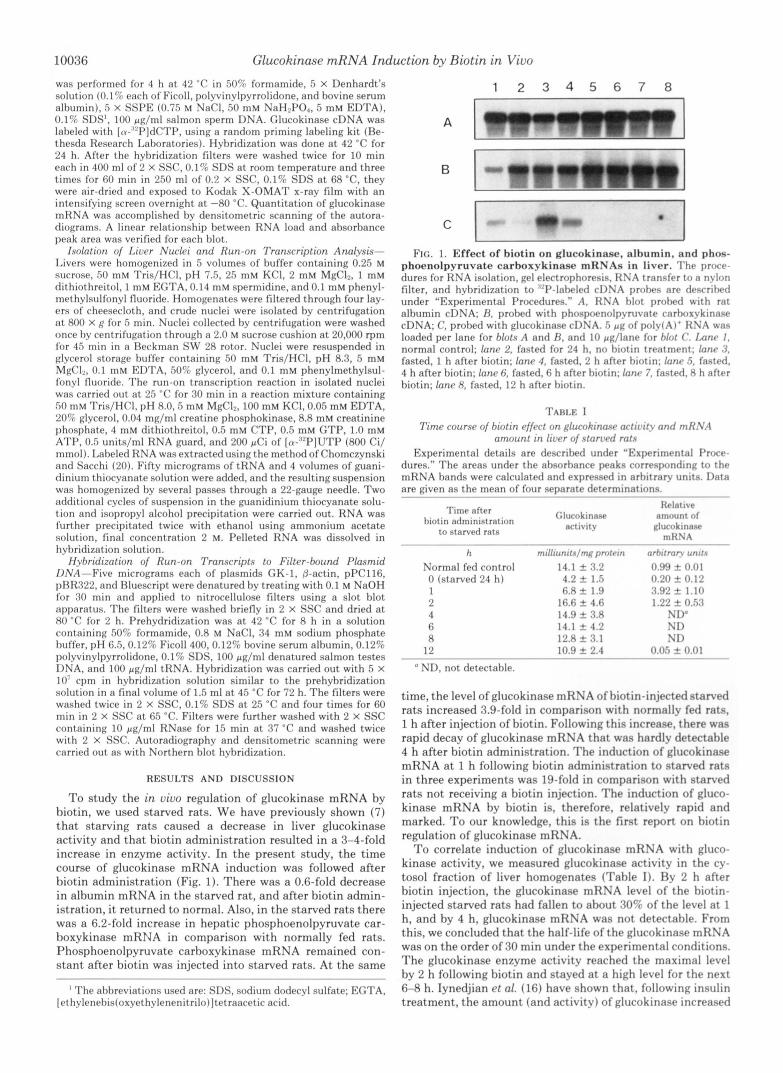
Time C min > 0 15 30 45 60 90 120
1
FIG. 2. Biotin-induced transcription of the glucokinase gene. A, nuclei were isolated at specified times after biotin adminis- tration. The procedure for nuclei isolation and nuclear run-on assay are described under “Experimental Procedures.” The radiolabeled transcripts were hybridized to filter-bound plasmids containing cDNA for phosphoenolpyruvate carboxykinase (PEPCK), glucoki- nase, and actin, respectively. Specific transcripts from the three genes were detected by autoradiography of the filters. The vector plasmids Bluescript and pBR322 were also bound to the filters as control for background hybridization. B, effect of biotin on glucokinase mRNA. RNA was isolated from the same rat livers as used for nuclear run- on assay in A. Experimental details for Northern blot analysis are described in the legend to Fig. 1.
TABLE I1 Effect of biotin on gene transcription in liver as shown by run-on
transcription assays with isolated nuclei Starved rats were treated with biotin for the time shown. Nuclear
run-on assay was performed as described in the legend to Fig. 2. The areas under absorbance peaks corresponding to individual spots were calculated and expressed in arbitrary units. An appropriate exposure of autoradiograms was used to allow a valid comparison between different genes. Data are the means of three separate assays using liver nuclei from different animals. -Fold effects were calculated by using a value of 1 for the transcriptional rate in nontreated starved rats (0 min).
Time after Phosphoenol-
biotin Glucokinase pyruvate &Actin carboxvkinase
Rate of gene transcription
min arbitrary units
0 1.56 f 0.41 15 1.83 f 0.53 30 3.71 f 0.78 45 10.51 f 3.96 60 8.06 f 2.55 90 2.10 f 0.85
120 0.51 k 0.22
-fold
1.0 1.2 2.4 6.7 5.2 1.3 0.3
arbitrary units
101.6 5 21.2 93.2 f 9.3 39.3 f 10.9 76.7 f 16.7
107.6 f 20.9 125.2 f 22.4 132.5 f 21.4
-fold
1.0 0.9 0.4 0.7 1.1 1.2 1.3
arbitrary units
8.0 f 1.5 7.2 f 1.6 7.4 f 1.2 7.1 k 1.9 7.9 & 1.2 7.8 f 0.9 6.5 f 1.8
-fold
1.0 0.9 0.9 0.9 1.0 1.0 0.8
in a time-dependent fashion, after an initial lag of 4 h, to reach 65% of the nondiabetic control level in 24 h. In contrast, glucokinase mRNA was shown to increase until 8 h, and subsequently, the level of the mRNA decayed rapidly so that little mRNA was left after 16 h and virtually none after 24 h. Although the actions of insulin and biotin are parallel, the effect of biotin on glucokinase induction seems to be more rapid than that of insulin.
The rapid and marked increase of glucokinase mRNA after biotin administration suggests a biotin effect at the transcrip- tional level. To examine this further, we carried out “run-on” transcription experiments with isolated liver nuclei. Run-on transcription experiments with isolated nuclei were prepared to estimate the relative rates of transcription of the glucoki- nase gene a t various times following biotin administration to the starved rat. As shown in Fig. 2, hybridization to vector pBR322 or Bluescript DNA was negligible. The transcription of the actin gene, included as internal control, was not influ- enced by biotin administration. Biotin administration in- creased glucokinase gene transcription by about 6.7-fold and
decreased phosphoenolpyruvate carboxykinase by about 2.6- fold (Table 11). The increased transcription of glucokinase gene within 45 min of biotin addition is not simply due to an increase in overall transcriptional efficiency, because the tran- scription of the P-actin gene is unaffected by biotin adminis- tration. Also, no significant differences in the total number of counts incorporated into RNA in isolated nuclei were observed after biotin administration to the starved rats. The difference between induction in mRNA and rate of transcrip- tion may be due to the stabilization of glucokinase mRNA by biotin. As shown in Fig. 2B, glucokinase mRNA was increased as early as 30 min after biotin administration, and the maxi- mum effect was observed a t 1 h. The glucokinase transcription rate was maximal a t 45 min, after which there was a decrease in transcriptional rate, and it was back to the prestimulated level by 2 h. When longer time intervals were studied, no significant change in the rate of transcription was observed as compared with the prestimulated level.
Spence and Koudelka (lo), using cultured rat hepatocytes, have shown that biotin and cGMP induce glucokinase activity and that the effect of biotin might be mediated through cGMP. They have previously shown that the cGMP effect on glucokinase was at the translational stage (21, 22). However, the results of the present study suggest that biotin can in- crease the total mRNA coding for glucokinase.
Although a role for biotin in protein and RNA synthesis was reported by us earlier (23, 24), this is the first report in which the regulation of the transcription of the gene coding for a specific protein has been shown. The prosthetic group function of biotin is well recognized. However, biotin is im- plicated in other areas of metabolism, where its role is not explained on the basis of its prosthetic group function (25, 26). A biotin requirement for various cell lines has been reported. Cells cultured in biotin deficient-medium were ar- rested in the GI phase of the cell cycle, and this block is removed upon addition of biotin to the medium (27, 28). We have also reported that guanylate cyclase and RNA polymer- ase I1 activities are related to the biotin status of cells in culture (29). The question of whether the role of biotin in enhancing the transcription of glucokinase gene has physio- logical significance cannot be answered now. The effects of biotin have been observed a t a high dose level. However, many of the nonprosthetic group effects of biotin have been shown in biotin-deficient animals or cell lines (25, 26).
It has been recently shown that the glucokinase gene uses different promoters, separated by 12 kilobases, in liver and pancreatic @-cells (12, 15). The use of alternative splice sites and tissue-specific promoters for the glucokinase gene offers a mechanism for the known differences in the regulation of glucokinase. If such a mechanism is operative for biotin, it is likely that, in liver, the proximal promoter region is involved. Insulin and glucagon are the most important regulators of glucokinase gene expression. Hoppner and Seitz (18) have shown that thyroid hormones are permissive for the induction of glucokinase. There are significant differences between the actions of thyroid hormones and biotin in this. Differences in glucokinase mRNA levels due to different thyroid states were observed only in the refed and not in starved rats. Biotin administration produces a significant increase in glucokinase mRNA in the starved rat. The intracellular signaling mecha- nism used by biotin to control gene activity is not understood. The glucokinase gene may provide a good system to investi- gate this point, especially if the activation of the gene can be reproduced in biotin-deficient rat hepatocytes.
Acknowledgment-We thank Dr. D. K. Granner for providing
10038 Glucokinase mRNA Induction by Biotin in Vivo
glucokinase cDNA (GK-1) and phosphoenolpyruvate carboxykinase cDNA (pPC116).
Granner, D. K. (1989) Proc. Natl. Acad. Sci. U. S. A. 86,4838- 4842
15. Iynedjian, P. B., Pilot, P., Nouspikel, T., Milburn, J. L., Quaade, REFERENCES C., Hughes, S., Ulca, C., and Newgard, C. B. (1989) Proc. Natl.
16. Iynedjian, P. B., Gjinovci, A., and Renold, A. E. (1988) J. Biol.
17. Iynedjian, P. B., Jotterand, D., Nouspikel, T., Asfari, M., and
18. Hoppner, W., and Seitz, H.-J. (1989) J. Biol. Chem. 264, 20643-
19. Bradford, M. M. (1976) Anal. Biochem. 72, 248-254
1. Weinhouse, S. (1976) Curr. Top. Cell. Regul. 1 1 , 1-50 Acad. Sci. U. S. A. 86, 7838-7842 2. Meglasson, M. D., and Matschnisky, F. M. (1984) Am. J. Physiol. Chem. 263, 740-744
3. Meglasson, M. D., Burch, P. T., Berner, D. K., Najafi, H., and Pilot, P. (1989) J. Biol. Chem. 264, 21824-21829
4. Bedoya, F. J., Wilson, J. M., Ghosh, A. K., Finegold, D., and 20647
5. Matschinsky, F. M. (1990) Diabetes 39, 647-652 20. Chomczynski, P., and Sacchi, N. (1987) Anal. Biochem. 162, 6. Dakshinamurti, K., and Cheah-Tan, C. (1968) Arch. Biochem. 156-159
Biophys. 127, 17-21 21. Spence, J. T., Merrill, and Pitot, H. C. (1981) J. Biol. Chem. 266, 7. Dakshinamurti, K., and Cheah-Tan, C. (1968) Can. J. Biochem. 1598-1603
8. Dakshinamurti, K., and Hong, H. C. (1969) Enzymol. Bid. C h . 23. Dakshinamurti, K., and Litvak, S. (1970) J. Biol. Chem. 245,
9. Dakshinamurti, K., Tarrago-Litvak, L., and Hang, H. c. (1970) 24. Boeckx, R. L., and Dakshinamurti, K. (1975) Biochim. BioPhYs.
246, El-El3
Matschinsky, F. M. (1986) Diabetes 35, 1163-1173
Matschinsky, F. M. (1986) Diabetes 35, 61-67
46, 75-80 22. Spence, J. T. (1983) J. Biol. Chem. 258,9143-9146
11,423-428 5600-5605
Can. J. Biochem. 48,493-500 Acta 383,282-289
6393-6396 spence, J. T., and Koudelka, A. p. (1984) J , Bioi, Chem, 259, 25. Dakshinamurti, K., and Chauhan, J. (1988) Annu. Rev. 8,
11. Iynedjian, P. B., Uela, C., and Mach, B. (1987) J. Biol. Chem. 26. Dakshinamurti, K., and Chauhan, J . (1989) Vitam. Horm. 45,
12. Magnuson, M. A., and Shelton, K. D. (1989) J. Bid. Chem. 264, 27. Dakshinamurti, K., and Chalifour, L. (1981) J. Cell. Physiol. 107,
28. Bhullar, R. P., and Dakshinamurti, K. (1985) J. Cell. Physiol. 13. Andreone, T. L., Printz, R. L., Pilkis, S. J., Magnuson, M. A.,
and Granner, D. K. (1989) J. Biol. Chem. 264,363-369 29. Singh, I., and Dakshinamurti, K. (1988) Mol. Cell. Biochem. 79, 14. Magnuson, M. A., Andreone, T. L., Printz, R. L., Koch, S., and 47-55
211-233
262,6032-6038 337-388
15936-15942 427-438
238,294-296
Related Documents