EXPERIMENTAL NEUROLOGY 59, JO-39 (1978) Communicating Hydrocephalus Induced by Mechanically Increased Amplitude of the lntraventricular Cerebrospinal Fluid Pulse Pressure: Rationale and Method V.E. PETTOROSSJ,C. DI Rocco,R. MANCINELLI, M. CALDARELLI, AND F. VELARDI~ Institutes of Human Physiology and Neurosurgery, Catholic University, Rome, Italy Received June 23,1977; revision received September 23,1977 The effect of increasing the amplitude of the intraventricular cerebrospinal fluid (CSF) pulse pressure in lambs was studied in acute experiments. The intraventricular CSF pulse pressure was mechanically increased by a device which rhythmically inflated and deflated an intraventricular balloon synchronously with the cardiac rate. No variation in mean intraventricular CSF pressure was recorded following the increase in CSF pulse pressure. The neuropathological findings indicated a direct pathogenic effect on peri- ventricular structures exerted by the intraventricular CSF pulsations, which were increased to three to six times the normal values. INTRODUCTION Cerebrospinal fluid (CSF) pressure is subject to continual variations. Although the origin of CSF pulsations was the object of many investiga- tions (1, 2, 4, 5, 7-11, 15, 16, 19-26, 29, 31, 32, 35) which pointed out the specific influence of vascular and respiratory factors, continuing contro- versy exists about their role in producing brain damage (28). Nevertheless, the hypothesis that excessive or maldirected CSF pulsations, of either an arterial or a venous source, might be at the basis of such pathogenic en- tities as, for example, hydrocephalus, hydromyelia, Dandy-Walker cyst, arachnoid cysts, growing fractures, empty sella, local thinning of calvaria 1 This work was supported by CNR. The authors are indebted to Dr. A. Bertuzzi (Centro di Studio dei Sistemi di Controllo e Calcolo Automatici de1 CNR) for his appreciated criticism. Abbreviations : CSF-cerebrospinal fluid; ECG-electrocardiogram. 30 0014-4886/78/0591-0030$02.00/0 Copyright@ 1978 by Academic Press, Inc. All rights of reproduction in any form reserved.

Welcome message from author
This document is posted to help you gain knowledge. Please leave a comment to let me know what you think about it! Share it to your friends and learn new things together.
Transcript

EXPERIMENTAL NEUROLOGY 59, JO-39 (1978)
Communicating Hydrocephalus Induced by Mechanically Increased Amplitude of the lntraventricular
Cerebrospinal Fluid Pulse Pressure: Rationale and Method
V.E. PETTOROSSJ,C. DI Rocco,R. MANCINELLI, M. CALDARELLI,
AND F. VELARDI~
Institutes of Human Physiology and Neurosurgery, Catholic University, Rome, Italy
Received June 23,1977; revision received September 23,1977
The effect of increasing the amplitude of the intraventricular cerebrospinal fluid (CSF) pulse pressure in lambs was studied in acute experiments. The intraventricular CSF pulse pressure was mechanically increased by a device which rhythmically inflated and deflated an intraventricular balloon synchronously with the cardiac rate. No variation in mean intraventricular CSF pressure was recorded following the increase in CSF pulse pressure. The neuropathological findings indicated a direct pathogenic effect on peri- ventricular structures exerted by the intraventricular CSF pulsations, which were increased to three to six times the normal values.
INTRODUCTION
Cerebrospinal fluid (CSF) pressure is subject to continual variations. Although the origin of CSF pulsations was the object of many investiga- tions (1, 2, 4, 5, 7-11, 15, 16, 19-26, 29, 31, 32, 35) which pointed out the specific influence of vascular and respiratory factors, continuing contro- versy exists about their role in producing brain damage (28). Nevertheless, the hypothesis that excessive or maldirected CSF pulsations, of either an arterial or a venous source, might be at the basis of such pathogenic en- tities as, for example, hydrocephalus, hydromyelia, Dandy-Walker cyst, arachnoid cysts, growing fractures, empty sella, local thinning of calvaria
1 This work was supported by CNR. The authors are indebted to Dr. A. Bertuzzi (Centro di Studio dei Sistemi di Controllo e Calcolo Automatici de1 CNR) for his appreciated criticism.
Abbreviations : CSF-cerebrospinal fluid; ECG-electrocardiogram.
30
0014-4886/78/0591-0030$02.00/0 Copyright@ 1978 by Academic Press, Inc. All rights of reproduction in any form reserved.
HYDROCEPHALUS MODEL 31
bones, has been viewed favorably, even recently, by many authors (3, 6, 1214, 17, 18, 26, 30, 33, 34). The experimental verification of the above hypothesis offers many technical difficulties. .In the present paper we re- port the results obtained in acute experiments on lambs in which a pulse pressure, mechanically created, was applied to the CSF directly in the ventricular system.
METHODS
Ten lambs of either sex? weighing 8 to 10 kg, were used in this study. Lambs were chosen on the basis of their relative large ventricular system, which allowed a satisfactory lodgement of the pulsating and recording balloons (see below) without interfering with the CSF circulation.
General Procedures. All the experiments were acute and were carried out under Nembutal (25 mg/kg, intravenous) anesthesia. The lambs were put in a stereotaxic apparatus and were allowed to breath spontaneously tflrough a cuffed cannula introduced into the trachea after tracheotomy. The calvarium was exposed and the lateral cerebral ventricles were ap- proached through two drill holes (diameter 4 mm) placed 1.5 cm anteriorly to the interauricular line and 0.5 cm from the middle line. Two latex balloons filled with distilled water (mean volume 0.3 ml after equilibrating the inner pressure with the atmospheric pressure) were inserted within each lateral ventricle, posteriorly to the foramen of Menro. For inserting the balloons, a micrometric device was used to set their medial external surfaces at a maximal distance of 2. mm. The skull holes were sealed with dental cement. At the end of the experiment, the animal was killed under deep anesthesia by perfusion of the fixative (10% formaldehyde) through the right common carotid artery while the left jugular vein was simul- taneously cut. The brain was removed en bloc with the inserted balloons and carefully inspected macroscopically; it was then microscopically ex- amined with various methods for neuronal cells, myelin sheaths, axons, and astrocyte fibers. (Particular attention was paid to the ventricular sys-
tem, periventricular structures, and CSF subarachnoid pathways.) Recording of CSF Pressure. The right intraventricular balloon was con-
nected through a rigid catheter (length 10 cm, inner diameter 1.5 mm) to a Sanborn Model 268/A transducer and to a Sanborn Carrier 350-3000 C preamplifier for recording the intraventricular pulse or mean CSF pressure. The pressure signal was sent to a dual-beam Tektronix 565 oscilloscope or to a Hewlett-Packard 5480 signal analyzer; it was re- corded through a Grass camera on photographic paper throughout the experiment. As a further control, the mean CSF pressure was transiently measured during the experiment by utilizing also the left intraventricular balloon, ordinarily used for the pulse administration (see below).
32 PETTOROSSI ET AL.
- TRIGGER
I ELECTRIC
ECG
PRFAMPLIFIER - CARDIAC
INPUT
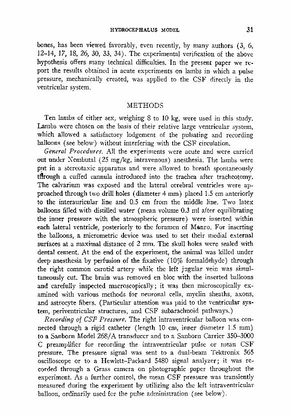
FIG. 1. Schematic drawing of the experimental arrangement for recording the h- traventricular CSF pressure, increasing the amplitude of the intraventricular CSF pulse pressure, and evaluating cerebral bulk compliance.
Increase in CSF Pulse Pressure. The left intraventricular balloon (Fig.
1) was connected via a rigid catheter (length 20 cm, inner diameter 1.5 mm) to an impulse pump as described in a previous paper (27). The electrocardiogram (ECG) was recorded (two bipolar leads) and amplified by a Grass preamplifier to obtain a stable R wave, after eliminating lower and higher frequencies. The ECG input was connected to the trigger circuit and the R wave was used for triggering the electromagnet of the impulse pump. The duration and amplitude of the mechanical pulse so obtained could be varied by the control circuit of the electromagnet power supply. The summation in phase of the artificial pulse and the physiologic intra- ventricular pulse was obtained by using a delaying circuit.
The mechanical pulse pressure was applied for a continuous period of time (3 h) in five animals in which the pulsatile and recording balloons had been inserted into the lateral ventricles and in two animals in which the pulsating balloons had been placed, respectively, above and below the lateral ventricle. In three more animals, the balloons were placed intra- ventricularly ; the left balloon was kept inflated for 3 h and did not receive mechanical pulses ; these animals were used as controls.
RESULTS
Combination of Physiological and Mechanical Pulses. The insertion of the pulsating balloon into the cerebral lateral ventricle was followed by a transient increase in mean CSF pressure, which was compensated in a
HYDROCEPHALUS MODEL 33
few minutes (3 to 5 min). By varying the intensity, the duration, as well as the delay of the discharge furnished by the power unit of the impulse pump, the combination of the physiological and mechanical pulse gave origin to different types of waves.
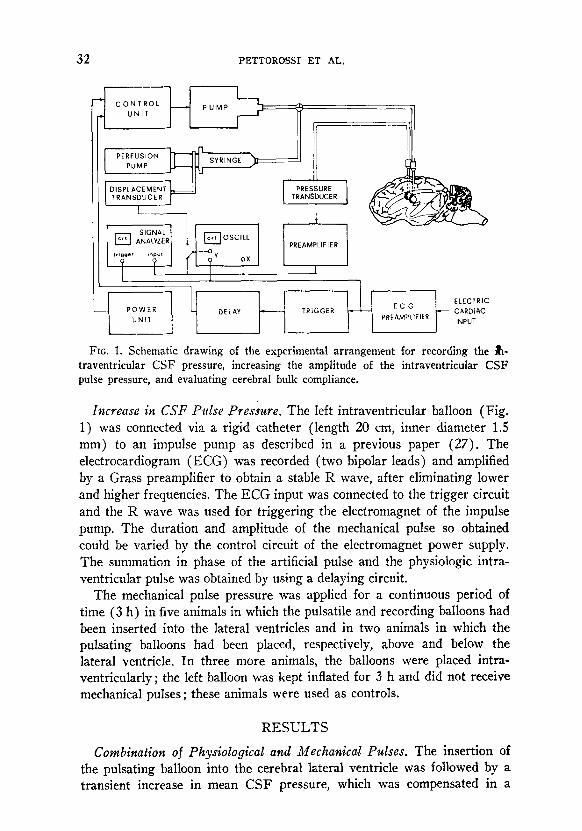
When maintaining the intensity and the duration constant, significant differences were recorded following variations in delay. In fact both the amplitude and the shape of the resulting wave differed considerably, whether or not the mechanical pulse was in phase with the physiological one (Fig. 2). Maximal constructive interference occurred when the delay of the impulse pump was adjusted to values between 60 and 100 ms from
FIG. 2. Variations in the amplitude and shape of the pressure wave resulting from the combination of physiological CSF intraventricular (A) and mechanical pulses, when the delay of the latter (black dots) is modified (B-H). Maximal summation occurs with the two waves in phase (E, F). ICP, intraventricular CSF pressure ; ECG electrocardiogram.
34 PETTOROSSI ET AL.
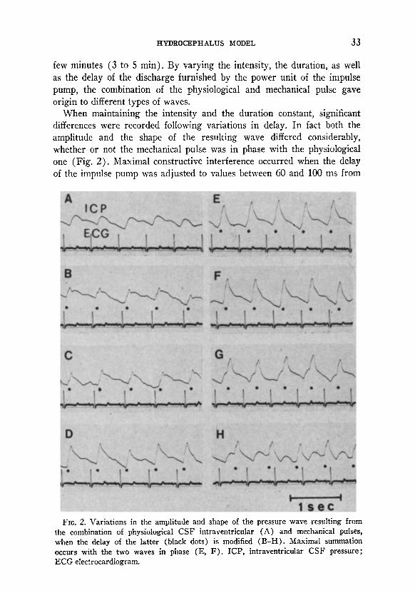
FIG. 3. Effects of the mechanical pulses of different types on the mean intraventricu- lar CSF pressure (ICP). A-The upper trace shows a slight increase in the mean CSF pressure induced (arrow) by longer-duration mechanical pulses (type A’). The increase can be compensated for by subtracting fluid from the pulsating balloon (dotted line). Lower trace electrocardiogram. B-No modification in the mean ICP (arrow) under short-duration mechanical pulses (type B’). A’ and B’-Each wave represents 32 averaged pulses of the CSF pressure. 1, Normal physiological CSF pulses; 2, pulses resulting from the combination of physiological and mechanical pulses.
the R wave of the ECG. The actual delivery of the mechanical pulse at the cerebral ventricular level occurred after a further interval of 40 ms, be- cause of the inertial and dissipative effects in the pump and the pulse transmission system. It is worth noting that such constructive interfer- ence recorded with the two pulses in phase was still observed when dis- placing the recording balloon frontally and occipitally in the contralateral ventricle, as was evidenced in two animals at the end of the experiments. When the pulses were exactly in phase, their constructive interaction re- sulted in an almost perfect summation of the respective amplitudes (Fig. 2). In this instance, modification of the shape of the resulting wave could be achieved by varying the duration of the artificial pulse. In fact, two types of waves were obtained, the first (type A) characteristically related to a longer duration and the second (type B) to a shorter duration (Fig. 3). In the first type the generated increase in pressure was such that the pressure of the resulting wave was maintained constantly above the pres- sure of the physiological pulse, whereas in the second type, a pressure level inferior to the physiological level was obtained in the descending phase of the pressure wave. The effects on mean CSF pressure of the application of the mechanical pulses of different duration are quite dissimilar. When a resulting pulse with a type A shape is utilized, the mean CSF pressure shows a slight but significant increase (which can be compensated by sub- tracting fluid from the pulsating balloon) (Fig. 3, dotted line). On the
HYDROCEPHALUS MODEL 35
contrary, when using a type B-shape wave, the mean CSF does not undergo any variation.
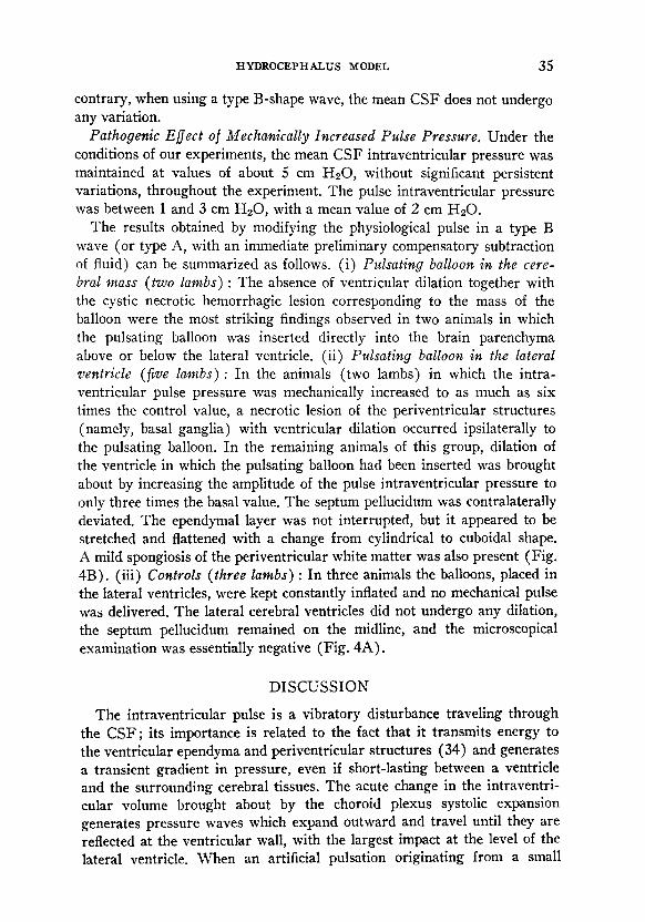
Pathogenic Efect of Mechanically Increased Pulse Pressure. Under the conditions of our experiments, the mean CSF intraventricular pressure was maintained at values of about 5 cm HzO, without significant persistent variations, throughout the experiment. The pulse intraventricular pressure was between 1 and 3 cm H20, with a mean value of 2 cm HsO.
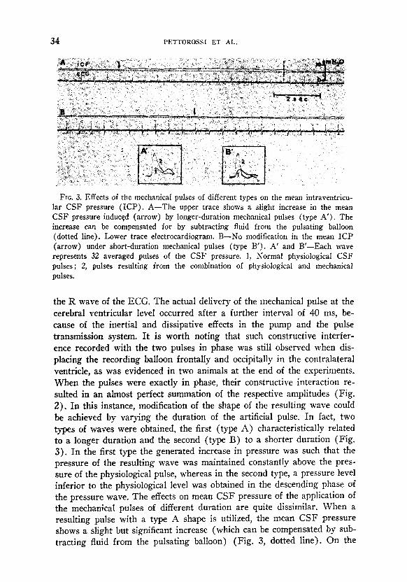
The results obtained by modifying the physiological pulse in a type B wave (or type A, with an immediate preliminary compensatory subtraction of fluid) can be summarized as follows. (i) Pulsating balloon in the cere- bral lnass (MO lambs) : The absence of ventricular dilation together with the cystic necrotic hemorrhagic lesion corresponding to the mass of the balloon were the most striking findings observed in two animals in which the pulsating balloon was inserted directly into the brain parenchyma above or below the lateral ventricle. (ii) Pulsating balloon in the lateral ventricle (five lambs) : In the animals (two lambs) in which the intra- ventricular pulse pressure was mechanically increased to as much as six times the control value, a necrotic lesion of the periventricular structures (namely, basal ganglia) with ventricular dilation occurred ipsilaterally to the pulsating balloon. In the remaining animals of this group, dilation of the ventricle in which the pulsating balloon had been inserted was brought about by increasing the amplitude of the pulse intraventricular pressure to only three times the basal value. The septum pellucidum was contralaterally deviated. The ependymal layer was not interrupted, but it appeared to be stretched and flattened with a change from cylindrical to cuboidal shape. A mild spongiosis of the periventricular white matter was also present (Fig. 4B). (iii) Controls (three lambs) : In three animals the balloons, placed in the lateral ventricles, were kept constantly inflated and no mechanical pulse was delivered, The lateral cerebral ventricles did not undergo any dilation, the septum pellucidum remained on the midline, and the microscopical examination was essentially negative (Fig. 4A).
DISCUSSION
The intraventricular pulse is a vibratory disturbance traveling through the CSF ; its importance is related to the fact that it transmits energy to the ventricular ependyma and periventricular structures (34) and generates a transient gradient in pressure, even if short-lasting between a ventricle and the surrounding cerebral tissues. The acute change in the intraventri- cular volume brought about by the choroid plexus systolic expansion generates pressure waves which expand outward and travel until they are reflected at the ventricular wall, with the largest impact at the level of the lateral ventricle. When an artificial pulsation originating from a small
36 PETTOROSSI ET AL.
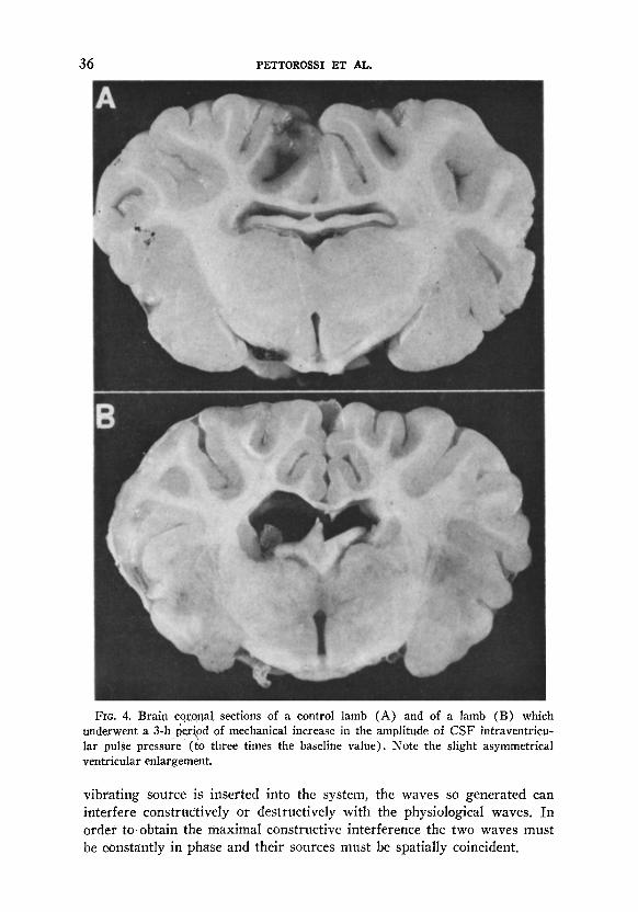
FIG. 4. Brain coro,qal sections of a control lamb (A) and of a lamb (B) which underwent a 3-h period of mechanical increase in the amplitude of CSF intraventricu- lar pulse pressure’ ((0 three times the baseline value). Note the slight asymmetrical ventricular enlargement.
vibrating source is inserted into the system, the waves so generated can interfere constructively or destructively with the physiological waves. In order to obtain the maximal constructive interference the two waves must be constantly in phase and their sources must be spatially coincident.

HYDROCEPHALUS MODEL 37
Two further criteria must be taken into account. The first is that the artificial pulse source by itself as well as the originated artificial pulses do not cause variations in the mean CSF pressure. The second criterion is re- lated to the shape of the intraventricular pulse wave. In fact, theoretically,
for the same increase in volume, a more rapid increase in pressure cor- responds to a major impact of the pressure wave on absorbing and dampen- ing structures.
In our opinion, the experiments we designed fulfill the theoretical re- quirements for the verification of a pathogenic role for intraventricular CSF pulsations in the development of ventricular dilation. In fact, (i) no sig- nificant changes in the mean CSF pressure have been observed following the insertion and the rhythmic expansion-deflation of the pulsating balloon into the ventricular system, and (ii) a maximal increase in the CSF pulse pressure for a determined increase in volume of the pulsating balloon was consistently obtained because of the continuous summation in phase of the mechanical and physiological pulses. Furthermore, when the mechanical and physiological pulses were perfectly in phase, the amplitude of the com- bined wave resulted from the summation of their respective amplitudes (Fig. 2). In other words, the mechanical pulse was such as not to inter- fere with the choroid plexus’s physiological pulsation. A combined wave, similar in shape to the physiological prominent “arterial” CSF pulse wave, such as that recorded following the abrupt decrease in carotid or systemic arterial resistance (S), was obtained by applying a short-duration me- chanical pulse.
As far as the pathogenic effect of the pulse CSF pressure is concerned, the results obtained in our experimental conditions allow the following conclusions : (i) The increase in the amplitude of the CSF pulse pressure, even if induced for a relatively short period (3 h), is evidently followed by significant neuropathologic lesions ; (ii) the characteristics of the lesions, their extent, as well as their location, are directly related to the amplitude of the pulse pressure and to the site where the source for mechanical pulses is inserted ; and (iii) for provoking a ventricular dilation the pulses must originate from within the ventricles.
REFERENCES
1. ADOLPH, R. J., H. FIJKUSUMI, AND N. 0. FOWLER. 1967. Origin of cerebrospinal fluid pulsations. AWL J. Physiol. 212: 840-846.
2. ANTONI, N. 1946. Pressure curves from the cerebrospinal fluid.. Acta Med. Stand. [Supfd.] 170 : 431-462.
3. BELLONI, G., C. DI Rocco, C. FOCACCI, G. GAL’LI, G. MAIRA, AND G. F. ROSSI. 1976. Surgical indications in normotensive hydrocephalus. A retrospective analysis of the relations of some diagnostic findings to the results of surgical treatment. Acta Neurochir. 33 : l-21.
38 PETTOROSSI ET AL.
4. BENABID, A. L., J. DE ROUGEMONT, AND M. BARGE. 1974. La pression intracranibnne. Etude thCorique. J. Physiol. (Paris) 68: 655-669.
5. BERING, E. A., JR. 1955. Choroid plexus and arterial pulsations of cerebrospinal fluid. Demonstration of the choroid plexuses as a cerebrospinal fluid pump. Arch. Neural. Psychiat. 73 : 165-172.
6. BERING, E. A., JR. 1962. Circulation of the cerebrospinal fluid. Demonstration of the choroid plexus as the generator of the force for flow of fluid and ventricular enlargement. I. Neurosurg. 19 : 405-413.
7. BERING, E. A., JR., AND F. D. INGRAHAM. 1953. Arterial pulsation of the cerebro- spinal fluid. Transations of the American Neurological Association, 78th Annual Meeting, pp. 49-54.
8. CALDARELLI, M., C. DI Rocco, R. MANCINELLI, V. E. PETTOROSSI, AND F. VELARDI. 1977. Studi sperimentali dei rapporti tra pressione e polso arteriosi e pressione intracranica. Riv. Neural. (in press).
9. COHEN, I., I. M. LEVINGER, AND M. HERZBERG. 1970. Haemodynamic factors affect- ing the cerebrospinal fluid pressure in the rabbit. Life Sci. 9: 569-576.
IO. DARDENNE, G., A. DEREYMAEKER, AND J. M. LACHERON. 1969. Cerebrospinal fluid pressure and pulsatility. An experimental study of circulatory and respira- tory influences in normal and hydrocephalic dogs. Eur. Neurol. 2: 193-216.
11. DEREYMAEKER, A., A. STEVENS, J. J. ROMBOUTS, J. LACHERON, AND A. PIERQUIN. 1971. Study of the influence of the arterial pressure upon the morphology of cisternal CSF pulsations. Eur. Neurol. 5 : 107-l 14.
12. Dr ROCCO, C., M. CALDARELLI, G. MAIRA, AND G. F. Rossr. 1977. The studies of cerebrospinal fluid dynamics in apparently arrested hydrocephalus in children. Child’s Brain 3 : 359-374.
13. DI Rocco, C., G. MAIRA, G. F. ROSSI, AND A. VIGNATI. 1976. Cerebrospinal fluid pressure studies in normal pressure hydrocephalus and cerebral atrophy. Eur. Neural. 14 : 119-128.
14. DI Rocco, C., D. G. MCLONE, T. SHIMOJI, AND A. J. RAIMONDI. 1975. Con- tinuous intraventricular cerebrospinal fluid pressure recording in hydrocephalic children during wakefulness and sleep. J. Neurosurg. 42: 683-689.
15. Du BOULAY, G. H. 1966. Pulsatile movements in the CSF pathways. Br. J. Radiol. 39 : 255-262.
16. DUNBAR, H. S., T. C. GUTWRIE, AND B. KARPELL. 1966. A study of the cerebro- spinal fluid pulse wave. Arch. Neural. 14: 624-630.
17. EKBOM, K., T. GREITZ, M. KALMER, J. LOPEZ, AND S. OTTOSSON. 1%9. Cere- brospinal fluid pulsations in occult hydrocephalus due to ectasia of basilar artery. Acta Neurochir. 20 : 1-S.
18. GARDNER, W. J. 1973. The Dysraphic States. Exccrpta Xedica, Amsterdam. 19. GERLACH, J. 1952. Zerebraler Grenzdruck und Hirnpuls. Acta Neurochir. 2: 120-
158. 20. GROTE, W. 1964. Gehirnpulsationen und Liquordynamik. Acta Neurochir. [Su~pl.]
12: l-130. 21. HAMER, J., E. ALB~XTI, AND S. HOYER. 1974. Effects of hypoxaemia, hypercapnia
and changes in cerebral perfusion pressure on mean cerebrospinal fluid and sagittal sinus pressure. Acta Neurochir. 30: 167-179.
22. HAMER, J., E. ALBJIRTI, S. HOYER, AND K. WIEDEMANN. 1977. Influence of systemic and cerebral vascular factors on the cerebrospinal fluid pulse waves. J. Neurosurg. 46: 36-45.
HYDROCEPHALUS MODEL 39
23. HAMIT, H. F., A. C. BEALL, JR., AND M. E. DEBAKEY. 1965. Haemodynamic in- fluences upon brain and cerebrospinal fluid pulsations and pressures. J. Trauma 5: 174-184.
24. HEMMER, R. 1960. Der Liquordruck. Thieme, Stuttgart. 25. LAITINEN, L. 1968. Origin of arterial pulsation of cerebrospinal fluid. Acta Neuvol.
Stand. 44 : 168-176. 26. MAIRA, G., C. DI Rocco, AND G. F. ROSSI. 1974. Pression intraventriculaire dans
l’hydrocephalie h pression “normale” pendant la veille et le sommeil. Neuro- chirurgie 20 : 462-467.
27. MANCINELLI, R., AND V. E. PETTOROSSI. 1977. A device for increasing the amplitude of the pulse pressure of cerebrospinal fluid. Physiol. Behav. 17: 719- 722.
28. MILHORAT, T. H. 1969. Choroid plexus and cerebrospinal fluid production. Science 166 : 1514-1516.
29. PARAICZ, E., AND J. VAJDA. 1975. Arterial and respiratory influences on intra- cranial pressure in infancy. Surg. Neural. 4: 361-366.
30. SATO, O., R. TSUGANE, AND N. KAGEYANA. 1975. Growing skull fractures of childhood. Child’s Brab 1: 148-156.
31. SYBAIAN, R. Q., P. C. BEGEMAN, A. I. KING, E. S. GURDJIAN, AND L. H. THOMAS. 1970. Experimental hydrocephalus. Ventricular cerebrospinal fluid pressure and waveform studies. Arch. Neural. 23: 165-172.
32. TURCHETTI, A. 1946. Le onde tensive de1 liquid0 cefalorachidiano in rapport0 all’attivith respiratoria e cardiocircolatoria. Boll. Sot. Ital. Biol. Sper. 22: 924- 927.
33. WEALTHALL, S. R. 1973. An investigation of the factors involved in relating ventricular size and the production of hydrocephalus. Dev. Med. Child Neural. [suppl.] 1s: l-11.
34. WILLIAMS, B. 1976. Cerebrospinal fluid pressure changes in response to coughing. Brain 99 : 331-346.
35. WILSON, C. B., AND V. BERTAN. 1967. Interruption of the anterior choroidal artery in experimental hydrocephalus. Arch. Neural. 17: 614-619.
Related Documents


![Management of subdural effusion and hydrocephalus ......hydrocephalus in patients with DC for TBI is 10 to 40% [10, 12, 13, 19]. SDE is defined as cerebrospinal fluid (CSF) accumula-tion](https://static.cupdf.com/doc/110x72/60f77946a97a3c60fd2cc41f/management-of-subdural-effusion-and-hydrocephalus-hydrocephalus-in-patients.jpg)