4/10/19 1 Common Apophyseal and Physeal injuries Shawn Spooner MD, FAAFP Family Medicine / Sports Medicine / Urgent Access UnityPoint Clinic, Urbandale Objectives Review anatomy of developing bones. Describe important clinical history differences for avulsion vs apophysitis Highlight specific apophyses/physes and treatment for apophysitis/epiphysitis Understand common apophyseal avulsion injuries and treatments Understand common epiphyseal injuries and treatments Skeletally Immature Clinical History What happened? Time frame of symptoms Where is the pain? Age of athlete Chronological vs skeletal

Welcome message from author
This document is posted to help you gain knowledge. Please leave a comment to let me know what you think about it! Share it to your friends and learn new things together.
Transcript
4/10/19
1
Common Apophyseal and Physeal injuries
Shawn Spooner MD, FAAFPFamily Medicine / Sports Medicine / Urgent Access
UnityPoint Clinic, Urbandale
Objectives� Review anatomy of developing bones.
� Describe important clinical history differences for avulsion vs apophysitis
� Highlight specific apophyses/physes and treatment for apophysitis/epiphysitis
� Understand common apophyseal avulsion injuries and treatments
� Understand common epiphyseal injuries and treatments
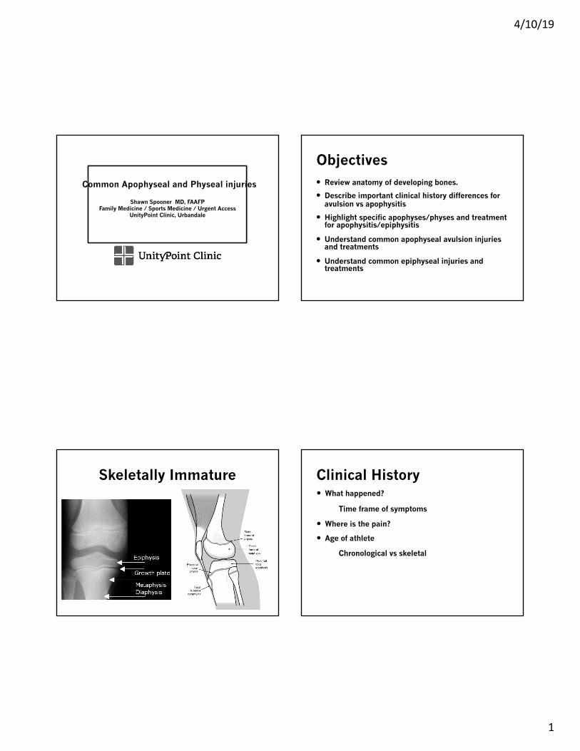
Skeletally Immature Clinical History� What happened?
Time frame of symptoms
� Where is the pain?
� Age of athlete
Chronological vs skeletal
4/10/19
2
Apophysitis� Osgood-Schlatter disease
� Sinding-Larsen-Johansson syndrome
� Sever’s disease
� Iselin’s disease
� Pelvic apophysitis
� Medial epicondyle, olecranon apophysitis
� Little League Elbow
General Treatment Approach - Apophysitis
� Activity Modification � Pain guided activity
� Symptomatic therapy� Icing, NSAIDs
� Stretching +/- physical therapy
� Generally self-limited
� Follow up if pain changes character/location, doesn’t improve with above treatment
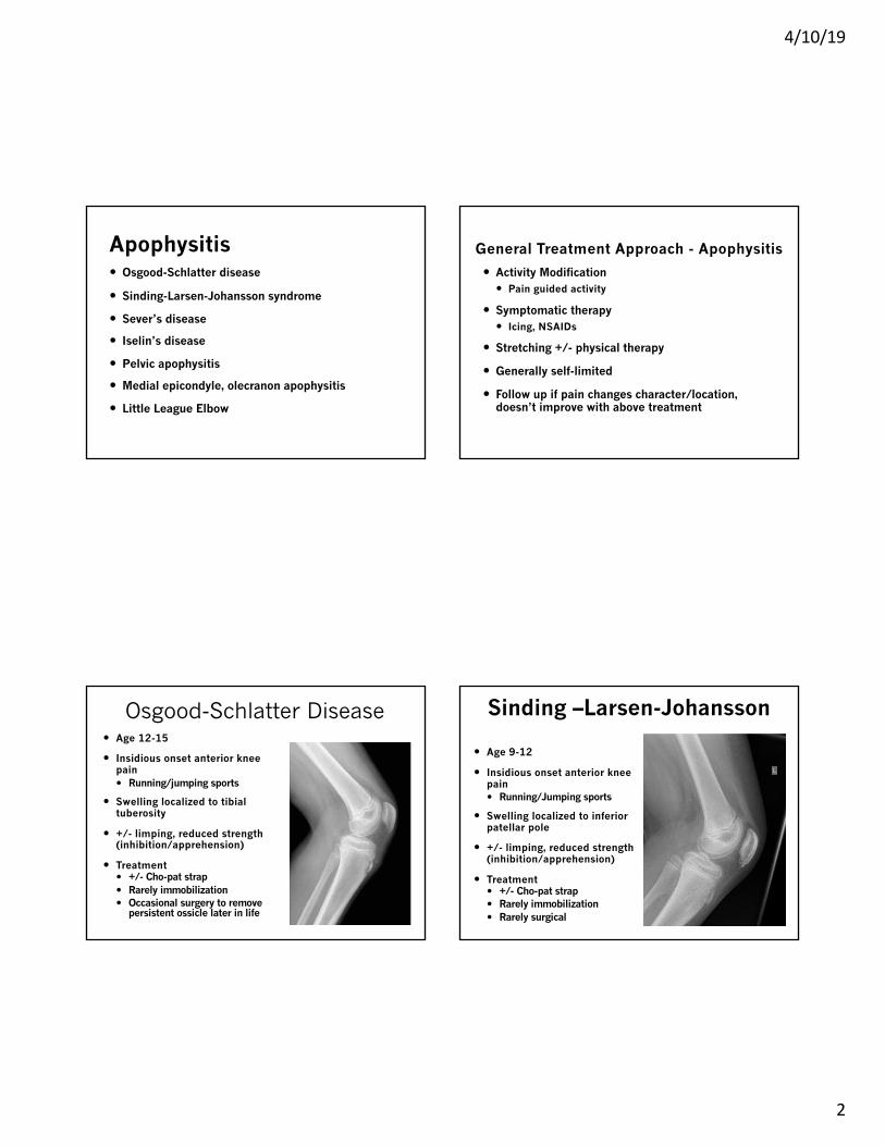
Osgood-Schlatter Disease� Age 12-15
� Insidious onset anterior knee pain� Running/jumping sports
� Swelling localized to tibialtuberosity
� +/- limping, reduced strength (inhibition/apprehension)
� Treatment� +/- Cho-pat strap� Rarely immobilization� Occasional surgery to remove
persistent ossicle later in life
Sinding –Larsen-Johansson
� Age 9-12
� Insidious onset anterior knee pain� Running/Jumping sports
� Swelling localized to inferior patellar pole
� +/- limping, reduced strength (inhibition/apprehension)
� Treatment� +/- Cho-pat strap� Rarely immobilization� Rarely surgical

4/10/19
3
Sever’s disease� Ages 8-13
� Insidious onset heel pain� Cleated/barefoot sports� Dynamic sports,
run/stop/turn
� Limping, toe walking
� Treatment:� Relative rest � Activity modification� Heel cups� Rarely immobilization
Iselin’s Disease� Ages 9-12
� Insidious onset lateral foot pain� Recent footwear change� Activity on uneven surface
� Swelling over 5th MT base
� +/- Limping
� Treatment: � Rest� Immobilization if needed� Footwear modification/arch supports
5th MT Apophysis
4/10/19
4
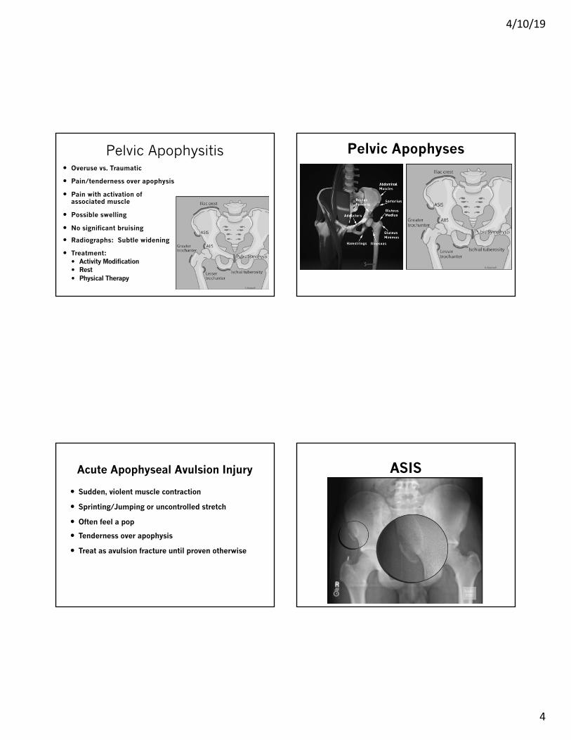
Pelvic Apophysitis� Overuse vs. Traumatic
� Pain/tenderness over apophysis
� Pain with activation of associated muscle
� Possible swelling
� No significant bruising
� Radiographs: Subtle widening
� Treatment: � Activity Modification� Rest� Physical Therapy
Pelvic Apophyses
Acute Apophyseal Avulsion Injury
� Sudden, violent muscle contraction
� Sprinting/Jumping or uncontrolled stretch
� Often feel a pop
� Tenderness over apophysis
� Treat as avulsion fracture until proven otherwise
ASIS

4/10/19
5
AIIS Ischial Apophyseal Avulsion
� Age 14-25
� Acute vs. Chronic
� Pain over origin of hamstrings
� Weakness and pain with resisted movement
� Passive stretching may cause pain
� Bruising may be present
Ischial Apophyseal Avulsion� Treatment:
� Majority treated conservatively� <2cm displacement or more� Better outcomes if treatment started early (<1 month from injury)
� Surgical management if large fragment and/or displaced > 2cm� Failed conservative treatment� High level athlete� Prolonged symptoms (>4 months) or symptomatic non-union
Ischial Apophyseal Avulsion� Conservative treatment
� Crutches until painless normal gait achieved� 2-4 weeks protected weight bearing
� Physical Therapy� Initial: gentle ROM and strengthening as pain resolves� 4-8 weeks: stretching and strengthening
� Return to sport after 8 weeks and asymptomatic
� Usually 8-12 weeks to return to full activity

4/10/19
6
Iliac Crest Avulsion� Rest
� 4-6 weeks
� Gradual Return
� Surgical� >3cm displaced
Tibial Tubercle Avulsion
� 12yo Male track athlete in 100m
� Feels “sharp blow” to front of shin during race
� Localized pain and swelling of anterior knee and tibial tubercle
� Unable to fully extend knee
Patellar Sleeve Fracture� 16yo male, football injury
� Struck on or falls on anterior knee
� TTP, swelling over patella, small effusion
� Unable to bear weight or extend knee
4/10/19
7
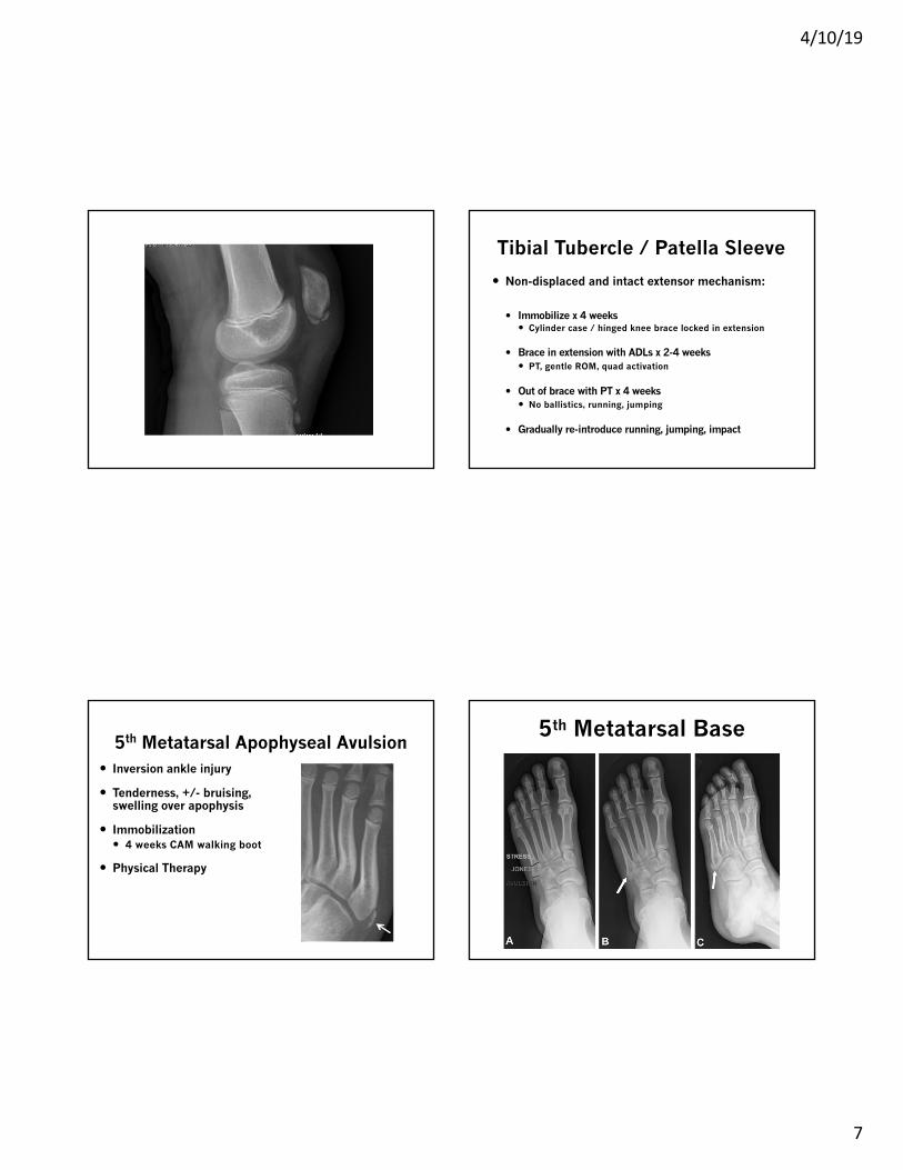
Tibial Tubercle / Patella Sleeve
� Non-displaced and intact extensor mechanism:
� Immobilize x 4 weeks� Cylinder case / hinged knee brace locked in extension
� Brace in extension with ADLs x 2-4 weeks� PT, gentle ROM, quad activation
� Out of brace with PT x 4 weeks� No ballistics, running, jumping
� Gradually re-introduce running, jumping, impact
5th Metatarsal Apophyseal Avulsion� Inversion ankle injury
� Tenderness, +/- bruising, swelling over apophysis
� Immobilization� 4 weeks CAM walking boot
� Physical Therapy
5th Metatarsal Base

4/10/19
8
Normal 5th MT Apophysis
� Apophysis and 5th base avulsion
More 5th MT • Normal apophysis• longitudinally oriented apophysis without fragmentation
• Base of 5th metatarsal fractures• transverse orientation of fracture fragment differentiates
it from nearby vertically oriented apophysis
• may be difficult to differentiate in adolescents
• Os vesalianum• intratendinous accessory bone within the substance of
the peroneus brevis• often incidental but can be associated with pain
Summary 5th MT
4/10/19
9
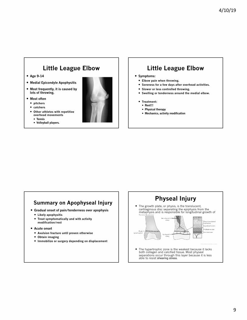
Little League Elbow� Age 9-14
� Medial Epicondyle Apophysitis
� Most frequently, it is caused by lots of throwing.
� Most often � pitchers � catchers
� Other athletes with repetitive overhead movements� Tennis � Volleyball players.
Little League Elbow� Symptoms:
� Elbow pain when throwing.� Soreness for a few days after overhead activities.
� Slower or less controlled throwing.� Swelling or tenderness around the medial elbow.
� Treatment:� Rest!!!� Physical therapy� Mechanics, activity modification
Summary on Apophyseal Injury� Gradual onset of pain/tenderness over apophysis
� Likely apophysitis� Treat symptomatically and with activity
modification/rest
� Acute onset� Avulsion fracture until proven otherwise� Obtain imaging� Immobilize or surgery depending on displacement
Physeal Injury� The growth plate, or physis, is the translucent,
cartilaginous disc separating the epiphysis from the metaphysis and is responsible for longitudinal growth of long bones.
� The hypertrophic zone is the weakest because it lacks both collagen and calcified tissue. Most physealseparations occur through this layer because it is less able to resist shearing stress.

4/10/19
10
Physeal Injuries� Pediatric bones are more porous than adult bones
� Remodeling capacity is highest in younger children
� Growth plate fractures occur in children
� The physis is responsible for longitudinal growth of long bones
� Fracture patterns in children are different from that of adults
Physeal Injury� Fractures through the Physis
� Salter-Harris Classification
� Non-displaced, displaced, bony bar, growth arrest
Salter-Harris I� Accounts for 6% of all physeal injuries.
� Transverse fracture through the growth plate.
� In a type I separation, the epiphysis separates from the metaphysis. The plane of separation is horizontal.
� When the periosteum is torn, displacement may occur.
� They are usually misdiagnosed as sprains because little is seen on the x-ray.
� Healing is rapid for type I fractures, within 2-3 weeks of injury and problems are rare especially in sites such as the distal radius.
4/10/19
11
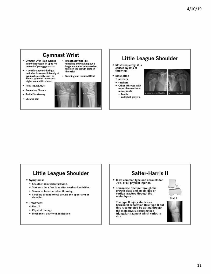
Gymnast Wrist� Gymnast wrist is an overuse
injury that occurs in up to 40 percent of young gymnasts.
� It usually appears during a period of increased intensity of gymnastic activity, such as when a gymnast moves to a higher competitive level.
� Rest, Ice, NSAIDs
� Premature Closure
� Radial Shortening
� Chronic pain
� Impact activities like tumbling and vaulting put a large amount of compressive force on the growth plate in the wrist.
� Swelling and reduced ROM
Little League Shoulder� Most frequently, it is
caused by lots of throwing.
� Most often � pitchers � catchers � Other athletes with
repetitive overhead movements� Tennis � Volleyball players.
Little League Shoulder� Symptoms:
� Shoulder pain when throwing.� Soreness for a few days after overhead activities.
� Slower or less controlled throwing.� Swelling or tenderness around the upper arm or
shoulder.
� Treatment:� Rest!!!
� Physical therapy� Mechanics, activity modification
Salter-Harris II� Most common type and accounts for
75% of all physeal injuries.
� Transverse fracture through the growth plate and an oblique or vertical fracture through the metaphysis.
The type II injury starts as a horizontal separation (like type I) but this is completed by exiting through the metaphysis, resulting in a triangular fragment which varies in size.

4/10/19
12
Salter-Harris III
� Accounts for 8% of all physeal injuries.
� Transverse fracture through the growth plate and a vertical fracture through the epiphysis.
� More commonly seen in older children where the growth plates have started to close.
� It is a combination of a horizontal fracture line through the physis and a vertical fracture line which runs from the growth plate through the epiphysis to the articular surface.
Salter-Harris IV� Accounts for 10% of all physeal
injuries.
� Vertical fracture through all three components, metaphysis, physisand epiphysis.
� In type IV separations, the fracture line is vertical. It extends through four distinct tissues/areas:
Salter-Harris V� This injury is rare and difficult to see on x-
ray.
� Compression fracture or crushing of the growth plate.
� These injuries are almost always diagnosed retrospectively, when a growth arrest has occurred.
Management of Physeal Injuries
� Look for and define the exact lines of separation on good quality x-rays using multiple views
� Occasionally views of the opposite side may help
� Classify the injury using the Salter-Harris classification
� If not readily classifiable, consider CT, MRI and urgent referral to orthopedics
� The majority of type I and II injuries are treated by closed reduction and cast immobilization
� The majority of type III and IV injuries require ORIF

4/10/19
13
Physeal Complications� The majority of physeal injuries heal quickly and
recover fully. In a minority, growth disturbance or arrest may occur, and can result in deformity and impaired function.
� Physeal growth may be disturbed by:� Avascular necrosis
� Direct crushing (Salter-Harris type V)� The formation of a bony bar� Non-union
� Hyperemia (Don’t Ultrasound Physis)
References� Apophyseal Injuries; Julie Wilson, 8/5/17, Sports
Medicine Center, Childrens Hospital Colorado
� Rockwood and Wilkin’s Fractures in Children; Chap 33
� The Royal Children’s Hospital Melbourne ; Fracture Education; https://www.rch.org.au/fracture-education/
� Brunker and Khan’s Clinical Sports Medicine, 4th ed.
Questions?
Related Documents