Chen et al. Journal of Biomedical Science 2010, 17:48 http://www.jbiomedsci.com/content/17/1/48 Open Access RESEARCH © 2010 Chen et al; licensee BioMed Central Ltd. This is an Open Access article distributed under the terms of the Creative Commons Attribution License (http://creativecommons.org/licenses/by/2.0), which permits unrestricted use, distribution, and reproduction in any medium, provided the original work is properly cited. Research Commensal microflora induce host defense and decrease bacterial translocation in burn mice through toll-like receptor 4 Lee-Wei Chen* †1,2 , Wei-Jung Chang 2 , Pei-Hsuan Chen 2 and Ching-Mei Hsu *†3 Abstract Background: Major burn is associated with decreased gut barrier function and increased bacterial translocation (BT). This study is to investigate whether commensal microflora induce host defense and decrease BT in burn mice. Methods: First, we treated Wild type (WT) mice with antibiotics in drinking water for 4 weeks to deplete gut commensal microflora. At week 3, drinking water was supplemented with lipopolysaccharide (LPS); a ligand for TLR4, to trigger TLRs in gut. The intestinal permeability, glutathione level, NF-κB DNA-binding activity, TLR4 expression of intestinal mucosa, BT to mesenteric lymph nodes (MLNs), and bacterial killing activity of peritoneal cells were measured after thermal injury. Second, lung of animals were harvested for MPO activity and TNFα mRNA expression assay. Third, WT animals were treated with oral antibiotics with or without LPS supplement after burn. At 48 hr after burn, TLR4 expression of intestinal mucosa and bacterial killing activity of cells were examined. Finally, bacterial killing activity and BT to MLNs after thermal injury in C3H/HeJ (TLR4 mutant) mice were measured. Results: Burn induced BT to MLNs in WT mice. Commensal depletion decreased TLR4 expression as well as NF-κB activation of intestine, myeloperoxidase (MPO) activity as well as TNFα expression of lung, and bacterial killing activity of peritoneal cells. Oral LPS supplement markedly reduced 81% of burn-induced BT and increased TLR4 expression, MPO activity of lung, as well as bacterial killing activity of peritoneal cells. LPS supplement did not change BT or bacterial killing activity in C3H/HeJ mice. Conclusions: Collectively, commensal microflora induce TLR4 expression of intestine and bacterial killing activity of inflammatory cells in burn. TLR4 ligand increases bacterial killing activity and decreases burn-induced BT. Taken together with the abolition of LPS effect in TLR4 mutant mice, we conclude that commensal microflora induce host defense and decrease bacterial translocation in burn mice through toll-like receptor 4. Background The human intestines are colonized by trillions of micro- organisms, including hundreds of different species of bacteria and viruses [1]. These microbes, collectively referred to as the commensal microflora, have an impor- tant role in human nutrition and health, by promoting nutrient supply, preventing pathogen colonization and shaping and maintaining normal mucosal immunity [2]. Major burn in humans and experimental animals is asso- ciated with a decrease in immune functions dependent upon T cells, the principal cells involved in initiating adaptive immune responses [3]. It is well accepted that clinical or experimental burn injury disrupts gut barrier function, allows dissemination of bacteria from the intes- tinal tract and facilitates the bacterial translocation (BT) to MLN, liver, and spleen [4,5]. The magnitude of BT is proportional to the severity of the burn injury [6]. Previ- ously, burn surgeons believed that feeding should not be started during the first 48 to 72 hours after burn or after injury because of theory that there was an obligatory pan- intestinal ileus. Today, there is overwhelming evidence that provision of enteral nutrients shortly after injury * Correspondence: [email protected], [email protected] 1 Institute of Emergency and Critical Care Medicine, National Yang-Ming University, Taipei, Taiwan 3 Department of Biological Sciences, National Sun Yat-Sen University, 70 Lien-Hai Road, Kaohsiung 804, Taiwan † Contributed equally Full list of author information is available at the end of the article

Welcome message from author
This document is posted to help you gain knowledge. Please leave a comment to let me know what you think about it! Share it to your friends and learn new things together.
Transcript
Chen et al. Journal of Biomedical Science 2010, 17:48http://www.jbiomedsci.com/content/17/1/48
Open AccessR E S E A R C H
ResearchCommensal microflora induce host defense and decrease bacterial translocation in burn mice through toll-like receptor 4Lee-Wei Chen*†1,2, Wei-Jung Chang2, Pei-Hsuan Chen2 and Ching-Mei Hsu*†3
AbstractBackground: Major burn is associated with decreased gut barrier function and increased bacterial translocation (BT). This study is to investigate whether commensal microflora induce host defense and decrease BT in burn mice.
Methods: First, we treated Wild type (WT) mice with antibiotics in drinking water for 4 weeks to deplete gut commensal microflora. At week 3, drinking water was supplemented with lipopolysaccharide (LPS); a ligand for TLR4, to trigger TLRs in gut. The intestinal permeability, glutathione level, NF-κB DNA-binding activity, TLR4 expression of intestinal mucosa, BT to mesenteric lymph nodes (MLNs), and bacterial killing activity of peritoneal cells were measured after thermal injury. Second, lung of animals were harvested for MPO activity and TNFα mRNA expression assay. Third, WT animals were treated with oral antibiotics with or without LPS supplement after burn. At 48 hr after burn, TLR4 expression of intestinal mucosa and bacterial killing activity of cells were examined. Finally, bacterial killing activity and BT to MLNs after thermal injury in C3H/HeJ (TLR4 mutant) mice were measured.
Results: Burn induced BT to MLNs in WT mice. Commensal depletion decreased TLR4 expression as well as NF-κB activation of intestine, myeloperoxidase (MPO) activity as well as TNFα expression of lung, and bacterial killing activity of peritoneal cells. Oral LPS supplement markedly reduced 81% of burn-induced BT and increased TLR4 expression, MPO activity of lung, as well as bacterial killing activity of peritoneal cells. LPS supplement did not change BT or bacterial killing activity in C3H/HeJ mice.
Conclusions: Collectively, commensal microflora induce TLR4 expression of intestine and bacterial killing activity of inflammatory cells in burn. TLR4 ligand increases bacterial killing activity and decreases burn-induced BT. Taken together with the abolition of LPS effect in TLR4 mutant mice, we conclude that commensal microflora induce host defense and decrease bacterial translocation in burn mice through toll-like receptor 4.
BackgroundThe human intestines are colonized by trillions of micro-organisms, including hundreds of different species ofbacteria and viruses [1]. These microbes, collectivelyreferred to as the commensal microflora, have an impor-tant role in human nutrition and health, by promotingnutrient supply, preventing pathogen colonization andshaping and maintaining normal mucosal immunity [2].Major burn in humans and experimental animals is asso-
ciated with a decrease in immune functions dependentupon T cells, the principal cells involved in initiatingadaptive immune responses [3]. It is well accepted thatclinical or experimental burn injury disrupts gut barrierfunction, allows dissemination of bacteria from the intes-tinal tract and facilitates the bacterial translocation (BT)to MLN, liver, and spleen [4,5]. The magnitude of BT isproportional to the severity of the burn injury [6]. Previ-ously, burn surgeons believed that feeding should not bestarted during the first 48 to 72 hours after burn or afterinjury because of theory that there was an obligatory pan-intestinal ileus. Today, there is overwhelming evidencethat provision of enteral nutrients shortly after injury
* Correspondence: [email protected], [email protected] Institute of Emergency and Critical Care Medicine, National Yang-Ming University, Taipei, Taiwan3 Department of Biological Sciences, National Sun Yat-Sen University, 70 Lien-Hai Road, Kaohsiung 804, Taiwan † Contributed equally
© 2010 Chen et al; licensee BioMed Central Ltd. This is an Open Access article distributed under the terms of the Creative CommonsAttribution License (http://creativecommons.org/licenses/by/2.0), which permits unrestricted use, distribution, and reproduction inany medium, provided the original work is properly cited.
Full list of author information is available at the end of the article
Chen et al. Journal of Biomedical Science 2010, 17:48http://www.jbiomedsci.com/content/17/1/48
Page 2 of 15
alters gut flora and protects the immuno-compromise,stressed, or thermally injured patients through unknownmechanism [7,8]. Defining the relationship between com-mensal microflora and gut barrier function is warrantedto characterize the beneficial effect of early enteral feed-ing in burn patients.
The innate immune system detects the invasion ofmicroorganism through toll-like receptors (TLRs), whichrecognize microbial components and trigger inflamma-tory responses. TLRs comprise a family of pattern-recog-nition receptors that detect conserved molecularproducts of microorganisms [9,10]. TLRs function as sen-sors of microbial infection and are critical for the initia-tion of inflammatory and immune defense responses.TLR2 and TLR4, have been shown to be essential for therecognition of distinct bacterial cell wall components.TLR2 discriminates peptidoglycan (PGN), lipoprotein,lipoarabinomannan (LAM) and zymosan, whereas TLR4recognizes lipopolysaccharide (LPS), lipoteichoic acid(LTA) and Taxol [11]. A major downstream effect of TLRsignaling is the activation of the transcription factor NF-κB, which is required for expression of many genesrelated to innate immunity and inflammation [12]. Previ-ous paper indicated that toll-like receptors played crucialroles in the maintenance of intestinal epithelial homeo-stasis [13]. The bacterial ligands recognized by TLRs arenot unique to pathogens, but rather are shared by entireclasses of bacteria, and are produced therefore by com-mensal microorganisms as well [13]. However, it is stillnot decided whether oral supplement TLR4 ligand couldenhance host defense in burn or not.
The role of commensal microflora in maintaininginnate immunity after thermal injury has not been wellcharacterized. Also, effect of TLR4 ligand on the bacterialkilling activity of inflammatory cells after burn has notbeen examined. We hypothesized that commensal micro-flora in gut maintained host defense against bacterialchallenge and TLR4 ligand decreased burn-induced BTin burn. Using a commensal depletion model in wild-typeand TLR4 mutant mice, we demonstrated that both com-mensal microflora and oral TLR4 ligand enhanced thegut barrier function in burn through the increase of toll-like receptor 4 expression. In the future, enteral feeding ofTLRs ligand could be a feasible way to increase hostdefense and gut barrier function in burn or major traumapatients.
MethodsAnimalsSpecific pathogen-free male C3H/HeN (wild-type, WT),weighing between 20 and 25 g were obtained from theNational Laboratory Breeding and Research Center(NLBRC, Taipei, Taiwan). C3H/HeJ (TLR4 mutant) mice
were purchased from The Jackson Laboratory (Bar Har-bor, ME). C3H/HeJ mice have been demonstrated to havea missense mutation in the third exon of TLR4, yielding anonfunctional TLR4 [14]. All animal procedures were incompliance with regulations on animal used for experi-mental and other scientific purposes approved by theNational Sun Yat-Sen University Animal ExperimentsCommittee.
Experimental designExperiment 1To evaluate the role of commensal microflora on thermalinjury-induced intestinal barrier dysfunction, WT micewere fed with vehicle or oral antibiotics for 4 wks todeplete the intestinal commensals with or without LPSsupplements in drinking water (10 μg/μl) at week 3. Wildtype (WT) mice were randomly divided into four shamgroups (control, LPS, antibiotics, antibiotics + LPS) (n =6) and four burn groups (control, LPS, antibiotics, antibi-otics + LPS) (n = 6 in each). The sham group was sub-jected to sham treatment and the burn groups weresubjected to a 30 - 35% total body surface area (TBSA)burn injury. All animals received sterile saline (50 ml/kgi.p.) for fluid resuscitation right after burn or sham treat-ment. At 24 hr after burn, mesenteric lymph nodes wereharvested for bacterial translocation assay. Also, the dis-tribution of fluorescein isothiocyanate-dextran (FITC-dextran) across the lumen of small intestine in animalsunder anesthesia (ketamine and xylazine) was measuredat 24 hr after injury to assess the intestinal permeability.In another experiment, the GSH level of the intestinalmucosa in animals with the same quantity and treatmentwas measured to assess the peroxidation produced afterinjury. Mid-ileum tissues were harvested for TLR4 immu-nohistochemical studies. In another experiment, theintestinal mucosa was harvested for NF-κB DNA-bindingactivity, TLR4 mRNA and protein expression assay at 8 hrafter burn injury.Experiment 2To evaluate the role of commensal microflora on thermalinjury-induced neutrophil deposition and cytokinesexpression in lung, WT mice were randomly divided intofour sham groups (n = 6) and four burn groups (n = 6 ineach) as experiment 1. The animals were sacrificed at 8 hrafter burn, and lung tissue was harvested for MPO activ-ity. In another experiment, the lung tissue was harvestedfor the assay of TLR2, TLR4, and TNFα mRNA expres-sion at 8 hr after injury.Experiment 3Most major burn patients suffered from ileus andreceived combined antibiotics treatment to prevent sep-sis [15,16]. To evaluate the effect of antibiotics treatmentwith or without LPS supplement in the thermal injury-
Chen et al. Journal of Biomedical Science 2010, 17:48http://www.jbiomedsci.com/content/17/1/48
Page 3 of 15
induced bacterial translocation, WT mice were randomlydivided into one sham burn groups (n = 6) and three burngroups (burn, antibiotics, antibiotics + LPS) (n = 6 ineach). The sham group was subjected to sham treatmentand oral saline feeding. The burn group was subjected toburn treatment and oral saline feeding. The antibioticsgroup was subjected to oral antibiotics administrationafter burn. The antibiotics + LPS group was subjected toLPS supplements (10 μg/μl) in oral antibiotics adminis-tration after burn. At 48 hr after burn or sham burn, peri-toneal cells as well as bone marrow cells were harvestedfor bacterial killing activity, mesenteric lymph nodes wereharvested for bacterial translocation, and intestinalmucosa was harvested for TLR4 mRNA assay.Experiment 4C3H/HeJ mice were randomly divided into three shamgroups (n = 6) and three burn groups (n = 6 in each) asexperiment 1. At 8 hr after thermal injury, lung was har-vested for TLR4 mRNA assay, intestinal mucosa was har-vested for TLR4 mRNA assay, and peritoneal cells wereharvested from the abdominal cavity for bacterial killingactivity and TLR4 as well as TNFα mRNA expressionassay.
Thermal InjuryThe thermal injury procedures were modified from thosedescribed by Walker et al [14]. Briefly, animals were anes-thetized intraperitoneally with ketamine (80 mg/kg) andxylazine (10 mg/kg), and a marked area of the shaved dor-sal skin was exposed from a wooden template andimmersed in 95°C water for 10 sec. This procedure pro-duced a 30 - 35% TBSA burn of the mice. Total body sur-face area was calculated using murine-specific data [17]and average 40 to 48 cm2 for mice of the weight used. Theburn injury caused 8% mortality within the first 4 hrsafter burn. Nonsurviving animals were excluded from thesubsequent study. The sham control animals were anes-thetized, shaved and maintained in identical settingsexcept that room temperature water was used for immer-sion.
Depletion of gut commensal microflora and reconstitution of commensal-depleted animals with TLR ligandsCommensal bacterial products have been known toengage TLRs and confer protection against dextran sul-fate sodium (DSS)-induced intestinal epithelial injury[13]. Animals were provided ampicillin (A; 1 g/L; Sigma),vancomycin (V; 500 mg/L; Abott Labs), neomycin sulfate(N: 1 g/L; Pharmacia/Upjohn), and metronidazole (M; 1g/L; Sidmack Labs) in drinking water for four weeks. Pre-viously, a four-week oral administration of vancomycin,neomycin, metronidazole, and ampicillin with the samedose described above in mice has been proved to depleteall detectable commensals [13]. Previously, this oral anti-
biotics protocol has no significant effect on nutrition andsystemic effect [13,18]. To those animals receiving LPS,drinking water was supplemented with 10 μg/μl of puri-fied E. coli 026:B6 LPS (Sigma) at week 3 and continuedin drinking water for the duration of sham treatment orthermal injury. LPS, a membrane constituent of gram-negative bacteria, was the best-studied TLR ligand andwas recognized by TLR4 and MD-2, a molecule associ-ated with the extracellular domain of TLR4 [19].
Quantification of intestinal permeabilityThe assay of intestinal permeability was modified fromthe method described by Otamiri et al. [20]. A 5-cm seg-ment of the jejunum and proximal ileum was dissectedwith the beginning at 5 cm distal to the ligament of Treitzwith well protected superior mesenteric vessels. Thebilateral end of the isolated intestine was clamped withrubber bands to prevent the leakage of FITC-dextran. 200μl of 0.1 M phosphate buffer saline (pH 7.2) containing 25mg of FITC-dextran (MW 4,400, Sigma) was injected intothe lumen. After 30 min, blood sample (100 μl) was takenby a puncture of the portal vein and immediately dilutedwith 1.9 ml of 50 mM Tris (pH 10.3) containing 150 mMNaCl. The diluted plasma was centrifuged at 4°C, 3,000 gfor 7 min and the supernatant was analyzed for FITC-dextran concentration with a fluorescence spectropho-tometer (Hitachi, F-2000) at the excitation wavelength of480 nm and the emission wavelength of 520 nm. Standardcurves for calculating the FITC-dextran concentration inthe samples were obtained by diluting various amounts ofFITC-dextran in a pool of mice plasma, then diluted andcentrifuged in the same manner as the samples beforemeasurement.
Determination of glutathione (GSH) levelThe intestinal mucosa glutathione level was quantitatedby the fluorescence probe o-phthalaldeyde (sigma) whichcan react with GSH and has high quantum yield. Mix1.89ml of 50 mM potassium phosphate buffer (pH8.0)with 10ul of supernatant obtained and add 100 μl of 1mg/ml o-phthalaldeyhyde (freshly prepared in absolute meth-anol). The samples were incubated at room temperaturefor 15 min and fluorescence was measured at an excita-tion wavelength of 350 nm and an emission wavelength of420 nm. The data was expressed as GSH content (mM).
Bacterial translocation to MLNThe collected mesenteric lymph nodes were weighed andhomogenized in 500 μl of sterile saline. Aliquots of thehomogenate from each tissue were plated onto TSB(Tryptic Soy Broth) agar plates (DIFCO, Detroit, Michi-gan, USA). The plates were examined after aerobic incu-bation at 37°C for 24 hr to determine whether commensaldepletion with or without LPS altered burn-induced
Chen et al. Journal of Biomedical Science 2010, 17:48http://www.jbiomedsci.com/content/17/1/48
Page 4 of 15
translocation. Representative colonies were expressed ascolony forming unit per gram of organ tissue (CFU/g tis-sue).
Determination of lung myeloperoxidase activityLung content of myeloperoxidase (MPO) was determinedto assess the degree of pulmonary neutrophil infiltration[21]. Mice were anesthetized and the thorax was openedwith median sternotomy. The bilateral lungs and heartwere harvested together and the pulmonary vasculaturewas cleared of blood by gentle injection of 10 ml sterilesaline into the right ventricle. The lungs were then blot-ted dry of surface blood and weighed.
Lung tissues was placed in 50 mM potassium phos-phate buffer (pH 6.0) with 0.5% hexadecyltrimethylam-monium bromide and homogenized. The homogenatewas sonicated on ice and centrifuged for 30 min at 3,000g, 4°C. An aliquot (0.1 ml) of supernatant was added to2.9 ml of 50 mM potassium phosphate buffer (pH 6.0)containing 0.167 mg/ml of O-dianisidine and 0.0005%hydrogen peroxide [22]. The rate of change in absorbanceat 460 nm was measured over 3 min. One unit of MPOactivity was defined as the amount of enzyme thatreduces 1 μmole of peroxide per min and the data wereexpressed as units per gram of lung tissue (Units/g tis-sue).
Polymerase chain reaction (PCR) and quantification of PCR productsTotal RNA was isolated from cells using TRIZOL reagent(Invitrogen, Life Technologies) as described previously[23]. Reverse transcription-generated cDNA encodingTLR2, TLR4, and TNFα genes were amplified using PCR.Sets of primers were designed according to those genesdocumented in GenBank. The sequences are 5'-AGTG-GGTCAAGGAACAGAAGCA-3' (sense) and 5'-CTT-TACCAGCTCATTTCTCACC-3' (antisense) for TLR4,5'-TCTGGGCAGTCTTGAACATTT-3' (sense) and 5'-AGAGTCAGGTGATGGATGTCG-3' (antisense) forTLR2, 5'-CAGCCTCTTCTCATTCCTGCTTGTG-3'(sense) and 5'-CTGGAAGACTCCTCCCAGGTATAT-3'(antisense) for TNFα, and 5' GTGGGCCGCTCTAGGCACCA3' (sense) and 5' CGGTTGGCCTTAGGGTTCAG3' (antisense) for β-actin gene as a control.
Bacterial killing activity of peritoneal cells and bone marrow cellsThe peritoneal cavity was washed with 5 ml PBS contain-ing 0.1% BSA and 10 mM EDTA. The peritoneal cellswere collected and resuspended in HBSS as 106 cells/ml.Bone marrow cells were harvested from bilateral femoraland tibial bone marrow. Red cells depletion was per-formed using erythrolysis. After 5 min of preincubation,
the cell suspension was incubated with E. coli (108/ml) at37°C for 1 h with shaking. The cells were removed as thepellet after centrifugation at 200 × g for 10 min, and E.coli number in the supernatant was counted [24,25].
Western immunoblotsProtein levels of TLR4 of intestinal mucosa were mea-sured by Western immunoblotting. Homogenized sam-ples (50 μg of protein each) were subjected to 12.5% SDS-PAGE under reducing conditions. Proteins were trans-ferred onto PVDF membranes (Millipore) by using aSemi-Dry Electrophoretic system (Bio-Rad). The TLR4was identified by rabbit monoclonal antibody (Cell Sig-naling Technology, Inc.). The membranes were incubatedwith the secondary antibody (Biotinylated anti-rabbitIgG) (Perkin-Elmer Life Science, Boston, USA) for 1 hr atroom temperature. Blots were developed by the ECLWestern blotting detection reagents (Perkin-Elmer).
TLR4 immunohistochemistryThe following antibodies were used for immunohis-tochemical stains: Rabbit monoclonal immunoglobulin Gto TLR4 (Cell Signaling Technology, Inc.), biotinylatedsecondary antibodies and peroxidase-conjugated strepta-vidin (Dako). The TLR4 antibody with a 1:300 dilutionwas used. Sections of the paraffin fixed mice lung (4 μmthickness) were deparaffinized with xylene and gradedethanol. For antigen retrieval in the TLR4 staining, sec-tions were soaked in a citrate buffer containing NP-40(pH 6.0, Sigma) and heated in a microwave oven (600W)for 10 min. Endogenous peroxidase was blocked with 2%hydrogen peroxide in 70% methanol for 10 min at roomtemperature. Sections were incubated with primary anti-bodies for 2 hr, biotinylated secondary antibodies for 20min at room temperature, and then subsequently pro-cessed by the avidin-biotin peroxidase complex methodwith 3-amino-9-ethylcarbazole (AEC) as the chromogen.Sections were lightly counterstained with Mayer's hema-toxylin and viewed under a light microscope. Negativecontrol sections were also incubated, but without pri-mary antibody.
Electrophoretic mobility shift assay for NF-κB and AP-1Nuclear extracts were prepared as described [26]. Intesti-nal mucosa were harvested in hypotonic buffer and pel-leted by centrifugation. The pellets were suspended innuclear extract buffer. After 15 min on ice the suspen-sions were centrifuged and the supernatants were trans-ferred to new tubes. The Bandshift kit (Promega Corp.Madison, WI) was used according to the manufacturer'sinstructions. Consensus and control oligonucleotides(Santa Cruz Biotechnology Inc.) were labeled by polynu-cleotides sequences included the AP-1 consensus (5' to 3')
Chen et al. Journal of Biomedical Science 2010, 17:48http://www.jbiomedsci.com/content/17/1/48
Page 5 of 15
(CGCTTGATGACTTGGCCGGAA) or the NF-κB con-sensus(5' to 3') (AGTTGAGGGGAC-TTTCCCAGGC)(1.75pmol/μl). After the oligonucleotide was radiola-beled, 5 μg of nuclear protein was incubated with 2 μg ofpoly(dI-dC) and 5,000-10,000 cpm of γ[32P]-ATP-labeledoligonucleotides. After 30 min at room temperature, thesamples were analyzed on a 4% polyacrylamide gel. Thegel was dried and visualized by autoradiography.
Statistical analysisValues are expressed as means SEs. Intergroup compari-sons were made using one-way ANOVA followed by Bon-ferroni correction. Statistical analysis was performed onPrism software (GraphPad). Data were expressed as mean± standard deviation of the mean in all figures, and p <0.05 is considered to be statistical significance. The bacte-rial count, MPO activity, FITC, glutathione level, andbacterial translocation between groups were assessedwith one-way analysis of variance (ANOVA), followed byScheffe's F test.
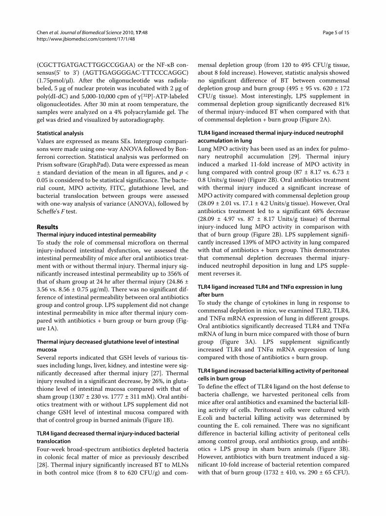
ResultsThermal injury induced intestinal permeabilityTo study the role of commensal microflora on thermalinjury-induced intestinal dysfunction, we assessed theintestinal permeability of mice after oral antibiotics treat-ment with or without thermal injury. Thermal injury sig-nificantly increased intestinal permeability up to 356% ofthat of sham group at 24 hr after thermal injury (24.86 ±3.56 vs. 8.56 ± 0.75 μg/ml). There was no significant dif-ference of intestinal permeability between oral antibioticsgroup and control group. LPS supplement did not changeintestinal permeability in mice after thermal injury com-pared with antibiotics + burn group or burn group (Fig-ure 1A).
Thermal injury decreased glutathione level of intestinal mucosaSeveral reports indicated that GSH levels of various tis-sues including lungs, liver, kidney, and intestine were sig-nificantly decreased after thermal injury [27]. Thermalinjury resulted in a significant decrease, by 26%, in gluta-thione level of intestinal mucosa compared with that ofsham group (1307 ± 230 vs. 1777 ± 311 mM). Oral antibi-otics treatment with or without LPS supplement did notchange GSH level of intestinal mucosa compared withthat of control group in burned animals (Figure 1B).
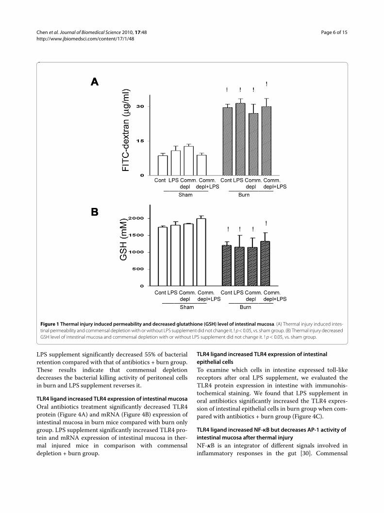
TLR4 ligand decreased thermal injury-induced bacterial translocationFour-week broad-spectrum antibiotics depleted bacteriain colonic fecal matter of mice as previously described[28]. Thermal injury significantly increased BT to MLNsin both control mice (from 8 to 620 CFU/g) and com-
mensal depletion group (from 120 to 495 CFU/g tissue,about 8 fold increase). However, statistic analysis showedno significant difference of BT between commensaldepletion group and burn group (495 ± 95 vs. 620 ± 172CFU/g tissue). Most interestingly, LPS supplement incommensal depletion group significantly decreased 81%of thermal injury-induced BT when compared with thatof commensal depletion + burn group (Figure 2A).
TLR4 ligand increased thermal injury-induced neutrophil accumulation in lungLung MPO activity has been used as an index for pulmo-nary neutrophil accumulation [29]. Thermal injuryinduced a marked 11-fold increase of MPO activity inlung compared with control group (87 ± 8.17 vs. 6.73 ±0.8 Units/g tissue) (Figure 2B). Oral antibiotics treatmentwith thermal injury induced a significant increase ofMPO activity compared with commensal depletion group(28.09 ± 2.01 vs. 17.1 ± 4.2 Units/g tissue). However, Oralantibiotics treatment led to a significant 68% decrease(28.09 ± 4.97 vs. 87 ± 8.17 Units/g tissue) of thermalinjury-induced lung MPO activity in comparison withthat of burn group (Figure 2B). LPS supplement signifi-cantly increased 139% of MPO activity in lung comparedwith that of antibiotics + burn group. This demonstratesthat commensal depletion decreases thermal injury-induced neutrophil deposition in lung and LPS supple-ment reverses it.
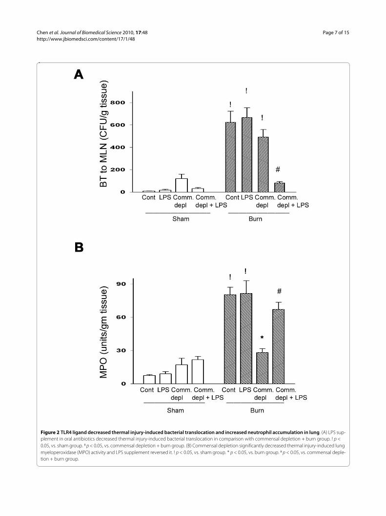
TLR4 ligand increased TLR4 and TNFα expression in lung after burnTo study the change of cytokines in lung in response tocommensal depletion in mice, we examined TLR2, TLR4,and TNFα mRNA expression of lung in different groups.Oral antibiotics significantly decreased TLR4 and TNFαmRNA of lung in burn mice compared with those of burngroup (Figure 3A). LPS supplement significantlyincreased TLR4 and TNFα mRNA expression of lungcompared with those of antibiotics + burn group.
TLR4 ligand increased bacterial killing activity of peritoneal cells in burn groupTo define the effect of TLR4 ligand on the host defense tobacteria challenge, we harvested peritoneal cells frommice after oral antibiotics and examined the bacterial kill-ing activity of cells. Peritoneal cells were cultured withE.coli and bacterial killing activity was determined bycounting the E. coli remained. There was no significantdifference in bacterial killing activity of peritoneal cellsamong control group, oral antibiotics group, and antibi-otics + LPS group in sham burn animals (Figure 3B).However, antibiotics with burn treatment induced a sig-nificant 10-fold increase of bacterial retention comparedwith that of burn group (1732 ± 410, vs. 290 ± 65 CFU).
Chen et al. Journal of Biomedical Science 2010, 17:48http://www.jbiomedsci.com/content/17/1/48
Page 6 of 15
Figure 1 Thermal injury induced permeability and decreased glutathione (GSH) level of intestinal mucosa. (A) Thermal injury induced intes-tinal permeability and commensal depletion with or without LPS supplement did not change it. ! p < 0.05, vs. sham group. (B) Thermal injury decreased GSH level of intestinal mucosa and commensal depletion with or without LPS supplement did not change it. ! p < 0.05, vs. sham group.
LPS supplement significantly decreased 55% of bacterialretention compared with that of antibiotics + burn group.These results indicate that commensal depletiondecreases the bacterial killing activity of peritoneal cellsin burn and LPS supplement reverses it.
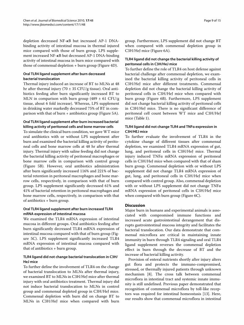
TLR4 ligand increased TLR4 expression of intestinal mucosaOral antibiotics treatment significantly decreased TLR4protein (Figure 4A) and mRNA (Figure 4B) expression ofintestinal mucosa in burn mice compared with burn onlygroup. LPS supplement significantly increased TLR4 pro-tein and mRNA expression of intestinal mucosa in ther-mal injured mice in comparison with commensaldepletion + burn group.
TLR4 ligand increased TLR4 expression of intestinal epithelial cellsTo examine which cells in intestine expressed toll-likereceptors after oral LPS supplement, we evaluated theTLR4 protein expression in intestine with immunohis-tochemical staining. We found that LPS supplement inoral antibiotics significantly increased the TLR4 expres-sion of intestinal epithelial cells in burn group when com-pared with antibiotics + burn group (Figure 4C).
TLR4 ligand increased NF-κB but decreases AP-1 activity of intestinal mucosa after thermal injuryNF-κB is an integrator of different signals involved ininflammatory responses in the gut [30]. Commensal
Chen et al. Journal of Biomedical Science 2010, 17:48http://www.jbiomedsci.com/content/17/1/48
Page 7 of 15
Figure 2 TLR4 ligand decreased thermal injury-induced bacterial translocation and increased neutrophil accumulation in lung. (A) LPS sup-plement in oral antibiotics decreased thermal injury-induced bacterial translocation in comparison with commensal depletion + burn group. ! p < 0.05, vs. sham group. # p < 0.05, vs. commensal depletion + burn group. (B) Commensal depletion significantly decreased thermal injury-induced lung myeloperoxidase (MPO) activity and LPS supplement reversed it. ! p < 0.05, vs. sham group. * p < 0.05, vs. burn group. # p < 0.05, vs. commensal deple-tion + burn group.
Chen et al. Journal of Biomedical Science 2010, 17:48http://www.jbiomedsci.com/content/17/1/48
Page 8 of 15
Figure 3 TLR4 ligand increased TNFα expression in lung and the bacterial killing activity of peritoneal cells. (A) Commensal depletion de-creased TLR4 as well as TNFα mRNA expression in lung and LPS supplement reversed them. The values represent the average fold increase (± s.d.) in mRNA expression in 3 different mice of each group. ! p < 0.05, vs. sham group. * p < 0.05, vs. burn group. # p < 0.05, vs. commensal depletion + burn group. (B) Commensal depletion induced a significant increase of bacterial retention of peritoneal cells in burn mice compared with that of burn group. LPS supplement significantly decreased 55% of bacterial retention compared with that of antibiotics + burn group. * p < 0.05, vs. burn group. # p < 0.05, vs. commensal depletion + burn group.
Chen et al. Journal of Biomedical Science 2010, 17:48http://www.jbiomedsci.com/content/17/1/48
Page 9 of 15
depletion decreased NF-κB but increased AP-1 DNA-binding activity of intestinal mucosa in thermal injuredmice compared with those of burn group. LPS supple-ment increased NF-κB but decreased AP-1 DNA-bindingactivity of intestinal mucosa in burn mice compared withthose of commensal depletion + burn group (Figure 4D).
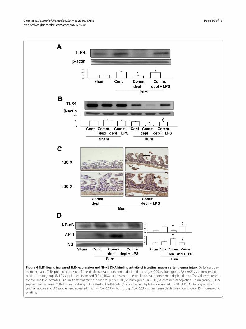
Oral TLR4 ligand supplement after burn decreased bacterial translocationThermal injury induced an increase of BT to MLNs at 48hr after thermal injury (70 ± 35 CFU/g tissue). Oral anti-biotics feeding after burn significantly increased BT toMLN in comparison with burn group (489 ± 61 CFU/gtissue, about 6 fold increase). Whereas, LPS supplementin drinking water markedly decreased 75% of BT in com-parison with that of burn + antibiotics group (Figure 5A).
Oral TLR4 ligand supplement after burn increased bacterial killing activity of peritoneal cells and bone marrow cellsTo simulate the clinical burn condition, we gave WT miceoral antibiotics with or without LPS supplement afterburn and examined the bacterial killing activity of perito-neal cells and bone marrow cells at 48 hr after thermalinjury. Thermal injury with saline feeding did not changethe bacterial killing activity of peritoneal macrophages orbone marrow cells in comparison with control group(Figure 5B). However, oral antibiotics administrationafter burn significantly increased 116% and 221% of bac-terial retention in peritoneal macrophages and bone mar-row cells, respectively, in comparison with that of burngroup. LPS supplement significantly decreased 61% and41% of bacterial retention in peritoneal macrophages andbone marrow cells, respectively, in comparison with thatof antibiotics + burn group.
Oral TLR4 ligand supplement after burn increased TLR4 mRNA expression of intestinal mucosaWe examined the TLR4 mRNA expression of intestinalmucosa in different groups. Oral antibiotics feeding afterburn significantly decreased TLR4 mRNA expression ofintestinal mucosa compared with that of burn group (Fig-ure 5C). LPS supplement significantly increased TLR4mRNA expression of intestinal mucosa compared withthat of antibiotics + burn group.
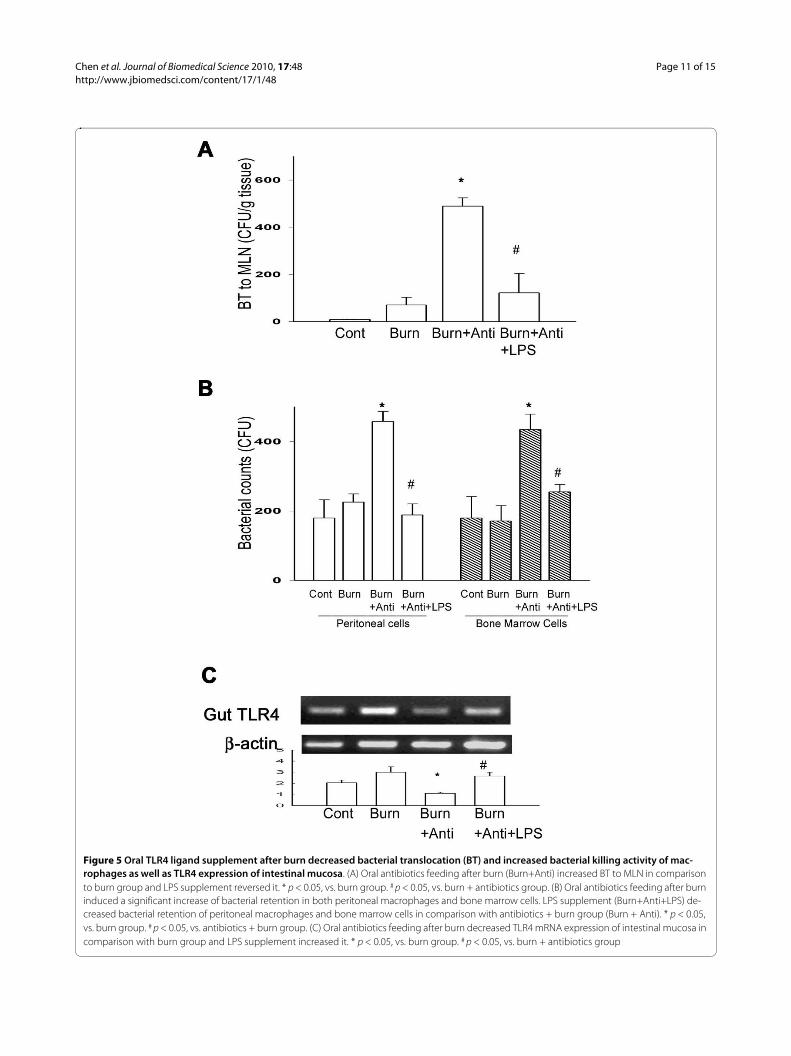
TLR4 ligand did not change bacterial translocation in C3H/HeJ miceTo further define the involvement of TLR4 on the changeof bacterial translocation to MLNs after thermal injury,we examined BT to MLNs in C3H/HeJ mice after thermalinjury with oral antibiotics treatment. Thermal injury didnot induce bacterial translocation to MLNs in controlgroup and commensal depleted group in C3H/HeJ mice.Commensal depletion with burn did on change BT toMLNs in C3H/HeJ mice when compared with burn
group. Furthermore, LPS supplement did not change BTwhen compared with commensal depletion group inC3H/HeJ mice (Figure 6A).
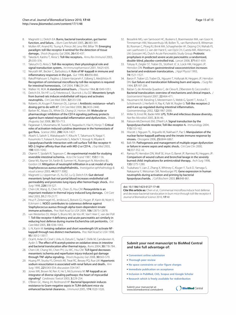
TLR4 ligand did not change the bacterial killing activity of peritoneal cells in C3H/HeJ miceTo further define the role of TLR4 on host defense againstbacterial challenge after commensal depletion, we exam-ined the bacterial killing activity of peritoneal cells inC3H/HeJ mice after different treatments. Commensaldepletion did not change the bacterial killing activity ofperitoneal cells in C3H/HeJ mice when compared withburn group (Figure 6B). Furthermore, LPS supplementdid not change bacterial killing activity of peritoneal cellsin C3H/HeJ mice. There is no significant difference ofperitoneal cell count between WT mice and C3H/HeJmice (Table 1).
TLR4 ligand did not change TLR4 and TNFα expression in C3H/HEJ miceTo further evaluate the involvement of TLR4 in thecytokine change of different tissues after commensaldepletion, we examined TLR4 mRNA expression of gut,lung, and peritoneal cells in C3H/HeJ mice. Thermalinjury induced TNFα mRNA expression of peritonealcells in C3H/HeJ mice when compared with that of shamburn group. Commensal depletion with or without LPSsupplement did not change TLR4 mRNA expression ofgut, lung, and peritoneal cells in C3H/HeJ mice whencompared with control group. Also, commensal depletionwith or without LPS supplement did not change TNFαmRNA expression of peritoneal cells in C3H/HeJ micewhen compared with burn group (Figure 6C).
DiscussionMajor burn in humans and experimental animals is asso-ciated with compromised immune functions andincreased acute gastrointestinal derangement that dis-rupts gastrointestinal mucosa integrity and facilitates thebacterial translocation. Our data demonstrate that com-mensal microflora are critical in maintaining innateimmunity in burn through TLR4 signaling and oral TLR4ligand supplement reverses the commensal depletioneffect in burn through the decrease of BT and theincrease of bacterial killing activity.
Provision of enteral nutrients shortly after injury altersgut flora and protects the immuno-compromised,stressed, or thermally injured patients through unknownmechanism [8]. The cross talk between commensalmicroflora in intestinal tract and systemic innate immu-nity is still undefined. Previous paper demonstrated thatrecognition of commensal microflora by toll-like recep-tors was required for intestinal homeostasis [13]. Here,our results show that commensal microflora in intestinal
Chen et al. Journal of Biomedical Science 2010, 17:48http://www.jbiomedsci.com/content/17/1/48
Page 10 of 15
Figure 4 TLR4 ligand increased TLR4 expression and NF-κB DNA binding activity of intestinal mucosa after thermal injury. (A) LPS supple-ment increased TLR4 protein expression of intestinal mucosa in commensal depleted mice. * p < 0.05, vs. burn group. # p < 0.05, vs. commensal de-pletion + burn group. (B) LPS supplement increased TLR4 mRNA expression of intestinal mucosa in commensal depleted mice. The values represent the average fold increase (± s.d.) in 3 different mice of each group. * p < 0.05, vs. burn group. # p < 0.05, vs. commensal depletion + burn group. (C) LPS supplement increased TLR4 immunostaining of intestinal epithelial cells. (D) Commensal depletion decreased the NF-κB DNA-binding activity of in-testinal mucosa and LPS supplement increased it. (n = 4). *p < 0.05, vs. burn group. * p < 0.05, vs. commensal depletion + burn group. NS = non-specific binding.
Chen et al. Journal of Biomedical Science 2010, 17:48http://www.jbiomedsci.com/content/17/1/48
Page 11 of 15
Figure 5 Oral TLR4 ligand supplement after burn decreased bacterial translocation (BT) and increased bacterial killing activity of mac-rophages as well as TLR4 expression of intestinal mucosa. (A) Oral antibiotics feeding after burn (Burn+Anti) increased BT to MLN in comparison to burn group and LPS supplement reversed it. * p < 0.05, vs. burn group. # p < 0.05, vs. burn + antibiotics group. (B) Oral antibiotics feeding after burn induced a significant increase of bacterial retention in both peritoneal macrophages and bone marrow cells. LPS supplement (Burn+Anti+LPS) de-creased bacterial retention of peritoneal macrophages and bone marrow cells in comparison with antibiotics + burn group (Burn + Anti). * p < 0.05, vs. burn group. # p < 0.05, vs. antibiotics + burn group. (C) Oral antibiotics feeding after burn decreased TLR4 mRNA expression of intestinal mucosa in comparison with burn group and LPS supplement increased it. * p < 0.05, vs. burn group. # p < 0.05, vs. burn + antibiotics group
Chen et al. Journal of Biomedical Science 2010, 17:48http://www.jbiomedsci.com/content/17/1/48
Page 12 of 15
Figure 6 TLR4 ligand did not change bacterial translocation (BT), bacterial killing activity of peritoneal cells, and TNFα mRNA expression in C3H/HeJ mice. (A) Commensal depletion with or without LPS supplement did not change BT in C3H/HeJ mice. (B) Commensal depletion with or without LPS supplement did not change bacterial retention of peritoneal cells in C3H/HeJ mice. (C) Commensal depletion with or without LPS sup-plement did not change TNFα mRNA expression of peritoneal cells in C3H/HeJ mice. The values represent the average fold increase (± s.d.) in mRNA expression in 3 different mice of each group. ! p < 0.05, vs. sham group.
Chen et al. Journal of Biomedical Science 2010, 17:48http://www.jbiomedsci.com/content/17/1/48
Page 13 of 15
tract induce TLR4 expression of intestinal epithelial cellsand enhance the bacterial killing activity of inflammatorycells in burn. Previously, macrophage of TLR4-deficientmice demonstrated impaired bacterial recognition andphagocytosis compared with wild-type mice [31]. Ourpresent data show that commensal depletion does notchange the bacterial killing activity and TLRα mRNAexpression of peritoneal cells in C3H/HeJ mice. Alto-gether, our data suggest that TLR4 signaling in intestinaltract is important in inducing bacterial killing activity ofinflammatory cells after thermal injury and commensalmicroflora are critical in maintaining innate immunity inburn through TLR4 signaling. Previous paper has provedthat enteral nutrients shortly after injury could alterintestinal flora [8]. Accordingly, the beneficiary effect ofearly enteral nutrients on systemic immunity in burncould be through the reestablishment of gut flora in intes-tinal tract. In a recent prospective randomized controlledtrial of using probiotics in pancreatitis patients, patientswho received the probiotics had a surprisingly high rateof nonocclusive bowel necrosis [32]. This paper used 10billion probiotic bacteria per day on top of enteral nutri-tion. This might have even further increased local oxygendemand, with a combined deleterious effect on an alreadycritically reduced blood flow. On the contrary, we provideanimals combined antibiotics to deplete commensalmicroflora and stimulate the TLR4 expression in intesti-nal mucosa with oral LPS supplement. All in all, our dataimply that oral supplement of certain flora to activateTLR4 in intestinal mucosa but not overload the intestinalflora could be a new therapeutic strategy to enhance sys-temic innate immunity in major burn patients.
The second important conclusion to be derived fromthe present results is that TLR4 ligand supplementdecreases burn-induced BT and reverses commensaldepletion-induced reduction of bacterial killing activityin burn. First, we demonstrate that thermal injuryinduces a significant increase of bacterial translocation tomesenteric lymph nodes. LPS supplement stimulates theTLR4 expression of intestinal mucosa and significantlydecreases thermal injury-induced bacterial translocation.Thermal injury is associated with mesenteric vasocon-striction that lead to damage of the gut mucosa and dys-function of the gut barrier, resulting in an increased gutpermeability, and absorption of bacteria and bacterialtoxins [33]. Burn-induced gut injury results in the pro-
duction of biologically active factors that are carried inthe mesenteric lymph, but not the portal plasma, whichinjure endothelial cells and activate neutrophil and con-tribute to distant organ injury [15]. The potential cause ofsepsis and subsequent multiple organ failure followingthermal injury could be the failure of intestinal mucosa toact as barrier against BT [34]. BT is a phenomenon inwhich live bacteria or its products cross the intestinalbarrier. The function of the gut barrier depends onmucous epithelia (mechanical barrier) and secreting IgAand immune cells (immune barrier) [35]. Gut transloca-tion of bacteria has been shown in both animal andhuman studies. BT and its complications have beenshown clearly to occur in animal models, but its existenceand importance in humans has been difficult to ascertain[35]. Our results demonstrate that TLR4 ligand signifi-cantly increases the bacterial killing activity of inflamma-tory cells and decreases thermal injury-induced bacterialtranslocation but has no effect on intestinal permeabilityor glutathione level. These results suggest that TLR4ligand improves the thermal injury-induced gut barrierfunction through the enhancement of the intestinalinnate immunity (immune barrier) rather than thedecrease of intestinal permeability (mechanical barrier).Second, LPS supplement reverses commensal depletion-induced reduction of bacterial killing activity of perito-neal cells after burn. Previously, TLRs of those mac-rophages which were resident in the lamina propria of theintestine has been implicated closely related to the potentinflammatory response, intestinal inflammation, and cor-responding injury [36]. On the contrary, our data demon-strate that LPS supplement reverses commensaldepletion-induced reduction of bacterial killing activityof peritoneal cells in WT mice but not in C3H/HeJ mice.This indicates that LPS supplement induces host defenseto bacterial challenge in burn through the TLR4 signal-ing. Recent research indicates that there is significantphylogenetic and diversity in TLR4-mediated responses[37]. A new TLR4 ligand that is originated from LPSstructures but without its toxicity could be developed inthe future to enhance host defense in major burnpatients.
Infection is a common source of morbidity and mortal-ity in critically burned patients. Burn injury produces acomplex interaction of gut-derived inflammatory reac-tion paralleled by systemic inflammatory responses. The
Table 1: Components of peritoneal cells of C3H/HeJ and wild-type (WT) mice.
Group Monos (%) Lymphos (%) PMN (%) Macrophages (%) Mast cells (%)
WT 51.7 ± 7.3 42.6 ± 4.1 2.9 ± 0.5 1.9 ± 0.3 0.9 ± 0.1
C3H/HeJ 54 ± 8 39.6 ± 5.1 3.2 ± 0.9 2.1 ± 0.5 1.1 ± 0.3
Chen et al. Journal of Biomedical Science 2010, 17:48http://www.jbiomedsci.com/content/17/1/48
Page 14 of 15
use of selective decontamination of the digestive tract(SDD) is of particular interest for application in patientswith major burn injury where the need for intubation andmechanical ventilation likely contributes to oropharyn-geal and intestinal colonization with pathogenic microor-ganisms. Recently, SDD treatment of experimental burnin adult rats resulted in attenuated septic challenge-related inflammatory responses and improved myocar-dial contractile response [5]. However, our data demon-strate that oral antibiotics treatment after burn is notwithout a cost: decreasing bacterial killing activity ofinflammatory cells. Also, our results suggest that oralTLR4 ligand supplement could reverse the unfavorableeffect of oral antibiotics treatment in burn through theincrease of gut barrier function and bacterial killing activ-ity of inflammatory cells.
TLRs are membrane signaling receptors that playessential roles in innate defense against microbes. TLRsare type I integral membrane glycoproteins that containleucine-rich repeats glanced by characteristic cysteine-rich motifs in their extracellular regions and a cytoplas-mic TIR homology domain. Ligand-induced TLRdimerization permits the binding of cytoplasmic adapterproteins, MyD88, to the TLR cytoplasmic tails [38]. Amajor downstream effect of TLR signaling is the activa-tion of the transcription factor NF-κB, which is requiredfor expression of many genes related to innate immunityand inflammation [12]. Studies have proved that TLR4stimulation maintained intestinal hemostasis through theNF-κB activation of the intestinal mucosa [13]. The stim-ulatory effect of LPS supplement on NF-κB activation ofintestinal mucosa further corroborates that LPS supple-ment enhances host defense in burn through the increaseof TLR4 signaling. Activation of the NF-κB transcriptionfactor pathways is an essential immediate early step ofimmune activation [39]. Altogether, our data suggest thatTLR4 ligand activates the NF-κB activation in intestinalmucosa and enhances the gut barrier function after ther-mal injury.
Septic shock has been reported to be the most commoncause of death in the noncoronary intensive care unit[40]. Pulmonary sepsis is the septic complication mostfrequently encountered in severely burn patients [41].MPO system plays an important role in the microbicidalactivity of phagocytes and neutrophil play an essentialrole in the Human's innate immune response to infection[42]. MPO, released by neutrophil, may attack normal tis-sue and thus contribute to the pathogenesis disease. Ourdata demonstrate that commensal depletion significantlydecreases TLR4 as well as TNFα expression in lung andLPS supplement increases TLR4 as well as TNFα expres-sion in lung after burn. NF-κB family members controltranscriptional activity of various promoters of proin-flammatory cytokines, cell surface receptors, transcrip-
tion factors, and adhesion molecules that are involved inintestinal inflammation such as TNFα[12]. Our data sug-gest that TLR4 ligand stimulation in the gut enhancesTLR4 expression and neutrophil deposition in lung. Thestimulatory effect of LPS supplement on TNFα expres-sion as well as MPO activity of lung could be through theinduction of NF-κB activation.
ConclusionsIn summary, commensal microflora induce gut barrierfunction and the bacterial killing activity of peritonealcells in burn through the increase of toll-like receptor 4.TLR4 ligand reverses oral antibiotics effect in burnthrough the decrease of BT and the increase of bacterialkilling activity.
AbbreviationsTLRs: toll-like receptors; MLNs: mesenteric lymph nodes; LPS: lipopolysaccha-ride; BT: bacterial translocation; LTA: lipoteichoic acid; TBSA: total body surfacearea; FITC: fluorescein isothiocyanate; MPO: myeloperoxidase; WT: Wild type;GSH: glutathione
Competing interestsThe authors declare that they have no competing interests.
Authors' contributionsLWC and CMH designed research; WJC and PHC performed research; LWC andCMH analyzed data; LWC and CMH wrote the paper. All authors read andapproved the final manuscript.
AcknowledgementsThis work was supported by grants from National Science Council, Taipei (NSC972314B010030MY3), Kaohsiung Veterans General Hospital, Kaohsiung (VGHNSU97-050, VGHKS98-052) to CLW, and National Sun Yat-Sen University-Kaohsiung Medical University Joint Research Center.
Author Details1Institute of Emergency and Critical Care Medicine, National Yang-Ming University, Taipei, Taiwan, 2Department of Surgery, Kaohsiung Veterans General Hospital, 386, Ta-chung 1st Road, Kaohsiung, Taiwan and 3Department of Biological Sciences, National Sun Yat-Sen University, 70 Lien-Hai Road, Kaohsiung 804, Taiwan
References1. Sonnenburg JL, Angenent LT, Gordon JI: Getting a grip on things: how
do communities of bacterial symbionts become established in our intestine? Nat Immunol 2004, 5:569-573.
2. Kelly D, Conway S, Aminov R: Commensal gut bacteria: mechanisms of immune modulation. Trends Immunol 2005, 26:326-333.
3. Lederer JA, Rodrick ML, Mannick JA: The effects of injury on the adaptive immune response. Shock (Augusta, Ga) 1999, 11:153-159.
4. Chung DH, Herndon DN: Multiple converging mechanisms for postburn intestinal barrier dysfunction. Crit Care Med 2004, 32:1803-1804.
5. Horton JW, Maass DL, White J, Minei JP: Reducing susceptibility to bacteremia after experimental burn injury: a role for selective decontamination of the digestive tract. J Appl Physiol 2007, 102:2207-2216.
6. Gianotti L, Alexander JW, Pyles T, James L, Babcock GF: Relationship between extent of burn injury and magnitude of microbial translocation from the intestine. J Burn Care Rehabil 1993, 14:336-342.
7. Fukatsu K, Zarzaur BL, Johnson CD, Lundberg AH, Wilcox HG, Kudsk KA: Enteral nutrition prevents remote organ injury and death after a gut ischemic insult. Ann Surg 2001, 233:660-668.
Received: 15 February 2010 Accepted: 12 June 2010 Published: 12 June 2010This article is available from: http://www.jbiomedsci.com/content/17/1/48© 2010 Chen et al; licensee BioMed Central Ltd. This is an Open Access article distributed under the terms of the Creative Commons Attribution License (http://creativecommons.org/licenses/by/2.0), which permits unrestricted use, distribution, and reproduction in any medium, provided the original work is properly cited.Journal of Biomedical Science 2010, 17:48
Chen et al. Journal of Biomedical Science 2010, 17:48http://www.jbiomedsci.com/content/17/1/48
Page 15 of 15
8. Magnotti LJ, Deitch EA: Burns, bacterial translocation, gut barrier function, and failure. J Burn Care Rehabil 2005, 26:383-391.
9. Mollen KP, Anand RJ, Tsung A, Prince JM, Levy RM, Billiar TR: Emerging paradigm: toll-like receptor 4-sentinel for the detection of tissue damage. Shock (Augusta, Ga) 2006, 26:430-437.
10. Takeda K, Kaisho T, Akira S: Toll-like receptors. Annu Rev Immunol 2003, 21:335-376.
11. Takeuchi O, Akira S: Toll-like receptors; their physiological role and signal transduction system. Int Immunopharmacol 2001, 1:625-635.
12. Neurath MF, Becker C, Barbulescu K: Role of NF-kappaB in immune and inflammatory responses in the gut. Gut 1998, 43:856-860.
13. Rakoff-Nahoum S, Paglino J, Eslami-Varzaneh F, Edberg S, Medzhitov R: Recognition of commensal microflora by toll-like receptors is required for intestinal homeostasis. Cell 2004, 118:229-241.
14. Walker HL MJA: A standard animal burn. J Trauma 1964, 8:1049-1051.15. Deitch EA, Shi HP, Lu Q, Feketeova E, Skurnick J, Xu DZ: Mesenteric lymph
from burned rats induces endothelial cell injury and activates neutrophils. Crit Care Med 2004, 32:533-538.
16. Roberts JA, Kruger P, Paterson DL, Lipman J: Antibiotic resistance--what's dosing got to do with it? Crit Care Med 2008, 36:2433-2440.
17. Barber RC, Maass DL, White DJ, Chang LY, Horton JW: Molecular or pharmacologic inhibition of the CD14 signaling pathway protects against burn-related myocardial inflammation and dysfunction. Shock (Augusta, Ga) 2008, 30:705-713.
18. Fagarasan S, Muramatsu M, Suzuki K, Nagaoka H, Hiai H, Honjo T: Critical roles of activation-induced cytidine deaminase in the homeostasis of gut flora. Science 2002, 298:1424-1427.
19. Akashi S, Saitoh S, Wakabayashi Y, Kikuchi T, Takamura N, Nagai Y, Kusumoto Y, Fukase K, Kusumoto S, Adachi Y, Kosugi A, Miyake K: Lipopolysaccharide interaction with cell surface Toll-like receptor 4-MD-2: higher affinity than that with MD-2 or CD14. J Exp Med 2003, 198:1035-1042.
20. Otamiri T, Sjodahl R, Tagesson C: An experimental model for studying reversible intestinal ischemia. Acta Chir Scand 1987, 153:51-56.
21. Giese MJ, Rayner SA, Fardin B, Sumner HL, Rozengurt N, Mondino BJ, Gordon LK: Mitigation of neutrophil infiltration in a rat model of early Staphylococcus aureus endophthalmitis. Investigative ophthalmology & visual science 2003, 44:3077-3082.
22. Magnotti LJ, Upperman JS, Xu DZ, Lu Q, Deitch EA: Gut-derived mesenteric lymph but not portal blood increases endothelial cell permeability and promotes lung injury after hemorrhagic shock. Ann Surg 1998, 228:518-527.
23. Chen LW, Wang JS, Chen HL, Chen JS, Hsu CM: Peroxynitrite is an important mediator in thermal injury-induced lung damage. Crit Care Med 2003, 31:2170-2177.
24. Hruz P, Zinkernagel AS, Jenikova G, Botwin GJ, Hugot JP, Karin M, Nizet V, Eckmann L: NOD2 contributes to cutaneous defense against Staphylococcus aureus through alpha-toxin-dependent innate immune activation. Proc Natl Acad Sci USA 2009, 106:12873-12878.
25. van Westerloo DJ, Weijer S, Bruno MJ, de Vos AF, Van't Veer C, van der Poll T: Toll-like receptor 4 deficiency and acute pancreatitis act similarly in reducing host defense during murine Escherichia coli peritonitis. Crit Care Med 2005, 33:1036-1043.
26. Li N, Karin M: Ionizing radiation and short wavelength UV activate NF-kappaB through two distinct mechanisms. Proc Natl Acad Sci USA 1998, 95:13012-13017.
27. Ocal K, Avlan D, Cinel I, Unlu A, Ozturk C, Yaylak F, Dirlik M, Camdeviren H, Aydin S: The effect of N-acetylcysteine on oxidative stress in intestine and bacterial translocation after thermal injury. Burns 2004, 30:778-784.
28. Chen LW, Chang WJ, Chen PH, Liu WC, Hsu CM: TLR ligand decreases mesenteric ischemia and reperfusion injury-induced gut damage through TNF-alpha signaling. Shock (Augusta, Ga) 2008, 30:563-570.
29. Huang PP, Stucky FS, Dimick AR, Treat RC, Bessey PQ, Rue LW: Hypertonic sodium resuscitation is associated with renal failure and death. Ann Surg 1995, 221:543-554; discussion 554-547.
30. Jones WK, Brown M, Ren X, He S, McGuinness M: NF-kappaB as an integrator of diverse signaling pathways: the heart of myocardial signaling? Cardiovasc Toxicol 2003, 3:229-254.
31. O'Brien GC, Wang JH, Redmond HP: Bacterial lipoprotein induces resistance to Gram-negative sepsis in TLR4-deficient mice via enhanced bacterial clearance. J Immunol 2005, 174:1020-1026.
32. Besselink MG, van Santvoort HC, Buskens E, Boermeester MA, van Goor H, Timmerman HM, Nieuwenhuijs VB, Bollen TL, van Ramshorst B, Witteman BJ, Rosman C, Ploeg RJ, Brink MA, Schaapherder AF, Dejong CH, Wahab PJ, van Laarhoven CJ, van der Harst E, van Eijck CH, Cuesta MA, Akkermans LM, Gooszen HG, Dutch Acute Pancreatitis Study Group: Probiotic prophylaxis in predicted severe acute pancreatitis: a randomised, double-blind, placebo-controlled trial. Lancet 2008, 371:651-659.
33. Tokyay R, Zeigler ST, Traber DL, Stothert JC Jr, Loick HM, Heggers JP, Herndon DN: Postburn gastrointestinal vasoconstriction increases bacterial and endotoxin translocation. J Appl Physiol 1993, 74:1521-1527.
34. Baron P, Traber LD, Traber DL, Nguyen T, Hollyoak M, Heggers JP, Herndon DN: Gut failure and translocation following burn and sepsis. J Surg Res 1994, 57:197-204.
35. Balzan S, de Almeida Quadros C, de Cleva R, Zilberstein B, Cecconello I: Bacterial translocation: overview of mechanisms and clinical impact. J Gastroenterol Hepatol 2007, 22:464-471.
36. Hausmann M, Kiessling S, Mestermann S, Webb G, Spottl T, Andus T, Scholmerich J, Herfarth H, Ray K, Falk W, Rogler G: Toll-like receptors 2 and 4 are up-regulated during intestinal inflammation. Gastroenterology 2002, 122:1987-2000.
37. Miller SI, Ernst RK, Bader MW: LPS, TLR4 and infectious disease diversity. Nat Rev Microbiol 2005, 3:36-46.
38. Palsson-McDermott EM, O'Neill LA: Signal transduction by the lipopolysaccharide receptor, Toll-like receptor-4. Immunology 2004, 113:153-162.
39. Hiscott J, Nguyen TL, Arguello M, Nakhaei P, Paz S: Manipulation of the nuclear factor-kappaB pathway and the innate immune response by viruses. Oncogene 2006, 25:6844-6867.
40. Balk RA: Pathogenesis and management of multiple organ dysfunction or failure in severe sepsis and septic shock. Crit Care Clin 2000, 16:337-352. vii.
41. Ramzy PI, Herndon DN, Wolf SE, Irtun O, Barret JP, Ramirez RJ, Heggers JP: Comparison of wound culture and bronchial lavage in the severely burned child: implications for antimicrobial therapy. Arch Surg 1998, 133:1275-1280.
42. Tsukahara Y, Lian Z, Zhang X, Whitney C, Kluger Y, Tuck D, Yamaga S, Nakayama Y, Weissman SM, Newburger PE: Gene expression in human neutrophils during activation and priming by bacterial lipopolysaccharide. J Cell Biochem 2003, 89:848-861.
doi: 10.1186/1423-0127-17-48Cite this article as: Chen et al., Commensal microflora induce host defense and decrease bacterial translocation in burn mice through toll-like receptor 4 Journal of Biomedical Science 2010, 17:48
http://www.ncbi.nlm.nih.gov/entrez/query.fcgi?cmd=Retrieve&db=PubMed&dopt=Abstract&list_uids=9824616
http://www.ncbi.nlm.nih.gov/entrez/query.fcgi?cmd=Retrieve&db=PubMed&dopt=Abstract&list_uids=3554866
http://www.ncbi.nlm.nih.gov/entrez/query.fcgi?cmd=Retrieve&db=PubMed&dopt=Abstract&list_uids=9790341
http://www.ncbi.nlm.nih.gov/entrez/query.fcgi?cmd=Retrieve&db=PubMed&dopt=Abstract&list_uids=9789032
http://www.ncbi.nlm.nih.gov/entrez/query.fcgi?cmd=Retrieve&db=PubMed&dopt=Abstract&list_uids=7748036
http://www.ncbi.nlm.nih.gov/entrez/query.fcgi?cmd=Retrieve&db=PubMed&dopt=Abstract&list_uids=8514665
http://www.ncbi.nlm.nih.gov/entrez/query.fcgi?cmd=Retrieve&db=PubMed&dopt=Abstract&list_uids=8041138
Related Documents


