In Silico-Experimental Approach for Drug Design: the Binding Mode of Peptidic and Non Peptidic Inhibitors to Hsp90 N- Terminal Domain Simona Tomaselli 1 , Massimiliano Meli 2 , Janet Plescia 3 , Lucia Zetta 1 , Dario C. Altieri 3 , Giorgio Colombo 2,* , and Laura Ragona 1,* 1 Istituto per lo Studio delle Macromolecole, Consiglio Nazionale delle Ricerche, via Bassini 15, 20133 Milano, Italy 2 Istituto di Chimica del Riconoscimento Molecolare, Consiglio Nazionale delle Ricerche, Via Mario Bianco 9, 20131 Milano, Italy 3 Department of Cancer Biology, University of Massachusetts Medical School, Plantation Street Worcester, Massachusetts 01605, USA Abstract Heat shock protein 90 (Hsp90) is a prime target for antitumor therapies. The information obtained by MD simulations is combined with NMR data to provide a cross-validated atomic resolution model of the complementary interactions of Hsp90 with a peptidic (Shepherdin) and a non peptidic (5-aminoimidazole-4-carboxamide-1-β-D-ribofuranoside, AICAR) inhibitor, showing antiproliferative and proapoptotic activity in multiple tumor cell lines. This approach highlights the relevant role of imidazolic moiety in the interaction of both antagonist molecules. In AICAR bound state one conformation of those present in solution is selected, where imidazolic, H4 and H5 protons have a key role in defining a non polar region contacting Hsp90 surface. The dynamic equilibrium between N-type and S-type puckered forms of AICAR ribofuranoside moiety is shown to be functional to inhibitor binding. The first experimental structural data on these inhibitors are presented and discussed as hints for future design of improved molecules. Keywords Antitumor Agents; Drug Design; Molecular Dynamics; NMR; Hsp90 Heat shock protein 90 (Hsp90) is a molecular chaperone and is one of the most abundant proteins expressed in cells (1). It is a member of the heat shock protein family which is up regulated during stress response. These kinds of proteins protect the cell when exposed to elevated temperatures but their overexpression can be triggered also by other kinds of stress such as inflammation, hypoxia, exposure of cells to toxins and other pathological conditions like tumours. In order to respond to these insults and to ensure cancer cell survival in the face of an otherwise hostile environment, Hsp90 oversees the correct conformational development of a diverse ensemble of client proteins. Hsp90 is indeed the most abundant chaperone expressed in eukaryotic and prokaryotic cells. In mammalian cells, there are mainly two cytosolic Hsp90 isoforms and the human Hsp90α form shows 85% sequence identity with Hsp90β (2). Hsp90 consists of three structural domains: N-terminal domain (NT) of about 25KDa, the middle domain (M) of about 40KDa and the C-terminal domain Corresponding authors: Dr. Giorgio Colombo, [email protected], Dr. Laura Ragona, [email protected]. NIH Public Access Author Manuscript Chem Biol Drug Des. Author manuscript; available in PMC 2011 November 1. Published in final edited form as: Chem Biol Drug Des. 2010 November ; 76(5): 382–391. doi:10.1111/j.1747-0285.2010.01015.x. NIH-PA Author Manuscript NIH-PA Author Manuscript NIH-PA Author Manuscript

Welcome message from author
This document is posted to help you gain knowledge. Please leave a comment to let me know what you think about it! Share it to your friends and learn new things together.
Transcript
In Silico-Experimental Approach for Drug Design: the BindingMode of Peptidic and Non Peptidic Inhibitors to Hsp90 N-Terminal Domain
Simona Tomaselli1, Massimiliano Meli2, Janet Plescia3, Lucia Zetta1, Dario C. Altieri3,Giorgio Colombo2,*, and Laura Ragona1,*
1Istituto per lo Studio delle Macromolecole, Consiglio Nazionale delle Ricerche, via Bassini 15,20133 Milano, Italy2Istituto di Chimica del Riconoscimento Molecolare, Consiglio Nazionale delle Ricerche, ViaMario Bianco 9, 20131 Milano, Italy3Department of Cancer Biology, University of Massachusetts Medical School, Plantation StreetWorcester, Massachusetts 01605, USA
AbstractHeat shock protein 90 (Hsp90) is a prime target for antitumor therapies. The information obtainedby MD simulations is combined with NMR data to provide a cross-validated atomic resolutionmodel of the complementary interactions of Hsp90 with a peptidic (Shepherdin) and a nonpeptidic (5-aminoimidazole-4-carboxamide-1-β-D-ribofuranoside, AICAR) inhibitor, showingantiproliferative and proapoptotic activity in multiple tumor cell lines. This approach highlightsthe relevant role of imidazolic moiety in the interaction of both antagonist molecules. In AICARbound state one conformation of those present in solution is selected, where imidazolic, H4 andH5 protons have a key role in defining a non polar region contacting Hsp90 surface. The dynamicequilibrium between N-type and S-type puckered forms of AICAR ribofuranoside moiety isshown to be functional to inhibitor binding. The first experimental structural data on theseinhibitors are presented and discussed as hints for future design of improved molecules.
KeywordsAntitumor Agents; Drug Design; Molecular Dynamics; NMR; Hsp90
Heat shock protein 90 (Hsp90) is a molecular chaperone and is one of the most abundantproteins expressed in cells (1). It is a member of the heat shock protein family which is upregulated during stress response. These kinds of proteins protect the cell when exposed toelevated temperatures but their overexpression can be triggered also by other kinds of stresssuch as inflammation, hypoxia, exposure of cells to toxins and other pathological conditionslike tumours. In order to respond to these insults and to ensure cancer cell survival in theface of an otherwise hostile environment, Hsp90 oversees the correct conformationaldevelopment of a diverse ensemble of client proteins. Hsp90 is indeed the most abundantchaperone expressed in eukaryotic and prokaryotic cells. In mammalian cells, there aremainly two cytosolic Hsp90 isoforms and the human Hsp90α form shows 85% sequenceidentity with Hsp90β (2). Hsp90 consists of three structural domains: N-terminal domain(NT) of about 25KDa, the middle domain (M) of about 40KDa and the C-terminal domain
Corresponding authors: Dr. Giorgio Colombo, [email protected], Dr. Laura Ragona, [email protected].
NIH Public AccessAuthor ManuscriptChem Biol Drug Des. Author manuscript; available in PMC 2011 November 1.
Published in final edited form as:Chem Biol Drug Des. 2010 November ; 76(5): 382–391. doi:10.1111/j.1747-0285.2010.01015.x.
NIH
-PA Author Manuscript
NIH
-PA Author Manuscript
NIH
-PA Author Manuscript
(CT) of about 12KDa. The most studied and best-understood domain is the N-terminal. Thisdomain contains the ATP binding site and shows high homology not only amongst membersof Hsp family but also amongst members of the ATPase/kinase superfamily (3). Inunstressed cells Hsp90 works as molecular chaperone assisting folding of newly synthesizedproteins and regulates the trafficking of the denaturated proteins in cells under pathologicaland stressed conditions. Cancerous cells overexpress a number of proteins including P13Kand AKT, whose inhibition causes apoptosis, and various steroid receptors such as p53,ErB2, Src, Abl, Raf which are all clients of Hsp90. It appears that all these Hsp90 clients areimplicated in signal transduction and cell-cycle control; mutations or misregulation of theseproteins cause uncontrolled development and proliferation of tumor cells. Hsp90 is alsorequired for induction of vascular endothelial growth factor and nitric oxide synthase, bothimportant for de novo angiogenesis. It also promotes the proliferation and invasion ofmetastasis by assisting the matrix metalloproteinase MMP2. Hsp90, with the help of itscochaperones, modulates tumour cell apoptosis trough effects on AKT (4), tumor necrosisfactor receptors (TNFR) and nuclear factor-kB (NF-kB) function (5). Moreover Hsp90stabilizes the mutants of these proteins involved in tumor growth and is involved inmetastastasis and proliferation.
The important role that Hsp90 plays in tumor growth has made it a prime target forantitumor therapies. Most of the designed inhibitors act on Hsp90 competing for the ATPbinding site at the N-terminal domain (6). The first known inhibitor of N-terminal domain ofHsp90 (Hsp90-NT) is the antibiotic geldanamycin which appeared to reduce tumor growthof 50% (7). In recent years a great interest has grown around the search for new Hsp90inhibitors competing for the ATP binding site. X-Ray and NMR first, and in silico studieslater, were used to design and optimize lead compounds able to inhibit Hsp90 (6,8).
An example showing the utility of computational biology approaches in the discovery ofnew small molecule inhibitors of Hsp90-NT is represented by the use of molecular dynamics(MD) simulations and structural analysis to develop the peptidomimetic antagonistShepherdin (9) into the small molecule 5-aminoimidazole-4-carboxamide-1-β-D-ribofuranoside (AICAR) (10). Shepherdin is the minimal peptidic sequence from survivin,an essential regulator of cell proliferation, differentiation and apoptosis overexpressed incancer (11), which is able to block the interaction of survivin with Hsp90 in vitro (12) andcause killing of tumour cells by apoptotic and nonapoptotic mechanisms (9). Starting from amodel of the Shepherdin-Hsp90 complex (9), long timescale MD simulations were used tosingle out the most relevant functional groups required for the antagonist to productivelybind the chaperone. This information was then translated into dynamic pharmacophoremodels taking the full protein and ligand flexibility into account. The use of thepharmacophore in a virtual screening effort allowed to identify AICAR as a novel, activeHsp90 inhibitor, able to compete with ATP for binding at the N terminal domain, destabilizevarious Hsp90-clients complex in vivo and inhibit cell proliferation in various tumor celllines, while leaving normal cells unscathed (10). The 3D structural model of the AICAR-Hsp90 complex, obtained by computational studies(10), has to be validated experimentallyto be really helpful in the context of rational drug design. NMR has recently emerged as ahigh-throughput experimental technique in drug discovery, in determining possible bindingaffinities and in determining the region of interaction.
In this paper we have set out to combine the information independently obtained by MDsimulations and experimental NMR data to provide a cross-validated and cross-filteredatomic resolution model of the complementary interactions within the binding site. All-atomMD simulations allowing full flexibility of the ligand and the receptor generate diverse setsof configurations for the complex, whereby both the ligand and the binding site of receptormay visit different conformations available on the complex free energy landscape. In
Tomaselli et al. Page 2
Chem Biol Drug Des. Author manuscript; available in PMC 2011 November 1.
NIH
-PA Author Manuscript
NIH
-PA Author Manuscript
NIH
-PA Author Manuscript
analogy with protein folding, it is important to discern native complex conformations, fromnear-native or non-native ones. In this context, native conformations (states) are the onesthat are mostly accessed and populated in solution at equilibrium conditions. Long timescale MD simulations provide a general view of the different possible states. NMR analysis,as discussed herein, helps select and filter only those conformations that verify specificstructural constraints obtained in solution at equilibrium, recapitulating ensemble propertiesthat are specific only to selected molecular configurations. This combined approach may beparticularly useful in the presence of large receptor proteins, not yet amenable for full NMRanalysis or in the presence of highly flexible ligands, for which X-ray may fail to provide anatomic structure. Molecular dynamics simulations have been previously successfully appliedto the optimisation of X-ray and NMR structures of macromolecules, significantlyimproving the efficiency of structure calculation and refinement, and allowing more andmore challenging systems to be analyzed (13,14). The efficiency of a similar in-silico-experimental approach was recently demonstrated for the development of new agentstargeting angiogenic factors (15,16).
In this study the first experimental structural data on Shepherdin and on the novel AICARinhibitor in complex with Hsp90-NT are presented and discussed with clear implications forfuture design of improved inhibitors.
Materials and MethodsHsp90-NT expression and inhibitor synthesis
Human Hsp90- NT, was cloned in pGex-4T3 and pFLAG-CMW 6c vector, expressed in E.Coli as GST fusion protein and purified from the GST frame by overnight thrombin (1 U/ml) cleavage as previously described(9,12). Shepherdin peptide was synthesized by theW.M Keck Biotechnology Research Center at Yale University School of Medicine usingsolid phase tBoc chemistry followed by reverse-phase high-pressure liquid chromatography,mass spectrometry and solid phase purification. AICAR was purchased by Sigma.
Sample preparationShepherdin peptide (NH2-K1HSSGCAFL9-COOH) was dissolved at 1.2 mM peptideconcentration in 30 mM phosphate buffer (95% H2O, 5% D2O), 6 mM DTT, 100 mM NaCl,pH 6.7. For NMR analysis of the interactions 1.6 mM shepherdin was studied in thepresence of Hsp90-NT (50 μM). AICAR (5-aminoimidazole-4-carboxamide-1-β-D-ribofuranoside) was dissolved at 1.8 mM in the same buffer and interaction studies wereperformed in the presence of Hsp90-NT 60 μM. To evaluate the J12 and J34 of AICAR inthe bound state, 0.1mM Hsp90 samples were titrated with the ligand from P:L ratios 1:1 to1:3.
NMRAll NMR spectra were recorded on Bruker DMX spectrometer operating at 500 MHzequipped with a triple resonance probehead, incorporating gradients in the z-axis. All datawere collected at 280 K on a spectral width of 6510 Hz. 2D-TOCSY (spin lock time of 80ms), NOESY (mixing times of 100 and 250 ms) and ROESY (spin lock time of 250 ms)spectra were recorded using standard sequences (17), (18) on Shepherdin peptide in order toassign all the resonances at pH 6.8. Data were processed with NMRPipe(19) and visualizedby NMRView(20). The proton assignments are reported in Table S1 of Supplemental Data.Longitudinal relaxation times T1 of Shepherdin and AICAR resonances were measured inthe presence of Hsp90-NT with the standard inversion recovery method. Data points (32 K)were acquired to cover a sweep width of 10 ppm and a relaxation delay of 6 s was used.Data were analysed using Bruker Topspin software. Shepherdin T1 values were found to be
Tomaselli et al. Page 3
Chem Biol Drug Des. Author manuscript; available in PMC 2011 November 1.
NIH
-PA Author Manuscript
NIH
-PA Author Manuscript
NIH
-PA Author Manuscript
rather constant along the peptide side-chains with values in the range 0.4-0.6 s. Theresonance at 2.1 ppm, corresponding to an impurity present in Shepherdin samples, showeda longer T1 value (0.9 s) and strong STD effects in the spectra. It should be mentioned that,as previously reported in the literature (21), T1 values affect STD signal intensities andhigher T1 gives rise to apparent stronger STD effects. AICAR T1 values were found to rangebetween 0.4-0.9 s.
For the acquisition of NMR Saturation Transfer Difference (STD) experiments a 1D pulsesequence incorporating a T1ρ filter to remove disturbing protein signals was used (22) andthe duration of the T1r filter was 80 ms. STD spectra were recorded with a spectral width of6510 Hz and 32 K data points. On-resonance irradiations were performed at differentfrequencies in the methyl region and off resonance irradiation was performed at −20000 Hz,using a series of Gaussian pulses with a 1% truncation and 50 ms duration to give totalsaturation times of 0.25, 0.5, 1, 2, 3, 4, 6, 8 sec. STD NMR spectra were acquired with atotal of 1024 transients in addition to 16 scans to allow the sample to come to equilibrium.The subtraction of the two FIDs was achieved by phase cycling.
1H chemical shift assignment of AICAR resonances was performed with standard methods.NOESY and ROESY spectra with mixing times of 100 and 250 ms were collected onAICAR alone and in the presence of Hsp90-NT at protein:ligand ratio 1:28 ratio. No purgingspin lock period to remove the NMR signals of the macromolecule background wasemployed. To minimize the unwanted Hartmann-Hahn effects in the ROESY spectra amoderate spin lock pulse was used with a field strength of 2390 Hz and the transmitterfrequency was set at 6 ppm (23). In order to compare NOESY and ROESY cross peaksvolumes in the absence and in the presence of Hsp90-NT protein, taking into account cross-peak intensity loss due to T1 relaxation, fractional volumes were calculated by dividingcross-peak volumes by the average of two resolved diagonal peaks. The contribution of thebound form of the ligand to NOESY/ROESY cross-relaxation rate, observed for protons iand s on a ligand in fast exchange with a high molecular-mass receptor, is described byEquation (1) (24):
(1)
where pf and pb are the mole fractions and σisf and σis
b are the cross-relaxation rates of thefree and bound ligand respectively. The bound fraction was assumed to be 0.04 at 1:28Hsp90-NT:AICAR ratio. The ROE volumes were corrected for the offset differencesbetween the correlation peaks and the spin lock pulse offset (25).
A rough estimate of distances from NOE/ROE cross peaks volumes was calculatedassuming an average distance between H4-H5 of 2.8 Å as deduced from MD simulations offree and bound AICAR (vide infra). The percentage of the two puckered forms S-type (C-2-endo, C-3-exo) and N-type (C-2-exo, C-3-endo) was estimated on the basis of themeasured 1H J12 and J34 coupling constants in the free and bound state (Table S2 inSupplemental data). J couplings of the bound form (Jb) were deduced from Equation (2):
(2)
where Jobs and Jf are the J couplings observed in the spectra of the bound and free AICAR,respectively and pb and pf are the mole fractions of bound and free species, respectively.
Tomaselli et al. Page 4
Chem Biol Drug Des. Author manuscript; available in PMC 2011 November 1.
NIH
-PA Author Manuscript
NIH
-PA Author Manuscript
NIH
-PA Author Manuscript
MD Simulations and analysisThe docking procedure and simulations for the Shepherdin-Hsp90-NT and AICAR-Hsp90-NT complexes were already described in Meli et al 2006 (10).
Conformational cluster analysis of the trajectories was performed using the methoddescribed in Daura et al. (26): the count number of neighbours using a cut-off of 2 Å RMSDbetween the optimal backbone superposition of different structures was performed and thestructure with the largest number of neighbors with all its neighbours was taken as clusterand eliminated from the pool. This procedure is repeated for the remaining structures in thepool. The three most populated clusters, representative of the most visited structures alongthe MD simulations, were selected. The central structure of each cluster, recapitulating themain structural properties of that cluster, was selected as representative for subsequentanalysis and cross-validation through NMR experimental measurements.
The same procedure as outlined above was used for the conformational analysis of isolatedAICAR free in solution.
The estimate of the population of the ribose ring puckered conformers (N or S-type) wasperformed by measuring the value of the C4-C3-C2-C1 dihedral angle over MD trajectory offree and bound ligand.
Pymol (DeLano, W.L. The PyMOL Molecular Graphics System, DeLano Scientific, PaloAlto, CA, USA) was used for graphical representation of the results.
Results and DiscussionNMR spectroscopy is exploited here to increase the knowledge of structural andconformational features favouring Hsp90 recognition/inhibition by Shepherdin and AICAR,so far widely described by MD and docking studies (10).
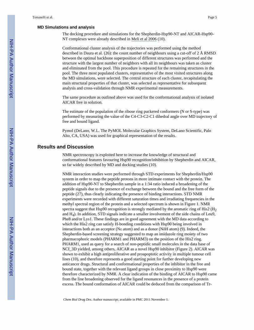
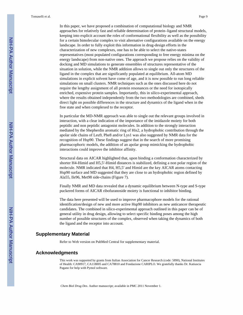
NMR interaction studies were performed through STD experiments for Shepherdin/Hsp90system in order to map the peptide protons in more intimate contact with the protein. Theaddition of Hsp90-NT to Shepherdin sample in a 1:34 ratio induced a broadening of thepeptide signals due to the presence of exchange between the bound and the free form of thepeptide (27), thus clearly indicating the presence of binding interactions. STD NMRexperiments were recorded with different saturation times and irradiating frequencies in themethyl spectral region of the protein and a selected spectrum is shown in Figure 1. NMRspectra suggest that Hsp90 recognition is strongly mediated by the aromatic ring of His2 (H2and H4). In addition, STD signals indicate a smaller involvement of the side chains of Leu9,Phe8 and/or Lys1. These findings are in good agreement with the MD data according towhich the His2 ring can satisfy H-bonding conditions with Hsp90 being involved ininteractions both as an acceptor (Nε atom) and as a donor (NδH atom) (9). Indeed, theShepherdin-based screening strategy suggested to map an imidazole ring moiety of twopharmacophoric models (PHARM1 and PHARM3) on the position of the His2 ring.PHARM1, used as query for a search of non-peptidic small molecules in the data base ofNCI_3D yielded, among others, AICAR as a novel Hsp90 inhibitor (Figure 2). AICAR wasshown to exhibit a high antiproliferative and proapoptotic activity in multiple tumour celllines (10), and therefore represents a good starting point for further developing newanticancer drugs. Structural and conformational properties of the inhibitor in the free andbound state, together with the relevant ligand groups in close proximity to Hsp90 weretherefore characterized by NMR. A clear indication of the binding of AICAR to Hsp90 camefrom the line broadening observed for the ligand resonances in the presence of a proteinexcess. The bound conformation of AICAR could be deduced from the comparison of Tr-
Tomaselli et al. Page 5
Chem Biol Drug Des. Author manuscript; available in PMC 2011 November 1.
NIH
-PA Author Manuscript
NIH
-PA Author Manuscript
NIH
-PA Author Manuscript
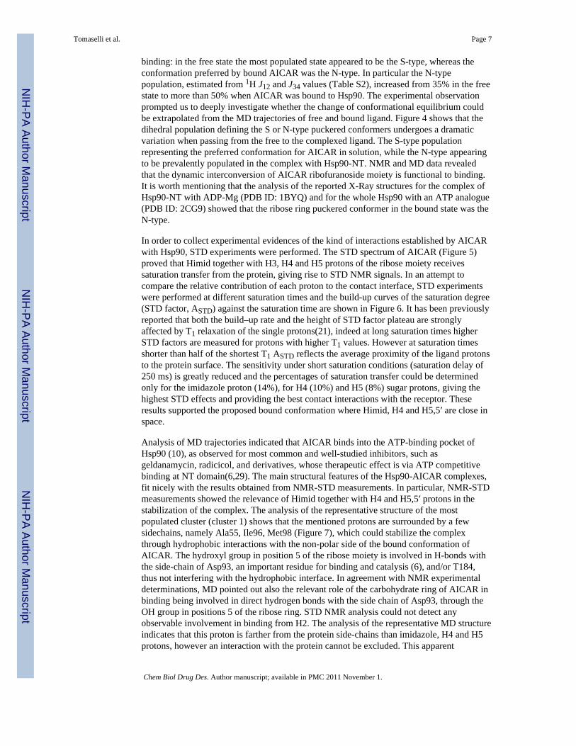
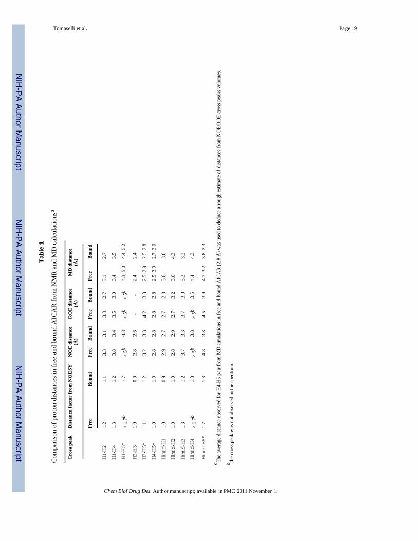
NOESY experiments recorded in the presence of Hsp90 with NOESY data recorded on thefree ligand. NOESY experiments on the free AICAR resulted in positive NOEs (oppositesign of diagonal) for all interproton vectors. Negative NOE cross-peaks were observed(same sign as the diagonal) in the presence of Hsp90 and this change in the sign correspondsto a slower tumbling molecule, as it is expected when the ligand is bound to the receptor(Figure 3). A new cross peak was observed between the imidazolic proton (Himid) and H4,suggesting the stabilization, upon binding, of an AICAR conformation characterized by ashorter distance between the two protons. In order to analyse the conformational changes,eventually affecting other inter-proton distances, a rough estimate of the contribution of thebound fraction of the ligand to the Tr-NOESY cross-peak intensities was calculated (seeExperimental Procedures). The fractional NOESY intensities of the free and bound peptideshowed a general increase of the NOESY intensities in the bound form, as expected whenthe ligand is bound to the protein. Assuming similar effective correlation times and spindiffusion pathways for the different proton pairs within the molecule in the same state (freeor bound), the ratio of the NOE volumes was used to estimate changes in inter-protondistances. The largest changes in distance factors (Table 1) affected Himid-H5 and Himid-H4 pairs, suggesting that these inter-proton distances are approximately 1.3-1.4 timesshorter than in the free state. These results showed that in the bound state one conformationof those present in solution is selected and is characterized by shorter distances between theimidazolic proton and H4 and H5 protons of the ribose ring. Tr-NOESY experiments couldnot be used for a quantitative deduction of inter-proton distances because the only protonpair separated by known distance are the geminal H5 protons, which are not resolved(isochronous) in the spectra. However a semi-quantitative picture from the available NMRdata was achieved converting the NOE volumes in inter-proton distances assuming anaverage distance of 2.8 Å between H4 and H5/H5′protons, as suggested by the analysis ofthe main cluster of free and bound MD conformers (vide infra). The results obtained fromNOESY experiments were cross checked by comparison with the distances derived fromROESY data (Table 1), since the artifacts which may appear in each of these twoexperiments are known to have different origins (28). The distances obtained from NOESYand ROESY showed a very good agreement suggesting that the cross peak volumes are notaffected by artifactual contributions.
Possible conformational changes characterizing the ligand upon complex formation wereinvestigated also with all atom MD simulations. Intramolecular inter-proton distances werecalculated for both free and complexed AICAR on the representative structures of the threeconformational clusters recapitulating more than 90% of all the conformations visited duringthe respective simulations. The comparison of theoretical and experimental distance values(Table S3 in Supplemental Data) suggests that for both free and bound simulations onlystructures belonging to the first and most populated MD clusters fit well with NMR data.Indeed structures belonging to second and third clusters show inter-proton distances betweenH5,5′ and Himid significantly higher than 5 Å, that would not give rise to an observabledipolar coupling, detectable through NOE measurements. The analysis of the representativestructure of the first cluster indicated a general good agreement between computationallyand experimentally determined inter-proton distances in the free inhibitor and in thecomplex with Hsp90 (Table 1), supporting the view that upon binding the preferredimidazolic ring conformation brings Himid closer to H4 and H5,5′protons (Figure S1Supporting information).
The analysis of NMR spectra also allowed to describe dynamic equilibrium of AICARribofuranoside moiety between the two puckered forms S-type (C-2-endo, C-3-exo) and N-type (C-2-exo, C-3-endo). Indeed the percentage of each conformer present in solution canbe estimated on the basis of the measured 1H J12 and J34 coupling constants in the free andbound state (25). Interestingly the proportions of the S-type/N-type conformers change upon
Tomaselli et al. Page 6
Chem Biol Drug Des. Author manuscript; available in PMC 2011 November 1.
NIH
-PA Author Manuscript
NIH
-PA Author Manuscript
NIH
-PA Author Manuscript
binding: in the free state the most populated state appeared to be the S-type, whereas theconformation preferred by bound AICAR was the N-type. In particular the N-typepopulation, estimated from 1H J12 and J34 values (Table S2), increased from 35% in the freestate to more than 50% when AICAR was bound to Hsp90. The experimental observationprompted us to deeply investigate whether the change of conformational equilibrium couldbe extrapolated from the MD trajectories of free and bound ligand. Figure 4 shows that thedihedral population defining the S or N-type puckered conformers undergoes a dramaticvariation when passing from the free to the complexed ligand. The S-type populationrepresenting the preferred conformation for AICAR in solution, while the N-type appearingto be prevalently populated in the complex with Hsp90-NT. NMR and MD data revealedthat the dynamic interconversion of AICAR ribofuranoside moiety is functional to binding.It is worth mentioning that the analysis of the reported X-Ray structures for the complex ofHsp90-NT with ADP-Mg (PDB ID: 1BYQ) and for the whole Hsp90 with an ATP analogue(PDB ID: 2CG9) showed that the ribose ring puckered conformer in the bound state was theN-type.
In order to collect experimental evidences of the kind of interactions established by AICARwith Hsp90, STD experiments were performed. The STD spectrum of AICAR (Figure 5)proved that Himid together with H3, H4 and H5 protons of the ribose moiety receivessaturation transfer from the protein, giving rise to STD NMR signals. In an attempt tocompare the relative contribution of each proton to the contact interface, STD experimentswere performed at different saturation times and the build-up curves of the saturation degree(STD factor, ASTD) against the saturation time are shown in Figure 6. It has been previouslyreported that both the build–up rate and the height of STD factor plateau are stronglyaffected by T1 relaxation of the single protons(21), indeed at long saturation times higherSTD factors are measured for protons with higher T1 values. However at saturation timesshorter than half of the shortest T1 ASTD reflects the average proximity of the ligand protonsto the protein surface. The sensitivity under short saturation conditions (saturation delay of250 ms) is greatly reduced and the percentages of saturation transfer could be determinedonly for the imidazole proton (14%), for H4 (10%) and H5 (8%) sugar protons, giving thehighest STD effects and providing the best contact interactions with the receptor. Theseresults supported the proposed bound conformation where Himid, H4 and H5,5′ are close inspace.
Analysis of MD trajectories indicated that AICAR binds into the ATP-binding pocket ofHsp90 (10), as observed for most common and well-studied inhibitors, such asgeldanamycin, radicicol, and derivatives, whose therapeutic effect is via ATP competitivebinding at NT domain(6,29). The main structural features of the Hsp90-AICAR complexes,fit nicely with the results obtained from NMR-STD measurements. In particular, NMR-STDmeasurements showed the relevance of Himid together with H4 and H5,5′ protons in thestabilization of the complex. The analysis of the representative structure of the mostpopulated cluster (cluster 1) shows that the mentioned protons are surrounded by a fewsidechains, namely Ala55, Ile96, Met98 (Figure 7), which could stabilize the complexthrough hydrophobic interactions with the non-polar side of the bound conformation ofAICAR. The hydroxyl group in position 5 of the ribose moiety is involved in H-bonds withthe side-chain of Asp93, an important residue for binding and catalysis (6), and/or T184,thus not interfering with the hydrophobic interface. In agreement with NMR experimentaldeterminations, MD pointed out also the relevant role of the carbohydrate ring of AICAR inbinding being involved in direct hydrogen bonds with the side chain of Asp93, through theOH group in positions 5 of the ribose ring. STD NMR analysis could not detect anyobservable involvement in binding from H2. The analysis of the representative MD structureindicates that this proton is farther from the protein side-chains than imidazole, H4 and H5protons, however an interaction with the protein cannot be excluded. This apparent
Tomaselli et al. Page 7
Chem Biol Drug Des. Author manuscript; available in PMC 2011 November 1.
NIH
-PA Author Manuscript
NIH
-PA Author Manuscript
NIH
-PA Author Manuscript
inconsistency points to the dynamic character of binding of AICAR to Hsp90, wherealternative inhibitor poses are possible in solution.
MD results showed that the inhibitor contacts the charged region of the Hsp90 ATP bindingsite, defined by Asn51 and Asp54, establishing several hydrogen bonding interactionsthrough the NH in position 3 and the carboxamide at position 4 of the imidazole ring. NMRcannot afford a clear indication of the role of the mentioned protons, which are hardlyobservable at the present experimental conditions due to their labile nature. However thehigh ASTD factor estimated for Himid strongly supports the relevant role of the imidazolering in Hsp90 binding.
MD and NMR data proposed a binding mode slightly different with respect to ATP/Hsp90complex observed in X-ray structure (PDB ID: 2CG9). It is however to be considered that inthe present analysis we are considering the isolated N-terminal Hsp90 domain, while the X-ray structure containing the ATP molecule was obtained using full-length Hsp90. Thisdetermines non-negligible conformational changes especially in the two N-terminaldomains, which form a tight interface and swap beta-strand 1 and helix 1 to stabilize theATP complex. When the N-terminal domain of the full-length dimer is superimposed to theisolated N-terminal domain in complex with AICAR the positioning of ATP woulddetermine a steric clash between the phosphate groups of the nucleotide and one of the loopsprotruding into the active site which can be avoided by allowing the ligand to move andadapt to the binding site, as it actually happens during MD simulations. Interestingly, theobserved positioning of AICAR in the active site is more consistent with the structures ofthe Hsp90 N-terminal domain in complex with several small molecule inhibitors observedfor instance by Wright et al (30) or by Immormino et al. (31). Indeed in the former case, it ispossible to see that the imidazol ring of AICAR occupies the same space as the aromaticrings of the Vernalis compounds, while the ribose moiety traces the hydrogen-bondingfunctionalities of the substituents (Figure S2 Supporting Information). The comparison withthe positioning of the inhibitors developed by the Chiosis group suggests that AICARaromatic imidazole ring traces the position of the purine ring of the inhibitor (Figure S3Supporting Information).
ConclusionsObtaining three-dimensional macromolecular structures of protein/inhibitor complexes is achallenging and time-consuming task. Methods allowing a quick and reliable determinationof the 3D structure of a complex are highly needed in the context of rational drug design.Experimental methods based on X-ray crystallography have been extremely successful, andare becoming a high-throughput method. However, it requires the easy availability ofdiffracting protein crystals, condition never met in the case of the Shepherdin/Hsp90complex. Moreover, crystal structures may provide a static representation of the complex, inwhich internal dynamics and conformational equilibria may not be accounted for.
NMR has the potential to provide dynamic information on the adduct in solution, but thedetermination of the three-dimensional structure may involve the assignment of thousands ofintra-protein nuclear Overhauser effects, while the only relevant ones are the fewintermolecular cross-peaks between protein and ligand signals.
Computational predictions obtained through docking programs have reached a high level ofsophistication and important results (32,33), being extremely valuable in the early liganddesign phases. One of the drawbacks is once more represented by the absence of dynamicinformation on the complexes generated.
Tomaselli et al. Page 8
Chem Biol Drug Des. Author manuscript; available in PMC 2011 November 1.
NIH
-PA Author Manuscript
NIH
-PA Author Manuscript
NIH
-PA Author Manuscript
In this paper, we have proposed a combination of computational biology and NMRapproaches for relatively fast and reliable determination of protein–ligand structural models,keeping into explicit account the roles of conformational flexibility as well as the possibilityfor a certain bimolecular complex to visit alternative configurations available on the energylandscape. In order to fully exploit this information in drug-design efforts in thecharacterization of new complexes, one has to be able to select the native-statesrepresentatives (most populated configurations corresponding to free energy minima on theenergy landscape) from non-native ones. The approach we propose relies on the validity ofdocking and MD simulations to generate ensembles of structures representative of thesituation in solution, while the NMR addition allows to single out only the structures of theligand in the complex that are significantly populated at equilibrium. All-atom MDsimulations in explicit solvent have come of age, and it is now possible to run long reliablesimulations on small clusters. NMR techniques such as the ones discussed here do notrequire the lengthy assignment of all protein resonances or the need for isotopicallyenriched, expensive protein samples. Importantly, this in silico-experimental approach,where the results obtained independently from the two methodologies are combined, shedsdirect light on possible differences in the structure and dynamics of the ligand when in thefree state and when complexed to the receptor.
In particular the MD-NMR approach was able to single out the relevant groups involved ininteraction, with a clear indication of the importance of the imidazole moiety for bothpeptidic and non peptidic antagonist molecules. In addition to the strongly interactionmediated by the Shepherdin aromatic ring of His2, a hydrophobic contribution through theapolar side chains of Leu9, Phe8 and/or Lys1 was also suggested by NMR data for therecognition of Hsp90. These findings suggest that in the search of more promisingpharmacophoric models, the addition of an apolar group mimicking the hydrophobicinteractions could improve the inhibitor affinity.
Structural data on AICAR highlighted that, upon binding a conformation characterized byshorter H4-Himid and H5,5′-Himid distances is stabilized, defining a non polar region of themolecule. NMR indicated that H4, H5,5′ and Himid are the key AICAR atoms contactingHsp90 surface and MD suggested that they are close to an hydrophobic region defined byAla55, Ile96, Met98 side-chains (Figure 7).
Finally NMR and MD data revealed that a dynamic equilibrium between N-type and S-typepuckered forms of AICAR ribofuranoside moiety is functional to inhibitor binding.
The data here presented will be used to improve pharmacophore models for the rationalidentification/design of new and more active Hsp90 inhibitors as new anticancer therapeuticcandidates. The combined in silico-experimental approach outlined in this paper can be ofgeneral utility in drug design, allowing to select specific binding poses among the highnumber of possible structures of the complex, observed when taking the dynamics of boththe ligand and the receptor into account.
Supplementary MaterialRefer to Web version on PubMed Central for supplementary material.
AcknowledgmentsThis work was supported by grants from Italian Association for Cancer Research (code: 5890), National Institutesof Health: CA90917, CA118005 and CA78810 and Fondazione CARIPLO. We gratefully thanks Dr. KatiusciaPagano for help with Pymol software.
Tomaselli et al. Page 9
Chem Biol Drug Des. Author manuscript; available in PMC 2011 November 1.
NIH
-PA Author Manuscript
NIH
-PA Author Manuscript
NIH
-PA Author Manuscript
References1. Csermely P, Schnaider T, Soti C, Prohaszka Z, Nardai G. The 90-kDa molecular chaperone family:
structure, function, and clinical applications. A comprehensive review. Pharmacol Ther. 1998;79:129–68. [PubMed: 9749880]
2. Chen B, Zhong D, Monteiro A. Comparative genomics and evolution of the HSP90 family of genesacross all kingdoms of organisms. BMC Genomics. 2006; 7:156. [PubMed: 16780600]
3. Prodromou C, Pearl LH. Structure and functional relationships of Hsp90. Curr Cancer Drug Targets.2003; 3:301–23. [PubMed: 14529383]
4. Sato S, Fujita N, Tsuruo T. Modulation of Akt kinase activity by binding to Hsp90. Proceedings ofthe National Academy of Sciences of the United States of America. 2000; 97:10832–7. [PubMed:10995457]
5. Whitesell L, Lindquist SL. HSP90 and the chaperoning of cancer. Nature reviews. 2005; 5:761–72.6. Sgobba M, Rastelli G. Structure-based and in silico design of Hsp90 inhibitors. ChemMedChem.
2009; 4:1399–409. [PubMed: 19685544]7. Goetz MP, Toft DO, Ames MM, Erlichman C. The Hsp90 chaperone complex as a novel target for
cancer therapy. Ann Oncol. 2003; 14:1169–76. [PubMed: 12881371]8. Huth JR, Park C, Petros AM, Kunzer AR, Wendt MD, Wang X, et al. Discovery and design of novel
HSP90 inhibitors using multiple fragment-based design strategies. Chemical biology & drug design.2007; 70:1–12. [PubMed: 17630989]
9. Plescia J, Salz W, Xia F, Pennati M, Zaffaroni N, Daidone MG, et al. Rational design of shepherdin,a novel anticancer agent. Cancer Cell. 2005; 7:457–68. [PubMed: 15894266]
10. Meli M, Pennati M, Curto M, Daidone MG, Plescia J, Toba S, et al. Small-molecule targeting ofheat shock protein 90 chaperone function: rational identification of a new anticancer lead. Journalof medicinal chemistry. 2006; 49:7721–30. [PubMed: 17181154]
11. Altieri DC. Validating survivin as a cancer therapeutic target. Nature reviews. 2003; 3:46–54.12. Fortugno P, Beltrami E, Plescia J, Fontana J, Pradhan D, Marchisio PC, et al. Regulation of
survivin function by Hsp90. Proceedings of the National Academy of Sciences of the UnitedStates of America. 2003; 100:13791–6. [PubMed: 14614132]
13. Brunger AT, Adams PD, Rice LM. New applications of simulated annealing in X-raycrystallography and solution NMR. Structure. 1997; 5:325–36. [PubMed: 9083112]
14. Brunger AT, Adams PD. Molecular dynamics applied to X-ray structure refinement. Accounts ofchemical research. 2002; 35:404–12. [PubMed: 12069625]
15. Colombo G, Margosio B, Ragona L, Neves M, Bonifacio S, Annis DS, et al. Non-peptidicthrombospondin-1 mimics as fibroblast growth factor-2 inhibitors: an integrated strategy for thedevelopment of new antiangiogenic compounds. The Journal of biological chemistry. 2010;285:8733–42. [PubMed: 20056600]
16. Leali D, Bianchi R, Bugatti A, Nicoli S, Mitola S, Ragona L, et al. Fibroblast Growth Factor 2-antagonist Activity of a Long-Pentraxin 3-derived Antiangiogenic Pentapeptide. Journal of cellularand molecular medicine. 2009
17. Bax A, D DG. Practical aspects of two-dimencional transverse NOE spectroscopy. Practicalaspects of two-dimencional transverse NOE spectroscopy. 1985:207–13.
18. Hwang TL, Shaka AJ. water supression that works. Exitation sculpting using arbitrary wave-formsand pulsed field gradients. journal of magnetic resonance. 1995; 112:275–9.
19. Delaglio F, Grzesiek S, Vuister GW, Zhu G, Pfeifer J, Bax A. NMRPipe: a multidimensionalspectral processing system based on UNIX pipes. Journal of biomolecular NMR. 1995; 6:277–93.[PubMed: 8520220]
20. Johnson BA. Using NMRView to visualize and analyze the NMR spectra of macromolecules.Methods in molecular biology (Clifton, NJ. 2004; 278:313–52.
21. Yan J, Kline AD, Mo H, Shapiro MJ, Zartler ER. The effect of relaxation on the epitope mappingby saturation transfer difference NMR. J Magn Reson. 2003; 163:270–6. [PubMed: 12914842]
22. Mayer M, Meyer B. Group epitope mapping by saturation transfer difference NMR to identifysegments of a ligand in direct contact with a protein receptor. J Am Chem Soc. 2001; 123:6108–17. [PubMed: 11414845]
Tomaselli et al. Page 10
Chem Biol Drug Des. Author manuscript; available in PMC 2011 November 1.
NIH
-PA Author Manuscript
NIH
-PA Author Manuscript
NIH
-PA Author Manuscript
23. Bax A. Correction of cross-peak intensities in 2D spin locked NOE spectroscopy for offset andartman-Hahn effects. Journal of magnetic resonance. 1988; 77:134–47.
24. Adams ER, D EA, Gizachew D, DeLeo FR, Yu L, Volpp BD, Vlases M, Jesaitis AJ, Quinn MT.interaction of human neutrophil flavocytochrome b with cytosolic proteins: transferred-NOESYNMR studies of a gp91phox C-terminal peptide bound to p47phox. Biochem J. 1997; 325:249–57.[PubMed: 9224653]
25. Ammalahti E, B M, Cadet J, Molko D. NMR assignments and conformational studies of twodiastereomeric oxidation products of 2′-deoxycytidine. Magnetic Resonance in Chemistry. 1998;36:363–70.
26. Daura X, Jaun B, Seebach D, van Gunsteren WF, Mark AE. Reversible peptide folding in solutionby molecular dynamics simulation. journal of Molecular Biology. 1998; 280:925–32. [PubMed:9671560]
27. Burritt JB, Busse SC, Gizachew D, Siemsen DW, Quinn MT, Bond CW, et al. Antibody imprint ofa membrane protein surface. Phagocyte flavocytochrome b. The Journal of biological chemistry.1998; 273:24847–52. [PubMed: 9733789]
28. Bazzo R, E CJ, Wormald MR, Rademacher TW, Dwek RA. Complete computer simulation ofROESY experiments, including Hartmann-Hahn effects. Chem Phys lett. 1990; 174:313–7.
29. Dehner A, Furrer J, Richter K, Schuster I, Buchner J, Kessler H. NMR chemical shift perturbationstudy of the N-terminal domain of Hsp90 upon binding of ADP, AMP-PNP, geldanamycin, andradicicol. Chembiochem. 2003; 4:870–7. [PubMed: 12964162]
30. Wright L, Barril X, Dymock B, Sheridan L, Surgenor A, Beswick M, et al. Structure-activityrelationships in purine-based inhibitor binding to HSP90 isoforms. Chemistry & biology. 2004;11:775–85. [PubMed: 15217611]
31. Immormino RM, Kang Y, Chiosis G, Gewirth DT. Structural and quantum chemical studies of 8-aryl-sulfanyl adenine class Hsp90 inhibitors. Journal of medicinal chemistry. 2006; 49:4953–60.[PubMed: 16884307]
32. Morra G, Verkhivker G, Colombo G. Modeling signal propagation mechanisms and ligand-basedconformational dynamics of the Hsp90 molecular chaperone full-length dimer. PLoScomputational biology. 2009; 5:e1000323. [PubMed: 19300478]
33. Kang BH, Plescia J, Song HY, Meli M, Colombo G, Beebe K, et al. Combinatorial drug designtargeting multiple cancer signaling networks controlled by mitochondrial Hsp90. The Journal ofclinical investigation. 2009; 119:454–64. [PubMed: 19229106]
Tomaselli et al. Page 11
Chem Biol Drug Des. Author manuscript; available in PMC 2011 November 1.
NIH
-PA Author Manuscript
NIH
-PA Author Manuscript
NIH
-PA Author Manuscript
Figure 1.STD NMR spectrum of Shepherdin peptide in the presence of Hsp90-NT. Upper panel: 1HNMR reference spectrum of 1.2 mM Shepherdin in the presence of 50 μM Hsp90-NT in 30mM buffer phosphate (95% H2O, 5% D2O), 100 mM NaCl, 6 mM DTT, pH 6.7 recorded at280K on a 500 MHz Bruker spectrometer. Lower panel: 1H NMR STD spectrum of thesame sample. A saturation time of 2 sec at -602 Hz was used. The assignment of theobserved STD signals is reported, an asterisk marks a signal due to an impurity (see Materialand Methods).
Tomaselli et al. Page 12
Chem Biol Drug Des. Author manuscript; available in PMC 2011 November 1.
NIH
-PA Author Manuscript
NIH
-PA Author Manuscript
NIH
-PA Author Manuscript
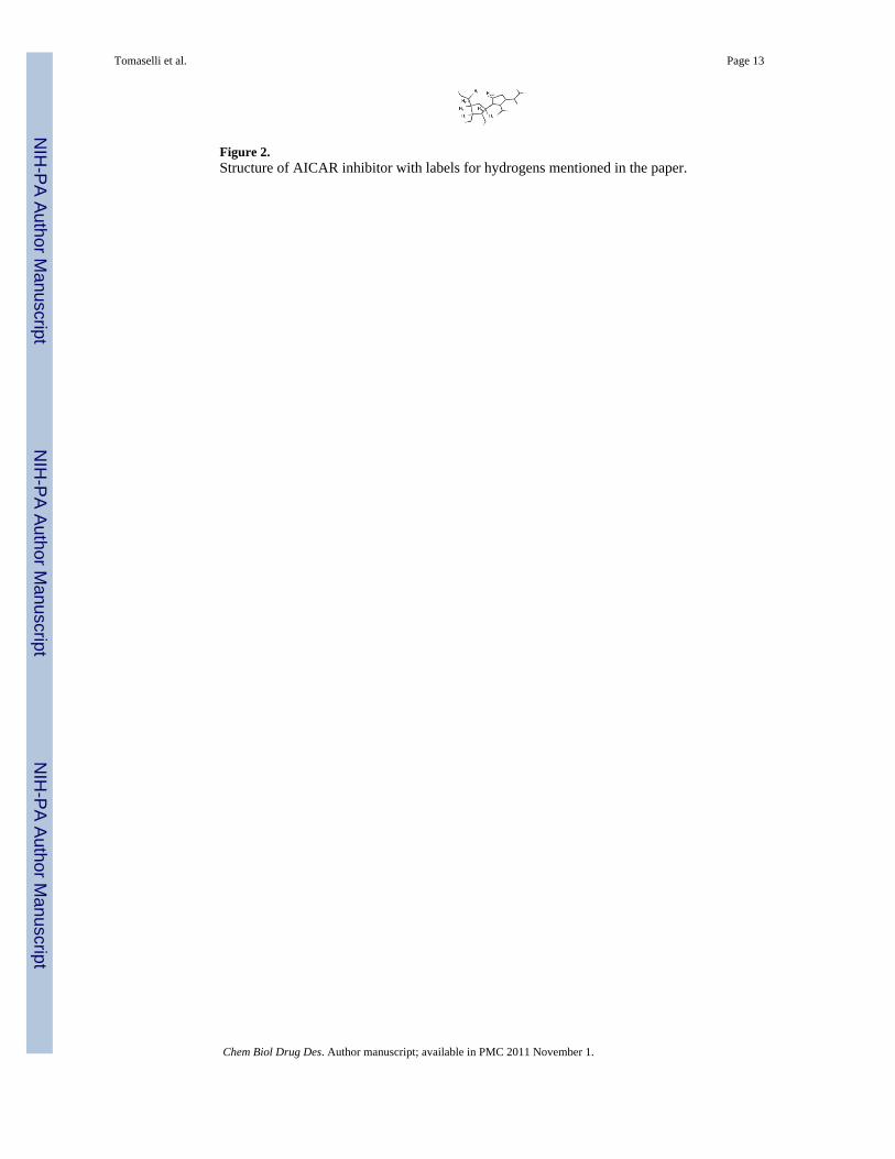
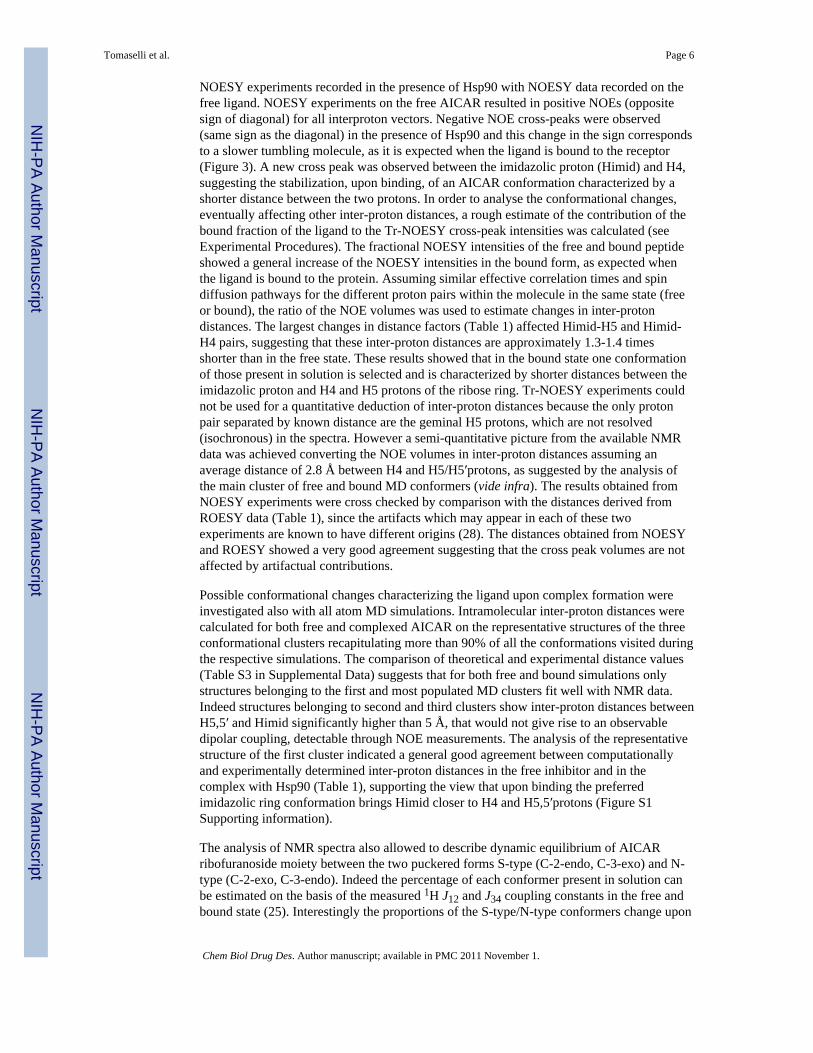
Figure 2.Structure of AICAR inhibitor with labels for hydrogens mentioned in the paper.
Tomaselli et al. Page 13
Chem Biol Drug Des. Author manuscript; available in PMC 2011 November 1.
NIH
-PA Author Manuscript
NIH
-PA Author Manuscript
NIH
-PA Author Manuscript
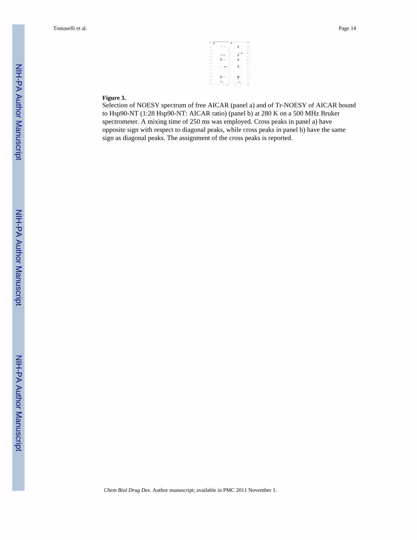
Figure 3.Selection of NOESY spectrum of free AICAR (panel a) and of Tr-NOESY of AICAR boundto Hsp90-NT (1:28 Hsp90-NT: AICAR ratio) (panel b) at 280 K on a 500 MHz Brukerspectrometer. A mixing time of 250 ms was employed. Cross peaks in panel a) haveopposite sign with respect to diagonal peaks, while cross peaks in panel b) have the samesign as diagonal peaks. The assignment of the cross peaks is reported.
Tomaselli et al. Page 14
Chem Biol Drug Des. Author manuscript; available in PMC 2011 November 1.
NIH
-PA Author Manuscript
NIH
-PA Author Manuscript
NIH
-PA Author Manuscript
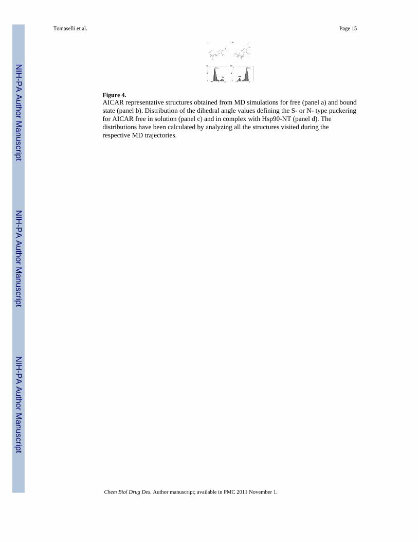
Figure 4.AICAR representative structures obtained from MD simulations for free (panel a) and boundstate (panel b). Distribution of the dihedral angle values defining the S- or N- type puckeringfor AICAR free in solution (panel c) and in complex with Hsp90-NT (panel d). Thedistributions have been calculated by analyzing all the structures visited during therespective MD trajectories.
Tomaselli et al. Page 15
Chem Biol Drug Des. Author manuscript; available in PMC 2011 November 1.
NIH
-PA Author Manuscript
NIH
-PA Author Manuscript
NIH
-PA Author Manuscript
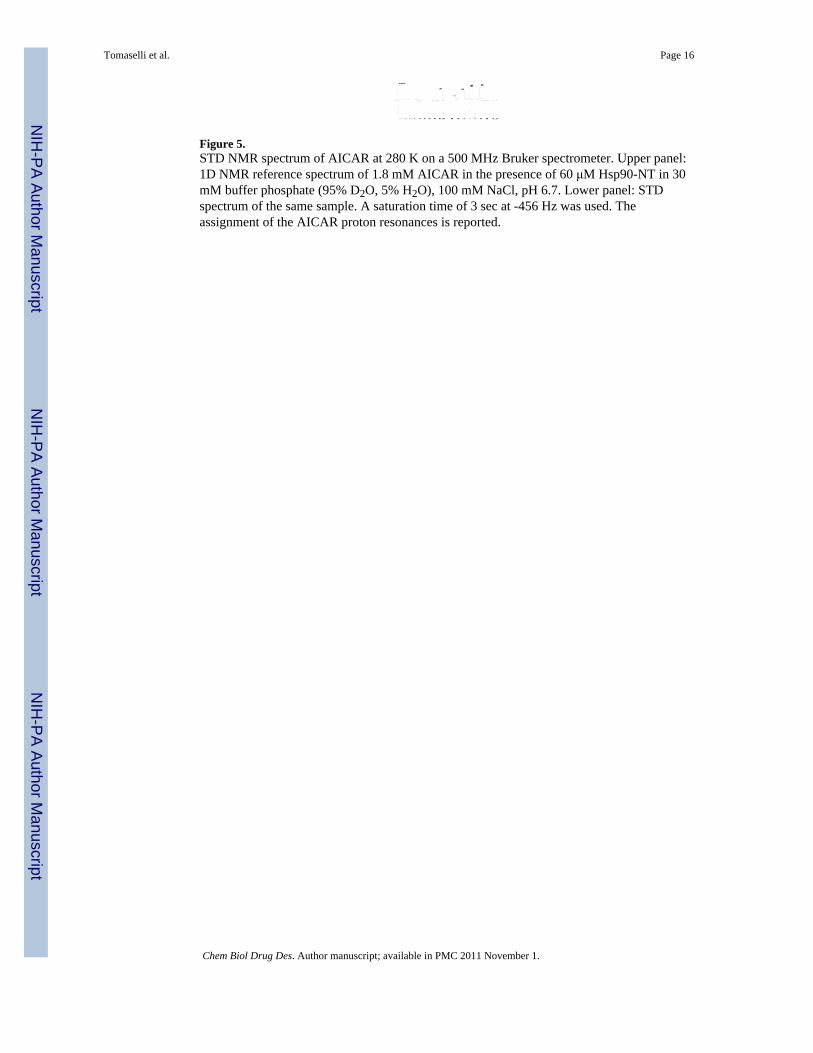
Figure 5.STD NMR spectrum of AICAR at 280 K on a 500 MHz Bruker spectrometer. Upper panel:1D NMR reference spectrum of 1.8 mM AICAR in the presence of 60 μM Hsp90-NT in 30mM buffer phosphate (95% D2O, 5% H2O), 100 mM NaCl, pH 6.7. Lower panel: STDspectrum of the same sample. A saturation time of 3 sec at -456 Hz was used. Theassignment of the AICAR proton resonances is reported.
Tomaselli et al. Page 16
Chem Biol Drug Des. Author manuscript; available in PMC 2011 November 1.
NIH
-PA Author Manuscript
NIH
-PA Author Manuscript
NIH
-PA Author Manuscript
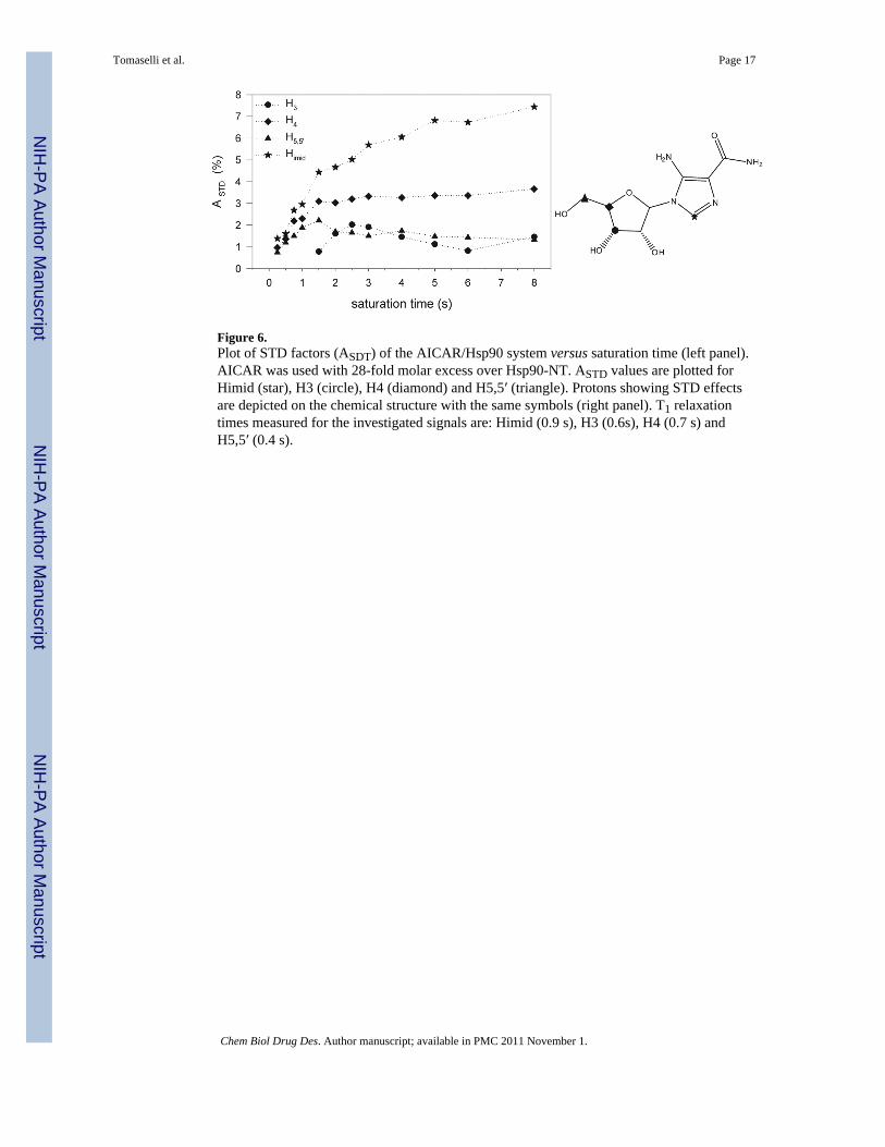
Figure 6.Plot of STD factors (ASDT) of the AICAR/Hsp90 system versus saturation time (left panel).AICAR was used with 28-fold molar excess over Hsp90-NT. ASTD values are plotted forHimid (star), H3 (circle), H4 (diamond) and H5,5′ (triangle). Protons showing STD effectsare depicted on the chemical structure with the same symbols (right panel). T1 relaxationtimes measured for the investigated signals are: Himid (0.9 s), H3 (0.6s), H4 (0.7 s) andH5,5′ (0.4 s).
Tomaselli et al. Page 17
Chem Biol Drug Des. Author manuscript; available in PMC 2011 November 1.
NIH
-PA Author Manuscript
NIH
-PA Author Manuscript
NIH
-PA Author Manuscript
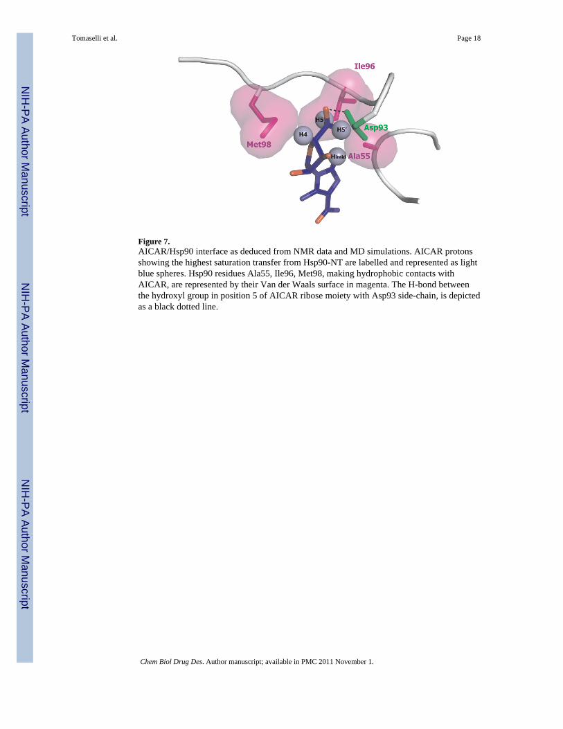
Figure 7.AICAR/Hsp90 interface as deduced from NMR data and MD simulations. AICAR protonsshowing the highest saturation transfer from Hsp90-NT are labelled and represented as lightblue spheres. Hsp90 residues Ala55, Ile96, Met98, making hydrophobic contacts withAICAR, are represented by their Van der Waals surface in magenta. The H-bond betweenthe hydroxyl group in position 5 of AICAR ribose moiety with Asp93 side-chain, is depictedas a black dotted line.
Tomaselli et al. Page 18
Chem Biol Drug Des. Author manuscript; available in PMC 2011 November 1.
NIH
-PA Author Manuscript
NIH
-PA Author Manuscript
NIH
-PA Author Manuscript
NIH
-PA Author Manuscript
NIH
-PA Author Manuscript
NIH
-PA Author Manuscript
Tomaselli et al. Page 19
Tabl
e 1
Com
paris
on o
f pro
ton
dist
ance
s in
free
and
bou
nd A
ICA
R fr
om N
MR
and
MD
cal
cula
tions
a
Cro
ss p
eak
Dis
tanc
e fa
ctor
from
NO
ESY
NO
E d
ista
nce
(Å)
RO
E d
ista
nce
(Å)
MD
dis
tanc
e(Å
)
Free
Bou
ndFr
eeB
ound
Free
Bou
ndFr
eeB
ound
H1-
H2
1.2
1.1
3.3
3.1
3.3
2.7
3.1
2.7
H1-
H4
1.3
1.2
3.8
3.4
3.5
3.0
3.4
3.5
H1-
H5*
> 1.
7b1.
7>
5b4.
8>
5b>
5b4.
3, 5
.04.
4, 5
.2
H2-
H3
1.0
0.9
2.8
2.6
--
2.4
2.4
H3-
H5*
1.1
1.2
3.2
3.3
4.2
3.3
2.5,
2.9
2.5,
2.8
H4-
H5*
1.0
1.0
2.8
2.8
2.8
2.8
2.5,
3.0
2.7,
3.0
Him
id-H
11.
00.
92.
92.
72.
72.
83.
63.
6
Him
id-H
21.
01.
02.
82.
92.
73.
23.
64.
3
Him
id-H
31.
31.
23.
73.
33.
73.
05.
23.
2
Him
id-H
4>
1.7b
1.3
> 5b
3.8
> 5b
3.5
4.4
4.3
Him
id-H
5*1.
71.
34.
83.
84.
53.
94.
7, 3
.23.
8, 2
.3
a The
aver
age
dist
ance
obs
erve
d fo
r H4-
H5
pair
from
MD
sim
ulat
ions
in fr
ee a
nd b
ound
AIC
AR
(2.8
Å) w
as u
sed
to d
educ
e a
roug
h es
timat
e of
dis
tanc
es fr
om N
OE/
RO
E cr
oss p
eaks
vol
umes
.
b the
cros
s pea
k w
as n
ot o
bser
ved
in th
e sp
ectru
m.
Chem Biol Drug Des. Author manuscript; available in PMC 2011 November 1.
Related Documents