RESEARCH ARTICLE Open Access Combined immune checkpoint protein blockade and low dose whole body irradiation as immunotherapy for myeloma Weiqing Jing 1 , Jill A Gershan 1 , James Weber 1 , Dominique Tlomak 1 , Laura McOlash 1 , Catherine Sabatos-Peyton 2 and Bryon D Johnson 1* Abstract Background: Multiple myeloma is characterized by the presence of transformed neoplastic plasma cells in the bone marrow and is generally considered to be an incurable disease. Successful treatments will likely require multi-faceted approaches incorporating conventional drug therapies, immunotherapy and other novel treatments. Our lab previously showed that a combination of transient lymphodepletion (sublethal whole body irradiation) and PD-1/PD-L1 blockade generated anti-myeloma T cell reactivity capable of eliminating established disease. We hypothesized that blocking a combination of checkpoint receptors in the context of low-dose, lymphodepleting whole body radiation would boost anti-tumor immunity. Methods: To test our central hypothesis, we utilized a 5T33 murine multiple myeloma model. Myeloma-bearing mice were treated with a low dose of whole body irradiation and combinations of blocking antibodies to PD-L1, LAG-3, TIM-3, CD48 (the ligand for 2B4) and CTLA4. Results: Temporal phenotypic analysis of bone marrow from myeloma-bearing mice demonstrated that elevated percentages of PD-1, 2B4, LAG-3 and TIM-3 proteins were expressed on T cells. When PD-L1 blockade was combined with blocking antibodies to LAG-3, TIM-3 or CTLA4, synergistic or additive increases in survival were observed (survival rates improved from ~30% to >80%). The increased survival rates correlated with increased frequencies of tumor-reactive CD8 and CD4 T cells. When stimulated in vitro with myeloma cells, CD8 T cells from treated mice produced elevated levels proinflammatory cytokines. Cytokines were spontaneously released from CD4 T cells isolated from mice treated with PD-L1 plus CTLA4 blocking antibodies. Conclusions: These data indicate that blocking PD-1/PD-L1 interactions in conjunction with other immune checkpoint proteins provides synergistic anti-tumor efficacy following lymphodepletive doses of whole body irradiation. This strategy is a promising combination strategy for myeloma and other hematologic malignancies. Keywords: Low dose whole body irradiation, Immune checkpoint proteins, Blockade, PD-L1, TIM-3, LAG-3, CTLA4, 2B4, Myeloma Background Reports of immunotherapy-induced clinical responses have brought the study of tumor immunity front and center in the mission to eliminate cancer. Central to tumor immun- ity is the killing potential of activated tumor-specific T cells. Tumor-specific T cells can be detected in both preclinical animal models and cancer patients, but due to multiple im- mune suppressive factors within the tumor microenviron- ment, T cells fail to maintain an activated state against progressing tumor and are rendered tolerant or exhausted. T cell activation is initiated through tumor antigen recogni- tion by the T cell receptor (TCR) and is regulated by a balance of activation and inhibitory intracellular signals. These signals are initiated by engagement of co-stimulatory and co-inhibitory receptors with their cognate ligands. One of the promising approaches to induce and maintain * Correspondence: [email protected] 1 Division of Hematology/Oncology/Transplant, Department of Pediatrics, Medical College of Wisconsin, Milwaukee, WI 53226, USA Full list of author information is available at the end of the article © 2015 Jing et al.; licensee BioMed Central. This is an Open Access article distributed under the terms of the Creative Commons Attribution License (http://creativecommons.org/licenses/by/4.0), which permits unrestricted use, distribution, and reproduction in any medium, provided the original work is properly credited. The Creative Commons Public Domain Dedication waiver (http://creativecommons.org/publicdomain/zero/1.0/) applies to the data made available in this article, unless otherwise stated. Jing et al. Journal for ImmunoTherapy of Cancer (2015) 3:2 DOI 10.1186/s40425-014-0043-z

Welcome message from author
This document is posted to help you gain knowledge. Please leave a comment to let me know what you think about it! Share it to your friends and learn new things together.
Transcript
Jing et al. Journal for ImmunoTherapy of Cancer (2015) 3:2 DOI 10.1186/s40425-014-0043-z
RESEARCH ARTICLE Open Access
Combined immune checkpoint protein blockadeand low dose whole body irradiation asimmunotherapy for myelomaWeiqing Jing1, Jill A Gershan1, James Weber1, Dominique Tlomak1, Laura McOlash1, Catherine Sabatos-Peyton2
and Bryon D Johnson1*
Abstract
Background: Multiple myeloma is characterized by the presence of transformed neoplastic plasma cells in thebone marrow and is generally considered to be an incurable disease. Successful treatments will likely require multi-facetedapproaches incorporating conventional drug therapies, immunotherapy and other novel treatments. Our lab previouslyshowed that a combination of transient lymphodepletion (sublethal whole body irradiation) and PD-1/PD-L1 blockadegenerated anti-myeloma T cell reactivity capable of eliminating established disease. We hypothesized that blockinga combination of checkpoint receptors in the context of low-dose, lymphodepleting whole body radiation wouldboost anti-tumor immunity.
Methods: To test our central hypothesis, we utilized a 5T33 murine multiple myeloma model. Myeloma-bearing micewere treated with a low dose of whole body irradiation and combinations of blocking antibodies to PD-L1, LAG-3,TIM-3, CD48 (the ligand for 2B4) and CTLA4.
Results: Temporal phenotypic analysis of bone marrow from myeloma-bearing mice demonstrated that elevatedpercentages of PD-1, 2B4, LAG-3 and TIM-3 proteins were expressed on T cells. When PD-L1 blockade was combinedwith blocking antibodies to LAG-3, TIM-3 or CTLA4, synergistic or additive increases in survival were observed (survivalrates improved from ~30% to >80%). The increased survival rates correlated with increased frequencies oftumor-reactive CD8 and CD4 T cells. When stimulated in vitro with myeloma cells, CD8 T cells from treated miceproduced elevated levels proinflammatory cytokines. Cytokines were spontaneously released from CD4 T cellsisolated from mice treated with PD-L1 plus CTLA4 blocking antibodies.
Conclusions: These data indicate that blocking PD-1/PD-L1 interactions in conjunction with other immunecheckpoint proteins provides synergistic anti-tumor efficacy following lymphodepletive doses of whole body irradiation.This strategy is a promising combination strategy for myeloma and other hematologic malignancies.
Keywords: Low dose whole body irradiation, Immune checkpoint proteins, Blockade, PD-L1, TIM-3, LAG-3, CTLA4,2B4, Myeloma
BackgroundReports of immunotherapy-induced clinical responses havebrought the study of tumor immunity front and center inthe mission to eliminate cancer. Central to tumor immun-ity is the killing potential of activated tumor-specific T cells.Tumor-specific T cells can be detected in both preclinical
* Correspondence: [email protected] of Hematology/Oncology/Transplant, Department of Pediatrics,Medical College of Wisconsin, Milwaukee, WI 53226, USAFull list of author information is available at the end of the article
© 2015 Jing et al.; licensee BioMed Central. ThCommons Attribution License (http://creativecreproduction in any medium, provided the orDedication waiver (http://creativecommons.orunless otherwise stated.
animal models and cancer patients, but due to multiple im-mune suppressive factors within the tumor microenviron-ment, T cells fail to maintain an activated state againstprogressing tumor and are rendered tolerant or exhausted.T cell activation is initiated through tumor antigen recogni-tion by the T cell receptor (TCR) and is regulated by abalance of activation and inhibitory intracellular signals.These signals are initiated by engagement of co-stimulatoryand co-inhibitory receptors with their cognate ligands. Oneof the promising approaches to induce and maintain
is is an Open Access article distributed under the terms of the Creativeommons.org/licenses/by/4.0), which permits unrestricted use, distribution, andiginal work is properly credited. The Creative Commons Public Domaing/publicdomain/zero/1.0/) applies to the data made available in this article,
Jing et al. Journal for ImmunoTherapy of Cancer (2015) 3:2 Page 2 of 15
tumor-specific T cells in an activated state is to inter-fere with signaling through inhibitory (also referred toas immune checkpoint) receptors.There are multiple known T cell checkpoint receptors,
and there is evidence that blocking interaction of thesereceptors with their respective ligands can increase anti-tumor immune responses. One of the most studiedcheckpoint receptors is CTLA4. Cell surface CTLA4 ex-pression is rapidly upregulated when T cells are activated,and it is constitutively expressed on Foxp3+ regulatory Tcells [1]. Signaling through CTLA4 arrests T cell activationby outcompeting co-stimulatory receptors (CD80 andCD86) for binding to CD28. CTLA4 binding to CD28 re-sults in reduced T cell survival, cytokine production andT cell cycle arrest [2]. Testament to the importance ofCTLA4 in dampening T cell activation is the occurrenceof a lethal polyclonal lymphoproliferative disease that oc-curs in CTLA4 knockout mice [3]. Antagonistic anti-CTLA4 antibodies have been extensively tested in cancermodels as a strategy to activate anti-tumor immunity,and CTLA4 was the first immune checkpoint targetedin the clinic for cancer therapy. The anti-tumor effectsassociated with blocking CTLA4 in vivo have been shownto involve depletion of regulatory T cells as well as restor-ing effector T cell function [4,5]. Notably, CTLA4 block-ade results in increased ratios of effector CD8 T cells toregulatory T cells in tumors, possibly due to higher levelsof CTLA4 expression by regulatory T cells [4]. In 2010,a phase III randomized controlled clinical trial showedprolonged survival of metastatic melanoma patients whentreated with the anti-CTLA4 antibody ipilimumab [6].In melanoma patients, blocking CTLA4 produced ahost of immune-related toxic side effects (referred to asimmune-related adverse events). However, based on thepromising responses in melanoma patients, ipilimumabwas the first checkpoint-blocking antibody to be FDA ap-proved (for the treatment of melanoma).Our laboratory has focused on blocking the checkpoint
receptor programmed death receptor-1 (PD-1) pathway inthe treatment of myeloma. PD-1 (CD279) is an im-munoglobulin superfamily transmembrane receptor thatis expressed on activated T cells, regulatory T cells, B cellsand NK cells. Ligands for PD-1 include PD-L1 (B7-H1,CD274) and PD-L2 (B7-DC, CD273) [7]. PD-L2 expres-sion is restricted to hematopoietic cells, notably myeloidcells including dendritic cells and macrophages, butPD-L1 is broadly expressed on hematopoietic and non-hematopoietic cells as well as on a variety of murineand human malignancies [8,9]. Most of data showingthe anti-tumor efficacy induced by blocking the PD-1/PD-L1 inhibitory receptor axis has been generated frompreclinical and clinical studies involving solid tumors. Pre-clinical studies have shown that blocking the PD-L1/PD-1axis with anti-PD-L1 or anti-PD-1 antibodies promotes
anti-tumor T cell responses in pancreatic carcinoma [10],B16 melanoma [11], and CT26 colon carcinoma [12]. In arecent clinical study, patients with colorectal cancer, renalcell carcinoma or melanoma showed objective responsesto anti-PD-L1 therapy [13]. Patients with PD-L1+ tumors,but not PD-L1− tumors, showed objective responses whentreated with an anti-PD-1 blocking antibody [14]. Clinic-ally, combining anti-CTLA4 (ipilimumab) with anti-PD-1(nivolumab) antibodies resulted in even greater anti-tumor efficacy, as tumor regression occurred in 80% ofpatients with advanced melanoma [15]. Importantly, im-mune adverse events were qualitatively similar to that expe-rienced with prior treatment of either antibody alone.Less well-characterized T cell immune checkpoint re-
ceptors include lymphocyte activating gene 3 (LAG-3 orCD223), T cell immunoglobulin and mucin domain 3(TIM-3), 2B4 (CD244), and others. LAG-3 is a memberof the immunoglobulin (Ig) superfamily that binds toMHC class II molecules, and has recently been reportedto also bind L-selectin [16]. LAG-3 is expressed on acti-vated and tolerized T cells, NK cells, plasmacytoid den-dritic cells, and regulatory T cells and it is known tonegatively regulate the expansion of activated T cells[17,18]. Preclinical studies have shown that combinedtreatment of LAG-3 and PD-1 blocking antibodies pro-vided a synergistic anti-tumor effect [19]. The TIM fam-ily of transmembrane receptor proteins includes severalmembers (TIM-1, 2, 3 and 4 in mice, but only TIM-1, 3and 4 are known to be expressed in humans). Ligationof TIM-1 regulates Th2 CD4 T cell responses, and inmice, TIM-1 promotes CD4 T cell activation [20]. TIM-3 is a checkpoint receptor that is co-expressed on PD-1+
CD8 T cells in mice harboring solid or hematologic malig-nancies [21,22]. The ligand for TIM-3 is galectin-9 whichis expressed by multiple tumors. Reduced galactin-9 ex-pression correlates with reduced disease progression in amajority of solid tumors [23]. PD-1+TIM-3+ T cells de-rived from patients with melanoma, non-small cell lungcancer or non-Hodgkin’s lymphoma are defective in pro-liferation and cytokine production [24-26]. TIM-3 is alsoexpressed on regulatory T cells, monocytes, NK cells, anddendritic cells [27]. Data suggests that TIM-3 can play anti-inflammatory or pro-inflammatory roles in cells dependingon the physiologic setting [28,29]. In a preclinical mouseB16F10 melanoma model, combined blockade of TIM-3 andPD-1, or TIM-3 and CTLA4, was more effective in prolong-ing survival than blocking either protein alone [30]. Inaddition, the combination of anti-CTLA4, anti-TIM-3 andanti-LAG-3 produced further suppression of B16F10 tumorgrowth [30]. These data demonstrate a mechanistic synergywhen multiple inhibitory receptors are blocked.2B4 (CD244) engagement with CD48 was originally
described as facilitating CD8 T cell proliferation [31]. Re-cent data examining hepatitis C-virus (HCV)-specific
Jing et al. Journal for ImmunoTherapy of Cancer (2015) 3:2 Page 3 of 15
CD8 T cells showed that crosslinking 2B4 on CD8 T cellswith low versus high 2B4 expression increased or de-creased proliferation, respectively, and 2B4 blockadepreferentially increased proliferation of HCV CD8 T cellswith high 2B4 expression [32]. Similar to other checkpointproteins, 2B4 is upregulated on exhausted virus-specificCD8 T cells [33]. Together these data suggest that 2B4plays a role in the regulation of CD8 T cells.Despite the promising results afforded by blocking
CTLA4 and the PD-1/PD-L1 axis in the treatment ofsolid tumors, targeting these checkpoints in hematologicmalignancies has been relatively understudied. Multiplemyeloma is a hematologic malignancy involving plasmacells. In humans, PD-L1 is expressed on CD138+ malig-nant plasma cells [34-38]. We have shown that PD-1 isupregulated on peripheral blood and bone marrow inmyeloma patients up to 30 days following autologoustransplant [35]. In humans, blocking the PD-L1/PD-1axis may act to prevent inhibitory signaling when ef-fector T cells engage with tumor cells and when T cellsare undergoing homeostatic expansion.Our preclinical studies have demonstrated improved
anti-myeloma T cell immunity when the PD-1/PD-L1axis is blocked. Using a murine model of myeloma, weshowed that administration of an anti-PD-L1 blockingantibody elicits rejection of PD-L1 expressing tumor cells.When anti-PD-L1 was administered immediately follow-ing hematopoietic stem cell transplantation in combin-ation with a tumor cell-based vaccine, myeloma waseliminated in approximately 40% of treated mice [35].We also reported that 5T33 tumor was eliminated inapproximately 50% of mice when treated with anti-PD-L1 following radiation-induced lymphopenia [39]. Basedon these data and the data of others, we hypothesized thatblocking a combination of checkpoint receptors in thecontext of lymphodepleting radiation would boost anti-tumor immunity. The results presented here confirm thehypothesis and show that PD-1/PD-L1 blockade in combin-ation with either TIM-3, LAG-3 or CTLA4 blockade syner-gistically improves the survival of myeloma bearing mice.
ResultsIncreased PD-1, TIM-3, LAG-3 and 2B4 expression on CD4and CD8 T cells directly correlates with myeloma burdenOur previous work demonstrated that the PD-1/PD-L1pathway is important in suppressing immune responsesto 5T33 myeloma, and that PD-1 expression on T cellsis related to 5T33 burden in the myeloma-resident tis-sues (bone marrow, spleen and liver) [35,39]. Besides ex-pression of the immune checkpoint protein PD-1, T cellswithin tumor environments may develop a dysfunctionalphenotype accompanied by the increased expression ofother checkpoint proteins. We therefore examined CD4and CD8 T cells for the expression of other checkpoint
receptors over time in myeloma-bearing animals. GFP+
5T33 cells could be observed in the bone marrow asearly as 7 days after iv inoculation, with increasing accu-mulation of myeloma cells over time (Figure 1A). Whenmyeloma-bearing mice became moribund (29-38 daysafter 5T33 inoculation), 20-35% of the cells in the bonemarrow consisted of GFP+ tumor cells. As previously de-scribed [39], there was a significant increase in the per-centages of PD-1+ bone marrow-derived CD4 and CD8T cells as early as 21 days after myeloma inoculation(Figure 1B). As myeloma burden progressed, the percent-ages of bone marrow-derived PD-1+ CD4 and CD8 T cellsincreased to 40-70% and 30-50%, respectively. Similar tothe increase in PD-1 expression, increasing percentages ofT cells in the bone marrow expressed TIM-3, LAG-3 and2B4 as myeloma burden progressed (Figure 1B). In con-trast, percentages of T cells expressing TIM-1 or BTLA didnot increase over time. Similar results were also observedin the spleen (Additional file 1: Figure S1). Together, thesedata show that there is upregulated expression of multiplecheckpoint receptors on T cells in tissues where myelomacells are present, and that an accumulation of checkpoint-expressing T cells occurs over time. In order to show thatcheckpoint receptors are co-expressed on T cells, CD4and CD8 cells in the bone marrow of moribund mice wereanalyzed for PD-1, TIM-3, LAG-3 and 2B4 expressionusing multicolor flow cytometry. For CD8 T cells(Figure 1C, top row), PD-1 was co-expressed on 85% ofTIM-3+ cells, 67% of LAG-3+ cells, and 51% of 2B4+ cells.For CD4 T cells (Figure 1C, bottom row), co-expression ofPD-1 was observed on 90% of TIM-3+ cells, 47% ofLAG-3+ cells, and 44% of 2B4+ cells. Notably, there werealso PD-1+ cells that did not co-express the other check-points (upper left quadrants in Figure 1C), as well asmajor subsets of LAG-3+ and 2B4+ cells that did notco-express PD-1 (lower right quadrants in Figure 1C).Next, we determined whether checkpoint ligands for
TIM-3, LAG-3 and CTLA4 were expressed on the tumorand cells within the tumor microenvironment (i.e., spleen).PD-L1 is highly expressed on 5T33 myeloma cells [35], butthe ligands for TIM-3 (galectin-9), LAG-3 (MHC class II),and CTLA4 (CD80) are not (Additional file 2: Figure S2A).In vitro irradiation with 500 cGy did not change thegalectin-9, MHC class II or CD80 expression on 5T33myeloma cells. Not surprisingly, galectin-9, MHC class IIand CD80 is present on a variety of cells in the tumormicroenvironment including B cells, macrophages, DCsand monocytes (Additional file 2: Figure S2B).
Immune checkpoint protein expression is increased onCD8 T cells in mice treated with lymphodepletingradiation and anti-PD-L1We previously showed that blocking the PD-1/PD-L1 axiswith a PD-L1-specific monoclonal antibody synergized

Figure 1 (See legend on next page.)
Jing et al. Journal for ImmunoTherapy of Cancer (2015) 3:2 Page 4 of 15

(See figure on previous page.)Figure 1 Expression of immune checkpoint proteins on T cells in bone marrow of myeloma bearing mice over time. KaLwRij mice wereinoculated with 2×10⁶ 5T33-GFP cells iv. Myeloma bearing mice were euthanized between days 7 and 28 and when moribund at days 29-40 aftermyeloma injection. Femoral bone marrow cells were harvested and (A) GFP+ tumor cell accumulation was monitored by flow cytometry. CD4+
and CD8+ T cells in the bone marrow were analyzed by flow cytometry for expression of (B) PD-1, TIM-3, LAG-3, BTLA and 2B4 at each of the timepoints indicated, or for surface expression of (C) CTLA4 when animals were moribund. T cells harvested from naïve non-myeloma bearing micewere analyzed as controls. Immune checkpoint protein percentages were calculated based on isotype controls. (D) Expression of TIM-3 and PD-1,LAG-3 and PD-1 or 2B4 and PD-1 on gated CD8+ or CD4+ T cells harvested from the BM of moribund mice. Data shown are representative ofmore than four independent analyses. *p < 0.05, **p < 0.01, ***p < 0.001 as compared to T cells from naïve non-myeloma bearing mice.
Jing et al. Journal for ImmunoTherapy of Cancer (2015) 3:2 Page 5 of 15
with lymphodepleting whole body irradiation (WBI) to fa-cilitate a T cell-mediated anti-myeloma response [39]. Todetermine the influence of this treatment on T cell im-mune checkpoint expression, mice with established mye-loma were treated with 500 cGy WBI 7 days after 5T33inoculation, followed by three treatments with anti–PD-L1 or control IgG on days 5, 7 and 12 after WBI. On day14 after WBI, CD8 T cells were harvested from bone mar-row and analyzed for expression of PD-1, TIM-3, LAG-3and 2B4 by flow cytometry. There were significant in-creases in the percentages of CD8 T cells that expressedTIM-3, LAG-3 or 2B4 in mice treated with anti-PD-L1 ascompared to controls treated with IgG1 (Figure 2A).Interestingly, the mice treated with anti-PD-L1 also had amarked increase in percentages of PD-1+ CD8 T cells(Figure 2B), and relatively large percentages of the PD-1+
cells co-expressed TIM-3 (52%), LAG-3 (60%) or 2B4(40%) (Figure 2B).
Blocking PD-L1 in combination with TIM-3, LAG-3 orCTLA4 blockade synergize to improve the survival oflymphodepleted myeloma-bearing miceSince multiple immune checkpoint proteins are upregu-lated on T cells in myeloma-bearing mice (Figure 1), andblocking the PD-L1/PD-1 axis in lymphodepleted ani-mals induces increased expression of LAG-3, TIM-3 and2B4 checkpoint proteins (Figure 2), we hypothesized thatanti-myeloma immunity would be enhanced by blockingcombinations of immune checkpoints. Mice were inocu-lated with 2×10⁶ 5T33 tumor cells iv, received 500 cGyWBI 7 days after tumor cell inoculation, and were treatedwith checkpoint blocking antibodies (200 ug of eachantibody ip) at the time points indicated in Figure 3A.Administration of anti-PD-L1 alone eliminated mye-loma in ~40% of mice (Figure 2B-E), which is consist-ent with our previous results [39]. While treatmentwith anti-TIM-3 or anti-LAG-3 alone had no affect onsurvival (Figure 2B, C), co-administration of eitherantibody with anti-PD-L1 synergistically improved sur-vival rates to greater than 80% (Figure 2B, C). Thecombination of anti-TIM-3 with anti-LAG-3 failed toimprove survival (Figure 2C). Therefore, PD-L1 block-ade was necessary in order to see any survival benefitfrom blocking the two other checkpoints. Survival was
also significantly improved by combining anti-PD-L1with anti-CTLA4 (Figure 2D). Anti-CTLA4 alone alsofacilitated the elimination of myeloma in approximately15% of animals. Since a blocking antibody to 2B4 was notavailable, an antibody to the 2B4 ligand, CD48, was usedto block the 2B4/CD48 axis. As shown in Figure 3E, anti-CD48 administered alone or with anti-PD-L1 failed tohave any impact on the elimination of myeloma.In order to test if survivors from the experiments in
Figure 3B-D had developed anti-tumor memory, theywere challenged with 1×10⁶ 5T33 tumor cells iv 100-110days after the initial tumor cell inoculation and followedfor tumor development. All survivors of the experimentsin Figure 3B-D survived the 5T33 re-challenge, indicat-ing that blocking the indicated combinations of check-point receptors does not compromise anti-tumorimmune memory (Figure 3F).
Combined checkpoint blockade increases the frequencyof myeloma-reactive CD8 and CD4 T cellsSince previous data from our laboratory demonstrated thatthe increased survival of myeloma-bearing mice treatedwith lymphodepleting WBI and anti-PD-L1 is T cell medi-ated [39], we set out to determine if combined checkpointblockade increases numbers of functional tumor-reactive Tcells. To do this, mice were treated according to the sched-ule in Figure 3A, but instead of 6 doses of blockingantibody, they received 3 doses on days 12, 14 and 19 fol-lowing tumor cell injection. Twenty-one days after tumorcell injection, T cells were harvested from the spleens andbone marrow and CD4 and CD8 T cells were enriched byimmunomagnetic cell sorting. Frequencies of tumor-reactive IFN-γ-producing cells were then assessed in ELI-SPOT assays. CD8 T cells were stimulated with wild-type5T33 cells, while MHC class II-expressing 5T33 cells (engi-neered to express CIITA) were used to stimulate CD4 Tcells. Both spleen and bone marrow-derived CD4 and CD8T cells showed a significant increase in tumor-specific IFN-γ producing cells when mice were treated with anti-PD-L1in combination with anti-TIM-3, or anti-LAG-3 or anti-CTLA4 as compared to anti-PD-L1 alone (Figure 4A).To determine if there were differences in bulk cytokine
production, splenic CD8 T cells were co-cultured with5T33 tumor cells for 48 hours, supernatants were collected,

Figure 2 Expression of immune checkpoint proteins are increased on T cells in mice treated with sublethal whole body irradiation andanti-PD-L1. Myeloma bearing KaLwRij mice were treated with 500 cGy whole body irradiation 7 days after tumor cell injection. Treatment withanti-PD-L1 or control IgG (200 μg ip) was initiated 5 days later and specifically given 12, 14, and 19 days after tumor injection. Mice were euthanizedat day 21, splenocytes were harvested, and the CD8 T cells were analyzed by flow cytometry for immune checkpoint protein expression. (A) Frequencyof CD8+TIM-3+, CD8+LAG-3+ and CD8+2B4+ cells in spleens of anti-PD-L1 treated mice as compared with spleens harvested from control antibody(IgG1) treated mice. ***p < 0.001. (B) Expression of TIM-3 and PD-1, LAG-3 and PD-1 or 2B4 and PD-1 on gated CD8+ T cells. Data shown arerepresentative of more than four independent analyses.
Jing et al. Journal for ImmunoTherapy of Cancer (2015) 3:2 Page 6 of 15
and cytokine secretion was determined using a multiplexcytokine assay. Similar to results of the IFN-γ ELISPOT as-says, there was a significant increase in type 1 cytokines(IL-2, IFN-γ and GM-CSF) produced by CD8 T cells har-vested from mice that had received anti-PD-L1 in combin-ation with anti-TIM-3, or anti-LAG-3 or anti-CTLA4 ascompared to anti-PD-L1 alone (Figure 4B). Notably, CD8T cells from mice treated with anti-PD-L1 in combinationwith anti-CTLA4 produced at least 2-fold higher concen-trations of cytokines as compared to mice treated withanti-PD-L1 in combination with anti-TIM-3 or anti-LAG-3.
Combined checkpoint blockade results in increasedexpression of PD-1 on T cells, and ongoing PD-L1 blockadein vitro results in elevated numbers of IFN-γ producing cellsThere is data suggesting that expression of inhibitory re-ceptors, including PD-1, correlates with T cell activationand/or differentiation rather than exhaustion [40]. Otherinvestigators have also shown that the majority of vaccine-
induced CD8+ T cells upregulate PD-1 [41], and PD-1 hasbeen found on clonally expanded tumor-reactive CD8+ Tcells isolated from tumors [42]. Similarly, we showed thatin mice with 5T33 myeloma, PD-1 expression was upreg-ulated only on host T cells capable of recognizing tumorantigens, and not on non-tumor-specific ovalbumin-reactive OT-1 T cells [39]. In sum, these data indicatethat PD-1 is a marker of activated tumor-specific T cells inthe cancer setting. Based on these observations, we hy-pothesized that combined immune checkpoint blockadeafter lymphodepleting WBI would result in increased per-centages of T cells that express PD-1, representing in-creased numbers of myeloma-reactive T cells. Mice weretreated as shown in Figure 3A, except they received onlythe first 3 doses of antibody. CD8 T cells were harvestedfrom the spleen and bone marrow 21 days after tumor in-jection and were analyzed for PD-1 expression. In supportof our hypothesis, we observed significant increases inpercentages of PD-1+ CD8 T cells in the spleens and bone

Figure 3 Blocking of PD-L1 in combination with TIM-3, LAG-3 or CTLA4 after lymphodepleting whole body irradiation synergisticallyimproved survival. (A) Experimental design: KaLwRij mice received 500 cGy irradiation 7 days after tumor cell injection. Treatment with blockingantibody or control IgG (200 μg ip) was initiated 5 days later and specifically given 12, 14, 19, 21, 26, and 28 days after tumor injection. Survivalcurves of mice treated with (B) anti-TIM-3 only, or in combination with anti-PD-L1, (C) anti-LAG-3 only, or in combination with anti-PD-L1, (D) anti-CTLA4only, or in combination with anti-PD-L1, (E) anti-CD48 only, or in combination with anti-PD-L1. Survival was compared with control antibody treated miceor mice treated with anti-PD-L1 only. Survival curves represent combined data from three (B, C, D) or two (E) independent experiments; n = 10-15 miceper experimental group. (F) Some of the survivors from panels B, C and D were re-challenged with 1×10⁶ 5T33 myeloma cells on day 110. P values weredetermined by the log-rank test.
Jing et al. Journal for ImmunoTherapy of Cancer (2015) 3:2 Page 7 of 15
marrow of mice treated with anti-PD-L1 in combinationwith anti-TIM-3, anti-LAG-3 or anti-CTLA4 as comparedto anti-PD-L1 alone (Figure 5A).To explain the increased anti-myeloma immunity after
WBI and checkpoint blockade, we have proposed thefollowing model: (a) PD-1+ tumor-reactive CD8 T cellsare rendered dysfunctional upon encounter with PD-L1
and other checkpoint ligands on the myeloma cells(5T33 expresses high levels of PD-L1) [39] or other cellsin the microenvironment, (b) that the PD-1+ cells areable to recover function after WBI due to the transientlymphopenic state through mechanisms yet to be identi-fied, and (c) ongoing checkpoint blockade is required tomaintain function of the re-activated T cells. To address
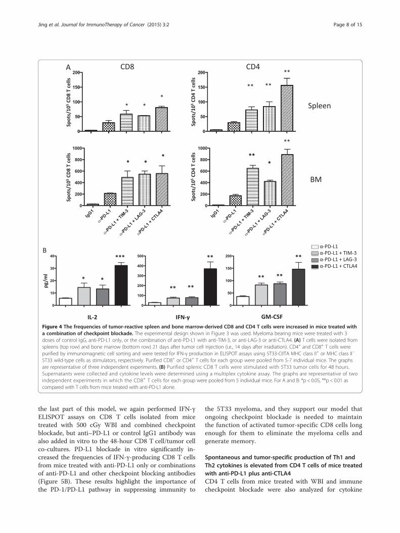
Figure 4 The frequencies of tumor-reactive spleen and bone marrow-derived CD8 and CD4 T cells were increased in mice treated witha combination of checkpoint blockade. The experimental design shown in Figure 3 was used. Myeloma bearing mice were treated with 3doses of control IgG, anti-PD-L1 only, or the combination of anti-PD-L1 with anti-TIM-3, or anti-LAG-3 or anti-CTLA4. (A) T cells were isolated fromspleens (top row) and bone marrow (bottom row) 21 days after tumor cell injection (i.e., 14 days after irradiation). CD4+ and CD8+ T cells werepurified by immunomagnetic cell sorting and were tested for IFN-γ production in ELISPOT assays using 5T33-CIITA MHC class II+ or MHC class II−
5T33 wild-type cells as stimulators, respectively. Purified CD8+ or CD4+ T cells for each group were pooled from 5-7 individual mice. The graphsare representative of three independent experiments. (B) Purified splenic CD8 T cells were stimulated with 5T33 tumor cells for 48 hours.Supernatants were collected and cytokine levels were determined using a multiplex cytokine assay. The graphs are representative of twoindependent experiments in which the CD8+ T cells for each group were pooled from 5 individual mice. For A and B: *p < 0.05, **p < 0.01 ascompared with T cells from mice treated with anti-PD-L1 alone.
Jing et al. Journal for ImmunoTherapy of Cancer (2015) 3:2 Page 8 of 15
the last part of this model, we again performed IFN-γELISPOT assays on CD8 T cells isolated from micetreated with 500 cGy WBI and combined checkpointblockade, but anti–PD-L1 or control IgG1 antibody wasalso added in vitro to the 48-hour CD8 T cell/tumor cellco-cultures. PD-L1 blockade in vitro significantly in-creased the frequencies of IFN-γ-producing CD8 T cellsfrom mice treated with anti-PD-L1 only or combinationsof anti-PD-L1 and other checkpoint blocking antibodies(Figure 5B). These results highlight the importance ofthe PD-1/PD-L1 pathway in suppressing immunity to
the 5T33 myeloma, and they support our model thatongoing checkpoint blockade is needed to maintainthe function of activated tumor-specific CD8 cells longenough for them to eliminate the myeloma cells andgenerate memory.
Spontaneous and tumor-specific production of Th1 andTh2 cytokines is elevated from CD4 T cells of mice treatedwith anti-PD-L1 plus anti-CTLA4CD4 T cells from mice treated with WBI and immunecheckpoint blockade were also analyzed for cytokine
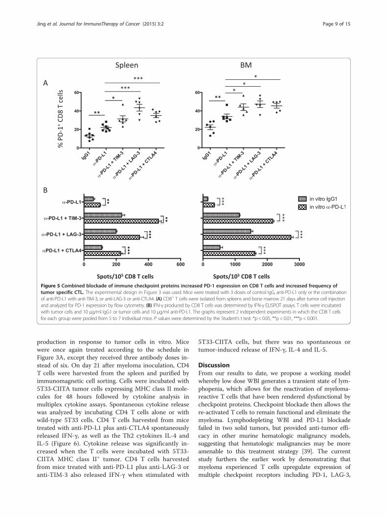
Figure 5 Combined blockade of immune checkpoint proteins increased PD-1 expression on CD8 T cells and increased frequency oftumor specific CTL. The experimental design in Figure 3 was used. Mice were treated with 3 doses of control IgG, anti-PD-L1 only or the combinationof anti-PD-L1 with anti-TIM-3, or anti-LAG-3 or anti-CTLA4. (A) CD8+ T cells were isolated from spleens and bone marrow 21 days after tumor cell injectionand analyzed for PD-1 expression by flow cytometry. (B) IFN-γ produced by CD8 T cells was determined by IFN-γ ELISPOT assays. T cells were incubatedwith tumor cells and 10 μg/ml IgG1 or tumor cells and 10 μg/ml anti-PD-L1. The graphs represent 2 independent experiments in which the CD8 T cellsfor each group were pooled from 5 to 7 individual mice. P values were determined by the Student’s t test. *p < 0.05, **p < 0.01, ***p < 0.001.
Jing et al. Journal for ImmunoTherapy of Cancer (2015) 3:2 Page 9 of 15
production in response to tumor cells in vitro. Micewere once again treated according to the schedule inFigure 3A, except they received three antibody doses in-stead of six. On day 21 after myeloma inoculation, CD4T cells were harvested from the spleen and purified byimmunomagnetic cell sorting. Cells were incubated with5T33-CIITA tumor cells expressing MHC class II mole-cules for 48 hours followed by cytokine analysis inmultiplex cytokine assays. Spontaneous cytokine releasewas analyzed by incubating CD4 T cells alone or withwild-type 5T33 cells. CD4 T cells harvested from micetreated with anti-PD-L1 plus anti-CTLA4 spontaneouslyreleased IFN-γ, as well as the Th2 cytokines IL-4 andIL-5 (Figure 6). Cytokine release was significantly in-creased when the T cells were incubated with 5T33-CIITA MHC class II+ tumor. CD4 T cells harvestedfrom mice treated with anti-PD-L1 plus anti-LAG-3 oranti-TIM-3 also released IFN-γ when stimulated with
5T33-CIITA cells, but there was no spontaneous ortumor-induced release of IFN-γ, IL-4 and IL-5.
DiscussionFrom our results to date, we propose a working modelwhereby low dose WBI generates a transient state of lym-phopenia, which allows for the reactivation of myeloma-reactive T cells that have been rendered dysfunctional bycheckpoint proteins. Checkpoint blockade then allows there-activated T cells to remain functional and eliminate themyeloma. Lymphodepleting WBI and PD-L1 blockadefailed in two solid tumors, but provided anti-tumor effi-cacy in other murine hematologic malignancy models,suggesting that hematologic malignancies may be moreamenable to this treatment strategy [39]. The currentstudy furthers the earlier work by demonstrating thatmyeloma experienced T cells upregulate expression ofmultiple checkpoint receptors including PD-1, LAG-3,

Figure 6 There is spontaneous release of Th1 and Th2 cytokines from splenic CD4 T cells harvested from mice treated with anti-PD-L1and anti-CTLA4. The experimental design shown in Figure 3 was used. Mice were treated with anti-PD-L1, or the combination of anti-PD-L1 withanti-TIM-3, or anti-LAG-3 or anti-CTLA4. T cells were isolated from spleens 21 days after tumor cell injection (14 days after irradiation). CD4 T cellswere purified by immunomagnetic cell sorting, then stimulated with MHC class II− 5T33 wild-type cells, 5T33-CIITA MHC class II+ cells or T cellsonly for 48 hours. Supernatants were collected and cytokine levels from were determined using a multiplex cytokine assay. The graphs are representativeof two independent experiments in which the CD4+ T cells for each group were pooled from 5 individual mice. p < 0.05, **p < 0.01.
Jing et al. Journal for ImmunoTherapy of Cancer (2015) 3:2 Page 10 of 15
TIM-3 and 2B4. Furthermore, treatment of myeloma-bearing mice with lymphodepletive WBI and dual check-point blockade induced a synergistic anti-myeloma effect,and this enhanced elimination of myeloma was associatedwith increased numbers of IFN-γ-producing tumor-reactive T cells and elevated cytokine production by T cellsin response to tumor antigens.Multiple immune checkpoints have been previously
shown to be upregulated on T cells in several murine solidtumor models, and targeting more than one pathway hasdemonstrated increased anti-tumor efficacy [19,21,22,43-52].However, there is a paucity of literature regarding expressionof immune checkpoints on T cells in hematologic malignan-cies or the effect of targeting more than one checkpoint astherapy for these cancers [22]. Clinically, PD-1 and CTLA4have been simultaneously targeted for the treatment of mel-anoma, and anti-tumor activity appears to be more robustthan targeting each pathway alone [15]. Notably, auto-immune manifestations with this combination did not ap-pear to be significantly worsened. In our studies, blockade ofthe PD-1/PD-L1 pathway appears to provide an activationthreshold for myeloma-reactive T cells that allows blockadeof other checkpoints to provide synergistic anti-myeloma re-sponses. Interestingly, blockade of LAG-3, TIM-3 or CTLA4alone had only modest or no effect on elimination of mye-loma after WBI. How inhibitory signals transmitted throughTIM-3 and LAG-3 synergize with those transmitted throughPD-1 is unknown. It could be that there is constitutive in-hibitory signaling through TIM-3 and LAG-3 by the pres-ence of pleotropic ligands (Additional file 2: Figure S2). Theblocking of these constitutive inhibitory signals may have anobservable anti-tumor effect only when dominant inhibitorysignaling through PD-1 is blocked. In contrast, when com-bined with PD-L1 blockade, potent anti-myeloma effectswere observed when each of these pathways was targeted(Figure 3).
Treatment with WBI and anti-PD-L1 resulted in in-creased expression of PD-1 as well as LAG-3, TIM-3and 2B4 (Figure 2). Furthermore, blockade of PD-L1with other immune checkpoints (TIM-3, LAG-3 andCTLA4) drove up PD-1 expression on T cells even fur-ther (Figure 5A). This result is surprising, as the expres-sion of checkpoint receptors on T cells have beenregarded as markers of dysfunction. In settings of chronicantigen exposure, such as cancer, expression of checkpointreceptors such as PD-1, CTLA4, TIM-3, LAG-3, and 2B4have been associated with dysfunctional T cells, oftentermed exhausted or tolerized [53-57]. However, there arerecent reports that PD-1+ T cells may not be functionallyimpaired, but instead represent T cells that have been acti-vated. In healthy adults, PD-1+ peripheral CD8 T cellswere shown to be effector memory cells and not exhaustedT cells [58]. In support of this data, Baitsch et al. foundthat the majority of human effector memory T cellsco-expressed PD-1, CTLA4, KLRG-1, 2B4, LAG-3 orCD160 [59]. Also, in human CD8 T cells, an increasein co-expression of inhibitory receptors such as PD-1,TIM-3, 2B4, CD160 and KLRG1 correlated with T celldifferentiation or activation status [40]. In support ofthese findings we found that sorted PD-1+ tumor-experienced CD8 T cells secrete IFN-γ when in vitroincubated with myeloma cells and anti-PD-L1 blockingantibody (unpublished data). The mechanism(s) bywhich checkpoint receptors are regulated by blockingantibodies and how this induces T cell activation is cur-rently unknown. In order to dissect the mechanism(s) in-volved, further investigation of the myeloma-reactive Tcells is required. Since the tumor antigens in the 5T33tumor model are unknown, we have genetically modi-fied 5T33 cells to express a model antigen, SIINFEKLovalbumin peptide, so that we can detect and isolate‘tumor-specific’ cells using a MHC/SIINFEKL pentamer
Jing et al. Journal for ImmunoTherapy of Cancer (2015) 3:2 Page 11 of 15
reagent. Once myeloma-specific CD8 T cells are identifiedand isolated, the mechanism of how checkpoint blockaderegulates checkpoint expression can be interrogated.Currently, there is no reliable way to predict which pa-
tients are going to benefit from checkpoint blockade.However, our data may provide clues as to which com-bination of checkpoint molecules to block based on theexpression pattern of checkpoint receptors on T cells.We observed increased expression of TIM-3, LAG-3,CTLA4, and 2B4 on both CD8 and CD4 T cells inmyeloma-bearing mice, and anti-myeloma synergy occurredwhen the PD-1/PD-L1 axis was blocked in combinationwith blocking TIM-3, LAG-3, or CTLA4. Despite upregula-tion of 2B4 on T cells with increasing myeloma burden,which has also been observed on CD8 and CD4 T cells in amouse pancreatic cancer model [60], blockade of PD-L1plus the ligand for 2B4, CD48, did not provide any add-itional benefits over PD-L1 blockade alone. It is possible thatthe CD48-specific antibody used in this study did not blockthe 2B4/CD48 receptor axis, although the antibody cloneused has been reported to block the pathway [61]. Anotherpossible reason for the lack of synergy stems from a reportthat 2B4 has both T cell proliferative and inhibitory effects[32]. Recently, CD48 was found to be expressed on morethan 90% of plasma cells from myeloma patients at higherlevels than those observed on normal lymphocytes [62], al-though it is unknown whether this elevated expressioncould have a negative impact on T cell reactivity. This willneed to be examined more closely in future studies.As noted earlier, we observed anti-myeloma synergy
when mice were treated with anti-PD-L1 and anti-CTLA4blocking antibodies. In clinical trials, treatment with anti-CTLA4 has been associated with multiple immune-related adverse events including colitis, hepatitis, andthyroiditis [63]. Treatment of patients with antibodiestargeting the PD-1/PD-L1 pathway has also resulted insome toxicities, although they have typically been lesssevere than those observed with CTLA4 antibodies[64]. We did not observe weight loss or any gross toxic-ities in mice treated with anti-PD-L1 and anti-CTLA4,despite the fact that this checkpoint blockade combinationproduced significantly more Th1 and Th2 cytokines thananti-PD-L1 in combination with anti-TIM-3 or anti-LAG-3 (Figures 4B and 6). Notably, the frequencies of IFN-γ-producing tumor-reactive CD8 T cells from anti-PD-L1/CTLA4 treated mice were similar to those treated withthe anti-PD-L1/LAG-3 and anti-PD-L1/TIM-3 combina-tions (Figures 4A and 5B). However, since the bulk pro-duction of IFN-γ was increased (Figure 4B), it appearsthat tumor-reactive CD8 T cells in anti-PD-L1/CTLA4treated mice produce more cytokines on a per cell basis.Similar to this observation, combined immunotherapyusing OX40 stimulation with CTLA4 inhibition enhancedTh1 and Th2 cytokine production by effector T cells [65].
T regulatory function was not inhibited by this ap-proach. In our study, there were no differences in thepercentages of Gr-1+CD11b+ myeloid cells or regula-tory T cells in the spleens of myeloma-bearing micetreated with anti-PD-L1 plus anti-CTLA4 as comparedto anti-PD-L1 plus other checkpoint antibodies (Additionalfile 3: Figure S3). These data suggest that these cells maynot be involved with regulating T cell cytokine production.However, confirmatory functional studies remain to bedone. It is possible that anti-tumor synergy produced byanti-PD-L1 in combination with anti-LAG-3 or anti-TIM-3 antibodies may be a less toxic alternative to anti-PD-L1in combination with anti-CTLA4 antibodies.Finally, it is important to note that we previously re-
ported treatment with a lymphodepleting dose of 500 cGyWBI prior to treatment with anti-PD-L1 was a prerequisitefor generating effective anti-myeloma immunity [39].Therefore, in the current study we also treated mice with500 cGy WBI prior to administering checkpoint-blockingantibodies. The mechanisms of how WBI sensitizes the im-mune system to produce effective checkpoint blockade hasyet to be determined. Multiple immune factors may be im-portant such as providing a lymphopenic environment toinduce homeostatic expansion of tumor-reactive T cells.
ConclusionsIn summary, we show that combined immune check-point blockade provides a synergistic anti-myeloma ef-fect in mice treated with low dose, lymphodepletingWBI. The anti-myeloma effect correlates with activationof T cells, which appears to be maintained by checkpointblockade. The importance of sustaining immune checkpointblockade until tumor cells are eliminated is highlighted bythe positive impact addition of anti-PD-L1 had on IFN-γ-producing T cells when added to T cell/myeloma cellco-cultures in ELISPOT assays (Figure 5B). IncreasedT cell expression of checkpoint molecules followingcheckpoint blockade suggests that expression of thesemolecules is an indicator of T cell activation, ratherthan a state of irreversible exhaustion. Given our anti-myeloma results using checkpoint blockade, continuedstudy of checkpoint blockade in other hematologic malig-nancies is warranted. In future studies, it will be interest-ing to see if targeting more than two checkpoint pathwayssimultaneously in myeloma and other hematologic malig-nancies can further improve anti-tumor immunity withoutgenerating unacceptable autoimmunity. Understandingthe mechanisms of tumor cell elimination induced bycombined checkpoint blockade and low dose WBI, anddetermining if other lymphodepleting strategies can beused, such as lymphodepleting drugs or low doses of T celldepleting antibodies, will help to expand the translationalapplications of this approach. Finally, this therapeutic
Jing et al. Journal for ImmunoTherapy of Cancer (2015) 3:2 Page 12 of 15
approach could serve as a platform for other immunetherapies, including T cell adoptive transfer.
MethodsMiceC57BL/KaLwRij (KaLwRij) and (KaLwRij × C57BL/6.SJL)F1 mice were bred and housed in the Medical College ofWisconsin Biomedical Resource Center, which is anAmerican Association for the Accreditation of LaboratoryAnimal Care (AAALAC)–accredited facility. All animalwork was reviewed and approved by the Medical College ofWisconsin Institutional Animal Care and Use Committee.
Tumor cellsThe 5T33 murine myeloma cell line was derived from amyeloma that spontaneously arose in a C57BL/KaLwRijmouse [40]. 5T33 cells were transduced to express emer-ald green fluorescent protein (5T33-GFP), as previouslydescribed [39]. MHC class II+ 5T33 cells (designated5T33-CIITA) were derived by transducing 5T33 cellswith a lentiviral expression vector (PLVX-N1; ClonTech,Mountain View, CA) encoding the MHC class II trans-activator (CIITA) gene. Mice were inoculated with 2×10⁶
5T33 or 5T33-GFP cells intravenously (iv). Myeloma-bearing mice were considered as moribund and eutha-nized when they developed paraparesis or paraplegia.Occasionally, 5T33-injected mice developed tumor massesor other related lesions and were euthanized when the sizeof the mass or lesion exceeded 250 mm2. Other symptomsof advanced tumor burden included splenomegaly, hep-atomegaly, or neurologic impairment.
Antibodies and flow cytometryThe following monoclonal anti-mouse antibodies andflow cytometry reagents were obtained from eBioscience(San Diego, CA): anti-CD4 (GK1.5), anti-CD8 (53-6.7),anti-PD-1 (J43), anti-TIM-1 (RMT1-4), anti-TIM-3 (RMT3-23), anti-LAG-3 (C987W), anti-2B4 (244F4), anti-CTLA4(UC10-4F10-11), anti-BTLA (8F4), anti-PD-L1 (MIH5),anti-galectin-9 (108A2), anti-I-Ab (AF6-120.1), anti-CD80(16-10A1), anti-H2Kb (AF6-88.5.5.3), anti-CD11b (M170),anti-CD11c (N418), anti-F4/80 (BM8), anti-Gr-1 (RB6-8C5), anti-Foxp3 (FJK-16 s) and propidium iodide stainingsolution. The following antibodies and reagents wereobtained from Biolegend (San Diego, CA): anti-CD8(53-6.7), anti-PD-1 (J43), and anti-TIM-3 (B8.2C12),anti-CD19(GD5). Flow cytometry was done on a BDBiosciences (Franklin Lakes, NJ) LSRII flow cytometer,and resulting data analyzed using FlowJo software(Tree Star, Inc.).
Antibody treatment of myeloma-bearing miceC57BL/KaLwRij or (KaLwRij × C57BL/6.SJL)F1 mice wereinjected with 2×10⁶ 5T33 cells iv. The myeloma bearing
mice were irradiated with 500 cGy whole body irradiation(WBI) using a cesium irradiator 7 days after myeloma in-oculation. Antibody treatment was initiated 5 days afterWBI and administered on days 12, 14, 19, 21, 26, and 28after myeloma inoculation. 5T33 tumor-bearing micewere treated with anti-PD-L1 (clone 10F.9G2; BioXCell),anti-LAG-3 (clone C9B7W), anti-TIM-3 (clone 5D12;CoStim/Novartis), anti-CTLA4 (clone 9H10; BioXCell), oranti-CD48 (clone HM48-1; BioXCell) monoclonal anti-bodies at the indicated time points. Some mice received acombination of anti-PD-L1 plus anti-LAG-3, anti-TIM-3or anti-CTLA4. Rat immunoglobulin G (IgG) was admin-istered as control antibody. All antibodies were given at adose of 200 μg by intraperitoneal (ip) injection. Myeloma-bearing mice were considered as moribund and eutha-nized when they developed hind-leg paralysis or otherdefined endpoints. Mice that survived the initial treat-ment were re-challenged with 1×10⁶ 5T33 tumor cells100-110 days after the first inoculation.
Interferon-gamma (IFN-γ) ELISPOT assaysTo assess for presence of tumor-reactive, interferon-gamma (IFN-γ)-secreting CD8 or CD4 T cells, T cellswere harvested from the spleen and bone marrow, andisolated by immunomagnetic cell sorting as previouslydescribed [35]. IFN-gamma enzyme-linked immunosorb-ent spot (ELISPOT) assays were done using mouse IFN-γELISPOT kits from BD Biosciences, as described pre-viously [39]. The ELISPOT data was quantified using aCellular Technology Limited (CTL) ImmunoSpot Analyzer(CTL Analyzers, Cleveland, OH).
Bio-plex cytokine assaysCD4 and CD8 T cells from antibody treated myeloma-bearing mice were cultured in media alone or in thepresence of 5T33 wild type or 5T33-CIITA tumor cells.Culture supernatants were harvested after 48 hours andstored at −80°C. Thawed supernatants were then ana-lyzed using a murine multiplex cytokine kit (Bio-Rad,Hercules, CA) to detect IL-2, IL-4, IL-5, IL-10, IL-12p70,granulocyte-macrophage colony stimulating factor (GM-CSF), tumor necrosis factor-alpha (TNF-α), and IFN-γ.Cytokines were quantified using a Bio-Plex protein 200array reader, and data was automatically processed andanalyzed by the Bio-Plex Manager Software 4.1 usingstandard curves generated from recombinant cytokinestandards. All samples were assayed in duplicate.
StatisticsSurvival curves were compared using the log-rank (MantelCox) test. Other experiments were compared using theStudent’s t test. P values <0.05 were considered as signifi-cant. Statistical analysis was done using Prism version 5.0asoftware (GraphPad Software, La Jolla, CA).
Jing et al. Journal for ImmunoTherapy of Cancer (2015) 3:2 Page 13 of 15
Additional files
Additional file 1: Figure S1. Expression of immune checkpointproteins on T cells in spleens of myeloma bearing mice over time.KaLwRij mice were inoculated with 2×10⁶ 5T33-GFP cells iv. Myelomabearing mice were euthanized between days 7 and 28 and when moribundat days 29-40 after myeloma injection. Spleens were harvested and CD4+ andCD8+ T cells were analyzed for immune checkpoint protein expression overtime by flow cytometry. T cells harvested from naïve nonmyeloma bearingmice were analyzed as controls. Immune checkpoint protein percentageswere calculated based on isotype controls. *p < 0.05, **p < 0.01, ***p < 0.001as compared to T cells from naïve non-myeloma bearing mice.
Additional file 2: Figure S2. Membrane expression of immunecheckpoint protein ligands on myeloma and splenocytes. (A) PD-L1,galectin-9, MHC class II (I-Ab) and CD80 expression on 5T33 tumor cells(solid line), and on 5T33 tumor cells irradiated in vitro with 500 cGy(dashed line). Isotype controls are shaded in gray. (B) Expression of PD-L1,galectin-9, MHC class II (I-Ab) and CD80 on B cells, macrophages, dendriticcells (DC) and monocytes in the spleens of moribund myeloma-bearingmice. Fluorescence minus one (FMO) controls are shaded in gray.
Additional file 3: Figure S3. Percentages of myeloid and regulatory T cellsin the spleens of mice treated with combinations of blocking antibodies toimmune checkpoint proteins. Mice were treated as shown in Figure 3A.Myeloma bearing mice were treated with three doses of control IgG, anti-PD-L1only, or a combination of anti-PD-L1 with anti-TIM-3, anti-LAG-3 or anti-CTLA4.At day 21 after myeloma inoculation, spleens were harvested and thepercentages of Foxp3+CD4+ regulatory T cells and Gr-1+CD11b+ myeloidcells were analyzed by flow cytometry.
AbbreviationsCTLA4: Cytotoxic T-lymphocyte antigen-4; CIITA: MHC class II transactivator;GFP: Green fluorescent protein; GM-CSF: Granulocyte-macrophagecolony-stimulating factor; IFN: Interferon; LAG-3: Lymphocyte activatinggene-3; PD-1: Programmed death receptor-1; PD-L1: Programmed deathreceptor ligand-1; TIL: Tumor-infiltrating lymphocyte; TIM-3: T cellimmunoglobulin and mucin domain-3; WBI: Whole body irradiation.
Competing interestsWJ, JG, JW, DT, LM and BJ declare that they have no competing interests.CSP is an employee of Novartis.
Authors’ contributionsWJ designed and performed all experiments and wrote the manuscript. BJdesigned the overall study and wrote the manuscript. JG assisted with thestudy design, data interpretation, and wrote the manuscript. JW, DT and LMassisted and performed experiments, and participated in editing of themanuscript. CSP assisted with the study design, data interpretation, and editingof the manuscript. All authors read and approved the final manuscript.
AcknowledgementsThe authors would like to thank Dr. Ana Anderson and Dr. Vijay Kuchroo,Harvard Medical School, for their contributions in generating the 5D12anti-TIM-3 monoclonal antibody. This work was supported by the MidwestAthletes against Childhood Cancer Fund and a Senior Research Grantfrom the Multiple Myeloma Research Foundation (to BJ).
Author details1Division of Hematology/Oncology/Transplant, Department of Pediatrics,Medical College of Wisconsin, Milwaukee, WI 53226, USA. 2Novartis Institutesfor BioMedical Research, Inc., Cambridge, MA 02139, USA.
Received: 26 August 2014 Accepted: 1 December 2014
References1. Takahashi T, Tagami T, Yamazaki S, Uede T, Shimizu J, Sakaguchi N, Mak TW,
Sakaguchi S: Immunologic self-tolerance maintained by CD25(+)CD4(+)regulatory T cells constitutively expressing cytotoxic T lymphocyte-associated antigen 4. J Exp Med 2000, 192(2):303–310.
2. Walunas TL, Bluestone JA: CTLA-4 regulates tolerance induction and T celldifferentiation in vivo. J Immunol 1998, 160:3855.
3. Waterhouse P, Penninger JM, Timms E, Wakeham A, Shahinian A,Lee KP, Thompson CB, Griesser H, Mak TW: Lymphoproliferativedisorders with early lethality in mice deficient in Ctla-4. Science 1995,270:985.
4. Simpson TR, Li F, Montalvo-Ortiz W, Sepulveda MA, Bergerhoff K, Arce F,Roddie C, Henry JY, Yagita H, Wolchok JD, Peggs KS, Ravetch JV, Allison JP,Quezada SA: Fc-dependent depletion of tumor-infiltrating regulatory Tcells co-defines the efficacy of anti-CTLA-4 therapy against melanoma.J Exp Med 2013, 210(9):1695–1710.
5. Peggs KS, Quezada SA, Chambers CA, Korman AJ, Allison JP: Blockade ofCTLA-4 on both effector and regulatory T cell compartments contributesto the antitumor activity of anti-CTLA-4 antibodies. J Exp Med 2009,206(8):1717–1725.
6. Hodi FS, O'Day SJ, McDermott DF, Weber RW, Sosman JA, Haanen JB,Gonzalez R, Robert C, Schadendorf D, Hassel JC, Akerley W, van den Eertwegh AJM,Lutzky J, Lorigan P, Vaubel JM, Linette GP, Hogg D, Ottensmeier CH, Lebbé C,Peschel C, Quirt I, Clark JI, Wolchok JD, Weber JS, Tian J, Yellin MJ, Nichol GM,Hoos A, Urba WJ: Improved survival with ipilimumab in patients withmetastatic melanoma. N Engl J Med 2010, 363(8):711–723.
7. Greenwald RJ, Freeman GJ, Sharpe AH: The B7 family revisited. Annu RevImmunol 2005, 23:515–548.
8. Seliger B, Marincola FM, Ferrone S, Abken H: The complex role of B7molecules in tumor immunology. Trends Mol Med 2008, 14(12):550–559.
9. Zou W, Chen L: Inhibitory B7-family molecules in the tumour microenvironment.Nat Rev Immunol 2008, 8(6):467–477.
10. Nomi T, Sho M, Akahori T, Hamada K, Kubo A, Kanehiro H, Nakamura S,Enomoto K, Yagita H, Azuma M, Nakajima Y: Clinical significance andtherapeutic potential of the programmed death-1 ligand/programmeddeath-1 pathway in human pancreatic cancer. Clin Cancer Res 2007,13(7):2151–2157.
11. Peng W, Liu C, Xu C, Lou Y, Chen J, Yang Y, Yagita H, Overwijk WW,Lizée G, Radvanyi L, Hwu P: PD-1 Blockade Enhances T-cell Migrationto Tumors by Elevating IFN-γ Inducible Chemokines. Cancer Res 2012,72(20):5209–5218.
12. Iwai Y, Terawaki S, Honjo T: PD-1 blockade inhibits hematogenous spreadof poorly immunogenic tumor cells by enhanced recruitment of effectorT cells. Int Immunol 2005, 17(2):133–144.
13. Lipson EJ, Sharfman WH, Drake CG, Wollner I, Taube JM, Anders RA, Xu H,Yao S, Pons A, Chen L, Pardoll DM, Brahmer JR, Topalian SL: Durable cancerregression off-treatment and effective reinduction therapy with ananti-PD-1 antibody. Clin Cancer Res 2013, 19(2):462–468.
14. Topalian SL, Hodi FS, Brahmer JR, Gettinger SN, Smith DC, McDermott DF,Powderly JD, Carvajal RD, Sosman JA, Atkins MB, Leming PD, Spigel DR,Antonia SJ, Horn L, Drake CG, Pardoll DM, Chen L, Sharfman WH, Anders RA,Taube JM, McMiller TL, Xu H, Korman AJ, Jure-Kunkel M, Agrawal S, McDonald D,Kollia GD, Gupta A, Wigginton JM, Sznol M: Safety, Activity, and ImmuneCorrelates of Anti-PD-1 Antibody in Cancer. N Engl J Med 2012,366(26):2455–2465.
15. Wolchok JD, Kluger H, Callahan MK, Postow MA, Rizvi NA, Lesokhin AM, Segal NH,Ariyan CE, Gordon R-A, Reed K, Burke MM, Caldwell A, Kronenberg SA,Agunwamba BU, Zhang X, Lowy I, Inzunza HD, Feely W, Horak CE, Hong Q,Korman AJ, Wigginton JM, Gupta A, Sznol M: Nivolumab plus ipilimumab inadvanced melanoma. N Engl J Med 2013, 369(2):122–133.
16. Xu F, Liu J, Liu D, Liu B, Wang M, Hu Z, Du X, Tang L, He F: LSECtin expressedon melanoma cells promotes tumor progression by inhibitingantitumor T-cell responses. Cancer Res 2014, 74(13):3418–3428.
17. Bettini M, Szymczak-Workman AL, Forbes K, Castellaw AH, Selby M, Pan X,Drake CG, Korman AJ, Vignali DAA: Cutting edge: accelerated autoimmunediabetes in the absence of LAG-3. J Immunol 2011, 187(7):3493–3498.
18. Matsuzaki J, Gnjatic S, Mhawech-Fauceglia P, Beck A, Miller A, Tsuji T, Eppolito C,Qian F, Lele S, Shrikant P, Old LJ, Odunsi K: Tumor-infiltrating NY-ESO-1-specificCD8+ T cells are negatively regulated by LAG-3 and PD-1 in human ovariancancer. Proc Natl Acad Sci U S A 2010, 107(17):7875–7880.
19. Woo S-R, Turnis ME, Goldberg MV, Bankoti J, Selby M, Nirschl CJ, Bettini ML,Gravano DM, Vogel P, Liu CL, Tangsombatvisit S, Grosso JF, Netto G,Smeltzer MP, Chaux A, Utz PJ, Workman CJ, Pardoll DM, Korman AJ,Drake CG, Vignali DAA: Immune inhibitory molecules LAG-3 and PD-1synergistically regulate T-cell function to promote tumoral immuneescape. Cancer Res 2012, 72(4):917–927.

Jing et al. Journal for ImmunoTherapy of Cancer (2015) 3:2 Page 14 of 15
20. Curtiss ML, Gorman JV, Businga TR, Traver G, Singh M, Meyerholz DK, Kline JN,Murphy AJ, Valenzuela DM, Colgan JD, Rothman PB, Cassel SL: Tim-1 regulatesTh2 responses in an airway hypersensitivity model. Eur J Immunol 2012,42(3):651–661.
21. Sakuishi K, Apetoh L, Sullivan JM, Blazar BR, Kuchroo VK, Anderson AC:Targeting TIM-3 and PD-1 pathways to reverse T cell exhaustion andrestore anti-tumor immunity. J Exp Med 2010, 207(10):2187–2194.
22. Zhou Q, Munger ME, Veenstra RG, Weigel BJ, Hirashima M, Munn DH,Murphy WJ, Azuma M, Anderson AC, Kuchroo VK, Blazar BR: Co-expression ofTIM-3 and PD-1 identifies a CD8+ T-cell exhaustion phenotype in mice withdisseminated acute myelogenous leukemia. Blood 2011, 117(17):4501–4510.
23. Heusschen R, Griffioen AW, Thijssen VL: Galectin-9 in tumor biology: a jackof multiple trades. Biochim Biophys Acta 2013, 1836(1):177–185.
24. Fourcade J, Sun Z, Benallaoua M, Guillaume P, Luescher IF, Sander C,Kirkwood JM, Kuchroo V, Zarour HM: Upregulation of TIM-3 and PD-1 expressionis associated with tumor antigen-specific CD8+ T cell dysfunction in melanomapatients. J Exp Med 2010, 207(10):2175–2186.
25. Gao Q, Wang X-Y, Qiu S-J, Yamato I, Sho M, Nakajima Y, Zhou J, Li B-Z,Shi Y-H, Xiao Y-S, Xu Y, Fan J: Overexpression of PD-L1 significantly associateswith tumor aggressiveness and postoperative recurrence in humanhepatocellular carcinoma. Clin Cancer Res 2009, 15(3):971–979.
26. Yang Z-Z, Grote DM, Ziesmer SC, Niki T, Hirashima M, Novak AJ, Witzig TE,Ansell SM: IL-12 upregulates TIM-3 expression and induces T cell exhaustionin patients with follicular B cell non-Hodgkin lymphoma. J Clin Invest 2012,122(4):1271–1282.
27. Anderson AC: TIM-3: an emerging target in the cancer immunotherapylandscape. Cancer Immunol Res 2014, 2(5):393–398.
28. Anderson AC, Anderson DE, Bregoli L, Hastings WD, Kassam N, Lei C,Chandwaskar R, Karman J, Su EW, Hirashima M, Bruce JN, Kane LP,Kuchroo VK, Hafler DA: Promotion of tissue inflammation by theimmune receptor TIM-3 expressed on innate immune cells. Science 2007,318(5853):1141–1143.
29. da Silva IP, Gallois A, Jimenez-Baranda S, Khan S, Anderson AC, Kuchroo VK,Osman I, Bhardwaj N: Reversal of NK-cell exhaustion in advanced melanomaby TIM-3 blockade. Cancer Immunol Res 2014, 2(5):410–422.
30. Ngiow SF, von Scheidt B, Akiba H, Yagita H, Teng MWL, Smyth MJ: Anti-TIM3antibody promotes T cell IFN-γ-mediated antitumor immunity andsuppresses established tumors. Cancer Res 2011, 71(10):3540–3551.
31. Kambayashi T, Assarsson E, Chambers BJ, Ljunggren HG: Cutting edge:regulation of CD8(+) T cell proliferation by 2B4/CD48 interactions. J Immunol2001, 167(12):6706–6710.
32. Schlaphoff V, Lunemann S, Suneetha PV, Jaroszewicz J, Grabowski J, Dietz J,Helfritz F, Bektas H, Sarrazin C, Manns MP, Cornberg M, Wedemeyer H:Dual function of the NK cell receptor 2B4 (CD244) in the regulation ofHCV-specific CD8+ T cells. PLoS Pathog 2011, 7(5):e1002045.
33. Wherry EJ, Ha SJ, Kaech SM, Haining WN, Sarkar S, Kalia V, Subramaniam S,Blattman JN, Barber DL, Ahmed R: Molecular signature of CD8+ T cellexhaustion during chronic viral infection. Immunity 2007, 27(4):670–684.
34. Benson DM, Bakan CE, Mishra A, Hofmeister CC, Efebera Y, Becknell B,Baiocchi RA, Zhang J, Yu J, Smith MK, Greenfield CN, Porcu P, Devine SM,Rotem-Yehudar R, Lozanski G, Byrd JC, Caligiuri MA: The PD-1/PD-L1 axismodulates the natural killer cell versus multiple myeloma effect: a therapeutictarget for CT-011, a novel monoclonal anti-PD-1 antibody. Blood 2010,116(13):2286–2294.
35. Hallett WHD, Jing W, Drobyski WR, Johnson BD: Immunosuppressive effectsof multiple myeloma are overcome by PD-L1 blockade. Biol Blood MarrowTransplant 2011, 17(8):1133–1145.
36. Kuranda K, Berthon C, Dupont C, Wolowiec D, Leleu X, Polakowska R, Jouy N,Quesnel B: A subpopulation of malignant CD34+CD138+B7-H1+ plasmacells is present in multiple myeloma patients. Exp Hematol 2010,38(2):124–131.
37. Liu J, Hamrouni A, Wolowiec D, Coiteux V, Kuliczkowski K, Hetuin D,Saudemont A, Quesnel B: Plasma cells from multiple myeloma patientsexpress B7-H1 (PD-L1) and increase expression after stimulation withIFN-{gamma} and TLR ligands via a MyD88-, TRAF6-, and MEK-dependentpathway. Blood 2007, 110(1):296–304.
38. Rosenblatt J, Glotzbecker B, Mills H, Vasir B, Tzachanis D, Levine JD, Joyce RM,Wellenstein K, Keefe W, Schickler M, Rotem-Yehudar R, Kufe D, Avigan D:PD-1 blockade by CT-011, anti-PD-1 antibody, enhances ex vivo T-cellresponses to autologous dendritic cell/myeloma fusion vaccine.J Immunother 2011, 34(5):409–418.
39. Kearl TJ, Jing W, Gershan JA, Johnson BD: Programmed Death Receptor-1/Programmed Death Receptor Ligand-1 Blockade after TransientLymphodepletion to Treat Myeloma. J Immunol 2013, 190(11):5620–5628.
40. Legat A, Speiser DE, Pircher H, Zehn D, Fuertes Marraco SA: Inhibitory receptorexpression depends more dominantly on differentiation and activation than“exhaustion” of human CD8 T cells. Front Immunol 2013, 4:455.
41. Fourcade J, Sun Z, Pagliano O, Chauvin J-M, Sander C, Janjic B, Tarhini AA,Tawbi HA, Kirkwood JM, Moschos S, Wang H, Guillaume P, Luescher IF,Krieg A, Anderson AC, Kuchroo VK, Zarour HM: PD-1 and TIM-3 regulatethe expansion of tumor antigen-specific CD8+ T cells induced by melanomavaccines. Cancer Res 2014, 74(4):1045–1055.
42. Gros A, Robbins PF, Yao X, Li YF, Turcotte S, Tran E, Wunderlich JR, Mixon A,Farid S, Dudley ME, Hanada K-I, Almeida JR, Darko S, Douek DC, Yang JC,Rosenberg SA: PD-1 identifies the patient-specific CD8+ tumor-reactiverepertoire infiltrating human tumors. J Clin Invest 2014, 124(5):2246–2259.
43. Baghdadi M, Nagao H, Yoshiyama H, Akiba H, Yagita H, Dosaka-Akita H,Jinushi M: Combined blockade of TIM-3 and TIM-4 augments cancervaccine efficacy against established melanomas. Cancer ImmunolImmunother 2013, 62(4):629–637.
44. Duraiswamy J, Freeman GJ, Coukos G: Dual blockade of PD-1 and CTLA-4combined with tumor vaccine effectively restores T-cell rejection functionin tumors–response. Cancer Res 2014, 74(2):633–634. discussion 635.
45. Goding SR, Wilson KA, Xie Y, Harris KM, Baxi A, Akpinarli A, Fulton A, Tamada K,Strome SE, Antony PA: Restoring immune function of tumor-specific CD4+ Tcells during recurrence of melanoma. J Immunol 2013, 190(9):4899–4909.
46. Bos R, Marquardt KL, Cheung J, Sherman LA: Functional differencesbetween low- and high-affinity CD8(+) T cells in the tumor environment.Oncoimmunology 2012, 1(8):1239–1247.
47. Park HJ, Kusnadi A, Lee E-J, Kim WW, Cho BC, Lee IJ, Seong J, Ha S-J:Tumor-infiltrating regulatory T cells delineated by upregulation of PD-1and inhibitory receptors. Cell Immunol 2012, 278(1–2):76–83.
48. Curran MA, Montalvo W, Yagita H, Allison JP: PD-1 and CTLA-4 combinationblockade expands infiltrating T cells and reduces regulatory T and myeloid cellswithin B16 melanoma tumors. Proc Natl Acad Sci U S A 2010, 107(9):4275–4280.
49. Mangsbo SM, Sandin LC, Anger K, Korman AJ, Loskog A, Tötterman TH:Enhanced tumor eradication by combining CTLA-4 or PD-1 blockadewith CpG therapy. J Immunother 2010, 33(3):225–235.
50. Yu P, Steel JC, Zhang M, Morris JC, Waldmann TA: Simultaneous blockadeof multiple immune system inhibitory checkpoints enhances antitumoractivity mediated by interleukin-15 in a murine metastatic coloncarcinoma model. Clin Cancer Res 2010, 16(24):6019–6028.
51. Allard B, Pommey S, Smyth MJ, Stagg J: Targeting CD73 enhances theantitumor activity of Anti-PD-1 and Anti-CTLA-4 mAbs. Clin Cancer Res2013, 19(20):5626–5635.
52. Berrien-Elliott MM, Jackson SR, Meyer JM, Rouskey CJ, Nguyen T-LM, Yagita H,Greenberg PD, Dipaolo RJ, Teague RM: Durable adoptive immunotherapy forleukemia produced by manipulation of multiple regulatory pathways ofCD8+ T-cell tolerance. Cancer Res 2013, 73(2):605–616.
53. Baitsch L, Baumgaertner P, Devêvre E, Raghav SK, Legat A, Barba L,Wieckowski S, Bouzourene H, Deplancke B, Romero P, Rufer N, Speiser DE:Exhaustion of tumor-specific CD8+ T cells in metastases from melanomapatients. J Clin Invest 2011, 121(6):2350–2360.
54. Crawford A, Angelosanto JM, Kao C, Doering TA, Odorizzi PM, Barnett BE,Wherry EJ: Molecular and transcriptional basis of CD4(+) T celldysfunction during chronic infection. Immunity 2014, 40(2):289–302.
55. Odorizzi PM, Wherry EJ: Inhibitory receptors on lymphocytes: insightsfrom infections. J Immunol 2012, 188(7):2957–2965.
56. Wherry EJ: T cell exhaustion. Nat Immunol 2011, 12(6):492–499.57. Yi JS, Cox MA, Zajac AJ: T-cell exhaustion: characteristics, causes and
conversion. Immunology 2010, 129(4):474–481.58. Duraiswamy J, Ibegbu CC, Masopust D, Miller JD, Araki K, Doho GH, Tata P,
Gupta S, Zilliox MJ, Nakaya HI, Pulendran B, Haining WN, Freeman GJ,Ahmed R: Phenotype, function, and gene expression profiles ofprogrammed death-1(hi) CD8 T cells in healthy human adults. J Immunol2011, 186(7):4200–4212.
59. Baitsch L, Fuertes Marraco SA, Legat A, Meyer C, Speiser DE: The three mainstumbling blocks for anticancer T cells. Trends Immunol 2012, 33(7):364–372.
60. Mittal R, Wagener M, Breed ER, Liang Z, Yoseph BP, Burd EM, Farris AB 3rd,Coopersmith CM, Ford ML: Phenotypic T cell exhaustion in a murinemodel of bacterial infection in the setting of pre-existing malignancy.PLoS One 2014, 9(5):e93523.
Jing et al. Journal for ImmunoTherapy of Cancer (2015) 3:2 Page 15 of 15
61. Gao N, Schwartzberg P, Wilder JA, Blazar BR, Yuan D: B cell induction ofIL-13 expression in NK cells: role of CD244 and SLAM-associated protein.J Immunol 2006, 176(5):2758–2764.
62. Hosen N, Ichihara H, Mugitani A, Aoyama Y, Fukuda Y, Kishida S, Matsuoka Y,Nakajima H, Kawakami M, Yamagami T, Fuji S, Tamaki H, Nakao T, Nishida S,Tsuboi A, Iida S, Hino M, Oka Y, Oji Y, Sugiyama H: CD48 as a novel moleculartarget for antibody therapy in multiple myeloma. Br J Haematol 2012,156(2):213–224.
63. Di Giacomo AM, Biagioli M, Maio M: The emerging toxicity profiles ofanti-CTLA-4 antibodies across clinical indications. Semin Oncol 2010,37(5):499–507.
64. Brahmer JR, Drake CG, Wollner I, Powderly JD, Picus J, Sharfman WH,Stankevich E, Pons A, Salay TM, McMiller TL, Gilson MM, Wang C, Selby M,Taube JM, Anders R, Chen L, Korman AJ, Pardoll DM, Lowy I, Topalian SL:Phase I study of single-agent anti-programmed death-1 (MDX-1106) inrefractory solid tumors: safety, clinical activity, pharmacodynamics, andimmunologic correlates. J Clin Oncol 2010, 28(19):3167–3175.
65. Redmond WL, Linch SN, Kasiewicz MJ: Combined targeting ofconstimulatory (OX40) and coinhibitory (CTLA-4) pathways elicits potenteffector T cells capable of driving robust antitumor immunity. Cancer ImmunolRes 2014, 2(2):142–153.
Submit your next manuscript to BioMed Centraland take full advantage of:
• Convenient online submission
• Thorough peer review
• No space constraints or color figure charges
• Immediate publication on acceptance
• Inclusion in PubMed, CAS, Scopus and Google Scholar
• Research which is freely available for redistribution
Submit your manuscript at www.biomedcentral.com/submit
Related Documents


![Novel immune checkpoint targets: moving beyond …...ment with anti-LAG-3 mAb upregulated the level of PD-1[38]. Their experiments indicated that the blockade of a single immune checkpoint](https://static.cupdf.com/doc/110x72/5f6654207fca8a09c8051ac7/novel-immune-checkpoint-targets-moving-beyond-ment-with-anti-lag-3-mab-upregulated.jpg)