Sensors and Actuators B 171–172 (2012) 866–871 Contents lists available at SciVerse ScienceDirect Sensors and Actuators B: Chemical j o ur nal homep a ge: www.elsevier.com/locate/snb Colorimetric bioassay using the catalytic ester hydrolysis by esterase-like Cu 2+ Amardeep Singh, Srikanta Patra, Md. Rajibul Akanda, Haesik Yang ∗ Department of Chemistry and Chemistry, Institute of Functional Materials, Pusan National University, Busan 609-735, Republic of Korea a r t i c l e i n f o Article history: Received 18 April 2012 Received in revised form 22 May 2012 Accepted 29 May 2012 Available online 6 June 2012 Keywords: Artificial enzyme Esterase Troponin T Electroless deposition Ester hydrolysis p-Nitrophenyl picolinate a b s t r a c t This paper reports an artificial enzyme-based assay using (i) rapid and selective electroless Cu deposition on DNA- and antibody-conjugated Au-nanoparticle (NP) labels and (ii) rapid catalytic hydrolysis of p- nitrophenyl picolinate (pNPP) by esterase-like Cu 2+ . Rapid and selective Cu deposition is achieved by the electroless Cu deposition in the presence of formaldehyde (reducing agent) and tartrate (complexing agent). Unwanted Cu deposition on DNA and sensing surface is significantly suppressed in pH 12 at 45 ◦ C. Rapid catalytic hydrolysis of pNPP to a colored product, p-nitrophenol, is achieved in acetate buffer (pH 6.0), which also minimizes self-hydrolysis of pNPP. The optimum conditions are applied to the sensitive colorimetric detection of DNA in buffer and troponin T in human serum. The calculated detection limits are ca. 1 pM and 100 pg/mL for DNA and troponin T, respectively. © 2012 Elsevier B.V. All rights reserved. 1. Introduction Enzymes are used widely as catalytic labels in bioassays to obtain high signal amplification via rapid and selective enzymatic reactions [1,2]. Of the enzymes available in nature, peroxidases and phosphatases are employed most often because of their high activity and robustness [1–3]. The main role of these enzymes is to catalyze redox or hydrolysis reactions. Many artificial enzymes that also participate in redox or hydrolysis reactions have been devel- oped [4–6] and artificial peroxidases based on metal complexes [7,8] or metal oxide nanoparticles (NPs) [9,10] have been applied to bioassays. However, the decrease in signal caused by the overoxi- dation of products readily occurs due to the low reaction selectivity of artificial peroxidases compared to natural peroxidases [7]. In the case of artificial enzymes related to a hydrolysis reaction, there is no report on their application to bioassays, even though many relevant enzymes have been developed [4,6,11–13]. In general, hydroly- sis reactions can be divided into ester, amide, and phosphoester hydrolysis. Obviously, rapid amide and phosphoester hydrolysis is not readily achievable with a simple artificial enzyme of a mononu- clear metal complex [14,15]. However, rapid ester hydrolysis can be obtained when an ester undergoes a proper coordination to a sim- ple artificial esterase of a metal complex [12,16,17]. Some esters, such as p-nitrophenyl picolinate (pNPP), have been developed for this purpose and used to evaluate the catalytic activities of artificial esterases spectrophotometrically [17,18]. In this ester hydrolysis, ∗ Corresponding author. Tel.: +82 51 510 3681; fax: +82 51 516 7421. E-mail address: [email protected] (H. Yang). uncolored pNPP is converted to colored p-nitrophenol (pNP). Of the metal complexes, copper (Cu) and zinc complexes generally show better catalytic activities [12]. In recent years, gold (Au) NPs have also been used widely as cat- alytic labels [19,20]. In particular, the rapid and selective growth of a signaling metal on Au NPs has been used to obtain high signal- to-background ratios [21,22]. Nevertheless, the selective growth of Cu is not easy [7,17], whereas that of silver and Au is readily achievable [21,22]. This limits the widespread use of the selec- tive growth of Cu in bioassays, highlighting the need for a better method for selective growth. On the other hand, “electroless Cu deposition” allows the selective growth of Cu on metal surfaces at a high rate when an appropriate reducing agent, complexing agent, pH and temperature are chosen [23,24]. This paper reports colori- metric biosensors using ester hydrolysis by the Cu 2+ obtained from electroless Cu deposition and subsequent Cu dissolution. The sens- ing scheme (Scheme 1) includes the following steps: (i) rapid and selective growth of Cu on Au-NP label by electroless Cu deposition; (ii) rapid dissolution of the deposited Cu into Cu 2+ ; and (iii) rapid catalytic conversion of pNPP to colored pNP by Cu 2+ . The optimum conditions for pNPP hydrolysis and Cu growth were investigated, and the sensing scheme was then applied to DNA and troponin T detection. 2. Experimental 2.1. Materials Neutravidin-coated microplates were obtained from R&D Sys- tems (Minneapolis, MN, USA). CuSO 4 , ethylenediaminetetraacetic 0925-4005/$ – see front matter © 2012 Elsevier B.V. All rights reserved. http://dx.doi.org/10.1016/j.snb.2012.05.085

Welcome message from author
This document is posted to help you gain knowledge. Please leave a comment to let me know what you think about it! Share it to your friends and learn new things together.
Transcript
C
AD
a
ARRAA
KAETEEp
1
oraacao[tdocreshncopste
0h
Sensors and Actuators B 171– 172 (2012) 866– 871
Contents lists available at SciVerse ScienceDirect
Sensors and Actuators B: Chemical
j o ur nal homep a ge: www.elsev ier .com/ locate /snb
olorimetric bioassay using the catalytic ester hydrolysis by esterase-like Cu2+
mardeep Singh, Srikanta Patra, Md. Rajibul Akanda, Haesik Yang ∗
epartment of Chemistry and Chemistry, Institute of Functional Materials, Pusan National University, Busan 609-735, Republic of Korea
r t i c l e i n f o
rticle history:eceived 18 April 2012eceived in revised form 22 May 2012ccepted 29 May 2012vailable online 6 June 2012
a b s t r a c t
This paper reports an artificial enzyme-based assay using (i) rapid and selective electroless Cu depositionon DNA- and antibody-conjugated Au-nanoparticle (NP) labels and (ii) rapid catalytic hydrolysis of p-nitrophenyl picolinate (pNPP) by esterase-like Cu2+. Rapid and selective Cu deposition is achieved bythe electroless Cu deposition in the presence of formaldehyde (reducing agent) and tartrate (complexingagent). Unwanted Cu deposition on DNA and sensing surface is significantly suppressed in pH 12 at 45 ◦C.Rapid catalytic hydrolysis of pNPP to a colored product, p-nitrophenol, is achieved in acetate buffer (pH
eywords:rtificial enzymesteraseroponin Tlectroless depositionster hydrolysis
6.0), which also minimizes self-hydrolysis of pNPP. The optimum conditions are applied to the sensitivecolorimetric detection of DNA in buffer and troponin T in human serum. The calculated detection limitsare ca. 1 pM and 100 pg/mL for DNA and troponin T, respectively.
© 2012 Elsevier B.V. All rights reserved.
-Nitrophenyl picolinate
. Introduction
Enzymes are used widely as catalytic labels in bioassays tobtain high signal amplification via rapid and selective enzymaticeactions [1,2]. Of the enzymes available in nature, peroxidasesnd phosphatases are employed most often because of their highctivity and robustness [1–3]. The main role of these enzymes is toatalyze redox or hydrolysis reactions. Many artificial enzymes thatlso participate in redox or hydrolysis reactions have been devel-ped [4–6] and artificial peroxidases based on metal complexes7,8] or metal oxide nanoparticles (NPs) [9,10] have been appliedo bioassays. However, the decrease in signal caused by the overoxi-ation of products readily occurs due to the low reaction selectivityf artificial peroxidases compared to natural peroxidases [7]. In thease of artificial enzymes related to a hydrolysis reaction, there is noeport on their application to bioassays, even though many relevantnzymes have been developed [4,6,11–13]. In general, hydroly-is reactions can be divided into ester, amide, and phosphoesterydrolysis. Obviously, rapid amide and phosphoester hydrolysis isot readily achievable with a simple artificial enzyme of a mononu-lear metal complex [14,15]. However, rapid ester hydrolysis can bebtained when an ester undergoes a proper coordination to a sim-le artificial esterase of a metal complex [12,16,17]. Some esters,
uch as p-nitrophenyl picolinate (pNPP), have been developed forhis purpose and used to evaluate the catalytic activities of artificialsterases spectrophotometrically [17,18]. In this ester hydrolysis,∗ Corresponding author. Tel.: +82 51 510 3681; fax: +82 51 516 7421.E-mail address: [email protected] (H. Yang).
925-4005/$ – see front matter © 2012 Elsevier B.V. All rights reserved.ttp://dx.doi.org/10.1016/j.snb.2012.05.085
uncolored pNPP is converted to colored p-nitrophenol (pNP). Of themetal complexes, copper (Cu) and zinc complexes generally showbetter catalytic activities [12].
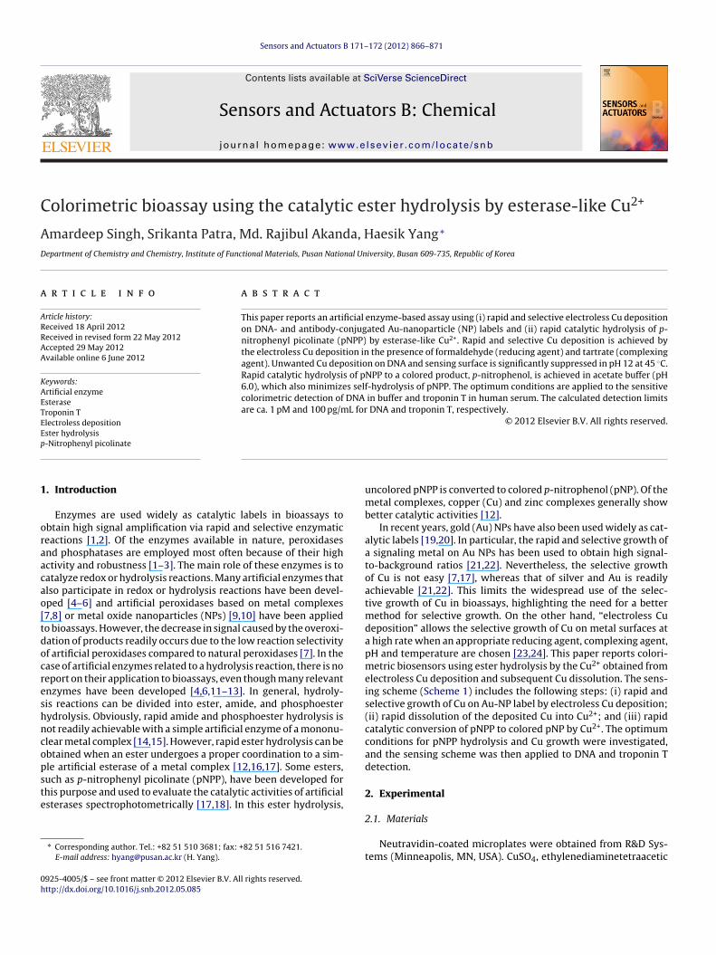
In recent years, gold (Au) NPs have also been used widely as cat-alytic labels [19,20]. In particular, the rapid and selective growth ofa signaling metal on Au NPs has been used to obtain high signal-to-background ratios [21,22]. Nevertheless, the selective growthof Cu is not easy [7,17], whereas that of silver and Au is readilyachievable [21,22]. This limits the widespread use of the selec-tive growth of Cu in bioassays, highlighting the need for a bettermethod for selective growth. On the other hand, “electroless Cudeposition” allows the selective growth of Cu on metal surfaces ata high rate when an appropriate reducing agent, complexing agent,pH and temperature are chosen [23,24]. This paper reports colori-metric biosensors using ester hydrolysis by the Cu2+ obtained fromelectroless Cu deposition and subsequent Cu dissolution. The sens-ing scheme (Scheme 1) includes the following steps: (i) rapid andselective growth of Cu on Au-NP label by electroless Cu deposition;(ii) rapid dissolution of the deposited Cu into Cu2+; and (iii) rapidcatalytic conversion of pNPP to colored pNP by Cu2+. The optimumconditions for pNPP hydrolysis and Cu growth were investigated,and the sensing scheme was then applied to DNA and troponin Tdetection.
2. Experimental
2.1. Materials
Neutravidin-coated microplates were obtained from R&D Sys-tems (Minneapolis, MN, USA). CuSO4, ethylenediaminetetraacetic
A. Singh et al. / Sensors and Actuators B 171– 172 (2012) 866– 871 867
ram o
abdssASata
TaI
Kn1bATApgb
0b(b1TENp
Ct
Scheme 1. Schematic diag
cid (EDTA), tris(hydroxymethyl)aminomethane (tris), and casein-ased blocking buffer were obtained from Sigma–Aldrich. Sodiumodecyl sulfate (SDS), polyethylene glycol (PEG, MW = 20,000), andodium chloride were obtained from Fluka. A solution of citrate-tabilized Au NP (10 nm, 0.01% HAuCl4) was purchased from Sigma.ll buffer reagents and other inorganic chemicals were supplied byigma–Aldrich, unless otherwise stated. All chemicals were useds received. All aqueous solutions were prepared in doubly dis-illed water. pNPP was synthesized as reported [25]. p-Nitrophenylcetate (pNPA) was obtained from Sigma–Aldrich.
Biotinylated monoclonal mouse antitroponin-T IgG (10R-127d), human serum (30-AT38), and monoclonal mousentitroponin-T IgG (10R-T127f) were obtained from Fitzerald,nc. (Acton, MA, USA).
HPLC-purified DNAs were obtained from Genotech (Daejeon,orea). The DNA assay was designed for the detection of singleucleotide polymorphism for the encoding residue 1038 of exon1 of the BRCA1 gene [26]. The DNAs had the following sequences:iotinylated capture probe, biotin-3′-(CH2)9-TCG ACC GAA GAATT T-5′; target DNA, 3′-TAA TCT CTT TTA CAA AAA TTT CTT CGGCG A-5′; thiolated detection probe, 3′-TTG TAA AAG AGA TTA-20-5′-(CH2)6-SH. The concentrations of DNAs (target, detectionrobe, capture probe) refer to those of strands. Au NPs were conju-ated with the thiolated detection probe containing an A20 spacery following our previous procedure [27].
Phosphate buffered saline (PBS) buffer (pH 7.4) consisted of.01 M sodium phosphate, 0.138 M NaCl, and 2.7 mM KCl. PBSBuffer contained all of the ingredients of the PBS buffer plus 1%w/v) BSA. Washing buffer was prepared by adding 0.1% SDS to PBSuffer. Hybridization buffer (pH 7.4) was composed of 20 mM tris,7.5 mM EDTA disodium salt, 150 mM NaCl, and 0.05% Tween 20.ris–EDTA buffer (pH 7.7) consisted of 100 mM tris and 25.3 mMDTA. Rinsing buffer was composed of 5 mM tris, 0.04 M HCl, 0.5 MaCl, BSA and 0.05% Tween 20. Acetate buffer (100 mM, pH 6.0) was
repared with sodium acetate and acetic acid.To prepare a solution for electroless Cu deposition, 120 mMuSO4 and 496 mM sodium potassium tartrate were dissolved andhe solution pH was then adjusted to 12.0 by adding a solution
f the detection procedure.
of 1 M NaOH. When electroless Cu deposition was carried out, thesolution and an aqueous solution of 37% (w/v) HCHO were mixedin a volume ratio of 10:1.
2.2. Apparatus
Optical density (OD) was collected with a microplate reader(Molecular devices, VERSA max). Spectrophotometric studies werecarried out with an UV-vis spectrophotometer (Shimadzu, UV-1650PC). The washing of microwells was performed with amicroplate strip washer (BioTek, ELx50).
2.3. Preparation of DNA-sensing surface and DNA detection
To immobilize a capture-probe DNA, 100 �L of 100 nM biotiny-lated capture-probe DNA was added to a well of neutravidin-coatedmicroplate and incubated for 2 h at 25 ◦C, followed by washingwith 300 �L of a PBS buffer solution containing 0.1% SDS (threetimes). 280 �L of a blocking buffer solution was then added to thewell and incubated for 3 h at 25 ◦C, followed by the same washingstep. To obtain the hybridization of a target DNA to the immo-bilized capture probe, 100 �L of a hybridization buffer solutioncontaining different concentrations of target DNA was added to thewell and incubated for 2 h at 25 ◦C with mild shaking (60–70 rpm),followed by washing with a washing buffer solution. Afterward,100 �L of a PBS solution containing detection probe-conjugated AuNP was added and incubated for 2 h at 25 ◦C, followed by wash-ing with a tris–EDTA buffer solution containing 200 mM MgSO4.The high concentration of MgSO4 helped to remove non-hybridizeddetection-probe-conjugated Au NP and to reduce the adsorptionof Cu2+. To obtain electroless Cu deposition on Au NP, 200 �L of afreshly prepared solution for Cu deposition and 20 �L of an aqueoussolution of 37% (w/v) HCHO was added to the well, and the solu-tion was incubated at 45 ◦C for 20 min, followed by washing with
a tris–EDTA buffer containing 200 mM MgSO4. To dissolve Cu onAu NP, 200 �L of 0.5 M HCl was added to the well, and the solutionwas incubated at 25 ◦C for 5 min with mild shaking. 100 �L of analiquot of the Cu2+-containing solution was taken and transferred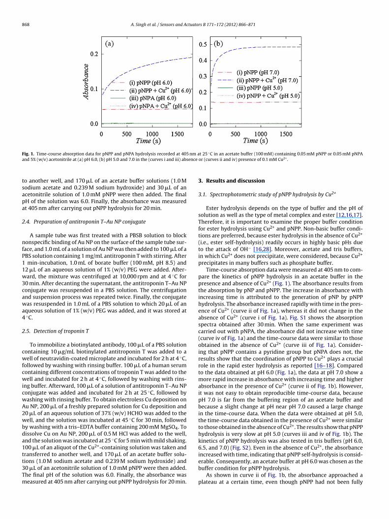
868 A. Singh et al. / Sensors and Actuators B 171– 172 (2012) 866– 871
F nm aa ence
tsapa
2
nfP11w3cawa4
2
cwfcwicwA2wbda1tt3Tm
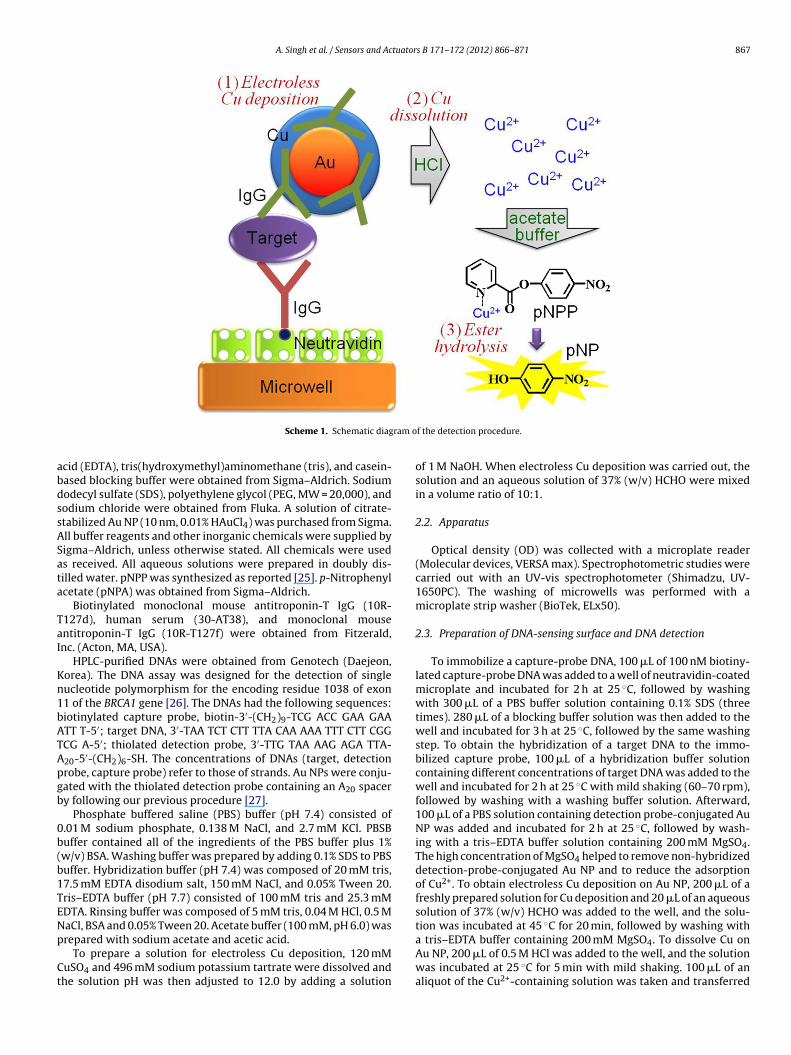
ig. 1. Time-course absorption data for pNPP and pNPA hydrolysis recorded at 405nd 5% (w/v) acetonitrile at (a) pH 6.0, (b) pH 5.0 and 7.0 in the (curves i and iii) abs
o another well, and 170 �L of an acetate buffer solutions (1.0 Modium acetate and 0.239 M sodium hydroxide) and 30 �L of ancetonitrile solution of 1.0 mM pNPP were then added. The finalH of the solution was 6.0. Finally, the absorbance was measuredt 405 nm after carrying out pNPP hydrolysis for 20 min.
.4. Preparation of antitroponin T–Au NP conjugate
A sample tube was first treated with a PBSB solution to blockonspecific binding of Au NP on the surface of the sample tube sur-
ace, and 1.0 mL of a solution of Au NP was then added to 100 �L of aBS solution containing 1 mg/mL antitroponin T with stirring. After
min-incubation, 1.0 mL of borate buffer (100 mM, pH 8.5) and2 �L of an aqueous solution of 1% (w/v) PEG were added. After-ard, the mixture was centrifuged at 10,000 rpm and at 4 ◦C for
0 min. After decanting the supernatant, the antitroponin T–Au NPonjugate was resuspended in a PBS solution. The centrifugationnd suspension process was repeated twice. Finally, the conjugateas resuspended in 1.0 mL of a PBS solution to which 20 �L of an
queous solution of 1% (w/v) PEG was added, and it was stored at◦C.
.5. Detection of troponin T
To immobilize a biotinylated antibody, 100 �L of a PBS solutionontaining 10 �g/mL biotinylated antitroponin T was added to aell of neutravidin-coated microplate and incubated for 2 h at 4 ◦C,
ollowed by washing with rinsing buffer. 100 �L of a human serumontaining different concentrations of troponin T was added to theell and incubated for 2 h at 4 ◦C, followed by washing with rins-
ng buffer. Afterward, 100 �L of a solution of antitroponin T–Au NPonjugate was added and incubated for 2 h at 25 ◦C, followed byashing with rinsing buffer. To obtain electroless Cu deposition onu NP, 200 �L of a freshly prepared solution for Cu deposition and0 �L of an aqueous solution of 37% (w/v) HCHO was added to theell, and the solution was incubated at 45 ◦C for 30 min, followed
y washing with a tris–EDTA buffer containing 200 mM MgSO4. Toissolve Cu on Au NP, 200 �L of 0.5 M HCl was added to the well,nd the solution was incubated at 25 ◦C for 5 min with mild shaking.00 �L of an aliquot of the Cu2+-containing solution was taken andransferred to another well, and 170 �L of an acetate buffer solu-
ions (1.0 M sodium acetate and 0.239 M sodium hydroxide) and0 �L of an acetonitrile solution of 1.0 mM pNPP were then added.he final pH of the solution was 6.0. Finally, the absorbance waseasured at 405 nm after carrying out pNPP hydrolysis for 20 min.t 25 ◦C in an acetate buffer (100 mM) containing 0.05 mM pNPP or 0.05 mM pNPAor (curves ii and iv) presence of 0.1 mM Cu2+.
3. Results and discussion
3.1. Spectrophotometric study of pNPP hydrolysis by Cu2+
Ester hydrolysis depends on the type of buffer and the pH ofsolution as well as the type of metal complex and ester [12,16,17].Therefore, it is important to examine the proper buffer conditionfor ester hydrolysis using Cu2+ and pNPP. Non-basic buffer condi-tions are preferred, because ester hydrolysis in the absence of Cu2+
(i.e., ester self-hydrolysis) readily occurs in highly basic pHs dueto the attack of OH− [16,28]. Moreover, acetate and tris buffers,in which Cu2+ does not precipitate, were considered, because Cu2+
precipitates in many buffers such as phosphate buffer.Time-course absorption data were measured at 405 nm to com-
pare the kinetics of pNPP hydrolysis in an acetate buffer in thepresence and absence of Cu2+ (Fig. 1). The absorbance results fromthe absorption by pNP and pNPP. The increase in absorbance withincreasing time is attributed to the generation of pNP by pNPPhydrolysis. The absorbance increased rapidly with time in the pres-ence of Cu2+ (curve ii of Fig. 1a), whereas it did not change in theabsence of Cu2+ (curve i of Fig. 1a). Fig. S1 shows the absorptionspectra obtained after 30 min. When the same experiment wascarried out with pNPA, the absorbance did not increase with time(curve iv of Fig. 1a) and the time-course data were similar to thoseobtained in the absence of Cu2+ (curve iii of Fig. 1a). Consider-ing that pNPP contains a pyridine group but pNPA does not, theresults show that the coordination of pNPP to Cu2+ plays a crucialrole in the rapid ester hydrolysis as reported [16–18]. Comparedto the data obtained at pH 6.0 (Fig. 1a), the data at pH 7.0 show amore rapid increase in absorbance with increasing time and higherabsorbance in the presence of Cu2+ (curve ii of Fig. 1b). However,it was not easy to obtain reproducible time-course data, becausepH 7.0 is far from the buffering region of an acetate buffer andbecause a slight change at pH near pH 7.0 caused a large changein the time-course data. When the data were obtained at pH 5.0,the time-course data obtained in the presence of Cu2+ were similarto those obtained in the absence of Cu2+. The results show that pNPPhydrolysis is very slow at pH 5.0 (curves iii and iv of Fig. 1b). Thekinetics of pNPP hydrolysis was also tested in tris buffers (pH 6.0,6.5, and 7.0) (Fig. S2). Even in the absence of Cu2+, the absorbanceincreased with time, indicating that pNPP self-hydrolysis is consid-
erable. Consequently, an acetate buffer at pH 6.0 was chosen as thebuffer condition for pNPP hydrolysis.As shown in curve ii of Fig. 1b, the absorbance approached aplateau at a certain time, even though pNPP had not been fully
A. Singh et al. / Sensors and Actuators B 171– 172 (2012) 866– 871 869
F at diffa ed line
hhccain
3
ahmaa
Fwcm
ig. 2. Optical densities (ODs) recorded at 670 nm after electroless Cu deposition (a)nd (ii) the microwells onto which DNA-conjugated Au NP was adsorbed. The dash
ydrolyzed. This suggests that ester hydrolysis was inhibited byydrolysis products. Picolinic acid, a product of pNPP hydrolysis,ould act as a ligand for Cu2+. Therefore, picolinic acid binds to Cu2+
ompetitively with pNPP [18]. When picolinic acid was added ton acetate buffer containing pNPP and Cu2+, pNPP hydrolysis wasnhibited significantly (Fig. S3). All results confirm that the coordi-ation of pNPP to Cu2+ plays an essential role in pNPP hydrolysis.
.2. Electroless Cu deposition on Au NP
Electroless Cu deposition has been studied extensively in therea of microfabrication, and many good conditions for deposition
ave been reported [23,24]. To obtain selective Cu growth on aetal, the difference between the formal potentials of “Cu2+/Cu”nd “reducing agent/its oxidized form” should be small [23]. Toccomplish this, the formal potential of Cu2+/Cu should be lowered
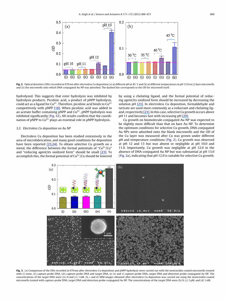
ig. 3. (a) Comparison of the ODs recorded at 670 nm after electroless Cu deposition andith (i) none, (ii) capture-probe DNA, (iii) capture-probe DNA and target DNA, or (iv an
oncentrations of the target DNA were (iv) 0 and (v) 1 nM. (b, c and d) SEM images obtaicrowells treated with capture-probe DNA, target DNA and detection probe-conjugated
erent pH at 45 ◦C and (b) at different temperatures at pH 12.0 on (i) bare microwells corresponds to the OD for microwell itself.
by using a chelating ligand, and the formal potential of reduc-ing agent/its oxidized form should be increased by decreasing thesolution pH [23]. In electroless Cu deposition, formaldehyde andtartrate are used most commonly as a reductant and chelating lig-and, respectively [23]. In this case, selective Cu growth occurs abovepH 11 and becomes fast with increasing pH [29].
Cu growth on biomolecule-conjugated Au NP was expected tobe slightly more difficult than that on bare Au NP. To determinethe optimum conditions for selective Cu growth, DNA-conjugatedAu NPs were adsorbed onto the blank microwells and the OD ofthe Cu layer was measured after Cu was grown under differentpH and temperature conditions (Fig. 2). Cu growth was observed
at pH 12 and 13 but was absent or negligible at pH 10.0 and11.0. Importantly, Cu growth was negligible at pH 12.0 in theabsence of DNA-conjugated Au NP but was substantial at pH 13.0(Fig. 2a), indicating that pH 12.0 is suitable for selective Cu growth.pNPP hydrolysis were carried out with the neutravidin-coated microwells treatedd v) capture-probe DNA, target DNA and detection probe-conjugated Au NP. Theined after electroless Cu deposition was carried out using the neutravidin-coated
Au NP. The concentrations of the target DNA were (b) 0, (c) 1 pM, and (d) 1 nM.
870 A. Singh et al. / Sensors and Actuators B 171– 172 (2012) 866– 871
F n humm es theo
Rfa4
3
aCmsooDtDn1(w1ntm(ttoomd
tD1dwlppstbrw
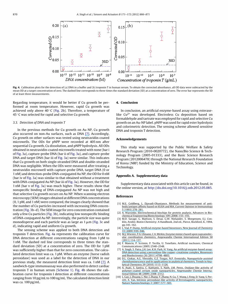
ig. 4. Calibration plots for the detection of (a) DNA in a buffer and (b) troponin T iean OD at a target concentration of zero. The dashed line corresponds to three tim
f at least three measurements.
egarding temperature, it would be better if Cu growth be per-ormed at room temperature. However, rapid Cu growth waschieved only above 40 ◦C (Fig. 2b). Therefore, a temperature of5 ◦C was selected for rapid and selective Cu growth.
.3. Detection of DNA and troponin T
In the previous methods for Cu growth on Au NP, Cu growthlso occurred on non-Au surfaces, such as DNA [7]. Accordingly,u growth on other surfaces was tested using neutravidin-coatedicrowells. The ODs for pNPP were recorded at 405 nm after
equential Cu growth, Cu dissolution, and pNPP hydrolysis. All ODsbtained in neutravidin-coated microwells treated with none (bar if Fig. 3a), capture-probe DNA (bar ii of Fig. 3a), and capture-probeNA and target DNA (bar iii of Fig. 3a) were similar. This indicates
hat Cu growth on both single-stranded DNA and double-strandedNA was negligible. When the ODs were measured after treating aeutravidin microwell with capture-probe DNA, target DNA (0 or
nM) and detection-probe DNA-conjugated Au NP, the OD for 0 nMbar iv of Fig. 3a) was similar to that obtained without a treatmentith DNA-conjugated Au NP (bar iii of Fig. 3a). However, the OD for
nM (bar v of Fig. 3a) was much higher. These results show thatonspecific binding of DNA-conjugated Au NP was not high andhat selective Cu growth occurs on Au NP. When scanning electron
icroscopy (SEM) images obtained at different DNA concentrations0, 1 pM, and 1 nM) were compared, the images clearly showed thathe number of Cu particles increased with increasing DNA concen-ration (Fig. 3b–d). The SEM image for zero concentration containednly a few Cu particles (Fig. 3b), indicating low nonspecific bindingf DNA-conjugated Au NP. Interestingly, the particle size was quiteonodisperse and each particle was as large as 1 �m (Fig. 3c and
), indicating rapid and uniform Cu growth.The sensing scheme was applied to both DNA detection and
roponin T detection. Fig. 4a represents the calibration curve forNA detection at different concentrations ranging from 1 pM to
nM. The dashed red line corresponds to three times the stan-ard deviation (SD) at a concentration of zero. The OD for 1 pMas sufficiently higher than that for zero concentration. The calcu-
ated detection limit was ca. 1 pM. When an enzyme (horse radisheroxidase) was used as a label for the detection of DNA in ourrevious study, the measured detection limit was ca. 1 nM [7]. Aandwich-type immunoassay was also performed to detect cardiac
roponin T in human serum (Scheme 1). Fig. 4b shows the cali-ration curve for troponin I detection at different concentrationsanging from 10 pg/mL to 100 ng/mL. The calculated detection limitas ca. 100 pg/mL.[
an serum. To obtain the corrected absorbance, all OD data were subtracted by the standard deviation (SD) at a concentration of zero. The error bar represents the SD
4. Conclusion
In conclusion, an artificial enzyme-based assay using esterase-like Cu2+ was developed. Electroless Cu deposition based onformaldehyde and tartrate was employed for rapid and selective Cugrowth on an Au-NP label. pNPP was used for rapid ester hydrolysisand colorimetric detection. The sensing scheme allowed sensitiveDNA and troponin T detection.
Acknowledgements
This study was supported by the Public Welfare & SafetyResearch Program (2010-0020772), the Nano/Bio Science & Tech-nology Program (2005-01333), and the Basic Science ResearchProgram (2012006478) through the National Research Foundationof Korea (NRF) funded by the Ministry of Education, Science andTechnology.
Appendix A. Supplementary data
Supplementary data associated with this article can be found, inthe online version, at http://dx.doi.org/10.1016/j.snb.2012.05.085.
References
[1] M.E. Goldberg, L. Djavadi-Ohaniance, Methods for measurement of anti-body/antigen affinity based on ELISA and RIA, Current Opinion in Immunology5 (1993) 278–281.
[2] A. Warsinke, Electrochemical biochips for protein analysis, Advances in Bio-chemical Engineering/Biotechnology 109 (2008) 155–193.
[3] M.D. Savage, G. Mattson, S. Desai, G.W. Nielander, S. Morgensen, E.J. Con-klin, Avidin–Biotin Chemistry: A Handbook, 2nd ed., Pierce, Rockford, IL, 1994,pp.145–162.
[4] L. Vial, P. Dumy, Artificial enzyme-based biosensors, New Journal of Chemistry33 (2009) 939–946.
[5] M.J. Wiester, P.A. Ulmann, C.A. Mirkin, Enzyme mimics based upon supramolec-ular coordination chemistry, Angewandte Chemie International Edition 50(2011) 114–137.
[6] F. Mancin, P. Scrimin, P. Tecilla, U. Tonellato, Artificial nucleases, ChemicalCommunications (2005) 2540–2548.
[7] A. Singh, S. Patra, J.H. Lee, K.H. Park, H. Yang, An artificial enzyme-based assay:DNA detection using a peroxidase-like copper–creatinine complex, Biosensorsand Bioelectronics 26 (2011) 4798–4803.
[8] D.L. Giokas, A.G. Vlessidis, G.Z. Tsogas, N.P. Evmiridis, Nanoparticle-assistedchemiluminescence and its applications in analytical chemistry, Trends in Ana-lytical Chemistry 29 (2010) 1113–1126.
[9] A. Asati, S. Santra, C. Kaittanis, S. Nath, J.M. Perez, Oxidase-like activity of
polymer-coated cerium oxide nanoparticles, Angewandte Chemie Interna-tional Edition 48 (2009) 2308–2312.10] L. Gao, J. Zhuang, L. Nie, J. Zhang, Y. Zhang, N. Gu, T. Wang, J. Feng, D. Yang, S. Per-rett, X. Yan, Intrinsic peroxidase-like activity of ferromagnetic nanoparticles,Nature Nanotechnology 2 (2007) 577–583.
tuator
[
[
[
[
[
[
[
[
[
[
[
[
[
[
[
[
[
[
[
University, Korea. He obtained his BS, MS, and PhD from Korea Advanced Insti-
A. Singh et al. / Sensors and Ac
11] R. Krämer, Bioinorganic models for the catalytic cooperation of metal ions andfunctional groups in nuclease and peptidase enzymes, Coordination ChemistryReviews 182 (1999) 243–261.
12] J. Suh, Synthetic artificial peptidases and nucleases using macromolecular cat-alytic systems, Accounts of Chemical Research 36 (2003) 562–570.
13] S. Bhattacharya, N. Kumari, Metallomicelles as potent catalysts for the esterhydrolysis reactions in water, Coordination Chemistry Reviews 253 (2009)2133–2149.
14] F. Mancin, P. Tecilla, Zinc(II) complexes as hydrolytic catalysts of phosphatediester cleavage: from model substrates to nucleic acids, New Journal of Chem-istry 31 (2007) 800–817.
15] E.L. Hegg, J.N. Burstyn, Toward the development of metal-based syntheticnucleases and peptidases, Coordination Chemistry Reviews 173 (1998)133–165.
16] J. Kovács, A. Mokhir, Catalytic hydrolysis of esters of 2-hydroxypyridine deriva-tives for Cu2+ detection, Inorganic Chemistry 47 (2008) 1880–1882.
17] L.-G. Qiu, A.-J. Xie, Y.-H. Shen, Gemini metallomicellar catalysis: hydrolysis ofp-nitrophenyl picolinate catalyzed by Cu(II) and Ni(II) complexes of macro-cyclic ligands in gemini surfactant micelles, Journal of Molecular Catalysis A:Chemical 244 (2006) 58–63.
18] M. Fanti, F. Mancin, P. Tecilla, U. Tonellato, Ester cleavage in reversed micelles byCu(II) complexes of functionalized ligands, Langmuir 16 (2000) 10115–10122.
19] N.L. Rosi, C.A. Mirkin, Nanostructures in biodiagnostics, Chemical Reviews 105(2005) 1547–1562.
20] J. Das, M.A. Aziz, H. Yang, A nanocatalyst-based assay for proteins: DNA-free ultrasensitive electrochemical detection using catalytic reduction ofp-nitrophenol by gold-nanoparticle labels, Journal of the American ChemicalSociety 128 (2006) 16022–16023.
21] S. Gupta, S. Huda, P.K. Kilpatrick, O.D. Velev, Characterization and optimizationof gold nanoparticle-based silver-enhanced immunoassays, Analytical Chem-istry 79 (2007) 3810–3820.
22] C. Deng, J. Chen, Z. Nie, M. Wang, X. Chu, X. Chen, X. Xiao, C. Lei, S. Yao, Impedi-metric aptasensor with femtomolar sensitivity based on the enlargement ofsurface-charged gold nanoparticles, Analytical Chemistry 81 (2009) 739–745.
23] Y. Shacham-Diamand, V. Dubin, M. Angyal, Electroless copper deposition forULSI, Thin Solid Films 262 (1995) 93–103.
24] M. Zhang, C. Lu, H. Cong, W. Cao, Selective electroless deposition of Cu on an
ultrathin Au film pattern, Macromolecular Rapid Communications 25 (2004)1917–1920.25] D.S. Sigman, C.T. Jorgensen, Models for metalloenzymes. Zinc (II)-catalyzedtransesterification of N-(�-hydroxyethyl)ethylenediamine by p-nitrophenylpicolinate, Journal of the American Chemical Society 94 (1972) 1724–1730.
s B 171– 172 (2012) 866– 871 871
26] A.M. Dunning, M. Chiano, N.R. Smith, J. Dearden, M. Gore, S. Oakes, C. Wilson, M.Stratton, J. Peto, D. Easton, D. Clayton, B.A. Ponder, Common BRCA1 variants andsusceptibility to breast and ovarian cancer in the general population, HumanMolecular Genetics 6 (1997) 285–289.
27] J. Das, C.-H. Huh, K. Kwon, S. Park, S. Jon, K. Kim, H. Yang, Comparisonof the nonspecific binding of DNA-conjugated gold nanoparticles betweenpolymeric and monomeric self-assembled monolayers, Langmuir 25 (2009)235–241.
28] J. Suh, B.N. Kwon, W.Y. Lee, S.H. Chang, Kinetics and mechanism of the metal ioncatalyzed hydrolysis of acetylpyridine ketoxime pyridinecarboxylates, Inor-ganic Chemistry 26 (1987) 805–808.
29] V.M. Dubin, Y. Shacham-Diamond, B. Zhao, P.K. Vasudev, C.H. Ting, Selective andblanket electroless copper deposition for ultralarge scale integration, Journalof the Electrochemical Society 144 (1997) 898–908.
Biographies
Amardeep Singh received his MS in 2003 from Indian Institute of Technology Roor-kee (IITR), India, and his PhD in 2010 from Indian Institute of Technology Guwahati(IITG), India. He is currently a postdoctoral scholar in Department of Chemistry,Pusan National University, Korea. His current research is focused on the develop-ment of artificial enzyme-based biosensors.
Srikanta Patra is an assistant professor in School of Basic Sciences, Indian Insti-tute of Technology Bhubaneswar (IITBBS), India. He obtained his PhD in 2005 fromIndian Institute of Technology Bombay (IITB), India. After carrying out his postdocin University of Iowa, USA, he worked in Department of Chemistry, Pusan NationalUniversity, Korea.
Md. Rajibul Akanda received his BS and MS in 2003 and 2004, respectively, fromThe University of Dhaka, Bangladesh. He is currently a PhD student in Department ofChemistry, Pusan National University, Korea. His research work is related to enzyme-based electrochemical biosensors.
Haesik Yang is an associate professor of Department of Chemistry in Pusan National
tute of Science and Technology (KAIST) in 1992, 1994, and 1997, respectively. Hiscurrent research includes the development of nanomaterial- and enzyme-basedelectrochemical biosensors and the investigation of electrochemical behaviors ofnanomaterials.
Related Documents