Colorectal Carcinoma- An Overview Dr C. L Chaw ST4, Clinical Oncology Tayside

Colorectal Carcinoma- An Overview Dr C. L Chaw ST4, Clinical Oncology Tayside.
Mar 26, 2015
Welcome message from author
This document is posted to help you gain knowledge. Please leave a comment to let me know what you think about it! Share it to your friends and learn new things together.
Transcript
Colorectal Carcinoma- An Overview
Dr C. L Chaw
ST4, Clinical Oncology
Tayside
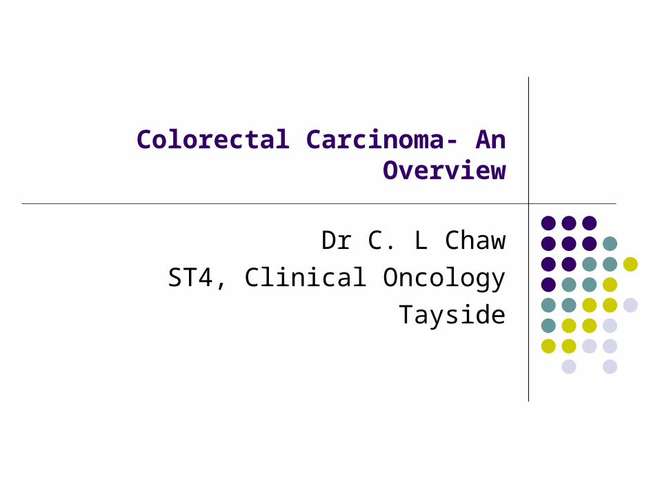
Colorectal Cancer (CRC)
3rd most common form of cancer and the 3rd leading cause of death in the Western World.
Annual incidence in UK -54/100 000, and 35000 new cases per year,>16000 deaths per year, making it the 2nd most common cause of cancer death in UK
Peak incidence ages: 60-70 yrs old ♂:♀ ratio for colonic and rectal cancer is 2:3 and 2:1 Colonic cancer ( 60%) is more common> than rectal
cancer (40%)

Risk Factors & Aetioloy
Environmental Factors Genetic
The aetiology is complex, involving interplay of environmental and genetic factors. These factors conspire to change the normal mucosa to a premalignant adenomatous polyp to a frank colorectal cancer over the course of many years
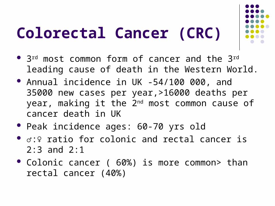
Environmental Factors
Diet Total calories – obesity and ↑ BMI (25-30kg/m2 )↑risk of CRC ↑ consumption of Meat (red meat) /Fat (saturated) /Protein - ↑ risk of
CRC High Fiber - ↓ risk of CRC Vegetables/ Fruits – Protective effects due to presence of antioxidants
Lifestyles Physical inactivity -↑ risk of CRC Cigarette smoking -↑ risk of CRC ↑ alcohol consumption -↑ risk of CRC
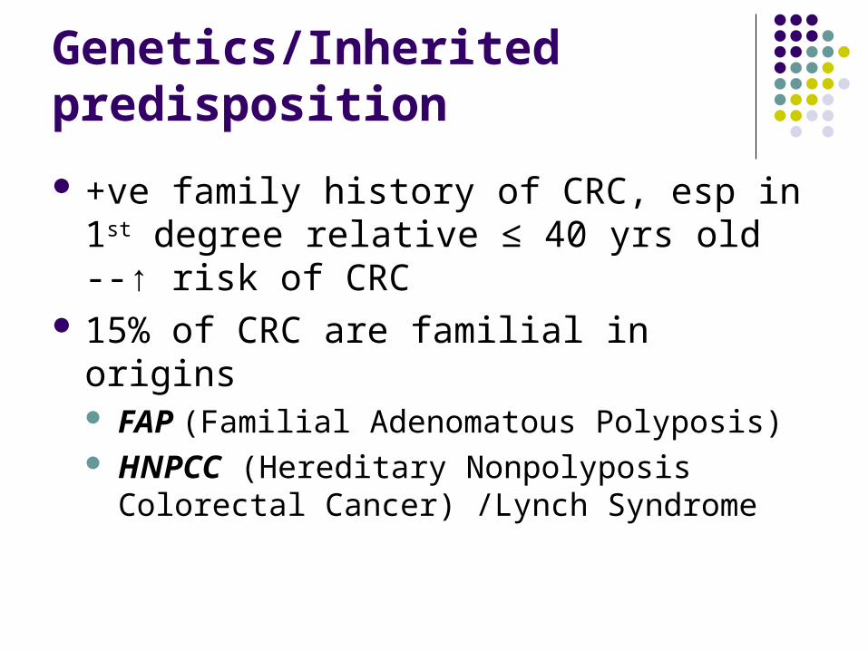
Genetics/Inherited predisposition
+ve family history of CRC, esp in 1st degree relative ≤ 40 yrs old --↑ risk of CRC
15% of CRC are familial in origins FAP (Familial Adenomatous Polyposis) HNPCC (Hereditary Nonpolyposis Colorectal
Cancer) /Lynch Syndrome
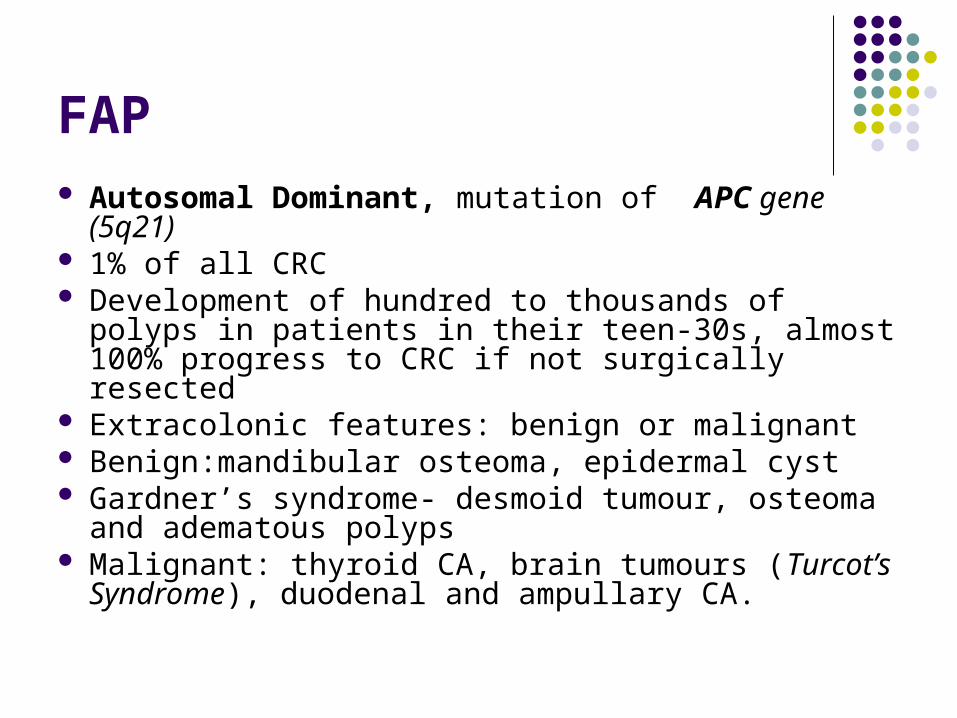
FAP Autosomal Dominant, mutation of APC gene
(5q21) 1% of all CRC Development of hundred to thousands of polyps in
patients in their teen-30s, almost 100% progress to CRC if not surgically resected
Extracolonic features: benign or malignant Benign:mandibular osteoma, epidermal cyst Gardner’s syndrome- desmoid tumour, osteoma and
adematous polyps Malignant: thyroid CA, brain tumours (Turcot’s
Syndrome), duodenal and ampullary CA.
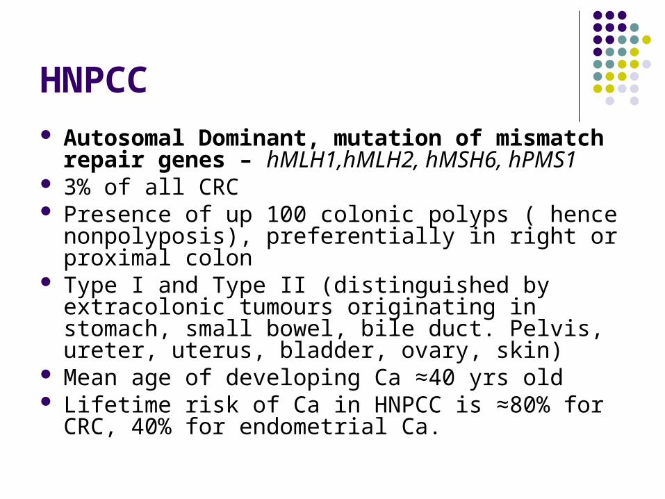
HNPCC Autosomal Dominant, mutation of mismatch
repair genes – hMLH1,hMLH2, hMSH6, hPMS1 3% of all CRC Presence of up 100 colonic polyps ( hence
nonpolyposis), preferentially in right or proximal colon
Type I and Type II (distinguished by extracolonic tumours originating in stomach, small bowel, bile duct. Pelvis, ureter, uterus, bladder, ovary, skin)
Mean age of developing Ca ≈40 yrs old Lifetime risk of Ca in HNPCC is ≈80% for CRC, 40%
for endometrial Ca.
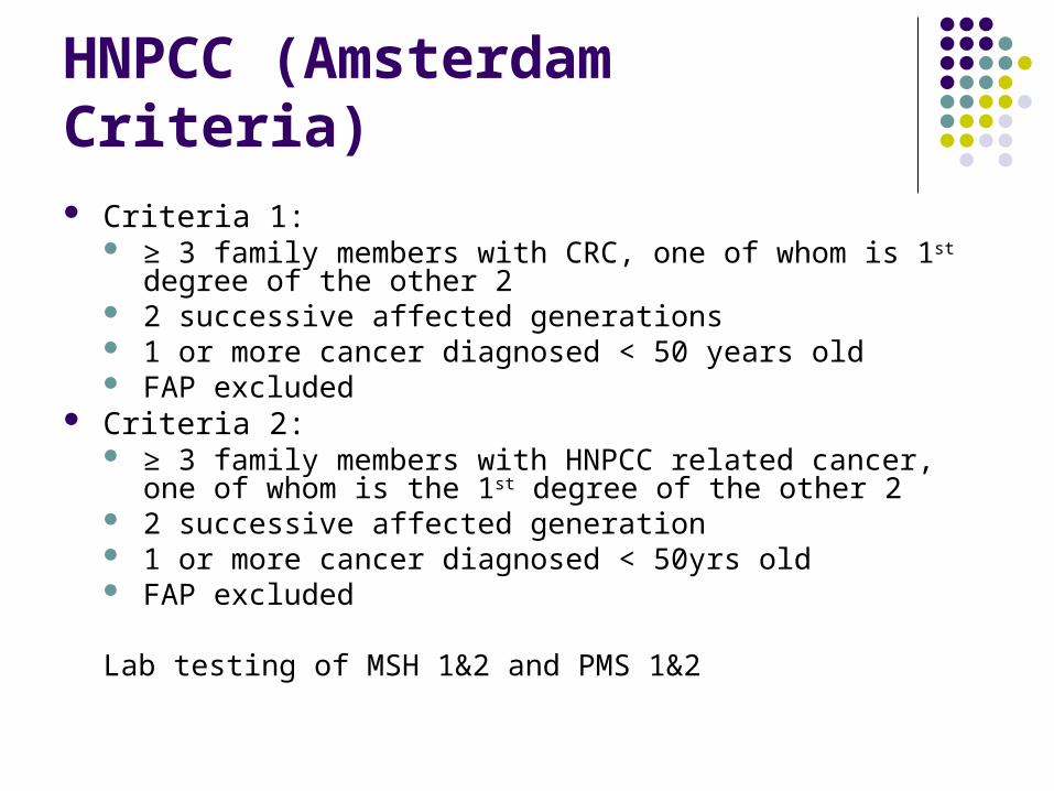
HNPCC (Amsterdam Criteria) Criteria 1:
≥ 3 family members with CRC, one of whom is 1st degree of the other 2
2 successive affected generations 1 or more cancer diagnosed < 50 years old FAP excluded
Criteria 2: ≥ 3 family members with HNPCC related cancer, one of whom is
the 1st degree of the other 2 2 successive affected generation 1 or more cancer diagnosed < 50yrs old FAP excluded
Lab testing of MSH 1&2 and PMS 1&2
Pathogenesis Vogelstein model Multistep to carcinoma formation
Mutation of APC gene –polyposis K-RAS (40-50%), P53, SMAD mutation DCC gene helps to initiate metastatic potential
Other pathway through MSI (DNA microsatellite instability) - HNPCC
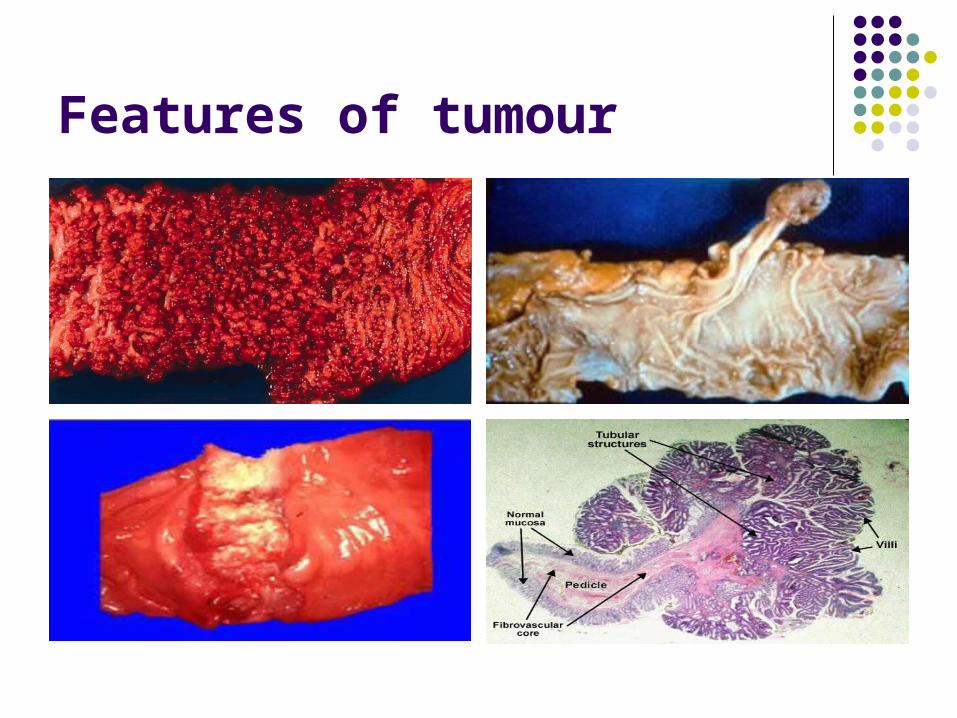
Features of tumour
Spread
Adjacent organs – small/ large bowel, bladder, uterus
Transcoelomic spread- peritoneal disease Regional lymph node involvement ( 40-70%) at
presentation – usually follows the supplying blood vessels– pararectal, hypogastric, pre-sacral)
Haematogenous – liver ≥ lung ≥ bone and brain 25-30% patients at presentations, the tumour will
have spread either locally or distant sites, and will be unsuitable for radical treatment
Assessment & Management:
History Presenting complaints Family history Systemic enquiries
Physical examination (PR examination) Investigations Treatments
Signs & Symptoms
Symptoms Lower GI bleeding Altered bowel habits Abdominal pain Weight loss Loss of appetite Obstructive symptoms – vomiting, unable to pass
wind, severe abdo pain
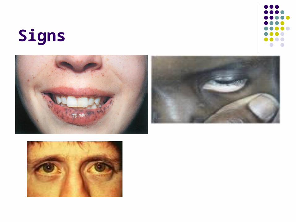
Signs
Inspection: Jaundice, pallor , (freckles around the lip, buccal mucosal –Peutze Jeghers)
Palpable abdominal / rectal mass–don’t forget about the liver –hepatomegaly – distant mets
PR bleeding – fresh red ( left-sided colon/ rectum), malena (right-sided colon)
Alarming signs –Abdominal distension, peritonism, rebound tenderness, tingling bowel sound – bowel obstruction, perforation.
Pulmonary signs and neurological signs can sometime present if the disease is advanced.
Signs
Investigations
FBC ( Iron –deficiency anaemia ) U+E LFT ( metastatic disease ) CEA (carcinoembryonic antigen), is raised in
85% of patients with CRC, higher value associated with worse prognosis
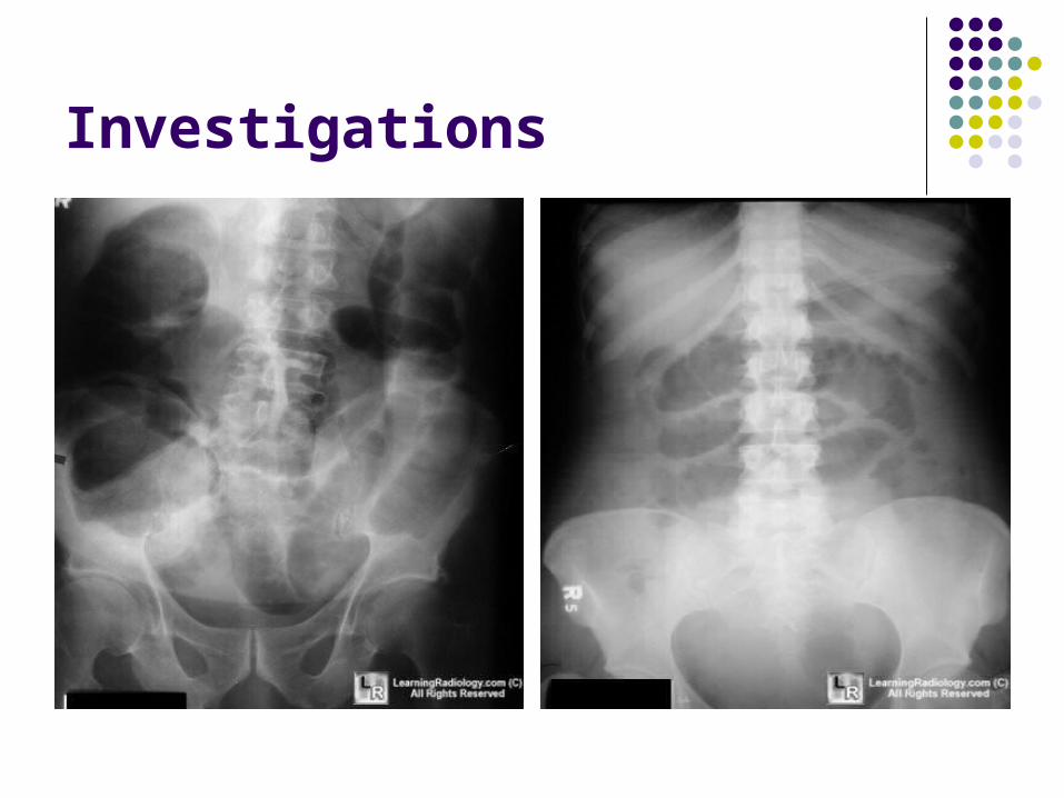
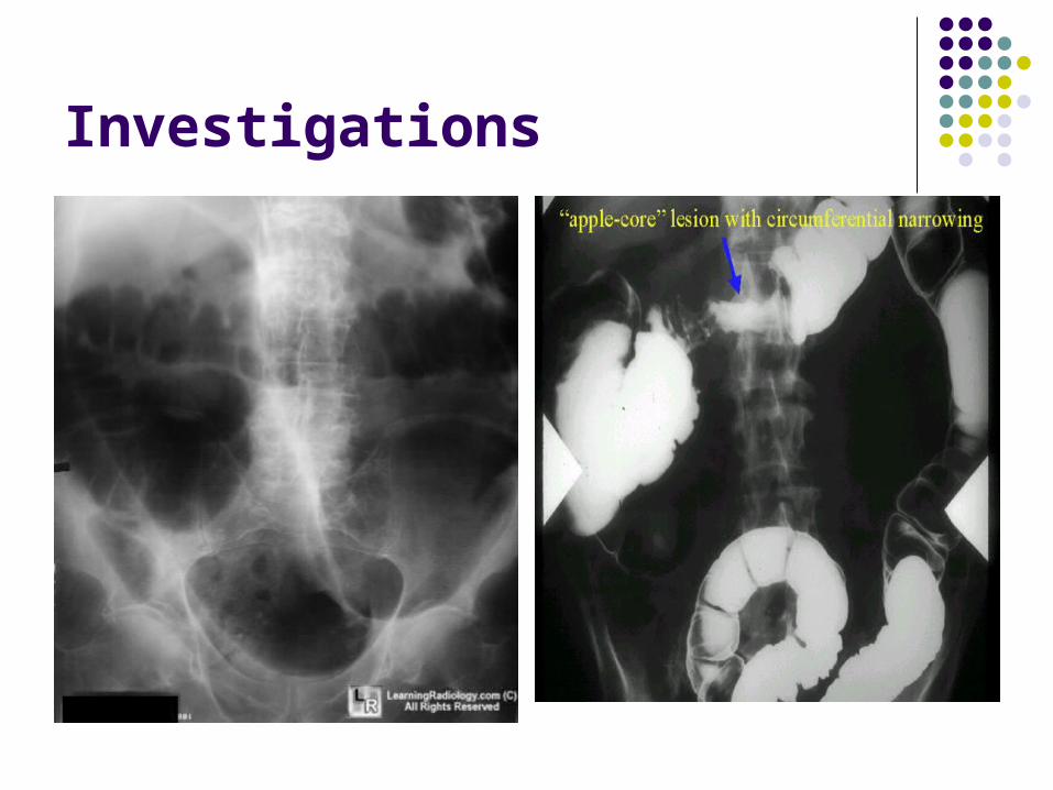
Investigations
AXR ( if suspicious of SBO/ BO) Double contrast barium enema Colonoscopy with biopsy, Flexi/ rigid sigmodoscopy CT scan –Thorax, abdo USS liver - liver metastasis Pelvic MRI –particularly for rectal Ca – To asses
CRM (Circumferential resection margin) Endo-anal USS to asses nodal involvement in rectal
Ca
Investigations
Investigations

Investigations
Screening
50-75 years old FOB; higher false positive rate Colonoscopy; more specific and better at picking
proximal lesion.
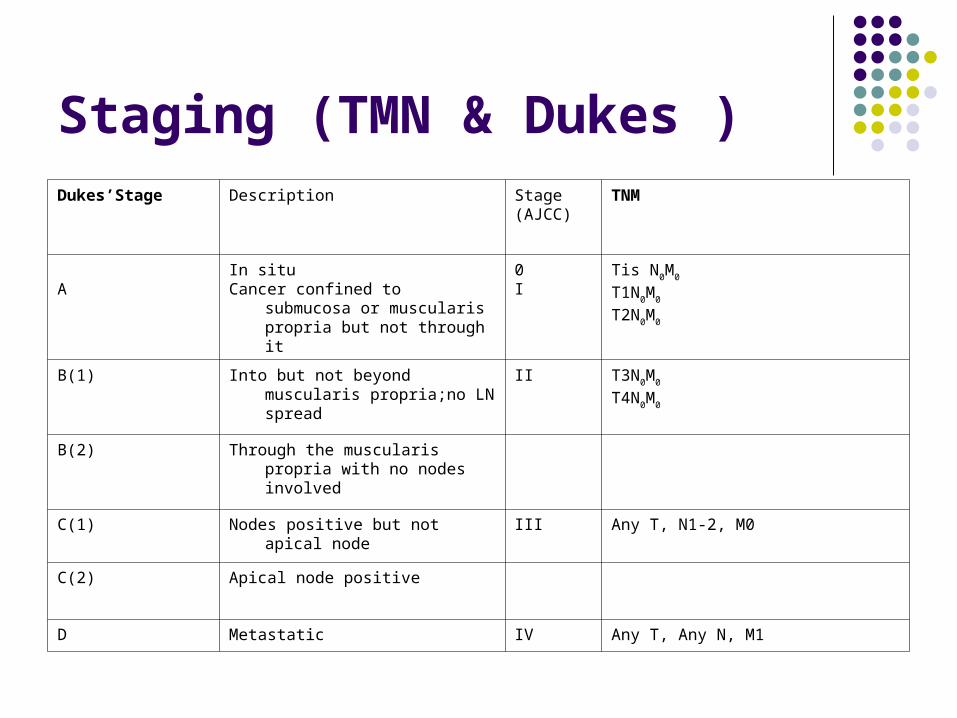
Staging (TMN & Dukes )Dukes’Stage Description Stage
(AJCC)TNM
AIn situCancer confined to submucosa or
muscularis propria but not through it
0I
Tis N0M0
T1N0M0
T2N0M0
B(1) Into but not beyond muscularis propria;no LN spread
II T3N0M0
T4N0M0
B(2) Through the muscularis propria with no nodes involved
C(1) Nodes positive but not apical node III Any T, N1-2, M0
C(2) Apical node positive
D Metastatic IV Any T, Any N, M1
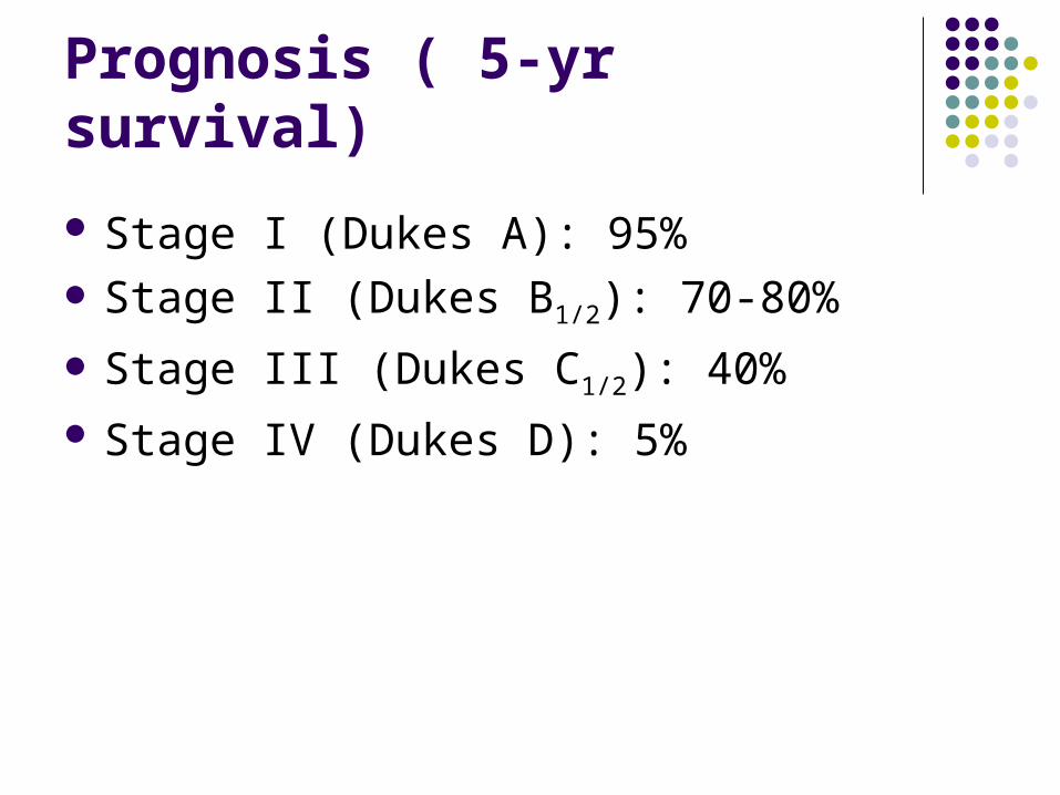
Prognosis ( 5-yr survival)
Stage I (Dukes A): 95% Stage II (Dukes B1/2): 70-80%
Stage III (Dukes C1/2): 40% Stage IV (Dukes D): 5%
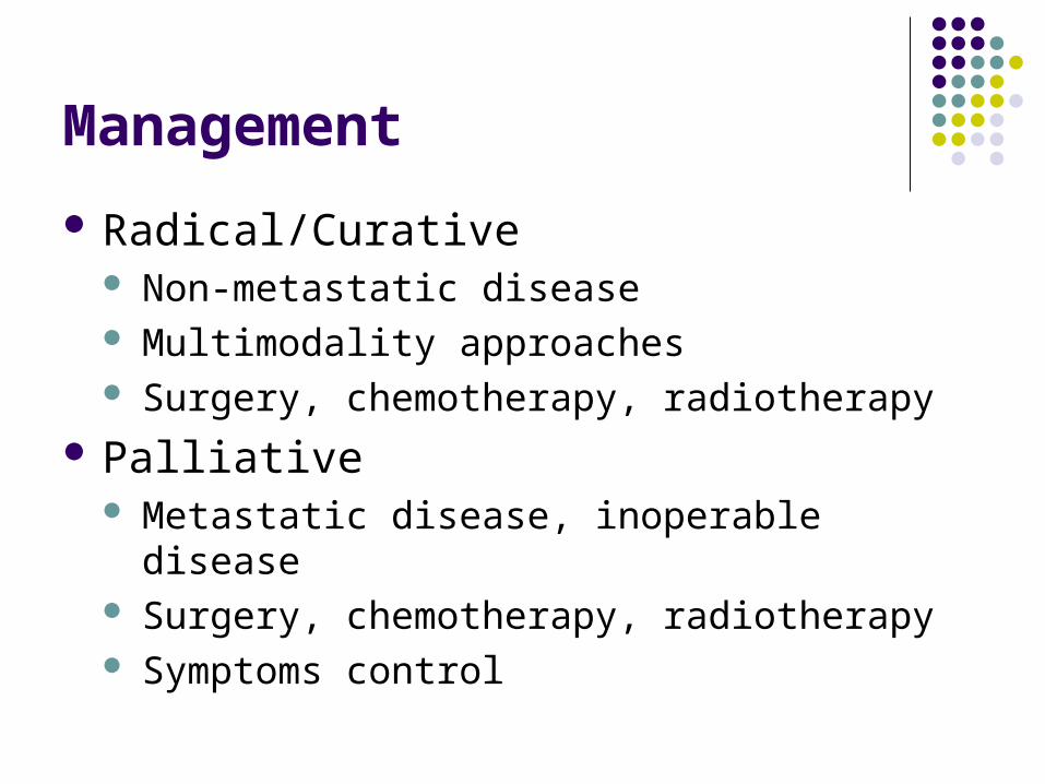
Management
Radical/Curative Non-metastatic disease Multimodality approaches Surgery, chemotherapy, radiotherapy
Palliative Metastatic disease, inoperable disease Surgery, chemotherapy, radiotherapy Symptoms control
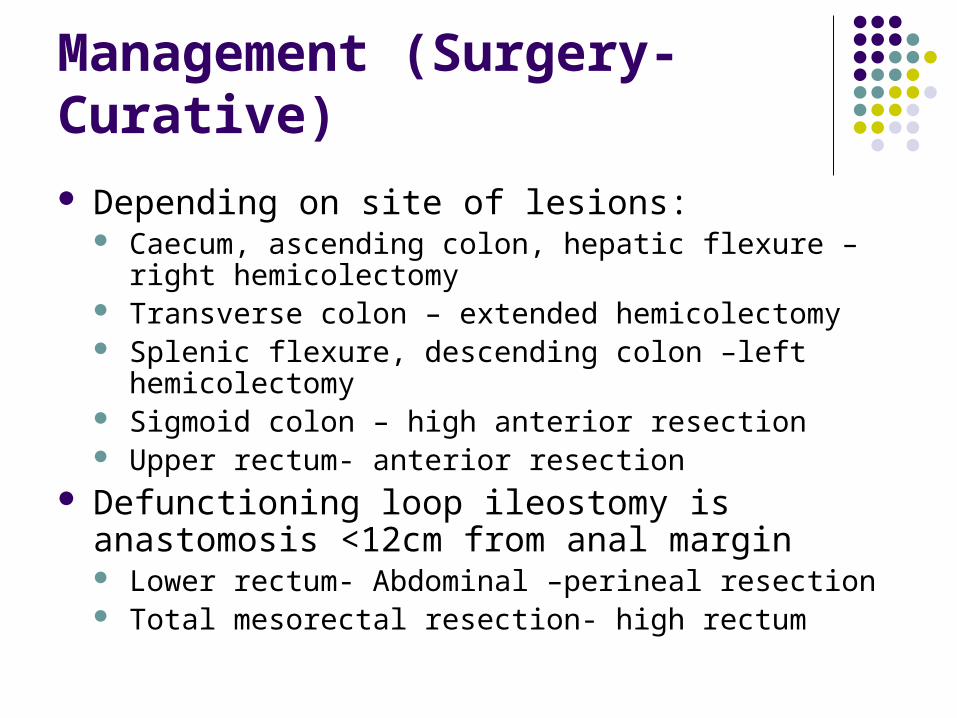
Management (Surgery- Curative)
Depending on site of lesions: Caecum, ascending colon, hepatic flexure – right
hemicolectomy Transverse colon – extended hemicolectomy Splenic flexure, descending colon –left hemicolectomy Sigmoid colon – high anterior resection Upper rectum- anterior resection
Defunctioning loop ileostomy is anastomosis <12cm from anal margin Lower rectum- Abdominal –perineal resection Total mesorectal resection- high rectum
Management (Chemothrapy)
Adjuvant Chemotherapy T3 disease or node positive tumours (Dukes C
disease, selective in Dukes B) – 4-13% survival benefits
Serosal involvement, perforated tumours, extramural vascular invasion or involvement of circumferential margin
5- Flourouracil based chemo, platinum based chemo
Side effects: nausea, myelosuppression, diarrhoea, neuropathy
Management ( Radiotherapy )
Rectal cancer – reduce rate of local recurrence, downstaging of inoperable disease
Pre-operatively or post-operatively 25Gy in 5#, 45Gy in 25# Side effects: erythema (local skin reaction),
cystitis, diarrhoea, sterility, urge incotinence, bowel dysfunction
Management ( Palliative )
Inoperable disease, medically unfit patients Defunctiong Colostomy, Surgical/ endoscopic
stenting – for obstructing lesions, aiming to improve quality of life
Resection for isolated liver and pulmonary metastasis if patients are fit.
Radiotherapy – palliation of local symtoms, bony pain, rectal bleeding
Management ( palliative )
Chemotherapy Patients selection- performance status 1-2 Aiming to improve quality of life and control of
disease Improves survival by 3-6 months 5-FU based chemo, platinum based.
References:
SIGN Guidelines no 67 Practical Clinical Oncology – Louise Hanna Cancer, Principle & Practice of Oncology –
DeVita, Hellman et al
Related Documents


![Compact Safety Beam Sensor [Type 4] ST4 SERIESST4-A1-J02 With emission amount adjuster ST4-A1-J02V Cable length 1 m 3.281 ft ST4-A1-J1 With emission amount adjuster ST4-A1-J1V Controllers](https://static.cupdf.com/doc/110x72/5f0f9beb7e708231d44502d6/compact-safety-beam-sensor-type-4-st4-series-st4-a1-j02-with-emission-amount-adjuster.jpg)