Color Doppler flow patterns and flow velocity waveforms of the intraplacental fetal circulation in growth-retarded fetuses Siegfried Rotmensch, MD: Marco Liberati, MD,c Jia-Sen Luo, MD: Harvey J. Kliman, MD, PhD,b Yvonne Gollin, MD: Umberto Bellati, MD,c John C. Hobbins, MD: and Joshua A. Copel, MD" New Haven, Connecticut, and Chieti, Italy OBJECTIVE: We examined intraplacental color Doppler flow patterns and spectral Doppler flow velocity waveforms of villous arteries in pregnancies with intrauterine growth retardation. STUDY DESIGN: A total of 192 uncomplicated pregnancies and 29 pregnancies with intrauterine growth retardation between 26 and 41 weeks' gestation were examined in this cross-sectional study. Intraplacental color Doppler flow findings and pulsatility indexes of umbilical and villous arteries were correlated with the presence of intrauterine growth retardation and multiple outcome variables. Villous arteries were identified by their intraplacental color Doppler flow image, and flow velocity waveforms were obtained by superimposition of pulse-wave Doppler. RESULTS: (1) Intraplacental color Doppler flow signals from two or more villous arteries were detected in all 192 normal pregnancies but were undetectable in 8 of 29 fetuses with intrauterine growth retardation (27.6%, p < 0.0001). Absence of intraplacental color Doppler flow signals was associated with fetal distress in 6 of 8 cases (87.5%) and perinatal death in two cases (25.0%), compared with 3 of 21 (14.2%, P < 0.005) and 0 of 21 {not significant} cases of intrauterine growth retardation with detectable intraplacental color Doppler flow. Median Apgar scores at 1 minute were 5 and 8 (P < 0.05), respectively, and at 5 minutes were 8 and 8 {not significant}, respectively. {2} Umbilical artery flow velocity waveforms were abnormal (> 95th percentile) in 8 of 21 cases of intrauterine growth retardation (38.0%) with detectable intraplacental color Doppler flow, including two cases with reversed end-diastolic flow. In contrast, the corresponding villous artery flow velocity waveforms were abnormal in only 1 of 21 cases (p < 0.04). CONCLUSION: (1) Failure to detect intraplacental color Doppler flow signals is associated with intrauterine growth retardation and fetal distress. (2) Flow velocity waveforms of detectable villous arteries are usually normal in intrauterine growth retardation, even in the presence of extremely abnormal umbilical artery flow velocity waveforms. (AM J OSSTET GYNECOl1994;171:1257-64.) Key words: Intrauterine growth retardation, placental Doppler Abnonnal Doppler waveform indexes of the umbilical artery have been demonstrated to be a measure of increased resistance in the umbilical-placental circula- tion. I The pathologic basis for this phenomenon ap- pears to be a decrease in the number of tertiary villous arteries and arterioles. 2 Decreased modal counts of muscular villous arteries in correlation with elevated umbilical artery Doppler waveform indexes have indeed been observed in the setting of preeclampsia, 2 intra- uterine growth retardation (IUCR)!" and fetal chromo- some anomalies. 5 It is presently unclear, however, whether the reduction in tertiary villous arteries is the result of primary failure of angiogenesis or of secondary vessel obliteration. 6 From the DzvislOn of Maternal-Fetal Medlczne, Department of Ob- stetrics and Gynecology, a and the Department of Pathology,' Yale University School of Mediczne, and the Department of Obstetrics and Gynecology, Chiet! Unzverszty.' Received for publicatIOn September I, 1992; revzsed November 23, 1992; accepted Aprzl14, 1994. Reprznt requests: Szegfrzed Rotmensch, MD, Department of Ohstetrzcs and Gynecology, Yale Unzverstty School of Medlczne, 333 Cedar St., P.O. Box 3333, New Haven, CT 06510-8063. Copyright © 1994 by Mosby-Year Book, Inc. 0002-9378/94 $3.00 + 0 6/1156740 The umbilical artery is the most extensively investi- gated fetal vessel in the assessment of fetal well-being by Doppler velocimetry. However, since the primary site of vascular abnormality is further downstream in the ter- tiary villi of the placenta, alterations in umbilical artery Doppler waveforms merely reflect the distal vascular abnormality and occur relatively late in the process. Thompson and Trudinger 7 have calculated that 60% of the intraplacental fetal vasculature has to be occluded before significant changes in umbilical artery Doppler waveforms occur.7 This has been confirmed by placental embolization experiments in the sheep." It would be 1257

Welcome message from author
This document is posted to help you gain knowledge. Please leave a comment to let me know what you think about it! Share it to your friends and learn new things together.
Transcript

Color Doppler flow patterns and flow velocity waveforms of the intraplacental fetal circulation in growth-retarded fetuses
Siegfried Rotmensch, MD: Marco Liberati, MD,c Jia-Sen Luo, MD: Harvey J. Kliman, MD, PhD,b Yvonne Gollin, MD: Umberto Bellati, MD,c John C. Hobbins, MD: and Joshua A. Copel, MD"
New Haven, Connecticut, and Chieti, Italy
OBJECTIVE: We examined intraplacental color Doppler flow patterns and spectral Doppler flow velocity waveforms of villous arteries in pregnancies with intrauterine growth retardation. STUDY DESIGN: A total of 192 uncomplicated pregnancies and 29 pregnancies with intrauterine growth retardation between 26 and 41 weeks' gestation were examined in this cross-sectional study. Intraplacental color Doppler flow findings and pulsatility indexes of umbilical and villous arteries were correlated with the presence of intrauterine growth retardation and multiple outcome variables. Villous arteries were identified by their intraplacental color Doppler flow image, and flow velocity waveforms were obtained by superimposition of pulse-wave Doppler. RESULTS: (1) Intraplacental color Doppler flow signals from two or more villous arteries were detected in all 192 normal pregnancies but were undetectable in 8 of 29 fetuses with intrauterine growth retardation (27.6%, p < 0.0001). Absence of intraplacental color Doppler flow signals was associated with fetal distress in 6 of 8 cases (87.5%) and perinatal death in two cases (25.0%), compared with 3 of 21 (14.2%, P < 0.005) and 0 of 21 {not significant} cases of intrauterine growth retardation with detectable intraplacental color Doppler flow. Median Apgar scores at 1 minute were 5 and 8 (P < 0.05), respectively, and at 5 minutes were 8 and 8 {not significant}, respectively. {2} Umbilical artery flow velocity waveforms were abnormal (> 95th percentile) in 8 of 21 cases of intrauterine growth retardation (38.0%) with detectable intraplacental color Doppler flow, including two cases with reversed end-diastolic flow. In contrast, the corresponding villous artery flow velocity waveforms were abnormal in only 1 of 21 cases (p < 0.04). CONCLUSION: (1) Failure to detect intraplacental color Doppler flow signals is associated with intrauterine growth retardation and fetal distress. (2) Flow velocity waveforms of detectable villous arteries are usually normal in intrauterine growth retardation, even in the presence of extremely abnormal umbilical artery flow velocity waveforms. (AM J OSSTET GYNECOl1994;171:1257-64.)
Key words: Intrauterine growth retardation, placental Doppler
Abnonnal Doppler waveform indexes of the umbilical artery have been demonstrated to be a measure of increased resistance in the umbilical-placental circulation. I The pathologic basis for this phenomenon appears to be a decrease in the number of tertiary villous arteries and arterioles. 2 Decreased modal counts of muscular villous arteries in correlation with elevated umbilical artery Doppler waveform indexes have indeed
been observed in the setting of preeclampsia,2 intrauterine growth retardation (IUCR)!" and fetal chromosome anomalies. 5 It is presently unclear, however, whether the reduction in tertiary villous arteries is the result of primary failure of angiogenesis or of secondary vessel obliteration. 6
From the DzvislOn of Maternal-Fetal Medlczne, Department of Obstetrics and Gynecology, a and the Department of Pathology,' Yale University School of Mediczne, and the Department of Obstetrics and Gynecology, Chiet! Unzverszty.' Received for publicatIOn September I, 1992; revzsed November 23, 1992; accepted Aprzl14, 1994. Reprznt requests: Szegfrzed Rotmensch, MD, Department of Ohstetrzcs and Gynecology, Yale Unzverstty School of Medlczne, 333 Cedar St., P.O. Box 3333, New Haven, CT 06510-8063. Copyright © 1994 by Mosby-Year Book, Inc. 0002-9378/94 $3.00 + 0 6/1156740
The umbilical artery is the most extensively investigated fetal vessel in the assessment of fetal well-being by Doppler velocimetry. However, since the primary site of vascular abnormality is further downstream in the tertiary villi of the placenta, alterations in umbilical artery Doppler waveforms merely reflect the distal vascular abnormality and occur relatively late in the process. Thompson and Trudinger7 have calculated that 60% of the intraplacental fetal vasculature has to be occluded before significant changes in umbilical artery Doppler waveforms occur.7 This has been confirmed by placental embolization experiments in the sheep." It would be
1257
1258 Rotmensch et al. November 1994 Am J Obstet Gynecol
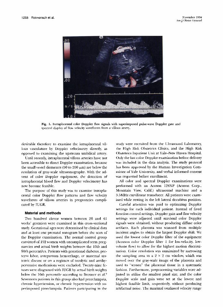
Fig. 1. Intraplacental color Doppler flow signals with superimposed pulse-wave Doppler gate and spectral display of flow velocity waveforms from a villous artery.
desirable therefore to examine the intraplacental villous vasculature by Doppler velocimetry directly, as opposed to examining the upstream umbilical artery.
Until recently, intraplacental villous arteries have not been accessible to direct Doppler examination, because the small vessel diameters (50 to 200 ILm) are below the resolution of gray-scale ultrasonography. With the advent of color Doppler equipment, the detection of intraplacental blood flow and Doppler velocimetry has now become feasible.
The purpose of this study was to examine intraplacental color Doppler flow patterns and flow velocity waveforms of villous arteries in pregnancies complicated by IUGR.
Material and methods
Two hundred eleven women between 26 and 41 weeks' gestation were examined in this cross-sectional study. Gestational ages were determined by clinical data and at least one prenatal sonogram before the scan of the Doppler examination. The normal control group consisted of 192 women with uncomplicated term pregnancies and actual birth weights between the 10th and 90th percentiles. Patients with gestational diabetes. preterm labor, antepartum hemorrhage, or maternal systemic disease or on a regimen of tocolytic and antihypertensive medications were excluded. Twenty-nine fetuses were diagnosed with IVGR by actual birth weights below the 10th percentile according to Brenner et al. 9
Seventeen patients in this group also had preeclampsia, chronic hypertension, or chronic hypertension with superimposed preeclampsia. Patients partcipating in the
study were recruited from the Ultrasound Laboratory, the High Risk Obstetrics Clinics, and the High Risk Obstetrics Inpatient Unit at Yale-New Haven Hospital. Only the last color Doppler examination before delivery was included in the data analysis. The study protocol has been approved by the Human Investigation Committee of Yale University, and verbal informed consent was requested before enrollment.
All color and spectral Doppler examinations were performed with an Acuson 128XP (Acuson Corp., Mountain View, Calif.) ultrasound machine and a 3.5MHz curvilinear transducer. All patients were examined while resting in the left lateral decubitus position.
Careful attention was paid to optimizing Doppler settings for each individual patient. Instead of fixed function control settings, Doppler gain and flow velocity settings were adjusted until maximal color Doppler signals were obtained, without producing diffuse color artifacts. Each placenta was scanned from multiple incident angles to obtain the largest Doppler shift. We used the lowest color Doppler filter of the equipment (Accuson color Doppler filter 1 for low-velocity, lowvolume flow) to allow for the highest motion discrimination. Color resolution was maximized by narrowing the sampling area to a 2 x 2 em window, which was moved over the gray-scale image of the placenta and used to "screen" the placental tissue in a systematic fashion . Furthermore, preprocessing variables were adjusted to utilize the smallest pixel size, and the color Doppler scale and gain were set at the lowest and highest feasible limit, respectively, without producing artifactual noise. The maximal unaliased velocity range

Volume 171 , Number 5 Am J Obstet Gynecol
Rotmensch et al. 1259
Fig. 2. A, Color Doppler flow signals from two villous trees. B, Multiple generations ofviJIous arterial branches. C, Color coding of blood flow velocities demonstrating gradual slowing of flow toward the basal plate.
utilIzed during the examination was 0.05 to 0.17 m/sec, corresponding to a pulse repetition frequency of 454.5 to 1545.5 Hz.
After localization of blood flow with color Dopplel~ a spectral Doppler display was obtained by superimposing the Doppler gate over the area of color flow on the screen (Fig. 1). Fetal versus maternal arterial flow was identified by the heart rate and the typical flow velocity waveform. At least two intraplacental vessels in each placenta were sampled except for the eight cases in which no intraplacental flow could be detected. The mean value of the pulsatility indexes was used for analysis. Areas within 2 em of the placental margin or cord insertion were not sampled, in accordance with histologic findings by Giles et a1." The intraobserver and interobserver error (coefficient of variation) in spectral Doppler measurements ofviIIous arteries was 7.9% and 9.1 %, respectively.
Assignment of Apgar scores to vaginally delivered neonates without distress was performed by the obstetric nursing staff. Operatively delivered fetuses or vagi-
nally delivered fetuses requmng resuscitative support were assigned Apgar scores by the neonatology team.
Fetal distress was defined as an abnormal fetal heart rate tracing or biophysical profile, which required immediate delivery in the judgment of the delivery attendant. Intraplacental color Doppler flow findings were not available to the delivery attendants. Umbilical artery Doppler findings were usually known. In five cases the diagnosis of fetal distress was made before labor, since the ominous fetal heart rate tracing precluded induction of/abor. In four cases the diagnosis was made in labor and substantiated by a fetal scalp blood pH of < 7.20. Newborn umbilical vein blood gas values were available in 14 of the 29 IUGR cases. Linear regression, Fisher's exact test, and the Wilcoxon rank sum test were used for statistical analysis.
Placentas were submitted for histopathologic examination in only J 7 of the 29 IUCR cases, because in some cases the presence of rVCR was only retrospectively recognized, when the newborn was weighed in the nursery. The available specimens included all 8 IUGR

1260 Rotmensch et al. November 1994 Am J Obstet Gynecol
Fig. 3. Intraplacental color Doppler flow image in a pregnancy with severe IVGR. Only small motion artifacts are noticeable at optimal system settings.
cases without intraplacental color Doppler flow and 9 IVCR cases with detectable intraplacental color Doppler flow. The pathologist was unaware of the placental color Doppler findings obtained before delivery. Histopathologic findings consistent with uteroplacental insufficiency were categorized as mild, moderate, or severe, on the basis of a semi-quantitative scoring system that is routinely used at Yale-New Haven Hospital.
Results
A minimum of two intraplacental fetal arteries could be detected by color Doppler flow in all 192 normal pregnancies (Fig. 2, A), and in most cases three to five villous arteries were easily detectable. Typically, two to four generations of arterial branching could be observed under optimal conditions (Fig. 2, B). The velocities were usually the highest in the main stem artery, as demonstrated by color coding, and gradually decreased toward the basal plate (Fig. 2, C). However, spectral Doppler signals could be obtained in all layers of the placenta. from the chorionic plate to the basal plate. No systematic differences were found in pulsatility index values of villous arteries in correlation with distance from the chorionic plate in a subset of 60 patients, in whom this aspect was investigated. In 8 of 29 IUCR pregnancies (27.6%), no intraplacental fetal blood flow could be detected (Fig. 3), as compared with 0 of 192 normal pregnancies (jJ < 0.000l). Four of these eight pregnancies were delivered by emergency cesarean sections for severe fetal distress before labor as evident on antenatal fetal heart rate testing or biophysical profile scores of 0 and 2 out of 10. Median Apgar scores at I minute in cases with and without intraplacental color
Doppler flow were 8 and 5 (jJ < 0.05), respectively, and at 5 minutes were 8 and 8 (not significant). Umbilical vein blood gas values confirmed fetal hypoxemia in all four cases (P02 range from 9 to 27 mm Hg) and all fetuses had low pH values (7.22 to 7.28) below the 5th percentile for gestational age by criteria for nonlaboring patients. 10 Cord blood gas values were only available
- for seven of the remaining IUCR cases, which did not allow for a statistical comparison of acid-base parameters between fetuses with and without detectable intraplacental color Doppler flow.
Fig. 4 shows regression curves for pulsatility indexes of the umbilical and intraplacental arteries. Both decreased with advancing gestational age. However, the villous artery pulsatility index remained consistently lower in absolute values throughout gestation (jJ < 0.001), even though the difference narrowed with advancing gestational age.
Fig. 5 shows the regression curve and the 5% and 95% confidence limits for pulsatility index values of intraplacental fetal arteries in normal pregnancies, as related to gestational age, and individual data for IUCR cases. Only I of 21 IUCR cases with detectable intraplacental color Doppler flow (4.8%) was associated with an abnormally high intraplacental artery pulsatility index (> 95% confidence limit), whereas 8 of 21 cases (38.0%) had abnormal umbilical artery pulsatility indexes (Fig. 6). Interestingly, the abnormal villous artery pulsatility index was concurrent with a normal umbilical artery flow velocity waveform. whereas all cases of abnormal umbilical artery flow velocity waveforms were associated with normal villous flow velocity waveforms. In two cases reversed diastolic flow in the umbilical

Volume 171, Number 5 Am J Obstet Gynecol
1.6
1.4
1.2
1.0
Plo.s
0.6
0.4
0.2
Rotmensch et al. 1261
-- --- -
- - - - - Umbilical Artery ----Intraplacental Artery
0.0 25 30 35 40
Gestational Age (weeks)
Fig. 4. Regression curves for umbilical artery and villous artery pulsatility indexes (PI).
1.5
• ---1.0
--- - .. -.------ ---• • • --- ---
• • 95%
PI • • • • • • • • --- - - -0.5 --- --- • • - -- --- --- _5Jo.
30 35 40
Gestational Age (weeks)
Fig. 5. Villous artery pulsatility indexes (PI) of normal and IVCR pregnancies.
artery was observed concurrent with normal diastolic forward flow in multiple intraplacental vessels (Fig. 7, A and B).
Five of eight placentas (62.5%) in which no intraplacental color Doppler flow was antenatally detectable were classified by the pathologist as demonstrating "severe uteroplacental insufficiency," and three placentas were categorized as "moderate" and "mild." Two placentas contained massive infarcts, involving > 30% of the placental tissue. Of the nine placental specimens of IUGR with antenatally detectable intraplacental color Doppler flow, 2 (22.2%) were classified as showing "severe utero placental insufficiency," 4 (44.4%) were "moderate," and 3 (33.3%) were "mild." Sample sizes
were insufficient for a meaningful statistical comparison between the two groups.
Comment
We examined intraplacental fetal blood flow characteristics in normal and IUGR pregnancies with the use of color Doppler flow mapping and spectral flow velocity waveform analysis of villous arteries. The main findings of this study are that color Doppler equipment can invariably visualize the fetal intraplacental vasculature in normal pregnancies but fails to do so in 27% of pregnancies with IUGR. However, contrary to our expectations, when placental color Doppler flow signals are detectable in the setting of IUGR, villous artery

1262 Rotmensch et al.
6
5 •
4
PI 3
2 •
•
• • • • •
November 1994 Am J Obstet Gynecol
- - - - -t- • -.- -.- - - 1- - - .. - - - - ---------------- . -------
O~_L~~~~ __ ~~ __ ~~ __ L_~~~~~ __ ~_J
25 30 35 40
Gestational Age (weeks)
Fig. 6. Umbilical artery pulsatility indexes (PI) of fetuses with IUGR and detectable intraplacental flow.
pulsatility index values are usually within the normal range, even when umbilical artery pulsatility index values are extremely abnormal.
Color Doppler equipment uses a pulse-echo technique to receive Doppler shifts created by a moving sound reflector and display the magnitude of the shift in brightness of color. The type of color displayed is arbitrary but, once selected, corresponds to the direction of flow as detected by a positive or negative Doppler shift. Most high-performance equipment can detect flow velocities as low as 1 em/sec. This feature has been used for the visualization of low-velocity and low-volume circulations such as tumor neovascularization in the ovary." Hsieh et al. 12 successfully visualized intraplacental fetal arteries in 39 normal pregnancies and demonstrated the feasibility of performing superimposed spectral Doppler examinations in this circulation. The successful Doppler examination of villous arteries in 192 normal pregnancies in our series confirms the applicability of this approach for the investigation of fetal placental blood flow.
The reasons for the lack of intraplacental color Doppler flow signals in 8 of our 29 IUGR cases can only be speculated on at this point. It is unlikely that this finding resulted from complete lack of placental perfusion because all of these fetuses were born alive, albeit depressed. Failure to demonstrate color Doppler flow in an organ in which blood is presumably present can be due to anyone of four factors or a combination thereof: (1) lack of system optimization for flow detection, (2) physical obstacles to sound transmission, (3) volume flow below the detectable threshold, and (4) flow velocity below the detectable threshold.
We paid careful attention to system optimization for each individual patient in this study, as outlined in the
Methods section. Furthermore, visualization of intraplacental color Doppler flow was successful in all 192 normal cases. It is unlikely therefore that inappropriate system settings could explain the absence of color Doppler flow in the 8 IUGR cases.
With regard to potential physical obstacles, morbid maternal obesity can occasionally interfere with sound wave transmission. However, none of the 8 IUeR cases in our series occurred in obese mothers. Therefore we do not believe that impaired sound wave transmission was responsible for our findings.
We speculate that decreased volume flow, flow velocity, or a combination of both, is the likely cause of un detectable intraplacental color Doppler flow. The primary determinant of umbilical-placental blood flow is the size and anatomy of the vascular bed. Even though histopathologic examination of available placentas from our series indicated a trend toward more severe uteroplacental insufficiency, sample sizes were insufficient to conclusively demonstrate that occlusive vascular abnormality is responsible for the observed color Doppler findings. Since neural control of villous blood flow is apparently nonexistent and biochemical control appears to be relatively limited under physiologic conditions, 13 vasospasm is an unlikely explanation.
Multiple studies have demonstrated that the intraplacental vascular anatomy of fetuses with IueR is altered and the vascular bed size appears to be reduced. Altshuler et al. 14 provided histopathologic evidence for a high incidence of obliterative lesions in stem and terminal villi in placentas of fetuses with IUGR. Lee and Yehl5 found that latex-injection corrosion casts of placentas in cases of IUeR display less branching of arteries and veins and more irregularities in luminal size as compared with placentas of normal fetuses. Las

Volume 171, Number 5 Am J Obstet Gynecol
Rotmensch et al. 1263
Fig. 7. A, Reversed dIastolic flow in the umbilical artery of a fetus WIth severe IUGR at 28 weeks' gestation. B, Normal intraplacental flow velocity waveforms in the same fetus. The spectral display of the villous artery flow velocity waveforms is bidirectional because of an incident angle approximating 90 degrees. Even with this large incident angle. abundant dIastolic forward flow is observed.
Heras et al. 16 concluded from a carefully conducted morphometric study that a significant reduction in the luminal diameter of villous stem arteries occurs in association with maternal hypertension and low birth weight.
The relevance of these anatomic findings to Doppler flow studies of the umbilical-placental circulation has been demonstrated by Giles et al. In their study umbilical artery flow velocity waveform patterns of increased resistance correlated well with a decreased arterial vessel count in tertiary stem villi. 2 Most pregnancies in this series were complicated by IUGR, preeclampsia. or both. It is conceivable therefore that anatomic changes in the intraplacental vascular anatomy of the IUGR cases reduced volume flow and/or flow velocity below the detectability threshold of our equipment. This is further supported by the finding of abnormal umbilical artery Doppler indexes in 5 of the 8 IUGR cases with undetectable intraplacental color Doppler flow, indicating increased downstream resistance in the placenta. A vascular morphometric study is currently in progress to examine this hypothesis.
It is equally conceivable that hemodynamic factors related to fetal cardiac output and redistribution of blood flow could explain the lowered placental perfusion. Reed et al. '7 found the ratio of right-sided to left-sided volume flow in the heart to be increased in fetuses with IUGR, suggesting that alterations in left ventricular function are associated with absence of enddiastolic flow in the umbilical artery. Furthermore, fetal acidosis has a cardiac depressant effect, which could further reduce placental perfusion.
Anecdotally, we have observed disappearance of intraplacental color Doppler flow signals in two cases of severe IUGR within a 3-day period, which would suggest an upstream cardiovascular factor or, alternatively. widespread acute thrombosis in the villous vascular bed. However, our study was not designed to obtain longitudinal data, and therefore no firm conclusions can be drawn from these findings.
Contrary to our expectations, Doppler velocimetry of the villous arteries does not appear to be a sensitive tool for the identification of the compromised fetus. Doppler flow indexes were within the normal range in all but one of the 21 IUGR cases with detectable intraplacental flow. In comparison, 8 of 21 (38.0%) umbilical artery Doppler indexes in this group were abnormal. In fact, the discordance between abnormal umbilical artery Doppler indexes and normal villous artery Doppler indexes in the same fetus are conceptually difficult to reconcile. In the most extreme cases of discordance, reversed diastolic flow in the umbilical artery was observed concurrently with normal diastolic forward flow in the intraplacental circulation. One possible explanation could be based on the assumption of an uneven distribution of vascular occlusion among villous arteries in the placenta. Umbilical artery Doppler indexes reflect the total resistance of the placental vascular bed. In contrast, Doppler examination of villous arteries is performed on individual vessels that are "self-selected" by their prominent color Doppler appearance but reflect only local flow and resistance within a particular villus. If we assume that critical stenosis or occlusion of a large number of villous arteries rendered them unde-

1264 Rotmensch et al.
tectable by color Doppler equipment, whereas others remained patent and possibly accommodated larger volume flow by an increased flow velocity, such an assumption could reconcile the contradictory observations of increased overall resistance of the placental circulation and normal resistance and flow detection in an isolated villus.
Whether the absence of intraplacental color Doppler flow patterns will prove to be useful as a predictor of fetal compromise in fetuses with IUCR remains to be seen. In our population this finding appeared to occur late in the course of fetal distress, as evidenced by abnormal fetal heart rate tracings or biophysical profiles in all IUCR cases without flow detection. However, selection bias related to patient referral patterns in our institution could have had an impact on the results. All of the patients without detectable placental flow were referred for intensive management because of severe IUCR, severe preclampsia, suspected abruptio placentae, or a combination of these. A prospective study is needed to determine at which point in the development of fetal distress intraplacental color Doppler flow becomes undetectable.
We conclude that failure to detect intraplacental color Doppler flow signals is associated with IUCR and tetal distress. Flow velocity waveforms of detectable villous arteries are usually normal in IUCR, even in the presence of an extremely abnormal umbilical artery flow velocity waveform. The role of this finding in assessing well-being of fetuses with IUCR remains to be determined in a prospective study.
REFERENCES 1. Trudinger BJ, Giles WB, Cook CM, BombardieriJ, Collins
L. Fetal umbilical artery flow VelOCIty waveforms and placental resistance: clinical significance. Br J Obstet Gynaecol 1985;92:23-30.
2. Giles WB, Trudinger BJ, Baird PJ. Fetal umbilical artery flow velocity waveforms and placental resistan<-e: pathological correlation. Br J Obstet Gynaecol 1985;92:31-8.
3. Bracero LA, Beneck D, Kirshenbaum N, Peiffer M, Stalter
November 1994 Am J Obstet Gynecol
P, Schulman H. Doppler velocimetry and placental disease. AM J OSSTET GYNECOL 1989;161:388-93.
4. Fok RY, Pavlova Z, Benirschke K, Paul RH, Platt LD. The correlation of arterial lesions with umbilical artery Doppler velocimetry in the placentas of small-for-dates pregnancies. Obstet Gynecol 1990;75:578-83.
5. Rochelson B, Kaplan C, Guzman E, Arato M, Hansen K, Trunca C. A quantitative analysis of placental vasculature in the third-trimester fetus with autosomal trisomy. Obstet Gynecol 1990;75:59-63.
6. Trudinger BJ, Giles WB. Clinical and pathological correlations of umbilical and uterine artery waveforms. Clin Obstet Gynecol 1989;32:669-78.
7. Thompson RS, Trudinger BJ. Doppler waveform pusatility index and reSIstance, pressure and flow in the umbilical placental circulation: an investigation using a mathematical model. Ultrasound Med BioI 1990; 16:449-58.
8. Trudinger BJ, Stevens D, Connelly A, et al. Umbilical artery flow velocity waveforms and placental resistance: the effect of embolization of the umbilical circulation. AM .I OSSTET GYNECOL 1987;157: 1443-8.
9. Brenner WE, Edelman DA, Hendricks CH. A standard of fetal growth for the United States of America. AM J OSSTET GYNECOL 1976; 126:555-64.
10. Weiner CP, Sipes SL, Wenstrom K. The effect of fetal age upon normal fetal laboratory values and venous pressure. Obstet Gynecol 1992;79:713-8.
11. Bourne T. Campbell S, Steer C, Whitehead MI, Collins WP. Transvaginal color flow imaging: a possible new screening technique for ovarian cancer. BMJ 1989;299: 1367-70.
12. Hsieh FJ, Kuo PL, Ko TM, Chang FM, Chen HY. Doppler velocimetry of intraplacental fetal arteries. Obstet Gynecol 1991;77:478-82.
13. Rankin JHG. Interaction between the maternal and fetal placental blood flows. In: Rosenfeld CR, ed. Volume 10: the uterine circulation. Ithaca, New York: Perinatology Press, 1989: 175-90.
14. Altshuler G, Russel P, Ermocilla R. The placental pathology of small for gestational age infants. AM J OSSTET GYNECOL 1975;121:351-9.
15. Lee MML, Yeh MN. Fetal circulation of the placenta: a comparative study of human and baboon placenta by scanning electron microscopy of vascular casts. Placenta 1983;4:515-26.
16. Las Heras J, Baskerville JC, Harding PG, Haust MD. Morphometric studies of fetal placental stem arteries in hypertensive disorders ('toxaemia') of pregnancy. Placenta 1985;6:217-27.
17. Reed KL, Anderson CF, Shenker L. Changes in intracardiac Doppler blood flow velocities in fetuses with absent umbilical artery diastolic flow. AM .I OSSTET GYNECOL 1987; 157:774-9.
Related Documents