Colloids and Surfaces B: Biointerfaces 110 (2013) 270–274 Contents lists available at SciVerse ScienceDirect Colloids and Surfaces B: Biointerfaces jou rn al hom epage: www.elsevier.com/locate/colsurfb Cibacron Blue F3GA modified disposable pencil graphite electrode for the investigation of affinity binding to bovine serum albumin Filiz Kuralay a,∗ , Erkut Yılmaz b , Lokman Uzun b,∗∗ , Adil Denizli b a Department of Chemistry, Faculty of Arts and Sciences, Ordu University, 52200 Ordu, Turkey b Department of Chemistry, Faculty of Science, Hacettepe University, 06800 Beytepe, Ankara, Turkey a r t i c l e i n f o Article history: Received 8 October 2012 Received in revised form 15 April 2013 Accepted 22 April 2013 Available online 28 April 2013 Keywords: Cibacron Blue F3GA Albumin Dye–protein interaction Pencil graphite electrode Cyclic voltammetry Electrochemical impedance spectroscopy a b s t r a c t In this work, Cibacron Blue F3GA (CB) modified pencil graphite electrodes (PGEs) were prepared and their affinities to bovine serum albumin were investigated. Preparation of the PGEs was performed using cyclic voltammetry (CV) and passive adsorption techniques. Improved electrochemical results were obtained with the PGEs prepared by CV technique compared to the PGEs prepared by passive adsorption technique. In order to obtain more sensitive results number of scans used in CV technique and the effect of con- centration of CB were studied. Scanning electron microscopy (SEM), atomic force microscopy (AFM) and electrochemical impedance spectroscopy (EIS) were used for the characterization of modified electrodes. The modified PGEs were then used for the electrochemical monitoring of affinity interaction between CB and bovine serum albumin. The effect of BSA concentration and interfering species (tryptophan, glucose and immunoglobulin G) on the response of the electrode were examined. The aim of this study was to prepare an easy, fast, stable and cheap modified electrode for the investigation of the well-known affinity of CB to serum albumin. The electrochemistry can provide alternative routes for dye–protein interaction instead of using classical time-consuming methods. © 2013 Published by Elsevier B.V. 1. Introduction Triazine-based dye molecules attracted great attention in the field of protein affinity studies [1–5]. They have several advan- tages to natural counterparts for specific affinity chromatography [6,7]. They are able to bind a variety of proteins based on highly specific molecular recognition in a reversible manner [8–11]. Triazine-based dyes are commercially available and cheap. The immobilization of these dyes on surfaces is also very easy [6,12]. Although these dye molecules are synthetic in nature, they are classified as affinity ligands due to their interaction with the active sites of proteins by mimicking the structure of the cofactors, substrates, or binding agents for those proteins [2]. A number of textile dyes, known as reactive dyes, can be used for protein related studies. Most of these reactive dyes contain a chromophore (either azo dyes, antraquinone, or phathalocya- nine) linked to a reactive group (often a mono- or dichlorotriazine ring). The synergic effect of combination of secondary forces such as electrostatic, hydrophobic and hydrogen binding plays an ∗ Corresponding author. Tel.: +90 452 2345010x1680; fax: +90 452 2339149. ∗∗ Corresponding author. Tel.: +90 312 29767963; fax: +90 312 2996084. E-mail addresses: kuralay.fi[email protected] (F. Kuralay), [email protected] (L. Uzun). important role in the interaction of these dyes with biomolecules [2,7]. Cibacron Blue F3GA (CB) is one of the most commonly used dye ligands for protein affinity studies [13]. It is a derivative of monochlorotriazine dye (antraquinone textile dye) which contains three titrable acidic sulfonate and four basic primary and secondary aromatic amine groups and it is also commercially important in the dyeing of cellulose fibers [12,14,15]. Its affinity to serum albumin is studied in details and reported in the literature [16–19]. It is known that CB interacts specifically and reversibly with albumin by the combination of hydrophobic, hydrogen bonding and electrostatic interactions [7]. However, the number of studies including elec- trochemistry of CB is very limited [6,15,20]. Nowadays, there is an increasing demand in electrochemical sensing strategies due to the fact that electrochemistry provides several advantages such as fast response, easy, stable, controllable and simple preparation of the modified surfaces [21]. Bovine serum albumin (BSA) is the most abundant protein. It plays an important role in the binding, transportation and depo- sition of variety of endogenous and exogenous ligands in blood. It is widely used as a blocking agent to reduce the non-specific protein–protein or protein–surface interactions. BSA can be used as a model protein applied in enzyme-linked immunosorbent assay because of its good stability and low cost. Thus, the development of detection systems for BSA holds great interest in various fields [22–24]. 0927-7765/$ – see front matter © 2013 Published by Elsevier B.V. http://dx.doi.org/10.1016/j.colsurfb.2013.04.024

Welcome message from author
This document is posted to help you gain knowledge. Please leave a comment to let me know what you think about it! Share it to your friends and learn new things together.
Transcript

Ct
Fa
b
ARRAA
KCADPCE
1
fit[h[T[twt[
fanrs
l
0h
Colloids and Surfaces B: Biointerfaces 110 (2013) 270– 274
Contents lists available at SciVerse ScienceDirect
Colloids and Surfaces B: Biointerfaces
jou rn al hom epage: www.elsev ier .com/ locate /co lsur fb
ibacron Blue F3GA modified disposable pencil graphite electrode forhe investigation of affinity binding to bovine serum albumin
iliz Kuralaya,∗, Erkut Yılmazb, Lokman Uzunb,∗∗, Adil Denizlib
Department of Chemistry, Faculty of Arts and Sciences, Ordu University, 52200 Ordu, TurkeyDepartment of Chemistry, Faculty of Science, Hacettepe University, 06800 Beytepe, Ankara, Turkey
a r t i c l e i n f o
rticle history:eceived 8 October 2012eceived in revised form 15 April 2013ccepted 22 April 2013vailable online 28 April 2013
eywords:ibacron Blue F3GA
a b s t r a c t
In this work, Cibacron Blue F3GA (CB) modified pencil graphite electrodes (PGEs) were prepared and theiraffinities to bovine serum albumin were investigated. Preparation of the PGEs was performed using cyclicvoltammetry (CV) and passive adsorption techniques. Improved electrochemical results were obtainedwith the PGEs prepared by CV technique compared to the PGEs prepared by passive adsorption technique.In order to obtain more sensitive results number of scans used in CV technique and the effect of con-centration of CB were studied. Scanning electron microscopy (SEM), atomic force microscopy (AFM) andelectrochemical impedance spectroscopy (EIS) were used for the characterization of modified electrodes.
lbuminye–protein interactionencil graphite electrodeyclic voltammetrylectrochemical impedance spectroscopy
The modified PGEs were then used for the electrochemical monitoring of affinity interaction between CBand bovine serum albumin. The effect of BSA concentration and interfering species (tryptophan, glucoseand immunoglobulin G) on the response of the electrode were examined. The aim of this study was toprepare an easy, fast, stable and cheap modified electrode for the investigation of the well-known affinityof CB to serum albumin. The electrochemistry can provide alternative routes for dye–protein interaction
time-
instead of using classical. Introduction
Triazine-based dye molecules attracted great attention in theeld of protein affinity studies [1–5]. They have several advan-ages to natural counterparts for specific affinity chromatography6,7]. They are able to bind a variety of proteins based onighly specific molecular recognition in a reversible manner8–11]. Triazine-based dyes are commercially available and cheap.he immobilization of these dyes on surfaces is also very easy6,12]. Although these dye molecules are synthetic in nature,hey are classified as affinity ligands due to their interactionith the active sites of proteins by mimicking the structure of
he cofactors, substrates, or binding agents for those proteins2].
A number of textile dyes, known as reactive dyes, can be usedor protein related studies. Most of these reactive dyes contain
chromophore (either azo dyes, antraquinone, or phathalocya-
ine) linked to a reactive group (often a mono- or dichlorotriazineing). The synergic effect of combination of secondary forcesuch as electrostatic, hydrophobic and hydrogen binding plays an∗ Corresponding author. Tel.: +90 452 2345010x1680; fax: +90 452 2339149.∗∗ Corresponding author. Tel.: +90 312 29767963; fax: +90 312 2996084.
E-mail addresses: [email protected] (F. Kuralay),[email protected] (L. Uzun).
927-7765/$ – see front matter © 2013 Published by Elsevier B.V.ttp://dx.doi.org/10.1016/j.colsurfb.2013.04.024
consuming methods.© 2013 Published by Elsevier B.V.
important role in the interaction of these dyes with biomolecules[2,7].
Cibacron Blue F3GA (CB) is one of the most commonly useddye ligands for protein affinity studies [13]. It is a derivative ofmonochlorotriazine dye (antraquinone textile dye) which containsthree titrable acidic sulfonate and four basic primary and secondaryaromatic amine groups and it is also commercially important in thedyeing of cellulose fibers [12,14,15]. Its affinity to serum albumin isstudied in details and reported in the literature [16–19]. It is knownthat CB interacts specifically and reversibly with albumin by thecombination of hydrophobic, hydrogen bonding and electrostaticinteractions [7]. However, the number of studies including elec-trochemistry of CB is very limited [6,15,20]. Nowadays, there is anincreasing demand in electrochemical sensing strategies due to thefact that electrochemistry provides several advantages such as fastresponse, easy, stable, controllable and simple preparation of themodified surfaces [21].
Bovine serum albumin (BSA) is the most abundant protein. Itplays an important role in the binding, transportation and depo-sition of variety of endogenous and exogenous ligands in blood.It is widely used as a blocking agent to reduce the non-specificprotein–protein or protein–surface interactions. BSA can be used
as a model protein applied in enzyme-linked immunosorbent assaybecause of its good stability and low cost. Thus, the developmentof detection systems for BSA holds great interest in various fields[22–24].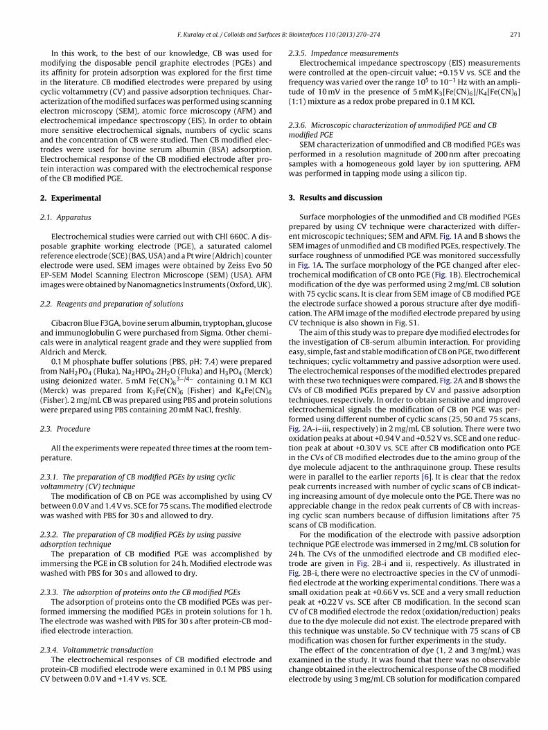
ces B:
miicaeematEto
2
2
preEi
2
acA
fu((w
2
p
2v
bw
2a
iw
2
fTi
2
pC
F. Kuralay et al. / Colloids and Surfa
In this work, to the best of our knowledge, CB was used forodifying the disposable pencil graphite electrodes (PGEs) and
ts affinity for protein adsorption was explored for the first timen the literature. CB modified electrodes were prepared by usingyclic voltammetry (CV) and passive adsorption techniques. Char-cterization of the modified surfaces was performed using scanninglectron microscopy (SEM), atomic force microscopy (AFM) andlectrochemical impedance spectroscopy (EIS). In order to obtainore sensitive electrochemical signals, numbers of cyclic scans
nd the concentration of CB were studied. Then CB modified elec-rodes were used for bovine serum albumin (BSA) adsorption.lectrochemical response of the CB modified electrode after pro-ein interaction was compared with the electrochemical responsef the CB modified PGE.
. Experimental
.1. Apparatus
Electrochemical studies were carried out with CHI 660C. A dis-osable graphite working electrode (PGE), a saturated calomeleference electrode (SCE) (BAS, USA) and a Pt wire (Aldrich) counterlectrode were used. SEM images were obtained by Zeiss Evo 50P-SEM Model Scanning Electron Microscope (SEM) (USA). AFMmages were obtained by Nanomagnetics Instruments (Oxford, UK).
.2. Reagents and preparation of solutions
Cibacron Blue F3GA, bovine serum albumin, tryptophan, glucosend immunoglobulin G were purchased from Sigma. Other chemi-als were in analytical reagent grade and they were supplied fromldrich and Merck.
0.1 M phosphate buffer solutions (PBS, pH: 7.4) were preparedrom NaH2PO4 (Fluka), Na2HPO4·2H2O (Fluka) and H3PO4 (Merck)sing deionized water. 5 mM Fe(CN)6
3−/4− containing 0.1 M KClMerck) was prepared from K3Fe(CN)6 (Fisher) and K4Fe(CN)6Fisher). 2 mg/mL CB was prepared using PBS and protein solutionsere prepared using PBS containing 20 mM NaCl, freshly.
.3. Procedure
All the experiments were repeated three times at the room tem-erature.
.3.1. The preparation of CB modified PGEs by using cyclicoltammetry (CV) technique
The modification of CB on PGE was accomplished by using CVetween 0.0 V and 1.4 V vs. SCE for 75 scans. The modified electrodeas washed with PBS for 30 s and allowed to dry.
.3.2. The preparation of CB modified PGEs by using passivedsorption technique
The preparation of CB modified PGE was accomplished bymmersing the PGE in CB solution for 24 h. Modified electrode was
ashed with PBS for 30 s and allowed to dry.
.3.3. The adsorption of proteins onto the CB modified PGEsThe adsorption of proteins onto the CB modified PGEs was per-
ormed immersing the modified PGEs in protein solutions for 1 h.he electrode was washed with PBS for 30 s after protein-CB mod-fied electrode interaction.
.3.4. Voltammetric transductionThe electrochemical responses of CB modified electrode and
rotein-CB modified electrode were examined in 0.1 M PBS usingV between 0.0 V and +1.4 V vs. SCE.
Biointerfaces 110 (2013) 270– 274 271
2.3.5. Impedance measurementsElectrochemical impedance spectroscopy (EIS) measurements
were controlled at the open-circuit value; +0.15 V vs. SCE and thefrequency was varied over the range 105 to 10−1 Hz with an ampli-tude of 10 mV in the presence of 5 mM K3[Fe(CN)6]/K4[Fe(CN)6](1:1) mixture as a redox probe prepared in 0.1 M KCl.
2.3.6. Microscopic characterization of unmodified PGE and CBmodified PGE
SEM characterization of unmodified and CB modified PGEs wasperformed in a resolution magnitude of 200 nm after precoatingsamples with a homogeneous gold layer by ion sputtering. AFMwas performed in tapping mode using a silicon tip.
3. Results and discussion
Surface morphologies of the unmodified and CB modified PGEsprepared by using CV technique were characterized with differ-ent microscopic techniques; SEM and AFM. Fig. 1A and B shows theSEM images of unmodified and CB modified PGEs, respectively. Thesurface roughness of unmodified PGE was monitored successfullyin Fig. 1A. The surface morphology of the PGE changed after elec-trochemical modification of CB onto PGE (Fig. 1B). Electrochemicalmodification of the dye was performed using 2 mg/mL CB solutionwith 75 cyclic scans. It is clear from SEM image of CB modified PGEthe electrode surface showed a porous structure after dye modifi-cation. The AFM image of the modified electrode prepared by usingCV technique is also shown in Fig. S1.
The aim of this study was to prepare dye modified electrodes forthe investigation of CB-serum albumin interaction. For providingeasy, simple, fast and stable modification of CB on PGE, two differenttechniques; cyclic voltammetry and passive adsorption were used.The electrochemical responses of the modified electrodes preparedwith these two techniques were compared. Fig. 2A and B shows theCVs of CB modified PGEs prepared by CV and passive adsorptiontechniques, respectively. In order to obtain sensitive and improvedelectrochemical signals the modification of CB on PGE was per-formed using different number of cyclic scans (25, 50 and 75 scans,Fig. 2A-i–iii, respectively) in 2 mg/mL CB solution. There were twooxidation peaks at about +0.94 V and +0.52 V vs. SCE and one reduc-tion peak at about +0.30 V vs. SCE after CB modification onto PGEin the CVs of CB modified electrodes due to the amino group of thedye molecule adjacent to the anthraquinone group. These resultswere in parallel to the earlier reports [6]. It is clear that the redoxpeak currents increased with number of cyclic scans of CB indicat-ing increasing amount of dye molecule onto the PGE. There was noappreciable change in the redox peak currents of CB with increas-ing cyclic scan numbers because of diffusion limitations after 75scans of CB modification.
For the modification of the electrode with passive adsorptiontechnique PGE electrode was immersed in 2 mg/mL CB solution for24 h. The CVs of the unmodified electrode and CB modified elec-trode are given in Fig. 2B-i and ii, respectively. As illustrated inFig. 2B-i, there were no electroactive species in the CV of unmodi-fied electrode at the working experimental conditions. There was asmall oxidation peak at +0.66 V vs. SCE and a very small reductionpeak at +0.22 V vs. SCE after CB modification. In the second scanCV of CB modified electrode the redox (oxidation/reduction) peaksdue to the dye molecule did not exist. The electrode prepared withthis technique was unstable. So CV technique with 75 scans of CBmodification was chosen for further experiments in the study.
The effect of the concentration of dye (1, 2 and 3 mg/mL) wasexamined in the study. It was found that there was no observablechange obtained in the electrochemical response of the CB modifiedelectrode by using 3 mg/mL CB solution for modification compared

272 F. Kuralay et al. / Colloids and Surfaces B: Biointerfaces 110 (2013) 270– 274
pared
tcupasFn
F5mC(
Fig. 1. SEM images of (A) unmodified PGE, (B) CB modified PGE pre
o the response obtained by using 2 mg/mL CB solution for modifi-ation. However, the redox peak currents were lower in the case ofsing 1 mg/mL CB solution for the modification of PGE (Fig. 3) com-ared to the results obtained by using 2 mg/mL CB solution givenbove (Fig. 2A). The CVs of CB modified PGEs after 25, 50 and 75
cans of CB modification using 1 mg/mL CB solution are shown inig. 3-i–iii, respectively. After 75 scans of CB modification, there waso appreciable change in the redox peak currents with increasingig. 2. (A) CVs of CB modified PGEs prepared with CV technique (i) 25 scans, (ii)0 scans, (iii) 75 scans of CB modification using 2 mg/mL CB solution (B) CVs of CBodified PGEs prepared with passive adsorption technique in PBS (pH 7.4) (24 h
B modification). CV measurement: at 100 mV/s scan rate between 0.0 V and +1.4 Vn = 3).
by using CV technique with 75 cyclic scans (2 mg/mL CB solution).
scan numbers. Thus 2 mg/mL CB concentration was chosen as opti-mum working concentration value for further studies.
EIS was also used to identify and differentiate the CB modifiedPGEs. EIS technique can provide information on the impedancechanges of the electrode surface during the modification process[25]. In the Nyquist plot of impedance spectra, the diameter ofthe semicircle represents the charge-transfer resistance (Rct) at theelectrode surface [26]. Fig. 4-i–iii shows the impedance spectra ofCB modified PGE with 25 scans of dye modification, CB modifiedPGE with 50 scans of dye modification and CB modified PGE with75 scans of dye modification, respectively. Rct value decreased withincreasing scan numbers of CB modification indicating improvedcharge-transfer occurred at the electrode surface [26]. The Nyquistplot of CB modified PGE prepared with 75 scans of modificationafter 2.5 mg/mL BSA interaction for 1 h was also performed (Fig. 4-iv). The Rct value increased compared to the value obtained in thecase of CB modified PGE with 75 scans of modification (Fig. 4-iii)due to BSA immobilization onto the electrode. Protein moleculeimproved the resistance to charge-transfer occurred at the elec-trode surface since it formed a more insulative layer.
For the application of this electrode, CB modified PGEs were usedfor the monitoring of CB–BSA interaction due to the fact that CB haswell-known affinity to serum albumin [16–19]. The affinity of CB
to bovine serum albumin is studied by using cyclic voltammetry.The CB modified PGEs were immersed in BSA solutions with differ-ent concentrations for 1 h. Then, the CVs of these electrodes wereFig. 3. CVs of CB modified PGEs prepared with CV technique (i) 25 scans, (ii) 50scans, (iii) 75 scans of CB modification using 1 mg/mL CB solution in PBS (pH 7.4) CVmeasurement: at 100 mV/s scan rate between 0.0 V and +1.4 V (n = 3).
F. Kuralay et al. / Colloids and Surfaces B: Biointerfaces 110 (2013) 270– 274 273
Fig. 4. Impedance spectra of (i) CB modified electrode prepared with 25 scans ofCB modification, (ii) CB modified electrode prepared with 50 scans of CB modifica-tion, (iii) CB modified electrode prepared with 75 scans of CB modification, (iv) CBmodified electrode prepared with 75 scans of CB modification after 2.5 mg/mL BSAisa
rmtmICriai
tbclvaeticartB
Fii1
nteraction for 1 h in 5 mM Fe(CN)63−/4− redox probe containing 0.1 M KCl. EIS mea-
urement: at open-circuit value of +0.15 mV between 105 and 10−1 Hz range withmplitude of 10 V (n = 3).
ecorded in 50 mM PBS (pH 7.4). Fig. 5-i–iv shows the CVs of CBodified PGE, CB modified electrode after 0.5 mg/mL BSA interac-
ion, CB modified electrode after 1.5 mg/mL BSA interaction and CBodified electrode after 2.5 mg/mL BSA interaction, respectively.
t was observed that BSA did not exhibit observable peak at theB modified PGE. This result was in accordance with the reportedesults [27,28]. However, the redox peak currents of CB decreasedn the presence of BSA. It was proposed that CB and BSA formedn electroinactive complex [27,28]. In addition to these results BSAnteraction had no effect on the redox peak potentials of CB.
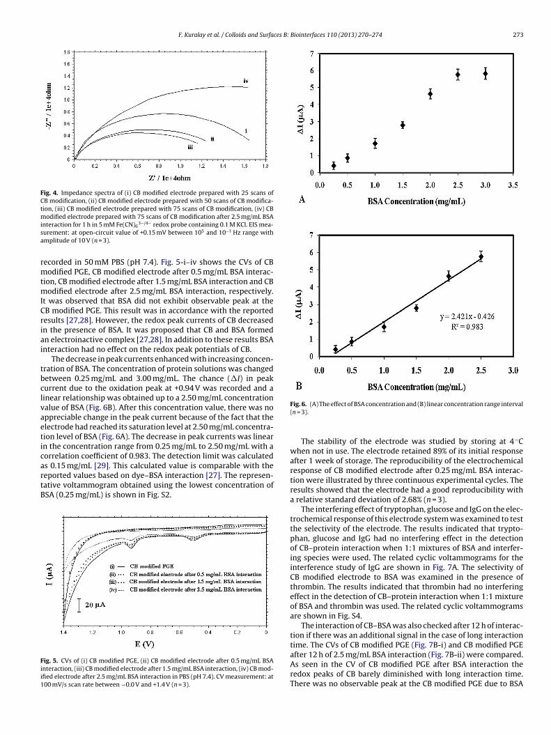
The decrease in peak currents enhanced with increasing concen-ration of BSA. The concentration of protein solutions was changedetween 0.25 mg/mL and 3.00 mg/mL. The chance (�I) in peakurrent due to the oxidation peak at +0.94 V was recorded and ainear relationship was obtained up to a 2.50 mg/mL concentrationalue of BSA (Fig. 6B). After this concentration value, there was noppreciable change in the peak current because of the fact that thelectrode had reached its saturation level at 2.50 mg/mL concentra-ion level of BSA (Fig. 6A). The decrease in peak currents was linearn the concentration range from 0.25 mg/mL to 2.50 mg/mL with aorrelation coefficient of 0.983. The detection limit was calculated
s 0.15 mg/mL [29]. This calculated value is comparable with theeported values based on dye–BSA interaction [27]. The represen-ative voltammogram obtained using the lowest concentration ofSA (0.25 mg/mL) is shown in Fig. S2.ig. 5. CVs of (i) CB modified PGE, (ii) CB modified electrode after 0.5 mg/mL BSAnteraction, (iii) CB modified electrode after 1.5 mg/mL BSA interaction, (iv) CB mod-fied electrode after 2.5 mg/mL BSA interaction in PBS (pH 7.4). CV measurement: at00 mV/s scan rate between −0.0 V and +1.4 V (n = 3).
Fig. 6. (A) The effect of BSA concentration and (B) linear concentration range interval(n = 3).
The stability of the electrode was studied by storing at 4 ◦Cwhen not in use. The electrode retained 89% of its initial responseafter 1 week of storage. The reproducibility of the electrochemicalresponse of CB modified electrode after 0.25 mg/mL BSA interac-tion were illustrated by three continuous experimental cycles. Theresults showed that the electrode had a good reproducibility witha relative standard deviation of 2.68% (n = 3).
The interfering effect of tryptophan, glucose and IgG on the elec-trochemical response of this electrode system was examined to testthe selectivity of the electrode. The results indicated that trypto-phan, glucose and IgG had no interfering effect in the detectionof CB–protein interaction when 1:1 mixtures of BSA and interfer-ing species were used. The related cyclic voltammograms for theinterference study of IgG are shown in Fig. 7A. The selectivity ofCB modified electrode to BSA was examined in the presence ofthrombin. The results indicated that thrombin had no interferingeffect in the detection of CB–protein interaction when 1:1 mixtureof BSA and thrombin was used. The related cyclic voltammogramsare shown in Fig. S4.
The interaction of CB–BSA was also checked after 12 h of interac-tion if there was an additional signal in the case of long interactiontime. The CVs of CB modified PGE (Fig. 7B-i) and CB modified PGEafter 12 h of 2.5 mg/mL BSA interaction (Fig. 7B-ii) were compared.
As seen in the CV of CB modified PGE after BSA interaction theredox peaks of CB barely diminished with long interaction time.There was no observable peak at the CB modified PGE due to BSA
274 F. Kuralay et al. / Colloids and Surfaces B:
Fig. 7. CVs of (A) (i) CB modified electrode after 1 h 1.0 mg/mL BSA interaction (ii)CC(
io(3pti
4
aftonfam
[[[[
[[[
[
[
[[[[[[[[
[
B modified electrode after 1 h 1.0 mg/mL BSA + 1.0 mg/mL IgG interaction. (B) (i)B modified PGE (ii) CB modified PGE after 12 h 2.5 mg/mL BSA interaction in PBSpH 7.4). CV measurement: at 100 mV/s scan rate between 0.0 V and +1.4 V (n = 3).
mmobilization. The comparison of the electrochemical responsef CB modified PGE (Fig. S3-i) and CB modified PGEs after 30 minFig. S3-ii) and 3 h (Fig. S3-iii) of BSA interaction was made. With0 min of CB–BSA interaction only small diminutions in oxidationeak currents of CB were obtained. However, with 3 h of interactionhe redox peaks of CB barely diminished as obtained with 12 h ofnteraction.
. Conclusions
In this study CB modification on PGEs was performed using CVnd passive adsorption techniques and the affinities of these newormed electrodes were examined to bovine serum albumin. Elec-rochemical CV technique provided simple, fast, well distributionf dye molecule onto the PGE compared to passive adsorption tech-
ique. The easy preparation of stable and cheap modified electrodeor the investigation of the well-known affinity of CB to serumlbumin was carried out compared to classical time-consumingethods for protein detection [30,31]. The electrode prepared with
[[
[[
Biointerfaces 110 (2013) 270– 274
CV technique showed more stable and reproducible results. Themodified electrodes were then used for the adsorption of BSA.The adsorption of protein onto the surface of the electrode waspresented simple and fast process. It was also easy to monitorthe electrochemical changes after CB–protein interaction at theelectrode surface in a good selectivity and sensitivity. The effectsof different concentrations of bovine serum albumin (BSA) andinterfering species such as tryptophan, glucose and immunoglob-ulin G (IgG) were examined. It was found that the decrease inCB oxidation peak current was linear in the concentration rangefrom 0.25 mg/mL to 2.50 mg/mL BSA with a correlation coefficientof 0.983 and the detection limit was calculated as 0.15 mg/mL.Hence, this procedure might be able to determine BSA, indirectly.The electrode showed good stability and reproducibility. Trypto-phan, glucose and IgG had no interfering effect for the detection ofCB–protein interaction in the study. This technology can be furtherused in protein–protein interactions.
Appendix A. Supplementary data
Supplementary data associated with this article can befound, in the online version, at http://dx.doi.org/10.1016/j.plantsci.2004.08.011.
References
[1] G. Jin, L. Zhang, Q. Yao, J. Membrane Sci. 287 (2007) 271.[2] A. Denizli, E. Pis kin, J. Biochem. Biophys. Methods 49 (2001) 391.[3] J. Zhang, Z. Zhang, Y. Song, H. Cai, React. Funct. Polym. 66 (2006) 916.[4] Z.Y. Ma, Y.P. Guan, H.Z. Liu, React. Funct. Polym. 66 (2006) 618.[5] Z. Ma, K. Masaya, S. Ramakrishna, J. Membrane Sci. 282 (2006) 237.[6] E.R.C. Viana, F.C. Pereira, M.V.B. Zanoni, Dyes Pigm. 71 (2006) 145.[7] N. Basar, L. Uzun, A. Güner, A. Denizli, J. Appl. Polym. Sci. 108 (2008) 3454.[8] C.R. Lowe, Advan, Mol. Cell. Biol. 15 (1996) 513.[9] C. Li, K.H. Lee, Anal. Biochem. 333 (2004) 381.10] E. Ruckenstein, X. Zeng, J. Membrane Sci. 142 (1998) 13.11] K.I. Shimazaki, N. Nishio, J. Dairy Sci. 4 (1991) 404.12] C.A. Andac, M. Andac, A. Denizli, Int. J. Biol. Macromol. 41 (2007) 430.13] C. Shirai, M. Sugai, H. Komatsuzawa, K. Ohta, M. Yamakido, H. Suginaka, Antimi-
crob. Agents Chemother. 42 (1998) 1278.14] H. Zolinger, Color in Chemistry, C.C.H., Publisher, New York, 1991.15] D.D. Schlereth, J. Electroanal. Chem. 425 (1997) 77.16] L. Uzun, H. Yavuz, R. Say, A. Ersöz, A. Denizli, Ind. Eng. Chem. Res. 43 (2004)
6507.17] C. Koch, L. Borg, K. Skjodt, G. Houen, J. Chromatogr. B Biomed. Appl. 718 (1998)
41.18] E.B. Altıntas , N. Tüzmen, L. Uzun, A. Denizli, Ind. Eng. Chem. Res. 46 (2007)
7802.19] N. Basar, L. Uzun, A. Güner, A. Denizli, Int. J. Biol. Macromol. 41 (2007) 234.20] D.D. Schlereth, R.P.H. Kooyman, J. Electroanal. Chem. 444 (1998) 231.21] A. Erdem, Talanta 74 (2007) 318.22] L. Fotouhi, S. Banafsheh, M.M. Heravi, Biolectrochemistry 77 (2009) 26.23] S.-B. Ji, Z.-H. Yan, J.-W. Wu, L.-L. Chen, H. Li, Biosens. Bioelectron. 39 (2013) 106.24] V.B. Kandimalla, V.S. Tripathi, H. Ju, Biomaterials 27 (2006) 1167–1174.25] P. Du, H. Li, Z. Mei, S. Liu, Bioelectrochemistry 75 (2009) 37.26] F. Kuralay, A. Erdem, S. Abacı, H. Özyörük, A. Yıldız, Anal. Chim. Acta 643 (2009)
83.27] L. Fotouhi, S. Banafsheh, M.M. Heravi, Bioelectrochemistry 77 (2009) 26.
28] H. Luo, Y. Du, Z.X. Guo, Bioelectrochemistry 74 (2009) 232.29] J.N. Miller, J.C. Miller, Statistics and Chemometrics for Analytical Chemistry,Pearson Education, London, 2000.30] S. Centi, S. Tombelli, M. Minunni, M. Mascini, Anal. Chem. 79 (2007) 1466.31] G.S. Bang, S. Cho, B.-G. Kim, Biosens. Biolectron. 21 (2005) 863.
Related Documents


![Colloids and Surfaces B: Biointerfaces Colloids Surfaces B... · Colloids and Surfaces B: Biointerfaces 116 (2014) ... antibiotics [3–6]. Their broad ... Alamethicin is most effective](https://static.cupdf.com/doc/110x72/5a94ecce7f8b9a9c5b8c50e4/colloids-and-surfaces-b-colloids-surfaces-bcolloids-and-surfaces-b-biointerfaces.jpg)