Critical Review Collagen Structure: The Madras Triple Helix and the Current Scenario Arnab Bhattacharjee and Manju Bansal Molecular Biophysics Unit, Indian Institute of Science, Bangalore, India Summary This year marks the 50th anniversary of the coiled – coil triple helical structure of collagen, first proposed by Ramachandran’s group from Madras. The structure is unique among the protein secondary structures in that it requires a very specific tripeptide sequence repeat, with glycine being mandatory at every third position and readily accommodates the imino acids proline/ hydroxyproline, at the other two positions. The original structure was postulated to be stabilized by two interchain hydrogen bonds, per tripeptide. Subsequent modeling studies suggested that the triple helix is stabilized by one direct inter chain hydrogen bond as well as water mediated hydrogen bonds. The hydroxyproline residues were also implicated to play an important role in stabilizing the collagen fibres. Several high resolution crystal structures of oligopeptides related to collagen have been determined in the last ten years. Stability of synthetic mimics of collagen has also been extensively studied. These have confirmed the essential correctness of the coiled- coil triple helical structure of collagen, as well as the role of water and hydroxyproline residues, but also indicated additional sequence- dependent features. This review discusses some of these recent results and their implications for collagen fiber formation. IUBMB Life, 57: 161 – 172, 2005 Keywords Collagen structure; triple helix; hydroxyproline; amino acid propensity. INTRODUCTION The fibrous protein collagen constitutes almost one quarter of the total protein content in most animals, being the major component of several connective tissues such as skin, tendons, ligaments, cartilage, bone, teeth, basement membranes, blood vessels etc. The term ‘collagen’ is in fact derived from the Greek word for glue and was initially used to describe that constituent of connective tissue which yields gelatin on boiling. However it was soon established that in some tissues, collagen is either heavily cross-linked or covalently bonded to some other stable structure so that it cannot be extracted by just heating. Thus, while the collagen in tendon forms long rope like structures, giving them great tensile strength, the hard rigid structure of bone and teeth arises due to calcification of the interstitial space between the molecules. Cross-links between fibers form flexible two dimensional sheets in skin, while a more complex arrangement in three dimensions is found in cartilage. The ubiquitous nature of this molecule has been confirmed by recent studies wherein it has been found that vertebrates have at least 27 collagen types (described as being type I to XXVII) with 42 distinct polypeptide chains. In addition, more than 20 other proteins have been reported to have collagen-like domains (1, 2). All collagens also possess non-collagenous domains in addition to the fibrous collagen domains. The collagen molecule consists of three polypeptide chains, called a chains, with the characteristic triplet repeat sequence Gly-X-Y and each chain is generally more than 1000 residue long. In some collagens all three chains are identical, while in others, the molecules contain two or even three different a chains, described as a1, a2 and a3, with the difference lying in the amino acids present in X and Y positions of the triplets. The most striking feature of the collagen molecule is its unique tertiary structure, arising from its characteristic triplet repeat sequence and the presence of a large amount of the imino acid proline, which in about half the case is hydroxylated at its C g position. The first essentially correct structure for collagen was proposed 50 years ago by Ramachandran and Kartha and consists of three left handed polypeptide helices, held together by interchain hydrogen bonds (3, 4, 5). The history of how this structure was first established and its current status is worth reviewing, in view of its importance in relation to connective tissue disorders. ORIGIN AND REFINEMENT OF THE COLLAGEN TRIPLE HELIX Astbury (6) was the first to suggest a structure for collagen in 1938, which consisted of a mixture of trans and cis peptide units and the same feature was incorporated by Pauling and Received 5 January 2005; accepted 23 February 2005 Address correspondence to: Manju Bansal, Molecular Biophysics Unit, Indian Institute of Science, Bangalore-560012, India. Tel: 91 80 2293 2534. Fax: 91 80 2360 0535. E-mail: [email protected] net.in IUBMB Life, 57(3): 161 – 172, March 2005 ISSN 1521-6543 print/ISSN 1521-6551 online # 2005 IUBMB DOI: 10.1080/15216540500090710

Welcome message from author
This document is posted to help you gain knowledge. Please leave a comment to let me know what you think about it! Share it to your friends and learn new things together.
Transcript

Critical Review
Collagen Structure: The Madras Triple Helix and the Current Scenario
Arnab Bhattacharjee and Manju BansalMolecular Biophysics Unit, Indian Institute of Science, Bangalore, India
Summary
This year marks the 50th anniversary of the coiled – coil triple
helical structure of collagen, first proposed by Ramachandran’s
group from Madras. The structure is unique among the proteinsecondary structures in that it requires a very specific tripeptide
sequence repeat, with glycine being mandatory at every third
position and readily accommodates the imino acids proline/hydroxyproline, at the other two positions. The original structure
was postulated to be stabilized by two interchain hydrogen bonds,
per tripeptide. Subsequent modeling studies suggested that the triple
helix is stabilized by one direct inter chain hydrogen bond as well aswater mediated hydrogen bonds. The hydroxyproline residues were
also implicated to play an important role in stabilizing the collagen
fibres. Several high resolution crystal structures of oligopeptides
related to collagen have been determined in the last ten years.Stability of synthetic mimics of collagen has also been extensively
studied. These have confirmed the essential correctness of the coiled-
coil triple helical structure of collagen, as well as the role of waterand hydroxyproline residues, but also indicated additional sequence-
dependent features. This review discusses some of these recent
results and their implications for collagen fiber formation.
IUBMB Life, 57: 161 – 172, 2005
Keywords Collagen structure; triple helix; hydroxyproline; aminoacid propensity.
INTRODUCTION
The fibrous protein collagen constitutes almost one quarter
of the total protein content in most animals, being the major
component of several connective tissues such as skin, tendons,
ligaments, cartilage, bone, teeth, basement membranes, blood
vessels etc. The term ‘collagen’ is in fact derived from the
Greek word for glue and was initially used to describe that
constituent of connective tissue which yields gelatin on boiling.
However it was soon established that in some tissues, collagen
is either heavily cross-linked or covalently bonded to some
other stable structure so that it cannot be extracted by just
heating. Thus, while the collagen in tendon forms long rope
like structures, giving them great tensile strength, the hard
rigid structure of bone and teeth arises due to calcification of
the interstitial space between the molecules. Cross-links
between fibers form flexible two dimensional sheets in skin,
while a more complex arrangement in three dimensions is
found in cartilage. The ubiquitous nature of this molecule has
been confirmed by recent studies wherein it has been found
that vertebrates have at least 27 collagen types (described as
being type I to XXVII) with 42 distinct polypeptide chains. In
addition, more than 20 other proteins have been reported to
have collagen-like domains (1, 2). All collagens also possess
non-collagenous domains in addition to the fibrous collagen
domains. The collagen molecule consists of three polypeptide
chains, called a chains, with the characteristic triplet repeat
sequence Gly-X-Y and each chain is generally more than 1000
residue long. In some collagens all three chains are identical,
while in others, the molecules contain two or even three
different a chains, described as a1, a2 and a3, with the
difference lying in the amino acids present in X and Y
positions of the triplets.
The most striking feature of the collagen molecule is its
unique tertiary structure, arising from its characteristic
triplet repeat sequence and the presence of a large amount
of the imino acid proline, which in about half the case is
hydroxylated at its Cg position. The first essentially correct
structure for collagen was proposed 50 years ago by
Ramachandran and Kartha and consists of three left
handed polypeptide helices, held together by interchain
hydrogen bonds (3, 4, 5). The history of how this structure
was first established and its current status is worth
reviewing, in view of its importance in relation to connective
tissue disorders.
ORIGIN AND REFINEMENT OF THE COLLAGEN TRIPLEHELIX
Astbury (6) was the first to suggest a structure for collagen
in 1938, which consisted of a mixture of trans and cis peptide
units and the same feature was incorporated by Pauling and
Received 5 January 2005; accepted 23 February 2005Address correspondence to: Manju Bansal, Molecular Biophysics
Unit, Indian Institute of Science, Bangalore-560012, India.Tel: 91 80 2293 2534. Fax: 91 80 2360 0535. E-mail: [email protected]
IUBMBLife, 57(3): 161 – 172, March 2005
ISSN 1521-6543 print/ISSN 1521-6551 online # 2005 IUBMB
DOI: 10.1080/15216540500090710

Corey in the model proposed by them in 1951 (7), which had
three co-axial helices. However neither of these structures were
in agreement with the observed X-ray diffraction pattern of
collagen fibers. It was Ramachandran’s group from Madras,
in India, who first postulated a triple helical structure for
collagen, containing only trans peptide bonds, as in other
natural proteins, in combination with the requirement that the
structure should necessarily have one third the total number of
residues as glycine (3). An additional requirement was that the
structure should be able to accommodate a large proportion of
imino acid residues (viz. proline and 4-hydroxyproline) which
have their side chains folded back to form rigid five membered
rings. This unique triple helical structure consisted of an
assembly of three parallel helices, in which the special type of
molecular packing is contributed by the occurrence of glycine
as every third residue, i.e., a repeating Gly-Xaa-Yaa sequence
in each polypeptide chain of the collagen protein. This
prototype structure is stabilized by inter-chain hydrogen
bonds, unlike the well established a-helical structure for
polypeptides (not to be confused with the nomenclature of
a-chains used for the single chains in the collagen triple helix),
which is stabilized by intra-chain hydrogen bonds and all the
main chain N-H and C=O groups are involved in these type
of interactions (8). Each of the three chains in the collagen
triple helix forms a left handed helix, with approximately three
residues per turn and the three chains are related by a three-
fold screw symmetry about a common axis (Fig. 1). Hence,
unlike the a-helix wherein all residues are in equivalent
positions, in the collagen triple helix there can be distinctly
different requirements for the three positions in the repeat
unit. In particular, every third position, which lies towards the
center of the triple helix cannot have any side chain attached
to it, since presence of even a b-carbon atom (as in alanine)
leads to unacceptable inter-chain atomic contacts. Hence this
position must necessarily have only glycine residues, thus
providing a rational explanation for the unique amino acid
composition and triplet repeat sequence (-Gly-X-Y-) of
collagen. All other amino acids including the imino acids
proline or hydroxyproline could be readily accommodated at
the X and Y positions. The torsion angle about the N-Ca bond
was ideal for the formation of the five-member pyrollidine
rings in case of Pro and Hyp.
The original triple helical model, proposed by Ramachan-
dran and Kartha in 1954 (3), was soon modified by the authors
themselves (4, 5), to fit the more accurate helical parameters
observed from x-ray diffraction of stretched collagen fibers (9),
which indicated that the number of residues per turn is closer
to 3.33, rather than 3. Consequently, they proposed a coiled-
coil triple-helical structure for collagen (shown in Fig. 1b),
with 10 residues in 3 turns of the left-handed minor helix of
each chain. The major helix formed by this single helix is right-
handed, accommodates 30 residues per turn and has a pitch of
*85.8 A. The neighboring helices in the triple helical assembly
are thus related by a twist of -1088 and rise of *2.86 A. In this
structure the requirement for glycine at every third position
was even more stringent and it was also postulated that the
imino acids will be preferentially accommodated at the Y
position. The triple helix is stabilized by the formation of two
inter-chain hydrogen bonds involving the amino groups of
Figure 1. (a) Schematic diagram showing the projection down
the helix axis of three left handed, parallel, polypeptide helices
in the prototype collagen structure with 3 residues per turn
and rise per residue of *3A (3). The positions 1, 4, 7. . . in all
three chains can only accommodate Gly residues, while all
other amino acids including proline can occur at other two
positions (b). The modified coiled-coil triple helical structure
for collagen with 3.33 residues per turn (5). Each helix
undergoes a twist of +368 and a translation of *8.7A about
the common axis relating the three chains. Neighboring chains
are related by a rotation of -1088 and a translation of *2.9A
(4,5).
162 BHATTACHARJEE AND BANSAL
glycine residues as well as the amino acids at the X position.
However, for the two hydrogen bonds to form, the neighbor-
ing chains have to approach quite close to each other and
some of the atoms in the proposed structure come very close to
each other leading to steric clashes, if standard van der Waal’s
radii are assumed for the various atoms (10). These clashes can
be avoided by a small rotation and translation of the three
chains in the triple helix, leading to a structure that retains all
the essential features of the Ramachandran coiled-coil triple-
helix, but involves only one amino group per tripeptide, that
from the glycyl residue, in the formation of inter-chain
hydrogen bonds and allows imino acids to be present in both
positions X and Y (shown in Fig. 2). Such a model was
proposed by Rich and Crick (11), soon after the publication of
the Ramachandran and Kartha coiled-coil structure (4, 5) in
1955 and has an inter chain hydrogen bond between the N-H
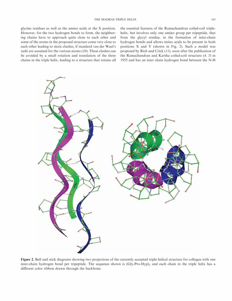
Figure 2. Ball and stick diagrams showing two projections of the currently accepted triple helical structure for collagen with one
inter-chain hydrogen bond per tripeptide. The sequence shown is (Gly-Pro-Hyp)3 and each chain in the triple helix has a
different color ribbon drawn through the backbone.
THE MADRAS TRIPLE HELIX 163

group of glycine and C=O group of the amino/imino acid in
the X position. This structure differs only marginally from the
original two bonded coiled-coil triple helical structure, as is
obvious from a comparison of the various parameters
discussed later in this review. Minor variants of this structure
have been subsequently proposed (12 – 15), but the essential
features remain invariant. One of the most interesting ideas to
resolve the one vs. two hydrogen bond debate came from
Ramachandran’s group (16), wherein it was suggested that,
while the triple helix may be stabilized by only one direct
hydrogen bond involving the glycine amino group, additional
inter-chain hydrogen bonds may be formed via water
molecules (shown in Fig. 3a). This hypothesis was further
extended by Ramachandran’s group to suggest that one of
these water molecules could form additional hydrogen bonds
with the hydroxyl group of hydroxyproline residues (as well as
other hydroxyl group containing amino acids, such as serine
and threonine) present at the Y position (also shown in Fig.
3a) of the repeating sequence (17 – 19), thus providing an
explanation for the observed correlation between the stability
and hydroxyproline content of various collagens (20 – 22).
These fiber-diffraction based models have been shown to be
essentially correct by single crystal X-ray analysis of collagen
related oligopeptides and other biophysical studies.
The individual triple helices or tropocollagen molecules, as
they are sometimes called, are arranged to form fibrils which
are of high tensile strength and flexibility and can be further
assembled and cross-linked (Fig. 4) so as to support stress
efficiently (23). Abnormalities in the collagen molecular
structure or its organization into mature fibers lead to different
diseases associated with connective tissues, such as Ethlers-
Danlos syndrome, osteogenesis imperfecta and some types of
osteoporosis and arthritis (1, 2). Most common mutations in
the collagen gene are single base substitutions that convert the
codon of the critical glycine residue to that of a bulkier
residue, which causes considerable distortion of the triple helix
or even prevent its formation beyond this point. Amino acid
changes in the other two positions of the triplet have milder
effects. Also since there are regions of high and low stability
within the collagen triple helix and stability within the collagen
triple helix depends on the amino acids present in the other
two positions of the repeating triplet, so mutations in different
regions can have different effects. In addition, only in fibril
forming collagens is a single continuous helix mandatory,
while other collagens that normally contain several interrup-
tions in the triplet repeat, can readily accommodate additional
disruptions with no major ill effects. Interestingly mutations
that produce some structural alterations in the polypeptide
chain but still allow the chains to assemble into a triple helix,
generally manifest as more severe phenotypes than those that
prevent triplex formation altogether (1). This is because the
triple helices containing the mutated chain will have an
abnormal structure, which will affect the formation of higher
order structure or alter their assembly and function. However
the exact relationship between the amino acid sequence change
and its effect on the structure and lethality of a mutation in the
collagen molecule is still not clear. The sequence dependent
variation observed in the structural parameters of collagen
triple helical structure and other biophysical studies on
stability of various collagen mimics can provide useful insights
into how differences in amino acid distribution may affect the
stability and higher order assembly of the triple helical
molecules in collagenous tissues.
COMPARISON OF THE STRUCTURAL PARAMETERS OFCOLLAGEN RELATED OLIGOPEPTIDES
Several crystal structures of collagen mimics (24 – 32) are
now available in the Protein Data Bank and we have
compared them with the various fiber models as well as with
each other, in order to get some insight into sequence
dependent variation in the structure. The details of the various
fiber models and tripeptide fragments from the crystal
structures that have been analyzed here, are given in Table
1. In order to avoid the end effects, only the middle regions of
these oligopeptides have been considered for this analysis. The
Gly?Ala mutant structure (1CAG) and the non imino acid
containing fragments from 1BKV and 1DZI are of particular
interest. In case of the fiber models, co-ordinates of the atoms
in a triplet were obtained from the published literature (12, 14,
15), and the intra, as well as inter helical twist and rise values
were used to generate the 9 amino acid long triple helices. The
various structural parameters in the crystal structures, helical
twist (t), helical rise (h), number of residues per helical turn
(n), as well as radii of Ca atom cylinders, were obtained using
our in house program HELANAL (33) and are listed in Table
2. It is interesting to note that the parameters for the non-
imino acid containing fragments are similar to those for the
fiber models. The phi (f) and psi (c) values of the triple helicalstructures have been plotted on a Ramachandran map (Fig. 5)
to get an idea of the variation in backbone torsion angles,
corresponding to the Gly, X and Y positions in the structures,
particularly in case of non Gly-Pro-Hyp type sequences. The
rmsd (Root Mean Square Deviation) values between the
various structures, in the case of a single chain as well as for
the triple chain assembly, when all the heavy atoms in the
backbone are considered, were also calculated. It was found
that the single chain conformation is fairly invariant in all the
structures, except in the case of HMB1, in which one of the
glycine residues in each chain has been replaced by an alanine.
Hence even though an alanine can be accommodated in lieu of
glycine in the single chain of the collagen triple helix, the steric
constraints imposed on an assembly of the three chains, forces
local distortion in individual helices. This is reflected in the
greater rmsd values for this structure as compared to all other
structures as well as larger dispersion of f and c values in this
structure (see Fig. 5). Similarly the small bend in the Integrin
bound collagen (JEMS) structure, following the G-E-R triplet,
164 BHATTACHARJEE AND BANSAL

Figure 3. (a) Projection down the helix axis of the one hydrogen-bonded fiber model (13), showing details of the direct interchain
hydrogen bond, between the glycyl amide group and the carbonyl oxygen of residue at X position in the neighboring chain of a
triple helix with Gly-X-Hyp sequence. The modeled water molecules (OW1 and OW2) linking the backbone of two neighboring
chains, as well as the Od of Hyp residues at Y position (16, 17) are also shown. (b) Interchain hydrogen bonds observed in the
1BKV crystal structure (29). Only the fragment Hyp 8 to Gly 12 in chain A and corresponding regions Pro 37 to Thr 41 in chain
B and Gly 66 to Ile 70 in chain C are shown here. The water mediated hydrogen bonds involving Thr 11 in chain A, Thr 41 in
chain B and Hyp 68 in chain C are also shown. The water molecules W1, W2 and W3 are indicated as +. The various atoms are
color coded as: carbon-green, nitrogen-blue, oxygen-red and hydrogen-white.
THE MADRAS TRIPLE HELIX 165

gives rise to slightly higher rmsd values for this triple helix.
Interestingly the rmsd between the original two hydrogen
bonded model of Ramachandran (GNR1) and the other
structures is only marginally higher than that found between
the various crystal structures and other fiber models.
It has recently been suggested that the small systematic
differences in the (f,c) values at X and Y positions lead to
differences in the preference for the proline ring pucker when
the imino acids occurs at these positions (34). This in turn is
related to the occurrence of hydroxyproline at Y position,
stabilizing the triple helix, since the pyrollidine ring in Hyp
prefers only one type of pucker (Cg – exo or up conforma-
tion). However since hydroxylation of proline occurs before
helix formation and the f, c variation is within the range
Figure 4. The individual triple helices or tropocollagen molecules, are arranged to form fibrils which are of high tensile strength
and flexibility and can be further assembled and cross-linked. The figure has been reproduced from Klug, W. S. and Cummings,
M. R. (23).
166 BHATTACHARJEE AND BANSAL

observed for standard deviation values, additional data is
required to accept this rationale for the occurrence of
hydroxyproline at Y position. Some other experimental
results have suggested that inductive effects, due to attach-
ment of an electronegative atom at Cg are the basis for the
contribution of Hyp residues to the stability of collagen (35 –
37). It should however be noted that, the earlier hypothesis
on the role of hydroxyproline in stabilizing the triple helical
structure through additional water mediated hydrogen bonds
(17, 18), has also been recently confirmed (shown in Fig 3b)
by the presence of such features in both the structures
containing (Gly-X-Hyp) type sequence in their local regions
(25, 29) as well as other biophysical studies (38, 39). Similar
hydrogen bonds are found to occur (29) in the presence of
threonine residues (also shown in Fig 3b). The Thr residues
have also been reported to stabilize fibrillar aggregates
through glycosylation at their Og position (40).
The oligopeptide fragments with mixed sequences show
values of helical parameters (particularly twist) that are
closer to those suggested from diffraction studies of native
collagen fibres (-1088) than the oligomers with (Gly-Pro-Pro)
type of repeats (which have twist values of about 7 1028).Since even a small variation in twist has implications for the
intermolecular interactions between triple helices and their
assembly (41), we examined the length distribution of
fragments consisting of the four types of triplets, (Gly-X-
Y), (Gly-Pro-Y), (Gly-X-Hyp) and (Gly-Pro-Hyp) in the
amino acid sequences of five types of collagen chains and
the data is shown in Fig. 6. Complete sequences have been
reported for these collagen chains, with Type I, II, and III
belonging to fibrillar collagen (42 – 45) and Type IV to
basement membrane collagen (46). It is clearly seen that
imino acid containing triplets are found scattered along the
collagen molecule, and rarely occur in stretches of more
Table 1.
List of triple helical collagen structures proposed from X-ray fiber diffraction as well as single crystal studies. Thesequence of the fragments used for the structural analysis is indicated in each case. ‘X’ indicates any amino acid in thesecond position of the triplets and ‘O’ indicates Hydroxyproline. The root mean square deviation (rmsd) value of the
backbone atoms in each structure when compared to the fiber model (RDBF) are also listed.
Serial
no.
Structure
description used PDB id Sequence of the fragment*
rmsd value w.r.t
RDBF structure
Reference
nos.
1 GNR1 Fiber model (GXPGXPGXP) 0.84 4,12
2 ARFC Fiber model (GPOGPOGPO) 0.47 10,11
3 GNR2 Fiber model (GPOGPOGPO) 0.66 13
4 RDBF Fiber model (GPOGPOGPO) – 14
5 HMB1 1CAG (G12POAPOGPO20) 1.12 24,25
(G42POAPOGPO50)
(G72POAPOGPO80)
6 HMB2 1QSU (G18POGPOGPO26) 0.93 27
(G48POGPOGPO56)
(G78POGPOGPO86)
7 HMB3 1QSU (G9POGEKGPO17) 0.65 27
(G39POGEKGPO47)
(G69POGEKGPO77)
8 HMB4 1BKV (G9ITGARGLA17) 0.34 28,29
(G39ITGARGLA47)
(G69ITGARGLA77)
9 JEMS 1DZI (G7FOGERGPO15) 1.06 30
(G7FOGERGPO15)
(G7FOGERGPO15)
10 ZAGA 1K6F (G3PPGPPGPP11) 0.74 31
(G3PPGPPGPP11)
(G3PPGPPGPP11)
11 OKUY 1IIT (G1PPGPP6) 0.34 32
(G2PPGPP7)
(G3PPGPP8)
* In case of crystal structures 5 – 9, these fragments are middle parts of longer molecules with flanking GPO or GPP sequences.
THE MADRAS TRIPLE HELIX 167
than two contiguous triplets. Hence the average helical
structure is expected to be closer to that seen in the (Gly-X-
Y) regions of oligopeptides rather than that observed in the
oligopeptides containing (Gly-Pro-Pro/Hyp) triplet repeats,
thus validating the earlier fiber models proposed for the
collagen molecule.
Interestingly the Type IV collagen that is known to have
more inter-triple helix interactions, contains a much higher
content of Hyp residues at Y position and a very significant
number of them occur as (Gly-X-Hyp) triplets, with no
increase in frequencies of Gly-Pro-Hyp triplets. This feature
also manifests itself in the higher frequency of occurrence of
3 and 4 mers of (Gly-X-Hyp) repeats, suggesting that
hydroxyproline, while stabilizing the triple helical structure,
can also play a role in intermolecular interactions, as
indicated by model building and crystal structure studies
(18, 25, 47). Similar interactions can also occur via other side
chains and there are also indications that several amino acids
selectively prefer X or Y position in natural collagen chains
(37, 48).
AMINO ACID PREFERENCES IN COLLAGEN CHAINS OFTYPE I-IV
A recent analysis of collagen sequence data has reported
the distribution of various triplets using sequence data from an
assortment of collagen chains of varying lengths (37, 48, 49).
We have carried out a detailed analysis to assess the
propensities of the amino acids to occur in X and Y positions
considering each of the five collagen chains, for which full
length sequences are available (42 – 46) and the results are
shown in Table 3.
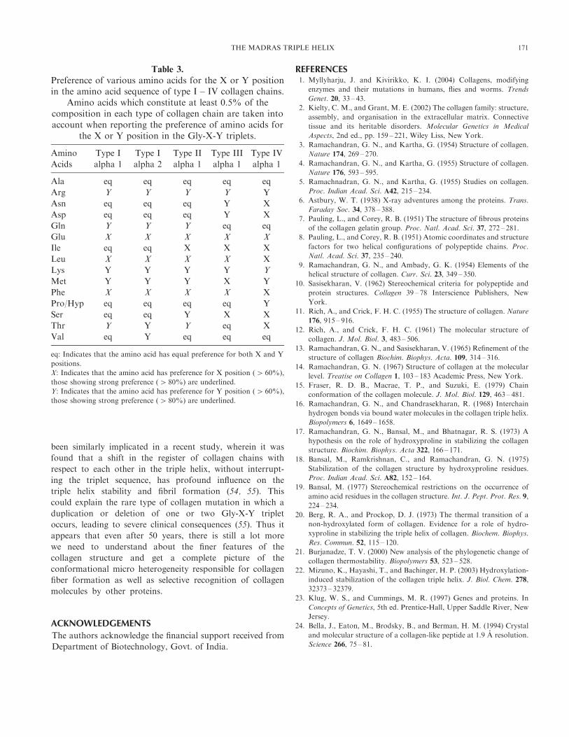
It is well known that alanine, which constitutes the most
frequently occuring amino acid after glycine and proline/
hydroxyproline, shows equal preference for both X and Y
positions in the Gly-X-Y triplets, in all types of collagens.
Since synthetic polypeptides rich in alanine do not take up
collagen like helical structures, it indicates that alanine is well
tolerated in collagen structures, but does not provide any
additional stability. This is confirmed by the melting
temperature studies on host guest peptides wherein Ala occurs
in the middle region of the stability scale (37, 49). There is a
clear bias for Glu, Leu and Phe in the X position whereas Arg
and Lys are seen to be preferred in the Y position of the Gly-
X-Y triplets in all the five collagen chains. Interestingly Gln
and Thr also show a preference for Y position in Type I and
Type II collagens while Met shows a similar preference in Type
I, II and IV collagens. The residues showing preference for Y
position can help stabilize triple helices as well as assemblies of
triple helices through additional interactions. Threonine can
mimic the Hyp residue (19, 29) by forming water mediated H-
bonds. Arginine in Y position has been shown to form inter
chain hydrogen bonds with a carbonyl oxygen of a neighbor-
ing chain (29, 30) in the crystal structures HMB4 and JEMS. It
has also been reported that replacement of Hyp with Arg in
this position results in a triple helix of nearly equal stability,
whereas all other amino acid substitutions, including Lys,
result in triple helices with lower melting temperatures (37, 49).
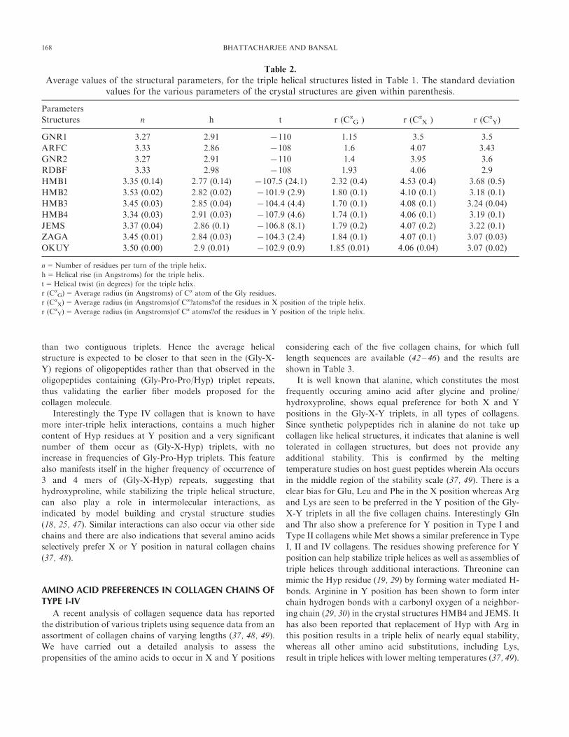
Table 2.
Average values of the structural parameters, for the triple helical structures listed in Table 1. The standard deviationvalues for the various parameters of the crystal structures are given within parenthesis.
Parameters
Structures n h t r (CaG ) r (Ca
X ) r (CaY)
GNR1 3.27 2.91 7110 1.15 3.5 3.5
ARFC 3.33 2.86 7108 1.6 4.07 3.43
GNR2 3.27 2.91 7110 1.4 3.95 3.6
RDBF 3.33 2.98 7108 1.93 4.06 2.9
HMB1 3.35 (0.14) 2.77 (0.14) 7107.5 (24.1) 2.32 (0.4) 4.53 (0.4) 3.68 (0.5)
HMB2 3.53 (0.02) 2.82 (0.02) 7101.9 (2.9) 1.80 (0.1) 4.10 (0.1) 3.18 (0.1)
HMB3 3.45 (0.03) 2.85 (0.04) 7104.4 (4.4) 1.70 (0.1) 4.08 (0.1) 3.24 (0.04)
HMB4 3.34 (0.03) 2.91 (0.03) 7107.9 (4.6) 1.74 (0.1) 4.06 (0.1) 3.19 (0.1)
JEMS 3.37 (0.04) 2.86 (0.1) 7106.8 (8.1) 1.79 (0.2) 4.07 (0.2) 3.22 (0.1)
ZAGA 3.45 (0.01) 2.84 (0.03) 7104.3 (2.4) 1.84 (0.1) 4.07 (0.1) 3.07 (0.03)
OKUY 3.50 (0.00) 2.9 (0.01) 7102.9 (0.9) 1.85 (0.01) 4.06 (0.04) 3.07 (0.02)
n=Number of residues per turn of the triple helix.
h=Helical rise (in Angstroms) for the triple helix.
t=Helical twist (in degrees) for the triple helix.
r (CaG)=Average radius (in Angstroms) of Ca atom of the Gly residues.
r (CaX)=Average radius (in Angstroms)of Ca?atoms?of the residues in X position of the triple helix.
r (CaY)=Average radius (in Angstroms)of Ca atoms?of the residues in Y position of the triple helix.
168 BHATTACHARJEE AND BANSAL

The amino acid preferences seen for X position cannot be
readily explained on the basis of any explicit interactions,
except for Glu in X position forming a stabilizing ion-pair
with Lys/Arg in Y position in a neighboring chain (29, 30,
50). It has been suggested that in the case of Leu and Phe it
is a negative preference for Y position, that makes X the
position of preference for these residues. However it may be
pertinent to point out that both these residues also show a
preference to sequester in (Gly-X-Hyp) rather than the
general (Gly-X-Y) triplet sequence, perhaps indicating a
more specific hydrophobic interaction between the two side
chains.
Interestingly, many of the sequence features observed in the
collagen sequences can be easily rationalized on the basis of
simple stereochemical restrictions on the occurrence of amino
acid residues in the triple helical molecules. One such study by
our group (19, 51) had suggested that Leu and Phe can be
accommodated at both locations X and Y, but when a proline
residue occurs at X position, it restricts the freedom of
orientation of these side chains at Y position in the
neighboring chain. It is also not possible to accommodate
the inter-chain hydrogen bond forming water molecules if
these large non-polar residues are present at position Y. In
contrast at X position, they have much greater freedom of
orientation and their presence will also help to shield the
hydrogen bonded water molecules as well as the direct
interchain hydrogen bonds from disturbance by the solvent
medium (19). Hence from the stability point of view, their
presence in conjunction with the Hyp residues at Y position
could make these triplet sequences almost as favorable as Gly-
Pro-Hyp triplets.
Thus interchain interactions appear to play a crucial
role in determining the variable local stability of different
regions of a collagen molecule (50, 52, 53). They have also
Figure 5. Value of the backbone torsion angle phi (f) is plotted against the psi (c) value for each residue is the collagen fiber
models and oligopeptide crystal structures listed in Table 1. The values corresponding to the three positions in the triplet
sequence Gly-X-Y are shown in black, blue and red colors respectively. Each structure is represented by a different symbol, as
indicated in top right corner.
THE MADRAS TRIPLE HELIX 169

Figure 6. Bar diagrams showing the length distribution of the four type of triplet stretches among representative examples of
Collagen types I to IV. The amino acid sequences correspond to type I a1 chain from skin tendon and bone of human
(N_P000079, 338, 42), type I a2 chain from placenta and skin of human (P08123, 342, 43), type II a1 chain from cartilaginous
tissue of house mouse (P28481, 340, 44), type III a1 chain from brain tissue of house mouse (P08121, 343, 45) and type IV a1chain from basement membrane of human (NP_001836, 375, 46) collagens. The Genbank accession no. of the sequences, the
number of amino acid triplets and the corresponding reference number are given above within parenthesis, for each collagen
chain.
170 BHATTACHARJEE AND BANSAL
been similarly implicated in a recent study, wherein it was
found that a shift in the register of collagen chains with
respect to each other in the triple helix, without interrupt-
ing the triplet sequence, has profound influence on the
triple helix stability and fibril formation (54, 55). This
could explain the rare type of collagen mutation in which a
duplication or deletion of one or two Gly-X-Y triplet
occurs, leading to severe clinical consequences (55). Thus it
appears that even after 50 years, there is still a lot more
we need to understand about the finer features of the
collagen structure and get a complete picture of the
conformational micro heterogeneity responsible for collagen
fiber formation as well as selective recognition of collagen
molecules by other proteins.
ACKNOWLEDGEMENTS
The authors acknowledge the financial support received from
Department of Biotechnology, Govt. of India.
REFERENCES1. Myllyharju, J. and Kivirikko, K. I. (2004) Collagens, modifying
enzymes and their mutations in humans, flies and worms. Trends
Genet. 20, 33 – 43.
2. Kielty, C. M., and Grant, M. E. (2002) The collagen family: structure,
assembly, and organisation in the extracellular matrix. Connective
tissue and its heritable disorders. Molecular Genetics in Medical
Aspects, 2nd ed., pp. 159 – 221, Wiley Liss, New York.
3. Ramachandran, G. N., and Kartha, G. (1954) Structure of collagen.
Nature 174, 269 – 270.
4. Ramachandran, G. N., and Kartha, G. (1955) Structure of collagen.
Nature 176, 593 – 595.
5. Ramachnadran, G. N., and Kartha, G. (1955) Studies on collagen.
Proc. Indian Acad. Sci. A42, 215 – 234.
6. Astbury, W. T. (1938) X-ray adventures among the proteins. Trans.
Faraday Soc. 34, 378 – 388.
7. Pauling, L., and Corey, R. B. (1951) The structure of fibrous proteins
of the collagen gelatin group. Proc. Natl. Acad. Sci. 37, 272 – 281.
8. Pauling, L., and Corey, R. B. (1951) Atomic coordinates and structure
factors for two helical configurations of polypeptide chains. Proc.
Natl. Acad. Sci. 37, 235 – 240.
9. Ramachandran, G. N., and Ambady, G. K. (1954) Elements of the
helical structure of collagen. Curr. Sci. 23, 349 – 350.
10. Sasisekharan, V. (1962) Stereochemical criteria for polypeptide and
protein structures. Collagen 39 – 78 Interscience Publishers, New
York.
11. Rich, A., and Crick, F. H. C. (1955) The structure of collagen. Nature
176, 915 – 916.
12. Rich, A., and Crick, F. H. C. (1961) The molecular structure of
collagen. J. Mol. Biol. 3, 483 – 506.
13. Ramachandran, G. N., and Sasisekharan, V. (1965) Refinement of the
structure of collagen Biochim. Biophys. Acta. 109, 314 – 316.
14. Ramachandran, G. N. (1967) Structure of collagen at the molecular
level. Treatise on Collagen 1, 103 – 183 Academic Press, New York.
15. Fraser, R. D. B., Macrae, T. P., and Suzuki, E. (1979) Chain
conformation of the collagen molecule. J. Mol. Biol. 129, 463 – 481.
16. Ramachandran, G. N., and Chandrasekharan, R. (1968) Interchain
hydrogen bonds via bound water molecules in the collagen triple helix.
Biopolymers 6, 1649 – 1658.
17. Ramachandran, G. N., Bansal, M., and Bhatnagar, R. S. (1973) A
hypothesis on the role of hydroxyproline in stabilizing the collagen
structure. Biochim. Biophys. Acta 322, 166 – 171.
18. Bansal, M., Ramkrishnan, C., and Ramachandran, G. N. (1975)
Stabilization of the collagen structure by hydroxyproline residues.
Proc. Indian Acad. Sci. A82, 152 – 164.
19. Bansal, M. (1977) Stereochemical restrictions on the occurrence of
amino acid residues in the collagen structure. Int. J. Pept. Prot. Res. 9,
224 – 234.
20. Berg, R. A., and Prockop, D. J. (1973) The thermal transition of a
non-hydroxylated form of collagen. Evidence for a role of hydro-
xyproline in stabilizing the triple helix of collagen. Biochem. Biophys.
Res. Commun. 52, 115 – 120.
21. Burjanadze, T. V. (2000) New analysis of the phylogenetic change of
collagen thermostability. Biopolymers 53, 523 – 528.
22. Mizuno, K., Hayashi, T., and Bachinger, H. P. (2003) Hydroxylation-
induced stabilization of the collagen triple helix. J. Biol. Chem. 278,
32373 – 32379.
23. Klug, W. S., and Cummings, M. R. (1997) Genes and proteins. In
Concepts of Genetics, 5th ed. Prentice-Hall, Upper Saddle River, New
Jersey.
24. Bella, J., Eaton, M., Brodsky, B., and Berman, H. M. (1994) Crystal
and molecular structure of a collagen-like peptide at 1.9 A resolution.
Science 266, 75 – 81.
Table 3.
Preference of various amino acids for the X or Y positionin the amino acid sequence of type I – IV collagen chains.
Amino acids which constitute at least 0.5% of thecomposition in each type of collagen chain are taken intoaccount when reporting the preference of amino acids for
the X or Y position in the Gly-X-Y triplets.
Amino
Acids
Type I
alpha 1
Type I
alpha 2
Type II
alpha 1
Type III
alpha 1
Type IV
alpha 1
Ala eq eq eq eq eq
Arg Y Y Y Y Y
Asn eq eq eq Y X
Asp eq eq eq Y X
Gln Y Y Y eq eq
Glu X X X X X
Ile eq eq X X X
Leu X X X X X
Lys Y Y Y Y Y
Met Y Y Y X Y
Phe X X X X X
Pro/Hyp eq eq eq eq Y
Ser eq eq Y X X
Thr Y Y Y eq X
Val eq Y eq eq eq
eq: Indicates that the amino acid has equal preference for both X and Y
positions.
X: Indicates that the amino acid has preference for X position (4 60%),
those showing strong preference (4 80%) are underlined.
Y: Indicates that the amino acid has preference for Y position (4 60%),
those showing strong preference (4 80%) are underlined.
THE MADRAS TRIPLE HELIX 171

25. Kramer, R. Z., and Berman, H. M. (1998) Patterns of hydration in
crystalline collagen peptides. J. Biomol. Struct. Dyn. 16, 367 – 380.
26. Kramer, R., Vitagliano, L., Bella, J., Berisio, R., Mazarella, L.
Brodsky, B., Zagari, A., and Berman, H. M. (1998) X-ray crystal-
lographic determination of a collagen-like peptide with the repeating
sequence (Pro-Pro-Gly). J. Mol. Biol. 280, 623 – 638.
27. Kramer, R. Z., Venugopal, M. G., Bella, J., Mayville, P., Brodsky, B.,
and Berman, H. M. (2000) Staggered molecular packing in crystals of
a collagen-like peptide with a single charged pair. J. Mol. Biol. 301,
1191 – 1205.
28. Kramer, R. Z., Bella, J., Mayville, P., Brodsky B., and Berman, H. M.
(1999) Sequence dependent conformational variations of collagen
triple-helical structure. Nat. Struct. Biol. 6, 454 – 457.
29. Kramer, R., Bella, J., Brodsky, B., and Berman, H.M. (2001) The
crystal and molecular structure of a collagen like peptide with a
biologically relevant sequence. J. Mol. Biol. 311, 131 – 147.
30. Emsley, J., Knight, C. G., Farndale, R.W., Barnes, M. J., and
Liddington, R. C. (2000) Structural basis of collagen recognition by
integrin a2b1. Cell 100, 47 – 56.31. Berisio, R., Vitagliano, L., Mazarella, L., and Zagari, A. (2002)
Crystal structure of the collagen triple helix model [(Pro-Pro-Gly)10]3.
Prot. Sci. 11, 262 – 270.
32. Hongo, C., Nagarajan, V., Noguchi, K., Kamitori, S., Okuyama, K.,
Tanaka, Y., and Nishino, N. (2001) Average crystal structure of (Pro-
Pro-Gly)9 at 1.0 angstroms resolution. Plym. J. 33, 812.
33. Bansal, M., Kumar, S., and Velavan, R. (2000) HELANAL: A
program to characterize helix geometry in proteins. J. Biomol. Struct.
Dyn. 17, 811 – 819.
34. Vitagliano, L., Berisio, R., Mazarella, L., and Zagari, A. (2001)
Structural basis of collagen stabilization induced by proline hydro-
xylation. Biopolymers 58, 459 – 464.
35. Holmgreen, S. K., Bretscher, L. E., Taylor, K. M., and Raines, R. T.
(1999) A hyperstable collagen mimic. Chem. Biol. 6, 63 – 70.
36. Mooney, S. D., Kollman, P. A., and Klein, T. E. (2002) Conforma-
tional preferences of substituted prolines in the collagen triple helix.
Biopolymers 64, 63 – 71.
37. Jenkins, C. L., and Raines R. T. (2002) Insights on the conformational
stability of collagen Nat. Prod. Rep. 19, 49 – 59.
38. Miles, C.A., and Bailey, A. J. (2001) Thermally labile domains in the
collagen molecule. Micron 32, 325 – 332.
39. Slatter, D. A., Miles, C. A., and Bailey, A. J. (2003) Asymmetry in the
triple helix of collagen-like heterotrimers confirms that external bonds
stabilize collagen structure. J. Mol. Biol. 329, 175 – 183.
40. Mann, K., Mechling, D. E., Bachinger, H. P., Eckerskorn, C., Gaill,
F., and Timpl, R. (1996) Glycosylated threonine but not 4-
hydroxyproline dominates the triple helix stabilizing positions in the
sequence of a hydrothermal vent worm cuticle collagen. J. Mol. Biol.
261, 255 – 266.
41. Kadler, K. E., Holmes, D. F., Trotter, J. A., and Chapman, J. A.
(1996) Collagen fibril formation. Biochem. J. 316, 1 – 11.
42. Ruza, E., Sotillo, E., Sierrasesumaga, L., Azcona, C., and Patino-
Garcia, A. (2003) Analysis of polymorphisms of the vitamin D
receptor, estrogen receptor, and collagen I alpha1 genes and their
relationship with height in children with bone cancer. J. Pediatr.
Hematol. Oncol. 25, 780 – 786.
43. De Wet, W., Bernard, M., Benson-Chanda, V., Chu, M. L., Dickson,
L., Weil, D., and Ramirez, F. (1987) Organization of the human pro-
alpha 2(I) collagen gene. J. Biol. Chem. 262, 16032 – 16036.
44. Metsaranta, M., Toman, D., De Crombrugghe, B., and Vuorio, E.
(1991) Mouse type II collagen gene. Complete nucleotide sequence,
exon structure, and alternative splicing. J. Biol. Chem. 266, 16862 –
16869.
45. Toman, P. D., and De Crombrugghe, B. (1994) The mouse type-III
procollagen-encoding gene: genomic cloning and complete DNA
sequence. Gene 147, 161 – 168.
46. Lauer-Fields, J. L., Malkar, N. B., Richet, G., Drauz K., and Fields,
G. B. (2003) Melanoma cell CD44 interaction with the alpha 1(IV)
1263-1277 region from basement membrane collagen is modulated by
ligand glycosylation. J. Biol. Chem. 278, 14321 – 14330.
47. Berisio, R., Vitagliano, L., Mazarella, L., and Zagari, A. (2001)
Crystal structure of a collagen-like polypeptide with repeating
sequence Pro-Hyp-Gly at 1.4 A resolution: Implications for collagen
hydration. Biopolymers 56, 8 – 13.
48. Ramshaw, J. A. M., Shah, N. K., and Brodsky, B. (1998) Gly-X-Y
tripeptide frequencies in collagen: a context for host-guest triple
helical peptides. J. Struct. Biol. 122, 86 – 91.
49. Persikov, A. V., Ramshaw, J. A. M., Kirkpatrick, A., and Brodsky, B.
(2000) Amino acid propensities for the collagen triple-helix. Biochem-
istry 39, 14960 – 14967.
50. Vitagliano, L., Nemethy, G., Zagari, A., and Scherega, H. A (1993)
Stabilization of the triple- helical structure of natural collagen by side-
chain interactions. Biochemistry 32, 7354 – 7359.
51. Bansal, M., and Ramachandran, G. N. (1978) A theoretical study on
the structure of (Gly-Pro-Leu)n and (Gly-Leu-Pro)n. Int. J. Pept.
Prot. Res. 11, 73 – 81.
52. Miles, C. A., Sims, T. J., Camacho, N. P., and Bailey, A. J. (2002) The
role of the alpha2 chain in the stabilization of the collagen type I
heterotrimer: a study of the type I homotrimer in oim mouse tissues. J.
Mol. Biol. 321, 797 – 805.
53. Sacca, B., Fiori, S., and Moroder, L. (2003) Studies on the local
conformational properties of the cell-adhesion domain of collagen
type IV in synthetic heterotrimeric peptides. Biochemistry 42, 3429 –
3436.
54. Sacca, B., Renner, C., and Moroder, L. (2002) The chain register in
heterotrimeric collagen peptides affects triple helix stability and
folding kinetics. J. Mol. Biol. 324, 309 – 318.
55. Cabral, W. A., Mertts, M. V., Makareeva, E., Colige, A., Tekin, M.,
Pandya, A., Leikin, S., and Marini, J. C. (2003) Type I collagen triplet
duplication mutation in lethal osteogenesis imperfecta shifts register
of alpha chains throughout the helix and disrupts incorporation of
mutant helices into fibrils and extracellular matrix. J. Biol. Chem. 278,
10006 – 10012.
172 BHATTACHARJEE AND BANSAL
Related Documents