Fröhlich Centenary International Symposium Coherence and Electromagnetic Fields in Biological Systems ABSTRACT BOOK July 1–4, 2005 Prague, Czech Republic

Welcome message from author
This document is posted to help you gain knowledge. Please leave a comment to let me know what you think about it! Share it to your friends and learn new things together.
Transcript

Fröhlich Centenary International Symposium
Coherence and Electromagnetic Fields in Biological Systems
ABSTRACT BOOK
July 1–4, 2005
Prague, Czech Republic

2

3
Abstracts of contributions sent for presentation at
the Symposium
Coherence and Electromagnetic Fields in
Biological Systems

4

5
EDITORIAL COMMENT
Symposium “Coherence and Electromagnetic Fields in Biological Systems” is focused on two main areas of research: One concerns biophysical principles and mechanisms of organization of living matter and the other the effects of external electromagnetic fields on living matter. Mechanisms of organization are still not well understood. It is assumed that organization forces and mechanisms are of electromagnetic nature. The majority of proteins are electrically polar and represent electric dipoles and/or multipoles. Strong electrically polar character of biological constituents makes possible longitudinal oscillations generating electric field. Some protein structures (such as e.g. microtubules in the cytoskeleton) are excited by energy supply from metabolic sources. H. Fröhlich formulated hypothesis of coherent vibrations in biological systems as their fundamental biophysical property. Some vibration modes are excited far from thermal equilibrium but the majority of modes in the system remain close to thermal equilibrium. Coherent excitation of some polar vibration modes can generate endogenous electromagnetic field with dominant electrical component. The endogenous electromagnetic field seems to be important for biophysical mechanism of organization. External electromagnetic field can change some properties of living matter. There is no uncertainty concerning the effects of the electromagnetic fields producing thermal effects. The main question of the effects of electromagnetic fields on biological systems concerns the exposure to the fields not producing thermal effects. The understanding of the mechanisms of non-thermal effects is far from comprehensive. Many authors claim that both adverse and beneficial responses are found. Electromagnetic field may affect chromatin conformation in living cells. Ions are transported along the DNA chain during polarization. Nonspecific pores in cellular membranes are formed by the electromagnetic fields. The process of pore formation may exhibit certain biological selectivity (the membranes of some cells are more liable to pore formation than the membranes of others) and special drugs can be transported through the pores inside cells (e.g. in cancer treatment). Immune system may be sensitive to the magnetic field. We cannot overwiev all effects and problems in this comment. Nevertheless, I would like to mention a highly controversial theme: the influence of external electromagnetic fields on the properties of water in the context of molecular biology. It is claimed that special structures might be formed in water and that the structures have effects on biological processes. I do not want to discuss here the measurement methods and results concerning this matter from biological point of view. I assume that physical measurements of properties of water based on theoretical analyses of model structures are necessary to prove or disprove existence of special structures which are anticipated to be connected with altered properties of water after exposure to the electromagnetic fields. This “Abstract Book” contains abstracts of contributions that we received. The authors are responsible for the content of their abstracts. Editor

6

7
THE LIVING AND THE NON-LIVING: DIFFERENT COMPOSITION MODES OF BASIC IMMATERIAL
CONNECTEDNESS - THE PHYSICAL BASIS OF LIFE -
Hans-Peter Dürr
Max-Planck-Institut für Physik, Werner-Heisenberg-Institut Föhringer Ring 6, D-80805 München, Germany
In classical physics the world is considered as a matter-based reality, an arrangement of spatially separated particles, the paths of which in time are uniquely determined by certain dynamic laws. By contrast, modern quantum physics reveals that matter is not composed of matter, but reality is merely potentiality. The world has not an ontic structure, admitting answers to questions like: What exists?, but rather a holistic, process-type structure, based on fundamental immaterial relations, referring only to questions like: What happens? Material objects and their small constituents, elementary particles, are replaced by ‘haps’, small immaterial happenings, which superimpose each other in a complex wave-like fashion and proceed in time in an open, indeterministic but still non-random way. In this more flexible quantum dynamic framework, the macroscopic living and the non-living are fundamentally alike but differ in the superpositions of large aggregates of the ‘pre-living’ immaterial ‘haps’. In situations close to static stable configurations effectively all the uncertainties, characterizing the basic ‘pre-living’ quality, are statistically averaged out, thus exhibiting the well-known unique and deterministic behavior of ordinary non-living matter. In the case, however, of statically instable but dynamically stable configurations, the “pre-lively” features of the underlying quantum structure, supported by a coherent superposition of the ‘haps’, have a chance to surface to the macroscopic level and to be connected with the feature we observe as the phenomenon of life. This supposition can be more explicitly treated and clarified by identifying the electric dipole moment of biomolecules as the ordering parameter of the corresponding macro-quantum system. This has important consequences for biology and medicine. In particular, it suggests the existence of a “software” to be essential for the logistics of biological processes, functioning as a hidden guide behind the material hardware, which in the conventional approach. is considered to be the only important process factor. Herbert Fröhlich performed pioneering work in elucidating the decisive role of quantum physics for understanding the phenomenon of life.

8
COHERENT QUANTUM ELECTRODYNAMICS (QED) IN LIVING MATTER
Emilio Del Giudice1, Antonella De Ninno1, Martin Fleishman2, G. Mengoli1, Marziale
Milani3, Getullio Talpo4, Giuseppe Vitiello5
1 Istituto Nazionale Fisica Nucleare, via Celoria 16, 20133 Milano, Italy [email protected]
2 Bury lodge, Duck Str., Tuisbury, UK 3 Dip. Scienza dei Materiali, Università Milano Bicocca, Via Cozzi 53, 20125 Milano, Italy
Tel: +39 (0)2 64485175, [email protected] 4 Padova Ricerche, Padova , Italy
[email protected] 5 Dipartimento di Fisica, Università degli Studi di Salerno, Italy
Coherence has been suggested by Herbert Fröhlich to be an essential feature of the dynamics of living matter. In this presentation we wish to analyse how the coherence emerges and works in biological matter, following an approach developed in the 1990’s together with Giuliano Preparata. The main points of the presentation are:
1. It is recalled a result derived in the frame of QED, namely that a system of N particles, having a multilevel internal spectrum and mutually coupled by electromagnetic (e.m.) interactions, above a critical particle density N/V and below a critical temperature T, are no longer describable as a set of independent particles by a density matrix of individual wave functions where the phase is undefined, the system enters into a quantum state, described by an unique wave function, which is an eigen-state of the phase, where all components oscillate in phase between two well defined states of their internal spectrum, synchronously with an e.m. coherent field self-trapped within a “coherence domain” (CD) whose size is the wavelength of the excitation connecting the two states involved in the coherent oscillation;
2. It is shown that, at room temperature and pressure, liquid water is a mixture of a coherent and a non coherent fractions. The evanescent tails of the coherent e.m. fields present in the CD’s produce selective forces able to connect molecules coresonating with the fields;
3. By using the above QED scheme it is shown that an “intelligent biochemistry” can emerge in the ensemble of biomolecules suspended in water. In particular the role of the enzymatic activity is analysed;
4. The onset of coherence in a more complex system made up of both water molecules and biomolecules is addressed; it is shown that the emergence of the spatial order involved in the protein folding could be the consequence of the onset of coherence;
5. The problem of the propagation of ions in living matter, in particular along helicoidal paths lying on the outer surfaces of the tubes of vicinal water surrounding the long macromolecular chains, is addressed; it is shown that a non-ohmic conductivity, based on a Josephson-like mechanism could be at work;
6. In the frame of the above mechanism the role of the magnetic field in driving selected ion species is discussed.
9
NEW THEORETICAL TREATMENT OF ION RESONANCE BIOLOGICAL PHENOMENA
C. Vincze, A. Szasz, A.R. Liboff*
Biotechnics Dept, Szent Istvan University, Budapest, Hungary *Center for Molecular Biology and Biotechnology, Florida Atlantic Univ., Boca Raton,
Florida, USA Although the experimental evidence for ion cyclotron resonance (ICR) effects in biological systems is rather convincing [1], the theoretical basis for this phenomenon is far from satisfying. One important theoretical requirement involves predicting changes in this effect as a function of the intensity of the AC magnetic field. The models that have surfaced in this regard [2,3] do not made use of Lorentz forces per se but suggest that changes in ionic binding probability are responsible for what is observed, with transitions occurring at the cyclotronic frequency between very closely spaced high frequency states. This type of approach, referred to as parametric resonance, results in an ICR dependence on the AC magnetic field that varies in terms of Bessel functions Jn( f [BAC,Ω] ), where BAC is the AC magnetic field intensity and Ω the cyclotronic frequency. In the following we approach this question in a completely different manner, making use of classical Drude-Lorentz theory to derive an alternative expression for the variation of particle motion with BAC. This new model details the changes in ionic mobility under resonance conditions as a function of the ratio of AC to DC magnetic field. Consider a particle with charge-to-mass ratio q/m moving under the influence of an electric field directed along the x-axis Ex = E’m/q and the magnetic field Bz along the z-axis, where Bz consists of a magnetostatic component Bo and a time-varying component varying as BAC cos ωt. If Ω = qBo/m is the cyclotronic frequency , τ = 1/2β is the collision rate, and ε is the ratio of AC to DC magnetic fields, BAC/BDC , the differential equation for the complex velocity v* = vx + ivy is given as
( )* *2 1 cos ' 0dv v i t Edt
β ε ω+ + Ω + − = (1)
It is readily shown that the general solution to Eq. (1) consists of two parts, one resulting from the solution to the homogeneous equation, which is simply a transient, and the second, a sum over Bessel functions:
( )
,
*( ) *( ) ' 2 ( )
m ni n m t
im n
J Jv t v t E e
i nω
ε εω ω
β ω
∞−
=−∞
Ω Ω⎛ ⎞ ⎛ ⎞⎜ ⎟ ⎜ ⎟⎝ ⎠ ⎝ ⎠= ++ Ω+∑ (2)
The first term on the right, vi*(t), disappears very quickly under the usual experimental conditions, and the second term can be decomposed into stationary and periodic parts:
( ) ( )
( )
, ,* ( ) '
2 2
k k m ni n m t
k m n m n
J J J Jv t E e
i k i nω
ε ε ε εω ω ω ω
β ω β ω
∞ ∞−
=−∞ =−∞ ≠
Ω Ω Ω Ω⎛ ⎞ ⎛ ⎞ ⎛ ⎞ ⎛ ⎞⎜ ⎟ ⎜ ⎟ ⎜ ⎟ ⎜ ⎟⎝ ⎠ ⎝ ⎠ ⎝ ⎠ ⎝ ⎠= ++ Ω + + Ω +∑ ∑ (3)
The first term in Eq. 3 is stationary. When k = 0, this corresponds to the Hall effect response to the electric and magnetic fields. When k = -1, a resonant response occurs in the vicinity of ω = Ω the effect of which is to increase the ionic mobility to (q/m) (ε2/2β). For

10
larger values of the ratio ε this mobility will be greater than ordinarily obtained (without B). This model also predicts the occurrence of subharmonics in the mobility as well as higher harmonics. Note that the Bessel function formalism previously found by Lednev (2) and Blanchard (3) is now broadened to include the dependence on frequencies ω not necessarily equal to the cyclotronic frequency Ω. This implies the possibility of improving the design of ICR experiments for purposes of theoretical verification. In addition, the present approach makes it unnecessary to invoke parametric-like transitions between binding states as an explanation for the effects that have been reported. References [1] A.R. Liboff: The charge-to-mass ICR signature in weak ELF bioelectromagnetic effects, In: Advances in Electromagnetic Fields in Living Systems, Vol. 4, J. Lin , Ed. Kluwer/ Plenum, New York, 2005 [2] V.V. Lednev: Possible mechanism for the influence of weak magnetic fields on biological systems, Bioelectromagnetics 12: 71-75, 1991. [3] J.P. Blanchard and C.F. Blackman: Clarification and amplification of an ion parametric resonance model for magnetic field interactions with biological systems, Bioelectromagnetics 15: 217-238, 1994.

11
A MODEL FOR THE AVIAN MAGNETIC COMPASS INVOLVING CENTRALLY-MEDIATED MAGNETIC FIELD TRANSDUCTION
Kenneth A. Jenro, Abraham R. Liboff*
Henry Ford Health Sciences Center, Detroit, MI 48202, USA, [email protected] *Center for Molecular Biology and Biotechnology, Florida Atlantic University, Boca Raton,
FL 33431, USA, [email protected] During the evolution of complex multicellular organisms, selective pressure likely resulted in the sensory organs for most physical stimuli being positioned at or near the periphery; however, since magnetostatic fields are not attenuated by biological tissues, a putative magnetoreceptor would not have been subject to this selective pressure. Thus, if the processes associated with such a receptor were initially independent of those for other physical stimuli, the transduction of DC magnetic fields may have remained internalized within the central nervous system. It is well established that many terrestrial vertebrates utilize the earth’s magnetic field for navigation, however, the anatomical and physiological basis for the underlying magnetoreception remains unclear [3, 20]. On the basis of numerous behavioral and physiological investigations at least two distinct forms of magnetic transduction have been proposed for migratory birds: one mediated by magnetite crystals [3], and the other mediated by a receptor that is both light-dependent [15] and affected by the spectral characteristics of light [18, 20]. Magnetite-mediated transduction may play a role in ‘map’ orientation, in which subtle variations in the geomagnetic field (GMF) are imprinted, enabling determination of geographic position. Light-dependent transduction has been most directly implicated in ‘compass’ orientation, in which a sensory process permits information derived from the total GMF vector to be used to enable migration over long distances along a fixed direction (1). Seminal investigations by Wiltschko et al. [16, 19] have elucidated the characteristics of the AMC on a behavioral basis. As mentioned above, the AMC appears to require the presence of light in order to function [15] and is also affected by the spectral characteristics of light (18, 20). Inexperienced birds become disoriented if they are transported in total darkness [15], and the AMC becomes ineffective if ambient light is restricted to the red portion of the visible spectrum [18, 20]. The AMC is also known to function as an ‘inclination compass’. It is apparently insensitive to field polarity, and instead uses the angle of inclination of the total GMF vector to distinguish between north and south directions [14, 16, 19]. The AMC has a relatively narrow range of response centered about the local GMF intensity. Abruptly increasing or decreasing this field intensity (by more than 30 %) renders the AMC temporarily useless [17]. The AMC can adapt its range of response to this abrupt change of field intensity; however, this adaptation requires a minimum of 72 hours before birds regain the use of their compass [17]. Finally, the AMC is apparently ineffective in purely horizontal magnetic fields [14]. The dependence of the AMC on light has led many to conclude that this form of transduction occurs within retinal photoreceptors [5, 11, 13]. Theoretical discussions regarding the biophysics of magnetoreception associated with the AMC have largely focused on biochemical radical pair reactions involving excited state macromolecules. Recent experimental data have been used to support the hypothesis that magnetic fields within the range of the GMF can affect radical-pair reaction product yields [11]. Such reactions are presumed to be associated with photopigments located within retinal photoreceptors [5, 11, 13]. Using this approach, Ritz et al [11] have recently proposed a model that accounts for many of the observed characteristics of the AMC. This model postulates that the orientation of radical pairs generated by photo-induced electron transfer within a subpopulation of retinal

12
photoreceptors is fixed, such that they are everywhere normal to the retinal surface. The proposed transduction of the GMF manifests as an orientation specific enhancement (or diminishment) of photo-induced reaction product, which selectively biases the output of the photoreceptors. This field-induced biasing is presumed to be combined with visual sensory input, and perceived by the bird as a discrete circular zone within its visual field, which translates across the retina and visual field as the head is rotated relative to the GMF. However, the radical pair hypothesis suffers from uncertainties about both the sensitivity of radical-pair formation at GMF intensities [21] as well as the manner in which anisotropic hyperfine coupling occurs [22]. In previous work [7], we suggested that an alternative form of magnetic transduction, involving electric field ion cyclotron resonance (EICR), may occur within the avian optic tectum (TeO), and that the TeO might therefore serve as the magnetoreceptor for the AMC. Here we present a generalized extension of this model in which EICR transduction of the total GMF results in a symmetrical pattern of excitation (or inhibition) across the approximately hemispheric geometry of the TeO, thereby providing the necessary orientational requirement for the compass. The oscillating electric field required for EICR transduction is hypothesized to derive from radially-aligned neurons in the superficial lamina of the TeO. These neurons receive the primary retinal projection and have been shown to exhibit sustained gamma oscillations in response to visual stimulation, producing an oscillating (20-50 Hz) electric field Er that is everywhere normal to the tectal surface [9,10]. Transduction of the GMF within the TeO is hypothesized to involve EICR coupling between Er and the GMF, where it is assumed that coupling will occur wherever the component of the GMF tangential to the surface of the TeO satisfies the resonance condition ω = (q/m)B [6]. It is further hypothesized that this resonance coupling primarily involves the calcium ion, because it is the only physiologically relevant ion for which resonances can occur over the entire range of the GMF. Such coupling within the TeO would give rise to orientation specific patterns of excitation (or inhibition) among tectal neurons that would bias their responses to visual input from the retina. These patterns contain information regarding the inclination angle, but not the polarity, of the total GMF, as well as the orientation of the head in relation to the north-south GMF axis, and would rotate and translate within the TeO as the birds head moves relative to the GMF. We postulate that the patterns produced by EICR coupling of the GMF are interpreted along with visual and other sensory data for navigational purposes. Similar to the radical pair transduction model [11], this EICR transduction model accounts for most of the observed characteristics of the AMC. The light dependency of the AMC is explained by the fact that visual input is required for the generation of Er within the TeO, and its sensitivity to the spectral characteristics of light may reflect a dependency of the oscillatory behavior within the TeO on input from specific subpopulations of photoreceptors. The nature of the EICR coupling within the TeO accounts for the insensitivity of the AMC to field polarity, since only the inclination angle of the total GMF is represented in the resultant pattern. The disruptive effect produced by abruptly shifting the intensity of the GMF is explained by the fact that such a shift would produce a similarly abrupt shift in the morphology of the pattern produced by EICR coupling within the TeO. We postulate that the remarkably slow adaptation (for a sensory system) of the AMC to such pattern shifts reflects processes analogous to those associated with functional plasticity within the cortex, which are of similarly long duration. Finally, the failure of the AMC in purely horizontal magnetic fields may be explained, in part, by the fact that the inclination angle is 0º, making it impossible to distinguish between north and south. Both the radical pair and the EICR models of AMC transduction require that the bird also has access to vestibular information, which can be derived directly from the vestibular organ or, as we have suggested previously, indirectly by visually aligning with the horizon [7].

13
Visual information is conveyed from the retina to the telencephalon by two main visual pathways: through the principal optic nuclei via the thalamofugal pathway, and through the TeO via the tectofugal pathway [12]. If magnetic transduction occurs in the retina, it might be expected that magnetic stimulation within the range of the GMF would increase glucose utilization within both of these pathways; however, when the horizontal component of the GMF is experimentally inverted, glucose utilization is increased selectively in the TeO and other nuclei associated with the tectofugal pathway [8]. The TeO is also bisected into upper and lower halves by a zone corresponding to the horizontal meridian of the retina, suggesting that the visual reference to the horizon may be explicitly represented within the TeO [2]. More generally, the TeO is known to function as a multimodal integration center within the avian central nervous system, containing superimposed and topographically organized representations of visual and auditory space, and is involved in guiding orientation behavior [4]. Collectively, these observations provide indirect support for the hypothesis that the avian TeO may function additionally as the magnetoreceptor for the AMC. Experimental verification will require further investigations using electrophysiological and/or imaging techniques aimed at elucidating the oscillatory behavior of the TeO under different lighting conditions, and the patterns produced within the TeO by static magnetic fields of different orientations and intensities. References [1] R.C. Beason, P. Semm: Neuroethological aspects of avian orientation. EXS 60: 106-127 (1991). [2] B.P. Hayes, K.E. Webster: Cytoarchitectural fields and retinal termination: An axonal transport study of laminar organization in the avian optic tectum. Neuroscience 16: 641-657 (1985). [3] J.L. Kirschvink, D.S. Jones, B.L. MacFadden: Magnetite biomineralization and magneto-reception in organisms. Plenum, New York, (1985). [4] E.I. Knudsen: Auditory and visual maps of space in the optic tectum of the owl. J Neurosci 2: 1177-1194 (1982). [5] M.J.M Leask: A physicochemical mechanism for magnetic field detection by migratory birds and homing pigeons. Nature 267: 144-145 (1977). [6] A.R. Liboff: Electric-field ion cyclotron resonance. Bioelectromagnetics 18: 85-87 (1997). [7] Liboff AR, Jenrow KA. New model for the avian magnetic compass. Bioelectromagnetics 212: 555-565 (2000). [8] J.K. Mai, P. Semm: Pattern of brain glucose utilization following magnetic stimulation. J Hirnforsch 31: 331-336 (1990). [9] S. Neuenschwander, F.J. Varla: Visually triggered neuronal oscillations in the pigeon: An autocorrelation study of tectal activity. Eur J Neurosci 5: 870-881 (1993). [10] S. Neuenschwander, A.K. Engel, P. Konig, W. Singer, F.J. Varla: Synchronization of neuronal responses in the optic tectum awake pigeons. Visual Neurosci 13: 575-584 (1996). [11] T. Ritz, S. Adem, K. Schulten: A model for photoreceptor-based magnetoreception in birds. Biophysical J 78: 707-718 (2000). [12] T. Schimzu, K. Cox, H.J. Karten. Intratelencephalic projectionsof the visual wulst in pigeons (Columba livia). J Comp Neurol 359: 551-572 (1995). [13] K. Schulten, A. Windemuth: Model for a physiological magnetic compass. In: Maret G, Boccara N, Kiepenheuer J (eds) Biophysical effects of of steady magnetic fields. Springer, Berlin Heidelberg New York, pp99-106 (1986).

14
[14] W. Wiltschko: Magnetic compass orientation in birds and other animals. In: The Royal Institute of Navigation (ed) Orientation and navigation-birds, humans and other animals. Oxford, Paper 12 (1993). [15] W. Wiltschko, R. Wiltschko: Disorientation of unexperienced young pigeons after transportation in total darkness. Nature 291: 433-434 (1981). [16] W. Wiltschko, R. Wiltschko: Magnetic compass of European Robins. Science 176: 62-64 (1972). [17] W. Wiltschko, R. Wiltschko: Magnetic orientation in birds. Current Ornithology 5: 67 – 121 (1988). [18] W. Wiltschko, R. Wiltschko: Migratory orientation of European robins is affected by the wavelength of light as well as by magnetic pulse. J Comp Physiol 177: 363-369 (1995). [19] W. Wiltschko:The earth’s magnetic field and bird orientation. Trends in NeuroSciences June: 140-144 (1980). [20] W. Wiltschko, U. Munro, H. Ford, Wiltschko R. Red light disrupts magnetic orientation of migratory birds. Nature 364: 525-527 (1993). [21] R.W. Eveson, C.R. Timmel, B. Brocklehurst, P.J. Hore: The effect of weak magnetic fields on radical reombination reaction in micelles. Int J Rad Biol 76: 1509-1522 (2000). [22] F. Cintolesi, T. Ritz, C.M.W. Kay, C.R. Timmel, P.J. Hore. Anisotropic recombination of an immobilized photoinduced radical pair in a 50 μT magnetic field: a model avian photomagnetoreceptor. Chem Phys 294: 385-389 (2003).

15
ARE THERE HOLISTIC PHYSICAL LAWS IN BIOLOGY?
A.R. Liboff Center for Molecular Biology and Biotechnology, Florida Atlantic University, Boca Raton,
FL, USA. Despite abundant evidence [1] indicating biological sensitivity to ion cyclotron resonance (ICR)-tuned magnetic fields, there is to date no reasonable explanation for this phenomenon. Recent work by Mikhail Zhadin [2] and Emilio DelGiudice [3] points towards an ICR-induced change in the state of water, which, because of its critical importance in living things, would undoubtedly account for the biological effects that are observed. Equally important, DelGiudice has finessed the kT energy problem, dispensing with Maxwell-Boltzmann statistics by suggesting an approach based on quantum electrodynamics. Even though the theoretical basis of the biological effects due to ICR is still nebulous, a number of interesting facts have emerged from the many experimental observations. First, it is clear that there is a remarkable range of organisms in which these effects are seen. Of course, this is immediately explained if there is indeed a weak-field ICR effect in water. But, whether water-related or not, we are apparently dealing with an effect that, in the language of biology, is highly conserved. That is, the ICR effect probably represents an original biological adaptation that is so old in evolutionary time that it is found in simpler organisms as well as in more complex systems. This is consistent with the fact that this phenomenon is clearly tied to the earth’s magnetic field (GMF), and that the presence of the GMF preceded the beginning of life by many hundreds of millions of years. There are other generalizations as well, but the one that may be the most intriguing is illustrated in Table 1. Starting with S.D. Smith’s work on the motility of diatoms, a number of observers have reported that the physiological response to the magnetic field can undergo a totally opposite shift as one goes from one tuning condition to another. Table 1. Opposite responses resulting from shifts in frequency or field
MODEL SYSTEM REFERENCE f (Hz)
B (μT)
ION RESPONSE
Diatom motility Smith et al (4) 16 16
20.9 41.0
Ca2+ K+
Motility ↑ Motility ↓
Embryonic bone Smith et al (5) Regling et al (6)
16 16
20.9 40.7
Ca2+ K+
Growth ↑ Growth ↓
Plant growth Smith et al (7) 60 60
78.3 153.3
Ca2+ K+
Growth↑ Growth ↓
Rat Behavior Lovely et al (8) 60 60
48 27
Mg2+ Ca2+
Learning ↑ Learning ↓
Rat behavior Zhadin et al (9) 63 38
50 50
Mg2+ Ca2+
Activity↑ Activity ↓
Gravitropic response
Belova & Lednev (10)
35.8 54.7
46.5 46.5
Ca2+ K+
Response↑ Response ↓
Glycosaminoglycans (GAGs) concentration
Regling et al (6) 16 16
20.9 40.7
Ca2+ K+
GAGs ↑ GAGs ↓
For example, diatoms will have their motility enhanced for Ca2+ but inhibited for K+. Similar reversals, but not necessarily for the same combination of ions, are found in embryonic bone growth, in rat behavior and in plant growth, among others. This is a true resonance effect in that the simple ICR expression, ω/B = q/m, is followed: one can vary
16
either the frequency f, or the magnetostatic field B, as long as the ratio corresponds to the charge-to-mass value for a specific ion. This is consistent with what is known in cell metabolism, for example the balance between transport by ion channels and by ion pumps that enables ionic concentrations to be maintained. By moving one type of cation into the cytoplasm, charge balance requires that there will be an efflux of the same or another cation. This similarity in behavior is a good argument for considering the ion channel as the site for the ICR interaction. What is surprising about the data in Table 1, however, is their sharpness. Obviously the living organism requires physiologically opposite end-points in countless ways: up or down, laugh or tremble, advance or retreat. But the biochemical paths to these end-points are far more complex than what is indicated in Table 1. Instead of the usual cascade of activated proteins, what is happening here is the result of merely a small change in an already small magnetic field. One can suggest that this magnetically controlled up-or-down response is related to the connection of the ICR mechanism in living things to the GMF, and that it reinforces the notion that ICR was “captured” by biology at an early evolutionary time. The results shown above also suggest that there may be alternate pathways from the ones that are known to happen biochemically. The author has argued [11] for the existence of an electromagnetic description for living things that reflects the present biochemical approach but is more universal, in that it also allows for the possibility of a transformation between the genome and an intrinsically generated electrogenomic field, thereby avoiding the pitfalls of anthropomorphic descriptions based on the so-called visible characteristics. We have expressed this intrinsic field [12] in terms of the Hertz electric polarization vector Π = Π(Φ, A, t). The source of this vector is the electric polarization P, itself dependent on the charge density ρ and the current density J. Each is obviously rather complicated, depending on contributions from untold numbers of protein bonds, enzymes, membranes, interstitial and extracellular regions, organ structures, as well as the more coherent activities such as found in the heart, the brain, and the gut. Although this might make it appear that the specification of Π is daunting, there may be other avenues available. For example, a first order equation describing the evolution of different species can be written as dΠ/dt = K, where K is a constant proportional to the mean mutation rate. Borrowing from what biologists describe in words (Ernst Haeckel: ontogeny recapitulates phylogeny), we can write the present-day Π as a layered sum equal to ΣamΠm, where early organisms, with lower-numbered m, are weighted less than are recent versions. We suggest that the ICR phenomenon, because of its dependence on endogenous electric fields (13) may reflect an early iteration of the electric polarization that still is preserved in the present-day vector field Π. This leads to an interesting question. To date, ICR effects in biological systems have always been regarded in local terms. Because the vector field Π that we have proposed is clearly holistic, describing the entire system, it is tempting to ask whether one can find any corresponding holistic measure of the ICR phenomenon. References [1] A.R. Liboff: in Adv. Electromagnetic Fields in Living Systems, Vol. 4, J. Lin, Ed. Kluwer/Plenum, NY, 2005. [2] M.N. Zhadin et al: Bioelectromagnetics 19: 41-45, 1998. [3] E. DelGiudice et al: Bioelectromagnetics 23: 522-530, 2002. [4] S.D. Smith et al: Bioelectromagnetics 8: 215-227, 1987. [5] S.D. Smith et al: J. Bioelectricity 10: 81-89, 1991. [6] C. Regling et al: Orthopedic Res. Soc. 48th ann. mtg., Dallas, 2002. [7] S.D. Smith et al: Bioelectricity and Bioenergetics 32: 67-76, 1993. [8] R.H. Lovely et al: Bioelectromagnetics Soc. 15th ann. mtg., Los Angeles, 1993.

17
[9] M.N. Zhadin et al: Bioelectromagnetics 20: 378-386, 1999. [10] N.A. Belova and V.V. Lednev: Biophysics 45: 1069-1074, 2000. [11] A.R. Liboff, J. Alternative and Complementary Med. 10: 41-47, 2004. [12] A.R. Liboff: Electro-MagnetoBiology 15: 245-252, 1996. [13] A.R. Liboff: Bioelectromagnetics 18: 85-87, 1997.

18
FRÖHLICH SYSTEM WITH MODULATED ACCESS TO PUMPING SOURCE
Fedor Šrobár Institute of Radio Engineering and Electronics, Academy of Sciences of the Czech Republic,
Chaberská 57, CZ-182 51 Praha 8, Czech Republic, [email protected]
1. Introduction To explain remarkable dielectric properties of biological cells and putative generation of weak electro-magnetic radiation by them, H. Fröhlich put forward a semi-phenomenological model of an open system consisting of an ensemble of N collective oscillators (modes) receiving energy from a source (pump) and exchanging it with a heat bath. The population dynamics is described by the set of rate equations [1]: with ni, νi, and si the occupancy, frequency, and pumping rate of the ith mode; coefficients Φi and Xij describing, respectively, the linear and nonlinear interaction of modes with the reservoir. (Symbols h, k, and T have their usual meaning.) In our previous work [2,3] we studied the causal structure of (1) and described quantitatively the feedback relationships implicit in these equations using diagrammatic representation described in [4]. [The basic convention here is that the differential relation between two quantities, δy = txyδx is represented by the diagram x → y. The quantity txy (or, sometimes t(xy), is termed the transmission function of the oriented edge xy.] Ensembles encompassing small numbers of modes (N = 2 and N = 3) were investigated to avoid combinatorial growth of complexity; higher-N systems were shown to behave qualitatively alike. In these studies, it was assumed that all modes have access to the full capacity of the pumping source, si = s, for all i. Here this assumption is replaced by the concept of the capacity of individual modes to absorb the disponible excitation s being the function of their occupancy ni. We feel this is more realistic for biological objects such as microtubules or protein molecules whose configuration and hence dipole attributes can change in consequence of the excitation process. 2. Modified kinetic equations for the case of two modes and their representation Assuming Φ1 = Φ2 = Φ and X12 = X21 = X, the steady-state form of (1) for two-mode system is
2
1 1 1 1 1 2 1 2 1 2
2 2 2 2 2 1 1 2 1
[ ( 1)] [ ( 1) ( 1) ]
[ ( 1)] [ ( 1) ( 1) ] ( 2 )
s n p n X n n p n n p
s n p n X n n p n n p
= Φ − + + + − +
= Φ − + + + − +
with pi = exp(hνi/kT). The modal pumping levels are fractions of the full disponible pump s: These functions are proportional to ni at small occupancy (and, hence, pumping) values; they saturate at the full values of s for large occupancy numbers. Parameters ai determine the location of the interval of ni values at which this transition occurs.
( )
( ) ( ), 1
exp 1
1 exp 1 exp , (1)
i i
ji
dn hi i i idt kT
N hhij i j i jkT kT
j i j
s n n
X n n n n
ν
νν
≠ =
⎡ ⎤⎛ ⎞= − Φ − +⎜ ⎟⎢ ⎥⎝ ⎠⎣ ⎦
⎡ ⎤⎛ ⎞⎛ ⎞− + − +⎜ ⎟ ⎜ ⎟⎢ ⎥⎝ ⎠ ⎝ ⎠⎣ ⎦∑
1 2
1 1 2 21 2, . (3)
n nn a n as ss s+ +
= =
19
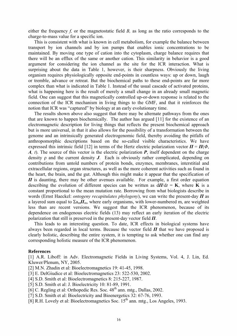
Choosing the disponible pump s, the modal pumping levels s1, s2, and the occupancy numbers n1, n2 as the leading variables, and following procedure outlined in [4], we get the representation of (2) and (3) shown in Fig. 1. The diagram contains two paths connecting input vertex s with the output n1, namely path1(sn1) ≡ ss1n1 and path2(sn1) ≡ ss2n2s1n1. It also includes three closed paths – feedback loops, namely loop1 ≡ n1s1n1, loop2 ≡ n2s2n2, and loop3 = n1s2n2s1n1. Transmission function of the whole diagram between vertices s and n1 is given by (4) and forms the right-hand side of the differential equation of the input-output characteristic, n1 versus s:
Fig. 1. Diagram depicting the causal relationships implicit in (2) and (3). It corresponds to situation in which an input δs is injected at the input port s; the signal propagates along the properly oriented paths and produces an increment δn1 observable at the output port n1.
3. Numerical results and discussion Equation (4) was evaluated using Runge-Kutta routine. The parameter values are inset in the figures; room temperature was assumed. Figures 2 and 3 show dependences of occupancy numbers and of the feedback loop terms in the denominator of (4) on the pumping level s for various values of the “saturation parameters” a1 and a2. In Fig. 2 the well-known phenomenon of selective channelling of energy flow can
be observed: at sufficient pumping level s, the lowest- order (i = 1) mode is preferentially driven. One can see that introduction of the modal pumping levels (3) shifts the onset of selective channeling to higher disponible pumping levels while making it, at the same time, more abrupt. Fig. 2. Dependence of the modal occupancy numbers on available pumping level for selected values of parameters a1 and a2.
This comportment is to be correlated with the s-dependences of the feedback loop terms in Fig. 3. One can see that the difference between the case of direct
access to pumping source (a1 = a2 = 0) and that of modulated pump (3) is significant in the middle portion of the s scale, i.e. near the onset of the selective channeling. This is due to the bell-shaped form of the thransmission functions t(loop1) and t(loop2) centered in this region (not shown here) whereas t(loop3) rolls off monotonically (curve labelled a1 = a2 = 0). One can see that influence of
s
s1
s2
n1
n2
0
0
1 1 1 2 1 1 1 23
1 21
1[ ( )] [ ( )] [ ( )] ( )
1 ( ) ( ) ( )( ) . (4)n t path sn t path sn t path sn t loop
s t loop t loop t loopt snδ
δ=
+ −
− +≡ =
∑ ll
-4 -3 -2 -1 0 10
1
2
3
4
log 10
n i
log10(s [s-1])
Φ = 0.01, X = 0.001, weak couplingν1 = 0.1THz, ν2 = 1THz
n2
n1
a1 = 0, a2 = 0
a1 = 10xn10, a2 = 10xn20
a1 = 1xn10, a2 = 1xn20
-4 -3 -2 -1 0 10.0
0.1
0.2
0.3
0.4
0.5
0.6
0.7
Σ t(l
oop i)
- t(lo
op1)*
t(loo
p 2) Φ = 0.01, X = 0.001, weak couplingν1 = 0.1THz, ν2 = 1THz
a1 = 10xn10, a2 = 10xn20a1 = 1xn10, a2 = 1xn20
a1 = 0, a2 = 0
log10s ([s-1])

20
Fig. 3. Dependence of the feedback loop term in the denominator of (4) on available pumping level for selected values of parameters a1 and a2. the two loops contributed by the concept of modulated pumping levels can, in the “onset” region of s values, become dominant. As suggested by topology of the feedback loops in Fig. 1, the feedback mechanisms are by their very nature composite phenomena, each of them created by synergy of several simpler processes. Together, the three feedback mechanisms control, in accord with (4), the overall properties of the Fröhlich systems. 4. Conclusions Substitution of the assumption of equal pumping of individual modes in the assembly of intracellular collective oscillators by the more general notion of occupancy-dependent energy intake leads to the appearance of two additional feedback loops in the diagrammatic representaton. These can modify in a profound way the characteristics of the system. In particular, the onset of the selective channeling is shifted to higher disponible pumping rates and becomes steeper. This could have serious repercussions in biological processes describable by Fröhlich model. Acknowledgment This work was supported by the project COST 281. References [1] H. Fröhlich: Int. J. Quant. Chem. 11 (1968) 641. [2] F. Šrobár, J. Pokorný: Bioelectrochem. Bioenerget. 41 (1996) 31. [3] F. Šrobár, J. Pokorný: Electro- and Magnetobiol. 18 (1999) 257. [4] F. Šrobár: Eur. J. Phys. 13 (1992) 1.

21
YEAST SUSPENSIONS: A CONTROLLABLE EXAMPLE OF A COHERENT QUANTUM MACHINE?
Marziale Milani1, Emilio Del Giudice2, Grazia Santisi1,Getullio Talpo3,Giuseppe Vitiello3
1Dip. Scienza dei Materiali, Università Milano Bicocca, Via Cozzi 53, 20125 Milano, Italy ph +39 (0)2 64485175, [email protected]
2Istituto Nazionale Fisica Nucleare, via Celoria 16, 20133 Milano, Italy 3 Padova Ricerche, Padova , Italy
4 Dipartimento di Fisica, Università degli Studi di Salerno, Italy
Coherence has been suggested by Herbert Fröhlich to be an essential feature of the dynamics of living matter. Coherence as a property of living systems has since then been the object of a large debate, especially devoted to the identification of the physical observables that can keep trace of coherence appearance and evolution. In this presentation we show that a real system made of an ensemble of yeast cells can be a good prototype for the discussion of the concept of coherence, for the classification of the concept of ordered states, and for probing the potentialities of coherence for the description and prediction of the behaviour of interacting systems. Some guiding ideas can be found also in the presentation of Del Giudice. The main points of the presentation are: 1. Experiments show that yeast cells among others exhibit under proper conditions a
typically quantum electrodynamical behaviour that relates currents, voltages and (absorbed and emitted) electromagnetic field according to a scheme typical of Josephson ac effects. The presence of differently ordered domains in space give the ruling parameters of the dynamics.
2. A metabolism monitoring technique has been settled to identify metabolically sustained time dependent processes in cell suspensions. This time evolution can be traced back to the role of initial conditions (preparation procedures), internal parameters for the cell systems (cell density, nutrient concentration, temperature) and external ones, such as exposures to chemical or physical agents (xenobiotics).
3. CO2 concentration variations inside sealed test-tubes containing cell suspensions are measured by differential pressure sensors; pressure variations are converted in voltage.
4. Focusing our attention onto S. cerevisiae, in the form of a cell suspension in a minimal medium (deionized water and glucose), it can be observed that different regimes appear in this curve for a time lapse of 120 h: an initial growth up to a maximum, followed by a decrement that leads to a typical “depression” (pressure value inside the test-tubes is lower than the initial one) after about 35 h from the beginning in the selected experimental set up. The curve is reproducible within an experimental error of 4%.
5. Systematic investigations show that on the average the following function y=y(t) faithfully describe the process:
y(t) = -[at3+bt]/[t4+c] ; a>0, b<0, c>0 a,b,c: parameters of the experiment y(t): sample averaged pressure increase over the control value; t: experiment absolute time 6. The batch culture can be seen as a whole system, built up of a collection of identical
subsystems, open as far as energy is concerned and closed as far as matter is concerned.
7. The number of subsystem is constant since the minimal medium does not allow any cell division process and cell vitality remains constant along the whole experiment.

22
8. Actually the system can receive or release energy under the form of thermal energy since it is coupled to the thermal bath. On the other way no chemicals can be exchanged with the exterior. We have to take into account the fact that the system is not in its lowest energy state since the presence of glucose provides the presence of a certain amount of energy in the initial state. In the typical dissipative structure language, the glucose is the energy supply “or pump” that is delivered to the cell ensemble in a time-dependent self-driven way.
9. The different cells start their glucose driven activity in a synchronous way as can be expected from the triggering role of hydration of the liophilized cells.
10. Each cell can be characterized by a wave function with the symmetry of a boson state. The whole set of cells is therefore like a set of independent oscillators that are coupled by their synchronous feeding on the common minimal medium.
11. The coupling is dictated by an order parameter that is dependent on cell concentration, sugar molecule concentration, initial synchronization and geometry factors.
12. The simplest and most powerful description of this system is therefore a boson condensate, whose states are the eigenstates of a macroscopic oscillator. At the equilibrium the ground state will be a coherent state that has the shape of a gaussian function.
13. Being in the presence of an external energy supply (the glucose dissolved in the suspension) we then can figure out that the system will not be in a ground state but in the first excited state of the macroscopic oscillator.
14. Finally there will be a damping factor during time evolution of the state that is dictated by the metabolically generated thermal energy dissipated toward the external thermal bath with the role of temperature controller.

23
MICROTUBULES IN ELECTRIC FIELDS
E. Unger1,2,4), R. Stracke 1, A. Michette 2, N. Mavromatos 2, J. Tuszynski3, K.J. Böhm 1 1 Institute of Molecular Biotechnology, Jena, Germany; 2 King’s College University of
London, UK 3 University of Alberta, Edmonton; 4 Correspondence: IMB Jena, D-07745 Germany
Tel:**493641 656160, Fax: **493641656166, e-mail: [email protected] Introductory remarks Microtubules (MTs) are ubiquitous proteinaceous complexes found in eukaryotic cells. MTs are involved in diverse cell physiological processes such as segregation of genetic material, intracellular transport, or maintenance of cell shape; the irreversible elimination or functional inactivation of MTs causes cell death. MTs are hollow cylinders with an outer diameter of about 25 nm, they are constituted of protofilaments (in mammalian cells typically 13) which are formed by longitudinal association of the heterodimeric bi-globular αβ-tubulin of about 8 nm length. MT lengths vary from a few to some tens of micrometers. Because of the strict alternation of α- and β-tubulin, one end of the protofilament resp. of the MT is terminated by an α-subunit and the opposite one by a β-subunit providing a certain kind of polarity. Due to the constituting amino acids and depending on the environmental buffer conditions, the tubulin is electrically charged whereby the charges are asymmetrically distributed forming electric dipoles that superimpose in tubulin assemblies. To understand the complex behaviour of MTs in electric fields not only the tubulin sequence and protein folding but also some other components have to be considered to contribute to the dipole moment formation in and around a microtubule: • the dipole moment obtained as a result of net charge neutralization by counter charges in
neighbouring monomers. • the induced dipole moment arising from the motion of mobile electrons (or protons) between
equivalent locations inside the tubulin, against the opposite charge background. Assuming 1 to 2 mobile elementary charges per subunit multiplied by a charge distance of 4 nm (in relation to the diameter of the α- and β-tubulin monomers), an induced dipole moment per dimer of p ~ 200 to 400 Debye has been estimated.
• the dipole moment of the highly negatively charged C-terminal tail of the α- and β-tubulin subunits sticking flexibly out of MT surface.
Applied external electric fields may interact with microtubules in relation to the dipole status, the general surface charge, the surrounding buffer conditions, especially ionic strength, pH values, and the mode of the electric field. The chemical polarization of the buffer constituents by electric fields reduces the effective forces exerted on MTs. Special field and electrode configurations allow MT field interactions resulting in MT transport and alignment. MT movement in constant electric fields Electric fields (field strength: 5 - 20 V/m; electrodes inside the sample) applied to suspended MTs at a physiological pH (about 6.8) move MTs to the positive electrode indicating a negative effective charge. An electrophoretic mobility (B = v / E ; v: velocity; E: electric field strength) of about 2.6 x 10⁻⁴ cm2/Vs was determined. The net charge of the tubulin dimer within microtubules has been estimated to 0.19 elementary charges per dimer on average. It is striking that this value is at least about 50 times lower than that calculated for the tubulin dimer on the basis of the crystallographic data (Nogales et al., 1998). MTs maintained within a cell at nearly neutral pH and especially the C-terminal regions are known to be surrounded by numerous monovalent cations tending to neutralize the charges (Wolff et al., 1996). Measurements at increasing ionic strengths from 3 mM to 120 mM reduce the electrophoretic mobility corroborating this hypothesis. Decrease of the buffer pH from 6.8 to 4.2 drops the electrophoretic mobility of (taxol-stabilized) MTs to zero indicating a net charge neutralization of the MT (reflecting the isoelectric point) by the protons, i.e. the isoelectric point of MTs is remarkably lower than that of the tubulin α- and β-
24
monomers found at pH 5.45 - 5.65 and pH 5.30 - 5.45, respectively (Detrich and Overton, 1986; Linhartová et al., 1993). The tubulin assembly obviously results into both conformational changes with a modified charge distribution and shielding of proton-binding amino acids of tubulin predicting higher proton concentrations for net charge neutralization of the tubulin monomers. Visual observation of the moving MTs did not show any tendency to a preferential orientation of the MT axis to the field or any other direction, also not at the highest field strength used (about 20 V/cm). The potential energy of a 5-µm long MT dipole in an electric field of E = 10 V/cm was calculated by Wdipole = E pMT (W: potential energy, E: electric field strength, pMT: dipole moment) to be Wdipole = 8.3 x 10⁻²¹ Ws (dipole moment vertical to field direction). The energy of the dipole-field interaction is only by a factor of about 2 higher than the thermal energy at room temperature, which is obviously not sufficient for a detectable alignment in electric fields of moderate strength as our experimental results showed. Orientation of actively driven MTs in constant electric fields MTs are used inside cells as rails for transporting organelles. The energy is provided from ATP hydrolysis by so-called motor proteins, e.g., kinesin. Kinesin is also active under cell-free conditions and is used to drive MTs on technical surfaces. The video-enhanced DIC-microscopy allows a direct MT visualization. Constant electric fields were applied to MTs driven by kinesin immobilized to glass surfaces. The majority of gliding microtubules also migrated preferentially to the positive electrode. Switching field direction turns direction of movement within 5 min. At adequate microtubule length and kinesin density, the leading ends of gliding MTs are forced to turn into direction of the anode. This field-caused mechanism seems to be suited to regulate the gliding direction. Based on our measurements performed on suspended microtubules, an electric force of 0.2 pN was calculated for a 5-µm long microtubule at 10 V/cm. The force exerted by only one kinesin molecule is about 4 - 8 pN (e.g., Hunt et al., 1994), i.e. 20-40 times greater than the value exerted by the electric field. While the electric field acts constantly (DC) on the microtubule, motor proteins provide bursts of activity only over 1-5% of the time of the mechanochemical cycle (Vale and Milligan, 2000), the motor molecules provide roughly the same time-averaged force as the electric field effect on the microtubule. In the case of gliding microtubules thermal effects do not play a significant role because they are bound to the substrate by kinesin molecules. But, for the orientation efficiency of MTs gliding across a kinesin-coated glass surface in constant electric fields, the influence of the electroosmotic flow (that is strongest near the glass surface where the MTs move) counteracting the field effect has to be considered. Electroosmotic flow is regarded to be a main cause why the orientation of the microtubules in the field is not complete. MT alignment in high-frequent strong alternating fields As clearly demonstrated, strong high-frequent alternating fields are able to align suspended MTs (length ~ 5 µm, pH 6.8) within some few seconds parallel to the field. The field strength (rms value) found to be sufficient was 140 kV/m (the maximum field strength applied was 210 kV/m) at a frequency of 200 kHz at the chemical conditions used. Increasing frequency to 2 MHz allows alignment at somewhat lower field strengths. In regions of inhomogeneous fields, the parallel alignment is superimposed by a movement of MT in field direction. Such a movement in inhomogeneous fields is generally known as dielectrophoresis. The effect of MT orientation in strong high frequency fields can be understood as a interaction of the induced electric dipoles with the high frequent alternating field due to the different dielectric constants of MTs and surrounding molecules. Summary We demonstrated that suspended and kinesin-driven MT are transported and oriented, respectively, by constant electric fields to the positive electrode. The space orientation of the MTs in relation to the fields is random for the suspended MTs and preferred parallel to the field for the kinesin-bound MTs. In homogeneous high frequency alternating fields microtubules become stationary and are aligned

25
parallel to the field. These phenomena are explained in terms of MT surface charges and dipoles properties. This work has been supported by the Deutsche Forschungsgemeinschaft grant Un82/5-1. Research support for the Canadian group was generously provided by NSERC, MITACS and a Consciousness Studies grant administered by the University of Arizona.

26
NONLINEAR DYNAMICS OF MICROTUBULES AND ITS IMPLICATIONS
M.V. Satarić Faculty of Technical Sciences,University of Novi Sad, Trg Dositeja Obradovica 6
Novi Sad, 21000, Serbia and Montenegro, e-mail: [email protected] A recently developed model of nonlinear dynamics for microtubules is further expanded based on the biophysical arguments involving the secondary structure of the constitutive protein tubulin and on the ferroelectric properties of microtubules. It is demonstrated that kink excitations arise due to GDP hydrolysis that causes a dynamical transition in the structure of tubulin.The presence of an intrinsic electric field associated with the structure of a microtubule leads to unidirectional propagation of the kink excitation along the microtubule axis. This mechanism offers an explanation of the dynamical instability phenomenon in terms of the electric field effects. Moreover,a possible elucidation of the unidirectional transport of cargo via motor proteins such as kinesin and dynein is proposed within the model developed in this paper. Within the framework of our model proposed in this paper,microtubules are not only passive tracks for active transport in the cell but also signal relays for electrical, mechanical, and chemical stimuli that may be transduced over distances compared to the cell size. Key words microtubule, tubulin, ferroelectric, kink propagation, GTP hydrolysis, dynamic instability.

27
NONLINEAR BRAIN DYNAMICS AND MANY-BODY FIELD DYNAMICS
Giuseppe Vitiello Dipartimento di Fisica "E.R.Caianiello", Università' di Salerno, 84100 Salerno, Italia
Sequential spatial patterns of neural activity with high information content are found in sensory cortices of trained animals between onsets of conditioned stimuli and conditioned responses. They resemble cinematographic frames. The large size, rapid formation, variety of detail, and perceptual remoteness from sensory input of normal frames could not be explained within the context of classical physics. A model for dealing with such neuroactivity is provided by field theory from condensed matter physics [1]. References [1] Walter J. Freeman, Giuseppe Vitiello: Nonlinear brain dynamics as macroscopic manifestation of the underlying many-body field dynamics, in preparation.
28
PHYSICAL PLASMA SWITCHABILITY IN THE BRAIN
Józef R. Zon Chair of Theoretical Biology, Department of Philosophy of Nature and Natural Sciences,
The Catholic University of Lublin, Al. Racławickie 14, 20-950 Lublin, Poland e-mail: [email protected]
The idea that brain processes depend on switching between the on- and off- states is an old one. The units in which this switching is performed are neurons. In them, at the synaptic junctions begin reversals of electric polarity of neural membranes and its propagation along them. The electrical activity of brain is considered to be a sum total of multitude processes stemming from the activities involving changes in ionic and membranes in individual and group of neurons, as well as in the glial cells. When physical plasma (seen as a transient collectivized state of aggregation of charged particles) that begins and ceases to exist in some micro-spaces of the brain is taken into account, a broader picture of the dynamics of that organ emerges. In it, ionic activity still plays essential, yet a secondary role. Primary role is played by coupling between elementary plasma-spaces, the characteristics of which (as: the size, frequencies, anisotropy, duration of existence) are tied to physiological, emotional and mental activities of the brain.

29
ENERGY SUPPLY AND PHOTON EMISSION BY SOLITONS IN ALPHA-HELICAL PROTEINS
L.S. Brizhik, A.A. Eremko
Bogolyubov Institute for Theoretical Physics, SRC of Quantum Medicine ‘Vidhuk’ 14-b Metrologichna Str., 03143 Kyiv, Ukraine
e-mail: [email protected], [email protected] We study the nonlinear mechanism of energy supply in alpha-helical proteins with three polypeptide spines, taking into account the helicity of proteins in the presence of electron-phonon interaction which results in the self-trapping of excitations in a localized soliton-like state [1]. We elucidate the important role of the helical symmetry of a macromolecule for the formation, stability and dynamical properties of Davydov's solitons. It is shown that the soliton with the lowest energy has an inner structure with the many-hump envelope. The total probability of the excitation in the helix is characterized by interspine oscillations with the frequency of oscillations, proportional to the soliton velocity. The radiative life-time of a soliton is calculated and shown to exceed the life-time of an excitation on an isolated peptide group by several orders of magnitude. The energetical spectrum of quasiparticles in an alpha-helical macromolecule, stabilized by three polypeptide chains of hydrogen bonds, is shown to contain three energy bands. The upper band is nondegenerate and has the minimum in the centre of the Brillouin zone. The lower band in the energy spectrum is degenerate into two bands which have minima at nonzero values of wave-vectors, symmetrically shifted with respect to the centre of the
Brillouin zone, by the values aLJ
Lk)18(
330 +
±=± . Here J and L are, respectively, intra- and
inter-chain exchange energies, a is hydrogen bond length. We show that the corresponding system of nonlinear equations admits several types of stationary soliton solutions: one-band solitons which are energetically split from the corresponding energy bands, and hybrid soliton which is formed by the hybridization of quasiparticle states from the two lowest degenerate energy bands. The hybrid soliton breaks spontaneously the local translational and helical symmetries. It possesses the lowest energy as compared with other type solitons, and has an inner structure which can be described as a modulated multi-hump amplitude, Pj(n), distributed on individual spines, shown in Fig.1.
Fig.1. Probability distributions of a hybrid soliton, Pj(n), in the three individual spines of alpha-helix, j=1,2,3 (three lower curves). Upper curve corresponds to the total probability, Ptot(n)= P1(n)+ P2(n)+ P3(n).
30
The total probability of the excitation on the j-th spine is given by the expression
∑ ⎥⎦
⎤⎢⎣
⎡−−==
nnjj jVtk
kk
tPtP )3/22cos()/sinh(
131)()( 0
0
0, π
κπκπ
, (1)
where )18(
9 2
LJwa +=
χκ is the localisation parameter of a hybrid soliton, χ is the electron-
phonon coupling, w is the elasticity coefficient, proportional to the elasticity of a hydrogen bond. We see from [1] that the probability of the excitation on a given spine is an oscillatory
function of time with the period of oscillations, determined by the soliton velocity: Vk
Th0
π= .
For instance, for the soliton velocity equal to 3/8 of the sound velocity [2] for an AMID-I excitation in alpha-helix (J=1.55 10-22 Jole, L=2.46 10-22 Jole, k0a=0.422), we obtain the value Th=2 10-12 s. Altogether, these results explain a soliton structure, interspine oscillations and their frequency, previously observed numerically by A.C. Scott [2]. These helical oscillations get mixed up with the oscillations that arise from the influence of the lattice discretness on the soliton dynamics which leads to the appearance of the Peierls-Nabarro potential. The period of these longitudinal oscillations is also determined by the soliton
velocity [3]: VaTl = , which is one order of magnitude smaller than Th.
In the processes of charge transport such a soliton is formed by a self-trapped electron, and, therefore, has a charge. The oscillating propagation of the electrosoliton is accompanied by electromagnetic radiation [4]. This radiation constitutes the endogenous electromagnetic field with characteristic resonant frequencies. The superposition of the longitudinal and helical oscillations of the electrosoliton results in a more complex spectral structure of this field with components of various polarization properties and radiation patterns. Namely, the longitudinal oscillations due to the Peierls-Nabarro periodical relief in a discrete chain cause the plane polarized radiation which can be described as the radiation of some effective dipoles, oriented along the helix axes [4]. Meanwhile, the component of radiation due to the helical oscillations caused by the helical structure of a peptide, has circular polarization. The other type three solitons are formed by single-band electron states. They preserve helical symmetry and break the translational symmetry. Such solitons posses higher energy than the hybrid soliton described above, and are dynamically unstable: once formed, they decay rapidly when they propagate, transforming into a hybrid soliton with the lowest energy. A central problem of bio-energetics is the question about the life-time of excitations in proteins. Note, one of the main mechanisms of relaxation in proteins is related to the dipole-active excitation radiation. To study this, we calculate the probability of photon emission due to transition of a protein from an excited hybrid soliton state into the ground one. The radiative life-time of a hybrid soliton, hτ , related to the transition is given by the expression.
⎥⎦⎤
⎢⎣⎡ +
=a
akah κ
ππϑπ
τκτ
2)6/2(
coshsin3
4 02220 . (2)
Here 0τ is a lifetime of an isolated excitation of a peptide group, ϑ is an angle between the orientation of the dipole momentum of an excitation and helix axis. For the parameters of an AMID-I excitation in alpha-helix we have aκ =0.176, o30=ϑ , which gives
90 108.5/ ⋅=ττ h . The radiation lifetime of a hybrid soliton in a helical structure is much

31
bigger than the lifetime of an AMID-I soliton, sτ , in a three-spine model with zero helicity [5] (in this case 00 =k ): 3102/ ⋅=sh ττ . Therefore, the helicity of a protein significantly increases the radiative life-time of AMID excitations in proteins. This conclusion agrees with recent experiments of Austin et al. [6], which show that the life-time of an excitation in myoglobin, that is essentially alpha-helical protein, is much bigger than in isolated aminoacid such as L-alanine. According to [6], such an excitation in myoglobin has necessarily the nonlinear nature. In our opinion, this nonlinear nature can originate from the electron-phonon interaction and anharmonicity of proteins, leading to the self-trapping of excitations and electrons in soliton-like states, which have significantly bigger life-time. Moreover, such solitons generate complex endogenous electromagnetic radiation of characteristic frequencies leading to synchronization of charge transport processes, which, in its turn, provides the coherence of the guiding electromagnetic field and self-regulation of metabolism [4]. References [1] L.S. Brizhik, A.A. Eremko, B. Piette, W.J. Zakrzewski: Phys. Rev. E 70 (2004) 031914. [2] A.C. Scott: Phys.Rev. A 26 (1982) 578. [3] L. Brizhik, L. Cruzeiro-Hansson, A. Eremko, Yu. Olkhovska: Phys. Rev. B 61 (2000)
1129. [4] L.S. Brizhik, A.A. Eremko: Electromagnetic Biology and Medicine 22 (2003) 31. [5] A.S. Davydov, A.A. Eremko, A.I. Sergienko: Ukr. J. Phys. 23 (1978) 983. [6] A. Xie, L. van der Meer, W. Hoff, R.H. Austin: Phys. Rev. Lett. 84 (2000) 5435.

32
STOCHASTIC DYNAMICS OF MAGNETIC NANOPARTICLES IN BIOLOGICAL CELLS
V.N. Binhi, D.S. Chernavskii*
A.M. Prokhorov General Physics Institute RAS. 38 Vavilova St., Moscow 119991, Russian Federation, [email protected]
*P.N. Lebedev Physics Institute RAS. 53 Leninsky Av., Moscow 119991, Russian Federation
There are many hypothetical mechanisms suggested to explain the biological effects of weak low-frequency magnetic fields. A brief review of the mechanisms may be found in [1] and the detailed discussion in [2]. At the same time, the physical nature of these effects remains unclear. The basic problem is that the interaction energy of biologically active molecules and the MF at the geomagnetic level is very small. It is much smaller than the energy of thermal fluctuations kT ~ 4×10-14 erg at physiological temperatures. However, many organisms are well known to contain submicron magnetic particles. The energy of their turn in a weak magnetic field H is substantially greater than kT. For single-domain magnetite particles of radius r = 100 nm in the geomagnetic field the energy μH equals approximately 24 kT. The cytoplasm near cell membranes features such visco-elastic properties that the turning of a microparticle may serve as a stimulus to cell division or ignite a nerve impulse. Magnetite particles found in the brain tissues of animals and humans are of particular interest; they are found to have a biogenic origin, i.e. they appear over time as a direct result of the crystallization in brain matter. Biogenic magnetite particles are often called ‘magnetosomes’; they were first discovered in bacteria that displayed magnetotaxis [3]. Magnetosomes were used repeatedly to explain biological effects of weak magnetic fields and in particular the avian magnetic navigation [4]. The density of magnetosomes in the human brain is more than 5×10⁶, and in meninges more than 108 crystals per gram [5]. The energy of the magnetosome in the geomagnetic field is about 24 kT; when exposed to an additional variable magnetic field h, its regular changes are about (h/Hgeo)24kT. If these changes exceed thermal fluctuations kT/2, they can cause a biological response. This sets a natural constraint on the MF magnitude capable of affecting a biophysical or biochemical system appreciably: h > 1–2 μT. However, in the special case of magnetosomes bound to a visco-elastic medium so that they move within a double-well potential, the thermal fluctuations work to facilitate rather than impede the capability of a weak magnetic stimulus to cause a response. Then the limit value of h might be less. We consider the dynamics of a magnetic nanoparticle embedded in the cytoskeleton. This fixes the position of the magnetosome and constrains its rotation to some extent. The stationary orientation of the magnetosome generally does not follow the constant MF direction. The balance of the elastic and ‘magnetic’ torques determines the orientation now. The idea is to study the dynamics of a magnetosome predominantly oriented in a direction opposite to that of a constant MF. The potential energy of a magnetosome for not too large turns has two potential wells. Within each of the wells, the motion of the magnetosome demonstrates no peculiarities. This sort of motion has been repeatedly considered in literature. At the same time, due to thermal disturbances, the transitions appear from well to well even with no ac MF signal. Given that, the stochastic turns of the particle take place with considerable angular displacements. A deterministic external force, the ac MF in our case, causes such transitions to be somewhat ordered, the maximum order attained just at the optimal level of the noise. It is essentially the phenomenon of the so-called stochastic resonance first introduced in [6] to explain some geophysical processes.

33
So far, the probable manifestation of the SR in the dynamics of magnetosomes has not been investigated. Several SR theories are known; we use the results of [7], where the general expression has been derived for the power spectrum of oscillations of a bistable system agitated by regular and random signals. The signal-to-noise ratio is determined as the ratio of the spectrum amplitude at the frequency of the regular signal, to the level of noise at the same frequency. This function attains its maximum at the optimal level of noise. This means there is an interval in the value of noise, where the signal-to-noise ratio unexpectedly increases along with increasing noise power — it is this that is the signature of SR. We calculated the signal-to-noise ratio function at various ac MF amplitudes and values of the noise parameter, which depends on the size of the magnetosome. A marked interval of the elasticity parameter of the bond magnetosome–cytoskeleton appeared to exist, wherein the signal-to-noise ratio is close to unity. The 100-nm magnetosome fixed in the cytoskeleton with plausible elasticity in the 13-μT ac MF and 46-μT geomagnetic field regularly turns at angles of the same order as the chaotic rotations. It is particularly evident for 50-nm particles: almost all of them are in the SR conditions. 200-nm particles make regular turns at relatively small MFs about 4.6 μT. Although in each of these cases there is no gain in the magnitude of the effective ac MF as compared to the case of a single-well motion, it is important that the rotation excursion is an order higher, about π/2. With such excursions it is easier to account for the influence of the magnetosome’s rotations on biochemical processes. Since the parameter of the elasticity depends on the constant MF, the ‘resonance’ will show itself also as a ‘window’ in constant MF values when the effect is possible. When the MF decreases, the potential function transforms into a single-well one and large rotational excursions are no longer possible. When the MF increases, the potential barrier grows and the magnetosome finally remains within one of the two wells. This case also rules out stochastic resonance manifestations. Apparently, for a portion of magnetosomes, large angular chaotic turns take place in the absence of an ac MF also. If some biochemical reaction depends on these turns, it is evident that it must be sensitive to the condition of a ‘magnetic vacuum’ h << H << Hgeo. Furthermore, the reaction must be sensitive to small variations of the constant MF, since the probability of transition W from well to well exponentially depends on the barrier height. What is of interest is the relative value of the changes in this probability at small variations of the constant MF, i.e., the quantity
)/d(d1
geoHHW
WS −=
The sensitivity S computed at several likely values of the elasticity of the bond between a mean magnetosome and cytoskeleton is equal to 10–20. This means a 1% MF change causes 10–20% changes in the transition probability. Thus, the limit of detectable values of the constant MF variations is found to be 200 nT, the level of geomagnetic fluctuations. Taking into account that the MF produced by a magnetosome may attain dozens mT and the changes in its mean level induced by those weak variations are the same 10–20%, we arrive to a ‘magnetic stochastic biological amplification’ of order of 104 functioning due to thermal disturbances. The rate of the transitions and the probability of magnetosomes to be in the different states depend as well on the magnetic field direction with respect to an averaged magnetosome’s orientation. This effect explains the ability of migrant birds to orient themselves faultlessly in long-term passages in the absence of the direct visibility of optical reference points. The sensitivity to deviation from an ‘ideal’ orientation is estimated to be 1–2 degrees.

34
Grants: RFBR No.04-04-97298, RFH No.04-03-00069. References [1] V.N. Binhi and A.V. Savin: Effects of weak magnetic fields on biological systems: Physical aspects. Physics–Uspekhi, 46(3):259–291, 2003. [2] V.N. Binhi: Magnetobiology: Underlying Physical Problems. Academic Press, San Diego, 2002. [3] R.P. Blakemore: Magnetotactic bacteria. Science, 190(4212):377–379, 1975. [4] J.L. Kirschvink, D.S. Jones, and B.J. MacFadden (editors): Magnetite Biomineralization and Magnetoreception in Organisms. A New Biomagnetism. Plenum, New York, 1985. [5] J.L. Kirschvink, A. Kobayashi-Kirschvink, and B.J. Woodford: Magnetite biomineralization in the human brain. Proc. Natl. Acad. Sci. USA, 89(16):7683–7687, 1992. [6] R. Benzi, A. Sutera, and A. Vulpiani: The mechanism of stochastic resonance. J. Phys. A, 14:L453–L457, 1981. [7] B. McNamara and K. Wiesenfeld: Theory of stochastic resonance. Phys. Rev. A, 39(9):4854–4869, 1989.

35
ENDOGENOUS ELECTRIC FIELD AND ORGANIZATION OF LIVING MATTER
J. Pokorný, J. Hašek*, F. Jelínek
Institue of Radio Engineering and Electronics, Chaberská 57, 182 51 Prague 8 *Institute of Microbiology, Vídeňská 1083, Praha 4
Academy of Sciences of the Czech Republic Organization of living matter depends on energy supply from metabolic sources but mechanism of transformation of chemical energy into useful work is still not elucidated. Frauenfelder et al. [1] claim that physical mechanisms of organization are of electromagnetic nature. This statement is supported by the fact that the majority of biological molecules and structures (in particular proteins) are electrical dipoles and/or multipoles. Intensity of the electromagnetic field can exert forces on charges, dipoles, and on neutral particles through dielectrophoretic effects. The electric field may be static, quasi-static, and oscillating whose amplitude may be large if vibrations generating the field are exited. Froehlich put forward a hypothesis of coherent vibrations and formation of a state far from thermodynamic equi-librium. Coherent vibrations of the plasma membrane of yeast cells were measured recently [2]. Cytoskeleton has importanf function in forming the structure and in organization of eukaryotic cells. The fundamental structure of the cytoskeleton is formed by microtubules composed of tubulin heterodimers. Microtubules are continuously depolymerized and polymerized by the mechanism of dynamic instability (in the interphase) and of treadmilling (in the M phase). Energy is supplied to microtubules by hydrolysis of GTP (guanosine triphosphate) to GDP (guanosine diphosphate) in the heterodimers after polymerization. Power supply is about 10-14Wcm-1 in the interphase and more than one order greater in the M phase. Large deformability, formation of ionic charge layer, and proteins attached to the microtubule surface are important for formation of slip boundary conditions for diminishing damping by viscosity effects. Microtubules satisfy conditions for excitation of vibrations, energy condensation, and generation of the electromagnetic field [3]. Mass and charge transport conditions biological activity. Electron can be carried by small diffusible molecule or transferred along the molecular chain. The biological molecules may have quasiperiodic or correlated potential profiles and electron behavior may be similar to that of conductivity electrons in semiconductors but with deformed conductivity band. Random transport driven by the thermal motion is isotropic if conditions for symmetry breaking are not satisfied. Directed transport may be pointed to certain target region and can contribute to the organization of chemical reactions. Transport along the microtubules is strictly directed to a region over a cell size distance but its rate is not hight. Transport mediated by electromagnetic fields might be important over smaller distances but its rate might by high. Random motion of the Brownian type may be described by the Wiener-Lévy process. The Wiener-Lévy process may also describe directed transport if the random walk steps are dependent on direction (symmetry breaking) or if a force acting in certain direction is included [4,5]. We used the Wiener-Lévy process with inclusion of a deterministic force for analysis of one, two, and three dimensional models of directed motion. We evaluated probability density f(x;t) and probability P for transport of spherical particles (molecules) driven by the thermal motion and by deterministic force. Fig. 1 shows probability density f(x;t) of finding a particle with radius r at a distance x [5]. Time t is a parameter. f(x;t) is spreading with increasing time. The dotted line (with short segments of vertical lines) denotes a path with length of 15nm. No deterministic force is assumed (F = 0). Effect of deterministic force is shown in Fig. 2 (F = 1pN) [5]. The deterministic force is directed from

36
the point 0 to the point 15nm and the probability density curve is shifted in the direction of action of the force. The target is assumed to be positioned at the point 15nm and the curve f(x;t) moves to the target point. The integral of the part of the probability density curve behind the full line at 15nm denotes the probability that the particle reaches the target. Fig. 3 shows probability (P as a function of time t) that the particle can reach the target [5]. d denotes the length of the path. The dashed and the full curves denote motion driven by thermally and motion driven by deterministic forces and thermally, respectively The probability of a particle driven by a deterministic force to reach the target is greater than that for no deterministic force when the particle is driven only by random thermal motion. Fig. 4 shows probability P as function of time t that electron will reach the target region at a distance d [5]. The distance d is a parameter. Intensity of the electrical field is 107V/m. Electron may be shifted over a distance about 20nm by the electrical field within the time interval less than 10ps with probability near to 1. One dimensional models were used.
x (nm)
0 25 50
f (x;
t)
(arb
itrar
y un
its)
0
1
2
3
t = 5nsF = 0Nr = 1nm
100ns500ns
x (nm)
0 25 50
f(x,t)
(
arbi
trary
uni
ts)
0
1
2
3
t = 5ns
100ns500ns
F = 10-12Nr = 1nm
Fig. 1 Fig. 2
t (s)
10-8 10-7 10-6
P
0.0
0.5
1.0 F = 10-12Nr = 1nm
1050
20d = 5nm
5020
10
5
t (s)
10-14 10-13 10-12 10-11
P
0,0
0,5
1,0 E = 107Vm-1
s = 1nm
d = 2nm 5 10 20
5 10
202
Fig. 3 Fig. 4
Electrical polar structures inside the cell with energy supply (such as microtubules) can get excited and generate endogenous electric field. Endogenous electric field can have dominant effect on directed transport of molecules and electrons. Acknowledgement This work was supported under grant OC 281.001 of Ministry of Education, Youth, and Sports of the Czech Republic in the framework of COST 281. References [1] H. Frauenfelder, P.G. Wolynes, R.H. Austin: Biological Physics. Rev. Mod. Phys., Centenary 1999, 71 (1999), 2, S419-S430.

37
[2] A.E. Pelling, S. Sehati, E.B. Gralla, J.S. Valentine, J.K. Gimzewski: Local Nanomechanical Motion of the Cell Wall of Saccharomyces cerevisiae. Science, 305 (2004), 1147-1150. [3] J. Pokorný: Excitation of vibrations in microtubules in living cells. Bioelectrochem. 63 (2004), 321-326. [4] J. Pokorný: Endogenous Electromagnetic Forces in Living Cells: Implications for Transfer of Reaction Components. Electro- Magnetobiol., 20 (2001), 1, 59-73. [5] J. Pokorný, J. Hašek, F. Jelínek: Electromagnetic Field of Microtubules: Effects on Transfer of Mass Particles and Electrons. Accepted for publication in J. Biol. Phys.

38
EVIDENCE OF LIGHT PIPING (MERIDIAN-LIKE CHANNELS) IN THE HUMAN BODY AND NON-LOCAL EMF EFFECTS
Fritz-Albert Popp1, Walburg Maric-Oehler2, K.-P. Schlebusch3
1International Institute of Biophysics, Landesstiftung Hombroich, Raketenstation, D-41472 Neuss, Germany
2International Council of Medical Acupuncture (ICMART), Johannes Gutenberg University, Mainz, Louisenstrasse 15-17, D-61348 Bad Homburg v.d.H., Germany
3 Gesellschaft zur Dokumentation für Naturheilverfahren, Hufelandstrasse 60, D- 45147 Essen, Germany
By means of Infrared Spectroscopy (FLIR Systems, Therma CAMTM, PM 290) there has been shown evidence of light channels in the body (Schlebusch et.al. 2004, 2005). These channels are following just the lines which are known from tradiational Chinese medicine as the “meridians”. They appear in the range from 3.4 to 4 micrometer and can be evoked by moxibustion which provides in addition to longer wavelenghts excitation of electronic states in this wavelength-range from about 3 to 5 micrometer. Similar observations on plants (Mandoli and Briggs, 1982) and also on the human body (Hu et.al. 1996) have been reported already since many years. Clear non-local biophoton emission of the human body which has been found by S.Cohen in 2002 (publication in preparation) is a further striking example of light channels in the living tissues. We used the rather general differential equations of R.N.Thomas (1961), in order to find a possible explanation of this phenomenon which could be described in terms of soliton-like functions of the type I(s) = I(0)exp(-ßs2), where ß is a positive constant parameter and s the vertical distance from the pipe (“meridian”). It turns out that without moxibustion under rather special conditions the same meridians can be observed, but now not warmer but colder than their surroundings, following the solution I(s) = I(0)(1-exp(-ßs2)) The analysis shows that the formation of thesechannels has to be traced back to excitations of electronic, vibrational, rotational and/or even translational states at the threshold between chaotic and coherent regimes. By transition from the one to the other, the properties of the biological matter may change dramatically, where a strong change of entropy results from rather small energy contributions. The physical basis can be truthfully described in terms of a permanent balance of photon trapping (“sucking”) and “emitting” regions due to “destructive” and “constructive” interference in corresponding cavities and waveguides. The mechanism is principally independent on the wavelength. It can be expressed in terms of a Hamiltonian that describes the formation and conservation of coherent states, and it may govern the biological system in all spectral parts down to the ULF-range. The interpretation confirms a rather general mechanism of the interaction of elmf´s and living tissues (Popp and Chang, 2000).
39
References [1] X. Hu, P. Wang, B.Wu, J. Xu: Displaying of the meridian courses over human body surface with thermal imaging system. Revista Paulista de Acupunctura (Rev. Paul. Acupunt.), 2, 7-12 (1996). [2] D. Mandoli, F. Briggs, and R. Windows: Optical properties of etiolated plant tissues. Proc.Natl.Acad.Sci. US, 79, 2902 (1982). [3] F.A. Popp, and J.J. Chang: Mechanism of the interaction between electromagnetic fields and living organisms. Science in China (Series C), 43 (5), 507-518 (2000). [4] K.-P. Schlebusch, W. Maric-Oehler, F.-A. Popp: Biophotonik beweist erstmals Meridianstruktur (Leitbahnen-Strutkur der Akupunktur) auf der Körperoberfläche, Erfahrungsheilkunde (Thieme-Verlag), 53. 619-622., JACM (2005), 52(1), in press (2004). [5] R.N. Thomas: Some aspects of non-equilibrium thermodynamics in the presence of a radiation field. Boulder, Colorado (1961).

40
NEW NANO-SCALE ONCOTHERAPY APPROACHES INSPIRED BY COMPUTATIONAL BIOPHYSICS
Jack A. Tuszyński
Department of Physics and Department of Oncology, University of Alberta, Edmonton, Alberta, T6G 2J1, Canada
My group has been involved in computational cell biophysics research over the past decade or so. In this talk I'll give an overview of our quantitative understanding of the key cellular protein, tubulin, and its cylindrical polymers called microtubules. Since 1998 when the Berkeley group of Ken Downing was able to crystallize tubulin, our focus shifted to the application of molecular and Brownian dynamics of tubulin in the quest to understand the relationship between atomic resolution structure and biological function. We have now concentrated our efforts entirely on one key issue, i.e. a rational drug design targeting tubulin with molecules that would interfere with the formation of mitotic spindles during cell division. In collaboration with biochemists and oncologists from the University of Texas our group is using computational methods applied to rational drug design of anti-mitotic compounds with specific preferences for tubulin isotypes. I'll describe our results in the early stages of this exciting research project. New promising compounds will be presented that represent a class of taxane derivatives with better targeting and binding properties. In addition, our better understanding of mechanical properties of microtubules has led to the design of new treatment modalities based on the use of ultrasound, laser action and magnetic fields. Acknowledgments This research has been supported by grants from NSERC, MITACS, PIMS, US Department of Defense and Technology Innovations LLC.
TRANSITIONS IN MICROTUBULE C-TERMINI CONFORMATIONS AS A POSSIBLE DENDRITIC SIGNALING PHENOMENON
Avner Priel
Department of Physics, University of Alberta, Edmonton, Alberta, T6G 2J1, Canada, e-mail: [email protected]
In this talk I'll describe a dynamical model of the states of the C-termini of tubulin dimers that comprise neuronal microtubules. We use molecular dynamics and other computational tools to explore the time-dependent behavior of conformational states of a C-terminus of tubulin within a microtubule and assume that each C-terminus interacts via screened Coulomb forces with the surface of a tubulin dimer, with neighboring C-termini and also with any adjacent microtubule-associated protein 2 (MAP2). We find that the preferential orientation of each C-terminus is away from the tubulin surface but binding to the surface may also take place, albeit at a lower probability. The results of our model suggest that perturbations generated by the C-termini interactions with counter-ions surrounding a MAP2 may propagate over distances greater than those between adjacent microtubules. Thus, the MAP2 structure is able to act as a kind of "biological wire" transmitting local electrostatic perturbations resulting in ionic concentration gradients from one microtubule to another. Implications the current dynamic modeling may have on synaptic activation and potentiation will be discussed briefly.
41
ABSORPTION OF IR RADIATION IN HYDROGEN-BONDED MOLECULAR CHAINS: THE INFLUENCE OF SMALL POLARON INDUCED MODIFICATION
OF PHONON SPECTRA
Slobodan Zeković, Zoran Ivić, Dalibor Čevizović Theoretical Physics Department-020, The "Vinča" Institute of Nuclear Sciences, 11001
Belgrade, P.O.Box, 522, Serbia and Montenegro
In the last few years the small-polaron model has been widely exploited as a theoretical framework for the understanding of various phenomena in a wide class of very important substances such as high-temperature superconductors, colossal magnetorestricitve manganites, highly conducting polymers and molecular chains like acetanilide (ACN) and biological macromolecules, &-helix and DNA, for example. In most of the theoretical studies on the polaron problem, the main attention is focused on the study of the electric properties of these materials while the possible changes of the vibrational spectra due to polaron-phonon interaction have been the subject of just a few studies. The influence of the small-polaron effect on the vibrational properties as investigated in [1]. It was found that polaron-phonon interaction may cause a noticeable renormalization of the phonon spectra. We also found that the rise of the small-polaron effective mass as a function of the coupling constant should be considerably moderated than that anticipated without taking into account the modification of the vibrational spectra, which may have important consequences for small-polaron transport properties, conductivity [2] for instance. In the present paper, the model that accounts for the influence of small-polaron induced phonon spectra modifications of linear macromolecules on spectral function has been presented. Experiments, of the class of Raman or neutron scattering or radiation absorption, in active biological systems are particularly difficult. For that reason, the comparison of the theory with experiment is done in the case of organic polymer (acetanilide and related hydrogen-bonded molecular chains), which constitutes a good mimic of biopolymers. For that purpose the method of double-time Green function at finite temperatures was utilized. The main advantage over the previous approaches is the inclusion of the terms of the first order in polaron-phonon interaction, which were not accounted in previous models. Taking into account such a modified phonon spectra we estimated the shift of IR (infrared) absorption maximum, as well as the change of its intensity, in comparison with the results published before [3]. In addition, we emphasized the temperature dependence of intensity and position of absorption maximum. From the obtained results we can see substantial influence of frequency modification. This modification is significant at low temperatures and decreases as temperature rises. Analogously, in small-polaron picture within Davydov model as a theoretical framework [3], changed values of Debye-Waller factor and small polaron binding energy, which depend on phonon frequency, will have an important influence on spectral function, or, respectively, on the intensity and position of IR radiation absorption maximum. Presented model enables more accurate IR absorption spectra modeling in hydrogen-bonded molecular chains. References [1] Zoran Ivić, Slobodan Zeković, Dalibor Čevizović, Dragan Kostić: Physica B 355 (2005) 417. [2] Z. Ivić, S. Zeković, D. Čevizović, D. Kostić, G. Vujičić: accepted for the publication in Physica B. [3] D.M. Alexander and J.A. Krumhansl: Phys. Rev. B, 33, (1986) 7172.

42
EXPERIMENTAL VERIFICATION OF THERMODYNAMIC THEORY OF ELECTROMAGNETIC RADIATION ENERGY CONVERSION. BIOEFFECTS
Yu.P. Chukova
Krasnopresnenskiy Ecological Fund, Moscow, Russian Federation Bioeffects under the electromagnetic radiation have been studied more than 100 years. Working in this sphere physicists understand that the clearness is achieved only for electromagnetic radiation thermal processes and some isothermal bioeffects of visible light (human vision, plant photosynthesis, photomovement of protozoa). Every isothermal bioeffect of electromagnetic radiation has been studied independently of one another. They have not had the general base. This situation will be changed in XX1 century, so far as the thermodynamic theory of the system under electromagnetic radiation has been worked out in second part of XX century. Thermodynamic theory of the systems under electromagnetic radiation was made on base of papers of L. Landau (1946) and I. Prigogine (1947). At the present time this theory is in outline [1, 2]. This theory gives the efficiency of electromagnetic energy conversion. This theory was developed for technical devices (luminescent lamps and solar cells), but it is valid for isothermal processes in living systems too. It was shown that the deductive method of thermodynamics of irreversible processes is very useful for study electromagnetic radiation effects of bioobjects, which can be considered as a open systems in the thermodynamic sense. The most important and interesting result of this theory is the existence of two regions of the electromagnetic energy into Helmholtz free energy conversion in scale from 0 to 1022 Hz of electromagnetic radiation. These regions are well known from the equilibrium radiation theory: W. Wien region (visible light, ultraviolet, X-rays, γ - radiation) and Rayleigh-Jeans region (radiofrequencies and extremely low frequencies). Infrared radiation is the boundary. The efficiency dependence of bioeffects on the absorbed power is sharply disparate in these regions. In Wien region the efficiency and absorbed power have the logarithmic relationship. In Rayleigh-Jeans region this dependence is very strong until it flattens out on a plateau, where any dependence on the absorbed power in general disappears. This theory shows that the main law of the subjective sensory physiology (Weber-Fechner law) is the experimental display of the general principles of thermodynamics. The same is right for photomovement of protozoa. This theory provides insight into: 1) the cause of the Purkinje (Purkyne) effect existence, 2) Tchizhevsky cycles in epidemiology and heliobiology, 3) the reason of sensitivity absence of bioobjects in infra red region, 4) new aspects in difference between the living systems and the inorganic ones. This theory gave an insight into why the replicability of bioeffects under weak radiofrequency radiation is poor. This theory give the hope and way out. There are the experimental data in support of this theory. References [1]. Yu.P. Chukova: Low-level influence effects. Thermodynamic, experimental (medical and biological), social, legislative, international and philosophical aspects of problem. Moscow, 2002, 428 p., ISBN 5-89047-013-2. [2]. Yu.P. Chukova: Advances in nonequilibrium thermodynamics of the systems under electromagnetic radiation. Khrizostom, Moscow, 2001, 118 p., ISBN 5-7508-9285-X.

43
A BIOLOGICAL GUIDE FOR ELECTROMAGNETIC SAFETY: THE STRESS RESPONSE
Martin Blank, Reba Goodman*
Departments of Physiology and Pathology*, Columbia University, USA Questions of safety of electromagnetic (EM) fields should be based on relevant biological properties, i.e., specific cellular reactions to potentially harmful stimuli. The stress response is a well documented protective reaction of plant and animal cells to a variety of environmental threats, and it is stimulated by both ELF and RF EM fields. It involves activation of DNA to initiate synthesis of stress proteins. Thermal and non-thermal stimuli affect different segments of DNA and utilize different biochemical pathways. However, both ELF and RF stimulate the same non-thermal pathway. Since the same biochemical reactions are stimulated in different frequency ranges with very different specific absorption rates (SAR), SAR level is not a valid basis for safety standards. Studies of EM field interactions with DNA and with model systems provide insight into a plausible mechanism that can be effective in ELF and RF ranges.

44
NON-THERMAL BIOLOGICAL EFEFCTS OF MICROWAVES: CURRENT KNOWLEDGE, FURTHER PERSPECTIVE AND URGENT NEEDS
Igor Belyaev
Department of Genetic and Cellular Toxicology, Stockholm University, Stockholm, Sweden Laboratory of Radiobiology, General Physics Institute, Russian Academy of Science,
Moscow, Russian Federation, e-mail: [email protected] Various biological responses to non-thermal microwaves (MWs), including both adverse and beneficial effects, have been described by many groups over the world. MWs of extremely high frequency (EHF, 30-300 GHz), which are often called millimeter waves because of wavelength in vacuum, 1-10 mm, have been applied for treatment of various diseases in the former Soviet Union since nineteen eighties [1]. There is strong evidence that non-thermal biological effects of MWs on cells depend on many physical parameters and biological variables, which must be controlled in replication studies [2]. Important features of these non-thermal MWs include: • Effects of resonance type within specific frequency windows. • Dependence on type of signal, modulation, and polarization. • Resonance effects are observed in specific intensity windows including super-low power
densities (PDs) down to 10-17 W/cm2 and without evident threshold in intensity. • With decrease in intensity, narrowing of the resonance windows and splitting of the
resonances into symmetrical sub-resonances with specific efficient circular polarization (helicity rules) occurs.
• Non-linear dependences of effects on duration of exposure and PD are observed. The effects are more sensitive to the duration of exposure than to the PD in the range of 10-17-10-6 W/cm2. Decreasing of PD by orders of magnitude can be compensated by several-fold increasing of exposure time. Therefore, the concept of dose seems to be not applicable to the effects of MWs at low intensities and duration of exposure has significantly larger role as compared to PD.
• The effects depend on physiological conditions during exposure. • The effects depend on cell density suggesting cell-to-cell interaction during response to
MWs. Theoretical modeling of experimental data suggested that electromagnetic field is involved in this intercellular communication and that cell density in tissues is determined by effective intercellular electromagnetic communication between cells in the millimeter-infrared frequency range.
• Genomic differences influence response to MWs. • Radical scavengers/antioxidants have a potential to abolish MW effects. The fundamental question is how MWs at so low intensities affect living cells. Our previous studies have shown that these effects should be considered using quantum-mechanical approach [3]. This is in line with the fundamental mechanism that has been suggested by Fröhlich [4]. Our data indicated that chromosomal DNA is the target for interaction with MWs [5, 6]. The quantum-mechanical physical model for primary reception of MWs in DNA has been proposed [7]. Based on the experimental data and theoretical considerations we proposed that genomic DNA contain two different codes. The first one is well-known genetic triplet code for coding of genes. The second one is a “physical code” that determine the spectrum of natural oscillations in chromosomal DNA including electromag-netic, mechanical and acoustic oscillations which are responsible for regulation of gene expression at different stages of ontogenesis [8]. The physical model describing these coupled oscillations in chromosomal DNA has been developed [6]. This model helps to resolve so-called C-paradox that addresses the issue of genome size, so-called C-value. In almost all

45
eukaryotic genomes only few percent of DNA encodes genes. The same amount is involved in regulation of gene expression by biochemical mechanisms. The function of the rest of DNA that does not depend on complexity of eukaryotic species and is represented by uncoding repetitive DNA sequences is not understood in molecular biology providing a basement for speculations such as “junk DNA”. The function of this major part of genomic DNA became clear given that the whole genomic DNA is responsible for the natural spectrum of oscillations as previously described [6]. The understanding of mechanisms for non-thermal MW effects is far away from comprehensive. Many questions remain to be addressed such as whether resonance effects of MWs depend on electromagnetic noise and static magnetic field during exposure. Beside fundamental importance, the development of comprehensive mechanisms is socially important for two main reasons. The first one is development of new medical treatment modalities using MWs [9]. The second reason is possible adverse health effects of MWs. The effects of MWs of mobile communications such as GSM and UMTS are of major concern because of increased exposure in many countries. Based on extrapolation from the EHF data, the values for half-width of resonances at the GSM/UMTS frequency range (0.9–2 GHz) might be 1-10 MHz depending on PD [2, 10]. Dependence of adverse effects of non-thermal MWs from GSM/UMTS mobile phones on carrier frequency and type of signal has recently been revealed in experiments with human lymphocytes [10, 12-14]. So far, most laboratory and epidemiological studies, including recent European REFLEX program, did not control important features of non-thermal MW effects as described above and therefore, only limited conclusion can be drawn based on these studies. Further investigations with human primary cells under optimized conditions of exposure, including control of several parameters of MW exposure as previously described [2], are urgently needed to elucidate possible adverse effects of MW signals that are currently used in wireless communication, especially in GSM/UMTS mobile telephony. The data about effects of MWs at super low intensities and significant role of duration of exposure in these effects [2, 3] along with the data showing that adverse effects of non-thermal MWs from GSM/UMTS mobile phones depend on carrier frequency and type of the MW signal [10, 12, 14, 15] suggest that MWs from base-stations/masts can also produce adverse effects at prolonged durations of exposure and encourage the mechanistic in vitro studies of these effects on human primary cells using real signals from base stations/masts under well-controlled parameters of exposure as previously described [2]. The dependence of adverse effects of non-thermal MWs from GSM/UMTS mobile phones on carrier frequency and type of signal should be taken into account in settings of safety standards and in planning of in vivo studies. One important conclusion stemming from the available mechanistic studies is that the epidemiological studies should not be given priority before proper design of these studies will be available as based on mechanistic understanding of non-thermal MW effects. This conclusion is based on two principle arguments. First, it is almost impossible to select control unexposed groups because whole population in industrial countries is exposed to wide range of MW signals from various sources such as base stations/masts, WILAN, wireless phones and given that duration of exposure (must be at least 10 years for cancer latency period) may be more important for adverse health effects of non-thermal MWs as compared to PD. Second, the adverse effects of “detrimental” signals are masked because people are exposed to various signals/frequencies including non-effective or even hypothetically beneficial. At this point, the epidemiological studies are either non-conclusive, if negative, or underestimate significantly the hazard of using specific signals, if positive. Identification of those types and frequency channels/bands for mobile communication, which do not affect human cells, and study of possibility to minimize the

46
adverse non-thermal MW effects using different approaches, is urgently needed as the high priority task. Financial support of Swedish Council for Working Life and Social Research, the Swedish Radiation Protection Authority, Russian Foundation for Basic Research is gratefully acknowledged. References [1] A.G. Pakhomov, Y. Akyel, O.N. Pakhomova, B.E. Stuck, and M.R. Murphy: "Current state and implications of research on biological effects of millimeter waves: a review of the literature," Bioelectromagnetics, vol. 19, pp. 393-413, 1998. [2] I.Y. Belyaev, V.S. Shcheglov, E.D. Alipov, and V.L. Ushakov: "Non-thermal effects of extremely high frequency microwaves on chromatin conformation in cells in vitro: dependence on physical, physiological and genetic factors," IEEE Transactions on Microwave Theory and Techniques, vol. 48, pp. 2172-2179, 2000. [3] I.Y. Belyaev, Y.D. Alipov, V.S. Shcheglov, V.A. Polunin, and O.A. Aizenberg: "Cooperative response of Escherichia Coli cells to the resonance effect of millimeter waves at super low intensity," Electro- and Magnetobiology, vol. 13, pp. 53-66, 1994. [4] H. Fröhlich: "Long-range coherence and energy storage in biological systems," Int J Quantum Chem, vol. 2, pp. 641-652, 1968. [5] I.Y. Belyaev, Y.D. Alipov, and V.S. Shcheglov: "Chromosome DNA as a target of resonant interaction between Escherichia coli cells and low-intensity millimeter waves," Electro- and Magnetobiology, vol. 11, pp. 97- 108, 1992. [6] I.Y. Belyaev, Y.D. Alipov, V.A. Polunin, and V.S. Shcheglov: "Evidence for dependence of resonant frequency of millimeter wave interaction with Escherichia coli Kl2 cells on haploid genome length," Electro- and Magnetobiology, vol. 12, pp. 39-49, 1993. [7] A.D. Arinichev, I.Y. Belyaev, V.V. Samedov, and S.P. Sit'ko: "The physical model of determining the electromagnetic characteristic frequencies of living cells by DNA structure," in 2nd International Scientific Meeting "Microwaves in Medicine". Rome, Italy: "La Sapienza" University of Rome, 1993, pp. 305-307. [8] I.Y. Belyaev: "Biological effects of low dose ionizing radiation and weak electromagnetic fields," in Proceedings of the 7th Workshop on Microdosimetry, S. G. Andreev, Ed. Suzdal, Russia: MIFI Publsiher, Moscow, Russia, 2003, pp. 128-146. [9] W.R. Adey: "Potential therapeutic applications of nonthermal electromagnetic fields: ensemble organization of cells in tissues as a factor in biological field sensing," in Bioelectromagnetic medicine, M. S. Markov, Ed. NY: Marcel Dekker, Inc., 2004, pp. 1-16. [10] R. Sarimov, L.O.G. Malmgren, E. Markova, B.R.R. Persson, and I.Y. Belyaev: "Non-thermal GSM microwaves affect chromatin conformation in human lymphocytes similar to heat shock," IEEE Transactions on Plasma Science, vol. 32, pp. 1600-1608, 2004. [11] V.S. Shcheglov, I.Y. Belyaev, V.L. Ushakov, and Y.D. Alipov: "Power-dependent rearrangement in the spectrum of resonance effect of millimeter waves on the genome conformational state of E. coli cells," Electro- and Magnetobiology, vol. 16, pp. 69-82, 1997. [12] E. Markova, L. Hillert, L.O.G. Malmgren, B.R.R. Persson, and I. Belyaev: "GSM microwaves affect 53BP1 and gamma-H2AX foci in human lymphocytes from hypersensitive and healthy persons dependent on carrier frequency," Environmental and Health Perspective, submitted, 2005. [13] I.Y. Belyaev, L. Hillert, M. Protopopova, C. Tamm, L. Malmgren, B. Persson, G. Selivanova, and M. Harms-Ringdahl: "915 MHz microwaves and 50 Hz magnetic field affect chromatin conformation and 53BP1 foci in human lymphocytes from hypersensitive and healthy persons," Bioelectromagnetics, in press, 2005.

47
[14] I. Belyaev, E. Markova, L. Hillert, L. Malmgren, and B. Persson: "Microwaves from mobile phones affect human lymphocytes from normal and hypersensitive persons," presented at 33rd Annual Meeting of the European Society for Radiation Biology (ESRB), Budapest, Hungary, http://www.osski.hu/err2004/, 2004. [15] I. Belyaev, E. Markova, L. Hillert, L. Malmgren, and B. Persson: "Effects of GSM and UMTS microwaves on human lymphocytes from normal and hypersensitive persons," presented at International conference MOBILE COMMUNICATION AND HEALTH: medical, biological and social problems, Moscow, Russia, 2004.

48
EFFECTS OF WEAK MAGNETIC FIELDS ON THE HYPERSENSITIVE RESPONSE OF TOBACCO TO TOBACCO MOSAIC VIRUS
G. Trebbi1,L. Betti1, L. Lazzarato1,E. Solm1, P. Torrigiani2, G.L. Calzon2, A. Pizzut3,
M. Brizzi4, F. Borghin5 1Department of Agro-Environmental Sciences and Technologies, Faculty of Agriculture,
Bologna University, Italy 2Department of Evolutionary Experimental Biology, Faculty of Science, Bologna University,
Italy 3ITEMA, Roma, Italy
4Department of Statistical Sciences, Faculty of Statistical Sciences, Bologna University, Italy 5 Department of Biomedical Sciences, Chieti-Pescara University, Italy
There is an increasing concern that weak, sinusoidal and extremely low frequency magnetic fields (ELF-MFs) may represent a substantial ecological hazard. This is mainly because these fields have become a very widespread constituent of the environment: humans in the modern world are constantly exposed to low levels of sinusoidally varying magnetic fields, generated by high-voltage electric lines as well as by electric applicances. Recently considerable evidence has been accumulated with regard to the biological effects, both in vivo and in vitro, of ELF-MFs in different plant models. In particular, several works have reported their influence on the processes of seed germination [1-4], plant growth [5-6], and other physiological and biochemical processes in plant cells [7-8], plants [9-10], and roots [11]. The present study was designed to evaluate the effects of ELF-MFs in a plant-based bioassay, represented by tobacco plants reacting to tobacco mosaic virus (TMV) with an hypersensitive response. This defence mechanism consists of rapid cell death (hypersensitive lesions) at the infection site and is associated with restricted pathogen multiplication and spread [12]. The higher the resistance level, the quicker the defence response, expressed by fewer and smaller necrotic lesions [13]. The exposure system consisted of a solenoid coil, powered by a signal generator. It can produce sinusoidal magnetic fields of frequencies between 3 and 38Hz and their flux densities can be regulated from 0 to 100μT. A highly uniform magnetic field can be generated in the centre of the coil when it is energized. Tobacco plants or leaf disks were either directly or indirectly exposed for 8 or 24h to magnetic fields with intensity of 50μT (frequencies of 38, 16 or 10Hz) and 100μT (frequency of 10Hz). In direct treatments, plants were directly exposed to ELF-MFs either before or after TMV inoculation; in indirect ones, leaf disks were treated by water previously irradiated. The working variables were the area and the number of local hypersensitive lesions per cm2 of leaf or per leaf disk. All the experiments were performed using a blind protocol. Weak intensity magnetic field with frequency of 38Hz did not induce any significant result with respect to control, both in direct and indirect experiments; frequency of 16Hz showed a signficant increase in the lesion area in direct experiment and an increase in lesion number in indirect ones. Finally, frequency of 10Hz always gave significant results, inducing in both kind of experiments a decrease in lesion area and number (i.e. resistance increase). Moreover, in order to check whether ELF-MFs induce changes at a biochemical level, two enzyme activities involved in resistance mechanisms were analyzed: i) ornithine decarboxylase (ODC) which synthesizes the aliphatic polyamine putrescine from ornithine [14-15] and ii) phenylalanine ammonio-lyase (PAL) which catalyzes the biosynthesis of trans-cinnamic acid [16-17]. An increase of ODC activity following ELF-MF irradiation was already reported [18-19]. In our experiments, the analysis of ODC and PAL activities was performed by radiometric and spectrophotometric methods, respectively [20-21]. Leaf

49
samples previously treated with magnetic fields at 50 and 100μT (frequency of 10Hz) for 8 and 24h and a non-treated sample (control) were used. The whole results showed a significant increase of ODC and PAL activity, in particular for 100μT (24h) treatment (three-fold increase with respect to control). In conclusion, ELF-MFs seem effective on the hypersensitive response of tobacco to TMV, as shown by lesion area and number decrease and changes in ODC and PAL activities, confirming the reliability of plant models in the study of the bio-electromagnetic interactions. References [1] C. Celestino, M.L. Picazo, M. Toribio: Influence of chronic exposure to an electromagnetic field on germination and early growth of Quercus suber seeds: preliminary study. Electro and Magnetobiology 19:115-120, 2000. [2] S.I. Aksenov, T.Yu. Grunina, S.N. Goryachev: Characteristics of low frequency magnetic field effect on swelling of wheat seeds at various stages. Biofizika, 46(6): 1127-1132, 2001. [3] S.I. Aksyonov, A.A. Bulychev, T.Yu. Grunina, S.N. Goryachev, V.B. Turovetsky: Effects of ELF-EMF treatment on wheat seeds at different stages of germination and possible mechanisms of their origin. Electro and Magnetobiology, 20: 231-253, 2001. [4] K. Takimoto, H. Yaguchi, J. Miyakosci: Extremely low frequency magnetic fields suppress the reduction on germination rate of Arabidopsis thaliana seeds kept in saturated humidity. Biosci Biotechnol Biochem. 65(11): 2552-2554, 2002. [5] M.L. Picazo, E. Martinez, M.V. Carbonell, A. Ray, J.M. Amaya, J.L. Bardasano: Inhibition in the growth of thistles (Cynara cardunculus L.) and lentils (Lens culinaris L.) due to chronic exposure to 50 Hz, 15 µT electromagnetic fields. Electro and Magnetobiology, 18: 147-156, 1999. [6] R. Ruzic, I. Jerman: Weak magnetic field decreases heat stress in cress seedlings. Electromagnetic Biology and Medicine 21 (1):69-80, 2002. [7] M.P Piacentini, E. Piatti, D. Fraternale, D. Ricci, M.C. Albertini, A. Accorsi: Phospholipase C-dependent phosphoinositide breakdown induced by ELF-EMF in Peganum harmala calli. Biochimie 86: 343-349, 2004. [8] G.-M. Shang, J.-C. Wu, Y.-J. Yuan: Improved cell growth and Taxol production of suspension-cultured Taxus chinensis var. mairei in altelrnating and direct current magnetic fields. Biotechnology Letters 26: 875-878, 2004. [9] M.P. Piacentini, D. Fraternale, E. Piatti, D. Ricci, F. Vetrano, M. Dacha’, A. Accorsi: Senescence delay and change of antioxidant enzyme levels in Cucumis sativus L. etiolated seedlings by ELF magnetic fields. Plant Science 161: 45-53, 2001. [10] E.B.I. Monselise, A.H. Parola, D. Kost: Low-frequency electromagnetic fields induce a stress effect upon higher plants, as evident by the universal stress signal, alanine. Bioc. and Bioph. Research Comm., 302: 427-434, 2003. [11] B.C. Stange, R.E. Rowland, B.I. Rapley, J.V. Podd: ELF magnetic field increase amino acid uptake into Vicia Faba L. roots and alter ion movement across the plasma membrane. Bioelectromagnetics 23 (5):347-354, 2002. [12] E. Kombrink, E. Schmelzer: The hypersensitive response and its role in local and systemic disease resistance. Eur J Plant Pathol 107:69-78, 2001. [13] Y. Takahashi, T. Berberich, A. Miyazaki, S. Seo, Y. Ohashi, T. Kusano: Spermine signalling in tobacco: activation of mitogen-activated protein kinases by spermine is mediated through mitocondrial dysfuncion. The Plant Journal 36: 820-829, 2003. [14] P. Torrigiani, A.L. Rabiti, G. Bortolotti, L. Betti, F. Marani, A. Canova, N. Bagni: Polyamine synthesis and accumulation in the hypersensitive response to TMV in Nicotiana tabacum. New Phytologist 135: 467-473, 1997.

50
[15] F. Marini, L. Betti, S. Scaramagli, S. Biondi, P. Torrigiani: Polyamine metabolism is upregulated in response to tobacco mosaic virus in hypersensitive, but not susceptible, tobacco. New Phytologist 149: 301-309, 2001. [16] L. Pellegrini, O. Rohfritsch, B. Friting, M. Legrand: Phenylalanina ammonia-lyase in tobacco. Molecular cloning and gene expression during the hyersensitive reaction to tobacco mosaic virus and the response to a fungal elicitor. Plant Physiol 106: 877-886, 1994. [17] R.A. Dixon, N.L. Pavia: Stess-induced phenylpropanoid metabolism. The plant cell 7: 1085-1097, 1995. [18] T. Kumlin, L. Alhonen, J. Janne, S. Lang, V.M. Kosma, J. Juutilainen: Epidermal ornithine decarboxylase and polyamines in mice exposed to 50Hz magnetic fields and UV radiation. Bioelectromagnetics 19: 388-391, 1998. [19] M. Mevissen, M. Haussler, W. Losher: Alterations in ornithine decarboxylase activity in the rat mammary gland after different periods of 50Hz magnetic fields exposure. Bioelectromagnetics 20: 338-346, 1999. [20] P. Torrigiani, D. Serafini Fracassini, N. Bagni: Polyamine biosynthesis and effect of dicyclohexylamine durino the cell cycle of Helianthus tuberosus tuber. Plant Physiology 84: 148-152, 1987. [21] M.Y. Li, W.Z. Lan , C. Chen L.J. Yu: The effects of oligosaccharide and spores from Aspergillus niger on the defence responses of Taxus chinensis leaves in vitro. J Phytopathology 151: 540-545, 2003.

51
LOW-FREQUENCY MAGNETIC FIELD EFFECTS ON DIFFERENT STRAINS OF BACTERIA
Luděk Strašá1, Lukáš Fojt1,2, Vladimír Vetterl,1,2, David Šmajs3
1Institute of Biophysics, Academy of Sciences of the Czech Republic, Královopolská 135, Brno 612 65, Czech Republic
2Centre of Biophysics, Faculty of Science, Masaryk University, Královopolská 135
Brno 612 65, Czech Republic 3Department of Biology, Faculty of Medicine, Masaryk University, Tomešova 12
Brno 602 00, Czech Republic Low-frequency magnetic fields are indivisible part of our environment. According to WHO they are in the category 2B - possible carcinogens for human. There is a lot of different results of biomagnetic effects. The distance still to travell is enormous to understand the possible mechanisms of magnetic field effects to the living organisms. Our previous study showed that 50 Hz magnetic fields with induction up to 10mT and duration of exposure till 24min affects the growth and metabolic activity of different strains of bacteria Escherichia coli, Paracoccus denitrificans and Staphylococcus aureus. Our contemporary study compares magnetic field effects on the other different bacterial strains.

52
ADVERSE EFFECTS OF MICROWAVES FROM GSM/UMTS MOBILE PHONES DEPEND ON CARRIER FREQUENCY AND TYPE OF SIGNAL
E. Marková 1, 2*, L. Hillert 3,4, R. Sarimov 2,5, L. O.G. Malmgren 6*, B. R. R. Persson 6*
, I. Belyaev 2,5.
1 Molecular Genetics, Cancer Research Institute, Bratislava, Slovak Republic 2 Genetics, Microbiology and Toxicology, Stockholm University, Stockholm, Sweden
3 Occupational and Environmental Health, Stockholm County Council, Stockholm, Sweden 4 Public Health Sciences, Occupational Medicine, Karolinska Institutet, Stockholm, Sweden
5 Radiobiology, General Physics Institute, Russian Academy of Science, Moscow, Russia 6 Radiation Physics, Lund University Hospital, Lund, Sweden
Biological effects of non-thermal microwaves (MWs) occur dependent on several physical parameters including carrier frequency [1]. Here, we investigated whether non-thermal MWs from a GSM (Global System for Mobile Communication) mobile phone at various carrier frequencies induce stress response or double strand breaks (DSBs) of DNA in human lymphocytes. Contrary to monochromatic GSM signal, mobile phones of the 3rd generation irradiate UMTS (Universal Global Telecommunications System) wide-band signal. MWs representing wide-band signal may result in higher biological effects because of eventual “effective” frequency windows. Lymphocytes were obtained from periphery blood of 26 healthy persons and 17 persons who reported hypersensitivity to electromagnetic fields. Exposure to MWs was performed during 10 min - 2 h in two identical transverse electromagnetic transmission line cells (TEM-cells). Lymphocytes were exposed to GSM MWs at frequencies in-between 895 - 915 MHz and UMTS MWs (1947.4 MHz, middle channel), all standard modulations included, output power being the same, 0.25 W, using GSM and UMTS test-mobile phones [2]. Changes in chromatin conformation, which are indicative of stress response and DNA damage, were measured by the method of anomalous viscosity time dependencies [3]. 53BP1 and γ-H2AX proteins that co-localize with double strand breaks (DSBs) of DNA in distinct foci were analyzed by immunofluorescence confocal laser microscopy [3]. The effects were compared by non-parametric tests. Heat shock, camptothecin and γ-rays were used as positive controls. Frequency-dependent effects of GSM MWs on chromatin conformation were observed. These effects depended on duration of exposure and saturated at 30-60 min and were similar to stress induced by heat shock. Exposure at 915 MHz induced statistically significant decrease in 53BP1 and γ-H2AX foci in cells from all tested donors. In contrast, exposure to 905 MHz induced either decrease or increase in 53BP1 and γ-H2AX foci suggesting induction of either stress response or DSBs, respectively, dependent on donor. The effects of MWs at 905 and 915 MHz on foci were statistically significantly different. UMTS MWs affected human lymphocytes stronger or in the same manner as GSM MWs. Remarkably, the effects of UMTS and GSM MWs on 53BP1/γ-H2AX foci persisted up to 72 h following exposure of cells indicating severe stress response. No significant differences in effects between groups of healthy and hypersensitive subjects were observed. In conclusion, GSM microwaves from UMTS and GSM mobile phones affect DSB-co-localizing 53BP1 and γ-H2AX foci and chromatin conformation in human lymphocytes. These persistent effects suggest severe stress response and/or DNA damage. Adverse effects of non-thermal MWs from GSM/UMTS mobile phones depend on carrier frequency and type of signal. This dependence should be taken into account in settings of safety standards and in planning of in vivo and epidemiological studies. Our data encourage identification of those frequency channels/bands for mobile communication, which do not affect human cells.
53
Acknowledgements: We thank Dr. T. Halazonetis (The Wistar Institute, University of Pennsylvania, USA) for donation of antibodies to 53BP1; Dr. S. Lang and Mr. P. Sinisalo (Nokia Research Center, Helsinki, Finland) for help with the UMTS exposure installation; Mr. W. D. Hurt and Dr. P. Mason, Air Force Research Laboratory, Human Effectiveness Directorate, Directed Energy Bioeffects Division for using the finite different time domain (FDTD)-method to predict the specific absorption rate in the test tube during exposure in the TEM-cell. The Swedish Council for Working Life and Social Research, the Swedish Authority for Radiation Protection supported these studies.
References [1] I.Y. Belyaev, V.S. Shcheglov, E.D. Alipov, and V.L. Ushakov: "Non-thermal effects of extremely high frequency microwaves on chromatin conformation in cells in vitro: dependence on physical, physiological and genetic factors," IEEE Transactions on Microwave Theory and Techniques, vol. 48, pp. 2172-2179, 2000. [2] R. Sarimov, L.O.G. Malmgren, E. Marková, B.R.R. Persson, and I.Y. Belyaev: "Non-thermal GSM microwaves affect chromatin conformation in human lymphocytes similar to heat shock," IEEE Transactions on Plasma Science, vol. 32, pp. 1600-1608, 2004. [3] I.Y. Belyaev, L. Hillert, M. Protopopova, C. Tamm, L. Malmgren, B. Persson, G. Selivanova, and M. Harms-Ringdahl: "915 MHz microwaves and 50 Hz magnetic field affect chromatin conformation and 53BP1 foci in human lymphocytes from hypersensitive and healthy persons," Bioelectromagnetics, in press, 2005.
54
ELECTRIC AND MAGNETIC FIELD EFFECTS ON NUCLEIC ACIDS
Vladimír Vetterl Institute of Biophysics, Academy of Sciences of the Czech Republic
Královopolská 135, CZ- 612 65 Brno, Czech Republic, e-mail: [email protected] The mobility of the counter-ions allows DNA to become polarized in an electric field. The conventional polarizability αe (electron polarizability) is based on the mobility of electrons in relation to the atomic skeleton of the molecule. With polyelectrolytes like DNA we encounter a new polarizability αi (interfacial polarizability) based on the mobility of counter-ions in relation to the polyion (DNA) chain. The later polarizability is about 1010 larger than the electron polarizability of small organic molecules. The corresponding induced dipole moment in the electric field of 1kV.cm-1 = 100 kV.m-1 may reach the value of up to 5 000 debye (1 debye = 3.336 x 10 –30 C.m). The time constant of the polarization of the ionic atmosphere is about 10 ns [1]. At the field strength higher than about 100V.cm-1 the orienting force of the field acting on the induced dipole moment can overcome the disorienting tendency of the thermal motion and a partial orientation of the DNA molecules with their long axis parallel to the electric field strength E occurs. The time constant of the orientation is in the range between 10 – 1000 ns depending on the field strength and chain length of DNA [1]. The main mechanism of the polarization is the transport of the positive counter-ions from the one end of DNA to the other end. A consequence of the resulting depletion of the counter-ions from the one end of the DNA molecule is the reduction of the screening of the Coulomb repulsion forces between the negatively charged phosphate groups of the opposite DNA chains. The enhanced repulsion between the phosphate groups may overcome the attraction due to hydrogen bonds and stacking forces and strand separation (i.e. partial denaturation of DNA) can take place. Strand separation (denaturation of DNA) starts to occur at field strengths higher then a certain threshold value Et which is higher with shorter DNA fragments and at higher ionic strengths [1]. Nucleic acids are adsorbed at the hydrophobic electrode surface [2]. At sufficiently high negative electrode potentials the disturbance of the double helical conformation of native DNA was observed [2]. At negatively charged electrode some segments of DNA molecule are adsorbed at the electrode surface by bases, while other unadsorbed segments are repulsed from the electrode electrostatically due to the negatively charged phosphate groups. DNA is thus subjected to considerable strain which could result in the disruption of the double helix. Nucleic acids are diamagnetic molecules and due to their magnetic anisotropy can be oriented in magnetic field. The origin of the magnetic anisotropy of nucleic acids is the diamagnetic anisotropy of bases. A double helical DNA is in magnetic field oriented with long axis in a plane perpendicular to the direction of the vector of magnetic field induction B. The degree of orientation of nucleic acids in a magnetic field is very low, contrary to the orientation in an electric field [1]. References [1] V. Brabec V. Kleinwächter and V.Vetterl V., Structure, chemical reactivity and electromagnetic properties of nucleic acids. Bioelectrochemistry: Principles and Practice (Editor in Chief G.Milazzo),Vol.5:Bioelectrochemistry of Biomacromolecules (Editors G. Lenaz and G. Milazzo), Chapter 1., Birghäuser Verlag, Basel,1997, p.1-104. [2] E. Paleček, M. Fojta, F. Jelen and V. Vetterl, Electrochemical analysis of nucleic acids., in Encyclopedia of Electrochemistry (Editors in Chief Allen J.Bard and Martin Stratman), Vol.9. Bioelectrochemistry (Editor George S.Wilson), Chapter 13, Wiley-VCH Verlag, Weinheim, 2002, p.365 – 430.
55
NEURONAL CHANGES IN HEALTHY AND DEFECTIVE MOUSE BRAIN AFTER HIGH-FREQUENCY ELECTROMNAGNETIC FIELD EXPOSURE
J. Barcal, J. Cendelín, I. Korelusová, V. Štenglová, F. Vožeh, V. Žalud
Charles University in Prague, Faculty of Medicine in Pilsen, Department of Pathophysiology, Lidická 1, 301 66 Plzeň, Czech Republic
Introduction The possible biological effects of short-term, repetitive, or long-term exposure to radiation from cellular phones remain unclear and controversial. Among experimental results using different animal species, exposure levels (producing either thermal or non-thermal effects), different measuring and evaluation techniques cause ambiguous explanations. Some experimental studies showed that high-frequncy electromagnetic field (HF EMF) can influence membrane transport of ions [1], activity of neurotransmitter systems [2], changes of learning and memory abilities [3]. In humans symptoms as headache, blood pressure changes, memory and attention defects can occur after mobile phone use. Other consequencies both as prolongation of reaction time [4] and as shortening [5] were decribed. HF EMF can induce changes in EEG as increase of slow frequencies in people during sleep [6] or increase of frequencies around 10 Hz and higher after GSM signal influencing [7] and also changes of evoked potentials [8]. Recent experimental studies found significant neuronal damage in hippocampus, cerebral cortex and basal ganglia after short-term exposure [9], on the other hand no significant effect of long-term radiation on hippocampal structures was published until now.[10]. The aim of this work was to study the effect of HF EMF on neuronal activity in healthy and Lurcher mutant mice, which represent a natural model of genetically determined olivocerebellar degeneration caused by inborn mutation in the δ2 glutamate receptor gene [11]. Heterozygote individuals (+/Lc) suffer from postnatal complete loss of cerebellar Purkinje cells (excitotoxic apoptosis), decreased number of granule cells and inferior olivary neurons (secondary to the loss of Purkinje cells). Lurcher mutants exhibit signs of cerebellar ataxia and deterioration of some cognitive functions including spatial learning. Affected homozygots (Lc/Lc) are not viable. Unaffected homozygots, wild type mice (+/+) are completely healthy. We investigated mice derived from two different strains - C3H and C57Bl/7. In spite of the fact the animals of both strains suffer from the same type of neurodegeneration they differ in some cognitive functions [12,13]. Methods Lurcher mutant and wild type mice of both sexes were used. Experimental mice were exposed to HF EMF of frequency 870-880 MHz, similar to the range used by mobile phones. As a source of the HF EMF a high-frequency generator with a high-frequency amplifier were used. The power output was 10 W. CNS excitability was tested using the audiogenic epilepsy method. The mouse was placed into a plastic box covered with a transparent lid. Inside the box was an electric doorbell which served as a source of sound stimulus of 90 dB intensity. The sound was switched on for 60 s and the reaction of the animal was observed. CNS inhibition was induced by intraperitoneal application of Pentobarbital in doses 50 and 80 mg/kg of the body weight. The length of barbiturate sleep and mortality were assessed. Spatial learning was tested using Morris water maze. After these experiments, brains of selected animals were subjected to histochemical (detection of NADPH-diaphorase) and immunocytochemical (c-fos protein expression) examination. Spontaneous EEG and hippocampal activity during HF EMF exposure were also evaluated.

56
Results The long-term exposure did not significantly change CNS excitability in any group of mice. Slight decrease of mean reaction was observed only in adult wild type mice. Generally, Lurchers showed more intensive reaction than wild type mice did. Acute exposure also had no effect on CNS excitability. In the dose of 80 mg/kg Pentobarbital induced deep sleep in all tested animals. The mortality was lower in HF EMF exposed animals. The difference was higher in wild type mice than in Lurchers but it was not statistically significant. The dose of 50 mg/kg was not lethal in any animal. Barbiturate sleep was longer in HF EMF exposed wild type mice than in controls, however without statistical significance. The examination of a spatial learning revealed only gentle changes between neurodefective animals and controls of both strains. Higher density of NADPH-d positive cells as higher activity of nitric oxide synthase was depicted after long-term HF EMF exposure in mice of C57Bl/7 strain which revealed generally higher NADPH-d positivity than C3H strain. C-fos positive neurons were described only in some irradiated wild type mice. Final evaluation of electrophysiological examination showed distinct shift of cortical activity to lower frequencies during HF EMF exposure as well as higher differences in wild type. Both types of animals revealed changes in spontaneous hippocampal activity, especially in theta oscillations.
Conclusions Taken together, main importance of this study was that neurodefective animals and their healthy littermates (as ideal controls) were used and most of experiments were realized in two different strains. Despite our experimental paradigm (i.e. whole-body HF EMF exposure characterised by lower amount of absorbed energy in comparison with human brain during cellular phone usage), presented results suggest that some brain functions by HF EMF are gently affected. References: [1] S.K. Dutta et al.: Radiofrequency radiation-induced calcium-ion-efflux enhancement from human and other neuroblastoma cells in culture. Bioelectromagnetics 10: 197-202, 1989. [2] R. Inaba et al.: Effects of whole body microwave exposure on the rat brain contents of biogenic amines. Eur. J. Appl. Physiol. 65: 124-127, 1992. [3] D. Dubreuil et al.: Does head-only exposure to GSM-900 electromagnetic fields affect the performance of rats in spatial learning tasks? Behav. Brain Res. 129: 203-210, 2002. [4] A. Pascual-Leone et al.: Modulation of cortical motor output maps during development of implicit and explicit knowledge. Science 263: 1287-1289, 1994. [5] A.W. Preece et al.: Effect of a 915-MHz simulated mobile phone signal on cognitive function in man. Int. J. Rad. Biol. 75: 447-456, 1999. [6] A.A. Borbely et al.: Pulsed high-frequency electromagnetic field affects human asleep and sleep electroencephalogram. Neurosci. Lett. 275: 207-210, 1999. [7] H. Reiser et al.: The influence of electromagnetic fields on human brain activity. Eur. J. Med. Res. l: 27, 1995. [8] R. Jech et al.: Electromagnetic field of mobile phones affects visual event related potential in patiens with narcolepsy. Bioeletromagnetics 22 (7): 519–28, 2001. [9] L.G. Salford et al.: nerve cell damane in mammalian brain after exposure to microwavws from GSM mobile phones. Environ. Health Persp. 111: 881-883, 2003. [10] J. Finnie J. and V. Gebski: Neuronal changes produced in mouse brain after short- and long-term exposure to global system for mobile communication (GSM)-like radiofrequency fields. Proceedings of „Biological Effects of EMFs 3rd Int. Workshop“, Kos, Greece, 4-8 October 2004, Vol.I: 393-398, 2004. ISBN 960-233-152-6.

57
[11] J. Zuo et al.: Neurodegeneration in Lurcher mice caused by mutation of δ2 glutamate receptor gene. Nature 388: 769-773, 1997. [12] J. Barcal et al.: Hippocampal electrical activity in two strains of Lurcher mutant mice. Homeostasis 41(5): 200-202, 2001/2002. [13] J. Cendelín et al.: The effect of high frequency electromagnetic field on spatial learning in two strains of Lurcher mutant mice. Homeostasis 41 (5): 206-208, 2001/2002.

58
THE EFFECTS OF MICROWAVE RADIATION ON PROCESSES OF THE NERVE CELL MEMBRANE
Elena Cocherová
Department of Radioelectronics, FEI STU Ilkovičova 3, 812 19 Bratislava, Slovak Republic Tel: +421 2 6029 1532, Fax: +421 2 6542 9683, e-mail: [email protected]
This article is devoted to simulation of effects of microwave (MW) field on the nerve cell membrane excitability. The effects of continuous (CW) and modulated (pulse modulated, amplitude modulated) microwave fields have been investigated. The research of MW health effects, especially the effects of wireless communication devices on the nervous processes, has become more and more important in the last decade [1] - [3]. The MW field effects on excitable nerve membranes have been theoretically and experimentally investigated, e.g. in [4], [5] and [6]. For frequencies below approximately 100 kHz, the presence of field may result in direct stimulation of excitable nervous tissue, i.e. in generation of nerve impulses (action potentials). Values of current density thresholds needed for generation of action potentials rise with frequency. For frequencies above approximately 100 kHz, the threshold values would be higher than those equivalent to energy absorption rate of about 1 W/kg [7], what is the level of significant thermal effects. The simulations of an effect of low frequency stimulation signal applied on nerve cell membrane simultaneously with MWs are performed in this article. The Hodgkin-Huxley (HH) model of unmyelinated nerve fibre [8] or more complex membrane models [9] have been used for the simulations. The time change of membrane potential difference V across the membrane (see Fig. 1) is dependent on membrane capacity
mC , ionic currents ionicI (nonlinear membrane conductivity) and on stimulation current )(st tI :
)(stionicm tIItdVdC =++ . (1)
Fig. 1. The HH model of the unmyelinated nerve fibre membrane.
In the HH model, the ionic current ionicI consists of the sodium, potassium and leakage ions flows: LKNaionic IIII ++= , since there are independent sodium, potassium and nonspecific channels in the membrane. Because it is more convenient to use the current densities rather than currents, we use the following settings:
xdiI Δstst π= , xdiI Δionicionic π= and xdcC Δmm π= , (2), (3), (4)
where d is the diameter of nerve fibre, xΔ is the nerve fibre length and mc is the capacity per cm2 of membrane. Thus Eq. 1 becomes:
Ist(t)
External cell solution
Internal cell solution
Membrane
V
Cm GNa
VNa
GL
VL
GK
VK
IC IL IK INa

59
( )m
stionic1)(
ctii
dtdV
+−= . (5)
The ionic channels are voltage-gated (voltage-dependent), e. g. in the HH model: ( ) ( ) ( )LLK
4KNa
3Naionic VVgVVngVVhmgi −+−+−= (6)
where activation (m, n) and inactivation (h) gating variables are given by differential equations with right sides dependent on variable V. The other parameters used in the HH model are constants. We consider that the membrane is stimulated by low frequency electrical signal )(ESst ti and that it is additionally exposed to the external MW field, which induces in the membrane the current density )(MW ti : )()()( MWESstst tititi += . (7) The external MW field )(MW ti can be described as:
( )( )MWstartMWMW 2sin)()(M
Ttftiti −= π , (8)
where f is frequency of MW field, tTMWstar determines time synchronization with )(ESst ti and )(
MMW ti is a modulation signal in the case of modulated MWs or constant)(MMW =ti for CWs.
The numerical solution of the set of differential equations describing membrane model have been performed in the MATLAB environment. The modificated ode23 MATLAB-function (named ode23mod [10]) was used to adjust an evaluation step to features of signal: during the "quick" processes (in presence of MWs) the maximum step of evaluation was lower in several orders than during the "slow" processes (without presence of MW signal). Additionally, there has been set relative tolerance parameter RelTol = 10-6, absolute tolerance parameter AbsTol = 10-8 for V variable and absolute tolerance parameter AbsTol = 10-9 for m, h, n variables.
References [1] C. Polk, E. Postow.: Handbook of biological effects of electromagnetic fields. CRC Press, New York, 1996. [2] D.M. Hermann, K.A. Hossmann: Neurological effects of microwave exposure related to mobile communication. J. of the Neurological Sciences, Vol. 152, N. 1, 1997, pp. 1-14. [3] G. Freude, P. Ullsperger, S. Eggert, I. Ruppe: Microwaves emitted by cellular telephones affect human slow brain potentials. European J. Applied Physiology, Vol. 81, 2000, pp. 18-27. [4] C.A. Cain: A theoretical basis for microwave and RF field effects on excitable cellular membranes. IEEE Trans. Microwave Theory Techniques, Vol. 28, N. 2, 1980, pp. 142-147. [5] F. Apollonio, G. Dinzeo, L. Tarricone: Modeling of neuronal cells exposed to RF fields from mobile telecommunication equipment. Bioelectrochemistry and Bioenergetics, Vol. 47, N. 2, 1998, pp. 199-205. [6] J.C. Lin: Neuronal interactions of amplitude-modulated 2450 MHz microwave radiation. Neural Network World, Vol. 5, N. 5, 1995, pp. 789-792. [7] WHO: Elecromagnetic fields (300 Hz to 300 GHz). Environmental Health Criteria 137. World Health Organization, Geneva, 1993, pp. 88-89. [8] F. Rattay: Electrical nerve stimulation. Springer-Verlag, Wien, 1990 [9] H.R. Wilson: Spikes, decisions, and actions. The dynamical foundations of neuroscience. Oxford University Press, Oxford, 2003. [10] E. Cocherová: Modification of ordinary differential equations MATLAB solver. Radioengineering, Vol. 12, N. 4, 2003, pp. 63-66.
60
LONG-TERM EFFECTS OF AUDIO-VISUAL STIMULATION ON HUMAN EEG
M. Teplan , A. Krakovská, S. Štolc Institute of Measurement Science, Slovak Academy of Sciences, Bratislava, Slovak Republic Introduction Audio-visual stimulation (AVS) is a simple method for external influence on the brain. The source of rhythmic stimulation such as light and sound synthesizers delivers the AVS signal to the brain through peripheral nerves. Headphones and glasses with light-emitting diodes are usually utilized. During AVS adaptation of dominant EEG frequency to external stimuli may be observed. This kind of resonant phenomena is known as entrainment of the brain waves. Audio-visual stimulation has been reported to influence sleep and learning disorders, neurological disorders, addictions, tension, anxiety, premenstrual syndrome, migraine headaches, etc.. AVS has become popular mainly for assumed induction of relaxing effects and altered states of consciousness. The main purpose of this study was to investigate the effects of AVS on the EEG on the long-term basis. Up to now, most of the EEG research on AVS has focused on direct and short-term effects of AVS (during and shortly after the stimulation). Moreover, we have utilized both linear and nonlinear approaches for EEG analysis of the AVS effects.
Materials and Methods Six right-handed healthy subjects (2 females and 4 males) volunteered for the AVS training. Overall training of each subject from the test group consisted of 25 AVS program sessions, each of 20-minute length. AVS was provided by commercially available light and sound synthesizer. We chose a program described as suitable for AVS beginners to make acquaintance with different "mind states" according to their frequency profile performed. The program stimulated the brain at following frequencies: 18 Hz during first 3 minutes, then 18-10-8 Hz at min. 4-8, 8-5 Hz at min. 8-9, 5-4 Hz at min. 9-12, 4-2 Hz at min. 13, 2 Hz at min. 14-17, 5,9, and 15 Hz at min. 18-19. As we were interested in changes of resting EEG, data from 3-minute period were recorded before each AVS session. Monopolar EEG montage comprised eight channels with electrodes placed on F3, F4, C3, C4, P3, P4, O1, O2 locations according the International 10-20 system. The reference electrode was located at Cz and the ground electrode at Fpz point.
Measures To uncover objective changes from obtained EEG data, we computed the following characteristics: (1) Spectral measures: total power, frequency band powers, and spectral edge frequency, spectral entropy; (2) nonlinear complexity measures: histogram based entropy, and correlation dimension; and (3) interdependency measures: linear correlation coefficient, mutual information, and coherence. Volunteer's subjective perception of the training process was also monitored.
Results Our aim was to uncover statistically significant trends in examined measures. Evolutions of test group averages during the course of the AVS training were calculated for each individual measure. Linear regression model was derived, and its significance was tested by an ANOVA F-test.

61
Frequency spectrum was divided into 9 bands: delta-1 (0.5-2 Hz), delta-2 (2-4 Hz), theta-1 (4-6 Hz), theta-2 (6-8 Hz), alpha-1 (8-10 Hz), alpha-2 (10-12 Hz), beta-1 (12-16 Hz), beta-2 (16-30 Hz), and gamma (30-45 Hz). Significant increase of power in theta-1, theta-2, and alpha-1 band was observed in frontal and central regions. While theta-1 and theta-2 displayed significant increase in F3C3, C3P3, and C4P4 locations, alpha-1 showed increase in all four locations F3C3, F4C4, C3P3, C4P4. Total power (0.5-45 Hz) increased significantly in central region. We detected left-right asymmetry of total power distribution in central cortex locations as well. During the training weeks the dominant activity of dipole sources in frequency interval 6-10 Hz moved from the left side to the right side of the cortex. Spectral entropy as a linear compexity measure significantly increased in C3P3 channel during the training period. Increase in histogram-based entropy estimators was obtained in F3C3 location. It has been mathematically established that, if we can measure any single variable of a dynamical system with sufficient accuracy, then it is possible to reconstruct the state portrait, topologically equivalent to the original system. In this context, complexity of the system is often estimated by correlation dimension (CD)[1]. The CD may indicate chaos or identify low-dimensional determinism and estimate the minimum number of variables that must be considered in the description of the dynamics. Topographically wider changes occurred in the CD that decreased in all six channels, significantly in three of them: F3C3, F4C4, and C4P4. Both linear correlation coefficient and mutual information significantly decreased in parieto-occipital parts. The concept of mutual information was firstly established in the field of communications theory, later it was adopted to EEG analysis for evaluating certain nonlinear correlation between two time series. In fact, mutual information measures the amount of information shared between two time series.
Coherence analysis revealed significantly increased coherence in the alpha-1 band in frontal regions, while theta-1 and theta-2 coherences displayed opposite trends in this region. While general well-being before each day relaxation period displayed no significant trend, subjective measure which evaluated general release accomplished during the relaxation interval showed significantly increasing trend towards better performance.
Discussion Our results show that regular training with AVS does induce changes in the cortex functioning, such as those commonly reported to be features specific to relaxation or altered states of consciousness. As a contribution to linear characteristics, we found significant trends in the behaviour of some non-linear measures. Nonlinear complexity measures might represent new possible indicators of dynamical changes of resting EEG, or these changes might be indexed better by a combination of linear and non-linear EEG variables. We can not exclude a possibility that certain contribution to increased relaxation effects could come from repetitive relaxation training itself, regardless the use of AVS. Subjects might adapt gradually to experimental conditions and develop some progress reflected in trends of studied measures. Despite our findings about the measurable influence of AVS, we are very skeptical regarding the declarations found in various manuals of popular AVS machines claiming that after some training (minimally 30 repetitions) one may learn to distinguish among beta, alpha, theta and delta "states" and even to induce these states voluntarily when desired. Gathered evidence in a form of trends of certain linear and nonlinear measures indicates that AVS training may serve as useful tool for evoking long-term changes in resting EEG and in the improvement of relaxation abilities. However, further research is needed to support
62
extensive clinical applications of AVS technology. For future studies we suggest investigation of long-term AVS with simultaneous recording of other relevant physiological parameters (e.g. electrodermal resistance, respiratory rate) for determination purposes, and post measurements after longer time-period from completion of long-term AVS experiment.
References [1] A. Galka, "Topics in Nonlinear Time Series Analysis. With Implications for EEG Analysis", World Scientific, 2000.

63
MODEL OF SLOW NONUNIFORM ROTATION OF THE CHARGED DNA DOMAIN FOR EFFECTS OF MICROWAVES, STATIC AND ALTERNATING
MAGNETIC FIELDS ON CONFORMATION OF NUCLEOID IN LIVING CELLS
A.Y. Matronchik1, I.Y. Belyaev2,3
1Department of General Physics, Moscow Engineering Physics Institute, Moscow, Russia 2Department of Genetics, Microbiology and Toxicology, Stockholm University, Stockholm,
Sweden
3Laboratory of Radiobiology, General Physics Institute, Russian Academy of Science, Moscow, Russia
It has previously been shown that microwaves (MWs) and extremely low frequency (ELF) magnetic fields (MF) at low intensities affect conformation of nucleoids in cells of different types [1,2] . Recent data have shown that MWs from mobile phone affect nucleoids in human lymphocytes [3,4] . Effects of ELF MF depended significantly on collinear static MF [5]. Experimental evidence has indicated that the MW effects have also been dependent on static MF [2] . The physical model was developed to describe effects of weak static and alternating magnetic fields [6]. Here, we present the model of slow nonuniform rotation of the charged DNA domain for combined effects of static magnetic field and microwaves. The oscillation of the center of mass zyx ,, of the charged nucleoid (DNA domain) in static MF TB μ60= , and high frequency electric fields, )sin()( 0 tEtE ω= :
,0)(
,)()(),sin)(()( 0
=++−=++
+=++
kzzrztmxBtQkyyrytm
tEyBtQkxxrxtm
&&&
&&&&
&&&& ω
where k is elasticity coefficient , r is resistance coefficient, )(tQ - a charge of nucleoid, )(tm - its mass, mVE /2000 = -amplitude, and 11010 −= sω -frequency of an electric field. The
axis z is directed along a vector of a magnetic field and passes through a place of fastening of nucleoid, the axis x is directed along an electric field. The mass and a charge of nucleoid slowly and periodically change in due course as a result of interaction with proteins and ions. From first two equations for complex coordinate iyx +=ξ we shall receive one equation: ,sin)(2 00
20 tEqti ωξξωξγξ +Ω−=++ &&&&
where average value of the damping coefficient is equal .10)2/( 8 smr ==γ Average natural frequency is ./2
0 mk=ω The specific charge is equal ,cos)( 10 tqqtq qω+= where ,10/ 5
0 kgcoulmQq == 1q -a variable specific charge, qω - its frequency. The Larmor frequency of nucleoid in a magnetic field is equal
,cos)( 10 tt qωΩ+Ω=Ω where average Larmor frequency is ,6.0 100
−==Ω sBq variable Larmor frequency is .11 Bq=Ω The solution of the equation of the constrained oscillations can be written as
,e)()( titCt ωξ = and ωγ << , .CC ω<<& Then, for the condition of a resonance 0ωω = , we obtain:
,
2
ee2
e2
)(0
21
sin2
0
00
01
q
titin
n qn
ti
n
KJEq
tCqq
q
ωωω
ωωω
+Ω
+⎟⎟⎠
⎞⎜⎜⎝
⎛ Ω−=
Ω−
∞
−∞=
Ω−
∑
64
where nJ - Bessel functions, K - a constant of integration. This solution suggest that combined static MF and microwaves result in slow nonuniform
rotation of nucleoid around of an axis z with angular speed tqωcos22
10 Ω+
Ω. This rotation
may affect binding of DNA with structural proteins and enzymes resulting in the experimentally observed effects. The predictions of this model are: 1. Amplitude of the electric field should be large enough to induce oscillations of nucleoids . 2. Effect should be observed only at specific frequencies of MWs. 3. Effect should be observed in specific intensity flux densities of static MF. References [1] Belyaev IY, Alipov ED, Harms-Ringdahl M. 1999. Effects of weak ELF on E. coli cells and human lymphocytes: role of genetic, physiological and physical parameters. In: Bersani F (ed.) Electricity and Magnetism in Biology and Medicine. NY: Kluwer Academic. p 481-484. [2] Belyaev IY, Shcheglov VS, Alipov ED, Ushakov VL. 2000. Non-thermal effects of extremely high frequency microwaves on chromatin conformation in cells in vitro: dependence on physical, physiological and genetic factors. IEEE Transactions on Microwave Theory and Techniques 48:2172-2179. [3] Belyaev IY, Hillert L, Protopopova M, Tamm C, Malmgren L, Persson B, Selivanova G, Harms-Ringdahl M. 2004. 915 MHz microwaves and 50 Hz magnetic field affect chromatin conformation and 53BP1 foci in human lymphocytes from hypersensitive and healthy persons. Bioelectromagnetics, in press. [4] Sarimov R, Malmgren LOG, Markova E, Persson BRR, Belyaev IY. 2004. Non-thermal GSM microwaves affect chromatin conformation in human lymphocytes similar to heat shock. IEEE Transactions on Plasma Science 32(4):1600-1608. [5] Belyaev IY, Matronchik AY, Alipov YD. 1994. Effect of weak static and alternating magnetic fields on the genome conformational state of E. coli cells: evidence for the model of modulation of high frequency oscillations. In: Allen MJ (ed.) Charge and Field Effects in Biosystems: World Scientific Publish. Co. PTE Ltd. p 174-184. [6] Matronchik AI, Alipov ED, Beliaev II. 1996. [A model of phase modulation of high frequency nucleoid oscillations in reactions of E. coli cells to weak static and low-frequency magnetic fields]. Biofizika 41(3):642-649.
65
INFLUENCE OF MAGNETIC FIELDS ON THE DENITRIFICATION ACTIVITY OF BACTERIA Paracoccus denitrificans
Lukáš Fojt1,2, Luděk Strašák2, Vladimír Vetterl1,2
1Centre of Biophysics, Faculty of Science, Masaryk University, Královopolská 135 Brno 612 65, Czech Republic
2Institute of Biophysics, Academy of Sciences of the Czech Republic, Královopolská 135 Brno 612 65, Czech Republic
Paracoccus denitrificans CCM 982 (NCIB8944) (Czech collection of microorganisms), are anaerobic soil bacteria. Their metabolic activity connected with respiratory process can be determined in the presence of duroquinone (DQ) by reducing nitrate and/or nitrite electrocatalytically (denitrification). We measured the denitrification activity by electrochemistry using pencil graphite, pyrolytic graphite and glassy carbon electrodes. Bacteria were exposed to low-frequency magnetic field (50Hz, 10mT, 24min). The reduction peak of I-E curves that represents denitrification activity of the cells significantly decreased after magnetic field exposure in comparison with the control samples. The decrease of peak current was about 25%. We can conclude that magnetic field changed respiratory activity of culture of P. denitri-ficans. The change is probably due to less number of bacteria in culture after magnetic field exposure.

66
MAGNETIC FIELD IN PUBLIC TRANSPORTATION IN CZECH REPUBLIC
Jan Novák1, Luděk Strašák2, Lukáš Fojt1,2 , Vladimír Vetterl1,2
1Centre of Biophysics, Faculty of Science, Masaryk University, Královopolská 135 Brno 612 65, Czech Republic
2Institute of Biophysics, Academy of Sciences of the Czech Republic, Královopolská 135 Brno 612 65, Czech Republic
There is a lot of epidemiological studies showing possible effects of magnetic fields on the living systems. The ICNIRP limit for maximal magnetic field exposure was established by WHO to protect people before not-fully understanding and not-fully proven harmful magnetic field effect on human. The public transportation is a place where we can meet magnetic fields. We measured magnetic fields in tramways, trolleys and buses in Brno city, tramways, trolley and buses in Ostrava city and subway and tramways in Prague. In addition the intercity trains (including express Pendolino) were measured. Our results stated no going overshooting of ICNIRPs limit.

67
THE EFFECT OF RADIOFREQUENCY FIELDS ON LIPID PEROXIDATION AND H2O2 CONTENT
M. Tkalec1, K. Malarić2, Ž. Vidaković-Cifrek1, B. Pevalek-Kozlina1
1Faculty of Science, Department of Botany, Rooseveltov Trg 6, HR-1000, Zagreb, Croatia 2Faculty of Electrical Engineering and Computing, Unska 3, HR-10000, Zagreb, Croatia
Introduction Radio frequency electromagnetic fields (RF EMF) can increase the temperature of biological material and cause damage of exposed tissue. These EMFs may also produce non-thermal biological effects [1] but the exact mechanisms are still unknown and some of them include the possible involvement of free radicals. Plants are essential components of a healthy ecosystem but they are also useful test organisms because they are eucaryotic multicellular organisms and many of them are sensitive to different kind of stresses and are easy to grow in aseptic and controlled laboratory conditions [2]. Since there is still a lack of investigations of radio and microwave frequency field effects on well-known model organisms including plants, in present study we investigated if the non-thermal exposure to the 400, 900 and 1800 MHz EMFs could increase the level of free radicals and cause oxidative stress in model plant Lemna minor.
Measurements Exposure to the RF EMFs was carried out in the Gigahertz Transversal Electromagnetic (GTEM) cell (Fig. 1), a transmission structure based on a TEM-cell approach [3]. Plants in plastic Petri dishes were exposed to the fields of 400 and 900 MHz frequencies at different field strengths (23, 41 and 120 V/m) for two hours and to 400, 900 and 1800 MHz at 10 V/m for two and 14 hours. At field strength of 23 V/m the effect of longer exposure (four hours) and field modulation (80% AM 1 kHz sinusoidal) was tested also. Oxidative stress was evaluated by measuring the level of lipid peroxidation and H2O2 content.
Fig. 1. GTEM-cell
Power input 50 Ω
Septum Plant Absorber

68
Fig. 2. The level of lipid peroxidation, as total lipid peroxides (A) and the content of H2O2 (B) in Lemna minor plants exposed to 400, 900 and 1800 MHz at different field strengths and exposure times. Control is expressed as 100 %. Significantly different value between exposed plants and control at P ≤ 0.05 is marked with * (Student t-test). Results The exposure of plants to the field of 900 MHz at field strength of 10 V/m for two and 14 hours and at 41 V/m for two hours as well as exposure to 400 MHz at 23 and 120 V/m for two hours increased the level of lipid peroxidation (Fig. 2A). Increase was significant in comparison with the control for longer exposure to the smallest field strength of 900 MHz and for the highest field strength of 400 MHz. The H2O2 content was less sensitive parameter because only the exposure to 400 MHz at 23 V/m for two hours and to the less extent exposure to 900 MHz at 41 V/m increased the content of H2O2 (Fig. 2B). Our results showed that the exposure to the investigated RF EMF could cause oxidative stress in Lemna minor plants therefore indicating the possibility that radiofrequency electromagnetic fields could increase the level of free radicals. References [1] de Pomerai DI, Daniells C, David H, Allan J, Duce I, Mutwakil M, Thomas D, Sewell P, Tattersall J, Jones D, Candido PNon-thermal heat-shock response to microwaves. 2000, Nature 405:417–418. [2] Wang W, Literature review on duckweed toxicity testing. Environ. Res. , 1991, 52:7-22. [3] Koenigstein D, Hansen D., A New Family of TEM-Cells with Enlarged Bandwidth and Optimized Working Volume, In: Proc. 7th Zurich Symp. and Techn. Exh. on EMC, March 1987, pp. 172-132.
*
0
20
40
60
80
100
120
140
10 V/m, 2hours
10 V/m,14hours
23 V/m, 2hours
41 V/m, 2hours
120 V/m, 2hours
23 V/m, 2hours
modulated
23 V/m, 4hours
H2O
2 con
tent
(m
ol g F
W -1
)
as %
of c
ontro
l
400 MHz900 MHz1800 MHz
**
0
20
40
60
80
100
120
140
10 V/m, 2hours
10 V/m,14hours
23 V/m, 2hours
41 V/m, 2hours
120 V/m, 2hours
23 V/m, 2hours
modulated
23 V/m, 4hours
Tota
l lip
id p
erox
ide
TBA
RS
(nm
ol g F
W -1
) as %
of c
ontro
l
400 MHz900 MHz1800 MHz

69
CONTROLLING EFFECT OF ELECTROMAGNETIC FIELD ON RESONANT TUNNELING PROCESS AS A POSSIBLE MECHANISM OF MICROWAVE
EMISSION INFLUENCE ON CHARGE TRANSPORT THROUGH CELLULAR MEMBRANES
E.A. Ponezha, V.N. Ermakov
Bogolyubov Institute for Theoretical Physics, NASU, 03143, Kyiv, Ukraine, E-mail: [email protected]
Scientific Research Center of Quantum Medicine “Vidhuk”, 01033, Kyiv, Ukraine It is well known that tunneling transfer of electrons plays an important role in processes taking place in an organism at cellular level. Cellular bioenergetics depends in a great amount on a correlated transport of electrons and protons through a mitochondrion membrane. This transfer fulfilled along proteins embedded in the membrane. Electrons move from a donor site to an acceptor one which are parts of a protein molecule. The lowest unoccupied states on the donor and acceptor fragments are separated from one another by a bridge that has electronic states with different energy. For short bridges (5÷10 Å), the process of coherent resonant tunneling dominates [1]. This process becomes very effective when the tunnelling particle energy coincides with the energy of localised state in the bridge. The observed conductivity [2] in molecular junctions usually differs radically from the Ohmic one and resembles a coherent transport in mesoscopic structures. In organic molecules containing paramagnetic atoms, a quantum effect known as a Kondo-resonance has been observed [3] that allow identifying them with quantum dots. It was shown in Ref. [4] that the current through a molecule of rotaxane placed between two metallic electrodes is determined by the process of resonant tunneling through electronic states of the molecule. Bistable states with high and low transparency differed by the current level in 15-30 times have been found. Thus, it can be supposed that throughout the electron movement along fragments of a respiratory chain there are the sites in which a transport process can be modeled as the process of resonant tunneling in the system with one potential well. The process of resonant tunneling through the quantum well confined by two barriers was described in Refs. [5, 6] with the help of Schrödinger equation with an account of electron interaction in the Hartree approach. Due to the electron interaction the tunneling process becomes nonlinear. In this case, some interesting effects appear such as bistability, self-oscillations, and also a deterministic chaos. The necessary condition for the transition of the tunneling process to chaotic regime is, as was shown in Ref. [7], the periodic modulation of the incident electron flow. Using numerical simulation, we have shown [8] that when the electron flow 0T is modulated in the form
20 0 sin ( )T h tω= where 0h is the amplitude of the modulation and 2ω is its frequency, the
transfer to chaotic regime is carried out through the Feigenbaum cascade of period doubling. The chaotic system differs from the regular one by sensitivity to initial conditions, namely, an infinitesimal small initial excitation leads to an absolute different behavior compared to unexcited one. Exactly, the high sensitivity of chaotic states to external forces causes increased interest to such states in biological objects. It is known that a chaotic process in systems affected by a periodic force can be stabilized by means of a weak electromagnetic field of definite frequency. We have investigated the process of stabilization of the chaotic tunneling process with the help of this method [8]. It was shown that acting by a weak harmonic field 0 sin( )w w t= Ω , where Ω is the frequency and 0w is the amplitude, the system is stabilized in a wide range of parameters. In the paper [10], the process of controlling chaos was investigated quantitatively by means of Lyapunov exponents which are related [11] to how chaotic a system is. Any system

70
having at least one positive characteristic exponent is considered to be chaotic and the value of the maximal exponent gives a quantitative measure of sensitivity to initial conditions, i.e. the measure of exponential in time divergence of nearby trajectories. Caring out the computation of the maximal characteristic exponent λ by the method of Benettin and Galgani [12], we have obtained that in some ranges of system parameters the value of the exponent is positive. This is an evidence of the chaotic dynamics for the considered process. The dependence of the Lyupunov exponent λ on the frequency of the driving signal has been obtained at those values of parameters when the system was in the chaotic state without the signal. These results confirme the suppression of chaos (λ becomes negative) at the resonant frequency of excitation ωΩ = when the amplitude of excitation exceeds some threshold value. This value was about 0.25 of the incident flow modulation amplitude 0h . The maximal influence on the system dynamics has been observed at the resonant value of the driving frequency (at which λ was the smallest) while at frequencies near the resonant value the driving field also decreases the Lyupunov exponent but by smaller degree. The dependence of the maximal Luapunov exponent on the amplitude 0w of the driving field has been obtained. By our opinion, the controlling effect of electromagnetic field on the tunneling processes can be one of the mechanisms that explain the therapeutic effect of the weak microwave emission on a human organism. It is connected with the harmonic field effect on the charge transport taking place in cellular membranes. Considering that the pathology of biological objects at cellular level is the result of a disbalance of metabolic processes causing by the instability of tunneling transport, we suppose that microwave emission can stabilize the transport processes conditioned by the resonant tunneling.
References [1] J.R. Heath and M.A. Ratner, Physics Today. May (2003) 43. [2] D. Polath, A. Bezryadin, S. de Vries, and C. Dekker, Nature, 403 (2000) 635. [3] C. Joachim, J.K. Gimzewski, and A. Aviram, Nature, 408 (2000) 541. [4] C.P. Coller, E.W. Wong, M. Belohraddsky et al., Science, 285 (1999) 392. [5] A.S. Davydov, V.N. Ermakov, Physica D. 28 (1987) 168. [6] V.N. Ermakov, E.A. Ponezha, Dokl. AN. USSR, ser. А. (1988), No. 12, 43. [7] K.N. Alekseev, G.P. Berman, D.K. Campbell, Phys. Rev. B, 58 (1998) 3954. [8] V.N. Ermakov, E.A. Ponezha, Ukr. Fiz. J., 48 (2003) 681. [9] P. Bryant, K. Wiesenfeld, Phys. Rev. A, 33 (1986) 2525. [10] E.A. Ponezha, Physics of Alive, 12 (2004) 35. [11] J.-P. Eckmann, Rev. Mod. Phys., 57 (1985) 617. [12] G. Benettin, L. Galgani, J-M. Strelcyn, Phys. Rev. A, 6 (1976) 2338.

71
WHAT MECHANISMS ARE RESPONSIBLE FOR BIOLOGICAL EFFECTS OF RF FIELDS?
Kenneth R. Foster, Asher R. Sheppard1, Mays L. Swicord2
U. Pennsylvania, Philadelphia, PA, USA 1A. Sheppard Consulting, Redlands, CA, USA
2Motorola Laboratories, Ft. Lauderdale, FL, USA
Biological effects of radiofrequency (RF) fields, particularly those that might be relevant for human health, have been much debated for many years. Present exposure limits in effect in most Western countries (IEEE and ICNIRP) are based on avoiding excessive heating of tissue, i.e. thermal effects. Establishing reliable exposure guidelines based on nonthermal effects requires some understanding of the underlying mechanisms to allow scientists to predict potentially hazardous exposure conditions. In addition, formulating and testing hypotheses about mechanisms helps scientists to interpret the frequently inconsistent reports of "nonthermal" effects and to design experiments to clarify their nature. In the past two years, several groups of investigators in the United States, under support of MMF (an industry group), have undertaken a new examination of possible mechanisms of interaction between RF fields and biological systems, in search of potential mechanisms for nonthermal effects under exposure conditions that might be allowed by present-day (IEEE and ICNIRP) exposure guidelines. Mechanisms considered include coherent excitation of vibrational modes of macromolecules, "microthermal" heating, disturbance of the counterion layer surrounding macromolecules, and quantum interactions. This talk will review the major findings of this research. The general conclusion from these studies is that a wide variety of interaction mechanisms is conceivable, but that quantitative analysis of the proposed mechanisms, taking into account dissipative effects, generally leads to thresholds for causing observable changes in biological systems by RF fields that are above those needed to produce thermally significant changes.

72
INFLUENCE OF A CHEMICAL AND/OR PHYSICAL FACTOR/S ON THE BRAIN
M. Vojtíšek, J. Knotková, L. Kašparová, M. Hornychová, E. Frantík, D. Hulínská, E. Švandová
National Institute of Public Health, Šrobárova 48, 100 42 Prague 10, Czech Republic Tel. :420 267 082 659, Fax.: 420 267 311 236, E-mail: [email protected]
The aim of the project were in vivo studies of a potential influence of chemical: inorganic manganese (Mn) and/or physical factor: high frequency electromagnetic field (EMF) on the rat´s brain. The problem was to follow the fate of a marker (Mn) on its way across blood-brain barrier (BBB). Manganese - essential element, with neurotoxic potential when in excess in the brain. In the focus of interest there was potential modification of BBB for Mn entry into the brain by the exposure to simulated mobile phone radiation. Methodical data biological material - Wistar female rats (220-250g, n=8/10 for acute/subchronic tests) were used in depicted tests. There were 4 test groups in acute or subchronic tests: C (without any exposure), C1 (exposure to Mn), C2 (exposure to EMF) and EX (exposure to Mn + EMF). There were two experimental protocols: acute (Mn dose, 24 hours later a single 30 min. exposure to EMF and 24 hours later analysis of Mn within the brain) and subchronic (a dose of Mn, then repeatedly 30 min. exposures to EMF for 6 or 12 weeks and then Mn analysis within the brain). Dosage with Mn marker was identical: a single dose of manganese was 2.88 mg Mn2+/kg b.wt. for both protocols. It was applied intratracheally at the start of test/s. Difference could be observed in EMF exposures: within acute test a single exposure to EMF of 900 MHz, irradiated power 1W, but in subchronic test rats were exposed twice a week for 6 or 12 weeks. At the end of 12 weeks rats were observed by a series of neurobehavioural tests to detect possible influence of factor/s applied. Penetration of Mn in the brain of rats was detected at the end of acute or subchronic tests after sacrificing the rats and isolation of brain tissues by means of atomic spectrometry. Results a) acute tests Significant Mn retention (P<0.05) within the brains of Mn treated animals: C1 and EX was observed as compared to Mn untreated control rats (C or C2). Moreover within the first acute test significant difference (P<0.05) was observed between EX and C1 groups, implicating influence of EMF on BBB. This was not confirmed within following 2 repetitions, only a tendency for such results. b) subchronic test Significant Mn retention (P<0.05) within the brains of Mn treated animals: C1 and EX was observed at the period of 6 weeks exposures, as compared to Mn untreated control rats (C or C2). No positive influence of EMF on Mn increase in rat´s brains was detected. Neurobehavioural observations after 12 weeks of exposures (according to protocol) enabled to detect significant changes (P<0.001) in a series of neurobehavioural parameters for all 3 exposed groups: C1, C2 as well as EX. Conclusion Both factors under study succeeded to demonstrate certain influences on brain tissue, meaning acute test: Mn retention and subchronic test: neurobehavioural changes.

73
References [1] Vojtíšek, M., Synecká, L., Štefl. B.: Biological Effects of Electromagnetic Fields. 80. Fysiol. dny Praha 3-5.2.2004, Physiol. Research, 53, 4, 42P, Imp. f. 0,939. [2] Vojtíšek M., Knotková J., Kašparová L., Hornychová M., Frantík E., Formánek J., Hulínská D., Švandová E.: Manganese, Brain and Mobile Phone Simulated Radiation. Abstr. 9th Interdiscipl. Slovak-Czech Toxicol. Conference, Píla-Častá, 14-17. 9., p. 62 (2004). [4] Vojtíšek M., Knotková J., Kašparová L., Hornychová M., Frantík E., Formánek J., Hulínská D., Švandová E.: Interaction of Chemical and Physical Factors within the Brain. Abstr. 5th Intern. Conference on Biological Physics, Gothenburg, Aug. 23-27., Sweeden, B12-412 (2004). Acknowledgement of COST OC 281.004 grant from the MŠMT.

74
NEAR-FIELD INFLUENCE OF ORGANISM'S ENDOGENOUS ELECTROMAGNETIC FIELD ON ENVIRONMENTAL LIGHT PARTICLES
Robert T. Leskovar, Igor Jerman, Metod Škarja
BION Institute, Stegne 21, SI-1000 Ljubljana, Slovenia, e-mail: [email protected] In the paper a new method for the detection and measurement of the endogenous electromagnetic field (EEMF) and its structure is discussed. The method is distinguished by at least three characteristics:
(a) it directly samples and visualizes the signal that originates from the observed biological system's EEMF,
(b) it uses widely available and relatively inexpensive electronic and optical equipment and
(c) it has harmless effects on the observed biological system. Related methods, which base on the Kirlian photography, are able to indirectly detect certain properties of the biological systems' EEMF. The contact of (a part) of a biological system with a plate that is under the high frequency, high voltage, ultra low current electrical energy, produces the corona discharge, which is captured by a camera. Some diagnostic systems have also been developed according to these principles (Mandel, Korotkov). On general, Kirlian photography has been extensively studied and has shown quite consistent correlations with many emotional and physical states. Its main disadvantages are:
(a) the necessity of a contact photography and, consequently, indirect estimation of the EEMF properties of other parts of the observed biological system and
(b) the sensitivity to physical conditions of the parts of the system that are in touch with the electrified plate (like skin humidity and thickness, length and thickness of finger nails, etc.) and to physical conditions of the environment (temperature, humidity, etc.).
Fröhlich already proposed that collective modes of both electromechanical oscillations (phonons) and electromagnetic radiations (photons) extend over macroscopic distances within the organism and perhaps also outside the organism [1]. This theory has been extended by a number of theoretical physicists (e.g. Vitiello, Giudice, Duffield), who show that such coherent excitations can arise under the most general conditions of energy processing (pumping and sharing), and that once established, they are stably maintained [2]. Czech group of scientists, lead by Pokorny, have been measuring radiations from the EEMF within organisms, which confirm Fröhlich's predictions, although at somewhat lower frequencies [3]. Popp is one of the pioneers in detecting ultra-weak photon emission from living systems. He and many others since, have found that all organisms emit light ('biophotons') at ultra-weak intensities, which are strongly correlated with the cell cycle and other functional states [4]. In accordance to these research approaches, from our experiments it can be inferred that the EEMF of the biological system influences, with some sort of a near-field effect, the environmental light particles [5][6][7]. The reach of this influence was observed to be from a few mm to 10 cm from the organism. Under this influence the environmental photons form clusters, although they should normally follow random noise patterns. Clusters appear in irregular time frames, and can be visualized in appropriate lighting conditions when captured by the camera and processed with certain computer image processing algorithms (Fig. 1). Resulting pixel formations have been analyzed by means of the spatial statistics for point patterns and it has been shown that they deviate significantly from the random (Poisson) formations up to the before-mentioned distance from the organism.

75
Fig. 1: The red lines denote regions of interest, where captured photons form streamer-like shapes stemming from the hand. Image colours result from the controlled lighting (red and blue spectra) and from the image processing methods (differentiation, contrast and Gaussian blur).
We believe the described method can lead to more holistic approaches towards EEMF detection and analysis, and towards better understanding of its real nature and principles. Much has to be done yet, at least in the following directions:
(a) to amplify the EEMF signal in relation to the various types of noise present; (b) to dampen the disturbing effect of biological system's movements and lighting
properties (oscillations, shadows, etc.); (c) to explore the principles of the near-field effect of EEMF on the environmental
photon particles, (d) to prove the coherence of the EEMF influenced photon clusters and (e) to prove the correlations of the EEMF with the biological system's functional
states.
References [1] H. Fröhlich. The biological effects of microwaves and related questions. Adv. Electronics
and Electron. Phys. 53. p. 85-152. 1980. [2] N.G. Duffield. Global stability of condensation in the continuum limit for Frohlich's
pumped phonon system. J. of Physics. 21. p. 625-641. 1988. [3] J. Pokorny, J. Hašek, F. Jelinek, J. Šaroch, B. Palan. Electromagnetic activity of yeast in
the M phase. Electr. Magnetobiol. 20(3). p. 371-396. 2002. [4] F.A. Popp, K.H. Li, Q. Gu. (Eds). Recent Advances in Biophoton Research, World
Scientific, Singapore, 1992. [5] R.T. Leskovar, M. Škarja, I. Jerman. Detection of biofield – ambient light interactions.
Mind body studies. Cognitive science. (eds. Kononenko I, Jerman I). Proceedings of the 6th International Multi Confernce. Information Society IS 2003. Ljubljana, Slovenia, p. 12-15, 2003.
[6] R.T. Leskovar, M. Škarja, I. Jerman. Photographing biofields. The 13th Orkney International Science Festival, Orkney, 2003.
[7] M. Škarja, I. Jerman, R. Ružič. Some evidence that organisms' endogenous field may influence ambient light (preliminary report). International Symposium Endogeneous Physical Fields in Biology, Prague, Czech Republic. p. 74-75, 2002.

76
CHANGES OF ELECTRIC POTENTIAL OF SENSORS DUE TO NEAR FIELD CONTACT WITH ORGANISMS
Metod Škarja, Igor Jerman, Robert T. Leskovar
BION, Institute for Bioelectromagnetics and New Biology; Stegne 21, 1000 Ljubljana, Slovenia, fax:+386 1 513 11 47, e-mail: [email protected], [email protected],
[email protected] Introduction An endogenous, possibly coherent, electromagnetic field of organisms was first postulated by Fröhlich [1] and then further elaborated by Italian scientists on the basis of the quantum field theory [2 – 4]. Indirectly, through the phenomenon of dielectrophoresis, it was empirically corroborated by Pollock and Pohl [5] and directly by recent measurements done by the Czech group around Pokorny [6]. The endogenous EM field may therefore be considered as a fact, it is only not yet clear whether such field is coherent or not, which is theoretically predicted but not yet proved. In our experiments we investigated the electric properties of organisms from somewhat different perspective. We measured the influence of organisms, through the near field influence, on the electric potential of different sensors. The near field influence can be established either through the vicinity or through a direct nonconductive contact. The results we obtained were quite surprising and differed markedly from the expected behaviour, especially if one considered the organisms only as simple dielectric/conductive bodies with given mass, volume, composition of water, ions, and other substances, and certain average electric properties. Measuring system Our system consisted of different sensors whereby we measured changes in the electric potential (they work as some sort of antenna). The sensors were made of conductive material inside a nonconductive outer part. The signal from the sensor is fed through a high impedance stage, amplified as necessary and measured electronically with an appropriate PC controlled integrated circuit, which enables real-time measurements and displaying of the results on the computer. The substance in which the electric potential is changed can be ordinary water or mixture of different substances, electrolytes etc. (the best composition of the sensors with maximum response is still under the investigation). The sensors are also very sensitive to the direct electrostatic influence. We studied such cases thoroughly by performing static influences or by depositing static charges onto the sensors in order to be able to distinguish such effects from the near field ones. Static influences have a distinct behaviour; they generally quickly fade away, since there occurs the equalization of the potential across the input stage with its high (but finite) impedance. We observed also anomalously slow increasing of signals that cannot be attributed to the normal (fast) response of the usual dielectrics and conductors. Three typical examples of sensors’ responses after placing the objects on them are shown in Fig. 1 below. The sensors can also pick up the environmental noise, mostly from the 50Hz electric grid. In such cases we have observed that such external fields can interact with the organism’s field and may be attenuated or amplified, with very different responses between different organisms. From such experiments we deduce, that there might be two processes at work, since we performed the experiments, where static part of the signal decreases, when an object was exposed to the additional time varying EM field, while the AC part of the signal increases. Such an example is shown in Fig. 2.

77
Results We performed most of our experiments with human beings. Although the experiments with human beings are often avoided due to too many unknown parameters, in our case we found a rich variety of responses, to what a certain meaning can be attributed. We made also experiments with other organisms, as for example with apples, where we tried to find if there could be found some differences between different sorts or different states of apples (healthy, partly rotten) etc.
Fig. 1: In the diagram on the left is seen normal (quick) response of the sensor (in all three diagrams look at the black curve). In the middle is a typical response where there is some static charge on the object (a spike, superimposed on the normal signal, that quickly fades away). On the right is an example of an unusually slow increasing of the signal.
Fig. 2: An example of a signal, when an object is exposed to the additional AC EM field. Static part of the signal decreases while the amplitude of the oscillations increases.
Fig. 3: Diagram shows measured signals of 9 persons, before and after two phases of relaxation. Persons feeling extrovert and energetic (marked with +) usually calm and relax and so does the signal, while persons not feeling very good (marked with -) usually regenerate during the relaxation and the signal increases.

78
In one type of experiments, persons were measured at the beginning of the experiments and later after the two phases of relaxation. We consistently found that persons that were exhausted on that day, were not well refreshed, or did not sleep well etc., had initial readings low, which improved later after the relaxation, as if the person regenerated him/her/self. On the other side the extrovert persons with a lot of energy at the beginning, had their initial readings higher, which later got lower, as a sign of calming and relaxation (see Fig. 3). In other experimetns we found that persons can strongly alter (usually increase) the readings, when performing some physical excerises before. Thus it seems that through near field measurements of organisms we can somewhat know something about their physiological state in a completely new and non-invasive way. We assume that their endogenous electromagnetic field is concerned. The investigations are proceeding. References [1] Fröhlich H (1988): Theoretical Physics and Biology In: Fröhlich H (ed.), Biological Coherence and Response to External Stimuli, Springer Verlag, Berlin, Heidelberg, New York, London, Paris, Tokyo. [2] Del Giudice E, Doglia S, Milani M, Vitiello G (1988): Coherence of electromagnetic radiation in biological systems, Cell Biophys. 13, 221-4. [3] Del Giudice E, Doglia S, Milani M, Vitiello G (1985): A quantum field theoretical aproach to the collective behaviour of biological systems, Nuclear Physics B251(FS13), 375-400. [4] Vitiello G 1992. Coherence and electromagnetic fields in living matter, Nanobiology, 1(2): 221-8. [5] Pollock KJ, Pohl DG (1988): Emission of radiation by active cells, In: Fröhlich H. (ed), Biological Coherence and Response to External Stimuli, Springer Verlag, Berlin, Heidelberg, New York, London, Paris, Tokyo, 141-7. [6] Pokorný J, Hašek J, Jelínek F, Šaroch J, Palán B (2002): Electromagnetic activity of yeast in the M phase. Electr. Magnetobiol. 20(3): 371-396.

79
THE INFLUENCE OF DIELECTRIC ANISOTROPY OF THE PHOSPHOLIPID BILAYER ON THE FREQUENCY-DEPENDENT PROLATE-TO-OBLATE SHAPE TRANSITION OF PHOSPHOLIPID VESICLES IN AN EXTERNAL AC ELECTRIC
FIELD
Primož Peterlin1, Saša Svetina1,2, Boštjan Žekš1,2
1University of Ljubljana, Faculty of Medicine, Institute of Biophysics, Lipičeva 2, SI-1000 Ljubljana, Slovenia
2Jožef Stefan Institute, Jamova 39, SI-1000 Ljubljana, Slovenia Experiments with phospholipid vesicles in external AC electric field show that the field tends to deform flaccid phospholipid vesicles in aqueous solution into approximately spheroidal shapes. Their axis of symmetry is aligned with the direction of the applied field. At low frequencies, the deformation is prolate. We found out experimentally that if the frequency is increased, the shape changes into oblate spheroid. We tried to find a suitable theoretical explanation for this phenomenon. Both the aqueous solution inside and outside vesicle are modelled as lossy dielectrics. The vesicle membrane is modelled as an anisotropic lossy dielectric, where the permittivity along the phospholipid molecules – or perpendicular to the membrane surface – is different to permittivity perpendicular to it. Membrane permittivity is thus characterized by an anisotropic permittivity tensor. The electric potential inside and outside the vesicle is obtained by solving the Laplace equation for a spherical shell immersed in a medium with different electrical properties (conductivity, permittivity). Due to the anisotropic permittivity of the phospholipid membrane, a different differential equation – but similar to Laplace equation – is obtained and solved for the spherical shell. Maxwell stress tensor is then calculated from the electric potential. The Maxwell stress tensor, evaluated on both the inner and the outer membrane-water boundary in the direction perpendicular to the boundary, amounts to the surface density of the force with which the electric field is acting on the membrane. Its scalar product with the local membrane displacement, integrated over the total membrane area, yields the work of electric forces acting on the vesicle. Equilibrium vesicle shape at given external parameters (vesicle size, electric field strength, frequency, conductivities and permittivities of both the aqueous medium and the membrane and the membrane bending constant) is then calculated by minimizing the total free energy, consisting of the membrane bending energy and the energy of the electric field. Described theoretical model predicts that the vesicle shape depends on the frequency in the same manner as was observed in the experiment. The transition frequency between the prolate and the oblate shape shows most pronounced dependency on two parameters: it increases with the conductivity of the aqueous medium and decreases with the vesicle size. Furthermore, comparing the model predictions with experiments allows an assessment of the ratio of the components of membrane permittivity tensor.

80
ELECTROMAGNETIC SPECTRA (LUMINESCENCE SPECTRA) AND INFORMATION IN BIOLOGICAL SYSTEM
B.R. Jovanić*, M.Ž. Sarvan
*Institute of Physics, Centre of Experimental Physics, Lab.Multidisc.Res., P.O.Box 68, 11080 Zemun, Serbia & Montenegro,
Faculty of Physics, Studentski trg 12-16, P.O.Box 368, 11000 Belgrade, Serbia & Montenegro
Information in biological assemblies (plant leaves) is determined by the morphology and by the physiological activity of an assembly. The information sent by a biological system (a plant leaf) can be connected with the electromagnetic spectra (luminescence spectra) emitted by the leaf. The quantity of information being emitted is specific for a biological assembly. The change of the information will condition the increase of our knowledge on the changes in a biological system.

81
COMPARISON OF THE EFFECTS OF SINUSOIDAL MAGNETIC FIELD (50HZ) 0.1mT AND 0.05mT ON LEUKOCYTE ADHERENCE INHIBITION AND
COMPARISON OF THE EFFECTS OF AC AND DC MAGNETIC FIELDS 0.1mT
A. Jandová, L. Mhamdi1, M. Nedbalová2, A. Čoček3, S. Trojan2, A. Dohnalová2, J. Pokorný Institute of Radio Engineering and Electronics, Academy of Sciences of the Czech Republic,
Chaberská 57, 182 51 Prague 8, Czech Republic e_mail: [email protected], tel: +420 266773432, fax: +420 284680222
1Centre de Génie Électrique de Lyon (CEGELY), Ecole Centrale de Lyon, Lyon, France 2Institute of Physiology, Ist Medical Faculty, Charles University, Albertov 5, Prague 2, Czech
Republic 3ORL Department of the 3rd Medical Faculty, Charles University, Prague, Czech Republic
Adherence properties of leukocytes (prepared from peripheral blood) are understood to correlate with the cell-mediated immunity. An in vitro quantitative technique widely used to monitor cell-mediated immunity is the leukocyte adherence inhibition (LAI) assay based on observation of adherence of leukocytes to solid substrates in the presence or absence of antigens (an overview is e.g. in [1-3]). Antigen is bound to its corresponding receptor at the plasma membrane and consequently changes the cell adherence properties. Adherence properties of leukocytes can be changed by other agents of chemical and physical nature. Magnetic field is one of the physical agents that are capable of changing the leukocyte behavior [4,5]. Specific antigen is an immunoactive fraction prepared from malignant tumor of the same type as that of the patient the blood was taken from. Organs with cancer were taken during surgery. Nonspecific antigen was an immunologically functional fraction prepared from the serum of inbred laboratory mouse strain C3H infected with the virus (LDV) enhancing the lactate dehydrogenase isoenzyme. Antigen was mixed with the suspension just before exposure to the magnetic field at a ratio of 1 part of antigen to 4 parts of T cell float. A comprehensive description of the preparation methods of the antigens is in [2]. Suspension of T cells in the test tubes from green Sial glass was positioned in the center of a coil generating magnetic field. Exposure time was 60min as we wanted to compare the results after exposure to the magnetic field with the standard LAI assay results. The test tubes of the sham experiments without exposure to the magnetic field was positioned in the same coil but with no magnetic field just after the exposed experiment ended (i.e. after 60min). The measurement method is described in detail in [2]. The coil producing magnetic field is 30cm in diameter and 33cm in length. The axis of the coil is oriented vertically. Magnetic induction was measured by the magnetometer (Gauss/Teslameter) model 7030, Sypris (F.W. Bell) with Hall probes [6]. The number of nonadherent T cells in suspension was counted after exposure and with no exposure to the magnetic field. The results of the LAI assay can be expressed as a relative number of nonadherent cells M (in percent), as an index of positivity IP (a normalized value of M), and as a nonadherent index NAI representing difference in adherence with the specific and with the nonspecific antigen. We investigated effects of exposure to the AC magnetic field 50Hz - 0.1 and 0.05mT and to the DC magnetic field 0.1mT on human T lymphocytes. Fig. 1 and Fig. 2 show the mean values of the relative number M of nonadherent cells and of index of positivity IP (a), and of the nonadherent index NAI (b) for magnetic fields 0.1 and 0.05mT, respectively. T lymphocytes were taken from healthy humans (H1 and H2) and from cancer patients before (Ca before - CaB) and after (Ca after - CaA) medical treatment. The T lymphocytes were taken from the same patients before and after the medical treatment (about 3-4 months after

82
treatment). The groups of healthy humans (H1 and H2) were investigated simultaneously with the groups of patients before and after medical treatment, respectively. Statistical significance was evaluated using ANOVA method (analysis of variance) and two groups t-test (TGt). Statistical significance of the difference between the mean values of M with no exposure and with exposure to the magnetic field 0.1mT is 0.0001. For 0.05mT the differences between the mean values of M for the group CaB are not statistically significant as the variance after exposure is to great. Further data on the statistical significance of the results are given in Tab. 1 and Tab. 2.
IP
0
1
2
3
M
(%)
0
20
40
60
80
100
IP = 1.6
1.3
0 0.1 0 0.1 0 0.1 0 0.1 (mT) H1 Ca H2 Ca before after
N = 21Antigen Ca
NAI
(%
)0
5
10
15
20
0 0.1 0 0.1 0 0.1 0 0.1 (mT) H1 Ca H2 Ca before after
N = 21AntigenCa-LDV
Fig. 1a Fig. 1b
IP
0
1
2
3
M
(%)
0
20
40
60
80
100
IP = 1.6
1.3
0 0.05 0 0.05 0 0.05 0 0.05 (mT) H1 Ca H2 Ca before after
N = 10Antigen Ca
NA
I (
%)
0
5
10
15
20
0 0.05 0 0.05 0 0.05 0 0.05 (mT) H1 Ca H2 Ca before after
N = 10AntigenCa-LDV
Fig. 2a Fig. 2b
IP
0
1
2
3
M
(%)
0
20
40
60
80
100
IP = 1.6
1.3
H1 Ca H2 Ca after after
N = 10Antigen Ca B = 0.1mT
AC DC
NA
I (
%)
0
5
10
15
20
H1 Ca H2 Ca after after
N = 10Antigen Ca-LDV
B = 0.1mTAC DC
Fig. 3a Fig. 3b
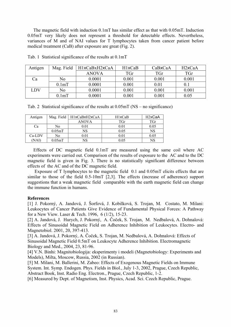
83
The magnetic field with induction 0.1mT has similar effect as that with 0.05mT. Induction 0.05mT very likely does not represent a threshold for detectable effects. Nevertheless, variances of M and of NAI values for T lymphocytes taken from cancer patient before medical treatment (CaB) after exposure are great (Fig. 2). Tab. 1 Statistical significance of the results at 0.1mT
H1xCaBxH2xCaA H1xCaB CaBxCaA H2xCaA Antigen Mag. Field ANOVA TGt TGt TGt
No 0.0001 0.001 0.001 0.001 Ca 0.1mT 0.0001 0.001 0.01 0.1
No 0.0001 0.001 0.001 0.001 LDV 0.1mT 0.0001 0.001 0.001 0.05
Tab. 2 Statistical significance of the results at 0.05mT (NS – no significance)
H1xCaBxH2xCaA H1xCaB H2xCaA Antigen Mag. Field
ANOVA TGt TGt No 0.01 0.01 0.05 Ca
0.05mT NS 0.05 NS No 0.01 0.01 0.05 Ca-LDV
(NAI) 0.05mT NS 0.05 NS Effects of DC magnetic field 0.1mT are measured using the same coil where AC experiments were carried out. Comparison of the results of exposure to the AC and to the DC magnetic field is given in Fig. 3. There is no statistically significant difference between effects of the AC and of the DC magnetic field. Exposure of T lymphocytes to the magnetic field 0.1 and 0.05mT elicits effects that are similar to those of the field 0.5-10mT [2,3]. The effects (increase of adherence) support suggestions that a weak magnetic field comparable with the earth magnetic field can change the immune function in humans. References [1] J. Pokorný, A. Jandová, J. Šorfová, J. Kobilková, S. Trojan, M. Costato, M. Milani: Leukocytes of Cancer Patients Give Evidence of Fundamental Physical Forces: A Pathway for a New View. Laser & Tech. 1996, 6 (1/2), 15-23. [2] A. Jandová, J. Hurych, J. Pokorný, A. Čoček, S. Trojan, M. Nedbalová, A. Dohnalová: Effects of Sinusoidal Magnetic Field on Adherence Inhibition of Leukocytes. Electro- and Magnetobiol. 2001, 20, 397-413. [3] A. Jandová, J. Pokorný, A. Čoček, S. Trojan, M. Nedbalová, A. Dohnalová: Effects of Sinusoidal Magnetic Field 0.5mT on Leukocyte Adherence Inhibition. Electromagnetic Biology and Med., 2004, 23, 81-96. [4] V.N. Binhi: Magnitobiologija: eksperimenty i modeli (Magnetobiology: Experiments and Models), Milta, Moscow, Russia, 2002 (in Russian). [5] M. Milani, M. Ballerini, M. Zabeo: Effects of Exogenous Magnetic Fields on Immune System. Int. Symp. Endogen. Phys. Fields in Biol., July 1-3, 2002, Prague, Czech Republic, Abstract Book, Inst. Radio Eng. Electron., Prague, Czech Republic, 1-2. [6] Measured by Dept. of Magnetism, Inst. Physics, Acad. Sci. Czech Republic, Prague.

84
NONLINEAR DNA DYNAMICS (PEYRARD-BISHOP-DAUXOIS MODEL AND A COHERENT MODE)
S. Zdravković
Faculty of Technical Sciences, University of Priština Kosovska Mitrovica, Yugoslavia Tel: + 381/11/516805, e-mail: [email protected]
Nonlinear DNA dynamics can be studied using different models. This work relies on the Peyrard-Bishop-Dauxois (PBD) model [1], which is an extended version of the PB model [2]. The model assumes only transversal displacements of nucleotides [1,2]. This transverse interaction, i.e. the potential of a pair of the nucleotides, is modeled by a Morse potential:
( )21−= − xaM eDV
Possible intervals for the parameters a and D were recently estimated according to both experiments and theory [3]. According to the PBD model, a modulated solitonic wave
[ ]( ) )O()(2cos)sech()cos()sech(2)( 2εδωκεωκ +−+−+−−= tΩΘnlCtnlAΩtΘnltnlAtΦn
moves along the DNA chain [1,2,4]. This function, as well as its envelope, is shown in the figure for a particular value of t . One can define a density of internal oscillations (density of carrier wave oscillations) as a number of wavelengths of the carrier wave contained within the length of the envelope [5]. Obviously, two definitions are possible: κΘDo = and
ωΩΓ o = . It is suggested, in this work, that the optimal value of the parameter a can be obtained from the requirement oo ΓD = . This is corroborated by the fact that the equality of those functions ( oo ΓD = ) represents the mode when phase velocities of both the envelope and the carrier signal are equal. This is a sort of a coherent mode, meaning that the wave is unchanged in time, i.e. the patern shown in Fig. 1 is the same at any position n of the DNA molecule. References [1] Dauxois, Phys. Lett. A 159 (1991) 390-395. [2] Peyrard and Bishop, Phys. Rev. Lett. 62 (1989) 2755-2758. [3] S. Zdravković and M. V. Satarić submitted to J. Mol. Biol. [4] S. Zdravković, Nonlinear Dynamics of DNA Chain – Peyrard-Bishop-Dauxois Model. In "Finely Dispersed Particles: Micro-, Nano-, and Atto-Engineering" Eds. A.M. Spasić and J.P. Hsu (Boca Raton, Florida: Marcel Dekker, Inc./Taylor & Francis) (in press). [5] S. Zdravković, J.A. Tuszyński and M.V. Satarić, will be published in J. Comput. Theor. Nanosci.

85
94 96 98 100 102 104
-5
0
5
10
15
t (ps)
Φn (n
m)
Fig. 1: Soliton )(tΦn for 300=n .

86
DETECTION SYSTEM FOR CELLULAR COHERENT VIBRATIONS
Ralph Hölzel Institute of Biology, Freie Universität Berlin, D-12165 Berlin, Germany
Fraunhofer Institute for Biomedical Engineering, D-14558 Bergholz-Rehbrücke, Germany Fröhlich's theory of coherent excitations in biological systems predicts vibrational modes in metabolically active systems like cells and cell organelles. Well known periodic phenomena on the level of single cells are, e.g., calcium waves and glycolytic oscillations, which, however, are of chemical origin and exhibit characteristic periods of seconds or even minutes. Fröhlich excitations are expected at much higher frequencies in the GHz and THz region. This, and the fact of being longitudinal vibrations, makes their experimental investigation a demanding task. Later theoretical work by other authors has demonstrated that effects at much lower frequencies might be possible. Indirect evidence of vibrational modes stems, e.g., from studies on the rouleaux formation of erythrocytes showing long range cell-to-cell interactions. Long range interactions also have been observed for spatially separated bacterial cell cultures. In cell suspensions of yeasts and algae highly polarizable particles are attracted towards the cells by dielectrophoretoc forces rendering information about the spatial distribution of the electromagnetic field. Recently mechanical vibrations of yeast cells in the kHz range have been recorded by atomic force microscopy. The most promising approach for the experimental investigation of Fröhlich's theory is the direct, electronic detection. Because of uncertainties on the theoretical side such a detection system should be i) very sensitive, ii) span a wide frequency range and iii) should exhibit a temporal resolution of seconds or less. Obviously, the amplifier should exhibit a good signal-to-noise level over a wide frequency range. Investigating single or few cells calls for a high impedance input. At the frequencies of interest this translates to a low input capacitance. Similar arguments hold for the coupling between biological object and the electronics, which can be done either by electrodes or by magnetic means like microcoils and SQUIDs. The registration and presentation of data is performed preferentially in the frequency domain. For this purpose analog signal analysis by spectrum analyzers or parallel filter arrays is feasible. Alternatively the signal is to be digitized and analyzed by Fast Fourier Transformation or specially developed algorithms.

87
MEASUREMENT OF ELECTROMAGNETIC EMISSION FROM PERTURBED BIOSYSTEMS
Janusz Slawinski, Zbigniew Górski
Institute of Chemistry & Technical Electrochemistry, Poznañ University of Technology Piotrowo Street 3, PL 60-965 Poznañ, Poland
Life on the Earth is imbedded in an „electromagnetic ocean” of terrestial, cosmic and man-made electomagnetic fields. However, all living systems (biosystems) themselves, regardless of differences in the biological hierarchy, create and emit very weak quasi-stationary electromagnetic fields of the power less than 10−16 W/cm2 or 103 photons/s cm2 in the spectral range 180-1500 nm or 2.1014 - 1015 Hz [1,2]. This ultraweak endogeneous electromagnetic emission (UEE) is associated with the production of electronic excited molecules in the exergonic steps of metabolic processes. Biosystems exposed to detrimental conditions i.e with the perturbed homeostasis, show changes in the parameters of UEE; usually an increased intensity, spectral shifts and a non-stationary kinetics of UEE. Lethal doses of stress factors that cause an irreversible perturbation of homeostasis and death of the organism evoke an enhanced „necrotic” UEE. Such an irreversible emission announces a decline of the living state [3-10]. Modern optoelectronic devices and computer software allow to record and analyse UEE. To record the time-dependent intensity variation in the UEE, the single photon counting (SPC) method exploiting a low-noise and a broad-wavelength photomultiplier tube was used. In this method one can obtain a collective signal from the whole sample in the time intervals longer than ca μs; a precise time moment after which there occurs a reaction of the tested biosystem to an applied stimulus (perturbation of homeostasis). The spectral distribution of the emitted radiation was analyzed combining the SPC method with a set of cut-off optical filters covering the wavelength range λ from 300-850 nm. Two-dimensional imaging of the UEE was done by accumulating the photons over a given period of time (seconds, minutes or hours) by means of an ultrasensitive slow-scan charge coupled device (CCD) camera Molecular Light Imager „Night Owl”. In this SPC-imaging method one can record the emission changes from the particular fragments ( region of interest, ROI) of the object under study and to construct pseudo-coloured 2-D maps of UEE. The time-dependent intensity of UEE for chosen ROI were obtained by calculating the number of photocounts from selected ROI pixels taken during the exposure time (usually min-h). For this purpose and in order to avoid artifacts and disturbances caused by reflections, meniscus and the vessel edge, special computer programs were elaborated [11,12]. Several examples of UEE from lower and higher plants, animal cells cultures and whole organisms exposed to the action of detrimental physical and chemical factors applied in lethal doses will be presented. UEE from yeast cells - a well defined eukaryotic type cell was extensively investigated [4-6]. Lethal concentrations of formaldehyde and trichloracetic acid - strong poisons that denaturate proteins - elicited a distinct increase in the intensity of UEE (a „necrotic peak”) and spectral alterations (the spectral shift from the red λ > 675 nm to λ = 540 nm for alive and dead cells, respectively). In the case of HCHO- elicited necrotic emission the oxygenation of cells enhanced UEE. The emission of coherent oscillations from synchronously dividing yeast cells centered around 8.5 MHz was also recorded [13]. The response of gigantic Chara and Nitella algae cells to local anaesthetics like procaine at different temperatures manifested as „necrotic” peaks correlated with the amount of alive/dead cells. A distinct oscillatory pattern of UEE from 30 cells reveals a cooperative, non-linear behavior of the cells during the dying process. Additionally, the rate of cytoplasmic streaming and conduction of action potentials was studied [8,9]. The SPC-two-dimensional

88
imaging of UEE dynamics observed by a Night Owl revealed morphological destructions after the anaesthetic treatment [10]. Several experiments with the delayed photosynthetic emission from Acetabularia acetabulum also shown an increased intensity from perturbed samples and changes in decay kinetics of UEE [14]. Many experiments on the effect of organic solvents, toxins, brine, acids and temperature on higher plant tissues revealed the existence of enhanced PE in the form of a necrotic maximum [15]. UEE from domestic animals spermatozoa cells exposed to the ascorbate—Fe(II) redox-cycling peroxidation system also shows an enhanced intensity which correlates (r = − 0.95 - 0.82, P = 0.05) with the vitality (number of alive/dead cells) and motility of the cells. UEE spectra from spermatozoa cells determined by the SPC-cut-off filters method covers the λ region 350-800 nm and are not a species characteristic [7]. Organs, tissues and the whole organisms of several lower animals treated with different toxic chemicals in lethal doses exhibited increased UEE [3]. Vital cells from rat brain homogenates or pig oligodendrocytes after disintegration emit radiation which is independent of external oxygen and not amplified by chemiluminescent probes. It is suggested that this cellular UEE represents a biophysical radiation which originates from the interruption of an intermolecular radiationless energy transfer [16]. In recent Japanise works two-dimensional SPC imaging was used to monitor UEE from an exposed rat’s cortex in vivo without adding any chemical agent or employing external stimulation. It was shown that the UEE intensity correlates with the electroencephalographic activity of the cortical surface and is associated with the cerebral blood flow and hyperoxia [17]. Recent investigations with mice intoxicated with formaldehyde, acetone or barbiturates observed by SPC-imaging have shown an increased UEE during the dying process [18]. Properties of UEE are derived from biochemical and physiological excitation processes. One can expect that properties of the primary excitations are transferred directly to those of the UEE. Therefore the multiparametric analysis of SPC and 2D-imaging of UEE might be used to elucidate mechanisms of the stress, toxin-organism interaction, to evaluate lethal doses, the resistance and adaptation capacity of biosystems. It is worthy to emphasize that these techniques are completely non-invasive and provide information on energetics and kinetics of physiological processes in real time. They offer an unique chance of tracing and recognizing perturbation of biosystems in a variety of scenarios.
References: [1] Chang J-J., Fisch J. Popp F. A. 1998. Biophotons, Kluwer Academic Publishers, Dordrecht.
[2] Inaba H. ed, 2000. Advances in biophoton and biophotonics research, vol.1, Tohoku Inst. Biophotonics Information Center & Photonics Res. Institute, Sendai, Japan. [3] Slawinski J.1990. Curr. Topics Biophys. 19, 8; 2001,Proc. SPIE, 4515,1; 2003, Indian J. Exp.Biol. 41, 483. [4] Slawinski J., Ezzahir A., Godlewski M., Kwiecinska T., Rajfur Z., Sitko D., Wierzuchowska D. 1992, Experientia 48, 1041. [5] Godlewski M., Rajfur Z., Slawinski J., Kobayashi M., Usa M., Inaba H. 1993.. J. Photochem. Photobiol.. B: Biol. 21, 29. [6] Rajfur Z. 1994, J. Biolumin. Chemilumin. 9, 59. [7] Laszczka A, Godlewski M., Kwiecinska T., Rajfur Z., Sitko D., Szczêsniak-Fabiañczyk B, Slawinski J., 1995, Cur. Topics Biophys. 19, 20. [8] Jaœkowska A, Borc R, Dudziak A, Œpiewla E. 1999. Acta Soc. Bot. Pol. 4: 281. [9] Jaœkowska A, Borc R, Dudziak A, Milczarek I., Œpiewla E.2001, Curr.Topics
89
Biophys.25(2), 95; 2001,Luminescence, 16,51. [10] Jaskowska A., Gorski Z., Dudziak A. 2004. Proc. SPIE, 5566, 15. [11] Gorski Z., Slawinski J. 2001, Proc, SPIE, 4887, 23. [12] Gorski Z., Bembnista T, Floryszak-Wieczorek J., Domanski M., Slawinski J. 2002, Proc, SPIE, 5064,1. [13] Smith C.W. In: Photon emission from biological systems, B. Je¿owska-Trzebiatowska, B. Kochel, J. Slawinski, W.Strek, eds., World Scientific, Singapore 1987, 120. [14] Van Wijk R., Van Wijk E PA, 2003, Indian J. Exper. Biol. 41, 411. [15] Ruth B., 1989. In: Electromagnetic bioinformation. F.A. Popp at al., eds.,Urban & Schwarzenberg, Munchen, Wien, Baltimore, 128.. [16] Reiber H-O. 1989. J. Biolumin. Chemilumin. 4, 245. [17] Kobayashi M., Takeda M., Tomoo S., Yamazaki Y., Kaneko K., Ito K-I, Kato H, Inaba H. 1999. Neurosci. Res. 34: 103. [18] Gorski Z., Lach H., Slawinski J. In: 12th Internat. Symp.”Molecular & Physiological Aspects of Regulatory Processes of the Organism”UNESCO/PAS, Cracow, June 5-6, 2003, H. Lach ed., 133.

90
E-FIELD PROBES AND THEIR CALIBRATION
Béla Szentpáli, György Thuróczy* Research Institute for Technical Physics and Materials Science, P.O.Box 49, Budapest,
Hungary, H-1525, fax: (36 1) 3922235, e-mail: [email protected] *National Research Institute for Radiobiology and Radiohygiene, Budapest, Hungary
The prompt spreading of the different applications of radio techniques raised the interest for the interaction of high-frequency electromagnetic fields with biological objects. Any laboratory work in this field demands the measurement of the intensity of the field, preferably within the biological object, or at least in phantom. In this paper the high frequency electric field measuring probes developed in the Research Institute for Technical Physics and Materials Science will be presented. The probe should meet to the following requirements: - Small size, ensuring good resolution in space. - The scattering of the field from the probe and the connecting wires etc. should keep as
small as possible. - The isotropic reception can be important in some cases, i.e. the sensitivity should not
depend on the polarisation of the field. The probes are constructed from short dipole antennas mounted with a detector diodes in the gaps and connected to the amplifier with highly resistive leads. The dipole antenna senses the electric field component parallel to it. One dimensional and three dimensional probes were fabricated.The 3D probes contain three mutually orthogonal dipoles, ensuring the isotropic sensitivity. The dipoles are connected to the amplifier with the resistive transmission lines, which attenuate effectively the longitudinal propagation of the high frequency signal. The probes are fabricated by thick film technology on 125 µm thick polyester substrate. The conductive parts are screen-printed: the dipoles from silver paste, the resistive lines from carbon paste. The detectors are zero bias diodes. Isolating lack covers the printed circuits. The typical widths of the probes are between 5 and 8 mm, the length – the distance between the probe and the amplifier – is typically 30 cm. The probes can be immersed into aqueous solutions, so the measurement within biological material or in phantom jelly is possible. The three dimensional probe is prepared from 3 equivalent parts, each of them consist of a dipole sensor tilted with an angle of 54.74o to the to the probe axis. The three individual parts are placed together at the long edges forming a cross section of a regular triangle. This construction is self-sustaining, so no any holder and outer tube are necessary. The probes are not fragile, they can be slightly deformed elastically. The inner part of the probe is empty; the jelly of the phantom or even the biological material can penetrate into this hole. The detector diodes work in the square-low regime, i.e. the output voltage is proportional with the square of the electric field. Therefore the summing of the signals of the three orthogonal detectors will give the square of the total field in that point. The measurement and calibration of the probes are performed in a cylindrical waveguide with inner radius R. The fundamental TE11 mode is used at 900 MHz. Therefore R=109 mm was chosen. The waveguide is cut into two parts that can rotate relative to each other. A muff ensures the good electric contact between the two parts. The waveguide termination is a pyramidal absorber. The microwave power is feed to an N type connector. Its lengthened central conductor couples the signal into the waveguide. The reflection is minimized by the tuning screw, at least S11 = –30dB can be achieved at each measuring frequencies within the bandwidth of the TE11 mode. The homogeneity of the E-field in the central region is utilized.
91
In the region extending to 0.1*R around the axis the maximum difference in the magnitude of the electric field is only 1.27 % and the orientation differs only with 0.24 degree. This theoretical statement was checked by measurements too. The probe is put into the waveguide trough a hole, it can revolve around its axis, and also the two parts of the cylinder containing the probe and the feed in point can rotate against each others. As a combination of these rotations the antenna characteristics can be determined along three perpendicular axes. The absolute sensitivity of the detectors can be measured in the same cylindrical waveguide too. The frequency dependence of the probe sensitivity is flat within 20% in the range of 0.5…2.0 GHz; below 300 MHz the sensitivity decreases drastically. The outputs of three detectors of the isotropic probes were summed and amplified (X200) by operation amplifiers having extra low bias currents (0,1 pA). The probe-amplifier system has an integrating time constant of 90 ms. This filtering decreases the noise arising mainly from the resistors and the disturbing 50 Hz scatter from the mains. The Johnson noise is suppressed in the microvolt range, however the most of the noise is the 1/f noise of the granulated carbon prints. The experienced noise levels at the output of the amplifier differ from probe to probe; they vary in the 1-5 mV range. The sensitivity of the probes is about (5…7)*10-4 V*/(V/m)2 , where V* means the voltage output of amplifier. The direct measurements the SAR sensitivity in brain phantom liquids resulted the values about 20 and 10 mV/(W/kg) for 900 MHz and 1800 MHz phantoms respectively.. Acknowledgments This work was supported by the Hungarian Academy of Sciences under contract No. 96/2-604 2,3, and by the Ministry of Economy and Industry under contract no: GVOP-3.1.1.-2004-05-0354/3.0. Some reference measurements were made in anechoic chambers in the Forschungzentrum Seibersdorf, where the kind help of G. Neubauer and G. Schmid is acknowledged and in the Laboratory of the SARTEST Ltd. where M. Manning, G. Morgan and D. Riley provided valuable assistance. The sensitivity in phantom material was calibrated in the Prof. Gandhi’s laboratory at the University of Utah.

92
FRACTAL ANALYSIS OF HUMAN BODY SPONTANEOUS PHOTON EMISSION
Roeland Van Wijk, Eduard P.A. Van Wijk International Institute of Biophysics, Landesstiftung Insel Hombroich
Kapellener Strasse, D-41472 Neuss, Germany
Background: There has been an increase in research on human body ultraweak photon emission (UPE) for isolated cells and tissues. There are only limited data on measurements of UPE from whole body. Objective: The goal of the present study was two-fold. At first, to describe a protocol for management of subjects that a) avoids interference with light-induced long-term delayed luminescence, and b) includes the time slots for recording photon emission. Secondly, to study photon count distribution, and analyse fractal properties of photon emission. Technology: A specially selected low noise end window photomultiplier was utilized for detection of ultraviolet / visible light (200-650 nm) photon emission. For multi-site recording it was manipulated in three directions in a dark room with a very low count rate. Results: Twenty-nine body sites are selected such that the distribution in UPE can be studied as right-left symmetry, dorsal-ventral symmetry, and the ratio between the central body part and extremities. Although data demonstrate the variability in patterns between subjects, the data suggest that a “typical” human emission pattern exists. The thorax-abdomen region emits lowest and most constant emission. The upper extremities and the head region emit the highest levels and increase during the day. It seems that generally the fluctuation in photon counts over the body is lower in the morning than in the afternoon. Spectral analysis of low, intermediate and high emission from the superior frontal part of the right leg, forehead and palms of left and right hand in the sensitivity range of the photomultiplier (200 – 650 nm) show the major spontaneous emission at 470-570 nm. Photon emission was characterized by kurtosis and skewness values of the photon count distribution. In addition data were characterized by the Fano factor [F(T); variance over mean of the number of photoelectrons observed within observation time T]. All data were compared to surrogate data sets which were constructed by random shuffling the data sets. Data indicated that commonly F(T) increased with observation time, significantly at window sizes above 6s. Conclusion: Examples of multi-site UPE recordings and spectral analysis demonstrate the individual patterns and dynamics of the spontaneous UPE over the body, and the spectral differences over the body. The spectral data suggest that measurements might well provide quantitative data on the individual pattern of peroxidative and anti-oxidative processes in vivo. Fano factor analysis demonstrates that the time series do not represent a simple Poisson process, the UPE has characteristics of a fractal process, showing long-range correlations. We expect the measurements provide physiological information to be useful in clinical examination.

93
RADIOMETRIC MEASURING INSTRUMENT OF THE SECONDARY MICROWAVE RADIATIONS
Yurij Skripnik, Konstantin Shevchenko, Aleksej Yanenko
The Chair of Automation and Computer Systems, Kiev National University of Technologies and Design, 2, Nemirovicha-Danchenka Str., 01010, Kiev, Ukraine
Phone: (+38044) 256-29-93, E-mail: [email protected] It is known, that all physical bodies including biological objects, absorb, reflect and pass electromagnetic waves in a wide range of frequencies. Intensity of electromagnetic radiation of object depends on his structure, temperature, condition of a surface, frequency of radiation, and also biochemical processes which proceed in the object [1]. Together with equilibrium radiothermal radiation in biological objects nonequilibrium radiation which is shown in a narrow range of frequencies takes place also. As a source of nonequilibrium radiation the non-uniform field of temperatures, gradients of concentration of substances, leading of energy from local sources, metabolic processes in an alive organism can act. To a nonequilibrium component of radiation carry also the phenomenon of secondary radiation by biological objects in a long-wave part of a spectrum. Such nonequilibrium radiation takes place in a decimetres range of lengths of waves at an irradiation of the researched biological environment low intensive (<10-6 W/sm2) electromagnetic waves of a millimetres range. The mechanism of his occurrence is in details considered in [2]. As secondary radiation which is observed, is superfluous above radiothermal radiation, it carry to a radio-frequency luminescence [3]. Experience of resonant therapy unequivocally shows, that the greatest influence on the person is carried out with electromagnetic fluctuations in a range of frequencies 40...70 GHz. The final medical effect which is easily fixed clinically, testifies that effective transformation of energy of chemical communications at a cellular level took place. There is an idea, that these transformations under influence of millimetres lengths of waves cause the phenomenon of secondary radiation in a decimetres range. Authors develop the device, allowing to measure a level of secondary radiation of
biological objects under influence low intensive radiation of millimetres lengths of waves. Structure of the device offered by authors shown on Fig. 1.
G
G~~~
G~ ~~
~~ 000
~
~~~U U2
dB
16
15
14
13 9 10
12 11 8 7
632
5 4

94
The device the microwave amplifier 3, a key 4, replaceable attenuator 5, the square-law detector 6, the low-frequency amplifier 7, the synchronous detector 8, the filter of the bottom frequencies 9, the voltmeter 10 and the generator 11 low frequencies contains the aerial of 1 decimetres range, the switchboard 2. The structure of the device also includes the aerial 12, the generator 13 millimetres ranges, a key 14 and the power supply 15. A position 16 the designated researched biological object. With the help of the device offered by authors it is possible not only to predict efficiency of resonant therapy in each concrete case, and to carry out wide researches on influence of change of capacity and frequency of electromagnetic fluctuations irradiating object. References [1] Левин М.Л., Рытов С.М.: Теория равновесных тепловых флюктуаций в электродинамике.–М.: Наука. – 1967. – 308с (Levin M.L., Rytov S.M.: Theory of equilibrium thermal fluctuations in electrodynamics. Nauka). [2] Синицын Н.И., Петросян В.И., Елкин В.А. и др.: Особая роль системы “миллиметровые волны – водная среда в природе”. Биомедицинская радиоэлектроника, 1. – 1998 (Sinicin N.I., Petrosjan V.I., Elkin V.A.: Special role of the system “millimeter waves – water medium in nature”. Biomedicinskaja radioelektronika). [3] Чукова Ю.П.: Механизмы биологических эффекиов электромагнитных полей. Биомедицинские технологии и радиоэлектроника, 11. – 2001. с.32-41 (Chukova Yu.P.: Mechanisms of biological effects of electromagnetic fields. Biomedicinskie technologii and radioelektronika).

95
EXPERIMENTAL INVESTIGATION OF ELECTROMAGNETIC ACTIVITY OF YEAST CELLS AT MILLIMETER WAVES
F. Jelínek, J. Hašek*, J. Šaroch, J. Pokorný
Institue of Radio Engineering and Electronics, Chaberská 57, 182 51 Prague 8 *Institute of Microbiology, Vídeňská 1083, Praha 4
Academy of Sciences of the Czech Republic
Direct measurement of the electromagnetic fields generated by living cells may deliver strong support for Fröhlich´s theory of coherent oscillations in biological systems [1]. We have exerted in our laboratory in recent years some experimental effort to realize direct measurements of cellular fields in rather broad frequency range. Our interest is attracted now by the interaction of living cells with mm-wave fields. The published results of investigation in this region are rather contradictious. Grundler et al. [2] observed resonant interactions of the external fields in the 42GHz region with yeast cells (frequency dependent growth of yeast cells during exposure over three growth cycles). Non-thermal effects at 40 and 50GHz on living cells are described in [3]. Measurements of Gandhi et al. [4] did not disclose the spectral resonances of biological samples and the authors concluded that spectral resonances in the mm-wave region reported by others are likely to be in error. We assume that electromagnetic field generated by subcellular structures has character of the near zone field and could be measured in the near vicinity of the cell. As a model system for experimental investigation we have used cold-sensitive β-tubulin mutants tub2-401 of yeast cells Saccharomyces cerevisiae. At the restrictive temperature (below 14˚C) the mutant tub2-401 has no microtubules. The yeast cells are at the boundary between the G2 and the M phase. After recovery of the arrested mutant tub2-401 at the permissive temperature (above 25ºC) microtubules were polymerized and mitotic spindles were formed (at about 20min from the initial time). After 40min of cultivation the majority of cells were in the anaphase A and some of them were in the anaphase B. Within 60min of cultivation the mitotic spindles of the majority of cells were in the anaphase B and some of them completed it. These data were verified by fluorescent microscopy [5]. We designed a laboratory equipment for the measurement of living cells activity in the 8 mm wave region. A block diagram of the measurement system of electromagnetic oscillations of living yeast cells is shown in Fig. 1. The sensor is formed by a fin-line structure. We described its configuration in [6]. The sensor with cells is screened against spurious fields. Preamplifier Spacek SL406-34-4 in the frequency band 37-43GHz is used followed by the spectrum analyzer Agilent E4448A. Measured sensitivity of the system for 300Hz analyzer resolution bandwidth is -140dBm. The frequency band 4MHz at 41-42GHz is divided into 20 subbands of 200kHz and is scanned during 60s. The system is controled by computer and measured data are stored in the computer memory for evaluation and for statistical processing. During measurement the temperature of the fin-line sensor with cell suspension is kept at 28 ± 0.2 0C using a computer controlled system.

96
Fig 2 shows measured signal in the subband 41.6996-41.6998GHz (reproduced from the data stored in the computer memory). Critical level for evaluation is about 5.5dB above the average level of the noise (detector of the maximum values is used). We evaluated sum of power of the spectral lines exceeding the threshold level in 5min intervals as a function of time. Data in the Grundler-Keilmann frequency range (41–42GHz) evaluated from measurements on synchronous cells, on the same mutant of yeast cells cultivated at 30ºC (nonsynchronous yeast cells), and on the sucrose solution will be presented. Experimental data are compared with the evolution of the M phase.
Fig. 2
References [1] H. Fröhlich: Quant. Mech. Concepts in Biol. In Theor. Phys. Biol.; Marois, M., Ed.; North Holland, Amsterdam, 1969; 13-22. [2] W. Grundler et al.: Nonthermal Resonant Effects of 42 GHz Microwaves on the Growth of Yeast Cult. In Coherent Excit. Biol. Syst.; Froehlich, H.; Kremer, F., Ed.; Springer, Berlin, 1983; 21-37. [3] N.D. Deviatkov, O.V. Betskii (Ed.): Biol. Asp. Low Intens. Millimeter Waves. Moscow, Seven Plus, 1994. [4] O.P. Gandhi et al.: Millimeter wave absorp. spectra of biol. samples. Bioelectromag., 1980, 1, 285-298. [5] J. Pokorný et al.: Electromagnetic Activity of Yeast Cells in the M Phase. Electro- and Magnetobiol. 2001, 20 (3), 371-396. [6] F. Jelínek et al.: Experimental investigation of electromagnetic activity of living cells at mm waves. In Book of Abstracts, XVIIth International Symposium on Bioelectrochem. and Bioenergetics, Florence, 2003, 68.
f (GHz)
41.6996 41.6997 41.6998
P
(dB
m)
-155
-150
-145
-140
-135
-130THRESHOLD FOR EVALUATION
97
NEW SENSOR FOR POSSIBLE MEASUREMENT OF BIOPLASMA STATE OF ORGANISMS
Igor Jerman, Romana Ružič, Metod Škarja, Robert T. Leskovar
BION, Institute for Bioelectromagnetics and New Biology; Stegne 21, 1000 Ljubljana, Slovenia.
Fax: +386 1 513 11 47, e-mail: [email protected], [email protected], [email protected], [email protected]
Introduction Among hypotheses concerning the nature of the living state of organisms is also the one that is built around the concept of the plasma state of ionic matter within and around organisms first proposed by Inyushin [1,2] and later elaborated by Zon and other researchers [3,4]. Inyushin suggested the existence of a bioplasma energy field composed of ions, free protons, and free electrons. He assumes that the bioplasma energy field is a fifth state of matter (besides the known four states: solids, liquids, gases, and plasma). The bioplasma particles should be constantly renewed by chemical processes in the cells and would be in a constant motion. There should be a balance of positive and negative particles within the relatively stable bioplasma. A severe shift in this balance should cause a change in the health of the organism. At the same time, some of this bioplasma should be radiated into space. Preliminary research methodology We developed a measuring device that is very sensitive to electric fields (potentials). Its distinctive feature is that while measuring the electric potential it consumes almost no electric current. Thus, it may measure even electric potential differences whose energies are almost negligible. With the device, we could measure small differences in electric potential of plants throughout the day (circadian rhythms). But it interested us most if there were differences between living and dead leaves of the plants (a leafy shoot of Setcreasea purpurea, one leaf of Begonia rex) while subjecting them to a stream of ions stemming from a fire. Our hypothesis was that due to its bioplasma the living leaf should compensate some portion of incoming ions or electrons while the dead one would not accomplish this.
The leaves (or shoots) were cut from the original plant and fixed on the wooden frame. During measurements a special device and a program detected the voltage of leaves in arbitrary units (the values were amplified) every second. The shape of the curves or its slope can be further analysed. The dead leaf (or shoot) was obtained by dipping them into boiled water for a few seconds. Results and discussion The results of our preliminary research confirm the hypothesis, namely, the living leaf is electrified a little slower than the dead one (see Figs. 1 and 2). As is seen in Figs. 1 and 2 the (almost completely linear) slope of the gaining of the negative charge is smaller with the live leaf than with the dead one. The experiments on this line are in progress.

98
Fig. 1. Absolute difference in the slope of the voltage lines of live shoot of Setcreasea and the same dead one.
Fig. 2. Comparison of the slopes of the voltage curves of live leaf and the dead one. The plant was Begonia rex. References [1] Inyushin VM (1977): Bioplasma: The fifth state of matter? In: White J, Krippner S. (eds.): Future Science, pp.115-120 [2] Inyushin VM (1983): Resonance biostimulation and the problem of bioplasma. In: Wolkowski, Z.W. (ed.): Proceedings Int. Symposium of Wave Therapeutics, pp.123-129. [3] Quickenden TI, Tilbury RN (1986): A critical examination of the bioplasma hypothesis, Physiol. Chem. Phys. Med. NMR,18(2), pp. 89-101 [4] Zon JR, Wnuk M (2000): The Role of Physical Plasma in Selforganisation in Biosystems, in Electromagnetic Aspects of Selforganisation in Biology, Abstract Book, July 9-12, Prague, Czech Republic, pp. 14-15.
0,043
0,044
0,045
0,046
0,047
0,048
0,049
live shoot dead shoot
volta
ge u
nits
0,039
0,04
0,041
0,042
0,043
0,044
0,045
1 2 3
time of measurement
volta
ge p
oint
s
live leaf
dead leaf

99
DELAYED LUMINESCENCE APPLICATION IN ALGAE AND HIGHER PLANTS
Alexis Zrimec, Maja Berden Zrimec, Luka Drinovec Institute of Physical Biology, Velika Loka 90, 1290 Grosuplje, Slovenia; [email protected]
Delayed luminescence (DL), also termed delayed fluorescence or delayed light emission, is a long-lived light emission by plants and cyanobacteria after a short illumination [1]. In contrast to rapid fluorescence, which is light emission on the nanoseconds time-scale, the characteristic times for DL are from milliseconds to seconds. It has a hyperbolic decay kinetics in the first seconds after illumination, which is sometimes followed by a more or less pronounced peak, sometimes called the afterglow peak [2]. The main source of DL is photosystem II (PSII) in the thylakoid membrane of chloroplasts. DL originates from the repopulation of excited states of chlorophyll a from stored energy after charge separation [3]. DL kinetics depends on the physiological state of organism and is influenced by stress and injury [4]. In physiological studies, delayed luminescence intensity (DLI) is frequently used as a parameter. DLI represents the integral under the DL decay curve. DLI was reported to be a measure of photosynthetic activity and living algal biomass in field studies [see ref. in [2]. Yacobi et al. [5] have analyzed natural phytoplankton samples using DL excitation spectra and were able to detect taxonomical changes in the algal communities. This technique is currently used in monitoring freshwater phytoplankton [6]. Delayed luminescence measurement DL is measured in a custom made photon-counting luminometer. The apparatus includes an illuminator for photo-excitation, a thermostated sample holder and a light detector. A 20 W halogen lamp is used as an illuminator, providing light intensity of 40 mmol m-2 s-1 PAR at the sample position. The sample holder for 5 ml cuvettes is thermostated. For light detection, a red-light-sensitive photomultiplier tube (Hamamatsu R1104) with a Hamamatsu C3866 Photon Counting Unit for signal conditioning and amplification is used, coupled to a personal computer for data collection. Background noise is measured prior to each DL measurement. The samples are than illuminated for 3 seconds and DL is measured in the interval between 1 to 60 seconds after the illumination.
Species specificity DL kinetics is a useful tool for taxonomic and growth phase determination of phytoplankton, especially during algal blooms when one algal species predominates [2]. The DL decay is hyperbolic in the first few seconds, and then the curves diverge markedly for the various algae (see the Figure above).

100
Living cell concentration DL can be used as for determination of living algal cell concentration (minimum detection limit of approximately 10 cells/ml in our luminometer) and biomass. The relationship between DLI and algal cell concentration is linear, allowing concentration measurements of natural samples [7]. Influence of external parameters DL kinetics is influenced also by different temperature regimes and illumination intensity [8]. The intensity and onset of the afterglow peak vary due to the external parameters. Delayed luminescence as a tool for toxicological tests on plants DL measurement is nondestructive, simple and fast compared to the standardized toxicological tests and allows rapid monitoring of physical or chemical factors that affect photosynthesis. Many toxicity endpoints in algae or higher plants require 1-2 weeks of exposure to xenobiotics before significant changes are detected [9]. Effects of heavy metals on DLI of Lemna minor can be seen already after 24-hours as opposed to the standard Lemna growth test (ISO 20079) which lasts 7 days. Exposure to different metal concentrations allows calculation of standard toxic parameters, like ECx (effective concentration). The afterglow peak is even more sensitive to chemicals affecting photosynthesis than DLI. For example, peak can be diminished already 25 minutes after adding DCCD (ATP-synthase inhibitor). Comparison of DL and growth measurements of unicellular algae Scenedesmus subspicatus, exposed to a toxic substances in a standard procedure, showed the differences were more pronounced with the DL measurements. Acknowledgments Slovene Ministry of Education, Science and Sport (Grants P1-1237, L4-6222) References [1] Strehler, B.L., Arnold, W. J. Gen. Physiol. 34: 809–820, 1951. [2] Monti, M., Zrimec, A., Beran, A., Berden Zrimec, M., Drinovec, L., Kosi, G., Tamberlich, F. Delayed luminescence of Prorocentrum minimum under controlled conditions. Harmful Algae, 2005. [3] Jursinic, P.A. In: Light emission by plant and bacteria, A.J.Govindjee & D.C.Fork (eds.). Academic Press, New York, p. 291–328, 1986. [4] Yan, Y., Popp, F.A., Sigrist, S., Schlesinger, D., Dolf, A., Yan, Z. Biophoton emission and delayed luminescence of plants. In: Weak Photon Emission from Living Tissues: Delayed Luminescence - Facts & Perspectives, Berden Zrimec, M., Milani, M., Drobne, D., Zrimec, A. (eds.). IFB, Grosuplje, 2004. [5] Yacobi, Y.Z., Gerhardt, V., Gonen-Zurgil, Y., Sukenik, A. Water Res. 32: 2577–2582, 1998. [6] Hakanson, L., Malmaeus, J.M., Bodemer, U., Gerhardt, V. Ecol. Model. 169: 179–196, 2003. [7] Berden Zrimec, M., Zrimec, A., Drinovec, L. Delayed luminescence of algae and higher plants. In: Weak Photon Emission from Living Tissues: Delayed Luminescence - Facts & Perspectives, Berden Zrimec, M., Milani, M., Drobne, D., Zrimec, A. (eds.). IFB, Grosuplje: p. 12-17, 2004. [8] Drinovec, L., Zrimec, A., Berden Zrimec, M. Analysis and interpretation of delayed luminescence kinetics in the seconds range. In: Weak Photon Emission from Living Tissues: Delayed Luminescence - Facts & Perspectives, Berden Zrimec, M., Milani, M., 3. Drobne, D., Zrimec, A. (eds.). IFB, Grosuplje: p. 59-63, 2004a.
101
[9] Drinovec, L., Drobne, D., Jerman, I., Zrimec, A. Delayed fluorescence of Lemna minor: A biomarker of the effects of copper, cadmium, and zinc. Bull. Environ. Contam. Toxicol. 72: 896-902, 2004b.

102
CHANGES OF SURFACE POTENTIAL AND MAXWELL CURRENTS OF LIPID MONOLAYER AT PRESENCE OF GRAMICIDIN A
P. Vitovič, M. Weis, P. Tomčík, J. Cirák, T. Hianik
Laboratory of Biophysics, FMFI UK, Mlynská dolina F1, 842 48 Bratislava, Slovakia Department of Physics, FEI STU, Ilkovičova 3, 812 19 Bratislava, Slovakia
We studied the electrical and mechanical properties of monolayers composed of pure model peptide gramicidin A (GRA) as well as lipid monolayers contained various mole ratio of GRA/Phospholipid. The area-pressure isotherms were measured both at gel and in a liquid–crystalline state. The measurements of area-pressure isotherm of monolayers of a pure GRA confirmed previously obtained results by other authors. However, in addition to these studies we for the first time measured the changes of Maxwell currents flowing between the surface of monolayer and ground and caused by reorientation of dipole moments of the monolayer. We have shown, that after initial growth of the current during compression of monolayer and at relatively large area per molecule, the growth was changed by decrease of the current at area per molecule around 4.7 nm2. This effect evidence of the changes of orientation of dipole moments of GRA and of the changes of secondary structure of the molecule, that switch to double helix conformation. GRA affect the mechanical and thermodynamical properties dimyristoylphosphatidylcholine (DMPC) monolayers. On the base of measurement L-B isotherms of mixed DMPC-GRA monolayers below and above phase transitions of phospholipids, we confirmed that aggregation of GRA exists both in liquid-crystalline and in a gel state of the monolayer and take place at DMPC:GRA mole ratio 3:1. These results are in agreement with that obtained recently [Giociautti et al. Biophys. J. 82 (2002) 3198] on a mixed dipalmitoylphosphatidylcholine (DPPC) monolayers in a gel state. Our results reveal that aggregation of GRA took place also in a liquid-crystalline state of the bilayers. Acknowledgement This work was supported by the Slovak Grant Agency for science VEGA (Project No. 1/1015/04) and by a NATO Collaborative linkage grant (LST.CLG.978567),

103
MEASUREMENT OF MOLECULAR ORIENTATIONAL ORDER IN CELL MEMBRANE MODELS BY THE MDC TECHNIQUE
D. Barančok, J. Cirák, P. Tomčík, J. Vajda, M. Weis
Department of Physics, Faculty of Electrical Engineering and Information Technology Slovak University of Technology, Ilkovičova 3, 812 19 Bratislava, Slovak Republic
Monolayers of surfactants at the ait/liquid interface (Langmuir films) exhibit very interesting physical properties attributed to low-dimensional systems as well as provide promising applications as models of biological membranes for studying lipid-protein interactions. Recently much attention has been paid to the dynamic behaviour of the cell memebrane (change of molecular orientation or conformation). Many various experimental techniques have been designed to the order parameter (molecular tilt) investigating in organic monolayers on the water surface, however only a few of them are suitable for the study of time-dependent orientational properties. The method of Maxwell‘s displacement current (MDC) measurement was developed by Iwamoto and Majima [1-2]. Partly modified Iwamoto's MDC setup appeared in [3] and was tested by measuring stearic acid (SA) and diacetylene (DA) monolayers. The further introduction of non-continuous compression, regularly interrupted [4-5] allowed to follow the relaxation processes during which the system of molecules reaches the equilibrium state after a stop of the barrier.
β
Fig. 1. (left up) Schematic view of the experimental setup for displacement current measurement. Rod-like polar molecules execute precessional motion on the water surface with maximal tilt angle θA (A and μ stand for the area per molecule and the dipole moment of molecule, respectively). Electrical shielding of the top electrode is not drawn. (left down) The course of compression was interrupted at four positions characterized by four selected areas per molecule. (right) Time recording of the MDC across the SA monolayer in the course of a step compression characterized by the compression rate β. The experiment is based on a short-circuited capacitor being formed by two parallel plates between which the Langmuir monolayer is situated. Maxwell's displacement current (MDC) is here generated by the change in dielectric polarization provided mechanically. The schematic view in Fig. 1 shows basic components of the experimental setup. More details about the experimental device (NIMA Technology, UK) are described in [5]. Compression of the monolayer is provided by a movable barrier, and it results in the change of the surface concentration of the molecules as well as in the orientational change of direction of the molecular electric dipoles. The change of the orientational polarization vector can be expressed as Maxwell's displacement current flowing through the monolayer.
104
As we showed in our previous studies [6] the current flowing in the outer circuit can be expressed as a time change of the induced charge
⎟⎟⎠
⎞⎜⎜⎝
⎛
∂
Θ∂+Θ
∂∂
μ=∂∂
=t
NtNG
tQ
I i coscos (1)
where μ is the dipole moment of one molecule, N is the number of molecules under the top electrode and G is the geometrical factor depending only on the distance between the top electrode and the top plane of the monolayer, and on the radius of the circular electrode.
Θcos stands for the statistical mean value Θcos , where Θ is the angle between the dipole moment vector and the vertical z-axis and is defined by a relationship Ω⎟⎟
⎠
⎞⎜⎜⎝
⎛−Θ=Θ ∫ d
kTU
Zexpcos1cos (2)
where dΩ is the solid angle in which the molecule is situated and U is the mean value of the interaction energy. The potential energy was determined according to our previous model of the monolayer [6]. The mutual pair interaction between molecules consists of dipole-dipole interaction and Buckingham's modified potential. For all our calculations a spatial averaged value of the cluster potential energy is used. For the mean value of the interaction energy we can obtain ( )A
ij
ji
ij
ijij rr
Brr
CU Θ−πε
μμ−−⎟⎟
⎠
⎞⎜⎜⎝
⎛−= cos1
4exp 3
06
0
(3)
where C, 0r and B are constants and r is the center-to-center separation of the particles. The molecules have a tilt AΘ∈Θ ,0 from vertical axis, where the maximum possible tilt angle AΘ is a function of the area per molecule and is defined by the relation
CA AA /1cos −=Θ ( CA is the critical area for the molecule lying on the water surface) [7]. The system has a spontaneous trend to expand, therefore we assume that the molecules have tilt
AΘ=Θ . In the spherical coordinates, where one molecule is at the centre of the system, for a generalized variable Θ the Lagrange motion equation of a cluster of n molecules has form 0=
Θ∂∂
−⎟⎠⎞
⎜⎝⎛Θ∂∂
∂∂ LLt &
(4)
The kinetic energy consists of rotation, precession and translation energy. The latter one consists of mean value kTi
2, where i is degree of freedom (constant value for given
temperature T; disappears by time differentiation) and its change 221 rm& . By substituting the
kinetic energy change into Eq. (4), it is possible to express the angular acceleration
1
222
2 )1(2coscossin2
)1(21
−
⎟⎟⎠
⎞⎜⎜⎝
⎛−
+Θ⎟⎟⎠
⎞⎜⎜⎝
⎛Θ⋅ΘΘ−
Θ∂∂
−−=Θ
l&
l&&
mnnJU
mn (5)
where J is the moment of inertia and m and l is the molecule mass and the molecule length, respectively. It can be shown that the second term in the denominator can be neglected. Because the observed relaxation phenomena remind of the inner resistance of the environment to the motion, we apply a friction term ( )Θξ′− & to the motion equation. Eq.(5) is rewritten as
⎟⎟⎠
⎞⎜⎜⎝
⎛Θ
Θξ′+Θ
∂∂
−π
−=Θ 2costan
)1(
&&&
AU
mn (6)
where CA2/ξ=ξ′ . For a numerical solution Eq. (6) was used in the form
⎟⎟⎠
⎞⎜⎜⎝
⎛Θ
Θ+Θ
∂∂
−−=Θ 2cos
tan)1(
1 &&& b
AUa
n (7)
with 1016.0 −= kga , 11.0 −= sb . These theoretical calculations satisfactorily fit the experimental data (Fig.2).

105
Fig. 2. (up) Detailed analysis of the relaxation process following a stop of the barrier in compression (left view) and expansion mode (right view). (left down) Development of designed potential energy, markers represent selected areas for experiment. Potential energy minimum is shown in the insert. (right down) Curves represent our calculations using Lagrange's equation.
Imperfect gas approximation for Langmuir films as a cell membrane models is a new view at a membrane. The analysis of the molecular motion by the Lagrange equation explains the observed orientation-translation relaxation process in the monolayer after stopping its compression at various molecular concentrations. Overshoots in expansions as a rapid return to zero (for low areas per moelcule) is caused by the determination of the maximum possible area per molecule with the barrier position. References [1] Y. Majima, M. Iwamoto, Jpn. J. Apl. Phys. 29(3), 564 (1990). [2] Y. Majima, M. Iwamoto, Rev. Sci. Instrum. 62(9), 2228 (1991). [4] . Barančok, J. Cirák, P. Tomčík, J. Vajda, Phys. Stat. Sol. A 169, 267 (1998). [5] J. Cirák, D. Barančok, P. Tomčík, J. Vajda, 7th European Conference Thin Organic Films, Potsdam, Germany, 99 (1998). [6] J. Cirák, D. Barančok, P. Tomčík, J. Vajda, Mat. Sci. Eng. C 8-9, 13 (1999). [7] Vajda, M. Weis, D. Barančok, J. Cirák, P. Tomčík, Appl. Surf. Sci. 229, 183 (2004). [8] M. Iwamoto, C.X. Wu, Phys. Rev. E 54(6), 6603 (1985).

106
CONDUCTING POLYMERS IN BIOMEDICAL SENSORS
M. S. Gaur Department of Physics, Hindustan College of Science & Technology
Agra- Delhi National Highway #2, Farah (Mathura) U.P. – 281122, India Many chemical substances in industrial processes, medical care and environmental monitoring are still evaluated by offline measurements. Appropriate sensors, which allow for online monitoring, are scarcely available. There is an urgent need of sensors for medical diagnostic industry, environmental monitoring, and food industries. A new class of polymers known as intrinsically conducting polymers or electroactive-conjugated polymers has recently emerged. There has been an increased interest in the research and development of conducting polymers that can be used for application to biosensor devices. This is because biosensor is likely to have significant impact on industrial and economic activity of the developing and developed world. In the near future biosensing devices in the broadest sense pertains to the implementation of materials, molecular devices, circuit and architecture as encountered in life sciences into molecular circuits and systems and miniaturized techniques and also to bring technology down to atomic scale (bio-chip). In this context it has recently been shown that it is possible to use receptors, enzymes, antibodies, organelles and plants cells and many other biological materials as the molecular recognition elements of sensing devices [1-2]. Electrically conducting polymers have considerable flexibility in the available chemical structure, which can be modified. Moreover the polymer itself can be funtionalized to bind protein molecules. This paper will briefly describe the overview of construction, designing and molecular architecting of biosensors. References [1] Handbook of Polymers in Electronics, Edited by B. D. Malhotra, Rapra Technology Ltd. (2003) [2] P. R. Birkin, synthetic metals, 61, 15 (1993)

107
BIOELECTROCHEMICAL METHODS FOR CANCER RESEARCH AND THERAPY
Hermann Berg
Laboratory Bioelectrochemistry, Campus Beutenberg Jena , Germany, [email protected]
Nearly the whole reservoir of electrochemical methods has been applied against cancer cells in vitro: d.c.(constant, pulsed) and a.c. (capacitively or inductively coupled) in the frequency regions 1 Hz up to THz . The main breakdown effects are : -electrolytic cell destruction ,electroporation of membranes by d.c.application, -changes of metabolism (e.g.inhibition of proliferation,HSP expression, apoptosis induction, and necrosis) by various a.c. treatments. All these effects can be strengthened synergistically by hyperthermia(up to 44°C), hyperacidity(down to pH6), photodynamic action(500-800 nm), and addition of cytotoxic agents(actinomycin,doxorubicin) or antisense oligodesoxynucleotides (AS-ODN) . Examples for these combinations on the cellular level will be presented and data evaluated [1].Using Helmholtz-coils in the region of extremely low frequencies the magnetic flux densities of 10-15 mT yield optimal efficacy. The tumor therapy of animals( mostly mice) has been tested for d.c.techniques and pulsating electromagnetic fields (PEMF), sometimes combined with cytotoxic agents (mitomycin, cis-platinum) orAs-ODN [2]. In most cases significant tumor necrosis and inhibition of angiogenesis have been found. Treatment of different tumor patients occurs mostly by the Nordenström d.c.electrode method. Depending on the kind of neoplasmic tissue about 15 000 patients in China showed more or less regressions. In contrast to these d.c. applications the noninvasive PEMF solenoid-technique is unfortunately still in the very beginning [3]! Nevertheless, this induction of apoptosis and cell necrosis by PEMF variations last not least in the high frequency region - combined with static magnetic fields and agents mentioned - will be the adjuvant noninvasive therapy in the future. References [1] M. Radeva, H. Berg.Differences in lethality between cancer cells and human lymphocytes caused by electromagnetic fields. Bioelectromagnetics 25 (2004) 503-507. [2] R. Elez et al. Tumor regression by combination (of electroporation and) antisense therapy against PLK1 and BCL2. Oncogene 22 (2003) 69-80. [3] P. Rosch, M. Markov.Bioelectromagnetic Medicine. Marcel Dekker NY (2004).
108
HYPERTHERMIA COMBINED WITH CYTOTOXIC CHEMOTHERAPY IN THERMOSENSITIVE LIPOSOMES
J. Dvořák, Z. Zoul, B. Melichar, J. Petera, M. Doležel
Department of Oncology and Radiotherapy, Charles University Medical School & Faculty Hospital, Hradec Králove, Czech Republic
The aim of this paper is a review of clinical experience with hyperthermia combined with cytotoxic chemotherapy in thermosensitive liposomes. Hyperthermia is a therapeutic modality that uses heating of tissue to 41-43o C, and can be performed locally, regionally or systemically. Hyperthermia alone has limited antineoplastic activity, but enhances efficacy of chemotherapy and radiotherapy. Preclinical and clinical studies indicate that hyperthermia may enhance the efficacy of variety of anticancer agents by increasing intratumoral drug concentration, membrane transport, oxygen free radical production and by inhibiting the reparation. Several cytotoxic drugs have demonstrated additive or synergistic effect in combination with hyperthermia, including (doxorubicin, cisplatin, nitrosoureas, cyclophosphamide, bleomycin, mitomycin C and gemcitabin). The first thermosensitive formulation of liposomes was proposed by Yatvin et al in 1978 [1]. In vitro studies demonstrated that combination of hyperthermia and thermosensitive liposomes augment drug delivery and antitumor effects of thermosensitive liposome encapsulated drugs as compared with free drug or thermosensitive liposome encapsulated drug that is administered under normothermic conditions. Hyperthermia increases extravasation of liposomes in the tumor vessels and increases cytotoxic drug release from thermosensitive liposomes. There is also direct cytotoxic effects of hyperthermia on tumor cells and synergistic or supra-additive effects of hyperthermia combined cytotoxic agents. In clinical practice, intravenous administration of cytotoxic chemotherapy is followed by application of local or regional hyperthermia using a hyperthermic device [2]. The clinical experience with the combination of hyperthermia and cytotoxic chemotherapy in thermosensitive liposomes is limited. In phase I/II clinical study 15 patients with locally recurrent breast cancer, previously treated with radical mastectomy and conventional radiotherapy, were enrolled on a protocol combining initial re-irradiation, monthly infusions of liposomal doxorubicin and local hyperthermia, using waveguides, with a rectangular or circular aperture (National Technical University of Athens), working at a frequency of 433 MHz [3]. All patients showed an objective measurable response and 3 patients (20%) had complete response. In another phase I/II study 23 patients with breast cancer metastatic to the chest wall were treated by sequential hyperthermia via 16-element planar array microwave or ultrasound applicators and liposomal doxorubicin [4]. All patients had prior radiotherapy and chemotherapy. Objective response was observed in 12 out of 20 evaluable patients, indicating that sequential hyperthermia and liposomal doxorubicin is effective and may be safely administered over multiple cycles. We have earlier reported a complete response in a patient with skin metastasis of breast carcinoma treated with intravenous infusion of liposomal doxorubicin in combination with local hyperthermia by Sonotherm® 1000 Ultrasound Therapy System, working at a frequency of 3,4 MHz [5]. Each treatment cycle consisted of intravenous administration of liposomal doxorubicin followed by 2 fractions of hyperthermia 41 to 43oC for 45 minutes 1 and 48 hours after infusion. We have observed a partial response in a patient with advanced inoperable hepatocellular carcinoma of the liver treated with liposomal doxorubicin in combination with regional hyperthermia of the liver by Sonotherm® 1000 Ultrasound Therapy System, working at a frequency of 1 MHz [6]. Each cycle consisted of intravenous administration of liposomal doxorubicin followed by 2 fractions of hyperthermia 41oC to 43oC for 45 minutes 1 and 48 hours after infusion. No toxicity was observed. Combination of

109
hyperthermia with cytotoxic chemotherapy in thermosensitive liposomes is technically feasible and well-tolerated treatment that should be further tested in prospective randomised clinical trials. References: [1] Yatvin MB, Weinstein JN, Dennis WH, et al. Science 1978; 202: 1290-1292. [2] Dvořák J., Zoul Z., Melichar B., et al. J Chemotherapy 2004;16,Suppl.5:45-47. [3] Kouloulias VE, et al. Clin Cancer Res 2002; 8 (2): 374-382. [4] Park JW, et al. Proc Am Soc Clin Oncol USA 2001; 20: 47a. [5] Dvořák J., Zoul Z., Melichar B., et al. Onkologie 2001;24:166-168. [6] Dvořák J., Zoul Z., Melichar B., et al. J Clin Gastroenterol 2002;34(1):96-98.

110
MEDICAL APPLICATION OF SPECIFIC FREQUENCY ELECTROMAGNETIC FIELDS.
(DIAGNOSIS, PROGNOSIS, TREATMENT)
I. Detlavs, K. Grundsteins, A. Turauska, V. Veliks* Medical-scientific center “ELMA-LA”
*Faculty of Biology, Department of Human and Animals Physiology, University of Latvia. Duntes street 12/22, Riga, Latvia, LV-1005
Introduction We have more than 30 years experience of using magnetic or electromagnetic fields (EMF) in treating approximately 20 000 patients with various diseases or traumata of support-motor apparatus, blood circulation and nervous system's disturbances. The development of fundamental science of magnethotherapy is very necessary, but the application of these studies in practical medicine is as well important. Our investigations serve the latter tendency. Objective The aim of this study was to investigate the possibility of application of EMF with specific frequency: 1) for treatment of various diseases; 2) for prognosis and prophylaxis; 3) for diagnostics of diseases and functional situations. Methods And Results Our activities in electromagnetic therapeutics were developing gradually. In 1970-ies our laboratory elaborated and handed over to production “Pulsar”, which generated pulsate low frequency EMF. During our research work we tested different frequencies, not all of them were effective. The best effect we acquired applying EMF in 80 Hz scope. Regarding the information of different authors about the influence of EMF with various frequencies, we improved our apparatus “Pulsar” with changeable frequencies in order to find out the most effective EMF frequencies for different diseases. We considerably broadened the usage of specific frequencies electromagnetic fields in therapeutics after 2000, when we obtained serial production apparatus “Deta” (Russian), which generates EMF with a wide scope of frequencies and programs for treatment of different diseases. Effective specific frequencies for different organs are from 1-2 and 6-7. This method may be put into practice not only in medical institutions but also at home. Approximately 200 patients have been treated in our out-patient department as well as at home under our super vision. The majority of patients improved their health. Some of them felt the improvement after the first procedure, while some of them received a positive affect only after 1-2 months. We have achieved improvement or even facts of healing for a group of patients who were suffering from such diseases as: bronchial asthma, podagra, rheumatic arthritis, psoriasis. In order to define the affects of specific frequency EMFs, we used a bioelectromagnetic diagnostic device “VitaTest” (Russian), which registered and analysed EMF specific frequencies generated by different cells and organs. The base of the method is to make use of virtual magnetic resonance between the sick and the healthy man's EMF frequencies.
111
With the help of this method we can identify pathological changes in several organs, as well as the general state of health, diagnosing disease before and after the course of therapeutics and if it is necessary, during the course of healing. This apparatus is very sensitive. It is possible to identify small details of several organs (f.e. the nucleus of hypophese's hypotolamus). It shows small organic and functional changes, so it is possible to begin prophylactic methods hindering the development of the disease. With the help of this method it is possible to make a prognosis of different medicine's positive influence. It is important, that this examination shows some medicine's negative influence on the health of the patient, the medicine's, which has been recommended as very positive for the given pathology. It is possible to identify and to compare the intensity of influence of separate medicine and so to recommend more effective methods of treatment. Conclusion Our experience allows us to conclude that the application of EMF with specific frequencies gives the best results in treatment of various diseases and organs. A very significant effect can be achieved by using apparatus which makes use of virtual electromagnetic fields resonance with the patient's generated electromagnetic fields in order to treat, to diagnose, to prognoses and to suggest prophylaxis for the patient.

112
PERSPECTIVES OF USE OF RADIOPROTECTIVE PROPERTIES OF LOW INTENSITY ELECTROMAGNETIC RADIATION OF MM-RANGE
S.P. Sit’ko, B.P. Grubnik, E.N. Gorban*, S.N. Peregudov,A.F. Yanenko
Scientific Research Center of Quantum Medicine “Vidhuk” of the Ministry of Public Health of Ukraine, Kiev, Ukraine
* Institute of Gerontology of Academy of Medical Sciences of Ukraine, Kiev, Ukraine
Working out of new methods of treatment which based on results of fundamental investigation is necessary requirement at present. Quantum medicine and its main technology – microwave resonance therapy (MRT) which used medical properties of low intensity electromagnetic waves of milimeter range is one of such methods [1]. The level of irradiation signals may ranges from 10-6…10-12 W/cm2 for the monochromatic to 10-15…10-21 W/(Hz·cm2) for noise generators within diapason of frequencies 37,5-78,3 GHz. Such generators are “AMRT-02”, “ARIA-SC”, “Ramed-expert”, “Porog-1”, “Porog-3” (Ukraine), “Artsakh” (Armenia), “Yarmarka”, “Stella-2” (Russia), “MU-2001” (Switzerland) etc. [2]. MRT have an influence on biochemical processes in organism, normalizes of metabolism without adding any exogenous chemicals. In other words the effects of MRT realizes on those levels of biological organization, when doesn’t yet appear changes which are specific for various nosologies. That meaks that MRT is a nonspecific method of treatment of common, normalize character. Just because this peculiarity MRT is effective method of treatment for diseases of various etiology and pathogenesis, and its effect is universal [1]. The positive effect of MRT is proved for treatment more than 60 various diseases: of digestive tract (stomach and intestine ulcers, gastritis, duodenitis, cholangitis); of respriratory system (chronic bronchitis, bronchial asthma); of locomotor apparatus (degenerative-dystrophy lesions of bones, articulations and vertebral column) etc. [3]. The clinical efficiency of MRT depends on age of patients, the nature of pathology, the stage of disease, the aggravation of disease symptoms, the degree of irreversible organic damages in organs, the character and duration of previous treatment. The question of principle is the possibility of MRT use as an original method of treatment. MRT provides of normalization of blood microcirculation: improves of blood reological properties and architectonics of microvessels ets. Normalization of microcirculation provides to a considerable extent of metabolism renovation in affected organ, promotes lowering of tissue hypoxia. It is understandable the analgetic and sedative mechanism of MRT taking into account the active reaction of neuro-endocrine system on MRT influence, especially raising of endogenous opiates levels. It determined that the immune system reactions on MRT in various diseases, including radiation and chemical injury, when the immunodeficiency’s component are available, consists in not only quantitative changes of immunocompetent cells’ balance, but in changes of the functional state of ones. The reparative processes in injured organs and tissues accelerate essentially after the first séance of MRT already. It is important that in mechanisms of sanitation which are conditioned by MRT the modulation of neuro-humoral (usually hormonal) regulation of functions play an essential role. It is proved that low intensity electromagnetic radiation of ultra-high-frequency is capable to modulate of specific functions of secretory cells of the endocrine glands [4]. It is most important that all enumerated positive changes which take place during treatment of patients with pathogenetically various processes, and intensity and direction of mentioned changes depends on initial state of patient’ health and to a considerable extent are determined by presence of irreversible organic changes in organs.

113
References [1] Sit’ko S. P., Mkrtchian L. N. Introduction to Quantum Medicine. – Kiev: “Pattern”, 1994,
172 p. [2] Sit’ko S. P., Skripnik Yu. A., Yanenko A. F. The technical maintenance of modern
technology of quantum medicine. – Kiev: “FADA, LTD”, 1999, 199 p. (in Russian). [3] Moskalenko V. F., Sit’ko S. P., Gorban’ E. N., Grubnik B. P., Yanenko A. F. Quantum
medicine: from fundamental to practical application. Ukrainian Journal – N 2(28), 2002, P. 106-109.
[4] Gorban’ E. N., Tronko N. D., Pasteur I. P., Nebozhina M. V. The influence of electromagnetic ultra-high-frequency radiation on absorption of iodine by the organic culture of thyroid gland. Physics of the Alive, 1996, V.4, N1, P. 133-136.

114
INFLUENCE OF THE SINUSOIDAL MAGNETIC FIELD 50Hz/0.5mT ON ADHERENCE OF LEUKOCYTES ACQUIRED FROM BLOOD OF PATIENTS
WITH HEAD AND NECK CARCINOMA: LONG TERM FOLLOWING UP AFTER MEDICAL TREATMENT
A. Čoček1, A. Jandová2, A. Dohnalová3, A. Hahn1, M. Ambruš4, J. Pokorný2
1ENT Department of the 3rd Medical Faculty, Charles University, Prague, Czech Republic 2Institute of Radio Engineering and Electronics, Academy of Science of the Czech Republic,
Prague, Czech Republic 3Institute of Physiology of the Ist Medical Fakulty, Charles University, Prague, Czech
Republic 4Department of Raditherapy and Oncology of the 3rd Medical Faculty, Charles University,
Prague, Czech Republic T lymphocytes acquired from blood of the patients with laryngeal and pharyngeal carcinoma demonstrate (after adding the antigen) limited adherence ability as compared with T lymphocytes of the check group (voluntary blood donors) when monitored with the help of the leukocyte adherence inhibition assay. After exposition to an external sinusoidal magnetic field of the induction 0.5mT the ability of the T lymphocytes acquired from the blood of the patients increases reaching the marginal value of adherence corresponding to that of healthy humans. Exposition of T lymphocytes from healthy humans increases their adherence too. After successful medical treatment of the cancer process the adherence properties of T lymphocytes are in the range of those for healthy humans. Long term following up with the adherence properties revealed the high adherence properties as long as the malignant process is suppressed. The results of this in vitro experiments support the suggestion that magnetic field has significant effect on immune function in humans and that the long term evolution of the immunity reflects cancer treatment. References [1] A. Čoček, A. Jandová, J. Pokorný, A. Dohnalová, M. Ambruš, A. Hahn: Influence of the sinusoidal electromagnetic field 50Hz/0.5mT on adherence of leukocytes acquired from blood of patients with head and neck carcinoma. 5th European Congress of Oto-Rhino-Laryngology Head and Neck Surgery. September 11-16, 2004, Rodos, Greece. Int. Proceedings 21-24.

115
MEDICAL APPLICATIONS OF MICROWAVES
J. Vrba Czech Technical University, Dept.of EM Field, Technická 2, 16627 Prague,
CzechRepublic, e-mail: [email protected] Microwave thermotherapy is successfully applied in clinics in the Czech Republic. Technical support is at present from the Czech Technical University in Prague. In this paper we outline new trends in microwave thermotherapy, both clinical and technical. New trends we can divide into two major groups: - clinical trends, - technical trends.
1. CLINICAL TRENDS The microwave thermotherapy is currently in clinics used in more scale. In present we distinguish this fields:
- Oncology: the treatment of tumors, - Physiotherapy: the treatment of sceletal problems, - Urology: the treatment of Benign Prostate Hyperplasia (BPH)
All three modalities are practised in clinics here in the Czech Republic and altogether several thousands of patients have been successfully treated by microwave thermotherapy. Some examples of this treatment will be given during the presenation. Our goal for the next clinical development is to support new clinical groups to learn and to begin to practice the microwave thermotherapy.
2. TECHNICAL TRENDS Most important technical fields of microwave thermotherapy development (covered also in our activities) can be specified as:
- Applicators: development and optimisation of new applicators for more effective local, intracavitary and regional treatment,
- Treatment planning: mathematical and experimental modelling of the effective treatment
- Noninvasive temperature measurement: research of the possibilities of new techniques (like NMR and US) for exact noninvasive measurements.
Our goal for the next technical development is: - improve the theory of the local and intracavitary applicator design and optimisation, - innovate the system for the applicator evaluation (mathematical modelling and
measurements), - develop system for regional treatment.

116
MICROWAVE TOMOGRAPHY
Mikael Persson , Parham Hashemzadeh, Andreas Fhager, Lars Bååth2 Chalmers University of Technology, S-412 96 Göteborg, Sweden
2University of Halmstad, 30118 Halmstad, Sweden Microwave imaging is an interesting and growing research field with a number of medical applications. This paper is based on the first series of experimental results using an iterative gradient algorithm based on the Finite Difference Time Domain method and synthetic pulses. Using our method, the permittivity and conductivity of an object is reconstructed layer by layer by minimizing a functional consisting of the difference between the measured and calculated electric field surrounding the object. This is done by surrounding the object with a number of antennas which are all in turn transmitting and receiving. The dielectric profiles used in the calculations are then iteratively updated until the functional is minimized. Results will be presented for phantoms of muscle and fat containing tumors and examples of the use of the method for detecting metallic and dielectric material in air and water will also be demonstrated.

117
TREATMENT OF CANCER WITH RADIO WAVES (PHYSIO THERAPY AND REHABILITATION TREATMENT) WITHOUT ANY SIDE EFFECTS
S. Seckiner Gorgun
Italy Electromagnetic Fields and Living Matter Neoplastic Cellular Culture Cell Fusion and Necrosis (Grade 2) The possibility that weak electric or magnetic fields can send signals past the strong potential barrier of the cytoplasmic membrane (100 KV/cm) can be explained by the hypothesis of the phenomena of resonance on certain kinds of ions (101), the co-operative gapjunction type phenomena (102, 103), and the amplification effects explained by the set up of a field gradient between the inside and outside of a spherical shell made up of three layers of dielectric properties (95). The treated cells were examined with an electron microscope that showed ultrastructural alterations in the following areas: • Cytoskeleton fiber—at the structure alteration level with an increase infibers compared to
the control and with a more irregular disposition and orientation • Mitochondrion—a different orientation of the mitochondrion crests and an alteration of
the mitochondrion matrix which appears dishomogeneous and pycnotic compared to the contol
• Autophages—intra-cytoplasmic bodies in many cells Moreover, the following can be noted: • Chromatin degeneration • Thickening of the chromatin at the nuclear membrane level • Nucleus vacuolisation • Mitochondrial degeneration These types of alterations, especially at the nuclear level, suggest the hypothesis that an apoptotic type of phenomenon was induced by the treatment. The characteristic of the equipment for these studies was as follows: low power (0.25 watts) electromagnetic waves with frequencies in the kilohertz range and magnetic fields and electrostatic fields specifically modulated according to the Gorgun method (GEMM: Modulated electro-magnetic generator).
Hypothesised Mechanism It is thought that the chromosomes, following the messages received as a results of the variations of potential in the cytoplasmic membrane, activate through electromechanical effects the emission of messages by the genes that regulate cell dynamics for normal cell functions or for the mitochondrial activities for ATP production. An electrical circuit composed of a zener diode attached to the base of a bipolar transistor is offered as a model for the operation of the mitochondrion. The zener diode represents the on/off pulse operation of some cell functions, the combined circuit impedance represents the impedance of the glycoproteinic sensors present on the mitochondrial membrane, and the transistor represents the ATP activation process. It is supposed that the excessive production of ATP is related to an alteration of the glyco-proteinic sensors present on the mitochondrion membrane with consequent lowering of the impedance that in turn does not discriminate between the signals in frequency and activates

118
the production of ATP in an almost continual way. The cancer cell would therefore go into mitosis due to the excess of ATP. Static magnetic fields and square wave pulsed electric fields are used to act on the mitochondrial membrane, increasing the impedance of the glycopro- teinic sensors through the lengthenin of the polyglycidic chain. A pulsed electromagnetic field in phase with the electrical signal is used to interfere with the communications between the genes and the protoplasmic glycoproteinic complexes involved in the promotion of cell mitosis. It is thought that the impedance of the mitochondrial membrane to the messages coming from the genes increases with the electromagnetic treatment and with increases in the malignancy (the highest impedance for undifferentiated tumours). This is related to a greater alteration of the sensors of the undifferentiated tumours and therefore to their greater predisposition to the bond with polyglycidic chains. The undifferentiated cancer cells, because of the high impedance induced on the mitochondrial membrane by the electromag-netic treatment, stop producing ATP and therefore enter into necrosis. Following the treatment the differentiated cancer cells have an impedance which is still sensitive to some messages coming from the chromosomes promoting the normal production of ATP, so these cells change their state of mitosis; however, they continue to live in a quiescent state (vegetative form of life). The normal cells are not influenced by the electromagnetic treatment as the impedance of their mitochondrial sensors is not modified and remain sensitive to messages that arrive from the chromosomes for the activation of the ATP synthesis.
Clinical Application for the terminal cancer patients with the good results. Studies recently carried out reinforce the hypothesis that different classes of proteins change in response to electrical field forces induces by oscillating electric and electromagnetic fields at predetermined frequencies and intensities, and suggest that there could be biological effects that might halt the mitosis of neoplastic cells. The use of a static magnetic field of 5 mT for 50 to 60 minutes has changed the lectinici bonds of specific sites on the membrane surface of erythrocytes with a consequent alteration of the ATP content (104). The variation of the lectinici bonds is considered by the changes of the glycoproteinic complex. Pulsed square wave magnetic fields with a frequency of 10 Hz and an intensity of 10 mT on animals in vivo modified some biochemical blood parameters and produced significant effects on the erythrocyte count and the concentration of haemoglobin, calcium, and plasmatic proteins. The mechanisms of the observed effects are probably tied to the influence of the magnetic fields on the ionic permeability and capacitive reactance of the membrane due to changes in its lipid component, on the liquid crystalline structure, and on the enzymatic activity of the ionic pumps dependent on ATPasi (105). Fields of 2 KV/m with frequencies from 1 KHz up to 1 MHz activate the Na+ and K+ pumps in the ATPasi in human erythrocytes. The authors suggest that the interactions that permit the free energetic coupling between the hydrolysis of the ATP and the pumping of the ions are of the coulomb type. The results obtained indicate that only the ionic modes of transport necessary for the synthesis of the ATP for specific physiological conditions were influenced by the applied electrical field, and some types of reactions are not explicable in chemical terms but only as related to electrogenic effects (106). The use of pulsed square wave electric fields with an amplitude of 1050 volts, an impulse width of 100 microseconds, and a frequency of 1 Hz have strengthened the anti-neoplastic effect of the bleomicina in the growth of fibro-sarcoma SA-1, malignant melanoma B!6, and Ehrlich ascitic tumours (EAT) (107, 108). Electromagnetic fields at a frequency of 7 MHz have bees measured concomitant with cell mitosis in culture

119
yeast cells (109). It is known that the ciclines (e.g., P16 and P21) have an important role in the processes of mitosis on cancer cells (110). The ciclines use the terso P. of the ATP. Classically this second type of interpretation has produced fundamental clinical instruments, such as, for example the electrocardiogram, the electroencephalogram, and more recently the nuclear magnetic resonance (2, 31, 32). Good results obtained with this treatment from the terminal cancer patients.

120
ABOUT THE POSSIBILITY OF APPLICATION THE MICROWAVE REFLECTION IN MEDICINE DIAGNOSTICS
Sergiy G. Ponezha1,2, Grygoriy V. Ponezha3, Valeriy I. Grygoruk1
1Taras Shevchenko National Kyiv University 2Ukrainian State Research-Production Center of Standardization, Metrology and Sertification
(UkrCSM), Metrologichna 4, 03143, Kyiv, Ukraine 3Scientific Research Center of Quantum Medicine “VIDHUK”, Volodymyrska 61-b, 01033,
Kyiv, Ukraine, E-mail: [email protected]
A microwave technique development stimulated its use not only in traditional branches such as broadcasting, radiolocation, radio navigation etc., but also in biology and medicine. For example, in medicine, low intensive electromagnetic radiation (EMR) is applied as a cure for treatment. Great expectations are put on the millimeter waves in medicine diagnostics with using of intrinsic thermal (non-coherent) emission of a human body. Thus, general investigation of the interaction of low intensity electromagnetic fields (EMF) with biological objects has a fundamental value for a science in general, and for practical application of these fields in biology and medicine as well. One of the important parameters of the estimation of the interaction between EMR and different objects is a reflection coefficient on the "air – irradiated body" border. First, the reflection coefficient characterizes a level of EMR perception by an object, which is essential, for example, under estimation of a risk for a certain person in an external EMF. The saturation of an environment with the microwave EMR from anthropogenic sources may involve unpredictable consequences for living organisms, and this problem requires of a full study. Second, to know the reflection coefficient of a body, which is opaque in the studied frequency range, we can estimate it’s thermal emission capacity. In some cases, it may be an informational signal, but in other cases it is a constant background, which can be taken into account under registration of a weak useful signal [1]. In the last case, a correct consideration of the own thermal emission of a human body has a great importance for the correct interpretation of the experimental results [1]. Third, the possibility of using the reflection coefficient of acupuncture points and biologically active zones of a skin (which are sensitive to the pathophysiological changes in internals and tissues) are of a great practical interest. In the present paper, methods and results of measurements of the reflectivity power coefficient in the range of 54÷78 GHz for some parts of a human body are given. A possibility of using microwave reflection coefficient in medicine diagnostics is shown. Measurements were carried out using a panoramic waveguide device for measuring a coefficient of standing waves (CSW) by voltage and attenuations of the P2–69 type within a range of frequencies of 54÷78 GHz (Fig. 1). Therefore, if the surface of the investigated object is perpendicular to a longitudinal axis of a horn antenna, optimally matched with the space, then the measured waveguide coefficient of a power reflection G2 is equal to an optical reflection coefficient Rν
of the wave in the case of its normal incidence on the surface of the object under investigation (G2 = Rν
). Certain parts of the human skin surface and water were investigated. On Fig.2, the results of CSW measured at 60 GHz for some parts of the man right hand palm are shown. For the palm, the CSW values were in the range of 1.2–1.45, and for a backside of a hand, the CSW was equal to 1.85. The differences of CSW values surely exceeded a measurement error. Table 1 presents the values of a reflection coefficient |G|2 for the corresponding parts of a hand palm (Fig.1) and for water in a polyethylene package with a film thickness of about 0.2 mm. Our results show that the measured microwave reflection coefficient values |G|2 for various parts of the body surely differ from one another, change in time and depend on a state
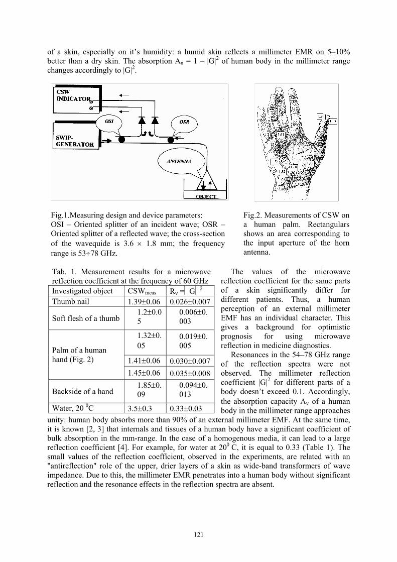
121
of a skin, especially on it’s humidity: a humid skin reflects a millimeter EMR on 5–10% better than a dry skin. The absorption An = 1 – |G|2 of human body in the millimeter range changes accordingly to |G|2.
Fig.1.Measuring design and device parameters: OSI – Oriented splitter of an incident wave; OSR – Oriented splitter of a reflected wave; the cross-section of the wavequide is 3.6 × 1.8 mm; the frequency range is 53÷78 GHz.
Fig.2. Measurements of CSW on a human palm. Rectangulars shows an area corresponding to the input aperture of the horn antenna.
The values of the microwave reflection coefficient for the same parts of a skin significantly differ for different patients. Thus, a human perception of an external millimeter EMF has an individual character. This gives a background for optimistic prognosis for using microwave reflection in medicine diagnostics. Resonances in the 54–78 GHz range of the reflection spectra were not observed. The millimeter reflection coefficient |G|2 for different parts of a body doesn’t exceed 0.1. Accordingly, the absorption capacity Aν of a human body in the millimeter range approaches
unity: human body absorbs more than 90% of an external millimeter EMF. At the same time, it is known [2, 3] that internals and tissues of a human body have a significant coefficient of bulk absorption in the mm-range. In the case of a homogenous media, it can lead to a large reflection coefficient [4]. For example, for water at 200 C, it is equal to 0.33 (Table 1). The small values of the reflection coefficient, observed in the experiments, are related with an "antireflection" role of the upper, drier layers of a skin as wide-band transformers of wave impedance. Due to this, the millimeter EMR penetrates into a human body without significant reflection and the resonance effects in the reflection spectra are absent.
Tab. 1. Measurement results for a microwave reflection coefficient at the frequency of 60 GHz Investigated object CSWmeas Rν =G2 Thumb nail 1.39±0.06 0.026±0.007
Soft flesh of a thumb 1.2±0.05
0.006±0.003
1.32±0.05
0.019±0.005
1.41±0.06 0.030±0.007Palm of a human hand (Fig. 2)
1.45±0.06 0.035±0.008
Backside of a hand 1.85±0.09
0.094±0.013
Water, 20 0C 3.5±0.3 0.33±0.03

122
References [1] G.V. Ponezha, S.G. Ponezha, A.I. Nizhelskaya: Physics of the Alive, 2001, 9, No 2, pp. 33 – 54. [2] V.I. Pjasetskij, O.I. Pysanko: In. Apparatnyj kompleks:Elektronika-KVCh i ego primenenie v medicine (Electronics of mm waves and its Application in Medicine) , Мoscow, pp. 10 – 18, 1991. [3] O.V. Betskij: In Apparatnyj kompleks: Elektronika-KVCh i ego primenenie v medicine (Electronics of mm waves and its Application in Medicine), Мoscow., pp. 4 – 10, 1991. [4] M. Born, E. Volf: Osnovy optiki (Fundamentals of Optics), Мoscow: Nauka, 1970.

123
OBSERVATION, PROPERTIES, AND MEDICAL APPLICATIONS OF NEGATIVE FLOWS OF MICROWAVE ELECTROMAGNETIC EMISSION
Grygoriy V. Ponezha
Scientific Research Center of Quantum Medicine “VIDHUK”, Volodymyrska 61-b, 01033, Kyiv, Ukraine, e-mail: [email protected]
Conception of negative flows (NF) of electromagnetic radiation (EMR) has been introduced by the well-known Soviet physicist B.I. Stepanov in optics. He has shown [1] that the regime of the so called negative excitation of an irradiated medium, resulting in transitions of material particles not to a higher level (as by usual excitation) but to a lower one and reducing the whole energy due to its capture, can be obtained with the help of NF of EMR. In practice for this purpose, it is sufficient to use a cooled source of a thermal emission that is equivalent to the irradiation of the investigating system by the NF. In a microwave range of 54 ÷ 78 GHz the NF EMR have been found [2] at the research of an intrinsic emission from physiological and biological objects. Registration devices respond to the NF EMR in unusual way, namely, the going out signal increases with decreasing the incident one. Unlike the ordinary (positive) flows (PF), the NF are limited in magnitude. The maximum value of its density can be obtained if an object under the investigation were surrounded by the source of radiation with temperature equal to absolute zero. Experimental investigations of NF of microwave radiation [3] in clinic show its high efficiency. Well expressed therapeutic effect (especially, when treating patients with inflammatory diseases accompanied by the painful syndrome) was observed when NF and PF of microwave radiation were applied together. The effect was seen even when only NF were applied without the using of PF. This was demonstrated [4] by the studying the influence of NF on dynamics of the painful syndrome for the cases of atherosclerotic discirculatory encephalopathy and neurodistrophic spinal column and joints injures (osteochondrosis, arthrosis). Positive results of the clinic investigations give evidence that the using of NF of millimetre radiation in medicine is perspective in the frame of studying the nature of pain and also for the creating of special methods for the eliminating of painful syndromes.
References [1] Stepanov B.I. Fundamentals of negative light flux spectroscopy. – Minsk: Izd. Bel.State University, 1961. – 124 p. (in Russian). [2] Ponezha G.V., Sit'ko S.P., Skripnik Yu.A., Yanenko A.F. Regular and reverse fluxes of microwave radiation from physical and biologocal objects. Physics of the Alive, v.6, N1. – 1998. – P.11–14. [3] Bundyuk L.S., Kuzmenko O.P., Ponezha G.V. Experimental and clinical approbation reverse fluxes of microwave electromagnetic radiation. Physics of the Alive, v.7, N2. – 1999. – P.11–18. [4] Kuzmenko V.M., Ponezha G.V. Negative (reverse) fluxes of mm-electromagnetic radiation used in the treatment of some neurologic diseases. Physics of the Alive, v. 10, N1. – 2002. – P.98 – 101.

124
THE IMPACT OF DIRECT CURRENT ON WALKER 256 CARCINOSARCOMA (W256) GROWTH
A. Zhukavets, E. Yunevich, E. Korobtsova
N.N. Alexandrov Research Institute of Oncology and Medical Radiology, Minsk, Belarus Introduction: Direct current (DC) delivered through implanted electrodes is used for local treatment of solid malignant neoplasms and metastases. The objective of the study is to evaluate the impact of current intensity and quantity of electricity on tumor growth in in vivo experiment. Materials and methods: Wistar female rats were employed. W256 was inoculated subcutaneously as 1106 tumor cell suspension. All the animals were allocated between three groups and a control one, 8-12 rats in each. The tumor volume was calculated by ellipsoid formula. There was a single electric current treatment of the tumor reaching the volume of 300-400 mm3. Two steel needle electrodes (anode and cathode) were directly inserted into the tumor, through which DC was delivered with current intensity of 1 mA, 3 mA, 10 mA for 60 min (the quantity of electricity being 3.6, 10.8 and 36 coulombs respectively). Tumor growth inhibition index was determined on the 7th day after the treatment, and pathologic changes in the tumor and surrounding tissues were estimated. Results: 3.6 coulombs DC did not affect the course of W256 growth. Tumor growth inhibition index made up 35% at 10.8 coulombs DC. The delivery of 36 coulombs to the tumor resulted in local tissue necrosis and cure of 3 of 8 animals. Twenty-four hours after the treatment, colliquative necrosis was found in the tumor fragment subjected to cathode treatment, and coagulation necrosis to anode exposure. Twenty-one days after the treatment, the excrescence of the friable fibroblastic tissue with peripheral angiomatosis and hemosiderosis was observed in the former tumor sites of the cured animals. Conclusion: DC causes dose-dependent growth inhibition of W256, even complete destruction of the neoplasm. The basic mechanism of DC direct antitumor effect is destruction of tumor tissues by the acid (HCl) on the anode and the alkali (NaOH) on the cathode which are produced by electrolytic processes.

125
ELECTRIC ACTIVITY OF PLANTS AND SEEDS INDUCED BY VISIBLE LIGHT AND MILLIMETER RANGE ELECTROMAGNETIC WAVES
Yu.Kh. Shogenov1, V.A. Vasil`ev2, Yu.M. Romanovsky2, and, A. S. Stepanyan2
1Russian Academy of Agricultural Sciences, Moscow, 117218 Russia 2Physical Faculty, Moscow State University, Moscow, 119992 Russia
For evaluating the reaction of plants and seeds to external visible light and longer electromagnetic waves stimuli, we used bioelectric potentials (BEP) generated by plant organisms that reflect their physiologic state. BEP of animals and plants can be reliably registered by applying surface electrodes. We studied BEP while irradiating a leaf and the upper part of a plant stem cultivated at normal illumination and temperature conditions with a low-intensity (below 1 mW/cm2) laser beam, or with light from a noncoherent source (halogen incandescent lamp) delivered through a light-fiber bundle, or with millimeter range electromagnetic radiation. In the irradiated area of 5 mm in diameter, a pulse is generated on the background of stationary BEP with amplitude of several millivolts. This nonstationary BEP propagates down the stem with a speed of around 0.3÷0.7 cm/s [1]. We designed a set of mathematical models enabling us to relate the processes of excitation of electroactive conducting bundles of cells in a plant stem. We measured the action spectrum of low intensity radiation generated by lasers, narrow-band noncoherent sources of visible light and mm-range waves in dependence on the wavelength (dependences of BEP on λ) in a wide range (from 380 nm to 9 mm, 35 wavelengths overall) and suggested an explanation of the presence of a set of maxima. The latter correspond to the absorption spectrum in the photosynthetic range and the absorption spectrum of water [1]. It was shown that the laser-induced BEP of the seeds can be used as an additional criterion for the diagnostics of major indices of the sowing qualities of seeds: germinating power, sprouting rate, and vitality [2]. As an alternative method of vital express-diagnostics of qualitative and, possibly, functional state of plant objects we suggested a method of visualization and image analysis of electro conducting biological objects (seeds and plants of different agricultural crops) in a luminous corona discharge of high-frequency electric field. Calculations and preliminary experiments show that the frequency range from 104 to106 Hz is most suitable for imaging the plants and seeds in the electric field of corona discharge. The lower limit of this range is more suitable for the objects with a large surface area. The amplitude of the applied alternative voltage should be from 5 to 10 kV. Analysis of obtained images shows that the source of luminescence is the corona discharge near the object surface. Classification of the methods of image formation in a high-frequency electric field of the corona discharge allowed optimizing the instrumentation. An experimental prototype of a generator of high-frequency discharge luminescence was designed for the diagnostics of seeds and plants quality. A technique of imaging the tissues, organs and whole structures of plants and seeds of different crops was suggested. In the majority of cases, plants and seeds have dielectric conductivity dependent in time and space all through the in-depth structure of the object. Fluctuations of the dielectric conductivity are due to changes in physico-chemical processes in cells, tissues and organs of the organism and reactions of the object to external stimuli. Pathological changes in the structure of a biological object form a luminous image different from the initial one. For instance, images of plant leaves obtained at intact and damaged states are considerably different.

126
We plan to conduct experiments with different seeds (damaged and intact) of agricultural crops in the electric field of the corona discharge. Such experiments are difficult because of the elliptic shape of these objects. References [1] Yu.Kh. Shogenov, E.A. Mironova, V.Yu. Moiseenkova, and Yu.M. Romanovsky: Effect of monochromatic electromagnetic irradiation in the wavelength range of 330-3390 nm on plant bioelectric activity, Russian Journal of Plant Physiology, Vol. 46, No. 5, 1999, pp. 697-703. [2] V.A. Vasil`ev, I.V. Garkusha, V.A. Petrov, Yu.M. Romanovsky, and Yu.Kh. Shogenov: Light-induced electrical activity of green plants, Biophysics, Vol. 48, No. 4, 2003, pp. 662-671. [3] A.F. Korolev, V.O. Morozov, Yu.M. Romanovsky, and A.V. Khakhalin: Membrane processes in generation of the action potential of hihger plants exposed to millimeter radiation, Physics of Vibrations, Vol. 9, No. 3, 2001, pp. 163-167.

127
INVESTIGATION OF MILLIMETER EMISSION INTERACTION WITH FLUIDS BY HOLOGRAPH INTERFERENCE
Sergiy G. Ponezha1,2, Grygoriy V. Ponezha3, Valeriy I. Grygoruk1
1Taras Shevchenko National Kyiv University 2Ukrainian State Research-Production Center of Standardization, Metrology and Sertification
(UkrCSM), Metrologichna 4, 03143, Kyiv, Ukraine 3Scientific Research Center of Quantum Medicine “VIDHUK”, Volodymyrska 61-b, 01033,
Kyiv, Ukraine, e-mail: [email protected]
The research of changing of matter physical properties under the influence of electromagnetic emission is of great importance due to the possibility of controlling of biological processes with the help of millimeter emission and especially because of high efficiency of microwave resonance therapy [1]. In this report, investigations of refractive indices variations based on the method of holograph interference at the wavelength λ=632.8 nm in a number of fluids (organic liquids, water, cell cultivations in nutritious medium) under their irradiation by an electromagnetic emission in a frequency range of 37.5 ÷ 78 GHz are presented. Fluid temperature was controlled during the process of measurement with an accuracy not less than 0.1 K by a copper-constantan thermo-couple having sensitivity of ∼ 40 μW/K. The magnitude and dynamics of a refractivity index variation under the irradiation in the frequency range of 37.5 ÷ 78 GHz depend on the irradiation power and physical parameters of objects (molecule dipole moment, viscosity, heat capacity, and heat conductivity of fluids, and their thermal refractivity index) and do not depend on the mutual orientations of polarization vectors of millimetre and optical fields in limits of experimental errors. The investigations have shown that mm-radiation with power up to 10 mW produces thermal variations in optical refractivity index in fluids of the order of 10–3. The variations of refractivity index in various fluids are different in magnitude but have the same sign – refraction index decreases under the irradiation. At large values of an absorption coefficient αmm of mm-emission (for instance, in water, where αmm ≈ 22 cm–1), the optical refractivity index variations exponentially depend on the depth of penetration and that is an essential feature of the process of heat transfer (mm-emission is absorbed by a thin layer of material just under a wave-guide, where the heating of a fluid may reach 5÷7 K). In liquids with symmetric molecules having no constant dipole moments (such as CCl4 and benzene), mm-emission is absorbed weakly (αmm < 0.003 cm–1) and the variation in the optical refractivity index is insignificant. For liquids with mean absorption coefficient: toluene (αmm≈ 0.22 cm-1), meta-xilol (αmm≈ 0.21 cm–1), ortho-xilol (αmm≈ 0.4 cm–1), ethanol, butanol, and aceton, comlicated interference fields were observed caused by a bulk absorption of mm-emission penetrating in medium on the depth of ∼ αmm
–1. References [1] P. Sit’ko, L.N. Mkrtchian. Introduction to quantum medicine. – Kiev: “Pattern”, 1994. – 128 p.

128
FREQUENCY-TEMPORAL DEPENDENCES OF YEAST CELLS RESPONSE TO NONTHERMAL ELECTROMAGNETIC RADIATION OF MILLIMETER
RANGE
Mykola Kosytskyy, Olena Nizhelska Scientific Research Center of Quantum Medicine “Vidhuk”, Volodymyrska 61-b, 01033
Kyiv, Ukraine, e-mail: [email protected]
Introduction Frequency-depended response of microorganisms to microwave radiation caused hot discussions in former times. Investigating identical biological objects in the same range of frequencies of external radiation, some groups of researchers succeeded to observe the frequency dependence in biological effects, but the others received only the random distribution of data. In our experiments, we have found that the frequency dependence of bio-objects has a good repeatability in a whole, but is connected with many factors. The conditions at which the yeast cells are maximally sensitive to microwave radiation have been defined. Our experiments confirmed that single impact of the radiation can be revealed in more than one hundred cells generations. Some decades earlier, the millimeter radiation diapason was related mainly with industry and did not affect the everyday life of people. Today, the character of the electromagnetic background is greatly changed and continues to become saturated with the high-frequency radiation. Among the sources of this radiation are mobile communication, computers, microwave technique and so on. Therefore, investigation of the properties of living cells interaction with microwave radiation represents not only a specialized interest but also is of practical importance in changed electromagnetic environment. In our opinion, the response of living cells to nonthermal microwave radiation can not be simple and always reproducible. The simple objects represented below may be used as biological detectors. Materials and methods Experiments were performed on the alcoholic yeasts of Saccharomyces cerevisiae of XII race, bakery Saccharomyces cerevisiae of 722 race, and brewing Saccharomyces carlsbergensis logos 5599. We used a suspension of yeast cells with the concentration of (1.5 - 2.0)⋅108 cells/cm3. The age of cell culture (time of growth in an optimal condition before irradiation) was 24-72 h. Before the start of irradiation, yeasts were cooled in a thermostat at T==40C - 60C during 1 h or 24 h. The suspension was irradiated in Petri cups having the diameter of 7 cm. After the treatment, all samples were transfered in test tubes, cooled down to T=+40C and kept in the thermostat. After interfusion, cells suspension (5 cm3 ) from every test tube was sown on sterile nutrient medium (100 cm3). Growing was produced in glass retorts with wadding-gauze corks in the thermostat at T=+300C without airing. After 20-24 h of the culture growth, the retorts were taken out, the suspension was mixed, and probes were taken away with the help of sterile pipettes. Then they were diluted with distilled water and investigated with a microscope in order to determine a concentration of cells. Power of radiation at the output of waveguide was about 3 mWt. Frequency of radiation was controlled. Instability of frequency during in the experiments was ± 2 MHz. Experimental results The frequency dependence in a range of 41.71—41.80 GHz was obtained after a few series of experiments with the cells of Saccharomyces cerevisiae. It was turned out that the most biologically active radiation is the radiation with the frequency of 41.747 GHz.

129
For the determination of the influence of the radiation duration on the yeast cells growth, the samples with suspension were exposed by 41.747 GHz using the same method. Optimal duration of influence was 8-10 minutes. After the determination of the most active frequency and the optimal duration of the radiation influence for the given culture, the direct observation of the rate of cells growth was performed. The samples with suspension after the irradiation were kept in a refrigerator at T=+40C during 13 hours. Then the samples were sown on fresh nutrient medium. The concentration of cells in the samples was examined after an interval of 2-3 hours. To remove restrictions on food at the experiments over a long period of time, it is necessary to resow the cells on fresh nutrient medium. With all this going on, the difference in growth rate of posterity of the exposed and control groups of cells was kept constant on the extent of at least 100 generations. It indicates on the fact that the genetic ways of information transfer are affected. Discussion In experiments, in which exposed and control cells were grown on a liquid nutrient medium, their concentration differed on average by 2.0. All cells were in this account: youth cells, just detaching oneself, old cells, and dead cells. We suppose that the result of influence is not simply the increase of cells number, but the increase of those cells which are able to enter into a new cycle of division and give posterity. Therefore, the frequency-dependent effects observed through an optical density on a spectrophotometre, do not exceed 10-15%. By the visual calculation, when it is possible to distinguish living and dead cells, the effect can reach 100%, and, finally, by sowing on agar, where colonies grow from viable cells only, it is possible to get a «resonance line» most pronouncely. Sowing the samples with suspension on a dense medium (agar) and growing during some days allows to receive colonies only from alive cells. Their part in population is 20-80%. At resonant irradiation (41.747 GHz) in combination with cooling, practically all living cells sown on agar give colonies. Cooling the suspension to the temperature much below the optimal one, when all cells are in the state of rest, is the necessary condition of the observation of the influence of millimeter radiation on growth of yeast cells. The time of cooling is far longer than the time of radiation influence. The millimeter radiation can not accelerate the rate of division of young, actively growing cells, which is limited by the speed of biosynthesis. Apparently, the cells with slow or disturbed biosynthesis reactions are more sensitive to microwave radiation. The influence of radiation at active frequency returns them to the normal rate of division. The reason of scattering of results is that the same frequency of irradiation can cause stimulation as well as decreasing of cells growth. The efficiency of influence of microwave radiation on growth of yeasts cells depends on the phase of a cell cycle. Resonance requires the accordance of two parts of the system: parameters of radiation and cells samples. In the initial stage of the experiment, one should not average the results or throw away untypical cases. The biological system behaves often ambiguously. It is necessary to give primary results taking into account all possible parameters. Single, but reliable experiments, also have a right to existence, as a fact that has taken a place. From the point of view of the theory of bifurcations, the result of weak irradiation can be different depending on the moment at which signal will translate the system in the other dynamical state. In summary, we can say that results substantially depend on the method of registration of the bioeffect and the state of biosystems.

130
CHANGE OF THE BIOPHYSICAL PROPERTIES OF CELLS OF PERIPHERAL BLOOD OF THE PC USERS UPON INFLUENCE OF EMF
E.N. Budyanskaya, O.N. Chernyshova, E.Ya. Nikolenko, M.M. Rezinkina, N.O. Philipenko,
L.V. Chepenko R&D Institute of Hygiene of Labour & Industrial Diseases, Kharkov, Ukraine
69 males working and 50 males not working at personal computers (PC) have been surveyed. The purpose of work is to reveal specificity of a functional condition of cells membranes of form elements of peripheral blood of the PC users and a degree of dependence of the revealed shifts on the labour loading at working at PC. Erythrocytes of blood were chosen as a model and the permeability of cell membranes has been taken as the basic integral index of their function. The indices of the systems of peroxidate oxidation of lipids (POL) and condition of the antioxidant protection (AOP), on which the structure of a lipid phase of cells membranes and a degree of their permeability appreciably depends were studied in parallel. The condition of the systems POL - AOP were judged by the intensity of chemiluminescence (CL) spontaneous (SCL) and induced by Fe2+ and also by CL at loading Н2О2. The measurements were carried out on a domestic chemiluminescence meter, allowing to measure intensity of a super-weak luminescence in the range 200-600 nm. The permeability of erythrocytes was investigated at step by step increase (6 tests) of the concentration of urea (univalent anions), which was brought to tests of blood with a physiological solution. The point of readout was a probe with 100 % hemolysis at addition only urea to the erythrocytes suspension. It seemed probable that such researches may allow presenting the model of development of deviations in the health of the PC users at action on an organism of the factors of physical nature of small intensity, the significant contribution among which is brought by the electromagnetic fields. It is established that erythrocytes of peripheral blood of the PC users have underwent the expressed shifts of functional condition shown as increase of their permeability at loading by the univalent anions. These deviations are registered at all spectrum of the investigated urea concentration and their expression appreciably depends on the duration and degree of the labour loading on an organism (number of business hours at PC per one month). The degree of expression of shifts depends on the urea concentration. It is almost always authentic, however it is shown mostly in the levels of permeability in the average part of the curve (2-4 points), where its increase in the users makes from 1,5 up to 4 times in comparison with the indices in the similar points in the persons who do not work at PC. The revealed shifts can testify the increase of the functional activity of the cells of peripheral blood of the users as well as intensification of the metabolic reactions of an organism as a whole. It is necessary to expect in such a situation, first of all, the increase of speed of free-radicals processes flowing, among them of peroxidate oxidation of the lipids of cell membranes and deviation of the stability of AOP factors of an organism to this or that side. As the carried out researches have shown, the index of peroxidate hemolysis of erythrocytes undergoes the expressed changes alongside with permeability, that may specify the shifts in a balance of POL - AOP systems to the side of weakening of the latter. The analysis of results of these data has allowed establishing also significant changes in the character of the course of flowing of the processes of peroxidate oxidation, the main of which is the intensity POL at loading of the system by the bivalent iron. Thus, the increase of a CL signal of blood whey in 1,5-2 times in various groups of the PC users was observed, testifying the accumulation of the most reactive forms amongst the POL products, i.e. hydroperoxide of lipids. The significant increase of the CL level (both flare and sum of luminescence) induced by the peroxide of hydrogen and indicating the weakening of the factors of antiradical protection, and also
131
accumulation of significant amount of sub-oxide products in blood was observed in parallel also. Obviously, these infringements have the direct relation to the very significant deviations of a degree of permeability and activity of the lysis of erythrocytes. It is important also to note, that the revealed shifts of the investigated parameters as a whole, were defined appreciably by the labour loading and duration of the labour experience of the PC users. In particular, if the labour loading of the users does not exceed 80 hours per one month, the negative character of described above shifts was registered at the small labour loading (less than 3 years). Whereas, if the experience was more than 3 years, the shifts were graded appreciably and did not posses the pathological character. At the same time, the labour loading exceeded 80 hours, non dependent on the working experience, has promoted transition of a degree of shifts to the area of pathological. Thus, the received results have allowed to establish features of modification of the biophysical properties of cells of peripheral blood of the PC users and to show one of the possible mechanisms of the development of these shifts. The obtained results allow to substantiate complex measures aimed on the recovery of an organism of the PC users.
132
THE METHODICAL APPROACHES TO STUDY OF INFLUENCE OF ELECTROMAGNETIC RADIATION ON BIOLOGICAL OBJECTS
T.Yu. Shchegoleva, E.N. Budjanscaja
Institute of radiophysics and electronics NASU, Kharkov,Ukraine GP “Kharkov Science Institute of work hygiene and professional daises, Kharkov, Ukraine
The influence of electromagnetic radiation (ER) on biological systems of different levels of organization is a theme of active discussion in modern literature. Physical and biological mechanisms of this influence are not clear now. The availability of these phenomena is put under doubt. Even the fact of influence ER is in discussion now. It is important to discover the allocation in biological objects the recipient of this radiation. But unequivocal conclusions are not received. Therefore represents the large interest the study of influence ER on systems of cells, which have properties of coherence. The methodical approaches with use EHF dielectrometry allow observing the work of cell regulation systems in real time, in conditions, excluding their destruction. The analysis of the cells answers by a method EHF-dielectrometry at realization of screening of bioregulators allows to study the systems of communication of macromolecular components of cells in conditions of their maximal viability. The researches, carried out by this method, of cells regulation systems eukaryote and procaryote have allowed to elaborate the uniform approach to supervision of universal systems of functioning of cells, such as adenylylcyclase system (АCS) and of unique for each type of cells - systems of cytosceleton. АCS is submitted in all cells of organisms and is one of systems realizing stress on cells level. Its structure is conservative and is well enough investigated. The stimulation of beta - receptors of cells results in inclusion of a complex processes, which represents ensemble cooperating among themselves macromolecules, carrying out transfer of signals on a way by G-proteins. The traditionally used test for function ACS on cAMP formation is integrated final parameter of functioning of very many molecular mechanisms. One of major mechanisms of ACS regulation is cytosceleton, which elements are connected with G - proteins, beta-receptors and adenylyl cyclase. In all cells, investigated by us, the close connection of ACS elements with system cytosceleton is revealed [1,2]. In modeling experiments, in real time the processes desensitization were observed [3]. Took place coherence of work of elements cytosceleton in alive cells and stochastic disorder of the data after destruction cytosceleton. Use of model ACS in a complex with cytosceleton has allowed to find out essential differences on cells level in regulation systems erythrocytes and platelets, on the basis of which it is possible to judge risk factors of occurrence cardio-pathologies [4] and adaptive opportunities organisms [5] in a population practically of healthy people. Now these methodical approaches allow using various cells: erythrocytes, platelets, sperms, microorganisms (E coli) and yeast as biosensor controls of a different type of radiations. The development the system of the tests for the analysis of influence ER on key sites of a cells and system a cytosceleton. References [1] Shchegoleva T.Yu.: The research of dielectric characteristics of bioobjects in millimeter-range of radio waves. The monograph. Kiev, Naukova Dumka, 1996, 187p. [2] Shchegoleva T.Yu., Kolesnikov V.G.: The adenylatecyclase system and calcium channels. Biophysics, 1995.-Vol.40, No.6, pp.1301-1304. [3] Shchegoleva T.Yu., Kolesnikov V.G.: Change in the hydrate environs of erythrocytes on hormonal stimulation. Biophysics, 1996.-Vol.41, No.5, pp.1093-1097.

133
[4] Shchegoleva T.Yu., Kolesnikov V.G., Vasileva E.V., Vasilev Yu. M., Altuchov A.L.: Using of millimeter-range waves in medicine. Kharkov, KIME -: 2000, 233 p. [5] Kovaleva O.N., Shchegoleva T.Yu., Sidorov D.Yu., Smirnova V.I., Kolesnikov V.G., Lepeeva E.A.: Molecular mechanisms of realization of stress for patients with an arterial hypertension. Vrachebnaya practica, 1999, 4, p. 7-10.13.

134
ELECTRO-MAGNETIC THERAPY INFLUENCE ON SURGICAL CUTANEOUS SLASHES RESOLUTION IN RANDOM RATS SUBMITTED TO NICOTINE
TREATMENT
C.M.B. Pinheiro, P.S.O. Reis, V.C. Rosário, R.R.B.T. Maciel, A. Oliveira, L.C. Simões, I. Pepe, P.H.P Aguiar
The electrotherapy through electromagnetism is a non-invasive technique and can be applied as coadjuvant in the resolution process of surgical wounds. For the treatment of surgical wounds, efforts should be made to ease problems which could interfere in the resolution process, accelerating healing in order to bring the individual back to his/her full functions as fast as possible, minimizing the period of inactivity and promoting a fast return to his/her daily activities. This project had as its objective to study the influence of electro-magnetic therapy on the resolution of surgical cutaneous slashes in rats submitted to the treatment with nicotine. Were studied possible histological alterations, healing period and hemogram evaluations, plaquetograma and dosages of serical markers in the animals after exposed to a continuous and pulsating electromagnetic field of 479 + 15 Gauss, feeding through an alternate current of 50 Hz of frequency, with sinusoidal waves and modulated impulses of 60 milliseconds and dead time of 450 msec, connected to the alternate current of 110 volts, adapted to individual cells. The application was realized during 21 days, once a day, in a total of 21 sessions, each session during 30 minutes. The animals from GII Control Group were submitted to the same procedure realized in the GI, however the apparatus was turned off while applied, therefore it was only a simulation of the application. The results obtained in the assays realized suggest a significant benefit in the healing process of surgical wounds, accelerating the healing process. This could be an alternative therapy to treat similar affections without adverse collateral effects. Its applicability still needs further studies with the objective of analysing its effect on tissues, its clinical efficiency and the mechanisms of action to be used in an efficacious and safe way in humans. Key words: electro-magnetic therapy, slashes, rats, nicotine

135
ELECTROMAGNETIC WAVES TREATMENT OF STROKE
Alexander Tamelo, V.Murav’ev, Galina Zobnina Belarussian State University of Informatics and Radioelectronics, 6. P. Brovki, 220027
Minsk,Belarus Tel.:375 17 398095, Fax: 375 17 310914, e-mail: [email protected]
New research results take advantage of complex spectrum for process stimulation of albumen syntez in mithachondries and it using in medicine[1]. 52 patients (27 women, 25 men) at the age of 48-72 years with chronic cerebral circulatory impairment within the basin of the carotid and vertebral arteries were treated. Before treating were used of 24 body’s points for real time diagnostic of human diseases. The treatment diseases are doing by help of complex spectrum use feedback from active body’s points for control of power lewel[2]. The cerebrovascular atherosclerosis was the major factor causing chronic cerebral circulatory impairment in 34 patients. 16 patients had a long history of hypertension. The patients were treated with the complex therapy including vasoactive, hypotensive and antisclerotic drugs and complex spectrum of electromagnetic wave. An average course of treatment was 8-10 days. The wave was exposed for 20-40 minutes depending on the individual adaptation response. The patients of the control group (20 men) were treated with the above medications and massage in the "collar" neck area. The complex spectrum of electromagnetic waves (EMW) was applied to the area of the shoulder joint contralateral to the site of lesion and to the sinocarotid area. During the course of treatment the neurological state and the cerebral blood flow circulation in the major head and neck vessels were examined by the US method. Some parameters of hemostasis and blood viscosity were studied. The individual response type was identified using the lymphocytic segmental/limfacitarno-segmentarn index (LSI). The neurological state was evaluated by calculating the main parameters. It was found that the patients treated with the non-heating millimeter wave had much better results (89.3%) than those of the control group (70%). Our experiments confirmed our hypothesis [1] about parametric influence of the EMW what able to improve biokinetic performance. References [1] Tamelo A.A.: Nanometrical approach into medicine and veterinary.15th International Conference Mittweida. Journal of the University of Applied Sciences. No 11, 2002, p.1-4. [2] Tamelo A.A.: Treatment method of diseases. Patent of Belarus 4381.A61N 1/61. 2001.

136
THE ESTIMATION OF ELECTROMAGNETIC FIELDS INFLUENCE TO FUNCTIONAL STATE OF HUMAN-OPERATOR
B. Kaduk, Ju. Melnik, A. Chunihin, A. Yanenko,
Scientific Research Center of Quantum Medicine “Vidhuk” of the Ministry of Public Health of Ukraine (Kiev)
It is theoretically and practically actual to allow external influences to human operator. Of prime importance is the allow in complex system control (guidance) area. The greater parts of various influences in modern industrial countries are precisely electromagnetic fields (EMF). The EMF influences to functional state of operator can be detected in biochemical, physiology and psychology levels [1]. It is necessary to note in this connection that there is important to search not only EMF influences in itself, but the extent of inducing interaction (interconnection) in all human functional subsystems as well. One such promising direction is the research of correlation between separate indexes of human state under EMF attacks. As this take place, the research only the correlation is not sufficient to complete estimating of human state. The authors suggest assessing the influences of EMF with not only collection of separate indexes, but generalized functional state index of operator. The generalized index ΔP is defined as the sum of products:
∑ Δ=Δ
iii pBP
where Bi is index of i-parameter influence, Δpi is a derivation of i-parameter under external influences.
Calculation of index Bi is made by help of sensitivity theory and probability theory. The main directions of the generalized functional state index searching are:
- unity of various indexes of functional subsystems; - ranking; - putting in the universal scale form.
In relation to linguistic form of given requirements to operator and wide variation of measurement scales it is suggested to use fuzzy set formalism to expresse generalized functional state index. References [1] Skrypnik Yu.A., Yanenko A.F., Manoylov V.F.: Microwave radiometry of physical and
biological objects. – Zhytomir: “Volyn'”, 2003. – 406 p. (in Russian).

137
SPECIES SPECIFICITY OF BACTERIAL RESPONCE TO LOW FREQUENCY MAGNETIC FIELDS OF LOW INTENSITY
D.I. Nikitin, L.E. Nikitin, V.I. Lobyshev*
Institute of Microbiology, Russian Academy of Siences, Moscow, 117811 Russia *M.V. Lomonosov Moscow State University, Moscow, 119992 Russia
The growth of suspended cultures of bacteria with different cell membrane lipid composition was studied after their expose to a magnetic field (about 0.1 mT, 0.02-2000 Hz) for 40 min. Three clusters of bacteria were used: a) Methylobacterium organophilum and Renobacter vacuolatum, whose cell membranes are rich in lecithin (to 60% of the total lipid), b) Arcocella aquatica and marine form Cyclobacterium marinum with high content of sphingolipids and ceramides, which are rare in microorganisms, c) Hyphomonas jannaschiana containing 95% of glycolipids and Caulobacter crescentus with 40% of glycolipids. The experiments revealed three new fundamental facts. The species specificity of the responses of the bacteria to the application of the magnetic field was observed, which included both inhibition and activation. The character of the changes in the kinetics of the bacterial culture growth correlates with the type of the membrane lipid complex. Pre-exposed distilled water to the magnetic field evokes a pronounced response in the cultures, which is sometimes much stronger than the responses of the same cultures exposed directly in the cultural medium. The effects obtained are frequency dependent. The culture of Caulobacter crescentus shows maximal inhibition at 2 Hz (78%) and 1000 Hz (71%), but at 20 Hz no reaction is registered. The inhibitory effect of pre-exposed water on the growth of microorganisms is also frequency dependent. Maximal inhibition in the experiments with pre-exposed water reach 67%. The frequencies which result in maximal inhibition in the experiment with pre-exposed water correspond to minimal inhibition in the experiment with the exposed culture and vice versa. All the results obtained are statistically significant with the probability coefficient P>0.95. The difference in the response on the exposition of culture and water to magnetic field shows that there are at least two mechanisms of the effects on low frequency magnetic fields. One of them should be explained by changes of aqueous solution properties under the exposure to magnetic field which are essential for the development of microorganisms. The work is supported by Russian Foundation for Basic Research, grant 03-04-49160

138
COLLECTIVE EXCITATIONS IN MEMBRANE- AND MEMBRANE-BIOMOLECULAR CHAIN-ASSOCIATED SPECIFIC CELLULAR PROCESSES
E.A. Preoteasa*, M.V. Apostol, D. Gurban
Horia Hulubei National Institute for Physics and Nuclear Engineering, RO-077125, Bucharest-Magurele, Romania (*e-mail: [email protected])
In the approach of life, molecular biology and its new-born branches such as genomics, proteomics and lipidomics recorded vast achievements by studying more or less separately the structure of a large number of components of biological systems. However, the activity of these components in the living cell is highly cooperative and does not follow in a simple way from structure. Therefore, as molecular biology did not induce or promote by itself the onset of a theory regarding the dynamics of living systems – or at least of its main physical hypotheses – complementary attempts from a more holistic perspective appeared necessary to account for the integrative features of life. The concept of resonant and cooperative Fröhlich polarization excitations proceeds along this line and is able to describe rewardingly a large variety of living cell processes. Thus coherent oscillations occur in a dipole set with macroscopic occupation of specific modes provided that metabolic energy is supplied at a rate above a certain threshold, and they can be associated both with biopolymers and with membranes [1]. Later Fröhlich suggested also another type of collective excitation, by which large electric fields arising on metabolic energy release induce biomolecules (e.g. globular proteins) into highly excited metastable states with much larger dipole moments than near the fundamental state [2]. Such models succeeded in understanding some overall biological attributes – self-preservation by metabolism (or far-from-thermal-equilibrium relative stability), a characteristic type of order, cell division, contact inhibition – and opened the way for specific applications. These focused somewhat more on quasi-unidimensional structures like microtubules and microfilaments as well as on their interactions with cytoplasmic globular proteins [3a] than on (intra)cellular biomembrane systems [3b], although the latter were experimentally confirmed [4]. Subsequent emphasis on the microscopic level showed that the symmetry breaking of the electromagnetic field associated with dipole density, the corresponding Goldstone mode and the coherent excitations may appear most likely in spatial regions contiguous to inhomogeneities with electrical activity in otherwise quasi-homogenous subcellular structures [5]. These may include ends or surfaces of biomolecular chains, and membrane surfaces, folds, trans-membrane proteins with bioenergetic function, or the loci where they aggregate spontaneously or due to external factors. Moreover, in regions around inhomogeneities collective modes of water molecules with strong field gradients at the borders may emerge [6], leading to long-range resonant forces. We recently suggested that in the superconducting- or Bose-Einstein condensation in such macroscopic quantum phases, the dipolar charge pair oriented by an electric center may act as an analogue to the Cooper pair or to the superfluid pair of excitations [7]. Also, we assumed biomembrane-associated polarization modes similar to circular elastic membrane oscillations and examined their relevance for processes involving sequential aggregation of integral membrane proteins. The above collective phenomena are dissipative and they create order while they release all the incoming energy. They have been invoked so far to understand processes involving separately chain-like biopolymers and membranes in their interactions with cell water and cytoplasmic globular proteins. Less attention has been paid, however, to systems where biomolecular chains come together with cell organelles formed by multiple folds of membranes. Here we will assume the statement of Del Giudice [8] that as far as quasi-unidimensional structures are concerned, another non-linear, collective, but energy-

139
conservative excitation – the Davydov soliton – plays also a role [9]. Implicitly, the role of the soliton concerted to that of the coherent oscillation modes, together with the coupling with radiation of local electric activity centres, is claimed essential in the feed-back control of the membrane- and combined membrane-biopolymer processes. In such complex systems, the ensemble of the above collective mechanisms should be necessary to explain features such as localization, selectivity, sequential functionality and self-regulatory capacity. In the following, several specific membrane- and membrane-biomolecular chain-associated subcellular systems are discussed from this perspective. Indeed a number of processes at the level of membranous organelles strikingly involve the aggregation of many different biomacromolecules (globular membrane integral or cytoplasmic proteins) or of their supermolecular associations (ribosomes, messenger RNA, polyribosomes, microtubules and microfilaments) in well-defined space-temporal order [10]. Specific and sequential interactions of up to ~100 or more biopolymer units around a membrane center hardly could be the result of merely Brownian diffusion and short-range recognition, but may be expected to result by a selective arrangement at the right places and times due to long-range resonant forces. Examples may include the supermolecular morphogenesis of nuclear envelope (NE) pores, of cellular gap junctions, of mitochondria (M) multienzyme complexes, and of chloroplasts (C) and chromatophores light reaction centers; the multi-stage “signal-sequence” polypeptide translocation from ribosomes through the rough endoplasmic reticulum (ER); and the apoptosis cascade taking place in NE, M, C and ER membranes. Such systems function by metabolic energy conversion, and ATPases and similar enzymes, receptor and translocator proteins of their membranes may represent electric activity centers, leading to the spontaneous self-organization of macromolecules in quasi-regular patterns. All of them exhibit central symmetric arrangements up to ~100 nm diameter of integral proteins and ribosomes on membranes, possibly driven by polarization modes similar to circular membrane oscillations. The redistribution of integral proteins in a hexagonal (50 nm in diameter) network-like array about in the core of the energized outer mitochondrial membrane [11] is a more simple case that might result from the equilibrium between attractive and repulsive forces. The ER polypeptide translocation from ribosomes as described by the signal peptide hypothesis (“correct in the outline but requiring additional components” [10]) is more complex. Here, a polyribosome (7-20 ribosomes bound to a mRNA macromolecule) forms a random coil when free in cytoplasm and displays a C7 to C9 or spiral symmetry when attached to the membrane. Thus one may suggest that a soliton in the mRNA chain triggered by the binding of the 5’ mRNA end to a first ER receptor protein bends the mRNA, due to coupling between the vibrational displacement of monomers and the chain deformation. Alternatively, the receptor proteins may be disposed under the action of a circularly polarized mode of the electric field, or both. Solitons may be involved in apoptosis as well. Here a number of proteins (Bcl-2 and Bax subfamilies) are membrane-localized, while the Bim and Bmf proteins are associated with the filamentous intracellular network. With the synchronous oscillations of water in microtubules as a cell clock [12], it may happen that cytoskeleton-bound proteins initiate the apoptosis following a soliton signal, and subsequently long-range forces activate specifically membrane proteins which complete the programmed cell death. Note finally that all considered cell organelles show parallel double- or multimembrane structures, which suggest that long-range resonant forces may stabilize them against electrostatic repulsions between similarly charged faces. Moreover, the above phenomena do not occur in the single plasma cell membrane, which is different in structure (larger thickness and lower protein/lipid ratio) and therefore in electrical properties. Thus although the above suggestions are predominantly speculative, our heuristic approach showed that different mechanisms of collective excitations evidence a still unexploited

140
potential for significant insight of the membrane- and membrane-biopolymer chain-associated cellular processes. References [1] Fröhlich H, Int. J. Quant. Chem. 2 (1968) 641. [2] Fröhlich H, J. Collect. Phenom. 1 (1973) 101. [3] (a) Clegg JS, p. 162, and (b) Pohl HA, p. 199, in: Coherent Excitations in Biological Systems (Fröhlich H, Kremer F, eds), Springer-Verlag, Berlin-Heidelberg (1983). [4] Rowlands S, Sewchand LS, Lovlin RE, Beck JS, Enns EG, Phys. Lett. A82 (1981) 436. [5] Del Giudice E, Doglia S, Milani M, Vitiello G, Nucl. Phys. B275 (1986) 185. [6] Del Giudice E, Preparata G, Vitiello G, Phys. Rev. Lett. 61 (1988) 1085. [7] Apostol M, Preoteasa EA; Preoteasa EA, Apostol M, EPS-13 Meet., Jul 2005, Bern (submit.). [8] Del Giudice E, Doglia S, Milani M, Phys. Scripta 26 (1982) 232. [9] Davydov AS, Sov. Phys. JETF 51 (1980) 397. [10] Alberts B, et al, Molecular Biology of the Cell, Garland Publ. Inc., New York (1994). [11] Al Hackenbrock CR, Ann. NY Acad. Sci. 195 (1972) 492. [12] Mandelkow EM, Mandelkow E, Cell. Motil. Cytoskeleton 22 (1992) 235.

141
WATER IS A SENSOR TO WEAK FORCES INCLUDING ELECTROMAGNETIC FIELDS OF LOW INTENSITY
V.I. Lobyshev
M.V. Lomonosov Moscow State University, Moscow, Russia A lot of works show the presence of non-thermal effects of weak external forces on alive organisms and allocated biological systems including electromagnetic and acoustic fields, direct current, vibration and flow through thin capillars and porous membranes. In the case of electromagnetic fields these effects spread from static and quasi-static fields to optical frequencies. The results yet published show that a large part of the effects of weak forces on biological systems may be explained by the influence of these factors on the properties of water itself. This conclusion is convincingly shown in the experiments with water previously exposed to an external field, keeping the acquired biological activity during enough long time. Besides the specific biological activity is observed in water after phase transitions, small variations of stable isotopes of hydrogen relative to its natural content, low and super low doses of different substances of organic and inorganic nature. It is true even for the so-called “imaginary concentrations” when formally calculated amount of dissolved molecules is less than the whole number. The common explanations for the above biological effects are reduced to changes of aqueous structure. To the present time the significant number of experimental works has appeared, in which the changes of physical-chemical properties of water and aqueous solutions under the action of weak fields of electromagnetic nature and at addition of low and super low concentration of organic and inorganic substances are registered. First of all, it is necessary to attribute the studies of the optical characteristics in UV, visible and IR ranges, electrical and thermodynamic characteristics. It is necessary to emphasize, that it is erroneous to consider water as pure substance consisting of H2O molecules only. Even at laboratory conditions it contains the dissolved gases and elements of substances of the vessel, in which it is stored. Water contains also stable isotopes - deuterium and two isotopes of oxygen, which contents changes at phase transitions and interaction of water with other substances. As water is easily dissociated, 9 isotope versions of molecules are submitted. Average total amount of isotopes in water is about 0,3 %. It would seem, such amount of isotopes can be neglected, however in a number of papers it is shown, that the small variations of natural isotope content can result in large nonlinear biological effects. The physics properties of water molecules can differ with relative spin orientation in atoms of hydrogen. The ratio of orto- and para-water can change under the action of external fields and during some chemical reactions. Water is characterized also by rather labile value of ox-red potential caused by occurrence of active forms of oxygen – radicals, peroxides and superoxides under external factors. Dissociation of water inevitably results in formation of hydroxonium and hydroxyl ions and, accordingly, ionic defects in aqueous structure. The above mentioned testifies that water should be considered as complex heterogeneous system with a set of variable parameters. The next aspect which should be taken into account and analyzed is – water may form large non-equilibrium parametric structures including fractal structures consisting of thousands of molecules. One of the main differences between crystallographic (as ice) and paratetric structures is as follows. Parametric structures demand some small energy for their formation, but they became more stable because the distortion of this high symmetrical structure leads to large changes in total energy and it is more profitable to “cure” the structure opposite to break it. The computer modeling of such kind of structures shows enormous amount of possible structures formed by water molecules including rods, spirals, web net, flat

142
and channel structures which are well complementary to various biological molecular and supramolecular structures. By the method of luminescent spectroscopy long evolution of spectra is found out within several days after preparation of a solution or field exposure. The duration of evolution considerably exceeds the characteristic “molecular” times, and the character of transient dynamics can be both monotonous, or oscillatory. The reaction of water to external fields essentially depends on a condition of solution and is maximal, when the system is far from equilibrium. The received results induce to consider water and aqueous solutions as non-equilibrium systems with dynamic chaos capable to self-organizing. It is shown experimentally that the induced change of properties of water results in changes of thermodynamic properties of biological membranes, that results in change of a cell homeostasis. Thus, water should be considered as a structural - dynamic sensor and actuator of hydrophobic-hydrophylic balance in biological systems. The work is supported by Russian Foundation for Basic Research, grant 03-04-49160

143
BIOLOGICAL EFFECTS OF ELECTROMAGNETIC INFORMATION IMPRINTED INTO WATER
Romana Ružič, Metod Škarja, Igor Jerman
BION, Institute for Bioelectromagnetics and New Biology; Stegne 21, 1000 Ljubljana, Slovenia
Fax +386 1 513 11 47, e-mail: [email protected], [email protected], [email protected],
In this report, we present our testing a hypothesis that biologically relevant information from various substances can be non-chemically transmitted to organisms by a combination of high voltage electric and magnetic fields that imprint information in a stable way into water or a water solution. In contact with organisms and under suitable conditions the stored imprinted information triggers a specific biological response without any chemical contact with the original substance. We constructed a special device whereby various experiments - using our well-explored biological sensor system - were performed. The biological effectiveness of the information transfer of electric and magnetic fields as well as three different biologically active substances were tested. We also tested the dependence of the biological effects on different types of recipient solution. Introduction Biological effects of magnetically treated water has been known for long decades [1,2] but the existence of the so called water memory is still an object of an intensive scientific debate because there is still no generally accepted theory that would explain, how biologically effective information can be imprinted and stored in water. One hypothesis predicts changes in water microstructures by forming more or less permanent clusters [2, 3], the others point to the unusual properties of water under certain treatments; in this case thermodynamics methods were used [4]. Some physicists proposed that one way, in which ‘memory’ might be stored in water, is through the collective motions of water dipoles, which have greater polarisability than the solitary ones. External sources with low-frequency polarisation field (for example other molecules) can be imprinted into water by modulating the fundamental frequency of the latter [5]. Beside physics that tried to explain this phenomenon, various biological effects of such "informed" water were published as well as experiments with regard to the biophysical properties of the information transfer process [6-8]. The present work was based on two hypotheses: a) that by means of a high voltage electric field chemical information can be imprinted into water and b) that imprinted water should have measurable biological effects. We were also interested in how long the information stored into water produced biological effects. Materials and Methods The plant sensor system [9] consisted of Lepidium sativum (cress seeds) that germinated in Petri dishes (400 seeds for one experiment, i.e. exposed and control group). After 24 hours, the plants were exposed to a controlled heat stress at 42°C (to enhance the susceptibility of plant organism to the environmental factors). Each experiment lasted 48 hours. After that, the length of radicles was measured, average mean and statistical tests performed. The effects of information transfer from the following (donor) compounds were tested: Amanita dust, herbicide Glyphosphate and cytokinin Benzylaminopurine (BAP). As recipient solution pure water, the Fe - Cu ion solution, 60% ethanol, 40% ethanol and 30% ethanol + trace minerals were tested. These informed solutions were further twice diluted with bidistilled water in the ratio 1:100, agitated (succused) and used for moistening the seeds by 3

144
75
80
85
90
95
100
information"water"
information"amanita
dust"
information"herbicide"
information"BAP"
% o
f con
trol
***
***
***
***
Fig. 1 The effect of informed vs. non- informed solution on the growth reaction of plant sensor system (seeds with high germinability) – recipient solution 60% ethanol.
Fig. 2 The effect of informed vs. non- informed solution on the growth reaction of plant sensor system – recipient solution 40% ethanol.
92
94
96
98
100
102
104
106
0 days 7 days 14 days
% o
f con
trol
information "w ater"information "herbicide"
information "BAP"
**
**
**
ml per Petri dish. The protection gloves were used during the whole procedure. During the germination the Petri dishes were covered with an aluminium foil; germination thus took place in dark. In some experiments we also tested how long from the imprinting the informed solution triggered biological effects. Results Using pure water instead of ethanol solution for imprinting did not yield detectable effects. When using water mixture of added Fe and Cu ions, stimulative 8-12% statistically significant (0.05 > p > 0.001) effects were obtained at concentrations 3.7 and 37 μM Fe plus 0.012 μM Cu (same for both). The stimulative effects decreased with the time elapsed since the imprinting. Experiments using 60% ethanol (diluted solution) showed inhibitory effects. Since the system is exposed to the heat stress this means that the exposure of the plant sensor system to electric field treated solution enhanced the stress effect of heat. When as a source of information pure (distilled) water was used, the average inhibition amounted to approximately 8%, was highly stable and statistically significant. Comparison with the effects of other compounds as a donor of information showed, that in most cases, the informed solution inhibited the growth reaction of plant sensor system at a similar level; these effect was highly significant (see Fig.1). It is also important to note that these results were obtained with the seeds of high germinability. Experiments using 40% ethanol (diluted solution) showed inhibitory effects for herbicide and stimulatory for BAP, after 7 days the same informed solutions produced inhibitory biological effect with either herbicide or BAP. After 14 days from the imprinting there were no effects left (Fig.2). Experiments using 30% ethanol and trace minerals produced varied biological effects depending on the age of the informed solution.

145
96
97
98
99
100
101
102
103
104
0 days 7 days 14 days 21 days
% o
f con
trol
information "water"information "herbicide"information "BAP"
Fig. 3 The effect of informed vs. non-informed solution on the growth reaction of plant sensor system – recipient solution 30% ethanol, dust minerals.
References [1] Singh P, Roy BK, Rai S (1996): Morphological and cytogenetic effect of 50 Hz EM field-induced nutrient solution on Vicia faba L. Electr Magnetobiol. 15(2), 109-118. [2] Fesenko EE, Gluvstein Ay (1995): Changes in the state of water, induced by radiofrequency electromagnetic fields. FEBS Lett 367, 53-55. [3] Rai S, Singh UP, Singh AK (1995): X-ray determination of magnetically treated liquid water structures. Electr Magnetobiol 14(1), 23-30. [4] Elia V, Nicolli M (1999): Thermodynamics of extremelly dilluted aqueous solutions. Ann.NY Acad Sci 879, 241-248. [5] Del Giudice E (1994): Is the “memory of water” a physical impossibility. In:.Endler PC, Schulte J (eds) Ultra High Dilution, Kluwer Academic Publishers, Netherlands, p. 117-9. [6] Endler PC, Pongratz W, Kastberger G, Wiegant FAC, Schulte J (1994): The effect of highly dilluted agitated thyroxine of the climbing activity of frogs. Vet Hum Toxicol 36, 56-59. [7] Endler PC, Pongratz W, Van Wijk R, Kastberger G, Haidvogl M (1991): Effects of highly diluted sucussed thyroxine on metamorphosis of highland frogs. Berlin J Res Hom 1, 151-160. [8] Aissa J, Litime MH, Attis E, Benveniste J (1993) Molecular signalling at high dilution or by means of electronic circuitry. J. Immunol. 150, A146. [9] Ružič R, Jerman I (2002): Weak magnetic field decreases heat stress in cress seedlings. Electromagnetic Biology and Medicine 21(1), 43-53.

146
ELECTROMAGNETIC INFORMATION IMPRINTED INTO MEDIUM ACT AS ENVIRONMENTAL SIGNAL FOR BACTERIA Escherichia coli
Rok Krašovec, Igor Jerman, Metod Škarja
BION, Institute for Bioelectromagnetics and New Biology; Stegne 21, 1000 Ljubljana, Slovenia.
Fax +386 1 513 11 47, e-mail: [email protected], [email protected], [email protected],
With a combination of a high voltage electric and magnetic fields, biologically relevant information from various donor substances can be non-chemically transferred and imprinted into water. By using such imprinted water it is possible to set up an imprinted agar medium. In a highly statistical significant way our experiments demonstrate that bacteria Escherichia coli respond to imprinted information of MgSO4 or to a surplus of MgSO4 in a molecular form in the same manner. The procedure of imprinting itself is not the reason for the observed phenomenon, namely, bacterial cells do not respond to imprinted information of distilled water or NH4SO4. We conclude with a proposal that information imprinted in the medium acts as a signal that informs bacterial cells about conditions in the environment and by inducing specific cell’s processes – such as adaptive mutation – relevant information can fundamentally affect the cell’s activity.
Introduction The water memory has no generally accepted hypothesis, though it seems that it is based on the quantum field theory [1, 2]. The theory shows how the collective vibrations of molecules in the coherent domain eventually become phase-locked to the fluctuations of the global electromagnetic field. In this way, long-lasting, stable oscillations could be maintained in water or other materials. One way in which the information imprint can be transferred to water is by a process of stepwise aqueous dilution and agitation (as in homeopathy) or by means of an electronic circuitry [3, 4, 5]. We tested the hypothesis that biologically relevant information from various substances can be non-chemically transmitted to water by a combination of high voltage electric and magnetic fields and that this imprint can have biological effect similar to the original substance. Material and methods The hypothesis was tested with bacteria Escherichia coli K-12 strain SJ134 and the process we monitored was the daily distribution of adaptive mutants. We used a fluctuation assay [6] which always begins with a number of independent bacterial cultures, each one inoculated with 2000-3000 genetically identical (non-mutant) SJ134 cells. They are then allowed to grow on a glycerol usually for 24 hours to approximately 107 cells. The entire culture is then plated on the agar minimal medium with a disaccharide lactulose as the only source of carbon and energy and plates are incubated at 30°C for at least six days. We treated a medium without any supplements as the control environment. In order to test the hypotheses we enriched the control environment either with a defined concentration of a chosen substance (for example we added 4.6 mM MgSO4) or we added to the medium the electric field imprint of a chosen substance. By counting an everyday increase in the number of mutant colonies we determined the distribution of mutants for every parallel plate in the specific environment. Then we calculated the frequency of adaptive mutations from all parallel plates of the same environment after 1st, 2nd and the 3rd day of incubation. We combined frequencies of adaptive mutations of the same environment and after the same day from at least three independent experiments and test them with the student T-test against frequencies of adaptive mutations in the control environment.

147
Results First, a thorough chemical analysis of imprinted water demonstrated no contamination of the receiver of information (distilled water) with the donor of information, which was an aqueous solution of MgSO4. Next, after first 24 hours of incubation the electric field imprint of MgSO4 and MgSO4 in a molecular form (final concentration was 5 mM) induced the same statistically significant (p<0.01) decrease in the number of adaptive mutants relative to the control environment (see graphs bellow). After the second day the number of adaptive mutants stayed accordant with the number in the control environment, the decreasing effect was again seen after the third day of the incubation. The electric field imprint of distilled water and NH4SO4 or NH4SO4 in a molecular form (final concentration was 5 mM) did not have statistically significant impact on the distribution of adaptive mutants in all three days of examination. Neither did the imprint of MgCl2 statistically significantly change a number of adaptive mutants although some decrease was evident.
Figure 1 Figure 2
Figure 3
Figs. 1-3. Daily appearance of new mutant- colonies in the strain SJ134. Vertical bars represent mean values (average frequencies) of newly appearing mutant colonies at each interval, error bars indicate the standard errors of those values, the significance is marked by * (p<0.05). The mean value was calculated: for the control environment (CE) out of 14 independent experiments (i.e.); (Fig. 1) for CE plus 4.6 mM of MgSO4 out of 5 i.e.; (Fig. 2) for CE plus the electric field imprint of MgSO4 out of 5 i.e.; (Fig. 3) for CE plus the electric field imprint of dH2O (distilled water) out of 3 i.e.
*
*0
10
20
30
40
Day 1 Day 2 Day 3New
mut
ant c
olon
ies p
er p
late
CECE plus electric field imprint of MgSO4
*
0
10
20
30
40
Day 1 Day 2 Day 3
New
mut
ant c
olon
ies p
er p
late
CE CE plus 4.6 mM of MgSO4
*
0
10
20
30
40
Day 1 Day 2 Day 3
CECE plus electric field imprint of dH2O
New
mut
ant c
olon
ies p
er p
late

148
Discussion Water can preserve information of the donor substance without any chemical contact between the latter and imprinted water. The imprinted information of the donor substance can induce a specific biological response in the bacterial cell similar to that caused by the donor substance itself. The fact that information is not molecular in its nature indicates that in the adaptive mutation process Mg2+ has a mediatory role as a signal [7]. It is much too early to say which cells component(s) sense the electric field imprint of Mg2+ but it is tempting to believe that cells communication network is involved. Namely, the adaptive mutation process in the strain SJ134 needs a functional bacterial two-component regulatory system PhoPQ (6) where the primary signal for the sensory protein PhoQ is an extracellular Mg2+. References [1] Giudice E. 1994. Is the “memory of water” a physical impossibility. In: Endler P.C., Schulte J. (eds.) Ultra High Dilution , Kluwer Academic Publishers, Netherlands , p. 117-119 [2] Vitiello G. 2001. My double unveiled. John Benjamin Publishing Company, Amsterdam , Philadelphia. [3] Aissa J., Litime M.H., Attis E., Benveniste J. 1993. Molecular signalling at high dilution or by means of electronic circuitry. J. Immunol., 150: A146 [4] Endler P.C., Pongratz W., Van Wijk R., Kastberger G., Haidvogl M. 1991. Effects of highly diluted sucussed thyroxine on metamorphosis of highland frogs. Berlin J. Res. Hom., 1: 151-160 [5] Endler P.C., Pongratz W., Kastberger G., Wiegant F.A.C., Schulte J. 1994. The effect of highly dilluted agitated thyroxine of the climbing activity of frogs. Vet. Hum. Toxicol., 36: 56-59 [6] Hall B.G. 1998. Adaptive Mutagenesis at ebgR is regulated by PhoPQ. Journal of Bacteriology, 180: 2862-2865 [7] Krašovec R., Škarja M., Jerman I. 2005. Environmental signals modulate frequency of adaptive mutations in Escherichia coli K-12. Submitted to Journal of Bacteriology.

149
COHERENCE AND CHANGES IN CELL PROLIFERATION CAUSED BY ELECTROMAGNETIC FIELDS
S. Kwee
Department of Medical Biochemistry, University of Aarhus, Build. 170, DK-8000 Aarhus C Denmark, [email protected]
A great number of studies have reported the effects of electromagnetic fields on cellular processes e.g. cell proliferation, transcription, enzyme activities, calcium transport. We have shown that there was a significant increase in cell growth in human epithelial amnion (AMA) cells, when exposed to an ELF electromagnetic field [1] or to microwave/RF radiation [2]. The change in proliferation rate was dependent on exposure times and field densities. A maximum increase in cell proliferation was only found at a specific field strength or absorption rate and exposure time. In other words there are "window" and adaptation effects. One explanation of these EMF/RF effects is that in order for a cell to respond to a constant exogenous electromagnetic field, the field must be coherent for a certain lenght of time. So if coherence of the EM field can be interupted, it should not be able to exert its effect on cellular processes. One way to do this is to superimpose an incoherent electromagnetic field on the coherent EMF. Such an incoherent EMF could be electromagnetic noise. It has been shown that an ELF noise field can block the biological effects, caused by EMFs, when superimposed on the latter [3-4]. In our experiments AMA cells were exposed to a sinusoidal 50 Hz, 50 μT electromagnetic field, which resulted in a significant increase in cell proliferation. Varying levels of ELF noise fields were then superimposed on the above mentioned field. No inhibition of the coherent magnetic field effects were seen at low noise field intensities. Only when the noise field reached a level of 70 - 80% of the coherent EMF, a significant inhibition of the increased cell proliferation was registered. The inhibition persisted up to a noise level equal to the coherent electromagnetic field. No significant change in proliferation rate was seen when the cell culture was exposed to a noise field alone [5]. The same inhibition by ELF noise was observed when the cells were exposed to the type of microwave radiation emitted by mobile telephones [6]. This effect has already found a pratical application as a radioprotective device in mobile telephones. References [1] S. Kwee and P. Raskmark, Bioelectrochem. Bioenerg. 36 (1995) 109. [2] S. Kwee and P. Raskmark, Bioelectrochem.Bioenerg. 44 (1998) 251. [3] T. A. Litovitz, C.J. Montrose, P. Doinov, K. M . Brown and M . Barber, Bioelectromagnetics 15 ( 1994) 105. [4] H. Lin and R. Goodman, Bioelectrochem.Bioenerg. 36 (1995) 33. [5] P. Raskmark and S. Kwee, Bioelectrochem. Bioenerg. 40 (1996) 193. [6] T. A. Litovitz, L. M. Penafiel, J. M. Farrel, D. Krause, R. Meister and J. M. Mullins, Bioelectromagnetics 18 (1997) 422.

150
PHYSICO-CHEMICAL MECHANISM IN THE ACTION OF WEAK EXTREMELY LOW FREQUENCY EMF AND SOLAR ACTIVITY ON LIVE ORGANISMS
Sergei I. Aksyonov
M.V. Lomonosov Moscow State University, Moscow 119899, Russia
After Chizhevsky works an ample material was obtained on the influence of solar activity and magnetic bursts on the organism condition and a set of other phenomena, right up to social processes. According to Chizhevsky, the analysis of data of historically important events during XV-XIX centuries (revolutions, various mass movements and other phenomena) reveals the definite correlation between the times of the appearance of these events and the times of maximums in the solar activity. The results for 500 years of observations showed that 60% of aforementioned events were recorded during 3 year periods near maximums of solar activity within 11 year solar cycles while during 3 years of quiet sun only 5% of such events was recorded. The correlation between similar phenomena was marked during XX century too. However, in spite of abundance of data on the action of weak extremely low frequency EMF and solar activity on biological and other processes, this problem remains discussed. This was conditioned by the absence of data on mechanisms which would explain the sensitivity of biological processes to external fields with strengths on 6-7 orders of magnitudes lower this value on the membranes and with energy on many orders of magnitudes lower than kT. A set of other paradox phenomena for biological effects of magnetic bursts and ELF EMF was marked too All these data allowed to conclude that “any biological effect of weak low frequency fields on the cell level must be out of the frame of ordinary physics” . The present paper suggests a physico-chemical mechanism for the action of weak extremely low frequency EMF on cellular processes which is discussed and experimentally verified further. This mechanism allow us to explain the main features of ELF EMF action on organisms, including sensitivity to geomagnetic fluctuations and the diminishing of effects with increasing field strength. A large effectiveness of weak EMF is due to nonlinear processes occurring in heterogeneous medium of the cell when ions, experiencing EMF induced by external electric field, pass over the oscillation period a part of distance between cellular membranes Very weak action in spatially separate points induced by external EMF within the whole volume of cell, can be increased during a set periods by nonlinear processes and ions are concentrated in a narrow layer near the membrane. As a result, nonlinear effects of ion movement affect the ionic strength and pH near the membrane, causing the transitions of the weakly bound peripheral proteins to the water phase. This process is associated with elevation of the number of freedom degrees for protein molecular group movements and accompanied with a slight change in free energy. When the fields of higher strength are applied, ions affected by the voltage, induced by an external EMF, pass all distance between membranes and more. Then the induced current would be confined to the membranes and would only affect the membrane noise level. Thus, consideration of the nonstationary processes, related to ion movement in the intermembrane space, provides an approach to elucidation of mechanisms that underlie the unusual dependencies of biological effects on the intensity of EMF. The appearance of such effects would depend not on the amplitude but on the ratio of amplitude to frequency of EMF,. when extremely low frequencies are combined with very low amplitude of the field. According to this assumption biological effects for frequencies of industrial EMF must be observed at field amplitudes 3 orders of magnitude higher compared with magnetic burst frequencies, that corresponds in general to published experimental data.

151
Experiments presented below on the action of low frequency EMF (50 Hz, 10-30 mT) on wheat seeds at different stages of germination confirmed the main points of the mechanism suggested. We observed the release of esterase ferments and pH changes after 10-15 min EMF treatment of seeds during definite initial stages of imbibition. We observed also the stimulation of root formation when seeds were treated by EMF at an appropriate stage of germination, whereas the same treatment at a later stage stimulated shoot growth only. The results are interpreted as being due to EMF effects on the release of proteins affecting these stages in the genetically programmed seed germination. It was shown also that effects of stimulation of germination of wheat seeds after the EMF treatment depend on the degree of membrane tension during imbibition of wheat seeds in sucrose water solutions. These data are considered as additional confirmation of EMF influence on the release of proteins from the bound state on the membranes. As a result, the EMF treatment during these stages leads to the increase of germinability of wheat seeds. Such effects were immediately observed also during magnetic storms. Data on the germination of seeds in sucrose solutions showed also that EMF treatment may initiate the reaction with higher activation barrier. The similar phenomenon does not exclude the possibility that two subsequent reaction stages are superposed. This may distort the process of realization of genetic programme, especially during morphogenesis. The increase of similar distortions was noted by some authors in the periods of the active sun compared with quiet sun years. . Another picture was observed in the course of continuous EMF treatment during several days of imbibition of wheat seeds. Such process caused suppression of seedling growth and even the decrease in seed germinability which can be as strong as twofold for seeds showing initial low germinability. These data were explained on the basis of assumption that processes, occurring during germination, are associated both with assembling and disassembling of complex structures. As a result, the desynchronization of germination processes observed because of stimulation of the release of proteins and inhibition of other stage – assembling of complex structures. It is noted also that activation of the release of proteins and inhibition of their binding during EMF treatment must elevate the cell excitability. The latter determines possible effects of influence of magnetic storms and high solar activity on the physiological state of organisms. It is important that similar action has the mass character. As a result, this action must affect the behavior of the large people masses. On the other hand, the rise of creative activity with mutual influence of other people become possible. The additional load on organisms is the other side of such phenomena that may to aggravate the course of various deceases. Thus, biological effects of EMF can be related to both stimulation and disruption of the normal course of metabolic processes. They depend on physiological state of an organism, as well as on duration of field treatment, and may cause profound functional disturbances, particularly in the case of preexisting physiological anomalies. The suggested physico –chemical mechanism allows to us to explain the main biological and social effects of high solar activity and extremely low frequency EMF without attraction of any additional assumptions. On this basis, one may be also explain the effects of sensitivity of a some animals to the predecessor of earthquakes, which are accompanied by appearance of extremely low frequency magnetic fields. The elevated excitability of observers during high solar activity may explain more frequent registration of unknown flying objects marked in these periods and so on.

152
TEM APPLICATORS WITH ENLARGED EFFECTIVE APERTURE
J. Herza, R. Chovanec, J. Cvek, J. Vrba Department of Electromagnetic Field, FEE CTU in Prague, Technická 2, 166 27 Prague,
Czech Republic, E-mail: [email protected] This contribution describes new types of applicators for microwave hyperthermia with large effective aperture, which are used for Head and Neck cancer treatment. Introduction The addition of Hyperthermia in the treatment of Head and Neck tumors has been shown in several randomized clinical studies. Because of specific tumor’s localization, it is very convenient to have a small applicator with a large effective aperture. Goal of our project was to develop new types of applicators with a large effective aperture. Its effective aperture has been compared with cases of Waveguide and EMW (Evanescent Mode Waveguide) applicator. We have compared four (2 new types and 2 well known) microwave applicators operating at frequency 434 MHz. 1) SH (Stripline Horn) applicator, 2) SME (Stripline with Magnetic Excitation) applicator, 3) EMW (Evanescent Mode Waveguide) applicator, 4) Waveguide applicator. We have designed and optimized these applicators by aid of numerical models based on FDTD. Vector analyzer based on sixport measured the radiation matching. The SAR distribution has been measured on muscle equivalent biological tissue by using the matrix of thermistory probes. SH applicator is an applicator (Fig. 1) with aperture dimensions 10x12 cm, which is filled by distilled water. The impedance matching was performed by capacity screw and it is shown on the fig. 2. Both lateral walls are made of dielectric material. It was studied that it increases the effective aperture.
Fig. 1: Stripline horn applicator

153
Due to dielectric walls the applicator doesn’t work like waveguide but it works like line. The excitation of TEM modes allow obtain deeper area of effective heating because of exponential attenuation of plane wave in electrical lossy biological tissue. The applicator is matched at the frequency 434 MHz. The computation of s11 parameter was performed by FDTD software and it was compared and controlled with measurement by aid of vector analyzer sixport.
Fig. 2: Impedance matching for SH applicator. Computation
Fig. 3 shows the SAR distribution inside agar phantom, which has the electrical parameters like human muscle. The depth of penetration see fig. 3 is round about 2-3 cm.
Fig. 3: SAR inside biological tissue. Measurement
Reference Waveguide applicator with aperture dimensions 10x12 is clinically used at the Department of Radiation Oncology Bulovka. To decrease the cut-off frequency it is filled by dielectric material. For some specific tumor localization it has small effective aperture round about 50% of physical aperture unfortunately. Fig. 3 shows that our effective aperture for SH
10 cm
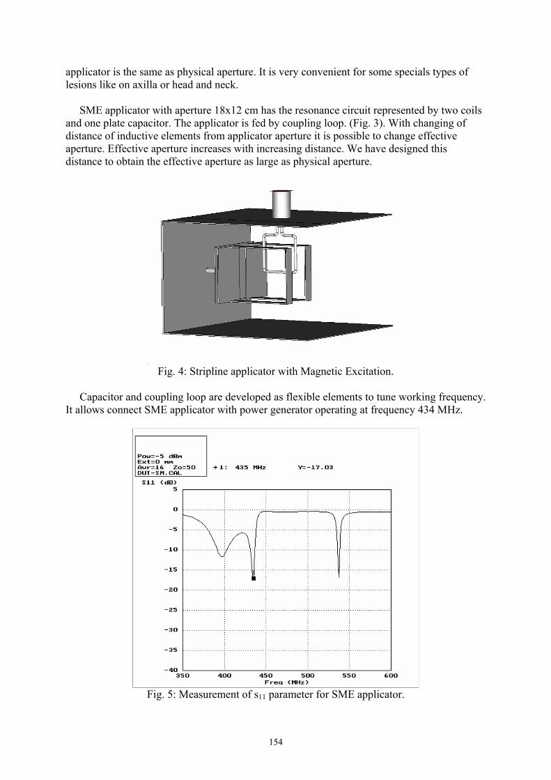
154
applicator is the same as physical aperture. It is very convenient for some specials types of lesions like on axilla or head and neck. SME applicator with aperture 18x12 cm has the resonance circuit represented by two coils and one plate capacitor. The applicator is fed by coupling loop. (Fig. 3). With changing of distance of inductive elements from applicator aperture it is possible to change effective aperture. Effective aperture increases with increasing distance. We have designed this distance to obtain the effective aperture as large as physical aperture.
Fig. 4: Stripline applicator with Magnetic Excitation.
Capacitor and coupling loop are developed as flexible elements to tune working frequency. It allows connect SME applicator with power generator operating at frequency 434 MHz.
Fig. 5: Measurement of s11 parameter for SME applicator.

155
Also this applicator works in TEM mode because it is based on strip line. It is linked with SH applicator mentioned above.
Fig. 6: Computation of effective aperture of SME applicator. New applicators were designed for large effective aperture. We have compared our new applicators with well-known types. The effective aperture of SH applicator was compared with the aperture of Waveguide applicator and the aperture of SME applicator was compared wit the aperture of EMW applicator. We have studied that our new types of applicators (SH, SME) allow obtain effective aperture as large as physical aperture in comparison with Waveguide applicator or EMW. Acknowledgement This work is supported by Grant Agency of the Czech Republic n. 102/05/0959 – Microwave Medical Applications: Therapy and Diagnostics. References [1] J. Vrba: Medical applications of medical technique, CTU Press, Prague, 2003.
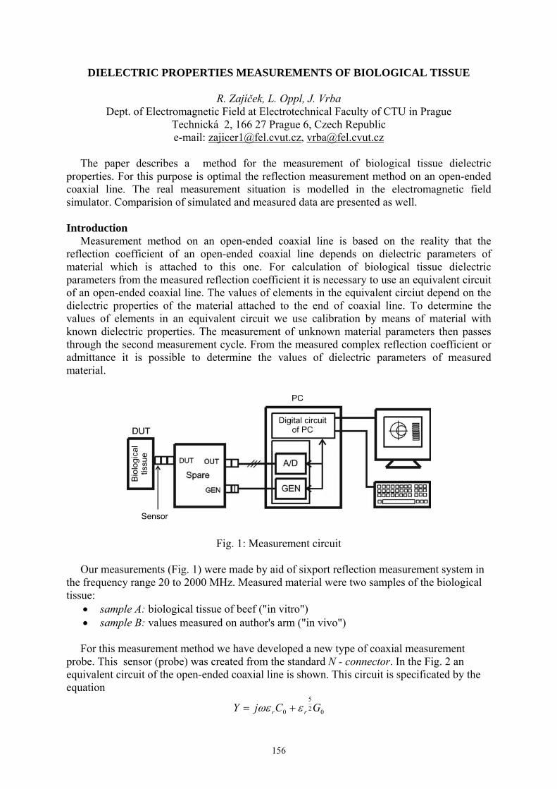
156
DIELECTRIC PROPERTIES MEASUREMENTS OF BIOLOGICAL TISSUE
R. Zajíček, L. Oppl, J. Vrba Dept. of Electromagnetic Field at Electrotechnical Faculty of CTU in Prague
Technická 2, 166 27 Prague 6, Czech Republic e-mail: [email protected], [email protected]
The paper describes a method for the measurement of biological tissue dielectric properties. For this purpose is optimal the reflection measurement method on an open-ended coaxial line. The real measurement situation is modelled in the electromagnetic field simulator. Comparision of simulated and measured data are presented as well. Introduction Measurement method on an open-ended coaxial line is based on the reality that the reflection coefficient of an open-ended coaxial line depends on dielectric parameters of material which is attached to this one. For calculation of biological tissue dielectric parameters from the measured reflection coefficient it is necessary to use an equivalent circuit of an open-ended coaxial line. The values of elements in the equivalent circiut depend on the dielectric properties of the material attached to the end of coaxial line. To determine the values of elements in an equivalent circuit we use calibration by means of material with known dielectric properties. The measurement of unknown material parameters then passes through the second measurement cycle. From the measured complex reflection coefficient or admittance it is possible to determine the values of dielectric parameters of measured material.
Fig. 1: Measurement circuit Our measurements (Fig. 1) were made by aid of sixport reflection measurement system in the frequency range 20 to 2000 MHz. Measured material were two samples of the biological tissue:
• sample A: biological tissue of beef ("in vitro") • sample B: values measured on author's arm ("in vivo")
For this measurement method we have developed a new type of coaxial measurement probe. This sensor (probe) was created from the standard N - connector. In the Fig. 2 an equivalent circuit of the open-ended coaxial line is shown. This circuit is specificated by the equation
025
0 GCjY rr εωε +=

157
where • C_0 is kapacity • G_0 is conductivity of the open-ended coaxial line in vacuum.
Fig. 2: Equivalent circuit, sensor and model of the senzor
Fig. 3 Simulated model
Results With increasing frequency the wave lenght of the electromagnetic wave lambda on measurement sensor is decreasing. In the animation (Fig. 4) we can see two maximum points of lambda.
Fig. 4: Time distribution of electromagnetic field From the theoretical analysis the reflection coefficient of the electromagnetic wave R of lossy dielectric should decrease with increasing frequency. In Fig. 5 we can see that the

158
parameter R is constant when measured by the sixport, but shows changes when modelled by the simulator of the EM field.
Fig. 6: Measured reflection coefficient At low frequencies (below 100 MHz) the reflection coefficient decreases according to hypothesis, but rises from the value of 0,35 to 0,78. In the graph there is the difference in phases, most of all at frequency 600 MHz, the difference being phi = 21. Complex permittivity is heavily frequency dependent quantity. At low frequencies is the real part of complex permittivity about value of 200 and with increasing frequency it has hyperbolic falling to value of 50.
Calculated permitivity and loss factor Conclusion The reflection method of an open-ended coaxial line is relatively exact method of the determination dielectric parameters of biological tissue. Explore of the mathematic and physical model of the reflection coefficient measurement on the interface biological tissue and measurement sensor we can make measurements more accurate. And by modelling of electromagnetic field we can in the measurements eliminate inconvenient step of the calibration measurement sensor by materials with known values of complex permitivity. Finaly it will rationalize total accurate of determination of complex permittivity epsilon. Acknowledgement This work has been conducted at the Department of Electromagnetic Field of the Czech Technical University in Prague and supported by the Czech Ministry of Education, Youth and

159
Sports in the frame of the project Microwave Medical Applications: Therapy and Diagnostics, 102/05/0959. References [1] J. Vrba: Lékařské aplikace mikrovlnné techniky. Skripta ČVUT, Vydavatelství ČVUT, Prague 2003 ((Medical application of microwave technique. Text of CTU, Edited by CTU, Prague 2003). [2] L. Oppl: Měření dielektrických parametrů biologické tkáně. Disertační práce, CVUT FEL, Praha 2002 (Measurement of dielectrical parameters of biological tissues. Doctoral Thesis, CTU, FEL, Prague 2002).

160
SPECIAL APPLICATOR FOR MICROWAVE THERMOTHERAPY
T. Dřížďal, J. Vrba Department of Electromagnetic Field, Faculty of Electrical Engineering, CTU in Prague
e-mail: [email protected], [email protected]
This paper deals with applicator for local thermotherapy, which works on the frequency 434 MHz. Applicator is a composed from feeding waveguide and strip horn, which has got lateral faces from PVC. Applicator was designed, made and measured. The impedance matching and the SAR thermal distribution was measured in the agar phantom. The results of simulations and measurements are compared as well. Introduction Microwave applicators are being used for local thermotherapy to treat cancer. The microwave energy is used for warming the biological tissue up to level 41°C - 45°C. Healthy cells can survive higher temperature than tumour tissue. After warming-up on temperature higher than 42°C the healthy tissue begins to defend next increase of the temperature. Tumour tissue has no defensive mechanism. Applicator for microwave thermotherapy Applicator consists of feeding waveguide and strip horn. Electromagnetic field is excited in feeding waveguide on the frequency 434 MHz, which is set by the parameters of feeding waveguide. N connector has been used to connect power generator to feeding waveguide. The horn of the applicator is created by the strip horn, which has lateral faces made from PVC r = 4 and thickness 3 mm. A dielectrical cone from the same material as the lateral faces has been inserted into the aperture of the horn to achieve larger warming-up surface. These parameters of the feeding waveguide are optimized by 3D simulator of electromagnetic field.
Fig. 1 describes the impedance matching of the simulated and the real applicator.
Fig.1: Simulated (left) a measured (right) course of the parameter S11.
Simulated impedance matching S11= -29,15 dB and measured S11= -43,31 dB . There is a clear exhibit of influence of the cone on the thermal distribution in agar phantom – peaks lays

161
around perimeter of the cone (Fig. 2). There are also denotation of the surface aperture and surface warming-up in biological tissue (100x110 mm) in the Fig. 2.
Fig. 2: SAR distribution in agar phantom in aperture of the applicator.
Fig. 3: Measured (top) and simulated SAR distribution (below) at the same cutting plane. For measurements a method of thermal incrementa in time period (time interval 6 – 9 minutes) has been used. Conclusion The cone achieve larger thermal distribution in the treatment areas. We achieve the same warming-up surface with the smaller dimensions of the hybrid applicator. Hybrid applicator was designed by aid of 3D simulator of electromagnetic field, made and measured. Simulated and measured courses are in very good agreement. References [1] Vrba, J. (2003). Medical applications of microwave (in czech). Press CTU in Prague, 2003.

162
INVOLVEMENT OF 50Hz ELECTROMAGNETIC FIELD IN OXIDATIVE LIVER INJURY
D.D. Krstić1, B.J. Djindjić2, V.V. Marković3, S.B. Radić2, D.M. Petković1, D.T. Sokolović2
1Faculty of Occuppational Safety, University of Nis, Serbia and Montenegro 2 Medical Faculty, University of Nis, Serbia and Montenegro
3Faculty of Electronic Engineering, University of Nis, Serbia and Montenegro
Introduction The biological effects of exposure to electromagnetic fields (EMF) have been brought sharply into focus by the enormous expansion of modern technologies. There is no clear agreement to the effects of long-term exposure to extremely low frequency (ELF) electromagnetic fields (50/60 Hz) [1]. There are conflicting evidences about whether the exposure to ELF field can directly alter cell biostructures and functions. The significance of oxidative stress has become increasingly recognised to the point that it is now considered to be a component of virtually every disease process. Recently, some significant in vitro and in vivo studies have pointed to direct or indirect effect on DNA. Several in vitro investigations into the biological consequences of EMF exposure effects were performed to estimate the risk of genotoxicity of EMF and to elucidate the underlying mechanisms [2]. Exposure to EMF may affect molecules associated with the signal transduction pathway, leading to an alteration of gene expression [3]. ELF fields have direct effect on enzyme damage in many tissues, especially in liver. The possibility of direct effects of EMF on the conformation of macromolecules has been investigated [4]. It has been shown that long-term exposition to EMF induces toxic effect on hepatocites [5]. The effect has been explained by a resonance interaction of EMF with paramagnetic molecules of biological tissues, which activate free radicals and initiate auto-supporting and auto-intensifying of chain chemical reactions. The aim of this study was to examine the influence of 50Hz electromagntic field in vivo on the oxidative modification of lipids, proteins and DNA in liver. Material and Methods In this experiment we used Wistar rats from the same clutch. The rats were 3 months old at the beginning of experiment and they were divided into two groups: I-experimental group and II-control, both with 7 female and 7 male animals. All animals from control group were suited in the same room with daylight and without near sources of EMF, in the cages 30x40x40cm (WxLxH). Experimental group was continually exposed to 50Hz EMF (B = 48 μT; E = 50 V/m) for 45 days. The source of electromagnetic field was a power cord longitudinaly orientated beside the wall of the cage. Blood samples were collected from abdominal aorta. Malondialdehyde (MDA) concentration (secondary product of lipid peroxydation) was measured by the modified method with thiobarbituric acid in 10% homogenates of liver tissue (the method of Ohkava et al.) [6]. Protein carbonyls and protein nitrotyrosine were measured in 10% homogenates of liver tissue according to the method of Oliver et al [7]. The extraction of liver DNA was performed according to the method of Wannermacher, and spectrophotometricaly scanning [8]. Results and Discussion The only histopathological finding in exposed rats was a slightly increased number of micronuclei and discrete perivenular fatty changes. The ELF-EMF exposed rats developed oxidative stress in hepatocites as evidenced by a significant increase in malondialdehyde level (MDA). The lipid peroxide level (MDA) significantly increased about 68% in exposed animals (2.24±0.28 vs control 1.53±0.21 nmol/mg prot.) which may be harmful by

163
accelerating the loss of plasma membrane integrity. Carbonyl group level was also significantly increased (8.23±1.31 vs 6.80±1.34nmol/mg prot. p<0.05), capable of altering many cellular functions. After the DNA purification and spectrophotometrical scanning, neither increased oxidative modification of purine bases (appearance of oxoguanine peak) nor the oxidative modification of deoxyribose (MDA/deoxyribose Ext ratio 0.60±0.14 vs 0.55±0.12 – non significant) was detected. It indicates that DNA might not be an appropriate biomarker or primary target of EMF-induced oxidative injury. But, variation in DNA configuration shifting from normal and stabile ″B-DNA″ to non-stabile ″Z-DNA″, which has been seen in our experiment, indicates to consequent DNA impairement and breaking after exposition. These findings suggest, that ELF exposure seems to have a negative impact on DNA repair mechanisms. Long-term exposition increases apoptosis process, which is expresed by slightly increased number of micronuclei and discrete perivenular fatty changes, as it has been seen in our study. Lipid peroxidation of unsaturated fatty acids is an unfavorable process that is followed by disturbances of lipid membranes and other cellular lipid structures. This process leads to massive generation of free radicals and induced membranes protein autooxydation. Results in our study show significant increasing of lipid peroxydation as a direct indicator of hepatocite injury. EMF radiation causes the activation of cellular stress responses, presumably reflecting increased levels of protein structural alteration. In hepatocites this cause changes in the conformation of biologically active macromolecules and induce disturbance in transport of biomolecules, decreasing detoxycation and cell adaptive mechanisms. Structural alteration of hepatocites in experimental animals also include increased contents of carbonyl group, which present sign of altered proteine structure and oxydative stress. Both acute and chronic exposures to EMF radiation alter the function of the rat canalicular membrane with decreasing permeability. Significant increase in ALT activity indicates citotoxic effect of non-ionizing radiation on hepatocites inducing apoptosis and necrosis [9]. This is another possible explanation for mechanism of rats hepatocites injury and DNA damage by EMF. Conclusion The capacity of 50Hz EMF to interfere with the weakness of liver is relevant because of incresing exposure in modern life. Functional abnormalities of hepatic cells membranes accompanied by increasing lipid peroxidation support the hypothesis that the liver is one of the major organ targets of EMF citotoxicity. Observed results support the view that 50Hz EMF stimulates oxidative stress response and that the possible functional liver disorders during prolonged exposure may be in part due to oxidative modification of biomolecules. References [1] H. Lai: Genetic Effects of Nonionizing Electromagnetic Fields. The International Workshop on Biol. Efects of Electromagnetic Fields. October 2-6; Sinaia, Romania; 2001. [2] J. McCann, F. Dietrich, C. Rafferty: The genotoxic potential of electric and magnetic fields: an update”, Mutat. Res. 411 (1998) 45–86. [3] C. Harvey, P. French: Effects on protein kinase c and gene expression in a human mast cell line, hmc-1, following microwave exposure. Cell Biol. Int., 23(11):739-748, 1999. [4] D.E. Koshland: Conformational changes: how small is big enough? Natl Med, 4:1110-4, 1998. [5] I.G. Akoev, M.S. Pashovkina, L.P. Dolgacheva: Enzymatic activity of some tissues and blood serum from animals and humans ...., Radiats Biol Radioecol, 42(3):322-30, 2002. [6] H. Ohkava, N. Ohishi, K. Yagi: Asssay for lipid peroxydes in animal tissue by thiobarbituric acid reaction. Anal Biochem 1979, 95:351-358.

164
[7] C.N. Oliver et al.: Agerelated changes in oxydised proteins”. J Biol Chem 1987, 262:5488-91. [8] R.E. Wannemacher et al.: Use of a single tissue extract to determine cellular protein and nucleid acid concentration and rate of aminoacid incorporation. Anal Biochem 1965, 11:320-326. [9] B. Djindjic et al.: Effect of long- term non-ionizing radiation on activity of hepatic enzymes in serum. Clinical Chemistry Laboratory Medicine. Spain: Monduzzi Editore S.p.A.-Medimond Inc 2003. p. 1063-1066.

165
REGIONAL APPLICATOR FOR MICROWAVE THERMOTERAPY - NUMERICAL SIMULATIONS
Hana Trefná, Jan Vrba
Department of Electromagnetic Field, CTU FEE in Prague, Technická 2, 166 27 Prague 6 Czech Republic, e-mail:[email protected]
Introduction Microwave thermotherapy is presently used as an adjuvant to the radiation therapy in the treatment of certain types of cancers. The goal of hyperthermia treatment is to raise the temperature of a localised cancerous tumour to a therapeutic level without overheating surrounding normal tissue. In hyperthermia applications, adequacy of efficient treatment can be related to the extent to which the tumour is enclosed by surfaces of 50% of SAR (Specific Absorbtion Rate) produced by applicator. One approach for providing the deep hyperthermia is to use an array of radiators placed in a circumferential array about the patient, relying on constructive wave interference to selectively heat the tumour. The deep of the heating depend on generated waves frequency which is moving between 27 - 434 MHz. Set up of applicator and Results The applicator consists of an array of the radiators orientated around the patient, which distributes the EM energy into the body. We used non-magnetic metallic sheets of minimized dimensions to create the conductive elements of the applicator. The applicator itself is created by 2 - 8 inductive loops tuned to resonance by capacitive element. The applicator operates at 434 MHz and focuses EM field in the phantom of biological tissue which has diameter of 10 cm. The dielectric parameters of this phantom are same as those of muscle tissue (εr = 56.85, σ = 0.8 S/m). Between the phantom and excited elements is placed a water bolus with a thickness of 1 cm.
Fig. 1: SAR distribution in the phantom along axis z for 8 elements
Fig. 2: SAR distribution after change of the position of connectors, case of 4 radiating elements.
166
In the pure simpliest configuration, when we used two excited elements, distance between them was too long and the resulting the EM field was quite mild. The distribution of SAR was in the best case less than 10% of maximal value on the surface of the phantom. Better results were obtained by applying four elements situated symmetrically around the phantom. We achieved symmetricaly SAR distribution in z axis cut. Simulation shows very good focusing of the EM power in center of the phantom, when 90% of SAR is achieved on the surface of the phantom. Finally, we used applicator containing eight elements. The level of absorbed energy (Fig. 1) was as large that this applicator could be usable for efficient treatment of parts with a diameter of 15 - 20 cm. Additionally, the focusing of the EM power was better and distribution of SAR was more homogenous then in the previous case. However, SAR was absorbed in a large area and could have inflict overheating of any part of body. Accumulated energy was clearly caused by connectors of elements. Thus displaced connectors of elements for reducing this area. Figure (Fig. 2) depicts that area of focusation was dispart to a main area under exciting elements and two added areas on the sides of phantom. This was evoked by basic principle of phantom, which was acting on as resonator structure. Maximum of SAR achieve insignificant values and in practise will be limited by distribution of heat. Conclusions In this paper we have discussed a regional applicator for a deep-seated tumour treatment in a neck and limbs which is compatible with non-invasive thermometry utilizing various number of radiators. We attain very good focusing of the EM energy by using 4 and 8 radiating elements. Especially, in the case of applicator containing 8 elements the distribution of SAR was extremely favourable. By transposition of connectors was improved SAR distribution in the center of phantom. Actually, associated areas on the sides of phantom attain neglectable values, nevertheless we dealing continually with this problem. Concurrently we solve situating of the focused EM energy in an arbitrary part of the body by amplitude and phase shift. Acknowledgement This research is supported by Grant Agency of the Czech Republic, project: Microwave Medical Applications: Therapy and Diagnostics, 102/05/0959 and project: 102/03/H086. References [1] Vrba, J., Medical applications of microwave technique, CTU Press, Prague, 2003. ISBN 80-01-02705-8.

167
NONLINEAR DYNAMIC OF EXCITATIONS IN MACROMOLECULAR CHAINS WITH ACOUSTICAL AND OPTICAL PHONON MODES
Yurko Natanzon1, L.S Brizhik2 and A.A. Eremko2
1National University of Kyiv-Mohyla Academy, Department of Physics and Mathematics Skovorody str., 2, 01001 Kyiv, Ukraine, e-mail [email protected]
Bogolyubov Institute for Theoretical Physics 2Metrologichna str., 14-B, 03143 Kyiv, Ukraine, e-mail [email protected],
We report the results of numerical modelling of a quasiparticle (electron, hole, excitation etc) in the field of chain deformation in one-dimensional molecular chains that model alpha-helical polypeptide macromolecules, DNA, RNA etc. Taking into account the interaction of a quasiparticle with both acoustical and optical phonon modes, we generalize the model of molecular solitons suggested by A.S. Davydov and based on the account of quasiparticle interaction with acoustical phonons [1]. We have got the nonlinear system of equations which admit special, soliton-like localized solutions, that can move along the chain and are dynamically stable.
Here we analyze the stability of such a soliton depending on the parameters and different initial conditions, including random initial conditions for phonons. The numerical modelling shows that in a broad interval of the parameters of a system a large class of initial conditions evolves in time into soliton-like states which are bound states of a quasiparticle and deformation and which propagates along the chain with finite velocity.
Fig. 1: Dependence of the average soliton velocity on the wave vector k for different values of the exchange constant, j.
We have found that in systems with random initial conditions for optical and acoustic phonons, a big enough energy impulse creates a self-consistent deformational well, in which it gets self-trapped and propagates from one site of the chain to another in the form of a localized soliton-like excitation, with minimal energy loss. The efficiency of such energy transfer can reach more than 80% and hundreds of angstroms can be overcome.
We calculated the dependence of soliton velocity on the wave vector k and analyzed the influence of Peierls-Nabarro barrier on soliton dynamics. Velocity of a soliton and its

168
amplitude are characterized by oscillations. Time behaviour of the maximum on-site value of a soliton shows that the propagation of a soliton is a superposition of two periodic propagations: propagation in the periodical field of optical phonons, and periodical propagation in the Peierls-Nabarro periodic barrier. According to Maxwell equations, this periodic character of soliton dynamics in the case of charged soliton (self-trapped electron) results in the emition of electromagnetic radiation, similar to conventional Davydov’s soliton [2].
Fig. 2: Distribution of the amplitude of soliton wave function on chain sites, n, at time t=50 for k=0.45, j=1.2.
Fig.3: Random deformation potential as the initial condition.
Thus, the results of numerical modelling prove the possibility of the energy storage and charge and energy transport in biopolymers in the form of self-trapped states, as it has been suggested by A.S. Davydov for α-helical proteins, taking into acount the interaction of a quasiparticle with acoustical phonons only.

169
Fig. 4: Soliton c.m.c. (curve with smaller amplitude) and maximum probability coordinate
(curve with greater amplitude) as a function of time in a chain with periodic boundary condition at the initial deformation condition represented in Fig. 3.
References [1] A.S. Davydov: Solitons in Molecular Systems, Dordrecht: Reidel, 1985. [2] L.S. Brizhik, A.A. Eremko: Nonlinear model of the origin of endogenous alternating electromagnetic fields and selfregulation of metabolic processes in biosystems. Electromagnetic Biology and Medicine, 2003, v. 22, No. 1, 31-39. [3] Yu. Natanzon, L.S. Brizhik, A.A. Eremko: Self-trapping and dynamics of quasiparticles in one-dimensional molecular chain under interaction with optical phonons. Submitted to Ukr. J. Phys.

170
Aguiar P.H.P. 134 Aksyonov S.I. 150 Ambruš M. 114 Apostol M.V. 138 Barančok D. 103 Barcal J. 55 Bååth L. 116 Belyaev I. 44, 52, 63 Berg H. 107 Betti L. 48 Berden Zrimec M. 99 Binhi V.N. 32 Blank M. 43
Borghini F. 48 Böhm K.J. 23 Brizzi M. 48 Brizhik L.S. 29, 167 Budyanskaya E.N.
130, 132 Calzoni G.L. 48 Cendelín J. 55 Chepenko L.V. 130 Chernavskii D.S. 32 Chernyshova O.N. 130 Chovanec R. 152 Chukova Yu.P. 42 Chunihin, A. 136 Cirák J. 102, 103 Cocherová E. 58 Cvek J. 152 Čevizović D. 41 Čoček A. 81, 114 Del Giudice E. 8, 21 Detlavs I. 110 Djindjic B. 162 Dohnalová A. 81, 114 Doležel M. 108 Drinovec L. 99 Dřížďal T. 160 Dürr H.-P. 7 Dvořák J. 108 Eremko A.A. 29,167 Ermakov V.N. 69 Fhager A. 116 Fleishman M. 8 Fojt L. 51, 65, 66
AUTHORS’ INDEX Foster K.R. 71 Frantík E. 72 Gaur M.S. 106 Goodman R. 43
Gorban E. N. 112 Gorgun S.S. 117 Górski Z. 87 Grubnik B. P. 112 Grundsteins K. 110 Grygoruk V.I. 120, 127 Gurban D. 138 Hahn A. 116 Hashemzadeh P. 118 Hašek J. 35, 95 Herza J 152 Hianik T. 102 Hillert L. 52 Hölzel R. 86 Hornychová M. 72 Hulínská D. 72 Ivić Z. 41 Jandová A. 81, 114 Jelínek F. 35, 95 Jenro K.A. 11 Jerman I. 74, 76, 97, 143, 146 Jovanić B.R. 80 Kaduk B. 136 Kašparová, L. 72 Knotková J. 72 Korelusová I. 55 Korobtsova E . 124 Kosytskyy M. 128 Krakovská A. 60 Krašovec R. 146 Krstic D. 162 Kwee S. 149 Lazzarato L. 48 Leskovar R.T. 74, 76, 97 Liboff A.R. 9, 11, 15 Lobyshev V.I. 137, 141 Malarić K. 67 Malmgren L.O.G. 52 Maciel R.R.B.T. 134 Maric-Oehler W. 38
Markova E. 52 Marković V. 162 Matronchik A.Y. 63 Mavromatos N. 23 Melichar B. 108 Melnik Ju. 136 Mhamdi L. 81 Michette A. 23 Milani M. 8, 21 Murav’ev V. 135 Natanzon Y. 167 Nedbalová M. 81 Nikitin D.I. 137 Nikitin L.E. 137
Nikolenko E.Ya. 130 Nizhelska O. 128 Novák J. 66 Oliveira A. 134 Oppl L. 156 Pepe I. 134 Peregudov S.N. 112 Persson B.R.R. 52 Persson M. 116 Petera J. 108 Peterlin P. 79 Petkovic D. 162 Pevalek-Kozlina B. 67 Philipenko N.O. 130 Pinheiro C.M.B. 134 Pizzuti A. 48 Pokorný J.
35, 81, 95, 114 Ponezha E.A. 69 Ponezha G.V.
120, 123, 127 Ponezha S.G. 120, 127 Popp F.-A. 38 Preoteasa E.A. 138 Priel A. 40 Radic S. 162 Reis P.S.O. 134 Rezinkina M.M. 130 Romanovsky Yu.M. 125 Rosário V.C. 134 Ružič R. 97, 143 Santisi G. 21
171
Sarimov R. 52 Sarvan M.Ž. 80 Satarić M.V. 26 Shchegoleva T.Yu. 132 Schlebusch K.-P. 38 Sheppard A.R. 71 Shevchenko K. 93 Shogenov Yu.Kh. 125 Simões L.C. 134 Sit’ko S.P. 112 Skripnik Yu. 93 Slawinski J. 87 Sokolovic D. 162 Solmi E. 48 Stepanyan A.S. 125 Stracke R. 23 Strašák L. 51, 65, 66 Svetina S. 79 Swicord M.L. 71 Szasz A. 9 Szentpáli B. 90 Šaroch J. 95 Škarja M. 74, 76, 97, 143, 146
Šmajs D. 51 Šrobár F. 18 Štenglová V. 55 Štolc S. 60 Švandová E. 72 Tamelo A. 135 Talpo G. 8, 21 Turauska A. 110 Teplan M. 60 Thuróczy G. 90 Tkalec M. 67
Tomčík P. 102, 103 Torrigiani P. 48 Trebbi G. 48 Trefná H. 165 Trojan S. 81 Tuszynski J. 23, 40 Unger E. 23 Vajda J. 102 Van Wijk E.P.A. 92 Van Wijk R. 92 Vasil`ev V.A. 125 Veliks V. 110 Vetterl V. 51, 54, 65, 66
Vidaković-Cifrek Ž. 67 Vincze C. 9 Vitiello G. 8, 21, 27 Vitovič P. 102 Vojtíšek M. 72 Vožeh F. 55 Vrba J. 115, 152, 156, 160, 165 Weis M. 102, 103 Yanenko A.
93, 112, 136 Yunevich E. 124 Zajíček R. 156 Zdravković S. 84 Zeković S. 41 Zhukavets A. 124 Zobnina G. 135 Zon J.R. 28 Zoul Z. 108 Zrimec A. 99 Žalud V. 55 Žekš B. 79
Related Documents


![moDel legislation For electromagnetic FielDs protection1].pdf · moDel legislation For electromagnetic FielDs protection. ... Model legislation for electromagnetic fields ... If a](https://static.cupdf.com/doc/110x72/5b166fc57f8b9a4a6d8befdf/model-legislation-for-electromagnetic-fields-1pdf-model-legislation-for-electromagnetic.jpg)