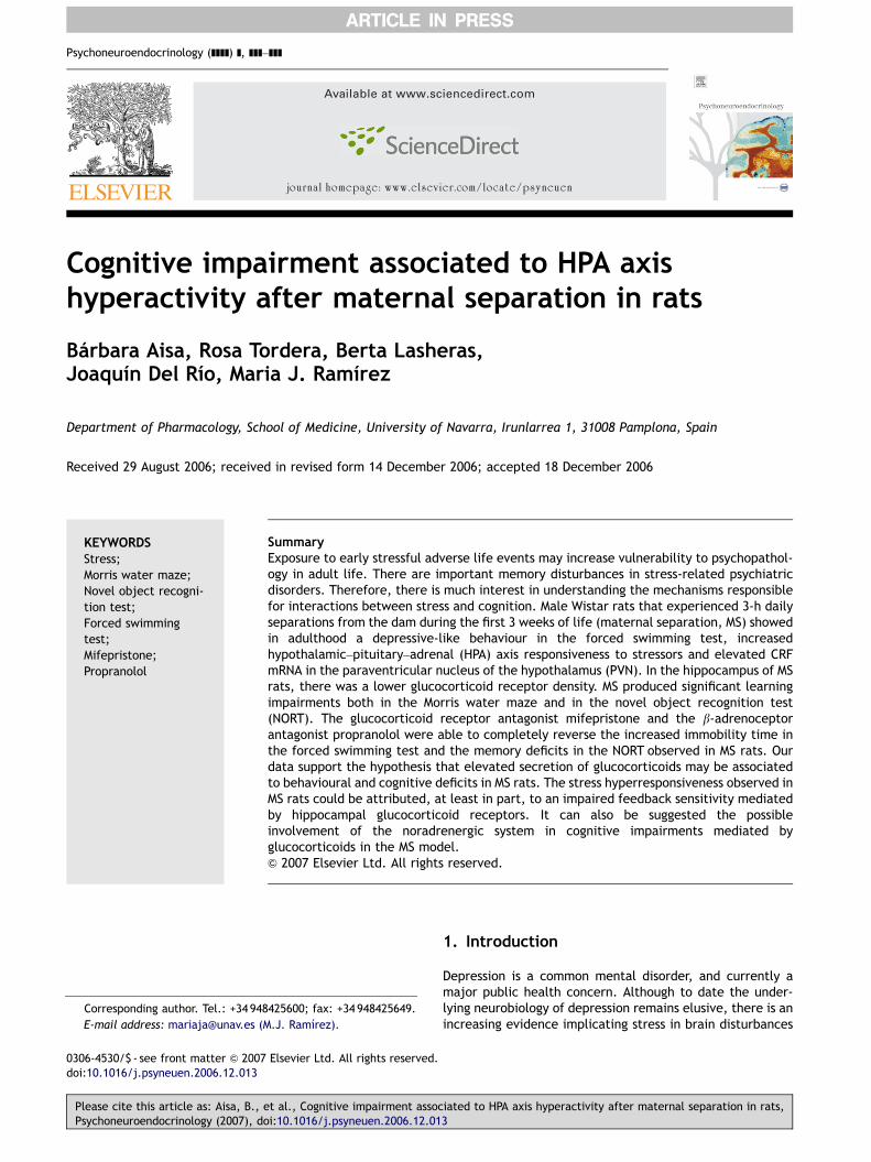
Available at www.sciencedirect.com journal homepage: www.elsevier.com/locate/psyneuen Cognitive impairment associated to HPA axis hyperactivity after maternal separation in rats Ba ´rbara Aisa, Rosa Tordera, Berta Lasheras, Joaquı ´n Del Rı ´o, Maria J. Ramı ´rez Department of Pharmacology, School of Medicine, University of Navarra, Irunlarrea 1, 31008 Pamplona, Spain Received 29 August 2006; received in revised form 14 December 2006; accepted 18 December 2006 KEYWORDS Stress; Morris water maze; Novel object recogni- tion test; Forced swimming test; Mifepristone; Propranolol Summary Exposure to early stressful adverse life events may increase vulnerability to psychopathol- ogy in adult life. There are important memory disturbances in stress-related psychiatric disorders. Therefore, there is much interest in understanding the mechanisms responsible for interactions between stress and cognition. Male Wistar rats that experienced 3-h daily separations from the dam during the first 3 weeks of life (maternal separation, MS) showed in adulthood a depressive-like behaviour in the forced swimming test, increased hypothalamic–pituitary–adrenal (HPA) axis responsiveness to stressors and elevated CRF mRNA in the paraventricular nucleus of the hypothalamus (PVN). In the hippocampus of MS rats, there was a lower glucocorticoid receptor density. MS produced significant learning impairments both in the Morris water maze and in the novel object recognition test (NORT). The glucocorticoid receptor antagonist mifepristone and the b-adrenoceptor antagonist propranolol were able to completely reverse the increased immobility time in the forced swimming test and the memory deficits in the NORT observed in MS rats. Our data support the hypothesis that elevated secretion of glucocorticoids may be associated to behavioural and cognitive deficits in MS rats. The stress hyperresponsiveness observed in MS rats could be attributed, at least in part, to an impaired feedback sensitivity mediated by hippocampal glucocorticoid receptors. It can also be suggested the possible involvement of the noradrenergic system in cognitive impairments mediated by glucocorticoids in the MS model. & 2007 Elsevier Ltd. All rights reserved. 1. Introduction Depression is a common mental disorder, and currently a major public health concern. Although to date the under- lying neurobiology of depression remains elusive, there is an increasing evidence implicating stress in brain disturbances ARTICLE IN PRESS 0306-4530/$ - see front matter & 2007 Elsevier Ltd. All rights reserved. doi:10.1016/j.psyneuen.2006.12.013 Corresponding author. Tel.: +34948425600; fax: +34948425649. E-mail address: [email protected] (M.J. Ramı ´rez). Psychoneuroendocrinology (]]]]) ], ]]]–]]] Please cite this article as: Aisa, B., et al., Cognitive impairment associated to HPA axis hyperactivity after maternal separation in rats, Psychoneuroendocrinology (2007), doi:10.1016/j.psyneuen.2006.12.013

Welcome message from author
This document is posted to help you gain knowledge. Please leave a comment to let me know what you think about it! Share it to your friends and learn new things together.
Transcript
ARTICLE IN PRESS
Available at www.sciencedirect.com
Psychoneuroendocrinology (]]]]) ], ]]]–]]]
0306-4530/$ - see frodoi:10.1016/j.psyne
�Corresponding au
E-mail address: m
Please cite this artPsychoneuroendoc
journal homepage: www.elsevier.com/locate/psyneuen
Cognitive impairment associated to HPA axishyperactivity after maternal separation in rats
Barbara Aisa, Rosa Tordera, Berta Lasheras,Joaquın Del Rıo, Maria J. Ramırez�
Department of Pharmacology, School of Medicine, University of Navarra, Irunlarrea 1, 31008 Pamplona, Spain
Received 29 August 2006; received in revised form 14 December 2006; accepted 18 December 2006
KEYWORDSStress;Morris water maze;Novel object recogni-tion test;Forced swimmingtest;Mifepristone;Propranolol
nt matter & 2007uen.2006.12.013
thor. Tel.: +34 948
icle as: Aisa, B., erinology (2007), do
SummaryExposure to early stressful adverse life events may increase vulnerability to psychopathol-ogy in adult life. There are important memory disturbances in stress-related psychiatricdisorders. Therefore, there is much interest in understanding the mechanisms responsiblefor interactions between stress and cognition. Male Wistar rats that experienced 3-h dailyseparations from the dam during the first 3 weeks of life (maternal separation, MS) showedin adulthood a depressive-like behaviour in the forced swimming test, increasedhypothalamic–pituitary–adrenal (HPA) axis responsiveness to stressors and elevated CRFmRNA in the paraventricular nucleus of the hypothalamus (PVN). In the hippocampus of MSrats, there was a lower glucocorticoid receptor density. MS produced significant learningimpairments both in the Morris water maze and in the novel object recognition test(NORT). The glucocorticoid receptor antagonist mifepristone and the b-adrenoceptorantagonist propranolol were able to completely reverse the increased immobility time inthe forced swimming test and the memory deficits in the NORT observed in MS rats. Ourdata support the hypothesis that elevated secretion of glucocorticoids may be associatedto behavioural and cognitive deficits in MS rats. The stress hyperresponsiveness observed inMS rats could be attributed, at least in part, to an impaired feedback sensitivity mediatedby hippocampal glucocorticoid receptors. It can also be suggested the possibleinvolvement of the noradrenergic system in cognitive impairments mediated byglucocorticoids in the MS model.& 2007 Elsevier Ltd. All rights reserved.
Elsevier Ltd. All rights reserved.
425600; fax: +34 948425649.
.J. Ramırez).
t al., Cognitive impairment associ:10.1016/j.psyneuen.2006.12.01
1. Introduction
Depression is a common mental disorder, and currently amajor public health concern. Although to date the under-lying neurobiology of depression remains elusive, there is anincreasing evidence implicating stress in brain disturbances
iated to HPA axis hyperactivity after maternal separation in rats,3
ARTICLE IN PRESS
B. Aisa et al.2
thought to underlie certain forms of depression or particularcomponents of the depressive syndrome (Kessler, 1997;Kendler et al., 1999; Van Praag, 2004). There are importantmemory disturbances in stress-related psychiatric disorders(Bremner and Narayan, 1998; Bremner et al., 2003), andtherefore, there is much interest in understanding themolecular mechanisms responsible for interactions betweenstress and cognition. The hypothalamic–pituitary–adrenal(HPA) axis is an essential component of an individual’scapacity to cope with stress and in fact, a hyperactivity ofthe HPA axis is observed in the majority of patients withdepression (review by Arborelius et al., 1999; De Kloet etal., 2005). Stress stimulation of the axis starts whencorticotropin releasing factor (CRF) released by the para-ventricular nucleus of the hypothalamus (PVN) stimulatesthe release of corticotropin (ACTH) from the anteriorpituitary, which in turn, stimulates secretion from theadrenal cortex. Many of the behavioural consequences ofstress are thought to be mediated by the activation of theglucocorticoid receptor by stress-induced high levels ofglucocorticoid hormones (De Kloet et al., 1998; Oitzl et al.,2001; Roozendaal et al., 2006a) and subsequent alteration ingene expression (see review by Berton and Nestler, 2006).
It is well documented that corticosteroids modulatelearning and memory processes in animals and humans in acomplex manner. A transient increase in circulating corti-costeroids associated with a learning task has been shown tofacilitate memory consolidation (Roozendaal et al., 1999;Buchanan and Lovallo, 2001; Abercrombie et al., 2003).However, it has also been shown that acute stress produces adeficit in hippocampal long-term potentiation (Foy et al.,1987), and the elevation of circulating corticosteroneconcentrations by systemic administration of corticosteroneor glucocorticoid receptor agonists exerts an inhibitoryinfluence on learning and memory retrieval (Bodnoff et al.,1995; Roozendaal et al., 2003; Roozendaal et al., 2004b, c).It has been suggested that the effects of glucocorticoids onmemory depend on noradrenergic activation of the amyg-dala and interactions of the amygdala with other brainregions, mainly hippocampus and frontal cortex (review byRoozendaal et al., 2006b).
There is compelling evidence that exposure to earlystressful adverse life events may increase vulnerability topsychopathology in adult life. In fact, individuals whoexperience early trauma, such as parental loss, sexualabuse or physical assault in childhood, present an increasedrisk for suffering depression later in life (Heim and Nemer-off, 2001). Based on these arguments, it has been shownthat prolonged periods (41 h) of maternal separation (MS)during the first weeks of life result in animals withbehavioural and neuroendocrine signs of elevated stressreactivity as adults (Anisman et al., 1998; Ladd et al., 2000;Lehman and Feldon, 2000; Ploj et al., 2003). In addition toan increase in immobility time in the Porsolt forcedswimming test, anhedonia, and an enhanced anxiety-likebehaviour, MS animals exhibit a dysfunction of the HPA axisreactivity to stress and therefore, the MS model in rat isconsidered nowadays as a robust model of enhanced stressresponsiveness and depressive-like behaviour (Ladd et al.,2000; Van den Hove et al., 2005).
In this study we hypothesized that (i) neonatal MS wouldlead to cognitive deficits in adulthood; (ii) such an effect
Please cite this article as: Aisa, B., et al., Cognitive impairment assoPsychoneuroendocrinology (2007), doi:10.1016/j.psyneuen.2006.12.01
could be dependent on a dysfunction of the HPA axis,specifically to a hypersecretion of corticosterone. To testthese hypotheses, Wistar rats that experienced 3-h dailyseparations from the dam during the first 3 weeks of lifewere tested in adulthood for HPA axis responsiveness,depressive-like behaviour, presence of cognitive deficitsand the effect of the glucocorticoid receptor antagonistmifepristone on behavioural performance in MS rats.Furthermore, the involvement of the noradrenergic systemin corticosterone-mediated effects will be also investigated.
2. Material and methods
2.1. Animals
All the experiments were carried out in strict compliancewith the recommendations of the EU (DOCE L 358/1 18/2/1986) for the care and use of laboratory animals. Timed-pregnant Wistar rats were provided on gestation day 16 fromCharles River Laboratories (Portage, MI, USA), individuallyhoused in a temperature (2171 1C) and humidity (5575%)controlled room on a 12-h light/dark cycle with food andwater freely available.
2.2. Maternal separation
All litters were born within a 2-day period. As previouslydescribed (Huot et al., 2001; Ladd et al., 2004), on postnatalday (PND) 2, all pups were sexed and randomly assigned tothe control group (animal facility rearing, AFR), pups wereonly briefly manipulated to change the bedding in theircages once weekly, or the separation group (MS), pupsseparated from their dam for 180min from PNDs 2–21inclusive. Before manipulation of the MS pups, each damwas removed from her home cage and placed in an adjacentcage and then, the pups were removed as complete litters,placed in an empty cage with standard bedding material andtransferred to an incubator in an adjacent room. Tocompensate for the mother’s body heat, the temperatureof the incubator was adjusted to the age of the neonates:3270.5 1C (PNDs 2–5), 3070.5 1C (PNDs 6–14) or 2870.5 1C(PNDs 15–21). Rats were weaned on PND 23 and only maleswere chosen for the present work. All subsequent experi-ments were performed in adulthood (60–75 days).
2.3. Experimental design and drug treatments
Different subsets of rats were used for the measurement ofeach of the behavioural or biochemical parameters studied.Animals from different litters were distributed over each ofthe experiments. It was also used a different subset ofanimals in which a forced swimming test of 15min was usedas an acute stressor.
Administration of mifepristone (10mg/kg, SC) or propra-nolol (2mg/kg, SC) took place 60min before the behaviouraltesting. Mifepristone and propranolol were dissolved insaline.
ciated to HPA axis hyperactivity after maternal separation in rats,3

ARTICLE IN PRESS
Cognitive deficits and HPA axis hyperactivity 3
2.4. Behavioural test
For all behavioural testing, observers were blind to eitherthe rearing condition or drug treatment. In a pilot set ofexperiments, the performance of a vehicle-injected groupwas compared to non-injected controls, and no differenceswere found in any of the behavioural test studied.A different animal cohort was used for carrying out eachof the different test.
2.5. Locomotor activity
Horizontal locomotor activity was measured for 30 min in anopen field, which consisted of nine square arenas(43� 51� 45 cm3) made of black wood, using a videotracking system (Ethovision 3.0, Noldus Information Tech-nology B.V., The Netherlands), in a softly illuminated room.Tracking system was set to determine the position of theanimal five times per second. Total path length (cm) wasanalysed.
2.6. Forced swimming
As described by Porsolt et al. (1977), two swimming sessionswere conducted: an initial 15-min pretest followed 24 hlater by a 5-min test. Rats were placed individually in avertical Plexiglas cylinder (height: 45 cm, diameter: 19 cm)filled with 28–30 cm of 26 1C water. Immobility wasconsidered as rats floating passively, making only smallmovements to keep its nose above the surface.
2.7. Sucrose intake
The protocol for sucrose preference was adapted fromD’Aquila et al. (1997). At the start of sucrose intake testing,all animals were first trained to drink 1% sucrose solution for24 h. Test consisted of presenting two bottles on the rat’scage (water and 1% sucrose) following a 20-h period of foodand water deprivation, and allowing the rat to drink freelyfor 1 h. The position of the two bottles (right/left) wasvaried randomly across the animals. Sucrose and waterconsumption were corrected with the animals’ weight.
2.8. Elevated plus maze
Rats were placed in the centre of a cross maze, facing anopen arm, and allowed to explore the maze for 5min. Twopaws had to be inside the line indicating the entrance to anarms (Hogg, 1996), which signalled the start of the timespent in the specific arm. Time spent in the different arms ofthe maze was measured. The number of entries into thevarious arms of the maze was also determined, and servedas a measure of the rat’s locomotor activity.
2.9. Morris water maze
This memory task was performed as previously described(Diez-Ariza et al., 2003). The maze consisted of a blackcircular tank (140 cm diameter� 55 cm high) and filled withwater (20–22 1C). A black invisible platform, 10 cm diameter,
Please cite this article as: Aisa, B., et al., Cognitive impairment assocPsychoneuroendocrinology (2007), doi:10.1016/j.psyneuen.2006.12.01
was positioned 1 cm below the water surface. A videocamera was set above the centre of the pool and connectedto a videotraction system (EthoVision; Noldus, The Nether-lands). The pool was surrounded by white curtains markedwith black geometric paintings, to enable the animals tolearn the platform location. On the first day, each ratbecomes habituated to the training environment for 60 s. Inthe acquisition phase (days 2–3), rats performed six trainingtrials per day (120 s each) with the escape platform in afixed position. Trials were started by placing the animalsinto the pool, close to and facing the wall at starting pointsdesigned as north, east, south, west, north, east. Timespent to reach the platform (latency); the swim path length(distance swam) and the swim speed (cm/s) were recorded.The results on time to reach the platform are not shown as,in all cases, parallel with distances swam. On day 4,(retention phase) a single transfer test was performed, inwhich the platform was removed from the tank. The animalwas allowed to free swim for 60 s in search of the platform.The distance swam in the quadrant 3 (Q3) where theplatform was previous located and the rest of quadrantswere measured.
2.10. Object recognition
The object recognition test was adapted from Ennaceur andDelacour (1988). The open field consisted of a square openfield (65 cm� 65 cm� 45 cm) made of black wood. On theprevious day to the experiment, animals were familiarizedwith the field during 30min. During the first trial of theexperiment, two objects similar in shape, size, color,texture, etc., equidistant from the sides (10 cm) wereplaced within the chamber. The animal was placed into thecentre of the open-field and allowed to freely explore for5min. It was considered that the animal was exploring theobject when the head of the rat was oriented toward theobject with its nose within 2 cm of the object. One hourlater a second trial took place, in which one object wasreplaced by a different one, and exploration was scored for5min. In order to eliminate olfactory stimuli, chamber andobjects were cleaned after testing each animal. To avoidpreference for one of the objects, the order of the objectswas balance between testing animals. Results were ex-pressed as percentage of time spent with the novel objectwith respect to the total exploration time (discriminationindex).
2.11. Tissue and blood collection
Rats were sacrificed by decapitation between 0800 and1000 h. AFR animals were taken immediately from theirhome cages to collect tissue and blood. When the acutestressor was used, rats were sacrificed immediately afterthe 15min stressor was finished. Brains were removed anddissected on ice to obtain the hippocampus, including theventral and dorsal parts, and frontal cortex (for noradrena-line determinations and western blotting experiments), orfrozen immediately at �40 1C in isopentane, and stored at�80 1C until sectioning (for CRF mRNA determinations). In asubset of experiments, a group of MS (and correspondingAFR controls) pups were sacrificed at the end of the
iated to HPA axis hyperactivity after maternal separation in rats,3
ARTICLE IN PRESS
B. Aisa et al.4
separation period (PND 21). Trunk blood was collected intoEDTA tubes, centrifuged at 1250g (15min, 4 1C), and plasmawas frozen until corticosterone and ACTH levels weredetermined.
2.12. Plasma corticosterone and ACTHdeterminations
Plasma corticosterone (30 ml) was determined using acommercially available enzymeimmunoassay kit (IDS OC-TEIA, USA) and ACTH was assayed in 200 ml plasma samplesusing the Allegros HS-ACTH radioinmmunoassay kit (NicholsInstitute, San Juan Capistrano, CA, USA), as previouslydescribed (e.g. Huot et al., 2001).
2.13. Noradrenaline determination
Noradrenaline content in the hippocampus and frontalcortex was measured using high performance liquid chro-matography (HPLC) with electrochemical detection (WatersSpheribors 5m ODS2 4.6� 150mm). The mobile phaseconsisted of 80:20 (v/v) mixture of buffer (KH2PO4 0.05M,pH 3) and methanol; the mixture was filtered and degassedthrough a 0.22 mm nitrocellulose membrane (Millipore, UK).Noradrenaline content was calculated by comparing with a1 ng standard. The limit of detection was 1 pg/10 ml.
2.14. In situ hybridization for CRF mRNA
Coronal brain sections (15mm thick) were serially cut with acryostat at the level of paraventricular hypothalamusnucleus (�0.3mm relative to bregma) according to theatlas of Paxinos and Watson. The sections were thaw-mounted on SuperFrostsPlus slides and then processed forin situ hybridization of CRF mRNA. Slides were fixed in ice-cold 4% paraformaldehyde in phosphate-buffered saline andacetylated with 0.25% acetic anhydride in 0.1M triethano-lamine buffer for 10min. After dehydration through gradedethanol, sections were immersed in chloroform for 10min,rehydrated with ethanol and air dried. The oligonucleotideused was 50-AGGGCAGAGCAGTTAGCTCAGCAAGCTCAC-30
(Sigma Genosis, UK). The probe was 30-tail labelled withaS [35S] dATP (specific activity 41000 Ci/mmol, AmershamBiosciences, UK). Negative controls including sense oligonu-cleotide showed minimal background signals. Relativeabundance of CRF mRNA was determined by densitometryquantification of autoradiograms using the MicrocomputerImaging Device (Imaging Research, St Catherines, Ontario,Canada) corrected for non-specific signals. Optical densityvalues were calibrated to 35S tissue equivalents using 14Cmicroscales (Amersham, UK). Densitometric values fromthree sections of each animal were averaged and expressedas nCi/g tissue. CRF mRNA expression in MS groups wasexpressed as a percentage of their respective controls.
2.15. Western blotting
Cytosolic and nuclear extract preparations from thehippocampus of acutely stressed rats were homogenized ina 50mM Tris buffer (pH 7.2, 4 1C), as previously described
Please cite this article as: Aisa, B., et al., Cognitive impairment assoPsychoneuroendocrinology (2007), doi:10.1016/j.psyneuen.2006.12.01
(Spencer et al., 2000). Each sample was adjusted to a finalprotein concentration of 4mg/ml (DC protein assay; Bio-Rad, Hercules, CA). Extracts were mixed with Laemmeli’ssample buffer boiled for 5min. Samples (40 mg) were loadedonto 8% bisacrylamide gels and separated by SDS-PAGE.Separated proteins were electrophoretically transferredfrom gels to PVDF membranes. Glucocorticoid receptorprotein was detected with the monoclonal antibody, GR M-20 (1:2000 TBST; Santa Cruz Biotechnology Inc., CA).Immunopositive bands were visualized by a chemilumines-cent method (ECL; Amersham, Arlington Heights, IL). Theoptical density of glucocorticoid receptor-reactive bands(�97 kDa) visible on X-ray film was determined densitome-trically (Murphy et al., 2002).
2.16. Data analysis
Data were analysed by SPSS for Windows, release 11.0.Normality was checked by Shapiro-Wilks0s test (p40.05).Behavioural data were analysed by unpaired t-tests (whenonly MS vs AFR groups are compared), or one-way or two-wayanalysis of variance ANOVA. Neuro- and bio-chemical datawas analysed by unpaired t-tests (AFR vs MS groups). For allanalyses, post hoc comparisons were conducted if appro-priate, using Tukey protected least significance test. Data arepresented as mean7SEM and the level of significance forANOVA and post hoc testing was set at po0.05.
3. Results
3.1. Behavioural characterization
Locomotor activity was not modified by rearing (Student’s t-test), and total path length travelled was 6910.017300.92for AFR (n ¼ 10) and 7704.137378.52 (n ¼ 10) for MS rats.
In the Porsolt forced swimming test (Fig. 1A), MS (n ¼ 10)produced a significant increase in immobility compared withAFR (n ¼ 10) rats (Student’s t-test, po0.05).
In the sucrose intake test, there was an interaction[F1,34 ¼ 3.66, po0.05] between rearing condition (AFR vsMS) and water/sucrose intake. Even though all animalstested showed preference for the sucrose solution comparedwith water, consumption of sucrose intake (sucrose/weightof the rat) was reduced in MS (n ¼ 12) rats compared withthe AFR group (n ¼ 12) (Student’s t-test, po0.01), a findingthat can be interpreted as a sign of anhedonia (Fig. 1B). Therewere no differences (Student’s t-test) in water intake betweenAFR rats (0.01870.003ml/g) and MS rats (0.01270.001ml/g).
MS rats (n ¼ 10) showed increased anxiety-like behaviour(Fig. 1C), as statistical analysis revealed that MS rats spent asignificantly smaller time in the open arms of the elevatedplus maze than controls (n ¼ 10) (Student’s t-test, po0.01).No differences were found in the number of entries into thedifferent arms of the elevated plus maze.
3.2. HPA responsiveness in MS animals
Fig. 2A shows an autoradiogram representative of sectionshybridized for CRF mRNA in the PVN. Quantification of thesignal (Fig. 2B, n ¼ 5 for both the AFR and MS groups)
ciated to HPA axis hyperactivity after maternal separation in rats,3

ARTICLE IN PRESS
Day 1 Day 2
0
50
100
150
*
*
Imm
obili
ty (
s)
0.00
0.05
0.10
0.15
**
Sucr
ose
inta
ke (
ml/g
)T
ime
open
fie
ld (
s)
0
10
20
30
40
50
**
AFR
MS
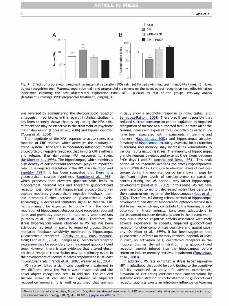
Fig. 1 Behavioural characterization of maternal separation(MS) rats (A) Effects of MS in the Porsolt forced swimming test.Results are expressed as immobility time. (B) Effects of MS insucrose intake. Results are expressed as consumption ofsucrose/weight of the rat. (C) Effects of MS in the elevatedplus maze. Results are expressed as time spent in the open armsof the maze. �po0.05 vs control (AFR) rats, Student’s t-test.
Cognitive deficits and HPA axis hyperactivity 5
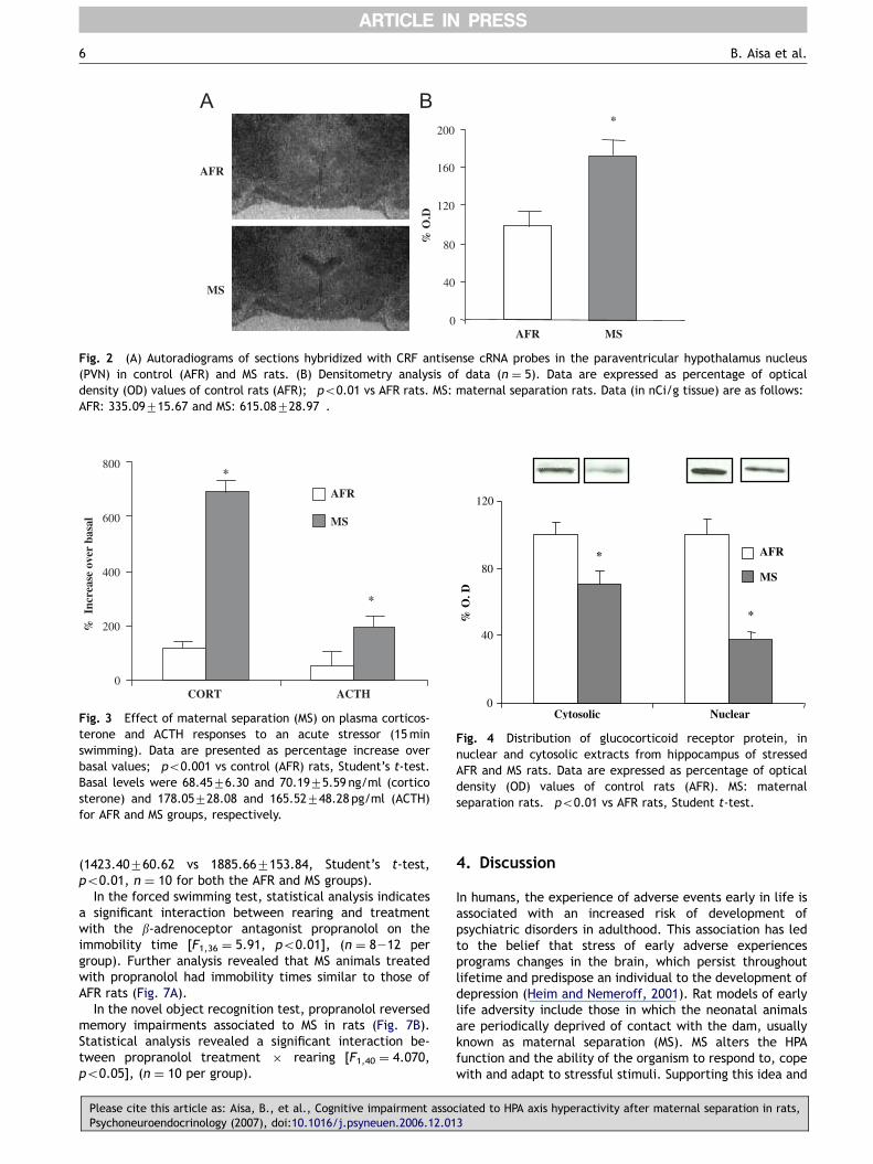
revealed an increase in CRF mRNA density in MS animals inthe PVN (Student’s t-test; po0.01).
As shown in Fig. 3, increases in corticosterone (n ¼ 12 forboth the AFR and MS groups) and ACTH levels (n ¼ 12 forboth the AFR and MS groups) in response to an acute
Please cite this article as: Aisa, B., et al., Cognitive impairment assocPsychoneuroendocrinology (2007), doi:10.1016/j.psyneuen.2006.12.01
swimming stress were significantly higher in MS rats(Student’s t-test; po0.005). During the MS period, corti-costerone and ACTH levels in pups (n ¼ 10) were signifi-cantly higher than those of AFR pups (Student’s t-test;po0.005): corticosterone levels were 165.12727.95 and699.62784.12 ng/ml, and ACTH values were 60.3576.32and 217.16794.44 pg/ml for AFR and MS pups, respectively.
In the hippocampus of MS rats (Fig. 4), there was a lowerglucocorticoid receptor density (n ¼ 6), measured both incytosolic and nuclear extracts, compared to AFR rats (n ¼ 8)(Student’s t-test, po0.01).
3.3. Effect of maternal separation on cognition
In the acquisition phase of Morris water maze task (Fig. 5A),overall analysis (repeated measures ANOVA) showed nosignificant effect of rearing in distance swam to find theplatform (n ¼ 10 for both the AFR and MS groups). However,rearing produced a statistically significant impairment in theretention phase (Fig. 5B), and distance swam by animalssearching for the platform was significant lower in the MSgroup compared to AFR rats (Student’s t-test, po0.05, n ¼10 for both the AFR and MS groups). Swim speed was notaffected by rearing in neither the acquisition phase nor theretention phase, indicating absence of effects of MS onlocomotor activity.
In the novel object recognition test, there was nodifference in the total amount of time spent exploring twoidentical objects between AFR and MS groups. However, MSrats showed a learning impairment (Student’s t-test,po0.01, n ¼ 12 for both the AFR and MS groups) as thediscrimination index was significantly lower (72.0074.28 vs54.5473.69, respectively).
3.4. Effects of mifepristone treatment on MSanimals
In the forced swimming test (Fig. 6A), there was a maineffect of treatment in the immobility time measured, andmifepristone was able to reduced the enhanced immobilitytime associated to MS [F1;40 ¼ 4:052, po0.05], (n ¼ 8–12per group).
When animals were treated with mifepristone before thefirst exposure to the objects in the novel object recognitiontest, statistical analysis indicates a significant interactionbetween rearing and mifepristone treatment on themeasure of discrimination between new and familiar objects[F1,43 ¼ 4.83, po0.05], (n ¼ 8212 per group). Furtheranalysis (Student’s t-test) revealed that MS animals treatedwith mifepristone did not show any memory impairment(Fig. 6B).
3.5. Involvement of the noradrenergic system inthe effects of maternal separation
Increases (%) in noradrenaline content in response to anacute swimming stress were significantly higher in MS ratscompared to AFR rats both in the hippocampus (1474.41762.50 vs 1726.05780.91, Student’s t-test, po0.05, n ¼ 10for both the AFR and MS groups) and frontal cortex
iated to HPA axis hyperactivity after maternal separation in rats,3
ARTICLE IN PRESS
% O
.D
0
40
80
120
160
200*
AFR MS
AFR
MS
A B
Fig. 2 (A) Autoradiograms of sections hybridized with CRF antisense cRNA probes in the paraventricular hypothalamus nucleus(PVN) in control (AFR) and MS rats. (B) Densitometry analysis of data (n ¼ 5). Data are expressed as percentage of opticaldensity (OD) values of control rats (AFR); �po0.01 vs AFR rats. MS: maternal separation rats. Data (in nCi/g tissue) are as follows:AFR: 335.09715.67 and MS: 615.08728.97�.
800
0
200
400
600
CORT ACTH
*
*
% I
ncre
ase
over
bas
al
AFR
MS
Fig. 3 Effect of maternal separation (MS) on plasma corticos-terone and ACTH responses to an acute stressor (15minswimming). Data are presented as percentage increase overbasal values; �po0.001 vs control (AFR) rats, Student’s t-test.Basal levels were 68.4576.30 and 70.1975.59 ng/ml (corticosterone) and 178.05728.08 and 165.52748.28 pg/ml (ACTH)for AFR and MS groups, respectively.
0
40
80
120
Cytosolic Nuclear
% O
. D
*
*
AFR
MS
Fig. 4 Distribution of glucocorticoid receptor protein, innuclear and cytosolic extracts from hippocampus of stressedAFR and MS rats. Data are expressed as percentage of opticaldensity (OD) values of control rats (AFR). MS: maternalseparation rats. �po0.01 vs AFR rats, Student t-test.
B. Aisa et al.6
(1423.40760.62 vs 1885.667153.84, Student’s t-test,po0.01, n ¼ 10 for both the AFR and MS groups).
In the forced swimming test, statistical analysis indicatesa significant interaction between rearing and treatmentwith the b-adrenoceptor antagonist propranolol on theimmobility time [F1,36 ¼ 5.91, po0.01], (n ¼ 8212 pergroup). Further analysis revealed that MS animals treatedwith propranolol had immobility times similar to those ofAFR rats (Fig. 7A).
In the novel object recognition test, propranolol reversedmemory impairments associated to MS in rats (Fig. 7B).Statistical analysis revealed a significant interaction be-tween propranolol treatment � rearing [F1,40 ¼ 4.070,po0.05], (n ¼ 10 per group).
Please cite this article as: Aisa, B., et al., Cognitive impairment assoPsychoneuroendocrinology (2007), doi:10.1016/j.psyneuen.2006.12.01
4. Discussion
In humans, the experience of adverse events early in life isassociated with an increased risk of development ofpsychiatric disorders in adulthood. This association has ledto the belief that stress of early adverse experiencesprograms changes in the brain, which persist throughoutlifetime and predispose an individual to the development ofdepression (Heim and Nemeroff, 2001). Rat models of earlylife adversity include those in which the neonatal animalsare periodically deprived of contact with the dam, usuallyknown as maternal separation (MS). MS alters the HPAfunction and the ability of the organism to respond to, copewith and adapt to stressful stimuli. Supporting this idea and
ciated to HPA axis hyperactivity after maternal separation in rats,3

ARTICLE IN PRESS
*
Dis
tanc
e m
oved
(cm
)
AFRMS
TRIAL
Day 1 Day 2
AFR
MS
Dis
tanc
e m
oved
(cm
)
0
100
200
300
400
1 2 3 4 5 6 7 8 9 10 11 120
500
1000
1500
2000
2500
Q 1 Q 2 Q 3 Q 4
A
B
Fig. 5 Effect of maternal separation (MS) in the Morris water maze test, acquisition (A) and retention phase (B). In (A), datarepresent distance swam to find the platform, in (B) data are presented as distance swam in each of the quadrants. Platform used tobe located on quadrant (Q) 3. �po0.05 vs control (AFR) rats, Student’s t-test.
AFR+MIF
Dis
crim
inat
ion
inde
x
0
25
50
75
100
*
AFR
MS
MS + MIF
0
50
100
150
*
Imm
obili
ty
A B
Fig. 6 Effects of mifepristone treatment on maternal separation (MS) rats. (A) Forced swimming test (immobility time). (B) Novelobject recognition test. Maternal separation (MS) and mifepristone treatment on the novel object recognition test (discriminationindex:time exploring the new object/total exploration time� 100). �po0.01 vs rest of the groups, two-way ANOVA(treatment� rearing). MIF: mifepristone treatment, 10mg/kg SC.
Cognitive deficits and HPA axis hyperactivity 7
accordingly to previous reported works, we have found that,compared to normally reared animals, MS rats show inadulthood depressive-like behaviour in the forced swimmingtest (Willner, 1990; Plotsky et al., 1998; Hall, 1998; Laddet al., 2000), anhedonic behaviour (Willner et al., 1987;Zurita and Molina, 1999; Huot et al., 2001) and anxietybehaviour (Wigger and Neumann, 1999; Huot et al., 2000),increased HPA axis responsiveness to stressors (Rosenfeldet al., 1992; Plotsky and Meany, 1993; Ladd et al., 1996;
Please cite this article as: Aisa, B., et al., Cognitive impairment assocPsychoneuroendocrinology (2007), doi:10.1016/j.psyneuen.2006.12.01
Wigger and Neumann, 1999) and elevated CRF mRNA in thePVN (Plotsky and Meany, 1993; Ladd et al., 1996). There-fore, neonatal MS in the rat can be considered as an animalmodel of vulnerability to development of depression-likesyndrome and an enhanced stress responsiveness (Sanchezet al., 2001; Ladd et al., 2005). It can be suggested thatalterations in the behavioural phenotype associated tostress are related to the increase HPA axis responsivenessto stressors as, in our hands, the depressive-like behaviour
iated to HPA axis hyperactivity after maternal separation in rats,3
ARTICLE IN PRESS
AFR+PROP
Dis
crim
inat
ion
inde
x
0
25
50
75
100
*
AFR
MS
MS + PROP
0
50
100
150
*
Imm
obili
ty
A B
Fig. 7 Effects of propranolol treatment on maternal separation (MS) rats. (A) Forced swimming test (immobility time). (B) Novelobject recognition test. Maternal separation (MS) and propranolol treatment on the novel object recognition test (discriminationindex:time exploring the new object/total exploration time� 100). �po0.01 vs rest of the groups, two-way ANOVA(treatment� rearing). PRO: propranolol treatment, 2mg/kg SC.
B. Aisa et al.8
was reversed by administering the glucocorticoid receptorantagonist mifepristone. In this regard, in clinical studies, ithas been recently shown that by regulating the HPA axis,mifepristone may be effective in the treatment of psychoticmajor depression (Flores et al., 2006) and bipolar disorder(Young et al., 2004).
The magnitude of the HPA response to acute stress is afunction of CRF release, which activates the pituitary–a-drenal system. There are also modulatory influences, mainlyglucocorticoid negative feedback that inhibits CRF synthesisand release, thus dampening HPA responses to stress(De Kloet et al., 1998). The hippocampus, which exhibits ahigh density of corticosteroid receptors, plays an importantrole in the negative regulation of the HPA axis (Jacobson andSapolsky, 1991). It has been suggested that there is aglucocorticoid cascade hypothesis (Sapolsky et al., 1986),which proposes that elevated glucocorticoids leads tohippocampal neuronal loss and therefore glucocorticoidreceptor loss. Given that hippocampal glucocorticoid re-ceptors mediates glucocorticoid negative feedback, theirloss promotes further increase in glucocorticoid levels.Accordingly, a decreased inhibitory signal to the PVN CRFneurons might be expected to follow from the down-regulation of hippocampal glucocorticoid receptors reportedhere, and previously observed in maternally separated rats(Sutanto et al., 1996, Ladd et al., 2004). Therefore, thestress hyperresponsiveness observed in MS rats has beenattributed, at least in part, to impaired glucocorticoid-mediated feedback sensitivity mediated by hippocampalglucocorticoid receptors (Plotsky et al., 1986; Vazquez,1998; Ladd et al., 2004). Changes in glucocorticoid receptorexpression may be secondary to an increased glucocorticoidtone. However, there is also evidence that altered gluco-corticoid receptor transcription may be a primary event inthe development of individual stress responsiveness, at leastin Long Evans rats (Francis et al., 2002; Weaver et al., 2004).
MS rats exhibited a significant cognitive impairment intwo different tests: the Morris water maze task and thenovel object recognition test. In addition, the reducedsucrose intake it can also be explained as impairedrecognition memory. It is well established that animals
Please cite this article as: Aisa, B., et al., Cognitive impairment assoPsychoneuroendocrinology (2007), doi:10.1016/j.psyneuen.2006.12.01
initially show a neophobic response to novel tastes (e.g.Bermudez-Rattoni, 2004). Therefore, it seems possible thatreduced sucrose consumption can be explained by impairedrecognition of sucrose as a purported familiar taste after thetraining. Stress and exposure to glucocorticoids early in lifehave been associated with impairments in learning andmemory (Huot et al., 2002) and hippocampal atrophy.Plasticity of hippocampal circuitry, essential for its functionin learning and memory, may increase its vulnerability tovarious insults including stress. The majority of hippocampalgranule neurons develops and extends their axons betweenPNDs days 1 and 21 (Amaral and Dent, 1981). This peakperiod of neurogenesis overlaps the stress hyporesponsiveperiod (PNDs 4–14). Exposure to elevated levels of corticos-terone during the neonatal period (as shown in pups bysignificant higher levels of corticosterone compared tocontrols during the MS period), may affect hippocampaldevelopment (Huot et al., 2002). In this sense, MS rats havebeen described to exhibit decreased mossy fibre density inthe stratum oriens region of the hippocampus (Huot et al.,2002). Therefore, MS during critical periods of hippocampaldevelopment can disrupt hippocampal cytoarchitecture in astable manner, which may contribute to the learning deficitsobserved in these animals. Long-term adaptations incorticosteroid receptor density, as seen in the present work,may also subserve cognitive deficits associated with earlyadverse experience. In rodents, impaired glucocorticoidreceptor function compromises cognitive and spatial capa-city (De Kloet et al., 1999). It has been suggested thatglucocorticoid effects on memory retrieval depend, at leastin part, on activation of glucocorticoid receptors in thehippocampus, as the administration of a glucocorticoidreceptor agonist infused into the hippocampus beforeretention induces memory retrieval impairment (Roozendaalet al., 2003).
In addition, MS rats exhibited a stress hyperresponsiveHPA in adulthood that could be contributing to the cognitivedeficits associated to early life adverse experiences.Elevation of circulating corticosterone concentrations bysystemic administration of corticosterone or glucocorticoidreceptor agonists exerts an inhibitory influence on learning
ciated to HPA axis hyperactivity after maternal separation in rats,3

ARTICLE IN PRESS
Cognitive deficits and HPA axis hyperactivity 9
and memory (see rev. by Douma et al., 1998), and stressexposure or glucocorticoid administration profoundly im-pairs working memory (Arnsten and Goldman-Rakic, 1998;Wolf, 2003) and long-term glucocorticoid exposure resultedin an impaired maze learning performance (Endo et al.,1996). Okuda et al. (2004) found that glucocorticoids givenimmediately after object recognition training inducedretention impairment at a 1 h interval, i.e., the sameinterval as used in the present study. Therefore, in order toprevent corticosterone from binding to glucocorticoidreceptors, the glucocorticoid receptor antagonist mifepris-tone had to be administered shortly pretraining or immedi-ately post-training, in order to achieve blockade of theaction of corticosterone in the context of learning whichthen resulted in impaired storage of information. In ourhands, mifepristone, 10mg/kg (Pugh et al., 1997; Oitzlet al., 1998), was able to completely reversed memorydeficits observed in MS rats in the novel object recognitiontest, supporting the hypothesis that elevated secretion ofglucocorticoids may be associated to the cognitive deficits inthe MS model.
As to the mechanisms responsible for the involvement ofglucocorticoids in memory deficits, in animal studies,hyperglucocortisolaemia can potentiate excitotoxicity ofhippocampal pyramidal neurons and chronic administrationof high doses of corticosterone leads to hippocampalneuronal loss (Sapolsky et al., 1985). However, anatomicalstudies using stereology-based methodology have failed toconfirm that chronically high glucocorticoid exposure resultsin evidence for increased hippocampal neuronal loss in rats(Leverenz et al., 1999). In addition, several studies indicatethat the hippocampus is only minimally involved in memoryfor objects (Brown and Aggleton, 2001). Altogether, it seemsglucocorticoid-induced neurotoxicity of the hippocampusmay not be directly responsible for the memory deficitsfound in the present work.
A major source of input to the PVN arises form thebrainstem noradrenergic neurons (Liu et al., 2000). CRFneurons in the PVN are potently stimulated by noradrenalineand there is a strong positive correlation of PVN noradrena-line activation and activity of the HPA axis (Pacak et al.,1995). In fact, it has been described that glucocorticoidfeedback to inhibit CRF release in the PVN is via attenuationof noradrenergic activation (Pacak et al., 1995). It has alsobeen shown that the impairing effects of glucocorticoids onmemory retrieval and working memory depend on noradre-nergic activation within the amygdala (Roozendaal et al.,2006b). Systemic administration of the b-adrenoceptorantagonist propranolol blocks the impairing effect ofcorticosterone on working memory, that is known to relyon the integrity of the medial prefrontal cortex (Fuster,1991). Moreover, propranolol, infused into the hippocampusprevents the impairing effect of concurrent intra-hippocam-pal administration of a glucocorticoid receptor agonist onmemory retrieval (Roozendaal et al., 2004a). These resultsindicate that glucocorticoids interact with noradrenergicmechanisms of the hippocampus and prefrontal cortex,areas reciprocally connected with the amygdala (Kim et al.,2001; Maroun and Richter-Levin, 2003), in regulatingmemory retrieval. In our hands, propranolol was able toreverse both the enhanced immobility time in the forcedswimming test and the memory deficits associated to MS in
Please cite this article as: Aisa, B., et al., Cognitive impairment assocPsychoneuroendocrinology (2007), doi:10.1016/j.psyneuen.2006.12.01
rats, confirming the involvement of the noradrenergicsystem in behavioural changes and cognitive impairmentsmediated by glucocorticoids in the MS model. In addition tointeracting with the noradrenergic signalling cascade at apostsynaptic level, glucocorticoids may influence noradre-nergic function by altering the synthesis of noradrenaline(McEwen et al., 1987). In fact, we found that increases innoradrenaline levels in response to an acute swimmingstress in the hippocampus and frontal cortex were signifi-cantly higher in MS animals.
In summary, we have shown that MS during the first weeksof life results in animals that exhibit, in addition tobehavioural and neuroendocrine signs of elevated stressreactivity as adults, cognition impairments that seem to bemediated by elevated levels of glucocorticoids. Thesebehavioural and cognitive deficits are reversed by adminis-tration of the glucocorticoid antagonist mifepristone andthe b-adrenergic antagonist propranolol. Currently, mife-pristone is in Phase III clinical trials for major depression andmight be the first non-monoaminergic-based antidepressanton the market (Berton and Nestler, 2006). In addition,preliminary clinical evidence has been provided thatglucocorticoid receptor antagonist may have useful cogni-tive-enhancing properties in bipolar disorder (Young et al.,2004). The present results may provide the proof-of-conceptfor the use of mifepristone in the treatment of depressive-like and memory disturbances in stress-related psychiatricdisorders.
Acknowledgements
This work has been supported by the Newmood integratedproject (EC, LSHM-CT-2004-503474). B. Aisa has a scholar-ship from Gobierno de Navarra (Spain).
References
Abercrombie, H.C., Kalin, N.H., Thurow, M.E., Rosenkranz, M.A.,Davidson, R.J., 2003. Cortisol variation in humans affectsmemory for emotionally laden and neutral information. Behav.Neurosci. 117, 505–516.
Amaral, D.G., Dent, J.A., 1981. Development of the mossy fibers ofthe dentate gyrus: I. A light and electron microscopic study ofthe mossy fibers and their expansions. J. Comp. Neurol. 195,51–86.
Anisman, H., Zaharia, M.D., Meaney, M.J., Merali, Z., 1998. Doearly-life events permanently alter behavioural and hormonalresponses to stressors? Int. J. Dev. Neurosci. 16, 149–164.
Arborelius, L., Owens, M.J., Plotsky, P.M., Nemeroff, C.B., 1999.The role of corticotropin-releasing factor in depression andanxiety disorders. J. Endocrinol. 160, 1–12.
Arnsten, A.F., Goldman-Rakic, P.S., 1998. Noise stress impairsprefrontal cortical cognitive function in monkeys: evidence fora hyperdopaminergic mechanism. Arch. Gen. Psychiatry 55,362–368.
Bermudez-Rattoni, F., 2004. Molecular mechanisms of taste-recog-nition memory. Nat. Rev. Neurosci. 5, 209–217.
Berton, O., Nestler, E.J., 2006. New approaches to antidepressantdrug discovery: beyond monoamines. Nat. Rev. Neurosci. 7,137–151.
Bodnoff, S.R., Humphreys, A.G., Lehman, J.C., Diamond, D.M.,Rose, G.M., Meaney, M.J., 1995. Enduring effects of chroniccorticosterone treatment on spatial learning, synaptic plasticity,
iated to HPA axis hyperactivity after maternal separation in rats,3

ARTICLE IN PRESS
B. Aisa et al.10
and hippocampal neuropathology in young and mid-aged rats.J. Neurosci. 15, 61–69.
Bremner, J.D., Narayan, M., 1998. The effects of stress on memoryand the hippocampus throughout the life cycle: implications forchildhood development and aging. Dev. Psychopathol. 10,871–885.
Bremner, J.D., Vythilingam, M., Vermetten, E., Southwick, S.M.,McGlashan, T., Staib, L.H., Soufer, R., et al., 2003. Neuralcorrelates of declarative memory for emotionally valencedwords in women with posttraumatic stress disorder related toearly childhood sexual abuse. Biol. Psychiatry 53, 879–889.
Brown, M.W., Aggleton, J.P., 2001. Recognition memory: what arethe roles of the perirhinal cortex and hippocampus? Nat. Rev.Neurosci. 2, 51–61.
Buchanan, T.W., Lovallo, W.R., 2001. Enhanced memory foremotional material following stress-level cortisol treatment inhumans. Psychoneuroendocrinology 26, 307–317.
D’Aquila, P.S., Newton, J., Willner, P., 1997. Diurnal variation in theeffect of chronic mild stress on sucrose intake and preference.Physiol. Behav. 62, 421–426.
De Kloet, E.R., Vreugdenhil, E., Oitzl, M.S., Joels, M., 1998. Braincorticosteroid receptor balance in health and disease. Endocr.Rev. 19, 269–301.
De Kloet, E.R., Oitzl, M.S., Joels, M., 1999. Stress and cognition: arecorticosteroids good or bad guys? Trends. Neurosci. 22, 422–426.
De Kloet, E.R., Joels, M., Holsboer, F., 2005. Stress and the brain:from adaptation to disease. Nat. Rev. Neurosci. 6, 463–475.
Diez-Ariza, M., Redondo, C., Garcia-Alloza, M., Lasheras, B., DelRio, J., Ramirez, M.J., 2003. Flumazenil and tacrine increasethe effectiveness of ondansetron on scopolamine-inducedimpairment of spatial learning in rats. Psychopharmacology169, 35–41.
Douma, B.R., Korte, S.M., Buwalda, B., la Fleur, S.E., Bohus, B.,Luiten, P.G., 1998. Repeated blockade of mineralocorticoidreceptors, but not of glucocorticoid receptors impairs foodrewarded spatial learning. Psychoneuroendocrinology 23,33–44.
Endo, Y., Nishimura, J.I., Kimura, F., 1996. Impairment of mazelearning in rats following long-term glucocorticoid treatments.Neurosci. Lett. 203, 199–202.
Ennaceur, A., Delacour, J., 1988. A new one-trial test forneurobiological studies of memory in rats. 1: Behavioural data.Behav. Brain Res. 31, 47–59.
Flores, B.H., Kenna, H., Keller, J., Solvason, H.B., Schatzberg, A.F.,2006. Clinical and biological effects of mifepristone treatment forpsychotic depression. Neuropsychopharmacology 31, 628–636.
Foy, M.R., Stanton, M.E., Levine, S., Thompson, R.F., 1987.Behavioural stress impairs long-term potentiation in rodenthippocampus. Behav. Neural. Biol. 48, 138–149.
Francis, D.D., Diorio, J., Plotsky, P.M., Meaney, M.J., 2002.Environmental enrichment reverses the effects of maternalseparation on stress reactivity. J. Neurosci. 22, 7840–7843.
Fuster, J.M., 1991. The prefrontal cortex and its relation tobehavior. Prog. Brain Res. 87, 201–211.
Hall, F.S., 1998. Social deprivation of neonatal, adolescent, andadult rats has distinct neurochemical and behavioural conse-quences. Crit. Rev. Neurobiol. 12, 129–162.
Heim, C., Nemeroff, C.B., 2001. The role of childhood trauma in theneurobiology of mood and anxiety disorders: preclinical andclinical studies. Biol. Psychiatry 49, 1023–1039.
Hogg, S., 1996. A review of the validity and variability of theelevated plus-maze as an animal model of anxiety. Pharmacol.Biochem. Behav. 54, 21–30.
Huot, R.L., Ladd, C.O., Plotsky, P.M., 2000. Maternal deprivation.In: Fink, G. (Ed.), Encyclopedia of Stress. Academic Press, SanDiego, pp. 699–707.
Huot, R.L., Thrivikraman, K.V., Meaney, M.J., Plotsky, P.M., 2001.Development of adult ethanol preference and anxiety as a
Please cite this article as: Aisa, B., et al., Cognitive impairment assoPsychoneuroendocrinology (2007), doi:10.1016/j.psyneuen.2006.12.01
consequence of neonatal maternal separation in Long Evans ratsand reversal with antidepressant treatment. Psychopharmacol-ogy 158, 366–373.
Huot, R.L., Plotsky, P.M., Lenox, R.H., McNamara, R.K., 2002.Neonatal maternal separation reduces hippocampal mossy fiberdensity in adult Long Evans rats. Brain Res. 950, 52–63.
Jacobson, L., Sapolsky, R., 1991. The role of the hippocampus infeedback regulation of the hypothalamic–pituitary–adrenocor-tical axis. Endocr. Rev. 12, 118–134.
Kendler, K.S., Gardner, C.O., Prescott, C.A., 1999. Clinicalcharacteristics of major depression that predict risk of depres-sion in relatives. Arch. Gen. Psychiatry 56, 322–327.
Kessler, R.C., 1997. The effects of stressful life events ondepression. Annu. Rev. Psychol. 48, 191–214.
Kim, J.J., Lee, H.J., Han, J.S., Packard, M.G., 2001. Amygdala iscritical for stress-induced modulation of hippocampal long-termpotentiation and learning. J. Neurosci. 21, 5222–5228.
Ladd, C.O., Owens, M.J., Nemeroff, C.B., 1996. Persistent changesin corticotropin-releasing factor neuronal systems induced bymaternal deprivation. Endocrinology 137, 1212–1218.
Ladd, C.O., Huot, R.L., Thrivikraman, K.V., Nemeroff, C.B., Meaney,M.J., Plotsky, P.M., 2000. Long-term behavioural and neuroendo-crine adaptations to adverse early experience. Prog. Brain Res.122, 81–103.
Ladd, C.O., Huot, R.L., Thrivikraman, K.V., Nemeroff, C.B., Plotsky,P.M., 2004. Long-term adaptations in glucocorticoidreceptor and mineralocorticoid receptor mRNA andnegative feedback on the hypothalamo–pituitary–adrenal axisfollowing neonatal maternal separation. Biol. Psychiatry 55,367–375.
Ladd, C.O., Thrivikraman, K.V., Huot, R.L., Plotsky, P.M., 2005.Differential neuroendocrine responses to chronic variable stressin adult Long Evans rats exposed to handling-maternal separa-tion as neonates. Psychoneuroendocrinology 30, 520–533.
Lehmann, J., Feldon, J., 2000. Long-term biobehavioural effects ofmaternal separation in the rat: consistent or confusing? Rev.Neurosci. 11, 383–408.
Leverenz, J.B., Wilkinson, C.W., Wamble, M., Corbin, S., Grabber,J.E., Raskind, M.A., et al., 1999. Effect of chronic high-doseexogenous cortisol on hippocampal neuronal number in agednonhuman primates. J. Neurosci. 19, 2356–2361.
Liu, D., Caldji, C., Sharma, S., Plotsky, P.M., Meaney, M.J., 2000.Influence of neonatal rearing conditions on stress-inducedadrenocorticotropin responses and norepinepherine release inthe hypothalamic paraventricular nucleus. J. Neuroendocrinol.12, 5–12.
Maroun, M., Richter-Levin, G., 2003. Exposure to acute stress blocksthe induction of long-term potentiation of the amygdala-prefrontal cortex pathway in vivo. J. Neurosci. 23, 4406–4409.
McEwen, B., Chao, H., Spencer, R., Brinton, R., Macisaac, L.,Harrelson, A., 1987. Corticosteroid receptors in brain: relation-ship of receptors to effects in stress and aging. Ann. N. Y. Acad.Sci. 512, 394–401.
Murphy, E.K., Spencer, R.L., Sipe, K.J., Herman, J.P., 2002.Decrements in nuclear glucocorticoid receptor (GR) proteinlevels and DNA binding in aged rat hippocampus. Endocrinology143, 1362–1370.
Oitzl, M.S., Fluttert, M., de Kloet, E.R., 1998. Acute blockade ofhippocampal glucocorticoid receptors facilitates spatial learningin rats. Brain Res. 797, 159–162.
Oitzl, M.S., Reichardt, H.M., Joels, M., De Kloet, E.R., 2001. Pointmutation in the mouse glucocorticoid receptor preventing DNAbinding impairs spatial memory. Proc. Natl. Acad. Sci. USA 98,12790–12795.
Okuda, S., Roozendaal, B., McGaugh, J.L., 2004. Glucocorticoideffects on object recognition memory require training-asso-ciated emotional arousal. Proc. Natl. Acad. Sci. USA 101,853–858.
ciated to HPA axis hyperactivity after maternal separation in rats,3

ARTICLE IN PRESS
Cognitive deficits and HPA axis hyperactivity 11
Pacak, K., Palkovits, M., Kopin, I.J., Goldstein, D.S., 1995. Stress-induced norepinephrine release in the hypothalamic paraven-tricular nucleus and pituitary-adrenocortical and sympathoadre-nal activity: in vivo microdialysis studies. Front. Neuro-endocrinol. 16, 89–150.
Ploj, K., Roman, E., Nylander, I., 2003. Long-term effects ofmaternal separation on ethanol intake and brain opioid anddopamine receptors in male Wistar rats. Neuroscience 121,787–799.
Plotsky, P., Owens, M.J., Nemeroff, C., 1998. Psychoneuroendocri-nology of depression. Psychiatr. Clin. North Am. 21, 293–307.
Plotsky, P.M., Meaney, M.J., 1993. Early, postnatal experience altershypothalamic corticotropin-releasing factor (CRF) mRNA, med-ian eminence CRF content and stress-induced release in adultrats. Brain Res. Mol. Brain Res. 18, 195–200.
Plotsky, P.M., Otto, S., Sapolsky, R.M., 1986. Inhibition ofimmunoreactive corticotropin-releasing factor secretion intothe hypophysial-portal circulation by delayed glucocorticoidfeedback. Endocrinology 119, 1126–1130.
Porsolt, R.D., Le Pichon, M., Jalfre, M., 1977. Depression: a newanimal model sensitive to antidepressant treatments. Nature266, 730–732.
Pugh, C.R., Tremblay, D., Fleshner, M., Rudy, J.W., 1997. A selectiverole for corticosterone in contextual-fear conditioning. Behav.Neurosci. 111, 503–511.
Roozendaal, B., Williams, C.L., McGaugh, J.L., 1999. Glucocorticoidreceptor activation in the rat nucleus of the solitary tractfacilitates memory consolidation: involvement of the basolateralamygdale. Eur. J. Neurosci. 11, 1317–1323.
Roozendaal, B., Griffith, Q.K., Buranday, J., De Quervain, D.J.,McGaugh, J.L., 2003. The hippocampus mediates glucocorticoid-induced impairment of spatial memory retrieval: dependence onthe basolateral amygdala. Proc. Natl. Acad. Sci. USA 100,1328–1333.
Roozendaal, B., de Quervain, D.J., Schelling, G., McGaugh, J.L.,2004a. A systemically administered beta-adrenoceptor antago-nist blocks corticosterone-induced impairment of contextualmemory retrieval in rats. Neurobiol. Learn. Mem. 81, 150–154.
Roozendaal, B., Hahn, E.L., Nathan, S.V., de Quervain, D.J.,McGaugh, J.L., 2004b. Glucocorticoid effects on memoryretrieval require concurrent noradrenergic activity in thehippocampus and basolateral amygdala. J. Neurosci. 24,8161–8169.
Roozendaal, B., McReynolds, J.R., McGaugh, J.L., 2004c. Thebasolateral amygdala interacts with the medial prefrontal cortexin regulating glucocorticoid effects on working memory impair-ment. J. Neurosci. 24, 1385–1392.
Roozendaal, B., Okuda, S., de Quervain, D.J., McGaugh, J.L.,2006a. Glucocorticoids interact with emotion-induced noradre-nergic activation in influencing different memory functions.Neuroscience 138, 901–910.
Roozendaal, B., Okuda, S., Van der Zee, E.A., McGaugh, J.L., 2006b.Glucocorticoid enhancement of memory requires arousal-in-duced noradrenergic activation in the basolateral amygdala.Proc. Natl. Acad. Sci. USA 103, 6741–6746.
Please cite this article as: Aisa, B., et al., Cognitive impairment assocPsychoneuroendocrinology (2007), doi:10.1016/j.psyneuen.2006.12.01
Rosenfeld, P., Wetmore, J., Levine, S., 1992. Effects of repeatedmaternal separations on the adrenocortical response to stress ofpreweanling rats. Physiol. Behav. 52, 787–791.
Sanchez, M.M., Ladd, C.O., Plotsky, P.M., 2001. Early adverseexperience as a developmental risk factor for later psycho-pathology: evidence from rodent and primate models. Dev.Psychopathol. 13, 419–449.
Sapolsky, R.M., Krey, L.C., McEwen, B.S., 1985. Prolonged gluco-corticoid exposure reduces hippocampal neuron number: im-plications for aging. J. Neurosci. 5, 1222–1227.
Sapolsky, R.M., Krey, L.C., McEwen, B.S., 1986. The neuroendocri-nology of stress and aging: the glucocorticoid cascade hypoth-esis. Endocr. Rev. 7, 284–301.
Spencer, R.L., Kalman, B.A., Cotter, C.S., Deak, T., 2000.Discrimination between changes in glucocorticoid receptorexpression and activation in rat brain using western blotanalysis. Brain Res. 868, 275–286.
Sutanto, W., Rosenfeld, P., de Kloet, E.R., Levine, S., 1996. Long-term effects of neonatal maternal deprivation and ACTH onhippocampal mineralocorticoid and glucocorticoid receptors.Brain Res. Dev. Brain Res. 92, 156–163.
Van den Hove, D.L., Blanco, C.E., Aendekerk, B., Desbonnet, L.,Bruschettini, M., Steinbusch, H.P., et al., 2005. Prenatalrestraint stress and long-term affective consequences. Dev.Neurosci. 27, 313–320.
Van Praag, H.M., 2004. Can stress cause depression? Prog.Neuropsychopharmacol. Biol. Psychiatry 28, 891–907.
Vazquez, D.M., 1998. Stress and the developing limbic-hypothala-mic-pituitary-adrenal axis. Psychoneuroendocrinology 23,663–700.
Weaver, I.C., Diorio, J., Seckl, J.R., Szyf, M., Meaney, M.J., 2004.Early environmental regulation of hippocampal glucocorticoidreceptor gene expression: characterization of intracellularmediators and potential genomic target sites. Ann. N.Y. Acad.Sci. 1024, 182–212.
Wigger, A., Neumann, I., 1999. Periodic maternal deprivationinduces gender-dependent alterations in behavioural and neu-roendocrine responses to emotional stress in adult rats. Physiol.Behav. 66, 293–302.
Willner, P., 1990. Animal models of depression: an overview.Pharmacol. Ther. 45, 425–455.
Willner, P., Towell, A., Sampson, D., Sophokleous, S., Muscat, R.,1987. Reduction of sucrose preference by chronic unpredictablemild stress, and its restoration by a tricyclic antidepressant.Psychopharmacology 93, 358–364.
Wolf, O.T., 2003. HPA axis and memory. Best. Pract. Res. Clin.Endocrinol. Metab. 17, 287–299.
Young, A.H., Gallagher, P., Watson, S., Del-Estal, D., Owen, B.M.,Ferrier, I.N., 2004. Improvements in neurocognitive functionand mood following adjunctive treatment with mifepristone(RU-486) in bipolar disorder. Neuropsychopharmacology 29,1538–1545.
Zurita, A., Molina, V., 1999. Prior morphine facilitates theoccurrence of immobility and anhedonia following stress.Physiol. Behav. 65, 833–837.
iated to HPA axis hyperactivity after maternal separation in rats,3
Related Documents