r Human Brain Mapping 31:1713–1726 (2010) r Cognitive and Default-Mode Resting State Networks: Do Male and Female Brains ‘‘Rest’’ Differently? Irit Weissman-Fogel, 1 Massieh Moayedi, 1,2 Keri S. Taylor, 1,2 Geoff Pope, 1 and Karen D. Davis 1,2,3 * 1 Brain, Imaging, and Behavior-Systems Neuroscience, Toronto Western Research Institute, University Health Network, Toronto, ON, Canada 2 Institute of Medical Science, University of Toronto, Toronto, ON, Canada 3 Department of Surgery, University of Toronto, Toronto, ON, Canada r r Abstract: Variability in human behavior related to sex is supported by neuroimaging studies showing dif- ferences in brain activation patterns during cognitive task performance. An emerging field is examining the human connectome, including networks of brain regions that are not only temporally-correlated during different task conditions, but also networks that show highly correlated spontaneous activity during a task-free state. Both task-related and task-free network activity has been associated with individual task performance and behavior under certain conditions. Therefore, our aim was to determine whether sex dif- ferences exist during a task-free resting state for two networks associated with cognitive task performance (executive control network (ECN), salience network (SN)) and the default mode network (DMN). Forty- nine healthy subjects (26 females, 23 males) underwent a 5-min task-free fMRI scan in a 3T MRI. An inde- pendent components analysis (ICA) was performed to identify the best-fit IC for each network based on specific spatial nodes defined in previous studies. To determine the consistency of these networks across subjects we performed self-organizing group-level ICA analyses. There were no significant differences between sexes in the functional connectivity of the brain areas within the ECN, SN, or the DMN. These important findings highlight the robustness of intrinsic connectivity of these resting state networks and their similarity between sexes. Furthermore, our findings suggest that resting state fMRI studies do not need to be controlled for sex. Hum Brain Mapp 31:1713–1726, 2010. V C 2010 Wiley-Liss, Inc. Key words: fMRI; functional connectivity; cognition; default mode network; sex differences r r INTRODUCTION Sex differences exist for many types of social behavior, cognitive performance, emotional processes, and personal- ity. For example, Crucian and Bernbaum [1998] found that men performed better than women in a mental rotation visuospatial task; whereas, another study found that women performed better than men in a verbal fluency task [Hyde and Linn, 1998]. Some studies suggest that women perform better in emotional tasks compared to men. For example, women identify facial emotional expressions more accurately [Rahman et al., 2004; Thayer and Johnsen, 2000], recall more emotional autobiographical Additional Supporting Information may be found in the online version of this article. *Correspondence to: Karen D. Davis, Ph.D., Division of Brain, Imaging and Behaviour—Systems Neuroscience, Toronto Western Research Institute, Toronto Western Hospital, University Health Network, Room MP14-306, 399 Bathurst Street, Toronto, Ontario, Canada M5T 2S8. E-mail: [email protected] Received for publication 24 July 2009; Revised 30 September 2009; Accepted 12 November 2009 DOI: 10.1002/hbm.20968 Published online 26 March 2010 in Wiley Online Library (wileyonlinelibrary.com). V C 2010 Wiley-Liss, Inc.

Welcome message from author
This document is posted to help you gain knowledge. Please leave a comment to let me know what you think about it! Share it to your friends and learn new things together.
Transcript
r Human Brain Mapping 31:1713–1726 (2010) r
Cognitive and Default-Mode Resting StateNetworks: Do Male and Female Brains ‘‘Rest’’
Differently?
Irit Weissman-Fogel,1 Massieh Moayedi,1,2 Keri S. Taylor,1,2 Geoff Pope,1
and Karen D. Davis1,2,3*
1Brain, Imaging, and Behavior-Systems Neuroscience, Toronto Western Research Institute, UniversityHealth Network, Toronto, ON, Canada
2Institute of Medical Science, University of Toronto, Toronto, ON, Canada3Department of Surgery, University of Toronto, Toronto, ON, Canada
r r
Abstract: Variability in human behavior related to sex is supported by neuroimaging studies showing dif-ferences in brain activation patterns during cognitive task performance. An emerging field is examiningthe human connectome, including networks of brain regions that are not only temporally-correlated duringdifferent task conditions, but also networks that show highly correlated spontaneous activity during atask-free state. Both task-related and task-free network activity has been associated with individual taskperformance and behavior under certain conditions. Therefore, our aim was to determine whether sex dif-ferences exist during a task-free resting state for two networks associated with cognitive task performance(executive control network (ECN), salience network (SN)) and the default mode network (DMN). Forty-nine healthy subjects (26 females, 23 males) underwent a 5-min task-free fMRI scan in a 3T MRI. An inde-pendent components analysis (ICA) was performed to identify the best-fit IC for each network based onspecific spatial nodes defined in previous studies. To determine the consistency of these networks acrosssubjects we performed self-organizing group-level ICA analyses. There were no significant differencesbetween sexes in the functional connectivity of the brain areas within the ECN, SN, or the DMN. Theseimportant findings highlight the robustness of intrinsic connectivity of these resting state networks andtheir similarity between sexes. Furthermore, our findings suggest that resting state fMRI studies do notneed to be controlled for sex. Hum Brain Mapp 31:1713–1726, 2010. VC 2010 Wiley-Liss, Inc.
Keywords: fMRI; functional connectivity; cognition; default mode network; sex differences
r r
INTRODUCTION
Sex differences exist for many types of social behavior,cognitive performance, emotional processes, and personal-ity. For example, Crucian and Bernbaum [1998] found thatmen performed better than women in a mental rotationvisuospatial task; whereas, another study found thatwomen performed better than men in a verbal fluencytask [Hyde and Linn, 1998]. Some studies suggest thatwomen perform better in emotional tasks compared tomen. For example, women identify facial emotionalexpressions more accurately [Rahman et al., 2004; Thayerand Johnsen, 2000], recall more emotional autobiographical
Additional Supporting Information may be found in the onlineversion of this article.
*Correspondence to: Karen D. Davis, Ph.D., Division of Brain,Imaging and Behaviour—Systems Neuroscience, Toronto WesternResearch Institute, Toronto Western Hospital, University HealthNetwork, Room MP14-306, 399 Bathurst Street, Toronto, Ontario,Canada M5T 2S8. E-mail: [email protected]
Received for publication 24 July 2009; Revised 30 September 2009;Accepted 12 November 2009
DOI: 10.1002/hbm.20968Published online 26 March 2010 in Wiley Online Library(wileyonlinelibrary.com).
VC 2010 Wiley-Liss, Inc.
events [Davis, 1999], and have better memory for emo-tional pictures compared to men [Canli et al., 2002]. Itshould be emphasized that such studies are based on rela-tively small group sizes and do not necessarily hold truefor all individuals. However, the findings raise the possi-bility of sex as a potential confounder in brain imagingstudies of human behavior. Indeed, some studies havefound sex differences in functional cerebral activity duringcognitive processes, even when controlling for perform-ance differences [Jordan et al., 2002; Weiss et al., 2003]. De-spite activating the same regions, the levels of activationsvaried between the sexes. Contradictory findings havebeen reported in some neuroimaging studies [Butler et al.,2007; Frings et al., 2006; Thomsen et al., 2000], in that nobehavioral differences accompanied differences in brainactivation, suggesting that different strategies may lead tothe same behavioral outcome.
The execution of a cognitive task, and the strategybehind it, are likely the result of interaction and integra-tion of connected brain networks that guide cognition,affect, and possibly interoceptive awareness, depending onthe complexity and salience of the task. A number ofgroups have focused on sex-based disparities in decision-making or working memory tasks, specifically examiningthe integration of cognition and emotion in tasks requiringcognitive control of emotions [Cahill et al., 2001; Canliet al., 2000, 2002; Hamann and Canli, 2004; Hamann et al.,1999; Koch et al., 2007; Meriau et al., 2006]. These studiesreported behavioral differences between men and womenin emotional memories that were accompanied by distinctfunctional organization. Women showed activation in thebrain areas associated with emotion in the right hemi-sphere during both the emotional experience and thememory encoding phases. In contrast, men activatedthe right hemisphere during the emotional experience andthe left hemisphere for memory encoding. This may pro-vide an advantage and contribute to the better memorycapabilities for emotional events in women [Cahill, 2003;Cahill et al., 2001; Canli et al., 2000, 2002; Hamann andCanli, 2004].
Several studies have demonstrated sex differences inunderlying mechanisms of cognitive control processes,specifically when resources are diverted from behavior toattend to emotional stimuli. For example, Koch et al.[2007] found that negative emotion induction impairedworking memory performance in both sexes. However,women showed strong activation in areas associated withemotion (amygdala and orbitofrontal cortex; OFC),whereas men showed activation in regions considered im-portant for attention and memory (superior parietal andmiddle temporal lobe). Therefore, there are sex differencesin the interaction between cognition and emotion. Thecompeting demands between emotion and cognition areexpressed in a reciprocal relationship, balancing emotionaland cognitive processing, between two adjacent brainregions: the ventral anterior cingulate cortex (vACC; ven-tral part of BA 32 and 24; equivalent to pregenual anterior
cingulate, pgACC, based on Vogt’s definition, see [Vogt,2005]) and the dorsal ACC (dACC; dorsal part of BA 24and 32; equivalent to anterior midcingulate cortex, aMCC,based on Vogt’s definition, see [Bush et al., 2000; Vogt,2005]). Specifically, when women, but not men, perform acognitive task, there is vACC suppression and anti-corre-lated functional connectivity with the dACC [Butler et al.,2007].
Furthermore, the execution of a cognitive task maydepend on the degree of saliency and its relevance,whether emotional or homeostatic, which consequentlyinfluence brain function and behavior. For example, stron-ger functional connectivity between the dACC and pre-frontal regions, especially the dorsolateral prefrontal cortex(DLPFC), has been associated with the ability to cogni-tively process emotions during perceptual decision-making[Meriau et al., 2006]. What’s more, emotional experienceshave been correlated with interoceptive awareness [Critch-ley et al., 2004; Pollatos et al., 2007a] and with the level ofactivation in the dACC during an interoceptive awarenessparadigm [Pollatos et al., 2007b]. Therefore, the dACCmay play a role in the cognitive control of emotion,including the control of bodily responses elicited by emo-tional events.
Recently, fMRI has identified brain regions with similarfunctions to have highly correlated low frequency sponta-neous fluctuations in neural activity during a task-freestate [Fox and Raichle, 2007]. Using independent compo-nent analysis (ICA), several studies [Beckmann et al., 2005;Damoiseaux et al., 2006; De Luca et al., 2006] have identi-fied a network implicated in a wide range of cognitiveprocesses and memory function. Seeley et al., [2007] fur-ther characterized this network and identified the exis-tence of two functionally-connected networks engagedduring cognitive tasks that operate in the task-free state.They defined one of these functionally-connected networksas an executive control network (ECN), containing nodesin frontoparietal cortical areas: bilateral DLPFC, frontal eyefields (FEF), ventrolateral PFC, and dorsolateral parietalcortex. This ECN is thought to contribute to directed atten-tion and working memory [Corbetta and Shulman, 2002;Curtis and D’Esposito, 2003; Miller and Cohen, 2001]. Thesecond task-free network is the salience network (SN) thatoperates when salient stimuli are perceived. The SNincludes nodes in limbic and paralimbic structures (aMCCand orbital-frontoinsula; OFI) as well as frontal, temporal,and parietal regions. These brain areas have also beenfound to be active while performing tasks that requireinteractions, depending on the task’s characteristics andcomplexity, between cognitive and either emotional, sen-sory, or interoceptive stimuli [Corbetta and Shulman, 2002;Critchley, 2005; Critchley et al., 2004; Gray et al., 2002;Kerns et al., 2004; Miller and Cohen, 2001; Ochsnerand Gross, 2005; Pessoa, 2008]. These two networks are dis-tinct from the so-called ‘‘default-mode network’’ (DMN)that contains a different set of functionally-connectedregions, including the posterior cingulate cortex/precuneus
r Weissman-Fogel et al. r
r 1714 r
(PCC/PCu) and the medial prefrontal cortex (mPFC),believed to be involved in self-referential processing of boththe internal and external environments [Fox et al., 2005;Greicius et al., 2003]. The DMN and the networks that areengaged during a cognitive task are anticorrelated: the for-mer is deactivated during a cognitive task; whereas the lat-ter are activated, suggesting that anticorrelated networkscompete ‘‘between externally focused attention and proc-esses subserving stimulus-independent thought’’ [Fox et al.,2005]. Thus, the activity within these three functionally con-nected networks (DMN, ECN, and SN) during a task-freecondition can provide insight into the fundamental neuralmechanisms of variability in human behavior.
The spontaneous intrinsic brain connectivity observableduring the task-free condition is also present during taskperformance and is thought to account for variability inhuman motor and cognitive behavior [Fox et al., 2006b;Hampson et al., 2006; Kelly et al., 2008] Therefore, thebrain activity during a task-free state might provide thebasis for investigating individual differences in brain func-tion [Buckner and Vincent, 2007]. Given the aforemen-tioned sex differences in human behavior and brainactivation during cognitive tasks, in addition to the associ-ation that was found between the spontaneous neural ac-tivity within networks of regions and human performance,it is possible that sex differences exist in cognitive net-works during a task-free state. Therefore, we hypothesizedthat in a task-free state, men and women will have dissim-ilar sets of functionally connected brain areas implicatedin cognitive task performance. These disparities mightoriginate from the strength of connectivity between dis-tinct brain areas that have a central role in cognition, suchas the prefrontal and parietal cortices (i.e., ECN) with orwithout functional connectivity to other areas that areactive during salience (aMCC and OFI) (i.e., SN). Morespecifically, based on cognitive behavioral studies, weexpected that, in women, the ECN would demonstratestronger functional connectivity to frontal brain areas andthat the SN would show stronger functional connectivityto aMCC and OFI. Furthermore, we expected that thesefindings would be more apparent on the right side of thebrain. To test this hypothesis, we acquired and analyzedtask-free fMRI data from healthy men and women andperformed an ICA analysis. For each subject, we selectedICs related to the cognition networks (ECN and SN) andDMN based on specific spatial nodes that have beendefined in previous studies [Beckmann et al., 2005; Damoi-seaux et al., 2006; De Luca et al., 2006; Seeley et al., 2007]and then performed a between-group comparison in anANOVA random effects analysis.
METHODS
Subjects
A total of forty-nine right-handed healthy subjects (26women, 23 men) participated in the study and provided
informed written consent to procedures approved by theUniversity Health Network Research Ethics Board. Therewas no statistically significant difference in the mean age� SD between the female (30 � 8 years, range: 21–49) andmale (30 � 9 years, range 21–50) groups (P ¼ 0.74).
MRI Data Acquisition and Preprocessing
Each subject underwent a T1-weighted anatomical scanfollowed by a T2*-weighted functional scan obtained on a3T GE MRI system using an eight channel phased arrayhead coil. A whole brain (124 sagittal slices, 24 � 24 cm2
FOV) high resolution (256 � 256 matrix, 1.5 mm � 0.94mm � 0.94 mm voxels) anatomical scan was obtainedusing a 3D fast spoiled gradient-echo (FSPGR) sequence(flip angle ¼ 45�, TE ¼ 5 ms, TR ¼ 25 ms). T2* weightedfMRI scans were acquired with an echo-planar pulseimaging (EPI) sequence (28 axial slices, 20 � 20 cm2 FOV,64 � 64 matrix, 3.125 mm � 3.125 mm � 4 mm voxels, TE¼ 40 ms, TR ¼ 2,000 ms). The 5 min and 8 s fMRI scanwas acquired under a task-free condition (i.e., restingstate) during which subjects were instructed to relax, keeptheir eyes closed and to ‘‘not think about anything in par-ticular’’ [Damoiseaux et al., 2006].
fMRI datasets were interpolated to 3 � 3 � 3 mm3 vox-els and underwent preprocessing that included: headmotion correction, slice timing correction, linear trend re-moval, and spatial smoothing with a 6 mm FWHM Gaus-sian kernel. fMRI data was aligned to the high-resolutionanatomical image, and normalized to standard Talairachspace [Talairach and Tournoux, 1988] (voxels are reportedas 1 mm � 1 mm � 1 mm). All data analysis was per-formed with BrainVoyager QX v1.8- v1.10 (Brain Innova-tion, Maastricht, Netherlands).
Functional Connectivity Network Analyses
We performed both individual spatial ICA (sICA) andself-organizing group ICA (sog-ICA). The ICA is a data-driven method that models observed signals as a sum ofstatistically independent signals. By decomposing the timeseries into spatial components, ICA maximizes the spatialstatistical independence of components each having aunique time course. Therefore, we used ICA to determineand exclude from further analysis components that reflectnoise and to identify spatial maps that are related anatom-ically to the cognitive system (ECN, SN and DMN) in thetask-free state. In addition, we used the sog-ICA to exam-ine the consistency of these networks across subjectswithin each group.
Individual-level ICA was applied to the preprocessedfunctional time series using a ‘‘plugin’’ extension of Brain-Voager QX v1.10 [Goebel et al., 2006]. The best-fit compo-nents for the DMN, ECN, and SN were selected by visualinspection of 30 components from each individual ICAwith each component thresholded for a z-score of 2
r Do Male and Female Brains ‘‘Rest’’ Differently? r
r 1715 r
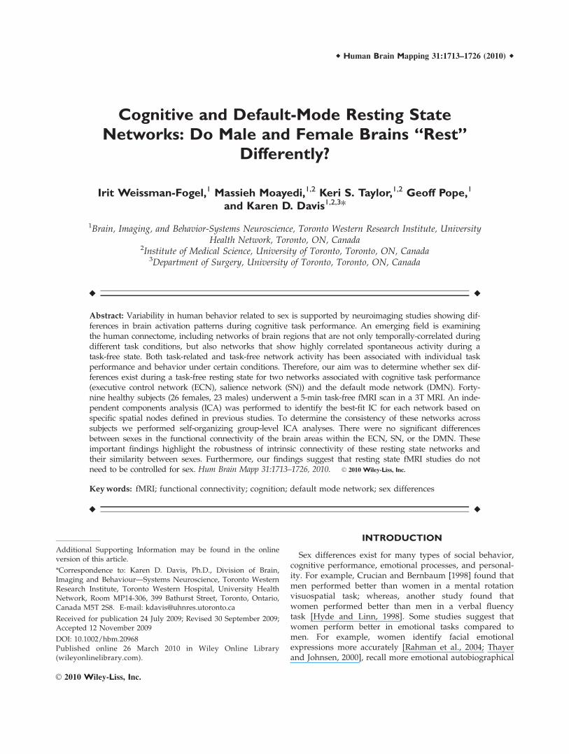
assigned to every voxel in the brain. Identification of theindividual best-fit ICs for the DMN, ECN, and SN wasbased on a three-step spatial and temporal pattern-match-ing process in each individual using specific spatial nodesfrom previous studies as well as spatial maps and time-course activity extracted from seed region analyses thatwe ran for each individual (see Fig. 1).
A seed-region analysis was performed using tworegions of interest (ROIs) identified as major nodes of
functional maps of previously reported cognitive tasks,task-free networks, and anatomical landmarks: The DMNseed was centered on the bilateral posterior cingulate/precuneus (PCC/PCu) (BA23/31 for PCC and BA7 forPCu: x ¼ �7, y ¼ �55, z ¼ 26; size: 12 � 12 � 27 mm3,bilaterally) [De Luca et al., 2006; Fox et al., 2005; Fransson,2005; Greicius et al., 2003; Harrison et al., 2008; Semino-wicz and Davis, 2007]. The seed for the ECN and SN wasthe right DLPFC (BA9 in the middle frontal gyrus; x ¼ 42,
Figure 1.
Three-step spatial and temporal-matching process used for
selection of individual best-fit independent components. To
select the executive control network (ECN) and salience net-
work (SN) components for each individual, the time course of
30 independent components (ICs) was correlated with the aver-
age time course from right dorsolateral prefrontal cortex (right
DLPFC) [region of interest (ROI) in our seed region analysis].
A: Two components from one representative subject are shown.
The most highly correlated ICs based on the Pearson correla-
tion coefficient was transferred to the second step in which the
spatial maps of these ICs were inspected for spatial matching
with specific nodes from previous studies and with the individual
right DLPFC-seed region analysis map (B1, B2). B: The left col-
umn in B1 shows the seed-region analysis map from a represen-
tative subject. In the right column two IC-spatial maps (green
and blue) from the same representative subject are superim-
posed. The two most matched ICs from each subject were iden-
tified as ECN (green) and SN (blue) based on specific spatial
nodes (B2). C: A fingerprint analysis was used to characterize
the chosen ICs for each network in space and time domain and
to verify the signal as arising from a neural BOLD response. The
11 fingerprint parameters are: (1) Degree of clustering; (2)
Skewness; (3) Kurtosis; (4) Spatial entropy; (5) One lag autocor-
relation; (6) Temporal entropy; (7) Power in the band: 0–0.008
Hz; (8) Power in the band: 0.008–0.02 Hz; (9) Power in the
band: 0.02–0.05 Hz; (10) Power in the band: 0.05–0.1 Hz; (11)
Power in the band: 0.1–0.25 Hz. The identical process was done
for the default mode network except that the ROI for temporal
correction and spatial matching was the posterior cingulate cor-
tex/precuneus and its functional connectivity map.
r Weissman-Fogel et al. r
r 1716 r
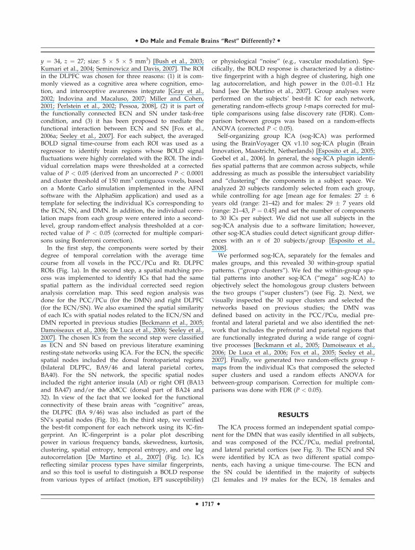
y ¼ 34, z ¼ 27; size: 5 � 5 � 5 mm3) [Bush et al., 2003;Kumari et al., 2004; Seminowicz and Davis, 2007]. The ROIin the DLPFC was chosen for three reasons: (1) it is com-monly viewed as a cognitive area where cognition, emo-tion, and interoceptive awareness integrate [Gray et al.,2002; Indovina and Macaluso, 2007; Miller and Cohen,2001; Perlstein et al., 2002; Pessoa, 2008], (2) it is part ofthe functionally connected ECN and SN under task-freecondition, and (3) it has been proposed to mediate thefunctional interaction between ECN and SN [Fox et al.,2006a; Seeley et al., 2007]. For each subject, the averagedBOLD signal time-course from each ROI was used as aregressor to identify brain regions whose BOLD signalfluctuations were highly correlated with the ROI. The indi-vidual correlation maps were thresholded at a correctedvalue of P < 0.05 (derived from an uncorrected P < 0.0001and cluster threshold of 150 mm3 contiguous voxels, basedon a Monte Carlo simulation implemented in the AFNIsoftware with the AlphaSim application) and used as atemplate for selecting the individual ICs corresponding tothe ECN, SN, and DMN. In addition, the individual corre-lation maps from each group were entered into a second-level, group random-effect analysis thresholded at a cor-rected value of P < 0.05 (corrected for multiple compari-sons using Bonferroni correction).
In the first step, the components were sorted by theirdegree of temporal correlation with the average timecourse from all voxels in the PCC/PCu and Rt. DLPFCROIs (Fig. 1a). In the second step, a spatial matching pro-cess was implemented to identify ICs that had the samespatial pattern as the individual corrected seed regionanalysis correlation map. This seed region analysis wasdone for the PCC/PCu (for the DMN) and right DLPFC(for the ECN/SN). We also examined the spatial similarityof each ICs with spatial nodes related to the ECN/SN andDMN reported in previous studies [Beckmann et al., 2005;Damoiseaux et al., 2006; De Luca et al., 2006; Seeley et al.,2007]. The chosen ICs from the second step were classifiedas ECN and SN based on previous literature examiningresting-state networks using ICA. For the ECN, the specificspatial nodes included the dorsal frontoparietal regions(bilateral DLPFC, BA9/46 and lateral parietal cortex,BA40). For the SN network, the specific spatial nodesincluded the right anterior insula (AI) or right OFI (BA13and BA47) and/or the aMCC (dorsal part of BA24 and32). In view of the fact that we looked for the functionalconnectivity of these brain areas with ‘‘cognitive’’ areas,the DLPFC (BA 9/46) was also included as part of theSN’s spatial nodes (Fig. 1b). In the third step, we verifiedthe best-fit component for each network using its IC-fin-gerprint. An IC-fingerprint is a polar plot describingpower in various frequency bands, skewedness, kurtosis,clustering, spatial entropy, temporal entropy, and one lagautocorrelation [De Martino et al., 2007] (Fig. 1c). ICsreflecting similar process types have similar fingerprints,and so this tool is useful to distinguish a BOLD responsefrom various types of artifact (motion, EPI susceptibility)
or physiological ‘‘noise’’ (e.g., vascular modulation). Spe-cifically, the BOLD response is characterized by a distinc-tive fingerprint with a high degree of clustering, high onelag autocorrelation, and high power in the 0.01–0.1 Hzband [see De Martino et al., 2007]. Group analyses wereperformed on the subjects’ best-fit IC for each network,generating random-effects group t-maps corrected for mul-tiple comparisons using false discovery rate (FDR). Com-parison between groups was based on a random-effectsANOVA (corrected P < 0.05).
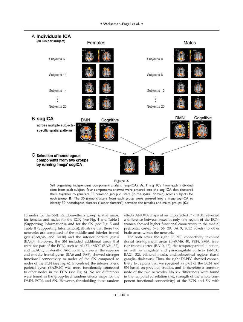
Self-organizing group ICA (sog-ICA) was performedusing the BrainVoyager QX v1.10 sog-ICA plugin (BrainInnovation, Maastricht, Netherlands) [Esposito et al., 2005;Goebel et al., 2006]. In general, the sog-ICA plugin identi-fies spatial patterns that are common across subjects, whileaddressing as much as possible the intersubject variabilityand ‘‘clustering’’ the components in a subject space. Weanalyzed 20 subjects randomly selected from each group,while controlling for age [mean age for females: 27 � 6years old (range: 21–42) and for males: 29 � 7 years old(range: 21–43, P ¼ 0.45] and set the number of componentsto 30 ICs per subject. We did not use all subjects in thesog-ICA analysis due to a software limitation; however,other sog-ICA studies could detect significant group differ-ences with an n of 20 subjects/group [Esposito et al.,2008].
We performed sog-ICA, separately for the females andmales groups, and this revealed 30 within-group spatialpatterns. (‘‘group clusters’’). We fed the within-group spa-tial patterns into another sog-ICA (‘‘mega’’ sog-ICA) toobjectively select the homologous group clusters betweenthe two groups (‘‘super clusters’’) (see Fig. 2). Next, wevisually inspected the 30 super clusters and selected thenetworks based on previous studies; the DMN wasdefined based on activity in the PCC/PCu, medial pre-frontal and lateral parietal and we also identified the net-work that includes the prefrontal and parietal regions thatare functionally integrated during a wide range of cogni-tive processes [Beckmann et al., 2005; Damoiseaux et al.,2006; De Luca et al., 2006; Fox et al., 2005; Seeley et al.,2007]. Finally, we generated two random-effects group t-maps from the individual ICs that composed the selectedsuper clusters and used a random effects ANOVA forbetween-group comparison. Correction for multiple com-parisons was done with FDR (P < 0.05).
RESULTS
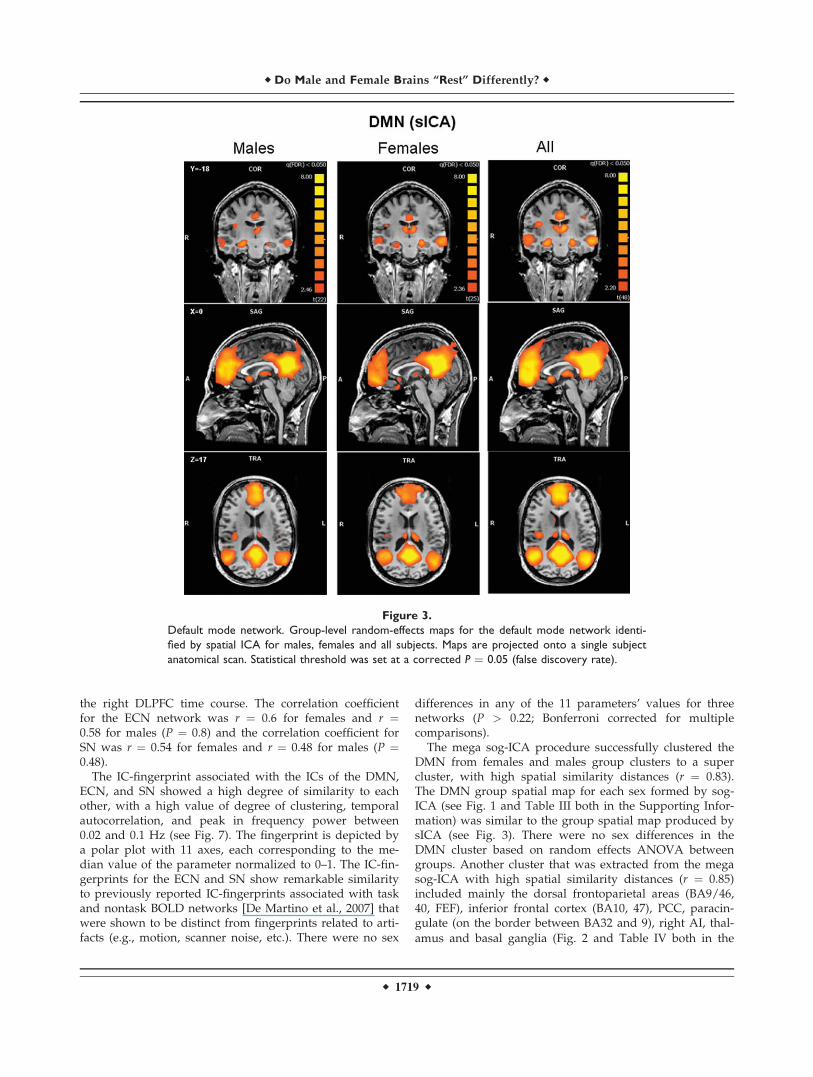
The ICA process formed an independent spatial compo-nent for the DMN that was easily identified in all subjects,and was composed of the PCC/PCu, medial prefrontal,and lateral parietal cortices (see Fig. 3). The ECN and SNwere identified by ICA as two different spatial compo-nents, each having a unique time-course. The ECN andthe SN could be identified in the majority of subjects(21 females and 19 males for the ECN, 18 females and
r Do Male and Female Brains ‘‘Rest’’ Differently? r
r 1717 r
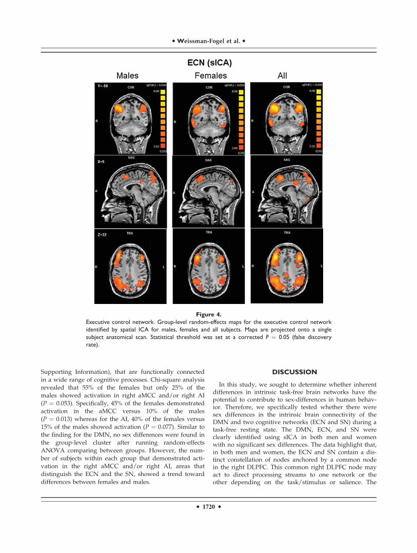
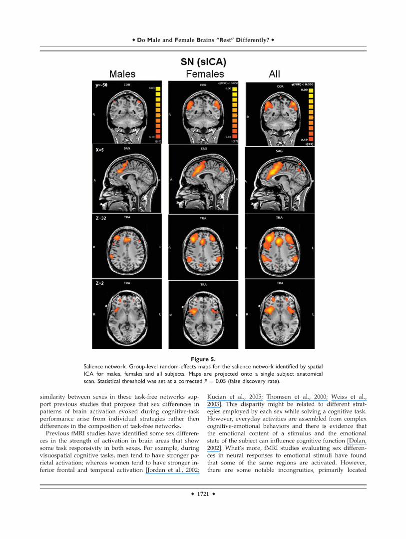
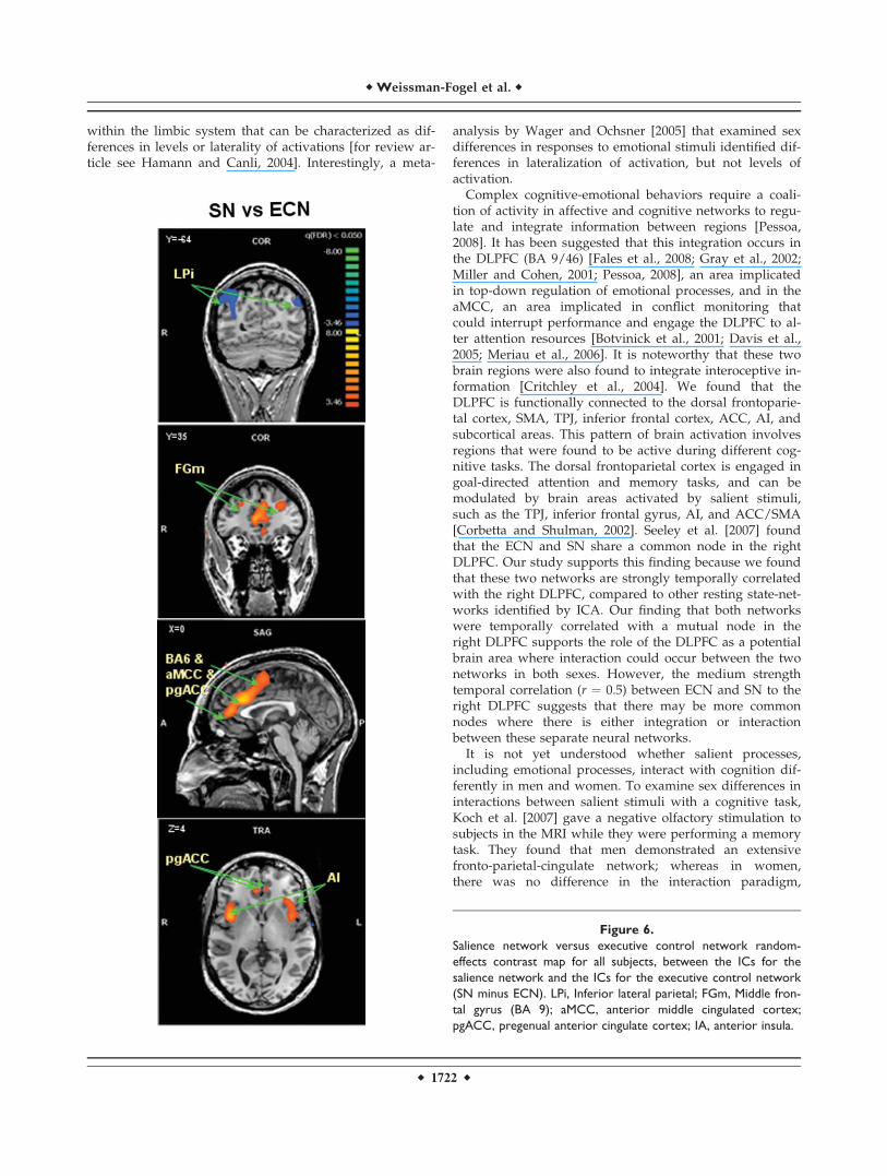
16 males for the SN). Random-effects group spatial maps,for females and males for the ECN (see Fig. 4 and Table I(Supporting Information)), and for the SN (see Fig. 5 andTable II (Supporting Information)), illustrate that these twonetworks are composed of the middle and inferior frontalgyri (BA9/46, and BA10) and the inferior parietal gyrus(BA40). However, the SN included additional areas thatwere not part of the ECN, such as AI/FI, aMCC (BA24, 32),and pgACC, bilaterally. Additionally, areas in the superiorand middle frontal gyrus (BA6 and BA9), showed strongerfunctional connectivity to nodes of the SN compared tonodes of the ECN (see Fig. 6). In contrast, the inferior lateralparietal gyrus (BA39,40) was more functionally connectedto other nodes in the ECN (see Fig. 6). No sex differenceswere found in the group-level random effects maps for theDMN, ECN, and SN. However, thresholding these random
effects ANOVA maps at an uncorrected P < 0.001 revealeda difference between sexes in only one region of the ECN;women showed higher functional connectivity in the medialprefrontal cortex (�3, 56, 29; BA 9, 2012 voxels) to otherbrain areas within the network.
For both sexes the right DLPFC connectivity involveddorsal frontoparietal areas (BA9/46, 40, FEF), SMA, infe-rior frontal cortex (BA10, 47), the temporoparietal junction,as well as cingulate and paracingulate cortices (aMCC;BA24, 32), bilateral insula, and subcortical regions (basalganglia, thalamus). Thus, the right DLPFC showed connec-tivity to regions that we specified as part of the ECN andSN based on previous studies, and is therefore a commonnode of the two networks. No sex differences were foundin the temporal correlation (i.e., strength of the whole com-ponent functional connectivity) of the ECN and SN with
Figure 2.
Self organizing independent component analysis (sog-ICA). A: Thirty ICs from each individual
(one from each subject, four components shown) were entered into the sog-ICA that clustered
them together to generate 30 common group clusters (in the spatial domain) across subjects for
each group. B: The 30 group clusters from each group were entered into a mega-sog-ICA to
identify 30 homologous clusters (‘‘super clusters’’) between the females and males groups (C).
r Weissman-Fogel et al. r
r 1718 r
the right DLPFC time course. The correlation coefficientfor the ECN network was r ¼ 0.6 for females and r ¼0.58 for males (P ¼ 0.8) and the correlation coefficient forSN was r ¼ 0.54 for females and r ¼ 0.48 for males (P ¼0.48).
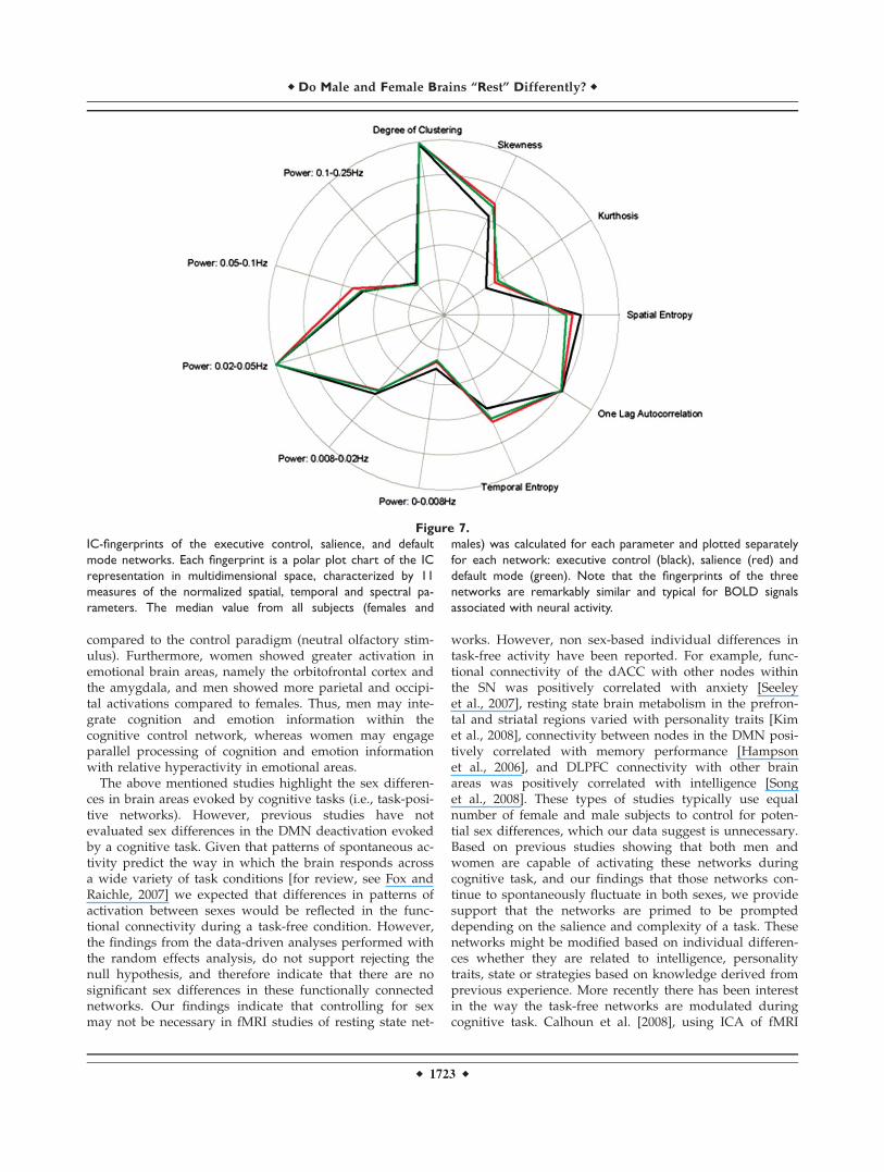
The IC-fingerprint associated with the ICs of the DMN,ECN, and SN showed a high degree of similarity to eachother, with a high value of degree of clustering, temporalautocorrelation, and peak in frequency power between0.02 and 0.1 Hz (see Fig. 7). The fingerprint is depicted bya polar plot with 11 axes, each corresponding to the me-dian value of the parameter normalized to 0–1. The IC-fin-gerprints for the ECN and SN show remarkable similarityto previously reported IC-fingerprints associated with taskand nontask BOLD networks [De Martino et al., 2007] thatwere shown to be distinct from fingerprints related to arti-facts (e.g., motion, scanner noise, etc.). There were no sex
differences in any of the 11 parameters’ values for threenetworks (P > 0.22; Bonferroni corrected for multiplecomparisons).
The mega sog-ICA procedure successfully clustered theDMN from females and males group clusters to a supercluster, with high spatial similarity distances (r ¼ 0.83).The DMN group spatial map for each sex formed by sog-ICA (see Fig. 1 and Table III both in the Supporting Infor-mation) was similar to the group spatial map produced bysICA (see Fig. 3). There were no sex differences in theDMN cluster based on random effects ANOVA betweengroups. Another cluster that was extracted from the megasog-ICA with high spatial similarity distances (r ¼ 0.85)included mainly the dorsal frontoparietal areas (BA9/46,40, FEF), inferior frontal cortex (BA10, 47), PCC, paracin-gulate (on the border between BA32 and 9), right AI, thal-
amus and basal ganglia (Fig. 2 and Table IV both in the
Figure 3.
Default mode network. Group-level random-effects maps for the default mode network identi-
fied by spatial ICA for males, females and all subjects. Maps are projected onto a single subject
anatomical scan. Statistical threshold was set at a corrected P ¼ 0.05 (false discovery rate).
r Do Male and Female Brains ‘‘Rest’’ Differently? r
r 1719 r
Supporting Information), that are functionally connected
in a wide range of cognitive processes. Chi-square analysis
revealed that 55% of the females but only 25% of the
males showed activation in right aMCC and/or right AI
(P ¼ 0.053). Specifically, 45% of the females demonstrated
activation in the aMCC versus 10% of the males
(P ¼ 0.013) whereas for the AI, 40% of the females versus
15% of the males showed activation (P ¼ 0.077). Similar to
the finding for the DMN, no sex differences were found in
the group-level cluster after running random-effects
ANOVA comparing between groups. However, the num-
ber of subjects within each group that demonstrated acti-
vation in the right aMCC and/or right AI, areas that
distinguish the ECN and the SN, showed a trend toward
differences between females and males.
DISCUSSION
In this study, we sought to determine whether inherentdifferences in intrinsic task-free brain networks have thepotential to contribute to sex-differences in human behav-ior. Therefore, we specifically tested whether there weresex differences in the intrinsic brain connectivity of theDMN and two cognitive networks (ECN and SN) during atask-free resting state. The DMN, ECN, and SN wereclearly identified using sICA in both men and womenwith no significant sex differences. The data highlight that,in both men and women, the ECN and SN contain a dis-tinct constellation of nodes anchored by a common nodein the right DLPFC. This common right DLPFC node mayact to direct processing streams to one network or theother depending on the task/stimulus or salience. The
Figure 4.
Executive control network. Group-level random-effects maps for the executive control network
identified by spatial ICA for males, females and all subjects. Maps are projected onto a single
subject anatomical scan. Statistical threshold was set at a corrected P ¼ 0.05 (false discovery
rate).
r Weissman-Fogel et al. r
r 1720 r
similarity between sexes in these task-free networks sup-port previous studies that propose that sex differences inpatterns of brain activation evoked during cognitive-taskperformance arise from individual strategies rather thendifferences in the composition of task-free networks.
Previous fMRI studies have identified some sex differen-ces in the strength of activation in brain areas that showsome task responsivity in both sexes. For example, duringvisuospatial cognitive tasks, men tend to have stronger pa-rietal activation; whereas women tend to have stronger in-ferior frontal and temporal activation [Jordan et al., 2002;
Kucian et al., 2005; Thomsen et al., 2000; Weiss et al.,2003]. This disparity might be related to different strat-egies employed by each sex while solving a cognitive task.However, everyday activities are assembled from complexcognitive-emotional behaviors and there is evidence thatthe emotional content of a stimulus and the emotionalstate of the subject can influence cognitive function [Dolan,2002]. What’s more, fMRI studies evaluating sex differen-ces in neural responses to emotional stimuli have foundthat some of the same regions are activated. However,there are some notable incongruities, primarily located
Figure 5.
Salience network. Group-level random-effects maps for the salience network identified by spatial
ICA for males, females and all subjects. Maps are projected onto a single subject anatomical
scan. Statistical threshold was set at a corrected P ¼ 0.05 (false discovery rate).
r Do Male and Female Brains ‘‘Rest’’ Differently? r
r 1721 r
within the limbic system that can be characterized as dif-ferences in levels or laterality of activations [for review ar-ticle see Hamann and Canli, 2004]. Interestingly, a meta-
analysis by Wager and Ochsner [2005] that examined sexdifferences in responses to emotional stimuli identified dif-ferences in lateralization of activation, but not levels ofactivation.
Complex cognitive-emotional behaviors require a coali-tion of activity in affective and cognitive networks to regu-late and integrate information between regions [Pessoa,2008]. It has been suggested that this integration occurs inthe DLPFC (BA 9/46) [Fales et al., 2008; Gray et al., 2002;Miller and Cohen, 2001; Pessoa, 2008], an area implicatedin top-down regulation of emotional processes, and in theaMCC, an area implicated in conflict monitoring thatcould interrupt performance and engage the DLPFC to al-ter attention resources [Botvinick et al., 2001; Davis et al.,2005; Meriau et al., 2006]. It is noteworthy that these twobrain regions were also found to integrate interoceptive in-formation [Critchley et al., 2004]. We found that theDLPFC is functionally connected to the dorsal frontoparie-tal cortex, SMA, TPJ, inferior frontal cortex, ACC, AI, andsubcortical areas. This pattern of brain activation involvesregions that were found to be active during different cog-nitive tasks. The dorsal frontoparietal cortex is engaged ingoal-directed attention and memory tasks, and can bemodulated by brain areas activated by salient stimuli,such as the TPJ, inferior frontal gyrus, AI, and ACC/SMA[Corbetta and Shulman, 2002]. Seeley et al. [2007] foundthat the ECN and SN share a common node in the rightDLPFC. Our study supports this finding because we foundthat these two networks are strongly temporally correlatedwith the right DLPFC, compared to other resting state-net-works identified by ICA. Our finding that both networkswere temporally correlated with a mutual node in theright DLPFC supports the role of the DLPFC as a potentialbrain area where interaction could occur between the twonetworks in both sexes. However, the medium strengthtemporal correlation (r ¼ 0.5) between ECN and SN to theright DLPFC suggests that there may be more commonnodes where there is either integration or interactionbetween these separate neural networks.
It is not yet understood whether salient processes,including emotional processes, interact with cognition dif-ferently in men and women. To examine sex differences ininteractions between salient stimuli with a cognitive task,Koch et al. [2007] gave a negative olfactory stimulation tosubjects in the MRI while they were performing a memorytask. They found that men demonstrated an extensivefronto-parietal-cingulate network; whereas in women,there was no difference in the interaction paradigm,
Figure 6.
Salience network versus executive control network random-
effects contrast map for all subjects, between the ICs for the
salience network and the ICs for the executive control network
(SN minus ECN). LPi, Inferior lateral parietal; FGm, Middle fron-
tal gyrus (BA 9); aMCC, anterior middle cingulated cortex;
pgACC, pregenual anterior cingulate cortex; IA, anterior insula.
r Weissman-Fogel et al. r
r 1722 r
compared to the control paradigm (neutral olfactory stim-ulus). Furthermore, women showed greater activation inemotional brain areas, namely the orbitofrontal cortex andthe amygdala, and men showed more parietal and occipi-tal activations compared to females. Thus, men may inte-grate cognition and emotion information within thecognitive control network, whereas women may engageparallel processing of cognition and emotion informationwith relative hyperactivity in emotional areas.
The above mentioned studies highlight the sex differen-ces in brain areas evoked by cognitive tasks (i.e., task-posi-tive networks). However, previous studies have notevaluated sex differences in the DMN deactivation evokedby a cognitive task. Given that patterns of spontaneous ac-tivity predict the way in which the brain responds acrossa wide variety of task conditions [for review, see Fox andRaichle, 2007] we expected that differences in patterns ofactivation between sexes would be reflected in the func-tional connectivity during a task-free condition. However,the findings from the data-driven analyses performed withthe random effects analysis, do not support rejecting thenull hypothesis, and therefore indicate that there are nosignificant sex differences in these functionally connectednetworks. Our findings indicate that controlling for sexmay not be necessary in fMRI studies of resting state net-
works. However, non sex-based individual differences intask-free activity have been reported. For example, func-tional connectivity of the dACC with other nodes withinthe SN was positively correlated with anxiety [Seeleyet al., 2007], resting state brain metabolism in the prefron-tal and striatal regions varied with personality traits [Kimet al., 2008], connectivity between nodes in the DMN posi-tively correlated with memory performance [Hampsonet al., 2006], and DLPFC connectivity with other brainareas was positively correlated with intelligence [Songet al., 2008]. These types of studies typically use equalnumber of female and male subjects to control for poten-tial sex differences, which our data suggest is unnecessary.Based on previous studies showing that both men andwomen are capable of activating these networks duringcognitive task, and our findings that those networks con-tinue to spontaneously fluctuate in both sexes, we providesupport that the networks are primed to be prompteddepending on the salience and complexity of a task. Thesenetworks might be modified based on individual differen-ces whether they are related to intelligence, personalitytraits, state or strategies based on knowledge derived fromprevious experience. More recently there has been interestin the way the task-free networks are modulated duringcognitive task. Calhoun et al. [2008], using ICA of fMRI
Figure 7.
IC-fingerprints of the executive control, salience, and default
mode networks. Each fingerprint is a polar plot chart of the IC
representation in multidimensional space, characterized by 11
measures of the normalized spatial, temporal and spectral pa-
rameters. The median value from all subjects (females and
males) was calculated for each parameter and plotted separately
for each network: executive control (black), salience (red) and
default mode (green). Note that the fingerprints of the three
networks are remarkably similar and typical for BOLD signals
associated with neural activity.
r Do Male and Female Brains ‘‘Rest’’ Differently? r
r 1723 r
data, found that the task-free networks are temporally andspatially modulated during cognitive task conditions. Thisraises the possibility that these task-free networks may bemodulated differently in both sexes and these modulationsmight even correlate with behavior in specific brain areas.
One limitation of our study arises from the issue of theestimated number of ICs for fMRI data analysis. Typicallythere are a large number of ICs in sICA, up to the numberof time samples in the scan, and so there are a variety ofmethods to estimate the optimal number of components.In this study, based on our experience and in accordancewith Brainvoyager recommendations, we used 30 compo-nents, equivalent to 20% of the 150 time points in ourtask-free scan that generate stable results in ICA. Otherstudies used principle component analysis prior to ICAdecomposition [Beckmann et al., 2005; Damoiseaux et al.,2006; De Luca et al., 2006], which determines how muchnoise is left in the data but does not take into account theactual structure of interesting signal in the data. Further-more, we did not use variance-based automated methodsfor choosing the number of components [Beckmann et al.,2005; Damoiseaux et al., 2006; De Luca et al., 2006, Liet al., 2007], because we were not concerned with the var-iance of the signals. Rather, we were concerned with opti-mizing the signals’ spatial patterns relative to ourhypotheses. Moreover, Li et al. [2007] proposed a newmethod to obtain a set of effectively independent andidentically distributed data samples for order selection.Although the networks that we found during task-freecondition are identical to those that were found in previ-ous studies, they are highly dependent on our choice ofthe number of ICs in ICA.
Another potential limitation in our study is that ourstudy participants spanned a wide age range from 21 to 42years old. This range was based on the commonly usedrange of ages in imaging studies. Although age-relatedchanges in brain activity under task-free condition havebeen reported, they were found for subjects older than ours[Damoiseaux et al., 2008; Sambataro et al., in press]. Fur-thermore, we age-matched our subjects between the twogroups. Therefore, although we cannot totally rule out ageeffects, their contribution to the findings are likely minimal.
In conclusion, we have found a remarkable similaritybetween sexes in spatial topography of activation of theDMN, ECN, and SN. This finding can be used for futurestudies as an a priori hypothesis that brains of both sexeswill potentially respond in the same way during cognitivetasks. Furthermore, our results advocate that resting statefMRI studies can be done without controlling for sex.
ACKNOWLEDGMENTS
The authors thank Fabrizio Esposito (Brain InnovationB.V.) for guidance in the use and interpretation of groupICA in BrainVoyager. Karen D. Davis is a Canada Re-search Chair in Brain and Behaviour. This study was sup-
ported from funds from the CRC program and a CIHRgrant to KDD.
REFERENCES
Beckmann CF, DeLuca M, Devlin JT, Smith SM (2005): Investiga-tions into resting-state connectivity using independent compo-nent analysis. Philos Trans R Soc Lond B Biol Sci 360:1001–1013.
Botvinick MM, Braver TS, Barch DM, Carter CS, Cohen JD (2001):Conflict monitoring and cognitive control. Psychol Rev108:624–652.
Buckner RL, Vincent JL (2007): Unrest at rest: The importance ofthe default activity and spontaneous network corellations.Neuroimage 37:1091–1099.
Bush G, Luu P, Posner MI (2000): Cognitive and emotional influ-ences in anterior cingulate cortex. Trends Cogn Sci 4:215–222.
Bush G, Shin LM, Holmes J, Rosen BR, Vogt BA (2003): Themulti-source interference task: Validation study with fMRI inindividual subjects. Mol Psychiatry 8:60–70.
Butler T, Imperato-McGinley J, Pan H, Voyer D, Cunningham-Bussel AC, Chang L, Zhu YS, Cordero JJ, Stern E, SilbersweigD (2007): Sex specificity of ventral anterior cingulate cortexsuppression during a cognitive task. Hum Brain Mapp28:1206–1212.
Cahill L (2003): Sex-related influences on the neurobiology of emo-tionally influenced memory. Ann N Y Acad Sci 985:163–173.
Cahill L, Haier RJ, White NS, Fallon J, Kilpatrick L, Lawrence C,Potkin SG, Alkire MT (2001): Sex-related difference in amyg-dala activity during emotionally influenced memory storage.Neurobiol Learn Mem 75:1–9.
Calhoun VD, Kiehl KA, Pearlson GD (2008): Modulation of tem-porally coherent brain networks estimated using ICA at restand during cognitive task. Hum Brain Mapp 29:828–838.
Canli T, Zhao Z, Brewer J, Gabrieli JD, Cahill L (2000): Event-related activation in the human amygdala associates with latermemory for individual emotional experience. J Neurosci20:RC99.
Canli T, Desmond JE, Zhao Z, Gabrieli JD (2002): Sex differencesin the neural basis of emotional memories. Proc Natl Acad SciUSA 99:10789–10794.
Corbetta M, Shulman GL (2002): Control of goal-directed andstimulus-driven attention in the brain. Nat Rev Neurosci3:201–215.
Critchley HD (2005): Neural mechanisms of autonomic, affective,and cognitive integration. J Comp Neurol 493:154–166.
Critchley HD, Wiens S, Rotshtein P, Ohman A, Dolan RJ (2004):Neural systems supporting interoceptive awareness. Nat Neu-rosci 7:189–195.
Crucian GP, Berenbaum SA (1998): Sex Differences in Right Hemi-sphere Tasks. Brain Cogn 36:377–389.
Curtis CE, D’Esposito M (2003): Persistent activity in the prefron-tal cortex during working memory. Trends Cogn Sci 7:415–423.
Damoiseaux JS, Rombouts SA, Barkhof F, Scheltens P, Stam CJ,Smith SM, Beckmann CF (2006): Consistent resting-state net-works across healthy subjects. Proc Natl Acad Sci USA103:13848–13853.
Damoiseaux JS, Beckmann CF, Arigita EJ, Barkhof F, Scheltens P,Stam CJ, Smith SM, Rombouts SA (2008): Reduced resting-statebrain activity in the ‘‘default network’’ in normal aging. CerebCortex 18:1856–1864.
r Weissman-Fogel et al. r
r 1724 r
Davis KD, Taylor KS, Hutchison WD, Dostrovsky JO, McAndrewsMP, Richter EO, Lozano AM (2005): Human anterior cingulatecortex neurons encode cognitive and emotional demands. JNeurosci 25:8402–8406.
Davis PJ (1999): Gender differences in autobiographical memoryfor childhood emotional experiences. J Pers Soc Psychol76:498–510.
De Luca M, Beckmann CF, De Stefano N, Matthews PM, SmithSM (2006): fMRI resting state networks define distinct modesof long-distance interactions in the human brain. Neuroimage29:1359–1367.
De Martino F, Gentile F, Esposito F, Balsi M, Di Salle F, Goebel R,Formisano E (2007): Classification of fMRI independent com-ponents using IC-fingerprints and support vector machineclassifiers. Neuroimage 34:177–194.
Dolan RJ (2002): Emotion, cognition, and behavior. Science298:1191–1194.
Esposito F, Scarabino T, Hyvarinen A, Himberg J, Formisano E,Comani S, Tedeschi G, Goebel R, Seifritz E, Di Salle F (2005):Independent component analysis of fMRI group studies byself-organizing clustering. Neuroimage 25:193–205.
Esposito F, Aragri A, Pesaresi I, Cirillo S, Tedeschi G, Marciano E,Goebel R, Di Salle F (2008): Independent component model ofthe default-mode brain function: Combining individual-leveland population-level analyses in resting-state fMRI. MagnReson Imaging 26:905–913.
Fales CL, Barch DM, Rundle MM, Mintun MA, Snyder AZ, CohenJD, Mathews J, Sheline YI (2008): Altered emotional interfer-ence processing in affective and cognitive-control brain cir-cuitry in major depression. Biol Psychiatry 63:377–384.
Fox MD, Raichle ME (2007): Spontaneous fluctuations in brain ac-tivity observed with functional magnetic resonance imaging.Nat Rev Neurosci 8:700–711.
Fox MD, Snyder AZ, Vincent JL, Corbetta M, Van Essen DC,Raichle ME (2005): The human brain is intrinsically organizedinto dynamic, anticorrelated functional networks. Proc NatlAcad Sci USA 102:9673–9678.
Fox MD, Corbetta M, Snyder AZ, Vincent JL, Raichle ME (2006a):Spontaneous neuronal activity distinguishes human dorsal andventral attention systems. Proc Natl Acad Sci USA 103:10046–10051.
Fox MD, Snyder AZ, Zacks JM, Raichle ME (2006b): Coherentspontaneous activity accounts for the trail-to-trail variability inhuman evoked brain responses. Nat Neurosci 9:23–25.
Fransson P (2005): Spontaneous low-frequency BOLD signal fluctu-ations: An fMRI investigation of the resting-state default modeof brain function hypothesis. Hum Brain Mapp 26:15–29.
Frings L, Wagner K, Unterrainer J, Spreer J, Halsband U, Schulze-Bonh-age A (2006): Gender-related differences in lateralization of hippo-campal activation and cognitive strategy. Neuroreport 17:417–421.
Goebel R, Esposito F, Formisano E (2006): Analysis of functionalimage analysis contest (FIAC) data with brainvoyager QX:From single-subject to cortically aligned group general linearmodel analysis and self-organizing group independent compo-nent analysis. Hum Brain Mapp 27:392–401.
Gray JR, Braver TS, Raichle ME (2002): Integration of emotion andcognition in the lateral prefrontal cortex. Proc Natl Acad SciUSA 99:4115–4120.
Greicius MD, Krasnow B, Reiss AL, Menon V (2003): Functionalconnectivity in the resting brain: A network analysis of thedefault mode hypothesis. Proc Natl Acad Sci USA 100:253–258.
Hamann S, Canli T (2004): Individual differences in emotion proc-essing. Curr Opin Neurobiol 14:233–238.
Hamann SB, Ely TD, Grafton ST, Kilts CD (1999): Amygdala activ-ity related to enhanced memory for pleasant and aversivestimuli. Nat Neurosci 2:289–293.
Hampson M, Driesen NR, Skudlarski P, Gore JC, Constable RT(2006): Brain connectivity related to working memory perform-ance. J Neurosci 26:13338–13343.
Harrison BJ, Pujol J, Lopez-Sola M, Hernandez-Ribas R, Deus J,Ortiz H, Soriano-Mas C, Yucel M, Pantelis C, Cardoner N(2008): Consistency and functional specialization in the defaultmode brain network. Proc Natl Acad Sci USA 105:9781–9786.
Hyde JS, Linn MC (1998): Gender differences in verbal ability: Ameta analysis. Psychol Bull 104:53–69.
Indovina I, Macaluso E (2007): Dissociation of stimulus relevanceand saliency factors during shifts of visuospatial attention.Cereb Cortex 17:1701–1711.
Jordan K, Wustenberg T, Heinze HJ, Peters M, Jancke L (2002):Women and men exhibit different cortical activation patternsduring mental rotation tasks. Neuropsychologia 40:2397–23408.
Kelly AMC, Uddin LQ, Biswal BB, Castellanos FX, Milham MP(2008): Competition between functional brain networks medi-ates behavioral variability. Neuroimage 39:527–537.
Kerns JG, Cohen JD, MacDonald AW III, Cho RY, Stenger VA,Carter CS (2004): Anterior cingulate conflict monitoring andadjustments in control. Science 303:1023–1026.
Kim S, Hwang J, Park H, Kim S (2008): Resting brain metaboliccorrelates of neuroticism and extraversion in young men. Neu-roreport 19:883–886.
Koch K, Pauly K, Kellermann T, Seiferth NY, Reske M, Backes V,Stocker T, Shah NJ, Amunts K, Kircher T, Schneider F, HabelU (2007): Gender differences in the cognitive control of emo-tion: An fMRI study. Neuropsychologia 45:2744–2754.
Kucian K, Loenneker T, Dietrich T, Martin E, von Aster M (2005): Gen-ser differences in brainactivation patterns during mental rotationand number related cognitive task. Psychol Sci 47:112–131.
Kumari V, ffytche DH, Williams SC, Gray JA (2004): Personalitypredicts brain responses to cognitive demands. J Neurosci24:10636–10641.
Li YO, Adali T, Calhoun VD (2007): Estimating the number of in-dependent components for functional magnetic resonanceimaging data. Hum Brain Mapp 11:1251–1266.
Meriau K, Wartenburger I, Kazzer P, Prehn K, Lammers CH, van derMeer E, Villringer A, Heekeren HR (2006): A neural networkreflecting individual differences in cognitive processing of emotionsduring perceptual decision making. Neuroimage 33:1016–1027.
Miller EK, Cohen JD (2001): An integrative theory of prefrontalcortex function. Annu Rev Neurosci 24:167–202.
Ochsner KN, Gross JJ (2005): The cognitive control of emotion.Trends Cogn Sci 9:242–249.
Perlstein WM, Elbert T, Stenger VA (2002): Dissociation in humanprefrontal cortex of affective influences on working memory-related activity. Proc Natl Acad Sci USA 99:1736–1741.
Pessoa L (2008): On the relationship between emotion and cogni-tion. Nat Rev Neurosci 9:148–158.
Pollatos O, Gramann K, Schandry R (2007a): Neural systems con-necting interoceptive awareness and feelings. Hum BrainMapp 28:9–18.
Pollatos O, Schandry R, Auer DP, Kaufmann C (2007b): Brainstructures mediating cardiovascular arousal and interoceptiveawareness. Brain Res 1141:178–87.
Rahman Q, Wilson GD, Abrahams S (2004): Sex, sexual orienta-tion, and identification of positive and negative facial affect.Brain Cogn 54:179–185.
r 1725 r
r Do Male and Female Brains ‘‘Rest’’ Differently? r
Sambataro F, Murty VP, Callicott JH, Tan HY, Das S, WeinbergerDR, Mattay VS: Age-related alterations in default mode net-work: Impact on working memory performance. NeurobiolAging (2008) Jul 30 [Epub ahead of print].
Seeley WW, Menon V, Schatzberg AF, Keller J, Glover GH, KennaH, Reiss AL, Greicius MD (2007): Dissociable intrinsic connec-tivity networks for salience processing and executive control. JNeurosci 27:2349–2356.
Seminowicz DA, Davis KD (2007): Interactions of pain intensityand cognitive load: The brain stays on task. Cereb Cortex17:1412–1422.
Song M, Zhou Y, Li J, Liu Y, Tian L, Yu C, Jiang T (2008): Brainspontaneous functional connectivity and intelligence. Neuro-image 41:1168–1176.
Talairach J, Tournoux, P (1988): Coplanar Stereotaxic Atlas of theHuman Brain. New York: Thieme Medical Publishers.
Thayer JF, Johnsen BH (2000): Sex differences in judgement of fa-cial affect: A multivariate analysis of recognition errors. ScandJ Psychol 41:243–246.
Thomsen T, Hugdahl K, Ersland L, Barndon R, Lundervold A,Smievoll AI, Roscher BE, Sundberg H (2000): Functionalmagnetic resonance imaging (fMRI) study of sex differencesin a mental rotation task. Med Sci Monit 6:1186–1196.
Vogt BA (2005): Pain and emotion interactions in subregions ofthe cingulate gyrus. Nat Rev Neurosci 6:533–544.
Wager TD, Ochsner KN (2005): Sex differences in the emotionalbrain. Neuroreport 16:85–87.
Weiss E, Siedentopf CM, Hofer A, Deisenhammer EA, Hoptman MJ,Kremser C, Golaszewski S, Felber S, Fleischhacker WW, DelazerM (2003): Sex differences in brain activation pattern during avisuospatial cognitive task: A functional magnetic resonance imag-ing study in healthy volunteers. Neurosci Lett 344:169–172.
r Weissman-Fogel et al. r
r 1726 r
Related Documents