Cocaine-Induced Plasticity in the Nucleus Accumbens is Cell- Specific and Develops Without Prolonged Withdrawal Alice Dobi # , Gail K. Seabold # , Christine H. Christensen, Roland Bock, and Veronica A. Alvarez * National Institute on Alcohol Abuse and Alcoholism, NIH, Bethesda, MD 20892 Abstract Cocaine induces plasticity at glutamatergic synapses in the nucleus accumbens (NAc). Withdrawal was suggested to play an important role in the development of this plasticity by studies showing that some changes only appear several weeks after the final cocaine exposure. In this study, the requirement for prolonged withdrawal was evaluated by comparing the changes in glutamatergic transmission induced by two different non-contingent cocaine treatments: a short treatment followed by prolonged withdrawal, and a longer treatment without prolonged withdrawal. Recordings were performed from mouse medium spiny neurons (MSNs) in the NAc at the same time after the first cocaine injection under both treatments. A similar increase in the frequency of glutamate-mediated miniature excitatory postsynaptic currents (mEPSCs) was observed in D1- expressing MSNs after both cocaine treatments, demonstrating that prolonged withdrawal was not required. Furthermore, larger AMPAR to NMDAR ratios, higher spine density and enlarged spine heads were observed in the absence of withdrawal following a long cocaine treatment. These synaptic adaptations expressed in D1-containing MSNs of the NAc core were not further enhanced by protracted withdrawal. In conclusion, a few repeated cocaine injections are enough to trigger adaptations at glutamatergic synapses in D1-expressing MSNs, which although they take time to develop, do not require prolonged cocaine withdrawal. Keywords dendritic spines; dendrite branching; dopamine receptors; glutamate receptors; nucleus accumbens core; shell Introduction The nucleus accumbens (NAc) is implicated in reward-motivated learning when involving both natural and pathological rewards (Hyman et al., 2006; Thomas et al., 2008). It is divided into core and shell subregions (Zahm, 1999; Humphries and Prescott, 2010) and receives major excitatory inputs from the hippocampus, amygdala and prefrontal cortex (Phillipson and Griffiths, 1985; Sesack et al., 1989; McDonald, 1991). These glutamatergic inputs form synapses onto the spines of MSNs. The NAc also receives dopaminergic innervation from the ventral tegmental area (VTA) (Bouyer et al., 1984; Freund et al., 1984) which modulates glutamatergic transmission and mediates the rewarding actions of psychostimulants (Fields et al., 2007; Kalivas, 2009; Kalivas et al., 2009). * Corresponding author address: Veronica A. Alvarez, NIAAA-NIH, 5625 Fishers Lane, Bethesda, MD 20892, [email protected]. # authors contributed equally to this work NIH Public Access Author Manuscript J Neurosci. Author manuscript; available in PMC 2011 August 1. Published in final edited form as: J Neurosci. 2011 February 2; 31(5): 1895–1904. doi:10.1523/JNEUROSCI.5375-10.2011. NIH-PA Author Manuscript NIH-PA Author Manuscript NIH-PA Author Manuscript

Welcome message from author
This document is posted to help you gain knowledge. Please leave a comment to let me know what you think about it! Share it to your friends and learn new things together.
Transcript
Cocaine-Induced Plasticity in the Nucleus Accumbens is Cell-Specific and Develops Without Prolonged Withdrawal
Alice Dobi#, Gail K. Seabold#, Christine H. Christensen, Roland Bock, and Veronica A.Alvarez*
National Institute on Alcohol Abuse and Alcoholism, NIH, Bethesda, MD 20892
AbstractCocaine induces plasticity at glutamatergic synapses in the nucleus accumbens (NAc). Withdrawalwas suggested to play an important role in the development of this plasticity by studies showingthat some changes only appear several weeks after the final cocaine exposure. In this study, therequirement for prolonged withdrawal was evaluated by comparing the changes in glutamatergictransmission induced by two different non-contingent cocaine treatments: a short treatmentfollowed by prolonged withdrawal, and a longer treatment without prolonged withdrawal.Recordings were performed from mouse medium spiny neurons (MSNs) in the NAc at the sametime after the first cocaine injection under both treatments. A similar increase in the frequency ofglutamate-mediated miniature excitatory postsynaptic currents (mEPSCs) was observed in D1-expressing MSNs after both cocaine treatments, demonstrating that prolonged withdrawal was notrequired. Furthermore, larger AMPAR to NMDAR ratios, higher spine density and enlarged spineheads were observed in the absence of withdrawal following a long cocaine treatment. Thesesynaptic adaptations expressed in D1-containing MSNs of the NAc core were not further enhancedby protracted withdrawal. In conclusion, a few repeated cocaine injections are enough to triggeradaptations at glutamatergic synapses in D1-expressing MSNs, which although they take time todevelop, do not require prolonged cocaine withdrawal.
Keywordsdendritic spines; dendrite branching; dopamine receptors; glutamate receptors; nucleus accumbenscore; shell
IntroductionThe nucleus accumbens (NAc) is implicated in reward-motivated learning when involvingboth natural and pathological rewards (Hyman et al., 2006; Thomas et al., 2008). It isdivided into core and shell subregions (Zahm, 1999; Humphries and Prescott, 2010) andreceives major excitatory inputs from the hippocampus, amygdala and prefrontal cortex(Phillipson and Griffiths, 1985; Sesack et al., 1989; McDonald, 1991). These glutamatergicinputs form synapses onto the spines of MSNs. The NAc also receives dopaminergicinnervation from the ventral tegmental area (VTA) (Bouyer et al., 1984; Freund et al., 1984)which modulates glutamatergic transmission and mediates the rewarding actions ofpsychostimulants (Fields et al., 2007; Kalivas, 2009; Kalivas et al., 2009).
*Corresponding author address: Veronica A. Alvarez, NIAAA-NIH, 5625 Fishers Lane, Bethesda, MD 20892,[email protected].#authors contributed equally to this work
NIH Public AccessAuthor ManuscriptJ Neurosci. Author manuscript; available in PMC 2011 August 1.
Published in final edited form as:J Neurosci. 2011 February 2; 31(5): 1895–1904. doi:10.1523/JNEUROSCI.5375-10.2011.
NIH
-PA Author Manuscript
NIH
-PA Author Manuscript
NIH
-PA Author Manuscript

Two subpopulations of GABAergic MSNs have been distinguished based on the expressionof D1- or D2-dopamine receptors (D1R, D2R) in the striatum and NAc (Kebabian andCalne, 1979; Sibley and Monsma, 1992). It remains unclear which of these two receptors isresponsible for the different actions of cocaine in these regions. On one hand,pharmacological, imaging and genetic approaches suggest an involvement of D2R incocaine responses in the CNS (Caine et al., 2002; Thanos et al., 2008; Asensio et al., 2010).On the other hand, D1R play a role in the response to cocaine (Zhang et al., 2002; Zhangand Xu, 2006; Heiman et al., 2008), D1R knockout mice fail to self-administer cocaine(Caine et al., 2007) and repeated administration of D1R antagonists block the cocaine-induced increase in spine density in the NAc (Ren et al., 2010).
Repeated cocaine administration causes plasticity at glutamatergic synapses in the NAc thatis expressed as changes in glutamate receptor surface expression, density of dendritic spinesand synaptic function (Zhang et al., 1998; Robinson et al., 2001; Li et al., 2004; Boudreauand Wolf, 2005; Martin et al., 2006; Kourrich et al., 2007; Huang et al., 2009; Kourrich andThomas, 2009; Moussawi et al., 2009). Many of these changes only develop several weeksafter the final cocaine exposure, suggesting that abstinence is an important mediator of theplasticity (Robinson et al., 2001; Li et al., 2004; Boudreau and Wolf, 2005; Boudreau et al.,2007; Kourrich et al., 2007; Guan et al., 2009). These observations raised the possibility thatwithdrawal itself might be the trigger for the reported functional and morphological changesin the NAc.
Some recent studies, however, have challenged this notion by demonstrating increased spinedensity 2 days after the final cocaine injection (Lee et al., 2006; Kim et al., 2009; Ren et al.,2010). The requirement for withdrawal has not been addressed previously with regards tothe functional plasticity. In this study, we test the role of withdrawal on glutamatergicplasticity and perform behavioral, morphological and electrophysiological analysis to thesame set of animals to correlate functional with morphological changes and evaluate cell-specific actions of cocaine. The results show that D1R expressing MSNs in the NAc core areparticularly susceptible to cocaine exposure and cocaine withdrawal is not required for thefunctional and morphological adaptations in the NAc.
Materials and MethodsAnimals
All experiments were performed in accordance with guidelines from the National Instituteon Alcohol Abuse and Alcoholism Animal Care and Use Committee. Male and female micewere maintained on a 12-h light/dark light cycles and housed in groups of two to four withfree access to food and water. Bacterial artificial chromosome transgenic mice (SwissWebster background) expressing enhanced green fluorescent protein (EGFP) under thecontrol of the D1a dopamine receptor promoter (Drd1a-EGFP, GENSAT) were used in thestudy (Gong et al., 2003). Wild type Swiss Webster mice were also used for one of themorphological experiments.
Drug treatmentMale and female mice (6–9 weeks old) were randomly assigned to one of five groups shownin Table 1 and were subject to one of the three following treatments: 1) short treatment (7consecutive days, groups A and B), 2) long treatment (20 injections: 5 consecutive daysfollowed by 2 injection free days for 4 weeks; groups C and D), and 3) long uninterruptedtreatment (28 consecutive injections, group E). The groups were balanced with respect togender, age and weight. Mice received daily intraperitoneal (i.p.) injections of saline orcocaine (30mg/kg, Sigma, MO) in a novel cage. Injections were performed in small cohorts
Dobi et al. Page 2
J Neurosci. Author manuscript; available in PMC 2011 August 1.
NIH
-PA Author Manuscript
NIH
-PA Author Manuscript
NIH
-PA Author Manuscript
of animals (2-6) in order to allow for electrophysiology and morphology study to occurwithin a narrow range of time (±1 day). Several cohorts were run over a period of 12 monthsduring which time the recorded behavior was very stable.
Locomotor activityAll cages used for behavioral testing were constructed of clear polycarbonate walls (10×6.5inches) with perforated stainless steel flooring. Horizontal activity was detected as infraredbeam crosses (1 inch spacing, 10 beams per cage) using Opto M3 activity monitors(Columbus Instruments, Columbus, OH). Mice were allowed to run freely in the cage for 5minutes prior to each injection. They were then injected with cocaine or saline and placedback into the same cage while ambulatory counts were recorded for 20 minutes under dimillumination (100 lux).
ElectrophysiologyAnimals were sacrificed 1, 20 or 30 days (+1 or 2) after the last cocaine injection accordingto the group described in Table 1. Alternating sagittal brain slices of the NAc were preparedfor the electrophysiological recording (250 μm) or neuronal morphology (200 μm), using avibrating slicer (Leica VT1200, Germany) in choline solution containing (in mM) 25NaHCO3, 1.25 NaH2PO4, 2.5 KCl, 7 MgCl2, 25 glucose, 0.5 CaCl2, 110(CH3)3N(Cl)CH2CH2OH, 11.6 C6H7NaO6, 3.1 C3H4O3. For recordings, slices were allowedto recover in oxygenated ACSF containing (in mM) 127 NaCl, 25 NaHCO3, 1.25 NaH2PO4,2.5 KCl, 1 MgCl2, 25 glucose for 30 min at 33°C and then transferred to a recordingchamber. Neurons in NAc core were recorded from slices where the rostral and caudal limbsof the anterior commisure, and the dorsal striatum were present. Shell neurons wererecorded from medial NAc slices that did not contain dorsal striatal tissue (as described in(Thomas et al., 2001)). D1(+) MSNs were identified based on the green fluorescence,average soma size of ~35 μm, and high resting membrane potential (−75 to −85 mV).Whole-cell voltage-clamp recordings were performed using patch electrodes (4–6MΩ) filledwith internal solution containing (in mM) 135 CsMeSO4, 4 MgCl2, 10 HEPES, 0.5 EGTA,0.4 GTP-sodium salt, 4 ATP-Na2, 10 phosphocreatine disodium salt, pH 7.2–7.4 (295–300mOsm) at 25°C. A multi-clamp 700B amplifier (Axon Instruments) was used and currentswere filtered at 2 kHz and digitized at 5 kHz. Membrane potential was held at −80 mV andseries resistance (6–25 MΩ) and input resistance were monitored with a +20 mV (100 ms)depolarizing step, throughout the experiment. Miniature EPSCs were recorded for 5 minutesin the presence of tetrodotoxin (1 μM) and bicuculine (20 μM). Quantal events weredetected and analyzed blind to treatment using Minianalysis software (Synaptosoft, Decatur,GA) with an amplitude threshold that was 3 times the noise amplitude. Evoked EPSC weregenerated by afferents stimulation (0.05 Hz, intensity 30uA) with a glass monopolarmicroelectrode filled with ACSF and placed 100–150 μm rostral to the recorded neurons inthe presence of GABAA blocker gabazine (5 μM) alone. eEPSCs were recorded whenholding at +40 mV and AMPAR/ NMDAR ratios were measured by a pharmacologicaldissection as described previously (Thomas et al., 2001). Briefly, the AMPA-receptormediated component was measured (3–6 ms around peak) in the presence of D-AP-5(50μM) and NMDA-receptor mediated response was determined by substracting the AMPA-component from the total (mean at 20 ms after stimulation).
Diolistic labelingSagittal slices of NAc (200 μm) were fixed in 4% paraformaldehyde/4% sucrose for 30 minand washed with PBS thoroughly. Particle-mediated ballistic delivery of fluorescent dyeswas used to label medium spiny neurons. Tungsten beads (1.7 μm in diameter, Biorad,Hercules, CA) coated with DiI (1-1′-dioctadecyl-3,3,3′,3′-tetramethyl-indocarbocyanineperchlorate (Invitrogen, Carlsbad, CA)) were shot through a membrane filter with a 3 μm
Dobi et al. Page 3
J Neurosci. Author manuscript; available in PMC 2011 August 1.
NIH
-PA Author Manuscript
NIH
-PA Author Manuscript
NIH
-PA Author Manuscript
pore size (Millipore, Billerica, MA) using a biolistic Helios gene gun (BioRad) (180 psihelium gas pressure) as described previously (Seabold et al., 2010). DiI labeled slices werepermeabilized with 0.01% Triton X-100 in PBS for 15 min and then incubated in blockingsolution (0.01% Triton X-100, 10% normal goat serum in PBS) for 30 min (Lee et al.,2006). Slices were then incubated with primary antibody anti-GFP (1:1000–1:2000;AB3080P, Chemicon/Millipore, Temecula, CA) for 1h, rinsed with PBS, and incubated withFITC-conjugated secondary antibody (1:1000; Molecular Probes/Invitrogen). All antibodieswere dissolved in blocking solution and incubations performed at room temperature. Sliceswere then rinsed with PBS and mounted on slides using ProLong Antifade Gold(Invitrogen). The specificity of primary and secondary antibodies was confirmed by the lackof immunostaining observed in GFP-negative littermate mice.
Morphological AnalysisImage acquisition and analysis were performed in a systematic way and blind from thetreatment. Image stacks of the distal portion of three dendrites (secondary and tertiarydendrites only) per cell were collected in at least four cells (2 from core, 2 from shell) permouse. Regions with dense DiI staining in which individual neurons could not bedistinguished were avoided. Image stacks (512×512, z-spacing=0.7 μm; x–y scaling=0.14μm/pixel) were acquired using a confocal microscope (Zeiss LSM 510 META, Thornwood,NY) with a 63× water objective (N.A.=1.2) at 1.0 μm optical section and 2× zoomcorresponding to a 71.4 × 71.4 μm image field. DiI was excited using a DPS 561 nm laserline. Dendritic spine morphology was analyzed using ImageViewer (Steiner et al., 2008), acustom software for spine analysis written in Matlab (MathWorks, Natick, MA). Spineswere identified manually in the 3D image stacks but spines protruding in the z-axis cannotbe distinguished clearly and are not counted. Thus, while the error is constant for allconditions and treatments, spine density values are an underestimation of the real value.Spine head width and spine length were also measured in the 3D stacks by drawing twolines, one longitudinal along the whole spine and the other transversal across the thickestpart of the spine head (parallel to the dendrite). Length and head width were thenautomatically determined from the fluorescent distribution along the longitudinal andtransversal lines as the length at the 30% fluorescence of the maximum. For dendritemorphology and branching, whole cell image stacks (512×512, z-spacing = 3.5 μm; x–yscaling = 0.879 μm/pixel) were acquired with a 20× air objective (N.A. = 0.8) at 3.8 μmoptical section and 1x zoom corresponding to a 450 μm × 450 μm image field. Imarisfilament tracer module (Bitplane, St. Paul, MN) was used for Scholl analysis andmeasurements of branch points and total dendrite length.
Statistical AnalysisResults are shown as mean ± SEM. Statistical analysis was carried out using Igor Prosoftware (Wavemetrics, Lake Oswego, OR). Student t-test was used unless noted.Kolmogorov-Smirnov or Mann Whitney tests were performed to compare distributionsdepending on the normality. ANOVA followed by a Tukey test was used for all multiplecomparisons.
ResultsTransgenic mice expressing the fluorescent reporter enhanced green fluorescent protein(EGFP) under the control of D1-dopamine receptor promoter (Drd1a-EGFP) were used inthis study to identify MSNs of the direct pathway. A sagittal brain section from these miceshows green fluorescent labeling in the dorsal striatum and the NAc (Fig. 1A). These micehave been characterized (Kramer et al. in press) and used successfully in the past for this
Dobi et al. Page 4
J Neurosci. Author manuscript; available in PMC 2011 August 1.
NIH
-PA Author Manuscript
NIH
-PA Author Manuscript
NIH
-PA Author Manuscript

purpose (Kreitzer and Malenka, 2005;Day et al., 2006;Lee et al., 2006;Wang et al.,2006;Surmeier et al., 2007;Day et al., 2008;Heiman et al., 2008).
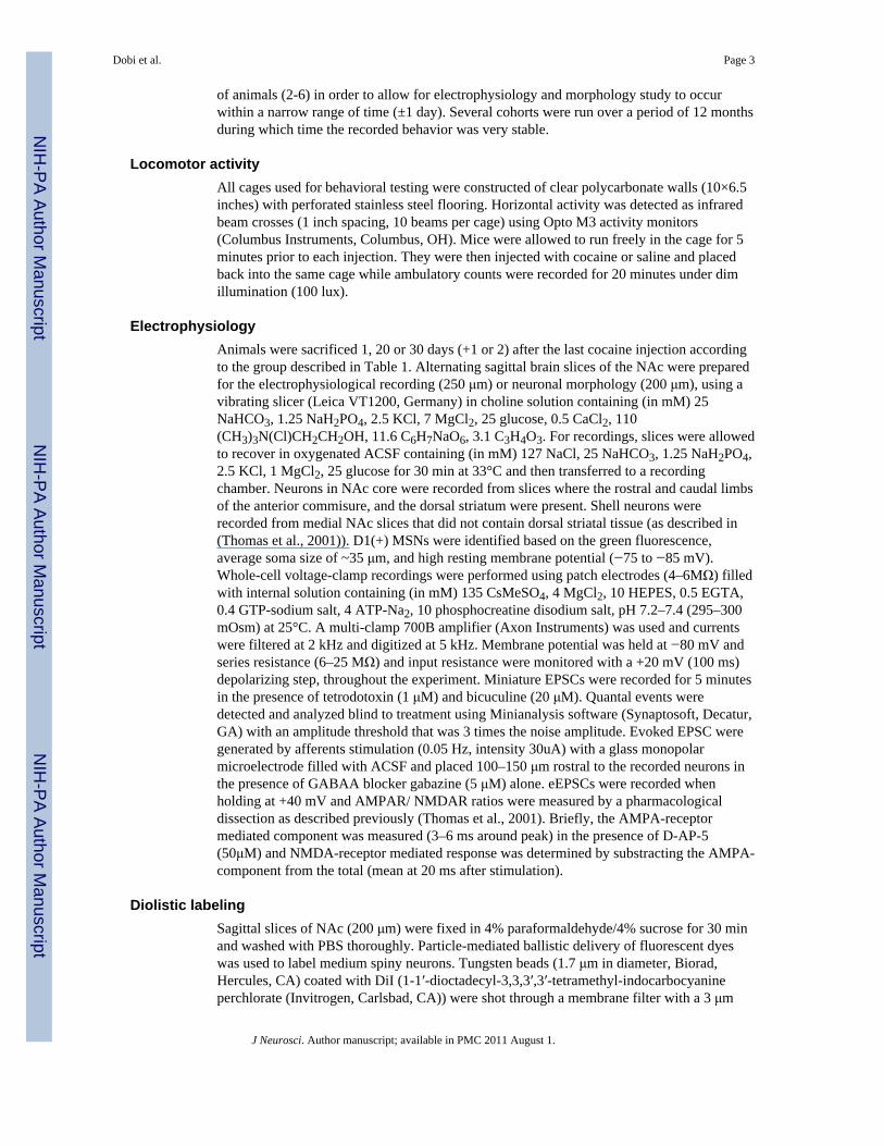
Comparable locomotor responses to cocaine in short and long treatmentMice were assigned to groups (Table 1) and received saline or cocaine (30 mg/kg) dailyinjections according to a short (7 consecutive days) or long treatment (20 injections: 5 daysON-2 days OFF for 4 weeks). Under these two treatments, mice received different totaldoses of cocaine (210 mg/kg in the short and 600 mg/kg in the long treatment). In order toaddress the requirement for cocaine withdrawal, mice that received the short treatment werestudied at 2 and 21 days after the last cocaine injection and compared to mice that receivedthe long treatment and were studied 2 days after the last cocaine injection (Fig. 1B).
Horizontal locomotor response was measured for 20 minutes after each injection. On thefirst day of the short treatment, mice that received a cocaine injection displayed an acutelocomotor response to the drug manifested by a mild increase in beam breaks per minutewhen compared to litter mates that received saline injections (day 1 saline = 43 ± 5.6 beambreaks/min, n = 6; cocaine = 64.1 ± 13.7 beam break/min, n =8; Fig 1C). Consecutive dailycocaine injections caused a larger increase in locomotor response, a phenomenon known aslocomotor sensitization to cocaine (day 7 cocaine =141.4 ± 17.3 beam break/min, n = 8;F(3,28) = 25.3 ANOVA and p < 0.01 for cocaine day 7 vs saline day 1, vs saline day 7 andvs cocaine day 1 by Tukey test; Fig 1C); while repeated saline injections led to a decline inthe activity as the animals habituated to the cage and the procedure (day 7 saline = 23.7 ±5.2 beam breaks/min, n = 6, p > 0.05 for saline day 7 vs saline day1 by ANOVA andTukey).
Animals exposed to the long drug treatment showed locomotor responses to saline andcocaine that were undistinguishable, during the first 7 days, from those who received theshort treatment (Fig. 1D). Mice showed a mild acute response to cocaine in day 1 (saline =46 ± 4.8 breaks/min, n = 10; cocaine = 75.7 ± 10.9 breaks/min, n = 12; Fig. 1D) andrepeated cocaine injections led to the development of psychomotor sensitization that wasexpressed as a larger increase in locomotor response to cocaine. The cocaine-inducedincrease in locomotor acitvity reached a maximum by day 7 and remained elevatedthroughout the treatment, (cocaine day 7 = 160.3 ± 22.1 breaks/min and day 20 = 163.4 ±42.7, n = 12; F(4,48) = 6.4 by ANOVA and p < 0.05 for cocaine day 7 vs saline day 1 andnot significant for cocaine day 7 vs cocaine day 20, p > 0.05 by Tukey test).
Outcome of short treatment in the NAc core depends on the presence or absence of aprolonged withdrawal
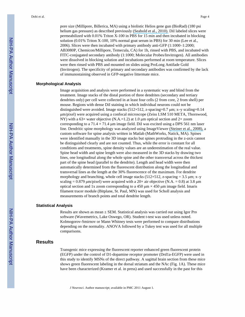
Mice that received short treatment were sacrificed after 2 days (group A) or after 21 days(group B) from the last injection (Fig. 2A). Acute brain slices were prepared and whole-cellvoltage-clamp recordings were made from D1(+) MSNs in the core, which were identifiedbased on the medium size of the cell bodies, their green fluorescence and a small holdingcurrent when voltage was held at −80 mV (Fig. 2B). The detection of green fluorescence infresh acute slices was limited by the sensitivity of the electrophysiology microscope(optimized for DIC visualization) and the medium to low levels of EGFP expression that areachieved when expression is driven by endogenous protein promoters. As a consequence ofthese, the number of EGFP positive neurons in fresh acute slices was underestimated and inthis study, recordings were only made from D1-receptors expressing neurons (D1(+) MSNs)showing clear green fluorescence in the soma (Fig. 2B).
Miniature excitatory postsynaptic currents (mEPSCs) were recorded in the presence ofsodium channel and GABAA receptor blockers to isolate glutamate-mediated transmission.
Dobi et al. Page 5
J Neurosci. Author manuscript; available in PMC 2011 August 1.
NIH
-PA Author Manuscript
NIH
-PA Author Manuscript
NIH
-PA Author Manuscript

Two days after the last cocaine injection, the mean distribution of mEPSC inter-eventintervals showed no significant difference from the mean distribution in saline-treatedanimals (Fig. 2D) and the mean frequency of mEPSC was similar to the mean frequency insaline-treated mice (2.1 ± 0.2 Hz for saline and 1.7 ± 0.2 Hz for cocaine, n = 23-16, p > 0.3by K-S test, Fig. 2F left).
However, in agreement with previous reports showing potentiation of glutamatergictransmission after several week withdrawal from a short cocaine treatment (Kourrich et al.,2007), the mean frequency of mEPSCs in D1(+) MSNs was significantly increased in thecocaine group three weeks after last cocaine injection ( 3.1 ± 0.2 Hz, for cocaine, n = 14; p <0.01 by K-S test, Fig. 2F right). Consequently, a shift to shorter inter-event intervals wasdetected in the mean distribution in the cocaine group after 3 weeks of withdrawal (Fig. 2E,p< 0.05 K-S test). The mean amplitude of mEPSC events was not significantly changed bythe cocaine treatment either two days or three weeks of withdrawal. (Fig. 2G).
Enhanced glutamatergic transmission in core and shell without cocaine withdrawalfollowing long treatment
The increase in mEPSC frequency observed 3 weeks, but not 2 days, after last cocaineinjection could have several possible explanations. One possibility is that extendedwithdrawal after repeated cocaine injections is the signal that triggers the cocaine-inducedplasticity. Another hypothesis is that the events leading to the changes in glutamatergictransmission in the NAc take several weeks to develop and thus, they are not expressed rightafter a short cocaine treatment. In this case, the changes would be observed even if thecocaine treatment is extended for 2–3 weeks and protracted withdrawal is avoided. In orderto discriminate between these two hypotheses, a long drug treatment was used (group C)(Fig. 1B).
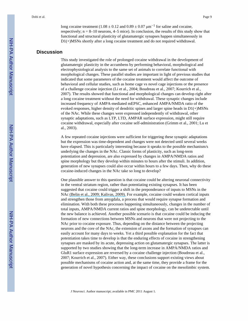
Two days after the last injection of the long treatment, acute brain slices were prepared fromsaline- and cocaine-treated mice. D1(+) MSNs from core and shell were recorded under thesame conditions described for mice that received short treatment (Fig. 3A, B). Thefrequency of mEPSCs was significantly increased in MSNs from the core and the shell incocaine treated mice compared to saline treated mice (core freq = 2.7 ± 0.3 Hz for saline and3.7 ± 0.3 Hz for cocaine, n = 16–20; shell freq = 1.8 ± 0.3 Hz for saline and 3.9 ± 0.7 Hz forcocaine, n = 13–16; * p < 0.05, Fig. 3C). An significant increase in the amplitude of theevents was seen in shell D1(+) MSNs after the cocaine treatment and no change wasdetected in the amplitude of the events in the core (shell amp = 13.2 ± 1.0 pA for saline and18.8 ± 1.4 pA for cocaine, n = 13–16 neurons; p <0.05, core amp = 16.9 ± 1.0 pA for salineand 18.2 ±1.0 pA for cocaine, n = 16–20 neurons; Fig. 3D).
A long uninterrupted treatment (28 consecutive injections; group E, Table 1) was alsoperformed in order to rule out any possible contribution of the three brief periods ofwithdrawal (2 days OFF) on the cocaine induced effect on glutamatergic transmission. Twodays after the last injection of the long uninterrupted treatment, recordings were obtainedfrom MSNs in NAc core. This subregion was chosen first because similar changes inmEPSC frequency were seen in core and shell after the long (20 days) treatment, and secondbecause of the larger yield of slices containing NAc core. Frequency of mEPSCs wassignificantly increased in core D1(+) MSNs from cocaine treated mice compared to salineanimals (2.0 ± 0.2 Hz for saline and 3.6 ± 0.6 Hz for cocaine, n = 8–9; * p < 0.05, Fig. 3F)and no effect was seen in the amplitude (14.4 ± 1.7 pA for saline and 11.5 ±1.7 pA forcocaine, n = 8–9; p > 0.05, Fig. 3F). The magnitude of the mEPSC frequency increase wassimilar between the two long treatments indicating that the brief withdrawal periods werenot required for the cocaine-induced synaptic adaptation.
Dobi et al. Page 6
J Neurosci. Author manuscript; available in PMC 2011 August 1.
NIH
-PA Author Manuscript
NIH
-PA Author Manuscript
NIH
-PA Author Manuscript

Evoked EPSCs were measured in core D1(+) MSNs after the long uninterrupted treatmentand AMPA/NMDA ratios were calculated from saline and cocaine treated mice. Cocaine-treated mice showed a significant increased in AMPAR/NMDAR ratios indicating that thesynaptic adaptations extend beyond miniature event (1.1 ± 0.1for saline and 1.5 ± 0.1 forcocaine, n = 5–6; 3 mice each group, p < 0.05, Fig. 3G–H). All together these experimentsshowed that the potentiation of glutamatergic transmission triggered by cocaine developsand expresses itself without the need for protracted or brief withdrawal after a long repeatedtreatment. Furthermore, when compared to the results of the short treatment, these resultsindicate that 7 repeated cocaine injections are sufficient for triggering these changes and thatit is the time, but not the prolonged withdrawal, that is required for the induction of thiscocaine-induced plasticity in the NAc.
Concurrent increase in spine density and glutamatergic transmission in D1(+) MSNsThe next experiments addressed whether the increase in mEPSC frequency detected inD1(+) MSNs from cocaine-treated mice could reflect the presence of more glutamatergicsynapses in these neurons after cocaine. Because the density of the dendritic spines can be amorphological readout for the abundance of glutamatergic synapses, the morphology ofdendrites and dendritic spines was studied in parallel with the electrophysiology. Wefocused in mice exposed to the long cocaine treatment (20 injections: 5 days ON-2 daysOFF for 4 weeks) and studied spine morphology 2 days after the last cocaine injection.
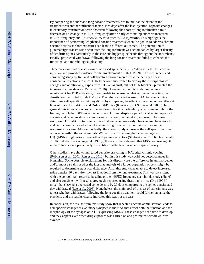
Diolistic labeling was used and combined with immunostaining to further amplify the EGFPsignal at the cell bodies. DiI-labeled MSNs with positive immunostaining were identified asD1(+) MSNs and those lacking EGFP immunostaining were classified as D1 receptornegative MSNs (D1(−) MSNs) (Fig. 4B). Confocal images were acquired from the distalportion of MSN dendrites in the core and shell of the NAc and they were analyzed blind tothe drug treatments. An increased density of spines was detected in D1(+) MSNs from incocaine-treated animals (core and shell combined) compared to saline-treated mice (0.91 ±0.03 μm−1 for saline and 1.10 ± 0.08 μm−1 for cocaine, n = 13–14 neurons, 3–4 mice; p <0.05) (Fig. 4E–F). No significant change was detected in the spine density of D1(−) MSNsbetween saline and cocaine-treated mice (0.91 ± 0.06 μm−1 for saline and 0.96 ± 0.07 μm−1
for cocaine, n = 11–13 neurons, 3–4 mice; P> 0.05). Note that there was no significantdifference in the mean density of spine in D1(+) and D1(−) MSNs in saline injected animals(Fig. 4F).
These experiments showed that functional and structural plasticities of glutamatergicsynapses happen simultaneously in D1(+) MSNs shortly after a long cocaine treatment.
Enlarged spine heads in cocaine-treated animalsThe morphology of dendritic spines is diverse and it can be linked to the functional diversityof glutamatergic synapses (Alvarez and Sabatini, 2007). Correlations between the size of thespine head and postsynaptic density (Harris and Stevens, 1989) and between the size of thespine head and the amplitude of AMPA currents evoked at each spine (Matsuzaki et al.,2001) have been determined in the past for gluatamatergic synapses in the hippocampus.
The structural diversity of spines was studied here by measuring the spine length and widthof the spine head. In saline-treated animals, the distribution of spines head width for MSNs(D1(+) and D1(−) MSNs combined) was similar from neurons in the core and shell with amean width value of 0.75 ± 0.05 μm for core and 0.76 ± 0.06 μm for the shell (n=1703–784spines / 16–12 neurons). Repetitive cocaine injections caused a rightward shift in thenormalized distribution of spine head width in the core MSNs (core width = 0.81± 0.06 μmin core, n = 1620 spines / 12 neurons D1(+) and D1(−) combined; Fig. 4G). There were less
Dobi et al. Page 7
J Neurosci. Author manuscript; available in PMC 2011 August 1.
NIH
-PA Author Manuscript
NIH
-PA Author Manuscript
NIH
-PA Author Manuscript

thin spines and larger percentage of wide spines in core MSNs 2 days after the last cocaineinjection but there was no detectable difference in the spine width distribution in shell MSNsafter cocaine treatment (shell width = 0.77 ± 0.005 μm, n = 2125 spines / 15 neurons). Whena median split was used to look at thin spines (the smaller half) and wide spines (the largerhalf), both thin and wide spines showed larger heads in the core, but not the shell, ofcocaine-treated animals (ANOVA, F = 991.8, p < 0.01, Fig. 4H). No change in the meanspine length was observed after cocaine treatment in core or shell MSNs (core spine length =1.86 ± 0.02 μm in saline and 1.83 ± 0.02 μm in cocaine; shell spine length = 1.77 ± 0.03 μmin saline and 1.81 ± 0.02 μm in cocaine, n = 784–2125 spines).
Dendrite morphology after long cocaine treatmentA model of each neuron was constructed from confocal image stacks of D1(+) and D1(−)MSNs acquired at low magnification to study dendrite branching and morphology (Fig. 5A,B) The mean number of dendrite branch points per neuron was similar for D1(+) and D1(−)MSNs from saline- and cocaine-treated animals (D1(+) MSNs = 13.8 ± 5.6 and 12.6 ± 6.6for saline and cocaine, respectively n = 9–10; D1(−) MSNs = 12.1± 5.8 and 11.6 ± 6.2 forsaline and cocaine, respectively, n = 14–17; Fig. 5C). Scholl analysis showed no significantchange in the number of intersections or total dendrite lengths in D1(+) MSNs and a smalldecrease in the total dendrite lengths in D1(−) MSNs after cocaine treatment (D1(+) MSNslength = 1.72 ± 0.52 and 1.31 ± 0.43 mm for saline and cocaine respectively; D1(−) MSNslength = 1.35 ± 0.48 and 1.05 ± 0.26 mm for saline and cocaine, respectively; p < 0.05 Fig.5D, E).
Protracted withdrawal after long cocaine treatment does not enhance plasticityThe next experiments investigate whether prolonged withdrawal after the long cocainetreatment can further enhance the functional and morphological plasticity observed in D1(+)MSNs. With this purpose, we measured in parallel mEPSC frequency and spine density 30days after the last injection of the long treatment (Fig. 6A, group D). The physiology andmorphology data were sorted in core and shell D1(+) MSNs and compared to the previousvalues obtained 2 days after the last injections from the long treatment (Fig. 6B–C). Whilethe frequency of mEPSCs was significantly increased 2 days after the long cocaine treatment(2 dw) in both core and shell, mEPSC frequency was not different from saline values 30days (30 dw) after the last injection in both subregions of the NAc, (core freq = 2.5± 0.2, 3.7± 0.3 and 2.7 ± 0.3 Hz for saline, cocaine 2dw and 30dw, respectively, n = 41–20–17neurons; F(2,75) = 4.5 ANOVA and p < 0.05 for cocaine 2dw vs saline (q = 4.2), Tukeytest; shell freq = 2.2 ± 0.2, 3.9 ± 0.7 and 2.0 ± 0.4 Hz for saline, cocaine 2dw and 30dw,respectively, n = 39–15–16 neurons ; F(2,67) = 6.4 ANOVA and p < 0.01 for cocaine 2dwvs saline (q=4.6) and for cocaine 2dw vs 30dw (q = 4.3), Tukey test, Fig. 6B).
Spine morphology was studies in parallel in D1(+)MSNs after the long treatment andconsistently with the physiological observations, spine density was not different from salinelevels 30 days after the end of the long cocaine treatment (core density = 0.94± 0.04, 1.23 ±0.13 and 0.91 ± 0.06 μm−1 for saline, cocaine 2 dw and 30dw, respectively, n = 6–8neurons; F(2,56) = 8.4 ANOVA and q = 5.6 and 3.9, p < 0.01 and 0.05 for cocaine 2 dw vssaline and cocaine 2 dw vs cocaine 30 dw respectively by Tukey test; shell density = 0.87 ±0.07, 1.0 ± 0.8 and 0.79 ± 0.06 μm−1 for saline, cocaine 2dw and 30dw, respectively, n = 6–7 neurons; (F(2,55) = 1.5 ANOVA); Fig. 6C). Interestingly, the increased in spine densitiesseen in D1(+)MSN 2 days after the long treatment were mainly driven by changes in densityof core D1(+)MNS. D1(−)MSNs from neither core or shell showed changes in spine densityafter the long cocaine treatment (F(2,14) = 2.6 for core and F(2,15) = 0.24 for shell; Suppl.Fig. 2). Finally, an independent set of experiments was performed in wild-type SwissWebster mice which also lacked the increase in the spine density 30 days following the same
Dobi et al. Page 8
J Neurosci. Author manuscript; available in PMC 2011 August 1.
NIH
-PA Author Manuscript
NIH
-PA Author Manuscript
NIH
-PA Author Manuscript
long cocaine treatment (1.08 ± 0.12 and 0.89 ± 0.07 μm−1 for saline and cocaine,respectively; n = 8–10 neurons, 4–5 mice). In conclusion, the results of this study show thatfunctional and structural plasticity of glutamatergic synapses happen simultaneously inD1(+)MSNs shortly after a long cocaine treatment and do not required withdrawal.
DiscussionThis study investigated the role of prolonged cocaine withdrawal in the development ofglutamatergic plasticity in the accumbens by performing behavioral, morphological andelectrophysiological analysis to the same set of animals to correlate functional withmorphological changes. These parallel studies are important in light of previous studies thatindicated that some parameters of the cocaine treatment would affect the outcome ofbehavioral and cellular studies, such as home cage vs novel cage injections or the presenceof a challenge cocaine injection (Li et al., 2004; Boudreau et al., 2007; Kourrich et al.,2007). The results showed that functional and morphological changes can develop right aftera long cocaine treatment without the need for withdrawal. These synaptic changes includeincreased frequency of AMPA-mediated mEPSC, enhanced AMPA/NMDA ratio of theevoked responses, higher density of dendritic spines and larger spine heads in D1(+)MSNsof the NAc. While these changes were expressed independently of withdrawal, othersynaptic adaptations, such as LTP, LTD, AMPAR surface expression, might still requirecocaine withdrawal, especially after cocaine self-administration (Grimm et al., 2001; Lu etal., 2003).
A few repeated cocaine injections were sufficient for triggering these synaptic adaptationsbut the expression was time-dependent and changes were not detected until several weekshave elapsed. This is particularly interesting because it speaks to the possible mechanism/sunderlying the changes in the NAc. Classic forms of plasticity, such as long-termpotentiation and depression, are also expressed by changes in AMPA/NMDA ratios andspine morphology but they develop within minutes to hours after the stimuli. In addition,generation of new synapses could also occur within hours to a few days. Then, why do thesecocaine-induced changes in the NAc take so long to develop?
One plausible answer to this question is that cocaine could be altering neuronal connectivityin the ventral striatum region, rather than potentiating existing synapses. It has beensuggested that cocaine could trigger a shift in the preponderance of inputs to MSNs in theNAc (Belin et al., 2009; Kalivas, 2009). For example, cocaine could weaken cortical inputsand strengthen those from amygdala, a process that would require synapse formation andelimination. With both these processes happening simultaneously, changes in the number oftotal inputs, AMPA/NMDA current ratios and spine morphology, can be undetectable untilthe new balance is achieved. Another possible scenario is that cocaine could be inducing theformation of new connections between MSNs and neurons that were not projecting to theNAc prior to cocaine exposure. Thus, depending on the distance between the projectingneurons and the core of the NAc, the extension of axons and the formation of synapses caneasily account for many days to weeks. Yet a third possible explanation for the fact thatpotentiation takes time to develop is that the enduring effects of cocaine in strengtheningsynapses are masked by its acute, depressing action on glutamatergic synapses. The latter issupported by two studies showing that the long-term increase in AMPA/NMDA ratios andGluR1 surface expression are reversed by a cocaine challenge injection (Boudreau et al.,2007; Kourrich et al., 2007). Either way, these conclusions support existing views aboutpossible mechanisms of cocaine action and, at the same time, they provide a frame for thegeneration of novel hypothesis concerning the impact of cocaine on the mesolimbic system.
Dobi et al. Page 9
J Neurosci. Author manuscript; available in PMC 2011 August 1.
NIH
-PA Author Manuscript
NIH
-PA Author Manuscript
NIH
-PA Author Manuscript
By comparing the short and long cocaine treatments, we found that the extent of thetreatment was another influential factor. Two days after the last injection, opposite changesin excitatory transmission were observed following the short or long treatments: a milddecrease or no change in mEPSC frequency after 7 daily cocaine injections vs increasedmEPSC frequency and AMPA/NMDA ratio after 20–28 injections. This highlights theimportance of performing lengthened cocaine treatments when the goal is to address chroniccocaine actions as short exposures can lead to different outcomes. The potentiation ofglutamatergic transmission seen after the long treatment was accompanied by larger densityof dendritic spines particularly in the core and bigger spine heads throughout the accumbens.Finally, protracted withdrawal following the long cocaine treatment failed to enhance thefunctional and morphological plasticity.
Three previous studies also showed increased spine density 1–2 days after the last cocaineinjection and provided evidence for the involvement of D1(+)MSNs. The most recent andconvincing study by Ren and collaborators showed increased spine density after 28consecutive injections in mice. D1R knockout mice failed to display these morphologicalchanges and additionally, exposure to D1R antagonist, but not D2R blockers, prevented theincrease in spine density (Ren et al., 2010). However, while this study pointed to arequirement for D1R activation, it was unable to determine whether the increase in spinedensity was restricted to D1(+)MSNs. The other two studies used BAC transgenic mice todetermine cell specificity but they did so by comparing the effect of cocaine on two differentlines of mice: Drd1-EGFP and Drd2-EGFP mice (Kim et al., 2009; Lee et al., 2006). Ingeneral, this is not a good experimental design but it is particularly worrisome in light of thefinding that Drd2-EGFP mice over-express D2R and display a paradoxical acute response tococaine and failed to show locomotor sensitization (Kramer et al., in press). The currentstudy used Drd1-EGFP transgenic mice that we have previously characterized behaviorallyand neurochemically and shown to be undistinguishable from wild-type mice in theirresponse to cocaine. More importantly, the current study addresses the cell specific actionsof cocaine within the same animals. While it is worth noting that a percentage ofD1(+)MSNs might also express other dopamine receptors (Shetreat et al., 1996; Hasbi et al.,2010) (but also see (Wong et al., 1999)), the results here showed that MSNs expressing D1Rin the NAc core are particularly susceptible to effects of cocaine on spine density.
Other studies have shown increased dendrite branching in NAc after chronic cocaine(Robinson et al., 2001; Ren et al., 2010), but in this study we could not detect changes inbranching. Some possible explanations for this disparity are the difference in animal speciesand/or mouse strains used or the fact that analysis of a larger population of cells might berequired to determine statistical difference. Also, this study was unable to detect increasedspine density 30 days after the last injection from the long treatment. This was consistentwith the concomitant return to baseline of the mEPSC frequency seen in this study (Fig. 6)and also consistent with results previously reported using these same mice (Drd1-EGFPmice) that showed a decreased spine density by 30 days compared to the spines density at 2day withdrawal (Lee et al., 2006). Nonetheless, the main goal of this set of experiments wasto test whether withdrawal following the long cocaine treatment could further enhance theplasticity and the results clearly indicated this was not the case.
In conclusion, the results from this study show that repeated cocaine administration leads tocell-specific changes at excitatory synapses in the NAc that affect both the function and themorphology of the synapse onto D1-expressing MSNs. These changes need time to developand they appear even when drug exposure was carried on and protracted withdrawal wasavoided.
Dobi et al. Page 10
J Neurosci. Author manuscript; available in PMC 2011 August 1.
NIH
-PA Author Manuscript
NIH
-PA Author Manuscript
NIH
-PA Author Manuscript

Supplementary MaterialRefer to Web version on PubMed Central for supplementary material.
AcknowledgmentsWe are grateful to Dr. Fumi Ono for sharing his confocal microscope. We would like to thank John T. Williams,Christina Gremel, Christopher Ford and the members of the Alvarez’ lab for the helpful comments and discussionsof the manuscript. This research was funded by the intramural programs of NIAAA and NINDS at the NationalInstitute of Health.
ReferencesAlvarez VA, Sabatini BL. Anatomical and physiological plasticity of dendritic spines. Annu Rev
Neurosci. 2007; 30:79–97. [PubMed: 17280523]Asensio S, Romero MJ, Romero FJ, Wong C, Alia-Klein N, Tomasi D, Wang GJ, Telang F, Volkow
ND, Goldstein RZ. Striatal dopamine D2 receptor availability predicts the thalamic and medialprefrontal responses to reward in cocaine abusers three years later. Synapse. 2010; 64:397–402.[PubMed: 20034014]
Belin D, Jonkman S, Dickinson A, Robbins TW, Everitt BJ. Parallel and interactive learning processeswithin the basal ganglia: relevance for the understanding of addiction. Behav Brain Res. 2009;199:89–102. [PubMed: 18950658]
Boudreau AC, Wolf ME. Behavioral Sensitization to Cocaine Is Associated with Increased AMPAReceptor Surface Expression in the Nucleus Accumbens. J Neurosci. 2005; 25:9144–9151.[PubMed: 16207873]
Boudreau AC, Reimers JM, Milovanovic M, Wolf ME. Cell Surface AMPA Receptors in the RatNucleus Accumbens Increase during Cocaine Withdrawal But Internalize after Cocaine Challengein Association with Altered Activation of Mitogen-Activated Protein Kinases. J Neurosci. 2007;27:10621–10635. [PubMed: 17898233]
Bouyer JJ, Joh TH, Pickel VM. Ultrastructural localization of tyrosine hydroxylase in rat nucleusaccumbens. J Comp Neurol. 1984; 227:92–103. [PubMed: 6147361]
Caine SB, Negus SS, Mello NK, Patel S, Bristow L, Kulagowski J, Vallone D, Saiardi A, Borrelli E.Role of dopamine D2-like receptors in cocaine self-administration: studies with D2 receptor mutantmice and novel D2 receptor antagonists. J Neurosci. 2002; 22:2977–2988. [PubMed: 11923462]
Caine SB, Thomsen M, Gabriel KI, Berkowitz JS, Gold LH, Koob GF, Tonegawa S, Zhang J, Xu M.Lack of self-administration of cocaine in dopamine D1 receptor knock-out mice. J Neurosci. 2007;27:13140–13150. [PubMed: 18045908]
Day M, Wokosin D, Plotkin JL, Tian X, Surmeier DJ. Differential excitability and modulation ofstriatal medium spiny neuron dendrites. J Neurosci. 2008; 28:11603–11614. [PubMed: 18987196]
Day M, Wang Z, Ding J, An X, Ingham CA, Shering AF, Wokosin D, Ilijic E, Sun Z, Sampson AR,Mugnaini E, Deutch AY, Sesack SR, Arbuthnott GW, Surmeier DJ. Selective elimination ofglutamatergic synapses on striatopallidal neurons in Parkinson disease models. Nat Neurosci.2006; 9:251–259. [PubMed: 16415865]
Fields HL, Hjelmstad GO, Margolis EB, Nicola SM. Ventral tegmental area neurons in learnedappetitive behavior and positive reinforcement. Annu Rev Neurosci. 2007; 30:289–316. [PubMed:17376009]
Freund TF, Powell JF, Smith AD. Tyrosine hydroxylase-immunoreactive boutons in synaptic contactwith identified striatonigral neurons, with particular reference to dendritic spines. Neuroscience.1984; 13:1189–1215. [PubMed: 6152036]
Gong S, Zheng C, Doughty ML, Losos K, Didkovsky N, Schambra UB, Nowak NJ, Joyner A, LeblancG, Hatten ME, Heintz N. A gene expression atlas of the central nervous system based on bacterialartificial chromosomes. Nature. 2003; 425:917–925. [PubMed: 14586460]
Grimm JW, Hope BT, Wise RA, Shaham Y. Neuroadaptation. Incubation of cocaine craving afterwithdrawal. Nature. 2001; 412:141–142. [PubMed: 11449260]
Dobi et al. Page 11
J Neurosci. Author manuscript; available in PMC 2011 August 1.
NIH
-PA Author Manuscript
NIH
-PA Author Manuscript
NIH
-PA Author Manuscript

Guan X, Zhang R, Xu Y, Li S. Cocaine withdrawal enhances long-term potentiation in rathippocampus via changing the activity of corticotropin-releasing factor receptor subtype 2.Neuroscience. 2009; 161:665–670. [PubMed: 19376201]
Harris KM, Stevens JK. Dendritic spines of CA 1 pyramidal cells in the rat hippocampus: serialelectron microscopy with reference to their biophysical characteristics. J Neurosci. 1989; 9:2982–2997. [PubMed: 2769375]
Hasbi A, O’Dowd BF, George SR. Heteromerization of dopamine D2 receptors with dopamine D1 orD5 receptors generates intracellular calcium signaling by different mechanisms. Curr OpinPharmacol. 2010; 10:93–99. [PubMed: 19897420]
Heiman M, Schaefer A, Gong S, Peterson JD, Day M, Ramsey KE, Suarez-Farinas M, Schwarz C,Stephan DA, Surmeier DJ, Greengard P, Heintz N. A translational profiling approach for themolecular characterization of CNS cell types. Cell. 2008; 135:738–748. [PubMed: 19013281]
Huang YH, Lin Y, Mu P, Lee BR, Brown TE, Wayman G, Marie H, Liu W, Yan Z, Sorg BA, SchluterOM, Zukin RS, Dong Y. In vivo cocaine experience generates silent synapses. Neuron. 2009;63:40–47. [PubMed: 19607791]
Humphries MD, Prescott TJ. The ventral basal ganglia, a selection mechanism at the crossroads ofspace, strategy, and reward. Prog Neurobiol. 2010; 90:385–417. [PubMed: 19941931]
Hyman SE, Malenka RC, Nestler EJ. Neural mechanisms of addiction: the role of reward- relatedlearning and memory. Annu Rev Neurosci. 2006; 29:565–598. [PubMed: 16776597]
Kalivas PW. The glutamate homeostasis hypothesis of addiction. Nat Rev Neurosci. 2009; 10:561–572. [PubMed: 19571793]
Kalivas PW, Lalumiere RT, Knackstedt L, Shen H. Glutamate transmission in addiction.Neuropharmacology. 2009; 56(Suppl 1):169–173. [PubMed: 18675832]
Kebabian JW, Calne DB. Multiple receptors for dopamine. Nature. 1979; 277:93–96. [PubMed:215920]
Kim Y, Teylan MA, Baron M, Sands A, Nairn AC, Greengard P. Methylphenidate-induced dendriticspine formation and DeltaFosB expression in nucleus accumbens. Proc Natl Acad Sci U S A.2009; 106:2915–2920. [PubMed: 19202072]
Kourrich S, Thomas MJ. Similar neurons, opposite adaptations: psychostimulant experiencedifferentially alters firing properties in accumbens core versus shell. J Neurosci. 2009; 29:12275–12283. [PubMed: 19793986]
Kourrich S, Rothwell PE, Klug JR, Thomas MJ. Cocaine experience controls bidirectional synapticplasticity in the nucleus accumbens. J Neurosci. 2007; 27:7921–7928. [PubMed: 17652583]
Kramer PF, Christensen CH, Hazelwood L, Dobi A, Bock R, Sibley DR, Mateo Y, Alvarez VA.Dopamine D2 receptor over-expression alters behavior and physiology in Drd2-EGFP mice. JNeurosci. (in press).
Kreitzer AC, Malenka RC. Dopamine modulation of state-dependent endocannabinoid release andlong-term depression in the striatum. J Neurosci. 2005; 25:10537–10545. [PubMed: 16280591]
Lee KW, Kim Y, Kim AM, Helmin K, Nairn AC, Greengard P. Cocaine-induced dendritic spineformation in D1 and D2 dopamine receptor-containing medium spiny neurons in nucleusaccumbens. Proc Natl Acad Sci U S A. 2006; 103:3399–3404. [PubMed: 16492766]
Li Y, Acerbo MJ, Robinson TE. The induction of behavioural sensitization is associated with cocaine-induced structural plasticity in the core (but not shell) of the nucleus accumbens. Eur J Neurosci.2004; 20:1647–1654. [PubMed: 15355332]
Lu L, Grimm JW, Shaham Y, Hope BT. Molecular neuroadaptations in the accumbens and ventraltegmental area during the first 90 days of forced abstinence from cocaine self-administration inrats. J Neurochem. 2003; 85:1604–1613. [PubMed: 12787079]
Martin M, Chen BT, Hopf FW, Bowers MS, Bonci A. Cocaine self-administration selectivelyabolishes LTD in the core of the nucleus accumbens. Nat Neurosci. 2006; 9:868–869. [PubMed:16732275]
Matsuzaki M, Ellis-Davies GC, Nemoto T, Miyashita Y, Iino M, Kasai H. Dendritic spine geometry iscritical for AMPA receptor expression in hippocampal CA1 pyramidal neurons. Nat Neurosci.2001; 4:1086–1092. [PubMed: 11687814]
Dobi et al. Page 12
J Neurosci. Author manuscript; available in PMC 2011 August 1.
NIH
-PA Author Manuscript
NIH
-PA Author Manuscript
NIH
-PA Author Manuscript

McDonald AJ. Topographical organization of amygdaloid projections to the caudatoputamen, nucleusaccumbens, and related striatal-like areas of the rat brain. Neuroscience. 1991; 44:15–33.[PubMed: 1722890]
Moussawi K, Pacchioni A, Moran M, Olive MF, Gass JT, Lavin A, Kalivas PW. N-Acetylcysteinereverses cocaine-induced metaplasticity. Nat Neurosci. 2009; 12:182–189. [PubMed: 19136971]
Phillipson OT, Griffiths AC. The topographic order of inputs to nucleus accumbens in the rat.Neuroscience. 1985; 16:275–296. [PubMed: 4080159]
Ren Z, Sun WL, Jiao H, Zhang D, Kong H, Wang X, Xu M. Dopamine D1 and N-methyl-d-aspartatereceptors and extracellular signal-regulated kinase mediate neuronal morphological changesinduced by repeated cocaine administration. Neuroscience. 2010
Robinson TE, Gorny G, Mitton E, Kolb B. Cocaine self-administration alters the morphology ofdendrites and dendritic spines in the nucleus accumbens and neocortex. Synapse. 2001; 39:257–266. [PubMed: 11169774]
Seabold GK, Daunais JB, Rau A, Grant KA, Alvarez VA. DiOLISTIC Labeling of Neurons fromRodent and Non-human Primate Brain Slices. J Vis Exp. 2010
Sesack SR, Deutch AY, Roth RH, Bunney BS. Topographical organization of the efferent projectionsof the medial prefrontal cortex in the rat: an anterograde tract-tracing study with Phaseolusvulgaris leucoagglutinin. J Comp Neurol. 1989; 290:213–242. [PubMed: 2592611]
Shetreat ME, Lin L, Wong AC, Rayport S. Visualization of D1 dopamine receptors on living nucleusaccumbens neurons and their colocalization with D2 receptors. J Neurochem. 1996; 66:1475–1482. [PubMed: 8627301]
Sibley DR, Monsma FJ Jr. Molecular biology of dopamine receptors. Trends Pharmacol Sci. 1992;13:61–69. [PubMed: 1561715]
Steiner P, Higley MJ, Xu W, Czervionke BL, Malenka RC, Sabatini BL. Destabilization of thepostsynaptic density by PSD-95 serine 73 phosphorylation inhibits spine growth and synapticplasticity. Neuron. 2008; 60:788–802. [PubMed: 19081375]
Surmeier DJ, Ding J, Day M, Wang Z, Shen W. D1 and D2 dopamine-receptor modulation of striatalglutamatergic signaling in striatal medium spiny neurons. Trends Neurosci. 2007; 30:228–235.[PubMed: 17408758]
Thanos PK, Michaelides M, Umegaki H, Volkow ND. D2R DNA transfer into the nucleus accumbensattenuates cocaine self-administration in rats. Synapse. 2008; 62:481–486. [PubMed: 18418874]
Thomas MJ, Kalivas PW, Shaham Y. Neuroplasticity in the mesolimbic dopamine system and cocaineaddiction. Br J Pharmacol. 2008; 154:327–342. [PubMed: 18345022]
Thomas MJ, Beurrier C, Bonci A, Malenka RC. Long-term depression in the nucleus accumbens: aneural correlate of behavioral sensitization to cocaine. Nat Neurosci. 2001; 4:1217–1223.[PubMed: 11694884]
Wang Z, Kai L, Day M, Ronesi J, Yin HH, Ding J, Tkatch T, Lovinger DM, Surmeier DJ.Dopaminergic control of corticostriatal long-term synaptic depression in medium spiny neurons ismediated by cholinergic interneurons. Neuron. 2006; 50:443–452. [PubMed: 16675398]
Wong AC, Shetreat ME, Clarke JO, Rayport S. D1- and D2-like dopamine receptors are co-localizedon the presynaptic varicosities of striatal and nucleus accumbens neurons in vitro. Neuroscience.1999; 89:221–233. [PubMed: 10051231]
Zahm DS. Functional-anatomical implications of the nucleus accumbens core and shell subterritories.Ann N Y Acad Sci. 1999; 877:113–128. [PubMed: 10415646]
Zhang J, Xu M. Opposite regulation of cocaine-induced intracellular signaling and gene expression bydopamine D1 and D3 receptors. Ann N Y Acad Sci. 2006; 1074:1–12. [PubMed: 17105899]
Zhang J, Zhang D, Xu M. Identification of chronic cocaine-induced gene expression through dopamined1 receptors by using cDNA microarrays. Ann N Y Acad Sci. 2002; 965:1–9. [PubMed:12105080]
Zhang XF, Hu XT, White FJ. Whole-cell plasticity in cocaine withdrawal: reduced sodium currents innucleus accumbens neurons. J Neurosci. 1998; 18:488–498. [PubMed: 9412525]
Dobi et al. Page 13
J Neurosci. Author manuscript; available in PMC 2011 August 1.
NIH
-PA Author Manuscript
NIH
-PA Author Manuscript
NIH
-PA Author Manuscript

Figure 1. Locomotor response to repeated cocaine injections in Drd1-GFP mice during the shortand long treatmentA. A bright field (top) and a fluorescent image (bottom) of a sagittal brain section from aDrd1a-GFP mouse showing green fluorescence in the dorsal striatum and nucleusaccumbens regions. B. Schematic diagram of the main two intra-peritoneal injectionstreatments used: short (7 consecutive injections) and long (20 injections: 5 days ON-2 daysOFF). C. Daily locomotor activity (mean ± SEM) during the short treatment measured asbeam breaks per minute during 20 minutes after the saline (black) or cocaine (red) injectionsfor mice from groups A and B, respectively. D. Daily locomotor activity (mean ± SEM)during the long treatment measured as beam breaks per minute during 20 minutes after thesaline (black) or cocaine (red) injection for mice from groups C and D, respectively.
Dobi et al. Page 14
J Neurosci. Author manuscript; available in PMC 2011 August 1.
NIH
-PA Author Manuscript
NIH
-PA Author Manuscript
NIH
-PA Author Manuscript
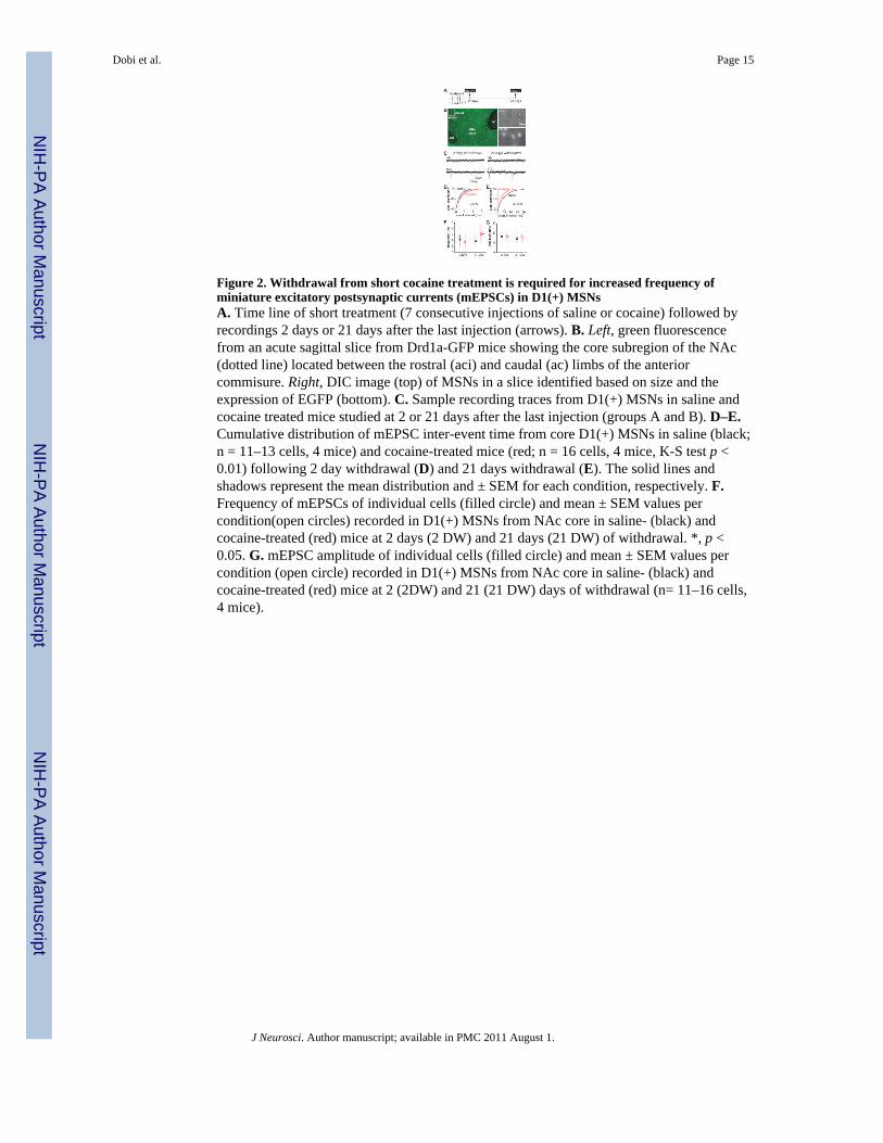
Figure 2. Withdrawal from short cocaine treatment is required for increased frequency ofminiature excitatory postsynaptic currents (mEPSCs) in D1(+) MSNsA. Time line of short treatment (7 consecutive injections of saline or cocaine) followed byrecordings 2 days or 21 days after the last injection (arrows). B. Left, green fluorescencefrom an acute sagittal slice from Drd1a-GFP mice showing the core subregion of the NAc(dotted line) located between the rostral (aci) and caudal (ac) limbs of the anteriorcommisure. Right, DIC image (top) of MSNs in a slice identified based on size and theexpression of EGFP (bottom). C. Sample recording traces from D1(+) MSNs in saline andcocaine treated mice studied at 2 or 21 days after the last injection (groups A and B). D–E.Cumulative distribution of mEPSC inter-event time from core D1(+) MSNs in saline (black;n = 11–13 cells, 4 mice) and cocaine-treated mice (red; n = 16 cells, 4 mice, K-S test p <0.01) following 2 day withdrawal (D) and 21 days withdrawal (E). The solid lines andshadows represent the mean distribution and ± SEM for each condition, respectively. F.Frequency of mEPSCs of individual cells (filled circle) and mean ± SEM values percondition(open circles) recorded in D1(+) MSNs from NAc core in saline- (black) andcocaine-treated (red) mice at 2 days (2 DW) and 21 days (21 DW) of withdrawal. *, p <0.05. G. mEPSC amplitude of individual cells (filled circle) and mean ± SEM values percondition (open circle) recorded in D1(+) MSNs from NAc core in saline- (black) andcocaine-treated (red) mice at 2 (2DW) and 21 (21 DW) days of withdrawal (n= 11–16 cells,4 mice).
Dobi et al. Page 15
J Neurosci. Author manuscript; available in PMC 2011 August 1.
NIH
-PA Author Manuscript
NIH
-PA Author Manuscript
NIH
-PA Author Manuscript

Figure 3. Increased mEPSCs frequency and AMPA/NMDA ratio after long cocaine treatmentwithout protracted withdrawalA. Schematic diagram of long treatment (20 injections: 5 days ON-2 days OFF) followed byrecordings 2 days after the last injection (arrow). B. Sample traces of mEPSCs recordingsfrom D1(+) MSNs in slices from saline- and cocaine-treated mice. C. mEPSC frequencyvalues of individual cells (filled circle) and mean ± SEM per condition (open circle)recorded in D1(+) MSNs from core and shell of saline- (black) and cocaine-treated (red)mice (n= 16–20 cells). *, p < 0.05. D. mEPSC amplitude of individual cells (filled circle)and mean ± SEM values per condition (open circle) recorded in D1(+) MSNs from core andshell of saline- (black) and cocaine-treated (red) mice. *, p < 0.05. E. Time line of longuninterrupted treatment (28 consecutive injections) and recordings 2 days after the lastinjection (arrow). F. mEPSCs frequency and amplitude (mean ± SEM) recorded from coreD1(+) MSNs of saline- and cocaine-treated mice (n=8–9 cells, 3 mice each group). *, p <0.05. G. Exemplary traces of evoked EPSCs showing isolated AMPA and NMDA receptormediated currents recorded at Vh= +40 mV. H. AMPAR/NMDAR ratio of individual cells(filled circles) and mean ± SEM values per condition (open circle) recorded in core D1(+)MSNs from saline (black)- and cocaine-treated (red) mice at 1 day of withdrawal (n=5–6cells, 3–4 mice). *, p < 0.05.
Dobi et al. Page 16
J Neurosci. Author manuscript; available in PMC 2011 August 1.
NIH
-PA Author Manuscript
NIH
-PA Author Manuscript
NIH
-PA Author Manuscript
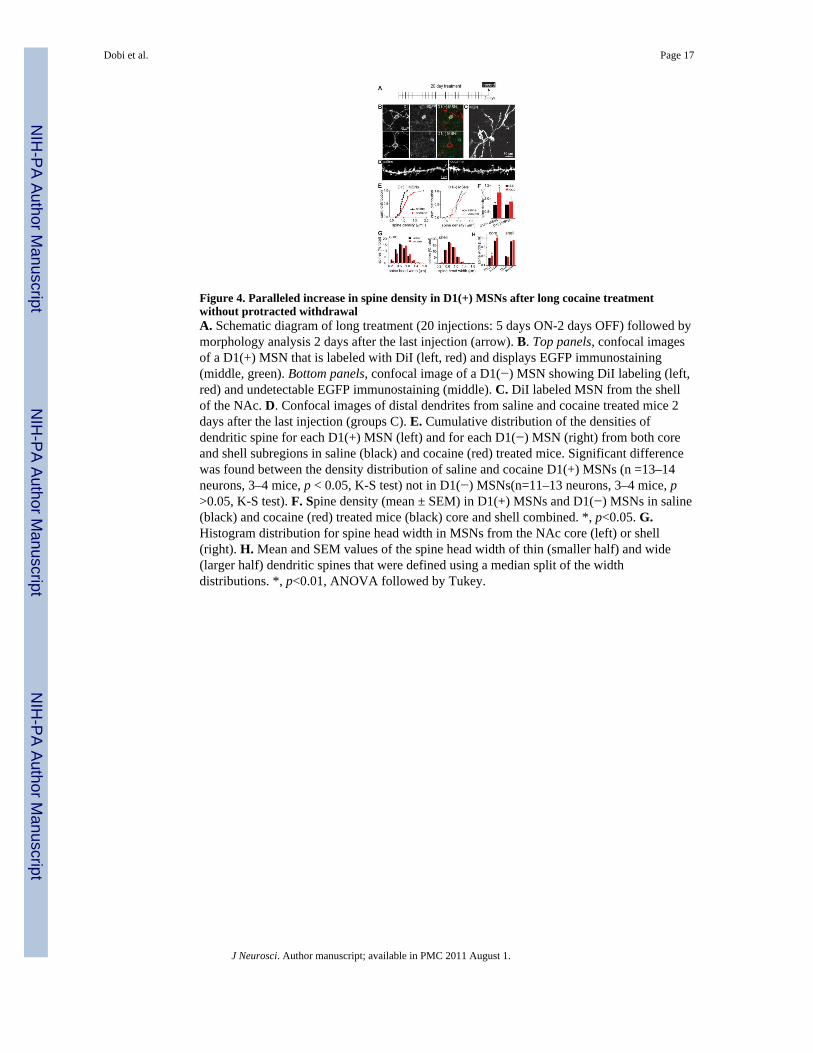
Figure 4. Paralleled increase in spine density in D1(+) MSNs after long cocaine treatmentwithout protracted withdrawalA. Schematic diagram of long treatment (20 injections: 5 days ON-2 days OFF) followed bymorphology analysis 2 days after the last injection (arrow). B. Top panels, confocal imagesof a D1(+) MSN that is labeled with DiI (left, red) and displays EGFP immunostaining(middle, green). Bottom panels, confocal image of a D1(−) MSN showing DiI labeling (left,red) and undetectable EGFP immunostaining (middle). C. DiI labeled MSN from the shellof the NAc. D. Confocal images of distal dendrites from saline and cocaine treated mice 2days after the last injection (groups C). E. Cumulative distribution of the densities ofdendritic spine for each D1(+) MSN (left) and for each D1(−) MSN (right) from both coreand shell subregions in saline (black) and cocaine (red) treated mice. Significant differencewas found between the density distribution of saline and cocaine D1(+) MSNs (n =13–14neurons, 3–4 mice, p < 0.05, K-S test) not in D1(−) MSNs(n=11–13 neurons, 3–4 mice, p>0.05, K-S test). F. Spine density (mean ± SEM) in D1(+) MSNs and D1(−) MSNs in saline(black) and cocaine (red) treated mice (black) core and shell combined. *, p<0.05. G.Histogram distribution for spine head width in MSNs from the NAc core (left) or shell(right). H. Mean and SEM values of the spine head width of thin (smaller half) and wide(larger half) dendritic spines that were defined using a median split of the widthdistributions. *, p<0.01, ANOVA followed by Tukey.
Dobi et al. Page 17
J Neurosci. Author manuscript; available in PMC 2011 August 1.
NIH
-PA Author Manuscript
NIH
-PA Author Manuscript
NIH
-PA Author Manuscript
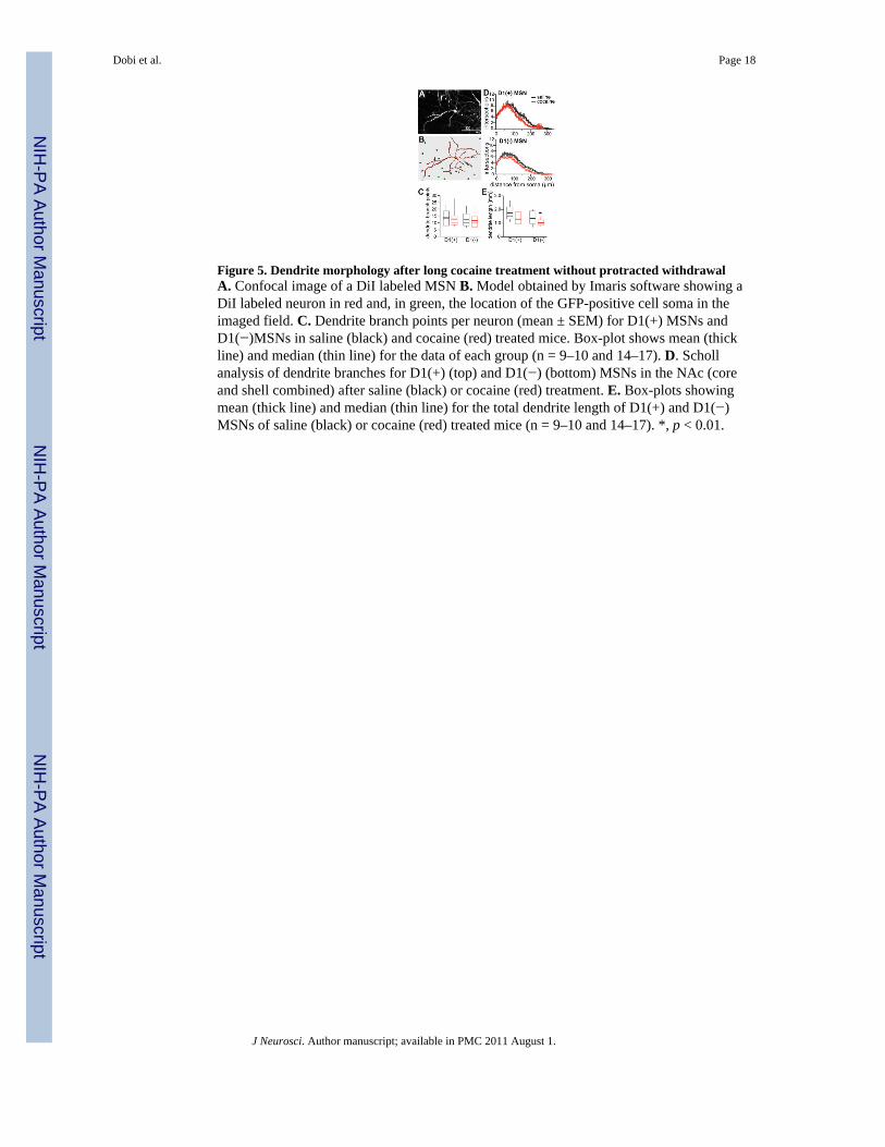
Figure 5. Dendrite morphology after long cocaine treatment without protracted withdrawalA. Confocal image of a DiI labeled MSN B. Model obtained by Imaris software showing aDiI labeled neuron in red and, in green, the location of the GFP-positive cell soma in theimaged field. C. Dendrite branch points per neuron (mean ± SEM) for D1(+) MSNs andD1(−)MSNs in saline (black) and cocaine (red) treated mice. Box-plot shows mean (thickline) and median (thin line) for the data of each group (n = 9–10 and 14–17). D. Schollanalysis of dendrite branches for D1(+) (top) and D1(−) (bottom) MSNs in the NAc (coreand shell combined) after saline (black) or cocaine (red) treatment. E. Box-plots showingmean (thick line) and median (thin line) for the total dendrite length of D1(+) and D1(−)MSNs of saline (black) or cocaine (red) treated mice (n = 9–10 and 14–17). *, p < 0.01.
Dobi et al. Page 18
J Neurosci. Author manuscript; available in PMC 2011 August 1.
NIH
-PA Author Manuscript
NIH
-PA Author Manuscript
NIH
-PA Author Manuscript

Figure 6. Protracted withdrawal following long cocaine treatment does not enhance plasticityA. Schematic diagram of long cocaine treatment (20 injections: 5 days ON-2 days OFF)followed by recordings and morphology (R+M) at 2 days or 30 days of withdrawal. B.mEPSCs frequency (mean ± SEM) recorded from D1(+) MSNs in core and shell of saline-treated (black bar, 2–30 day combined) and cocaine-treated mice 2 days (gray bar) or 30days (white bar) after the last cocaine injection (n=15–41 neurons/ 4–7 mice) ; *, p < 0.05by ANOVA and Tukey. C. Spine density (mean ± SEM) in D1(+) MSNs from core andshell in saline-treated mice or cocaine-treated mice 2 days (gray bar) and 30 days (white bar)after the last cocaine injection (n= 6–8 neurons, 3–5 mice ) *, p < 0.05 by ANOVA andTukey).
Dobi et al. Page 19
J Neurosci. Author manuscript; available in PMC 2011 August 1.
NIH
-PA Author Manuscript
NIH
-PA Author Manuscript
NIH
-PA Author Manuscript

NIH
-PA Author Manuscript
NIH
-PA Author Manuscript
NIH
-PA Author Manuscript
Dobi et al. Page 20
Table 1
Experimental design for the short and long drug treatments of animals with either saline or cocaine.
group # injections withdrawal study
A 7 days i.p. 2 days electrophysiology
B 7 days i.p. 21 days electrophysiology
C 20 days i.p. (5 days ON, 2 day OFF) × 4 weeks 2 days electrophysiology and morphology
D 20 days i.p. (5 days ON, 2 day OFF) × 4 weeks 30 days electrophysiology and morphology
E 28 days i.p. 1 day electrophysiology
Dobi et al. “Cell-specific plasticity without cocaine withdrawal”
J Neurosci. Author manuscript; available in PMC 2011 August 1.
Related Documents