AIMS Microbiology, 3(4): 846-871. DOI: 10.3934/microbiol.2017.4.846 Received: 21 September 2017 Accepted: 17 October 2017 Published: 23 October 2017 http://www.aimspress.com/journal/microbiology Research article CO 2 and carbonate as substrate for the activation of the microbial community in 180 m deep bedrock fracture fluid of Outokumpu Deep Drill Hole, Finland Malin Bomberg 1, *, Mari Raulio 1,2 , Sirpa Jylhä 1 , Carsten W. Mueller 3 , Carmen Höschen 3 , Pauliina Rajala 1 , Lotta Purkamo 1 , Riikka Kietävä inen 4 , Lasse Ahonen 4 , and Merja Itävaara 1 1 VTT Technical Research Centre of Finland, P.O. Box 1000, FIN-02044 VTT, Finland 2 Tikkurila Oyj, P.O. Box 53, Kuninkaalantie 1, FI-01301 Vantaa, Finland 3 Lehrstuhl für Bodenkunde, Department Ecology and Ecosystem Management, Center of Life and Food Sciences Weihenstephan, Technische Universitä t München, D-85350, Freising-Weihenstephan, Germany 4 Geological Survey of Finland (GTK), P.O. Box 96, 02151 Espoo, Finland * Correspondence: Email: [email protected]; Tel: +358-40-1863869. Abstract: Microbial communities in deep subsurface environments comprise a large portion of Earth’s biomass, but the metabolic activities in these habitats are largely unknown. Here the effect of CO 2 and carbonate on the microbial community of an isolated groundwater fracture zone at 180 m depth of the Outokumpu Deep Scientific Drill Hole (Finland) was tested. Outokumpu groundwater at 180 m depth contains approximately 0.45 L L −1 dissolved gas of which methane contributes 76%. CO 2 , on the other hand, is scarce. The number of microbial cells with intracellular activity in the groundwater was low when examined with redox staining. Fluorescence Assisted Cell Sorting (FACS) analyses indicated that only 1% of the microbial community stained active with the redox sensing dye in the untreated groundwater after 4 weeks of starvation. However, carbon substrate and sulfate addition increased the abundance of fluorescent cells up to 7%. CO 2 and CO 2 + sulfate activated the greatest number of microbes, especially increasing the abundance of Pseudomonas sp., which otherwise was present at only low abundance in Outokumpu. Over longer exposure time (2 months) up to 50% of the bacterial cells in the groundwater were shown to incorporate inorganic carbon from carbonate into biomass. Carbon recapture is an important feature in this ecosystem since it may decrease the rate of carbon loss in form of CO 2 released from cellular processes.

Welcome message from author
This document is posted to help you gain knowledge. Please leave a comment to let me know what you think about it! Share it to your friends and learn new things together.
Transcript

AIMS Microbiology 3(4) 846-871
DOI 103934microbiol20174846
Received 21 September 2017
Accepted 17 October 2017
Published 23 October 2017
httpwwwaimspresscomjournalmicrobiology
Research article
CO2 and carbonate as substrate for the activation of the microbial
community in 180 m deep bedrock fracture fluid of Outokumpu Deep
Drill Hole Finland
Malin Bomberg 1 Mari Raulio
12 Sirpa Jylhauml
1 Carsten W Mueller
3 Carmen Houmlschen
3
Pauliina Rajala 1 Lotta Purkamo
1 Riikka Kietaumlvaumlinen
4 Lasse Ahonen
4 and Merja Itaumlvaara
1
1 VTT Technical Research Centre of Finland PO Box 1000 FIN-02044 VTT Finland
2 Tikkurila Oyj PO Box 53 Kuninkaalantie 1 FI-01301 Vantaa Finland
3 Lehrstuhl fuumlr Bodenkunde Department Ecology and Ecosystem Management Center of Life and
Food Sciences Weihenstephan Technische Universitaumlt Muumlnchen D-85350 Freising-Weihenstephan
Germany 4
Geological Survey of Finland (GTK) PO Box 96 02151 Espoo Finland
Correspondence Email malinbombergvttfi Tel +358-40-1863869
Abstract Microbial communities in deep subsurface environments comprise a large portion of
Earthrsquos biomass but the metabolic activities in these habitats are largely unknown Here the effect of
CO2 and carbonate on the microbial community of an isolated groundwater fracture zone at 180 m
depth of the Outokumpu Deep Scientific Drill Hole (Finland) was tested Outokumpu groundwater at
180 m depth contains approximately 045 L Lminus1
dissolved gas of which methane contributes 76
CO2 on the other hand is scarce The number of microbial cells with intracellular activity in the
groundwater was low when examined with redox staining Fluorescence Assisted Cell Sorting (FACS)
analyses indicated that only 1 of the microbial community stained active with the redox sensing dye in
the untreated groundwater after 4 weeks of starvation However carbon substrate and sulfate addition
increased the abundance of fluorescent cells up to 7 CO2 and CO2 + sulfate activated the greatest
number of microbes especially increasing the abundance of Pseudomonas sp which otherwise was
present at only low abundance in Outokumpu Over longer exposure time (2 months) up to 50 of
the bacterial cells in the groundwater were shown to incorporate inorganic carbon from carbonate
into biomass Carbon recapture is an important feature in this ecosystem since it may decrease the
rate of carbon loss in form of CO2 released from cellular processes
847
AIMS Microbiology Volume 3 Issue 4 846-871
Keywords groundwater crystalline bedrock NanoSIMS FACS methane sulfate Pseudomonas
autotroph Outokumpu
1 Introduction
Precambrian shield areas such as the Fennoscandian Shield preserve the oldest geological
records of our planet but have also been affected by the continuous evolution of life atmosphere
and climate Processes taking place in the continental crust are not only of scientific interest but
microbial and geochemical processes in fractured crystalline rock play a key role also eg in
assessing the safety of geological disposal of hazardous wastes Microorganisms have a great impact
on the elemental cycles in the upper crust and are responsible for the conversion of organic matter to
be reused for the maintenance of the whole microbial community [1] Carbon derived from
photosynthesis only slowly diffuses into the terrestrial subsurface if at all Thus alternative means of
carbon assimilation such as inorganic carbon fixation or methane oxidation may be important to
support the microbial community
The Outokumpu Deep Drill Hole provides access to study isolated groundwaters in the
Paleoproterozoic part of the Fennoscandian Shield in eastern Finland [2] The drill hole reaches a
total depth of 2516 m and spans several fluid-filled fracture zones The deep groundwater in
Outokumpu is highly reducing and has low organic carbon content Methane is the most abundant
hydrocarbon and contributes up to 80 of the total volume of gas released from the groundwaters
and is continuously discharging on the drill hole collar [34] The microbial communities in the
Outokumpu deep subsurface have been reported to contain genes for different autotrophic carbon
fixation pathways [5] and methane cycling [16] In addition it has recently been shown that methane
activates the microbial community residing in the fracture zone at 500 m in Outokumpu but does not
increase the number of active microbial cells in the fracture at 180 m [7] Nevertheless the
transcription of bacterial 16S rRNA genes increased showing that especially Pseudomonas bacteria
were affected by the added carbon substrates at 180 m depth while at 500 m bacteria belonging to
the order Rhodobacterales were activated [7] Only one other study exists to date that show specific
functional activity of the deep subsurface microbial community in Outokumpu [6] Thus more
indication of actual microbial metabolic activity and ability to assimilate simple carbon compounds
in deep subsurface environments is needed
Previous studies of the Outokumpu scientific drill hole microbial community has concentrated
on depths from 500 m and below [568] and only few studies exist that describe the microbial
community in the upper parts of the Outokumpu subsurface [910] In addition only little is presently
known about the utilization of small carbon compounds by deep terrestrial subsurface microbial
communities Carbon is lost from biomass as carbon dioxide that is produced in different catabolic
cellular processes such as fermentation or oxidation of organic carbon if carbon dioxide is not
recaptured One of the main objectives of this study was thus to determine the proportion and
identity of microorganisms responding to and harvesting inorganic carbon in the microbial
community of groundwater in Outokumpu In addition we aimed to expand the knowledge of the
deep subsurface microbial community in Outokumpu to include a fracture zone from a more
moderate depth (180 m) than the previously studied deeper located fracture zones
848
AIMS Microbiology Volume 3 Issue 4 846-871
2 Materials and Method
21 Description of the sampling site
The Outokumpu Deep Drill Hole is situated in Eastern Finland in Paleoproterozoic
approximately 19 Ga old bedrock The lithology hydrogeochemistry and gas composition of the
drill hole water column and fracture zone fluids have been described previously [1ndash511] Shortly
the fracture zone at 180 m depth is situated in a metasedimentary rock sequence predominated by
mica schist
22 Sampling and analytical methods for chemistry
The fracture zone at 180 m depth was isolated from the rest of the drill hole by inflatable
packers as described in [10] The isolated fracture zone was purged by pumping fluid via an air-tight
poly acetate (PA) tube In total around 23 m3 of water and 10 m
3 of gas (at standard T and P) was
drawn between the packers during the period between 5th
of May and 18th
of June 2012 with a
typical pumping rate of 05 m3 of water per day [4] Temperature pH electrical conductivity (EC)
concentration of dissolved O2 and redox potential (Eh) of the pumped fluid was continuously monitored
in a flow-through cell Redox potential was measured using combined ORP electrode (Hamilton) with
AgAgCl reference and values tested against a known reference solution Measured values were
converted to Eh-scale according to the operation manual of the electrode Gaswater volume ratio
was obtained from collected gas volume in a vessel under water and total water flow Samples for
geochemical analysis were taken from the pumped fluid usually twice a week Samples for cation
analysis (100 mL each) were filtered in the field using pore size of 045 μm and acidified with
ultrapure HNO3 (05 mL per 100 mL of sample water) to prevent flocculation Untreated samples of
250 mL each were used for anion analysis Samples for sulfide analysis were fixed with 05 mL of 1 M
NaOH and 05 mL of 1 M zinc acetate per 100 mL of water Samples for Fe2+
were taken in glass
bottles (Winkler) and fixed with concentrated HCl (4 mL per 100 mL of sample water) Gas samples
were collected into inverted glass bottles (Schott Duran Group Main Germany) under sample water
and sealed with black butyl rubber stoppers All samples were kept refrigerated until analyzed
Cations were analyzed at Labtium Oy (Espoo Finland) The concentrations of K Mn Zn and I
were determined using inductively coupled plasma mass spectrometer (ICP-MS Perkin Elmer and
Agilent Technologies) and S Na Mg Fe(tot) and Ca using inductively coupled plasma optical
emission spectrometer (ICP-OES Thermo Jarrell Ash Corp) according the standard methods
SFS-EN ISO 17294-2 and SFS-EN ISO 11885 respectively Fe2+
was determined
spectrophotometrically Anions were determined by ion chromatography at Labtium Oy according
the standard method SFS-EN ISO 10304-1 or at TVO Nuclear Services (Eurajoki Finland) using a
Dionex ICS-2000 device Concentrations of sulfide were determined within 24 h from four parallel
samples at Ramboll Analytics (Vantaa Finland) using a spectrophotometer Total and dissolved
carbon was determined using a TOC analyzer at Labtium Oy according the standard method
SFS-EN 1484 which is a pyrolytic method based on infra-red (IR) detection End-point titration to
pH 45 (standardized potentiometric method SFS 3005) was used to determine alkalinity Gas
compositions were determined by gas chromatography at Ramboll Analytics Carbon speciation was
calculated using PHREEQC software (USGS 2014)
849
AIMS Microbiology Volume 3 Issue 4 846-871
23 Sampling for microbiology and sample handling
Groundwater samples for microbiology were obtained on June 12ndash14 2012 by pumping the
fracture fluid via the PA tube directly in to an anaerobic chamber as described in [8] Altogether 10
individual 50 mL samples were collected into sterile acid washed 120 mL glass serum bottles sealed
with butyl rubber stoppers (Bellco Glass Inc NJ USA) and aluminum crimp caps (Sigma MO
USA) were collected for nano-scale secondary ion mass spectrometry (NanoSIMS) analysis
fluorescence microscopy substrate induction (activation) and cell sorting and cultivation In addition
for the detection of transcription activation four 2 L water samples were collected into sterile
anaerobic borosilicate glass bottles equipped with gas-tight butyl rubber septa The biomass from two
parallel 1 L water samples one for DNA extraction and one for RNA extraction was collected on 02
μm pore-size cellulose acetate filters (Corning) by vacuum suction as described by Purkamo et al [8]
directly in the field The membrane filters were directly cut out of the filter funnels and fixed in dry
ice The rest of the water samples transported cooled (+4 degC) to the laboratory In the laboratory the
water samples in the sealed bottles (both 50 mL and 1 L) were starved for 4 weeks by keeping the
sampling bottles at +4 degC protected from light prior to the experiment
24 Determination of cell numbers and proportion of viable microbial cells by epifluorescence
microscopy
The total number of microbial cells in the groundwater at the time of sampling was determined
by epifluorescence microscopy of 4rsquo6 diamidino-2-phenylindole dihydrochloride (DAPI) (Sigma
MO USA) stained cells as previously described [7] In addition the percentage of intact microbial
cells (ie microbial cells containing plasma membrane integrity) in the fracture fluid at the time of
sampling and after 4 weeks of starvation was determined by LIVEDEADreg BacLighttrade Bacterial
Viability staining (Molecular Probes Invitrogen CA USA) according to the manufacturerrsquos
instructions followed by epifluorescence microscopy as previously described [11] The analyses were
done with two parallel fracture fluid samples of 1 mL or 25 mL The microbial cells in the stained
groundwater were collected on black GTPB Membrane filter (Millipore Billercia MA USA) using a
Millipore filtering unit (Millipore Billercia MA USA) The average number of microbial cells mLminus1
groundwater was calculated from 30 randomly chosen microscopic fields from each subsample taking
into account the volume (1 or 25 mL) of the sample collected on the filter and the total (28353 mm2)
and the active (0014888 mm2) area of the polycarbonate filter and magnification (1000times)
25 Induction of respiration and transcription by addition of substrates
The activating effect of small carbon substrates on the microbial community was identified by
the redox indicating dye 5-cyano-23-ditolyl tetrazolium chloride (CTC Polysciences Inc PA
USA) Tetrazolium salts function as artificial electron acceptors that are reduced in metabolically
active microbial cells by the components of the electron transport chain or by dehydrogenase activity
to form brightly fluorescing formazan crystals in the cells 50 mL sterile acid washed glass serum
bottles (Wheaton NJ USA) were flushed with sterile N2 gas for 30 min each containing 055 mL 50 mM
CTC dye and sealed with sterile butyl rubber stoppers (Bellco Glass Inc NJ USA) and open top
aluminum crimp caps (Sigma MO USA) Fracture fluid after 4 weeks of starvation was anaerobically
850
AIMS Microbiology Volume 3 Issue 4 846-871
aliquoted (5 mL) through the butyl rubber stoppers into the sealed infusion bottles using a N2 flushed
sterile syringe and needle The treatments used were (1) no substrate additions to the fracture fluid
and addition of (2) sulfate (3) CO2 and (4) CO2 + sulfate to fracture fluid samples Sulfate was added
through a 02 μm pore size syringe filter with an anaerobic N2-flushed syringe and needle through the
rubber stoppers to the fracture fluid to a final concentration of 0375 mmolL CO2 gas (20 mL
equaling a total of 09 mmoles of which a maximum concentration of 018 mmolL may be dissolved
in the water sample) was added to the sealed infusion bottles using a syringe and needle pushed
through the butyl rubber stopper All gases were filter sterilized before injection by attaching a 02 μm
pore size filter between the syringe and the needle The sulphate solution (Na2SO4 888 mg mLminus1
in
anaerobic MilliQ water stock solution Sigma) was rendered anaerobic by N2 flush for 30 min The
samples were incubated together with the CTC dye for 6 h according to the manufacturers
recommendations at +14 degC on a shaker (45 rpm) after which 05 mL glycerol-TE buffer was added
to the samples for preservation at minus80 degC according to the protocol by the Single Cell Genomics
Center (httpsscgcbigeloworgPDFsSample_cryopreservation_glyTEpdf) Briefly the samples
were divided into 1 mL aliquots in sterile cryo tubes and immediately frozen in liquid N2 and stored
at minus80 degC A non-dyed reference sample was prepared and cryo-preserved in the same way as the
dyed samples
2 L batches of the stored groundwater were prepared for RNA extraction of the transcriptionally
activated microbial community using the same substrates as described above In addition one 2 L
batch of stored groundwater was used as baseline for the activation process The experiment was
performed in 2 L borosilicate bottles (Schott) equipped with butyl rubber stoppers and open-top screw
caps Anaerobic sterile sulphate solution (Na2SO4) was added to a concentration of 0375 mmolL
using a syringe and needle pushed through the butyl rubber stopper as described above Carbon
dioxide gas 320 ml was added with a sterile syringe and needle through the butyl rubber stopper as
described above The 2 L bottled were shaken vigorously by hand at the beginning of the incubation
and kept at +14 degC for 2 h After the incubation the biomass from the groundwater samples was
collected on 02 μm pore-size cellulose acetate bottle-top filters (Corning) by vacuum suction The
filters were cut out of the filter funnels using sterile scalpels divided into two replicate samples of
each treatment representing two 1 L replicates which were inserted into sterile 50 mL screw-cap
test tubes (Corning) and immediately frozen at minus80 degC until RNA extraction
26 Sorting of active cells by FACS
The CTC dyed and un-dyed fracture fluid aliquots were carefully thawed on ice prior to
screening by flow cytometry (BD FACSaria flow cytometer Becton Dickinson NJ USA) Samples
were injected into a sterile phosphate buffered saline (PBS) flow stream and fluorescence was
detected using a 655 nm long pass and 67520 nm band pass filters For excitation of CTC an argon
laser was used (488 nm) To detect the metabolically active cells simultaneous measurements of
forward light scatter (relative size) side light scatter (cell granularity) and CTC fluorescence
emission were used by setting the PMT voltage to 250 250 and 500 volts respectively The side
scatter threshold was set to 2000 To investigate the concentration of metabolically active cells
events were acquired over a time of 30 s with a flow rate of 100 μL minminus1
from each sample The
fluorescence signal was plotted to the side scatter and analyzed with the BD FACSDivatrade 51
software (Becton Dickinson NJ USA) The CTC positive cells were gated comparing the
851
AIMS Microbiology Volume 3 Issue 4 846-871
fluorescence intensities of the dyed to the un-dyed fracture fluid sample For downstream analysis
and identification of the active microbial population CTC positive cells were sorted into wells on
sterile 8-strip 250 μL tubes 10000 microbial cellstube in approximately 100 μL solution In addition
10000 randomly selected cells were also collected from the un-dyed groundwater sample in order to
determine the composition of the general community in the groundwater Empty wells were left in
between each sorted sample as negative controls The sorted cells were immediately frozen on dry
ice and stored at minus80 degC until further analysed
27 Nucleic acid extraction
271 DNA extraction from sorted cells
All cell lysis and DNA amplification work was conducted in a UV light-treated laminar flow
hood using filter-equipped gamma-sterilized pipette tips DNA was extracted from the sorted
active microbial populations by freeze-thaw cycles The sorted cells were frozen (minus25 degC) and
heated for 10 min at 99 degC block temperature and at 105 degC lid temperature in a MasterCycler
Thermal cycler (Eppendorf Hamburg Germany) and re-frozen on a minus25 degC close-fitting freeze
block (Eppendorf Hamburg Germany) in a freezer for 30 min This cycle was repeated 2 times
after which the released DNA was precipitated with 05 mL 94 ethanol The released DNA was
allowed to precipitate at minus25 degC for 15 min after which the precipitates were collected by
centrifugation +4 degC at 14000 rpm for 20 min in a table-top centrifuge (Eppendorf Hamburg
Germany) The ethanol was decanted and the DNA pellet was allowed to dry at 37 degC in a heat block
in a laminar flow hood The DNA was resuspended in 10 μL molecular grade water Negative
reagents controls were treated parallel to the samples in order to control possible contamination of
the samples by external sources
272 DNA and RNA extraction from groundwater samples
The filters with the collected microbial biomass were shortly thawed on ice before RNA
extraction The RNA was separately extracted from each half filter using the Mobio PowerWater
RNA extraction kit (Mobio Laboratories Inc Carlsbad CA USA) according to the manufacturerrsquos
instructions The RNA of each extraction was eluted into 100 μl of buffer PWR8 (Mobio
Laboratories Inc Carlsbad CA USA) Negative control extractions were performed in parallel to
the samples The RNA extracts were tested for remnant DNA by performing PCR with general
primers targeting the bacterial 16S rRNA gene The primers used were the U968f and U1401r [12]
using the Dynazyme II DNA polymerase as described in [13] E coli DNA was used as positive
control for the PCR No PCR product in the PCR reaction of the RNA extracts indicated that no
residual DNA was present The DNA from the groundwater from the day of sampling was extracted
using the Mobio PowerSoil DNA extraction kit according to the manufacturerrsquos instructions The
DNA was eluated in 100 μl buffer CS6
28 Reverse transcription of RNA
The extracted RNA was reverse transcribed using the Superscript III First Strand Synthesis
852
AIMS Microbiology Volume 3 Issue 4 846-871
SuperMix (Invitrogen Carlsbad CA USA) according to the manufacturerrsquos instructions First
115 μL aliquots of RNA were incubated together with 250 ng random hexamers (Promega WI USA)
and 083 mmolL (final concentration) dNTP (Finnzymes Espoo Finland) at 65 degC for 5 min
Thereafter the reactions were cooled on ice for 1 min followed by the reverse transcription reaction
when 4 μL 5times First strand buffer 40 U DTT and 200 U Superscript III enzyme were added The RNA
was protected from degradation by addition of 40 U recombinant RNase inhibitor RNaseOut (Promega
WI USA) The reactions were incubated at 25 degC for 5 min 50 degC for 1 h and finally inactivated at
70 degC for 15 min
29 Amplicon library preparation and sequence analysis
The bacterial and archaeal community composition of the sorted cell batches and the 1 L DNA
and RNA samples were examined by high throughput amplicon sequencing using the Ion Torrent
PGM platform as described in [7] The amplification libraries were prepared by PCR from the DNA
and cDNA samples Bacterial 16S rRNA genes were targeted with primers
S-D-Bact-0341-b-S-17S-D-Bact-0785-a-A-21 [14] targeting the variable region V3ndashV4 of the 16S rDNA
gene and the archaeal 16S genes with primers S-D-Arch-0349-a-S-17S-D-Arch-0787-a-A-20 [15]
targeting the V4 region of the gene The fungal communities were targeted with primers ITS1 and
58A2R flanking the internal transcribed spacer region 1 (ITS1) [16] PCR amplifications were
performed in parallel 25 μl reactions for every sample using the MyTaqTM Red Mix (Bioline
London UK) Each reaction contained 20 pmol forward and reverse primers and 2 μL of template
The PCR program consisted of an initial denaturation step at 95 degC for 3 min 40 cycles of 15 s at
95 degC 15 s at 50 degC and 15 s at 72 degC A final elongation step of 30 s was performed at 72 degC The
PCR products were verified with agarose gel electrophoresis Amplicons were sent for sequencing to
Bioser at the University of Oulu (Finland) where the amplicons underwent purification and size
selection before sequencing
The sequence reads obtained from the Ion Torrent sequencing were subjected to quality control
using the QIIME software version 19 as described in [7] The sequences were grouped in to
Operational Taxonomic Units (OTUs) following the open-reference OTU-picking protocol of
QIIME and using the Silva version 128 database [17] for bacteria and archaea and the UNITE
database version 6 [18] for fungi Microbial community composition between the original
groundwater sorted cells and actively transcribing communities was tested by principal coordinates
analysis (PCoA) using the Phyloseq package in R [1920]
The sequences were deposited in ENA under study number PRJEB22697
210Isolation of bacterial strains from fracture fluid
Bacterial strains were isolated on anaerobic Basal Mineral Medium [21] amended with 10 g Lminus1
NaCl to obtain pure cultures able to utilize carbonate as carbon source The medium was composed
of Lminus1
Na2HPO412 H2O 9 g KH2PO4 15 g NH4Cl 10 g MgSO47 H2O 02 g trace elements Lminus1
ZnSO47 H2O 100 μg MnCl2 30 μg H3BO3 300 μg CoCl26 H2O 200 μg CuCl22 H2O 10 μg
NiCl26 H2O 20 μg Na2MoO42 H2O 30 μg ferric ammonium citrate 5 mg CaCl22 H2O 10 mg
and 5 g NaHCO3 The medium was aliquoted in 10 mL portions into glass serum bottles (Wheaton
NJ USA) and purged with filtered N2 gas for 1 h Lminus1
medium in order to render the medium
853
AIMS Microbiology Volume 3 Issue 4 846-871
anaerobic The bottles were sealed with butyl rubber stoppers (Bellco Glass Inc Vineland NJ USA)
and open top aluminum crimp caps (Sigma MO USA) before autoclaving The medium was
solidified with 17 agar Before the medium solidified 50 μL L-cystein hydrochloride reduced
resazurine (10 μg L-cystein hydrochloride and 25 μg resazurin mLminus1
stock solution) was added to the
medium in order to maintain reducing conditions in the culture bottles The resazurine functioned as
oxygen indicator After solidification the medium was inoculated by anaerobically injecting 1 mL
groundwater sample into the sealed medium-containing flasks through the butyl rubber septum using
a sterile N2 flushed syringe and needle Bacterial cells in the water sample were allowed to attach to
the agar surface for 30 min before the excess water was removed Colonies were allowed to form for
10 days at +14 degC after which 10 colonies were picked for purification The colonies were purified
with three consecutive streak dilutions on individual agar plates after which DNA was extracted from
individual colonies by suspending them in water (50 μL) and lysing the bacterial cells by heating in a
heat block at 99 degC for 10 min The bacterial strains were identified by 16S rRNA gene PCR and
sequencing using the fD1 and U1401r primers [1222]
211Nano-scale secondary ion mass spectroscopy (NanoSIMS)
Aliquots (50 mL) of groundwater were amended with anaerobic filter sterilized 13
C sodium
carbonate or 12
C sodium carbonate (Sigma MO USA) as to a final concentration of 4 mmolL by
injection through the butyl rubber stopper as described above The samples were incubated at +14 degC
for 60 days In addition pure cultured bacterial strains (described above) were re-grown in Basal
Mineral Medium supplemented with 13
C or 12
C sodium carbonate (Sigma MO USA) to a final
concentration of 4 mmolL for 2 weeks Subsamples of the prepared groundwater samples and the
pure cultures were collected on membrane filters pre-coated with AuPt (30 nm 208 HR High
Resolution Sputter Coater Cressington Scientific Instruments Inc Cranberry PA USA) for scanning
electron microscopy (SEM) and nano-scale secondary ion mass spectrometry (NanoSIMS) The
preparations were fixed in phosphate (01 M pH 72) buffered 25 glutaraldehyde at +4 degC for 20 h
and rinsed with phosphate buffer three times Dehydration was carried out with an ethanol series
from 30 to 50 to 70 to 80 to 96 and absolute followed by hexamethyldisilazane (Fluka
Buchs Switzerland) The samples were examined with a Hitachi S-4800 FESEM (Tokyo Japan)
operated at 1 kV and with NanoSIMS (Cameca NanoSIMS 50 L Gennevilliers Cedex France) at the
Lehrstuhl fuumlr Bodenkunde (TU Munich Germany) A fine focused Cs+ primary ion beam (about 1 pA)
was used to produce secondary ions at a lateral resolution of about 100 nm The instrument was tuned
for a mass resolving power eligible to distinguish between 12
C1H and
13C The
12C
minus
13C
minus and
12C
14N
minus
secondary ions were simultaneously collected in imaging mode using electron multipliers with a set
dead time of 44 ns Raster images of 13 times 13 microm2 and 15 times 15 microm
2 256 times 256 pixel were recorded
with 30 ms dwell time The instrument was checked regularly before measurements using a Si
engraved reference sample The 12
C14
Nminus images were used for the ROI selection as these best
represented the bacterial cells The cumulative counts of every ROI were used to calculate the 13
Cminus12
Cminus ratio for every bacterial cell which are shown as box plots illustrating natural abundance
versus labelled bacterial cells The background natural abundance value was obtained from the
non-labelled bacterial cell ROI values All analyses including the calculation of the 13
Cminus12
Cminus ratio
images were processed using the Cameca WinImage Software
854
AIMS Microbiology Volume 3 Issue 4 846-871
212Sequence analysis and phylogeny of the pure cultured strains
The obtained sequences were imported into Geneious Pro (version 601 Biomatters Ltd
Auckland New Zealand) and manually checked assembled and edited before being subjected to
phylogenetic analysis The sequences were compared with the blastn tool in Geneious Pro to the
NCBI nucleotide database The closest matching sequences relevant reference sequences and
sequences of suitable type species were included in the phylogenetic analyses The bacterial 16S
rRNA gene sequences were aligned using Muscle in Geneious Pro and the alignment was edited
manually A maximum likelihood cladogram was calculated for each domain using PhyML [23] with
the Jukes-Cantor substitution model [24] Bootstrap support for the branches was calculated with
1000 random repeats The sequences were submitted to Genebank under accession numbers
KP192167ndashKP192240
3 Results
31 Physicochemical parameters
Fracture fluid was pumped from 180 m depth of the Outokumpu Deep Scientific Drill Hole to
investigate the environmental characteristics and the microbial populations present in this habitat
During the pumping the pH decreased from 11 to 85 indicating that the drill hole water which has
been disturbed by the alkaline effect of concrete casting of the uppermost 22 m of the drill hole was
effectively removed by the time of microbiological sampling The fracture fluid at 180 m depth is
brackish (Na-Ca-Cl type) with total dissolved solids (TDS) around 6 g Lminus1
(Table 1) Conditions were
reducing with dissolved O2 below the limit of detection in the on-line measurements Eh
approximately minus01 V and iron occurring as Fe2+
Gaswater ratio of discharging fluid was
approximately 045 L Lminus1
Gaswater ratios determined from pressurized total fluid samples in the lab
gave comparable results between 0425 and 0440 Dissolved gas phase comprised mostly of
methane (up to 76 vol-) and nitrogen (up to 22 vol-) with minor amounts of He Ar ethane and
propane Some O2 was detected in the laboratory analyses of gas samples but is likely due to minor
contamination from air during sampling andor analysis H2 was occasionally detected but generally
remained below the detection limit of 0003 vol- Likewise CO2 was not present in detectable
amounts Very little inorganic carbon of which 81 (023 mmolL) was HCO3minus was found The total
amount of carbonic acid anions (HCO3minus CO3
2minus) was low as indicated by the low alkalinity (~03 mmolL)
and low concentration of dissolved inorganic carbon (021 mmolL) Consequently the dominant form
of carbon in the fracture fluid was methane (ie about 15 mM or 240 mg Lminus1
) Low concentrations
of both sulfate (0007 mmolL) and sulfide (00018 mmolL) were found
32 Microbial cell numbers and proportion of active respiring cells
The concentration of microbial cells as determined by DAPI staining and microscopy was
30 times 105 mL
minus1 (std 88 times 10
4)
of which 87 (26 times 10
5 mL
minus1) were viable as determined with the
livedead staining After 4 weeks of storage in order to deplete carbon and nutrients the cell number
had slightly increased to 36 times 105 mL
minus1 (std 56 times 10
4) and all cells appeared viable as determined
with the livedead staining The number of microbial cells determined by FACS in the redox stained
855
AIMS Microbiology Volume 3 Issue 4 846-871
Table 1 Geochemical characteristics of the groundwater from the isolated fracture at
180 m depth in the Outokumpu Deep Drill Hole Groundwater pH Eh EC and
dissolved oxygen were measured continuously in a flow-through cell Dissolved gas
phase composition analysed for gas phase extracted from water (gas to water ratio 045)
Measurement Value Date
pH 85 1362012
Eh minus95 mV 1362012
EC 1035 mSm 1362012
O2
Alkalinity
00 mgL
031 mmolL
1362012
1362012
O2 037 vol- 1362012
N2 21 vol- 1362012
CO2 lt0003 vol- 1362012
CH4 74 vol- 1362012
H2 lt0003 vol- 1362012
C2H6 086 vol- 1362012
C3H8 0025 vol- 1362012
He 15 vol- 1362012
Ar 027 vol- 1362012
TOC 048 mmolL 662012
DOC 045 mmolL 662012
TIC 023 mmolL 662012
DIC 021 mmolL 662012
SO4 07 mgL 562012
Sulfide 006 mgL 662012
NO3 lt20 mgL 1362012
Br 23 mgL 1362012
Cl 3280 mgL 1362012
I 300 mgL 1362012
F 02 mgL 562012
S 127 mgL 1362012
Na 1070 mgL 1362012
Mg 167 mgL 1362012
Fe(tot)
Fe2+
034 mgL
049 mgL
1362012
662012
Ca 1060 mgL 1362012
Zn 463 μgL 1362012
Mn 133 μgL 1362012
K 137 mgL 1362012
856
AIMS Microbiology Volume 3 Issue 4 846-871
samples was 42 times 105 mL
minus1 (std 19 times 10
4) 53 times 10
5 mL
minus1 (std 46 times 10
4) and 48 times 10
5 mL
minus1 (std
89 times 104) in the samples that had been incubated together with CO2 CO2 + SO4 and SO4
respectively In contrast to the viability staining the redox indicating CTC staining showed that only
approximately 1 of the measured events (equivalent to microbial cells) fluoresced and were
detected by FACS (Figure 1 and Figure 2) The response of the inactive microbial cells to methane
as the most common carbon compound on Outokumpu deep subsurface and CO2 as substrate for
autotrophic microorganisms was tested with CTC In addition SO4 was added as terminal electron
acceptor since it is the most common one detected in Outokumpu although the concentration is low
The CO2 with or without SO4 had the greatest activating effect on the cells The CO2 + SO4 treatment
increased the relative abundance of active cells to 69 and CO2 alone to 65 CH4 and CH4 + SO4
increased the proportion of activated cells to 6 and 59 respectively Sulfate alone increased the
proportion of activated cells to 34 of the microbial cells
33 Characterization of the general and actively respiring fraction of the microbial community
The composition of the microbial community in the untreated groundwater and the actively
respiring community was studied from 10000 cells collected from each treatment with 16S rRNA
gene PCR followed by high throughput amplicon sequencing of the bacterial and archaeal 16S rRNA
gene and fungal ITS1 region In addition the bacterial and archaeal 16S rRNA and rRNA gene
profiles and fungal ITS1 profiles were characterized from the DNA and RNA from parallel 500 mL
water samples of the original groundwater as well as RNA from parallel 500 mL water after storage
and addition of CO2 andor SO4 with the same high throughput sequencing technique as above
Figure 1 Relative amount of metabolically active fluorescent cells as determined by
CTC staining and FACS The average values were calculated from three parallel
measurement and the error bars indicate standard error of mean
857
AIMS Microbiology Volume 3 Issue 4 846-871
Figure 2 A Forward scatter (showing the relative size of the particles) vs fluorescence
plots of the microbial population of the (A) undyed sample (B) CTC dyed sample
without addition of substrates and CTC dyed samples treaded with (C) sulfate (D)
methane (E) methane + sulfate (F) CO2 and (G) CO2 + sulfate amended samples The
red dots describe the nonfluorescent ie inactive population the green dots the activated
fluorescent population B The defined sorting gates for the microbial population AndashG as
in Figure 2A The red histograms indicate the inactive (negative) population and the
green histograms the active fluorescent (positive) population
Between 2701 and 12476 bacterial 1807 and 12270 archaeal and 401 and 20352 fungal sequence
reads were obtained from each DNA and RNA fraction of the groundwater samples (Table 2)
Nevertheless the RNA fraction of the groundwater samples that had received no substrate additions
did not yield any archaeal 16S rRNA or fungal ITS transcripts ITS transcripts were also very scarce
A
B
858
AIMS Microbiology Volume 3 Issue 4 846-871
in the RNA fraction of the groundwater collected directly in the field and were detected from only
one of the parallel samples The number of sequence reads obtained from the DNA of the sorted cells
varied between 1362 and 6811 and belonged only to bacteria No archaeal 16S rRNA or fungal ITS
transcripts were detected in the sorted cells The number of bacterial OTUs detected was high
ranging from 530 to 2030 per sample while the archaeal OTUs ranged from 225 to 793 The number
of fungal ITS OTUs was high in the DNA fraction of the groundwater (532 to 637 OTUs) but in the
RNA fractions the OTU numbers stayed below 70 According to the Chao1 richness estimate
between 30 and 53 of the estimated number of bacterial OTUs present in the samples were
detected and 38ndash60 of the archaeal OTUs The fungal diversity was lower than that of the bacterial
and archaeal and between 87 and 99 of the predicted fungal ITS OTUs were detected The
Shannon diversity index calculated based on the number and abundance of detected OTUs was
highest for the bacterial community ranging between 49 and 100 (Table 2) For the archaeal
community the Shannon index ranged between 30 and 42 and the fungal one between 11 and 56
The original fracture zone bacterial community consisted mostly of Betaproteobacteria (587ndash
643) with Clostridia (105ndash13) Bacteroidia (116ndash134) and Mollicutes (63ndash76) as minor
groups (Figure 3A) The same bacterial groups were detected in the RNA fraction but Clostridia
contributed with 396ndash426 while the proportion of Betaproteobacteria was 387ndash434 Over the
time of storage of the groundwater the relative abundance of 16S rRNA of alphaproteobacteria had
increased from approximately 1ndash3 in the original groundwater to 478ndash484 in the stored
groundwater at the time of the experiment The relative abundance of betaprotobacterial 16S rRNA
sequences had decreased to 113ndash120 and gammaproteobacterial 16S rRNA sequences from below
1 of the sequence reads to 307ndash318 of the sequence reads After addition of CO2 the relative
abundance of gammaproteobacterial 16S rRNA sequence reads increased to 607ndash647 the
betaproteobacterial 16S rRNA sequence reads remained at 85ndash112 while the transcription of
epsilonproteobacterial 16S rRNA genes increased from negligent to 168ndash220 The majority of the
detected gammaproteobacterial sequences belonged to the Pseudomonas and the betaproteobacterial
sequences to Hydrogenophaga Of the epsilonproteobacterial sequences one third belonged to
Sulfuricurvum and the rest to Sulfurimonas Clostridial 16S rRNA sequences were not detected in the
untreated stored water but after addition of CO2 the relative abundance of clostridial 16S rRNA
sequences increased to 57ndash65 The most abundant clostridial sequences belonged to
Desulfosporosinus and an uncultured Peptococcaceae genus The bacterial 16S rRNA profile
detected in the water samples that had received CO2 + SO4 or only SO4 were very similar with 770ndash
813 gammaproteobacteria 82ndash163 Betaproteobacteria 13ndash19 Alphaproteobacteria and 35ndash
66 Clostridia
The bacterial 16S rRNA and rRNA gene profiles obtained from the sorted cell fractions
differed from the profile of the extracted nucleic acids (Figure 3A) The structure of the bacterial
community in the groundwater consisted of 392 Gammaproteobacteria 458 Betaproteobacteria
and 145 Actinobacteria and the active bacterial cells belonged to Spirochaetes (51)
Gammaproteobacteria (149) Deltaproteobacteria (181) Betaproteobacteria (106)
Alphaproteobacteria (33) Clostridia (332) and Bacteroidia (136) CO2 activated especially
Betaproteobacteria (822) and Negativicutes (144) and CO2 + SO4 or only SO4 activated
Betaproteobacteria (470ndash541) and Alphaproteobacteria (357ndash455) In addition CO2 + SO4
also activated Actinobacteria (133)
The archaeal profiles in the original groundwater and in the different treatments were similar to
859
AIMS Microbiology Volume 3 Issue 4 846-871
each other (Figure 3B) The majority of the archaeal community detected from both the DNA and
RNA fractions of the original water consisted of members of the genus Methanobacteria
contributing with 586ndash843 of the sequence reads (Figure 4B) In addition Methanoregula (74ndash
315) and Methanolobus (11ndash40) were abundant in the original groundwater Hadesarchaea
were present in the DNA fraction (12ndash34) but below 1 of the archaeal 16S rRNA sequences in
the RNA fraction belonged to this group In addition small amounts of Bathyarchaeota
Woesearchaeota and Thaumarchaeota were detected No archaeal 16S rRNA sequences were
obtained from the stored untreated water However after addition of CO2 andor SO4 archaea were
again detected as the transcription of the archaeal 16S rRNA resumed and the 16S rRNA profiled
were similar to that of the groundwater at the time of sampling with Methanobacterium as the
dominating archaeal group No archaea were detected from the sorted cells
The fungal consortia in the original groundwater consisted mainly of an unidentified clade
within the Ascomycota (737ndash100 of the ITS sequence reads) (Figure 3C) In addition an
unidentified fungal group was detected in the DNA fraction (130ndash213) and in the other RNA
sample of the original groundwater the transcripts detected belonged to Malassezia (790) and
Alternaria (195) No ITS transcripts were detected after storage of the groundwater or in the
second RNA sample from the original groundwater However addition of CO2 andor SO4 increased
the transcription rate The fungal ITS sequence profile differed a lot between the different treatments
and between the replicate samples of the treatments After CO2 addition ITS sequences belonging to
Cryptococcus (702) unidentified Ascomycota (81) and unidentified Pleosporaceae (214)
were detected in one of the replicate samples while the other contained Fusarium (821) and
Malassezia (158) CO2 + SO4 activated Aureobasidium (735) and Malassezia (239) in one
replicate sample and Cryptococcus (391) Sakaguchia (241) and unidentified Ascomycetes (400)
in the other SO4 activated unidentified Pleosporaceae (622) and Steccherinum (344) in one
sample and Phaeoacremonium (856) and Alternaria (140) in the other As the archaea no
fungal ITS sequences were obtained from the sorted cells
The clustering of the samples based on the detected community composition reflected the
community composition seen in the taxonomic analyses (Figure 3 and Figure 4) In the Principal
Coordinates Analysis (PCoA) the bacterial communities detected in the original groundwater
separated these samples from the rest in the upper left quadrant of the plot The active community in
the sample water after storage (RNA-baseline) were located in the opposite left corner of the plot
and the active community detected after addition of substrates fell to the right side of the plot
indicating that a different bacterial population was activated by the added substrates compared to that
which was active before adding the substrates The composition of active cells that were sorted by
FACS before DNA extraction and sequencing fell separately from the rest of the samples The result
indicates that the bacterial cells that increase respiration (as detected by CTC fluorescence) may
not increase their transcription of ribosomal genes and that the rate of transcription of ribosomal
genes may be triggered by other metabolic activities than respiration The archaeal community
detected in the original groundwater and the activated samples were very similar The samples that
had received SO4 or CO2 + SO4 fell close to each other on the PCoA plot together with samples
from the original groundwater while the samples that had received only CO2 were located in the
opposite direction (Figure 4B) The distribution of the fungal communities on the PCoA plot
reflected the same variability as seen in the taxonomic analyses (Figure 4C)
860
AIMS Microbiology Volume 3 Issue 4 846-871
Table 2 Alpha-diversity metrics based on the absolute number of sequence reads belonging to bacteria archaea or fungi respectively
Bacteria Archaea Fungi
Number of
sequences
Number
of OTUs Chao1 Shannon Hrsquo
Number of
sequences
Number of
OTUs Chao1 Shannon Hrsquo
Number of
OTUs Chao1 Shannon Hrsquo
DNA sampling
day
10541ndash12311 1166ndash1266 2207ndash2813 52ndash57 8341ndash11081 652ndash793 1282ndash1381 41ndash42 532ndash677 600ndash707 49ndash56
RNA sampling
day
7099ndash12467 886ndash1278 1954ndash2654 56ndash59 6039ndash12270 570ndash875 1005ndash1481 4 14 15 17
No additions 2701ndash3247 538ndash633 1376ndash1733 61 nd nd nd nd nd nd nd
CO2 4562ndash12420 607ndash1226 1202ndash2821 50ndash54 1807ndash3226 225ndash398 588ndash922 30ndash41 24ndash26 26ndash28 14
CO2 + SO4 9882ndash10863 1093ndash1200 2509ndash2552 49ndash50 4688ndash4718 434ndash492 870ndash1022 30ndash35 32ndash67 33ndash67 19ndash23
SO4 9957ndash12410 1077ndash1231 2193ndash2952 49ndash50 3840ndash3916 397ndash451 762ndash932 33ndash36 26ndash44 27ndash44 11ndash17
Community 3272 1475 5619 86 nd
Active 6811 1248 2818 64 nd
CO2 1361 925 3178 92 nd
CO2 + SO4 4317 2030 5680 99 nd
SO4 4505 1900 4202 10 nd
sorted cells nd not deteted
861
AIMS Microbiology Volume 3 Issue 4 846-871
Figure 3 The relative abundances of (A) bacterial classes (B) archaeal and (C) fungal
genera identified from the sorted cells and the DNA and RNA fraction of the original
fracture zone water and from the RNA fraction of the treated water Major microbial taxa
are indicated with italics and underline indicates unidentified taxon of a specific group
862
AIMS Microbiology Volume 3 Issue 4 846-871
Figure 4 PCoA analysis calculated based on Bray-Curtis dissimilarity of the (A)
bacterial (B) archaeal and (C) fungal community profiles identified by high throughput
amplicon sequencing of sorted cells (squares) and DNA or RNA extracted from the
fracture zone and induced groundwater (circles)
34 Identification of pure cultured bacterial strains
In order to test the incorporation of inorganic carbon into bacterial cell structures of indigenous
groundwater bacteria colonies from the groundwater were isolated in autotrophic and anoxic culture
conditions Sample water was inoculated into sealed anaerobic infusion bottles containing Basal
Mineral Media containing Na-carbonate Colonies formed on the anaerobic agar were identified by
16S rRNA gene sequencing The bacterial strains obtained belonged to Betaproteobacteria (three
strains) and Gammaproteobacteria (1 strain) or remained unidentified because the cultures ceased to
grow One betaproteobacterial strain belonging to the genus Burkholderia (isolate 61) and one
gammaproteobacterial strain belonging to the Pseudomonas (isolate 69) (Figure 5) were included in
the NanoSIMS experiment SEM analysis showed that the cells of both isolates were approximately
02 times 2 μm (isolate 61) and 025 times 15 μm (isolate 69) rods
35 Enrichment of 13
C carbon in microbial cells
The incorporation of C from carbonate in to microbial cells over 2 weeks for the pure cultures
and 2 months for the groundwater community was examined with NanoSIMS The 13
C enrichment in
the microbial cells was tested by using control samples that had been treated with 12
C carbonate In the 12
C carbonate control samples the 13
C12
C ratio was approximately 001 in all detected cells (Figure 6 12
C treatment) For the fracture fluid samples treated with 13
C carbonate as carbon source 50 of the
detected microbial cells (n = 6) revealed by the 12
C14
N-signal showed enrichment in 13
C with a mean 13
C12
C ratio of up to 0024 (Figure 6 Figure 7A Figure 7B) However when examining the pure
cultures belonging to Pseudomonas sp (Isolate 69) and Burkholderia sp (Isolate 61) (Figure 5) all
observed microbial cells had incorporated 13
C (Figure 7 CndashF) and the 13
C12
C mean ratio was 0016
and 0023 respectively (Figure 6B and Figure 6C)
863
AIMS Microbiology Volume 3 Issue 4 846-871
Figure 5 Phylogenetic tree placing the two isolated bacterial strains tested by
NanoSIMSs with the Pseudomonas sp and the Burkholderia sp Bootstrap support for
nodes was calculated from 1000 random repeats and are shown for nodes with gt50
support
Figure 6 Box plots displaying the mean 13
C enrichment of the individual microbial cells
detected by NanoSIMS in the (A) fracture fluid community enrichment (B) isolate 61
and (C) isolate 69 cultures The left box displays the background 13
C enrichment of the 12
C carbonate treated cells and the right box the 13
C enrichment of the 13
C carbonate
treated cells The n-value in the plots indicated the number of individual cells detected in
the different treatments from which the enrichment ratio has been calculated
864
AIMS Microbiology Volume 3 Issue 4 846-871
Figure 7 NanoSIMS images of the (AndashB) fracture fluid community (CndashD)
Pseudomonas sp isolate 69 and (EndashF) Burkholderia sp isolate 61 The left column (A
C E) displays measurements of ion masses 12
C14
N the right column (B D F) the
enrichment of 13
C in the bacterial cells
4 Discussion
Outokumpu Deep Drill Hole with a total depth of 2516 m has been under intensive
investigations since its drilling in 2004ndash2005 [2] The deep subsurface at Outokumpu presents a wide
diversity of microorganisms with metabolically diverse communities [15910] The use of carbon
sources and electron acceptors by the microbial communities as well as their activity are some of the
major questions in deep subsurface research Here we have addressed them by using
substrate-induced activation of the microorganisms sorting of activated cells sequencing of 16S
rRNA and ITS1 transcripts and NanoSIMS
The metabolic activity of microorganisms in deep subsurface environments is extremely low [2627]
This may be due to specific limiting factors such as lack of electron donors or acceptors in the
environment In the deep subsurface of Outokumpu the most abundant carbon source is methane
which is constantly released from deep fracture zones [34] In addition the sulfur cycle is tightly
connected to the carbon cycle ie carbon compounds are oxidized through sulfate reduction CO2
assimilation by chemoorganotrophic microorganisms has been shown to be a major process in the
Outokumpu deep subsurface environment with autotrophic pathways becoming more common only
at greater depths [1] We have also previously demonstrated that the microbial community in the
865
AIMS Microbiology Volume 3 Issue 4 846-871
fracture zone at 500 m depth in Outokumpu were substantially revived by methanol and methane and
that sulfate as electron acceptor together with these carbon sources greatly increased the proportion
of metabolically active microbial cells in the fracture fluid [6] In addition we showed that methane
and methanol activates the transcription of 16S rRNA genes of different bacteria and archaea in the
fracture zone at 180 m compared to that at 500 m depth [7]
The abundance of microbial cells as well as their viability (ie membrane integrity) and
respirational activity can be determined using different fluorescent dyes (eg [2728]) For example
4rsquo6 diamidino-2-phenylindole dihydrochloride (DAPI) is used to identify DNA-containing microbial
cells for total cell counts the livedead staining is used to confirm bacterial membrane integrity and
5-cyano-23-ditolyl tetrazolium chloride (CTC) is used to evaluate respiratory activity in bacterial
cells In the present study we used the livedead staining to determine the proportion of intact
microbial cells in Outokumpu fracture fluid from the depth of 180 m and CTC to detect respiring
microbial cells in the fluids CTC has previously been used for detection of respiratory active
microbial cells for example in sediments [29] in the water column of Lake Kinneret [27] and an
eutrophied river [30] CTC stained fluorescing cells have also been shown to be better compatible
with flow cytometry than with microscopy because the flow cytometer may detect even lightly
fluorescing active cells [31] Here when the livedead stained and CTC stained samples were
compared we found a great difference in the proportion of intact cells compared to the respiratory
active cells in the untreated fracture fluid This indicates that most of the bacterial cells were not
respiring but may use for example fermentation as means of energy generation or that their level of
respiration is too low to be detected by CTC After addition of substrates to the groundwater samples
the relative abundance of respiratory active microbial cells increased as detected by reduction of
CTC to fluorescent formazan in the cells and subsequent detection by FACS This has been reported
before with eg Vibrio cholerae cells in freshwater which during prolonged starvation remained viable
according to the livedead staining but lost cultivability over time [32] Nevertheless Creacuteach et al [33]
showed that cultures of respiring E coli still reduced small amounts of CTC in the lag phase of the
culture although the CO2 production had ceased indicating that the cells could maintain enough energy
for CTC reduction although the respiratory activity had stopped Sieracki et al [31] also showed that
some microbial cells may transport CTC through their plasma membranes but do not reduce it and
would thus go undetected in this assay
In contrast to Rajala et al [6] and possibly due to the different method of detection or higher
original number of viable microorganisms in untreated groundwater the number of microbial cells
activated by substrates in this study was low only around 7 at the most This is an indication that
only a limited part of the microbial community in the fracture fluid at 180 m depth in Outokumpu
utilizes carbon dioxide or carbonate Similar results were previously obtained using methane and
methanol where the added carbon sources only activated a small part of the microbial cells at 180 m
depth in Outokumpu while much higher activation was detected in the fracture zone at 500 m depth [7]
Nevertheless the archaeal 16S rRNA gene analyses (Figure 3B) indicate that the methanogenic
population consist of CO2 + H2 utilizing methanogens This result also agrees Kietaumlvaumlinen et al [4]
who observed that isotopic fractionation between methane and water at 180 m is consistent with
methanogenesis through CO2 reduction (or possibly some other mechanisms in which all hydrogen is
derived from water such as in the hypothetic case of methanogenesis from graphite and water) rather
than acetate fermentation
The respiratory active bacterial population differed greatly from the major bacterial groups
866
AIMS Microbiology Volume 3 Issue 4 846-871
present in the untreated fracture fluid Interestingly Clostridia have previously been shown to
constitute only a minority in the upper groundwater of Outokumpu drill hole [5910] and in the
present study they failed detection from the 10000 sorted cells from the general microbial community
Nevertheless in the active population harvested from samples that had not received any additional
substrates the Clostridia constituted one of the major bacterial groups This was also the situation
when examining the RNA fraction of the groundwater on the day of sampling However in the
substrate-amended samples the Clostridia were detected from the RNA fraction of the water samples
that had received substrates but not in the control water that had not received any amendments
The DNA based methods reveal the microorganisms that are present (also dormant or dead cells
and extracellular DNA) while the RNA based approach detects living and active microorganisms by
their ribosomal RNA domains and mRNA transcripts Previous studies of the Finnish deep biosphere
have shown that the microbial community profile detected using 16S rRNA genes and ITS regions
may differ significantly from the profile when 16S rRNA and ITS transcripts are targeted [343536]
It has been reported that the turnover rate of extracellular DNA in marine water may be as low as 10 h
while in sediments it may take up to 29ndash93 days for the extracellular DNA to degrade [37] In
groundwater the turnover rate of DNA is not known In addition the activation of microbial
processes such as the transcription of specific genes or the production of ribosomes cannot be
discerned and therefore we studied the active and activated proportion of the microbial communities
by specifically targeting the RNA fraction and using the DNA fraction in the original fracture water
only as reference
The most abundantly detected group of actively transcribing bacteria in Outokumpu bedrock
fracture fluid at 180 m depth was the Pseudomonas which dominated the 16S rRNA profiles in the
amended water samples and were also a major group in the untreated control water Nevertheless
16S rRNA gene sequences of Pseudomonas bacteria have not been detected in the groundwater of
Outokumpu at any depth before [5810] and they were clearly below the limit of detection in the
fracture zone water on the day of sampling Nevertheless Purkamo et al [1] found a low number of
Pseudomonas-like nitrate reductase (narG) genes in Outokumpu groundwater specifically detected
by narG targeted PCR Additionally Rajala et al [6] demonstrated that transcription of
Pseudomonas-like narG genes was activated when methane and methanol were available Our
results indicate that the Pseudomonas constitutes minority in the Outokumpu deep bedrock
environment although they have readily been detected in other Fennoscandian deep subsurface
studies [1338] These bacteria appear to have the ability to rapidly respond to changing
environmental conditions This ability may have great effects on eg storage of hazardous waste in
deep geological repositories
Betaproteobacteria were one of the bacterial groups detected in the total bacterial community in
the starved untreated water but were also metabolically induced by especially CO2
Betaproteobacteria such as the autotrophic hydrogen-oxidizing Hydrogenophaga sp have
previously been shown to be one of the major bacterial groups in the upper parts of the Outokumpu
deep drill hole and it has been hypothesized that they may play an important role in autotrophic
carbon fixation processes in this environment [10] Here Hydrogenophaga were not detected in the
sorted total microbial community from the fracture fluid at 180 m depth nor as a group increasing
their respiration in response to the addition of CO2 as carbon substrate Instead the
Betaproteobacteria detected belonged to Ralstonia sp Nevertheless both 16S rRNA genes and
transcripts were abundant in the DNA and RNA fractions of the groundwater and treated water
867
AIMS Microbiology Volume 3 Issue 4 846-871
samples indicating that these bacteria still respond to the added substrates These bacteria may have
potentially important roles in capturing CO2 released in fermentation processes and assimilate it into
biomolecules which can be used by the rest of the microbial community
The archaeal lineages detected from the RNA fraction of the water samples treated with CO2 or
CO2 + SO4 were the same groups as were activated by the addition of methane or methanol to water
from the 180 m fracture zone in our previous study [7] Methanobacterium was the dominating
archaeal type found in any of the samples at 180 m and it has been detected from this depth
previously [34] Methanoregula accounted for the second largest group of sequences and also
belongs to the archaeal groups previously detected in this fracture zone [34] It is possible that CO2
alone or together with SO4 are not sufficient to activate the archaeal community present in the
fracture zone at 180 m depth in Outokumpu The same effect was shown previously when methane
of methanol was used [7] It should be noted however that without the addition of any substrates no
archaeal sequences were obtained and that even if the changes in the profile of the active archaeal
community is only small the addition of substrates was still needed for active transcription of the
16S rRNA genes to occur
Fungi are present in the groundwater of Outokumpu at 180 m depth (Figure 4C) Previously
Nyyssoumlnen et al [5] showed that approximately 06 of the sequence reads obtained from
metagenomics shot gun sequencing of water samples from the Outokumpu drill hole water column
originating from 600 m 1500 m and 2300 m depth belonged to Eukaryotes However our study is
the first to show that fungi are also present in groundwater and that the fungi are actively transcribing
the rRNA operon because the sequence reads we have obtained contain the ITS1 region flanked by
the 58S and 18S rRNA gene regions These unspliced transcripts would not be detected without
on-going active transcription because in order to produce functioning ribosomes the ITS regions are
removed from the long transcript and the rRNA subunits are freed Fungal communities in deep
groundwater has been reported from Olkiluoto situated on the western coast of Finland from depths
between 100 m and 800 m [35] and from the deep saline groundwater from below 2200 m in the
Pyhaumlsalmi Mine [36] but in general the pelagic fungal communities are quite scarce Nevertheless
the fungal community profiles in different locations differ with Sordariomycetes being the most
commonly detected clade in Olkiluoto Sordariomycetes and Eurotiomycetes in Pyhaumlsalmi Mine
deep groundwater and an unidentified group belonging to the Ascomycetes in Outokumpu 180 m
groundwater The scarcity of fungi in the water may be due to their preference to attach to solid
surfaces Thus as shown by Rajala et al [39] a considerably higher number of fungal 58S rRNA
genes found on the surface of carbon steel immersed in Olkiluoto groundwater compared to the
number detected in the groundwater itself
Despite an apparent minority of bacteria rapidly initiating cellular respiration activity in
response to CO2 in Outokumpu deep groundwater bacteria able to utilize C from carbonate over a
longer period of time were detected (Figure 6 Figure 7) NanoSIMS experiments demonstrated that
isolated pure cultured Pseudomonas sp and Burkholderia sp strains incorporate carbon from
inorganic carbon source (carbonate) in to cell structures Burkholderia were generally present at very
low abundances and were detected only from the sorted cells and from the RNA fraction of the
fracture zone water on sampling day and in the RNA fraction of the stored untreated water However
the Pseudomonas sp 69 isolate represented well the majority of the gammaproteobacterial bacteria
detected in the RNA fraction of the treated water samples In the original fracture fluid sample a
fraction of bacterial cells was shown to slowly incorporate the carbon from the provided carbonate
868
AIMS Microbiology Volume 3 Issue 4 846-871
into their cell structures A greater proportion of the microbial community appeared to be able to
perform this fixation of inorganic carbon than was expected based on the portion that was rapidly
activated by the added CO2 detected by FACS This subpopulation consisting at least of
heterotrophic Pseudomonas and Burkholderiales bacteria may fix inorganic carbon for the benefit of
the whole microbial community The importance of inorganic carbon fixation by heterotrophs in
deep dark oceanic environments has previously been shown by Yakimov et al [40] who by use of 14
C-protein-SIP showed that heterotrophic proteobacteria were the major inorganic carbon fixing
group of the microbial community They also isolated gammaproteobacterial pure cultures from this
habitat and demonstrated that these bacteria assimilated inorganic carbon under in situ conditions [40]
5 Conclusion
In this study microbial community from an isolated fracture zone at 180 m depth in the
Outokumpu Deep Drill Hole was examined for the presence of microbial groups able to utilize CO2
One of the important ecological roles of these microorganisms is to replenish the carbon pool of the
microbial community that would otherwise be lost through metabolic activities Our results show that
inorganic carbon fixation is performed by a specific part of the microbial community These
carbon-fixing bacteria belong to either gamma- or betaproteobacterial clades which generally have
very versatile metabolic capabilities Methanogens using CO2 and H2 as substrates for
methanogenesis were detected and shown to become activated by availability of CO2 In addition
fungi were detected from the different treatments but the activation of different fungal clades by the
added substrates appeared to be sporadic Ability of carbon recapture by microbial groups is
important because carbon would otherwise be lost in form of carbon dioxide during fermentation
processes cell respiration or anaerobic methane oxidation Thus our results indicate a putatively
important role of carbon recapture by the small population of microorganisms performing carbon
fixation or recapture in this environment
Acknowledgments
This research was funded by the Academy of Finland Project DeepLife the Academy of Finland
Post-doctoral researcher grant (261220) and Germany-Finland DAAD Travelling grant for
NanoSIMS collaboration VTT Technical Research Centre of Finland and KYT Finnish Research
Program on Nuclear Waste Management (Geomicro and SALAMI 2011ndash2014) are acknowledged for
sampling campaigns geochemistry and gas analysis Mirva Pyrhoumlnen is acknowledged for excellent
assistance in the laboratory and Dr Mari Nyyssoumlnen for critical comments on the manuscript
Conflict of Interest
There are no conflicts of interest among the authors concerning this work
869
AIMS Microbiology Volume 3 Issue 4 846-871
References
1 Purkamo L Bomberg M Nyyssoumlnen M et al (2015) Heterotrophic communities supplied by
ancient organic carbon predominate in deep Fennoscandian bedrock fluids Microb Ecol 319ndash
332
2 Kukkonen IT (2011) Introduction to the Outokumpu Deep Drilling Project 2003ndash2010 In
Kukkonen IT Editor Outokumpu Deep Drilling Project 2003ndash2010 Special Paper 51 Espoo
Geological Survey of Finland 11ndash16
3 Kietaumlvaumlinen R Ahonen L Kukkonen IT et al (2013) Characterisation and isotopic evolution of
saline waters of the Outokumpu Deep Drill Hole FinlandmdashImplications for water origin and
deep terrestrial biosphere Appl Geochem 32 37ndash51
4 Kietaumlvaumlinen R Ahonen L Niinikoski P et al (2017) Abiotic and biotic controls on methane
formation down to 25 km depth within the Precambrian Fennoscandian Shield Geochim
Cosmochim Acta 202 124ndash145
5 Nyyssoumlnen M Hultman J Ahonen L et al (2014) Taxonomically and functionally diverse
microbial communities in deep crystalline rocks of the Fennoscandian shield ISME J 8 126ndash
138
6 Rajala P Bomberg M Kietaumlvaumlinen R et al (2015) Deep subsurface microbes rapidly reactivate
in the presence of C-1 Compounds Microorganisms 3 17ndash33
7 Rajala P Bomberg M (2017) Reactivation of deep subsurface microbial community in response to
methane or methanol amendment Front Microbiol 8
8 Purkamo L Bomberg M Nyyssoumlnen M et al (2013) Retrieval and analysis of authentic
microbial communities from packer-isolated deep crystalline bedrock fractures evaluation of the
method and time of sampling FEMS Microbiol Ecol 85 324ndash337
9 Itaumlvaara M Nyyssoumlnen M Bomberg M et al (2011) Microbiological sampling and analysis of
Outokumpu Deep Borehole biosphere in 2007ndash2008 In Kukkonen IT Editor Outokumpu Deep
Drilling Project 2003ndash2010 Special Paper 51 Espoo Geological Survey of Finland 199ndash206
10 Itaumlvaara M Nyyssoumlnen M Kapanen A et al (2011) Characterization of bacterial diversity down
to a depth of 1500 m of the Outokumpu deep borehole FEMS Microbiol Ecol 77 295ndash309
11 Ahonen L Kietaumlvaumlinen R Kortelainen N et al (2011) Hydrogeological characteristics of the
Outokumpu Deep Drill Hole In Kukkonen IT Editor Outokumpu Deep Drilling Project 2003ndash
2010 Special Paper 51 Espoo Geological Survey of Finland 151ndash168
12 Weisburg WG Barns SM Pelletier DA et al (1991) 16S ribosomal DNA amplification for
phylogenetic study J Bacteriol 173 697ndash703
13 Nyyssoumlnen M Bomberg M Kapanen A et al (2012) Methanogenic and sulphate-reducing
microbial communities in deep groundwater of crystalline rock fractures in Olkiluoto Finland
Geomicrobiol J 29 863ndash878
14 Herlemann DP Labrenz M Juumlrgens K et al (2011) Transitions in bacterial communities along
the 2000 km salinity gradient of the Baltic Sea ISME J 5 1571ndash1579
15 Klindworth A Pruesse E Schweer T et al (2013) Evaluation of general 16S ribosomal RNA gene
PCR primers for classical and next-generation sequencing-based diversity studies Nucleic Acids
Res 41 1ndash11
16 Buee M Reich M Murat C et al (2009) 454 Pyrosequencing analyses of forest soils reveal an
unexpectedly high fungal diversity New Phytol 184 449ndash456
870
AIMS Microbiology Volume 3 Issue 4 846-871
17 Quast C Pruesse E Yilmaz P et al (2013) The SILVA ribosomal RNA gene database project
improved data processing and web-based tools Nucleic Acids Res 41 D590ndashD596
18 Kotildeljalg U Nilsson RH Abarenkov K et al (2013) Towards a unified paradigm for
sequence-based identification of Fungi Mol Ecol 22 5271ndash5277
19 McMurdie P Holmes S (2015) Shiny-phyloseq web application for interactive microbiome
analysis with provenance tracking Bioinformatics 31 282ndash283
20 R Development Core Team R A Language and Environment for Statistical Computing 2013
Available from httpwwwr-projectorg
21 Aragno M (1998) The aerobic hydrogen-oxidizing (Knallgas) bacteria In Burlage RS Atlas R
Stahl D et al Editors Techniques in microbial ecology 1 Eds New York Oxford University
Press 79
22 Nuumlbel U Engelen B Felske A et al (1996) Sequence heterogeneities of genes encoding 16S
rRNAs in Paenibacillus polymyxa detected by temperature gradient gel electrophoresis J
Bacteriol 78 5636ndash5643
23 Guidon S Gascuel O (2003) A simple fast and accurate algorithm to estimate large phylogenies
by maximum likelihood Syst Biol 52 696ndash704
24 Jukes TH Cantor CR (1969) Evolution of protein molecules In Munro HN Editor Mammalian
protein metabolism 1 Eds New York Academic Press 21ndash132
25 DrsquoHondt S Rutherford S Spivack JS (2002) Metabolic activity of subsurface life in deep-sea
sediments Science 295 2067ndash2070
26 Parkes RJ Cragg BA Banning N et al (2007) Biogeochemistry and biodiversity of methane
cycling in subsurface marine sediments (Skagerrak Denmark) Environ Microbiol 9 1146ndash
1161
27 Berman T Kaplan B Chava S et al (2001) Metabolically active bacteria in Lake Kinneret
Aquat Microb Ecol 23 213ndash224
28 Senjarini K Karsten U Schumann R (2013) Application of fluorescence markers for the
diagnosis of bacterial abundance and viability in aquatic ecosystem J Microbiol Res 3 143ndash147
29 Proctor LM Souza AC (2001) Method for enumeration of 5-cyano-23-ditoyl tetrazolium
chloride (CTC)-active cells and cell-specific CTC activity of benthic bacteria in riverine
estuarine and coastal sediments J Microbiol Meth 43 213ndash222
30 Freese HM Karsten U Schumann R (2006) Bacterial abundance activity and viability in the
eutrophic river Warnow Northeast Germany Microb Ecol 51 117ndash127
31 Sieracki ME Cucci TL Nicinski J (1999) Flow cytometric analysis of 5-cyano-23-ditolyl
tetrazolium chloride activity of marine bacterioplankton in dilution cultures Appl Environ
Microbiol 65 2409ndash2417
32 Mishra A Taneja N Sharma M (2012) Viability kinetics induction resuscitation and
quantitative real-time polymerase chain reaction analyses of viable but nonculturable Vibrio
cholerae O1 in freshwater microcosm J Appl Microbiol 112 945ndash953
33 Creacuteach V Baudoux AC Bertru G et al (2003) Direct estimate of active bacteria CTC use and
limitations J Microbiol Meth 52 19ndash28
34 Purkamo L Bomberg M Kietaumlvaumlinen R et al (2016) Microbial co-occurrence patterns in deep
Precambrian bedrock fracture fluids Biogeosciences 13 3091ndash3108
35 Sohlberg E Bomberg M Miettinen H et al (2015) Revealing the unexplored fungal communities in
deep groundwater of crystalline bedrock fracture zones in Olkiluoto Finland Front Microbiol 6 573
871
AIMS Microbiology Volume 3 Issue 4 846-871
36 Miettinen H Kietaumlvaumlinen R Sohlberg E et al (2015) Microbiome composition and geochemical
characteristics of deep subsurface high-pressure environment Pyhaumlsalmi mine Finland Front
Microbiol 6
37 DellrsquoAnno A Cinzia C (2004) Degradation and turnover of extracellular DNA in marine
sediments ecological and methodological considerations Appl Environ Microbiol 70 4384ndash
4386
38 Bomberg M Nyyssoumlnen M Nousiainen A et al (2014) Evaluation of molecular techniques in
characterization of deep terrestrial biosphere OJE 4 468ndash487
39 Rajala P Bomberg M Vepsaumllaumlinen M et al (2017) Microbial fouling and corrosion of carbon steel
in deep anoxic alkaline groundwater Biofouling 33 195ndash209
40 Yakimov MM La CV Smedile F et al (2014) Heterotrophic bicarbonate assimilation is the
main process of de novo organic carbon synthesis in hadal zone of the Hellenic Trench the
deepest part of Mediterranean Sea Environ Microbiol 6 709ndash722
copy 2017 Malin Bomberg et al licensee AIMS Press This is an open
access article distributed under the terms of the Creative Commons
Attribution License (httpcreativecommonsorglicensesby40)
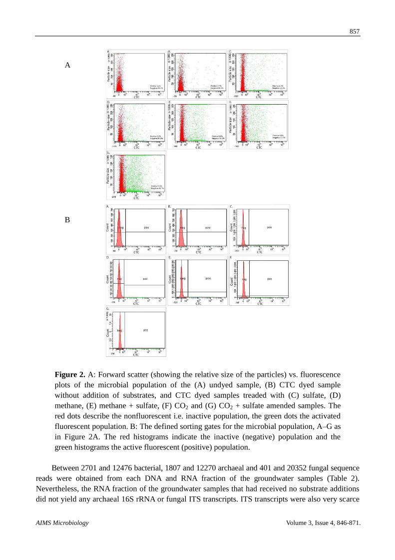
Related Documents