CNS Wound Healing Is Severely Depressed in Metallothionein I- and II-Deficient Mice Milena Penkowa, 1 Javier Carrasco, 2 Mercedes Giralt, 2 Torben Moos, 1 and Juan Hidalgo 2 1 Institute of Medical Anatomy, Section C, The Panum Institute, University of Copenhagen, DK-2200 Copenhagen, Denmark, and 2 Departamento de Biologı ´a Celular, de Fisiologı ´a y de Inmunologı ´a, Unidad de Fisiologı ´a Animal, Facultad de Ciencias, Universidad Auto ´ noma de Barcelona, Barcelona, Spain 08193 To characterize the physiological role of metallothioneins I and II (MT-I1II) in the brain, we have examined the chronological effects of a freeze injury to the cortex in normal and MT-I1II null mice. In normal mice, microglia/macrophage activation and astrocytosis were observed in the areas surrounding the lesion site, peaking at ;1 and 3 d postlesion (dpl), respectively. At 20 dpl, the parenchyma had regenerated. Both brain macrophages and astrocytes surrounding the lesion increased the MT-I1II immunoreactivity, peaking at ;3 dpl, and at 20 dpl it was similar to that of unlesioned mice. In situ hybridization analysis indi- cates that MT-I1II immunoreactivity reflects changes in the messenger levels. In MT-I1II null mice, microglia/macrophages infiltrated the lesion heavily, and at 20 dpl they were still present. Reactive astrocytosis was delayed and persisted at 20 dpl. In contrast to normal mice, at 20 dpl no wound healing had occurred. The rate of apoptosis, as determined by using termi- nal deoxynucleotidyl transferase-mediated dUTP–biotin nick end labeling, was drastically increased in neurons of ipsilateral cortex of the MT-I1II null mice. Our results demonstrate that MT-I1II are essential for a normal wound repair in the CNS, and that their deficiency impairs neuronal survival. Key words: brain inflammation; MT-I1II; superoxide dis- mutase; oxidative stress; zinc; brain macrophages; astrocytes; neurons; apoptosis; regeneration; degeneration Metallothioneins (MTs) are a family of low molecular weight, heavy metal-binding, cysteine-rich proteins. In the mouse, there are four isoforms, MT-I to MT-IV (Palmiter et al., 1992; Quaife et al., 1994). In the C NS, MTs occur in the isoforms MT-I, MT-II, and MT-III. MT-I1II are expressed in virtually all tissues, and in the brain they are localized mainly in astrocytes, microglia, lep- tomeningeal cells, ependyma, and choroid plexus epithelium (Young et al., 1991; Masters et al., 1994b; Penkowa and Moos, 1995; Penkowa et al., 1997). MT-III is primarily confined to the brain, but the data regarding its cellular localization are conflict- ing, because in situ hybridization analysis suggests neurons as the main site of expression (Masters et al., 1994b), whereas immuno- cytochemistry studies suggest microglia/macrophages and astro- cytes as the cells with the highest MT-III protein content (Ho- zumi et al., 1996; Yamada et al., 1996; Penkowa et al., 1999). The actual physiological role(s) of the different MT isoforms in the brain still remains to be established. Those of MT-I1II could be related to their putative antioxidant functions as well as zinc and/or copper metabolism (Sato and Bremner, 1993; Kelly et al., 1996; Aschner et al., 1997; Hidalgo et al., 1997). Presumably MT-III functions will differ from those of their normal counter- parts MT-I1II, as suggested from in vitro (Uchida et al., 1991; Erickson et al., 1994; Palmiter, 1995; Sewell et al., 1995) and in vivo studies (Quaife et al., 1998). The intracerebral expression of MT-I1II is clearly upregulated during pathological conditions induced by trauma (Penkowa and Moos, 1995), immobilization stress (Hidalgo et al., 1990), kainic acid-induced seizures (Dalton et al., 1995; Zheng et al., 1995), excitotoxic NMDA cortex damage (Hidalgo et al., 1997), and administration of 6-aminonicotinamide (Penkowa et al., 1997). Furthermore, MT-I1II expression is increased in the myelin-deficient jimpy mouse (Vela et al., 1997) and in several human adult neurodegenerative disorders such as Alzheimer’s disease (AD), Pick’s disease (Duguid et al., 1989), and amyotrophic lateral sclerosis (Sillevis Smitt et al., 1992), as well as in aging (Suzuki et al., 1992) and after brain ischemia (Neal et al., 1996). MT-III was discovered unexpectedly as a factor decreased in AD (Uchida et al., 1991), and a number of animal models have shown that MT-III mRNA or protein levels are significantly altered during CNS damage (Hozumi et al., 1995, 1996; Yamada et al., 1996). Taken together, these studies strongly suggest that MTs are important proteins in the brain for coping with the tissue damage elicited by a wide array of factors and diseases. However, what the protective roles are, if any, remains unknown. The MT-I1II (Masters et al., 1994a)-deficient mice used in this report represent a unique experimental approach for determining the putative importance of these MT isoforms in the CNS during traumatic conditions. MATERIALS AND METHODS Production of the MT-I1II- and MT-III-deficient mice. Homozygous MT- I1II knock-out (KO) mice were generated as previously described (Mas- Received Oct. 6, 1998; revised Jan. 15, 1999; accepted Jan. 25, 1999. This work was supported by The Novo Nordisk Fonden, Direktør Leo Nielsens Fond, and Warwara Larsen’s Fond (M.P. and T.M.) and by Comisio ´n Interminis- terial de C iencia y Tecnologı ´a Grant SAF96-0189, Programa Sectorial de Promocio ´n General del Conocimiento Grant PM98-0170, and Fundacio ´n “La Caixa” Grant 97/102-00 (J.H.). J.C. is a fellow of Comissio ´ Interdepartamental de Recerca i Innovacio ´ Tecnolo ´gica (Grant FI 96/2613). We acknowledge Dr. R. D. Palmiter for critically reading this manuscript and for the MT-I probe. Thanks are given to Hanne Hadberg, Pernille S. Thomsen, and Jordi Canto for excellent technical assistance and to Keld Stub and Birgit Risto for superb photographic assistance. The help of the Laboratori d’Ana ´lisi Bioquimica del Departament de Bioquimica i Biologia Molecular is acknowledged. Correspondence should be addressed to Dr. Juan Hidalgo, Departamento de Biologı ´a Celular, de Fisiologı ´a y de Inmunologı ´a, Unidad de Fisiologı ´a Animal, Facultad de C iencias, Universidad Auto ´noma de Barcelona, Bellaterra, Barcelona, Spain 08193. Copyright © 1999 Society for Neuroscience 0270-6474/99/192535-11$05.00/0 The Journal of Neuroscience, April 1, 1999, 19(7):2535–2545

Welcome message from author
This document is posted to help you gain knowledge. Please leave a comment to let me know what you think about it! Share it to your friends and learn new things together.
Transcript
CNS Wound Healing Is Severely Depressed in Metallothionein I-and II-Deficient Mice
Milena Penkowa,1 Javier Carrasco,2 Mercedes Giralt,2 Torben Moos,1 and Juan Hidalgo2
1Institute of Medical Anatomy, Section C, The Panum Institute, University of Copenhagen, DK-2200 Copenhagen,Denmark, and 2Departamento de Biologıa Celular, de Fisiologıa y de Inmunologıa, Unidad de Fisiologıa Animal, Facultadde Ciencias, Universidad Autonoma de Barcelona, Barcelona, Spain 08193
To characterize the physiological role of metallothioneins I andII (MT-I1II) in the brain, we have examined the chronologicaleffects of a freeze injury to the cortex in normal and MT-I1II nullmice. In normal mice, microglia/macrophage activation andastrocytosis were observed in the areas surrounding the lesionsite, peaking at ;1 and 3 d postlesion (dpl), respectively. At 20dpl, the parenchyma had regenerated. Both brain macrophagesand astrocytes surrounding the lesion increased the MT-I1IIimmunoreactivity, peaking at ;3 dpl, and at 20 dpl it was similarto that of unlesioned mice. In situ hybridization analysis indi-cates that MT-I1II immunoreactivity reflects changes in themessenger levels. In MT-I1II null mice, microglia/macrophages
infiltrated the lesion heavily, and at 20 dpl they were stillpresent. Reactive astrocytosis was delayed and persisted at 20dpl. In contrast to normal mice, at 20 dpl no wound healing hadoccurred. The rate of apoptosis, as determined by using termi-nal deoxynucleotidyl transferase-mediated dUTP–biotin nickend labeling, was drastically increased in neurons of ipsilateralcortex of the MT-I1II null mice. Our results demonstrate thatMT-I1II are essential for a normal wound repair in the CNS, andthat their deficiency impairs neuronal survival.
Key words: brain inflammation; MT-I1II; superoxide dis-mutase; oxidative stress; zinc; brain macrophages; astrocytes;neurons; apoptosis; regeneration; degeneration
Metallothioneins (MTs) are a family of low molecular weight,heavy metal-binding, cysteine-rich proteins. In the mouse, thereare four isoforms, MT-I to MT-IV (Palmiter et al., 1992; Quaifeet al., 1994). In the CNS, MTs occur in the isoforms MT-I, MT-II,and MT-III. MT-I1II are expressed in virtually all tissues, and inthe brain they are localized mainly in astrocytes, microglia, lep-tomeningeal cells, ependyma, and choroid plexus epithelium(Young et al., 1991; Masters et al., 1994b; Penkowa and Moos,1995; Penkowa et al., 1997). MT-III is primarily confined to thebrain, but the data regarding its cellular localization are conflict-ing, because in situ hybridization analysis suggests neurons as themain site of expression (Masters et al., 1994b), whereas immuno-cytochemistry studies suggest microglia/macrophages and astro-cytes as the cells with the highest MT-III protein content (Ho-zumi et al., 1996; Yamada et al., 1996; Penkowa et al., 1999).
The actual physiological role(s) of the different MT isoforms inthe brain still remains to be established. Those of MT-I1II couldbe related to their putative antioxidant functions as well as zincand/or copper metabolism (Sato and Bremner, 1993; Kelly et al.,
1996; Aschner et al., 1997; Hidalgo et al., 1997). PresumablyMT-III functions will differ from those of their normal counter-parts MT-I1II, as suggested from in vitro (Uchida et al., 1991;Erickson et al., 1994; Palmiter, 1995; Sewell et al., 1995) and invivo studies (Quaife et al., 1998). The intracerebral expression ofMT-I1II is clearly upregulated during pathological conditionsinduced by trauma (Penkowa and Moos, 1995), immobilizationstress (Hidalgo et al., 1990), kainic acid-induced seizures (Daltonet al., 1995; Zheng et al., 1995), excitotoxic NMDA cortex damage(Hidalgo et al., 1997), and administration of 6-aminonicotinamide(Penkowa et al., 1997). Furthermore, MT-I1II expression isincreased in the myelin-deficient jimpy mouse (Vela et al., 1997)and in several human adult neurodegenerative disorders such asAlzheimer’s disease (AD), Pick’s disease (Duguid et al., 1989),and amyotrophic lateral sclerosis (Sillevis Smitt et al., 1992), aswell as in aging (Suzuki et al., 1992) and after brain ischemia(Neal et al., 1996).
MT-III was discovered unexpectedly as a factor decreased inAD (Uchida et al., 1991), and a number of animal models haveshown that MT-III mRNA or protein levels are significantlyaltered during CNS damage (Hozumi et al., 1995, 1996; Yamadaet al., 1996).
Taken together, these studies strongly suggest that MTs areimportant proteins in the brain for coping with the tissue damageelicited by a wide array of factors and diseases. However, what theprotective roles are, if any, remains unknown. The MT-I1II(Masters et al., 1994a)-deficient mice used in this report representa unique experimental approach for determining the putativeimportance of these MT isoforms in the CNS during traumaticconditions.
MATERIALS AND METHODSProduction of the MT-I1II- and MT-III-deficient mice. Homozygous MT-I1II knock-out (KO) mice were generated as previously described (Mas-
Received Oct. 6, 1998; revised Jan. 15, 1999; accepted Jan. 25, 1999.This work was supported by The Novo Nordisk Fonden, Direktør Leo Nielsens
Fond, and Warwara Larsen’s Fond (M.P. and T.M.) and by Comision Interminis-terial de Ciencia y Tecnologıa Grant SAF96-0189, Programa Sectorial de PromocionGeneral del Conocimiento Grant PM98-0170, and Fundacion “La Caixa” Grant97/102-00 (J.H.). J.C. is a fellow of Comissio Interdepartamental de Recerca iInnovacio Tecnologica (Grant FI 96/2613). We acknowledge Dr. R. D. Palmiter forcritically reading this manuscript and for the MT-I probe. Thanks are given toHanne Hadberg, Pernille S. Thomsen, and Jordi Canto for excellent technicalassistance and to Keld Stub and Birgit Risto for superb photographic assistance. Thehelp of the Laboratori d’Analisi Bioquimica del Departament de Bioquimica iBiologia Molecular is acknowledged.
Correspondence should be addressed to Dr. Juan Hidalgo, Departamento deBiologıa Celular, de Fisiologıa y de Inmunologıa, Unidad de Fisiologıa Animal,Facultad de Ciencias, Universidad Autonoma de Barcelona, Bellaterra, Barcelona,Spain 08193.Copyright © 1999 Society for Neuroscience 0270-6474/99/192535-11$05.00/0
The Journal of Neuroscience, April 1, 1999, 19(7):2535–2545

ters et al., 1994a). The KO mice were raised on the 129/Sv geneticbackground; therefore, mice from this strain were used as controls.
Experimental procedures. Normal and genetically MT-I1II null adultmice were lesioned under tribromethanol anesthesia. The skull over theright frontoparietal cortex was exposed, and a focal cryoinjury on thesurface of the brain was produced with dry ice (278°C) (Penkowa andMoos, 1995). The animals were housed in cages with free access to foodand water. The handling of the animals were approved by the propercommittees of animal research and ethics of Spain and Denmark.
We performed three experiments. In the first one, control and MT-I1II null mice were lesioned and killed 3 d postlesion (dpl) along withunlesioned animals. In a second experiment, the animals were killed after1, 3, 10, and 20 dpl. In a third experiment designed for in situ hybrid-ization studies, the animals were killed by decapitation without tissuefixation 6 and 24 hr after the lesion along with unlesioned mice. Three tosix mice per group were used in each experiment.
Mice were deeply anesthetized with Brietal and perfused with Zam-boni’s fixative for immunohistochemistry, histochemistry, and terminaldeoxynucleotidyl transferase (TdT)-mediated dUTP–biotin nick end la-beling (TUNEL). For Timm’s silver sulfide staining we added Na2S tothe Zamboni’s fixative. For in situ hybridization studies, mice were killedby cervical dislocation, and the brains were immediately frozen in liquidnitrogen and stored at 280°C.
Cellular countings were performed for lectin-, GFAP-, neuronal-specific enolase (NSE)-, MT-I1II-, Cu/Zn-superoxide dismutase (Cu/Zn-SOD)-, and TUNEL-positive cells for statistical evaluation of theresults. To this end, positively stained cells were counted from a 0.4 mm 2
area of unlesioned hemispheres (data not shown) as well as from theipsilateral site of the lesioned mice. Representative counting areas areshown in Figures 1, 2, and 4; they are in the border of the lesion, wheregliosis is prominent.
Histochemistry. Biotinylated tomato lectin from the Lycopersicon escu-lentum (Sigma, St. Louis, MO; code L9389), 1:500, was used as a markerfor cells of the myelocytic and monocytic cell lineages, such as microglia /macrophages, as well as a marker for vessels. The lectin was developedusing streptavidin–biotin–peroxidase complex (StreptABComplex/HRP; Dakopatts, Copenhagen, Denmark; code K377) prepared at themanufacturer’s recommended dilutions and performed for 30 min atroom temperature. The reaction product was visualized using 0.015%H2O2 in 3,39-diaminobenzidine/Tris-buffered saline (DAB/TBS), withDAB as a chromogen.
Immunohistochemistry. Sections were preincubated with pronase E(protease type XIV, Sigma, number P5147; 0.025 gm dissolved in 50 mlof TBS) for 10 min, pH 7.4, at 37°C followed by incubation in 10% goatserum in TBS (0.05 M Tris, pH 7.4, and 0.15 M NaCl) with 0.01% NonidetP-40 for 15 min at room temperature. Afterward, sections were incubatedovernight with one of the following primary antibodies: polyclonal rabbitanti-bovine GFAP (1:250; Dakopatts, code Z 334) (as a marker forastrocytes), polyclonal rabbit anti-human NSE (1:1000; Dakopatts, codeA589) (as a neuronal marker), monoclonal mouse anti-human neuro-filament (NF) (1:250; Dakopatts, code M762) (as a neuronal marker),polyclonal rabbit anti-rat liver MT-I1II (1:500; Gasull et al., 1993, 1994),and monoclonal mouse anti-human Cu/Zn-SOD (1:50; Sigma, codeS2147). The NF and Cu/Zn-SOD antibodies were used after preincu-bation of the sections with blocking solutions from the HistoMouse-SPkit (Zymed, San Francisco, CA; code 95-9544) to quench endogenousIgG background staining by the secondary antibody used in mice tissue.The primary antibodies were detected using biotinylated monoclonalanti-rabbit IgG (Sigma, code B3275), 1:400, or biotinylated goat anti-mouse IgG (Sigma, code B8774), 1:200. These secondary antibodies weredetected by StreptABComplex/HRP prepared at the manufacturer’srecommended dilutions. These secondary and tertiary steps in the im-munoreaction were performed for 30 min at room temperature. Theimmunoreaction was visualized using 0.015% H2O2 in DAB/TBS withDAB as a chromogen.
Immunofluorescence. The presence of MT-I1II in microglia, macro-phages, and astrocytes was verified by simultaneous staining for lectinand MT-I1II or GFAP and MT-I1II by using double-labeling immuno-fluorescence histochemistry. For this purpose we used Texas Red-labeledlectin (1:50; Sigma, number L-9139) simultaneously with monoclonalmouse anti-horse MT-I1II (1:40; Dakopatts, code M0638). The MT-I1II antibody was used after preincubation of the sections with blockingsolutions from the HistoMouse-SP kit to quench endogenous IgG
background staining by the secondary antibody in mice tissue. TheMT-I1II were detected using goat anti-mouse IgG linked with amino-methylcoumarin (1:20) for 30 min at room temperature (Dakopatts, codeW0477). Simultaneous detection of GFAP and MT-I1II was performedby using monoclonal rat anti-bovine GFAP (1:100; Zymed, code 13-0300)(as a marker for astrocytes). Anti-GFAP antibodies were detected usingfluorescein-conjugated goat anti-rat IgG (H and L, 1:50) for 30 min atroom temperature (Sigma, code F6258). Monoclonal mouse anti-horseMT-I1II were detected as mentioned above.
To evaluate the extent of nonspecific binding of the antisera in theimmunohistochemical experiments, 1:100–1:1000 of normal rabbit ormouse serum or just the preincubation agent was substituted for theprimary antibody step described above. Results were considered only ifthese controls were negative.
MT-I in situ hybridization. Brain MT-I mRNA levels were assayed by insitu hybridization in control and cryolesioned mice, which were killed 6and 24 hr after the lesion. Serial coronal 30-mm sections were cut on acryostat and mounted on poly-L-lysine-coated slides. For MT-I mRNAstudies, we used the mouse cDNA kindly provided by Dr. R. D. Palmiter(University of Washington, Seattle, WA). MT-I and MT-II are isoformsregulated coordinately, and thus we assume that MT-I mRNA levels arerepresentative of the MT-I1II isoforms (Masters et al., 1994b). TheMT-I cDNA was labeled with [a-35S]UTP using an SP6/T7 transcriptionkit (Boehringer Mannheim, Mannheim, Germany). Preparation of senseand antisense probes and the in situ hybridization procedure wereperformed as previously described (Hernandez et al., 1997a; Carrasco etal., 1998). Autoradiography was performed exposing the film (Hyper-film-MP; Amersham, Arlington Heights, IL) to the slides for severaldays. All sections to be compared were prepared simultaneously andexposed to the same autoradiographic film.
Double immunohistochemistry–in situ hybridization. To colocalize MTmRNA and MT protein, we simultaneously performed in situ hybridiza-tion for MT-I or MT-III mRNA and immunohistochemistry for MT-I1IIand MT-III protein. The in situ hybridization procedure, except visual-ization, was performed before immunohistochemistry was carried out.The autoradiography of MT-I or MT-III mRNA (Hypercoat LM-1;Amersham, code RPN 40) was performed just previously to the visual-ization of MT-I1II and MT-III antibodies using DAB as chromogen.
Neo-Timm staining. Sections were physically (autometallographically)developed for 60 min at room temperature in solution containing silverlactate (Moos, 1993).
TUNEL technique. TUNEL staining was performed after tissue pro-cessing as mentioned above. Sections were deparaffinized and incubatedwith 20 mg/ml proteinase K (Sigma) for 5 min to strip off nuclearproteins. TUNEL was accomplished using the Apoptag Plus in situapoptosis detection kit (Oncor, Gaithersburg, MD; code S7101-KIT).After immersion in equilibration buffer for 10 min, sections were incu-bated with TdT and dUTP-digoxigenin in a humidified chamber at 37°Cfor 1 hr and then incubated in the stop–wash buffer at 37°C for 30 min tostop the reaction. After washing in PBS buffer, the sections were incu-bated in anti-digoxigenin-peroxidase solution for 30 min. Afterward,DAB was used as chromogen, and the sections were counterstained withmethyl green. Control sections were treated similarly but incubated inthe absence of TdT enzyme, dUTP-digoxigenin, or anti-digoxigeninantibody. Furthermore, we performed control slides from Oncor (codeS7115) for comparison. Besides detecting the staining reactivity forTUNEL, we also evaluated the morphological criteria of apoptosis, suchas cell shrinkage and compactation of chromatin, to distinguish apoptosisfrom necrosis.
Double TUNEL–immunofluorescence histochemistry. To determinewhich cells were undergoing apoptosis, we performed double-labelingimmunofluorescence histochemistry by using a fluorescein-linked apo-ptosis detection kit (TUNEL; Oncor, code S7110-KIT) simultaneouslywith one of the following antibodies: Texas Red-labeled lectin, polyclonalGFAP, and polyclonal NSE (as mentioned above). Anti-GFAP andanti-NSE were detected using swine anti-rabbit IgG linked with rhoda-mine (1:30) for 30 min at room temperature (Dakopatts, code R156).
Statistical analysis. Results were evaluated by two-way ANOVA, withstrain and freeze lesion as main factors. When the interaction wassignificant, it was interpreted to be the consequence of a specific effect ofthe MT-I1II deficiency during the lesion. MT-I1II expression in MT-I1II null mice was evaluated by one-way ANOVA.
2536 J. Neurosci., April 1, 1999, 19(7):2535–2545 Penkowa et al. • Role of Metallothioneins in CNS Wound Healing
RESULTSGeneralIn unlesioned mice, gross inspection of the brain did not revealsignificant differences between control and MT-I1II-deficientmice in the brain anatomy and histology of glial cells. In thelesioned mice, by gross examination of the brains a focal hemor-rhage was seen in the right frontoparietal cortex at 1 and 3 dpl,whereas at 10 and 20 dpl only MT-I1II null mice displayed avisible hemorrhage. In toluidine blue-stained frontal sections, the
freeze lesion was seen as a cortical area with no neuronal cells,but instead necrotic cells and debris were present. The tissuedamage observed was higher in MT-I1II null mice.
Injury to the CNS produces a characteristic inflammatoryresponse that results in migration of hematogenous cells into thedamaged neural tissue and reactive gliosis (Ghirnikar et al.,1998). Activated microglia and bone marrow-derived monocytestransform to round phagocytes, generally referred to as micro-glia/macrophages (Stevens and Bahr, 1993; Perry et al., 1995).
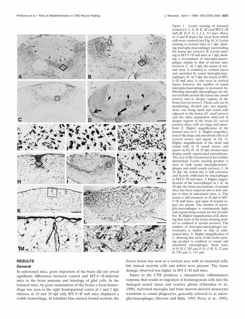
Figure 1. Lectin staining of lesionednormal (A, C, E, H, K, M ) and MT-I1IInull (B, D, F, G, I, J, L, N ) mice. Boxesin A and B depict the areas from whichcells were counted (see Fig. 8). A, Lectinstaining in normal mice at 1 dpl, show-ing microglia/macrophages surroundingthe lesion site (arrows). B, Lectin stain-ing in MT-I1II null mice at 1 dpl, show-ing a recruitment of microglia/macro-phages similar to that of normal mice(arrows). C, At 3 dpl, the lesion of nor-mal mice is confined to cortical layersand encircled by round microglia/mac-rophages. D, At 3 dpl, the lesion of MT-I1II null mice is also seen in corticallayers; however, the number of roundmicroglia/macrophages is increased. In-filtrating microglia/macrophages are ob-served both around the lesion site (smallarrows) and in deeper regions of thebrain (curved arrows). These cells are bymorphology divided into two popula-tions, one being small and round cellsadjacent to the lesion (D, small arrows)and the other population observed indeeper regions of the brain (D, curvedarrows); these cells are large and amoe-boid. E, Higher magnification of theframed area in C. F, Higher magnifica-tion of the large and amoeboid cells in D(curved arrows and square in D). G,Higher magnification of the small andround cells in D (small arrows andsquare in D). H, At 10 dpl, normal micedisplay partly regenerated parenchyme.The size of the lesioned area has furtherdiminished. Lectin staining product isseen in both round microglia/macro-phages and small vessels (arrows). I, At10 dpl, the lesion site is still extensiveand heavily infiltrated by macrophagesin MT-I1II null mice. J, Higher magni-fication of the macrophages in I. K, At20 dpl, the brain parenchyma of normalmice has been repaired and is now sim-ilar to that of unlesioned mice. L, Thelesion is still extensive at 20 dpl in MT-I1II null mice, and signs of wound re-pair are absent. The number of micro-glia/macrophages is continuously high,and regenerating vessels are few in num-ber. M, Higher magnification of K, show-ing that most of the lectin staining prod-uct is confined to vessels (arrows). Thenumber of microglia/macrophages (ar-rowheads) is similar to that of unle-sioned mice. N, Higher magnification ofL, showing that most of the lectin stain-ing product is confined to round andamoeboid macrophages. Scale bars:A–D, H, I, 285 mm; E–G, J, M, N, 25 mm;K, 320 mm; L, 115 mm.
Penkowa et al. • Role of Metallothioneins in CNS Wound Healing J. Neurosci., April 1, 1999, 19(7):2535–2545 2537
Astrocytes undergo hypertrophy and hyperplasia, generally re-ferred to as reactive astrocytosis. These inflammatory cells of thebrain are thought to be essential for maintaining neuronal sur-vival and for tissue regeneration after CNS damage. These re-sponses were thoroughly examined chronologically in the freezelesion model used in this study, in line with previous results in therat (Penkowa and Moos, 1995), providing a framework for char-acterizing the putative role of MTs in the CNS.
Microglia/macrophagesIn normal mice, dramatic changes in the number and morphologyof microglia/macrophages were observed surrounding the lesionin a highly temporal-specific manner (see Figs. 1, 8). Microglia/macrophages appeared to a great extent in the injured area at 1dpl, and at 3 dpl the cells made a line of demarcation around thelesion (Fig. 1A,C). Microglia/macrophages were round or amoe-boid without ramifications (Fig. 1E). At 10 dpl, a majority of therecruited microglia/macrophages had disappeared, and instead,small capillaries appeared in the injured area (Fig. 1H). At 20 dpl,the tissue had regenerated, and the parenchyme appeared as that
of unlesioned mice. Lectin staining product was seen in capillariesrather than round microglia/macrophages (Fig. 1K,M).
In MT-I1II null mice, an increase in microglia/macrophageswas also observed around the lesion, but the temporal responsewas significantly different from that of normal mice (see Figs. 1,8). At 1 dpl the number of microglia/macrophages in MT-I1IInull mice was similar to that of normal mice (Fig. 1B). At 3 dplthe microglia/macrophages of MT-I1II null mice were signifi-cantly increased, and they appeared in two populations aroundthe lesion (Fig. 1D). One population, seen in the periphery of thelesioned area, was composed of large amoeboid cells (Fig. 1D,F).The other population, seen adjacent to the necrotic area of thelesion, was composed of small and round cells (Fig. 1D,G). At 10dpl, the parenchyma was heavily inflamed, and microglia/macro-phages were still significantly increased in number in MT-I1II-deficient mice (Fig. 1I,J). At 20 dpl, the freeze lesion still persisted,and the injured area was heavily infiltrated by round microglia/macrophages (Fig. 1L,N). Signs of tissue regeneration such as newvessel formation were almost absent in MT-I1II null mice.
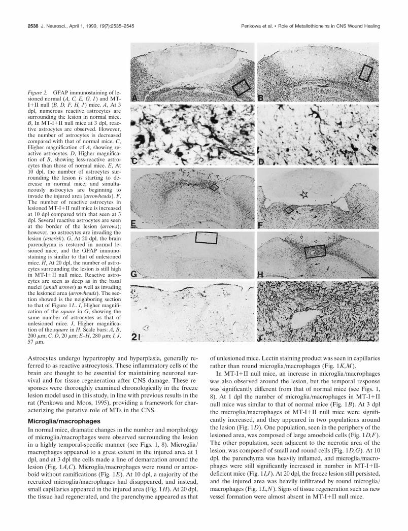
Figure 2. GFAP immunostaining of le-sioned normal (A, C, E, G, I ) and MT-I1II null (B, D, F, H, J ) mice. A, At 3dpl, numerous reactive astrocytes aresurrounding the lesion in normal mice.B, In MT-I1II null mice at 3 dpl, reac-tive astrocytes are observed. However,the number of astrocytes is decreasedcompared with that of normal mice. C,Higher magnification of A, showing re-active astrocytes. D, Higher magnifica-tion of B, showing less-reactive astro-cytes than those of normal mice. E, At10 dpl, the number of astrocytes sur-rounding the lesion is starting to de-crease in normal mice, and simulta-neously astrocytes are beginning toinvade the injured area (arrowheads). F,The number of reactive astrocytes inlesioned MT-I1II null mice is increasedat 10 dpl compared with that seen at 3dpl. Several reactive astrocytes are seenat the border of the lesion (arrows);however, no astrocytes are invading thelesion (asterisk). G, At 20 dpl, the brainparenchyma is restored in normal le-sioned mice, and the GFAP immuno-staining is similar to that of unlesionedmice. H, At 20 dpl, the number of astro-cytes surrounding the lesion is still highin MT-I1II null mice. Reactive astro-cytes are seen as deep as in the basalnuclei (small arrows) as well as invadingthe lesioned area (arrowheads). The sec-tion showed is the neighboring sectionto that of Figure 1L. I, Higher magnifi-cation of the square in G, showing thesame number of astrocytes as that ofunlesioned mice. J, Higher magnifica-tion of the square in H. Scale bars: A, B,200 mm; C, D, 20 mm; E–H, 280 mm; I, J,57 mm.
2538 J. Neurosci., April 1, 1999, 19(7):2535–2545 Penkowa et al. • Role of Metallothioneins in CNS Wound Healing
Reactive astrocytesAfter the freeze injury, a linear progression and afterward adecline in the number of reactive astrocytes were observed innormal mice, as verified from GFAP immunostaining (see Figs. 2,8). Only a few reactive astrocytes appeared at 1 dpl, whereasnumerous reactive astrocytes around the lesion were present at 3dpl in normal mice (Fig. 2A,C). At 10 dpl, reactive astrocyteswere delineating the partly regenerated injury (Fig. 2E). At 20dpl, the transient reactive astrocytosis had disappeared, andGFAP immunostainings were even lower than in unlesionedmice, probably because of alterations in the structure of cytoskel-etal proteins as well as in the extracellular matrix caused by thelesion. Some astrocytes were seen around capillaries and as partof the glia limitans (Fig. 2G,I).
In MT-I1II null mice, the expression of GFAP was delayedcompared with that of normal mice (see Figs. 2, 8). Thus, at 1 dpl
only a few reactive astrocytes were present, whereas at 3 dpl thenumber of reactive astrocytes had increased, but compared withthat of normal mice at 3 dpl, the astrocytosis was reduced (seeFigs. 2A–D, 8). At 10 dpl, numerous reactive astrocytes appearedin MT-I1II null mice (Fig. 2F). Both around the lesion site andin deeper areas such as in the basal nuclei reactive astrocytosiswas observed. In contrast to normal mice, in MT-I1II null micethe reactive astrogliosis continued at 20 dpl, with a thick layer ofreactive astrocytes surrounding the lesion. In addition, severalastrocytes were still identified in deeper areas of the injuredhemisphere (Fig. 2H,J).
NeuronsAs might be expected, the number of neurons was significantlydecreased by the freeze lesion (see Figs. 3, 8). In normal mice,decreased numbers of NSE- and NF-expressing neurons were
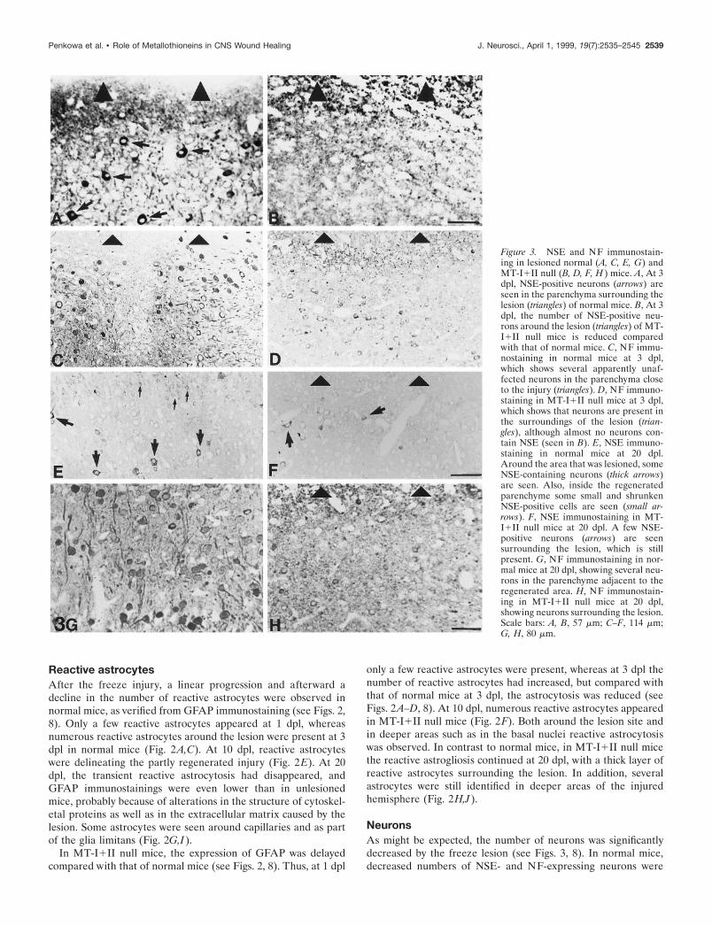
Figure 3. NSE and NF immunostain-ing in lesioned normal (A, C, E, G) andMT-I1II null (B, D, F, H ) mice. A, At 3dpl, NSE-positive neurons (arrows) areseen in the parenchyma surrounding thelesion (triangles) of normal mice. B, At 3dpl, the number of NSE-positive neu-rons around the lesion (triangles) of MT-I1II null mice is reduced comparedwith that of normal mice. C, NF immu-nostaining in normal mice at 3 dpl,which shows several apparently unaf-fected neurons in the parenchyma closeto the injury (triangles). D, NF immuno-staining in MT-I1II null mice at 3 dpl,which shows that neurons are present inthe surroundings of the lesion (trian-gles), although almost no neurons con-tain NSE (seen in B). E, NSE immuno-staining in normal mice at 20 dpl.Around the area that was lesioned, someNSE-containing neurons (thick arrows)are seen. Also, inside the regeneratedparenchyme some small and shrunkenNSE-positive cells are seen (small ar-rows). F, NSE immunostaining in MT-I1II null mice at 20 dpl. A few NSE-positive neurons (arrows) are seensurrounding the lesion, which is stillpresent. G, NF immunostaining in nor-mal mice at 20 dpl, showing several neu-rons in the parenchyme adjacent to theregenerated area. H, NF immunostain-ing in MT-I1II null mice at 20 dpl,showing neurons surrounding the lesion.Scale bars: A, B, 57 mm; C–F, 114 mm;G, H, 80 mm.
Penkowa et al. • Role of Metallothioneins in CNS Wound Healing J. Neurosci., April 1, 1999, 19(7):2535–2545 2539
observed in neocortical layers surrounding the lesion zone at 1and 3 dpl (Fig. 3A,C). The neuronal morphology of these neuronswas similar to that of unlesioned control mice, suggesting thatthey remained relatively unaffected. The number of NSE- andNF-expressing neurons was further decreased at 10 and 20 dpl(Fig. 3E,G).
In MT-I1II null mice, the number of NSE-positive neurons inthe cortex of unlesioned animals was decreased in comparisonwith normal mice (see Fig. 8). After the freeze lesion, the numberof NSE- and NF-positive neurons dramatically decreased, signif-icantly more in the MT-I1II null mice than in control mice (seeFigs. 3, 8). Most of the neurons present in MT-I1II null mice at3 dpl were either NSE-negative or showed a shrunken cytoplasmcompared with those of normal mice at 3 dpl (Fig. 3B,D). At 20dpl, neurons surrounding the lesion in MT-I1II null mice eitherwere still NSE-negative or had small, shrunken cell bodies withdecreased levels of both NSE and NF (Fig. 3F,H).
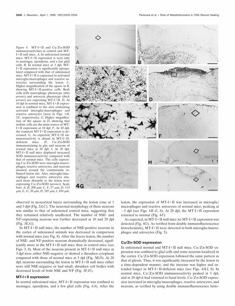
MT-I1II expressionIn normal unlesioned mice, MT-I1II expression was confined tomeninges, ependyma, and a few glial cells (Fig. 4A). After the
lesion, the expression of MT-I1II was increased in microglia/macrophages and reactive astrocytes of normal mice, peaking at;3 dpl (see Figs. 4B–E, 8). At 20 dpl, the MT-I1II expressionreturned to normal (Fig. 4F).
As expected, in MT-I1II null mice no MT-I1II expression wasdetected (Fig. 4G). As verified from double immunofluorescencehistochemistry, MT-I1II were detected in both microglia/macro-phages and astrocytes (Fig. 5).
Cu/Zn-SOD expressionIn unlesioned normal and MT-I1II null mice, Cu/Zn-SOD ex-pression was confined to glial cells and some neurons localized inthe cortex. Cu/Zn-SOD expression followed the same pattern asthat of gliosis. Thus, it was significantly increased by the lesion ina time-dependent manner, and the increase was higher and ex-tended longer in MT-I1II-deficient mice (see Figs. 4H,I, 8). Innormal mice, Cu/Zn-SOD immunoreactivity peaked at 3 dpl,and at 20 dpl it had returned to basal levels. Cu/Zn-SOD expres-sion increased in microglia/macrophages, reactive astrocytes, andneurons, as verified by using double immunofluorescence histo-
Figure 4. MT-I1II and Cu/Zn-SODimmunoreactivities in control and MT-I1II null mice. A, In unlesioned normalmice, MT-I1II expression is seen onlyin meninges, ependyma, and a few glialcells. B, In normal mice at 3 dpl, MT-I1II expression is significantly upregu-lated compared with that of unlesionedmice. MT-I1II is expressed in activatedmicroglia/macrophages and reactive as-trocytes surrounding the lesion. C,Higher magnification of the square in B,showing MT-I1II-positive cells. Bothcells with macrophage phenotype (thinarrows) and astrocyte phenotype (thickarrows) are expressing MT-I1II. D, At10 dpl in normal mice, MT-I1II expres-sion is confined to the area containingactivated microglia/macrophages andreactive astrocytes (seen in Figs. 1 H,2E, respectively). E, Higher magnifica-tion of the square in D, showing thatstellate cells are the main source of MT-I1II expression at 10 dpl. F, At 20 dpl,the transient MT-I1II expression is de-creased. G, As expected, MT-I1II im-munoreactivity is absent in MT-I1II-deficient mice. H, Cu/Zn-SODimmunostaining in glia and neurons ofnormal mice at 20 dpl. I, At 20 dpl,MT-I1II null mice displayed increasedSOD immunoreactivity compared withthat of normal mice. The cells express-ing Cu/Zn-SOD were microglia/macro-phages, reactive astrocytes, and neuronssituated around the continuously in-flamed lesion site. Also, microglia/mac-rophages and reactive astrocytes situ-ated more distantly to the lesion wereexpressing Cu/Zn-SOD (arrows). Scalebars: A, B, 200 mm; C, F, 57 mm; D, 114mm; E, G, 20 mm; H, 285 mm; I, 450 mm.
2540 J. Neurosci., April 1, 1999, 19(7):2535–2545 Penkowa et al. • Role of Metallothioneins in CNS Wound Healing
chemistry (data not shown). In MT-I1II-deficient mice, Cu/Zn-SOD expression increased significantly more than in normal miceand remained high even at 20 dpl (see Fig. 8).
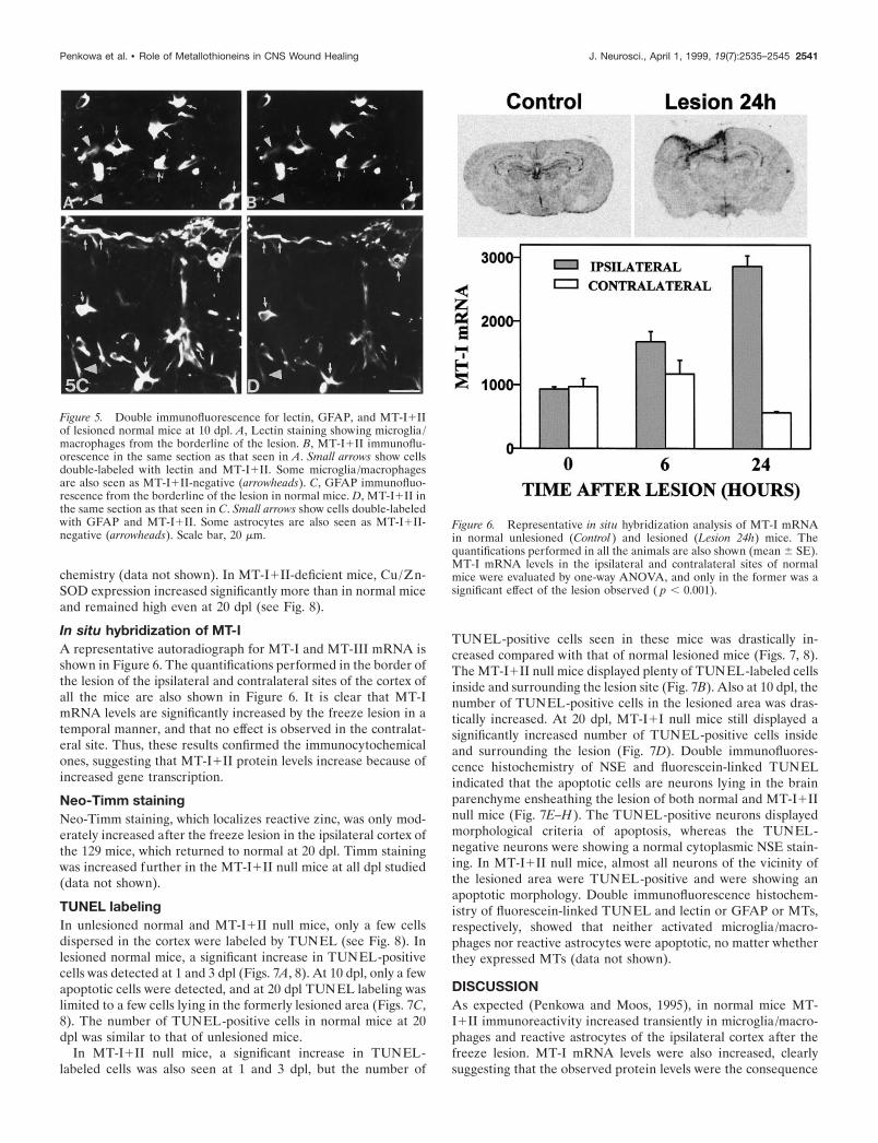
In situ hybridization of MT-IA representative autoradiograph for MT-I and MT-III mRNA isshown in Figure 6. The quantifications performed in the border ofthe lesion of the ipsilateral and contralateral sites of the cortex ofall the mice are also shown in Figure 6. It is clear that MT-ImRNA levels are significantly increased by the freeze lesion in atemporal manner, and that no effect is observed in the contralat-eral site. Thus, these results confirmed the immunocytochemicalones, suggesting that MT-I1II protein levels increase because ofincreased gene transcription.
Neo-Timm stainingNeo-Timm staining, which localizes reactive zinc, was only mod-erately increased after the freeze lesion in the ipsilateral cortex ofthe 129 mice, which returned to normal at 20 dpl. Timm stainingwas increased further in the MT-I1II null mice at all dpl studied(data not shown).
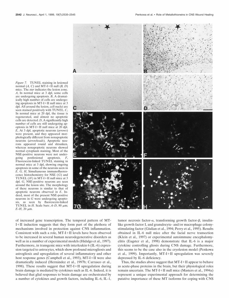
TUNEL labelingIn unlesioned normal and MT-I1II null mice, only a few cellsdispersed in the cortex were labeled by TUNEL (see Fig. 8). Inlesioned normal mice, a significant increase in TUNEL-positivecells was detected at 1 and 3 dpl (Figs. 7A, 8). At 10 dpl, only a fewapoptotic cells were detected, and at 20 dpl TUNEL labeling waslimited to a few cells lying in the formerly lesioned area (Figs. 7C,8). The number of TUNEL-positive cells in normal mice at 20dpl was similar to that of unlesioned mice.
In MT-I1II null mice, a significant increase in TUNEL-labeled cells was also seen at 1 and 3 dpl, but the number of
TUNEL-positive cells seen in these mice was drastically in-creased compared with that of normal lesioned mice (Figs. 7, 8).The MT-I1II null mice displayed plenty of TUNEL-labeled cellsinside and surrounding the lesion site (Fig. 7B). Also at 10 dpl, thenumber of TUNEL-positive cells in the lesioned area was dras-tically increased. At 20 dpl, MT-I1I null mice still displayed asignificantly increased number of TUNEL-positive cells insideand surrounding the lesion (Fig. 7D). Double immunofluores-cence histochemistry of NSE and fluorescein-linked TUNELindicated that the apoptotic cells are neurons lying in the brainparenchyme ensheathing the lesion of both normal and MT-I1IInull mice (Fig. 7E–H). The TUNEL-positive neurons displayedmorphological criteria of apoptosis, whereas the TUNEL-negative neurons were showing a normal cytoplasmic NSE stain-ing. In MT-I1II null mice, almost all neurons of the vicinity ofthe lesioned area were TUNEL-positive and were showing anapoptotic morphology. Double immunofluorescence histochem-istry of fluorescein-linked TUNEL and lectin or GFAP or MTs,respectively, showed that neither activated microglia/macro-phages nor reactive astrocytes were apoptotic, no matter whetherthey expressed MTs (data not shown).
DISCUSSIONAs expected (Penkowa and Moos, 1995), in normal mice MT-I1II immunoreactivity increased transiently in microglia/macro-phages and reactive astrocytes of the ipsilateral cortex after thefreeze lesion. MT-I mRNA levels were also increased, clearlysuggesting that the observed protein levels were the consequence
Figure 5. Double immunofluorescence for lectin, GFAP, and MT-I1IIof lesioned normal mice at 10 dpl. A, Lectin staining showing microglia/macrophages from the borderline of the lesion. B, MT-I1II immunoflu-orescence in the same section as that seen in A. Small arrows show cellsdouble-labeled with lectin and MT-I1II. Some microglia/macrophagesare also seen as MT-I1II-negative (arrowheads). C, GFAP immunofluo-rescence from the borderline of the lesion in normal mice. D, MT-I1II inthe same section as that seen in C. Small arrows show cells double-labeledwith GFAP and MT-I1II. Some astrocytes are also seen as MT-I1II-negative (arrowheads). Scale bar, 20 mm.
Figure 6. Representative in situ hybridization analysis of MT-I mRNAin normal unlesioned (Control ) and lesioned (Lesion 24h) mice. Thequantifications performed in all the animals are also shown (mean 6 SE).MT-I mRNA levels in the ipsilateral and contralateral sites of normalmice were evaluated by one-way ANOVA, and only in the former was asignificant effect of the lesion observed ( p , 0.001).
Penkowa et al. • Role of Metallothioneins in CNS Wound Healing J. Neurosci., April 1, 1999, 19(7):2535–2545 2541
of increased gene transcription. The temporal pattern of MT-I1II induction suggests that they form part of the plethora ofmechanisms involved in protection against CNS inflammation.Consistent with such a role, MT-I1II levels have been observedto be increased in several human neurodegenerative disorders aswell as in a number of experimental models (Hidalgo et al., 1997).Furthermore, in transgenic mice with interleukin 6 (IL-6) expres-sion targeted to astrocytes, which show profound microgliosis andastrogliosis and upregulation of several inflammatory and otherhost response genes (Campbell et al., 1993), MT-I1II were alsodramatically induced (Hernandez et al., 1997b; Carrasco et al.,1998). These results suggest that MT-I1II upregulation duringbrain damage is mediated by cytokines such as IL-6. Indeed, it isbelieved that glial responses to brain damage are orchestrated bya number of cytokines and growth factors, including IL-6, IL-1,
tumor necrosis factor-a, transforming growth factor-b, insulin-like growth factor I, and granulocyte- and/or macrophage colony-stimulating factor (Giulian et al., 1994; Perry et al., 1995). Resultsobtained in IL-6 null mice after the facial nerve transection(Klein et al., 1997) or experimental autoimmune encephalomy-elitis (Eugster et al., 1998) demonstrate that IL-6 is a majorcytokine controlling gliosis during CNS damage. Furthermore,this seems to be the case also in the cryolesion model (Penkowaet al., 1999). Importantly, MT-I1II upregulation was severelydepressed by IL-6 deficiency.
Thus, the studies above suggest that MT-I1II appear to behaveas acute-phase proteins in the brain, but their physiological rolesremain uncertain. The MT-I1II null mice (Masters et al., 1994a)represent a unique experimental approach for determining theputative importance of these MT isoforms for coping with CNS
Figure 7. TUNEL staining in lesionednormal (A, C) and MT-I1II null (B, D)mice. The star indicates the lesion zone.A, In normal mice at 3 dpl, some cellsare undergoing apoptosis. B, A dramat-ically high number of cells are undergo-ing apoptosis in MT-I1II null mice at 3dpl. All around the lesion, cell nuclei areseen stained positively with TUNEL. C,In normal mice at 20 dpl, the tissue isregenerated, and almost no apoptoticcells are detected. D, A significantly highnumber of cells are still undergoing ap-optosis in MT-I1II null mice at 20 dpl.E, At 3 dpl, apoptotic neurons (arrows)were present, and they appeared mor-phologically different from nonapoptoticneurons (arrowheads). Apoptotic neu-rons appeared round and shrunken,whereas nonapoptotic neurons showednormal cytoplasm staining. Most of theNSE-positive neurons were not under-going postlesional apoptosis. F,Fluorescein-linked TUNEL staining innormal mice at 3 dpl, showing ongoingapoptosis in some of the neurons seen inE. G, H, Simultaneous immunofluores-cence histochemistry for NSE (G) andTUNEL (H ) in MT-I1II null mice at 3dpl. G, NSE-positive neurons are seenaround the lesion site. The morphologyof these neurons is similar to that ofapoptotic neurons observed in E. In-deed, most of the present NSE-positiveneurons in G were undergoing apopto-sis, as seen by fluorescein-linkedTUNEL in H. Scale bars: A–D, 80 mm;E–H, 20 mm.
2542 J. Neurosci., April 1, 1999, 19(7):2535–2545 Penkowa et al. • Role of Metallothioneins in CNS Wound Healing
injury. The results with the cryolesion model demonstrate thatthese proteins are of major importance.
A major finding of this study is that the activation of microglia/macrophages is dramatically extended in MT-I1II-deficientmice. As noted above, microglia/macrophages result from theactivation of resident microglia as well as from the infiltration andactivation of bone marrow-derived monocytes. The inspection ofthe temporal pattern of microglia/macrophages appearance sug-gests that both the activation process and the recruitment of bloodmonocytes are normal in the MT-I1II null mice. However, thenormal functions of activated microglia/macrophages do notseem to proceed afterward, because no wound healing occurredeven at 20 dpl, when complete healing was evident in normalmice. Thus, MT-I1II could be important for the normal func-tions of these cells in the brain. Strong support for this hypothesisis given by results obtained in the human monocyte-derived cellline THP-1 (Leibbrandt and Koropatnick, 1994; Leibbrandt et al.,1994; Koropatnick and Zalups, 1997), because the respiratoryburst was significantly reduced by a number of experimentalMT-I1II decreases, including antisense downregulation. Inter-estingly, monocytes of MT-I1II null mice also show an impairedrespiratory burst in vitro (J. Koropatnick, personal communica-tion). The present in vivo results with MT-I1II null mice suggestthat microglia/macrophages are not functioning properly, whichprobably explains the lack of tissue regeneration and woundhealing, as well as the delay in the astrocytosis. Both the sustainedmicrogliosis and the heterogeneous populations of lectin-positivecells observed in the MT-I1II null mice, suggestive of the releaseof both undifferentiated and differentiated myelocytes and mono-
cytes from the bone marrow, might be related to an unbalancedcytokine profile (our unpublished observations), which deservesfurther attention.
The present results also demonstrate that, in the absence ofMT-I1II, neuronal apoptosis was dramatically increased in thecryolesion model. A role of MT-I1II in the control of apoptosishas received much support in recent in vitro and in vivo studies(Zheng et al., 1996a; Hamada et al., 1997; Hellquist, 1997;Houben et al., 1997; Kondo et al., 1997; Lie et al., 1998), but toour knowledge the present report is the first one to demonstratea significant role of MT-I1II in apoptosis of brain cells. Theactual mechanisms through which MT-I1II deficiency would becausing cells to engage in apoptosis are yet unknown, but severalpossibilities are likely. First, MT-I1II are major factors control-ling zinc and copper metabolism (Palmiter and Findley, 1995;Dalton et al., 1996; Kelly and Palmiter, 1996; Kelly et al., 1996),and thus the absence of MT-I1II could likely increase theamount of free zinc, which in excessive amounts could affectneuronal survival on its own (Yokoyama et al., 1986; Koh et al.,1996). Indeed, the results obtained with Timm staining suggestedthat the amount of free zinc was increased in the MT-I1II nullmice compared with controls in the lesioned area. However,further studies are needed considering the role of zinc transport-ers (Palmiter and Findley, 1995; Palmiter, 1998). Second, MT-I1II have also been suggested to be antioxidant proteins (Thor-nalley and Vasak, 1985; Sato and Bremner, 1993). MT-I1II nullembryonic cells show enhanced sensitivity to oxidative stresscaused by tert-butylhydroperoxide and the redox cycling toxinparaquat, as well as enhanced sensitivity to anticancer drugs suchas cisplatin, melphalan, bleomycin, cytarabine, and N-methyl-N9-nitro-N-nitrosoguanidine (Kondo et al., 1995; Lazo et al., 1995;Woo and Lazo, 1997). In vivo, MT-I1II null mice are also moresensitive to the oxidative stress caused by paraquat (Sato et al.,1996), paracetamol (Rofe et al., 1998), and cisplatin (Lie et al.,1998). Also, hepatocytes in culture of MT-I1II null mice aremore sensitive to tert-butylhydroperoxide (Zheng et al., 1996b)and cisplatin (Lie et al., 1998). Thus, the absence of MT-I1IIduring traumatic injury to the CNS, such as after a cryolesion,which will undoubtedly have increased free radicals because ofthe cell disruption and the associated inflammatory response(Giulian et al., 1994; Perry et al., 1995), might render the CNSwith less antioxidant defenses. This could lead to increased oxi-dative stress, which has been proposed as a mediator of apoptosis(Buttke and Sandstrom, 1994). We have observed that the hemeoxygenase 1 (data not shown) and SOD levels were increased inthe lesioned MT-I1II null mice, suggesting an increased oxida-tive stress (Maines, 1997). Third, the ongoing exacerbated in-flammatory response in the MT-I1II null mice could also pro-duce additional signals affecting the apoptosis pathway (Merrilland Jonakait, 1995; Thompson, 1995). Finally, neuronal apoptosiscould also be attributable to the lack of proper glia function (seeabove).
In summary, the present study demonstrates that the MT-I1IIisoforms appear to be essential for normal wound healing in theCNS after traumatic injury. Furthermore, they are also essentialfor neuronal survival. Thus, these functions provide some ratio-nale for the importance of MT-I1II proteins, which are highlypreserved and represented in nature but which, despite a tremen-dous number of studies, did not appear to have clear physiologicalfunctions (Palmiter, 1998).
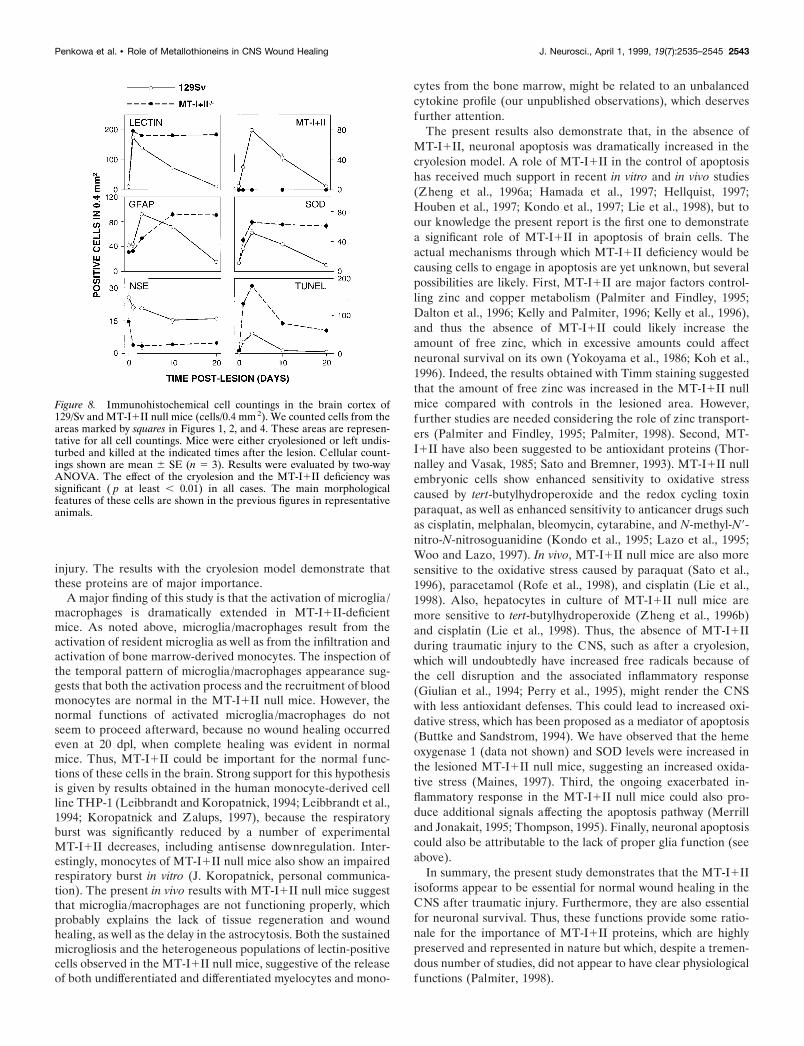
Figure 8. Immunohistochemical cell countings in the brain cortex of129/Sv and MT-I1II null mice (cells/0.4 mm 2). We counted cells from theareas marked by squares in Figures 1, 2, and 4. These areas are represen-tative for all cell countings. Mice were either cryolesioned or left undis-turbed and killed at the indicated times after the lesion. Cellular count-ings shown are mean 6 SE (n 5 3). Results were evaluated by two-wayANOVA. The effect of the cryolesion and the MT-I1II deficiency wassignificant ( p at least , 0.01) in all cases. The main morphologicalfeatures of these cells are shown in the previous figures in representativeanimals.
Penkowa et al. • Role of Metallothioneins in CNS Wound Healing J. Neurosci., April 1, 1999, 19(7):2535–2545 2543
REFERENCESAschner M, Cherian MG, Klaassen CD, Palmiter RD, Erickson JC, Bush
AI (1997) Metallothioneins in brain—the role in physiology and pa-thology. Toxicol Appl Pharmacol 142:229–242.
Buttke T, Sandstrom P (1994) Oxidative stress as a mediator of apopto-sis. Immunol Today 15:7–10.
Campbell IL, Abraham CR, Masliah E, Kemper P, Inglis JD, OldstoneMBA, Mucke L (1993) Neurologic disease in transgenic mice by ce-rebral overexpression of interleukin 6. Proc Natl Acad Sci USA90:10061–10065.
Carrasco J, Hernandez J, Gonzalez B, Campbell I, Hidalgo J (1998)Localization of metallothionein-I and -III expression in the CNS oftransgenic mice with astrocyte-targeted expression of interleukin 6.Exp Neurol 153:184–194.
Dalton T, Pazdernik TL, Wagner J, Samson F, Andrews GK (1995)Temporalspatial patterns of expression of metallothionein-I and -IIIand other stress related genes in rat brain after kainic acid-inducedseizures. Neurochem Int 27:59–71.
Dalton T, Fu K, Palmiter RD, Andrews GK (1996) Transgenic mice thatoverexpress metallothionein-I resist dietary zinc deficiency. J Nutr126:825–833.
Duguid JR, Bohmont CW, Liu NG, Tourtellotte WW (1989) Changes inbrain gene expression shared by scrapie and Alzheimer disease. ProcNatl Acad Sci USA 86:7260–7264.
Erickson JC, Sewell AK, Jensen LT, Winge DR, Palmiter RD (1994)Enhanced neurotrophic activity in Alzheimer’s disease cortex is notassociated with down-regulation of metallothionein-III (GIF). BrainRes 649:297–304.
Eugster H-P, Frei K, Kopf M, Lassmann H, Fontana A (1998) IL-6deficient mice resist myelin oligodendrocyte glycoprotein-induced au-toimmune encephalomyelitis. Eur J Immunol 28:2178–2187.
Gasull T, Rebollo DV, Romero B, Hidalgo J (1993) Development of acompetitive double antibody radioimmunoassay for rat metallothio-nein. J Immunoassay 14:209–225.
Gasull T, Giralt M, Hernandez J, Martinez P, Bremner I, Hidalgo J(1994) Regulation of metallothionein concentrations in rat brain: effectof glucocorticoids, zinc, copper, and endotoxin. Am J Physiol266:E760–E767.
Ghirnikar R, Lee Y, Eng L (1998) Inflammation in traumatic braininjury: roles of cytokines and chemokines. Neurochem Res 23:329–340.
Giulian D, Li J, Li X, George J, Rutecki P (1994) The impact ofmicroglia-derived cytokines upon gliosis in the CNS. Dev Neurosci16:128–136.
Hamada T, Tanimoto A, Sasaguri Y (1997) Apoptosis induced by cad-mium. Apoptosis 2:359–367.
Hellquist H (1997) Apoptosis in epithelial hyperplastic laryngeal lesions.Acta Otolaryngol (Stockh) 527:25–59.
Hernandez J, Carrasco J, Arbones ML, Hidalgo J (1997a) IFN-gR2/2mice show an enhanced liver and brain metallothionein I1II responseto endotoxin but not to immobilization stress. J Endotoxin Res4:363–370.
Hernandez J, Molinero A, Campbell IL, Hidalgo J (1997b) Transgenicexpression of interleukin 6 in the central nervous system regulates brainmetallothionein-I and -III expression in mice. Mol Brain Res48:125–131.
Hidalgo J, Borras M, Garvey JS, Armario A (1990) Liver, brain, andheart metallothionein induction by stress. J Neurochem 55:651–654.
Hidalgo J, Castellano B, Campbell IL (1997) Regulation of brain me-tallothioneins. Curr Top Neurochem 1:1–26.
Houben R, Troppmair J, Hidalgo J, Rapp U (1997) Differential geneexpression in apoptotic 32Dcl3 cells: induction of metallothionein.Apoptosis 2:40–46.
Hozumi I, Inuzuka T, Hiraiwa M, Uchida Y, Anezaki T, Ishiguro H,Kobayashi H, Uda Y, Miyatake T, Tsuji S (1995) Changes of growthinhibitory factor after stab wounds in rat brain. Brain Res 688:143–148.
Hozumi I, Inuzuka T, Ishiguro H, Hiraiwa M, Uchida Y, Tsuji S (1996)Immunoreactivity of growth inhibitory factor in normal rat brain andafter stab wounds—an immunocytochemical study using confocal laserscan microscope. Brain Res 741:197–204.
Kelly EJ, Palmiter RD (1996) A murine model of Menkes disease re-veals a physiological function of metallothionein. Nat Genet13:219–222.
Kelly EJ, Quaife CJ, Froelick GJ, Palmiter RD (1996) Metallothionein Iand II protect against zinc deficiency and zinc toxicity in mice. J Nutr126:1782–1790.
Klein M, Moller J, Jones L, Bluethmann H, Kreutzberg G, Raivich G(1997) Impaired neuroglial activation in interleukin-6 deficient mice.Glia 19:227–233.
Koh J, Suh S, Gwag B, He Y, Hsu C, Choi D (1996) The role of zinc inselective neuronal death after transient global cerebral ischemia. Sci-ence 272:1013–1016.
Kondo Y, Woo ES, Michalska AE, Choo KH, Lazo JS (1995) Metallo-thionein null cells have increased sensitivity to anticancer drugs. Can-cer Res 55:2021–2023.
Kondo Y, Rusnak J, Hoyt D, Settineri C, Pitt B, Lazo J (1997) En-hanced apoptosis in metallothionein null cells. Mol Pharmacol52:195–201.
Koropatnick J, Zalups R (1997) Effect of non-toxic mercury, zinc orcadmium pretreatment on the capacity of human monocytes to undergolipopolysaccharide-induced activation. Br J Pharmacol 120:797–806.
Lazo JS, Kondo Y, Dellapiazza D, Michalska AE, Choo KH, Pitt BR(1995) Enhanced sensitivity to oxidative stress in cultured embryoniccells from transgenic mice deficient in metallothionein I and II genes.J Biol Chem 270:5506–5510.
Leibbrandt ME, Koropatnick J (1994) Activation of human monocyteswith lipopolysaccharide induces metallothionein expression and is di-minished by zinc. Toxicol Appl Pharmacol 124:72–81.
Leibbrandt ME, Khokha R, Koropatnick J (1994) Antisense down-regulation of metallothionein in a human monocytic cell line altersadherence, invasion, and the respiratory burst. Cell Growth Differ5:17–25.
Lie J, Liu Y, Habeebu S, Klaassen C (1998) Metallothionein (MT)-nullmice are sensitive to cisplatin-induced hepatotoxicity. Toxicol ApplPharmacol 149:24–31.
Maines M (1997) The heme oxygenase system: a regulator of secondmessenger gases. Annu Rev Pharmacol Toxicol 37:517–554.
Masters BA, Kelly EJ, Quaife CJ, Brinster RL, Palmiter RD (1994a)Targeted disruption of metallothionein I and II genes increases sensi-tivity to cadmium. Proc Natl Acad Sci USA 91:584–588.
Masters BA, Quaife CJ, Erickson JC, Kelly EJ, Froelick GJ, ZambrowiczBP, Brinster RL, Palmiter RD (1994b) Metallothionein III is ex-pressed in neurons that sequester zinc in synaptic vesicles. J Neurosci14:5844–5857.
Merrill GM, Jonakait GM (1995) Interactions of the nervous and im-mune systems in development, normal brain homeostasis, and disease.FASEB J 9:611–618.
Moos T (1993) Simultaneous application of Timm’s sulphide silvermethod and immunofluorescence histochemistry. J Neurosci Methods48:149–156.
Neal JW, Singhrao SK, Jasani B, Newman GR (1996) Immunocyto-chemically detectable metallothionein is expressed by astrocytes in theischaemic human brain. Neuropathol Appl Neurobiol 22:243–247.
Palmiter R (1998) The elusive function of metallothioneins. Proc NatlAcad Sci USA 95:8428–8430.
Palmiter RD (1995) Constitutive expression of metallothionein-III(MT-III), but not MT-I, inhibits growth when cells become zinc defi-cient. Toxicol Appl Pharmacol 135:139–146.
Palmiter R, Findley S (1995) Cloning and functional characterization ofa mammalian zinc transporter that confers resistance to zinc. EMBO J14:639–649.
Palmiter RD, Findley SD, Whitmore TE, Durnam DM (1992) MT-III, abrain-specific member of the metallothionein gene family. Proc NatlAcad Sci USA 89:6333–6337.
Penkowa M, Moos T (1995) Disruption of the blood-brain interface inneonatal rat neocortex induces a transient expression of metallothio-nein in reactive astrocytes. Glia 13:217–227.
Penkowa M, Hidalgo J, Moos T (1997) Increased astrocytic expressionof metallothioneins I1II in brain stem of adult rats treated with6-aminonicotinamide. Brain Res 774:256–259.
Penkowa M, Moos T, Carrasco J, Hadberg H, Molinero A, BluethmannH, Hidalgo J (1999) Strongly compromised inflammatory response tobrain injury in interleukin-6 deficient mice. Glia 25:343–357.
Perry V, Bell M, Brown H, Matyszak M (1995) Inflammation in thenervous system. Curr Opin Neurobiol 5:636–641.
Quaife CJ, Findley SD, Erickson JC, Froelick GJ, Kelly EJ, ZambrowiczBP, Palmiter RD (1994) Induction of a new metallothionein isoform(MT-IV) occurs during differentiation of stratified squamous epithelia.Biochemistry 33:7250–7259.
Quaife C, Kelly E, Masters B, Brinster R, Palmiter R (1998) Ectopic
2544 J. Neurosci., April 1, 1999, 19(7):2535–2545 Penkowa et al. • Role of Metallothioneins in CNS Wound Healing
expression of metallothionein-III causes pancreatic acinar cell necrosisin transgenic mice. Toxicol Appl Pharmacol 148:148–157.
Rofe A, Barry E, Shelton T, Philcox J, Coyle P (1998) Paracetamolhepatotoxicity inmetallothionein-null mice. Toxicology 125:131–140.
Sato M, Bremner I (1993) Oxygen free radicals and metallothionein.Free Radic Biol Med 14:325–337.
Sato M, Apostolova M, Hamaya M, Yamaki J, Choo K, Michalska A,Kodama N, Tohyama C (1996) Susceptibility of metallothionein-nullmice to paraquat. Environ Toxicol Pharmacol 1:221–225.
Sewell AK, Jensen LT, Erickson JC, Palmiter RD, Winge DR (1995)Bioactivity of metallothionein-3 correlates with its novel beta domainsequence rather than metal binding properties. Biochemistry34:4740–4747.
Sillevis Smitt PA, Blaauwgeers HG, Troost D, de Jong JM (1992) Me-tallothionein immunoreactivity is increased in the spinal cord of pa-tients with amyotrophic lateral sclerosis. Neurosci Lett 144:107–110.
Stevens A, Bahr M (1993) Origin of macrophages in central nervoustissue. J Neurol Sci 118:117–122.
Suzuki K, Nakajima K, Kawaharada U, Uehara K, Hara F, Otaki N,Kimura M, Tamura Y (1992) Metallothionein in the human brain.Acta Histochem Cytochem 25:617–622.
Thompson C (1995) Apoptosis in the pathogenesis and treatment ofdisease. Science 267:1456–1462.
Thornalley PJ, Vasak M (1985) Possible role for metallothionein inprotection against radiation-induced oxidative stress. Kinetics andmechanism of its reaction with superoxide and hydroxyl radicals. Bio-chim Biophys Acta 827:36–44.
Uchida Y, Takio K, Titani K, Ihara Y, Tomonaga M (1991) The growthinhibitory factor that is deficient in the Alzheimer’s disease brain is a68 amino acid metallothionein-like protein. Neuron 7:337–347.
Vela JM, Hidalgo J, Gonzalez B, Castellano B (1997) Induction ofmetallothionein in astrocytes and microglia in the spinal cord from themyelin-deficient jimpy mouse. Brain Res 767:345–355.
Woo E, Lazo J (1997) Nucleocytoplasmic functionality of metallothio-nein. Cancer Res 57:4236–4241.
Yamada M, Hayashi S, Hozumi I, Inuzuka T, Tsuji S, Takahashi H (1996)Subcellular localization of growth inhibitory factor in rat brain: lightand electron microscopic immunohistochemical studies. Brain Res735:257–264.
Yokoyama M, Koh J, Choi D (1986) Brief exposure to zinc is toxic tocortical neurons. Neurosci Lett 71:351–335.
Young JK, Garvey JS, Huang PC (1991) Glial immunoreactivity formetallothionein in the rat brain. Glia 4:602–610.
Zheng H, Berman NE, Klaassen CD (1995) Chemical modulation ofmetallothionein I and III mRNA in mouse brain. Neurochem Int27:43–58.
Zheng H, Liu J, Choo KH, Michalska AE, Klaassen CD (1996a)Metallothionein-I and -II knock-out mice are sensitive to cadmium-induced liver mRNA expression of c-jun and p53. Toxicol Appl Phar-macol 136:229–235.
Zheng H, Liu J, Liu Y, Klaassen CD (1996b) Hepatocytes frommetallothionein-I and II knock-out mice are sensitive to cadmium- andtert-butylhydroperoxide-induced cytotoxicity. Toxicol Lett 87:139–145.
Penkowa et al. • Role of Metallothioneins in CNS Wound Healing J. Neurosci., April 1, 1999, 19(7):2535–2545 2545
Related Documents