Journal of Neurology, Neurosurgery, and Psychiatry. Special Supplement 1989:22-28 Clues to the mechanism underlying dopamine cell death in Parkinson's disease P JENNER From the Parkinson's Disease Society Research Centre, University Department of Neurology, Institute of Psychiatry and King's College Hospital Medical School, London UK SUMMARY The primary pathological change in Parkinson's disease is the destruction of dopamine containing cells in the zona compacta of substantia nigra. The cause of nigral cell death and the underlying mechanism remains elusive. However, the discovery of the selective nigral neurotoxin MPTP and its ability to inhibit mitochondrial energy metabolism via its metabolite MPP+ and to generate superoxide radicals suggests processes by which nigral cell death might occur. Recent post- mortem evidence in brain tissue from patients dying with Parkinson's disease also suggests the occurrence of some on-going toxic mechanism. This may be a free radical process stimulated by an excess of iron within subsantia nigra coupled to a generalised decrease in brain ferritin content. These data suggest altered iron handling occurs in Parkinson's disease which may lead to the generation of toxic oxygen species such as superoxide radicals. There is also evidence for an inhibition of mitochondrial function in the substantia nigra in patients with Parkinson's disease. So there may be a close association between the actions of the synthetic neurotoxin MPTP and the underlying cause of idiopathic Parkinson's disease. There are widespread pathological and biochemical changes in the brain of patients dying with Parkinson's disease.'2 The primary alteration appears to be a loss of dopamine containing cells in zona compacta of substantia nigra with a corresponding generalised loss of dopamine content throughout the forebrain. While the details of these changes are extensively documen- ted3 there has been little evidence as to the underlying cause of dopamine cell death or the mechanism by which dopamine cells degenerate. Many theories have been advanced (for example, involvement of viruses, aberrant metabolism of dopamine, involvement of neuromelanin) but none of these proven. More recen- tly interest has centred on the manner in which endogenous or environmental toxins may be involved as a cause of Parkinson's disease. This stems from the discovery of the selective nigral toxin, I -methyl-4- phenyl-1,2,3,6-tetrahydropyrdine (MPTP) which was found to induce persistent Parkinsonism in man and other primate species by destroying dopamine con- taining cells within substantia nigra.' The discovery of MPTP provided a major impetus for research into the cause of Parkinson's disease. Address for reprint requests: Dr P Jenner, Department of Neurology, Institute of Psychiatry, De Crespigny Park, London SE5 8AF, UK. Accepted March 1989 However, MPTP does not provide an exact model of idiopathic Parkinson's disease since in general neurotoxic effects are limited to substantia nigra and its corresponding losses of caudate-putamen dopamine content.78 Overall other neuronal systems do not appear to be involved and there is no occurrence of Lewy bodies as a marker of the process underlying idiopathic Parkinson's disease.'"'2 One reason for the discrepancies between MPTP induced Parkinsonism and the idiopathic disease may relate to the age of the animals studied. In general, young adult primates are used in such experiments but where older animals have been employed the pathology has been more extensive, involving also the locus coeruleus and Lewy body like inclusions have been observed.'"'5 However, MPTP has provided the most appropriate animal model of Parkinson's disease so far devised. It has found use as a test-bed in which to evaluate novel therapies for Parkinson's disease'"'9 and also to evaluate long-term complications of levodopa treat- ment of Parkinson's disease.20'2 Similarly, MPTP treated primates provide an ideal test-bed in which to evaluate the usefulness of the implantation of foetal nigral dopamine containing cells as a "cure" for Parkinson's disease.2223 However, it may be one other aspect of the actions of MPTP which is the most important, namely the mechanism by which MPTP kills nigral dopamine containing cells. This may 22 Protected by copyright. on December 4, 2021 by guest. http://jnnp.bmj.com/ J Neurol Neurosurg Psychiatry: first published as 10.1136/jnnp.52.Suppl.22 on 1 June 1989. Downloaded from

Welcome message from author
This document is posted to help you gain knowledge. Please leave a comment to let me know what you think about it! Share it to your friends and learn new things together.
Transcript

Journal ofNeurology, Neurosurgery, and Psychiatry. Special Supplement 1989:22-28
Clues to the mechanism underlying dopamine celldeath in Parkinson's diseaseP JENNER
From the Parkinson's Disease Society Research Centre, University Department ofNeurology, Institute ofPsychiatry and King's College Hospital Medical School, London UK
SUMMARY The primary pathological change in Parkinson's disease is the destruction of dopaminecontaining cells in the zona compacta of substantia nigra. The cause of nigral cell death and theunderlying mechanism remains elusive. However, the discovery of the selective nigral neurotoxinMPTP and its ability to inhibit mitochondrial energy metabolism via its metabolite MPP+ and togenerate superoxide radicals suggests processes by which nigral cell death might occur. Recent post-mortem evidence in brain tissue from patients dying with Parkinson's disease also suggests theoccurrence of some on-going toxic mechanism. This may be a free radical process stimulated by an
excess ofiron within subsantia nigra coupled to a generalised decrease in brain ferritin content. Thesedata suggest altered iron handling occurs in Parkinson's disease which may lead to the generation oftoxic oxygen species such as superoxide radicals. There is also evidence for an inhibition ofmitochondrial function in the substantia nigra in patients with Parkinson's disease. So there may be a
close association between the actions of the synthetic neurotoxin MPTP and the underlying cause ofidiopathic Parkinson's disease.
There are widespread pathological and biochemicalchanges in the brain ofpatients dying with Parkinson'sdisease.'2 The primary alteration appears to be a lossof dopamine containing cells in zona compacta ofsubstantia nigra with a corresponding generalised lossof dopamine content throughout the forebrain. Whilethe details of these changes are extensively documen-ted3 there has been little evidence as to the underlyingcause of dopamine cell death or the mechanism bywhich dopamine cells degenerate. Many theories havebeen advanced (for example, involvement of viruses,aberrant metabolism of dopamine, involvement ofneuromelanin) but none of these proven. More recen-tly interest has centred on the manner in whichendogenous or environmental toxins may be involvedas a cause of Parkinson's disease. This stems from thediscovery of the selective nigral toxin, I-methyl-4-phenyl-1,2,3,6-tetrahydropyrdine (MPTP) which wasfound to induce persistent Parkinsonism in man andother primate species by destroying dopamine con-taining cells within substantia nigra.'The discovery of MPTP provided a major impetus
for research into the cause of Parkinson's disease.
Address for reprint requests: Dr P Jenner, Department of Neurology,Institute of Psychiatry, De Crespigny Park, London SE5 8AF, UK.
Accepted March 1989
However, MPTP does not provide an exact modelof idiopathic Parkinson's disease since in generalneurotoxic effects are limited to substantia nigra andits corresponding losses of caudate-putamendopamine content.78 Overall other neuronal systemsdo not appear to be involved and there is nooccurrence of Lewy bodies as a marker of the processunderlying idiopathic Parkinson's disease.'"'2 Onereason for the discrepancies between MPTP inducedParkinsonism and the idiopathic disease may relate tothe age of the animals studied. In general, young adultprimates are used in such experiments but where olderanimals have been employed the pathology has beenmore extensive, involving also the locus coeruleus andLewy body like inclusions have been observed.'"'5However, MPTP has provided the most appropriateanimal model of Parkinson's disease so far devised. Ithas found use as a test-bed in which to evaluate noveltherapies for Parkinson's disease'"'9 and also toevaluate long-term complications of levodopa treat-ment of Parkinson's disease.20'2 Similarly, MPTPtreated primates provide an ideal test-bed in which toevaluate the usefulness of the implantation of foetalnigral dopamine containing cells as a "cure" forParkinson's disease.2223 However, it may be one otheraspect of the actions of MPTP which is the mostimportant, namely the mechanism by which MPTPkills nigral dopamine containing cells. This may
22
Protected by copyright.
on Decem
ber 4, 2021 by guest.http://jnnp.bm
j.com/
J Neurol N
eurosurg Psychiatry: first published as 10.1136/jnnp.52.S
uppl.22 on 1 June 1989. Dow
nloaded from
Clues to the mechanism underlying dopamine cell death in Parkinson's disease
provide clues to the selective vulnerability of this cellgroup and to the pathological process underlyingdopamine cell death in Parkinson's disease.
Mechanism ofaction ofMPTPMany important steps in the mechanism of action ofMPTP have been uncovered. Following the adminis-tration ofMPTP to primates it was found that anothersubstance, namely, l-methyl-4-phenyl-pyridiniumspecies (MPP+), and not MPTP itself, accumulatedand persisted within the brain.2425 The reason for thisbecame clear when it was discovered that MPTP was
an unexpected substrate for MAO B2"2' So it appearedthat MPP+ rather than MPTP might be the neurotoxicspecies. Indeed, MPP+ was actively accumulated bydopaminergic neurons since it is a substrate fordopamine reuptake mechanisms29 and this accumula-tion may be enhanced by an association withneuromelanin.? MPTP neurotoxicity could be preven-ted by prior treatment of animals with MAO Binhibitors, such as deprenyl, or dopamine reuptakeblockers, such as nomifensine.3"32 Also, MPP+ wasmore toxic than MPTP itself in destroying dopaminecontaining cells. Thus, intranigral infusions of MPP+,but not MPTP, destroyed nigral neurones in rats.33Similarly, intracerebroventricular injections of MPP+destroyed nigral cells in the brains of mice.? In cellculture MPP+ is toxic to mesencephalic cells.3536 So allthis data led to the conclusion that it was MPP+ andnot MPTP that was responsible for the toxic actionsobserved.
These data did not, however, explain the mechanismby which MPTP (or MPP+) killed dopamine contain-ing cells. This issue was clarified with the discoverythat MPP+ induces oxidative stress owing to its abilityto inhibit the oxidation ofmitochondrial NAD-linkedsubstrates.373' More specifically MPP+ inhibits mito-chondrial energy metabolism at the level of complexI3. However, millimolar concentrations ofMPP+ wererequired in vitro to produce this effect and it wasthought unlikely that these would be achieved in vivo.But MPP+ is actively accumulated by mitochondriasuch that 50-100 times the external concentration canbe achieved.A"2 So at present it is believed that MPP+interferes with mitochondrial energy metabolismsomewhere between NADH dehydrogenase and co-
enzyme Q 10.43 Exactly how and where this occursremains unknown. Nevertheless, its consequences canbe observed in terms of depletion of cellular ATP, adecrease in reduced glutathione content, and altera-tions in cellular calcium content."44"
Free radical generation as a mechanism of MPTPtoxicityOne basic mechanism initially proposed to explain theneurotoxic actions of MPP+ was the production of
free radicals. This was derived from the chemicalsimilarity between MPP+ and a paraquat, a knownredox cycler. Although MPP+ bears a strikingchemical resemblance to paraquat, MPP+ is extremelystable compared with paraquat and does not undergosingle electron reduction to produce oxygenradicals.474 This is emphasised by the electrochemicalpotential of MPP+ of < 1-0 V which compared withparaquat makes it improbable that it will undergobioreduction to stimulate toxic oxygen radical for-mation.49 However, this conclusion presumes that it isMPP+ alone which causes the neurotoxicity associatedwith MPTP administration.MPTP is converted to MPP+ in a two-stage reaction
via the intermediate dihydropyridine derivative,MPDP+."52 This sequence of events gives more scopefor the production of oxygen species. Indeed, there isrecent evidence that oxygen radicals may be involvedin the toxicity of MPTP in a manner previouslyunsuspected. Thus, in aerobic mitochondrial prepara-tions MPTP stimulated an electron spin resonance(ESR) signal compatible with free radical formation.53This signal was prevented by inclusion of superoxidedismutase suggesting the generation of superoxideradicals. The metabolism ofMPTP appeared involvedsince the ESR signal was prevented by the inclusion ofthe MAO B inhibitor deprenyl but not by the MAO Ainhibitor clorgyline. In the absence of mitochondrianeither of the MPTP metabolites, MPP+ or MPDP+,alone produced an ESR signal. However, togetherthey caused a spectrum which increased in intensitywith time. These data suggest a redox reaction occursbetween MPP+ and MPDP+ to produce toxic oxygenradicals and that these may be involved in the ability ofMPTP to destroy nigral containing cells. Whether anaction of MPTP based on superoxide formation iscompatible with inhibition of mitochondrial functionremains to be determined.So the MPTP story reveals possible mechanisms by
which nigral dopamine containing cells may be des-troyed. But what is its relevance to Parkinson'sdisease? It may be that there is an MPTP like toxininvolved as a cause of Parkinson's disease although atpresent this appears unlikely. On the other hand, itmay be that the mechanism by which MPTP killsdopamine cells by interference with mitochondrialfunction or by superoxide formation may reflect aselective vulnerability of dopamine cells in substantianigra which is also apparent in idiopathic Parkinson'sdisease. So, are there any similarities between theactions of MPTP and biochemical changes occurringin brain in Parkinson's disease itself?
Evidencefor a neurotoxic process occurring in brain inParkinson's diseaseOne approach to detecting a cause or mechanism
23
Protected by copyright.
on Decem
ber 4, 2021 by guest.http://jnnp.bm
j.com/
J Neurol N
eurosurg Psychiatry: first published as 10.1136/jnnp.52.S
uppl.22 on 1 June 1989. Dow
nloaded from

24underlying idiopathic Parkinson's disease is toexamine post-mortem brain tissue. However, successdepends partially on how cell death in Parkinson'sdisease occurs. For example, if Parkinson's diseaseoccurs as a result of some fault in utero or due to asingle toxic insult during life then there might be noindication ofa toxic process in the post-mortem tissuesfrom patients at the end stage of their disease.Nevertheless, we have examined some generalindicators of cellular toxicity in post-mortem tissuesfrom patients dying with Parkinson's disease.The tissues from these studies came from patients
with a history of idiopathic Parkinson's disease all ofwhom received levodopa treatment up to the time ofdeath and whose brain showed evidence of cell lossand the presence of Lewy bodies in substantia nigraand a markedly decreased caudate dopamine content.These tissues were matched with tissues from controlpatients of similar age who died from non-neurological and non-psychiatric disorders and whosesubstantia nigra appeared normal in histologicalexamination. The tissues were also matched for thetime between death and body refrigeration and thetime between death and autopsy and subsequentfreezing of the brain material.
Initially, we assessed markers of lipid peroxidationas a non-specific index of cell death."' Twoparameters were studied, namely, the brain content ofpolyunsaturated fatty acids (PUFA), the substrate forlipid peroxidation, and the content of malondialde-hyde, a stable intermediate in the process of lipidperoxidation (table 1). The PUFA content of substan-tia nigra was reduced compared to levels found incontrol brain tissue and this change was selective tosubstantia nigra since no difference in PUFA contentwas found in any other brain region examined. So itappeared that perhaps increased degradation ofPUFAs was occurring in Parkinsonian nigra. Thisview was confirmed by the finding of an increasedbasal level of malondialdehyde in Parkinsonian sub-stantia nigra compared to control tissue which againdid not occur in any other brain region examined.These data suggest that even at the end stage ofParkinson's disease there is evidence for some on-going toxic process such as might occur due to freeradical attack. It should be noted that all the patientsexamined received levodopa until the time of deathand it cannot be excluded that the changes observedwere not due to drug treatment. However, sincelevodopa accumulates in many brain regions it mightbe expected that similar changes would 'oe observedelsewhere, particularly within the caudate-putamenwhere levodopa occurs in high amounts.
If there is some on-going toxic process occurringwithin the Parkinsonian brain then what could be thestimulating factor? It could be some environmental
JennerTable 1 Polytmsaturatedfatty acid (PUFA) and basalMDA levels in the substantia nigra and cerebellum ofParkinsonian and control hwnan brains
PUFA 's Basal MDA(nmol linoleic levels (nmol Ratio basalacid/mg MDA/mg MDA/PUFA's
Brain region protein) protein) (x 10-3)
CerebellumControl (n = 13) 211 (11) 3-4 (0-4) 16-0(1.4)Parkinsonian
(n = 10) 186 (10) 4-2 (0 4) 21-0 (1-6)Substantia nigraControl (n = 19) 298 (12) 2-0 (0.1) 7-0 (0;5)Parkinsonian
(n = 14) 254 (14)* 27 (03)* 11-2 (1-2)*
Values are expressed as means (SEM). *p < 0-05 compared withcontrol tissue (Student's t test). Data taken from Dexter et al.' "
toxin such as MPTP but so far none has beenidentified. On the other hand, some endogenous agentmight act to stimulate free radical formation. In thisrespect we decided to investigate the iron content ofthe Parkinsonian brain since iron is able to stimulateoxygen radical formation.'5 Indeed, in 1968 Earle57had suggested an increase in brain iron content inParkinson's disease using formalin fixed tissuesexamined by X-ray fluorescent spectroscopy.The total iron content of the Parkinsonian brain
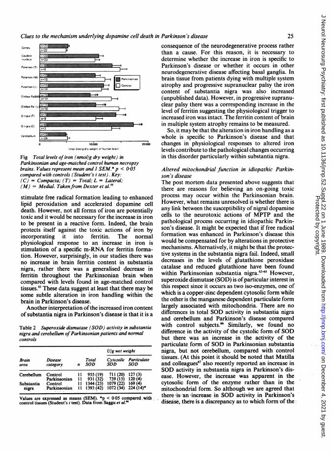
was measured compared with age-matched controltissue using inductively coupled plasma spectros-copy.58 59 This showed that within the substantia nigraas a whole and also within the zona compacta ofsubstantia nigra there was a substantial increase intotal iron content (fig). No increase in iron levels wasfound in any other brain area examined although therewas a reduction in iron content within the medial andlateral segments of the globus pallidus. So thereappeared to be a specific increase in iron within thatarea of the brain which is thought to bear the primarypathological change in Parkinson's disease. Recently,the finding of an increased iron content of substantianigra in Parkinson's disease was confirmed by others.'At this point it should be noted that we also found alarge increase in the zinc content of substantia nigraalthough this also occurred in caudate-putamen andso was not therefore specifically related to path-ological change. In addition, the copper content ofsubstantia nigra was reduced. There were no changesin the levels of manganese or lead in any of the brainareas examined when compared to control tissues.Once again, the issue of drug treatment must be
considered as being relevant to the changes in brainiron content. However, any effect of drug treatmentwould appear unlikely since the samples examined byEarle in 1968 were collected between 1867 and 1954, inother words prior to the levodopa era.57One interpretation of the data so far would be that
enhanced levels of iron within substantia nigra
Protected by copyright.
on Decem
ber 4, 2021 by guest.http://jnnp.bm
j.com/
J Neurol N
eurosurg Psychiatry: first published as 10.1136/jnnp.52.S
uppl.22 on 1 June 1989. Dow
nloaded from
Clues to the mechanism underlying dopamine cell death in Parkinson's disease
Cortex
Caudatenucleus
Putamen (T)
Putamen (M) - _
Putamen (L)
n=22
N Parkinsonian
l: E] Control-1 I
Globus Pal#M)-*
Globus Pal (L
S nigra (T)
S nigra (C)
Cerebellum
i=14
i=13
1=9
n=3
n024 100 20
O 10000 20000nrnol iron/g dry weight of human brain
Fig Total levels ofiron (nmol/g dry weight) inParkinsonian and age-matched control human necropsybrains. Values represent mean and I SEM.* p < 0 05compared with controls (Student's t test). Key:(C) = Compacta; (T) = Total; L = Lateral;(M) = Medial. Takenfrom Dexter et al.5'
stimulate free radical formation leading to enhancedlipid peroxidation and accelerated dopamine celldeath. However, not all forms of iron are potentiallytoxic and it would be necessary for the increase in ironto be present in a reactive form. Indeed, the brainprotects itself against the toxic actions of iron byincorporating it into ferritin. The normalphysiological response to an increase in iron isstimulation of a specific m-RNA for ferritin forma-tion. However, surprisingly, in our studies there wasno increase in brain ferritin content in substantianigra, rather there was a generalised decrease inferritin throughout the Parkinsonian brain whencompared with levels found in age-matched controltissues.6' These data suggest at least that there may besome subtle alteration in iron handling within thebrain in Parkinson's disease.Another interpretation ofthe increased iron content
of substantia nigra in Parkinson's disease is that it is a
Table 2 Superoxide dismutase (SOD) activity in substantianigra and cerebellum ofParkinsonian patients and normalcontrols
U/g wet weight
Brain Disease Total Cytosolic Particulatearea category n SOD SOD SOD
Cerebellum Control 11 955 (19) 711(20) 127 (3)Parkinsonian 11 931 (32) 739 (15) 120(4)
Substantia Control 11 1344 (23) 1079 (22) 169 (4)nigra Parkinsonian 11 1393 (42) 1072 (34) 224 (14)*
Values are expressed as means (SEM). *p < 0-05 compared with.control tissues (Student's t test). Data from Saggu et al.66
consequence of the neurodegenerative process ratherthan a cause. For this reason, it is necessary todetermine whether the increase in iron is specific toParkinson's disease or whether it occurs in otherneurodegenerative disease affecting basal ganglia. Inbrain tissue from patients dying with multiple systematrophy and progressive supranuclear palsy the ironcontent of substantia nigra was also increased(unpublished data). However, in progressive supranu-clear palsy there was a corresponding increase in thelevel of ferritin suggesting the physiological trigger toincreased iron was intact. The ferritin content of brainin multiple system atrophy remains to be measured.
So, it may be that the alteration in iron handling as awhole is specific to Parkinson's disease and thatchanges in physiological responses to altered ironlevels contribute to the pathological changes occurringin this disorder particularly within substantia nigra.
Altered mitochondrial function in idiopathic Parkin-son's diseaseThe post mortem data presented above suggests thatthere are reasons for believing an on-going toxicprocess may occur within the Parkinsonian brain.However, what remains unresolved is whether there isany link between the susceptibility of nigral dopaminecells to the neurotoxic actions of MPTP and thepathological process occurring in idiopathic Parkin-son's disease. It might be expected that if free radicalformation was enhanced in Parkinson's disease thiswould be compensated for by alterations in protectivemechanisms. Alternatively, it might be that the protec-tive systems in the substantia nigra fail. Indeed, smalldecreases in the levels of glutathione peroxidasecatalase and reduced glutathione have been foundwithin Parkinsonian substantia nigra.62"5 However,superoxide dismutase (SOD) is ofparticular interest inthis respect since it occurs as two iso-enzymes, one ofwhich is a copper-zinc dependent cytosolic form whilethe other-is the manganese dependent particulate formlargely associated with mitochondria. There are nodifferences in total SOD activity in substantia nigraand cerebellum and Parkinson's disease comparedwith control subjects.'a Similarly, we found nodifference in the activity of the cystolic form of SODbut there was an increase in the activity of theparticulate form of SOD in Parkinsonian substantianigra, but not cerebellum, compared with controltissues. (At this point it should be noted that Matillaand colleagues67 also recently reported an increase inSOD activity in substantia nigra in Parkinson's dis-ease. However, the increase was apparent in thecytosolic form of the enzyme rather than in themitochondrial form. So although we are agreed thatthere 'is- an increase in SOD activity in Parkinson's
- disease, there is a discrepancy as to which form-of the
:22MMEMEEMMEMMMER-4=23
__ _ ,.
n,
n
ng
a-.El
PI
25
L,
Protected by copyright.
on Decem
ber 4, 2021 by guest.http://jnnp.bm
j.com/
J Neurol N
eurosurg Psychiatry: first published as 10.1136/jnnp.52.S
uppl.22 on 1 June 1989. Dow
nloaded from

26enzyme is implicated.) Our data raise the tantalisingconcept that in Parkinson's disease mitochondria maybe under attack from superoxide, so providing a linkwith ideas on MPTP toxicity.
If mitochondrial superoxide dismutase activity isenhanced in Parkinson's disease then does this suggestsome underlying fault in mitochondria as a cause ofthe disorder? Very recently Schapira and colleagues68assessed mitochondrial function in substantia nigra ofthe Parkinsonian brain. Rotenone sensitive mitochon-drial NADH cytochrome c reductase activity (com-plex I and III) was reduced in Parkinsonian nigracompared with control tissues while succinate cyto-chrome c reductase activity (complex II and III) wasunchanged. Levels ofa non-respiratory mitochondrialenzyme, citrate synthase, and a non-mitochondrialenzyme, rotenone insensitive NADH cytochrome creductase were unaffected. So the results suggest animpairment of complex I of the mitochondrial res-piratory chain in Parkinson's disease. The similarity toalterations occurring in the presence of MPTP isstriking.
Conclusions
The discovery of the neurotoxic actions of MPTP onsubstantia nigra has provided ideas on how thetoxicity to these cells might involve inhibition ofmitochondrial energy metabolism and/or superoxideradical generation. This has stimulated the idea that atoxin or a continuous toxic process may be responsiblefor the pathological changes which occur in Parkin-son's disease. Indeed, examination of post-mortemParkinsonian tissues appear to indicate that a continu-ing toxic process may indeed occur in substantia nigrain Parkinson's disease and that potentially this may berelated to alterations in iron handling in this disorder.The changes in superoxide dismutase activity suggestthat it may be the production of superoxide radicalswhich lead to cellular damage and in this respect thereare striking similarities to the recent findings of freeradical formation due to a redox reaction betweenmetabolites of MPTP. Our findings of a selectiveincrease in mitochondrial superoxide dismutaseactivity raises once again the concept that it may bealterations in mitochondrial function that is a primarycause of cell death in Parkinson's disease. Indeed, therecent finding of Schapira and colleagues of inhibitionofcomplex I in Parkinson's disease provides a tantalis-ing link between the MPTP story and the idiopathicdisorder. It may be that MPTP will lead us closer todiscovering the cause of Parkinson's disease than wasat first thought.
These investigations were funded by the Parkinson'sDisease Society, the Medical Research Council and
Jenner
the Research Funds of the Bethlem Royal and Maud-sley Hospitals.
References
I Forno LS. Pathology of Parkinson's disease. In: Marsden CD,Fahn S, eds. Neurology, 2: Movement Disorders. London:Butterworth Scientific, 1981:25-40.
2 Hornykiewicz 0. Brain neurotransmitter changes in Parkinson'sdisease. In: Marsden CD, Fahn S, eds. Neurology, 2: MovementDisorders. London: Butterworth Scientific 1981:41-58.
3 Agid Y, Javoy-Agid F, Ruberg M. Biochemistry ofneurotransmit-ters in Parkinson's disease. In: Marsden CD, Fahn S, eds.Movement Disorders 2. London: Butterworth, 1987:166-230.
4 Davis GC, Williams AC, Markey SP, Ebert MH, Caine ED,Reichert CM, Kopin IJ. Chronic Parkinsonism secondary tointravenous injection of meperidine analogues. Psychiatry Res1979;1:249-54.
5 Langston JW, Ballard P, Tetrud JW, Irwin I. Chronic parkinson-ism in humans due to a product of meperidine analog synthesis.Science 1982;219:979-80.
6 Ballard PA, Tetrud JW, Langston JW. Permanent human parkin-sonism due to I -methyl4-phenyl- 1,2,3,6-tetrahydropyridine(MPTP). Neurology 1985;35:949-56.
7 Burns RS, Chiueh CC, Markey SP, Ebert MH, Jacobowitz DM,Kopin IJ. A primate model of parkinsonism: Selective destruc-tion of dopaminergic neurons in the pars compacta of thesubstantia nigra by N-methyl-4-phenyl-1,2,3,6-tetra-hydropyridine. Proc Nat Acad Sci USA 1983;80:4546-50.
8 Langston JW, Forno LS, Rebert CS, Irwin I. Selective nigraltoxicity after systemic administration of l-methyl-4-phenyl1,2,5,6-tetrahydropyridine (MPTP) in the squirrel monkey.Brain Res 1984;292:390-4.
9 Jenner P, Rupniak NMJ, Rose S, Kelly E, Kilpatrick G, Lees A,Marsden CD. I-Methyl-4-phenyl-1 ,2,3,6-tetrahydropyridine-induced parkinsonism in the common marmoset. Neurosci Lett1984;50:85-90.
10 Jenner P, Marsden CD. MPTP-induced parkinsonism in primatesand its use in the assessment ofnovel strategies for the treatmentof Parkinson's disease. In: Clifford Rose F, ed. Parkinson'sDisease: Clinical and Experimental Advances. London: JohnLibbey, 87:149-62.
11 Garvey J, Petersen M, Waters CM, et al. Administration ofMPTPto the common marmoset does not alter cortical cholinergicfunction. Movement Disorders 1986;1:129-34.
12 Jenner P, Taquet H, Mauborgne A, et al. Lack of change in basalganglia neuropeptide content following subacute 1-methyl-4-phenyl-1,2,3,6-tetrahydropyridine treatment of the commonmarmoset. J Neurochem 1986;46:1548-51.
13 Mitchel IJ, Cross AJ, Sambrook MA, Crossman AR. Sites of theneurotoxic actions of 1-methyl-4-phenyl-1,2,3,6-tetra-hydropyridine in the macaque monkey include the ventraltegmental area and the locus coeruleus. Neurosic Lett1985;61: 195-200.
14 Forno LS, Langston JW, DeLanney LE, Irwin I, Ricaurte GA.Locus ceruleus lesions and eosinophilic inclusions in MPTP-treated monkeys. Ann Neurol 1986;20:449-55.
15 Forno LS, Langston JW, DeLanney LE, Irwin I. An electronmicroscopic study of MPTP-induced inclusion bodies in an oldmonkey. Brain Res 1988;448:150-7.
16 Nomoto M, Jenner P, Marsden CD. The dopamine D-2 agonistLY 141865, but not the D-1 agonist SKF 38393, reversesparkinsonism induced by 1-methyl-4-phenyl-1,2,3,6-tetra-hydropyridine (MPTP) in thecommon marmoset. Neurosci Lett1985;57:37-41.
17 Nomoto M, Jenner P, Marsden CD. The D-l agonist SKF 38393inhibits the antiparkinsonian activity of the D-2 agonist LY171555 in the MPTP-treated marmoset. Neurosci Lett1988;93:275-80.
Protected by copyright.
on Decem
ber 4, 2021 by guest.http://jnnp.bm
j.com/
J Neurol N
eurosurg Psychiatry: first published as 10.1136/jnnp.52.S
uppl.22 on 1 June 1989. Dow
nloaded from

Clues to the mechanism underlying dopamine cell death in Parkinson's disease18 Nomoto M, Stahl S, Jenner P, Marsden CD. Antiparkinsonian
activity of (+)-PHNO in the MPTP-treated common mar-
moset. Movement Disorders 1987;2:37-45.19 Temlett JA, Chong PN, Oertel WH, Jenner P, Marsden CD. The
D-1 doparnine receptor partial agonist, CY 208-243, exhibitsantiparkinsonian activity in the MPTP-treated marmoset. Eur JPharmacol 1988;156:197-206.
20 Bedard PJ, Di Paolo T, Falardeau P, Boucher R. Chronictreatment with L-DOPA but not bromocriptine induced dyskin-esia in MPTP-parkinsonian monkeys. Correlation with [3H]sp-iperone binding. Brain Res 1986;379:294-9.
21 Crossman AR, Clarke CE, Boyce S, Robertson RG, SambrookMA. MPTP-induced parkinsonism in the monkey: Neuro-chemical pathology, complications of treatment and patho-physiology mechanisms. Can J Neurol Sci 1987;14:428-35.
22 Redmond DE, Roth RH, Elsworth JD, et al. Fetal neuronal graftsin monkeys given methylphenyltetrahydropyridine. Lancet1986;ii:1 125-7.
23 Fine A, Oertel WH, Hunt SP, et al. Transplantation ofembryonicmarmoset dopaminergic neurons to the corpus striatum ofmarmosets rendered parkinsonian by MPTP. In: Gash DM,Sladek JR, eds. Prog Brain Res vol 74: Transplantation into themammalian CNS: preclinical and clinical studies. 1989 (in press).
24 Markey SP, Johannessen JN, Chiueh CC, Burns RS, HerkenhamMA. Intraneuronal generation of a pyridinium metabolite maycause drug-induced parkinsonism. Nature 1984;311:464-7.
25 Johannessen JN, Chiueh CC, Burns RS, Markey SP. Differences inmetabolism of MPTP in rodent and primate brain paralleldifferences in sensitivity to its neurotoxic effects. Life Sci1985;36:219-24.
26 Chiba K, Trevor A, Castagnoli N. Metabolism of the neurotoxictertiary amine, MPTP, by brain monoamine oxidase. BiochemBiophys Res Commun 1984;120:574-8.
27 Salach JI, Singer TP, Castagnoli N Jr, Trevor A. Oxidationof the neurotoxic amine I -methyl-4-phenyl- 1 ,2,3,6-tetra-hydropyridine (MPTP) by monoamine oxidases A and B andsuicide inactivation of the enzymes by MPTP. Biochem BiophysRes Commun 1984;125:831-5.
28 Heikkila RE, Manzino L, Cabbat FS, Duvoisin RC. Studies on theoxidation of the dopaminergic neurotoxin l-methyl-4-phenyl-1,2,3,6-tetrahydropyridine by monoamine oxidase B. J Neuro-chem 1985;45:1049-54.
29 Javitch JA, D'Amato RJ, Strittmatter SM, Snyder SH. Parkinson-ism-inducing neurotoxin, N-methyl-4-phenyl- 1 ,2,3,6-tetra-hydropyridine: Uptake of the metabolite N-methyl-4-phenyl-pyridine by dopamine neurons explains selective toxicity. ProcNatl Acad Sci USA 1985;82:2173-7.
30 D'Amato RJ, Benham DF, Snyder SH. Characterization of thebinding of N-methyl-4-phenylpyridine, the toxic metabolite ofthe parkinsonian neurotoxin N-methyl-4-phenyl-1,2,3,6-tetra-hydropyridine to neuromelanin. J Neurochem 1987;48:653-8.
31 Heikkila RE, Manzino L, Cabbat FS, Duvoisin RC. Protectionagainst the dopaminergic neurotoxicity of l-methyl-4-phenyl-1,2,3,6-tetrahydropyridine by monoamine oxidase inhibitors.Nature 1984;311:467-9.
32 Mayer RA, Kindt MV, Heikkila RE. Prevention of the nigros-triatal toxicity of I -methyl-4-phenyl- 1,2,3,6-tetrahydropyridineby inhibitors of 3, 4-dihydroxyphenylethylamine transport. JNeurochem 1986;47: 1073-9.
33 Bradbury AJ, Costall B, Domeney AM, et al. l-Methyl-4-phenyl-pyridine is neurotoxic to the nigrostriatal dopamine pathway.Nature 1986;319:56-7.
34 Bradbury AJ, Costall B, Jenner PG, Kelly ME, Marsden CD,Naylor RJ. The neurotoxic actions of 1-methyl-4-phenyl-pyridine (MPP+) are not prevented by deprenyl treatment.Neurosci Lett 1985;58:177-81.
35 Sanchez-Ramos J, Barrett JN, Goldstein M, Weiner WJ, Hefti F.l-Methyl-4-phenylpyridinium (MPP+) but not l-methyl4.phenyl-1,2,3,6-tetrahydropyridine (MPTP) selectively destroysdopaminergic neurons in cultures of dissociated rat mesence-
phalic neurons. Neurosci Lett 1986;72:215-20.
36 Mytilineou C, Cohen G, Heikkila RE. I-Methyl-4-phenylpyridine(MPP+) is toxic to mesencephalic dopamine neurons in culture.Neurosci Lett 1985;57:19-24.
37 Johannessen JN, Adams JD, Schuller HM, Bacon JP, Markey SP.I-Methyl-4phenylpyridine (MPP+) induces oxidative stress inthe rodent. Life Sci 1986;38:743-9.
38 Nicklas WJ, Vyas I, Heikkila RE. Inhibition of NADH-linkedoxidation in brain mitochondria by l-methyl-4-phenyl-pyridine, a metabolite of the neurotoxin, I-methyl-4-phenyl-1,2,3,6-tetrahydropyridine. Life Sci 1985;36:2503-8.
39 Vyas I, Heikkila RE, Nicklas WJ. Studies on the neurotoxicity ofl-methyl-4phenyl-1,2,3,6-tetrahydropyridine: Inhibition ofNAD-linked substrate oxidation by its metabolite, I -methyl-4-phenylpyridinium. J Neurochem 1986;46:1501-7.
40 Ramsay RR, Singer TP. Energy-dependent uptake ofN-methyl-4-phenylpyridinium, the neurotoxic metabolite of I -methyl-4-phenyl-1,2,3,6-tetrahydropyridine, by mitochondria. J BiolChem 1986;261:7585-7.
41 Ramsay RR, Dadgar J, Trevor A, Singer TP. Energy-drivenuptake of N-methyl 4-phenylpyridine by brain mitochondriamediates the neurotoxicity of MPTP. Life Sci 1986;39:581-8.
42 Ramsay RR, Salach JI, Singer TP. Uptake of the neurotoxin 1-methyl-4-phenylpyridine (MPP+) by mitochondria and itsrelation to the inhibition of the mitochondrial oxidation ofNAD+-linked substrates by MPP+. Biochem Biophys ResCommun 1986;134:743-8.
43 Singer TP, Castagnoli N Jr, Ramsay RR, Trevor AJ. Biochemicalevents in the development of parkinsonism induced by 1-
methyl-4-phenyl- 1 ,2,3,6-tetrahydropyridine. J Neurochem1987;49:1-8.
44 Di Monte D, Jewell SA, Ekstrom G, Sandy MS, Smith MT. 1-methyl-4-phenyl- 1 ,2,3,6-tetrahydropyridine (MPTP) and 1-
methyl-4-phenylpyridine (MPP+) cause rapid ATP depletion inisolated hepatocytes. Biochem Biophys Res Commun1986;137:310-5.
45 KassGEN, Wright JM, Nicotera P, Orrenius S. The mechanism of1-methyl-4-phenyl-1,2,3,6-tetrahydropyridine toxicity: Role ofintracellular calcium. Arch Biochem Biophys 1988;260:789-97.
46 Di Monte D, Sandy MS, Smith MT. Increased efflux rather thanoxidation is the mechanism of glutathione depletion by 1-methyl-4-phenyl-1,2,3,6-tetrahydropyridine (MPTP). BiochemBiophys Res Commun 1987;148:153-60.
47 Di Monte D, Sandy MS, Ekstrom G, Smith MT. Comparativestudies on the mechanisms of paraquat and l-methyl-4-phenyl-pyridine (MPP+) cytotoxicity. Biochem Biophys Res Commun1986;137:303-9.
48 Frank DM, Arora PK, Blumer JL, Sayre LM. Model study of thebioreduction of paraquat, MPP+, and analogs. Evidenceagainst a "redox cycling" mechanism in MPTP neurotoxicity.Biochem Biophys Res Commun 1987;147:1095-104.
49 Sayre LM, Arora PK, Feke SC, Urback FL. J Am Chem Soc1986;108:2464-6.
50 Castagnoli N Jr, Chiba K, Trevor AJ. Potential bioactivationpathways for the neurotoxin I-methyl-4-phenyl-1,2,3,6-tetra-hydropyridine (MPTP). Life Sci 1985;36:225-30.
51 Peterson LA, Caldera PS, Trevor A, Chiba K, Castagnoli N Jr.Studies on the l-methyl-4-phenyl-2,3-dihydropyridiniumspecies 2,3-MPDP+, the monoamine oxidase catalyzed oxida-tion product of the nigrostriatal toxin l-methyl-4-phenyl-1,2,3,6-tetrahydropyridine (MPTP). J Med Chem 1985;28:1432-6.
52 Trevor AJ, Castagnoli N Jr, Caldera P, Ramsay RR, Singer TP.Bioactivation of MPTP: Reactive metabolites and possiblebiochemical sequelae. Life Sci 1987;40:713-9.
53 Rossetti ZL, Sotgiu A, Sharp D, Hadjiconstantinou M, Neff NH.I-Methyl-4-phenyl-1 ,2,3,6-tetrahydropyridine (MPTP) andfree radicals in vitro. Biochem Pharmacol 1988;37:4573-4.
54 Dexter DT, Carter C, Agid F, et al. Lipid peroxidation as a cause
of nigral cell death in Parkinson's disease. Lancet 1986;1:639-40.
55 Dexter CT, Carter CJ, Wells FR, et al. Basal lipid peroxidation in
27
Protected by copyright.
on Decem
ber 4, 2021 by guest.http://jnnp.bm
j.com/
J Neurol N
eurosurg Psychiatry: first published as 10.1136/jnnp.52.S
uppl.22 on 1 June 1989. Dow
nloaded from

28substantia nigra is increased in Parkinson's disease. J Neuro-chem 1989;52:381-9.
56 Halliwell B, Gutteridge JMC. Free Radicals in Biology andMedicine. Oxford: Clarendon Press, 1985.
57 Earle KM. Studies in Parkinson's disease including X-ray fluores-cent spectroscopy of formalin fixed brain tissue. J NeuropatholExp Neurol 1968;27:1-14.
58 Dexter DT, Wells FR, Agid F, Agid Y, Lees AJ, Jenner P,Marsden CD. Increased nigral iron content in post-mortemparkinsonian brain. Lancet 1987;ii: 1219-20.
59 Dexter DT, Wells FR, Lees AJ, Agid F, Agid Y, Jenner P,Marsden CD. Increased nigral iron content, and alterations inother metal ions occurring in brain in Parkinson's disease. JNeurochem 1989 (in press).
60 Sofic E, Riederer P, Heinsen H, Beckmann H, Reynolds GP,Hebenstreit G, Youdim MBH. Increased iron (111) and totalcontent in post mortem substantia nigra of parkinsonian brain.J Neural Transm 1988;74:199-205.
61 Dexter DT, Carayon A, Vidailhet M, Ruberg M, Agid F, Agid Y,Lees AJ, Wells FR, Jenner P, Marsden CD. Decreased ferritinlevels in brain in Parkinson's disease. (in preparation).
62 Ambani C, Van Woert MH, Murphy S. Brain peroxidase and
Jennercatalase in Parkinsoiz'z disease. Arch Neurol 1975;32:114-8.
63 Kish SJ, Morito CH, Hornyk..ewicz 0. Glutathione peroxidaseactivity in Parkinson's disease brain. Neurosci Lett1985;58:343-6.
64 Perry TL, Godin DV, Hansen S. Parkinson's disease: A disorderdue to nigral glutathione deficiency. Neurosci Lett 1982;33:305-10.
65 Perry T, Yong VW. Idiopathic Parkinson's disease, progressivesupranuclear palsy and glutathione metabolism in the substan-tia nigra of patients. Neurosci Lett 1986;67:269-74.
66 Saggu H, Cooksey J, Dexter D, Wells FR, Lees A, Jenner P,Marsden CD. A selective increase in particulate superoxidedismutase activity in parkinsonian substantia nigra. J Neuro-chem 1989 (in press).
67 Marttila RJ, Lorentz H, Rinne UK. Oxygen toxicity protectingenzymes in Parkinson's disease. Increased superoxide dis-mutase-like activity in the substantia nigra and basal nucleus. JNeurol Sci 1988;86:321-31.
68 Schapira AHV, Cooper JM, Dexter D, Jenner P, Clark JB,Marsden CD. Mitochondrial complex I deficiency in Parkin-son's disease. (in preparation).
Protected by copyright.
on Decem
ber 4, 2021 by guest.http://jnnp.bm
j.com/
J Neurol N
eurosurg Psychiatry: first published as 10.1136/jnnp.52.S
uppl.22 on 1 June 1989. Dow
nloaded from
Related Documents