Policy Directive Ministry of Health, NSW 73 Miller Street North Sydney NSW 2060 Locked Mail Bag 961 North Sydney NSW 2059 Telephone (02) 9391 9000 Fax (02) 9391 9101 http://www.health.nsw.gov.au/policies/ space space Closed Head Injury in Adults - Initial Management space Document Number PD2012_013 Publication date 08-Feb-2012 Functional Sub group Clinical/ Patient Services - Critical care Clinical/ Patient Services - Anaesthetics Clinical/ Patient Services - Surgical Summary The purpose of this policy is to advise that the Initial Management of Closed Head Injury in Adults clinical practice guideline has been updated to reflect the latest evidence based practice for the management of adults with a closed head injury. The policy is to ensure that all Local Health Districts have protocols in place based on the key principles of the guideline. The guideline was developed by the Institute of Trauma and Injury Management, and provides clinicians with practical evidence based recommendations to assist in the initial management of adults with mild, moderate and severe head injury. Replaces Doc. No. Closed Head Injury in Adults - Initial Management [PD2008_008] Author Branch Agency for Clinical Innovation Branch contact Agency for Clinical Innovation Applies to Local Health Districts, Board Governed Statutory Health Corporations, Chief Executive Governed Statutory Health Corporations, Specialty Network Governed Statutory Health Corporations, Affiliated Health Organisations, Public Health System Support Division, NSW Ambulance Service, Public Hospitals Audience All clinical staff, emergency departments Distributed to Public Health System, Divisions of General Practice, Health Associations Unions, NSW Ambulance Service, Ministry of Health, Private Hospitals and Day Procedure Centres, Tertiary Education Institutes Review date 08-Feb-2017 Policy Manual Patient Matters File No. 06/308 Status Active Director-General space This Policy Directive may be varied, withdrawn or replaced at any time. Compliance with this directive is mandatory for NSW Health and is a condition of subsidy for public health organisations.

Welcome message from author
This document is posted to help you gain knowledge. Please leave a comment to let me know what you think about it! Share it to your friends and learn new things together.
Transcript
Policy Directive
Ministry of Health, NSW73 Miller Street North Sydney NSW 2060
Locked Mail Bag 961 North Sydney NSW 2059Telephone (02) 9391 9000 Fax (02) 9391 9101
http://www.health.nsw.gov.au/policies/
spacespace
Closed Head Injury in Adults - Initial Managementspace
Document Number PD2012_013
Publication date 08-Feb-2012
Functional Sub group Clinical/ Patient Services - Critical careClinical/ Patient Services - AnaestheticsClinical/ Patient Services - Surgical
Summary The purpose of this policy is to advise that the Initial Management ofClosed Head Injury in Adults clinical practice guideline has been updatedto reflect the latest evidence based practice for the management of adultswith a closed head injury. The policy is to ensure that all Local HealthDistricts have protocols in place based on the key principles of theguideline. The guideline was developed by the Institute of Trauma andInjury Management, and provides clinicians with practical evidence basedrecommendations to assist in the initial management of adults with mild,moderate and severe head injury.
Replaces Doc. No. Closed Head Injury in Adults - Initial Management [PD2008_008]
Author Branch Agency for Clinical Innovation
Branch contact Agency for Clinical Innovation
Applies to Local Health Districts, Board Governed Statutory Health Corporations,Chief Executive Governed Statutory Health Corporations, SpecialtyNetwork Governed Statutory Health Corporations, Affiliated HealthOrganisations, Public Health System Support Division, NSW AmbulanceService, Public Hospitals
Audience All clinical staff, emergency departments
Distributed to Public Health System, Divisions of General Practice, Health AssociationsUnions, NSW Ambulance Service, Ministry of Health, Private Hospitalsand Day Procedure Centres, Tertiary Education Institutes
Review date 08-Feb-2017
Policy Manual Patient Matters
File No. 06/308
Status Active
Director-GeneralspaceThis Policy Directive may be varied, withdrawn or replaced at any time. Compliance with this directive is mandatoryfor NSW Health and is a condition of subsidy for public health organisations.
POLICY STATEMENT
PD2012_013 Issue date: February 2012 Page 1 of 1
CLOSED HEAD INJURY IN ADULTS - INITIAL MANAGEMENT
PURPOSE The purpose of this policy is to advise that the Initial Management of Closed Head Injury in Adults clinical practice guideline has been updated to reflect the latest evidence based practice for the management of adults with a closed head injury. The guideline provides clinicians with practical evidence based recommendations to assist in the initial management of adults with mild, moderate and severe head injury.
The policy is to ensure that all Local Health Districts have protocols in place based on the key principles of the guideline.
The clinical practice guideline was prepared for the Ministry of Health by an expert clinical reference group under the auspice of the NSW Institute of Trauma and Injury Management.
MANDATORY REQUIREMENTS This policy requires all health services to have local guidelines/protocols based on the clinical practice guideline in place in all hospitals and facilities likely to be required to assess or manage patients with a closed head injury. The clinical practice guideline reflects what is currently regarded as a safe and appropriate approach to the acute management of head injury. However, as in any clinical situation there may be factors which cannot be covered by a single set of guidelines. The document should be used as a guide, rather than as a complete authoritative statement of procedures to be followed in respect of each individual presentation. It does not replace the need for the application of clinical judgement to each individual presentation.
IMPLEMENTATION Chief Executives must ensure:
Local protocols are developed based on the Initial Management of Closed Head Injury in Adults clinical practice guideline.
Local protocols are in place in all hospitals and facilities likely to be required to assess or manage patients with a closed head injury.
Ensure that all staff treating patients with a head injury are educated in the use of the locally developed protocols.
Directors of Clinical Governance are required to inform relevant clinical staff treating patients of the revised protocols.
REVISION HISTORY Version Approved by Amendment notes February 2012 (PD2012_013)
Deputy Director-General, Strategy and Resources
Policy updated to support new clinical practice guidelines: Initial Management of Closed Head Injury in Adults, 2nd edition. Replaced PD2008_008.
January 2008 (PD2008_008)
Director-General
New policy
ATTACHMENTS 1. Closed Head Injury in Adults - Initial Management: Procedures.
Closed Head Injury in Adults - Initial Management
PROCEDURES
Issue date: February 2012
PD2012_013
Closed Head Injury in Adults - Initial Management
PROCEDURES
PD2012_013 Issue date: February 2012 Contents page
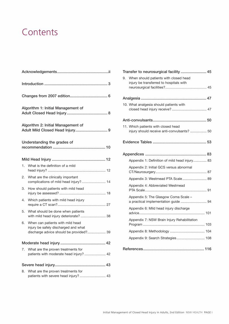
CONTENTS
1 BACKGROUND .................................................................................................................... 1
1.1 About this document ...................................................................................................... 1 1.2 Key definitions ............................................................................................................... 1
2 KEY PRINCIPLES ................................................................................................................ 2
2.1 Mild closed head injury .................................................................................................. 2 2.2 Moderate head injury ..................................................................................................... 2 2.3 Severe head injury ........................................................................................................ 3 2.4 Analgesia ...................................................................................................................... 3 2.5 Anti convulsants ............................................................................................................ 3
3 LIST OF ATTACHMENTS .................................................................................................... 4
Closed Head Injury in Adults - Initial Management
PROCEDURES
PD2012_013 Issue date: February 2012 Page 1 of 4
1 BACKGROUND
1.1 About this document
The NSW Institute of Trauma and Injury Management (ITIM) has updated the Initial Management of Closed Head Injury in Adults clinical practice guideline to reflect the latest evidence based practice for the management of adults with a closed head injury. The guideline is intended for use by clinicians in all facilities which provide initial care to the mild, moderate and severely head injured patient. The practical evidence based recommendations are regarded as a safe and appropriate approach to the acute management of adults with closed head injury. However, as with any clinical guideline the document should be used as a guide, rather than as a complete authoritative statement of procedures. Each LHD must have clear and readily available protocols incorporating the following principles.
1.2 Key definitions
Must – indicates a mandatory action that must be complied with Should – indicates a recommended action that should be followed unless there are sound clinical reasons for taking a different course of action Mild head injury - A patient with an initial GCS score of 14-15 on arrival at hospital following acute blunt head trauma with or without a definite history of loss of consciousness or post traumatic amnesia. Moderate head injury - A patient with an initial GCS score of 9-13 on arrival at hospital following acute blunt head trauma. Severe head injury - A patient with an initial GCS score of 3-8 on arrival at hospital following acute blunt head trauma. Post traumatic amnesia - period of time during which a person is unable to lay down new memories following an injury. Post concussion syndrome- a set of symptoms which are commonly experienced following blunt acute head trauma. The symptoms may include headaches; dizziness; fatigue; memory impairment; poor concentration; mood swings; behavioural changes; sleep disturbances and social dysfunction.
Closed Head Injury in Adults - Initial Management
PROCEDURES
PD2012_013 Issue date: February 2012 Page 2 of 4
2 KEY PRINCIPLES
2.1 Mild closed head injury
Patients with mild closed head injury (initial Glasgow Coma Scale 14-15) should be risk stratified into high and low risk groups based on the presence or absence of specified clinical risk factors. Patients with a mild head injury should be assessed by a process of structured clinical assessment involving a combination of:
• Initial clinical history and examination • Serial clinical observations • CT scanning if clinically assessed as being at increased risk of clinically significant
lesions requiring acute neurosurgical intervention or prolonged observation in hospital.
Patients with persistent acute clinical symptoms (including post traumatic amnesia, disorientation, confusion, drowsiness, dizziness, nausea, vomiting, headache) at four hours post injury require prolonged clinical observation; and a CT scan should be performed (if not already done) to exclude a structural lesion. Where CT scanning is unavailable patients with high risk mild head injury will require either admission for prolonged observation or early transfer for CT scanning depending on clinical assessment of risk. If a patient with mild head injury deteriorates, the priorities are exclusion of other injuries, supportive care of the ABCDEs and early CT scan to identify a neurosurgically significant lesion. If a neurosurgically significant lesion is identified, further management should be discussed with a neurosurgical service.
Mild head injury patients can be safely discharged for home observation after an initial period of in-hospital observation if they meet specified clinical, social and discharge advice criteria.
All patients with mild head injury must be given both verbal and written discharge advice covering signs and symptoms of acute deterioration, when to seek urgent medical attention, lifestyle advice to assist recovery, information about typical post concussion symptoms and reasons for seeking further medical follow up.
2.2 Moderate head injury
Patients who present initially with moderate head injuries should all have an early CT scan and close clinical observation. They should be admitted to hospital for at least 24 hours observation unless they rapidly return to normal, have a normal CT scan and absence of other clinical risk factors.
Closed Head Injury in Adults - Initial Management
PROCEDURES
PD2012_013 Issue date: February 2012 Page 3 of 4
The majority of patients who suffer moderate head injuries will have some degree of cognitive behavioural social sequelae and should be considered for routine follow up with a brain injury rehabilitation service or a neurologist.
2.3 Severe head injury
Resuscitation with adequate oxygenation and fluid resuscitation and the treatment of other immediately life threatening injuries should be the priority for patients with severe head injury followed by the CT identification of focal intracranial lesions requiring acute neurosurgical intervention. Early intubation to prevent hypoxaemia and facilitate management is recommended. A neurosurgical service must be consulted about further management of patients with severe head injury as soon as practical after the initial primary survey and resuscitation. Patients with closed head injury assessed at hospitals without CT scanning facilities should be transferred to the nearest appropriate hospital if there is significant risk of intracranial injury. Transfer of patients to a hospital with CT scanning facilities but without neurosurgical services should be avoided wherever possible.
2.4 Analgesia
Most headaches associated with isolated mild head injury will respond to simple analgesia such as paracetamol. If paracetamol is ineffective as a sole agent then stronger analgesia such as oral opioids or parenteral opioids should not be prescribed to patients with isolated mild head injury unless the need for an initial or repeat CT scan to exclude clinically important intracranial lesions has been considered and a senior clinician has been consulted. Most moderate head injury patients and nearly all severe head injury patients will require titrated intravenous analgesia and sedation for associated injuries, clinical management or intubation. These patients will all require close clinical observation in a high dependency area following initial clinical assessment and CT scanning.
2.5 Anti convulsants
Post traumatic seizures are a recognised complication of closed head injuries with incidence depending largely on severity of injury. Acute post traumatic seizures occurring in hospital require systematic reassessment of the ABCDEs to exclude systemic causes and termination with benzodiazepines if required. Underlying structural lesions should be excluded with CT scan and then the need for prophylactic anti-convulsants considered. Prophylactic anti-convulsants are not indicated for patients with uncomplicated mild head injury. Prophylactic anti-convulsants, such as phenytoin, should be considered in patients with complicated mild head injury or moderate to severe head injury who have specific risk factors that put them at increased risk of seizures. Clinical judgment is required and neurosurgical consultation is advisable.
Closed Head Injury in Adults - Initial Management
PROCEDURES
PD2012_013 Issue date: February 2012 Page 4 of 4
3 LIST OF ATTACHMENTS
1. Initial Management of Closed Head Injury in Adults (2nd Ed) Available as a single document at: http://www.itim.nsw.gov.au/images/3/3d/Closed_Head_Injury_CPG_2nd_Ed_Full_document.pdf
2. Initial Management of Closed Head Injury in Adults (2nd Ed) Summary Document Available as a single document at: http://www.itim.nsw.gov.au/images/d/d0/Closed_Head_Injury_CPG_2nd_Ed_Summary_document.pdf
3. Algorithm: Initial Management of Adult Closed Head Injury Available as a single document at: http://www.itim.nsw.gov.au/images/8/83/Closed_Head_Injury_CPG_2nd_Ed_Algorithm_1.pdf
4. Algorithm: Initial Management of Adult Mild Closed Head Injury Available as a single document at: http://www.itim.nsw.gov.au/images/7/74/Closed_Head_Injury_CPG_2nd_Ed_Algorithm_2.pdf
5. Implementation Checklist
ADULT TRAUMA CLINICAL PRACTICE GUIDELINES
Initial Management of Closed Head Injury in Adults
2nd Edition

NSW Ministry of Health
73 Miller St
NORTH SYDNEY NSW 2060
Tel (02) 9391 9000
Fax (02) 9391 9101
www.health.nsw.gov.au
This work is copyright. It may be reproduced in whole or in part for study
or training purposes subject to the inclusion of an acknowledgement
of the source. It may not be reproduced for commercial usage or sale.
Reproduction for purposes other than those indicated above requires
written permission from the NSW Ministry of Health.
This Clinical Practice Guideline is extracted from PD2012_013 and as a
result, this booklet may be varied, withdrawn or replaced at anytime.
Compliance with information in this booklet is mandatory for NSW Health
© NSW Ministry of Health 2011
SHPN: (SSD) 110189
ISBN: 978-1-74187-587-4
For further copies contact:
NSW Institute of Trauma and Injury Management
PO Box 6314, North Ryde, NSW 2113
Ph: (02) 9887 5726
http://www.itim.nsw.gov.au
Furhter copies of this document can be downloaded from the
NSW Health website http://www.health.nsw.gov.au
November 2011
A revision of this document is due in 2015
Initial Management of Closed Head Injury in Adults, 2nd Edition NSW HEALTH PAGE i
Acknowledgements ................................................ii
Introduction ........................................................... 3
Changes from 2007 edition ................................... 6
Algorithm 1: Initial Management of Adult Closed Head Injury ...................................... 8
Algorithm 2: Initial Management of Adult Mild Closed Head Injury .............................. 9
Understanding the grades of recommendation ................................................. 10
Mild Head Injury .................................................. 12
1. What is the definition of a mild head injury? ............................................................ 12
2. What are the clinically important complications of mild head injury? ......................... 14
3. How should patients with mild head injury be assessed? ................................................ 18
4. Which patients with mild head injury require a CT scan?.................................................. 27
5. What should be done when patients with mild head injury deteriorate? .......................... 38
6. When can patients with mild head injury be safely discharged and what discharge advice should be provided? ................... 39
Moderate head injury .......................................... 42
7. What are the proven treatments for patients with moderate head injury? ...................... 42
Severe head injury ............................................... 43
8. What are the proven treatments for patients with severe head injury? ........................... 43
Transfer to neurosurgical facility ........................ 45
9. When should patients with closed head injury be transferred to hospitals with neurosurgical facilities?........................................... 45
Analgesia ............................................................. 47
10. What analgesia should patients with closed head injury receive? .................................... 47
Anti-convulsants .................................................. 50
11. Which patients with closed head injury should receive anti-convulsants? ................. 50
Evidence Tables .................................................. 53
Appendices ......................................................... 83
Appendix 1: Definition of mild head injury .............. 83
Appendix 2: Initial GCS versus abnormal CT/Neurosurgery ..................................................... 87
Appendix 3: Westmead PTA Scale ......................... 89
Appendix 4: Abbreviated Westmead PTA Scale ................................................................ 91
Appendix 5: The Glasgow Coma Scale – a practical implementation guide ........................... 94
Appendix 6: Mild head injury discharge advice .................................................................... 101
Appendix 7: NSW Brain Injury Rehabilitation Program ................................................................ 103
Appendix 8: Methodology .................................... 104
Appendix 9: Search Strategies ............................. 108
References ......................................................... 116
Contents
PAGE ii Initial Management of Closed Head Injury in Adults, 2nd Edition NSW HEALTH
The following individuals are acknowledged for their contribution to the development of this document.
AuthorDr Duncan Reed, Emergency Physician, Director of Trauma, Gosford Hospital.
Editorial teamMr Glenn Sisson, NSW Trauma Education Manager, NSW Institute of Trauma and Injury Management
Ms Suzanne Davies, Research Fellow, Ambulance Research Institute, Ambulance Service of NSW
Assoc. Prof. Paul Middleton, Director, Ambulance Research Institute, Ambulance Service of NSW
Review GroupDr Rod Bishop, Director Emergency Services, Nepean Hospital
Dr Peter Clark, Clinical Director, NSW ITIM
Dr Scott D’Amours, Trauma Director, Liverpool Hospital
Assoc. Prof. Michael Fearnside AM (Emeritus), Neurosurgeon, Westmead Hospital
Dr Adeline Hodgkinson, Director Brain Injury Rehabilitation Unit, Liverpool Hospital
Mr Peter Mackay, Trauma Clinical Nurse Consultant, Gosford Hospital
Assoc. Prof. Mark Sheridan, Neurosurgeon, Director of Neurosciences, Liverpool Hospital
Dr Declan Stewart, Emergency Physician, Central Coast Health
Dr Alan Tankel, Director Emergency Services, Coffs Harbour Hospital
Ms Nichole Woodward, Emergency Clinical Nurse Consultant, Central Coast Health
Ms Wendy Fischer, Project Manager, Trauma Service, Liverpool Hospital (2nd Ed.)
Ms Merridy Gina, Project Officer, Trauma Service, Liverpool Hospital (2nd Ed.)
Ms Joan Lynch, Project Manager, Trauma Service, Liverpool Hospital (1st Ed.)
Assoc. Prof. Michael Sugrue, Trauma Director, Trauma Service, Liverpool Hospital (1st Ed.)
Ms Gail Long, Secretary, Emergency Department, Gosford Hospital (1st Ed.)
Ms Nikole McCoy, Secretary, Emergency Department, Gosford Hospital (2nd Ed)
Art and Design Unit, Gosford Hospital (1st Ed.)
Acknowledgements
Initial Management of Closed Head Injury in Adults, 2nd Edition NSW HEALTH PAGE 3
Trauma is the leading cause of death and disability in
children and young adults in New South Wales and
closed head injuries cause a significant proportion of this
burden.1, 2 Closed head injury may result in lifelong physical,
cognitive, behavioural and social dysfunction for patients
which in turn may place major social and financial burdens
on their families and society.3 Recent Australian figures
indicate there are approximately 150 patients per 100,000
population admitted to hospital each year with closed head
injuries.3-5 Worldwide figures suggest an incidence range
of 200-350 per 100,000 population per year for patients
with closed head injury with mild head injury accounting
for 80%.6 Despite the fact that closed head injuries are
common, the classification and management of closed
head injures remains surprisingly controversial and subject
to variation in clinical practice.6-10 Due to the large numbers
of patients involved it has been estimated that even
small improvements in closed head injury management
could have significant impact.11 Furthermore, it has been
suggested that the greatest improvements can be made
in the better management of those patients with mild to
moderate head injury rather than those with severe head
injury.12
Much of the controversy that exists about closed head
injury management stems from the combination of a lack
of uniformity in definitions with a paucity of large well
designed studies in the area.11, 13, 14 ‘Head injury’ is typically
used to describe the initial clinical presentation whilst
‘traumatic brain injury’ or “concussion” are used to describe
the subsequent functional outcome. The terms “mild head
injury”, “mild traumatic brain injury” and “concussion” are
largely interchangeable and which term is used depends on
whether you are examining emergency medicine, trauma,
rehabilitation or sports medicine literature. It is difficult to
find two studies that define mild head injury in exactly the
same way so comparison of data can be difficult.6, 8-10, 13
Similarly, comparison of data in moderate to severe head
injury studies is made difficult because controversy exists
about how and when best to apply Glasgow Coma Scale
(GCS) to sedated or intubated patients.15 Perhaps most
significantly there have been very few large prospective
randomised controlled trials of sufficient power and quality
to guide management.11, 13, 14 However, in the past few
years there has been some progress in working toward
uniform definitions and some better quality trials and meta-
analyses have been published.6, 8-10, 15-35
The variety of clinical practice observed worldwide
cannot be explained solely by the lack of uniformity of
definitions and good quality studies. Much of the variation
in management strategies between the USA, Canada,
Europe and Australasia is driven by local issues such as
the availability of resources, the medico-legal environment
and in recent years the concerns about the potential harm
from CT radiation.6, 36, 37 Thus the USA has higher rates of
CT scanning for mild head injuries compared to Canada,
Europe and the UK. Even within countries and within
institutions, considerable variation in practice has been
shown to exist.7, 12, 35, 38 Whilst some variation in clinical
practice is to be expected, the introduction of clinical
practice guidelines can potentially improve care and ensure
adequate access to resources for more isolated areas.6, 35
Furthermore, clinical guidelines can potentially reduce
unnecessary tests and hospital admissions for mild head
injury patients by identifying those patients at low risk of
neurosurgically significant lesions.6, 13, 33-35
Scope of the guideline
The guideline is intended for use by clinicians managing
patients with closed head injury in major and regional
trauma services, and urban and rural hospitals. The
guideline is concerned with the initial care of the mild,
moderate and severely head injured patient. The guideline
will make evidence based recommendations on the
diagnosis, resuscitation, and disposal of patients with closed
head injuries.
Introduction
PAGE 4 Initial Management of Closed Head Injury in Adults, 2nd Edition NSW HEALTH
The initial management plan for adults is based upon
recommendations to be followed subject to the clinician's
judgement in each case.
The recommendations however, are not prescriptive nor
are they rigid procedural paths. It is recognised that the
recommendations may not suit all patients in all clinical
situations. They are intended to provide a clinically practical
approach to the initial management of closed head injuries
based on the current best available evidence. However, as
with all guidelines, it should be remembered that they are a
clinical tool and should not replace clinical judgement. The
guideline relies on individual clinicians to decipher the needs
of individual patients.
All recommendations regarding pre-hospital care should be
read and considered in conjunction with the Ambulance
Service of NSW.
Guidelines for the initial management of head injury in
children can be found at http://www.health.nsw.gov.au/
policies/pd/2011/pdf/PD2011_024.pdf
Aims and objectives
The guideline is intended to assist clinicians throughout
NSW in delivering optimal care to patients with closed head
injury. It aims to provide information to support clinical
decision making, rather than dictate what decisions should
be made.
The broad objectives of the guideline are to reduce
morbidity and mortality in adult patients with closed head
injury by providing clinicians with practical evidence based
recommendations to assist them in managing such patients.
It is also hoped that the guidelines may prevent unnecessary
diagnostic tests and hospital admissions especially in the
mild head injury group.
The process of constructing the guideline began
with the clinicians on the Trauma Clinical Guidelines
Committee posing a series of questions about the initial
management of closed head injuries. The final questions
were derived from the guideline priority areas identified
by the committee; that is, the management of mild head
injuries and the timing of transfer of patients with closed
head injury from centres with limited resources. The
initial management of patients with moderate to severe
head injury was felt to be less controversial. This edition
also includes recommendations in relation to the use of
analgesia and anti-convulsants.
An extensive description of the methodology used for this
guideline can be found at Appendix 8, together with the
search terms used at Appendix 9.
The clinical questions addressed:
1 What is the defi nition of a mild head injury?
2 What are the clinically important complications of mild head injury?
3 How should patients with mild head injury be assessed?
4 Which patients with mild head injury require a CT scan?
5 What should be done with high risk mild head injury patients when CT scan is unavailable?
6 What should be done when patients with mild head injury deteriorate?
7 When can patients with mild head injury be safely discharged?
8 What discharge advice should be provided?
9 What are the proven treatments for patients with moderate head injury?
10 What are the proven treatments for patients with severe head injury?
11 When should patients with closed head injury be transferred to hospitals with neurosurgical facilities?
12 What analgesia should patients with closed head injury receive?
13 Which patients with closed head injury should receive anti-convulsants?
Initial Management of Closed Head Injury in Adults, 2nd Edition NSW HEALTH PAGE 5
Defining closed head injury
This guideline uses the terms ‘closed head injury’ and ‘mild,
moderate or severe head injury’ to identify and classify
patients on arrival to hospital. The outcome following
presentation with a ‘closed head injury’ will vary from rapid
complete recovery to a mixture of structural lesions and
functional deficits ranging from trivial to life threatening.
The terms “concussion” and “traumatic brain injury” refer
to the patient outcome following their initial presentation
with a “closed head injury” and are retrospective
diagnoses. Important functional deficits following ‘closed
head injury’ range from post concussion symptoms and
post traumatic amnesia to a variety of disabling persistent
physical-cognitive-behavioural-social sequelae.
Many patients who suffer a “mild head injury” will have
“mild concussion symptoms” or “mild traumatic brain injury
symptoms”. If these acute “concussion” symptoms persist
beyond the first few hours they are usually referred to as
“post concussion symptoms”. The term “post concussion
symptoms” is used to describe the clinical symptoms of
mild brain injury that mild head injury patients may suffer
for a few days to weeks following their injury. In the
situation where multiple post concussion symptoms persist
for several months they are called a “post concussion
syndrome”
As this guideline concentrates on the initial management
of the patients presenting to hospital, it was felt that the
term ‘head injury’ was more relevant to the initial clinical
presentation than the term ‘traumatic brain injury’ that
essentially refers to the subsequent functional outcome. It
was also felt that the clinicians at whom this guideline is
aimed would be far more familiar and comfortable with
using the term ‘head injury.’ The definition of closed head
injury is further discussed in Question 1.
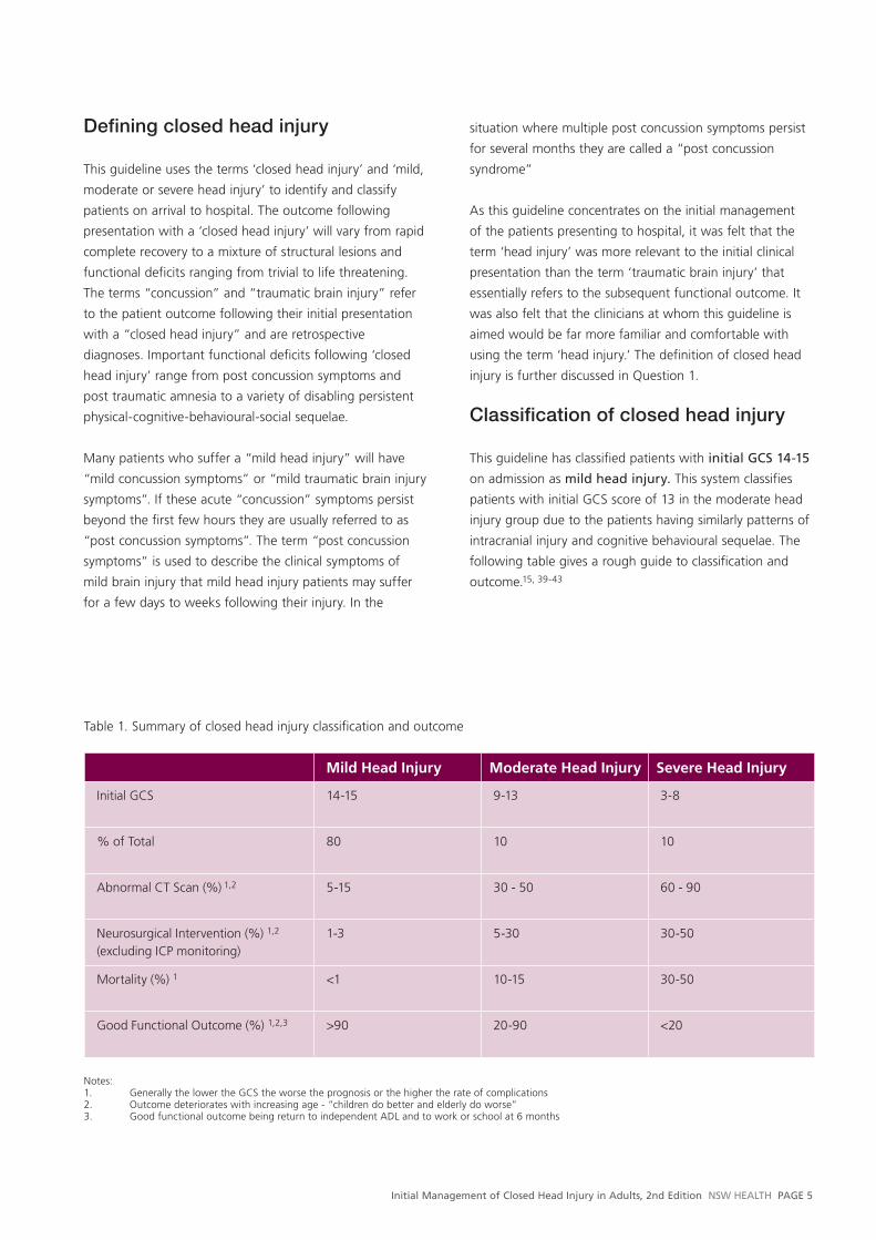
Classification of closed head injury
This guideline has classified patients with initial GCS 14-15
on admission as mild head injury. This system classifies
patients with initial GCS score of 13 in the moderate head
injury group due to the patients having similarly patterns of
intracranial injury and cognitive behavioural sequelae. The
following table gives a rough guide to classification and
outcome.15, 39-43
Table 1. Summary of closed head injury classification and outcome
Notes:1. Generally the lower the GCS the worse the prognosis or the higher the rate of complications 2. Outcome deteriorates with increasing age - “children do better and elderly do worse”3. Good functional outcome being return to independent ADL and to work or school at 6 months
Mild Head Injury Moderate Head Injury Severe Head Injury
Initial GCS 14-15 9-13 3-8
% of Total 80 10 10
Abnormal CT Scan (%) 1,2 5-15 30 - 50 60 - 90
Neurosurgical Intervention (%) 1,2
(excluding ICP monitoring)1-3 5-30 30-50
Mortality (%) 1 <1 10-15 30-50
Good Functional Outcome (%) 1,2,3 >90 20-90 <20
PAGE 6 Initial Management of Closed Head Injury in Adults, 2nd Edition NSW HEALTH
Background
The first edition of this guideline was written in 2005
using evidence available until December 2004. The aim of
this new edition is to review the evidence published since
December 2004 and to provide some additional information
on specific topics including the role of anticonvulsants and
analgesics in the management of closed head injury.
The aim of the original guideline was to provide a clinically
practical evidence based guideline that summarised
the initial management of adult closed head injury. It
was piloted by the NSW Institute of Trauma and Injury
Management (ITIM) and then formally adopted and
published by NSW Health in January 2007. There was a
conscious effort by the initial guideline team to provide a
clinically practical document with clinically useful resources
such as algorithms, summaries and discharge advice sheets
backed up by a detailed evidence review. The guideline
team has continued the same principles for this update,
incorporating feedback from clinicians to improve the
guideline. The algorithms and mild head injury discharge
sheets have been revised to reflect the changes in the body
of the guideline and the feedback received.
The guideline team would emphasise that this guideline is a
clinical tool designed to assist clinicians and should be used
to assist rather than replace the clinical judgement of an
experienced clinician caring for an individual patient.
The information provided is based on the best available
information at the time of writing, which is May 2010.
These guidelines will be updated every five years and
consider new evidence as it becomes available.
New evidence
Since 2004 there have been many new studies and
guidelines published about the management of closed
head injury. There have been some advances in our
understanding of the assessment and treatment of
closed head injury but these have been incremental and
evolutionary rather than revolutionary. The basic principles
of management of closed head injury remain the same in
2010 as they were five years ago.
The following section briefly outlines the most significant
advances in knowledge from the recent literature
incorporated in this update.
Definition of mild head injury
■ Recent literature emphasises that significant intracranial
injury may occur without loss of consciousness or
amnesia■ Patients with initial GCS 13 have a significantly higher
rate of intracranial injury and should not be considered
as having mild head injury
Clinically important complications of mild head injury
■ Recent literature emphasises that mild post concussion
symptoms are common and that patients should
receive appropriate discharge advice to assist recovery■ Acute neurosurgical complications are uncommon but
important to identify
Assessment of patients with mild head injury
■ Recent literature emphasises that if structured clinical
assessment indicates the risk of intracranial injury is
low, the routine use of CT scanning is not warranted
and is potentially harmful.■ Structured clinical assessment should include initial
clinical history and examination, serial clinical
observations and clinical risk factor assessment to
determine the need for CT scanning■ A variety of clinical decision rules have been developed
to determine which patients are at higher risk of
intracranial injury and require CT scanning. However,
they all require that the clinician is familiar with their
inclusion / exclusion criteria and should be used as
tools to support clinical decision making, rather than
dictate management■ Post traumatic amnesia testing in the emergency
Changes from 2007 edition
Initial Management of Closed Head Injury in Adults, 2nd Edition NSW HEALTH PAGE 7
department, eg Abbreviated Westmead PTA Scale
(A-WPTAS) can be useful in identifying patients with
cognitive impairment at increased risk of structural
lesions and post concussion symptoms.
Indications for CT scan for mild head injury
■ Recent literature emphasises that patients can be risk
stratified according to clinical risk factors and clinical
decision rules. Patients who are classified as high risk
should have CT scans to exclude clinically important
intracranial lesions■ Significant head injuries can occur without loss of
consciousness or amnesia and that the absence of
these features should not be used to determine the
need for CT scanning.■ Persistent abnormal mental status manifested by either
abnormal GCS or abnormal alertness, behaviour or
cognition is a strong indication for CT scanning ■ Known coagulopathy and particularly supra-
therapeutic anticoagulation are significant risk factors
for intracranial injury and that these patients should
have early CT scans and be considered for reversal of
anticoagulation■ There have been several very large studies addressing
this issue in the paediatric literature that have come up
with very similar risk factors to the adult literature and
have also confirmed that it is safe to discharge low risk
patients without CT scanning.
Acute neurological deterioration
■ Recommendations essentially unchanged■ Previously covered within guideline but now given
separate question
Discharge of patients with mild head injury
■ Recent literature emphasises that patients can be safely
discharged for home observation if structured clinical
assessment reveals no clinical risk factors indicating the
need for CT scanning or following a normal CT scan if
indicated.■ Deterioration of mild head injury patients following
a normal CT scan is rare. Caution is advised for
patients with known coagulopathy and elderly patients
where the risk of a delayed subdural haemorrhage is
increased.
Discharge advice for patients with mild head injury
■ New section to emphasise importance of discharge
advice■ Recent literature emphasises that all patients with
mild head injury should be given both verbal and
written discharge advice covering symptoms and signs
of acute deterioration, when to seek urgent medical
attention, lifestyle advice to assist recovery, information
about typical post concussion symptoms and reasons
for seeking further medical follow up. As with all
discharge advice this should be time specific and action
specific.■ An improved version of the original mild head injury
advice sheet associated with this guideline has been
developed and is now available in several languages.
Initial management of moderate head injury
(GCS 9-13)
■ Recommendations essentially unchanged
Initial management of severe head injury
(GCS 3-8)
■ Recommendations essentially unchanged
Transfer of patients with closed head injury to
hospitals with neurosurgical facilities
■ Recommendations essentially unchanged
Analgesia for closed head injury
■ New section
Anticonvulsants for closed head injury
■ New section
PAGE 8 Initial Management of Closed Head Injury in Adults, 2nd Edition NSW HEALTH
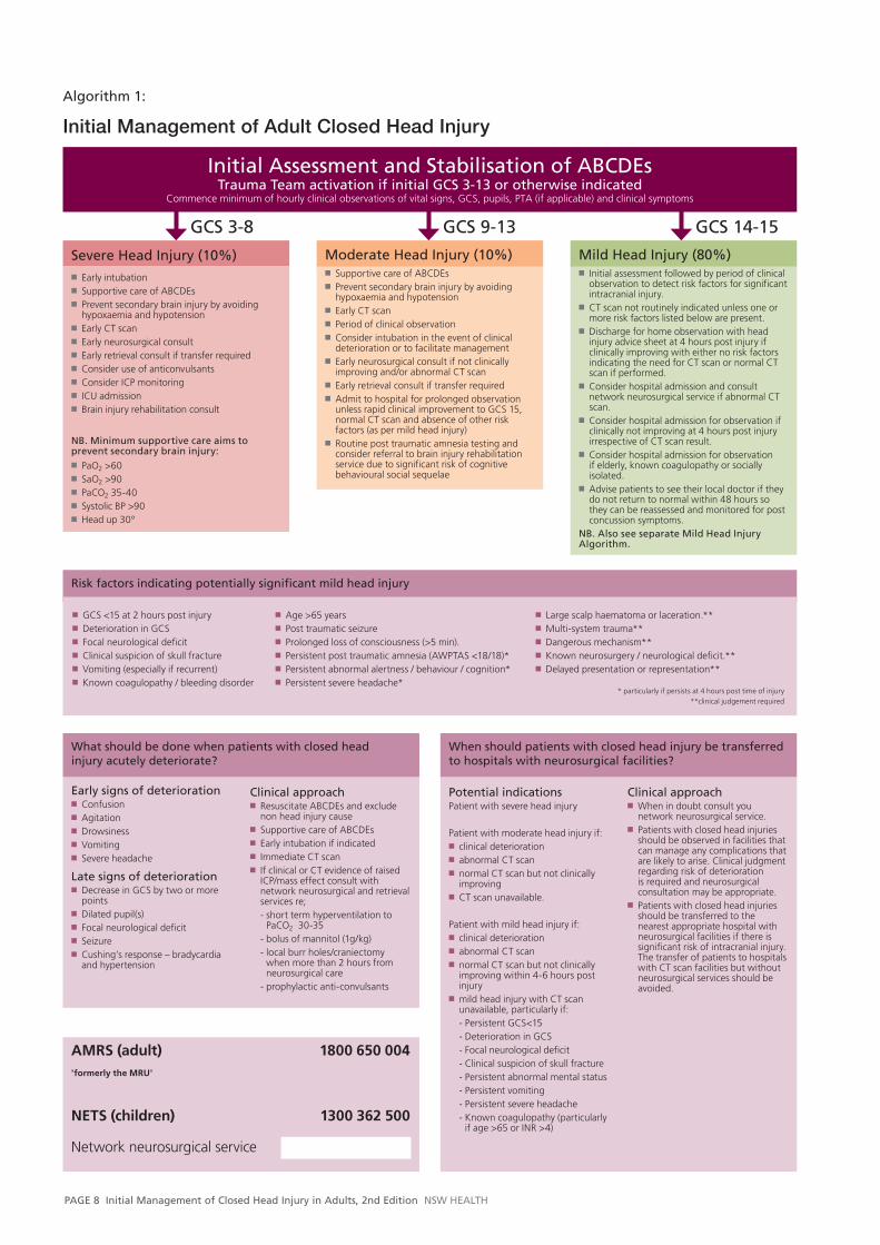
Risk factors indicating potentially signifi cant mild head injury
Initial Management of Adult Closed Head Injury
Severe Head Injury (10%)■ Early intubation■ Supportive care of ABCDEs ■ Prevent secondary brain injury by avoiding
hypoxaemia and hypotension ■ Early CT scan ■ Early neurosurgical consult■ Early retrieval consult if transfer required■ Consider use of anticonvulsants ■ Consider ICP monitoring ■ ICU admission■ Brain injury rehabilitation consult
NB. Minimum supportive care aims to prevent secondary brain injury:■ PaO2 >60■ SaO2 >90■ PaCO2 35-40■ Systolic BP >90■ Head up 30º
Mild Head Injury (80%)■ Initial assessment followed by period of clinical
observation to detect risk factors for significant intracranial injury.
■ CT scan not routinely indicated unless one or more risk factors listed below are present.
■ Discharge for home observation with head injury advice sheet at 4 hours post injury if clinically improving with either no risk factors indicating the need for CT scan or normal CT scan if performed.
■ Consider hospital admission and consult network neurosurgical service if abnormal CT scan.
■ Consider hospital admission for observation if clinically not improving at 4 hours post injury irrespective of CT scan result.
■ Consider hospital admission for observation if elderly, known coagulopathy or socially isolated.
■ Advise patients to see their local doctor if they do not return to normal within 48 hours so they can be reassessed and monitored for post concussion symptoms.
NB. Also see separate Mild Head Injury Algorithm.
Moderate Head Injury (10%)■ Supportive care of ABCDEs■ Prevent secondary brain injury by avoiding
hypoxaemia and hypotension ■ Early CT scan ■ Period of clinical observation■ Consider intubation in the event of clinical
deterioration or to facilitate management ■ Early neurosurgical consult if not clinically
improving and/or abnormal CT scan■ Early retrieval consult if transfer required■ Admit to hospital for prolonged observation
unless rapid clinical improvement to GCS 15, normal CT scan and absence of other risk factors (as per mild head injury)
■ Routine post traumatic amnesia testing and consider referral to brain injury rehabilitation service due to significant risk of cognitive behavioural social sequelae
GCS 3-8 GCS 9-13 GCS 14-15
Initial Assessment and Stabilisation of ABCDEsTrauma Team activation if initial GCS 3-13 or otherwise indicated
Commence minimum of hourly clinical observations of vital signs, GCS, pupils, PTA (if applicable) and clinical symptoms
■ GCS <15 at 2 hours post injury■ Deterioration in GCS■ Focal neurological deficit■ Clinical suspicion of skull fracture ■ Vomiting (especially if recurrent)■ Known coagulopathy / bleeding disorder
What should be done when patients with closed head injury acutely deteriorate?
Early signs of deterioration■ Confusion■ Agitation■ Drowsiness■ Vomiting■ Severe headache
Late signs of deterioration■ Decrease in GCS by two or more
points■ Dilated pupil(s)■ Focal neurological deficit■ Seizure■ Cushing’s response – bradycardia
and hypertension
Clinical approach■ Resuscitate ABCDEs and exclude
non head injury cause ■ Supportive care of ABCDEs■ Early intubation if indicated■ Immediate CT scan■ If clinical or CT evidence of raised
ICP/mass effect consult with network neurosurgical and retrieval services re;
� - short term hyperventilation to PaCO2 30-35
� - bolus of mannitol (1g/kg)� - local burr holes/craniectomy
when more than 2 hours from neurosurgical care
� - prophylactic anti-convulsants
When should patients with closed head injury be transferred to hospitals with neurosurgical facilities?
Potential indicationsPatient with severe head injury
Patient with moderate head injury if:■ clinical deterioration■ abnormal CT scan■ normal CT scan but not clinically
improving■ CT scan unavailable.
Patient with mild head injury if:■ clinical deterioration■ abnormal CT scan■ normal CT scan but not clinically
improving within 4-6 hours post injury
■ mild head injury with CT scan unavailable, particularly if:
� - Persistent GCS<15 � - Deterioration in GCS � - Focal neurological deficit� - Clinical suspicion of skull fracture� - Persistent abnormal mental status � - Persistent vomiting� - Persistent severe headache � - Known coagulopathy (particularly
if age >65 or INR >4)
Clinical approach■ When in doubt consult you
network neurosurgical service.■ Patients with closed head injuries
should be observed in facilities that can manage any complications that are likely to arise. Clinical judgment regarding risk of deterioration is required and neurosurgical consultation may be appropriate.
■ Patients with closed head injuries should be transferred to the nearest appropriate hospital with neurosurgical facilities if there is significant risk of intracranial injury. The transfer of patients to hospitals with CT scan facilities but without neurosurgical services should be avoided.
AMRS (adult) 1800 650 004'formerly the MRU'
NETS (children) 1300 362 500
Network neurosurgical service
Algorithm 1:
■ Age >65 years ■ Post traumatic seizure■ Prolonged loss of consciousness (>5 min).■ Persistent post traumatic amnesia (AWPTAS <18/18)* ■ Persistent abnormal alertness / behaviour / cognition*■ Persistent severe headache*
■ Large scalp haematoma or laceration.**■ Multi-system trauma**■ Dangerous mechanism**■ Known neurosurgery / neurological deficit.**■ Delayed presentation or representation**
* particularly if persists at 4 hours post time of injury**clinical judgement required
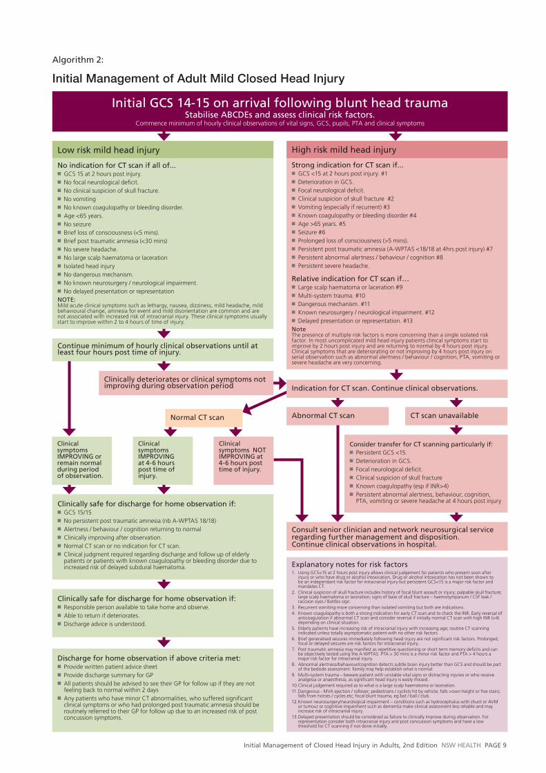
Initial Management of Closed Head Injury in Adults, 2nd Edition NSW HEALTH PAGE 9
Clinically safe for discharge for home observation if:■ Responsible person available to take home and observe.■ Able to return if deteriorates.■ Discharge advice is understood.
Consult senior clinician and network neurosurgical service regarding further management and disposition. Continue clinical observations in hospital.
Consider transfer for CT scanning particularly if:■ Persistent GCS <15.■ Deterioration in GCS.■ Focal neurological deficit.■ Clinical suspicion of skull fracture ■ Known coagulopathy (esp if INR>4)■ Persistent abnormal alertness, behaviour, cognition,
PTA, vomiting or severe headache at 4 hours post injury
Abnormal CT scan CT scan unavailable
Clinical symptoms IMPROVING or remain normal during period of observation.
Clinical symptomsIMPROVING at 4-6 hours post time of injury.
Clinical symptoms NOT IMPROVING at 4-6 hours post time of injury.
Clinically safe for discharge for home observation if:■ GCS 15/15 ■ No persistent post traumatic amnesia (nb A-WPTAS 18/18)■ Alertness / behaviour / cognition returning to normal ■ Clinically improving after observation.■ Normal CT scan or no indication for CT scan.■ Clinical judgment required regarding discharge and follow up of elderly
patients or patients with known coagulopathy or bleeding disorder due to increased risk of delayed subdural haematoma.
Continue minimum of hourly clinical observations until at least four hours post time of injury.
Clinically deteriorates or clinical symptoms not improving during observation period Indication for CT scan. Continue clinical observations.
Normal CT scan
Explanatory notes for risk factors1. Using GCS<15 at 2 hours post injury allows clinical judgement for patients who present soon after
injury or who have drug or alcohol intoxication. Drug or alcohol intoxication has not been shown to be an independant risk factor for intracranial injury but persistent GCS<15 is a major risk factor and mandates CT.
2. Clinical suspicion of skull fracture includes history of focal blunt assault or injury; palpable skull fracture; large scalp haematoma or laceration; signs of base of skull fracture – haemotympanum / CSF leak / raccoon eyes / Battles sign.
3. Recurrent vomiting more concerning than isolated vomiting but both are indications.4. Known coagulopathy is both a strong indication for early CT scan and to check the INR. Early reversal of
anticoagulation if abnormal CT scan and consider reversal if initially normal CT scan with high INR (>4) depending on clinical situation.
5. Elderly patients have increasing risk of intracranial injury with increasing age; routine CT scanning indicated unless totally asymptomatic patient with no other risk factors.
6. Brief generalised seizures immediately following head injury are not significant risk factors. Prolonged, focal or delayed seizures are risk factors for intracranial injury.
7. Post traumatic amnesia may manifest as repetitive questioning or short term memory deficits and can be objectively tested using the A-WPTAS. PTA > 30 mins is a minor risk factor and PTA > 4 hours a major risk factor for intracranial injury.
8. Abnormal alertness/behaviour/cognition detects subtle brain injury better than GCS and should be part of the bedside assessment. Family may help establish what is normal.
9. Multi-system trauma – beware patient with unstable vital signs or distracting injuries or who receive analgesia or anaesthesia, as significant head injury is easily missed.
10. Clinical judgement required as to what is a large scalp haematoma or laceration.11. Dangerous - MVA ejection / rollover; pedestrians / cyclists hit by vehicle; falls >own height or five stairs;
falls from horses / cycles etc; focal blunt trauma, eg bat / ball / club.12. Known neurosurgery/neurological impairment – conditions such as hydrocephalus with shunt or AVM
or tumour or cognitive impairment such as dementia make clinical assessment less reliable and may increase risk of intracranial injury.
13. Delayed presentation should be considered as failure to clinically improve during observation. For representation consider both intracranial injury and post concussion symptoms and have a low threshold for CT scanning if not done initially.
Low risk mild head injury
No indication for CT scan if all of...■ GCS 15 at 2 hours post injury.■ No focal neurological deficit.■ No clinical suspicion of skull fracture. ■ No vomiting ■ No known coagulopathy or bleeding disorder.■ Age <65 years. ■ No seizure■ Brief loss of consciousness (<5 mins). ■ Brief post traumatic amnesia (<30 mins)■ No severe headache.■ No large scalp haematoma or laceration■ Isolated head injury ■ No dangerous mechanism.■ No known neurosurgery / neurological impairment.■ No delayed presentation or representationNOTE:Mild acute clinical symptoms such as lethargy, nausea, dizziness, mild headache, mild behavioural change, amnesia for event and mild disorientation are common and are not associated with increased risk of intracranial injury. These clinical symptoms usually start to improve within 2 to 4 hours of time of injury.
Discharge for home observation if above criteria met:■ Provide written patient advice sheet■ Provide discharge summary for GP■ All patients should be advised to see their GP for follow up if they are not
feeling back to normal within 2 days■ Any patients who have minor CT abnormalities, who suffered significant
clinical symptoms or who had prolonged post traumatic amnesia should be routinely referred to their GP for follow up due to an increased risk of post concussion symptoms.
High risk mild head injury
Strong indication for CT scan if...■ GCS <15 at 2 hours post injury. #1■ Deterioration in GCS.■ Focal neurological deficit.■ Clinical suspicion of skull fracture #2■ Vomiting (especially if recurrent) #3■ Known coagulopathy or bleeding disorder #4■ Age >65 years. #5■ Seizure #6■ Prolonged loss of consciousness (>5 mins). ■ Persistent post traumatic amnesia (A-WPTAS <18/18 at 4hrs post injury) #7■ Persistent abnormal alertness / behaviour / cognition #8■ Persistent severe headache.
Relative indication for CT scan if…■ Large scalp haematoma or laceration #9■ Multi-system trauma. #10■ Dangerous mechanism. #11■ Known neurosurgery / neurological impairment. #12■ Delayed presentation or representation. #13Note The presence of multiple risk factors is more concerning than a single isolated risk factor. In most uncomplicated mild head injury patients clinical symptoms start to improve by 2 hours post injury and are returning to normal by 4 hours post injury. Clinical symptoms that are deteriorating or not improving by 4 hours post injury on serial observation such as abnormal alertness / behaviour / cognition, PTA, vomiting or severe headache are very concerning.
Initial Management of Adult Mild Closed Head Injury
Initial GCS 14-15 on arrival following blunt head traumaStabilise ABCDEs and assess clinical risk factors.
Commence minimum of hourly clinical observations of vital signs, GCS, pupils, PTA and clinical symptoms
Algorithm 2:

PAGE 10 Initial Management of Closed Head Injury in Adults, 2nd Edition NSW HEALTH
Strength of recommendations
This guideline uses the National Health and Medical
Research Council's (NHMRC) overall grades of
recommendation to indicate the strength of the body of
evidence underpinning each recommendation. The body
of evidence reflects the evidence components of all the
studies relevant to each recommendation. The evidence
components are assessed according to the NHMRC body
of evidence matrix (Table 2). The overall grade of the
recommendation is determined based on a summation of
the rating for each individual component of the body of
evidence. Please note that a recommendation cannot be
graded A or B unless the evidence base and consistency of
the evidence are both rated A or B.44
Understanding the grades of recommendation
Table 2: Body of evidence matrix44
ComponentsA B C D
Excellent Good Satisfactory Poor
Evidence base one or more level I studies with a low risk of bias or several level II studies with a low risk of bias
one or two level II studies with a low risk of bias or a SR/several level III studies with a low risk of bias
one or two level III studies with a low risk of bias, or level I or II studies with a moderate risk of bias
level IV studies, or level I to III studies/SRs with a high risk of bias
Consistency all studies consistent most studies consistent and inconsistency may be explained
some inconsistency refl ecting genuine uncertainty around clinical question
evidence is inconsistent
Clinical Impact very large substantial moderate slight or restricted
Generalisability population/s studied in body of evidence are the same as the target population for the guideline
population/s studied in the body of evidence are similar to the target population for the guideline
population/s studied in body of evidence differ to target population for guideline but it is clinically sensible to apply this evidence to target population
population/s studied in body of evidence differ to target population and hard to judge whether it is sensible to generalise to target population
Applicability directly applicable to Australian healthcare context
applicable to Australian healthcare context with few caveats
probably applicable to Australian healthcare context with some caveats
not applicable to Australian healthcare context
Initial Management of Closed Head Injury in Adults, 2nd Edition NSW HEALTH PAGE 11
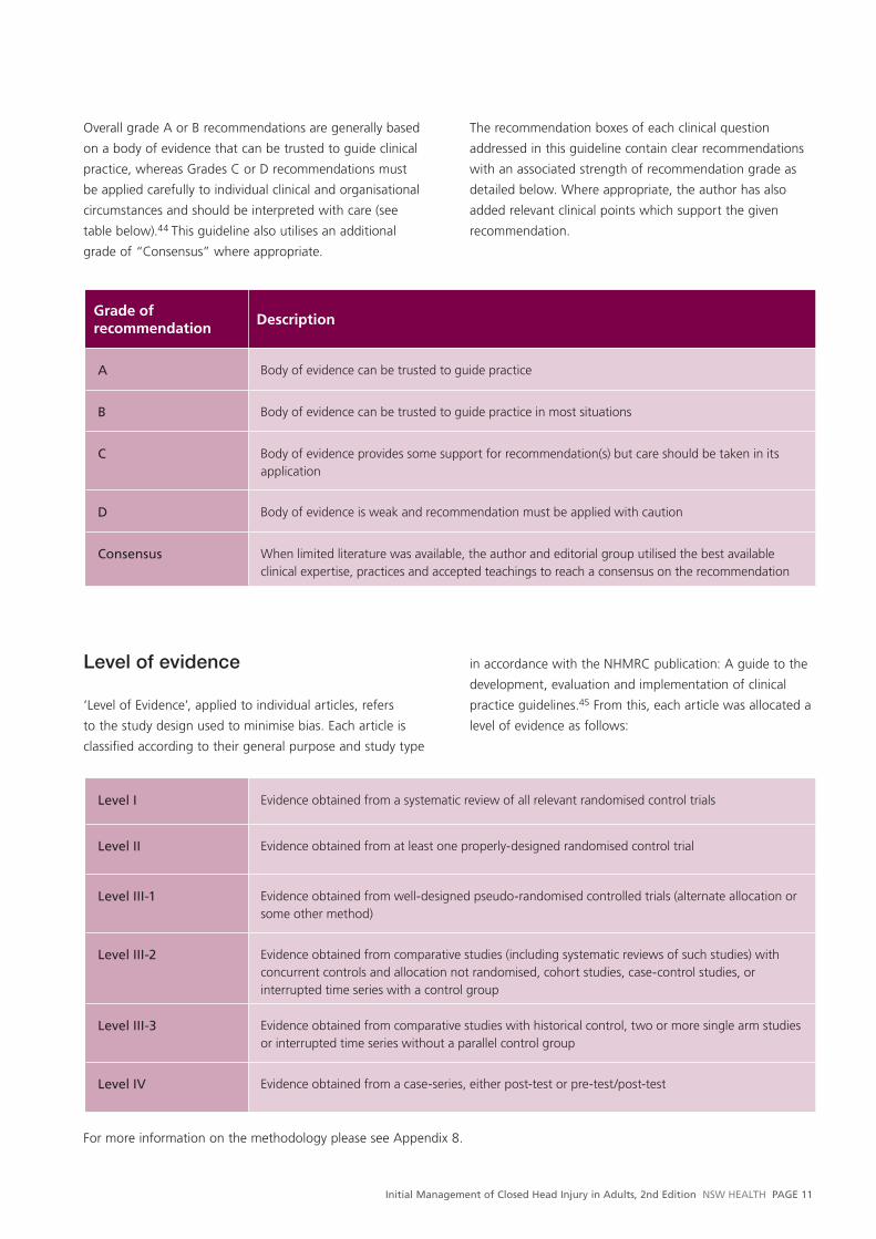
Overall grade A or B recommendations are generally based
on a body of evidence that can be trusted to guide clinical
practice, whereas Grades C or D recommendations must
be applied carefully to individual clinical and organisational
circumstances and should be interpreted with care (see
table below).44 This guideline also utilises an additional
grade of “Consensus” where appropriate.
The recommendation boxes of each clinical question
addressed in this guideline contain clear recommendations
with an associated strength of recommendation grade as
detailed below. Where appropriate, the author has also
added relevant clinical points which support the given
recommendation.
Level of evidence
‘Level of Evidence’, applied to individual articles, refers
to the study design used to minimise bias. Each article is
classified according to their general purpose and study type
in accordance with the NHMRC publication: A guide to the
development, evaluation and implementation of clinical
practice guidelines.45 From this, each article was allocated a
level of evidence as follows:
For more information on the methodology please see Appendix 8.
Grade of recommendation
Description
A Body of evidence can be trusted to guide practice
B Body of evidence can be trusted to guide practice in most situations
C Body of evidence provides some support for recommendation(s) but care should be taken in its application
D Body of evidence is weak and recommendation must be applied with caution
Consensus When limited literature was available, the author and editorial group utilised the best available clinical expertise, practices and accepted teachings to reach a consensus on the recommendation
Level I Evidence obtained from a systematic review of all relevant randomised control trials
Level II Evidence obtained from at least one properly-designed randomised control trial
Level III-1 Evidence obtained from well-designed pseudo-randomised controlled trials (alternate allocation or some other method)
Level III-2 Evidence obtained from comparative studies (including systematic reviews of such studies) with concurrent controls and allocation not randomised, cohort studies, case-control studies, or interrupted time series with a control group
Level III-3 Evidence obtained from comparative studies with historical control, two or more single arm studies or interrupted time series without a parallel control group
Level IV Evidence obtained from a case-series, either post-test or pre-test/post-test
PAGE 12 Initial Management of Closed Head Injury in Adults, 2nd Edition NSW HEALTH
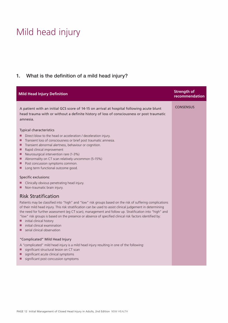
Mild Head Injury Defi nitionStrength of recommendation
A patient with an initial GCS score of 14-15 on arrival at hospital following acute blunt
head trauma with or without a definite history of loss of consciousness or post traumatic
amnesia.
Typical characteristics
■ Direct blow to the head or acceleration / deceleration injury.■ Transient loss of consciousness or brief post traumatic amnesia.■ Transient abnormal alertness, behaviour or cognition.■ Rapid clinical improvement■ Neurosurgical intervention rare (1-3%)■ Abnormality on CT scan relatively uncommon (5-15%)■ Post concussion symptoms common.■ Long term functional outcome good.
Specifi c exclusions:
■ Clinically obvious penetrating head injury.■ Non-traumatic brain injury.
Risk Stratifi cationPatients may be classifi ed into “high” and “low” risk groups based on the risk of suffering complications of their mild head injury. This risk stratifi cation can be used to assist clinical judgement in determining the need for further assessment (eg CT scan), management and follow up. Stratifi cation into “high” and “low” risk groups is based on the presence or absence of specifi ed clinical risk factors identifi ed by:■ initial clinical history■ initial clinical examination■ serial clinical observation
“Complicated” Mild Head Injury
A “complicated” mild head injury is a mild head injury resulting in one of the following:■ signifi cant structural lesion on CT scan■ signifi cant acute clinical symptoms■ signifi cant post concussion symptoms
CONSENSUS
1. What is the definition of a mild head injury?
Mild head injury
Initial Management of Closed Head Injury in Adults, 2nd Edition NSW HEALTH PAGE 13
Recently published studies and guidelines use a variety of
criteria to define mild head injury, which is variably referred
to as either mild head injury or mild traumatic brain injury.6,
8-10, 13, 31-35, 46-48 The most common variations concern
the initial classification according to GCS and different
requirements for loss of consciousness and post traumatic
amnesia (summarised in Appendix 1). This variation in the
literature makes comparison between studies difficult.
The main reason for this variability is a uniform desire to
identify those patients at higher risk of intracranial injury in
what is a heterogenous but essentially low risk group. There
is ample evidence to suggest that patients with an initial
GCS of 13 should be considered as part of the moderate
head injury group due to the frequency of intracranial
lesions (25-38%) and cognitive-behavioural-social sequelae
(see Evidence Table 1 and Appendix 2).9, 36, 43, 49-56
Since 2004 the adult literature has clearly identified that
patients may sustain significant head injuries without loss
of consciousness or post traumatic amnesia.9, 33, 43, 47,
57-59 Therefore, the presence of loss of consciousness or
post traumatic amnesia should not be used to define mild
head injury or guide management. In 2008 Jagoda et al,
representing the American College of Emergency Physicians
/ Centre for Disease Control, updated their definition of
mild head injury to reflect the change in the evidence and
now include patients with GCS 14 on initial assessment
and have eliminated loss of consciousness or post traumatic
amnesia as necessary inclusion criteria.9
Further risk stratification of mild head injury is then
dependent on the presence of associated risk factors
and different authors have different approaches. The
approaches of some of the best quality studies and
guidelines are summarised in Appendix 1. It is interesting
to note that when all the initial GCS criteria, inclusion/
exclusion criteria and sub-classification systems are all
taken into account, that the findings are very similar. These
findings are that mild head injury is a heterogenous group
with patients at higher risk of increased intracranial injury
identified by persistently abnormal GCS and certain other
risk factors.1, 6, 8-10, 13, 31-36, 47-54, 56-81
It is important to recognise that these risk factors for
intracranial injury do not necessarily predict the risk of
post concussive symptoms which are the more common
complication of mild head injury. It is important that
doctors, patients and their families understand that the
absence of a structural lesion on CT scan following a mild
head injury does not exclude the possibility of significant
cognitive-behavioural-social sequelae.9, 82
The recent paediatric literature has come up with similar
definitions for mild head injury to the adult literature and
identified persistently abnormal GCS or mental status
and other specified risk factors as the major indicators of
intracranial injury.83-87
PAGE 14 Initial Management of Closed Head Injury in Adults, 2nd Edition NSW HEALTH
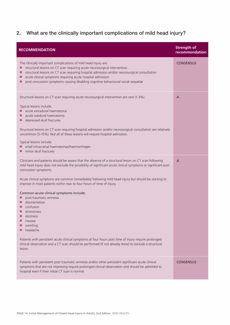
RECOMMENDATIONStrength of recommendation
The clinically important complications of mild head injury are:■ structural lesions on CT scan requiring acute neurosurgical intervention■ structural lesions on CT scan requiring hospital admission and/or neurosurgical consultation■ acute clinical symptoms requiring acute hospital admission■ post concussion symptoms causing disabling cognitive behavioural social sequelae
CONSENSUS
Structural lesions on CT scan requiring acute neurosurgical intervention are rare (1-3%).
Typical lesions include:■ acute extradural haematoma■ acute subdural haematoma■ depressed skull fractures
Structural lesions on CT scan requiring hospital admission and/or neurosurgical consultation are relatively uncommon (5-15%). Not all of these lesions will require hospital admission.
Typical lesions include:■ small intracranial haematomas/haemorrhages■ minor skull fractures
A
Clinicians and patients should be aware that the absence of a structural lesion on CT scan following mild head injury does not exclude the possibility of signifi cant acute clinical symptoms or signifi cant post concussion symptoms.
Acute clinical symptoms are common immediately following mild head injury but should be starting to improve in most patients within two to four hours of time of injury.
Common acute clinical symptoms include:■ post traumatic amnesia■ disorientation■ confusion■ drowsiness■ dizziness■ nausea■ vomiting■ headache
Patients with persistent acute clinical symptoms at four hours post time of injury require prolonged clinical observation and a CT scan should be performed (if not already done) to exclude a structural lesion.
A
Patients with persistent post traumatic amnesia and/or other persistent signifi cant acute clinical symptoms that are not improving require prolonged clinical observation and should be admitted to hospital even if their initial CT scan is normal.
CONSENSUS
2. What are the clinically important complications of mild head injury?
Initial Management of Closed Head Injury in Adults, 2nd Edition NSW HEALTH PAGE 15
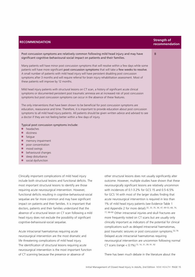
Post concussion symptoms are relatively common following mild head injury and may have signifi cant cognitive-behavioural-social impact on patients and their families. Many patients will have minor post concussion symptoms that will resolve within a few days while some patients will have more signifi cant post concussion symptoms that will take a few weeks to resolve.A small number of patients with mild head injury will have persistent disabling post concussion symptoms after 3 months and will require referral for brain injury rehabilitation assessment. Most of these patients will improve by 12 months.
Mild head injury patients with structural lesions on CT scan, a history of signifi cant acute clinical symptoms or documented persistent post traumatic amnesia are at increased risk of post concussion symptoms but post concussion symptoms can occur in the absence of these features.
The only interventions that have been shown to be benefi cial for post concussion symptoms are education, reassurance and time. Therefore, it is important to provide education about post concussion symptoms to all mild head injury patients. All patients should be given written advice and advised to see a doctor if they are not feeling better within a few days of injury.
Typical post concussion symptoms include:■ headaches■ dizziness■ fatigue■ memory impairment■ poor concentration■ mood swings■ behavioural changes■ sleep disturbance■ social dysfunction
B
RECOMMENDATIONStrength of recommendation
Clinically important complications of mild head injury
include both structural lesions and functional deficits. The
most important structural lesions to identify are those
requiring acute neurosurgical intervention. However,
functional deficits resulting in cognitive-behavioural-social
sequelae are far more common and may have significant
impact on patients and their families. It is important that
doctors, patients and their families understand that the
absence of a structural lesion on CT scan following a mild
head injury does not exclude the possibility of significant
cognitive-behavioural-social sequelae.
Acute intracranial haematomas requiring acute
neurosurgical intervention are the most dramatic and
life threatening complications of mild head injury.
The identification of structural lesions requiring acute
neurosurgical intervention is the most important function
of CT scanning because the presence or absence of
other structural lesions does not usually significantly alter
outcome. However, multiple studies have shown that these
neurosurgically significant lesions are relatively uncommon
with incidences of 0.1-3.2% for GCS 15 and 0.5-6.5%
for GCS 14 with most of the larger studies finding that
acute neurosurgical intervention is required in less than
1% of mild head injury patients (see Evidence Table 1
and Appendix 2 for more detail).32, 33, 35, 36, 47, 49-53, 68, 74,
77, 88-90 Other intracranial injuries and skull fractures are
more frequently noted on CT scans but are usually only
clinically important as indicators of the potential for clinical
complications such as delayed intracranial haematomas,
post traumatic seizures or post concussion symptoms.52,76
Delayed acute intracranial haematomas requiring
neurosurgical intervention are uncommon following normal
CT scans (range < 0.1%).13, 14, 31, 39, 91, 92
There has been much debate in the literature about the
PAGE 16 Initial Management of Closed Head Injury in Adults, 2nd Edition NSW HEALTH
importance of identifying abnormalities on CT scan that do
not require clinical intervention, such as small intracranial
haematomas and small non-depressed skull fractures.
Clearly it is important to identify intracranial lesions that
require neurosurgical intervention but is it beneficial to
identify abnormalities on CT scan that do not require
intervention? Concerns about radiation exposure and
resource utilisation have influenced this debate. The trend
in the literature is to develop strategies to identify clinically
important lesions while minimising the number of CT
scans performed. The outcome of this strategy is that a
small number of minor abnormalities on CT will be missed.
Therefore, not all abnormalities detected on CT scan should
be regarded as clinically important.
Acute clinical symptoms associated with mild head injury
are common and are sometimes referred to as concussion
symptoms. These include abnormal mental status (alertness/
behaviour/cognition), post traumatic amnesia, vomiting,
headache, dizziness and lethargy. In the majority of mild
head injury patients, their acute clinical symptoms will
rapidly improve and they may be left with mild post
concussion symptoms or return to completely normal. In
most patients these symptoms start to improve within a
couple of hours of injury and it is unusual for significant
symptoms to persist for more than 4 hours post time
of injury. Persistent acute clinical symptoms indicate a
significant functional injury and an underlying structural
lesion should be ruled out with a CT scan. Patients with
persistent acute clinical symptoms with a normal CT scan
should be admitted to hospital for prolonged observation
until their symptoms start to improve. They should have
continued neurological observations and post traumatic
amnesia (PTA) testing.
Post concussion symptoms are relatively common following
mild head injury and may have significant cognitive-
behavioural-social impact on patients and their families.3,
8-10, 76, 93-99 Post concussion symptoms include headaches,
dizziness, fatigue, memory problems and other cognitive,
behavioural and social dysfunction. Post concussion
symptoms have been shown in some studies to occur in
up to 25 - 50% of patients with mild head injury,3, 8, 10, 76,
94-96, 98, 99 but in about 10% of cases they may persist with
at times significant psychological overlay as post concussion
syndrome.3, 8, 10, 76, 94, 98, 99 In an Australian study Faux et
al100 found that 15% of patients with mild traumatic brain
injury continued to complain of post traumatic headache
at 3 months compared to 2% of controls. These symptoms
usually resolve within three months. The cognitive-
behavioural-social dysfunction caused by mild head
injury can be quite disabling, and some researchers have
suggested that the severity of impact on lifestyle makes the
term ‘mild’ inappropriate for some patients.3, 98, 99 Patients
with significant persistent post concussive symptoms
should be referred to a brain injury rehabilitation service or
neurologist by their GP (see Appendix 7).
Most of the studies looking at post concussion
symptoms included patients with initial GCS 13-15 with
either transient confusion or disorientation or loss of
consciousness (<30 min) or PTA (<24 hours) who did not
require neurosurgery. Therefore, they tended to exclude
lower risk patients without loss of consciousness or amnesia
and include higher risk patients with initial GCS 13 when
compared to the definition of mild head injury used in
this guideline. The inclusion of patients without loss of
consciousness or amnesia and the classification of patients
with initial GCS 13 as moderate head injury means that
the incidence of post concussion symptoms may be less
common in the patients classified as mild head injury in
this guideline. However, Lannsjo et al96 in a population
based study of patients with initial GCS 15 found that
about 34% of patients reported multiple (3 or more on
the Rivermead Questionnaire) significant ongoing post
concussion symptoms at three months. Kraus et al95 found
about 30% of their patients (GCS 13-15) had a similar
frequency of multiple symptoms although it is interesting
to note that about 20% of their control group of patients
attending ED for other problems reported multiple
symptoms. Kraus et al95 found that headaches, dizziness,
forgetfulness and frustration were the Rivermead symptoms
that best identified mild head injury patients from the
controls. Clearly, post concussion symptoms occur in many
mild head injury patients but it is difficult to define which
patients will get multiple persistent symptoms due to the
mild head injury as many symptoms are common to other
conditions, as well as the general population. The findings
of these recent studies again emphasised the importance
of providing education and follow up information regarding
post concussion symptoms to all patients with mild
head injury as a significant minority may have persistent
symptoms.
Post concussion symptoms are relatively common following
mild head injury and may have significant cognitive-
behavioural-social impact on patients and their families.
Mild head injury patients with structural lesions on CT scan,
significant acute clinical symptoms or significant PTA are
at increased risk of post concussion symptoms but post

Initial Management of Closed Head Injury in Adults, 2nd Edition NSW HEALTH PAGE 17
concussion symptoms can occur in the absence of these
features. The only interventions that have been shown to
be beneficial for post concussion symptoms are education,
reassurance and time.95, 101, 102 Therefore, it is important to
provide education about post concussion symptoms to all
mild head injury patients.
Further information on post concussion symptoms and
brain injury rehabilitation can be found in the Motor
Accidents Authority of NSW 'Guidelines for mild traumatic
brain injury following a closed head injury103 and Evidence
Table 2.
■ The identifi cation of patients requiring early acute neurosurgical intervention.
■ The identifi cation of patients requiring admission to hospital due to the increased risk of deterioration from complications.
■ The identifi cation of patients who can be safely discharged for home observation.
■ The provision of discharge advice to allow the identifi cation and early return of patients with unexpected deterioration.
■ The provision of discharge advice to allow the identifi cation, treatment and follow-up of patients who develop signifi cant post concussion symptoms.
The clinically important complications of mild head injury suggest that the management priorities should be:
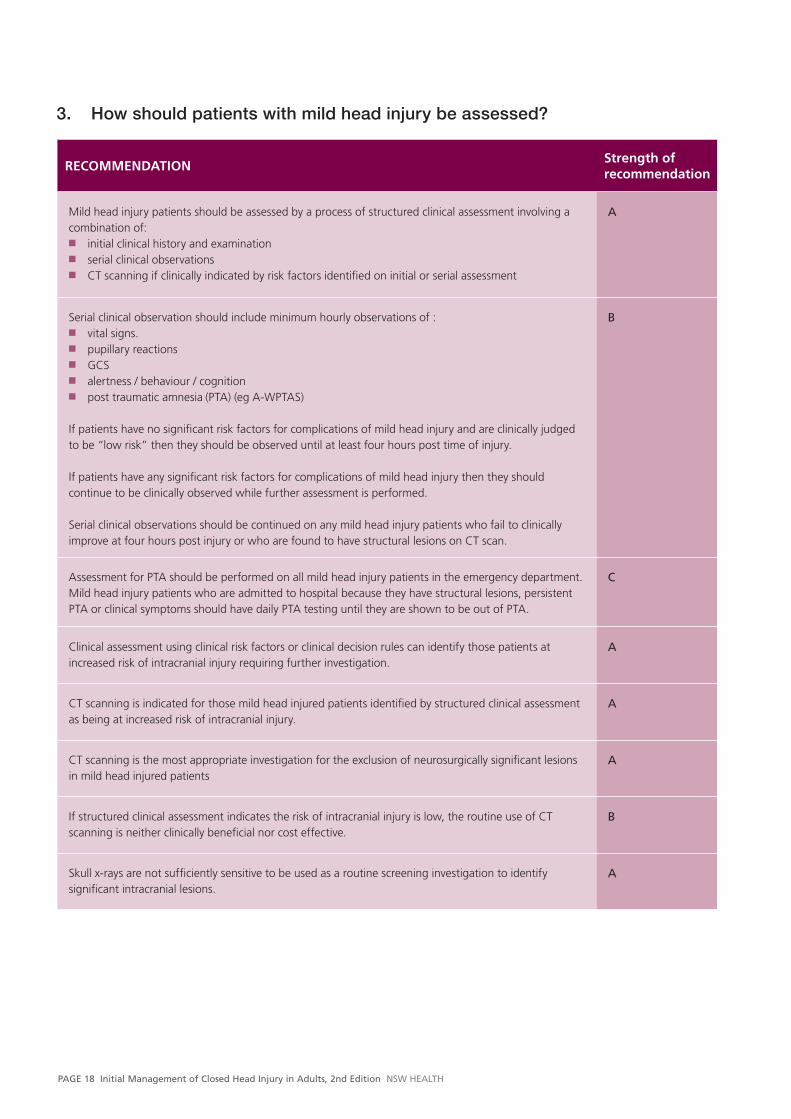
PAGE 18 Initial Management of Closed Head Injury in Adults, 2nd Edition NSW HEALTH
3. How should patients with mild head injury be assessed?
RECOMMENDATIONStrength of recommendation
Mild head injury patients should be assessed by a process of structured clinical assessment involving a combination of:■ initial clinical history and examination■ serial clinical observations ■ CT scanning if clinically indicated by risk factors identifi ed on initial or serial assessment
A
Serial clinical observation should include minimum hourly observations of :■ vital signs. ■ pupillary reactions ■ GCS ■ alertness / behaviour / cognition■ post traumatic amnesia (PTA) (eg A-WPTAS)
If patients have no signifi cant risk factors for complications of mild head injury and are clinically judged to be “low risk” then they should be observed until at least four hours post time of injury.
If patients have any signifi cant risk factors for complications of mild head injury then they should continue to be clinically observed while further assessment is performed.
Serial clinical observations should be continued on any mild head injury patients who fail to clinically improve at four hours post injury or who are found to have structural lesions on CT scan.
B
Assessment for PTA should be performed on all mild head injury patients in the emergency department. Mild head injury patients who are admitted to hospital because they have structural lesions, persistent PTA or clinical symptoms should have daily PTA testing until they are shown to be out of PTA.
C
Clinical assessment using clinical risk factors or clinical decision rules can identify those patients at increased risk of intracranial injury requiring further investigation.
A
CT scanning is indicated for those mild head injured patients identifi ed by structured clinical assessment as being at increased risk of intracranial injury.
A
CT scanning is the most appropriate investigation for the exclusion of neurosurgically signifi cant lesions in mild head injured patients
A
If structured clinical assessment indicates the risk of intracranial injury is low, the routine use of CT scanning is neither clinically benefi cial nor cost effective.
B
Skull x-rays are not suffi ciently sensitive to be used as a routine screening investigation to identify signifi cant intracranial lesions.
A
Initial Management of Closed Head Injury in Adults, 2nd Edition NSW HEALTH PAGE 19
Clinical history and examination remain the basis for the
initial assessment of patients with closed head injury.
However, additional clinical tools are available to assist in
assessment and management. These clinical tools include
Glasgow Coma Scale (GCS), initial serial neurological
observation, post traumatic amnesia (PTA) testing,
clinical decision rules, CT scanning and prolonged clinical
observation in hospital.
Glasgow coma scale
The Glasgow Coma Scale (Appendix 5) was originally
developed by Teasdale and Jennett104 for the neurological
observation of patients with prolonged coma. It was
intended to ensure inter-observer reliability and to identify
deterioration of patients over time. Since its original
introduction its use has been extended such that it is now
the standard tool for assessment of level of consciousness
in many clinical settings.
GCS is used both for the initial assessment and classification
of closed head injuries and for serial assessment of closed
head injuries. Initial GCS on admission to hospital is used
to classify head injuries into the broad prognostic groups
of mild (GCS 14-15), moderate (GCS 9-13) and severe
(GCS 3-8). The Brain Trauma Foundation concluded that
there is good quality evidence to relate initial GCS score
to outcome.15 However, it must be noted that these are
broad outcome measures and initial GCS is only about 75%
accurate so treatment should be individualised.15
GCS is unreliable if measured before initial resuscitation
and stabilisation of the ABCDE’s has been completed.15
Fearnside et al105 identified that both intubation and
sedation interfered with accurate assessment of initial
GCS in more severely injured patients and there is lack
of uniformity of approach to classifying GCS in these
situations.
The other significant area of controversy relates to timing of
initial GCS. In unstable patients requiring resuscitation, the
optimal time to record initial GCS remains controversial.15,
105 Similarly, in mild head injury patients the time of
presentation related to time of injury will influence the
initial GCS and therefore potentially influence clinical
decision making in relation to CT scanning.6, 10, 31, 35
Perhaps the most crucial point to note about initial GCS is
that it cannot predict individual outcome for patients with
similar GCS scores. Thus, an individual patient with an initial
GCS of 14-15 may have a variety of outcomes including
no significant injury, long-term cognitive-behavioural
dysfunction or a life threatening extradural haematoma.
This is particularly important for mild head injury patients
because GCS primarily assesses the risk of structural lesions.
Subtle cognitive changes are not well discriminated within
the mild head injury group. This led to the development of
the extended Glasgow Coma Scale and the Abbreviated
Westmead PTA Scale that assess the duration of post
traumatic amnesia as a means of identifying patients at
increased risk of cognitive problems.106-109 Despite these
limitations initial GCS on admission remains the standard
method for initial classification of head injuries.
When assessing initial GCS in patients with head injury it is
worthwhile considering time of injury. Clearly initial GCS for
a given patient may vary depending on time of presentation
to hospital. Few studies have related GCS to the time of
injury with the exception of Stiell et al35 who found GCS
<15 at two hour post injury was a significant predictor of
intracranial injury for mild head injury patients. Most recent
studies that have reported time of presentation have shown
that mild head injuries present around 60 to 90 minutes
post injury.31, 43, 54 An abnormal initial GCS taken within
one hour of injury is therefore likely to overestimate the
risk of intracranial injury in mild head injury patients who
present early.
Prehospital GCS, motor score and return of orientation
are other factors to consider when assessing initial GCS.
Prehospital GCS was felt to be unreliable,110 but with
more organised prehospital systems, it is gaining further
attention.15, 111 Motor score is the best predictor of outcome
of the GCS components.15 Orientation returns most
commonly in the sequence of person, place then time.112
The Glasgow Coma Scale is used as one of the parameters
in serial observation of head injury patients. This is what
it was originally designed for and what it is most useful
for. Both the original studies and subsequent studies have
validated its use in this fashion and, prior to the advent
of CT scanning, alteration in GCS was the most useful
tool in predicting intracranial injury.9, 104 Borg et al101 in
a meta analysis for the World Health Organisation on
mild traumatic brain injury concluded that in the absence
of CT scanning that hospital observation for at least 24
hours for patients with GCS 15 and other risk factors was
a reasonable strategy. Currently, serial GCS remains a
standard tool in the monitoring of head injuries when CT
scanning is unavailable or when clinical symptoms persist
PAGE 20 Initial Management of Closed Head Injury in Adults, 2nd Edition NSW HEALTH
despite normal CT scanning.
Summary
■ Initial GCS scores are useful to classify closed head
injuries into broad prognostic groups requiring further
assessment and serial observation.■ Serial GCS scores are useful to observe the trend in
clinical condition over time for all patients■ Serial GCS scores may be used as alternative to CT
scanning in patients with mild head injury and GCS 15
when CT scanning is not available■ Serial GCS scores are useful for identifying patients
with significant risk of intracranial injury in:
� - patients with an initial abnormal GCS score who fail
to improve
� - patients whose initial GCS score deteriorates.■ Initial and serial GCS scores are not as valuable in
excluding significant injury in:
� - patients with an initial normal GCS score who remain
normal
� - patients with an initial abnormal GCS score who
improve.■ Initial GCS should only be used for prognostic purposes
after initial resuscitation and stabilisation of ABCDE’s.■ The motor component of the GCS is the best predictor
of outcome ■ The extended GCS or Abbreviated Westmead PTA
Scale which assess both GCS and duration of PTA are
designed to help identify mild head injury patients
at increased risk of post concussion symptoms by
identifying more subtle cognitive changes.
Serial clinical neurological observation
Serial clinical neurological observations remain a standard
tool for assessing mild head injury patients despite the
advent of CT scanning. CT scanning is primarily used to
identify structural abnormalities at a given point in time
while serial neurological observations are used to monitor
clinical condition over a longer period of time. Serial
neurological observation typically consists of at least hourly
pupillary reactions and GCS assessment in conjunction
with vital signs. The symmetry of motor responses and a
standardised orientation assessment should be routinely
noted as part of the GCS. Neurological assessment should
also include qualitative assessment of alertness, behaviour
and cognition as this may identify more subtle neurological
impairment.
In mild head injury patients the primary aims of serial
neurological observation are the early identification of
acute neurological deterioration and the identification and
monitoring of persistent mild neurological deficits. Serial
neurological observations remain the basic standard of care
for the initial management of mild head injury patients and
should be used in conjunction with clinical decision rules
to determine the need for CT scanning and / or prolonged
observation. The qualitative aspects of serial neurological
observation may assist in identifying acute deterioration
or subtle functional abnormality before there is a change
in GCS. Subtle drowsiness, mild disorientation to time,
inattention and mildly disorganised thought processes with
subtle post traumatic amnesia can easily be missed by over
reliance on GCS in a patient who is awake and orientated in
person and place.
Controversy exists over the appropriate duration of serial
neurological observation for both mild head injury patients
who are improving and those who have persistent clinical
symptoms or abnormalities on CT scan. Although four
hours of initial neurological observations post injury are
fairly standard following mild head injury, there is little
evidence to support this. There is also some debate as to
whether the initial period of observation should be until
four hours post injury or for four hours following arrival
at hospital. Studies that reported time of presentation
showed that most patients present at one to two hours
post injury. Clinically most uncomplicated mild head injury
patients will start to improve within two to four hours post
time of injury. Stiell et al35 demonstrated that GCS<15 at
two hours post injury was one of the most significant risk
factors for intracranial injury. Since this guideline has used
these clinically important criteria, it was felt that the initial
clinical observation period should also be based on time
post injury rather than four hours observation from the
time of arrival at hospital. It is important that patients who
present one hour post injury are not treated the same as
those who present at four hours post injury with “routine
observations” for four hours. If the patient who presents
at four hours post injury is not clinically improving then
they are at increased risk of intracranial injury and warrant
CT scanning whereas the same patient at one hour post
injury probably only requires observation unless there is
another strong indication for CT scanning eg suspected
skull fracture, seizure, warfarin etc. The initial period of
neurological observation should therefore be until at
least four hours post time of head injury at which point
clinical decisions should be made if they are not clinically
improving.
Initial Management of Closed Head Injury in Adults, 2nd Edition NSW HEALTH PAGE 21
If the serial clinical neurological observations of patients
with an initial GCS 15 at 2 hours post injury are improving
or have returned to normal at four hours post injury then
the evidence would suggest it is safe to discharge them for
home observation if they have no risk factors indicating the
need for CT scan or if they have an initial normal CT scan.
There is no strong evidence to support a specific duration
of time of observation. Observation until 4 hours post time
of injury should be used as an initial guide. Patients with
trivial injuries could be considered for earlier discharge
by an experienced senior clinician and patients with high
risk factors should be considered for longer periods of
observation.
If serial neurological observations are not improving at
four hours post injury then serial neurological observations
should be continued and clinical decisions need to be
made about the need for CT scanning and/or admission
to hospital for prolonged observation. The period of
admission to hospital for prolonged observation is also
controversial as there is little evidence to support the
general recommendation of twenty four hours. This period
of observation is derived from studies that show that
clinical deterioration is unusual in mild head injury patients
after twenty four hours. Indeed, prior to the advent of CT
scanning serial clinical observation was the standard of
care and the need for neurosurgical intervention in mild
head injury patients was largely determined on the basis
of clinical deterioration. The best location for prolonged
neurological observation for lower risk patients is also
debated because some studies have shown that admission
to hospital does not guarantee that regular neurological
observation will occur.53
If patients are not clinically improving or have abnormalities
on CT scan that warrant hospital admission, then serial
clinical observations should be continued. The duration
of this time of observation remains poorly defined.
Basically, clinical observation should be continued until
clinical symptoms improve or until it is felt there is little
risk of deterioration. This needs to be individualised but a
minimum of 24 hours is normally recommended.101
Summary
■ Serial neurological observation is a useful tool for the
early identification of acute neurological deterioration
and the identification and monitoring of persistent mild
neurological deficits.
■ Serial neurological observations should include
a minimum of hourly GCS assessment, pupillary
reactions, PTA and vital signs. Neurological
observations should also include qualitative assessment
of alertness, behaviour and cognition to detect subtle
changes in mental status not assessed by the Glasgow
Coma Scale.■ Mild head injury patients should have initial serial
neurological observations until at least four hours post
time of injury at which point decisions about further
management should be made.■ Serial neurological observations should be continued
on patients who are admitted to hospital for at least
24 hours and until patients clinically improve and are
discharged home.
Post traumatic amnesia (PTA) testing
Amnesia for the event, short-term memory loss,
anterograde amnesia and PTA are all terms used to
describe the disruption of memory that typically results
from a traumatic head injury. The different terms describe
a continuum of memory disruption, and the use of the
individual terms depends on the duration of memory
loss and individual preference. Amnesia for the event
is common and of little clinical significance. Retrograde
and anterograde amnesia are typically used to refer to
the duration of loss of memory for events preceding or
following an injury. PTA is the period of time during which
a person is unable to lay down new memories following
an injury. PTA and anterograde amnesia essentially refer to
the same phenomena but some patients may have memory
for events yet still be unable to lay down new memories
– the so called islands of memory. Stiell et al56 identified a
duration of greater than 30 minutes of retrograde amnesia
as being a significant risk factor for intracranial injury.
Smits et al47 identified a duration of PTA of greater than 2
hours as being a minor risk factor and greater than 4 hours
as being a major risk factor for intracranial injury. Many
studies have shown that persistent PTA is a significant risk
factor for poor functional outcome in all grades of head
injury. Any persistent inability to lay down new memories
following blunt trauma is perhaps most simply referred to
as PTA and the duration of PTA may be used to predict
the risk of intracranial injury and the risk of persistent
post concussion symptoms. PTA is the term that will be
predominantly used in this guideline.
PAGE 22 Initial Management of Closed Head Injury in Adults, 2nd Edition NSW HEALTH
While it has been identified that prolonged PTA is a risk
factor for significant post concussion symptoms,3, 109 the
standard tool for identifying PTA in NSW, the Westmead
PTA Scale (Appendix 3), is designed to be used over several
days. There has been considerable interest in developing
bedside PTA screening tools to allow early prediction of
which patients are at greatest risk of developing post
concussion symptoms. A simple bedside test that can
determine whether the patient is out of post traumatic
amnesia at the time of the test would be beneficial for both
emergency department testing and ward based testing.
The Extended Glasgow Coma Scale and the Abbreviated
Westmead PTA Scale are examples of tools developed to try
to identify persistent PTA in mild head injury patients as a
marker for increased risk of post concussion symptoms.106,
109 Recent Australian studies have looked at using specific
bedside tools such as the Revised Westmead Post Traumatic
Amnesia Scale in the emergency department to better
identify patients with persistent PTA so that these patients
can be more closely followed up as they are more likely
to suffer significant post concussion symptoms.106-108 The
Abbreviated Westmead PTA Scale (A-WPTAS), developed
and validated113 from the previously validated Revised
Westmead PTA Scale, has been successfully trialled
in various New South Wales emergency departments
(Appendix 4).
From a practical point of view, all patients with mild head
injury should be assessed for post traumatic amnesia. It
is useful to assess the patient’s recall of events following
their injury by asking specific questions such as what is
their first clear memory, who helped them at the scene and
how they got to hospital. This can be used to estimate the
period of post traumatic amnesia (anterograde amnesia).
Clinicians should also look for symptoms of post traumatic
amnesia such as repetitive questioning, failure to remember
clinical staff and inability to remember events during their
hospital stay. A formal assessment tool like the A-WPTAS
or a simple memory assessment technique such as three
object recall can be used as a bedside screening test for
post traumatic amnesia, to supplement the ‘history’ of
amnesia for events. Patients who have persistent post
traumatic amnesia should be considered for CT scanning,
prolonged observation and close follow up. Mild head
injury patients who are admitted to hospital because
they have structural lesions, persistent PTA or clinical
symptoms should have daily PTA testing until they are
shown to be out of PTA.
Summary
■ The identification of persistent PTA in mild head injury
patients is a potentially useful marker for the risk of
intracranial injury and the risk of developing post
concussive symptoms.■ PTA testing should be performed on any patient
presenting to hospital following mild head injury. ■ The Revised and Abbreviated Westmead PTA Scales
are useful bedside tools for assessing PTA in the
emergency department
Clinical decision rules
Clinical decision rules are increasingly being used to
assist clinicians in determining the need for particular
investigations or management. By identifying individual risk
factors and combining them to establish clinical decision
rules, which are then prospectively validated, useful
evidence based diagnostic tools to assist management can
be developed. Well established clinical decision rules also
include the NEXUS criteria for cervical spine assessment and
the Ottawa Ankle Rules.114, 115
Although clinical decision rules are potentially very useful,
clinicians need to be aware of the specific inclusion/
exclusion criteria used to develop them and the overall
quality of the original studies before applying them to their
patients.9, 48 The other important point for clinicians to
consider is what level of risk they are prepared to accept.
No clinical decision rule can entirely rule out a significant
finding for an individual patient, and different clinical
decision rules will have different levels of risk. In patients
with mild head injury it is important to have high negative
predictive value for ruling out significant intracranial injuries
but this comes at a cost of lower specificity and therefore
the need for more CT scanning. While all the clinical
decision rules aim to rule out significant neurosurgical
lesions, they have different approaches to the value of
identifying intracranial lesions on CT scan that do not
require intervention. Clearly it is important to identify
intracranial lesions that require neurosurgical intervention,
but is it beneficial to identify abnormalities on CT scan
that do not require intervention? Concerns about radiation
exposure and resource utilisation have influenced this
debate. The trend in the literature is to develop strategies
to identify clinically important lesions while minimising the
number of CT scans performed. Essentially, if you want
to do less CT scans you have to accept that you will miss
some intracranial abnormalities that are unlikely to require
intervention.
Initial Management of Closed Head Injury in Adults, 2nd Edition NSW HEALTH PAGE 23
Clinical decision rules for adult patients with mild head
injury have been developed through studies of large
cohorts of patients and prospectively applied to patients
to determine their sensitivity, specificity and negative
predictive value.31, 32, 35, 47, 48, 56, 58, 116 These studies aim to
identify those patients at increased risk of intracranial injury
and develop clinical decision rules about which patients
require CT scanning or prolonged observation. The best
studies should be well designed and prospectively internally
validated in the original population and then externally
validated by other groups in other populations. The main
design features of these trials are summarised in Evidence
Table 1 and Appendix 1.
The most consistent findings of these studies are that
abnormal GCS or mental status, clinical suspicion of skull
fracture, focal neurological deficit, vomiting, coagulopathy
and age > 65 are the best predictors of risk of intracranial
injury. Other relevant predictors of risk include mechanism
of injury, prolonged loss of consciousness, post traumatic
amnesia, severe headache and seizure. Depending on their
inclusion / exclusion criteria the authors used combinations
of these risk factors to derive clinical decision rules of
varying sensitivity and specificity (Evidence Table 1 and
Appendix 1). At present, the findings of both Haydel et
al32 and Stiell et al35 have been adopted as policy by the
American College of Emergency Physicians (USA),9 whilst
the findings of Stiell et al35 have been adapted by the
NICE guidelines (UK).117 The other widely known clinical
prediction rule is NEXUS II.58
Ibanez et al33 in 2004 attempted to prospectively identify
clinical risk factors predicting intracranial injury and
to assess the reliability of previously published clinical
guidelines. They found that while clinical risk factors could
not detect all intracranial injuries they could be used to
detect clinically relevant lesions with a negative predictive
value approaching 99%. They also concluded clinicians
should be aware of the limitations of clinical decision rules
when using clinical guidelines. An Australian study in 2004
by Rosengren et al118 looked at applying the clinical decision
rules developed by Haydel et al32 and Stiell et al35 to
Australian practice and concluded that both had limitations.
More recently an Australian study by Fong et al57 developed
a local guideline by adapting the findings of Haydel et al32
and Stiell et al35 and applying the resultant guideline to an
Australian population with satisfactory results. The authors
stressed the point that no clinical decision rule is infallible
and that appropriate explanation and discharge advice was
important.
A recent review by Stein et al in 2009 compared most
of the best known clinical decision rules for mild head
injury by applying them retrospectively to a prospectively
collected data base and found that they all performed well
for identifying acute neurosurgical lesions but differed in
their ability to identify other lesions and their predicted CT
scan ordering rates. The authors concluded that NEXUS
II probably performed the best but the accompanying
editorial commented that it would be reasonable to use any
of the clinical decision rules.48
The Canadian CT Head Rules are the most widely studied
and have been externally validated several times. However,
they were applied to GCS 13-15 patients, require loss of
consciousness or amnesia and excluded unstable multi-
system trauma, coagulopathy, pre hospital seizure and
neurological deficit. The Canadian CT Head rule therefore
needs to be adapted for application to the broader
population or clinicians need to be fully aware of these
limitations when using the original rule. The NEXUS II
clinical decision rule has not been as extensively externally
validated but has the advantage of being relatively simple
to use and can be used for all patients with head injury
as there were no exclusion criteria other than delayed
presentation and both adult and paediatric populations
were studied. From a clinically practical point of view the
mnemonic “BEAN BASH” can be used to remember the
NEXUS II indications for CT scanning patients with head
injury at the bedside:
B – behaviour abnormal; E – emesis; A – alertness abnormal; N – neurological deficit; B – bleeding disorder; A – age>65; S – skull fracture suspected;
H – haematoma scalp.
The most important point for any clinician to recognise
is that clinical decision rules should be used as tools to
support clinical decisions and should not override clinical
judgement. If clinicians choose to use a clinical decision
rule, they should be aware of the limitations and inclusion
/ exclusion criteria of whichever clinical decision rule they
decide to use.
Summary
■ Clinical decision rules provide useful adjuncts to the
assessment and management of mild head injury
patients.
PAGE 24 Initial Management of Closed Head Injury in Adults, 2nd Edition NSW HEALTH
■ Studies by Haydel et al32 (New Orleans Criteria), Stiell
et al35 (Canadian CT Head Rules) and Mower et al58
(NEXUS II) have provided externally validated evidence
based clinical decision rules for mild head injury in
adults. The Canadian CT Head Rules are the most
extensively studied of the clinical decision rules.■ Clinical decision rules should be used as tools to
support clinical decisions and should not override
clinical judgement■ If clinicians choose to use a clinical decision rule, they
should be aware of the limitations and inclusion /
exclusion criteria of whichever clinical decision rule they
decide to use.
Skull x-rays
The literature clearly identifies that both the clinical
suspicion of skull fractures and the radiological evidence of
skull fracture are significant risk factors for the presence of
an intracranial lesion requiring neurosurgical intervention.6,
31, 32, 35, 66, 74
If CT scanning is available, the current indications for skull
x-rays are few. However, if CT scanning is unavailable,
the role of skull x-rays as a screening test is less clear. A
detailed meta analysis by Hofman et al66 concluded that
whilst the presence of skull fracture greatly increased the
risk of intracranial injury, the absence of a skull fracture
did not rule it out (calculated sensitivity 38% calculated
specificity 95%). In subsequent clinical guidelines, authors
have differed as to whether skull x-rays should be used to
detect patients at higher risk of intracranial injury. Jagoda et
al9 argued that the sensitivity of skull x-ray is not sufficient
to be used as a screening test. Vos et al10 and Servadei et
al6 felt that in the absence of CT scanning, a positive skull
x-ray can be useful to help allocate patients into higher risk
groups for management purposes. On existing evidence,
both approaches seem reasonable depending on local
management guidelines.
Summary
■ Clinical evidence or suspicion of skull fracture is
associated with increased risk of intracranial injury.■ Skull x-rays are not sufficiently sensitive to be used as a
routine screening test to identify patients at increased
risk of intracranial injury.■ Where CT scanning is unavailable, skull x-ray may
be used as an adjunct to identify patients with skull
fractures who are at greater risk of intracranial injury
(but not to exclude intracranial injury).
CT scanning
The widespread availability of CT scanning has greatly
assisted the management of patients with head injuries. CT
scanning has been particularly useful in identifying focal
injuries in patients with altered level of consciousness or
other risk factors. CT scanning is regarded as mandatory
for all head injury patients with a persistent altered level
of consciousness. However, the role of the CT scanning
in a patient with mild head injury with a normal level
of consciousness remains controversial. Multiple clinical
decision rules have been developed to try to identify which
patients should have a CT scan because of concerns about
the routine use of CT scanning for all patients. Particular
concerns about the routine use of CT scanning for mild
head injury include the financial-resource burden, the
potential hazards of radiation and the potential pitfalls
of reliance on technology at the expense of clinical
assessment.37, 71, 88, 119-123 Furthermore, CT scans do not
identify patients who have cognitive dysfunction which is
the most significant complication for most patients. The
various pros and cons of CT scanning are summarised
below:
Pros
■ Early identification of patients with intracranial injuries
requiring acute neurosurgical intervention■ Early identification of patients with other intracranial
injuries requiring admission to hospital due to risk of
deterioration ■ Identification of patients at low risk of deterioration
and suitable for discharge■ Identification of patients with structural lesions
indicating increased risk of post concussive
symptoms.76
■ Potential cost benefit due to early CT scanning allowing
discharge home rather than hospital admission in some
patients.119
Cons
■ Routine use of CT scanning for mild head injury
potentially has a huge financial and resource impact
given that more than 90% of scans are negative
and less than 1% of scans indicate the need for
neurosurgical intervention.13, 35, 124
■ Some patients, particularly the elderly or those with
a known coagulopathy, may develop delayed focal
neurosurgical lesions (especially subdural lesions)
despite initial normal CT scanning.69, 125, 126
Initial Management of Closed Head Injury in Adults, 2nd Edition NSW HEALTH PAGE 25
■ Early CT scans may not demonstrate intra-cerebral
contusions which take time to become apparent on CT
scanning.■ CT scanning will not demonstrate diffuse axonal injury
in most patients.3
■ Patients may suffer significant post concussive
symptoms despite an initial normal CT scan.3, 94, 98, 99,
110
■ Routine use of CT scanning does not guarantee better
identification of significant intracranial injuries if
different institutions are compared.12, 37
■ The risk of cumulative radiation exposure especially
among children is of concern.120-123
■ May delay definitive management of more significant
injuries in multi system trauma patients.
CT scanning and radiation
There is increasing concern about the potential harm
from radiation associated with CT scanning.122, 123, 127-130
The main risk associated with radiation exposure from CT
scanning is an increased lifetime risk of fatal cancer.122, 123
There have also been concerns about the effects of
radiation on cognitive development in children. Hall
et al120 showed a significant reduction in educational
performance in young children who received the equivalent
dose of radiation of one or two CT heads for childhood
haemangiomas. The risk posed by radiation is greatest
in young children due to the relatively increased dose on
more sensitive tissue and the increased time for a cancer to
develop. Thus a person who has a CT scan as a child is at
much greater risk than a person who has their first CT scan
age 65 where the risk is very small. The risk posed by CT
scan radiation is cumulative and is greatest in patients with
chronic disease who have multiple scans. In patients with
moderate to severe head injury there is a clear benefit from
performing a CT scan because the probability of identifying
a life threatening injury clearly outweighs the small increase
in the lifetime risk of cancer. In the case of patients with
mild head injury the risk of harm from radiation needs
to be weighed against the relatively low probability of
identifying a neurosurgically significant lesion on CT scan.
This is of most concern in younger patients where the
effective radiation exposure is higher, the time available for
developing a cancer greater and the chance of cumulative
lifetime exposure greater. Hence the interest in developing
clinical decision rules for mild head injury patients to
determine who really needs a CT scan.
The risk from radiation can either be expressed as an
estimated lifetime risk of fatal cancer or as an estimated
equivalent dose of radiation which takes into account organ
doses and their relative radiosensitivity.122, 123, 127, 128, 130 It
is important to recognise that these risks are all estimates
based on epidemiological studies of atomic bomb survivors
extrapolated to current estimated doses of radiation
delivered by CT scans. Perhaps the best way to put the risk
of CT scanning into perspective is to compare the estimated
risks with more common everyday risks.129
Put into context the risk of radiation from an individual
head CT scan is very low.122, 123, 127-130
■ Estimated overall lifetime risk of fatal cancer (1 in 3)■ Estimated risk of clinically important lesion on CT in
mild head injury (1 in 100)■ Estimated additional lifetime risk of fatal cancer from
adult trauma panscan (1 in 1,000)■ Estimated additional lifetime risk of fatal cancer from
single child CT head (1 in 5,000)■ Estimated additional lifetime risk of fatal cancer from
single adult CT head (1 in 10,000)■ Estimated additional lifetime risk of fatal cancer from
trauma series x-rays (1 in 20,000)■ Estimated additional lifetime risk of fatal cancer from
single chest x-ray (1 in 1,000,000)■ Estimated equivalent dose of radiation from a chest
x-ray (0.02 mSv per CXR)■ Estimated equivalent dose of radiation from adult CT
head (2 mSv per scan) ■ Estimated equivalent dose of radiation from annual
background radiation (2 mSv per year)■ Estimated equivalent dose of radiation from adult CT
trauma panscan (20 mSv per scan)■ Annual safety limit for radiation exposure for radiation
workers (.20 mSv per year)■ Estimated mean equivalent dose of radiation exposure
for atomic bomb survivors linked to increased rates of
fatal cancer (40 mSv dose)
It is important to recognise that the absolute risk to an
individual is relatively small particularly with advancing
age. More caution is recommended in children due to
the concerns on cognitive development as well as lifetime
risk of cancer. The lifetime risk of cancer for a young child
receiving a CT head would be roughly double that of an
adult (1 in 5000).
PAGE 26 Initial Management of Closed Head Injury in Adults, 2nd Edition NSW HEALTH
The following points try to put the risk into context and are
adapted and simplified from the original references: 121-123,
127-130
■ Single adult CT head equivalent risk to having 100
chest x-rays■ Single adult CT head equivalent risk to smoking 1000
cigarettes■ Single adult CT head equivalent risk to driving 5000
km on a highway■ Single adult CT head equivalent risk to one year of
background radiation■ Single adult CT head increases lifetime risk of fatal
cancer from 30% to 30.01%■ Single young child CT head increases lifetime risk of
fatal cancer from 30% to 30.02%■ Single adult CT trauma panscan increases lifetime risk
of fatal cancer from 30% to 30.1%
Timing of CT scanning
There is no direct evidence to confirm what the best time
to perform CT scanning in relation to time of injury is. The
primary role of early CT scanning in mild head injury is early
recognition of extradural or subdural haematomas prior
to clinical deterioration.69 Early neurosurgical intervention
prior to clinical deterioration is associated with improved
outcome. However, early CT scan may potentially miss other
intracranial injuries such as delayed subdural haematomas
or contusions which are slower to become evident.69
Fortunately, most studies have shown that an initial normal
CT scan allows safe discharge and that the few patients
who deteriorate usually have good outcome.9, 53, 89, 130
Therefore, it is reasonable to suggest that CT scans should
be performed shortly after a decision is made that one is
necessary.
Adjunctive CT scanning
Some mild head injury patients will require CT scanning for
other reasons such as cervical spine clearance in the elderly.
Clearly, in these circumstances, clinicians should have a
lower threshold for performing head CT scans at the same
time for ease of management.
Repeat CT scanning
There has been some debate in the literature about
whether mild head injury patients who have initially
abnormal CT scans and require admission for hospital
observation should have a routine repeat CT scan.132-136 The
evidence from most of these small studies suggests that
most mild head injury patients with minor abnormalities
on CT scan do not require routine repeat CT scanning if
they are clinically improving with a normal GCS and no
neurological deficit unless they are anti-coagulated.
The other question that is often asked is should elderly
anti-coagulated patients with normal initial CT scans
have routine repeat CT scans and if so when should they
be performed and should the patient be admitted for
observation. There is little evidence to guide management
in these situations. The consensus appears to be that
the older the patient, and the more the patient is anti-
coagulated (higher INR), the greater the risk of delayed
bleeding. However, how to manage that risk remains
unclear.
Summary
■ CT scanning is the best investigation for the early
identification of neurosurgically significant focal
intracranial lesions following mild head injury.■ CT scanning should be used as an adjunct to clinical
assessment.■ Where structured clinical assessment indicates the risk
of significant intracranial lesion is low, the routine use
of CT scanning is unlikely to be of benefit. ■ CT scanning does not accurately predict the risk of
post concussion symptoms in mild head injury patients.■ Early CT scanning may theoretically not demonstrate
some subdural haematomas and cerebral contusions
but there is little evidence to suggest that this is
clinically relevant in most patients. ■ There is little evidence to guide the management of
anti-coagulated elderly mild head injury patients with
initially normal CT scans who are potentially at risk
of delayed bleeds, especially subdural haematomas.
There is increased risk with increased age and degree
of coagulopathy but how to manage that risk remains
unclear.■ Routine repeat CT scanning is not indicated for most
clinically improving mild head injury patients with
minor abnormalities on initial CT scan.
Initial Management of Closed Head Injury in Adults, 2nd Edition NSW HEALTH PAGE 27
4. Which patients with mild head injury require a CT scan?
RECOMMENDATIONStrength of recommendation
'High risk’ mild head injury requiring CT scanThe following risk factors identify patients with mild head injury (initial GCS 14-15) at increased risk of clinically signifi cant lesions requiring acute neurosurgical intervention or prolonged observation in hospital. These patients should have early CT scanning if available, if they have any of the following features:
On initial assessment
■ GCS<15 at two hours post injury**■ Focal neurological defi cit■ Clinical suspicion of skull fracture■ Vomiting ■ Known coagulopathy or bleeding disorder■ Age >65 ■ Witnessed seizure■ Prolonged loss of consciousness (>5min)
On serial assessment
■ Decrease in GCS■ Persistent GCS<15 at two hours post injury■ Persistent abnormal alertness/behaviour/cognition■ Persistent post traumatic amnesia (A-WPTAS<18/18 at 4hrs post injury)■ Persistent vomiting ( 2 occasions)■ Persistent severe headache■ Post traumatic seizure
Clinical judgement required if
■ Initial GCS 14 within two hours of injury** ■ Large scalp haematoma or laceration■ Associated multi-system injuries■ Dangerous mechanism■ Known neurosurgery/neurological impairment■ Delayed presentation or representation
** NOTE: Includes patients with abnormal GCS due to drug or alcohol ingestion.
A
If CT scanning is unavailable“High risk” mild head injury patients should be closely observed and be considered for transfer to a hospital with neurosurgical and CT scan facilities when CT scan is unavailable.
A clear decision about the need for transfer for CT scanning for “high risk” patients should be made at the time of initial assessment or after a brief period of observation. A local senior clinician should be consulted and the patient discussed with the network neurosurgical service.
The clinical symptoms of patients with mild head injury typically improve within two to four hours post time of injury. Patients with persistently abnormal or worsening clinical symptoms are at “high risk” of intracranial injury. A clear decision about the need for transfer for CT scanning should be made no later than 4 hours post time of injury.
CONSENSUS
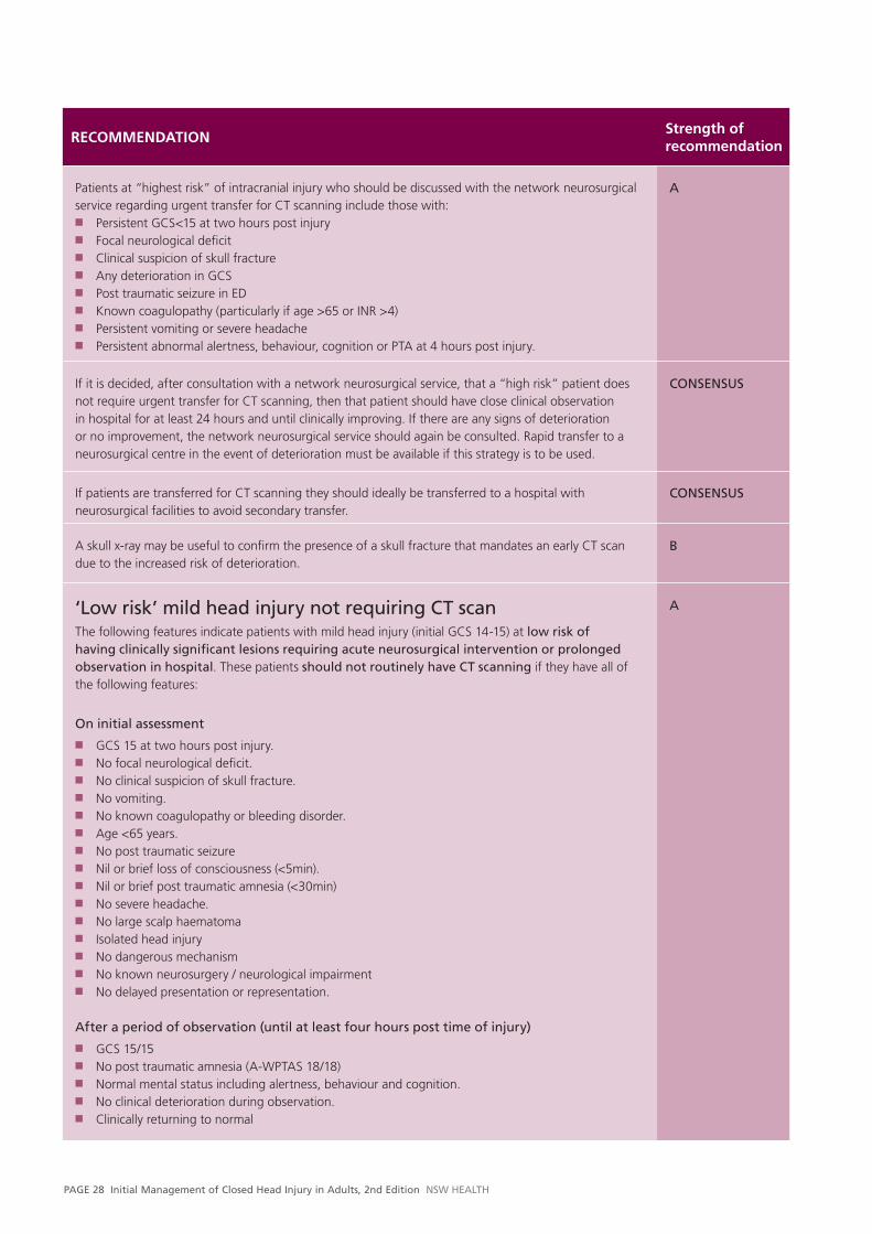
PAGE 28 Initial Management of Closed Head Injury in Adults, 2nd Edition NSW HEALTH
RECOMMENDATIONStrength of recommendation
Patients at “highest risk” of intracranial injury who should be discussed with the network neurosurgical service regarding urgent transfer for CT scanning include those with:■ Persistent GCS<15 at two hours post injury■ Focal neurological defi cit■ Clinical suspicion of skull fracture■ Any deterioration in GCS■ Post traumatic seizure in ED ■ Known coagulopathy (particularly if age >65 or INR >4)■ Persistent vomiting or severe headache■ Persistent abnormal alertness, behaviour, cognition or PTA at 4 hours post injury.
A
If it is decided, after consultation with a network neurosurgical service, that a “high risk” patient does not require urgent transfer for CT scanning, then that patient should have close clinical observation in hospital for at least 24 hours and until clinically improving. If there are any signs of deterioration or no improvement, the network neurosurgical service should again be consulted. Rapid transfer to a neurosurgical centre in the event of deterioration must be available if this strategy is to be used.
CONSENSUS
If patients are transferred for CT scanning they should ideally be transferred to a hospital with neurosurgical facilities to avoid secondary transfer.
CONSENSUS
A skull x-ray may be useful to confi rm the presence of a skull fracture that mandates an early CT scan due to the increased risk of deterioration.
B
‘Low risk’ mild head injury not requiring CT scanThe following features indicate patients with mild head injury (initial GCS 14-15) at low risk of having clinically signifi cant lesions requiring acute neurosurgical intervention or prolonged observation in hospital. These patients should not routinely have CT scanning if they have all of the following features:
On initial assessment
■ GCS 15 at two hours post injury.■ No focal neurological defi cit.■ No clinical suspicion of skull fracture.■ No vomiting.■ No known coagulopathy or bleeding disorder.■ Age <65 years. ■ No post traumatic seizure■ Nil or brief loss of consciousness (<5min).■ Nil or brief post traumatic amnesia (<30min)■ No severe headache.■ No large scalp haematoma■ Isolated head injury■ No dangerous mechanism■ No known neurosurgery / neurological impairment■ No delayed presentation or representation.
After a period of observation (until at least four hours post time of injury)
■ GCS 15/15 ■ No post traumatic amnesia (A-WPTAS 18/18)■ Normal mental status including alertness, behaviour and cognition. ■ No clinical deterioration during observation.■ Clinically returning to normal
A
Initial Management of Closed Head Injury in Adults, 2nd Edition NSW HEALTH PAGE 29
Mild head injury patients (initial GCS 14-15) require a CT
scan if they are clinically assessed as being at significant risk
of acute deterioration from an underlying intracranial injury.
The WHO Taskforce on MTBI (Borg et al 2004101) concluded
that on the basis of their meta-analysis that “there is
strong evidence that clinical factors can predict CT scan
abnormalities and the need for (neurosurgical) intervention
in adults.” There have been many recent studies looking at
which risk factors predict intracranial injury and the need
for CT scan and a variety of clinical decision rules have
been developed. These studies are mostly based on large
prospectively collected data bases in the USA, Canada, and
Europe although their findings have been reproduced in
smaller studies throughout the world. Haydel et al32 (2000
– New Orleans Criteria), Stiell et al35 (2001 – Canadian CT
Head Rules), Mower et al58 (2005 – NEXUS II) and Smits
et al47 (2007 – CHIP Rule) have all derived clinical decision
rules with slightly varying inclusion and exclusion criteria
and identified risk factors. Ibanez et al33 (2004), Fabbri et
al125 (2004) and Stein et al48 (2009) have also done large
studies looking at clinical risk factors and assessing the
validity of the various clinical decision rules and guidelines
while Rosengren et al118 (2004) and Fong et al57 (2008) have
performed similar smaller studies on Australian populations.
Ono et al137 (2007), Turedi et al81 (2008) and Saboori et
al80 (2007) have published recent studies on Japanese,
Turkish and Iranian populations. Kuppermam et al85 (2009)
and Dunning et al138 (2006) have recently published large
studies in the paediatric population in which they identified
clinical risk factors and developed clinical decision rules
that are similar to those developed for adults. There is now
a large body of evidence suggesting that mild head injury
patients can be risk stratified based on clinical assessment.
The clinical risk factors that indicate patients are at
increased risk of intracranial injury are discussed in the
following text, and most of the relevant studies summarised
in the evidentiary tables.
Mild head injury patients can be risk stratified into “low”
or “high” risk groups based on the presence or absence
of identified clinical risk factors. The absence of clinical
risk factors on initial assessment combined with a period
of observation during which the patient clinically improves
makes the probability of a significant intracranial injury
extremely unlikely. These low risk mild head injury patients
can be discharged for home observation without CT
scanning. Among the patients with identified risk factors,
there are those who clearly require CT scanning and those
in whom clinical judgement may be required. In the high
risk group, which includes initially low risk patients who
fail to clinically improve, both CT scanning and prolonged
clinical observation are indicated. In the patients where
clinical judgement is required this should be based on the
clinician’s experience, the number of identified risk factors
and clinical observation of the patient.
Early CT scanning allows identification of acute extradural
or subdural haematomas or depressed skull fractures
requiring neurosurgical intervention and other structural
lesions such as intracerebral haematomas or minor
skull fractures that put the patient at increased risk of
deterioration or complications such as post traumatic
seizures, and disabling post concussive symptoms. However,
it is important to recognise that the absence of a structural
lesion on CT scan does not exclude the possibility of
deterioration, post traumatic seizures or significant post
concussion symptoms.
Patients with an abnormal CT scan should be discussed
with a neurosurgical service and considered for prolonged
observation in hospital. Conversely, a normal CT scan
makes acute clinical deterioration highly unlikely and
allows safe discharge for home observation as long as
the patient is clinically improving and does not have a
known coagulopathy. A normal CT scan is useful to rule
out structural lesions but does not exclude the possibility
of significant post concussion symptoms. It is important
to stress that CT scanning should be used as a clinical tool
in conjunction with clinical assessment and observation as
part of an overall management strategy for mild head injury
patterns.
Individual factors predicting risk of intracranial injury and therefore the need for CT scanning in patients with mild head injury:
Initial GCS
A persistent GCS <15 at two hours post injury
is a strong indication for CT scanning. An initial
GCS 14 on admission is a relative indication for CT
scanning.31, 33, 35, 36, 47, 48, 50-53, 56, 58, 62, 70, 74, 139
Several studies have noted the heterogeneity of the original
GCS 13-15 mild head injury classification and these findings
are summarised in Appendix 1. Patients with an initial GCS
13 have been shown to have similar rates of intracranial
injury to those with initial GCS 9-12.53, 105 Recent studies
on mild head injury that published data on patients with
PAGE 30 Initial Management of Closed Head Injury in Adults, 2nd Edition NSW HEALTH
GCS 13 have confirmed that an initial GCS 13 has a much
higher rate of intracranial injury.35, 47, 56 Many other recent
studies have used the definition of mild head injury as GCS
14 or 15 on arrival and excluded patients with an initial
GCS 13. Although patients with an initial GCS 14-15 have
lower rates of intracranial injury as a group, the more recent
studies have confirmed a higher risk of intracranial injury
for an initial GCS 14.31, 32, 35, 47, 48, 56, 58 A number of the
recent, well designed studies have attempted to identify risk
factors which can predict which patients with an initial GCS
14-15 are at highest risk.31, 32, 35, 47, 48, 56, 58 This evidence
confirms that other risk factors can be used to successfully
stratify risk within the initial GCS 14-15 group.
One strategy is to relate the significance of the GCS to the
time of injury. In most of the studies on GCS 14-15, the
reported higher risk of intracranial injury for an initial GCS
14 does not take into account the time of injury. Recent
studies that have reported time of presentation (Fabbri et
al,31 Smits et al54 and Styrke et al43) have shown that mild
head injuries present around 60 to 90 minutes post injury.
An abnormal initial GCS taken within one hour of injury is
therefore likely to overestimate the risk of intracranial injury
in mild head injury patients who present early. One of the
most relevant findings to clinical practice is that of Stiell et
al35 who showed that for patients presenting with an initial
GCS 13-15 that GCS <15 at two hours post injury was a
useful predictor of risk of intracranial injury. By applying this
criteria, both time of injury and GCS are usefully combined
in a clinically practical assessment tool. Using GCS <15
at two hours post injury allows for clinical judgement
regarding patients who present immediately following
injury or with drug or alcohol ingestion, allows for lack
of inter observer reliability and stresses the importance of
relating persistently abnormal GCS to time of injury. It also
emphasises the significance of a patient presenting with
a GCS of less than 15 or other signs of abnormal mental
status at more than two hours after injury.
Focal neurological deficits
Focal neurological deficit is a strong indication for CT
scanning.31-35, 47, 48, 53, 58, 62, 68-70, 139-141
All the major clinical decision rules use neurological deficit
as an indication for CT scan or list it as an exclusion
criteria. Focal neurological deficits have been shown to
significantly increase the risk of intracranial injury.31, 34, 47-49,
53, 58 Both Haydel et al32 and Stiell et al35, 56 excluded focal
neurological deficits in their studies due to the previously
proven nature of risk. Conversely, Vilke et al75 showed
that a normal neurological examination does not rule out
underlying brain injury in mild head injuries.
Skull fractures
Clinical suspicion or evidence of skull fracture is a
strong indication for CT scanning.32-35, 47-49, 56, 58, 62,
66, 68, 70, 71, 74, 88, 140
Clinical suspicion or evidence of skull fracture has been
shown to be a significant risk factor for intracranial injury.
The meta analyses by Hofman et al66 showed that the
x-ray presence of skull fracture had a specificity of 95%
for intracranial injury. Clinical suspicion or evidence of
skull fracture has been shown by several authors including
Stiell et al,35 Haydel et al32 and Palchek et al34 to be a
major risk factor for the presence of intracranial injury.
This has been supported in subsequent studies by Stiell et
al,56 Mower et al,58 Smits et al47 and Stein et al.48 Clinical
suspicion of open, depressed or base of skull fractures
is based on the presence of large scalp lacerations or
haematomas (especially in children <2 years), obvious skull
depression, and base of skull signs such as raccoon eyes,
haemotympanum, Battles sign, or CSF leak. Skull fracture
should also be suspected on the basis of the mechanism of
injury with a significant focal blunt force to the skull such
as a bat, ball, bar, boot or club. The presence of significant
facial fractures may also indicate the possibility of skull
fracture.
Loss of consciousness
The absence of loss of consciousness does not rule
out intracranial injury. Brief loss of consciousness (<5
minutes) slightly increases risk of intracranial injury
but should not be considered a routine indication
for CT scan in the absence of other risk factors.
Prolonged loss of consciousness (>5 minutes) should
be considered a strong indication for CT scanning.1,
6, 9, 31-34, 47, 54, 57-59, 61, 63, 65, 67, 72-74, 141
Initial Management of Closed Head Injury in Adults, 2nd Edition NSW HEALTH PAGE 31
Loss of consciousness increases the risk of intracranial injury
and is used by many authorities to define mild head injury
(Evidence Table 1). However, the absolute risk associated
with loss of consciousness is small.6, 74
The absence of loss of consciousness or amnesia has been
used to classify patients as ‘minimal risk’.6, 8-10 However,
more recent studies have shown that the absence of loss of
consciousness does not rule out intracranial lesions in either
adults or children.9, 33, 34, 47, 54, 57-59, 65, 85 Similarly, adult
studies have shown that transient LOC does not accurately
predict the risk of intracranial injury.33, 61, 67, 73, 141, 142
Duration of loss of consciousness is also controversial.
‘Brief’ loss of consciousness in mild head injury patients is
usually associated with good functional outcome while a
specific time for ‘prolonged’ loss of consciousness is not
clearly associated with poorer outcome.10, 63, 72, 74 The
exact definitions of what should be considered a low-risk
duration vary greatly from momentary to five minutes,
to 20 minutes to 30 minutes.6, 8-10 In the NEXUS II study,
prolonged loss of consciousness > 5 minutes was found to
increase risk of intracranial injury but was not a sufficiently
useful discriminator to include in their clinical decision
rule derivation.58 The clinical recommendation of the
neurosurgical committee of the Royal Australasian College
of Surgeons (RACS) is that loss of consciousness should be
considered brief if less than five minutes.1
From a practical viewpoint, obtaining a definite history and
duration of loss of consciousness is often difficult.47, 143 In a
recent review, Ruff et al143 detailed the difficulties in getting
a reliable history of loss of consciousness. Head injuries are
frequently unwitnessed, observers unreliable and patients
often affected by alcohol or post traumatic amnesia. It is
perhaps simplest to consider loss of consciousness in terms
of no loss of consciousness, brief loss of consciousness
less than five minutes or prolonged loss of consciousness
greater than five minutes. Most patients with a witnessed
prolonged loss of consciousness are likely to have other
indications for CT scan but in the unlikely event that none
were present then a CT scan should be performed.
Post traumatic amnesia
Persistent post traumatic amnesia is a strong
indication for CT scanning. Amnesia for the event
does not warrant CT scanning.31, 32, 35, 47, 56, 74
Several studies have noted that prolonged anterograde
or retrograde post traumatic amnesia are risk factors for
intracranial injury and the recent study by Smits et al47
identified persistent post traumatic amnesia (PTA) as a
significant independent risk factor. Amnesia for the event
implies transient neurological dysfunction and indicates
mildly increased risk of intracranial injury although the
absolute risk remains small.74 Anterograde amnesia (typically
defined as the period of loss of short term memory for
events following the head injury) and post traumatic
amnesia (typically defined as the period of inability to lay
down new memories following a head injury) are essentially
a continuum. A prolonged duration of both anterograde
amnesia and post traumatic amnesia have both been shown
to be associated with risk of intracranial injury.32, 35, 47, 56
Haydel et al32 identified short term memory deficit as a
significant risk factor for intracranial injury. Stiell et al35 also
identified anterograde amnesia of more than 30 minutes as
a risk factor but did not include it in their clinical decision
rule. Mower et al58 found that abnormal alertness, in which
they included short term memory deficits and perseverating
speech, was a significant risk factor for intracranial injury.
Retrograde amnesia (defined as the period of loss of short
term memory for events prior to the head injury) has been
shown by Stiell et al35 to be of significance if greater than
30 minutes duration.
Post traumatic amnesia that persists for more than 24
hours has been shown to be a significant risk factor for
persistent cognitive-behavioural-social dysfunction and
is a clinical indicator of moderate traumatic brain injury.3
Recent Australian studies have looked at using specific
bedside tools in the emergency department to better
identify patients with persistent PTA so that these patients
can be more closely followed up as they are more likely
to suffer significant post concussion symptoms.3, 106-108
From a practical point of view, all patients with mild head
injury should be assessed for post traumatic amnesia. As
Ruff et al143 pointed out, it may be difficult to establish an
accurate assessment of the period of PTA due to the patient
being told what happened by others, the influence of
drugs or alcohol or psychological stress and the limitations
of clinical assessment. It is useful to assess the patient’s
recall of events following their injury by asking specific
questions such as what is their first clear memory, who
helped them at the scene and how they got to hospital.
This can be used to estimate the period of post traumatic
amnesia. Clinicians should also look for symptoms of post
traumatic amnesia such as repetitive questioning, failure to
remember clinical staff and inability to remember events
PAGE 32 Initial Management of Closed Head Injury in Adults, 2nd Edition NSW HEALTH
during their hospital stay. A formal assessment tool like the
A-WPTAS (see Appendix 4) or a simple memory assessment
technique such as three object recall can be used as an
objective bedside screening test for post traumatic amnesia,
to supplement the ‘history’ of post traumatic amnesia
for events. Patients who have evidence of prolonged
post traumatic amnesia and particularly those who have
persistent post traumatic amnesia at four hours post injury
should be considered for CT scanning.
Post traumatic seizure
Post traumatic seizures have not been shown to be
a major risk factor for intracranial injury but clinical
considerations make them a strong indication for CT
scanning.74, 133, 162
Post traumatic seizures are normally classified as immediate,
early (<7days) or delayed (>7days).144 Brief generalised post
traumatic seizures immediately following mild head injury
are relatively common and are not usually associated with
poor outcome. They are frequently seen on sporting fields
and in young children. It has been proposed that these
immediate seizures be called “concussive convulsions”
and it has been suggested that they are not an epileptic
phenomena.64, 107, 134 McCrory et al145 demonstrated
that outcome was universally good for these “concussive
convulsions” in elite Australian sportsmen. Prolonged
or focal post traumatic seizures are more likely to be
associated with significant intracranial injury. Some patients
who have seizures associated with trauma may have pre-
existing epilepsy which may either have caused the seizure
or resulted in a lower seizure threshold.146 However, most
mild head injury studies do not differentiate between types
of seizures when assessing risk factors for intracranial
injury.74, 132 The literature is somewhat contradictory about
the risk of seizures being associated with intracranial injury.
Many of the larger studies found that post traumatic
seizures were not significantly associated with intracranial
injury.85, 115, 168, 172, 174 Haydel et al,32 however, found that
they were significant and Smits et al47 found that they
were not statistically significant but included them in their
decision rule because of their perceived clinical importance.
Neidlinger et al146 found that the “yield of unsuspected
major intracranial abnormality on CT scan justifies a policy
of its routine use in trauma patients with seizure or who are
post ictal, regardless of prior seizure history.”
From a practical viewpoint, any patient who has a definite
pre or post traumatic seizure witnessed by a reliable
observer probably warrants a CT scan to exclude significant
underlying pathology even though the yield is likely to be
low. A patient who has an early post traumatic seizure
while in the emergency department would be considered
to have deteriorated and would warrant a CT scan. Patients
who have early post traumatic seizures with structural
lesions on CT scan are at increased risk of further seizures
and developing post traumatic epilepsy and need to be
considered for prophylactic anti-convulsants and given
appropriate lifestyle advice.
Vomiting
Persistent or recurrent vomiting is a strong indication
for CT scanning. Any vomiting is a relative indication
for CT scanning.38, 49, 53, 60, 74, 85, 94, 97, 108, 109, 115, 131,
133, 150, 162, 168, 172, 174, 184
Vomiting has been identified as a significant risk factor for
intracranial injury in many studies.38, 49, 60, 74, 94, 97, 108, 115,
133, 162, 168, 172, 174 There has been some debate whether
persistent vomiting is more relevant than isolated vomiting.
Stiell et al35 identified repeated vomiting (more than one
occasion) as being a significant risk factor for intracranial
injury. All the major adult clinical decision rules have either
vomiting or recurrent vomiting as a major risk factor for
intracranial injury. In the paediatric literature recurrent
vomiting is also considered a significant risk factor although
it is noted that isolated vomiting is more common in
younger children. Clement et al77 identified any vomiting
as a significant risk factor for neurosurgical intervention in
patients with initial GCS 15.
Headache
Persistent severe headache is a strong indication for
CT scanning.38, 53, 74, 85, 97, 108, 109, 115, 133, 168
The literature is somewhat contradictory about the
significance of headache. Mild headache is a common
symptom of mild head injury but severe headache appears
to be a significant risk factor for intracranial injury. Many
studies have identified either headache or more commonly
severe headache as a significant risk factor for intracranial
injury.38, 74, 85, 97, 108, 133, 168 However, Stiell et al35 and
Smits et al47 both found that headache was not a good
discriminator compared to other risk factors and did not
Initial Management of Closed Head Injury in Adults, 2nd Edition NSW HEALTH PAGE 33
include it in their clinical decision rules. Mower et al58
found that severe headache was a risk factor but did not
include it in their clinical decision rule. Interestingly, in a
later study, Clement et al77 found that severe headache was
a significant risk factor for those patients with GCS 15 who
required neurosurgical intervention.
The general trend of the literature would suggest that mild
headache is not a significant concern but persistent severe
headache should be considered a significant risk factor for
intracranial injury.
Coagulopathy or bleeding disorder
Known coagulopathy or bleeding disorder is both
a strong indication for early CT scan and also an
indication to check the INR and to consider reversal
of anticoagulation. Anticoagulated patients with any
evidence of haemorrhage on CT scan should have
early rapid reversal of anticoagulation. Patients with
a supra-therapeutic INR (>4) should be considered
for either partial or full reversal and admitted to
hospital for prolonged observation. Prolonged
observation and follow up repeat CT scan should
be considered for any anticoagulated patients or
patients with bleeding disorders.39, 42, 53, 54, 63, 74,
85-87, 110, 115, 153, 162, 172, 174, 187
Most guidelines and studies on mild head injury mention
coagulopathy or bleeding disorder either as a significant
risk factor or an exclusion criteria in the assessment of
risk of intracranial injury.42, 53, 74, 85, 87, 115, 153, 162, 172,
174, 187 However, there was no compelling evidence to
either support or refute this reasonable assertion in
the mild head injury patient group until recently. Mina
et al147 demonstrated that pre-existing anticoagulation
significantly increased the risk of death from intracranial
injury in trauma patients with head injury. However, this
was a heterogenous patient group with significantly
abnormal ISS (mean 17.0 +/- 7.8) and GCS (mean 11.8
+/- 4.0). Subsequent small studies by Franko et al,78 Ivascu
et al,79 Cohen et al,148 Fabbri et al41 and Allard et al149
have all demonstrated significant risk of both intracranial
haemorrhage and mortality in anti-coagulated head injury
patients. Cohen et al148 and Ivascu et al79 both found that
patients with mild head injury were at significant risk of
intracranial injury and acute deterioration particularly if
they had an increased INR. An initial normal CT scan did
not exclude the possibility of deterioration. Franko et al78
also demonstrated a link between increased mortality
and increased INR (>4). Ivascu et al79 demonstrated that
early rapid reversal of warfarin in patients with intracranial
haemorrhage significantly improved mortality.
Mild head injury patients who are warfarinised are at
significantly increased risk of traumatic intracranial
haemorrhage particularly if they are elderly or over-
warfarinised. Note that this increased risk applies to
asymptomatic patients. They should all receive an urgent CT
scan and have an early INR checked. Patients who have a
traumatic injury on CT scan or who have a supratherapeutic
INR (>4) should be admitted for observation and should
be strongly considered for short term reversal of their anti-
coagulation as they are at high risk of acute deterioration
and death. A routine repeat CT scan within 24 hours or an
urgent repeat CT scan if there are any signs of deterioration
is recommended for these patients. Clinical judgement is
required about the disposition of patients with an initial
normal CT scan and normal INR. Prolonged observation
and close follow up either in hospital or in the community
is reasonable until further evidence is available to guide
management.
The evidence is less clear about the risk of traumatic ICH
associated with anti-platelet agents or bleeding disorders.
There are very few studies specifically addressing the issue
and those that have been done provide contradictory
findings.9, 89, 129, 164 There is a clear trend to suggest
patients on anti-platelets have an increased risk of bleeding
following intracranial haemorrhage but limited evidence
to prove that anti-platelets independently increase the risk
of intracranial haemorrhage for mild head injury patients.
Extrapolating from existing anti-coagulation protocols,
population studies on stroke and other studies on mild head
injury, it would be reasonable to postulate that increasing
age and the presence of more than one anti-platelet agent
would increase the risk of bleeding. Since most patients
on anti-platelet agents are elderly and elderly patients are
recommended to have routine CT scans, the remaining
clinical dilemma is what is the risk of delayed bleeding. At
present this remains unknown and prolonged observation
and close follow up in the community is probably prudent.
Age
Patient age >65 years is a strong indication for CT
scanning.20, 38, 47, 53, 60, 74, 85, 88, 115, 153, 162, 172, 174
PAGE 34 Initial Management of Closed Head Injury in Adults, 2nd Edition NSW HEALTH
The literature has shown that there is an increased risk of
intracranial injury for patients aged over 60-65 years with
mild head injury.20, 38, 47, 53, 60, 74, 85, 88, 101, 115, 162, 172, 174 All
the recently published clinical decision rules have included
age over 60 or 65 years as a major risk factor. Mack et al150
recommended routine head CT for elderly patients suffering
mild head injury as they could not identify any useful clinical
predictors of intracranial injury in the elderly. Similarly,
Rathlev et al151 analysed the elderly patients from the
NEXUS II trial and found that there was an increased risk of
intracranial risk with age and that occult presentation was
more common. The Brain Trauma Foundation15 concluded
that ‘increasing age is a strong independent factor in
prognosis, with significant increase in poor outcome above
60 years of age’ for patients with severe head injuries.
Similarly, Williams et al76 demonstrated that elderly patients
were more likely to sustain complications of mild head
injury. Servadei et al6 have pointed out it is unlikely there is
a specific age at which risk of intracranial injury dramatically
increases. Fabbri et al31 found that using age >60 years
alone to predict the need for CT scanning in patients with
mild head injury was impractical from a cost-resource
consideration during a study to validate a set of guidelines.
Interestingly, of the 705 patients meeting guidelines criteria
for CT scanning based on age >60 years alone who did not
have CT scans, Fabbri et al31 found that only one patient
deteriorated within 48 hours. It is worth noting that the
NSW Institute of Trauma and Injury Management Trauma
Death Review Committee has identified that in 2003 /
2004 elderly NSW patients with head injuries represented a
significant number of potentially preventable deaths.152
Patients aged >65 with a mild head injury should have a
CT scan due to the increased risk of intracranial injury. If CT
scan is not available and the patient has no other identified
risk factors then the absolute risk is probably small and
clinical judgement can be used to justify prolonged
observation rather than transfer for CT scan. Prolonged
observation in hospital or at home should be considered
even if an initial CT scan is normal due to the increased risk
of delayed complications.
Abnormal alertness, behaviour or cognition
Persistent abnormal mental status manifested by
abnormal alertness, abnormal behaviour or cognitive
impairment is a strong indication for CT scanning.38,
49, 55, 58, 74, 94, 97, 103, 115, 133, 174
Persistent abnormal mental status manifested by abnormal
alertness, abnormal behaviour or cognitive impairment
has been shown to be strongly associated with intracranial
injury.38, 74, 115, 174 Mower et al58 used abnormal alertness
or behaviour as major criteria in their clinical decision rule
and Clement et al77 found confusion, restlessness and
abnormal 3 object recall as significant risks. Similarly, in
the paediatric literature, abnormal mental status has been
identified as a major risk factor.49, 74, 94 Although initial
GCS partially assesses mental status, it primarily addresses
level of consciousness. Clinical observation of alertness,
behaviour and cognition will detect more subtle changes
in mental status than GCS and should be part of routine
neurological observation. Persistent abnormal PTA testing is
also an indication of abnormal mental status. Detection of
abnormal alertness or behaviour and cognitive impairment
requires careful observation by staff and relatives and
is particularly important for adults with pre-existing
neurological impairment. In patients with pre-existing
neurological conditions, a lower threshold for CT scanning
is appropriate.6, 33
Most studies do not identify at what time post injury that
abnormal mental status becomes a definite indication
for CT scanning. Extrapolating from the GCS <15 at two
hours post injury risk factor, it would be reasonable to
identify abnormal mental status as a risk factor at two
hours post injury and certainly by four hours post injury.
Clinical judgement is required depending on the degree of
abnormality as to when to order a CT scan.
Large scalp haematoma or laceration
Large scalp haematomas or lacerations are relative
indications for CT scanning in adults. Large non
frontal scalp lacerations have been identified as
significant risk factors in young children.14, 49, 74, 94,
115, 123, 130, 162, 174
The literature is somewhat unclear about the importance
of scalp haematomas and lacerations as clinical risk factors.
Haydel et al32 identified “visible trauma above the clavicles”
as a major risk factor. Mower et al58 identified scalp
haematoma as a major risk factor and included it in their
clinical decision rule. Smits et al47 included “contusion to
the skull” as a minor risk factor in their CHIP rule. In the
other major adult clinical decision rules and studies, scalp
haematoma was not identified as a major risk factor but
was potentially implied under the broad term “suspected
skull fracture.” In the paediatric literature, non frontal
Initial Management of Closed Head Injury in Adults, 2nd Edition NSW HEALTH PAGE 35
scalp haematoma is included in most clinical decision rules
particularly among young children.14, 49, 94, 123, 130, 174 There
is no clear guidance in the literature as to what should be
considered a significant scalp haematoma or laceration
other than a trend to “large’ and “non frontal”. There is
no evidence for the use of descriptors such as “boggy” or
“tense” to help discriminate between significant and non
significant haematomas.
Drug or alcohol intoxication
Drug or alcohol ingestion with a normal mental state
is not an indication for CT scanning. Drug or alcohol
intoxication resulting in an abnormal mental state is
an indication for CT scanning.22, 38, 40, 53, 74, 85, 115, 162,
168, 172, 174
Drug or alcohol intoxication is frequently present in patients
with head injury and makes patients difficult to assess and
manage. Cook et al,60 in a study of alcohol intoxicated
patients, found that clinical examination could not predict
which alcohol intoxicated patients had abnormal CT scans.
However, they observed that the rate of abnormal CT scan
and neurosurgical intervention was similar to that of the
non-intoxicated mild head injury population. Several studies
have suggested that drug or alcohol intoxication is a risk
factor for intracranial injury but the exact definition of
intoxication remains vague.31, 32
Since 2004 there have been several large well designed
studies and clinical decision rules published that have found
that alcohol is not an independent risk factor for intracranial
injury in patients with mild head injury.22, 38, 85, 115, 162, 168,
172 Bracken et al153 looked at the intoxicated patients (3356
patients) in the NEXUS II database who had received CT
scans and found that the rate of intracranial injury was
lower than the non-intoxicated group. Although it would
appear that alcohol intoxication is not an independent
risk factor for intracranial injury it remains a common
clinical dilemma to decide what to do with a head injured
patient with an abnormal mental status when it is not clear
whether it is the alcohol or the head injury that is causing
the abnormality.
Stiell et al35 took a slightly different approach and used GCS
<15 at two hours post injury as their variable to predict
abnormal mental status for whatever reason. They argued
that drug or alcohol intoxication was not an independent
predictor of intracranial injury if the patient had a normal
mental status at two hours post injury.
From a practical viewpoint, clinically obvious drug or alcohol
intoxication should be treated as a risk factor for intracranial
injury because it manifests as abnormal mental status which
impairs clinical assessment and must be assumed to be
due to intracranial injury. It seems reasonable to use the
approach of Stiell et al35 in determining when to order a CT
scan in the absence of other risk factors.
Dangerous mechanisms of injury
Specifically identified high risk dangerous
mechanisms that are a strong indication for CT
scanning include pedestrian/cyclist struck by vehicle,
ejection from vehicle, falls (>1m) and focal blunt
trauma to the head (bat, ball, foot). In the absence
of these specified risk factors or other risk factors,
dangerous mechanism is a relative indication for CT
scan in mild head injury patients. Clinical judgment is
required.20, 38, 60, 72, 88, 115, 162, 172, 174
Epidemiological studies have generally identified motor
vehicle accidents, falls and assaults as the commonest
causes of head injuries. In studies on patients with mild
head injuries, specific high risk factors for intracranial injury
that have been identified include focal blunt trauma to the
head,38, 60, 72, 88, 115 pedestrians or cyclists struck by motor
vehicles,20, 60, 88, 172, 174 ejection from motor vehicle,172, 174
and falls with variable heights specified.38, 172, 174
Multi-system trauma
In mild head injury patients with multi-system
trauma, clinical judgement is required regarding the
need for CT scanning, particularly in the presence of
unstable vital signs or associated injuries requiring
significant amounts of analgesia, procedural sedation
or general anaesthesia.162, 172, 174
Most guidelines and studies on mild head injury have
specifically excluded patients with multi-system trauma
or unstable vital signs.162, 172, 174 It is therefore difficult
to make evidence based recommendations and clinical
judgment is required. A low threshold for performing head
CT scan in multi-system trauma patients should be used as
subtle neurological signs are easily missed in the presence
of distracting injuries. Similarly, clinicians should have a
PAGE 36 Initial Management of Closed Head Injury in Adults, 2nd Edition NSW HEALTH
low threshold for performing CT scans where associated
injuries require significant amounts of analgesia, procedural
sedation or general anaesthesia.
Pre-existing neurosurgery/neurological impairment
Pre-existing neurosurgery or neurological impairment
is a relative indication for CT scanning.85, 153, 168
Pre-existing neurosurgery has been suggested as an
indication for CT scanning particularly in the presence
of hydrocephalus and shunt placement.6, 33 Sevadei et
al6 recommended routine CT scanning for patients with
either previous neurosurgery or epilepsy. From a practical
viewpoint, any pre-existing medical condition resulting
in neurological impairment (eg stroke, dementia, and
developmental delay) may make clinical assessment difficult.
More recent studies have been unable to enrol sufficient
numbers of patients to give clear guidance about these risk
factors.48
Delayed presentation or representation
Delayed presentation or representation are relative
indications for CT scanning. Clinical judgement is
required. Patients who present more than four hours
post injury with persistent or new clinical symptoms
should be regarded as being at relatively high risk for
intracranial injury.4, 6, 44, 53, 114, 119
Although mild head injuries are very common it is thought
that the majority do not present to hospital.114, 119
Therefore, those that do present to hospital are already a
group at slightly increased risk. Of particular concern are
those who have a delayed presentation due to persistence
of symptoms or those who represent because of ongoing
or new symptoms. Most of the larger studies have excluded
patients who presented more than 24 hours post injury
or who represented, so there is a lack of evidence about
these patients. However, it is clear from most of the
studies that patient’s who have persistent or new abnormal
mental status, or persistent or new clinical symptoms such
as vomiting or severe headache, have increased risk of
intracranial injury.
Delayed presentation due to ongoing symptoms should be
regarded as being the same as failing to return to normal
after clinical observation. Representation due to persistent
or worsening symptoms should be regarded as the same as
clinical deterioration during clinical observation. Therefore,
both patients who have a delayed presentation or who
represent with new or worsening symptoms should be
regarded as being at relatively high risk of intracranial injury.
However, the overall risk of intracranial injury in patients
who represent after mild head injuries is low if their initial
risk was low and particularly if they had an initial normal CT
scan.4, 6, 44, 53
Unwitnessed event/unreliable history
A good history of injury may help predict risk of intracranial
injury by identifying dangerous mechanism of injury or
significant features such as prolonged loss of consciousness
or seizures. This is of particular importance in children.
However, in the absence of other significant risk factors
there are few studies identifying unwitnessed event or
unreliable history as a significant independent risk factor.140
Paediatrics
Although paediatrics were excluded from the search
strategy, the author believes that the evidence in the
paediatric literature discussed below may assist clinicians
in their decision making in the management of adults with
closed head injury. The evidence was not identified in an
exhaustive systematic literature search but represents the
most relevant studies identified by the author.
In the past few years there have been several large studies
looking at paediatric head injuries that have attempted to
identify clinical risk factors indicating the need for CT scan
and use these risk factors to develop clinical decision rules.
Clinical decision rules have been developed for children by
Palchak et al34 (2003), Haydel et al65 (2003 – New Orleans
group), Oman et al87 (2006 – NEXUS group), Dunning
et al138 (2006 – CHALICE group), Atakabi et al83 (2008 -
Canadian CT Head group) and most recently Kupperman
et al85 (2009 – PECARN group). Maguire et al86 published
a systematic view of paediatric clinical prediction rules
in 2009 that assessed all the major studies except the
Kupperman study. The studies by Dunning et al enrolled
22,772 patients and Kupperman et al enrolled 42,412
patients and these are the two largest well designed studies
yet performed on either adult or paediatric patients with
head injuries.
The best predictors of the need for CT scan were similar
in all the studies and similar to the findings of the adult
literature. The best predictors present in nearly all prediction
Initial Management of Closed Head Injury in Adults, 2nd Edition NSW HEALTH PAGE 37
rules were abnormal mental status or behaviour (including
GCS <15 or qualitative assessment), focal neurological
deficit, clinically suspected skull fracture (non frontal scalp
haematoma), vomiting and dangerous mechanism. Loss of
consciousness was significant if “definite” in some and if
“prolonged” in others. Severe headache and seizures were
identified as significant in several.
Kupperman et al85 took the clinically practical step of
identifying those risk factors which strongly suggested
the need for CT scan and differentiating them from those
that required clinical judgement to decide whether to
observe the child or proceed to CT scan. They made the
practical point that where clinical judgement was used it
should be based on experience, number of risk factors and
a period of observation. In their summary of their clinical
decision rule, they included their figures on the percentage
of the population likely to be in each group, and the risk
of clinically important brain injury in each group. This
information could be used to help clinicians and parents to
decide how to proceed for an individual child.
In summary, the indications for CT scan and the clinical
prediction rules in the paediatric literature are similar to
those in the adult literature. There have been large well
designed studies that support the use of structured clinical
assessment to identify which patients should have a CT
scan. Refer to NSW Health Infants & Children: Acute
Management of Head Injury, second edition (PD2011_024)
for management of head injury children.
What should be done with high risk mild head injury patients when CT scan is unavailable?
In patients with high risk mild head injury, a normal CT scan
combined with clinical assessment will allow the patient to
be safely discharged for home observation. If CT scan is
unavailable then the patient will require either admission
for prolonged observation or early transfer for CT scanning
depending on clinical assessment of risk. Prolonged clinical
observation for at least 24 hours, associated with clinical
improvement, has been shown to make a significant injury
unlikely in the majority of mild head injury patients.91
However, those patients at highest risk for an intracranial
injury identified by persistently abnormal GCS or clinical
symptoms, deterioration in GCS, focal neurological deficit,
or significant clinical suspicion of skull fracture should be
transferred for CT scan to allow the early identification of
potentially neurosurgically significant injury.
It is reasonable to admit some mild head injury patients to
hospital for serial observation if CT scan is unavailable and
urgent transfer to neurosurgical care impractical. Prior to
the widespread availability of CT scanning, admission for
serial assessment of GCS was the standard treatment for
patients with head injury. Borg et al101 (2004 diagnostic
procedures), in a meta analysis for the World Health
Organisation on mild traumatic brain injury, concluded that
in the absence of CT scanning, that hospital observation for
at least 24 hours for patients with GCS 15 and other risk
factors was a reasonable strategy. Similarly, af Geijerstam et
al91 found in a large population based study on mild head
injury patients with GCS 15 that the outcome for a serial
observation in hospital strategy was similar to the outcome
for an immediate CT strategy at 3 month follow up. This
presumed that appropriate care could be delivered in the
event of deterioration. Clinical judgement is clearly required
where patients have GCS 15 but clinical symptoms that
fail to improve with observation. The primary advantage
of performing an early CT scan is to be able to safely
discharge a patient if the CT scan is normal. Thus, when
CT is unavailable, serial clinical observation is a reasonable
strategy for mild head injury patients with GCS 15 as long
as patients can be transferred to neurosurgical care in a
timely fashion in case of deterioration.
PAGE 38 Initial Management of Closed Head Injury in Adults, 2nd Edition NSW HEALTH
RECOMMENDATIONStrength of recommendation
Early signs of deterioration:■ Confusion■ Agitation■ Drowsiness■ Vomiting■ Severe headache
B
Late signs of deterioration:■ Decrease in GCS by two or more points■ Dilated pupil■ Focal neurological defi cit■ Seizure■ Cushing’s response – bradycardia and hypertension
A
Clinical approach to neurological deterioration:■ Resuscitation and stabilisation of ABCDEs to exclude non head injury cause ■ Supportive care of ABCDEs■ Early intubation if indicated■ Immediate CT scan if available■ Early neurosurgical consult■ Early retrieval consult ■ If clinical or CT evidence of raised ICP/mass effect consider in consultation with network
neurosurgical service:� - short term hyperventilation to PaCO2 30-35� - bolus of mannitol (1g/kg)� - surgical decompression if more than 2 hours from neurosurgical care� - prophylactic anti-convulsants
B
5. What should be done when patients with mild head injury deteriorate?
Acute neurological deterioration is uncommon in patients
with mild head injury. As discussed in Question 3, the
recent literature would suggest that acute neurosurgical
intervention is required in 1-3% of patients with mild
head injury. Patients at highest risk of deterioration can
be identified using clinical risk factors and should have a
CT scan. It is very uncommon for patients to deteriorate if
they have had a normal CT scan. The exception would be
elderly patients who are anticoagulated who are at risk of
delayed subdural haemorrhage. The other situation in which
unexpected deterioration may occur is when an injury
present on CT scan has been missed.
The clinical signs vary from early subtle signs of
deterioration to more obvious late signs including reduction
in GCS or signs of raised intracranial pressure. Clement et
al77 looked at those patients in the Canadian CT Head Rules
database with initial GCS 15 who deteriorated and required
neurosurgery to try to identify risk factors for deterioration.
They found that the development of confusion, agitation,
drowsiness, vomiting or severe headache were potential
early signs of deterioration in this group.
Once a mild head injury patient deteriorates then the
priorities are exclusion of other injuries, supportive care of
the ABCDEs and early CT scan to identify a neurosurgically
significant lesion. If a neurosurgically significant lesion
is identified, further management should be discussed
with a neurosurgical service including measures to reduce
intracranial pressure and prevent seizures.
There is good evidence to support the systematic
resuscitation of ABCDEs with prevention of hypoxia and
hypotension and the early identification and decompression
of acute neurosurgical lesions.25,103 There is some evidence
to support short-term hyperventilation and mannitol while
awaiting definitive surgery.2, 23, 102, 144, 188 There is some
evidence to support the prophylactic use of anticonvulsants
to prevent early seizures.2, 23, 102, 151
Initial Management of Closed Head Injury in Adults, 2nd Edition NSW HEALTH PAGE 39
6. When can patients with mild head injury be safely discharged and what discharge advice should be provided?
RECOMMENDATIONStrength of recommendation
Mild head injury patients can be safely discharged for home observation after an initial period of in-hospital observation if they meet the following clinical, social and discharge advice criteria:
CONSENSUS
1. Clinical criteria:
■ Normal mental status (alertness / behaviour / cognition) with clinically improving minor post concussion symptoms after observation until at least four hours post injury.
■ No clinical risk factors indicating the need for CT scanning or normal CT scan if performed due to risk factors being present.
■ No clinical indicators for prolonged hospital observation (irrespective of CT scan result) such as:� - clinical deterioration� - persistent abnormal GCS or focal neurological defi cit� - persistent abnormal mental status � - persistent severe clinical symptoms (vomiting / severe headache) � - presence of known coagulopathy (clinical judgement required)� - persistent drug or alcohol intoxication (clinical judgement required)� - presence of multi-system injuries (clinical judgement required)� - presence of concurrent medical problems (clinical judgement required)� - age >65 (clinical judgement required)
A
2. Social criteria:
■ Responsible person available to take patient home.■ Responsible person available for home observation.■ Patient able to return easily in case of deterioration.■ Written and verbal discharge advice able to be understood.
CONSENSUS
3. Discharge advice criteria:
■ Discharge summary for local doctor.■ Written and verbal head injury advice given to patient and nominated responsible person covering:� - symptoms and signs of acute deterioration� - reasons for seeking urgent medical attention� - typical post concussion symptoms� - reasons for seeking routine follow up.
CONSENSUS
Written and verbal head injury discharge advice should be given to the patient and a nominated responsible person covering:■ symptoms and signs of acute deterioration■ reasons for seeking urgent medical attention■ lifestyle advice to assist recovery■ typical post concussion symptoms■ reasons for seeking further medical follow up.
A
PAGE 40 Initial Management of Closed Head Injury in Adults, 2nd Edition NSW HEALTH
Discharge criteria
Mild head injury patients can be discharged for home
observation after initial period of in-hospital observation if
they meet clinical, social and discharge advice criteria.4, 6,
15, 44, 47, 49, 52, 56, 60, 65, 74, 81, 87, 94, 97, 99, 115, 118, 154, 158, 169, 170,
174, 178, 181, 184, 191 Mild head injury patients can be safely
discharged from hospital for home observation when the
risk of acute deterioration from an underlying intracranial
injury is assessed as being low. Safe discharge also
requires that the patient has adequate social supports and
appropriate advice on when to return to hospital.
The duration of in-hospital observation required will
be determined by clinical assessment combined with
selective use of imaging. Deterioration following mild head
injury may occur due to missed or delayed intracranial
haematomas or other complications such as SIADH, post
traumatic seizures or severe post concussive symptoms.
Although clinical assessment and observation combined
with appropriate imaging will identify most at risk patients,
the risk of deterioration is never zero. Although uncommon,
deterioration may occur even after prolonged periods of
observation and / or following normal CT scanning. The
challenge of managing mild head injuries is to identify what
is reasonable risk and to ensure that the patient is aware of
the potential for delayed deterioration.
All the recent clinical decision rules have shown that safe
discharge is possible after clinical assessment and/or CT scan.
In both a series of meta analyses and a large population
study, af Geijerstam et al3, 4, 6 have shown that the risk
of deterioration following mild head injury is low and the
risk of deterioration following normal CT scans is very low.
De Broussard et al92 also found that the risk of delayed
intracranial complications following mild head injury were
very low in a large population study.
The Initial Management of Adult Mild Head Injury algorithm
summaries the key points in management relating to safe
discharge and some of the significant studies relating to
safe discharge are presented in Evidence Table 6. Mild
head injury patients should essentially be divided into
low and high risk groups based on clinical assessment.
Low risk mild head injury patients can be discharged for
home observation after a short period of observation in
hospital if clinically improving. High risk patients require CT
scanning and/or prolonged observation. High risk patients
with clinically important abnormalities on CT scan require
admission for prolonged observation. High risk patients
with normal CT scanning should also be admitted for
prolonged observation unless rapid clinical improvement
occurs. In both high and low risk mild head injury
patients, potential clinical indications for admission such
as intercurrent medical problems and injuries need to be
considered. Whatever the period of observation selected,
the provision of safe discharge advice and assessment
of the patient’s social situation is mandatory because
occasional cases of deterioration following discharge are
unavoidable. An example of a suitable head injury discharge
advice sheet is attached at Appendix 6.
Discharge advice
All patients with mild head injury should be given both
verbal and written discharge advice covering signs and
symptoms of acute deterioration, when to seek urgent
medical attention, lifestyle advice to assist recovery,
information about typical post concussion symptoms and
reasons for seeking further medical follow up. As with all
discharge advice this should be time specific and action
specific.
There have been multiple studies that have shown that the
risk of acute deterioration following mild head injury is very
small particularly if the patient has been assessed as being
low risk clinically or has been assessed as high risk and has
had a normal CT scan. However, all the authors of major
guidelines and clinical prediction rules consistently stress the
point that there is a very small risk of deterioration for an
individual patient, not to mention the possibility of medical
error, which is why all patients should be advised about
symptoms and signs of deterioration and when to seek
urgent medical attention.
The most important complications of mild head injury
to identify are those requiring acute neurosurgical
intervention. However, functional deficits resulting in
cognitive-behavioural-social sequelae are far more common
and may have significant impact on patients and their
families. It is important that doctors, patients and their
families understand that the absence of a structural lesion
on CT scan following a mild head injury does not exclude
the possibility of significant cognitive-behavioural-social
sequelae. Mild head injury discharge advice should include
information about post concussion symptoms including
what they are, how to minimise them and when to seek
further medical attention if they persist. It is very important
that patients are informed about the potential that they
will have post concussion symptoms so that they are able
Initial Management of Closed Head Injury in Adults, 2nd Edition NSW HEALTH PAGE 41
to adjust their lifestyle if required. The best advice to give a
patient who suffers from post concussion symptoms is that
the symptoms will resolve with time and that they should
take a stepwise graded return to sport, work and study. The
sports medicine approach to concussion of graded return
to play translates well to all mild head injury patients.153
However, there has been a tendency in the past not to
mention post concussion symptoms in discharge advice
which may lead to unnecessary distress and confusion
for patients and their relatives and hinder their recovery.
As Jagoda et al9 for the American College of Emergency
Physicians pointed out, “a glaring omission from most mild
TBI discharge instructions is the lack of any mention of
the possibility of the patient developing post-concussive
symptoms.” Holm et al102 for the WHO Collaborating
Taskforce on Mild Traumatic Brain Injury concluded that the
only interventions that have been shown to be beneficial
for post concussion symptoms are education, reassurance
and time. Therefore, it is important to provide education
about post concussion symptoms to all mild head injury
patients. All patients should be given written advice and
advised to see a doctor if they are not feeling better within
a few days of injury.
Both Fung et al155 and Bazarian et al82 found that most
head injury discharge advice sheets in a selection of US
emergency departments were either not routinely provided
or were difficult to understand or did not include sufficient
information. Similarly, Yates et al156 in a New Zealand
study found that a head injury discharge sheet was better
understood when written in a simplified form using less
complex language.
The mild head injury advice sheet developed for the
original version of these guidelines included most of the
relevant information suggested by the literature and was
well received during the implementation trials and after
publication. It has since been further modified by the
Motor Accidents Authority (MAA) NSW103 after community
trials to simplify the language and format in line with the
recommendations of Fung et al.155 This updated mild head
injury discharge advice sheet is included in this guideline
and is available in several languages through the MAA
website.
All patients with mild head injury should be advised to
follow up with their local doctor if they are not feeling
better within a few days. Patients at higher risk of post
concussions symptoms should be advised to routinely follow
up with their local doctor within two or three days of
discharge from hospital. These patients would include those
admitted to hospital for observation, those with minor
structural lesions on CT scan, those with significant acute
clinical symptoms in the emergency department and those
with documented post traumatic amnesia in the emergency
department. Elderly patients and those on anticoagulants
should also be advised to have routine follow up organised
due to the increased risk of complications.
It is important that patients with mild head injury are
able to access appropriate follow up from their local
doctor following discharge from hospital, particularly if
they develop significant post concussive symptoms. The
MAA NSW Guidelines for mild traumatic brain injury
following closed head injury103 (2008) were developed
to assist prehospital clinicians, emergency department
clinicians and general practitioners with the management
of patients suffering from persistent brain injury
symptoms following closed head injury. The MAA NSW
MTBI guideline complements this guideline and provides
detailed information and evidence about the recovery and
rehabilitation of patients with mild brain injury following
closed head injury.
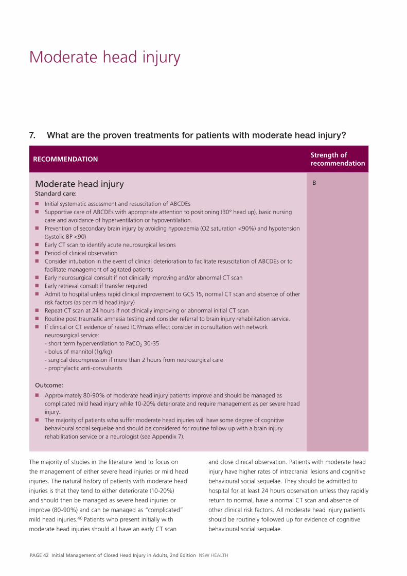
PAGE 42 Initial Management of Closed Head Injury in Adults, 2nd Edition NSW HEALTH
7. What are the proven treatments for patients with moderate head injury?
RECOMMENDATIONStrength of recommendation
Moderate head injury Standard care:
■ Initial systematic assessment and resuscitation of ABCDEs■ Supportive care of ABCDEs with appropriate attention to positioning (30° head up), basic nursing
care and avoidance of hyperventilation or hypoventilation.■ Prevention of secondary brain injury by avoiding hypoxaemia (O2 saturation <90%) and hypotension
(systolic BP <90)■ Early CT scan to identify acute neurosurgical lesions ■ Period of clinical observation■ Consider intubation in the event of clinical deterioration to facilitate resuscitation of ABCDEs or to
facilitate management of agitated patients■ Early neurosurgical consult if not clinically improving and/or abnormal CT scan■ Early retrieval consult if transfer required■ Admit to hospital unless rapid clinical improvement to GCS 15, normal CT scan and absence of other
risk factors (as per mild head injury)■ Repeat CT scan at 24 hours if not clinically improving or abnormal initial CT scan■ Routine post traumatic amnesia testing and consider referral to brain injury rehabilitation service.■ If clinical or CT evidence of raised ICP/mass effect consider in consultation with network
neurosurgical service:� - short term hyperventilation to PaCO2 30-35� - bolus of mannitol (1g/kg)� - surgical decompression if more than 2 hours from neurosurgical care� - prophylactic anti-convulsants
Outcome:
■ Approximately 80-90% of moderate head injury patients improve and should be managed as complicated mild head injury while 10-20% deteriorate and require management as per severe head injury..
■ The majority of patients who suffer moderate head injuries will have some degree of cognitive behavioural social sequelae and should be considered for routine follow up with a brain injury rehabilitation service or a neurologist (see Appendix 7).
B
Moderate head injury
The majority of studies in the literature tend to focus on
the management of either severe head injuries or mild head
injuries. The natural history of patients with moderate head
injuries is that they tend to either deteriorate (10-20%)
and should then be managed as severe head injuries or
improve (80-90%) and can be managed as “complicated”
mild head injuries.40 Patients who present initially with
moderate head injuries should all have an early CT scan
and close clinical observation. Patients with moderate head
injury have higher rates of intracranial lesions and cognitive
behavioural social sequelae. They should be admitted to
hospital for at least 24 hours observation unless they rapidly
return to normal, have a normal CT scan and absence of
other clinical risk factors. All moderate head injury patients
should be routinely followed up for evidence of cognitive
behavioural social sequelae.
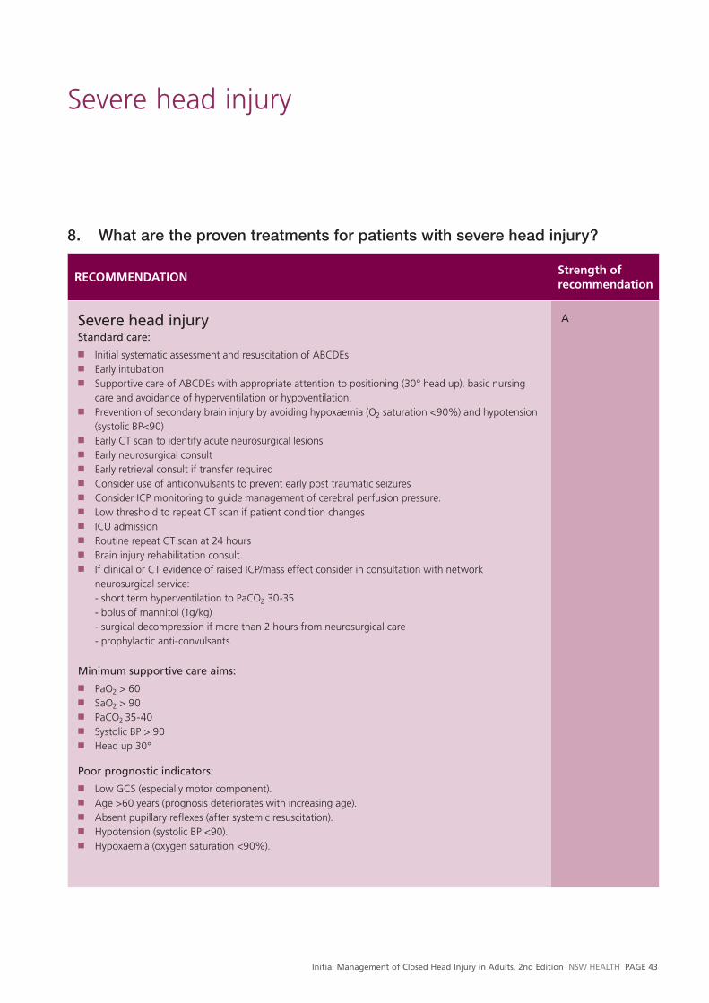
Initial Management of Closed Head Injury in Adults, 2nd Edition NSW HEALTH PAGE 43
8. What are the proven treatments for patients with severe head injury?
Severe head injury
RECOMMENDATIONStrength of recommendation
Severe head injuryStandard care:
■ Initial systematic assessment and resuscitation of ABCDEs■ Early intubation■ Supportive care of ABCDEs with appropriate attention to positioning (30° head up), basic nursing
care and avoidance of hyperventilation or hypoventilation.■ Prevention of secondary brain injury by avoiding hypoxaemia (O2 saturation <90%) and hypotension
(systolic BP<90)■ Early CT scan to identify acute neurosurgical lesions■ Early neurosurgical consult■ Early retrieval consult if transfer required■ Consider use of anticonvulsants to prevent early post traumatic seizures■ Consider ICP monitoring to guide management of cerebral perfusion pressure.■ Low threshold to repeat CT scan if patient condition changes■ ICU admission■ Routine repeat CT scan at 24 hours■ Brain injury rehabilitation consult■ If clinical or CT evidence of raised ICP/mass effect consider in consultation with network
neurosurgical service:� - short term hyperventilation to PaCO2 30-35� - bolus of mannitol (1g/kg)� - surgical decompression if more than 2 hours from neurosurgical care� - prophylactic anti-convulsants
Minimum supportive care aims:
■ PaO2 > 60■ SaO2 > 90■ PaCO2 35-40■ Systolic BP > 90■ Head up 30°
Poor prognostic indicators:
■ Low GCS (especially motor component).■ Age >60 years (prognosis deteriorates with increasing age).■ Absent pupillary refl exes (after systemic resuscitation).■ Hypotension (systolic BP <90).■ Hypoxaemia (oxygen saturation <90%).
A
PAGE 44 Initial Management of Closed Head Injury in Adults, 2nd Edition NSW HEALTH
Recent exhaustive reviews by the Brain Trauma Foundation15
and the Cochrane Review Group16-30 have looked at the
management of severe head injuries. The findings of
these detailed reviews are summarised in Evidence Table
4. This guideline summarises the generally accepted initial
management steps for severe head injury including those
recommended by the current Advanced Trauma Life
Support course and the Brain Trauma Foundation.15, 40 The
network neurosurgical service should be consulted about
further management of patients with severe head injury
as soon as practical after the initial primary survey and
resuscitation is completed.
It is important to recognise that for the majority of severe
head injury patients the most important aspect of care is
systematic resuscitation of the ABCDEs with prevention
of secondary brain injury as per current ATLS guidelines.40
Resuscitation of the ABCDEs with adequate oxygenation
and fluid resuscitation and the treatment of other
immediately life threatening injuries should be the priority
for patients with severe head injury followed by the CT
identification of focal intracranial lesions requiring acute
neurosurgical intervention.40, 157 Early intubation to prevent
hypoxaemia and facilitate management is recommended.40
Hyperventilation should be avoided and patients should
normally be ventilated to maintain normocarbia.24, 158 Fluid
resuscitation with normal saline or Hartmanns followed
by blood products to maintain normovolaemia and mean
arterial pressure is recommended.157 Anticonvulsants are
usually recommended to prevent early post traumatic
seizures particularly if there is an abnormal CT scan or
a history of a witnessed seizure.17, 29, 151 Blood glucose
and temperature should be monitored and maintained
in a normal range.159, 160 Thus, good supportive care
of the ABCDEs, with the prevention of hypoxaemia
and hypotension, remain the cornerstone of initial
management.40
The network neurological service should be urgently
consulted about any patient with an acute neurological
deterioration suggestive of acutely raised intracranial
pressure. In the event of acute deterioration, it is important
to remember that hyperventilation24, 158 and intravenous
mannitol boluses161,162 are short-term measures to reduce
intracranial pressure whilst the patient is urgently assessed
for the need for acute neurosurgical intervention. If an
acutely deteriorating patient with a proven extradural
or subdural haematoma cannot be transferred to a
neurosurgical service within two hours, then the option of
local surgical decompression should be discussed with the
neurosurgical service.
If the referring clinician is unsure about the need for a
particular therapy, such as prophylactic anticonvulsants
or antibiotics, then the network neurosurgical service
should be consulted. There are many other promising
areas of treatment such as induced hypothermia159, 160
and hypertonic saline.161 However, there is currently
insufficient evidence to recommend these as first line
therapy. Corticosteroids have been shown to worsen the
patient outcome and are not recommended for the initial
management of closed head injury.163, 164
Detailed evaluation of subsequent management of severe
head injuries by the neurosurgical services are beyond the
scope of these guidelines. There is limited high quality
evidence to guide management and the relative merits
of different strategies are hotly debated in the literature.
The Brain Trauma Foundation review, the Cochrane Group
reviews, the BMJ Clinical Evidence review165 and recent
Australian reviews2, 117 all agree that there is no class 1
evidence to guide management of severe head injury
patients. However, there is broad consensus agreement
that strategies to control cerebral perfusion using ICP
monitoring,27, 28, 61 mild hypothermia26, 66, 135 and
decompressive craniectomy166 are promising strategies that
are being widely used and require further study.
Predicting outcome following closed head injury is difficult.
There have been many attempts to provide scoring
systems to predict outcome with one of the best known
being that provided by the CRASH Investigators167. It
is perhaps prudent to err on the side of caution in the
initial management setting as at least 24-48 hours of
investigation and management are required before offering
any sort of prognosis.
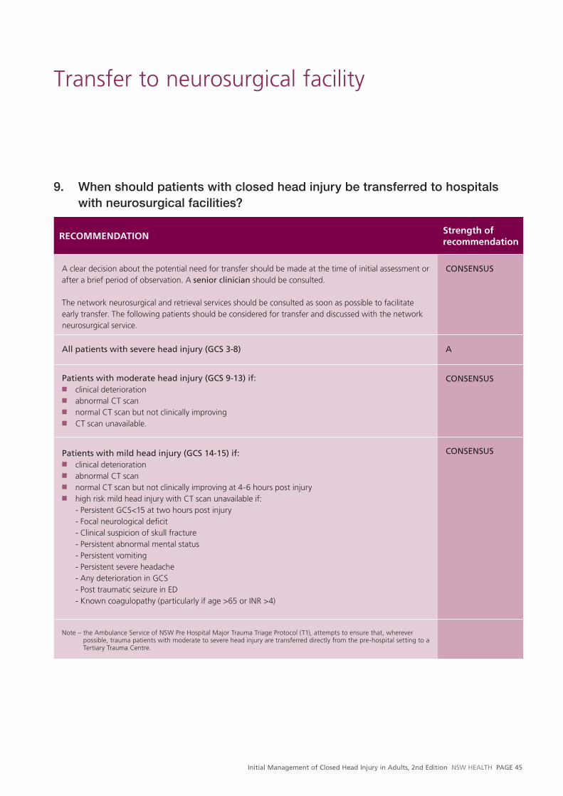
Initial Management of Closed Head Injury in Adults, 2nd Edition NSW HEALTH PAGE 45
9. When should patients with closed head injury be transferred to hospitals with neurosurgical facilities?
Transfer to neurosurgical facility
RECOMMENDATIONStrength of recommendation
A clear decision about the potential need for transfer should be made at the time of initial assessment or after a brief period of observation. A senior clinician should be consulted.
The network neurosurgical and retrieval services should be consulted as soon as possible to facilitate early transfer. The following patients should be considered for transfer and discussed with the network neurosurgical service.
CONSENSUS
All patients with severe head injury (GCS 3-8) A
Patients with moderate head injury (GCS 9-13) if: ■ clinical deterioration■ abnormal CT scan■ normal CT scan but not clinically improving■ CT scan unavailable.
CONSENSUS
Patients with mild head injury (GCS 14-15) if:■ clinical deterioration■ abnormal CT scan■ normal CT scan but not clinically improving at 4-6 hours post injury■ high risk mild head injury with CT scan unavailable if:� - Persistent GCS<15 at two hours post injury� - Focal neurological defi cit� - Clinical suspicion of skull fracture� - Persistent abnormal mental status � - Persistent vomiting� - Persistent severe headache � - Any deterioration in GCS� - Post traumatic seizure in ED � - Known coagulopathy (particularly if age >65 or INR >4)
CONSENSUS
Note – the Ambulance Service of NSW Pre Hospital Major Trauma Triage Protocol (T1), attempts to ensure that, wherever possible, trauma patients with moderate to severe head injury are transferred directly from the pre-hospital setting to a Tertiary Trauma Centre.
PAGE 46 Initial Management of Closed Head Injury in Adults, 2nd Edition NSW HEALTH
Patients with closed head injury should be observed in
facilities that can manage any complications that are likely
to arise. Clinical judgment regarding risk of deterioration is
required and early neurosurgical consultation is advisable.
Patients with closed head injury assessed at hospitals
without CT scanning facilities should be transferred to the
nearest appropriate hospital if there is significant risk of
intracranial injury. Transfer of patients to a hospital with CT
scanning facilities but without neurosurgical services should
be avoided wherever possible.
Fabbri et al41 recently published a study in which they
compared the outcome for mild to moderate head injury
patients and initial non-neurosurgical lesions on CT scan
when managed in neurosurgical units versus peripheral
hospitals. Their system allowed for rapid transfer of patients
to the tertiary centre in the event of deterioration and used
a teleradiology link to review CT scans. The outcome for
patients was not shown to be significantly different. Huynh
et al168 also showed in a retrospective study that trauma
patients with GCS 15 and an abnormal CT scan could be
safely managed without neurosurgical consultation. Both
these studies would support the current NSW practice of
managing some head injury patients in non-neurosurgical
centres following consultation with a network neurosurgical
centre.
The current guideline aims to address the question of when
to transfer patients from a non neurosurgical facility to a
neurosurgical facility. However, there has been increasing
interest in the issue of pre-hospital bypass of trauma
patients to ensure that they go directly to a tertiary hospital
with neurosurgical facilities. The primary benefit proposed
by this strategy is to avoid undue delay in transfer to
definitive care. There have been many studies such as the
one by Hartl et al169 that have shown reduced mortality
using such strategies. The Ambulance Service of NSW Pre
Hospital Major Trauma Triage Protocol (T1)170 has adopted
such a pre-hospital strategy for transferring all major
trauma patients directly to a major tertiary trauma hospital
or neurosurgical facility wherever possible.
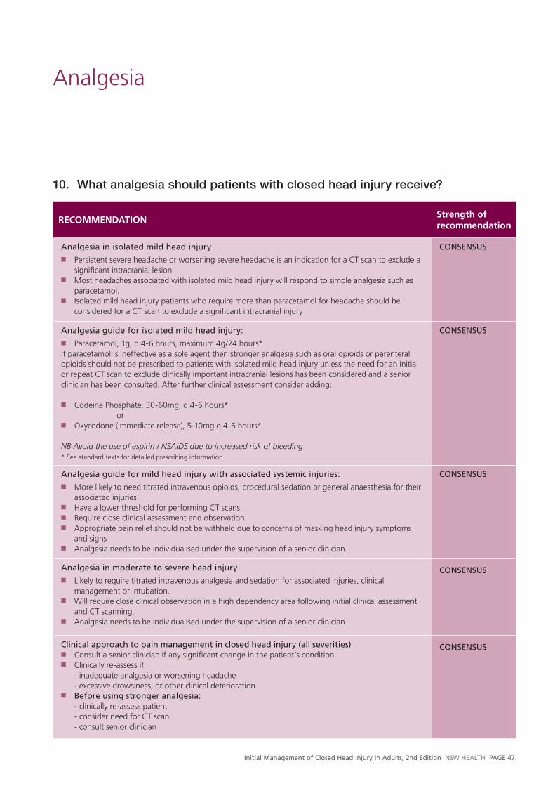
Initial Management of Closed Head Injury in Adults, 2nd Edition NSW HEALTH PAGE 47
10. What analgesia should patients with closed head injury receive?
RECOMMENDATIONStrength of recommendation
Analgesia in isolated mild head injury■ Persistent severe headache or worsening severe headache is an indication for a CT scan to exclude a
signifi cant intracranial lesion■ Most headaches associated with isolated mild head injury will respond to simple analgesia such as
paracetamol. ■ Isolated mild head injury patients who require more than paracetamol for headache should be
considered for a CT scan to exclude a signifi cant intracranial injury
CONSENSUS
Analgesia guide for isolated mild head injury:■ Paracetamol, 1g, q 4-6 hours, maximum 4g/24 hours*If paracetamol is ineffective as a sole agent then stronger analgesia such as oral opioids or parenteral opioids should not be prescribed to patients with isolated mild head injury unless the need for an initial or repeat CT scan to exclude clinically important intracranial lesions has been considered and a senior clinician has been consulted. After further clinical assessment consider adding;
■ Codeine Phosphate, 30-60mg, q 4-6 hours* or■ Oxycodone (immediate release), 5-10mg q 4-6 hours*
NB Avoid the use of aspirin / NSAIDS due to increased risk of bleeding* See standard texts for detailed prescribing information
CONSENSUS
Analgesia guide for mild head injury with associated systemic injuries:■ More likely to need titrated intravenous opioids, procedural sedation or general anaesthesia for their
associated injuries. ■ Have a lower threshold for performing CT scans.■ Require close clinical assessment and observation.■ Appropriate pain relief should not be withheld due to concerns of masking head injury symptoms
and signs■ Analgesia needs to be individualised under the supervision of a senior clinician.
CONSENSUS
Analgesia in moderate to severe head injury■ Likely to require titrated intravenous analgesia and sedation for associated injuries, clinical
management or intubation. ■ Will require close clinical observation in a high dependency area following initial clinical assessment
and CT scanning. ■ Analgesia needs to be individualised under the supervision of a senior clinician.
CONSENSUS
Clinical approach to pain management in closed head injury (all severities)■ Consult a senior clinician if any signifi cant change in the patient’s condition■ Clinically re-assess if: � - inadequate analgesia or worsening headache� - excessive drowsiness, or other clinical deterioration■ Before using stronger analgesia:� - clinically re-assess patient� - consider need for CT scan� - consult senior clinician
CONSENSUS
Analgesia
PAGE 48 Initial Management of Closed Head Injury in Adults, 2nd Edition NSW HEALTH
No prospective randomised multi-centre trials specifically
addressing the issue of analgesia in patients with closed
head injury could be identified. Interestingly, Bazarian
et al82 in a review of management of mild head injury
patients in selected emergency departments in the USA
found that nearly 50% of patients did not have assessment
of pain documented and 50% of those received no
analgesia. There are many studies addressing the issue of
analgesia in the broader patient population. The following
recommendations are based on existing analgesia clinical
practice guidelines including those available via the NSW
Health Clinical Information Access Program (CIAP) such as
Therapeutic Guidelines.171, 172
Mild head injury
Isolated mild head injury patients
■ Most headaches associated with isolated mild head
injuriy will respond to simple analgesia such as
paracetamol.■ If paracetamol (1g q 4-6 hr max 4g/day) alone is
ineffective then codeine phosphate (60mg q 4-6hr
max 240mg/daily) or low dose immediate release
oxycodone (5-10mg q 4-6hr) can be added after
the need for an initial or repeat CT scan to exclude
a clinically significant intracranial lesion has been
considered.■ The aim of analgesia in patients with isolated mild
head injury with associated headache should be to
relieve pain without causing excessive drowsiness.
Patients should always remain easy to rouse.■ Analgesics containing aspirin or NSAIDs should not be
used due to the increased risk of bleeding from platelet
dysfunction.■ Patients with persistent or worsening severe headaches
requesting increasing analgesia should be clinically
reassessed and the need for a CT scan to exclude a
clinically significant intracranial lesion considered.■ Patients who are already drowsy or difficult to rouse
should not be given additional opioids due to the risk
of respiratory depression. Patients with persistent or
worsening drowsiness should be clinically reassessed.■ If a headache has not responded to simple analgesics
or a patient is abnormally drowsy following analgesia
then the patient should be clinically re-assessed for
potential complications. Specific questions that should
be considered include:
1) Are there other signs of clinical deterioration
such as persistently abnormal or worsening
mental status, behaviour, drowsiness or
vomiting?
2) Does the patient require an initial CT scan or a
repeat CT scan to exclude a clinically important
cranial lesion?
3) Has a senior clinician been notified/consulted
about the change in the patient’s condition?
■ Stronger analgesia such as increased dosage or oral
opioid or parenteral opioid should not be prescribed to
patients with isolated mild head injury unless a senior
clinician has been consulted and clinically important
complications considered.
Notes:
■ see standard texts for precautions and toxicity eg,
Therapeutic Guidelines / MIMS■ avoid aspirin / NSAIDs due to risk of bleeding■ consult a senior clinician if any significant change in the
patient’s condition■ clinically re-assess if:
� - inadequate analgesia or worsening headache
� - excessive drowsiness, or other clinical deterioration
■ before using stronger analgesia:
� -clinically re-assess patient
� -consider need for CT scan
� -consult senior clinician
Mild head injury patients with other associated
injuries
■ The same general principles outlined for isolated
mild head injury patients apply. However, mild head
injury patients with other associated injuries are more
likely to need titrated intravenous opioids, procedural
sedation or general anaesthesia for their associated
injuries.■ Clearly, this has the potential to mask signs of
worsening head injury. Therefore, these patients
require close clinical observation and clinicians should
have a low threshold for performing CT scans on mild
head injury patients requiring intravenous opioids,
Initial Management of Closed Head Injury in Adults, 2nd Edition NSW HEALTH PAGE 49
procedural sedation or general anaesthesia for
associated injuries.■ Appropriate pain relief should not be withheld due to
concerns of masking head injury symptoms and signs
as the patients pain is likely to make clinical assessment
more difficult and mask other signs of injury and the
patient should not be allowed to suffer unnecessarily.
Moderate to severe head injury
■ Isolated moderate head injury patients who rapidly
clinically improve can be treated in a similar way to
mild head injury patients. They all require an initial CT
scan.■ However, most moderate head injury patients and
nearly all severe head injury patients will require
titrated intravenous analgesia and sedation for
associated injuries, clinical management or intubation.
These patients will all require close clinical observation
in a high dependency area following initial clinical
assessment and CT scanning. Analgesia needs to be
individualised under the supervision of senior clinicians.■ Routine analgesia with intravenous opioids is
recommended for most intubated patients as pain
may cause adverse effects on blood pressure and
intracranial pressure.
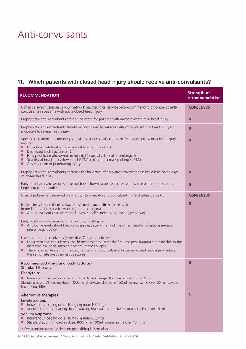
PAGE 50 Initial Management of Closed Head Injury in Adults, 2nd Edition NSW HEALTH
11. Which patients with closed head injury should receive anti-convulsants?
RECOMMENDATIONStrength of recommendation
Consult a senior clinician or your network neurosurgical service before commencing prophylactic anti-convulsants in patients with acute closed head injury
CONSENSUS
Prophylactic anti-convulsants are not indicated for patients with uncomplicated mild head injury B
Prophylactic anti-convulsants should be considered in patients with complicated mild head injury or moderate to severe head injury.
B
Specifi c indications to consider prophylactic anti-convulsants in the fi rst week following a head injury include:■ Extradural, subdural or intracerebral haematoma on CT■ Depressed skull fracture on CT■ Early post traumatic seizure in hospital (especially if focal or prolonged)■ Severity of head injury (low initial GCS / prolonged coma / prolonged PTA)■ Any suspicion of penetrating injury
B
Prophylactic anti-convulsants decrease the incidence of early post traumatic seizures within seven days of closed head injury.
B
Early post traumatic seizures have not been shown to be associated with worse patient outcomes in large population studies.
B
Clinical judgment is required on whether to prescribe anti-convulsants for individual patients. CONSENSUS
Indications for anti-convulsants by post traumatic seizure typeImmediate post traumatic seizures (at time of injury) ■ Anti-convulsants not warranted unless specifi c indication present (see above)
Early post traumatic seizures ( up to 7 days post injury)■ Anti-convulsants should be considered especially if any of the other specifi c indications are also
present (see above)
Late post traumatic seizures (more than 7 days post injury)■ Long term anti-convulsants should be considered after the fi rst late post traumatic seizure due to the
increased risk of developing post traumatic epilepsy■ There is no evidence that the routine use of anti-convulsants following closed head injury reduces
the risk of late post traumatic seizures.
B
Recommended drugs and loading doses*Standard therapy:Phenytoin:■ Intravenous loading dose: 20 mg/kg in NS (<6.7mg/ml) no faster than 50mg/min Standard adult IVI loading dose: 1000mg phenytoin diluted in 150ml normal saline over 60 mins with in line micron fi lter
B
Alternative therapies:Levetiracetam:■ Intravenous loading dose: 10mg /kg (max 1000mg)■ Standard adult IVI loading dose: 1000mg levetiracetam in 100ml normal saline over 15 mins
Sodium Valproate:■ Intravenous loading dose: 10mg /kg (max 800mg)■ Standard adult IVI loading dose: 800mg in 100ml normal saline over 15 mins
* See standard texts for detailed prescribing information
C
Anti-convulsants
Initial Management of Closed Head Injury in Adults, 2nd Edition NSW HEALTH PAGE 51
Post traumatic seizures are a recognised complication of
closed head injuries with incidence depending largely on
severity of injury. Penetrating injuries have a much higher
incidence of post traumatic seizures. Post traumatic seizures
are classified as either immediate, early (0-7 days) or late/
delayed (>7 days).144 Immediate and early post traumatic
seizures are thought to be associated with the acute injury
and are not significantly associated with the development
of post traumatic epilepsy. Late post traumatic seizures
are less likely to be related to the acute injury and are
more likely to be associated with the development of post
traumatic epilepsy.144
Brief generalised post traumatic seizures immediately
following mild head injury are not usually associated with
poor outcome or intracranial injury. These immediate
seizures are frequently seen on sporting fields and in
young children. It has been proposed that these immediate
seizures be called “concussive convulsions” and it has been
suggested that they are not an epileptic phenomena.64,
107, 134 McCrory et al145 demonstrated that outcome was
universally good for these “concussive convulsions” in elite
Australian sportsmen.
Immediate and early post traumatic seizures are relatively
common in patients with mild closed head injury with a
reported incidence of up to 5%.144, 173 The literature is
somewhat contradictory about the risk of seizures being
associated with intracranial injury. Many of the larger
studies found that post traumatic seizures were not
significantly associated with intracranial injury.85, 115, 168, 172,
174 However, Haydel et al32 found that they were significant
and Smits et al47 found that while they were not statistically
significant, included them in their decision rule because
of their perceived clinical importance. Neidlinger et al146
found that the “yield of unsuspected major intracranial
abnormality on CT scan justifies a policy of its routine
use in trauma patients with seizure or who are post ictal,
regardless of prior seizure history.”
Immediate or early post traumatic seizures are more
common (up to 30%) in patients with moderate to severe
closed head injury, and are more likely to be associated with
significant intracranial injury.144, 173 There is an association
with underlying structural lesions and the potential for
secondary brain injury especially with prolonged, focal
or delayed seizures. Any moderate to severe head injury
patient who has a post traumatic seizure warrants a CT
scan to exclude significant underlying pathology and
then the need for prophylactic anti-convulsants should be
considered.
The major risk factors for developing early post traumatic
seizures include lower initial GCS, depressed skull fracture,
penetrating injury, extradural/subdural/intracerebral
haematomas and young age.12, 17, 29, 64, 182 The risk is
therefore related to the amount of structural damage.
Penetrating injury provides the greatest risk. The risk posed
by an intracranial bleed is proportional to the amount of
blood.
Delayed or late post traumatic seizures (incidence range
1-15%) that occur more than seven days after injury are
associated with the development of post traumatic epilepsy.
Risk factors for late post traumatic seizures include lower
initial GCS, depressed skull fracture, penetrating injury,
extradural/subdural/intracerebral haematomas, elderly (age
>65), neurosurgical intervention and early post traumatic
seizures.12, 17, 29, 64, 182
Acute post traumatic seizures require systematic
reassessment of the ABCDEs to exclude systemic causes and
termination with benzodiazepines if required. Underlying
structural lesions should be excluded with CT scan and then
the need for prophylactic anti-convulsants considered.
If prophylactic anti-convulsants are recommended then
phenytoin (dilantin) is normally given as there has been
extensive experience with its use and it can be given as
either an oral or an intravenous loading dose.11, 17, 29, 69,
151 Alternatives include sodium valproate (epilim) and
levetiracetam (keppra). Levetiracetam is being increasingly
used in both non traumatic and traumatic epilepsy due
to its better side effect profile and may become first line
therapy in the future.174, 175 However, there is limited
experience with its use in the trauma setting and phenytoin
remains the first line therapy at present.
■ Current evidence suggests that anti-convulsants
decrease the incidence of early post traumatic seizures
within seven days of closed head injury.17, 29, 151 ■ Early post traumatic seizures have not been shown to
be clearly associated with worse patient outcome.17, 29,
151 ■ There is no evidence that prophylactic anti-convulsants
following closed head injury reduce the risk of late post
traumatic seizures.17, 29, 151
■ Anti-convulsants as a group have many potential side
effects and are relatively poorly tolerated by patients
long term.17, 29, 151
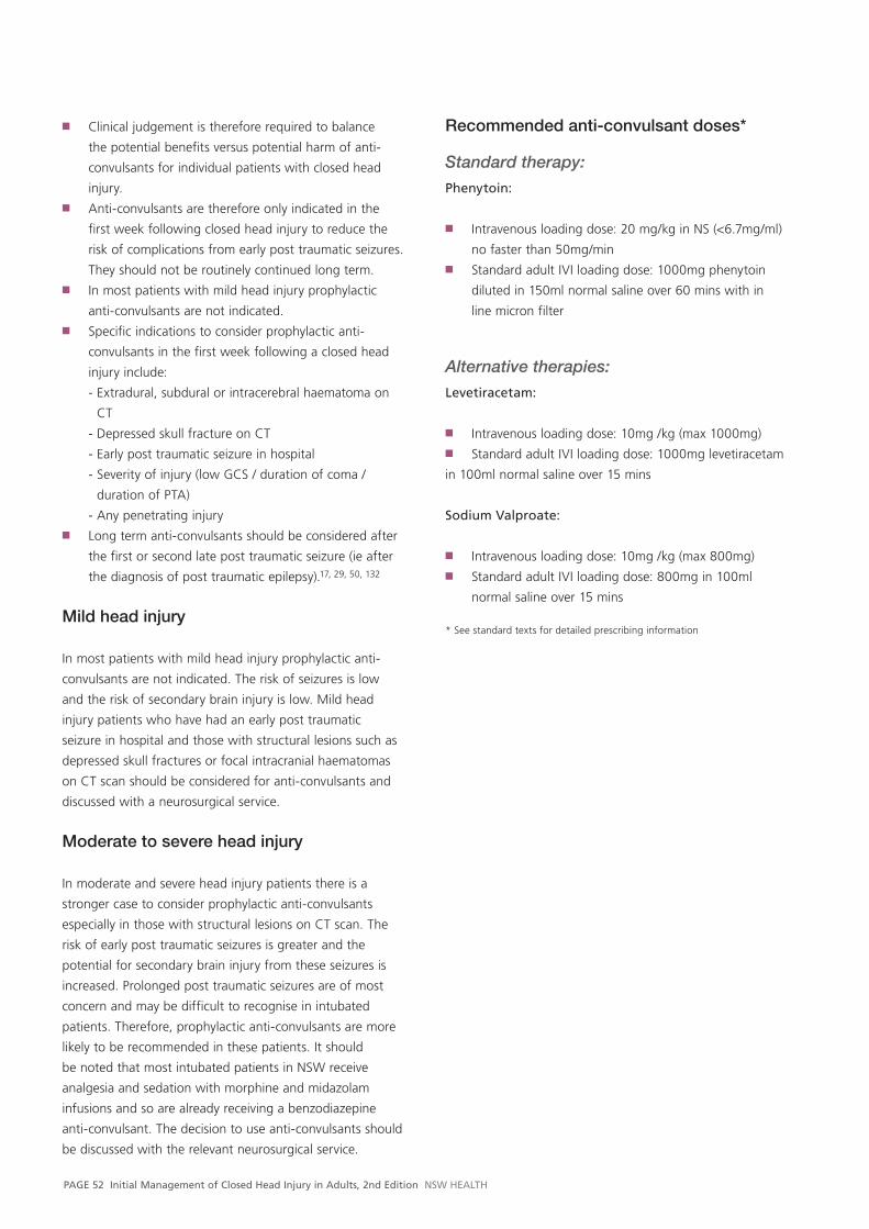
PAGE 52 Initial Management of Closed Head Injury in Adults, 2nd Edition NSW HEALTH
■ Clinical judgement is therefore required to balance
the potential benefits versus potential harm of anti-
convulsants for individual patients with closed head
injury. ■ Anti-convulsants are therefore only indicated in the
first week following closed head injury to reduce the
risk of complications from early post traumatic seizures.
They should not be routinely continued long term.■ In most patients with mild head injury prophylactic
anti-convulsants are not indicated.■ Specific indications to consider prophylactic anti-
convulsants in the first week following a closed head
injury include:
- Extradural, subdural or intracerebral haematoma on
CT
- Depressed skull fracture on CT
- Early post traumatic seizure in hospital
- Severity of injury (low GCS / duration of coma /
duration of PTA)
- Any penetrating injury■ Long term anti-convulsants should be considered after
the first or second late post traumatic seizure (ie after
the diagnosis of post traumatic epilepsy).17, 29, 50, 132
Mild head injury
In most patients with mild head injury prophylactic anti-
convulsants are not indicated. The risk of seizures is low
and the risk of secondary brain injury is low. Mild head
injury patients who have had an early post traumatic
seizure in hospital and those with structural lesions such as
depressed skull fractures or focal intracranial haematomas
on CT scan should be considered for anti-convulsants and
discussed with a neurosurgical service.
Moderate to severe head injury
In moderate and severe head injury patients there is a
stronger case to consider prophylactic anti-convulsants
especially in those with structural lesions on CT scan. The
risk of early post traumatic seizures is greater and the
potential for secondary brain injury from these seizures is
increased. Prolonged post traumatic seizures are of most
concern and may be difficult to recognise in intubated
patients. Therefore, prophylactic anti-convulsants are more
likely to be recommended in these patients. It should
be noted that most intubated patients in NSW receive
analgesia and sedation with morphine and midazolam
infusions and so are already receiving a benzodiazepine
anti-convulsant. The decision to use anti-convulsants should
be discussed with the relevant neurosurgical service.
Recommended anti-convulsant doses*
Standard therapy:
Phenytoin:
■ Intravenous loading dose: 20 mg/kg in NS (<6.7mg/ml)
no faster than 50mg/min ■ Standard adult IVI loading dose: 1000mg phenytoin
diluted in 150ml normal saline over 60 mins with in
line micron filter
Alternative therapies:
Levetiracetam:
■ Intravenous loading dose: 10mg /kg (max 1000mg)■ Standard adult IVI loading dose: 1000mg levetiracetam
in 100ml normal saline over 15 mins
Sodium Valproate:
■ Intravenous loading dose: 10mg /kg (max 800mg)■ Standard adult IVI loading dose: 800mg in 100ml
normal saline over 15 mins
* See standard texts for detailed prescribing information
Initial Management of Closed Head Injury in Adults, 2nd Edition NSW HEALTH PAGE 53
1. Studies examining the definition of mild head injury and the assessment of mild head injury patients (including CT scanning)
2. Complications of mild closed head injury
3. Optimal management strategy for high-risk mild head injury patients when CT scanning is not available
4. Proven treatments for moderate to severe head injury
5. Transfer of patients with a closed head injury
6. Discharge of patients with a mild head injury
Evidence Tables
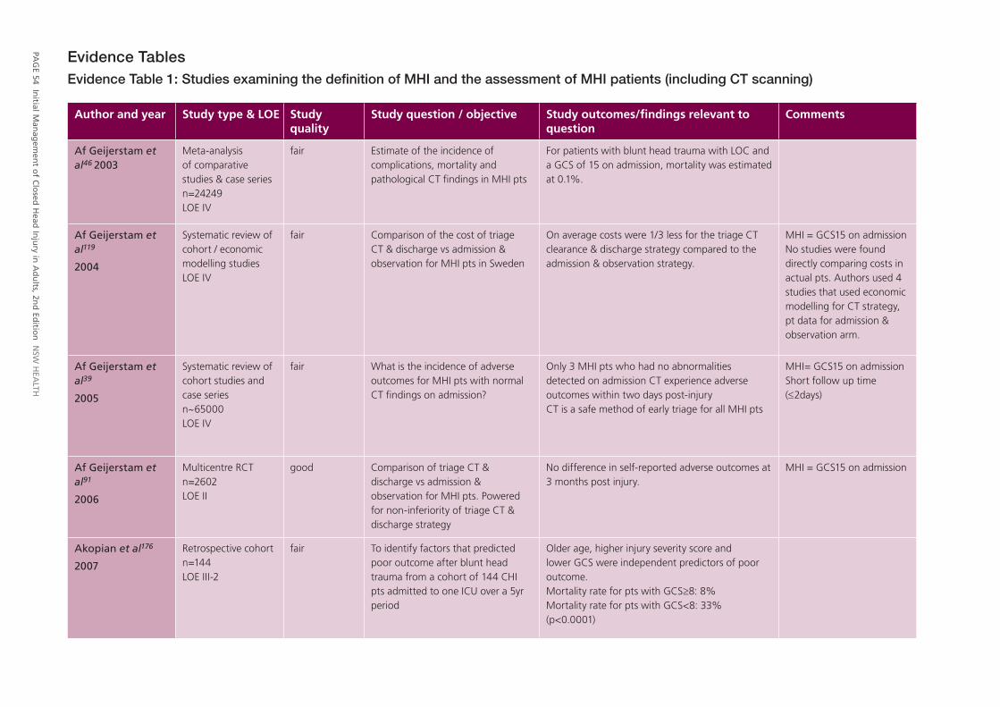
PAG
E 54 Initial M
anag
emen
t of C
losed
Head
Inju
ry in A
du
lts, 2nd
Editio
n N
SW H
EALTH
Evidence TablesEvidence Table 1: Studies examining the definition of MHI and the assessment of MHI patients (including CT scanning)
Author and year Study type & LOE Study quality
Study question / objective Study outcomes/fi ndings relevant to question
Comments
Af Geijerstam et al46 2003
Meta-analysis of comparative studies & case series n=24249 LOE IV
fair Estimate of the incidence of complications, mortality and pathological CT fi ndings in MHI pts
For patients with blunt head trauma with LOC and a GCS of 15 on admission, mortality was estimated at 0.1%.
Af Geijerstam et al119
2004
Systematic review of cohort / economic modelling studiesLOE IV
fair Comparison of the cost of triage CT & discharge vs admission & observation for MHI pts in Sweden
On average costs were 1/3 less for the triage CT clearance & discharge strategy compared to the admission & observation strategy.
MHI = GCS15 on admissionNo studies were found directly comparing costs in actual pts. Authors used 4 studies that used economic modelling for CT strategy, pt data for admission & observation arm.
Af Geijerstam et al39
2005
Systematic review of cohort studies and case seriesn~65000LOE IV
fair What is the incidence of adverse outcomes for MHI pts with normal CT fi ndings on admission?
Only 3 MHI pts who had no abnormalities detected on admission CT experience adverse outcomes within two days post-injuryCT is a safe method of early triage for all MHI pts
MHI= GCS15 on admissionShort follow up time (2days)
Af Geijerstam et al91
2006
Multicentre RCTn=2602LOE II
good Comparison of triage CT & discharge vs admission & observation for MHI pts. Powered for non-inferiority of triage CT & discharge strategy
No difference in self-reported adverse outcomes at 3 months post injury.
MHI = GCS15 on admission
Akopian et al176
2007
Retrospective cohort n=144LOE III-2
fair To identify factors that predicted poor outcome after blunt head trauma from a cohort of 144 CHI pts admitted to one ICU over a 5yr period
Older age, higher injury severity score and lower GCS were independent predictors of poor outcome.Mortality rate for pts with GCS8: 8%Mortality rate for pts with GCS<8: 33%(p<0.0001)
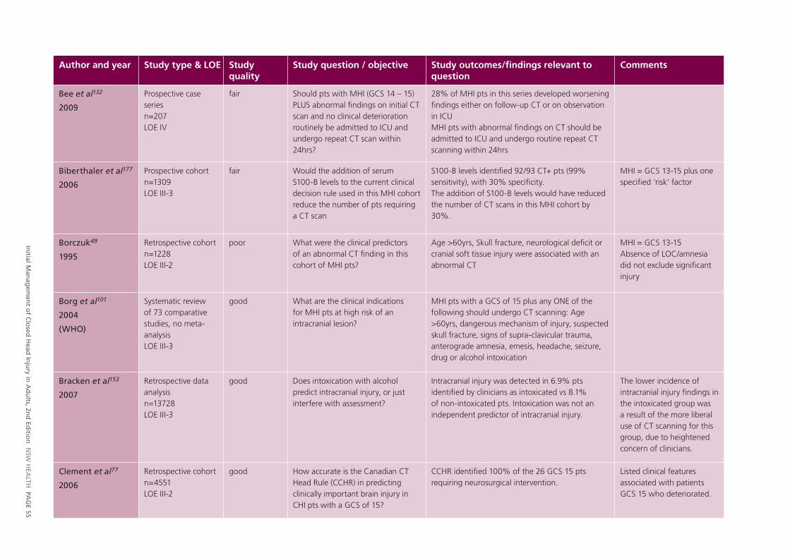
Initial M
anag
emen
t of C
losed
Head
Inju
ry in A
du
lts, 2nd
Editio
n N
SW H
EALTH
PAG
E 55
Author and year Study type & LOE Study quality
Study question / objective Study outcomes/fi ndings relevant to question
Comments
Bee et al132
2009
Prospective case seriesn=207 LOE IV
fair Should pts with MHI (GCS 14 – 15) PLUS abnormal fi ndings on initial CT scan and no clinical deterioration routinely be admitted to ICU and undergo repeat CT scan within 24hrs?
28% of MHI pts in this series developed worsening fi ndings either on follow-up CT or on observation in ICUMHI pts with abnormal fi ndings on CT should be admitted to ICU and undergo routine repeat CT scanning within 24hrs
Biberthaler et al177
2006
Prospective cohortn=1309LOE III-3
fair Would the addition of serum S100-B levels to the current clinical decision rule used in this MHI cohort reduce the number of pts requiring a CT scan
S100-B levels identifi ed 92/93 CT+ pts (99% sensitivity), with 30% specifi city.The addition of S100-B levels would have reduced the number of CT scans in this MHI cohort by 30%.
MHI = GCS 13-15 plus one specifi ed ‘risk’ factor
Borczuk49
1995
Retrospective cohortn=1228LOE III-2
poor What were the clinical predictors of an abnormal CT fi nding in this cohort of MHI pts?
Age >60yrs, Skull fracture, neurological defi cit or cranial soft tissue injury were associated with an abnormal CT
MHI = GCS 13-15Absence of LOC/amnesia did not exclude signifi cant injury
Borg et al101
2004
(WHO)
Systematic review of 73 comparative studies, no meta-analysisLOE III-3
good What are the clinical indications for MHI pts at high risk of an intracranial lesion?
MHI pts with a GCS of 15 plus any ONE of the following should undergo CT scanning: Age >60yrs, dangerous mechanism of injury, suspected skull fracture, signs of supra-clavicular trauma, anterograde amnesia, emesis, headache, seizure, drug or alcohol intoxication
Bracken et al153
2007
Retrospective data analysisn=13728LOE III-3
good Does intoxication with alcohol predict intracranial injury, or just interfere with assessment?
Intracranial injury was detected in 6.9% pts identifi ed by clinicians as intoxicated vs 8.1% of non-intoxicated pts. Intoxication was not an independent predictor of intracranial injury.
The lower incidence of intracranial injury fi ndings in the intoxicated group was a result of the more liberal use of CT scanning for this group, due to heightened concern of clinicians.
Clement et al77
2006
Retrospective cohortn=4551LOE III-2
good How accurate is the Canadian CT Head Rule (CCHR) in predicting clinically important brain injury in CHI pts with a GCS of 15?
CCHR identifi ed 100% of the 26 GCS 15 pts requiring neurosurgical intervention.
Listed clinical features associated with patients GCS 15 who deteriorated.
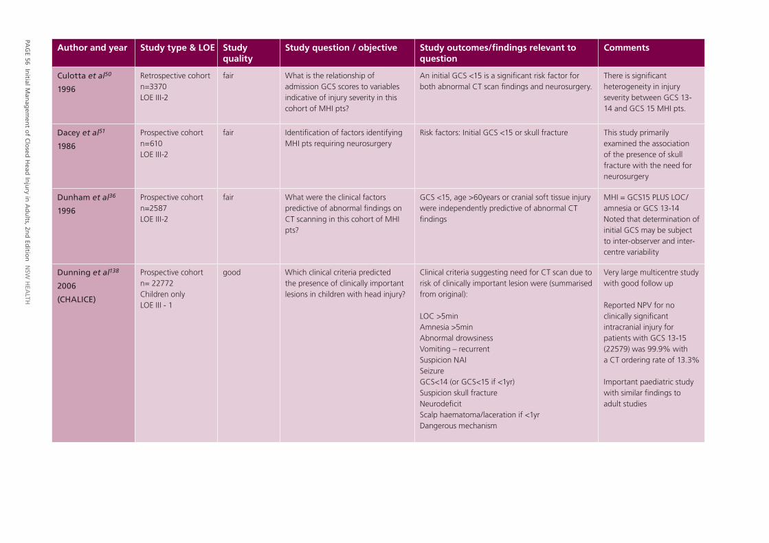
PAG
E 56 Initial M
anag
emen
t of C
losed
Head
Inju
ry in A
du
lts, 2nd
Editio
n N
SW H
EALTH
Author and year Study type & LOE Study quality
Study question / objective Study outcomes/fi ndings relevant to question
Comments
Culotta et al50
1996
Retrospective cohortn=3370LOE III-2
fair What is the relationship of admission GCS scores to variables indicative of injury severity in this cohort of MHI pts?
An initial GCS <15 is a signifi cant risk factor for both abnormal CT scan fi ndings and neurosurgery.
There is signifi cant heterogeneity in injury severity between GCS 13-14 and GCS 15 MHI pts.
Dacey et al51
1986
Prospective cohortn=610LOE III-2
fair Identifi cation of factors identifying MHI pts requiring neurosurgery
Risk factors: Initial GCS <15 or skull fracture This study primarily examined the association of the presence of skull fracture with the need for neurosurgery
Dunham et al36
1996
Prospective cohortn=2587LOE III-2
fair What were the clinical factors predictive of abnormal fi ndings on CT scanning in this cohort of MHI pts?
GCS <15, age >60years or cranial soft tissue injury were independently predictive of abnormal CT fi ndings
MHI = GCS15 PLUS LOC/amnesia or GCS 13-14Noted that determination of initial GCS may be subject to inter-observer and inter-centre variability
Dunning et al138
2006
(CHALICE)
Prospective cohort n= 22772 Children onlyLOE III - 1
good Which clinical criteria predicted the presence of clinically important lesions in children with head injury?
Clinical criteria suggesting need for CT scan due to risk of clinically important lesion were (summarised from original):
LOC >5minAmnesia >5minAbnormal drowsinessVomiting – recurrentSuspicion NAISeizureGCS<14 (or GCS<15 if <1yr)Suspicion skull fractureNeurodefi citScalp haematoma/laceration if <1yrDangerous mechanism
Very large multicentre study with good follow up
Reported NPV for no clinically signifi cant intracranial injury for patients with GCS 13-15 (22579) was 99.9% with a CT ordering rate of 13.3%
Important paediatric study with similar fi ndings to adult studies
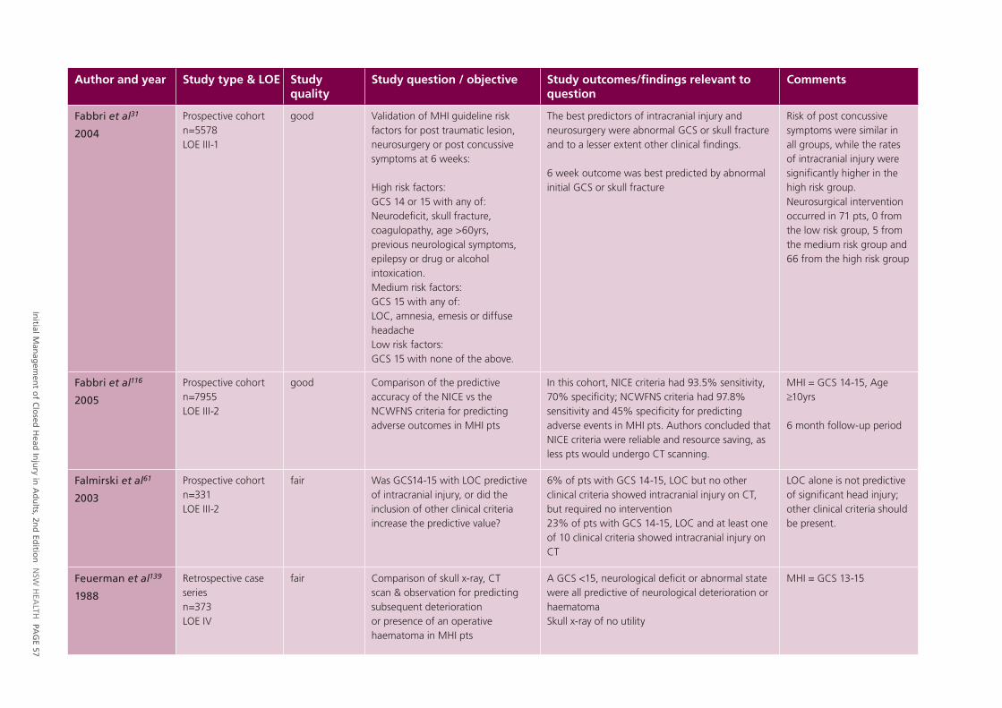
Initial M
anag
emen
t of C
losed
Head
Inju
ry in A
du
lts, 2nd
Editio
n N
SW H
EALTH
PAG
E 57
Author and year Study type & LOE Study quality
Study question / objective Study outcomes/fi ndings relevant to question
Comments
Fabbri et al31
2004
Prospective cohortn=5578LOE III-1
good Validation of MHI guideline risk factors for post traumatic lesion, neurosurgery or post concussive symptoms at 6 weeks:
High risk factors:GCS 14 or 15 with any of:Neurodefi cit, skull fracture, coagulopathy, age >60yrs,previous neurological symptoms, epilepsy or drug or alcohol intoxication.Medium risk factors:GCS 15 with any of:LOC, amnesia, emesis or diffuse headacheLow risk factors:GCS 15 with none of the above.
The best predictors of intracranial injury and neurosurgery were abnormal GCS or skull fracture and to a lesser extent other clinical fi ndings.
6 week outcome was best predicted by abnormal initial GCS or skull fracture
Risk of post concussive symptoms were similar in all groups, while the rates of intracranial injury were signifi cantly higher in the high risk group.Neurosurgical intervention occurred in 71 pts, 0 from the low risk group, 5 from the medium risk group and 66 from the high risk group
Fabbri et al116
2005
Prospective cohortn=7955LOE III-2
good Comparison of the predictive accuracy of the NICE vs the NCWFNS criteria for predicting adverse outcomes in MHI pts
In this cohort, NICE criteria had 93.5% sensitivity, 70% specifi city; NCWFNS criteria had 97.8% sensitivity and 45% specifi city for predicting adverse events in MHI pts. Authors concluded that NICE criteria were reliable and resource saving, as less pts would undergo CT scanning.
MHI = GCS 14-15, Age 10yrs
6 month follow-up period
Falmirski et al61
2003
Prospective cohortn=331LOE III-2
fair Was GCS14-15 with LOC predictive of intracranial injury, or did the inclusion of other clinical criteria increase the predictive value?
6% of pts with GCS 14-15, LOC but no other clinical criteria showed intracranial injury on CT, but required no intervention23% of pts with GCS 14-15, LOC and at least one of 10 clinical criteria showed intracranial injury on CT
LOC alone is not predictive of signifi cant head injury; other clinical criteria should be present.
Feuerman et al139
1988
Retrospective case seriesn=373LOE IV
fair Comparison of skull x-ray, CT scan & observation for predicting subsequent deterioration or presence of an operative haematoma in MHI pts
A GCS <15, neurological defi cit or abnormal state were all predictive of neurological deterioration or haematomaSkull x-ray of no utility
MHI = GCS 13-15
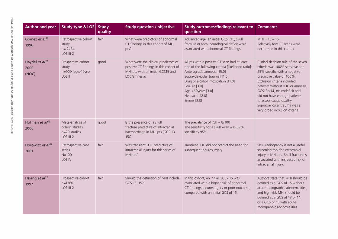
PAG
E 58 Initial M
anag
emen
t of C
losed
Head
Inju
ry in A
du
lts, 2nd
Editio
n N
SW H
EALTH
Author and year Study type & LOE Study quality
Study question / objective Study outcomes/fi ndings relevant to question
Comments
Gomez et al62
1996
Retrospective cohort studyn= 2484LOE III-2
fair What were predictors of abnormal CT fi ndings in this cohort of MHI pts?
Advanced age, an initial GCS <15, skull fracture or focal neurological defi cit were associated with abnormal CT fi ndings
MHI = 13 – 15Relatively few CT scans were performed in this cohort
Haydel et al32
2000
(NOC)
Prospective cohort studyn=909 (age>10yrs)LOE II
good What were the clinical predictors of positive CT fi ndings in this cohort of MHI pts with an initial GCS15 and LOC/amnesia?
All pts with a positive CT scan had at least one of the following criteria [likelihood ratio]:Anterograde amnesia [15.0]Supra-clavicular trauma [11.0]Drug or alcohol intoxication [11.0]Seizure [3.0]Age >60years [3.0]Headache [2.0]Emesis [2.0]
Clinical decision rule of the seven criteria was 100% sensitive and 25% specifi c with a negative predictive value of 100%.Exclusion criteria included patients without LOC or amnesia, GCS13or14, neurodefi cit and did not have enough patients to assess coagulopathy. Supraclavicular trauma was a very broad inclusion criteria.
Hofman et al66
2000
Meta-analysis of cohort studiesn=20 studiesLOE III-2
good Is the presence of a skull fracture predictive of intracranial haemorrhage in MHI pts (GCS 13-15)?
The prevalence of ICH ~ 8/100The sensitivity for a skull x-ray was 39%, specifi city 95%
Horowitz et al67
2001
Retrospective case series N=100LOE IV
fair Was transient LOC predictive of intracranial injury for this series of MHI pts?
Transient LOC did not predict the need for subsequent neurosurgery
Skull radiography is not a useful screening tool for intracranial injury in MHI pts. Skull fracture is associated with increased risk of intracranial injury.
Hsiang et al52
1997
Prospective cohortn=1360LOE III-2
fair Should the defi nition of MHI include GCS 13 -15?
In this cohort, an initial GCS <15 was associated with a higher risk of abnormal CT fi ndings, neurosurgery or poor outcome, compared with an initial GCS of 15.
Authors state that MHI should be defi ned as a GCS of 15 without acute radiographic abnormalities, and high-risk MHI should be defi ned as a GCS of 13 or 14, or a GCS of 15 with acute radiographic abnormalities
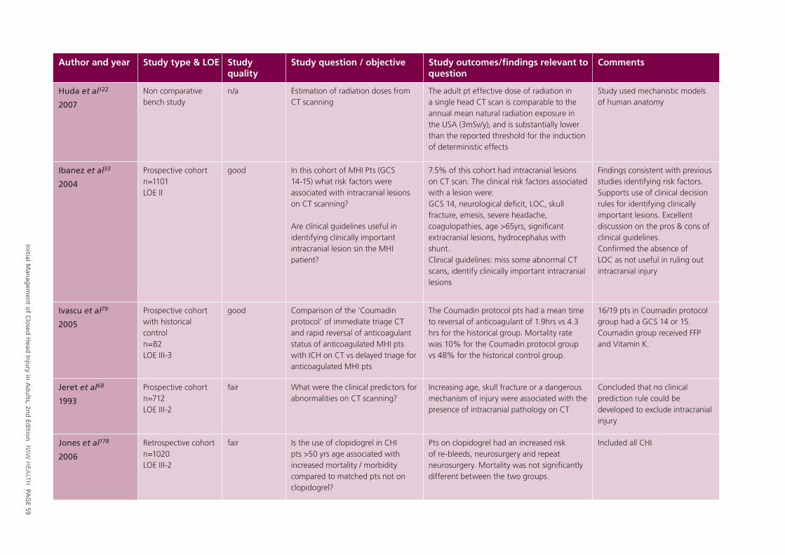
Initial M
anag
emen
t of C
losed
Head
Inju
ry in A
du
lts, 2nd
Editio
n N
SW H
EALTH
PAG
E 59
Author and year Study type & LOE Study quality
Study question / objective Study outcomes/fi ndings relevant to question
Comments
Huda et al122
2007
Non comparative bench study
n/a Estimation of radiation doses from CT scanning
The adult pt effective dose of radiation in a single head CT scan is comparable to the annual mean natural radiation exposure in the USA (3mSv/y), and is substantially lower than the reported threshold for the induction of deterministic effects
Study used mechanistic models of human anatomy
Ibanez et al33
2004
Prospective cohortn=1101LOE II
good In this cohort of MHI Pts (GCS 14-15) what risk factors were associated with intracranial lesions on CT scanning? Are clinical guidelines useful in identifying clinically important intracranial lesion sin the MHI patient?
7.5% of this cohort had intracranial lesions on CT scan. The clinical risk factors associated with a lesion were:GCS 14, neurological defi cit, LOC, skull fracture, emesis, severe headache, coagulopathies, age >65yrs, signifi cant extracranial lesions, hydrocephalus with shunt.Clinical guidelines: miss some abnormal CT scans, identify clinically important intracranial lesions
Findings consistent with previous studies identifying risk factors. Supports use of clinical decision rules for identifying clinically important lesions. Excellent discussion on the pros & cons of clinical guidelines.Confi rmed the absence of LOC as not useful in ruling out intracranial injury
Ivascu et al79
2005
Prospective cohort with historical controln=82LOE III-3
good Comparison of the ‘Coumadin protocol’ of immediate triage CT and rapid reversal of anticoagulant status of anticoagulated MHI pts with ICH on CT vs delayed triage for anticoagulated MHI pts
The Coumadin protocol pts had a mean time to reversal of anticoagulant of 1.9hrs vs 4.3 hrs for the historical group. Mortality rate was 10% for the Coumadin protocol group vs 48% for the historical control group.
16/19 pts in Coumadin protocol group had a GCS 14 or 15.Coumadin group received FFP and Vitamin K.
Jeret et al68
1993
Prospective cohortn=712LOE III-2
fair What were the clinical predictors for abnormalities on CT scanning?
Increasing age, skull fracture or a dangerous mechanism of injury were associated with the presence of intracranial pathology on CT
Concluded that no clinical prediction rule could be developed to exclude intracranial injury
Jones et al178
2006
Retrospective cohortn=1020LOE III-2
fair Is the use of clopidogrel in CHI pts >50 yrs age associated with increased mortality / morbidity compared to matched pts not on clopidogrel?
Pts on clopidogrel had an increased risk of re-bleeds, neurosurgery and repeat neurosurgery. Mortality was not signifi cantly different between the two groups.
Included all CHI
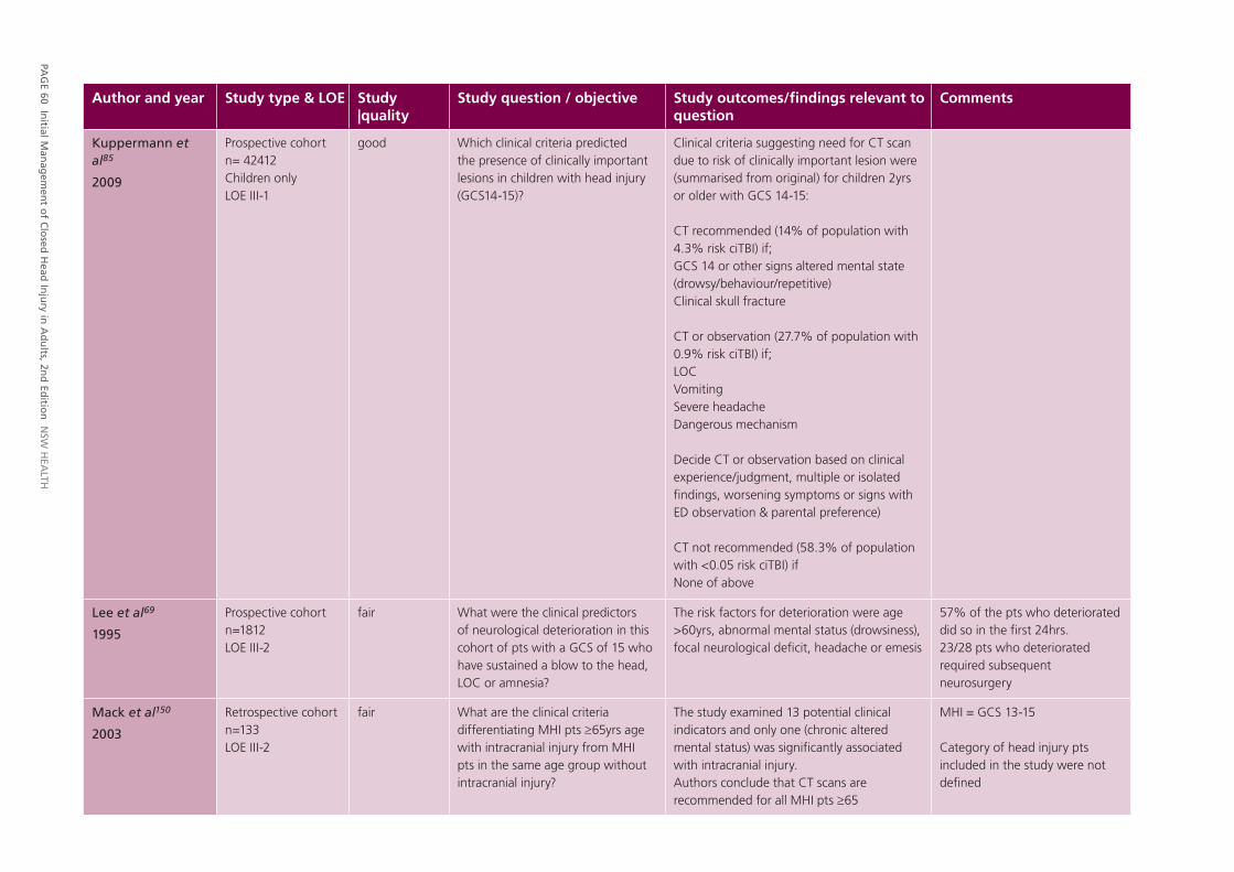
PAG
E 60 Initial M
anag
emen
t of C
losed
Head
Inju
ry in A
du
lts, 2nd
Editio
n N
SW H
EALTH
Author and year Study type & LOE Study |quality
Study question / objective Study outcomes/fi ndings relevant to question
Comments
Kuppermann et al85
2009
Prospective cohort n= 42412 Children onlyLOE III-1
good Which clinical criteria predicted the presence of clinically important lesions in children with head injury (GCS14-15)?
Clinical criteria suggesting need for CT scan due to risk of clinically important lesion were (summarised from original) for children 2yrs or older with GCS 14-15:
CT recommended (14% of population with 4.3% risk ciTBI) if;GCS 14 or other signs altered mental state (drowsy/behaviour/repetitive)Clinical skull fracture
CT or observation (27.7% of population with 0.9% risk ciTBI) if;LOCVomitingSevere headacheDangerous mechanism
Decide CT or observation based on clinical experience/judgment, multiple or isolated fi ndings, worsening symptoms or signs with ED observation & parental preference)
CT not recommended (58.3% of population with <0.05 risk ciTBI) ifNone of above
Lee et al69
1995
Prospective cohortn=1812LOE III-2
fair What were the clinical predictors of neurological deterioration in this cohort of pts with a GCS of 15 who have sustained a blow to the head, LOC or amnesia?
The risk factors for deterioration were age >60yrs, abnormal mental status (drowsiness), focal neurological defi cit, headache or emesis
57% of the pts who deteriorated did so in the fi rst 24hrs.23/28 pts who deteriorated required subsequent neurosurgery
Mack et al150
2003
Retrospective cohortn=133LOE III-2
fair What are the clinical criteria differentiating MHI pts 65yrs age with intracranial injury from MHI pts in the same age group without intracranial injury?
The study examined 13 potential clinical indicators and only one (chronic altered mental status) was signifi cantly associated with intracranial injury.Authors conclude that CT scans are recommended for all MHI pts 65
MHI = GCS 13-15
Category of head injury pts included in the study were not defi ned
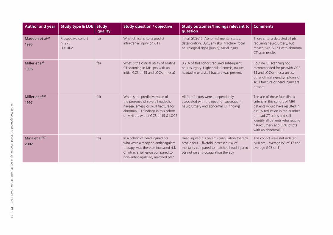
Initial M
anag
emen
t of C
losed
Head
Inju
ry in A
du
lts, 2nd
Editio
n N
SW H
EALTH
PAG
E 61
Author and year Study type & LOE Study |quality
Study question / objective Study outcomes/fi ndings relevant to question
Comments
Madden et al70
1995
Prospective cohortn=273LOE III-2
fair What clinical criteria predict intracranial injury on CT?
Initial GCS<15, Abnormal mental status, deterioration, LOC, any skull fracture, focal neurological signs (pupils), facial injury
These criteria detected all pts requiring neurosurgery, but missed two 2/273 with abnormal CT scan results
Miller et al71
1996
fair What is the clinical utility of routine CT scanning in MHI pts with an initial GCS of 15 and LOC/amnesia?
0.2% of this cohort required subsequent neurosurgery. Higher risk if emesis, nausea, headache or a skull fracture was present.
Routine CT scanning not recommended for pts with GCS 15 and LOC/amnesia unless other clinical signs/symptoms of skull fracture or head injury are present
Miller et al88
1997
fair What is the predictive value of the presence of severe headache, nausea, emesis or skull fracture for abnormal CT fi ndings in this cohort of MHI pts with a GCS of 15 & LOC?
All four factors were independently associated with the need for subsequent neurosurgery and abnormal CT fi ndings
The use of these four clinical criteria in this cohort of MHI patients would have resulted in a 61% reduction in the number of head CT scans and still identify all patients who require neurosurgery and 65% of pts with an abnormal CT
Mina et al147
2002
fair In a cohort of head injured pts who were already on anticoagulant therapy, was there an increased risk of intracranial lesion compared to non-anticoagulated, matched pts?
Head injured pts on anti-coagulation therapy have a four – fi vefold increased risk of mortality compared to matched head-injured pts not on anti-coagulation therapy
This cohort were not isolated MHI pts – average ISS of 17 and average GCS of 11
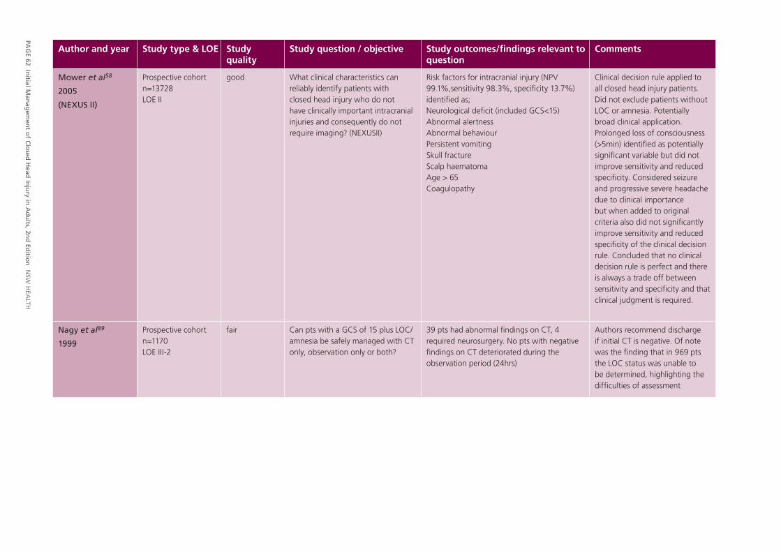
PAG
E 62 Initial M
anag
emen
t of C
losed
Head
Inju
ry in A
du
lts, 2nd
Editio
n N
SW H
EALTH
Author and year Study type & LOE Study quality
Study question / objective Study outcomes/fi ndings relevant to question
Comments
Mower et al58
2005
(NEXUS II)
Prospective cohort n=13728LOE II
good What clinical characteristics can reliably identify patients with closed head injury who do not have clinically important intracranial injuries and consequently do not require imaging? (NEXUSII)
Risk factors for intracranial injury (NPV 99.1%,sensitivity 98.3%, specifi city 13.7%) identifi ed as;Neurological defi cit (included GCS<15)Abnormal alertnessAbnormal behaviourPersistent vomitingSkull fractureScalp haematomaAge > 65Coagulopathy
Clinical decision rule applied to all closed head injury patients. Did not exclude patients without LOC or amnesia. Potentially broad clinical application. Prolonged loss of consciousness (>5min) identifi ed as potentially signifi cant variable but did not improve sensitivity and reduced specifi city. Considered seizure and progressive severe headache due to clinical importance but when added to original criteria also did not signifi cantly improve sensitivity and reduced specifi city of the clinical decision rule. Concluded that no clinical decision rule is perfect and there is always a trade off between sensitivity and specifi city and that clinical judgment is required.
Nagy et al89
1999
Prospective cohortn=1170LOE III-2
fair Can pts with a GCS of 15 plus LOC/amnesia be safely managed with CT only, observation only or both?
39 pts had abnormal fi ndings on CT, 4 required neurosurgery. No pts with negative fi ndings on CT deteriorated during the observation period (24hrs)
Authors recommend discharge if initial CT is negative. Of note was the fi nding that in 969 pts the LOC status was unable to be determined, highlighting the diffi culties of assessment
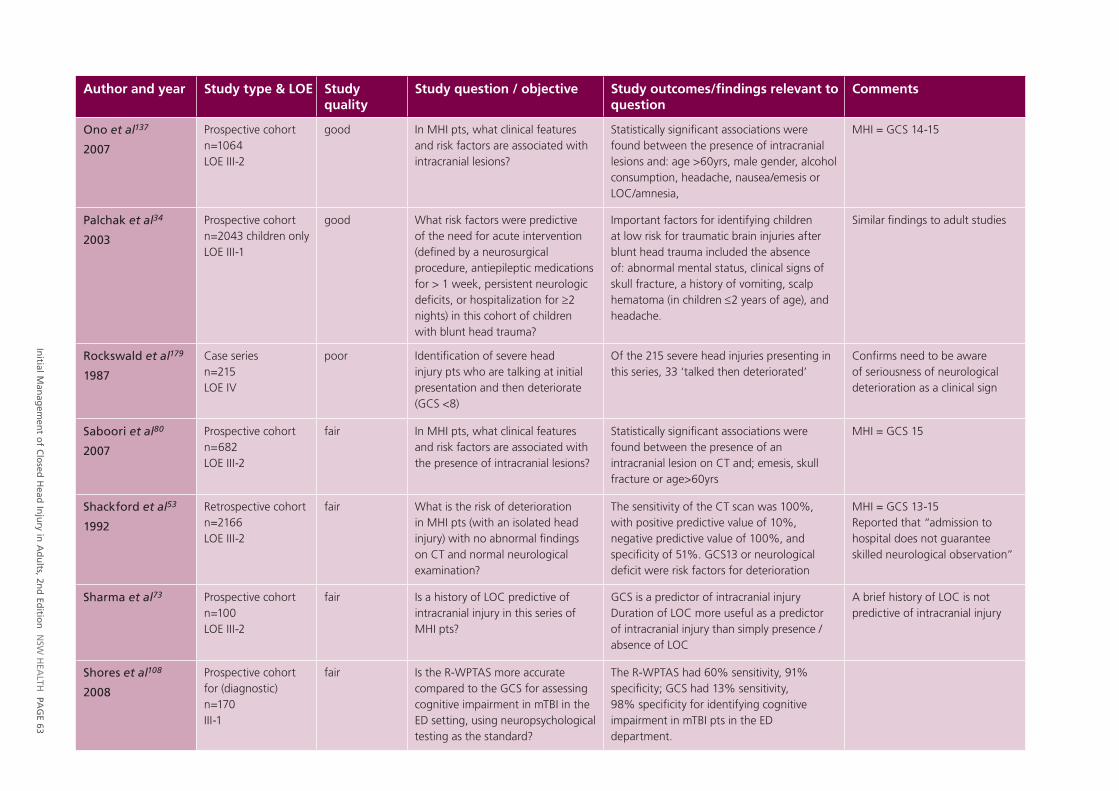
Initial M
anag
emen
t of C
losed
Head
Inju
ry in A
du
lts, 2nd
Editio
n N
SW H
EALTH
PAG
E 63
Author and year Study type & LOE Study quality
Study question / objective Study outcomes/fi ndings relevant to question
Comments
Ono et al137
2007
Prospective cohortn=1064LOE III-2
good In MHI pts, what clinical features and risk factors are associated with intracranial lesions?
Statistically signifi cant associations were found between the presence of intracranial lesions and: age >60yrs, male gender, alcohol consumption, headache, nausea/emesis or LOC/amnesia,
MHI = GCS 14-15
Palchak et al34
2003
Prospective cohortn=2043 children onlyLOE III-1
good What risk factors were predictive of the need for acute intervention (defi ned by a neurosurgical procedure, antiepileptic medications for > 1 week, persistent neurologic defi cits, or hospitalization for 2 nights) in this cohort of children with blunt head trauma?
Important factors for identifying children at low risk for traumatic brain injuries after blunt head trauma included the absence of: abnormal mental status, clinical signs of skull fracture, a history of vomiting, scalp hematoma (in children 2 years of age), and headache.
Similar fi ndings to adult studies
Rockswald et al179
1987
Case seriesn=215LOE IV
poor Identifi cation of severe head injury pts who are talking at initial presentation and then deteriorate (GCS <8)
Of the 215 severe head injuries presenting in this series, 33 ‘talked then deteriorated’
Confi rms need to be aware of seriousness of neurological deterioration as a clinical sign
Saboori et al80
2007
Prospective cohortn=682LOE III-2
fair In MHI pts, what clinical features and risk factors are associated with the presence of intracranial lesions?
Statistically signifi cant associations were found between the presence of an intracranial lesion on CT and; emesis, skull fracture or age>60yrs
MHI = GCS 15
Shackford et al53
1992
Retrospective cohortn=2166LOE III-2
fair What is the risk of deterioration in MHI pts (with an isolated head injury) with no abnormal fi ndings on CT and normal neurological examination?
The sensitivity of the CT scan was 100%, with positive predictive value of 10%, negative predictive value of 100%, and specifi city of 51%. GCS13 or neurological defi cit were risk factors for deterioration
MHI = GCS 13-15Reported that “admission to hospital does not guarantee skilled neurological observation”
Sharma et al73 Prospective cohortn=100LOE III-2
fair Is a history of LOC predictive of intracranial injury in this series of MHI pts?
GCS is a predictor of intracranial injuryDuration of LOC more useful as a predictor of intracranial injury than simply presence / absence of LOC
A brief history of LOC is not predictive of intracranial injury
Shores et al108
2008
Prospective cohort for (diagnostic)n=170III-1
fair Is the R-WPTAS more accurate compared to the GCS for assessing cognitive impairment in mTBI in the ED setting, using neuropsychological testing as the standard?
The R-WPTAS had 60% sensitivity, 91% specifi city; GCS had 13% sensitivity, 98% specifi city for identifying cognitive impairment in mTBI pts in the ED department.
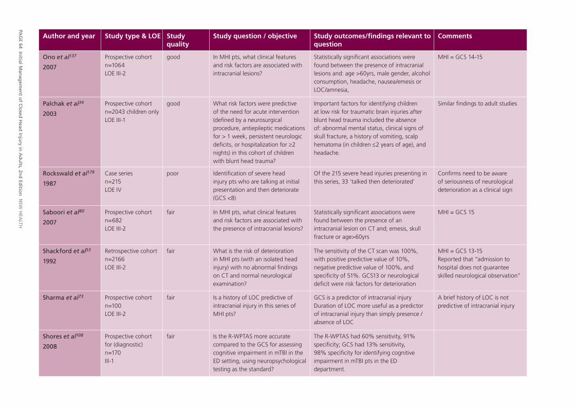
PAG
E 64 Initial M
anag
emen
t of C
losed
Head
Inju
ry in A
du
lts, 2nd
Editio
n N
SW H
EALTH
Author and year Study type & LOE Study quality
Study question / objective Study outcomes/fi ndings relevant to question
Comments
Ono et al137
2007
Prospective cohortn=1064LOE III-2
good In MHI pts, what clinical features and risk factors are associated with intracranial lesions?
Statistically signifi cant associations were found between the presence of intracranial lesions and: age >60yrs, male gender, alcohol consumption, headache, nausea/emesis or LOC/amnesia,
MHI = GCS 14-15
Palchak et al34
2003
Prospective cohortn=2043 children onlyLOE III-1
good What risk factors were predictive of the need for acute intervention (defi ned by a neurosurgical procedure, antiepileptic medications for > 1 week, persistent neurologic defi cits, or hospitalization for 2 nights) in this cohort of children with blunt head trauma?
Important factors for identifying children at low risk for traumatic brain injuries after blunt head trauma included the absence of: abnormal mental status, clinical signs of skull fracture, a history of vomiting, scalp hematoma (in children 2 years of age), and headache.
Similar fi ndings to adult studies
Rockswald et al179
1987
Case seriesn=215LOE IV
poor Identifi cation of severe head injury pts who are talking at initial presentation and then deteriorate (GCS <8)
Of the 215 severe head injuries presenting in this series, 33 ‘talked then deteriorated’
Confi rms need to be aware of seriousness of neurological deterioration as a clinical sign
Saboori et al80
2007
Prospective cohortn=682LOE III-2
fair In MHI pts, what clinical features and risk factors are associated with the presence of intracranial lesions?
Statistically signifi cant associations were found between the presence of an intracranial lesion on CT and; emesis, skull fracture or age>60yrs
MHI = GCS 15
Shackford et al53
1992
Retrospective cohortn=2166LOE III-2
fair What is the risk of deterioration in MHI pts (with an isolated head injury) with no abnormal fi ndings on CT and normal neurological examination?
The sensitivity of the CT scan was 100%, with positive predictive value of 10%, negative predictive value of 100%, and specifi city of 51%. GCS13 or neurological defi cit were risk factors for deterioration
MHI = GCS 13-15Reported that “admission to hospital does not guarantee skilled neurological observation”
Sharma et al73 Prospective cohortn=100LOE III-2
fair Is a history of LOC predictive of intracranial injury in this series of MHI pts?
GCS is a predictor of intracranial injuryDuration of LOC more useful as a predictor of intracranial injury than simply presence / absence of LOC
A brief history of LOC is not predictive of intracranial injury
Shores et al108
2008
Prospective cohort for (diagnostic)n=170III-1
fair Is the R-WPTAS more accurate compared to the GCS for assessing cognitive impairment in mTBI in the ED setting, using neuropsychological testing as the standard?
The R-WPTAS had 60% sensitivity, 91% specifi city; GCS had 13% sensitivity, 98% specifi city for identifying cognitive impairment in mTBI pts in the ED department.
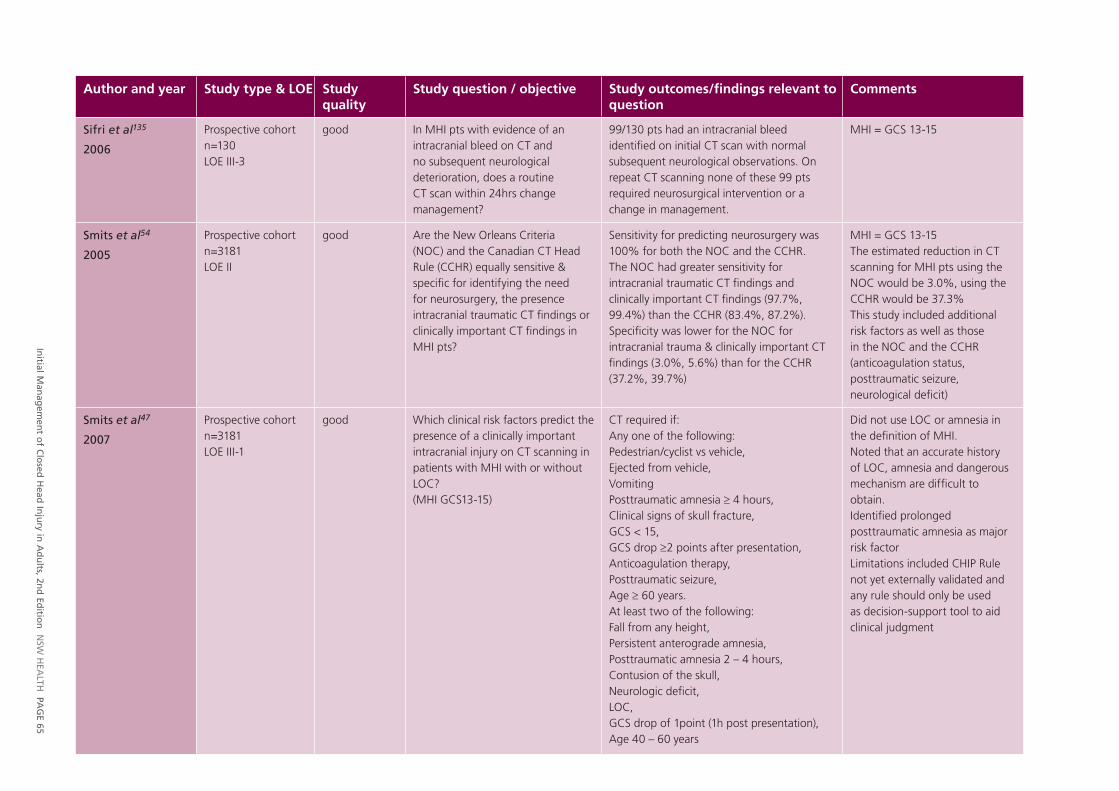
Initial M
anag
emen
t of C
losed
Head
Inju
ry in A
du
lts, 2nd
Editio
n N
SW H
EALTH
PAG
E 65
Author and year Study type & LOE Study quality
Study question / objective Study outcomes/fi ndings relevant to question
Comments
Sifri et al135
2006
Prospective cohortn=130LOE III-3
good In MHI pts with evidence of an intracranial bleed on CT and no subsequent neurological deterioration, does a routine CT scan within 24hrs change management?
99/130 pts had an intracranial bleed identifi ed on initial CT scan with normal subsequent neurological observations. On repeat CT scanning none of these 99 pts required neurosurgical intervention or a change in management.
MHI = GCS 13-15
Smits et al54
2005
Prospective cohortn=3181LOE II
good Are the New Orleans Criteria (NOC) and the Canadian CT Head Rule (CCHR) equally sensitive & specifi c for identifying the need for neurosurgery, the presence intracranial traumatic CT fi ndings or clinically important CT fi ndings in MHI pts?
Sensitivity for predicting neurosurgery was 100% for both the NOC and the CCHR.The NOC had greater sensitivity for intracranial traumatic CT fi ndings and clinically important CT fi ndings (97.7%, 99.4%) than the CCHR (83.4%, 87.2%). Specifi city was lower for the NOC for intracranial trauma & clinically important CT fi ndings (3.0%, 5.6%) than for the CCHR (37.2%, 39.7%)
MHI = GCS 13-15The estimated reduction in CT scanning for MHI pts using the NOC would be 3.0%, using the CCHR would be 37.3%This study included additional risk factors as well as those in the NOC and the CCHR (anticoagulation status, posttraumatic seizure, neurological defi cit)
Smits et al47
2007
Prospective cohort n=3181LOE III-1
good Which clinical risk factors predict the presence of a clinically important intracranial injury on CT scanning in patients with MHI with or without LOC?(MHI GCS13-15)
CT required if:Any one of the following: Pedestrian/cyclist vs vehicle,Ejected from vehicle, VomitingPosttraumatic amnesia 4 hours, Clinical signs of skull fracture, GCS < 15, GCS drop 2 points after presentation, Anticoagulation therapy, Posttraumatic seizure, Age 60 years.At least two of the following:Fall from any height, Persistent anterograde amnesia, Posttraumatic amnesia 2 – 4 hours, Contusion of the skull, Neurologic defi cit, LOC, GCS drop of 1point (1h post presentation), Age 40 – 60 years
Did not use LOC or amnesia in the defi nition of MHI.Noted that an accurate history of LOC, amnesia and dangerous mechanism are diffi cult to obtain.Identifi ed prolonged posttraumatic amnesia as major risk factor Limitations included CHIP Rule not yet externally validated and any rule should only be used as decision-support tool to aid clinical judgment
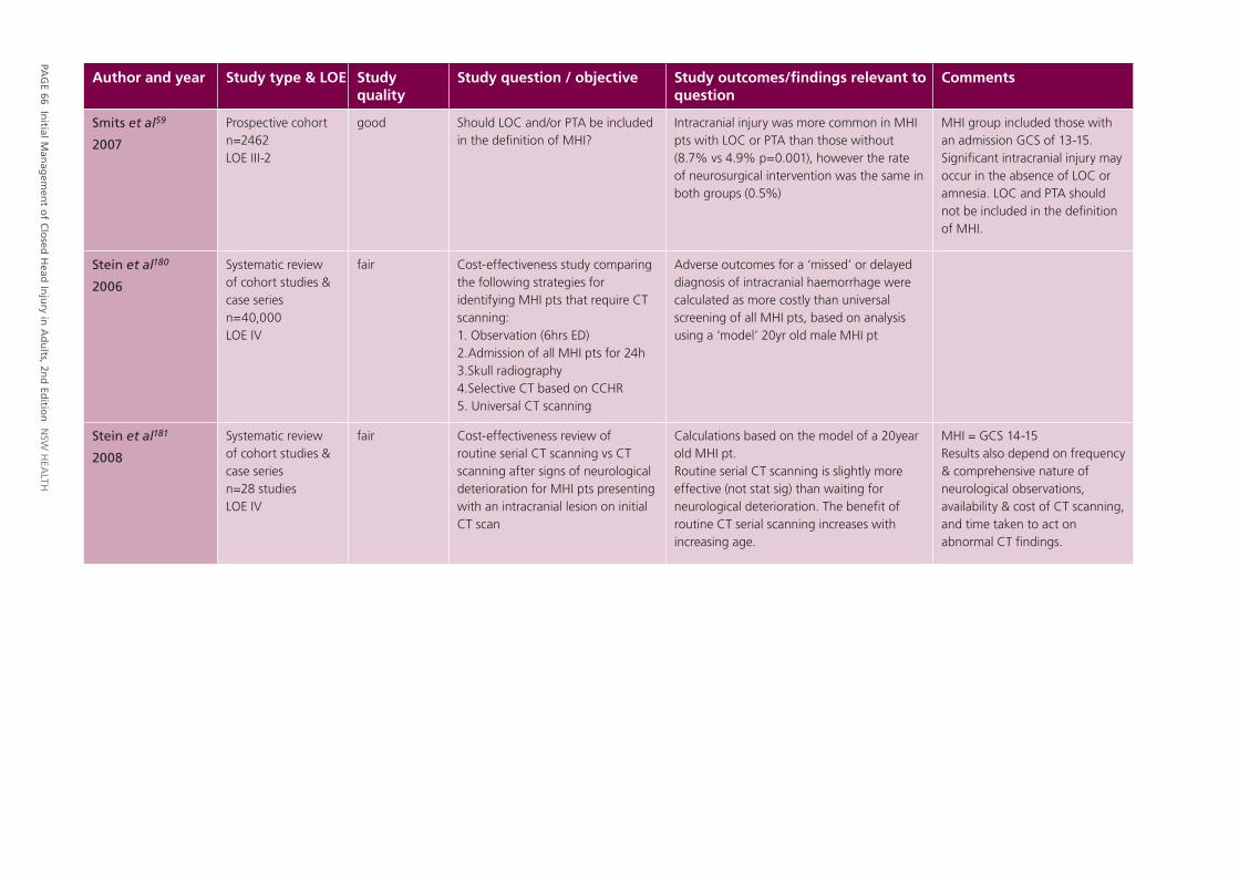
PAG
E 66 Initial M
anag
emen
t of C
losed
Head
Inju
ry in A
du
lts, 2nd
Editio
n N
SW H
EALTH
Author and year Study type & LOE Study quality
Study question / objective Study outcomes/fi ndings relevant to question
Comments
Smits et al59
2007
Prospective cohortn=2462LOE III-2
good Should LOC and/or PTA be included in the defi nition of MHI?
Intracranial injury was more common in MHI pts with LOC or PTA than those without (8.7% vs 4.9% p=0.001), however the rate of neurosurgical intervention was the same in both groups (0.5%)
MHI group included those with an admission GCS of 13-15. Signifi cant intracranial injury may occur in the absence of LOC or amnesia. LOC and PTA should not be included in the defi nition of MHI.
Stein et al180
2006
Systematic review of cohort studies & case seriesn=40,000LOE IV
fair Cost-effectiveness study comparing the following strategies for identifying MHI pts that require CT scanning:1. Observation (6hrs ED)2.Admission of all MHI pts for 24h3.Skull radiography4.Selective CT based on CCHR5. Universal CT scanning
Adverse outcomes for a ‘missed’ or delayed diagnosis of intracranial haemorrhage were calculated as more costly than universal screening of all MHI pts, based on analysis using a ‘model’ 20yr old male MHI pt
Stein et al181
2008
Systematic review of cohort studies & case seriesn=28 studiesLOE IV
fair Cost-effectiveness review of routine serial CT scanning vs CT scanning after signs of neurological deterioration for MHI pts presenting with an intracranial lesion on initial CT scan
Calculations based on the model of a 20year old MHI pt.Routine serial CT scanning is slightly more effective (not stat sig) than waiting for neurological deterioration. The benefi t of routine CT serial scanning increases with increasing age.
MHI = GCS 14-15Results also depend on frequency & comprehensive nature of neurological observations, availability & cost of CT scanning, and time taken to act on abnormal CT fi ndings.
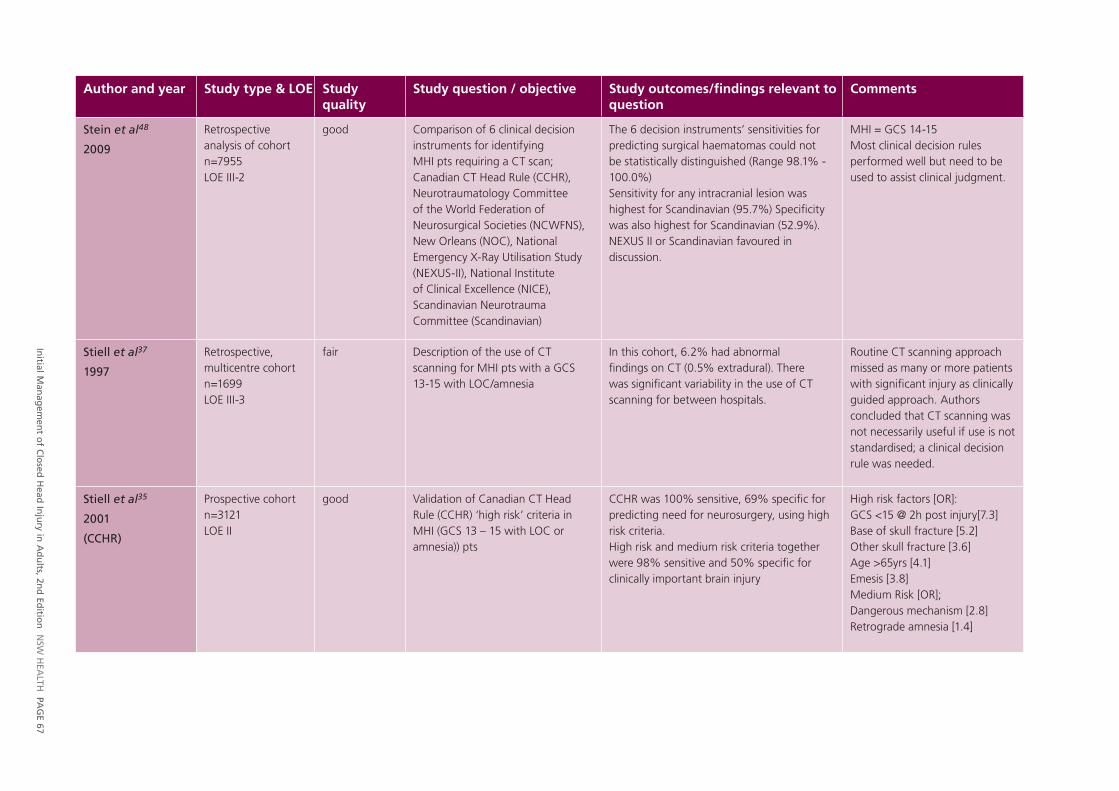
Initial M
anag
emen
t of C
losed
Head
Inju
ry in A
du
lts, 2nd
Editio
n N
SW H
EALTH
PAG
E 67
Author and year Study type & LOE Study quality
Study question / objective Study outcomes/fi ndings relevant to question
Comments
Stein et al48
2009
Retrospective analysis of cohort n=7955LOE III-2
good Comparison of 6 clinical decision instruments for identifying MHI pts requiring a CT scan; Canadian CT Head Rule (CCHR), Neurotraumatology Committee of the World Federation of Neurosurgical Societies (NCWFNS),New Orleans (NOC), National Emergency X-Ray Utilisation Study (NEXUS-II), National Institute of Clinical Excellence (NICE), Scandinavian Neurotrauma Committee (Scandinavian)
The 6 decision instruments’ sensitivities for predicting surgical haematomas could not be statistically distinguished (Range 98.1% - 100.0%)Sensitivity for any intracranial lesion was highest for Scandinavian (95.7%) Specifi city was also highest for Scandinavian (52.9%).NEXUS II or Scandinavian favoured in discussion.
MHI = GCS 14-15Most clinical decision rules performed well but need to be used to assist clinical judgment.
Stiell et al37
1997
Retrospective, multicentre cohortn=1699LOE III-3
fair Description of the use of CT scanning for MHI pts with a GCS 13-15 with LOC/amnesia
In this cohort, 6.2% had abnormal fi ndings on CT (0.5% extradural). There was signifi cant variability in the use of CT scanning for between hospitals.
Routine CT scanning approach missed as many or more patients with signifi cant injury as clinically guided approach. Authors concluded that CT scanning was not necessarily useful if use is not standardised; a clinical decision rule was needed.
Stiell et al35
2001
(CCHR)
Prospective cohortn=3121LOE II
good Validation of Canadian CT Head Rule (CCHR) ‘high risk’ criteria in MHI (GCS 13 – 15 with LOC or amnesia)) pts
CCHR was 100% sensitive, 69% specifi c for predicting need for neurosurgery, using high risk criteria. High risk and medium risk criteria together were 98% sensitive and 50% specifi c for clinically important brain injury
High risk factors [OR]:GCS <15 @ 2h post injury[7.3]Base of skull fracture [5.2]Other skull fracture [3.6]Age >65yrs [4.1]Emesis [3.8]Medium Risk [OR];Dangerous mechanism [2.8]Retrograde amnesia [1.4]
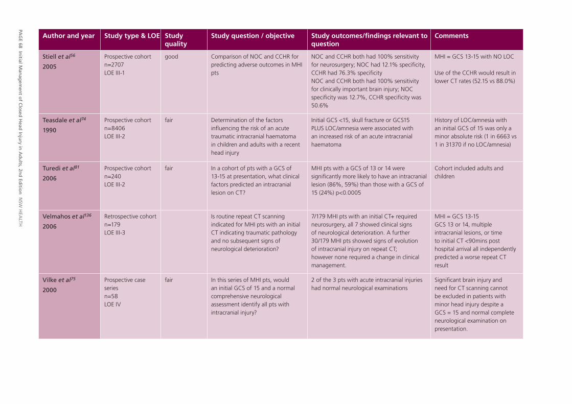
PAG
E 68 Initial M
anag
emen
t of C
losed
Head
Inju
ry in A
du
lts, 2nd
Editio
n N
SW H
EALTH
Author and year Study type & LOE Study quality
Study question / objective Study outcomes/fi ndings relevant to question
Comments
Stiell et al56
2005
Prospective cohortn=2707LOE III-1
good Comparison of NOC and CCHR for predicting adverse outcomes in MHI pts
NOC and CCHR both had 100% sensitivity for neurosurgery; NOC had 12.1% specifi city, CCHR had 76.3% specifi cityNOC and CCHR both had 100% sensitivity for clinically important brain injury; NOC specifi city was 12.7%, CCHR specifi city was 50.6%
MHI = GCS 13-15 with NO LOC
Use of the CCHR would result in lower CT rates (52.15 vs 88.0%)
Teasdale et al74
1990
Prospective cohortn=8406LOE III-2
fair Determination of the factors infl uencing the risk of an acute traumatic intracranial haematoma in children and adults with a recent head injury
Initial GCS <15, skull fracture or GCS15 PLUS LOC/amnesia were associated with an increased risk of an acute intracranial haematoma
History of LOC/amnesia with an initial GCS of 15 was only a minor absolute risk (1 in 6663 vs 1 in 31370 if no LOC/amnesia)
Turedi et al81
2006
Prospective cohortn=240LOE III-2
fair In a cohort of pts with a GCS of 13-15 at presentation, what clinical factors predicted an intracranial lesion on CT?
MHI pts with a GCS of 13 or 14 were signifi cantly more likely to have an intracranial lesion (86%, 59%) than those with a GCS of 15 (24%) p<0.0005
Cohort included adults and children
Velmahos et al136
2006
Retrospective cohortn=179LOE III-3
Is routine repeat CT scanning indicated for MHI pts with an initial CT indicating traumatic pathology and no subsequent signs of neurological deterioration?
7/179 MHI pts with an initial CT+ required neurosurgery, all 7 showed clinical signs of neurological deterioration. A further 30/179 MHI pts showed signs of evolution of intracranial injury on repeat CT; however none required a change in clinical management.
MHI = GCS 13-15GCS 13 or 14, multiple intracranial lesions, or time to initial CT <90mins post hospital arrival all independently predicted a worse repeat CT result
Vilke et al75
2000
Prospective case seriesn=58LOE IV
fair In this series of MHI pts, would an initial GCS of 15 and a normal comprehensive neurological assessment identify all pts with intracranial injury?
2 of the 3 pts with acute intracranial injuries had normal neurological examinations
Signifi cant brain injury and need for CT scanning cannot be excluded in patients with minor head injury despite a GCS = 15 and normal complete neurological examination on presentation.
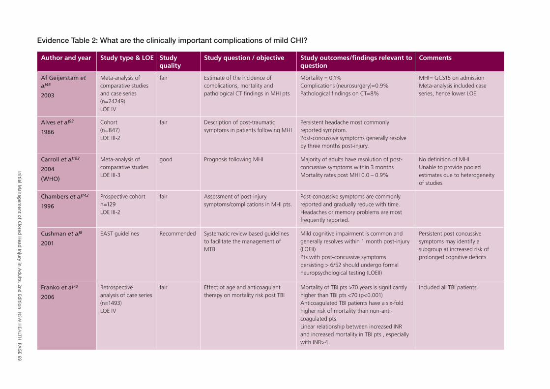
Initial M
anag
emen
t of C
losed
Head
Inju
ry in A
du
lts, 2nd
Editio
n N
SW H
EALTH
PAG
E 69
Author and year Study type & LOE Study quality
Study question / objective Study outcomes/fi ndings relevant to question
Comments
Af Geijerstam et al46
2003
Meta-analysis of comparative studies and case series (n=24249)LOE IV
fair Estimate of the incidence of complications, mortality and pathological CT fi ndings in MHI pts
Mortality = 0.1%Complications (neurosurgery)=0.9%Pathological fi ndings on CT=8%
MHI= GCS15 on admissionMeta-analysis included case series, hence lower LOE
Alves et al93
1986
Cohort(n=847)LOE III-2
fair Description of post-traumatic symptoms in patients following MHI
Persistent headache most commonly reported symptom.Post-concussive symptoms generally resolve by three months post-injury.
Carroll et al182
2004
(WHO)
Meta-analysis of comparative studiesLOE III-3
good Prognosis following MHI Majority of adults have resolution of post-concussive symptoms within 3 monthsMortality rates post MHI 0.0 – 0.9%
No defi nition of MHIUnable to provide pooled estimates due to heterogeneity of studies
Chambers et al142
1996
Prospective cohortn=129LOE III-2
fair Assessment of post-injury symptoms/complications in MHI pts.
Post-concussive symptoms are commonly reported and gradually reduce with time. Headaches or memory problems are most frequently reported.
Cushman et al8
2001
EAST guidelines Recommended Systematic review based guidelines to facilitate the management of MTBI
Mild cognitive impairment is common and generally resolves within 1 month post-injury (LOEII)Pts with post-concussive symptoms persisting > 6/52 should undergo formal neuropsychological testing (LOEII)
Persistent post concussive symptoms may identify a subgroup at increased risk of prolonged cognitive defi cits
Franko et al78
2006
Retrospective analysis of case series (n=1493)LOE IV
fair Effect of age and anticoagulant therapy on mortality risk post TBI
Mortality of TBI pts >70 years is signifi cantly higher than TBI pts <70 (p<0.001)Anticoagulated TBI patients have a six-fold higher risk of mortality than non-anti-coagulated pts.Linear relationship between increased INR and increased mortality in TBI pts , especially with INR>4
Included all TBI patients
Evidence Table 2: What are the clinically important complications of mild CHI?
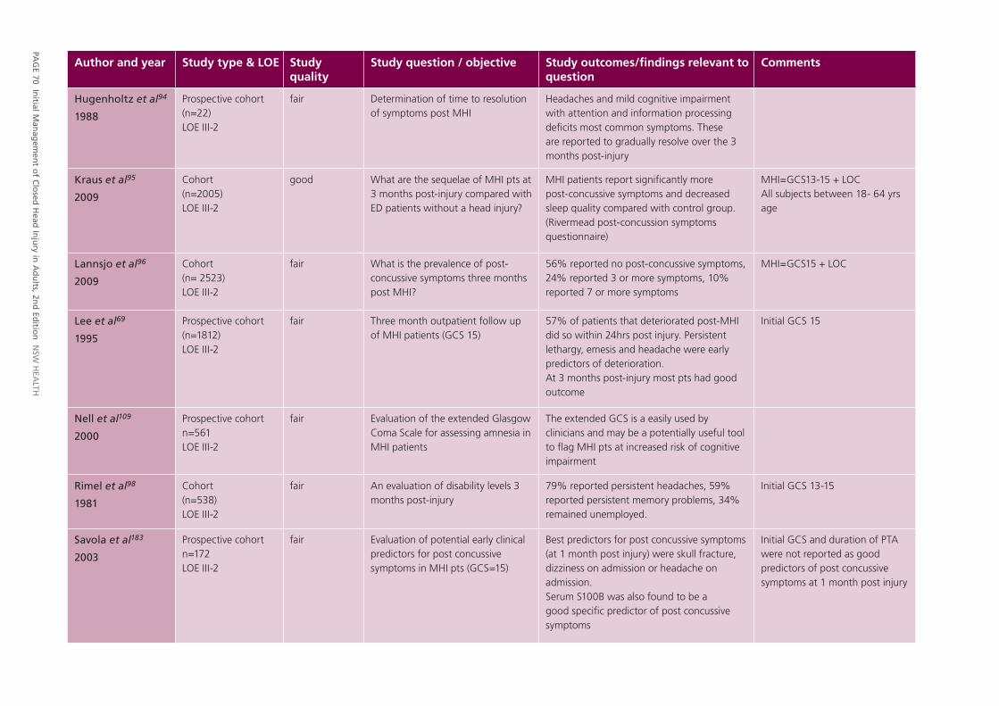
PAG
E 70 Initial M
anag
emen
t of C
losed
Head
Inju
ry in A
du
lts, 2nd
Editio
n N
SW H
EALTH
Author and year Study type & LOE Study quality
Study question / objective Study outcomes/fi ndings relevant to question
Comments
Hugenholtz et al94
1988
Prospective cohort(n=22)LOE III-2
fair Determination of time to resolution of symptoms post MHI
Headaches and mild cognitive impairment with attention and information processing defi cits most common symptoms. These are reported to gradually resolve over the 3 months post-injury
Kraus et al95
2009
Cohort (n=2005)LOE III-2
good What are the sequelae of MHI pts at 3 months post-injury compared with ED patients without a head injury?
MHI patients report signifi cantly more post-concussive symptoms and decreased sleep quality compared with control group. (Rivermead post-concussion symptoms questionnaire)
MHI=GCS13-15 + LOCAll subjects between 18- 64 yrs age
Lannsjo et al96
2009
Cohort(n= 2523)LOE III-2
fair What is the prevalence of post-concussive symptoms three months post MHI?
56% reported no post-concussive symptoms, 24% reported 3 or more symptoms, 10% reported 7 or more symptoms
MHI=GCS15 + LOC
Lee et al69
1995
Prospective cohort(n=1812)LOE III-2
fair Three month outpatient follow up of MHI patients (GCS 15)
57% of patients that deteriorated post-MHI did so within 24hrs post injury. Persistent lethargy, emesis and headache were early predictors of deterioration.At 3 months post-injury most pts had good outcome
Initial GCS 15
Nell et al109
2000
Prospective cohortn=561LOE III-2
fair Evaluation of the extended Glasgow Coma Scale for assessing amnesia in MHI patients
The extended GCS is a easily used by clinicians and may be a potentially useful tool to fl ag MHI pts at increased risk of cognitive impairment
Rimel et al98
1981
Cohort(n=538)LOE III-2
fair An evaluation of disability levels 3 months post-injury
79% reported persistent headaches, 59% reported persistent memory problems, 34% remained unemployed.
Initial GCS 13-15
Savola et al183
2003
Prospective cohortn=172LOE III-2
fair Evaluation of potential early clinical predictors for post concussive symptoms in MHI pts (GCS=15)
Best predictors for post concussive symptoms (at 1 month post injury) were skull fracture, dizziness on admission or headache on admission.Serum S100B was also found to be a good specifi c predictor of post concussive symptoms
Initial GCS and duration of PTA were not reported as good predictors of post concussive symptoms at 1 month post injury
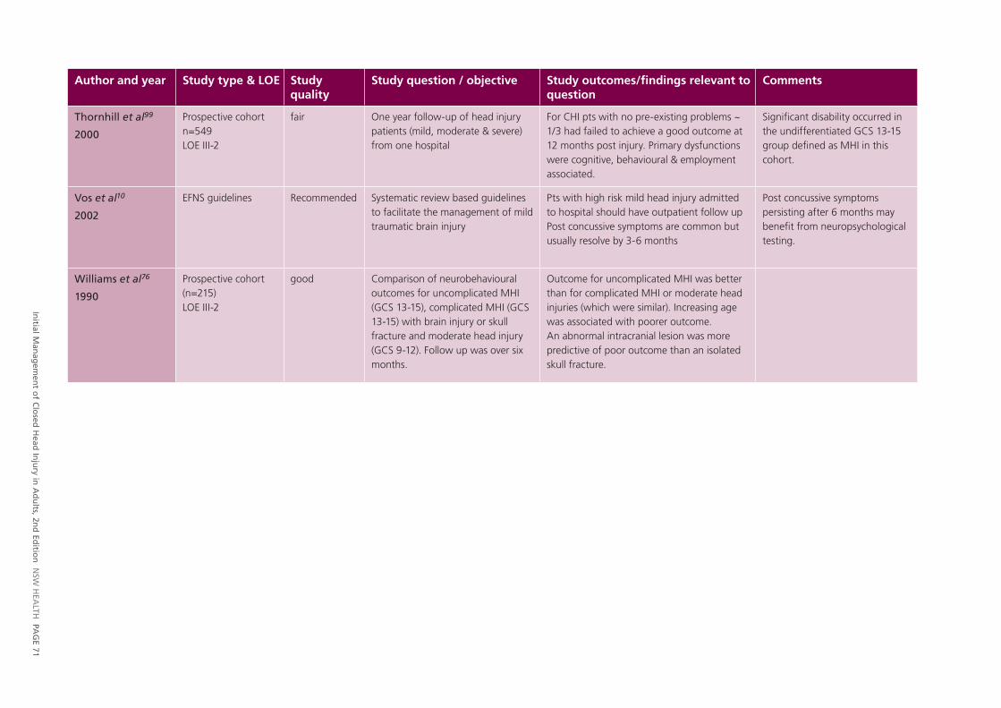
Initial M
anag
emen
t of C
losed
Head
Inju
ry in A
du
lts, 2nd
Editio
n N
SW H
EALTH
PAG
E 71
Author and year Study type & LOE Study quality
Study question / objective Study outcomes/fi ndings relevant to question
Comments
Thornhill et al99
2000
Prospective cohortn=549LOE III-2
fair One year follow-up of head injury patients (mild, moderate & severe) from one hospital
For CHI pts with no pre-existing problems ~ 1/3 had failed to achieve a good outcome at 12 months post injury. Primary dysfunctions were cognitive, behavioural & employment associated.
Signifi cant disability occurred in the undifferentiated GCS 13-15 group defi ned as MHI in this cohort.
Vos et al10
2002
EFNS guidelines Recommended Systematic review based guidelines to facilitate the management of mild traumatic brain injury
Pts with high risk mild head injury admitted to hospital should have outpatient follow upPost concussive symptoms are common but usually resolve by 3-6 months
Post concussive symptoms persisting after 6 months may benefi t from neuropsychological testing.
Williams et al76
1990
Prospective cohort(n=215)LOE III-2
good Comparison of neurobehavioural outcomes for uncomplicated MHI (GCS 13-15), complicated MHI (GCS 13-15) with brain injury or skull fracture and moderate head injury (GCS 9-12). Follow up was over six months.
Outcome for uncomplicated MHI was better than for complicated MHI or moderate head injuries (which were similar). Increasing age was associated with poorer outcome. An abnormal intracranial lesion was more predictive of poor outcome than an isolated skull fracture.
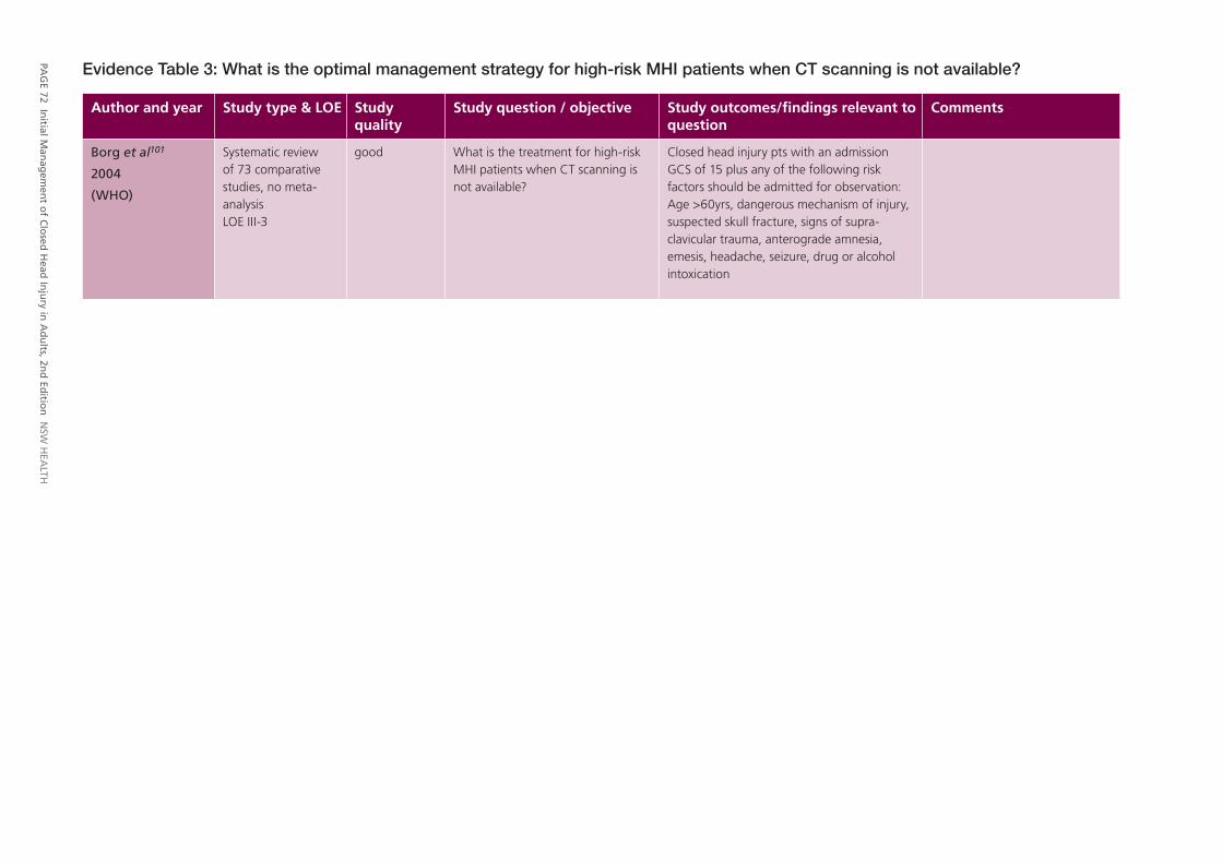
PAG
E 72 Initial M
anag
emen
t of C
losed
Head
Inju
ry in A
du
lts, 2nd
Editio
n N
SW H
EALTH
Author and year Study type & LOE Study quality
Study question / objective Study outcomes/fi ndings relevant to question
Comments
Borg et al101
2004
(WHO)
Systematic review of 73 comparative studies, no meta-analysisLOE III-3
good What is the treatment for high-risk MHI patients when CT scanning is not available?
Closed head injury pts with an admission GCS of 15 plus any of the following risk factors should be admitted for observation:Age >60yrs, dangerous mechanism of injury, suspected skull fracture, signs of supra-clavicular trauma, anterograde amnesia, emesis, headache, seizure, drug or alcohol intoxication
Evidence Table 3: What is the optimal management strategy for high-risk MHI patients when CT scanning is not available?
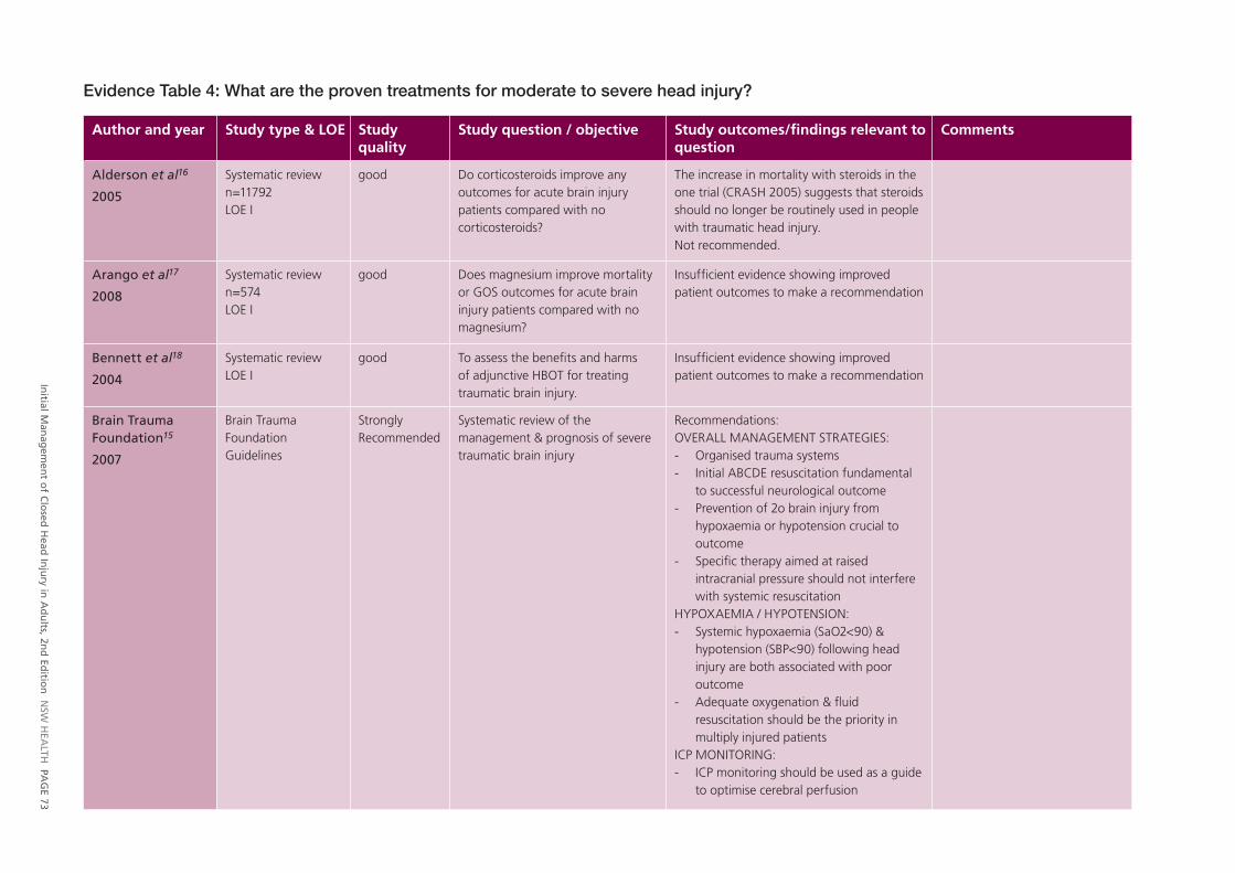
Initial M
anag
emen
t of C
losed
Head
Inju
ry in A
du
lts, 2nd
Editio
n N
SW H
EALTH
PAG
E 73
Author and year Study type & LOE Study quality
Study question / objective Study outcomes/fi ndings relevant to question
Comments
Alderson et al16
2005
Systematic reviewn=11792LOE I
good Do corticosteroids improve any outcomes for acute brain injury patients compared with no corticosteroids?
The increase in mortality with steroids in the one trial (CRASH 2005) suggests that steroids should no longer be routinely used in people with traumatic head injury.Not recommended.
Arango et al17
2008
Systematic reviewn=574LOE I
good Does magnesium improve mortality or GOS outcomes for acute brain injury patients compared with no magnesium?
Insuffi cient evidence showing improved patient outcomes to make a recommendation
Bennett et al18
2004
Systematic reviewLOE I
good To assess the benefi ts and harms of adjunctive HBOT for treating traumatic brain injury.
Insuffi cient evidence showing improved patient outcomes to make a recommendation
Brain Trauma Foundation15
2007
Brain Trauma Foundation Guidelines
Strongly Recommended
Systematic review of the management & prognosis of severe traumatic brain injury
Recommendations:OVERALL MANAGEMENT STRATEGIES:- Organised trauma systems- Initial ABCDE resuscitation fundamental
to successful neurological outcome- Prevention of 2o brain injury from
hypoxaemia or hypotension crucial to outcome
- Specifi c therapy aimed at raised intracranial pressure should not interfere with systemic resuscitation
HYPOXAEMIA / HYPOTENSION:- Systemic hypoxaemia (SaO2<90) &
hypotension (SBP<90) following head injury are both associated with poor outcome
- Adequate oxygenation & fl uid resuscitation should be the priority in multiply injured patients
ICP MONITORING:- ICP monitoring should be used as a guide
to optimise cerebral perfusion
Evidence Table 4: What are the proven treatments for moderate to severe head injury?
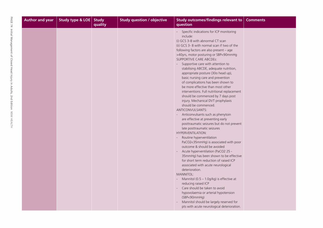
PAG
E 74 Initial M
anag
emen
t of C
losed
Head
Inju
ry in A
du
lts, 2nd
Editio
n N
SW H
EALTH
Author and year Study type & LOE Study quality
Study question / objective Study outcomes/fi ndings relevant to question
Comments
- Specifi c indications for ICP monitoring include:
(i) GCS 3-8 with abnormal CT scan(ii) GCS 3- 8 with normal scan if two of the following factors are also present – age >40yrs, motor posturing or SBP<90mmHgSUPPORTIVE CARE ABCDEs:- Supportive care with attention to
stabilising ABCDE, adequate nutrition, appropriate posture (30o head up), basic nursing care and prevention of complications has been shown to be more effective than most other interventions. Full nutritional replacement should be commenced by 7 days post injury. Mechanical DVT prophylaxis should be commenced.
ANTICONVULSANTS:- Anticonvulsants such as phenytoin
are effective at preventing early posttraumatic seizures but do not prevent late posttraumatic seizures
HYPERVENTILATION:- Routine hyperventilation
PaCO2<35mmHg) is associated with poor outcome & should be avoided
- Acute hyperventilation (PaCO2 25 - 35mmHg) has been shown to be effective for short term reduction of raised ICP associated with acute neurological deterioration.
MANNITOL:- Mannitol (0.5 – 1.0g/kg) is effective at
reducing raised ICP- Care should be taken to avoid
hypovolaemia or arterial hypotension (SBP<90mmHg)
- Mannitol should be largely reserved for pts with acute neurological deterioration.
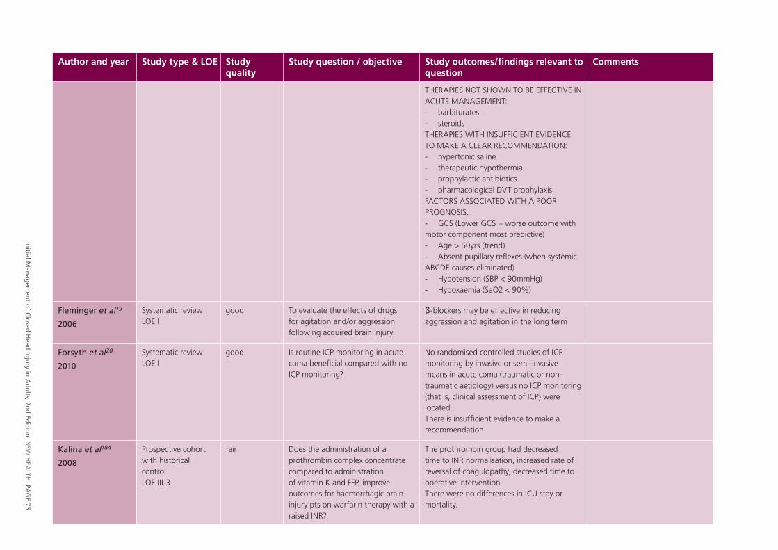
Initial M
anag
emen
t of C
losed
Head
Inju
ry in A
du
lts, 2nd
Editio
n N
SW H
EALTH
PAG
E 75
Author and year Study type & LOE Study quality
Study question / objective Study outcomes/fi ndings relevant to question
Comments
THERAPIES NOT SHOWN TO BE EFFECTIVE IN ACUTE MANAGEMENT:- barbiturates- steroidsTHERAPIES WITH INSUFFICIENT EVIDENCE TO MAKE A CLEAR RECOMMENDATION:- hypertonic saline- therapeutic hypothermia- prophylactic antibiotics- pharmacological DVT prophylaxisFACTORS ASSOCIATED WITH A POOR PROGNOSIS:- GCS (Lower GCS = worse outcome with motor component most predictive)- Age > 60yrs (trend)- Absent pupillary refl exes (when systemic ABCDE causes eliminated)- Hypotension (SBP < 90mmHg)- Hypoxaemia (SaO2 < 90%)
Fleminger et al19
2006
Systematic reviewLOE I
good To evaluate the effects of drugs for agitation and/or aggression following acquired brain injury
β-blockers may be effective in reducing aggression and agitation in the long term
Forsyth et al20
2010
Systematic review LOE I
good Is routine ICP monitoring in acute coma benefi cial compared with no ICP monitoring?
No randomised controlled studies of ICP monitoring by invasive or semi-invasive means in acute coma (traumatic or non-traumatic aetiology) versus no ICP monitoring (that is, clinical assessment of ICP) were located.There is insuffi cient evidence to make a recommendation
Kalina et al184
2008
Prospective cohort with historical controlLOE III-3
fair Does the administration of a prothrombin complex concentrate compared to administration of vitamin K and FFP, improve outcomes for haemorrhagic brain injury pts on warfarin therapy with a raised INR?
The prothrombin group had decreased time to INR normalisation, increased rate of reversal of coagulopathy, decreased time to operative intervention.There were no differences in ICU stay or mortality.
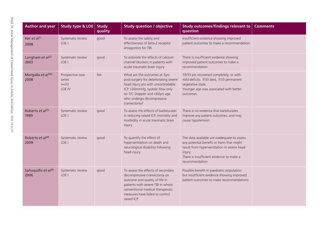
PAG
E 76 Initial M
anag
emen
t of C
losed
Head
Inju
ry in A
du
lts, 2nd
Editio
n N
SW H
EALTH
Author and year Study type & LOE Study quality
Study question / objective Study outcomes/fi ndings relevant to question
Comments
Ker et al21
2008
Systematic review LOE I
good To assess the safety and effectiveness of beta-2 receptor antagonists for TBI.
Insuffi cient evidence showing improved patient outcomes to make a recommendation
Langham et al22
2003Systematic reviewLOE I
good To estimate the effects of calcium channel blockers in patients with acute traumatic brain injury
There is insuffi cient evidence showing improved patient outcomes to make a recommendation
Morgalla et al185
2008Prospective case seriesn=33LOE IV
fair What are the outcomes at 3yrs post-surgery for deteriorating severe head injury pts with uncontrollable ICP >30mmHg, systolic fl ow only on T/C Doppler and <60yrs age who undergo decompressive craniectomy?
19/33 pts recovered completely, or with mild defi cits. 7/33 died, 7/33 permanent vegetative state.Younger age was associated with better outcomes.
Roberts et al25
1999Systematic reviewLOE I
good To assess the effects of barbiturates in reducing raised ICP, mortality and morbidity in acute traumatic brain injury
There is no evidence that barbiturates improve any patient outcomes, and may cause hypotension.
Roberts et al24
2009Systematic reviewLOE I
good To quantify the effect of hyperventilation on death and neurological disability following head injury.
The data available are inadequate to assess any potential benefi t or harm that might result from hyperventilation in severe head injury.There is insuffi cient evidence to make a recommendation
Sahuquillo et al26
2006Systematic review LOE I
good To assess the effects of secondary decompressive craniectomy on outcome and quality of life in patients with severe TBI in whom conventional medical therapeutic measures have failed to control raised ICP
Possible benefi t in paediatric population but insuffi cient evidence showing improved patient outcomes to make recommendations
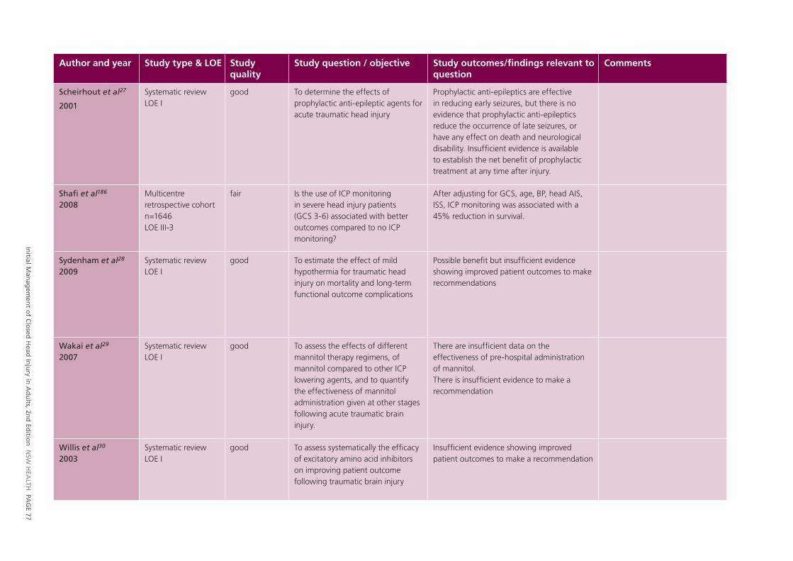
Initial M
anag
emen
t of C
losed
Head
Inju
ry in A
du
lts, 2nd
Editio
n N
SW H
EALTH
PAG
E 77
Author and year Study type & LOE Study quality
Study question / objective Study outcomes/fi ndings relevant to question
Comments
Scheirhout et al27
2001
Systematic reviewLOE I
good To determine the effects of prophylactic anti-epileptic agents for acute traumatic head injury
Prophylactic anti-epileptics are effective in reducing early seizures, but there is no evidence that prophylactic anti-epileptics reduce the occurrence of late seizures, or have any effect on death and neurological disability. Insuffi cient evidence is available to establish the net benefi t of prophylactic treatment at any time after injury.
Shafi et al186
2008Multicentre retrospective cohortn=1646LOE III-3
fair Is the use of ICP monitoring in severe head injury patients (GCS 3-6) associated with better outcomes compared to no ICP monitoring?
After adjusting for GCS, age, BP, head AIS, ISS, ICP monitoring was associated with a 45% reduction in survival.
Sydenham et al28
2009Systematic reviewLOE I
good To estimate the effect of mild hypothermia for traumatic head injury on mortality and long-term functional outcome complications
Possible benefi t but insuffi cient evidence showing improved patient outcomes to make recommendations
Wakai et al29
2007Systematic review LOE I
good To assess the effects of different mannitol therapy regimens, of mannitol compared to other ICP lowering agents, and to quantify the effectiveness of mannitol administration given at other stages following acute traumatic brain injury.
There are insuffi cient data on the effectiveness of pre-hospital administration of mannitol.There is insuffi cient evidence to make a recommendation
Willis et al30
2003Systematic reviewLOE I
good To assess systematically the effi cacy of excitatory amino acid inhibitors on improving patient outcome following traumatic brain injury
Insuffi cient evidence showing improved patient outcomes to make a recommendation
PAG
E 78 Initial M
anag
emen
t of C
losed
Head
Inju
ry in A
du
lts, 2nd
Editio
n N
SW H
EALTH
Author and year Study type & LOE Study quality
Study question / objective Study outcomes/fi ndings relevant to question
Comments
Devrill et al187
2007
Retrospective cohortn=261LOE III-3
fair What are the clinical outcomes for patients with an extra-dural haemorrhage presenting to neurosurgical centres compared with patients presenting to hospitals with no neurosurgical facilities?
All 5 deaths and 4/7 serious disabilities occurred in the group that were transferred from a hospital with no neurosurgical facilities to a neurosurgical centre before undergoing craniotomy. This group had a median time to surgery of 8hrs 5min. 8/9 pts who received emergency burr-hole treatment in non-neurosurgical hospitals had good outcomes
This study was characterised by the especially lengthy inter-hospital transfer times (rural Queensland).
Fabbri et al41
2008
Prospective cohortn=700LOE III-2
good What are the effects on 6month outcomes for mild and moderate head injury pts with adverse CT fi ndings not requiring immediate surgery when observed in a non-neurosurgical facility compared with those transferred to a neurosurgical centre?
The outcome was unfavourable for 18% of pts transferred to a neurosurgical centre for observation, compared with 10% for pts kept in non-neurosurgical centres for observation (NS: p=0.143)
Transfer times 30 – 60 minutes.The hospital with no neurosurgical facilities had access to neurosurgical expertise via a teleradiology systemMHI GCS 14-15Moderate HI GCS 9 – 13
McConnell et al188
2005
Retrospective cohortn=542LOE III-2
fair Do CHI pts transferred to US level I trauma centres have a reduced mortality rate relative to matched pts transferred to US level II trauma centres?
The inter-hospital transfer of CHI pts to level I trauma centres results in a signifi cant mortality benefi t (10% reduction) compared to those transferred to a level II trauma centre.
Evidence Table 5: When should patients with a closed head injury be transferred to a hospital with neurosurgical facilities?
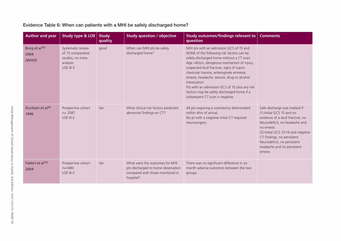
Initial M
anag
emen
t of C
losed
Head
Inju
ry in A
du
lts, 2nd
Editio
n N
SW H
EALTH
PAG
E 79
Author and year Study type & LOE Study quality
Study question / objective Study outcomes/fi ndings relevant to question
Comments
Borg et al101
2004
(WHO)
Systematic review of 73 comparative studies, no meta-analysisLOE III-3
good When can MHI pts be safely discharged home?
MHI pts with an admission GCS of 15 and NONE of the following risk factors can be safely discharged home without a CT scan:Age >60yrs, dangerous mechanism of injury, suspected skull fracture, signs of supra-clavicular trauma, anterograde amnesia, emesis, headache, seizure, drug or alcohol intoxicationPts with an admission GCS of 15 plus any risk factors may be safely discharged home if a subsequent CT scan is negative.
Dunham et al36
1996
Prospective cohortn= 2587LOE III-2
fair What clinical risk factors predicted abnormal fi ndings on CT?
All pts requiring a craniotomy deteriorated within 4hrs of arrival.No pt with a negative initial CT required neurosurgery
Safe discharge was implied if:(1) Initial GCS 15 and no evidence of a skull fracture, no Neurodefi cit, no headache and no emesis(2) Initial GCS 13-14 and negative CT fi ndings, no persistent Neurodefi cit, no persistent headache and no persistent emesis
Fabbri et al125
2004
Prospective cohortn=1480LOE III-2
fair What were the outcomes for MHI pts discharged to home observation compared with those monitored in hospital?
There was no signifi cant difference in six month adverse outcomes between the two groups
Evidence Table 6: When can patients with a MHI be safely discharged home?
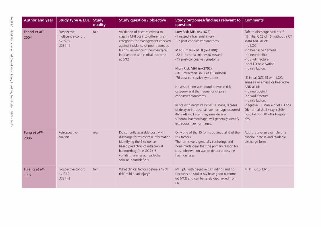
PAG
E 80 Initial M
anag
emen
t of C
losed
Head
Inju
ry in A
du
lts, 2nd
Editio
n N
SW H
EALTH
Author and year Study type & LOE Study quality
Study question / objective Study outcomes/fi ndings relevant to question
Comments
Fabbri et al31
2004
Prospective, multicentre cohortn=5578LOE III-1
fair Validation of a set of criteria to classify MHI pts into different risk categories for management checked against incidence of post-traumatic lesions, incidence of neurosurgical intervention and clinical outcome at 6/12
Low Risk MHI (n=1676):-1 missed intracranial injury-52 post-concussive symptoms
Medium Risk MHI (n=1200):-22 intracranial injuries (0 missed)-49 post-concussive symptoms
High Risk MHI (n=2702):-301 intracranial injuries (15 missed)-76 post-concussive symptoms
No association was found between risk category and the frequency of post-concussive symptoms.
In pts with negative initial CT scans, 8 cases of delayed intracranial haemorrhage occurred (8/1774) – CT scan may miss delayed subdural haemorrhage, will generally identify extradural haemorrhages.
Safe to discharge MHI pts if:(1) Initial GCS of 15 (without a CT scan) AND all of:-no LOC-no headache / emesis-no neurodefi cit-no skull fracture-brief ED observation-no risk factors
(2) Initial GCS 15 with LOC/amnesia or emesis or headache AND all of:-no neurodefi cit-no skull fracture-no risk factors-negative CT scan + brief ED obs OR normal skull x-ray + 24hr hospital obs OR 24hr hospital obs.
Fung et al155
2006
Retrospective analysis
n/a Do currently available post MHI discharge forms contain information identifying the 6 evidence-based predictors of intracranial haemorrhage? (ie GCS<15, vomiting, amnesia, headache, seizure, neurodefi cit)
Only one of the 15 forms outlined all 6 of the risk factors.The forms were generally confusing, and none made clear that the primary reason for close observation was to detect a possible haemorrhage.
Authors give an example of a concise, precise and readable discharge form
Hsiang et al52
1997
Prospective cohortn=1360LOE III-2
fair What clinical factors defi ne a ‘high risk’ mild head injury?
MHI pts with negative CT fi ndings and no fractures on skull x-ray have good outcome (at 6/12) and can be safely discharged from ED
MHI = GCS 13-15
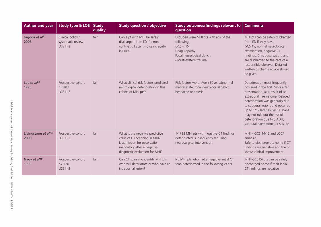
Initial M
anag
emen
t of C
losed
Head
Inju
ry in A
du
lts, 2nd
Editio
n N
SW H
EALTH
PAG
E 81
Author and year Study type & LOE Study quality
Study question / objective Study outcomes/fi ndings relevant to question
Comments
Jagoda et al9
2008Clinical policy / systematic reviewLOE III-2
fair Can a pt with MHI be safely discharged from ED if a non-contrast CT scan shows no acute injuries?
Excluded were MHI pts with any of the following: GCS < 15CoagulopathyFocal neurological defi cit<Multi-system trauma
MHI pts can be safely discharged from ED if they have:GCS 15, normal neurological examination, negative CT fi ndings, 6hrs observation, and are discharged to the care of a responsible observer. Detailed written discharge advice should be given.
Lee et al69
1995Prospective cohortn=1812LOE III-2
fair What clinical risk factors predicted neurological deterioration in this cohort of MHI pts?
Risk factors were: Age >60yrs, abnormal mental state, focal neurological defi cit, headache or emesis
Deterioration most frequently occurred in the fi rst 24hrs after presentation, as a result of an extradural haematoma. Delayed deterioration was generally due to subdural lesions and occurred up to 1/52 later. Initial CT scans may not rule out the risk of deterioration due to SIADH, subdural haematoma or seizure
Livingstone et al131
2000Prospective cohort LOE III-2
fair What is the negative predictive value of CT scanning in MHI?Is admission for observation mandatory after a negative diagnostic evaluation for MHI?
1/1788 MHI pts with negative CT fi ndings deteriorated, subsequently requiring neurosurgical intervention.
MHI = GCS 14-15 and LOC/amnesiaSafe to discharge pts home if CT fi ndings are negative and the pt shows clinical improvement
Nagy et al89
1999Prospective cohortn=1170LOE III-2
fair Can CT scanning identify MHI pts who will deteriorate or who have an intracranial lesion?
No MHI pts who had a negative initial CT scan deteriorated in the following 24hrs
MHI (GCS15) pts can be safely discharged home if their initial CT fi ndings are negative.
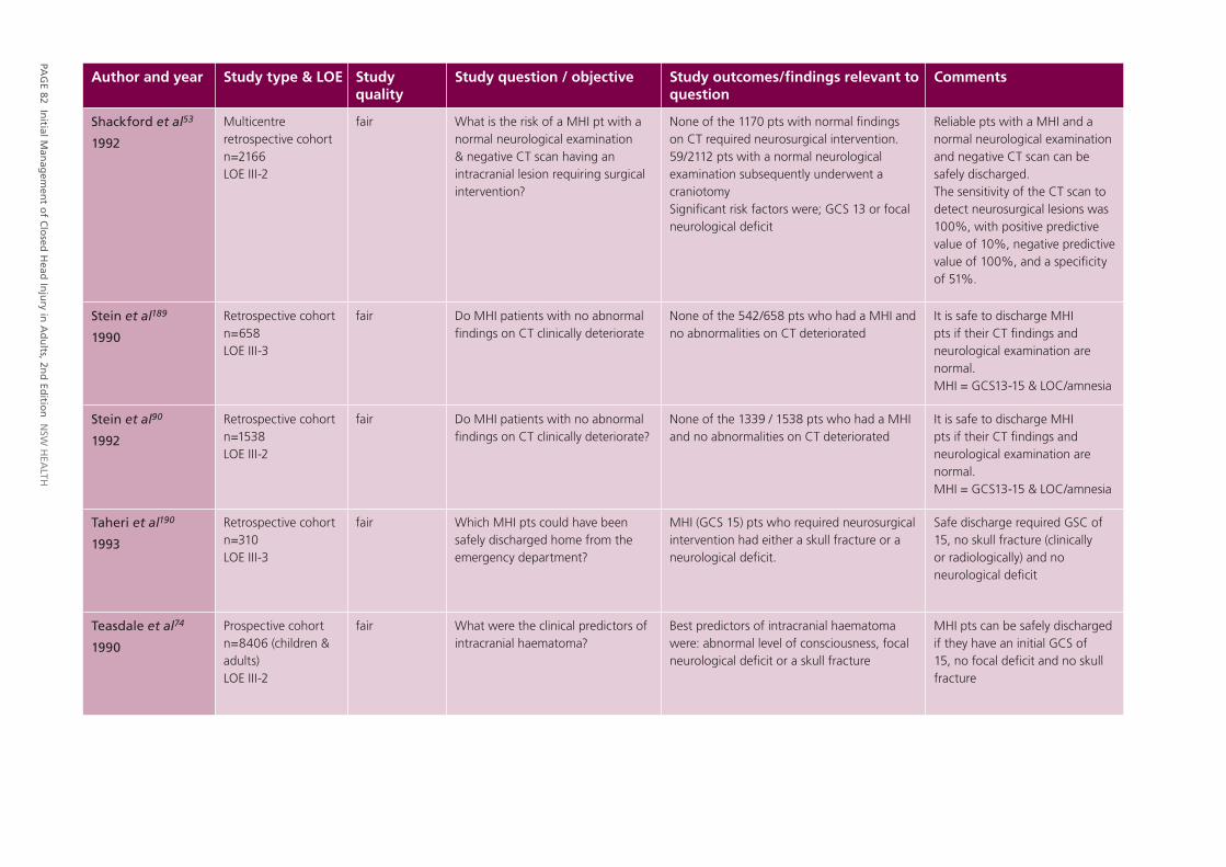
PAG
E 82 Initial M
anag
emen
t of C
losed
Head
Inju
ry in A
du
lts, 2nd
Editio
n N
SW H
EALTH
Author and year Study type & LOE Study quality
Study question / objective Study outcomes/fi ndings relevant to question
Comments
Shackford et al53
1992
Multicentre retrospective cohortn=2166LOE III-2
fair What is the risk of a MHI pt with a normal neurological examination & negative CT scan having an intracranial lesion requiring surgical intervention?
None of the 1170 pts with normal fi ndings on CT required neurosurgical intervention. 59/2112 pts with a normal neurological examination subsequently underwent a craniotomySignifi cant risk factors were; GCS 13 or focal neurological defi cit
Reliable pts with a MHI and a normal neurological examination and negative CT scan can be safely discharged.The sensitivity of the CT scan to detect neurosurgical lesions was 100%, with positive predictive value of 10%, negative predictive value of 100%, and a specifi city of 51%.
Stein et al189
1990
Retrospective cohortn=658LOE III-3
fair Do MHI patients with no abnormal fi ndings on CT clinically deteriorate
None of the 542/658 pts who had a MHI and no abnormalities on CT deteriorated
It is safe to discharge MHI pts if their CT fi ndings and neurological examination are normal.MHI = GCS13-15 & LOC/amnesia
Stein et al90
1992
Retrospective cohortn=1538LOE III-2
fair Do MHI patients with no abnormal fi ndings on CT clinically deteriorate?
None of the 1339 / 1538 pts who had a MHI and no abnormalities on CT deteriorated
It is safe to discharge MHI pts if their CT fi ndings and neurological examination are normal.MHI = GCS13-15 & LOC/amnesia
Taheri et al190
1993
Retrospective cohortn=310LOE III-3
fair Which MHI pts could have been safely discharged home from the emergency department?
MHI (GCS 15) pts who required neurosurgical intervention had either a skull fracture or a neurological defi cit.
Safe discharge required GSC of 15, no skull fracture (clinically or radiologically) and no neurological defi cit
Teasdale et al74
1990
Prospective cohortn=8406 (children & adults)LOE III-2
fair What were the clinical predictors of intracranial haematoma?
Best predictors of intracranial haematoma were: abnormal level of consciousness, focal neurological defi cit or a skull fracture
MHI pts can be safely discharged if they have an initial GCS of 15, no focal defi cit and no skull fracture
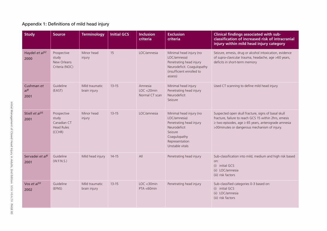
Initial M
anag
emen
t of C
losed
Head
Inju
ry in A
du
lts, 2nd
Editio
n N
SW H
EALTH
PAG
E 83
Study Source Terminology Initial GCS Inclusioncriteria
Exclusioncriteria
Clinical fi ndings associated with sub-classifi cation of increased risk of intracranial injury within mild head injury category
Haydel et al32
2000
Prospective studyNew Orleans Criteria (NOC)
Minor head injury
15 LOC/amnesia Minimal head injury (no LOC/amnesia)Penetrating head injuryNeurodefi cit. Coagulopathy (insuffi cient enrolled to assess)
Seizure, emesis, drug or alcohol intoxication, evidence of supra-clavicular trauma, headache, age >60 years, defi cits in short-term memory
Cushman et al8
2001
Guideline(EAST)
Mild traumatic brain injury
13-15 AmnesiaLOC <20minNormal CT scan
Minimal head injury Penetrating head injuryNeurodefi citSeizure
Used CT scanning to defi ne mild head injury
Stiell et al35
2001
Prospective studyCanadian CT Head Rules (CCHR)
Minor head injury
13-15 LOC/amnesia Minimal head injury (no LOC/amnesia)Penetrating head injuryNeurodefi citSeizureCoagulopathyRepresentationUnstable vitals
Suspected open skull fracture, signs of basal skull fracture, failure to reach GCS 15 within 2hrs, emesis two episodes, age 65 years, anterograde amnesia >30minutes or dangerous mechanism of injury.
Servadei et al6
2001
Guideline(W.F.N.S.)
Mild head injury 14-15 All Penetrating head injury Sub-classifi cation into mild, medium and high risk based on:(i) initial GCS(ii) LOC/amnesia(iii) risk factors
Vos et al10
2002
Guideline(EFNS)
Mild traumatic brain injury
13-15 LOC <30minPTA <60min
Penetrating head injury Sub-classifi ed categories 0-3 based on:(i) initial GCS(ii) LOC/amnesia(iii) risk factors
Appendix 1: Definitions of mild head injury
PAG
E 84 Initial M
anag
emen
t of C
losed
Head
Inju
ry in A
du
lts, 2nd
Editio
n N
SW H
EALTH
Study Source Terminology Initial GCS Inclusioncriteria
Exclusioncriteria
Clinical fi ndings associated with sub-classifi cation of increased risk of intracranial injury within mild head injury category
Haydel et al32
2000
Prospective studyNew Orleans Criteria (NOC)
Minor head injury
15 LOC/amnesia Minimal head injury (no LOC/amnesia)Penetrating head injuryNeurodefi cit. Coagulopathy (insuffi cient enrolled to assess)
Seizure, emesis, drug or alcohol intoxication, evidence of supra-clavicular trauma, headache, age >60 years, defi cits in short-term memory
Cushman et al8
2001
Guideline(EAST)
Mild traumatic brain injury
13-15 AmnesiaLOC <20minNormal CT scan
Minimal head injury Penetrating head injuryNeurodefi citSeizure
Used CT scanning to defi ne mild head injury
Stiell et al35
2001
Prospective studyCanadian CT Head Rules (CCHR)
Minor head injury
13-15 LOC/amnesia Minimal head injury (no LOC/amnesia)Penetrating head injuryNeurodefi citSeizureCoagulopathyRepresentationUnstable vitals
Suspected open skull fracture, signs of basal skull fracture, failure to reach GCS 15 within 2hrs, emesis two episodes, age 65 years, anterograde amnesia >30minutes or dangerous mechanism of injury.
Servadei et al6
2001
Guideline(W.F.N.S.)
Mild head injury 14-15 All Penetrating head injury Sub-classifi cation into mild, medium and high risk based on:(i) initial GCS(ii) LOC/amnesia(iii) risk factors
Vos et al10
2002
Guideline(EFNS)
Mild traumatic brain injury
13-15 LOC <30minPTA <60min
Penetrating head injury Sub-classifi ed categories 0-3 based on:(i) initial GCS(ii) LOC/amnesia(iii) risk factors
Initial M
anag
emen
t of C
losed
Head
Inju
ry in A
du
lts, 2nd
Editio
n N
SW H
EALTH
PAG
E 85
Study Source Terminology Initial GCS Inclusioncriteria
Exclusioncriteria
Clinical fi ndings associated with sub-classifi cation of increased risk of intracranial injury within mild head injury category
Jagoda et al9
2002
Guideline(ACEP)
Mild traumatic brain injury
15 LOC/amnesia Minimal head injuryPenetratingNeurodefi citCoagulopathyMultisystem trauma
Headache, emesis, age 60 years,defi cits in short-term memory, evidence of supra-clavicular trauma, seizure, drug or alcohol intoxication
Fabbri et al31
2004
Prospective study
Mild head injury 14-15 All Penetrating head injury As per Servadei et al (2001)
Ibanez et al33
2004
Prospective , observational
Mild head injury 14-15 With or without LOC
MHI patients further sub-classifi ed into:a. Minimal: GCS 15 & no LOCb. Mild: GCS14-15 & LOC<5min or PTAc. Moderate: GCS14-15 & LOC 5min
Mower et al58
2005
Prospective studyNEXUS II
Minor head injury
15 All Penetrating head injury Risk factors for intracranial injury identifi ed as any one of the following:Neurological defi cit (included GCS<15)Abnormal alertnessAbnormal behaviourPersistent vomitingSkull fractureScalp haematomaAge > 65Coagulopathy
Smits et al47
2007
Prospective, observational study.CHIP Rule
Minor head injury
13 - 15 With or without LOC or amnesia
Penetrating head injury Any one of the following: Pedestrian/cyclist vs vehicle, ejected from vehicle, emesis, posttraumatic amnesia 4 hours, clinical signs of skull fracture, GCS < 15, GCS drop 2 points after presentation, current anticoagulation therapy, posttraumatic seizure, age 60 years.At least two of the following:Fall from any height, persistent anterograde amnesia, posttraumatic amnesia 2 – 4 hours, contusion of the skull, neurologic defi cit, LOC, GCS drop of 1point (1h post presentation), age 40 – 60 years
PAG
E 86 Initial M
anag
emen
t of C
losed
Head
Inju
ry in A
du
lts, 2nd
Editio
n N
SW H
EALTH
Study Source Terminology Initial GCS Inclusioncriteria
Exclusioncriteria
Clinical fi ndings associated with sub-classifi cation of increased risk of intracranial injury within mild head injury category
Jagoda et al9
2008
GuidelineACEP / CDC
Mild traumatic brain injury
14 - 15 Presentation within 24h post injury with or without LOC or amnesia
Penetrating head injury LOC with any one of the following:Headache, vomiting, Age > 60y, drug or alcohol intoxication, defi cit in short-term memory, physical evidence of supra-clavicular trauma, posttraumatic seizure, GCS<15, focal neurologic defi cit, coagulopathy.No LOC with any one of the following:Focal neurologic defi cit, severe headache, Age 65y, physical signs of basilar skull fracture, GCS<15, coagulopathy, dangerous mechanism of injury.
Stein et al48
2009
Prospective study comparing six clinical decision guidelines
Mild traumatic brain injury
14 – 15 Presentation within 24h post injury
Penetrating head injuryUnstable vital signRe-presentation
Application of the Nexus II or Scandinavian Guidelines resulted in the highest sensitivity and specifi city in this sample.
Initial M
anag
emen
t of C
losed
Head
Inju
ry in A
du
lts, 2nd
Editio
n N
SW H
EALTH
PAG
E 87
Author and Year
Level of Evidence
Quality GCS 13 GCS 14 GCS 15
Patients (No.)
CT Scan Abnormal (%)
Neuro SX Required (%)
Patients (No.)
CT Scan Abnormal (%)
Neuro SX Required (%)
Patients (No.)
CT Scan Abnormal (%)
Neuro SX Required (%)
Dacey et al51
1986
III-2 fair 18 N/A 33 59 N/A 6.5 533 N/A 1.5
Teasdale et al74
1990
III-2 fair 7838 0.03
Shackford et al53
1992
III-2 fair 221 33 10.8 646 17.5 3.8 1899 14.8 3.2
Stein et al90
1992
IV fair 120 37.5 - 301 24.2 - 1117 13.2 -
Jeret et al68
1993
III-2 fair 712 9.4 0.3
Borczuk49
1995
III-2 poor 40 27.5 7.5 197 18.2 3.6 1211 5.9 0.1
Dunham et al36
1996
III-2 fair 1160 Age 14-6013 Age >60
2530
3.5-
435 Age 14-6057 Age >60
12.428
0.5-
1481 Age 14-60
3.010
0.13_
Culotta et al50
1996
III-2 fair 173 28 4.5 755 16 1.6 150 Age >60
4 0.4
Hsiang et al52
1997
III-2 fair 45 57.8 20 138 35.5 5.1 2179 18.5 2.2
Appendix 2: Initial GCS versus abnormal CT/Neurosurgery
Summary of studies examining the relationship between initial GCS and frequency of abnormal findings on CT scans and/or neurosurgical intervention.
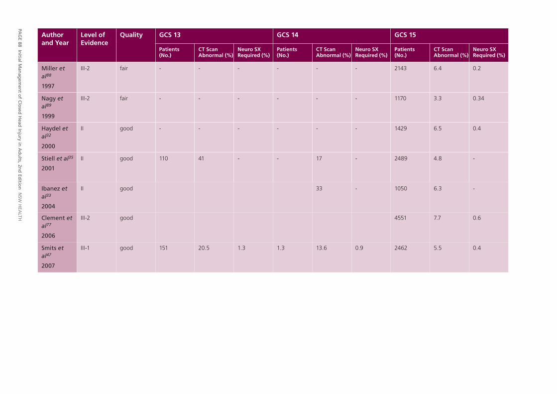
PAG
E 88 Initial M
anag
emen
t of C
losed
Head
Inju
ry in A
du
lts, 2nd
Editio
n N
SW H
EALTH
Author and Year
Level of Evidence
Quality GCS 13 GCS 14 GCS 15
Patients (No.)
CT Scan Abnormal (%)
Neuro SX Required (%)
Patients (No.)
CT Scan Abnormal (%)
Neuro SX Required (%)
Patients (No.)
CT Scan Abnormal (%)
Neuro SX Required (%)
Miller et al88
1997
III-2 fair - - - - - - 2143 6.4 0.2
Nagy et al89
1999
III-2 fair - - - - - - 1170 3.3 0.34
Haydel et al32
2000
II good - - - - - - 1429 6.5 0.4
Stiell et al35
2001
II good 110 41 - - 17 - 2489 4.8 -
Ibanez et al33
2004
II good 33 - 1050 6.3 -
Clement et al77
2006
III-2 good 4551 7.7 0.6
Smits et al47
2007
III-1 good 151 20.5 1.3 1.3 13.6 0.9 2462 5.5 0.4
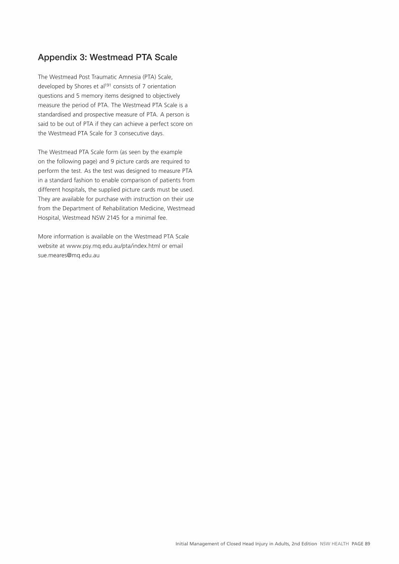
Initial Management of Closed Head Injury in Adults, 2nd Edition NSW HEALTH PAGE 89
Appendix 3: Westmead PTA Scale
The Westmead Post Traumatic Amnesia (PTA) Scale,
developed by Shores et al191 consists of 7 orientation
questions and 5 memory items designed to objectively
measure the period of PTA. The Westmead PTA Scale is a
standardised and prospective measure of PTA. A person is
said to be out of PTA if they can achieve a perfect score on
the Westmead PTA Scale for 3 consecutive days.
The Westmead PTA Scale form (as seen by the example
on the following page) and 9 picture cards are required to
perform the test. As the test was designed to measure PTA
in a standard fashion to enable comparison of patients from
different hospitals, the supplied picture cards must be used.
They are available for purchase with instruction on their use
from the Department of Rehabilitation Medicine, Westmead
Hospital, Westmead NSW 2145 for a minimal fee.
More information is available on the Westmead PTA Scale
website at www.psy.mq.edu.au/pta/index.html or email
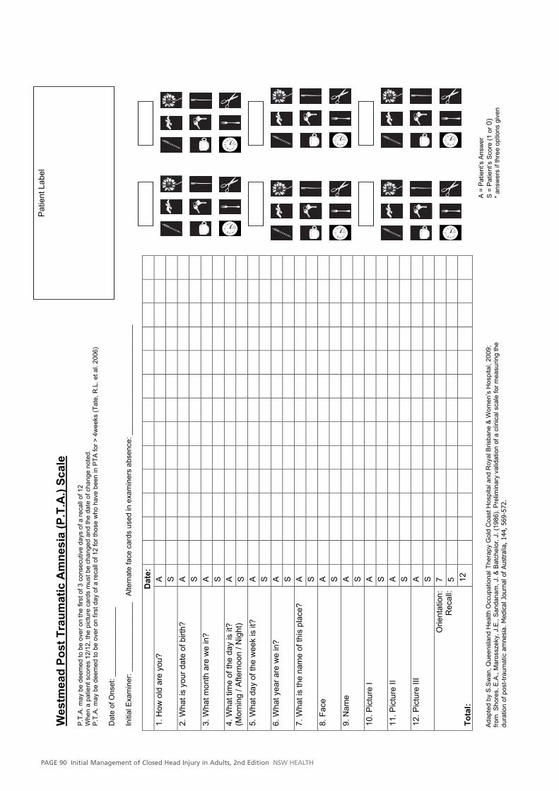
PAGE 90 Initial Management of Closed Head Injury in Adults, 2nd Edition NSW HEALTH
Wes
tmea
d Po
st T
raum
atic
Am
nesi
a (P
.T.A
.) Sc
ale
P.T
.A. m
ay b
e de
emed
to b
e ov
er o
n th
e fir
st o
f 3 c
onse
cutiv
e da
ys o
f a re
call
of 1
2 W
hen
a pa
tient
sco
res
12/1
2, th
e pi
ctur
e ca
rds
mus
t be
chan
ged
and
the
date
of c
hang
e no
ted.
P
.T.A
. may
be
deem
ed to
be
over
on
first
day
of a
reca
ll of
12
for t
hose
who
hav
e be
en in
PTA
for >
4w
eeks
(Tat
e, R
.L. e
t al.
2006
)
Pat
ient
Lab
el
Dat
e of
Ons
et: _
____
____
____
____
Initi
al E
xam
iner
: ___
____
____
____
__
Alte
rnat
e fa
ce c
ards
use
d in
exa
min
ers
abse
nce:
___
____
____
____
____
____
____
Dat
e:A
1. H
ow o
ld a
re y
ou?
S
A2.
Wha
t is
your
dat
e of
birt
h?
S A
3. W
hat m
onth
are
we
in?
S A4.
Wha
t tim
e of
the
day
is it
?
(M
orni
ng /
Afte
rnoo
n / N
ight
)
S A
5. W
hat d
ay o
f the
wee
k is
it?
S A6.
Wha
t yea
r are
we
in?
S A
7. W
hat i
s th
e na
me
of th
is p
lace
?
S A8.
Fac
e S A
9. N
ame
S A10
. Pic
ture
I S A
11. P
ictu
re II
S A
12. P
ictu
re II
I S 7 5
Orie
ntat
ion:
R
ecal
l:
Tota
l:12
A =
Pat
ient
’s A
nsw
er
S =
Pat
ient
’s S
core
(1 o
r 0)
* an
swer
s if
thre
e op
tions
giv
en
Ada
pted
by
S.S
wan
, Que
ensl
and
Hea
lth O
ccup
atio
nal T
hera
py G
old
Coa
st H
ospi
tal a
nd R
oyal
Bris
bane
& W
omen
’s H
ospi
tal,
2009
; fro
m S
hore
s, E
.A.,
Mar
ossz
eky,
J.E
., S
anda
nam
, J. &
Bat
chel
or, J
. (19
86).
Pre
limin
ary
valid
atio
n of
a c
linic
al s
cale
for m
easu
ring
the
du
ratio
n of
pos
t-tra
umat
ic a
mne
sia.
Med
ical
Jou
rnal
of
Aust
ralia
, 14
4, 5
69-5
72.
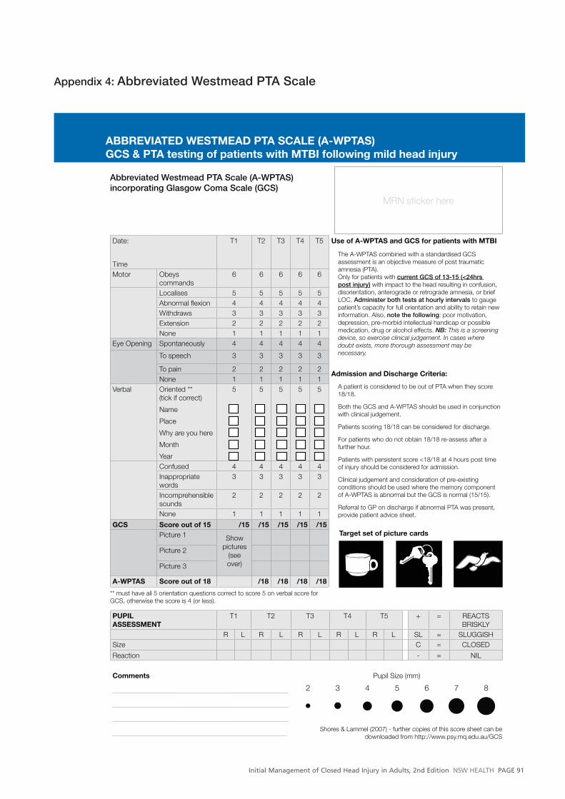
Initial Management of Closed Head Injury in Adults, 2nd Edition NSW HEALTH PAGE 91
PUPIL ASSESSMENT
T1 T2 T3 T4 T5 + = REACTS BRISKLY
R L R L R L R L R L SL = SLUGGISHSize C = CLOSED
Reaction - = NIL
Comments
Pupil Size (mm)
2 3 4 5 6 7 8
Date:
Time
T1 T2 T3 T4 T5 Use of A-WPTAS and GCS for patients with MTBI
The A-WPTAS combined with a standardised GCS assessment is an objective measure of post traumatic amnesia (PTA). Only for patients with current GCS of 13-15 (<24hrs post injury) with impact to the head resulting in confusion, disorientation, anterograde or retrograde amnesia, or brief LOC. Administer both tests at hourly intervals to gauge patient’s capacity for full orientation and ability to retain new information. Also, note the following: poor motivation, depression, pre-morbid intellectual handicap or possible medication, drug or alcohol effects. NB: This is a screening device, so exercise clinical judgement. In cases where doubt exists, more thorough assessment may benecessary.
Admission and Discharge Criteria:
A patient is considered to be out of PTA when they score 18/18.
Both the GCS and A-WPTAS should be used in conjunction with clinical judgement.
Patients scoring 18/18 can be considered for discharge.
For patients who do not obtain 18/18 re-assess after a further hour.
Patients with persistent score <18/18 at 4 hours post time of injury should be considered for admission.
Clinical judgement and consideration of pre-existing conditions should be used where the memory component of A-WPTAS is abnormal but the GCS is normal (15/15).
Referral to GP on discharge if abnormal PTA was present, provide patient advice sheet.
Motor Obeys commands
6 6 6 6 6
Localises 5 5 5 5 5Abnormal flexion 4 4 4 4 4Withdraws 3 3 3 3 3Extension 2 2 2 2 2None 1 1 1 1 1
Eye Opening Spontaneously 4 4 4 4 4
To speech 3 3 3 3 3
To pain 2 2 2 2 2None 1 1 1 1 1
Verbal Oriented ** (tick if correct)
Name
Place
Why are you here
Month
Year
5 5 5 5 5
Confused 4 4 4 4 4Inappropriate words
3 3 3 3 3
Incomprehensible sounds
2 2 2 2 2
None 1 1 1 1 1
GCS Score out of 15 /15 /15 /15 /15 /15 Target set of picture cardsPicture 1
Picture 2
Picture 3
A-WPTAS Score out of 18 /18 /18 /18 /18
Abbreviated Westmead PTA Scale (A-WPTAS)incorporating Glasgow Coma Scale (GCS)
MRN sticker here
Show pictures
(see over)
Shores & Lammel (2007) - further copies of this score sheet can be downloaded from http://www.psy.mq.edu.au/GCS
ABBREVIATED WESTMEAD PTA SCALE (A-WPTAS)GCS & PTA testing of patients with MTBI following mild head injury
** must have all 5 orientation questions correct to score 5 on verbal score for GCS, otherwise the score is 4 (or less).
Appendix 4: Abbreviated Westmead PTA Scale

PAGE 92 Initial Management of Closed Head Injury in Adults, 2nd Edition NSW HEALTH
Administration and Scoring 1. Orientation QuestionsQuestion 1: WHAT IS YOUR NAME? The patient must provide their full name.Question 2: WHAT IS THE NAME OF THIS PLACE? The patient has to be able to give the name of the hospital. For example: Westmead Hospital. (NB: The patient does not get any points for just saying ‘hospital’.) If the patient can not name the hospital, give them a choice of 3 options. To do this, pick 2 other similar sized hospitals in your local area or neighbouring region. In Westmead Hospital’s case the 3 choices are ‘Nepean Hospital, Westmead Hospital or Liverpool Hospital’. Question 3: WHY ARE YOU HERE? The patient must know why they were brought into hospital. e.g. they were injured in a car accident, fell, assaulted or injured playing sport. If the patient does not know, give them three options, including the correct reason.Question 4: WHAT MONTH ARE WE IN? For emphasis the examiner can ask what month are we in now? The patient must name the month. For example, if the patient answers ‘the 6th month’, the examiner must ask the further question ‘What is the 6th month called?’. Question 5: WHAT YEAR ARE WE IN? It is considered correct for patients to answer in the short form ‘08’, instead of ‘2008’. Also, an acceptable alternative prompt (for the rest of the 2000’s) is ‘The year is 2000 and what?’
2. Picture recognition Straight after administering the GCS (standardised questions), administer the A-WPTAS by presenting the 3 Westmead PTA cards. Picture Cards the first time - T1 : Show patients the target set of picture cards for about 5 seconds and ensure that they can repeat the names of each card. Tell the patient to remember the pictures for the next testing in about one hour. Picture Cards at each subsequent time T2-T5: Ask patient, “What were the three pictures that I showed you earlier?” Scoring:
For patients who free recall all 3 pictures correctly, assign a score of 1 per picture and add up the patient’s GCS (out of 15) and A-WPTAS memory component to give the A-WPTAS score (total = 18). Present the 3 target pictures again and re-test in 1 hour. For patients who can not free recall, or only partially free recall, the 3 correct pictures, present the 9-object recognition chart. If patient can recognise any correctly, score 1 per correct item and record their GCS and A-WPTAS score (total = 18). Present the target set of pictures again and re-test in 1 hour. For patients who neither remember any pictures by free call nor recognition, show the patient the target set of 3 picture cards again for re-test in 1 hour.
GLASGOW COMA SCALE (GCS) AND ABBREVIATED WESTMEAD PTA SCALE (A-WPTAS)
Shores & Lammel (2007) - further copies of this score sheet can be downloaded from http://www.psy.mq.edu.au/GCS
Research and development of the A-WPTAS supported by the Motor Accidents Authority NSW
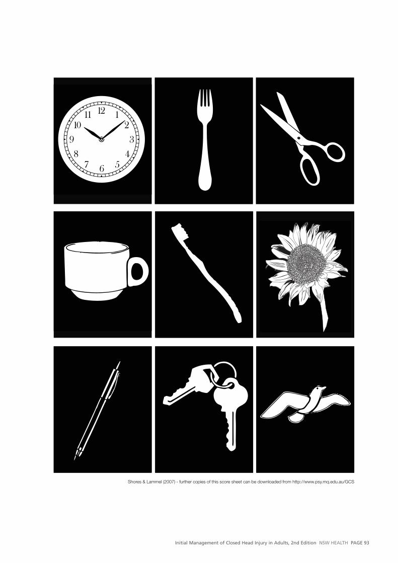
Initial Management of Closed Head Injury in Adults, 2nd Edition NSW HEALTH PAGE 93
Shores & Lammel (2007) - further copies of this score sheet can be downloaded from http://www.psy.mq.edu.au/GCS
PAGE 94 Initial Management of Closed Head Injury in Adults, 2nd Edition NSW HEALTH
Appendix 5: The Glasgow Coma Scale - a practical implementation guide
Associate Professor Paul M Middleton
Introduction
Impairment of consciousness is one of the most consistent
features of head injury, and the Glasgow Coma Scale
(GCS) was described by Teasdale and Jennett in 1974192,
based on a theoretical model of level of consciousness.
It was introduced as a simple tool, not to allow absolute
distinctions between levels of consciousness, but to be an
effective method of accurately describing the various states
of impairment within the continuum of consciousness.192
As the authors stated in the original paper “In the acute
stage, changes in conscious level provide the best indication
of the development of complications such as intracranial
haematoma, whilst the depth of coma and its duration
indicate the degree of ultimate recovery which can be
expected”. Prior to this most descriptions of altered
levels of consciousness revolved around very subjective
portrayals such as “comatose”, “drowsy”, “obtunded”, and
“stuporose”.193
The GCS was originally described by the authors as a
repeated bedside assessment of the “…depth and duration
of impaired consciousness and coma”192, and was used to
objectively determine the severity of coma and underlying
brain dysfunction at six hours following head trauma. This
time frame was chosen to avoid overestimation of brain
damage produced by temporary factors such as alcohol,
hypoxia or hypotension,193 and similarly the GCS should be
recorded prior to any sedation.
The GCS has also been incorporated as the neurological
component of assessment into various aggregate scores
such as APACHE and TRISS,193 and it has been found that
taking out this neurological component worsened predictive
ability, which led to the presumption that neurological
status is the best predictor of overall functional outcome.194
The GCS makes up 17% of the theoretical maximum Acute
Physiology Score (APS) in APACHE II, 19% of the APS in
APACHE III195 and is the basis of the World Federation of
neurosurgeons (WFNS) subarachnoid haemorrhage (SAH)
grading scale.196
The GCS, in actual fact, has evolved through both design
and common usage to fulfil multiple functions, which are
summarised in the list below. The GCS can be said to…
■ Aid in clinical decision making in interventions such as
airway management or intensive care admission■ Describe, quantitate and add structure to the
assessment of coma■ Facilitate and standardise communication between
clinicians■ Enable monitoring of change in both component
and overall scores, i.e. trends in the early stages after
injury, allowing rapid detection of complications and
discriminating between those at higher or lower risk of
complications■ Be an indicator of the severity of illness■ Facilitate comparison between groups of patients■ Allow triage of patients after injury■ Provide a tool for prognostication■ Allow standardisation of patients and patient groups
for research
The important primary uses for the GCS can be distilled
from this list to…
■ Act as an indicator of the level injury and illness,
allowing triage and immediate intervention when
required, as well as to enable monitoring by the
provision of valid measurements and trends of level of
consciousness■ Facilitate understanding, clear description and
communication between clinicians. This should enable
one clinician to describe the level of consciousness to
another, whether face-to-face or remotely, in the sure
knowledge that this description precisely represents
the injury, physiological and functional state of the
patient and that the receiving clinician may accurately
comprehend this from the description.
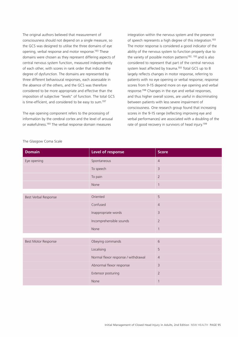
Initial Management of Closed Head Injury in Adults, 2nd Edition NSW HEALTH PAGE 95
The original authors believed that measurement of
consciousness should not depend on a single measure, so
the GCS was designed to utilise the three domains of eye
opening, verbal response and motor response.193 These
domains were chosen as they represent differing aspects of
central nervous system function, measured independently
of each other, with scores in rank order that indicate the
degree of dysfunction. The domains are represented by
three different behavioural responses, each assessable in
the absence of the others, and the GCS was therefore
considered to be more appropriate and effective than the
imposition of subjective “levels” of function. The total GCS
is time-efficient, and considered to be easy to sum.197
The eye opening component refers to the processing of
information by the cerebral cortex and the level of arousal
or wakefulness.193 The verbal response domain measures
integration within the nervous system and the presence
of speech represents a high degree of this integration.193
The motor response is considered a good indicator of the
ability of the nervous system to function properly due to
the variety of possible motion patterns192, 193 and is also
considered to represent that part of the central nervous
system least affected by trauma.193 Total GCS up to 8
largely reflects changes in motor response, referring to
patients with no eye opening or verbal response; response
scores from 9-15 depend more on eye opening and verbal
response.198 Changes in the eye and verbal responses,
and thus higher overall scores, are useful in discriminating
between patients with less severe impairment of
consciousness. One research group found that increasing
scores in the 9-15 range (reflecting improving eye and
verbal performances) are associated with a doubling of the
rate of good recovery in survivors of head injury.199
Domain Level of response Score
Eye opening Spontaneous 4
To speech 3
To pain 2
None 1
Best Verbal Response Oriented 5
Confused 4
Inappropriate words 3
Incomprehensible sounds 2
None 1
Best Motor Response Obeying commands 6
Localising 5
Normal fl exor response / withdrawal 4
Abnormal fl exor response 3
Extensor posturing 2
None 1
The Glasgow Coma Scale
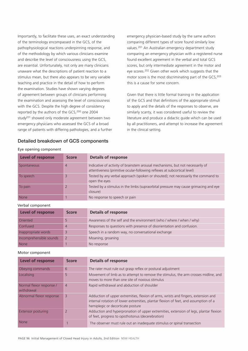
PAGE 96 Initial Management of Closed Head Injury in Adults, 2nd Edition NSW HEALTH
Importantly, to facilitate these uses, an exact understanding
of the terminology encompassed in the GCS, of the
pathophysiological reactions underpinning response, and
of the methodology by which various clinicians examine
and describe the level of consciousness using the GCS,
are essential. Unfortunately, not only are many clinicians
unaware what the descriptions of patient reaction to a
stimulus mean, but there also appears to be very variable
teaching and practice in the detail of how to perform
the examination. Studies have shown varying degrees
of agreement between groups of clinicians performing
the examination and assessing the level of consciousness
with the GCS. Despite the high degree of consistency
reported by the authors of the GCS,200 one 2004
study201 showed only moderate agreement between two
emergency physicians who assessed the GCS of a broad
range of patients with differing pathologies, and a further
emergency physician-based study by the same authors
comparing different types of score found similarly low
values.201 An Australian emergency department study
comparing an emergency physician with a registered nurse
found excellent agreement in the verbal and total GCS
scores, but only intermediate agreement in the motor and
eye scores.202 Given other work which suggests that the
motor score is the most discriminating part of the GCS,203
this is a cause for some concern.
Given that there is little formal training in the application
of the GCS and that definitions of the appropriate stimuli
to apply and the details of the responses to observe, are
similarly scanty, it was considered useful to review the
literature and produce a didactic guide which can be used
by all practitioners, and attempt to increase the agreement
in the clinical setting.
Level of response Score Details of response
Spontaneous 4 Indicative of activity of brainstem arousal mechanisms, but not necessarily of attentiveness (primitive ocular-following refl exes at subcortical level)
To speech 3 Tested by any verbal approach (spoken or shouted); not necessarily the command to open the eyes
To pain 2 Tested by a stimulus in the limbs (supraorbital pressure may cause grimacing and eye closure)
None 1 No response to speech or pain
Level of response Score Details of response
Oriented 5 Awareness of the self and the environment (who / where / when / why)
Confused 4 Responses to questions with presence of disorientation and confusion.
Inappropriate words 3 Speech in a random way, no conversational exchange
Incomprehensible sounds 2 Moaning, groaning
None 1 No response
Detailed breakdown of GCS components
Eye opening component
Verbal component
Level of response Score Details of response
Obeying commands 6 The rater must rule out grasp refl ex or postural adjustment
Localising 5 Movement of limb as to attempt to remove the stimulus, the arm crosses midline, and moves to more than one site of noxious stimulus
Normal fl exor response / withdrawal
4 Rapid withdrawal and abduction of shoulder
Abnormal fl exor response 3 Adduction of upper extremities, fl exion of arms, wrists and fi ngers, extension and internal rotation of lower extremities, plantar fl exion of feet, and assumption of a hemiplegic or decorticate posture
Extensor posturing 2 Adduction and hyperpronation of upper extremities, extension of legs, plantar fl exion of feet, progress to opisthotonus (decerebration)
None 1 The observer must rule out an inadequate stimulus or spinal transection
Motor component
Initial Management of Closed Head Injury in Adults, 2nd Edition NSW HEALTH PAGE 97
Assessment
Eyes
An eye component score of 3 or 4 implies that information
processing is occurring and that the related arousal
mechanisms at the brain stem are functioning,193 whereas
a eye component score of 2 indicates that lower levels of
the brain are functioning.193 It is not true however that
eye opening indicates awareness; for instance patients in
a persistent vegetative state may have spontaneous eye
opening, and in this instance this is a reflexive action and
does not indicate awareness of self or surroundings.197
Verbal
As stated above, presence of speech implies a high level of
integration in the nervous system, although it is important
to remember that a lack of speech may be attributed to
other factors such as tracheostomy or dysphasia. A lack of
speech due to local factors such as this need to be carefully
considered, as including a low score cause by local factors
into a GCS, especially when only the sum is being used,
falsely decreases the score. It is important to remember that
the GCS is a measure of level of consciousness, and use a
tracheostomy in an otherwise fully conscious patient to give
a decreased GCS is clearly counter-intuitive and incorrect.
In terms of the gradations of verbal component score,
oriented indicates that the patient is aware of his / her
self and the surrounding environment,197 and is usually
described in terms of questions about patient’s name, the
role of the person asking the questions, the month and
year, and the name of the hospital or health care facility.
Confused patients can carry on a conversation but the
content betrays disorientation and misunderstanding of
the components described above. Inappropriate words
describes clear and comprehensible speech,197 but using
random words or swearing and cursing. Repeating words or
perseveration also falls into this category. Incomprehensible
sounds refers to moaning and groaning without
recognisable words,197 even when an attempt to articulate
words is being made. It is important to differentiate
between a patient with a decreased level of consciousness
and reduced cognition, who is unable to form words in
response to stimulus, and an awake stroke patient for
instance; whose dysphasia may make the task impossible.
Clearly, the second case does not represent the situation
which the GCS is designed to measure. None means that
the patient is unable to verbalise at all,197 and is subject to
the factors described above.
Motor
Motor component scores of 6, 5 and 4 imply the presence
of a degree of cerebral function and thus the ability
to react appropriately to a noxious stimulus. Obeys
commands indicates an ability to process and obey verbal
commands;197 localisation means that the patient is able
to identify the location of a painful stimulus and attempt
to remove it, an action often accompanied by the upper
extremity of a patient purposefully crossing the midline
to remove the stimulus193, 197. Withdrawal means that
the patient is attempting to move away from the noxious
stimulus, sometimes by adopting a fetal position.197 This
last position is particularly important when there is an
inexperienced observer, as differentiating a localising
response from an abnormal flexion response may prove
difficult.195
A motor component score of 3, or an abnormal flexor
response, implies that the lesion is located in the cerebral
hemispheres or internal capsule,193 whereas a score of
2 describes a midbrain to upper pontine damage.193
Abnormal flexor response is complex, but involves
adduction of upper limbs, with flexion of arms, wrists
and fingers. Accompanying this are extension and
internal rotation of lower limbs, and plantar flexion of
feet. This must be differentiated from the normal flexor
response or withdrawal, but also from extensor posturing
which indicates a lesion lower in the central nervous
system, and therefore reflecting CNS function at a lower
level.193 Extensor posturing includes the same lower limb
appearances as in abnormal flexion, but with the upper
limbs adopting a different position; this is described as
extension of the upper limbs along the sides of the body,
accompanied by pronation of the forearms. Abnormal
flexion and extensor posturing are often known by the
terms decerebrate and decorticate response, implying the
level of loss of CNS function, and studies have shown that
patients showing extensor posturing are more likely to
have a poor outcome than those with abnormal flexion.197
If a patient demonstrates flexion on one side of the body,
and extension on the other, the best of the two responses
needs to be recorded.193
None means that the patient is flaccid, and does not make
any movement in response to a painful stimulus. In these
circumstances, it is essential to check that the patient is
not pharmacologically or pathologically paralysed. Bear in
mind again, that as the GCS is endeavouring to measure
cognition and that abnormal motor responses, due to
the presence of anaesthetic paralysis or spinal cord injury
PAGE 98 Initial Management of Closed Head Injury in Adults, 2nd Edition NSW HEALTH
invalidates the motor score at this time, in this patient, as
a means to measure consciousness. Another important
caveat to the measurement of the motor score component
of the GCS is that the simple “squeeze my fingers” is NOT
sufficient or appropriate to demonstrate this function.197 A
grasp reflex an be elicited in many patients with decreased
cognition, similar to that found in babies, and attempts at
least have to include the command to release the fingers
after squeezing them, and this must be seen to be obeyed.
More specific commands such as “show me two fingers”
are more appropriate.
In the sections above, there are various conditions discussed
which invalidate the measurement of specific domains of
the GCS, however it must always be borne in mind that
this tool is designed to assess consciousness and cognition,
and that local lesions of many descriptions invalidate this
measurement, and should therefore not be counted into
an overall score at all, and should not be counted into
a domain score without documented explanation. If a
domain of the GCS, such as eye opening or verbal response
is confounded by local lesions, then both snapshots and
trends should be limited to the use of the other domains.192,
193 Since the motor score has been shown to contain most
of the predictive power of the GCS, especially in the more
severely head injured patients, it would be reasonable to
use this alone in these circumstances. When this occurs,
it has been recommended that a 1 is scored,197 however
if this is done it has to be accompanied by a written
explanation and the caveat that this cannot be used in an
overall score.
Conditions such as alcohol, drugs, inability to understand
commands due to language barriers, and hearing
impairment are all conditions that may confound the
performance of a GCS, and once again consideration
needs to be given to the reason for measuring cognition.
For example, if the reason the GCS is being measured
is to assess the level of consciousness associated with a
head injury or pathological cause of decreased conscious
level, conditions such as alcohol or sedative drugs are a
confounder which invalidate the GCS; however, if the
measurement is being used to assess the effect of drugs on
the level of consciousness, this is then the relevant effect
being measured. In these circumstances, however, many of
the correlates of a decreased level of consciousness in head
injury measured by GCS may not be accurate; such as an
inability to protect the airway associated with a GCS 8,
which is often not true in patients obtunded with certain
drugs of abuse. An extended list of potential confounders is
shown below.
The GCS is NOT a scale to measure an altered sensorium,
so cannot be used to test sensation. It is also not substitute
for either a full neurological examination or an assessment
of orientation.193, 197 It also does not account for true
lateralisation as it measures the best response rather than
the worst.193
The sternal rub has been documented to cause injury,
particularly pressure area damage204 and cannot be
recommended. Supraorbital nerve pressure has caused
damage and is less reliable and consistent than other
methods or applying a central noxious stimulus.205 The
trapezius pinch causes no damage as it simply comprises
pressure on a large muscular area, but does provide a
suitable painful stimulus.204 Ensure that you note whether
the upper limb localised to the painful stimulus by crossing
the midline or not; if it does not, carefully assess to
discriminate between this and abnormal flexion.
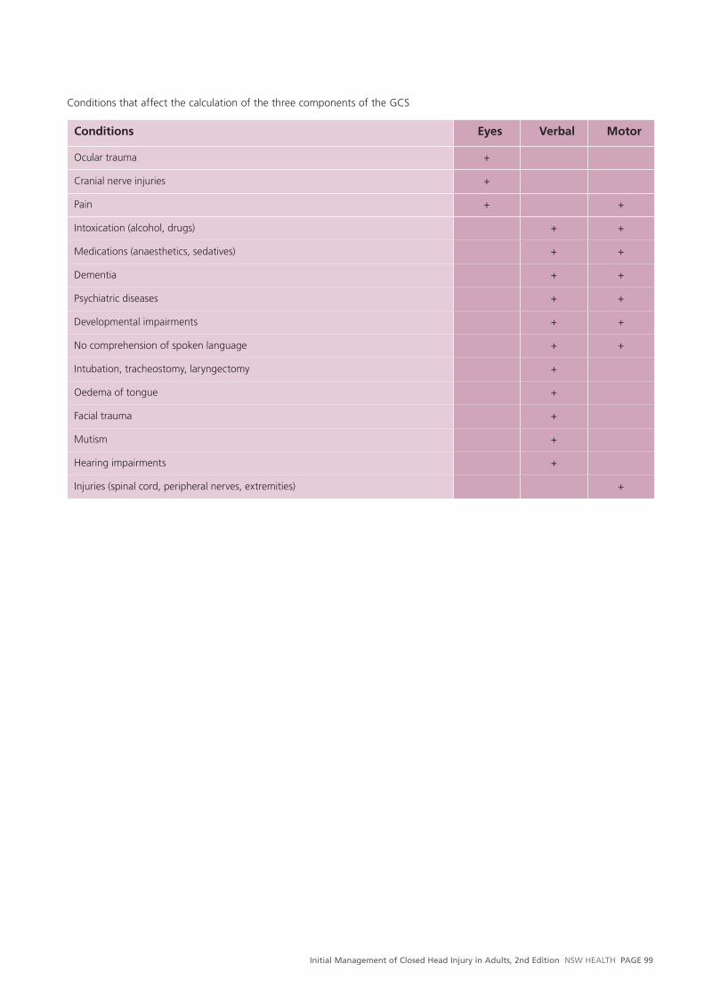
Initial Management of Closed Head Injury in Adults, 2nd Edition NSW HEALTH PAGE 99
Conditions Eyes Verbal Motor
Ocular trauma +
Cranial nerve injuries +
Pain + +
Intoxication (alcohol, drugs) + +
Medications (anaesthetics, sedatives) + +
Dementia + +
Psychiatric diseases + +
Developmental impairments + +
No comprehension of spoken language + +
Intubation, tracheostomy, laryngectomy +
Oedema of tongue +
Facial trauma +
Mutism +
Hearing impairments +
Injuries (spinal cord, peripheral nerves, extremities) +
Conditions that affect the calculation of the three components of the GCS
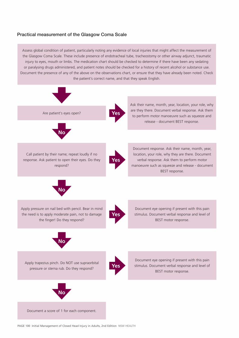
PAGE 100 Initial Management of Closed Head Injury in Adults, 2nd Edition NSW HEALTH
Practical measurement of the Glasgow Coma Scale
Assess global condition of patient, particularly noting any evidence of local injuries that might affect the measurement of
the Glasgow Coma Scale. These include presence of endotracheal tube, tracheostomy or other airway adjunct, traumatic
injury to eyes, mouth or limbs. The medication chart should be checked to determine if there have been any sedating
or paralysing drugs administered, and patient notes should be checked for a history of recent alcohol or substance use.
Document the presence of any of the above on the observations chart, or ensure that they have already been noted. Check
the patient’s correct name, and that they speak English.
Are patient's eyes open?
Call patient by their name; repeat loudly if no
response. Ask patient to open their eyes. Do they
respond?
Document response. Ask their name, month, year,
location, your role, why they are there. Document
verbal response. Ask them to perform motor
manoeuvre such as squeeze and release - document
BEST response.
Ask their name, month, year, location, your role, why
are they there. Document verbal response. Ask them
to perform motor manoeuvre such as squeeze and
release - document BEST response.
Apply pressure on nail bed with pencil. Bear in mind
the need is to apply moderate pain, not to damage
the finger! Do they respond?
Document eye opening if present with this pain
stimulus. Document verbal response and level of
BEST motor response.
Apply trapezius pinch. Do NOT use supraorbital
pressure or sterna rub. Do they respond?
Document eye opening if present with this pain
stimulus. Document verbal response and level of
BEST motor response.
Document a score of 1 for each component.
Yes
No
No
No
No
Yes
Yes
Yes
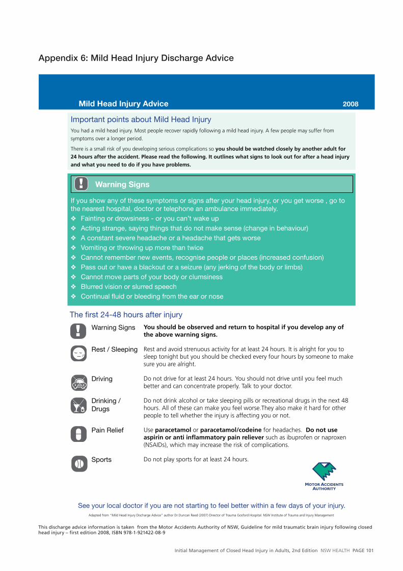
Initial Management of Closed Head Injury in Adults, 2nd Edition NSW HEALTH PAGE 101
Appendix 6: Mild Head Injury Discharge Advice
Important points about Mild Head InjuryYou had a mild head injury. Most people recover rapidly following a mild head injury. A few people may suffer from
symptoms over a longer period.
There is a small risk of you developing serious complications so you should be watched closely by another adult for 24 hours after the accident. Please read the following. It outlines what signs to look out for after a head injury and what you need to do if you have problems.
If you show any of these symptoms or signs after your head injury, or you get worse , go to the nearest hospital, doctor or telephone an ambulance immediately.
Fainting or drowsiness - or you can’t wake up ❖
Acting strange, saying things that do not make sense (change in behaviour) ❖
A constant severe headache or a headache that gets worse ❖
Vomiting or throwing up more than twice ❖
Cannot remember new events, recognise people or places (increased confusion) ❖
Pass out or have a blackout or a seizure (any jerking of the body or limbs) ❖
Cannot move parts of your body or clumsiness ❖
Blurred vision or slurred speech ❖
Continual fluid or bleeding from the ear or nose ❖
Warning Signs!
See your local doctor if you are not starting to feel better within a few days of your injury.Adapted from “Mild Head Injury Discharge Advice” author Dr Duncan Reed (2007) Director of Trauma Gosford Hospital. NSW Institute of Trauma and Injury Management
The first 24-48 hours after injury
Warning Signs You should be observed and return to hospital if you develop any of the above warning signs.
Rest / Sleeping Rest and avoid strenuous activity for at least 24 hours. It is alright for you to sleep tonight but you should be checked every four hours by someone to make sure you are alright.
Driving Do not drive for at least 24 hours. You should not drive until you feel much better and can concentrate properly. Talk to your doctor.
Drinking / Drugs
Do not drink alcohol or take sleeping pills or recreational drugs in the next 48 hours. All of these can make you feel worse.They also make it hard for other people to tell whether the injury is affecting you or not.
Pain Relief Use paracetamol or paracetamol/codeine for headaches. Do not use aspirin or anti inflammatory pain reliever such as ibuprofen or naproxen (NSAIDs), which may increase the risk of complications.
Sports Do not play sports for at least 24 hours.
zz
!
Mild Head Injury Advice 2008
This discharge advice information is taken from the Motor Accidents Authority of NSW, Guideline for mild traumatic brain injury following closed head injury – first edition 2008, ISBN 978-1-921422-08-9
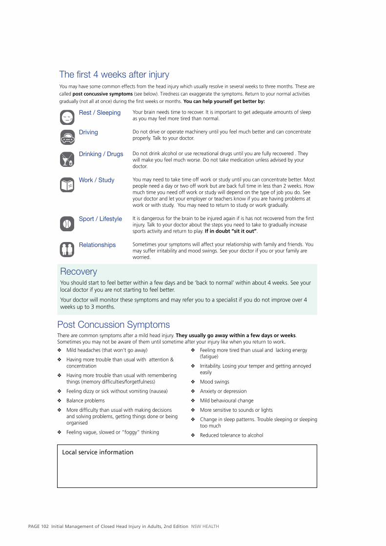
PAGE 102 Initial Management of Closed Head Injury in Adults, 2nd Edition NSW HEALTH
The first 4 weeks after injury You may have some common effects from the head injury which usually resolve in several weeks to three months. These are
called post concussive symptoms (see below). Tiredness can exaggerate the symptoms. Return to your normal activities
gradually (not all at once) during the first weeks or months. You can help yourself get better by:
Rest / Sleeping Your brain needs time to recover. It is important to get adequate amounts of sleep as you may feel more tired than normal.
Driving Do not drive or operate machinery until you feel much better and can concentrate properly. Talk to your doctor.
Drinking / Drugs Do not drink alcohol or use recreational drugs until you are fully recovered . They will make you feel much worse. Do not take medication unless advised by your doctor.
Work / Study You may need to take time off work or study until you can concentrate better. Most people need a day or two off work but are back full time in less than 2 weeks. How much time you need off work or study will depend on the type of job you do. See your doctor and let your employer or teachers know if you are having problems at work or with study. You may need to return to study or work gradually.
Sport / Lifestyle It is dangerous for the brain to be injured again if is has not recovered from the first injury. Talk to your doctor about the steps you need to take to gradually increase sports activity and return to play. If in doubt “sit it out”.
Relationships Sometimes your symptoms will affect your relationship with family and friends. You may suffer irritability and mood swings. See your doctor if you or your family are worried.
zz
RecoveryYou should start to feel better within a few days and be ‘back to normal’ within about 4 weeks. See your local doctor if you are not starting to feel better.
Your doctor will monitor these symptoms and may refer you to a specialist if you do not improve over 4 weeks up to 3 months.
Local service information
Post Concussion Symptoms There are common symptoms after a mild head injury. They usually go away within a few days or weeks. Sometimes you may not be aware of them until sometime after your injury like when you return to work.
Mild headaches (that won’t go away) ❖
Having more trouble than usual with attention & ❖
concentration
Having more trouble than usual with remembering ❖
things (memory difficulties/forgetfulness)
Feeling dizzy or sick without vomiting (nausea) ❖
Balance problems ❖
More difficulty than usual with making decisions ❖
and solving problems, getting things done or being organised
Feeling vague, slowed or “foggy” thinking ❖
Feeling more tired than usual and lacking energy ❖
(fatigue)
Irritability. Losing your temper and getting annoyed ❖
easily
Mood swings ❖
Anxiety or depression ❖
Mild behavioural change ❖
More sensitive to sounds or lights ❖
Change in sleep patterns. Trouble sleeping or sleeping ❖
too much
Reduced tolerance to alcohol ❖
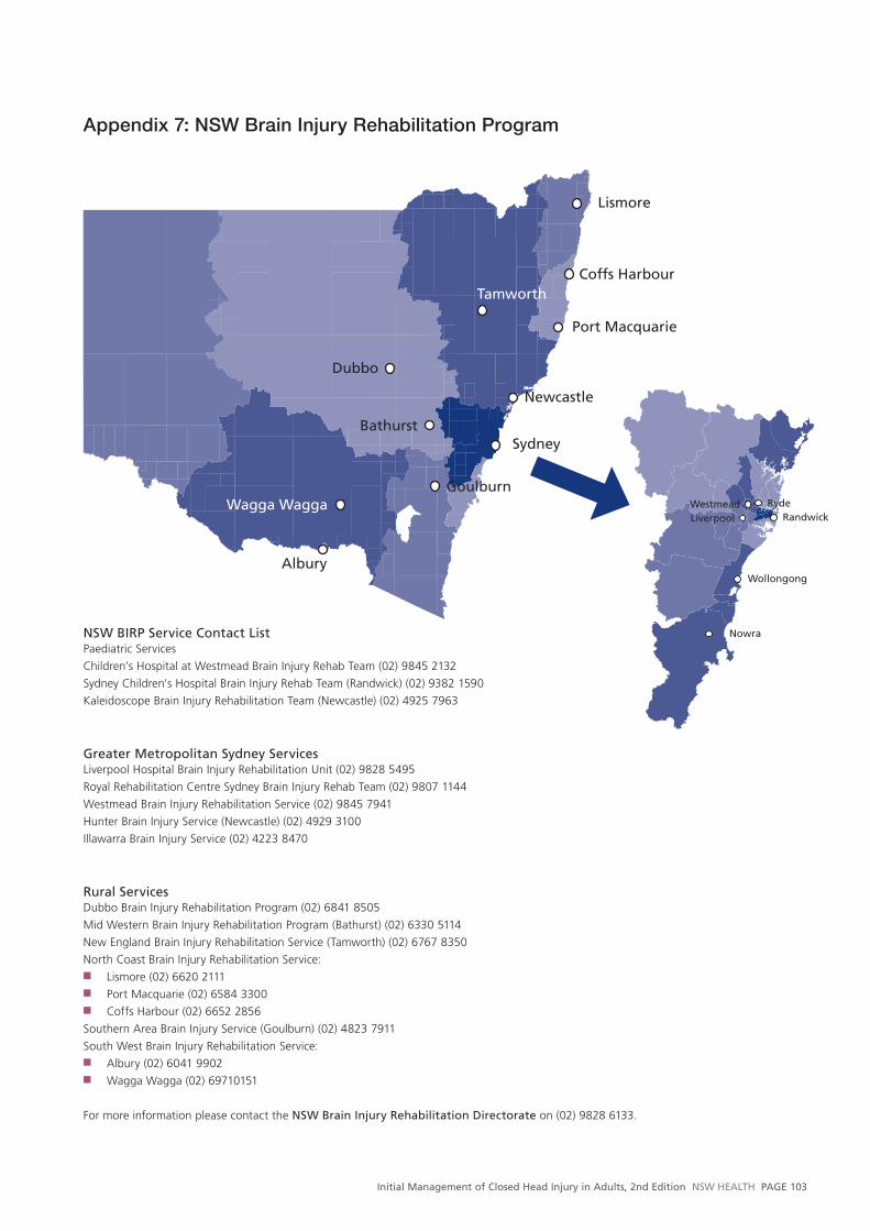
Initial Management of Closed Head Injury in Adults, 2nd Edition NSW HEALTH PAGE 103
Appendix 7: NSW Brain Injury Rehabilitation Program
NSW BIRP Service Contact ListPaediatric Services
Children's Hospital at Westmead Brain Injury Rehab Team (02) 9845 2132
Sydney Children's Hospital Brain Injury Rehab Team (Randwick) (02) 9382 1590
Kaleidoscope Brain Injury Rehabilitation Team (Newcastle) (02) 4925 7963
Greater Metropolitan Sydney ServicesLiverpool Hospital Brain Injury Rehabilitation Unit (02) 9828 5495
Royal Rehabilitation Centre Sydney Brain Injury Rehab Team (02) 9807 1144
Westmead Brain Injury Rehabilitation Service (02) 9845 7941
Hunter Brain Injury Service (Newcastle) (02) 4929 3100
Illawarra Brain Injury Service (02) 4223 8470
Rural ServicesDubbo Brain Injury Rehabilitation Program (02) 6841 8505
Mid Western Brain Injury Rehabilitation Program (Bathurst) (02) 6330 5114
New England Brain Injury Rehabilitation Service (Tamworth) (02) 6767 8350
North Coast Brain Injury Rehabilitation Service:■ Lismore (02) 6620 2111 ■ Port Macquarie (02) 6584 3300■ Coffs Harbour (02) 6652 2856
Southern Area Brain Injury Service (Goulburn) (02) 4823 7911
South West Brain Injury Rehabilitation Service:■ Albury (02) 6041 9902■ Wagga Wagga (02) 69710151
For more information please contact the NSW Brain Injury Rehabilitation Directorate on (02) 9828 6133.
Nowra
Wollongong
RandwickRyde
LiverpoolWestmead
Lismore
Coffs Harbour
Port Macquarie
Tamworth
Newcastle
Sydney
GoulburnWagga Wagga
Albury
Dubbo
Bathurst
PAGE 104 Initial Management of Closed Head Injury in Adults, 2nd Edition NSW HEALTH
Appendix 8: Methodology
A8.1 General search strategy
Comprehensive search strategies for both Medline and
Embase were guided by each of the clinical questions. A
general text-word based strategy was used to search the
Cochrane Database of Systematic Reviews (CDSR), the
Cochrane Central Register of Controlled Trials (CENTRAL)
and SCOPUS. The searches were executed for the period
1980 to 30th October 2004 (1st Ed.) and updated for
the 2nd Ed. (2004-2010). The results from each of these
searches were filtered in accordance with the exclusion
and inclusion criteria (see below) and then assessed for
relevance to the clinical questions. The search strategies are
listed in Appendix 9.
In addition, reference lists of previous guidelines and key
papers were used to identify other key references, including
pre-2004 literature. SCOPUS and Google Scholar were used
to execute author-based searches, citation mapping and
grey literature searching. The following websites were also
searched (using relevant free text terms):
Scottish Intercollegiate Guidelines Network
www.sign.ac.uk
Bandolier
www.medicine.ox.ac.uk/bandolier
TRIP database
www.tripdatabase.com
ClinicalTrials.gov
www.ClinicalTrials.gov
National Guideline Clearing House
www.guideline.gov
Brain Trauma Foundation
www.braintrauma.org
National Institute for Health & Clinical Evidence
www.nice.org.uk
Agency for Healthcare Research & Quality
www.ahrq.gov
Conditions Exclusion Criteria
■ Closed head injury studies■ Aged > 16 years■ Meta-analyses, systematic reviews, clinical guidelines
incorporating systematic reviews, controlled trials, comparative studies.
■ Penetrating head injury or brain damage from stroke/ cerebrovascular incidents
■ Aged < 16 years ■ Narrative reviews, letters, editorials, case studies/ series■ Studies using non-human subjects
A8.2 Inclusion and exclusion criteria
A8.3 Strength of recommendations
This guideline uses the National Health and Medical
Research Council (NHMRC) overall grades of
recommendation to indicate the strength of the body of
evidence underpinning each recommendation. The body
of evidence reflects the evidence components of all the
studies relevant to each recommendation. The evidence
components are assessed according to the NHMRC body
of evidence matrix (see table below). The overall grade of
the recommendation is determined based on a summation
of the rating for each individual component of the body of
evidence. Please note that a recommendation cannot be
graded A or B unless the evidence base and consistency of
the evidence are both rated A or B.44
Overall grade A or B recommendations are generally based
on a body of evidence that can be trusted to guide clinical
practice, whereas Grades C or D recommendations must
be applied carefully to individual clinical and organisational
circumstances and should be interpreted with care44 This
guideline also utilises an additional grade of “Consensus”
where appropriate.
The recommendation boxes of each clinical question
addressed in this guideline contain clear recommendations
with an associated strength of recommendation grade as
per above. Where appropriate, the author has also added
relevant clinical points to the boxes which support the given
recommendation.
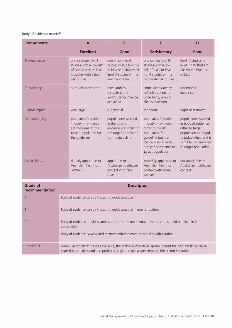
Initial Management of Closed Head Injury in Adults, 2nd Edition NSW HEALTH PAGE 105
Components A B C D
Excellent Good Satisfactory Poor
Evidence base one or more level I studies with a low risk of bias or several level II studies with a low risk of bias
one or two level II studies with a low risk of bias or a SR/several level III studies with a low risk of bias
one or two level III studies with a low risk of bias, or level I or II studies with a moderate risk of bias
level IV studies, or level I to III studies/SRs with a high risk of bias
Consistency all studies consistent most studies consistent and inconsistency may be explained
some inconsistency refl ecting genuine uncertainty around clinical question
evidence is inconsistent
Clinical impact very large substantial moderate slight or restricted
Generalisability population/s studied in body of evidence are the same as the target population for the guideline
population/s studied in the body of evidence are similar to the target population for the guideline
population/s studied in body of evidence differ to target population for guideline but it is clinically sensible to apply this evidence to target population
population/s studied in body of evidence differ to target population and hard to judge whether it is sensible to generalise to target population
Applicability directly applicable to Australian healthcare context
applicable to Australian healthcare context with few caveats
probably applicable to Australian healthcare context with some caveats
not applicable to Australian healthcare context
Body of evidence matrix44
Grade of recommendation
Description
A Body of evidence can be trusted to guide practice
B Body of evidence can be trusted to guide practice in most situations
C Body of evidence provides some support for recommendation(s) but care should be taken in its application
D Body of evidence is weak and recommendation must be applied with caution
Consensus When limited literature was available, the author and editorial group utilised the best available clinical expertise, practices and accepted teachings to reach a consensus on the recommendation
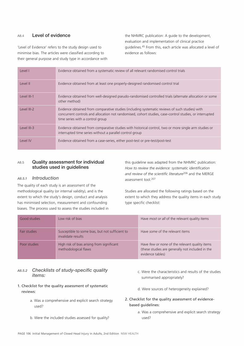
PAGE 106 Initial Management of Closed Head Injury in Adults, 2nd Edition NSW HEALTH
A8.4 Level of evidence
‘Level of Evidence’ refers to the study design used to
minimise bias. The articles were classified according to
their general purpose and study type in accordance with
the NHMRC publication: A guide to the development,
evaluation and implementation of clinical practice
guidelines.45 From this, each article was allocated a level of
evidence as follows:
Level I Evidence obtained from a systematic review of all relevant randomised control trials
Level II Evidence obtained from at least one properly-designed randomised control trial
Level III-1 Evidence obtained from well-designed pseudo-randomised controlled trials (alternate allocation or some other method)
Level III-2 Evidence obtained from comparative studies (including systematic reviews of such studies) with concurrent controls and allocation not randomised, cohort studies, case-control studies, or interrupted time series with a control group
Level III-3 Evidence obtained from comparative studies with historical control, two or more single arm studies or interrupted time series without a parallel control group
Level IV Evidence obtained from a case-series, either post-test or pre-test/post-test
A8.5 Quality assessment for individual studies used in guidelines
A8.5.1 Introduction
The quality of each study is an assessment of the
methodological quality (or internal validity), and is the
extent to which the study’s design, conduct and analysis
has minimised selection, measurement and confounding
biases. The process used to assess the studies included in
this guideline was adapted from the NHMRC publication:
How to review the evidence: systematic identification
and review of the scientific literature206 and the MERGE
assessment tool.207
Studies are allocated the following ratings based on the
extent to which they address the quality items in each study
type specific checklist:
A8.5.2 Checklists of study-specific quality items:
1. Checklist for the quality assessment of systematic
reviews:
a. Was a comprehensive and explicit search strategy
used?
b. Were the included studies assessed for quality?
c. Were the characteristics and results of the studies
summarised appropriately?
d. Were sources of heterogeneity explained?
2. Checklist for the quality assessment of evidence-
based guidelines:
a. Was a comprehensive and explicit search strategy
used?
Good studies Low risk of bias Have most or all of the relevant quality items
Fair studies Susceptible to some bias, but not suffi cient to invalidate results
Have some of the relevant items
Poor studies High risk of bias arising from signifi cant methodological fl aws
Have few or none of the relevant quality items (these studies are generally not included in the evidence tables)
Initial Management of Closed Head Injury in Adults, 2nd Edition NSW HEALTH PAGE 107
b. Have all relevant interventions and outcomes
been considered, both benefits and harms?
c. Is the level and quality of evidence for each
recommendation given?
d. Do the recommendations address benefit versus
harm according to the level of risk in different
patient sub-groups?
3. Checklist for the quality assessment of intervention
studies:
a. Has selection bias (including allocation bias) been
minimised?
b. Have adequate adjustments been made for
residual confounding?
c. Was the follow-up for final outcomes adequate?
(Follow-up rate reported and adequately high?)
d. Has measurement or misclassification bias been
minimised? (Blinding of outcome measurements?)
4. Checklist for the quality assessment of diagnostic
studies:
a. Has selection bias been minimised? (Were
participants consecutively enrolled?)
b. Have adequate adjustments been made for
residual confounding? (Were interventions blind
to the test result?)
c. Was follow-up for final outcomes adequate?
(Were all enrolled verified by the reference
standard?)
d. Has measurement or misclassification bias
been minimised? (Was the reference standard
validated and measured blindly?)
5. Checklist for the quality assessment of prognostic
studies (cohort studies):
a. Has selection bias been minimised? (A random/
consecutive sample of participants at the same
point in their disease?)
b. Were all potentially important prognostic factors
assessed?
c. Was follow-up for final outcomes adequately
long and complete?
d. Has measurement or misclassification bias been
minimised? (Were outcomes measured blind?)
A8.6 Quality assessment for guidelines referred to in this document
The Appraisal of Guidelines for Research and Evaluation
(AGREE) instrument,208 a validated tool, was used for
the quality appraisal of all guidelines referenced in this
document. Each appraised guideline is scored on six criteria
resulting in an overall assessment of strongly recommended,
recommended, would not recommend or unsure.
For further information please see the AGREE Collaboration
website www.agreecollaboration.org.
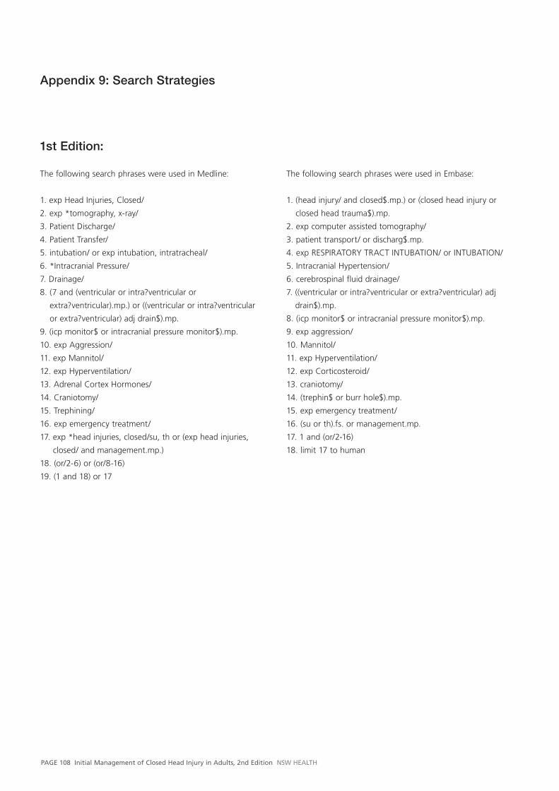
PAGE 108 Initial Management of Closed Head Injury in Adults, 2nd Edition NSW HEALTH
Appendix 9: Search Strategies
1st Edition:
The following search phrases were used in Medline:
1. exp Head Injuries, Closed/
2. exp *tomography, x-ray/
3. Patient Discharge/
4. Patient Transfer/
5. intubation/ or exp intubation, intratracheal/
6. *Intracranial Pressure/
7. Drainage/
8. (7 and (ventricular or intra?ventricular or
extra?ventricular).mp.) or ((ventricular or intra?ventricular
or extra?ventricular) adj drain$).mp.
9. (icp monitor$ or intracranial pressure monitor$).mp.
10. exp Aggression/
11. exp Mannitol/
12. exp Hyperventilation/
13. Adrenal Cortex Hormones/
14. Craniotomy/
15. Trephining/
16. exp emergency treatment/
17. exp *head injuries, closed/su, th or (exp head injuries,
closed/ and management.mp.)
18. (or/2-6) or (or/8-16)
19. (1 and 18) or 17
The following search phrases were used in Embase:
1. (head injury/ and closed$.mp.) or (closed head injury or
closed head trauma$).mp.
2. exp computer assisted tomography/
3. patient transport/ or discharg$.mp.
4. exp RESPIRATORY TRACT INTUBATION/ or INTUBATION/
5. Intracranial Hypertension/
6. cerebrospinal fluid drainage/
7. ((ventricular or intra?ventricular or extra?ventricular) adj
drain$).mp.
8. (icp monitor$ or intracranial pressure monitor$).mp.
9. exp aggression/
10. Mannitol/
11. exp Hyperventilation/
12. exp Corticosteroid/
13. craniotomy/
14. (trephin$ or burr hole$).mp.
15. exp emergency treatment/
16. (su or th).fs. or management.mp.
17. 1 and (or/2-16)
18. limit 17 to human
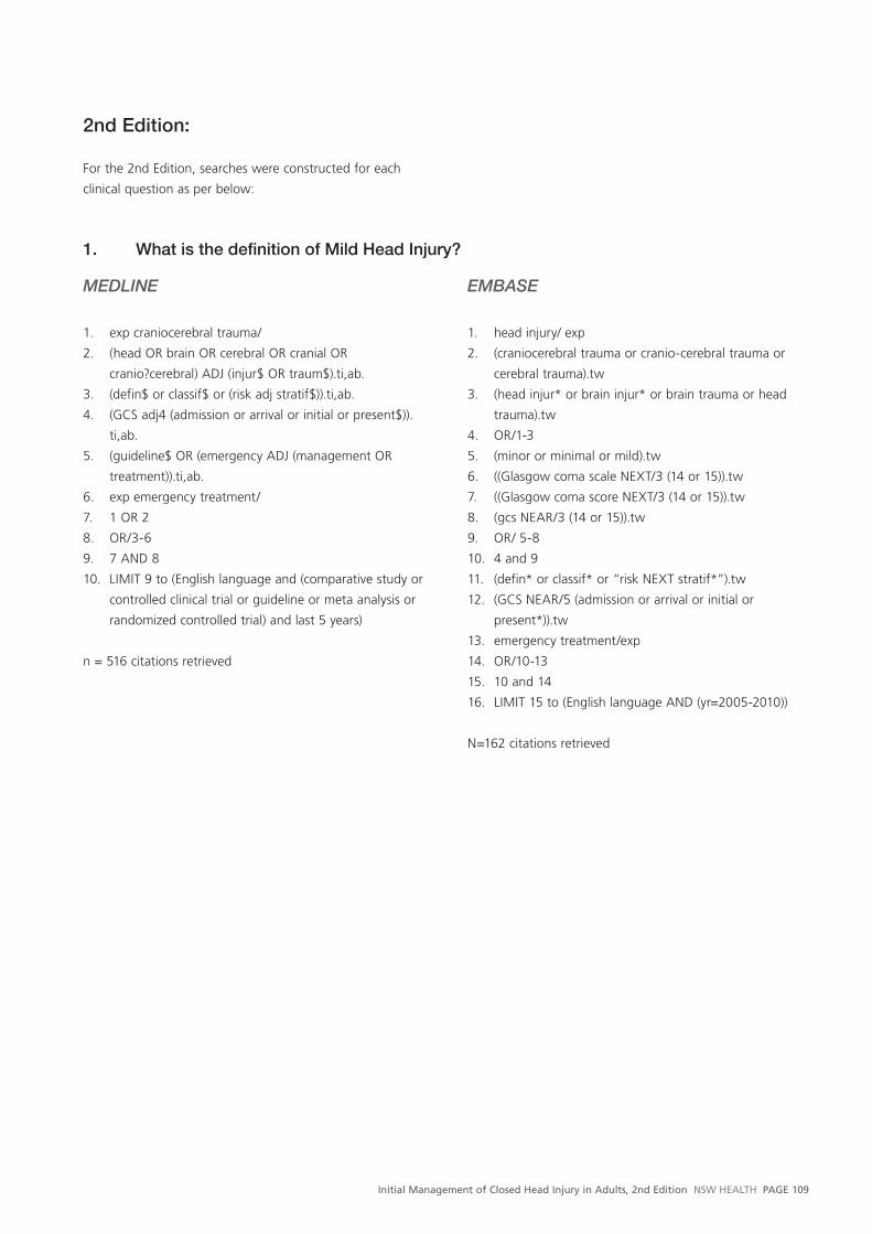
Initial Management of Closed Head Injury in Adults, 2nd Edition NSW HEALTH PAGE 109
1. What is the definition of Mild Head Injury?
MEDLINE
1. exp craniocerebral trauma/
2. (head OR brain OR cerebral OR cranial OR
cranio?cerebral) ADJ (injur$ OR traum$).ti,ab.
3. (defin$ or classif$ or (risk adj stratif$)).ti,ab.
4. (GCS adj4 (admission or arrival or initial or present$)).
ti,ab.
5. (guideline$ OR (emergency ADJ (management OR
treatment)).ti,ab.
6. exp emergency treatment/
7. 1 OR 2
8. OR/3-6
9. 7 AND 8
10. LIMIT 9 to (English language and (comparative study or
controlled clinical trial or guideline or meta analysis or
randomized controlled trial) and last 5 years)
n = 516 citations retrieved
EMBASE
1. head injury/ exp
2. (craniocerebral trauma or cranio-cerebral trauma or
cerebral trauma).tw
3. (head injur* or brain injur* or brain trauma or head
trauma).tw
4. OR/1-3
5. (minor or minimal or mild).tw
6. ((Glasgow coma scale NEXT/3 (14 or 15)).tw
7. ((Glasgow coma score NEXT/3 (14 or 15)).tw
8. (gcs NEAR/3 (14 or 15)).tw
9. OR/ 5-8
10. 4 and 9
11. (defin* or classif* or “risk NEXT stratif*”).tw
12. (GCS NEAR/5 (admission or arrival or initial or
present*)).tw
13. emergency treatment/exp
14. OR/10-13
15. 10 and 14
16. LIMIT 15 to (English language AND (yr=2005-2010))
N=162 citations retrieved
2nd Edition:
For the 2nd Edition, searches were constructed for each
clinical question as per below:
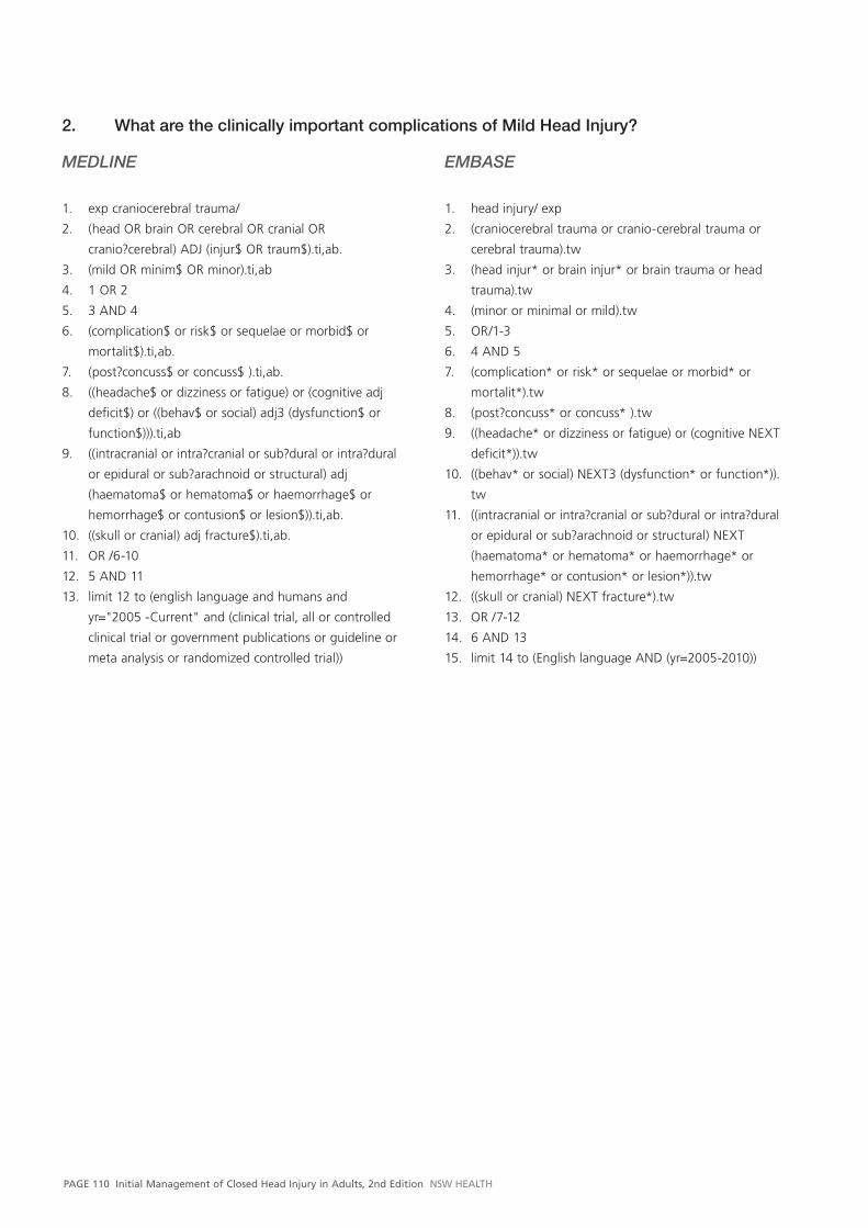
PAGE 110 Initial Management of Closed Head Injury in Adults, 2nd Edition NSW HEALTH
2. What are the clinically important complications of Mild Head Injury?
MEDLINE
1. exp craniocerebral trauma/
2. (head OR brain OR cerebral OR cranial OR
cranio?cerebral) ADJ (injur$ OR traum$).ti,ab.
3. (mild OR minim$ OR minor).ti,ab
4. 1 OR 2
5. 3 AND 4
6. (complication$ or risk$ or sequelae or morbid$ or
mortalit$).ti,ab.
7. (post?concuss$ or concuss$ ).ti,ab.
8. ((headache$ or dizziness or fatigue) or (cognitive adj
deficit$) or ((behav$ or social) adj3 (dysfunction$ or
function$))).ti,ab
9. ((intracranial or intra?cranial or sub?dural or intra?dural
or epidural or sub?arachnoid or structural) adj
(haematoma$ or hematoma$ or haemorrhage$ or
hemorrhage$ or contusion$ or lesion$)).ti,ab.
10. ((skull or cranial) adj fracture$).ti,ab.
11. OR /6-10
12. 5 AND 11
13. limit 12 to (english language and humans and
yr="2005 -Current" and (clinical trial, all or controlled
clinical trial or government publications or guideline or
meta analysis or randomized controlled trial))
EMBASE
1. head injury/ exp
2. (craniocerebral trauma or cranio-cerebral trauma or
cerebral trauma).tw
3. (head injur* or brain injur* or brain trauma or head
trauma).tw
4. (minor or minimal or mild).tw
5. OR/1-3
6. 4 AND 5
7. (complication* or risk* or sequelae or morbid* or
mortalit*).tw
8. (post?concuss* or concuss* ).tw
9. ((headache* or dizziness or fatigue) or (cognitive NEXT
deficit*)).tw
10. ((behav* or social) NEXT3 (dysfunction* or function*)).
tw
11. ((intracranial or intra?cranial or sub?dural or intra?dural
or epidural or sub?arachnoid or structural) NEXT
(haematoma* or hematoma* or haemorrhage* or
hemorrhage* or contusion* or lesion*)).tw
12. ((skull or cranial) NEXT fracture*).tw
13. OR /7-12
14. 6 AND 13
15. limit 14 to (English language AND (yr=2005-2010))
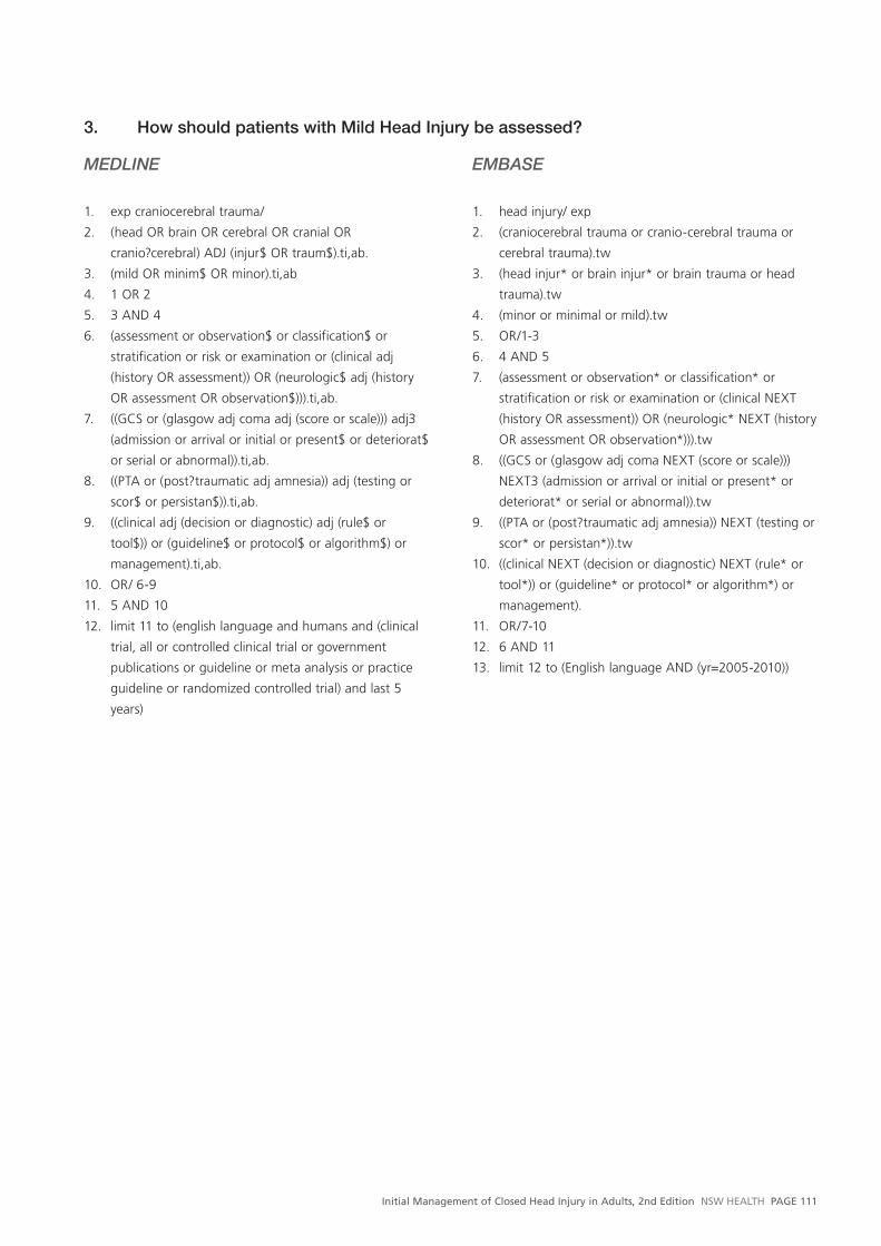
Initial Management of Closed Head Injury in Adults, 2nd Edition NSW HEALTH PAGE 111
3. How should patients with Mild Head Injury be assessed?
MEDLINE
1. exp craniocerebral trauma/
2. (head OR brain OR cerebral OR cranial OR
cranio?cerebral) ADJ (injur$ OR traum$).ti,ab.
3. (mild OR minim$ OR minor).ti,ab
4. 1 OR 2
5. 3 AND 4
6. (assessment or observation$ or classification$ or
stratification or risk or examination or (clinical adj
(history OR assessment)) OR (neurologic$ adj (history
OR assessment OR observation$))).ti,ab.
7. ((GCS or (glasgow adj coma adj (score or scale))) adj3
(admission or arrival or initial or present$ or deteriorat$
or serial or abnormal)).ti,ab.
8. ((PTA or (post?traumatic adj amnesia)) adj (testing or
scor$ or persistan$)).ti,ab.
9. ((clinical adj (decision or diagnostic) adj (rule$ or
tool$)) or (guideline$ or protocol$ or algorithm$) or
management).ti,ab.
10. OR/ 6-9
11. 5 AND 10
12. limit 11 to (english language and humans and (clinical
trial, all or controlled clinical trial or government
publications or guideline or meta analysis or practice
guideline or randomized controlled trial) and last 5
years)
EMBASE
1. head injury/ exp
2. (craniocerebral trauma or cranio-cerebral trauma or
cerebral trauma).tw
3. (head injur* or brain injur* or brain trauma or head
trauma).tw
4. (minor or minimal or mild).tw
5. OR/1-3
6. 4 AND 5
7. (assessment or observation* or classification* or
stratification or risk or examination or (clinical NEXT
(history OR assessment)) OR (neurologic* NEXT (history
OR assessment OR observation*))).tw
8. ((GCS or (glasgow adj coma NEXT (score or scale)))
NEXT3 (admission or arrival or initial or present* or
deteriorat* or serial or abnormal)).tw
9. ((PTA or (post?traumatic adj amnesia)) NEXT (testing or
scor* or persistan*)).tw
10. ((clinical NEXT (decision or diagnostic) NEXT (rule* or
tool*)) or (guideline* or protocol* or algorithm*) or
management).
11. OR/7-10
12. 6 AND 11
13. limit 12 to (English language AND (yr=2005-2010))
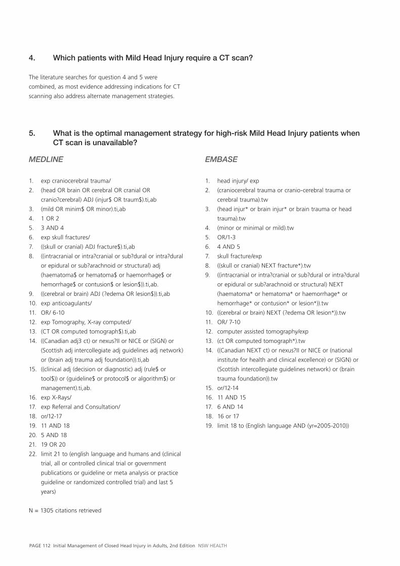
PAGE 112 Initial Management of Closed Head Injury in Adults, 2nd Edition NSW HEALTH
4. Which patients with Mild Head Injury require a CT scan?
The literature searches for question 4 and 5 were
combined, as most evidence addressing indications for CT
scanning also address alternate management strategies.
5. What is the optimal management strategy for high-risk Mild Head Injury patients when CT scan is unavailable?
MEDLINE
1. exp craniocerebral trauma/
2. (head OR brain OR cerebral OR cranial OR
cranio?cerebral) ADJ (injur$ OR traum$).ti,ab
3. (mild OR minim$ OR minor).ti,ab
4. 1 OR 2
5. 3 AND 4
6. exp skull fractures/
7. ((skull or cranial) ADJ fracture$).ti,ab
8. ((intracranial or intra?cranial or sub?dural or intra?dural
or epidural or sub?arachnoid or structural) adj
(haematoma$ or hematoma$ or haemorrhage$ or
hemorrhage$ or contusion$ or lesion$)).ti,ab.
9. ((cerebral or brain) ADJ (?edema OR lesion$)).ti,ab
10. exp anticoagulants/
11. OR/ 6-10
12. exp Tomography, X-ray computed/
13. (CT OR computed tomograph$).ti,ab
14. ((Canadian adj3 ct) or nexus?II or NICE or (SIGN) or
(Scottish adj intercollegiate adj guidelines adj network)
or (brain adj trauma adj foundation)).ti,ab
15. ((clinical adj (decision or diagnostic) adj (rule$ or
tool$)) or (guideline$ or protocol$ or algorithm$) or
management).ti,ab.
16. exp X-Rays/
17. exp Referral and Consultation/
18. or/12-17
19. 11 AND 18
20. 5 AND 18
21. 19 OR 20
22. limit 21 to (english language and humans and (clinical
trial, all or controlled clinical trial or government
publications or guideline or meta analysis or practice
guideline or randomized controlled trial) and last 5
years)
N = 1305 citations retrieved
EMBASE
1. head injury/ exp
2. (craniocerebral trauma or cranio-cerebral trauma or
cerebral trauma).tw
3. (head injur* or brain injur* or brain trauma or head
trauma).tw
4. (minor or minimal or mild).tw
5. OR/1-3
6. 4 AND 5
7. skull fracture/exp
8. ((skull or cranial) NEXT fracture*).tw
9. ((intracranial or intra?cranial or sub?dural or intra?dural
or epidural or sub?arachnoid or structural) NEXT
(haematoma* or hematoma* or haemorrhage* or
hemorrhage* or contusion* or lesion*)).tw
10. ((cerebral or brain) NEXT (?edema OR lesion*)).tw
11. OR/ 7-10
12. computer assisted tomography/exp
13. (ct OR computed tomograph*).tw
14. ((Canadian NEXT ct) or nexus?II or NICE or (national
institute for health and clinical excellence) or (SIGN) or
(Scottish intercollegiate guidelines network) or (brain
trauma foundation)).tw
15. or/12-14
16. 11 AND 15
17. 6 AND 14
18. 16 or 17
19. limit 18 to (English language AND (yr=2005-2010))
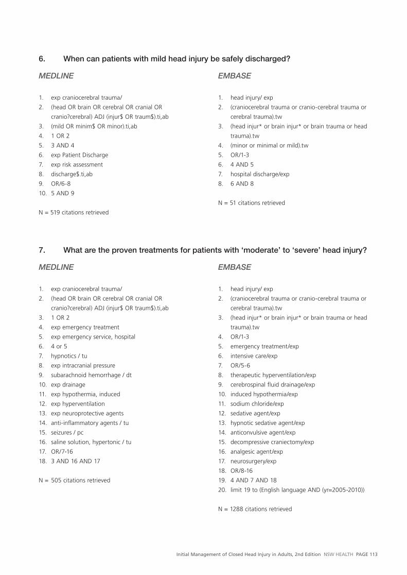
Initial Management of Closed Head Injury in Adults, 2nd Edition NSW HEALTH PAGE 113
6. When can patients with mild head injury be safely discharged?
MEDLINE
1. exp craniocerebral trauma/
2. (head OR brain OR cerebral OR cranial OR
cranio?cerebral) ADJ (injur$ OR traum$).ti,ab
3. (mild OR minim$ OR minor).ti,ab
4. 1 OR 2
5. 3 AND 4
6. exp Patient Discharge
7. exp risk assessment
8. discharge$.ti,ab
9. OR/6-8
10. 5 AND 9
N = 519 citations retrieved
EMBASE
1. head injury/ exp
2. (craniocerebral trauma or cranio-cerebral trauma or
cerebral trauma).tw
3. (head injur* or brain injur* or brain trauma or head
trauma).tw
4. (minor or minimal or mild).tw
5. OR/1-3
6. 4 AND 5
7. hospital discharge/exp
8. 6 AND 8
N = 51 citations retrieved
7. What are the proven treatments for patients with ‘moderate’ to ‘severe’ head injury?
MEDLINE
1. exp craniocerebral trauma/
2. (head OR brain OR cerebral OR cranial OR
cranio?cerebral) ADJ (injur$ OR traum$).ti,ab
3. 1 OR 2
4. exp emergency treatment
5. exp emergency service, hospital
6. 4 or 5
7. hypnotics / tu
8. exp intracranial pressure
9. subarachnoid hemorrhage / dt
10. exp drainage
11. exp hypothermia, induced
12. exp hyperventilation
13. exp neuroprotective agents
14. anti-inflammatory agents / tu
15. seizures / pc
16. saline solution, hypertonic / tu
17. OR/7-16
18. 3 AND 16 AND 17
N = 505 citations retrieved
EMBASE
1. head injury/ exp
2. (craniocerebral trauma or cranio-cerebral trauma or
cerebral trauma).tw
3. (head injur* or brain injur* or brain trauma or head
trauma).tw
4. OR/1-3
5. emergency treatment/exp
6. intensive care/exp
7. OR/5-6
8. therapeutic hyperventilation/exp
9. cerebrospinal fluid drainage/exp
10. induced hypothermia/exp
11. sodium chloride/exp
12. sedative agent/exp
13. hypnotic sedative agent/exp
14. anticonvulsive agent/exp
15. decompressive craniectomy/exp
16. analgesic agent/exp
17. neurosurgery/exp
18. OR/8-16
19. 4 AND 7 AND 18
20. limit 19 to (English language AND (yr=2005-2010))
N = 1288 citations retrieved
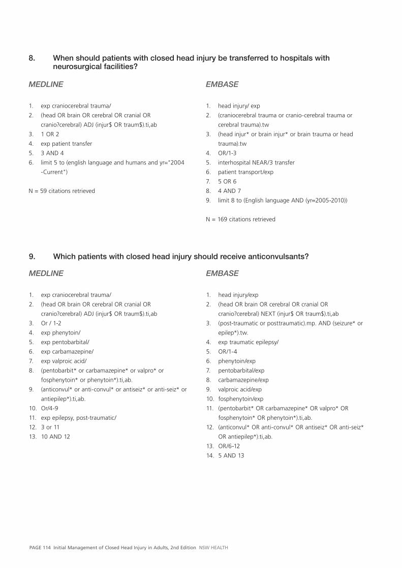
PAGE 114 Initial Management of Closed Head Injury in Adults, 2nd Edition NSW HEALTH
8. When should patients with closed head injury be transferred to hospitals with neurosurgical facilities?
MEDLINE
1. exp craniocerebral trauma/
2. (head OR brain OR cerebral OR cranial OR
cranio?cerebral) ADJ (injur$ OR traum$).ti,ab
3. 1 OR 2
4. exp patient transfer
5. 3 AND 4
6. limit 5 to (english language and humans and yr="2004
-Current")
N = 59 citations retrieved
EMBASE
1. head injury/ exp
2. (craniocerebral trauma or cranio-cerebral trauma or
cerebral trauma).tw
3. (head injur* or brain injur* or brain trauma or head
trauma).tw
4. OR/1-3
5. interhospital NEAR/3 transfer
6. patient transport/exp
7. 5 OR 6
8. 4 AND 7
9. limit 8 to (English language AND (yr=2005-2010))
N = 169 citations retrieved
9. Which patients with closed head injury should receive anticonvulsants?
MEDLINE
1. exp craniocerebral trauma/
2. (head OR brain OR cerebral OR cranial OR
cranio?cerebral) ADJ (injur$ OR traum$).ti,ab
3. Or / 1-2
4. exp phenytoin/
5. exp pentobarbital/
6. exp carbamazepine/
7. exp valproic acid/
8. (pentobarbit* or carbamazepine* or valpro* or
fosphenytoin* or phenytoin*).ti,ab.
9. (anticonvul* or anti-convul* or antiseiz* or anti-seiz* or
antiepilep*).ti,ab.
10. Or/4-9
11. exp epilepsy, post-traumatic/
12. 3 or 11
13. 10 AND 12
EMBASE
1. head injury/exp
2. (head OR brain OR cerebral OR cranial OR
cranio?cerebral) NEXT (injur$ OR traum$).ti,ab
3. (post-traumatic or posttraumatic).mp. AND (seizure* or
epilep*).tw.
4. exp traumatic epilepsy/
5. OR/1-4
6. phenytoin/exp
7. pentobarbital/exp
8. carbamazepine/exp
9. valproic acid/exp
10. fosphenytoin/exp
11. (pentobarbit* OR carbamazepine* OR valpro* OR
fosphenytoin* OR phenytoin*).ti,ab.
12. (anticonvul* OR anti-convul* OR antiseiz* OR anti-seiz*
OR antiepilep*).ti,ab.
13. OR/6-12
14. 5 AND 13
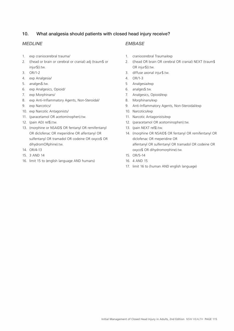
Initial Management of Closed Head Injury in Adults, 2nd Edition NSW HEALTH PAGE 115
10. What analgesia should patients with closed head injury receive?
MEDLINE
1. exp craniocerebral trauma/
2. ((head or brain or cerebral or cranial) adj (traum$ or
injur$)).tw.
3. OR/1-2
4. exp Analgesia/
5. analges$.tw.
6. exp Analgesics, Opioid/
7. exp Morphinans/
8. exp Anti-Inflammatory Agents, Non-Steroidal/
9. exp Narcotics/
10. exp Narcotic Antagonists/
11. (paracetamol OR acetominophen).tw.
12. (pain ADJ rel$).tw.
13. (morphine or NSAID$ OR fentanyl OR remifentanyl
OR diclofenac OR meperidine OR alfentanyl OR
sulfentanyl OR tramadol OR codeine OR oxyco$ OR
dihydromORphine).tw.
14. OR/4-13
15. 3 AND 14
16. limit 15 to (english language AND humans)
EMBASE
1. craniocerebral Trauma/exp
2. ((head OR brain OR cerebral OR cranial) NEXT (traum$
OR injur$)).tw.
3. diffuse axonal injur$.tw.
4. OR/1-3
5 Analgesia/exp
6. analges$.tw.
7. Analgesics, Opioid/exp
8. Morphinans/exp
9. Anti-Inflammatory Agents, Non-Steroidal/exp
10. Narcotics/exp
11. Narcotic Antagonists/exp
12. (paracetamol OR acetominophen).tw.
13. (pain NEXT rel$).tw.
14. (morphine OR NSAID$ OR fentanyl OR remifentanyl OR
diclofenac OR meperidine OR
alfentanyl OR sulfentanyl OR tramadol OR codeine OR
oxyco$ OR dihydromorphine).tw.
15. OR/5-14
16. 4 AND 15
17. limit 16 to (human AND english language)

PAGE 116 Initial Management of Closed Head Injury in Adults, 2nd Edition NSW HEALTH
1. Newcombe R, Merry G. The management of acute
neurotrauma in rural and remote locations: A set of
guidelines for the care of head and spinal injuries. J
Clin Neuro 1999;6(1):85-93.
2. NSW Institute of Trauma and Injury Management.
NSW Trauma Registry Profile of Serious to Critical
Injuries: 2007. NSW Health; 2010.
3. Khan F, Baguley IJ, Cameron ID, Khan F, Baguley IJ,
Cameron ID. 4: Rehabilitation after traumatic brain
injury. MJA 2003;178(6):290-5.
4. Fortune N. The definition, incidence and prevalence
of acquired brain injury in Australia. Australian
Institute of Health and Welfare; 2009.
5. Tate R, McDonald S, Lulham J. Incidence of hospital-
treated traumatic brain injury in an Australian
community. ANZ J Pub Health 1998;22(4):419.
6. Servadei F, Teasdale G, Merry G,
Neurotraumatology Committee of the World
Federation of Neurosurgical S. Defining acute
mild head injury in adults: a proposal based on
prognostic factors, diagnosis, and management. J
Neurotrauma 2001;18(7):657-64.
7. Bellner J, Ingebrigtsen T, Romner B. Survey of
the management of patients with minor head
injuries in hospitals in Sweden. Acta Neurol Scand
1999;100(6):355-9.
8. Cushman J, Agarwal N, Fabian T, Garcia V, Nagy K,
Pasquale M, et al. Practice management guidelines
for the management of mild traumatic brain injury:
the EAST practice management guidelines work
group. J Trauma 2001;51(5):1016-26.
9. Jagoda AS, Bazarian JJ, Bruns JJ, Jr., Cantrill
SV, Gean AD, Howard PK, et al. Clinical policy:
neuroimaging and decisionmaking in adult mild
traumatic brain injury in the acute setting. Ann
Emerg Med 2008;52(6):714-48.
10. Vos PE, Battistin L, Birbamer G, Gerstenbrand F,
Potapov A, Prevec T, et al. EFNS guideline on mild
traumatic brain injury: report of an EFNS task force.
Eur J Neurology 2002;9(3):207-19.
11. Dickinson K, Bunn F, Wentz R, Edwards P, Roberts
I. Size and quality of randomised controlled trials
in head injury: review of published studies. BMJ.
2000;320(7245):1308-11.
12. Klauber M, Marshall L, Luerssen T, Frankowski R,
Tabaddor K, Eisenberg H. Determinants of Head
Injury Mortality: Importance of the Low Risk
Patient. Neurosurgery 1989;24(1):31-6.
13. Mower W, Hoffman J, Herbert M, Wolfson A,
Pollack C, Zucker M, et al. Developing a clinical
decision instrument to rule out intracranial injuries
in patients with minor head trauma: methodology
of the NEXUS II investigation. Ann Emerg Med
2002;40(5):505-14.
14. Narayan R, Michel ME, Ansell B, Baethmann A,
Biegon A, Bracken MB, et al. Clinical trials in head
injury. J Neurotrauma 2002;19(5):503-57.
15. Brain Trauma Foundation, American Association of
Neurological Surgeons, Congress of Neurological
Surgeons, Joint Section on Neurotrauma and
Critical Care. Guidelines for the Management
of Severe Traumatic Brain Injury J Neurotrauma
2007;24(S1):1-106.
16. Alderson P, Roberts I. Corticosteroids for acute
traumatic brain injury. Cochrane Database of
Systematic Reviews 2005(1).
References
Initial Management of Closed Head Injury in Adults, 2nd Edition NSW HEALTH PAGE 117
17. Arango MF, Bainbridge D. Magnesium for acute
traumatic brain injury. Cochrane Database of
Systematic Reviews 2008(4).
18. Bennett MH, Trytko B, Jonker B. Hyperbaric oxygen
therapy for the adjunctive treatment of traumatic
brain injury. Cochrane Database of Systematic
Reviews. 2004(4).
19. Fleminger S, Greenwood R, Oliver D.
Pharmacological management for agitation and
aggression in people with acquired brain injury.
Cochrane Database of Systematic Reviews 2006(4).
20. Forsyth R, Wolny S, Rodrigues B. Routine
intracranial pressure monitoring in acute coma.
Cochrane Database of Systematic Reviews. 2010(2).
21. Ker K, Blackhall K. Bradykinin beta-2 receptor
antagonists for acute traumatic brain injury.
Cochrane Database of Systematic Reviews. 2008(1).
22. Langham J, Goldfrad C, Teasdale G, Shaw D, Rowan
K. Calcium channel blockers for acute traumatic
brain injury. Cochrane Database of Systematic
Reviews. 2003(4).
23. Roberts I. Aminosteroids for acute traumatic brain
injury. Cochrane Database of Systematic Reviews.
1999(3).
24. Roberts I, Schierhout G. Hyperventilation therapy
for acute traumatic brain injury. Cochrane Database
of Systematic Reviews. 2009(3).
25. Roberts I, Sydenham E. Barbiturates for acute
traumatic brain injury. Cochrane Database of
Systematic Reviews. 1999(3).
26. Sahuquillo J, Arikan F. Decompressive craniectomy
for the treatment of refractory high intracranial
pressure in traumatic brain injury. Cochrane
Database of Systematic Reviews. 2006(1).
27. Schierhout G, Roberts I. Anti-epileptic drugs for
preventing seizures following acute traumatic brain
injury. Cochrane Database of Systematic Reviews.
2001(4).
28. Sydenham E, Roberts I, Alderson P. Hypothermia
for traumatic head injury. . Cochrane Database of
Systematic Reviews. 2009(2).
29. Wakai A, Roberts IG, Schierhout G. Mannitol for
acute traumatic brain injury. Cochrane Database of
Systematic Reviews. 2007(1).
30. Willis C, Lybrand S, Bellamy N. Excitatory amino
acid inhibitors for traumatic brain injury. Cochrane
Database of Systematic Reviews. 2003(1).
31. Fabbri A, Servadei F, Marchesini G, Morselli-Labate
A, Dente M, Iervese T, et al. Prospective validation
of a proposal for diagnosis and management of
patients attending the emergency department for
mild head injury. J Neurol Neurosurg Psychiatry
2004;75(3):410-6.
32. Haydel M, Preston C, Mills T, Luber S, Blaudeau E,
DeBlieux P. Indications for computed tomography
in patients with minor head injury. NEJM
2000;343(2):100-5.
33. Ibanez J, Arikan F, Pedraza S, Sanchez E, Poca MA,
Rodriguez D, et al. Reliability of clinical guidelines in
the detection of patients at risk following mild head
injury: results of a prospective study. J Neurosurgery
2004;100(5):825-34.
34. Palchak MJ, Holmes JF, Vance CW, Gelber RE,
Schauer BA, Harrison MJ, et al. A decision rule
for identifying children at low risk for brain
injuries after blunt head trauma. Ann Emerg Med
2003;42(4):492-506.
35. Stiell IG, Wells GA, Vandemheen K, Clement C,
Lesiuk H, Laupacis A, et al. The Canadian CT Head
Rule for patients with minor head injury. Lancet
2001;357(9266):1391-6.
36. Dunham C, Coates S, Cooper C. Compelling
evidence for discretionary brain computed
tomographic imaging in those patients with mild
cognitive impairment after blunt trauma. J Trauma
1996;41(4):679-86.
PAGE 118 Initial Management of Closed Head Injury in Adults, 2nd Edition NSW HEALTH
37. Stiell IG, Wells GA, Vandemheen K, Laupacis A,
Brison R, Eisenhauer MA, et al. Variation in ED Use
of Computed Tomography for Patients With Minor
Head Injury. Ann Emerg Med 1997;30:14-22.
38. Bulger E, Nathens A, Rivara F, Moore M, MacKenzie
E, Jurkovich G, et al. Management of severe head
injury: institutional variations in care and effect on
outcome. Crit Care Med 2002;30(8):1870-6.
39. Af Geijerstam J, Britton M. Mild head injury:
reliability of early computed tomographic findings in
triage for admission. Emerg Med J 2005;22(2):103-
7.
40. American College of Surgeons Committee on
Trauma. Advanced Trauma Life Support for Doctors.
8th ed2007.
41. Fabbri A, Servadei F, Marchesini G, Stein S,
Vandelli A. Observational approach to subjects
with mild-to-moderate head injury and initial non-
neurosurgical lesions. J Neurol Neurosurg Psychiatry
2008;79(10):1180-5.
42. Myburgh JA, Cooper DJ, Finfer SR, Venkatesh
B, Jones D, Higgins A, et al. Epidemiology
and 12-month outcomes from traumatic brain
injury in australia and new zealand. J Trauma
2008;64(4):854-62.
43. Styrke J, Stalnacke B-M, Sojka P, Bjornstig
U. Traumatic Brain Injuries in a Well-Defined
Population: Epidemiological Aspects and Severity. J
Neurotrauma. 2007;24(9):1425-36.
44. National Health & Medical Research Council.
NHMRC levels of evidence and grades for
recommendations for developers of guidelines.
2009: Available from: www.nhmrc.gov.au/_files_
nhmrc/file/guidelines/evidence_statement_form.pdf.
45. National Health & Medical Research Council. A
guide to the development, implementation and
evaluation of clinical practice guidelines. Canberra
ACT. NHMRC; 1999.
46. Af Geijerstam J, Britton M. Mild head injury -
mortality and complication rate: meta-analysis
of findings in a systematic literature review. Acta
Neurochir 2003;145(10):843-50.
47. Smits M, Dippel DW, Steyerberg EW, de Haan GG,
Dekker HM, Vos PE, et al. Predicting intracranial
traumatic findings on computed tomography in
patients with minor head injury: the CHIP prediction
rule. Ann Emerg Med 2007;146(6):397-405.
48. Stein SC, Fabbri A, Servadei F, Glick HA. A critical
comparison of clinical decision instruments for
computed tomographic scanning in mild closed
traumatic brain injury in adolescents and adults.
Ann Emerg Med 2009;53(2):180-8.
49. Borczuk P. Predictors of intracranial injury in
patients with mild head trauma. Ann Emerg Med
1995;25(6):731-6.
50. Culotta V, Sementilli M, Gerold K, Watts
C. Clinicopathological heterogeneity in the
classification of mild head injury. Neurosurgery
1996;38(2):245-50.
51. Dacey R, Alves W, Rimel R, Winn H, Jane J.
Neurosurgical complications after apparently minor
head injury. Assessment of risk in a series of 610
patients. J Neurosurgery 1986;65(2):203-10.
52. Hsiang JN, Yeung T, Yu AL, Poon WS. High-risk mild
head injury. J Neurosurg 1997;87(2):234-8.
53. Shackford SR, Wald SL, Ross SE, Cogbill TH, Hoyt
DB, Morris JA, et al. The clinical utility of computed
tomographic scanning and neurologic examination
in the management of patients with minor head
injuries. J Trauma 1992;33(3):385-94.
54. Smits M, Dippel DW, de Haan GG, Dekker HM,
Vos PE, Kool DR, et al. External validation of the
Canadian CT Head Rule and the New Orleans
Criteria for CT scanning in patients with minor head
injury. JAMA 2005;294(12):1519-25.
55. Stein SCMD. Minor Head Injury: 13 Is an Unlucky
Number. [Report]. J Trauma. 2001;50(4):759-60.
Initial Management of Closed Head Injury in Adults, 2nd Edition NSW HEALTH PAGE 119
56. Stiell IG, Clement CM, Rowe BH, Schull MJ, Brison
R, Cass D, et al. Comparison of the Canadian CT
Head Rule and the New Orleans Criteria in patients
with minor head injury. JAMA 2005;294(12):1511-8.
57. Fong C, Chong W, Villaneuva E, Segal AY.
Implementation of a guideline for computed
tomography head imaging in head injury: a
prospective study. Emerg Med Aust 2008;20(5):410-
9.
58. Mower WR, Hoffman JR, Herbert M, Wolfson
AB, Pollack CV, Jr., Zucker MI, et al. Developing
a decision instrument to guide computed
tomographic imaging of blunt head injury patients.
J Trauma. 2005;59(4):954-9.
59. Smits M, Hunink MG, Nederkoorn PJ, Dekker
HM, Vos PE, Kool DR, et al. A history of loss of
consciousness or post-traumatic amnesia in minor
head injury: "conditio sine qua non" or one of
the risk factors? J Neurol Neurosurg Psychiatry
2007;78(12):1359-64.
60. Cook L, Levitt M, Simon B, Williams V. Identification
of ethanol-intoxicated patients with minor head
trauma requiring computed tomography scans.
Acad Emerg Med 1994;1(3):227-34.
61. Falimirski M, Gonzalez R, Rodriguez A, Wilberger
J. The need for head computed tomography in
patients sustaining loss of consciousness after mild
head injury. J Trauma 2003;55(1):1-6.
62. Gomez P, Lobato R, Ortega J, De La Cruz J. Mild
head injury: differences in prognosis among
patients with a Glasgow Coma Scale score of 13 to
15 and analysis of factors associated with abnormal
CT findings. Br J Neurosurg 1996;10(5):453-60.
63. Hahn Y, McLone D. Risk factors in the outcome of
children with minor head injury. Pediatr Neurosurg
1993;19(3):135-42.
64. Harad FT, Kerstein MD. Inadequacy of bedside
clinical indicators in identifying significant
intracranial injury in trauma patients. J Trauma
1992;32(3):359-61.
65. Haydel MJ, Shembekar AD, Haydel MJ, Shembekar
AD. Prediction of intracranial injury in children aged
five years and older with loss of consciousness after
minor head injury due to nontrivial mechanisms.
Ann Emerg Med 2003;42(4):507-14.
66. Hofman PA, Nelemans P, Kemerink GJ, Wilmink JT.
Value of radiological diagnosis of skull fracture in
the management of mild head injury: meta-analysis.
J Neurol Neurosurg Psychiatry 2000;68(4):416-22.
67. Horowitz BZ, Earle OJ. Should transient loss of
consciousness in blunt head trauma be a pre-
hospital trauma triage criterion? J Emerg Med
2001;21(4):381-6.
68. Jeret JS, Mandell M, Anziska B, Lipitz M, Vilceus AP,
Ware JA, et al. Clinical predictors of abnormality
disclosed by computed tomography after mild head
trauma. Neurosurgery 1993;32(1):9-15.
69. Lee S, Liu T, Wong C, Yeh Y, Tzaan W. Relative risk
of deterioration after mild closed head injury. Acta
Neurochir 1995;135(3-4):136-40.
70. Madden C, Witzkc D, Sanders B, Valente J, Fritz M.
High-yield selection criteria for cranial computed
tomography after acute trauma. Acad Emerg Med
1995;2(4):248-53.
71. Miller E, Derlet R, Kinser D. Minor head trauma:
Is computed tomography always necessary? Ann
Emerg Med 1996;27(3):290-4.
72. Ruff R, Jurica P. In search of a unified definition
for mild traumatic brain injury. Brain Injury
1999;13(12):943-52.
73. Sharma P, Dahiya RS, Dass B, Dewan Y, Arora
AL. How important is history of unconsciousness
in head injury patients? J Indian Med Assoc
2001;99(2):81-3.
74. Teasdale GM, Murray G, Anderson E, Mendelow
AD, MacMillan R, Jennett B, et al. Risks of acute
traumatic intracranial haematoma in children and
adults: implications for managing head injuries. BMJ
1990;300(6721):363-7.
PAGE 120 Initial Management of Closed Head Injury in Adults, 2nd Edition NSW HEALTH
75. Vilke GM, Chan TC, Guss DA. Use of a complete
neurological examination to screen for significant
intracranial abnormalities in minor head injury. Am J
Emerg Med. 2000;18(2):159-63.
76. Williams DH, Levin HS, Eisenberg HM. Mild head
injury classification. Neurosurgery 1990;27(3):422-8.
77. Clement C, Stiell I, Schull M, Rowe B, Brison R,
Lee J, et al. Clinical features of head injury patients
presenting with a Glasgow Coma Scale score of 15
and who require neurosurgical intervention. Ann
Emerg Med 2006;48(3):245-51.
78. Franko J, Kish K, O'Connell B, Subramanian S,
Yuschak J. Advanced age and preinjury warfarin
anticoagulation increase the risk of mortality after
head trauma. J Trauma 2006;61(1):107-10.
79. Ivascu F, Howells G, Junn F, Bair H, Bendick
P, Janczyk R, et al. Rapid warfarin reversal in
anticoagulated patients with traumatic intracranial
hemorrhage reduces hemorrhage progression and
mortality. J Trauma 2005;59(5):1131-7.
80. Saboori M, Ahmadi J, Farajzadegan Z. Indications
for brain CT scan in patients with minor head injury.
Clin Neurol Neurosurg 2007;109(5):399-405.
81. Turedi S, Hasanbasoglu A, Gunduz A, Yandi M.
Clinical decision instruments for CT scan in minor
head trauma. J Emerg Med. 2008;34(3):253-9.
82. Bazarian JJ, McClung J, Cheng YT, Flesher W,
Schneider SM. Emergency department management
of mild traumatic brain injury in the USA. EMJ
2005;22(7):473-7.
83. Atabaki SM, Stiell IG, Bazarian JJ, Sadow KE, Vu
TT, Camarca MA, et al. A Clinical Decision Rule
for Cranial Computed Tomography in Minor
Pediatric Head Trauma. Arch Pediatr Adolesc Med.
2008;162(5):439-45.
84. Dunning J, Batchelor J, Stratford-Smith P, Teece
S, Browne J, Sharpin C, et al. A meta-analysis
of variables that predict significant intracranial
injury in minor head trauma. Arch Dis Child.
2004;89(7):653-9.
85. Kuppermann N, Holmes JF, Dayan PS, Hoyle Jr JD,
Atabaki SM, Holubkov R, et al. Identification of
children at very low risk of clinically-important brain
injuries after head trauma: a prospective cohort
study. Lancet. 2009;374(9696):1160-70.
86. Maguire JL, Boutis K, Uleryk EM, Laupacis A, Parkin
PC. Should a Head-Injured Child Receive a Head
CT Scan? A Systematic Review of Clinical Prediction
Rules. Pediatrics. 2009;124(1):e145-54.
87. Oman JA, Cooper RJ, Holmes JF, Viccellio P, Nyce
A, Ross SE, et al. Performance of a Decision Rule to
Predict Need for Computed Tomography Among
Children With Blunt Head Trauma. Pediatrics. 2006
January 17, 2006:peds.2005-1063.
88. Miller EC, Holmes JF, Derlet RW. Utilizing clinical
factors to reduce head CT scan ordering for minor
head trauma patients. Journal of Emergency
Medicine. 1997;15(4):453-7.
89. Nagy K, Joseph K, Krosner S, Roberts R, Leslie
C, Dufty K, et al. The utility of head computed
tomography after minimal head injury. J Trauma
1999;46(2):268-70.
90. Stein SC, Ross SE. Mild head injury: a plea for
routine early CT scanning. J Trauma 1992;33(1):11-3.
91. Af Geijerstam J, Oredsson S, Britton M. Medical
outcome after immediate computed tomography
or admission for observation in patients with
mild head injury: randomised controlled trial. BMJ
2006;333(7566):465.
92. de Boussard CN, Bellocco R, af Geijerstam JL, Borg
J, Adami J. Delayed intracranial complications after
concussion. J Trauma. 2006;61(3):577-81.
93. Alves WM, Colohan ART, O'Leary TJ, Rimel RW,
Jane JA. Understanding posttraumatic symptoms
after minor head injury. J Head Trauma Rehab.
1986;1(2):1-12.
94. Hugenholtz H, Stuss D, Stethem L, Richard M. How
long does it take to recover from a mild concussion?
Neurosurgery 1988;22(5):853-8.
Initial Management of Closed Head Injury in Adults, 2nd Edition NSW HEALTH PAGE 121
95. Kraus J, Hsu P, Schaffer K, Vaca F, Ayers K, Kennedy
F, et al. Preinjury factors and 3-month outcomes
following emergency department diagnosis of
mild traumatic brain injury. J Head Trauma Rehab.
2009;24(5):344-54.
96. Lannsjo M, af Geijerstam J, Johansson U, Bring
J, Borg J. Prevalence and structure of symptoms
at 3 months after mild traumatic brain injury in a
national cohort. Brain Injury 2009;23(3):213-9.
97. Mittenberg W, Strauman S. Diagnosis of mild head
injury and the postconcussion syndrome. J Head
Trauma Rehab 2000;15(2):783-91.
98. Rimel R, Giordani B, Barth J, Boll T, Jane J. Disability
Caused by Minor Head Injury. Neurosurgery
1981;9(3):221-8.
99. Thornhill S, Teasdale GM, Murray GD, McEwen
J, Roy CW, Penny KI. Disability in young people
and adults one year after head injury: prospective
cohort study. BMJ 2000;320(7250):1631-5.
100. Faux S, Sheedy J. A Prospective Controlled Study
in the Prevalence of Posttraumatic Headache
Following Mild Traumatic Brain Injury. Pain
Medicine. 2008;9:1001-11.
101. Borg J, Holm L, Cassidy J, Peloso P, Carroll L,
von Holst H, et al. Diagnostic procedures in
mild traumatic brain injury: results of the WHO
Collaborating Centre Task Force on Mild Traumatic
Brain Injury. J Rehab Med 2004(43 Suppl):61-75.
102. Holm L, Cassidy JD, Carroll L, Borg J. Summary of
the WHO collaborating centre for neurotrauma task
force on mild traumatic brain injury. J Rehab Med.
2005;37(3):137 - 41.
103. Motor Accidents Authority NSW. Guidelines for
mild traumatic brain injury following closed head
injury. Sydney 2008.
104. Teasdale G, Jennett B. Assessment of coma and
impaired consciousness. A practical scale. Lancet
1974;2(7872):81-4.
105. Fearnside M, McDougall P. Moderate head injury:
a system of neurotrauma care. ANZ J Surgery
1998;68(1):58-64.
106. Ponsford J, Willmott C, Rothwell A, Kelly AM,
Nelms R, Ng KT. Use of the Westmead PTA scale to
monitor recovery of memory after mild head injury.
Brain Injury 2004;18(6):603-14.
107. Shores EA, Lammel A, Batchelor J, Hullick C, Flynn
M. Utility of the revised Westmead PTA scale in
the acute assessment of mild traumatic brain injury
(mTBI) - further analysis. Cambridge University
Press; 2006.
108. Shores EA, Lammel A, Hullick C, Sheedy J, Flynn
M, Levick W, et al. The diagnostic accuracy of the
Revised Westmead PTA Scale as an adjunct to the
Glasgow Coma Scale in the early identification
of cognitive impairment in patients with mild
traumatic brain injury. J Neurol Neurosurg Psychiatry
2008;79(10):1100-6.
109. Nell V, Yates D, Kruger J. An extended glasgow
coma scale (GCS-E) with enhanced sensitivity
to mild brain injury. Arch Phys Med Rehab
2000;81(5):614-7.
110. Winkler JV, Rosen P, Alfry EJ. Prehospital use of the
glasgow coma scale in severe head injury. J Emerg
Med 1984;2(1):1-6.
111. Garner A, Schoettker P. Efficacy of pre-hospital
interventions for the management of severe blunt
head injury. Injury 2002;33(4):329-37.
112. High WM, Levin HS, Gary HE. Recovery of
orientation following closed-head injury. J Clin Exp
Neuropsych 1990;12(5):703 - 14.
113. Meares S, Shores EA, Taylor AJ, Lammel A,
Batchelor J. Validation of the Abbreviated
Westmead Post-traumatic Amnesia Scale: a brief
measure to identify acute cognitive impairment in
mild traumatic brain injury. Brain Inj. 2011; 25 (12):
1198-205.
114. Hoffman JR, Wolfson AB, Todd K, Mower WR.
Selective cervical spine radiography in blunt
trauma: methodology of the National Emergency
PAGE 122 Initial Management of Closed Head Injury in Adults, 2nd Edition NSW HEALTH
X-Radiography Utilization Study (NEXUS). Ann
Emerg Med 1998;32(4):461-9.
115. Stiell IG, Greenberg GH, McKnight RD, Nair RC,
McDowell I, Worthington JR. A study to develop
clinical decision rules for the use of radiography
in acute ankle injuries. Acad Emerg Med
1992;21(4):384-90.
116. Fabbri A, Servadei F, Marchesini G, Dente M,
Iervese T, Spada M, et al. Clinical performance of
NICE recommendations versus NCWFNS proposal
in patients with mild head injury. J Neurotrauma
2005;22(12):1419-27.
117. National Institute for Health and Clinical Excellence.
Head Injury: triage, assessment, investigation and
early management of head injuries in infants,
children and adults. National Collaborating Centre
for Acute Care; 2007.
118. Rosengren D, Rothwell S, Brown AF, Chu K. The
application of North American CT scan criteria to
an Australian population with minor head injury.
Emerg Med Aust 2004;16(3):195-200.
119. Af Geijerstam J, Britton M, Marke L. Mild head
injury: observation or computed tomography?
Economic aspects by literature review and decision
analysis. Emerg Med J 2004;21(1):54-8.
120. Brenner D, Elliston C, Hall E, Berdon W. Estimated
risks of radiation-induced fatal cancer from pediatric
CT. Am J Roent 2001;176(2):289-96.
121. Hall P, Adami H, Trichopoulos D, Pedersen N,
Lagiou P, Ekbom A, et al. Effect of low doses of
ionising radiation in infancy on cognitive function in
adulthood: Swedish population based cohort study.
BMJ 2004;328(7430):19.
122. Huda W, Vance A, Huda W, Vance A. Patient
radiation doses from adult and pediatric CT. Am J
Roent 2007;188(2):540-6.
123. Brenner DJ, Hall EJ. Computed Tomography -- An
Increasing Source of Radiation Exposure. N Engl J
Med. 2007 November 29, 2007;357(22):2277-84.
124. Smits M, Dippel DW, Hunink MG. Cost
effectiveness of using computed tomography (CT)
for minor head injury compared with several other
management strategies. J Trauma 2007;62(5):1314-
5.
125. Fabbri A, Servadei F, Marchesini G, Dente M,
Iervese T, Spada M, et al. Which type of observation
for patients with high-risk mild head injury and
negative computed tomography? Eur J Emerg Med
2004;11(2):65-9.
126. Riesgo P, Piquer J, Botella C, Orozco M, Navarro J,
Cabanes J. Delayed extradural hematoma after mild
head injury: report of three cases. Surg Neurology
1997;48(3):226-31.
127. Diagnostic Imaging Pathways. Ionising Radiation.
[Internet]: Western Australia Department of
Health; [updated March 2008; cited 2010 30 June];
Available from: http://www.imagingpathways.
health.wa.gov.au/includes/general/ir.html.
128. New Zealand Guidelines Group. Traumatic
Brain Injury: Diagnosis, Acute Management and
Rehabilitation. New Zealand Guidelines Group;
2006.
129. Picano E. Informed consent and communication
of risk from radiological and nuclear medicine
examinations: how to escape from a
communication inferno. BMJ. 2004;329(7470):849-
51.
130. RANZCR. Radiation Risk of Medical Imaging for
Adults and Children. [Internet]: Inside Radiology;
[updated 1st May 2009; cited 2010 30 June];
Available from: http://www.insideradiology.com.au/
pages/view.php?T_id=57.
131. Livingston D, Lavery R, Passannante M, Skurnick
J, Baker S, Fabian T, et al. Emergency department
discharge of patients with a negative cranial
computed tomography scan after minimal head
injury. Ann Surg 2000;232(1):126-32.
132. Bee T, Magnotti L, Croce M, Maish G, Minard G,
Schroeppel T, et al. Necessity of repeat head CT
and ICU monitoring in patients with minimal brain
injury. J Trauma 2009;66(4):1015-8.
Initial Management of Closed Head Injury in Adults, 2nd Edition NSW HEALTH PAGE 123
133. Brown C, Zada G, Salim A, Inaba K, Kasotakis
G, Hadjizacharia P, et al. Indications for routine
repeat head computed tomography (CT) stratified
by severity of traumatic brain injury. J Trauma
2007;62(6):1339-44.
134. Schuster R, Waxman K. Is Repeated Head
Computed Tomography Necessary for Traumatic
Intracranial Hemorrhage? Am Surgeon.
2005;71(9):701-4.
135. Sifri ZC, Homnick AT, Vaynman A, Lavery R, Liao W,
Mohr A, et al. A prospective evaluation of the value
of repeat cranial computed tomography in patients
with minimal head injury and an intracranial bleed.
J Trauma 2006;61(4):862-7.
136. Velmahos GC, Gervasini A, Petrovick L, Dorer DJ,
Doran ME, Spaniolas K, et al. Routine repeat head
CT for minimal head injury is unnecessary. J Trauma
2006;60(3):494-9.
137. Ono K, Wada K, Takahara T, Shirotani T. Indications
for computed tomography in patients with mild
head injury. Neurol Medico-Chirurg 2007;47(7):
291-7.
138. Dunning J, Daly JP, Lomas J-P, Lecky F, Batchelor J,
Mackway-Jones K. Derivation of the children's head
injury algorithm for the prediction of important
clinical events decision rule for head injury in
children. Arch Dis Child. 2006;91(11):885-91.
139. Feuerman T, Wackym P, Gade G, Becker D. Value
of skull radiography, head computed tomographic
scanning, and admission for observation in cases of
minor head injury. Neurosurgery 1988;22(3):449-
53.
140. Masters S, McClean P, Arcarese J, Brown R,
Campbell J, Freed H, et al. Skull x-ray examinations
after head trauma. Recommendations by a
multidisciplinary panel and validation study. NEJM
1987;316(2):84-91.
141. Lucchi S, Giua G, Bettinelli A, Farabola M, Sina C,
Man-Giagalli E, et al. The role of computerized
tomography in the management of mild head injury
patients. Rivista di Neuroradiologia. 1995;8(3):
415-28.
142. Chambers J, Cohen S, Hemminger L, Prall J, Nichols
J. Mild traumatic brain injuries in low-risk trauma
patients. J Trauma 1996;41(6):976-80.
143. Ruff RM, Iverson GL, Barth JT, Bush SS, Broshek
DK, N. A. N. Policy Planning Committee.
Recommendations for diagnosing a mild
traumatic brain injury: a National Academy of
Neuropsychology education paper. Archives of
Clinical Neuropsychology. 2009;24(1):3-10.
144. Frey LC. Epidemiology of Posttraumatic Epilepsy: A
Critical Review. Epilepsia. 2003;44(s10):11-7.
145. McCrory PR, Bladin PF, Berkovic SF. Retrospective
study of concussive convulsions in elite Australian
rules and rugby league footballers: phenomenology,
aetiology, and outcome. BMJ. 1997;314(7075):171.
146. Neidlinger NA, Pal JD, Victorino GP. Head
Computed Tomography Scans in Trauma Patients
With Seizure Disorder: Justifying Routine Use. Arch
Surg. 2005;140(9):858-64.
147. Mina A, Knipfer J, Park D, Bair H, Howells G,
Bendick P. Intracranial complications of preinjury
anticoagulation in trauma patients with head injury.
J Trauma 2002;53(4):668-72.
148. Cohen DB, Rinker C, Wilberger JE. Traumatic
Brain Injury in Anticoagulated Patients. J Trauma.
2006;60(3):553-7
149. Allard CB, Scarpelini S, Rhind SG, Baker AJ, Shek
PN, Tien H, et al. Abnormal coagulation tests are
associated with progression of traumatic intracranial
hemorrhage. J Trauma 2009;67(5):959-67.
150. Mack L, Chan S, Silva J, Hogan T. The use of head
computed tomography in elderly patients sustaining
minor head trauma. J Emerg Med 2003;24(2):157-
62.
151. Rathlev NK, Medzon R, Lowery D, Pollack C,
Bracken M, Barest G, et al. Intracranial Pathology
in Elders with Blunt Head Trauma. Academic
Emergency Medicine. 2006;13(3):302-7.
PAGE 124 Initial Management of Closed Head Injury in Adults, 2nd Edition NSW HEALTH
152. NSW Institute of Trauma and Injury Management.
2004 Trauma Death Review Report. NSW Trauma
Death Review Committee; 2004.
153. Bracken M, Medzon R, Rathlev N, Mower W,
Hoffman J. Effect of intoxication among blunt
trauma patients selected for head computed
tomography scanning. Ann Emerg Med
2007;49(1):45-51.
154. McCrory P, Meeuwisse W, Johnston K, Dvorak J,
Aubry M, Molloy M, et al. Consensus Statement
on Concussion in Sport: the 3rd International
Conference on Concussion in Sport held in Zurich,
November 2008. Br J Sports Med. 2009;43(Suppl
1):i76-i84.
155. Fung M, Willer B, Moreland D, Leddy J. A proposal
for an evidenced-based emergency department
discharge form for mild traumatic brain injury. Brain
Injury 2006;20(9):889-94.
156. Yates K, Pena A. Comprehension of discharge
information for minor head injury: a randomised
controlled trial in New Zealand. NZ Med J
2006;119(1239):U2101.
157. Brain Trauma Foundation, American Association of
Neurological Surgeons, Congress of Neurological
Surgeons Joint Section on Neurotrauma Critical
Care, Bratton S, Chestnut R, Ghajar J, et al.
Guidelines for the management of severe traumatic
brain injury. I. Blood pressure and oxygenation. J
Neurotrauma 2007;24 Suppl 1:S7-13.
158. Brain Trauma Foundation, American Association of
Neurological Surgeons, Congress of Neurological
Surgeons: Joint Section on Neurotrauma Critical
Care, Bratton S, Chestnut R, Ghajar J, et al.
Guidelines for the management of severe traumatic
brain injury. XIV. Hyperventilation. J Neurotrauma
2007;24 Suppl 1:S87-90.
159. Gadkary C, Alderson P, Signorini D. Therapeutic
hypothermia for head injury. Cochrane Database of
Systematic Reviews. 2004(4):CD001048.
160. Peterson K, Carson S, Carney N. Hypothermia
treatment for traumatic brain injury: a systematic
review and meta-analysis. J Neurotrauma
2008;25(1):62-71.
161. Brain Trauma Foundation, American Association of
Neurological Surgeons, Congress of Neurological
Surgeons Joint Section on Neurotrauma Critical
Care, Bratton S, Chestnut R, Ghajar J, et al.
Guidelines for the management of severe
traumatic brain injury. II. Hyperosmolar therapy. J
Neurotrauma 2007;24 Suppl 1:S14-20.
162. Wakai A, Roberts IG, Schierhout G. Mannitol for
acute traumatic brain injury. Cochrane Database of
Systematic Reviews 2009(4).
163. Alderson P, Roberts I. Corticosteroids for acute
traumatic brain injury. Cochrane Database of
Systematic Reviews 2009(4).
164. Brain Trauma Foundation, American Association of
Neurological Surgeons, Congress of Neurological
Surgeons Joint Section on Neurotrauma Critical
Care, Bratton S, Chestnut R, Ghajar J, et al.
Guidelines for the management of severe traumatic
brain injury. XV. Steroids. J Neurotrauma 2007;24
Suppl 1:S91-5.
165. Maconochie I, Roos M. Head injury (moderate to
severe). BMJ Clinical Evidence; 2009 [cited 2010 30
June ]; Available from: http://clinicalevidence.bmj.
com.ezproxy1.library.usyd.edu.au/ceweb/conditions/
nud/1210/1210.jsp.
166. Bullock R, Chestnut R, J G, Gordan D, Hartl
R, Newell D, et al. Guidelines for the Surgical
Management of Traumatic Brain Injury.
Neurosurgery. 2006;58(3):S2-S6.
167. MRC CRASH Trial Collaborators. Predicting outcome
after traumatic brain injury: practical prognostic
models based on large cohort of international
patients. BMJ. 2008;336(7641):425-9.
168. Huynh T, Jacobs DG, Dix S, Sing RF, Miles WS,
Thomason MH. Utility of Neurosurgical Consultation
for Mild Traumatic Brain Injury. American Surgeon.
2006;72:1162-7.
Initial Management of Closed Head Injury in Adults, 2nd Edition NSW HEALTH PAGE 125
169. Hartl R, Gerber LM, Iacono L, Ni Q, Lyons K,
Ghajar J. Direct transport within an organized state
trauma system reduces mortality in patients with
severe traumatic brain injury. Journal of Trauma-
Injury Infection & Critical Care. 2006;60(6):1250-6;
discussion 6.
170. Statewide Services Development Branch. Selected
Specialty and Statewide Service Plans: NSW Trauma
Services. NSW Health; 2009.
171. Trauma Pain [revised]. In: eTG complete. [Internet]
Melbourne: Therapeutic Guidelines Limited;
[updated May 2007; cited 2010 Jan 01]; Available
from: www.ciap.health.nsw.gov.au.
172. Greater Metropolitan Clinical Taskforce. Guidelines
for the administration of analgesia and the use of
anticonvulsant therapy in the treatment of mild
closed head injury. Sydney: NSW Department of
Health; 2007.
173. Annegers JF, Hauser WA, Coan SP, Rocca WA. A
Population-Based Study of Seizures after Traumatic
Brain Injuries. N Engl J Med. 1998;338(1):20-4.
174. Shah D, Husain AM. Utility of levetiracetam in
patients with subarachnoid hemorrhage. Seizure.
2009;18(10):676-9.
175. Szaflarski J, Meckler J, Szaflarski M, Shutter L,
Privitera M, Yates S. Levetiracetam use in critically ill
patients. Neurocritical Care. 2007;7(2):140-7.
176. Akopian G, Gaspard D, Alexander M. Outcomes
of blunt head trauma without intracranial pressure
monitoring. Am Surgery 2007;73(5):447-50.
177. Biberthaler P, Linsenmeier U, Pfeifer K, Kroetz
M, Mussack T, Kanz K, et al. Serum S-100B
concentration provides additional information fot
the indication of computed tomography in patients
after minor head injury: a prospective multicenter
study. Shock 2006;25(5):446-53.
178. Jones K, Sharp C, Mangram A, Dunn E. The
effects of preinjury clopidogrel use on older
trauma patients with head injuries. Am J Surgery
2006;192(6):743-5.
179. Rockswold GL, Leonard PR, Nagib MG. Analysis
of management in thirty-three closed head
injury patients who "talked and deteriorated".
Neurosurgery 1987;21(1):51-5.
180. Stein S, Burnett M, Glick H. Indications for CT
Scanning in Mild Traumatic Brain Injury: A Cost-
Effectiveness Study. J Trauma 2006;61(3):558-66.
181. Stein SC, Fabbri A, Servadei F. Routine serial
computed tomographic scans in mild traumatic
brain injury: when are they cost-effective? J Trauma
2008;65(1):66-72.
182. Carroll L, Cassidy J, Peloso P, Borg J, von Holst H,
Holm L, et al. Prognosis for mild traumatic brain
injury: results of the WHO Collaborating Centre
Task Force on Mild Traumatic Brain Injury. J Rehab
Med 2004(43 Suppl):84-105.
183. Savola O, Hillbom M. Early predictors of post-
concussion symptoms in patients with mild head
injury. Eur J Neurol 2003;10(2):175-81.
184. Kalina M, Tinkoff G, Gbadebo A, Veneri P, Fulda
G. A protocol for the rapid normalization of INR
in trauma patients with intracranial hemorrhage
on prescribed warfarin therapy. Am Surgery
2008;74(9):858-61.
185. Morgalla M, Will B, Roser F, Tatagiba M. Do long-
term results justify decompressive craniectomy
after severe traumatic brain injury? J Neurosurg
2008;109(4):685-90.
186. Shafi S, Diaz-Arrastia R, Madden C, Gentilello
L, Shafi S, Diaz-Arrastia R, et al. Intracranial
pressure monitoring in brain-injured patients is
associated with worsening of survival. J Trauma
2008;64(2):335-40.
187. Deverill J, Aitken L. Treatment of extradural
haemorrhage in Queensland: interhospital transfer,
preoperative delay and clinical outcome. Emerg
Med Aust 2007;19(4):325-32.
188. McConnell K, Newgard C, Mullins R, Arthur M,
Hedges J. Mortality benefit of transfer to level
I versus level II trauma centers for head-injured
patients. Health Serv Res 2005;40(2):435-57.
PAGE 126 Initial Management of Closed Head Injury in Adults, 2nd Edition NSW HEALTH
189. Stein SC, Ross SE. The value of computed
tomographic scans in patients with low-risk head
injuries. Neurosurgery 1990;26(4):638-40.
190. Taheri PA, Karamanoukian H, Gibbons K, Waldman
N, Doerr RJ, Hoover EL. Can patients with minor
head injuries be safely discharged home? Arch
Surgery 1993;128(3):289-92.
191. Shores EA, Marosszeky JE, Sandanam J, Batchelor
J. Preliminary validation of a clinical scale for
measuring the duration of post-traumatic amnesia.
Med J Aust. 1986; 144 (11): 569-572.
192. Jennett B, Teasdale G. Aspects of coma after severe
head injury. The Lancet. 1977;309(8017):878-81.
193. Matis G, Birbilis T. The Glasgow Coma Scale - a
brief review. Past, present, future. Acta Neurologica
Belgica. 2008;108(3):75-89.
194. Hartley C, Cozens A, Mendelow AD, Stevenson
JC. The Apache II scoring system in neurosurgical
patients: a comparison with simple Glasgow
coma scoring. British Journal of Neurosurgery.
1995;9(2):179 - 87.
195. Livingston BM, Mackenzie SJ, MacKirdy FN,
Howie JC, Group obotSICSA. Should the pre-
sedation Glasgow Coma Scale value be used when
calculating Acute Physiology and Chronic Health
Evaluation scores for sedated patients? Critical Care
Medicine. 2000;28(2):389-94.
196. Ogungbo B. The World Federation of Neurological
Surgeons scale for subarachnoid haemorrhage.
Surgical Neurology. 2003;59(3):236-7.
197. Iacono LA, Lyons KA. Making GCS as Easy as
1, 2, 3, 4, 5, 6. Journal of Trauma Nursing.
2005;12(3):77-81.
198. Teasdale G, Jennett B, Murray L, Murray G. Glascow
Coma Scale: To sum or not to sum? The Lancet.
1983;322(8351):678.
199. Rimel RW, Jane JA, Edlich RF. An injury severity
scale for comprehensive management of central
nervous system trauma. Journal of the American
College of Emergency Physicians. 1979;8(2):64-7.
200. Teasdale G, Knill-Jones R, van der Sande J. Observer
variability in assessing impaired consciousness
and coma. Journal of Neurology, Neurosurgery &
Psychiatry. 1978 July 1, 1978;41(7):603-10.
201. Gill M, Martens K, Lynch EL, Salih A, Green SM.
Interrater Reliability of 3 Simplified Neurologic
Scales Applied to Adults Presenting to the
Emergency Department With Altered Levels of
Consciousness. Annals of Emergency Medicine.
2007;49(4):403-7.e1.
202. Holdgate A, Ching N, Angonese L. Variability
in agreement between physicians and nurses
when measuring the Glasgow Coma Scale in the
emergency department limits its clinical usefulness.
Blackwell Publishing Asia; 2006. p. 379-84.
203. Healey CM, Osler TMMD, Rogers FBMD, Healey
MAMD, Glance LGMD, Kilgo PDMS, et al.
Improving the Glasgow Coma Scale Score: Motor
Score Alone Is a Better Predictor. Journal of Trauma-
Injury Infection & Critical Care. 2003;54(4):671-80.
204. Iankova A. The Glasgow Coma Scale. Clinical
application in emergency departments. Emergency
Nurse. 2006;14(8):30-5.
205. Fischer J, Mathieson C. The History of the Glasgow
Coma Scale: Implications for Practice. Critical Care
Nursing Quarterly. 2001;23(4):52-8.
206. National Health & Medical Research Council. How
to review the evidence: systematic identification and
review of the scientific literature. Canberra ACT.
NHMRC 2000.
207. Liddle J, Williamson M, Irwig L. Method for
evaluating research and guideline evidence. Sydney.
NSW Health; 1996.
208. The AGREE Collaboration. Appraisal of guidelines
for research and evaluation (AGREE) instrument
2001: Available from: www.agreecollaboration.org.

SHPN (SSD) 110189
ADULT TRAUMA CLINICAL PRACTICE GUIDELINES
Initial Management of Closed Head Injury in Adults
Summary Document2nd Edition

NSW Ministry of Health
73 Miller St
NORTH SYDNEY NSW 2060
Tel (02) 9391 9000
Fax (02) 9391 9101
www.health.nsw.gov.au
This work is copyright. It may be reproduced in whole or in part for study
or training purposes subject to the inclusion of an acknowledgement
of the source. It may not be reproduced for commercial usage or sale.
Reproduction for purposes other than those indicated above requires
written permission from the NSW Ministry of Health.
This Clinical Practice Guideline is extracted from PD2012_013 and as a
result, this booklet may be varied, withdrawn or replaced at anytime.
Compliance with information in this booklet is mandatory for NSW Health
© NSW Ministry of Health 2011
SHPN: (SSD) 110187
ISBN: 978-1-74187-581-2
For further copies contact:
NSW Institute of Trauma and Injury Management
PO Box 6314, North Ryde, NSW 2113
Ph: (02) 9887 5726
http://www.itim.nsw.gov.au
Furhter copies of this document can be downloaded from the
NSW Health website http://www.health.nsw.gov.au
November 2011
A revision of this document is due in 2015
Initial Management of Closed Head Injury in Adults, 2nd Edition Summary Report NSW HEALTH PAGE i
Acknowledgements ................................................ii
Introduction ........................................................... 3
Changes from 2007 edition ................................... 6
Algorithm 1: Initial Management of Adult Closed Head Injury ...................................... 8
Algorithm 2: Initial Management of Adult Mild Closed Head Injury .............................. 9
Understanding the grades of recommendation ................................................. 10
Recommendations .............................................. 12
1. What is the definition of a mild head injury? ............................................................ 12
2. What are the clinically important complications of mild head injury? ......................... 13
3. How should patients with mild head injury be assessed? ................................................ 15
4. Which patients with mild head injury require a CT scan?.................................................. 16
5. What should be done when patients with mild head injury deteriorate? .......................... 18
6. When can patients with mild head injury be safely discharged and what discharge advice should be provided? ................... 19
7. What are the proven treatments for patients with moderate head injury? ...................... 20
8. What are the proven treatments for patients with severe head injury? ........................... 21
9. When should patients with closed head injury be transferred to hospitals with neurosurgical facilities?........................................... 22
10. What analgesia should patients with closed head injury receive? .................................... 23
11. Which patients with closed head injury should receive anti-convulsants? ................. 24
Appendices ......................................................... 25
Appendix 1: Abbreviated Westmead PTA Scale ................................................................ 25
Appendix 2: Mild head injury discharge advice ...................................................................... 28
References ........................................................... 30
Contents
PAGE ii Initial Management of Closed Head Injury in Adults, 2nd Edition Summary Report NSW HEALTH
The following individuals are acknowledged for their contribution to the development of this document.
AuthorDr Duncan Reed, Emergency Physician, Director of Trauma, Gosford Hospital.
Editorial teamMr Glenn Sisson, NSW Trauma Education Manager, NSW Institute of Trauma and Injury Management
Ms Suzanne Davies, Research Fellow, Ambulance Research Institute, Ambulance Service of NSW
Assoc. Prof. Paul Middleton, Director, Ambulance Research Institute, Ambulance Service of NSW
Review GroupDr Rod Bishop, Director Emergency Services, Nepean Hospital
Dr Peter Clark, Clinical Director, NSW ITIM
Dr Scott D’Amours, Trauma Director, Liverpool Hospital
Assoc. Prof. Michael Fearnside AM (Emeritus), Neurosurgeon, Westmead Hospital
Dr Adeline Hodgkinson, Director Brain Injury Rehabilitation Unit, Liverpool Hospital
Mr Peter Mackay, Trauma Clinical Nurse Consultant, Gosford Hospital
Assoc. Prof. Mark Sheridan, Neurosurgeon, Director of Neurosciences, Liverpool Hospital
Dr Declan Stewart, Emergency Physician, Central Coast Health
Dr Alan Tankel, Director Emergency Services, Coffs Harbour Hospital
Ms Nichole Woodward, Emergency Clinical Nurse Consultant, Central Coast Health
Ms Wendy Fischer, Project Manager, Trauma Service, Liverpool Hospital (2nd Ed.)
Ms Merridy Gina, Project Officer, Trauma Service, Liverpool Hospital (2nd Ed.)
Ms Joan Lynch, Project Manager, Trauma Service, Liverpool Hospital (1st Ed.)
Assoc. Prof. Michael Sugrue, Trauma Director, Trauma Service, Liverpool Hospital (1st Ed.)
Ms Gail Long, Secretary, Emergency Department, Gosford Hospital (1st Ed.)
Ms Nikole McCoy, Secretary, Emergency Department, Gosford Hospital (2nd Ed)
Art and Design Unit, Gosford Hospital (1st Ed.)
Acknowledgements
Initial Management of Closed Head Injury in Adults, 2nd Edition Summary Report NSW HEALTH PAGE 3
Trauma is the leading cause of death and disability in
children and young adults in New South Wales and
closed head injuries cause a significant proportion of this
burden.1, 2 Closed head injury may result in lifelong physical,
cognitive, behavioural and social dysfunction for patients
which in turn may place major social and financial burdens
on their families and society.3 Recent Australian figures
indicate there are approximately 150 patients per 100,000
population admitted to hospital each year with closed head
injuries.3-5 Worldwide figures suggest an incidence range
of 200-350 per 100,000 population per year for patients
with closed head injury with mild head injury accounting
for 80%.6 Despite the fact that closed head injuries are
common, the classification and management of closed
head injures remains surprisingly controversial and subject
to variation in clinical practice.6-10 Due to the large numbers
of patients involved it has been estimated that even
small improvements in closed head injury management
could have significant impact.11 Furthermore, it has been
suggested that the greatest improvements can be made
in the better management of those patients with mild to
moderate head injury rather than those with severe head
injury.12
Much of the controversy that exists about closed head
injury management stems from the combination of a lack
of uniformity in definitions with a paucity of large well
designed studies in the area.11, 13, 14 ‘Head injury’ is typically
used to describe the initial clinical presentation whilst
‘traumatic brain injury’ or “concussion” are used to describe
the subsequent functional outcome. The terms “mild head
injury”, “mild traumatic brain injury” and “concussion” are
largely interchangeable and which term is used depends on
whether you are examining emergency medicine, trauma,
rehabilitation or sports medicine literature. It is difficult to
find two studies that define mild head injury in exactly the
same way so comparison of data can be difficult.6, 8-10, 13
Similarly, comparison of data in moderate to severe head
injury studies is made difficult because controversy exists
about how and when best to apply Glasgow Coma Scale
(GCS) to sedated or intubated patients.15 Perhaps most
significantly there have been very few large prospective
randomised controlled trials of sufficient power and quality
to guide management.11, 13, 14 However, in the past few
years there has been some progress in working toward
uniform definitions and some better quality trials and meta-
analyses have been published.6, 8-10, 15-35
The variety of clinical practice observed worldwide
cannot be explained solely by the lack of uniformity of
definitions and good quality studies. Much of the variation
in management strategies between the USA, Canada,
Europe and Australasia is driven by local issues such as
the availability of resources, the medico-legal environment
and in recent years the concerns about the potential harm
from CT radiation.6, 36, 37 Thus the USA has higher rates of
CT scanning for mild head injuries compared to Canada,
Europe and the UK. Even within countries and within
institutions, considerable variation in practice has been
shown to exist.7, 12, 35, 38 Whilst some variation in clinical
practice is to be expected, the introduction of clinical
practice guidelines can potentially improve care and ensure
adequate access to resources for more isolated areas.6, 35
Furthermore, clinical guidelines can potentially reduce
unnecessary tests and hospital admissions for mild head
injury patients by identifying those patients at low risk of
neurosurgically significant lesions.6, 13, 33-35
Scope of the guideline
The guideline is intended for use by clinicians managing
patients with closed head injury in major and regional
trauma services, and urban and rural hospitals. The
guideline is concerned with the initial care of the mild,
moderate and severely head injured patient. The guideline
will make evidence based recommendations on the
diagnosis, resuscitation, and disposal of patients with closed
head injuries.
Introduction
PAGE 4 Initial Management of Closed Head Injury in Adults, 2nd Edition Summary Report NSW HEALTH
The initial management plan for adults is based upon
recommendations to be followed subject to the clinician's
judgement in each case.
The recommendations however, are not prescriptive nor
are they rigid procedural paths. It is recognised that the
recommendations may not suit all patients in all clinical
situations. They are intended to provide a clinically practical
approach to the initial management of closed head injuries
based on the current best available evidence. However, as
with all guidelines, it should be remembered that they are a
clinical tool and should not replace clinical judgement. The
guideline relies on individual clinicians to decipher the needs
of individual patients.
All recommendations regarding pre-hospital care should be
read and considered in conjunction with the Ambulance
Service of NSW.
Guidelines for the initial management of head injury in
children can be found at http://www.health.nsw.gov.au/
policies/pd/2011/pdf/PD2011_024.pdf
Aims and objectives
The guideline is intended to assist clinicians throughout
NSW in delivering optimal care to patients with closed head
injury. It aims to provide information to support clinical
decision making, rather than dictate what decisions should
be made.
The broad objectives of the guideline are to reduce
morbidity and mortality in adult patients with closed head
injury by providing clinicians with practical evidence based
recommendations to assist them in managing such patients.
It is also hoped that the guidelines may prevent unnecessary
diagnostic tests and hospital admissions especially in the
mild head injury group.
The process of constructing the guideline began
with the clinicians on the Trauma Clinical Guidelines
Committee posing a series of questions about the initial
management of closed head injuries. The final questions
were derived from the guideline priority areas identified
by the committee; that is, the management of mild head
injuries and the timing of transfer of patients with closed
head injury from centres with limited resources. The
initial management of patients with moderate to severe
head injury was felt to be less controversial. This edition
also includes recommendations in relation to the use of
analgesia and anti-convulsants.
An extensive description of the methodology used for
this guideline can be found in the full guideline document
at Appendix 8, together with the search terms used at
Appendix 9.
The clinical questions addressed:
1 What is the defi nition of a mild head injury?
2 What are the clinically important complications of mild head injury?
3 How should patients with mild head injury be assessed?
4 Which patients with mild head injury require a CT scan?
5 What should be done with high risk mild head injury patients when CT scan is unavailable?
6 What should be done when patients with mild head injury deteriorate?
7 When can patients with mild head injury be safely discharged?
8 What discharge advice should be provided?
9 What are the proven treatments for patients with moderate head injury?
10 What are the proven treatments for patients with severe head injury?
11 When should patients with closed head injury be transferred to hospitals with neurosurgical facilities?
12 What analgesia should patients with closed head injury receive?
13 Which patients with closed head injury should receive anti-convulsants?
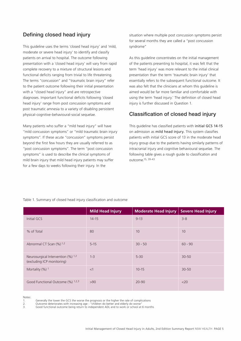
Initial Management of Closed Head Injury in Adults, 2nd Edition Summary Report NSW HEALTH PAGE 5
Defining closed head injury
This guideline uses the terms ‘closed head injury’ and ‘mild,
moderate or severe head injury’ to identify and classify
patients on arrival to hospital. The outcome following
presentation with a ‘closed head injury’ will vary from rapid
complete recovery to a mixture of structural lesions and
functional deficits ranging from trivial to life threatening.
The terms “concussion” and “traumatic brain injury” refer
to the patient outcome following their initial presentation
with a “closed head injury” and are retrospective
diagnoses. Important functional deficits following ‘closed
head injury’ range from post concussion symptoms and
post traumatic amnesia to a variety of disabling persistent
physical-cognitive-behavioural-social sequelae.
Many patients who suffer a “mild head injury” will have
“mild concussion symptoms” or “mild traumatic brain injury
symptoms”. If these acute “concussion” symptoms persist
beyond the first few hours they are usually referred to as
“post concussion symptoms”. The term “post concussion
symptoms” is used to describe the clinical symptoms of
mild brain injury that mild head injury patients may suffer
for a few days to weeks following their injury. In the
situation where multiple post concussion symptoms persist
for several months they are called a “post concussion
syndrome”
As this guideline concentrates on the initial management
of the patients presenting to hospital, it was felt that the
term ‘head injury’ was more relevant to the initial clinical
presentation than the term ‘traumatic brain injury’ that
essentially refers to the subsequent functional outcome. It
was also felt that the clinicians at whom this guideline is
aimed would be far more familiar and comfortable with
using the term ‘head injury.’ The definition of closed head
injury is further discussed in Question 1.
Classification of closed head injury
This guideline has classified patients with initial GCS 14-15
on admission as mild head injury. This system classifies
patients with initial GCS score of 13 in the moderate head
injury group due to the patients having similarly patterns of
intracranial injury and cognitive behavioural sequelae. The
following table gives a rough guide to classification and
outcome.15, 39-43
Table 1. Summary of closed head injury classification and outcome
Notes:1. Generally the lower the GCS the worse the prognosis or the higher the rate of complications 2. Outcome deteriorates with increasing age - “children do better and elderly do worse”3. Good functional outcome being return to independent ADL and to work or school at 6 months
Mild Head Injury Moderate Head Injury Severe Head Injury
Initial GCS 14-15 9-13 3-8
% of Total 80 10 10
Abnormal CT Scan (%) 1,2 5-15 30 - 50 60 - 90
Neurosurgical Intervention (%) 1,2
(excluding ICP monitoring)1-3 5-30 30-50
Mortality (%) 1 <1 10-15 30-50
Good Functional Outcome (%) 1,2,3 >90 20-90 <20
PAGE 6 Initial Management of Closed Head Injury in Adults, 2nd Edition Summary Report NSW HEALTH
Background
The first edition of this guideline was written in 2005
using evidence available until December 2004. The aim of
this new edition is to review the evidence published since
December 2004 and to provide some additional information
on specific topics including the role of anticonvulsants and
analgesics in the management of closed head injury.
The aim of the original guideline was to provide a clinically
practical evidence based guideline that summarised
the initial management of adult closed head injury. It
was piloted by the NSW Institute of Trauma and Injury
Management (ITIM) and then formally adopted and
published by NSW Health in January 2007. There was a
conscious effort by the initial guideline team to provide a
clinically practical document with clinically useful resources
such as algorithms, summaries and discharge advice sheets
backed up by a detailed evidence review. The guideline
team has continued the same principles for this update,
incorporating feedback from clinicians to improve the
guideline. The algorithms and mild head injury discharge
sheets have been revised to reflect the changes in the body
of the guideline and the feedback received.
The guideline team would emphasise that this guideline is a
clinical tool designed to assist clinicians and should be used
to assist rather than replace the clinical judgement of an
experienced clinician caring for an individual patient.
The information provided is based on the best available
information at the time of writing, which is May 2010.
These guidelines will be updated every five years and
consider new evidence as it becomes available.
New evidence
Since 2004 there have been many new studies and
guidelines published about the management of closed
head injury. There have been some advances in our
understanding of the assessment and treatment of
closed head injury but these have been incremental and
evolutionary rather than revolutionary. The basic principles
of management of closed head injury remain the same in
2010 as they were five years ago.
The following section briefly outlines the most significant
advances in knowledge from the recent literature
incorporated in this update.
Definition of mild head injury
■ Recent literature emphasises that significant intracranial
injury may occur without loss of consciousness or
amnesia■ Patients with initial GCS 13 have a significantly higher
rate of intracranial injury and should not be considered
as having mild head injury
Clinically important complications of mild head injury
■ Recent literature emphasises that mild post concussion
symptoms are common and that patients should
receive appropriate discharge advice to assist recovery■ Acute neurosurgical complications are uncommon but
important to identify
Assessment of patients with mild head injury
■ Recent literature emphasises that if structured clinical
assessment indicates the risk of intracranial injury is
low, the routine use of CT scanning is not warranted
and is potentially harmful.■ Structured clinical assessment should include initial
clinical history and examination, serial clinical
observations and clinical risk factor assessment to
determine the need for CT scanning■ A variety of clinical decision rules have been developed
to determine which patients are at higher risk of
intracranial injury and require CT scanning. However,
they all require that the clinician is familiar with their
inclusion / exclusion criteria and should be used as
tools to support clinical decision making, rather than
dictate management■ Post traumatic amnesia testing in the emergency
Changes from 2007 edition

Initial Management of Closed Head Injury in Adults, 2nd Edition Summary Report NSW HEALTH PAGE 7
department, eg Abbreviated Westmead PTA Scale
(A-WPTAS) can be useful in identifying patients with
cognitive impairment at increased risk of structural
lesions and post concussion symptoms.
Indications for CT scan for mild head injury
■ Recent literature emphasises that patients can be risk
stratified according to clinical risk factors and clinical
decision rules. Patients who are classified as high risk
should have CT scans to exclude clinically important
intracranial lesions■ Significant head injuries can occur without loss of
consciousness or amnesia and that the absence of
these features should not be used to determine the
need for CT scanning.■ Persistent abnormal mental status manifested by either
abnormal GCS or abnormal alertness, behaviour or
cognition is a strong indication for CT scanning ■ Known coagulopathy and particularly supra-
therapeutic anticoagulation are significant risk factors
for intracranial injury and that these patients should
have early CT scans and be considered for reversal of
anticoagulation■ There have been several very large studies addressing
this issue in the paediatric literature that have come up
with very similar risk factors to the adult literature and
have also confirmed that it is safe to discharge low risk
patients without CT scanning.
Acute neurological deterioration
■ Recommendations essentially unchanged■ Previously covered within guideline but now given
separate question
Discharge of patients with mild head injury
■ Recent literature emphasises that patients can be safely
discharged for home observation if structured clinical
assessment reveals no clinical risk factors indicating the
need for CT scanning or following a normal CT scan if
indicated.■ Deterioration of mild head injury patients following
a normal CT scan is rare. Caution is advised for
patients with known coagulopathy and elderly patients
where the risk of a delayed subdural haemorrhage is
increased.
Discharge advice for patients with mild head injury
■ New section to emphasise importance of discharge
advice■ Recent literature emphasises that all patients with
mild head injury should be given both verbal and
written discharge advice covering symptoms and signs
of acute deterioration, when to seek urgent medical
attention, lifestyle advice to assist recovery, information
about typical post concussion symptoms and reasons
for seeking further medical follow up. As with all
discharge advice this should be time specific and action
specific.■ An improved version of the original mild head injury
advice sheet associated with this guideline has been
developed and is now available in several languages.
Initial management of moderate head injury
(GCS 9-13)
■ Recommendations essentially unchanged
Initial management of severe head injury
(GCS 3-8)
■ Recommendations essentially unchanged
Transfer of patients with closed head injury to
hospitals with neurosurgical facilities
■ Recommendations essentially unchanged
Analgesia for closed head injury
■ New section
Anticonvulsants for closed head injury
■ New section

PAGE 8 Initial Management of Closed Head Injury in Adults, 2nd Edition Summary Report NSW HEALTH
Risk factors indicating potentially signifi cant mild head injury
Initial Management of Adult Closed Head Injury
Severe Head Injury (10%)■ Early intubation■ Supportive care of ABCDEs ■ Prevent secondary brain injury by avoiding
hypoxaemia and hypotension ■ Early CT scan ■ Early neurosurgical consult■ Early retrieval consult if transfer required■ Consider use of anticonvulsants ■ Consider ICP monitoring ■ ICU admission■ Brain injury rehabilitation consult
NB. Minimum supportive care aims to prevent secondary brain injury:■ PaO2 >60■ SaO2 >90■ PaCO2 35-40 ■ Systolic BP >90■ Head up 30º
Mild Head Injury (80%)■ Initial assessment followed by period of clinical
observation to detect risk factors for significant intracranial injury.
■ CT scan not routinely indicated unless one or more risk factors listed below are present.
■ Discharge for home observation with head injury advice sheet at 4 hours post injury if clinically improving with either no risk factors indicating the need for CT scan or normal CT scan if performed.
■ Consider hospital admission and consult network neurosurgical service if abnormal CT scan.
■ Consider hospital admission for observation if clinically not improving at 4 hours post injury irrespective of CT scan result.
■ Consider hospital admission for observation if elderly, known coagulopathy or socially isolated.
■ Advise patients to see their local doctor if they do not return to normal within 48 hours so they can be reassessed and monitored for post concussion symptoms.
NB. Also see separate Mild Head Injury Algorithm.
Moderate Head Injury (10%)■ Supportive care of ABCDEs■ Prevent secondary brain injury by avoiding
hypoxaemia and hypotension ■ Early CT scan ■ Period of clinical observation■ Consider intubation in the event of clinical
deterioration or to facilitate management ■ Early neurosurgical consult if not clinically
improving and/or abnormal CT scan■ Early retrieval consult if transfer required■ Admit to hospital for prolonged observation
unless rapid clinical improvement to GCS 15, normal CT scan and absence of other risk factors (as per mild head injury)
■ Routine post traumatic amnesia testing and consider referral to brain injury rehabilitation service due to significant risk of cognitive behavioural social sequelae
GCS 3-8 GCS 9-13 GCS 14-15
Initial Assessment and Stabilisation of ABCDEsTrauma Team activation if initial GCS 3-13 or otherwise indicated
Commence minimum of hourly clinical observations of vital signs, GCS, pupils, PTA (if applicable) and clinical symptoms
■ GCS <15 at 2 hours post injury■ Deterioration in GCS■ Focal neurological deficit■ Clinical suspicion of skull fracture ■ Vomiting (especially if recurrent)■ Known coagulopathy / bleeding disorder
What should be done when patients with closed head injury acutely deteriorate?
Early signs of deterioration■ Confusion■ Agitation■ Drowsiness■ Vomiting■ Severe headache
Late signs of deterioration■ Decrease in GCS by two or more
points■ Dilated pupil(s)■ Focal neurological deficit■ Seizure■ Cushing’s response – bradycardia
and hypertension
Clinical approach■ Resuscitate ABCDEs and exclude
non head injury cause ■ Supportive care of ABCDEs■ Early intubation if indicated■ Immediate CT scan■ If clinical or CT evidence of raised
ICP/mass effect consult with network neurosurgical and retrieval services re;
� - short term hyperventilation to PaCO2 30-35
� - bolus of mannitol (1g/kg)� - local burr holes/craniectomy
when more than 2 hours from neurosurgical care
� - prophylactic anti-convulsants
When should patients with closed head injury be transferred to hospitals with neurosurgical facilities?
Potential indicationsPatient with severe head injury
Patient with moderate head injury if:■ clinical deterioration■ abnormal CT scan■ normal CT scan but not clinically
improving■ CT scan unavailable.
Patient with mild head injury if:■ clinical deterioration■ abnormal CT scan■ normal CT scan but not clinically
improving within 4-6 hours post injury
■ mild head injury with CT scan unavailable, particularly if:
� - Persistent GCS<15 � - Deterioration in GCS � - Focal neurological deficit� - Clinical suspicion of skull fracture� - Persistent abnormal mental status � - Persistent vomiting� - Persistent severe headache � - Known coagulopathy (particularly
if age >65 or INR >4)
Clinical approach■ When in doubt consult you
network neurosurgical service.■ Patients with closed head injuries
should be observed in facilities that can manage any complications that are likely to arise. Clinical judgment regarding risk of deterioration is required and neurosurgical consultation may be appropriate.
■ Patients with closed head injuries should be transferred to the nearest appropriate hospital with neurosurgical facilities if there is significant risk of intracranial injury. The transfer of patients to hospitals with CT scan facilities but without neurosurgical services should be avoided.
AMRS (adult) 1800 650 004'formerly the MRU'
NETS (children) 1300 362 500
Network neurosurgical service
Algorithm 1:
■ Age >65 years ■ Post traumatic seizure■ Prolonged loss of consciousness (>5 min).■ Persistent post traumatic amnesia (AWPTAS <18/18)* ■ Persistent abnormal alertness / behaviour / cognition*■ Persistent severe headache*
■ Large scalp haematoma or laceration.**■ Multi-system trauma**■ Dangerous mechanism**■ Known neurosurgery / neurological deficit.**■ Delayed presentation or representation**
* particularly if persists at 4 hours post time of injury**clinical judgement required

Initial Management of Closed Head Injury in Adults, 2nd Edition Summary Report NSW HEALTH PAGE 9
Clinically safe for discharge for home observation if:■ Responsible person available to take home and observe.■ Able to return if deteriorates.■ Discharge advice is understood.
Consult senior clinician and network neurosurgical service regarding further management and disposition. Continue clinical observations in hospital.
Consider transfer for CT scanning particularly if:■ Persistent GCS <15.■ Deterioration in GCS.■ Focal neurological deficit.■ Clinical suspicion of skull fracture ■ Known coagulopathy (esp if INR>4)■ Persistent abnormal alertness, behaviour, cognition,
PTA, vomiting or severe headache at 4 hours post injury
Abnormal CT scan CT scan unavailable
Clinical symptoms IMPROVING or remain normal during period of observation.
Clinical symptomsIMPROVING at 4-6 hours post time of injury.
Clinical symptoms NOT IMPROVING at 4-6 hours post time of injury.
Clinically safe for discharge for home observation if:■ GCS 15/15 ■ No persistent post traumatic amnesia (nb A-WPTAS 18/18)■ Alertness / behaviour / cognition returning to normal ■ Clinically improving after observation.■ Normal CT scan or no indication for CT scan.■ Clinical judgment required regarding discharge and follow up of elderly
patients or patients with known coagulopathy or bleeding disorder due to increased risk of delayed subdural haematoma.
Continue minimum of hourly clinical observations until at least four hours post time of injury.
Clinically deteriorates or clinical symptoms not improving during observation period Indication for CT scan. Continue clinical observations.
Normal CT scan
Explanatory notes for risk factors1. Using GCS<15 at 2 hours post injury allows clinical judgement for patients who present soon after
injury or who have drug or alcohol intoxication. Drug or alcohol intoxication has not been shown to be an independant risk factor for intracranial injury but persistent GCS<15 is a major risk factor and mandates CT.
2. Clinical suspicion of skull fracture includes history of focal blunt assault or injury; palpable skull fracture; large scalp haematoma or laceration; signs of base of skull fracture – haemotympanum / CSF leak / raccoon eyes / Battles sign.
3. Recurrent vomiting more concerning than isolated vomiting but both are indications.4. Known coagulopathy is both a strong indication for early CT scan and to check the INR. Early reversal of
anticoagulation if abnormal CT scan and consider reversal if initially normal CT scan with high INR (>4) depending on clinical situation.
5. Elderly patients have increasing risk of intracranial injury with increasing age; routine CT scanning indicated unless totally asymptomatic patient with no other risk factors.
6. Brief generalised seizures immediately following head injury are not significant risk factors. Prolonged, focal or delayed seizures are risk factors for intracranial injury.
7. Post traumatic amnesia may manifest as repetitive questioning or short term memory deficits and can be objectively tested using the A-WPTAS. PTA > 30 mins is a minor risk factor and PTA > 4 hours a major risk factor for intracranial injury.
8. Abnormal alertness/behaviour/cognition detects subtle brain injury better than GCS and should be part of the bedside assessment. Family may help establish what is normal.
9. Multi-system trauma – beware patient with unstable vital signs or distracting injuries or who receive analgesia or anaesthesia, as significant head injury is easily missed.
10. Clinical judgement required as to what is a large scalp haematoma or laceration.11. Dangerous - MVA ejection / rollover; pedestrians / cyclists hit by vehicle; falls >own height or five stairs;
falls from horses / cycles etc; focal blunt trauma, eg bat / ball / club.12. Known neurosurgery/neurological impairment – conditions such as hydrocephalus with shunt or AVM
or tumour or cognitive impairment such as dementia make clinical assessment less reliable and may increase risk of intracranial injury.
13. Delayed presentation should be considered as failure to clinically improve during observation. For representation consider both intracranial injury and post concussion symptoms and have a low threshold for CT scanning if not done initially.
Low risk mild head injury
No indication for CT scan if all of...■ GCS 15 at 2 hours post injury.■ No focal neurological deficit.■ No clinical suspicion of skull fracture. ■ No vomiting ■ No known coagulopathy or bleeding disorder.■ Age <65 years. ■ No seizure■ Brief loss of consciousness (<5 mins). ■ Brief post traumatic amnesia (<30 mins)■ No severe headache.■ No large scalp haematoma or laceration■ Isolated head injury ■ No dangerous mechanism.■ No known neurosurgery / neurological impairment.■ No delayed presentation or representationNOTE:Mild acute clinical symptoms such as lethargy, nausea, dizziness, mild headache, mild behavioural change, amnesia for event and mild disorientation are common and are not associated with increased risk of intracranial injury. These clinical symptoms usually start to improve within 2 to 4 hours of time of injury.
Discharge for home observation if above criteria met:■ Provide written patient advice sheet■ Provide discharge summary for GP■ All patients should be advised to see their GP for follow up if they are not
feeling back to normal within 2 days■ Any patients who have minor CT abnormalities, who suffered significant
clinical symptoms or who had prolonged post traumatic amnesia should be routinely referred to their GP for follow up due to an increased risk of post concussion symptoms.
High risk mild head injury
Strong indication for CT scan if...■ GCS <15 at 2 hours post injury. #1■ Deterioration in GCS.■ Focal neurological deficit.■ Clinical suspicion of skull fracture #2■ Vomiting (especially if recurrent) #3■ Known coagulopathy or bleeding disorder #4■ Age >65 years. #5■ Seizure #6■ Prolonged loss of consciousness (>5 mins). ■ Persistent post traumatic amnesia (A-WPTAS <18/18 at 4hrs post injury) #7■ Persistent abnormal alertness / behaviour / cognition #8■ Persistent severe headache.
Relative indication for CT scan if…■ Large scalp haematoma or laceration #9■ Multi-system trauma. #10■ Dangerous mechanism. #11■ Known neurosurgery / neurological impairment. #12■ Delayed presentation or representation. #13Note The presence of multiple risk factors is more concerning than a single isolated risk factor. In most uncomplicated mild head injury patients clinical symptoms start to improve by 2 hours post injury and are returning to normal by 4 hours post injury. Clinical symptoms that are deteriorating or not improving by 4 hours post injury on serial observation such as abnormal alertness / behaviour / cognition, PTA, vomiting or severe headache are very concerning.
Initial Management of Adult Mild Closed Head Injury
Initial GCS 14-15 on arrival following blunt head traumaStabilise ABCDEs and assess clinical risk factors.
Commence minimum of hourly clinical observations of vital signs, GCS, pupils, PTA and clinical symptoms
Algorithm 2:
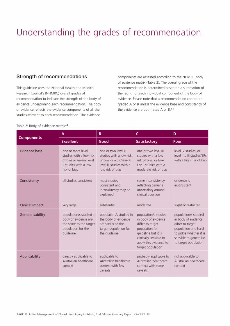
PAGE 10 Initial Management of Closed Head Injury in Adults, 2nd Edition Summary Report NSW HEALTH
Strength of recommendations
This guideline uses the National Health and Medical
Research Council's (NHMRC) overall grades of
recommendation to indicate the strength of the body of
evidence underpinning each recommendation. The body
of evidence reflects the evidence components of all the
studies relevant to each recommendation. The evidence
components are assessed according to the NHMRC body
of evidence matrix (Table 2). The overall grade of the
recommendation is determined based on a summation of
the rating for each individual component of the body of
evidence. Please note that a recommendation cannot be
graded A or B unless the evidence base and consistency of
the evidence are both rated A or B.44
Understanding the grades of recommendation
Table 2: Body of evidence matrix44
ComponentsA B C D
Excellent Good Satisfactory Poor
Evidence base one or more level I studies with a low risk of bias or several level II studies with a low risk of bias
one or two level II studies with a low risk of bias or a SR/several level III studies with a low risk of bias
one or two level III studies with a low risk of bias, or level I or II studies with a moderate risk of bias
level IV studies, or level I to III studies/SRs with a high risk of bias
Consistency all studies consistent most studies consistent and inconsistency may be explained
some inconsistency refl ecting genuine uncertainty around clinical question
evidence is inconsistent
Clinical Impact very large substantial moderate slight or restricted
Generalisability population/s studied in body of evidence are the same as the target population for the guideline
population/s studied in the body of evidence are similar to the target population for the guideline
population/s studied in body of evidence differ to target population for guideline but it is clinically sensible to apply this evidence to target population
population/s studied in body of evidence differ to target population and hard to judge whether it is sensible to generalise to target population
Applicability directly applicable to Australian healthcare context
applicable to Australian healthcare context with few caveats
probably applicable to Australian healthcare context with some caveats
not applicable to Australian healthcare context
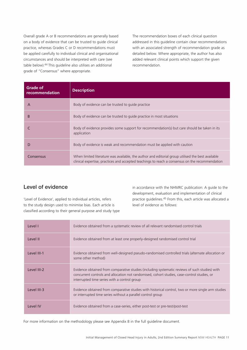
Initial Management of Closed Head Injury in Adults, 2nd Edition Summary Report NSW HEALTH PAGE 11
Overall grade A or B recommendations are generally based
on a body of evidence that can be trusted to guide clinical
practice, whereas Grades C or D recommendations must
be applied carefully to individual clinical and organisational
circumstances and should be interpreted with care (see
table below).44 This guideline also utilises an additional
grade of “Consensus” where appropriate.
The recommendation boxes of each clinical question
addressed in this guideline contain clear recommendations
with an associated strength of recommendation grade as
detailed below. Where appropriate, the author has also
added relevant clinical points which support the given
recommendation.
Level of evidence
‘Level of Evidence’, applied to individual articles, refers
to the study design used to minimise bias. Each article is
classified according to their general purpose and study type
in accordance with the NHMRC publication: A guide to the
development, evaluation and implementation of clinical
practice guidelines.45 From this, each article was allocated a
level of evidence as follows:
For more information on the methodology please see Appendix 8 in the full guideline document.
Grade of recommendation
Description
A Body of evidence can be trusted to guide practice
B Body of evidence can be trusted to guide practice in most situations
C Body of evidence provides some support for recommendation(s) but care should be taken in its application
D Body of evidence is weak and recommendation must be applied with caution
Consensus When limited literature was available, the author and editorial group utilised the best available clinical expertise, practices and accepted teachings to reach a consensus on the recommendation
Level I Evidence obtained from a systematic review of all relevant randomised control trials
Level II Evidence obtained from at least one properly-designed randomised control trial
Level III-1 Evidence obtained from well-designed pseudo-randomised controlled trials (alternate allocation or some other method)
Level III-2 Evidence obtained from comparative studies (including systematic reviews of such studies) with concurrent controls and allocation not randomised, cohort studies, case-control studies, or interrupted time series with a control group
Level III-3 Evidence obtained from comparative studies with historical control, two or more single arm studies or interrupted time series without a parallel control group
Level IV Evidence obtained from a case-series, either post-test or pre-test/post-test
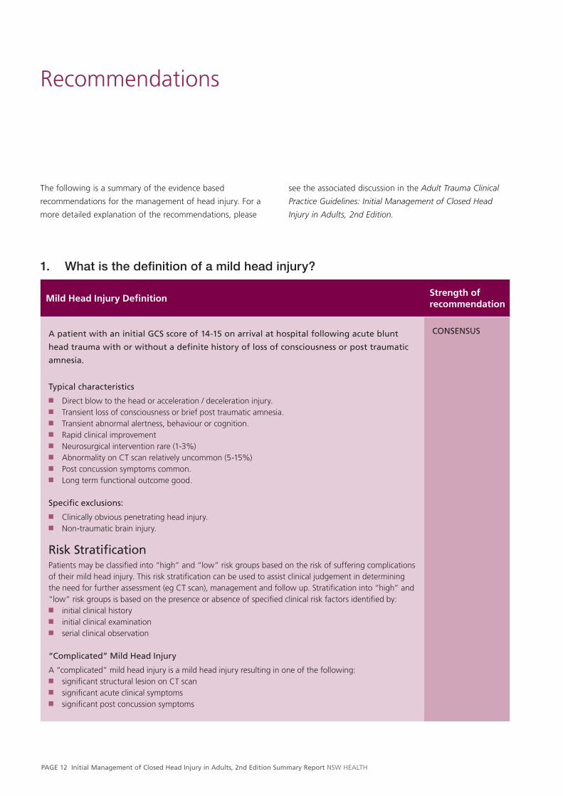
PAGE 12 Initial Management of Closed Head Injury in Adults, 2nd Edition Summary Report NSW HEALTH
Mild Head Injury Defi nitionStrength of recommendation
A patient with an initial GCS score of 14-15 on arrival at hospital following acute blunt
head trauma with or without a definite history of loss of consciousness or post traumatic
amnesia.
Typical characteristics
■ Direct blow to the head or acceleration / deceleration injury.■ Transient loss of consciousness or brief post traumatic amnesia.■ Transient abnormal alertness, behaviour or cognition.■ Rapid clinical improvement■ Neurosurgical intervention rare (1-3%)■ Abnormality on CT scan relatively uncommon (5-15%)■ Post concussion symptoms common.■ Long term functional outcome good.
Specifi c exclusions:
■ Clinically obvious penetrating head injury.■ Non-traumatic brain injury.
Risk Stratifi cationPatients may be classifi ed into “high” and “low” risk groups based on the risk of suffering complications of their mild head injury. This risk stratifi cation can be used to assist clinical judgement in determining the need for further assessment (eg CT scan), management and follow up. Stratifi cation into “high” and “low” risk groups is based on the presence or absence of specifi ed clinical risk factors identifi ed by:■ initial clinical history■ initial clinical examination■ serial clinical observation
“Complicated” Mild Head Injury
A “complicated” mild head injury is a mild head injury resulting in one of the following:■ signifi cant structural lesion on CT scan■ signifi cant acute clinical symptoms■ signifi cant post concussion symptoms
CONSENSUS
1. What is the definition of a mild head injury?
Recommendations
The following is a summary of the evidence based
recommendations for the management of head injury. For a
more detailed explanation of the recommendations, please
see the associated discussion in the Adult Trauma Clinical
Practice Guidelines: Initial Management of Closed Head
Injury in Adults, 2nd Edition.
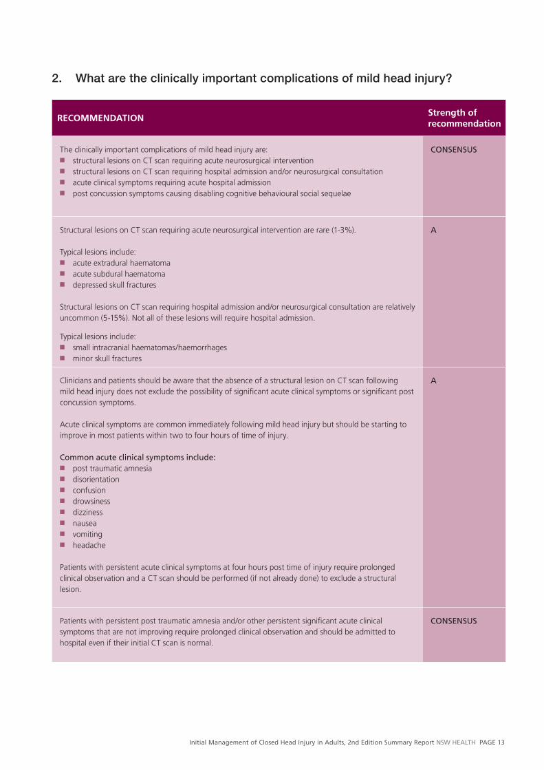
Initial Management of Closed Head Injury in Adults, 2nd Edition Summary Report NSW HEALTH PAGE 13
RECOMMENDATIONStrength of recommendation
The clinically important complications of mild head injury are:■ structural lesions on CT scan requiring acute neurosurgical intervention■ structural lesions on CT scan requiring hospital admission and/or neurosurgical consultation■ acute clinical symptoms requiring acute hospital admission■ post concussion symptoms causing disabling cognitive behavioural social sequelae
CONSENSUS
Structural lesions on CT scan requiring acute neurosurgical intervention are rare (1-3%).
Typical lesions include:■ acute extradural haematoma■ acute subdural haematoma■ depressed skull fractures
Structural lesions on CT scan requiring hospital admission and/or neurosurgical consultation are relatively uncommon (5-15%). Not all of these lesions will require hospital admission.
Typical lesions include:■ small intracranial haematomas/haemorrhages■ minor skull fractures
A
Clinicians and patients should be aware that the absence of a structural lesion on CT scan following mild head injury does not exclude the possibility of signifi cant acute clinical symptoms or signifi cant post concussion symptoms.
Acute clinical symptoms are common immediately following mild head injury but should be starting to improve in most patients within two to four hours of time of injury.
Common acute clinical symptoms include:■ post traumatic amnesia■ disorientation■ confusion■ drowsiness■ dizziness■ nausea■ vomiting■ headache
Patients with persistent acute clinical symptoms at four hours post time of injury require prolonged clinical observation and a CT scan should be performed (if not already done) to exclude a structural lesion.
A
Patients with persistent post traumatic amnesia and/or other persistent signifi cant acute clinical symptoms that are not improving require prolonged clinical observation and should be admitted to hospital even if their initial CT scan is normal.
CONSENSUS
2. What are the clinically important complications of mild head injury?
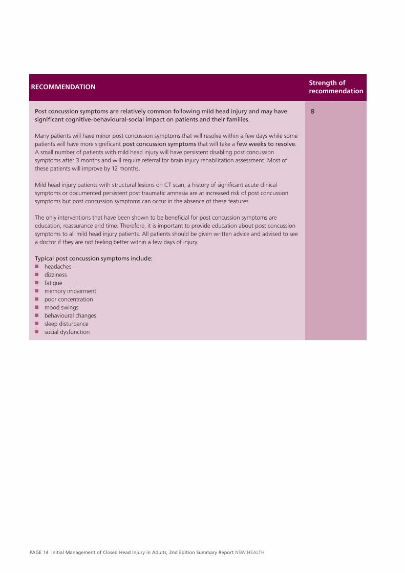
PAGE 14 Initial Management of Closed Head Injury in Adults, 2nd Edition Summary Report NSW HEALTH
Post concussion symptoms are relatively common following mild head injury and may have signifi cant cognitive-behavioural-social impact on patients and their families. Many patients will have minor post concussion symptoms that will resolve within a few days while some patients will have more signifi cant post concussion symptoms that will take a few weeks to resolve.A small number of patients with mild head injury will have persistent disabling post concussion symptoms after 3 months and will require referral for brain injury rehabilitation assessment. Most of these patients will improve by 12 months.
Mild head injury patients with structural lesions on CT scan, a history of signifi cant acute clinical symptoms or documented persistent post traumatic amnesia are at increased risk of post concussion symptoms but post concussion symptoms can occur in the absence of these features.
The only interventions that have been shown to be benefi cial for post concussion symptoms are education, reassurance and time. Therefore, it is important to provide education about post concussion symptoms to all mild head injury patients. All patients should be given written advice and advised to see a doctor if they are not feeling better within a few days of injury.
Typical post concussion symptoms include:■ headaches■ dizziness■ fatigue■ memory impairment■ poor concentration■ mood swings■ behavioural changes■ sleep disturbance■ social dysfunction
B
RECOMMENDATIONStrength of recommendation
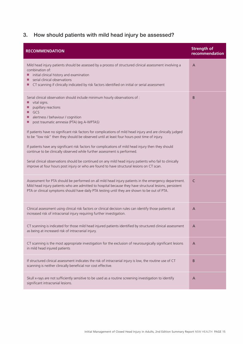
Initial Management of Closed Head Injury in Adults, 2nd Edition Summary Report NSW HEALTH PAGE 15
3. How should patients with mild head injury be assessed?
RECOMMENDATIONStrength of recommendation
Mild head injury patients should be assessed by a process of structured clinical assessment involving a combination of:■ initial clinical history and examination■ serial clinical observations ■ CT scanning if clinically indicated by risk factors identifi ed on initial or serial assessment
A
Serial clinical observation should include minimum hourly observations of :■ vital signs. ■ pupillary reactions ■ GCS ■ alertness / behaviour / cognition■ post traumatic amnesia (PTA) (eg A-WPTAS)
If patients have no signifi cant risk factors for complications of mild head injury and are clinically judged to be “low risk” then they should be observed until at least four hours post time of injury.
If patients have any signifi cant risk factors for complications of mild head injury then they should continue to be clinically observed while further assessment is performed.
Serial clinical observations should be continued on any mild head injury patients who fail to clinically improve at four hours post injury or who are found to have structural lesions on CT scan.
B
Assessment for PTA should be performed on all mild head injury patients in the emergency department. Mild head injury patients who are admitted to hospital because they have structural lesions, persistent PTA or clinical symptoms should have daily PTA testing until they are shown to be out of PTA.
C
Clinical assessment using clinical risk factors or clinical decision rules can identify those patients at increased risk of intracranial injury requiring further investigation.
A
CT scanning is indicated for those mild head injured patients identifi ed by structured clinical assessment as being at increased risk of intracranial injury.
A
CT scanning is the most appropriate investigation for the exclusion of neurosurgically signifi cant lesions in mild head injured patients
A
If structured clinical assessment indicates the risk of intracranial injury is low, the routine use of CT scanning is neither clinically benefi cial nor cost effective.
B
Skull x-rays are not suffi ciently sensitive to be used as a routine screening investigation to identify signifi cant intracranial lesions.
A
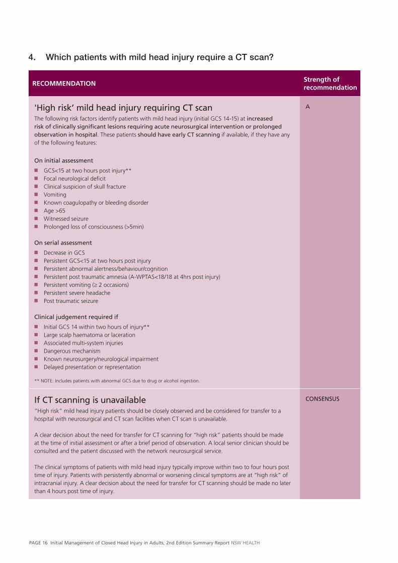
PAGE 16 Initial Management of Closed Head Injury in Adults, 2nd Edition Summary Report NSW HEALTH
4. Which patients with mild head injury require a CT scan?
RECOMMENDATIONStrength of recommendation
'High risk’ mild head injury requiring CT scanThe following risk factors identify patients with mild head injury (initial GCS 14-15) at increased risk of clinically signifi cant lesions requiring acute neurosurgical intervention or prolonged observation in hospital. These patients should have early CT scanning if available, if they have any of the following features:
On initial assessment
■ GCS<15 at two hours post injury**■ Focal neurological defi cit■ Clinical suspicion of skull fracture■ Vomiting ■ Known coagulopathy or bleeding disorder■ Age >65 ■ Witnessed seizure■ Prolonged loss of consciousness (>5min)
On serial assessment
■ Decrease in GCS■ Persistent GCS<15 at two hours post injury■ Persistent abnormal alertness/behaviour/cognition■ Persistent post traumatic amnesia (A-WPTAS<18/18 at 4hrs post injury)■ Persistent vomiting ( 2 occasions)■ Persistent severe headache■ Post traumatic seizure
Clinical judgement required if
■ Initial GCS 14 within two hours of injury** ■ Large scalp haematoma or laceration■ Associated multi-system injuries■ Dangerous mechanism■ Known neurosurgery/neurological impairment■ Delayed presentation or representation
** NOTE: Includes patients with abnormal GCS due to drug or alcohol ingestion.
A
If CT scanning is unavailable“High risk” mild head injury patients should be closely observed and be considered for transfer to a hospital with neurosurgical and CT scan facilities when CT scan is unavailable.
A clear decision about the need for transfer for CT scanning for “high risk” patients should be made at the time of initial assessment or after a brief period of observation. A local senior clinician should be consulted and the patient discussed with the network neurosurgical service.
The clinical symptoms of patients with mild head injury typically improve within two to four hours post time of injury. Patients with persistently abnormal or worsening clinical symptoms are at “high risk” of intracranial injury. A clear decision about the need for transfer for CT scanning should be made no later than 4 hours post time of injury.
CONSENSUS
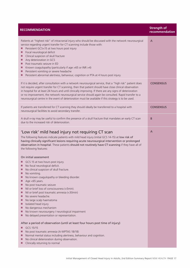
Initial Management of Closed Head Injury in Adults, 2nd Edition Summary Report NSW HEALTH PAGE 17
RECOMMENDATIONStrength of recommendation
Patients at “highest risk” of intracranial injury who should be discussed with the network neurosurgical service regarding urgent transfer for CT scanning include those with:■ Persistent GCS<15 at two hours post injury■ Focal neurological defi cit■ Clinical suspicion of skull fracture■ Any deterioration in GCS■ Post traumatic seizure in ED ■ Known coagulopathy (particularly if age >65 or INR >4)■ Persistent vomiting or severe headache■ Persistent abnormal alertness, behaviour, cognition or PTA at 4 hours post injury.
A
If it is decided, after consultation with a network neurosurgical service, that a “high risk” patient does not require urgent transfer for CT scanning, then that patient should have close clinical observation in hospital for at least 24 hours and until clinically improving. If there are any signs of deterioration or no improvement, the network neurosurgical service should again be consulted. Rapid transfer to a neurosurgical centre in the event of deterioration must be available if this strategy is to be used.
CONSENSUS
If patients are transferred for CT scanning they should ideally be transferred to a hospital with neurosurgical facilities to avoid secondary transfer.
CONSENSUS
A skull x-ray may be useful to confi rm the presence of a skull fracture that mandates an early CT scan due to the increased risk of deterioration.
B
‘Low risk’ mild head injury not requiring CT scanThe following features indicate patients with mild head injury (initial GCS 14-15) at low risk of having clinically signifi cant lesions requiring acute neurosurgical intervention or prolonged observation in hospital. These patients should not routinely have CT scanning if they have all of the following features:
On initial assessment
■ GCS 15 at two hours post injury.■ No focal neurological defi cit.■ No clinical suspicion of skull fracture.■ No vomiting.■ No known coagulopathy or bleeding disorder.■ Age <65 years. ■ No post traumatic seizure■ Nil or brief loss of consciousness (<5min).■ Nil or brief post traumatic amnesia (<30min)■ No severe headache.■ No large scalp haematoma■ Isolated head injury■ No dangerous mechanism■ No known neurosurgery / neurological impairment■ No delayed presentation or representation.
After a period of observation (until at least four hours post time of injury)
■ GCS 15/15 ■ No post traumatic amnesia (A-WPTAS 18/18)■ Normal mental status including alertness, behaviour and cognition. ■ No clinical deterioration during observation.■ Clinically returning to normal
A
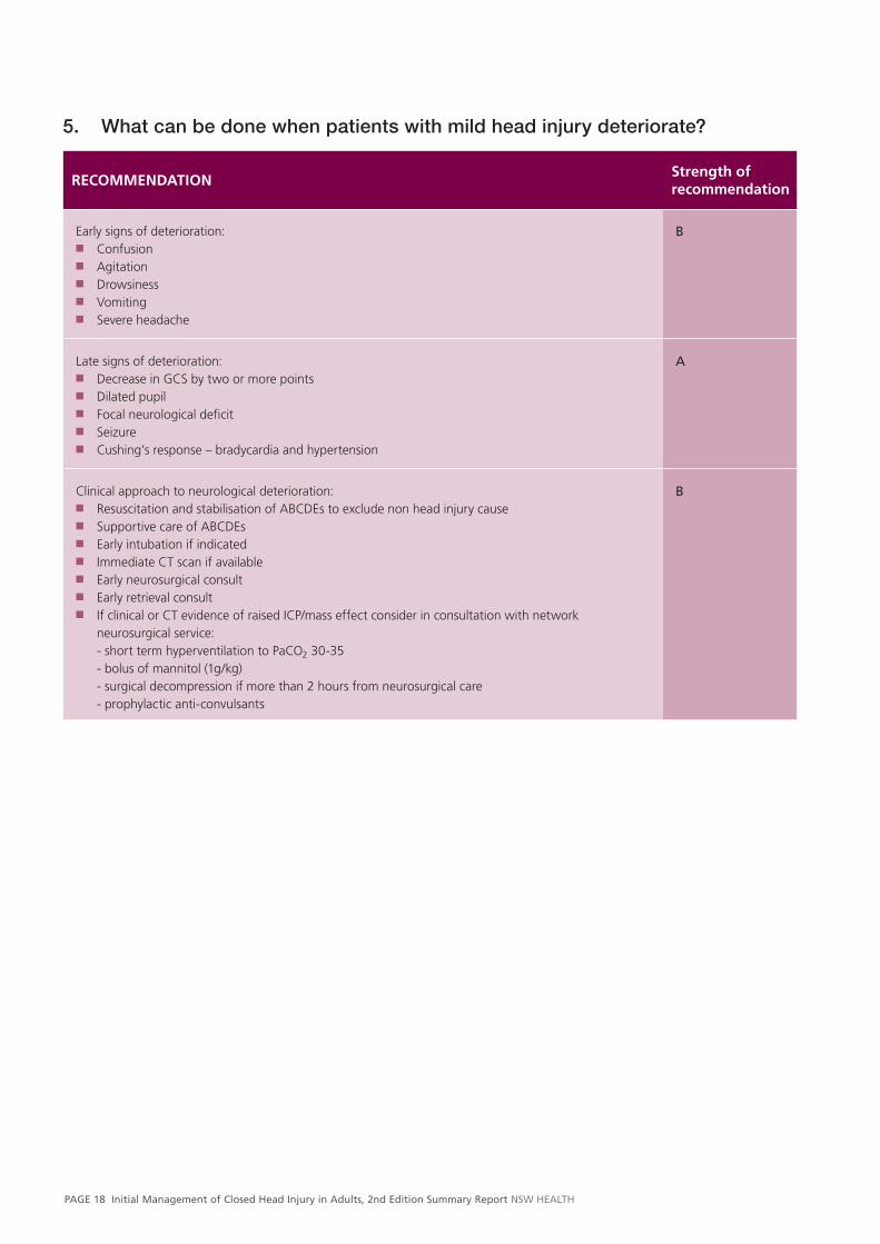
PAGE 18 Initial Management of Closed Head Injury in Adults, 2nd Edition Summary Report NSW HEALTH
RECOMMENDATIONStrength of recommendation
Early signs of deterioration:■ Confusion■ Agitation■ Drowsiness■ Vomiting■ Severe headache
B
Late signs of deterioration:■ Decrease in GCS by two or more points■ Dilated pupil■ Focal neurological defi cit■ Seizure■ Cushing’s response – bradycardia and hypertension
A
Clinical approach to neurological deterioration:■ Resuscitation and stabilisation of ABCDEs to exclude non head injury cause ■ Supportive care of ABCDEs■ Early intubation if indicated■ Immediate CT scan if available■ Early neurosurgical consult■ Early retrieval consult ■ If clinical or CT evidence of raised ICP/mass effect consider in consultation with network
neurosurgical service:� - short term hyperventilation to PaCO2 30-35� - bolus of mannitol (1g/kg)� - surgical decompression if more than 2 hours from neurosurgical care� - prophylactic anti-convulsants
B
5. What can be done when patients with mild head injury deteriorate?
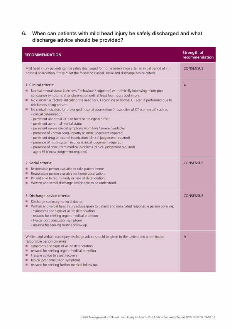
Initial Management of Closed Head Injury in Adults, 2nd Edition Summary Report NSW HEALTH PAGE 19
6. When can patients with mild head injury be safely discharged and what discharge advice should be provided?
RECOMMENDATIONStrength of recommendation
Mild head injury patients can be safely discharged for home observation after an initial period of in-hospital observation if they meet the following clinical, social and discharge advice criteria:
CONSENSUS
1. Clinical criteria:
■ Normal mental status (alertness / behaviour / cognition) with clinically improving minor post concussion symptoms after observation until at least four hours post injury.
■ No clinical risk factors indicating the need for CT scanning or normal CT scan if performed due to risk factors being present.
■ No clinical indicators for prolonged hospital observation (irrespective of CT scan result) such as:� - clinical deterioration� - persistent abnormal GCS or focal neurological defi cit� - persistent abnormal mental status � - persistent severe clinical symptoms (vomiting / severe headache) � - presence of known coagulopathy (clinical judgement required)� - persistent drug or alcohol intoxication (clinical judgement required)� - presence of multi-system injuries (clinical judgement required)� - presence of concurrent medical problems (clinical judgement required)� - age >65 (clinical judgement required)
A
2. Social criteria:
■ Responsible person available to take patient home.■ Responsible person available for home observation.■ Patient able to return easily in case of deterioration.■ Written and verbal discharge advice able to be understood.
CONSENSUS
3. Discharge advice criteria:
■ Discharge summary for local doctor.■ Written and verbal head injury advice given to patient and nominated responsible person covering:� - symptoms and signs of acute deterioration� - reasons for seeking urgent medical attention� - typical post concussion symptoms� - reasons for seeking routine follow up.
CONSENSUS
Written and verbal head injury discharge advice should be given to the patient and a nominated responsible person covering:■ symptoms and signs of acute deterioration■ reasons for seeking urgent medical attention■ lifestyle advice to assist recovery■ typical post concussion symptoms■ reasons for seeking further medical follow up.
A
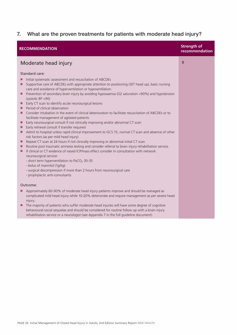
PAGE 20 Initial Management of Closed Head Injury in Adults, 2nd Edition Summary Report NSW HEALTH
7. What are the proven treatments for patients with moderate head injury?
RECOMMENDATIONStrength of recommendation
Moderate head injury
Standard care:
■ Initial systematic assessment and resuscitation of ABCDEs■ Supportive care of ABCDEs with appropriate attention to positioning (30° head up), basic nursing
care and avoidance of hyperventilation or hypoventilation.■ Prevention of secondary brain injury by avoiding hypoxaemia (O2 saturation <90%) and hypotension
(systolic BP <90)■ Early CT scan to identify acute neurosurgical lesions ■ Period of clinical observation■ Consider intubation in the event of clinical deterioration to facilitate resuscitation of ABCDEs or to
facilitate management of agitated patients■ Early neurosurgical consult if not clinically improving and/or abnormal CT scan■ Early retrieval consult if transfer required■ Admit to hospital unless rapid clinical improvement to GCS 15, normal CT scan and absence of other
risk factors (as per mild head injury)■ Repeat CT scan at 24 hours if not clinically improving or abnormal initial CT scan■ Routine post traumatic amnesia testing and consider referral to brain injury rehabilitation service.■ If clinical or CT evidence of raised ICP/mass effect consider in consultation with network
neurosurgical service:� - short term hyperventilation to PaCO2 30-35� - bolus of mannitol (1g/kg)� - surgical decompression if more than 2 hours from neurosurgical care� - prophylactic anti-convulsants
Outcome:
■ Approximately 80-90% of moderate head injury patients improve and should be managed as complicated mild head injury while 10-20% deteriorate and require management as per severe head injury..
■ The majority of patients who suffer moderate head injuries will have some degree of cognitive behavioural social sequelae and should be considered for routine follow up with a brain injury rehabilitation service or a neurologist (see Appendix 7 in the full guideline document).
B

Initial Management of Closed Head Injury in Adults, 2nd Edition Summary Report NSW HEALTH PAGE 21
8. What are the proven treatments for patients with severe head injury?
RECOMMENDATIONStrength of recommendation
Severe head injury
Standard care:
■ Initial systematic assessment and resuscitation of ABCDEs■ Early intubation■ Supportive care of ABCDEs with appropriate attention to positioning (30° head up), basic nursing
care and avoidance of hyperventilation or hypoventilation.■ Prevention of secondary brain injury by avoiding hypoxaemia (O2 saturation <90%) and hypotension
(systolic BP<90)■ Early CT scan to identify acute neurosurgical lesions■ Early neurosurgical consult■ Early retrieval consult if transfer required■ Consider use of anticonvulsants to prevent early post traumatic seizures■ Consider ICP monitoring to guide management of cerebral perfusion pressure.■ Low threshold to repeat CT scan if patient condition changes■ ICU admission■ Routine repeat CT scan at 24 hours■ Brain injury rehabilitation consult■ If clinical or CT evidence of raised ICP/mass effect consider in consultation with network
neurosurgical service:� - short term hyperventilation to PaCO2 30-35� - bolus of mannitol (1g/kg)� - surgical decompression if more than 2 hours from neurosurgical care� - prophylactic anti-convulsants
Minimum supportive care aims:
■ PaO2 > 60■ SaO2 > 90■ PaCO2 35-40■ Systolic BP > 90■ Head up 30°
Poor prognostic indicators:
■ Low GCS (especially motor component).■ Age >60 years (prognosis deteriorates with increasing age).■ Absent pupillary refl exes (after systemic resuscitation).■ Hypotension (systolic BP <90).■ Hypoxaemia (oxygen saturation <90%).
A
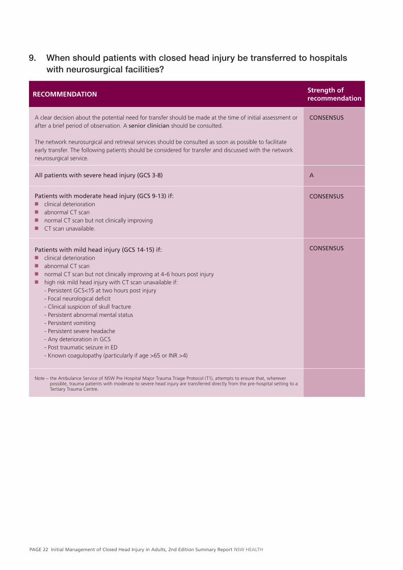
PAGE 22 Initial Management of Closed Head Injury in Adults, 2nd Edition Summary Report NSW HEALTH
9. When should patients with closed head injury be transferred to hospitals with neurosurgical facilities?
RECOMMENDATIONStrength of recommendation
A clear decision about the potential need for transfer should be made at the time of initial assessment or after a brief period of observation. A senior clinician should be consulted.
The network neurosurgical and retrieval services should be consulted as soon as possible to facilitate early transfer. The following patients should be considered for transfer and discussed with the network neurosurgical service.
CONSENSUS
All patients with severe head injury (GCS 3-8) A
Patients with moderate head injury (GCS 9-13) if: ■ clinical deterioration■ abnormal CT scan■ normal CT scan but not clinically improving■ CT scan unavailable.
CONSENSUS
Patients with mild head injury (GCS 14-15) if:■ clinical deterioration■ abnormal CT scan■ normal CT scan but not clinically improving at 4-6 hours post injury■ high risk mild head injury with CT scan unavailable if:� - Persistent GCS<15 at two hours post injury� - Focal neurological defi cit� - Clinical suspicion of skull fracture� - Persistent abnormal mental status � - Persistent vomiting� - Persistent severe headache � - Any deterioration in GCS� - Post traumatic seizure in ED � - Known coagulopathy (particularly if age >65 or INR >4)
CONSENSUS
Note – the Ambulance Service of NSW Pre Hospital Major Trauma Triage Protocol (T1), attempts to ensure that, wherever possible, trauma patients with moderate to severe head injury are transferred directly from the pre-hospital setting to a Tertiary Trauma Centre.
Initial Management of Closed Head Injury in Adults, 2nd Edition Summary Report NSW HEALTH PAGE 23
10. What analgesia should patients with closed head injury receive?
RECOMMENDATIONStrength of recommendation
Analgesia in isolated mild head injury■ Persistent severe headache or worsening severe headache is an indication for a CT scan to exclude a
signifi cant intracranial lesion■ Most headaches associated with isolated mild head injury will respond to simple analgesia such as
paracetamol. ■ Isolated mild head injury patients who require more than paracetamol for headache should be
considered for a CT scan to exclude a signifi cant intracranial injury
CONSENSUS
Analgesia guide for isolated mild head injury:■ Paracetamol, 1g, q 4-6 hours, maximum 4g/24 hours*If paracetamol is ineffective as a sole agent then stronger analgesia such as oral opioids or parenteral opioids should not be prescribed to patients with isolated mild head injury unless the need for an initial or repeat CT scan to exclude clinically important intracranial lesions has been considered and a senior clinician has been consulted. After further clinical assessment consider adding;
■ Codeine Phosphate, 30-60mg, q 4-6 hours* or■ Oxycodone (immediate release), 5-10mg q 4-6 hours*
NB Avoid the use of aspirin / NSAIDS due to increased risk of bleeding* See standard texts for detailed prescribing information
CONSENSUS
Analgesia guide for mild head injury with associated systemic injuries:■ More likely to need titrated intravenous opioids, procedural sedation or general anaesthesia for their
associated injuries. ■ Have a lower threshold for performing CT scans.■ Require close clinical assessment and observation.■ Appropriate pain relief should not be withheld due to concerns of masking head injury symptoms
and signs■ Analgesia needs to be individualised under the supervision of a senior clinician.
CONSENSUS
Analgesia in moderate to severe head injury■ Likely to require titrated intravenous analgesia and sedation for associated injuries, clinical
management or intubation. ■ Will require close clinical observation in a high dependency area following initial clinical assessment
and CT scanning. ■ Analgesia needs to be individualised under the supervision of a senior clinician.
CONSENSUS
Clinical approach to pain management in closed head injury (all severities)■ Consult a senior clinician if any signifi cant change in the patient’s condition■ Clinically re-assess if: � - inadequate analgesia or worsening headache� - excessive drowsiness, or other clinical deterioration■ Before using stronger analgesia:� - clinically re-assess patient� - consider need for CT scan� - consult senior clinician
CONSENSUS
PAGE 24 Initial Management of Closed Head Injury in Adults, 2nd Edition Summary Report NSW HEALTH
11. Which patients with closed head injury should receive anti-convulsants?
RECOMMENDATIONStrength of recommendation
Consult a senior clinician or your network neurosurgical service before commencing prophylactic anti-convulsants in patients with acute closed head injury
CONSENSUS
Prophylactic anti-convulsants are not indicated for patients with uncomplicated mild head injury B
Prophylactic anti-convulsants should be considered in patients with complicated mild head injury or moderate to severe head injury.
B
Specifi c indications to consider prophylactic anti-convulsants in the fi rst week following a head injury include:■ Extradural, subdural or intracerebral haematoma on CT■ Depressed skull fracture on CT■ Early post traumatic seizure in hospital (especially if focal or prolonged)■ Severity of head injury (low initial GCS / prolonged coma / prolonged PTA)■ Any suspicion of penetrating injury
B
Prophylactic anti-convulsants decrease the incidence of early post traumatic seizures within seven days of closed head injury.
B
Early post traumatic seizures have not been shown to be associated with worse patient outcomes in large population studies.
B
Clinical judgment is required on whether to prescribe anti-convulsants for individual patients. CONSENSUS
Indications for anti-convulsants by post traumatic seizure typeImmediate post traumatic seizures (at time of injury) ■ Anti-convulsants not warranted unless specifi c indication present (see above)
Early post traumatic seizures ( up to 7 days post injury)■ Anti-convulsants should be considered especially if any of the other specifi c indications are also
present (see above)
Late post traumatic seizures (more than 7 days post injury)■ Long term anti-convulsants should be considered after the fi rst late post traumatic seizure due to the
increased risk of developing post traumatic epilepsy■ There is no evidence that the routine use of anti-convulsants following closed head injury reduces
the risk of late post traumatic seizures.
B
Recommended drugs and loading doses*Standard therapy:Phenytoin:■ Intravenous loading dose: 20 mg/kg in NS (<6.7mg/ml) no faster than 50mg/min Standard adult IVI loading dose: 1000mg phenytoin diluted in 150ml normal saline over 60 mins with in line micron fi lter
B
Alternative therapies:Levetiracetam:■ Intravenous loading dose: 10mg /kg (max 1000mg)■ Standard adult IVI loading dose: 1000mg levetiracetam in 100ml normal saline over 15 mins
Sodium Valproate:■ Intravenous loading dose: 10mg /kg (max 800mg)■ Standard adult IVI loading dose: 800mg in 100ml normal saline over 15 mins
* See standard texts for detailed prescribing information
C

Initial Management of Closed Head Injury in Adults, 2nd Edition Summary Report NSW HEALTH PAGE 25
PUPIL ASSESSMENT
T1 T2 T3 T4 T5 + = REACTS BRISKLY
R L R L R L R L R L SL = SLUGGISHSize C = CLOSED
Reaction - = NIL
Comments
Pupil Size (mm)
2 3 4 5 6 7 8
Date:
Time
T1 T2 T3 T4 T5 Use of A-WPTAS and GCS for patients with MTBI
The A-WPTAS combined with a standardised GCS assessment is an objective measure of post traumatic amnesia (PTA). Only for patients with current GCS of 13-15 (<24hrs post injury) with impact to the head resulting in confusion, disorientation, anterograde or retrograde amnesia, or brief LOC. Administer both tests at hourly intervals to gauge patient’s capacity for full orientation and ability to retain new information. Also, note the following: poor motivation, depression, pre-morbid intellectual handicap or possible medication, drug or alcohol effects. NB: This is a screening device, so exercise clinical judgement. In cases where doubt exists, more thorough assessment may benecessary.
Admission and Discharge Criteria:
A patient is considered to be out of PTA when they score 18/18.
Both the GCS and A-WPTAS should be used in conjunction with clinical judgement.
Patients scoring 18/18 can be considered for discharge.
For patients who do not obtain 18/18 re-assess after a further hour.
Patients with persistent score <18/18 at 4 hours post time of injury should be considered for admission.
Clinical judgement and consideration of pre-existing conditions should be used where the memory component of A-WPTAS is abnormal but the GCS is normal (15/15).
Referral to GP on discharge if abnormal PTA was present, provide patient advice sheet.
Motor Obeys commands
6 6 6 6 6
Localises 5 5 5 5 5Abnormal flexion 4 4 4 4 4Withdraws 3 3 3 3 3Extension 2 2 2 2 2None 1 1 1 1 1
Eye Opening Spontaneously 4 4 4 4 4
To speech 3 3 3 3 3
To pain 2 2 2 2 2None 1 1 1 1 1
Verbal Oriented ** (tick if correct)
Name
Place
Why are you here
Month
Year
5 5 5 5 5
Confused 4 4 4 4 4Inappropriate words
3 3 3 3 3
Incomprehensible sounds
2 2 2 2 2
None 1 1 1 1 1
GCS Score out of 15 /15 /15 /15 /15 /15 Target set of picture cardsPicture 1
Picture 2
Picture 3
A-WPTAS Score out of 18 /18 /18 /18 /18
Abbreviated Westmead PTA Scale (A-WPTAS)incorporating Glasgow Coma Scale (GCS)
MRN sticker here
Show pictures
(see over)
Shores & Lammel (2007) - further copies of this score sheet can be downloaded from http://www.psy.mq.edu.au/GCS
ABBREVIATED WESTMEAD PTA SCALE (A-WPTAS)GCS & PTA testing of patients with MTBI following mild head injury
** must have all 5 orientation questions correct to score 5 on verbal score for GCS, otherwise the score is 4 (or less).
Appendix 1: Abbreviated Westmead PTA Scale
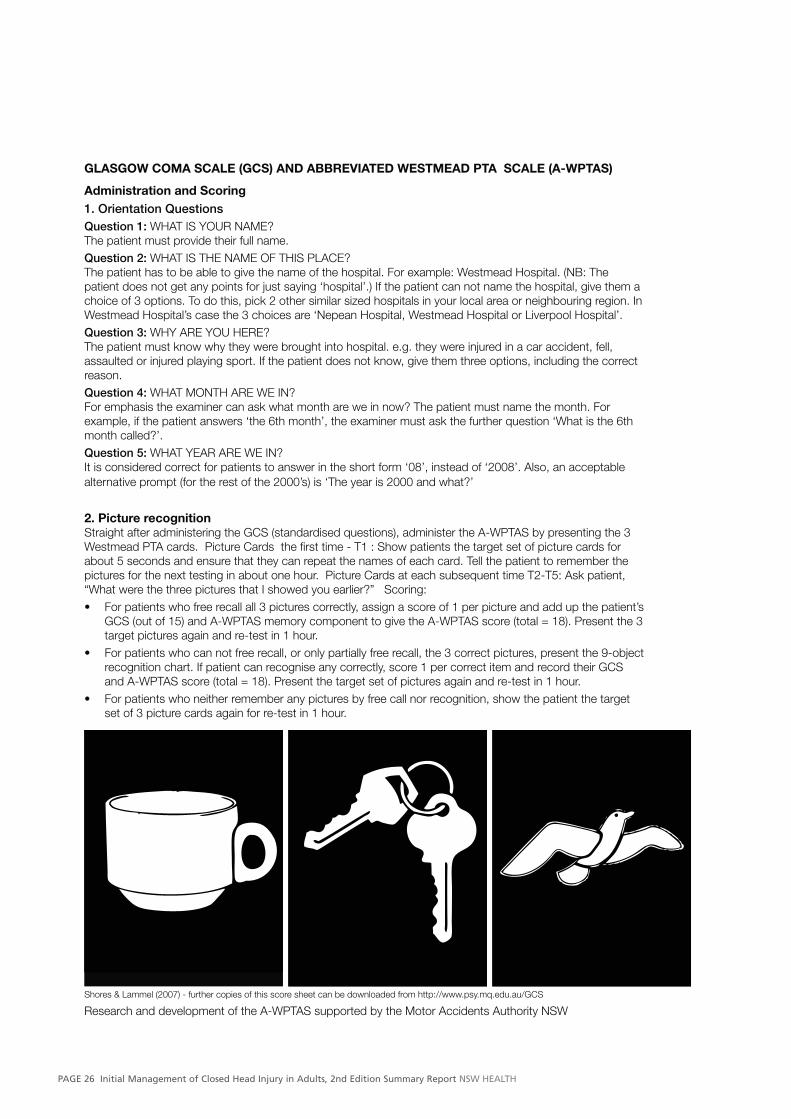
PAGE 26 Initial Management of Closed Head Injury in Adults, 2nd Edition Summary Report NSW HEALTH
Administration and Scoring 1. Orientation QuestionsQuestion 1: WHAT IS YOUR NAME? The patient must provide their full name.Question 2: WHAT IS THE NAME OF THIS PLACE? The patient has to be able to give the name of the hospital. For example: Westmead Hospital. (NB: The patient does not get any points for just saying ‘hospital’.) If the patient can not name the hospital, give them a choice of 3 options. To do this, pick 2 other similar sized hospitals in your local area or neighbouring region. In Westmead Hospital’s case the 3 choices are ‘Nepean Hospital, Westmead Hospital or Liverpool Hospital’. Question 3: WHY ARE YOU HERE? The patient must know why they were brought into hospital. e.g. they were injured in a car accident, fell, assaulted or injured playing sport. If the patient does not know, give them three options, including the correct reason.Question 4: WHAT MONTH ARE WE IN? For emphasis the examiner can ask what month are we in now? The patient must name the month. For example, if the patient answers ‘the 6th month’, the examiner must ask the further question ‘What is the 6th month called?’. Question 5: WHAT YEAR ARE WE IN? It is considered correct for patients to answer in the short form ‘08’, instead of ‘2008’. Also, an acceptable alternative prompt (for the rest of the 2000’s) is ‘The year is 2000 and what?’
2. Picture recognition Straight after administering the GCS (standardised questions), administer the A-WPTAS by presenting the 3 Westmead PTA cards. Picture Cards the first time - T1 : Show patients the target set of picture cards for about 5 seconds and ensure that they can repeat the names of each card. Tell the patient to remember the pictures for the next testing in about one hour. Picture Cards at each subsequent time T2-T5: Ask patient, “What were the three pictures that I showed you earlier?” Scoring:
For patients who free recall all 3 pictures correctly, assign a score of 1 per picture and add up the patient’s GCS (out of 15) and A-WPTAS memory component to give the A-WPTAS score (total = 18). Present the 3 target pictures again and re-test in 1 hour. For patients who can not free recall, or only partially free recall, the 3 correct pictures, present the 9-object recognition chart. If patient can recognise any correctly, score 1 per correct item and record their GCS and A-WPTAS score (total = 18). Present the target set of pictures again and re-test in 1 hour. For patients who neither remember any pictures by free call nor recognition, show the patient the target set of 3 picture cards again for re-test in 1 hour.
GLASGOW COMA SCALE (GCS) AND ABBREVIATED WESTMEAD PTA SCALE (A-WPTAS)
Shores & Lammel (2007) - further copies of this score sheet can be downloaded from http://www.psy.mq.edu.au/GCS
Research and development of the A-WPTAS supported by the Motor Accidents Authority NSW
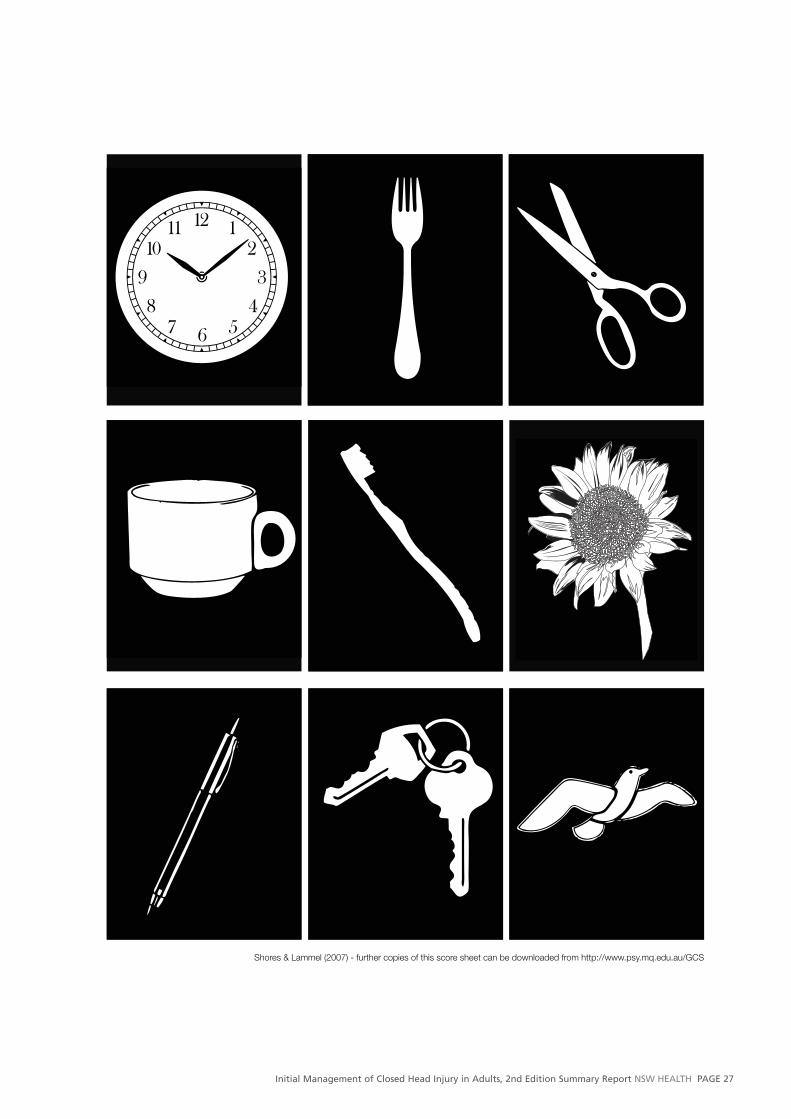
Initial Management of Closed Head Injury in Adults, 2nd Edition Summary Report NSW HEALTH PAGE 27
Shores & Lammel (2007) - further copies of this score sheet can be downloaded from http://www.psy.mq.edu.au/GCS
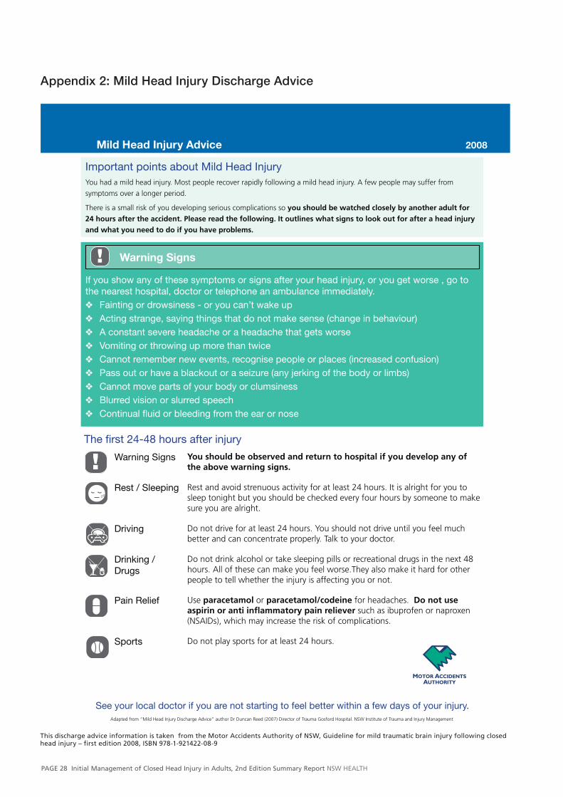
PAGE 28 Initial Management of Closed Head Injury in Adults, 2nd Edition Summary Report NSW HEALTH
Appendix 2: Mild Head Injury Discharge Advice
Important points about Mild Head InjuryYou had a mild head injury. Most people recover rapidly following a mild head injury. A few people may suffer from
symptoms over a longer period.
There is a small risk of you developing serious complications so you should be watched closely by another adult for 24 hours after the accident. Please read the following. It outlines what signs to look out for after a head injury and what you need to do if you have problems.
If you show any of these symptoms or signs after your head injury, or you get worse , go to the nearest hospital, doctor or telephone an ambulance immediately.
Fainting or drowsiness - or you can’t wake up ❖
Acting strange, saying things that do not make sense (change in behaviour) ❖
A constant severe headache or a headache that gets worse ❖
Vomiting or throwing up more than twice ❖
Cannot remember new events, recognise people or places (increased confusion) ❖
Pass out or have a blackout or a seizure (any jerking of the body or limbs) ❖
Cannot move parts of your body or clumsiness ❖
Blurred vision or slurred speech ❖
Continual fluid or bleeding from the ear or nose ❖
Warning Signs!
See your local doctor if you are not starting to feel better within a few days of your injury.Adapted from “Mild Head Injury Discharge Advice” author Dr Duncan Reed (2007) Director of Trauma Gosford Hospital. NSW Institute of Trauma and Injury Management
The first 24-48 hours after injury
Warning Signs You should be observed and return to hospital if you develop any of the above warning signs.
Rest / Sleeping Rest and avoid strenuous activity for at least 24 hours. It is alright for you to sleep tonight but you should be checked every four hours by someone to make sure you are alright.
Driving Do not drive for at least 24 hours. You should not drive until you feel much better and can concentrate properly. Talk to your doctor.
Drinking / Drugs
Do not drink alcohol or take sleeping pills or recreational drugs in the next 48 hours. All of these can make you feel worse.They also make it hard for other people to tell whether the injury is affecting you or not.
Pain Relief Use paracetamol or paracetamol/codeine for headaches. Do not use aspirin or anti inflammatory pain reliever such as ibuprofen or naproxen (NSAIDs), which may increase the risk of complications.
Sports Do not play sports for at least 24 hours.
zz
!
Mild Head Injury Advice 2008
This discharge advice information is taken from the Motor Accidents Authority of NSW, Guideline for mild traumatic brain injury following closed head injury – first edition 2008, ISBN 978-1-921422-08-9
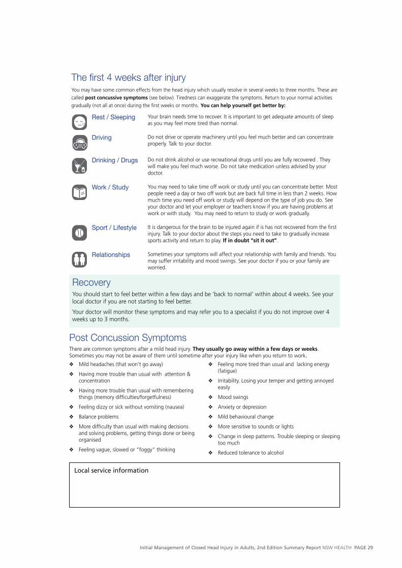
Initial Management of Closed Head Injury in Adults, 2nd Edition Summary Report NSW HEALTH PAGE 29
The first 4 weeks after injury You may have some common effects from the head injury which usually resolve in several weeks to three months. These are
called post concussive symptoms (see below). Tiredness can exaggerate the symptoms. Return to your normal activities
gradually (not all at once) during the first weeks or months. You can help yourself get better by:
Rest / Sleeping Your brain needs time to recover. It is important to get adequate amounts of sleep as you may feel more tired than normal.
Driving Do not drive or operate machinery until you feel much better and can concentrate properly. Talk to your doctor.
Drinking / Drugs Do not drink alcohol or use recreational drugs until you are fully recovered . They will make you feel much worse. Do not take medication unless advised by your doctor.
Work / Study You may need to take time off work or study until you can concentrate better. Most people need a day or two off work but are back full time in less than 2 weeks. How much time you need off work or study will depend on the type of job you do. See your doctor and let your employer or teachers know if you are having problems at work or with study. You may need to return to study or work gradually.
Sport / Lifestyle It is dangerous for the brain to be injured again if is has not recovered from the first injury. Talk to your doctor about the steps you need to take to gradually increase sports activity and return to play. If in doubt “sit it out”.
Relationships Sometimes your symptoms will affect your relationship with family and friends. You may suffer irritability and mood swings. See your doctor if you or your family are worried.
zz
RecoveryYou should start to feel better within a few days and be ‘back to normal’ within about 4 weeks. See your local doctor if you are not starting to feel better.
Your doctor will monitor these symptoms and may refer you to a specialist if you do not improve over 4 weeks up to 3 months.
Local service information
Post Concussion Symptoms There are common symptoms after a mild head injury. They usually go away within a few days or weeks. Sometimes you may not be aware of them until sometime after your injury like when you return to work.
Mild headaches (that won’t go away) ❖
Having more trouble than usual with attention & ❖
concentration
Having more trouble than usual with remembering ❖
things (memory difficulties/forgetfulness)
Feeling dizzy or sick without vomiting (nausea) ❖
Balance problems ❖
More difficulty than usual with making decisions ❖
and solving problems, getting things done or being organised
Feeling vague, slowed or “foggy” thinking ❖
Feeling more tired than usual and lacking energy ❖
(fatigue)
Irritability. Losing your temper and getting annoyed ❖
easily
Mood swings ❖
Anxiety or depression ❖
Mild behavioural change ❖
More sensitive to sounds or lights ❖
Change in sleep patterns. Trouble sleeping or sleeping ❖
too much
Reduced tolerance to alcohol ❖
PAGE 30 Initial Management of Closed Head Injury in Adults, 2nd Edition Summary Report NSW HEALTH
1. Newcombe R, Merry G. The management of acute
neurotrauma in rural and remote locations: A set of
guidelines for the care of head and spinal injuries. J
Clin Neuro 1999;6(1):85-93.
2. NSW Institute of Trauma and Injury Management.
NSW Trauma Registry Profile of Serious to Critical
Injuries: 2007. NSW Health; 2010.
3. Khan F, Baguley IJ, Cameron ID, Khan F, Baguley IJ,
Cameron ID. 4: Rehabilitation after traumatic brain
injury. MJA 2003;178(6):290-5.
4. Fortune N. The definition, incidence and prevalence
of acquired brain injury in Australia. Australian
Institute of Health and Welfare; 2009.
5. Tate R, McDonald S, Lulham J. Incidence of hospital-
treated traumatic brain injury in an Australian
community. ANZ J Pub Health 1998;22(4):419.
6. Servadei F, Teasdale G, Merry G,
Neurotraumatology Committee of the World
Federation of Neurosurgical S. Defining acute
mild head injury in adults: a proposal based on
prognostic factors, diagnosis, and management. J
Neurotrauma 2001;18(7):657-64.
7. Bellner J, Ingebrigtsen T, Romner B. Survey of
the management of patients with minor head
injuries in hospitals in Sweden. Acta Neurol Scand
1999;100(6):355-9.
8. Cushman J, Agarwal N, Fabian T, Garcia V, Nagy K,
Pasquale M, et al. Practice management guidelines
for the management of mild traumatic brain injury:
the EAST practice management guidelines work
group. J Trauma 2001;51(5):1016-26.
9. Jagoda AS, Bazarian JJ, Bruns JJ, Jr., Cantrill
SV, Gean AD, Howard PK, et al. Clinical policy:
neuroimaging and decisionmaking in adult mild
traumatic brain injury in the acute setting. Ann
Emerg Med 2008;52(6):714-48.
10. Vos PE, Battistin L, Birbamer G, Gerstenbrand F,
Potapov A, Prevec T, et al. EFNS guideline on mild
traumatic brain injury: report of an EFNS task force.
Eur J Neurology 2002;9(3):207-19.
11. Dickinson K, Bunn F, Wentz R, Edwards P, Roberts
I. Size and quality of randomised controlled trials
in head injury: review of published studies. BMJ.
2000;320(7245):1308-11.
12. Klauber M, Marshall L, Luerssen T, Frankowski R,
Tabaddor K, Eisenberg H. Determinants of Head
Injury Mortality: Importance of the Low Risk
Patient. Neurosurgery 1989;24(1):31-6.
13. Mower W, Hoffman J, Herbert M, Wolfson A,
Pollack C, Zucker M, et al. Developing a clinical
decision instrument to rule out intracranial injuries
in patients with minor head trauma: methodology
of the NEXUS II investigation. Ann Emerg Med
2002;40(5):505-14.
14. Narayan R, Michel ME, Ansell B, Baethmann A,
Biegon A, Bracken MB, et al. Clinical trials in head
injury. J Neurotrauma 2002;19(5):503-57.
15. Brain Trauma Foundation, American Association of
Neurological Surgeons, Congress of Neurological
Surgeons, Joint Section on Neurotrauma and
Critical Care. Guidelines for the Management
of Severe Traumatic Brain Injury J Neurotrauma
2007;24(S1):1-106.
16. Alderson P, Roberts I. Corticosteroids for acute
traumatic brain injury. Cochrane Database of
Systematic Reviews 2005(1).
References
Initial Management of Closed Head Injury in Adults, 2nd Edition Summary Report NSW HEALTH PAGE 31
17. Arango MF, Bainbridge D. Magnesium for acute
traumatic brain injury. Cochrane Database of
Systematic Reviews 2008(4).
18. Bennett MH, Trytko B, Jonker B. Hyperbaric oxygen
therapy for the adjunctive treatment of traumatic
brain injury. Cochrane Database of Systematic
Reviews. 2004(4).
19. Fleminger S, Greenwood R, Oliver D.
Pharmacological management for agitation and
aggression in people with acquired brain injury.
Cochrane Database of Systematic Reviews 2006(4).
20. Forsyth R, Wolny S, Rodrigues B. Routine
intracranial pressure monitoring in acute coma.
Cochrane Database of Systematic Reviews. 2010(2).
21. Ker K, Blackhall K. Bradykinin beta-2 receptor
antagonists for acute traumatic brain injury.
Cochrane Database of Systematic Reviews. 2008(1).
22. Langham J, Goldfrad C, Teasdale G, Shaw D, Rowan
K. Calcium channel blockers for acute traumatic
brain injury. Cochrane Database of Systematic
Reviews. 2003(4).
23. Roberts I. Aminosteroids for acute traumatic brain
injury. Cochrane Database of Systematic Reviews.
1999(3).
24. Roberts I, Schierhout G. Hyperventilation therapy
for acute traumatic brain injury. Cochrane Database
of Systematic Reviews. 2009(3).
25. Roberts I, Sydenham E. Barbiturates for acute
traumatic brain injury. Cochrane Database of
Systematic Reviews. 1999(3).
26. Sahuquillo J, Arikan F. Decompressive craniectomy
for the treatment of refractory high intracranial
pressure in traumatic brain injury. Cochrane
Database of Systematic Reviews. 2006(1).
27. Schierhout G, Roberts I. Anti-epileptic drugs for
preventing seizures following acute traumatic brain
injury. Cochrane Database of Systematic Reviews.
2001(4).
28. Sydenham E, Roberts I, Alderson P. Hypothermia
for traumatic head injury. . Cochrane Database of
Systematic Reviews. 2009(2).
29. Wakai A, Roberts IG, Schierhout G. Mannitol for
acute traumatic brain injury. Cochrane Database of
Systematic Reviews. 2007(1).
30. Willis C, Lybrand S, Bellamy N. Excitatory amino
acid inhibitors for traumatic brain injury. Cochrane
Database of Systematic Reviews. 2003(1).
31. Fabbri A, Servadei F, Marchesini G, Morselli-Labate
A, Dente M, Iervese T, et al. Prospective validation
of a proposal for diagnosis and management of
patients attending the emergency department for
mild head injury. J Neurol Neurosurg Psychiatry
2004;75(3):410-6.
32. Haydel M, Preston C, Mills T, Luber S, Blaudeau E,
DeBlieux P. Indications for computed tomography
in patients with minor head injury. NEJM
2000;343(2):100-5.
33. Ibanez J, Arikan F, Pedraza S, Sanchez E, Poca MA,
Rodriguez D, et al. Reliability of clinical guidelines in
the detection of patients at risk following mild head
injury: results of a prospective study. J Neurosurgery
2004;100(5):825-34.
34. Palchak MJ, Holmes JF, Vance CW, Gelber RE,
Schauer BA, Harrison MJ, et al. A decision rule
for identifying children at low risk for brain
injuries after blunt head trauma. Ann Emerg Med
2003;42(4):492-506.
35. Stiell IG, Wells GA, Vandemheen K, Clement C,
Lesiuk H, Laupacis A, et al. The Canadian CT Head
Rule for patients with minor head injury. Lancet
2001;357(9266):1391-6.
36. Dunham C, Coates S, Cooper C. Compelling
evidence for discretionary brain computed
tomographic imaging in those patients with mild
cognitive impairment after blunt trauma. J Trauma
1996;41(4):679-86.
PAGE 32 Initial Management of Closed Head Injury in Adults, 2nd Edition Summary Report NSW HEALTH
37. Stiell IG, Wells GA, Vandemheen K, Laupacis A,
Brison R, Eisenhauer MA, et al. Variation in ED Use
of Computed Tomography for Patients With Minor
Head Injury. Ann Emerg Med 1997;30:14-22.
38. Bulger E, Nathens A, Rivara F, Moore M, MacKenzie
E, Jurkovich G, et al. Management of severe head
injury: institutional variations in care and effect on
outcome. Crit Care Med 2002;30(8):1870-6.
39. Af Geijerstam J, Britton M. Mild head injury:
reliability of early computed tomographic findings in
triage for admission. Emerg Med J 2005;22(2):103-
7.
40. American College of Surgeons Committee on
Trauma. Advanced Trauma Life Support for Doctors.
8th ed2007.
41. Fabbri A, Servadei F, Marchesini G, Stein S,
Vandelli A. Observational approach to subjects
with mild-to-moderate head injury and initial non-
neurosurgical lesions. J Neurol Neurosurg Psychiatry
2008;79(10):1180-5.
42. Myburgh JA, Cooper DJ, Finfer SR, Venkatesh
B, Jones D, Higgins A, et al. Epidemiology
and 12-month outcomes from traumatic brain
injury in australia and new zealand. J Trauma
2008;64(4):854-62.
43. Styrke J, Stalnacke B-M, Sojka P, Bjornstig
U. Traumatic Brain Injuries in a Well-Defined
Population: Epidemiological Aspects and Severity. J
Neurotrauma. 2007;24(9):1425-36.
44. National Health & Medical Research Council.
NHMRC levels of evidence and grades for
recommendations for developers of guidelines.
2009: Available from: www.nhmrc.gov.au/_files_
nhmrc/file/guidelines/evidence_statement_form.pdf.
45. National Health & Medical Research Council. A
guide to the development, implementation and
evaluation of clinical practice guidelines. Canberra
ACT. NHMRC; 1999.

SHPN (SSD) 110187
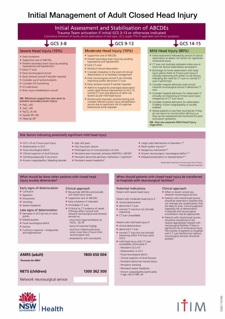
Initial Management of Adult Closed Head Injury
(SSD
) 110
190
Risk factors indicating potentially significant mild head injury
Severe Head Injury (10%)■ Early intubation■ Supportive care of ABCDEs ■ Prevent secondary brain injury by avoiding
hypoxaemia and hypotension ■ Early CT scan ■ Early neurosurgical consult■ Early retrieval consult if transfer required■ Consider use of anticonvulsants ■ Consider ICP monitoring ■ ICU admission■ Brain injury rehabilitation consult
NB. Minimum supportive care aims to prevent secondary brain injury:■ PaO2 >60■ SaO2 >90■ PaCO2 35-40 ■ Systolic BP >90■ Head up 30º
Mild Head Injury (80%)■ Initial assessment followed by period of clinical
observation to detect risk factors for significant intracranial injury.
■ CT scan not routinely indicated unless one or more risk factors listed below are present.
■ Discharge for home observation with head injury advice sheet at 4 hours post injury if clinically improving with either no risk factors indicating the need for CT scan or normal CT scan if performed.
■ Consider hospital admission and consult network neurosurgical service if abnormal CT scan.
■ Consider hospital admission for observation if clinically not improving at 4 hours post injury irrespective of CT scan result.
■ Consider hospital admission for observation if elderly, known coagulopathy or socially isolated.
■ Advise patients to see their local doctor if they do not return to normal within 48 hours so they can be reassessed and monitored for post concussion symptoms.
NB. Also see separate Mild Head Injury Algorithm.
Moderate Head Injury (10%)■ Supportive care of ABCDEs■ Prevent secondary brain injury by avoiding
hypoxaemia and hypotension ■ Early CT scan ■ Period of clinical observation■ Consider intubation in the event of clinical
deterioration or to facilitate management ■ Early neurosurgical consult if not clinically
improving and/or abnormal CT scan■ Early retrieval consult if transfer required■ Admit to hospital for prolonged observation
unless rapid clinical improvement to GCS 15, normal CT scan and absence of other risk factors (as per mild head injury)
■ Routine post traumatic amnesia testing and consider referral to brain injury rehabilitation service due to significant risk of cognitive behavioural social sequelae
GCS 3-8 GCS 9-13 GCS 14-15
Initial Assessment and Stabilisation of ABCDEsTrauma Team activation if initial GCS 3-13 or otherwise indicated
Commence minimum of hourly clinical observations of vital signs, GCS, pupils, PTA (if applicable) and clinical symptoms
■ GCS <15 at 2 hours post injury■ Deterioration in GCS■ Focal neurological deficit■ Clinical suspicion of skull fracture ■ Vomiting (especially if recurrent)■ Known coagulopathy / bleeding disorder
What should be done when patients with closed head injury acutely deteriorate?
Early signs of deterioration■ Confusion■ Agitation■ Drowsiness■ Vomiting■ Severe headache
Late signs of deterioration■ Decrease in GCS by two or more
points■ Dilated pupil(s)■ Focal neurological deficit■ Seizure■ Cushing’s response – bradycardia
and hypertension
Clinical approach■ Resuscitate ABCDEs and exclude
non head injury cause ■ Supportive care of ABCDEs■ Early intubation if indicated■ Immediate CT scan■ If clinical or CT evidence of raised
ICP/mass effect consult with network neurosurgical and retrieval services re;
-shorttermhyperventilationtoPaCO2 30-35
-bolusofmannitol(1g/kg) -localburrholes/craniectomy
when more than 2 hours from neurosurgical care
-prophylacticanti-convulsants
When should patients with closed head injury be transferred to hospitals with neurosurgical facilities?
Potential indicationsPatient with severe head injury
Patient with moderate head injury if:■ clinical deterioration■ abnormal CT scan■ normal CT scan but not clinically
improving■ CT scan unavailable.
Patient with mild head injury if:■ clinical deterioration■ abnormal CT scan■ normal CT scan but not clinically
improving within 4-6 hours post injury
■ mild head injury with CT scan unavailable, particularly if:
-PersistentGCS<15 -DeteriorationinGCS -Focalneurologicaldeficit -Clinicalsuspicionofskullfracture -Persistentabnormalmentalstatus -Persistentvomiting -Persistentsevereheadache -Knowncoagulopathy(particularly
if age >65 or INR >4)
Clinical approach■ When in doubt consult you
network neurosurgical service.■ Patients with closed head injuries
should be observed in facilities that can manage any complications that are likely to arise. Clinical judgment regarding risk of deterioration is required and neurosurgical consultation may be appropriate.
■ Patients with closed head injuries should be transferred to the nearest appropriate hospital with neurosurgical facilities if there is significant risk of intracranial injury. The transfer of patients to hospitals with CT scan facilities but without neurosurgical services should be avoided.
AMRS (adult) 1800 650 004 'formerly the MRU'
NETS (children) 1300 362 500
Network neurosurgical service
■ Age >65 years ■ Post traumatic seizure■ Prolonged loss of consciousness (>5 min).■ Persistent post traumatic amnesia (AWPTAS <18/18)* ■ Persistent abnormal alertness / behaviour / cognition*■ Persistent severe headache*
■ Large scalp haematoma or laceration.**■ Multi-system trauma**■ Dangerous mechanism**■ Known neurosurgery / neurological deficit.**■ Delayed presentation or representation**
* particularly if persists at 4 hours post time of injury**clinical judgement required
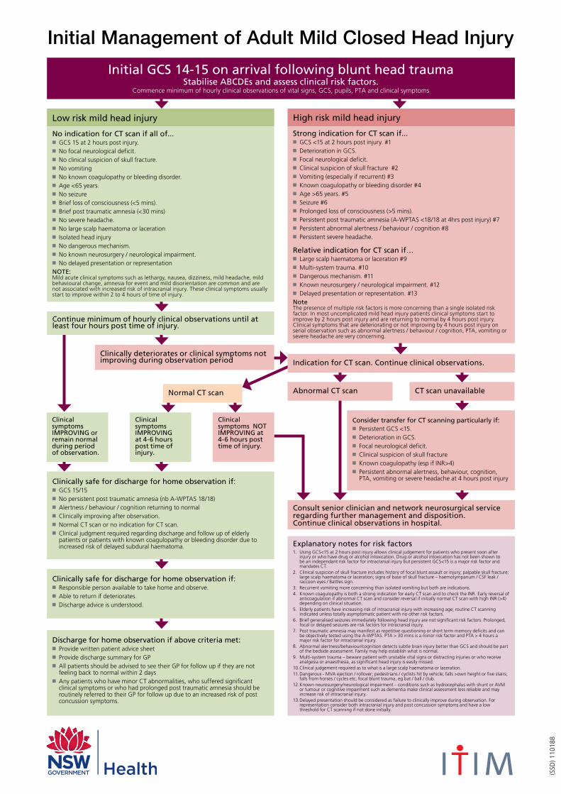
(SSD
) 110
188
Initial Management of Adult Mild Closed Head Injury
Clinically safe for discharge for home observation if:■ Responsible person available to take home and observe.■ Able to return if deteriorates.■ Discharge advice is understood.
Consult senior clinician and network neurosurgical service regarding further management and disposition. Continue clinical observations in hospital.
Consider transfer for CT scanning particularly if:■ Persistent GCS <15.■ Deterioration in GCS.■ Focal neurological deficit.■ Clinical suspicion of skull fracture ■ Known coagulopathy (esp if INR>4)■ Persistent abnormal alertness, behaviour, cognition,
PTA, vomiting or severe headache at 4 hours post injury
Abnormal CT scan CT scan unavailable
Clinical symptoms IMPROVING or remain normal during period of observation.
Clinical symptoms IMPROVING at 4-6 hours post time of injury.
Clinical symptoms NOT IMPROVING at 4-6 hours post time of injury.
Clinically safe for discharge for home observation if:■ GCS 15/15 ■ No persistent post traumatic amnesia (nb A-WPTAS 18/18)■ Alertness / behaviour / cognition returning to normal ■ Clinically improving after observation.■ Normal CT scan or no indication for CT scan.■ Clinical judgment required regarding discharge and follow up of elderly
patients or patients with known coagulopathy or bleeding disorder due to increased risk of delayed subdural haematoma.
Continue minimum of hourly clinical observations until at least four hours post time of injury.
Clinically deteriorates or clinical symptoms not improving during observation period Indication for CT scan. Continue clinical observations.
Normal CT scan
Explanatory notes for risk factors1. Using GCS<15 at 2 hours post injury allows clinical judgement for patients who present soon after
injury or who have drug or alcohol intoxication. Drug or alcohol intoxication has not been shown to be an independant risk factor for intracranial injury but persistent GCS<15 is a major risk factor and mandates CT.
2. Clinical suspicion of skull fracture includes history of focal blunt assault or injury; palpable skull fracture; large scalp haematoma or laceration; signs of base of skull fracture – haemotympanum / CSF leak / raccoon eyes / Battles sign.
3. Recurrent vomiting more concerning than isolated vomiting but both are indications.4. Known coagulopathy is both a strong indication for early CT scan and to check the INR. Early reversal of
anticoagulation if abnormal CT scan and consider reversal if initially normal CT scan with high INR (>4) depending on clinical situation.
5. Elderly patients have increasing risk of intracranial injury with increasing age; routine CT scanning indicated unless totally asymptomatic patient with no other risk factors.
6. Brief generalised seizures immediately following head injury are not significant risk factors. Prolonged, focal or delayed seizures are risk factors for intracranial injury.
7. Post traumatic amnesia may manifest as repetitive questioning or short term memory deficits and can be objectively tested using the A-WPTAS. PTA > 30 mins is a minor risk factor and PTA > 4 hours a major risk factor for intracranial injury.
8. Abnormal alertness/behaviour/cognition detects subtle brain injury better than GCS and should be part of the bedside assessment. Family may help establish what is normal.
9. Multi-system trauma – beware patient with unstable vital signs or distracting injuries or who receive analgesia or anaesthesia, as significant head injury is easily missed.
10. Clinical judgement required as to what is a large scalp haematoma or laceration.11. Dangerous - MVA ejection / rollover; pedestrians / cyclists hit by vehicle; falls >own height or five stairs;
falls from horses / cycles etc; focal blunt trauma, eg bat / ball / club.12. Known neurosurgery/neurological impairment – conditions such as hydrocephalus with shunt or AVM
or tumour or cognitive impairment such as dementia make clinical assessment less reliable and may increase risk of intracranial injury.
13. Delayed presentation should be considered as failure to clinically improve during observation. For representation consider both intracranial injury and post concussion symptoms and have a low threshold for CT scanning if not done initially.
Low risk mild head injury
No indication for CT scan if all of...■ GCS 15 at 2 hours post injury.■ No focal neurological deficit.■ No clinical suspicion of skull fracture. ■ No vomiting ■ No known coagulopathy or bleeding disorder.■ Age <65 years. ■ No seizure■ Brief loss of consciousness (<5 mins). ■ Brief post traumatic amnesia (<30 mins)■ No severe headache.■ No large scalp haematoma or laceration■ Isolated head injury ■ No dangerous mechanism.■ No known neurosurgery / neurological impairment.■ No delayed presentation or representationNOTE:Mild acute clinical symptoms such as lethargy, nausea, dizziness, mild headache, mild behavioural change, amnesia for event and mild disorientation are common and are not associated with increased risk of intracranial injury. These clinical symptoms usually start to improve within 2 to 4 hours of time of injury.
Discharge for home observation if above criteria met:■ Provide written patient advice sheet■ Provide discharge summary for GP■ All patients should be advised to see their GP for follow up if they are not
feeling back to normal within 2 days■ Any patients who have minor CT abnormalities, who suffered significant
clinical symptoms or who had prolonged post traumatic amnesia should be routinely referred to their GP for follow up due to an increased risk of post concussion symptoms.
High risk mild head injury
Strong indication for CT scan if...■ GCS <15 at 2 hours post injury. #1■ Deterioration in GCS.■ Focal neurological deficit.■ Clinical suspicion of skull fracture #2■ Vomiting (especially if recurrent) #3■ Known coagulopathy or bleeding disorder #4■ Age >65 years. #5■ Seizure #6■ Prolonged loss of consciousness (>5 mins). ■ Persistent post traumatic amnesia (A-WPTAS <18/18 at 4hrs post injury) #7■ Persistent abnormal alertness / behaviour / cognition #8■ Persistent severe headache.
Relative indication for CT scan if…■ Large scalp haematoma or laceration #9■ Multi-system trauma. #10■ Dangerous mechanism. #11■ Known neurosurgery / neurological impairment. #12■ Delayed presentation or representation. #13Note The presence of multiple risk factors is more concerning than a single isolated risk factor. In most uncomplicated mild head injury patients clinical symptoms start to improve by 2 hours post injury and are returning to normal by 4 hours post injury. Clinical symptoms that are deteriorating or not improving by 4 hours post injury on serial observation such as abnormal alertness / behaviour / cognition, PTA, vomiting or severe headache are very concerning.
Initial GCS 14-15 on arrival following blunt head traumaStabilise ABCDEs and assess clinical risk factors.
Commence minimum of hourly clinical observations of vital signs, GCS, pupils, PTA and clinical symptoms
Closed Head Injury in Adults - Initial Management
PROCEDURES
PD2012_013 Issue date: February 2012
Attachment 5: Implementation checklist
LHD/Facility:
Assessed by: Date of Assessment:
IMPLEMENTATION REQUIREMENTS Not commenced
Partial compliance Full compliance
1. Evidence of documented local protocols based on the key principles for head injury management.
Notes:
2. Regular education of staff in the use of the clinical guidelines and locally developed protocols.
Notes:
3. Communication process in place to ensure all clinical staff are aware of guidelines for head injury management.
Notes:
Related Documents