Closed Chest Drainage Systems 26 SKILLS AND PROCEDURES Skill 26-1 Managing Closed Chest Drainage Systems, p. 659 Skill 26-2 Assisting with Removal of Chest Tubes, p. 668 Skill 26-3 Autotransfusion of Chest Tube Drainage, p. 671 MEDIA RESOURCES * ©VOlve http://evolve.elsevier.com/Perry/skills Review Questions Audio Glossary • NSO] Nurs ' n 9 Skills Online KEY TERMS Air leak Atmospheric pressure Chest tube Hemothorax Intrapleural pressure Mediastinal shift Negative pressure Parietal pleura Pneumothorax Positive pressure Subcutaneous emphysema Tension pneumothorax Tidaling Visceral pleura Mastery of content in this chapter will enable the nurse to: • Explain the physiology of normal respiration. • List three common sites for chest tube placement. • List three conditions requiring chest tube insertion. • Describe closed chest drainage systems: water-seal and waterless systems. • Descnbe principles and mechanisms of chest tube suction. • Discuss measures to maintain patient safety during chest tube insertion, maintenance, and removal. • Describe methods of troubleshooting chest tube systems. • Discuss the nursing principles in caring for patients with chest tubes. • Describe autotransfusion. T he chest cavity is a closed structure bound by muscle, bone, connective tissue, vascular structures, and the dia- phragm. This cavity has three distinct sections, each sealed from the others: one section for each lung and a third section for the mediastinum, which surrounds structures such as the heart, esophagus, trachea, and great vessels. The lungs are covered with a membrane called the visceral pleura. The interior chest wall is lined with a membrane called the parietal pleura (Fig. 26-1). The space between the visceral and parietal pleura is called the pleural space and is filled with approximately 7-to-20 mL of lubricating fluid to help the pleura slide during respiration (Twedell, 2009). During inspi- ration the intercostal muscles pull outward, and the diaphragm contracts and pulls downward, thereby increasing the size of the chest cavity. As a result an increase in the amount of negative pressure (vacuum effect) is exerted in the intrapleural space. During inspiration increased negative pressure pulls the lungs against the expanded chest cavity, increasing their size. The expanding lungs cause the intrapulmonic (alveolar) pres- sure to fall lower than atmospheric pressure, thus increasing the negative pressure within the lungs. This change in pressure causes air to rush into the lungs until the intrapulmonic pres- sure is equal to the pressure in the atmosphere. When the chest cavity stops expanding and the lungs are full of air, the respiratory muscles and diaphragm relax and return the chest cavity to its resting stage. Expiration (exhalation) is a passive process that results from relaxation of the inspiratory muscles that decrease the space in the chest cavity. Trauma, disease, or surgery can result in air, blood, pus, or lymph fluid leaking into the intrapleural space, creating a positive pressure that collapses lung tissue (Durai et al., 2010). Small leaks (24% or less) are sometimes absorbed spontane- ously and may not require a chest tube. The usual intervention for larger leaks is a chest tube to remove air and fluid from the pleural space, prevent air and/or fluid from reentering the pleural space, and reestablish normal intrapleural and intra- pulmonary pressures.

Welcome message from author
This document is posted to help you gain knowledge. Please leave a comment to let me know what you think about it! Share it to your friends and learn new things together.
Transcript
Closed Chest Drainage Systems
26
SKILLS AND PROCEDURES Skill 26-1 Managing Closed Chest Drainage Systems, p. 659
Skill 26-2 Assisting with Removal of Chest Tubes, p. 668
Skill 26-3 Autotransfusion of Chest Tube Drainage, p. 671
MEDIA RESOURCES
* ©VOlve http://evolve.elsevier.com/Perry/skills
Review Questions Audio Glossary
• NSO] Nurs'n9 Skills Online
KEY TERMS Air leak Atmospheric pressure Chest tube Hemothorax
Intrapleural pressure Mediastinal shift Negative pressure Parietal pleura
Pneumothorax Positive pressure Subcutaneous emphysema
Tension pneumothorax Tidaling Visceral pleura
Mastery of content in this chapter will enable the nurse to: • Explain the physiology of normal respiration. • List three common sites for chest tube placement. • List three conditions requiring chest tube insertion. • Describe closed chest drainage systems: water-seal and
waterless systems. • Descnbe principles and mechanisms of chest tube
suction.
• Discuss measures to maintain patient safety during chest tube insertion, maintenance, and removal.
• Describe methods of troubleshooting chest tube systems.
• Discuss the nursing principles in caring for patients with chest tubes.
• Describe autotransfusion.
The chest cavity is a closed structure bound by muscle, bone, connective tissue, vascular structures, and the dia
phragm. This cavity has three distinct sections, each sealed from the others: one section for each lung and a third section for the mediastinum, which surrounds structures such as the heart, esophagus, trachea, and great vessels.
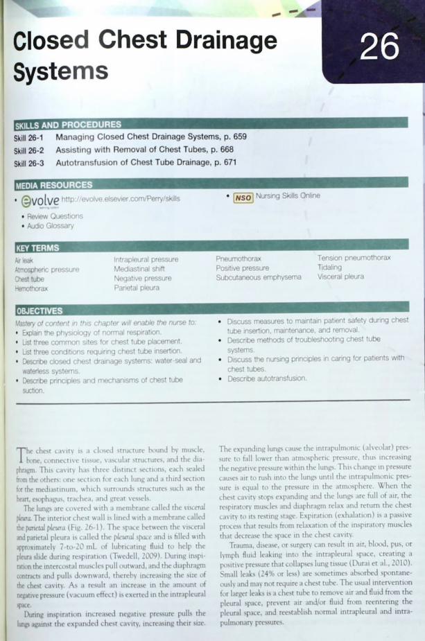
The lungs are covered with a membrane called the visceral
pleura. The interior chest wall is lined with a membrane called the parietal pleura (Fig. 26-1). The space between the visceral and parietal pleura is called the pleural space and is filled with approximately 7-to-20 mL of lubricating fluid to help the pleura slide during respiration (Twedell, 2009). During inspiration the intercostal muscles pull outward, and the diaphragm contracts and pulls downward, thereby increasing the size of the chest cavity. As a result an increase in the amount of negative pressure (vacuum effect) is exerted in the intrapleural
space. During inspiration increased negative pressure pulls the
lungs against the expanded chest cavity, increasing their size.
The expanding lungs cause the intrapulmonic (alveolar) pressure to fall lower than atmospheric pressure, thus increasing the negative pressure within the lungs. This change in pressure causes air to rush into the lungs until the intrapulmonic pressure is equal to the pressure in the atmosphere. When the chest cavity stops expanding and the lungs are full of air, the respiratory muscles and diaphragm relax and return the chest cavity to its resting stage. Expiration (exhalation) is a passive process that results from relaxation of the inspiratory muscles that decrease the space in the chest cavity.
Trauma, disease, or surgery can result in air, blood, pus, or lymph fluid leaking into the intrapleural space, creating a positive pressure that collapses lung tissue (Durai et al., 2010). Small leaks (24% or less) are sometimes absorbed spontaneously and may not require a chest tube. The usual intervention for larger leaks is a chest tube to remove air and fluid from the pleural space, prevent air and/or fluid from reentering the pleural space, and reestablish normal intrapleural and intra-
pulmonary pressures.
656 C H A P T E R 2 6 Closed Chest Drainage Systems
Lung
Pleural space
Rib cage
Parietal pleura
Visceral pleura
FIG 26-1 Partial structures of lungs.
A number of clinical conditions such as cancer, infection, pancreatitis, connective tissue disease, autoimmune diseases, asbestos exposure, certain drugs, or collagen vascular diseases increase pleural fluid entry or decrease fluid exit from the lung. This is called a pleural effusion; and, when present, a patient usually needs a diagnostic thoracentesis and pleural fluid analysis to determine the cause of the exudate (see Chapter 44) (Twedell, 2009). Patients usually need one or more chest tubes to promote drainage of the excess fluid and lung expansion (Mertin et al., 2009; Twedell, 2009).
A pneumothorax is collapse of the lung caused by a collection of air in the pleural space. The loss of negative intrapleural pressure causes the lung to collapse. A variety of mechanisms cause a pneumothorax. A traumatic pneumothorax develops as a result of penetrating chest trauma such as a stabbing (open) or the chest striking the steering wheel in an automobile accident (closed). A spontaneous or primary pneumothorax sometimes occurs from the rupture of a small bleb (blister) on the surface of the lung or an invasive procedure such as insertion of a subclavian intravenous (IV) line. Secondary pneumothorax occurs because of underlying disease such as emphysema. A patient with a pneumothorax usually feels sharp chest pain that worsens on inspiration or coughing because atmospheric air irritates the parietal pleura. As a pneumothorax worsens, a patient will experience easy fatigue, a rapid heart rate, and low blood pressure (U.S. National Library of Medicine, 2011).
A tension pneumothorax, a life-threatening situation, occurs from rupture in the pleura when air accumulates in the pleural space more rapidly than it is removed. The pleural space functions as a one-way valve, causing an increase in the amount of air and pressure. If left untreated, the lung on the affected side collapses; and the mediastinum shifts to the opposite (unaffected side), leading to tracheal deviation, reduced venous return, and subsequent decrease in cardiac output. Tracheal deviation is a late sign and may be absent in some cases (Bethel, 2008). A patient has sudden chest pain, a fall in blood pressure, tachycardia, acute pleuritic pain, diaphoresis, dry cough, and cardiopulmonary arrest can occur. Patients with chest trauma, fractured ribs, invasive thoracic bedside procedures (e.g., insertion of central lines), and those on high-pressure mechanical ventilation are at risk for tension pneumothorax (Bethel, 2008). If emergent treatment is required, a needle decompression is achieved with a large-gauge needle (14 or 16 gauge) inserted into the second intercostal space, midclavicular line. A "hissing" sound is noted, followed by a rapid stabilization of the patients vital signs and respiratory status (Briggs, 2010).
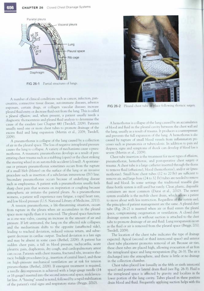
FIG 26-2 Pleural chest tube iif place following thoracic surgery
A hemothorax is collapse of the lung caused by an accumulation of blood and fluid in the pleural cavity between the chest wall and the lung, usually as a result of trauma. It produces a counterpressure and prevents the full expansion of the lung. A hemothorax is also caused by rupture of small blood vessels from inflammatory processes such as pneumonia or tuberculosis. In addition to pain and dyspnea, signs and symptoms of shock can develop it blot*! loss is severe (Mertin et al., 2009).
Chest tube insertion is the treatment for most types of effusions, pneumothorax, hemothorax, and postoperative chest surgery or trauma. A chest tube is a large catheter inserted through the thorax to remove fluid (effusions), blood (hemothorax), and/or air (pneumothorax). Small-bore chest tubes (12 to 20 Fr) are sufficient to remove air, and large-bore (24 to 32 Fr) tubes are needed to remove fluid and blood. In some settings the traditional reusable glass three-bottle system is still used but rarely. Clear, plastic, disposable containers are most common (Durai et al., 2010). The newest system available is the mobile chest drain, which allows a patient to move about with less restriction. Regardless of the system used, the principles of patient management are the same. A pleural chest tube (Fig. 26-2) is inserted when air or fluid enters the pleural space, compromising oxygenation or ventilation. A closed chest drainage system with or without suction is attached to the chest tube to promote drainage of air and fluid. Lung reexpansion occurs as the fluid or air is removed from the pleural space (Briggs, 2010; Twedell, 2009).
The location of the chest tube indicates the type of drainage expected. Apical (second or third intercostal space) and anterior chest tube placement promotes removal of air. Because air rises, these chest tubes are placed high, allowing evacuation of air from the intrapleural space and lung reexpansion (Fig. 26-3). The air is discharged into the atmosphere, and there is little or no drainage in the collection chamber.
Chest tubes placed low (usually in the fifth or sixth intercostal space) and posterior or lateral drain fluid (see Fig. 26-3). Fluid in the intrapleural space is affected by gravity and localizes in the lower portion of the lung cavity. Tubes placed in these positions drain blood and fluid. Frequently applying suction helps with this
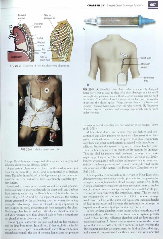
FIG 26-3 Diagram of sites for chest tube placement.
FIG 26-4 Mediastinal chest tube.
drainage. Fluid drainage is expected after open-chest surgery and with some chest trauma (Briggs, 2010).
A mediastinal chest tube is placed in the mediastinum, just below the sternum (Fig. 26-4), and is connected to a drainage system. This tube drains blood or fluid, preventing its accumulation around the heart. A mediastinal tube is commonly used after open-heart surgery.
Occasionally in emergency situations and for a small pneumothorax a catheter is inserted through the chest wall, and a rubber flutter one-way valve (e.g., a Heimlich valve) is attached to the catheter (Fig. 26-5, A and B). As a patient exhales, the positive pressure generated by the air leaving the chest enters the tubing, causing the valve to open so air is released. During inspiration the tube collapses on itself, preventing air from reentering the chest. No drainage chamber is used with this device; therefore it is not used when patients need fluid drained such as from a hemothorax or a pleural effusion (Lewis et al., 2011).
Smaller "pigtail catheters" are also used and are less traumatic than the large-bore tubes. In addition, if they occlude the health care provider can irrigate them with sterile water. However, because these tubes are small, the size of the tube lumen does not promote
C H A P T E R 2 6 Closed Chest Drainage Systems
B
FIG 26-5 A, Heimlich chest drain valve is a specially designed flutter valve that is used in place of a chest drainage unit for small uncomplicated pneumothorax with little or no drainage and no need for suction. The valve allows for escape of air but prevents reentry of air into the pleural space. (Image courtesy Becton, Dickenson and Company, Franklin Lakes, New Jersey. All rights reserved.) B, Placement of valve between chest tube and drainage bag, which can be worn under clothing.
drainage of blood, and they are not used for chest trauma (Lewis
et al., 2011). Mobile chest drains are devices that are lighter and self-
contained and allow patients to move with less restriction. As a result there is a decreased risk for deep vein thrombosis, pulmonary embolism, and other complications associated with immobility. In addition, because the system is lighter, a patient has less pain. These mobile systems rely on gravity or dry suction for drainage. They are ideal for patients with persistent drainage or air leaks requiring prolonged need for a chest tube (Varela et al., 2010). Patients who require a mobile chest drainage system at home need sufficient discharge planning and patient teaching directed toward safe operation of the system (see Home Care Considerations for
Skill 26-1). The disposable systems such as an Atrium or Pleur-Evac chest
drainage system are one-piece molded plastic units that provide for a single- or multiple-chamber closed drainage system (Fig. 26-6). A single-chamber system allows air from a pneumothorax to bubble out of the water seal and escape through the air outlet while preventing air from reentering the intrapleural space. This system is not recommended for the evacuation of fluid because drainage would raise the level of the water-seal liquid. An increased height of fluid in the water seal increases the resistance to drainage on expiration and eventually stops the drainage entirely.
A two- or three-chamber system drains both a hemothorax and a pneumothorax effectively. The two-chamber system permits liquid to flow into the collection chamber, and air flows into the water-seal chamber. A three-chamber system promotes the drainage of fluid and air with controlled suction. In both systems the first chamber provides a compartment for fluid or blood drainage, and a second compartment for either a water seal or a one-way
Visceral pleura
Pleural space
Site to drain fluid
Parietal Site to remove air
Chest tube
Heimlich valve
Drainage bag
658 C H A P T E R 2 6 Closed Chest Drainage Systems
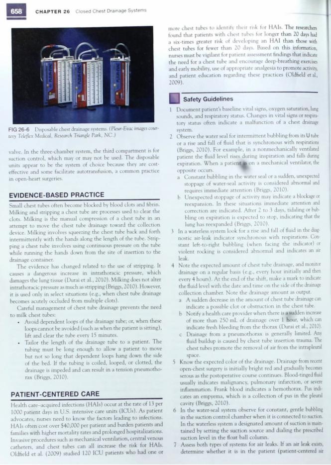
FIG 26-6 Disposable chest drainage systems. (Pleur-Evac images courtesy Teleflex Medical, Research Triangle Park, NC.)
valve. In the three-chamber system, the third compartment is for suction control, which may or may not be used. The disposable units appear to be the system of choice because they are cost-effective and some facilitate autotransfusion, a common practice in open-heart surgeries.
EVIDENCE-BASED PRACTICE
Small chest tubes often become blocked by blood clots and fibrin. Milking and stripping a chest tube are processes used to clear the clots. Milking is the manual compression of a chest tube in an attempt to move the chest tube drainage toward the collection device. Milking involves squeezing the chest tube back and forth intermittently with the hands along the length of the tube. Stripping a chest tube involves using continuous pressure on the tube while running the hands down from the site of insertion to the drainage container.
The evidence has changed related to the use of stripping. It causes a dangerous increase in intrathoracic pressure, which damages the lung tissue (Durai et al., 2010). Milking does not alter intrathoracic pressure as much as stripping (Briggs, 2010). However, it is used only in select situations (e.g., when chest tube drainage becomes acutely occluded from multiple clots).
Careful management of chest tube drainage prevents the need to milk chest tubes:
• Avoid dependent loops of the drainage tube; or, when these loops cannot be avoided (such as when the patient is sitting), lift and clear the tube every 15 minutes.
• Tailor the length of the drainage tube to a patient. The tubing must be long enough to allow a patient to move but not so long that dependent loops hang down the side of the bed. If the tubing is coiled, looped, or clotted, the drainage is impeded and can result in a tension pneumothorax (Briggs, 2010).
PATIENT-CENTERED CARE
Health care—acquired infections (HAls) occur at the rate of 1) per 1000 patient days in U.S. intensive care units (ICUs). As patient advocates, nurses need to know the factors leading to infections. HAIs often cost over $40,000 per patient and burden patients and families with higher mortality rates and prolonged hospitalizations. Invasive procedures such as mechanical ventilation, central venous catheters, and chest tubes can all increase the risk for HAIs. Oldfield et al. (2009) studied 120 ICU patients who had one or
more chest tubes to identity their risk tor HAIs. The researchers found that patients with chest tubes tor longer than 20 days had a six-times greater risk of developing an HA1 than those with chest tubes for fewer than 20 days. Based on this information, nurses must be vigilant for patient assessment findings that indicate the need for a chest tube and encourage deep-breathing exercises and early mobility, use of appropriate analgesia to promote activity, and patient education regarding these practices (Oldfield et al., 2009).
Safety Guidelines
1 Document patient's baseline vital signs, oxygen saturation, lung sounds, and respiratory status. Changes in vital signs or respiratory status often indicate a malfunction of a chest drainage system.
2 Observe the water seal for intermittent bubbling from its U tube or a rise and fall of fluid that is synchronous with respirations (Briggs, 2010). For example, in a nonmechanically ventilated patient the fluid level rises during inspiration and falls during expiration. When a patient 16 on a mechanical ventilator, the opposite occurs. a Constant bubbling in the water seal or a sudden, unexpected
stoppage of water-seal activity is considered abnormal and requires immediate attention (Briggs, 2010).
b Unexpected stoppage of activity may indicate a blockage or reexpansion. In these situations immediate attention and correction are indicated. After 2 to 3 days, tidaling or bubbling on expiration is expected to stop, indicating that the lung has reexpanded (Briggs, 2010).
3 In a waterless system look for a rise and fall of fluid in the diagnostic air-leak indicator synchronous with respirations. Gin-stant left-to-right bubbling (when facing the indicator) or violent rocking is considered abnormal and indicates an ait leak.
4 Note the expected amount of chest tube drainage, and monitor drainage on a regular basis (e.g., every hour initially and then every 4 hours). At the end of the shift, make a mark to indicate the fluid level with the date and time on the side of the drainage collection chamber. Note the drainage amount as output. a A sudden decrease in the amount of chest tube drainage can
indicate a possible clot or obstruction in the chest tube, b Notify a health care provider when there is a sudden increase
of more than 250 mL of drainage over 1 hour, which can indicate fresh bleeding from the thorax (Durai et al., 2010).
c Drainage from a pneumothorax is generally limited. Any fluid buildup is caused by chest tube insertion trauma. The chest tubes promote the removal of air from the intrapleural space.
5 Know the expected color of the drainage. Drainage from recent open-chest surgery is initially bright red and gradually becomes serous as the postoperative course continues. Blood-tinged fluid usually indicates malignancy, pulmonary infarction, or severe inflammation. Frank blood indicates a hemothorax. Pus indicates an empyema, which is a collection of pus in the pleural cavity (Briggs, 2010).
6 In the water-seal system observe for constant, gentle bubbling in the suction control chamber when it is connected to suction. In the waterless system a designated amount of suction is maintained by setting the suction source and dialing the prescribed suction level in the float ball column.
7 Assess both types of systems for air leaks. If an air leak exists, determine whether it is in the patient (patient-centered air

SKILL 26-1 Managing Closed Chest Drainage Systems 659
leak) or in the chest tube system (system-centered air leak). To rule out an air leak as patient-centered, you assess the patient's respiratory status. Document and report any changes in lung sounds, pulse oximetry, respiratory rate, or mentation.
Remember that continuous bubbling in the water-seal chamber with an absence of bubbles in the suction control chamber indicates that there is a leak in the system (Briggs, 2010). Ensure that all tubing connections are tight.
SKILL 26-1 Managing Closed Chest Drainage Systems
A/SO | Chest Tubes Module / Lessons 1 and 3 If the suction source delivers more negative pressure than the
There are two types of commercial drainage systems: the water-seal >uction control chamber water level allows> there is ^danger and the waterless systems (Table 26-1). This skill reviews the b,ecause atmosPher'c air is pulled into the suct.on control chamber
nursing responsibilities and interventions related to the safe man- throu«h an ,nletl causin* the excess suction to dlss'Pate- The extra
agement of chest tubes. Review the roles and responsibilities of the health care provider for chest tube placement (Table 26-2).
Water-Seal Systems NSO Chest Tubes Module / Lesson 2
Two-Chamber Water-Seal System On expiration fluid or air is forced out of the intrapleural space. Suction pulls air or fluid through the chest tube into the drainage collection chamber. On entering the drainage collection chamber, this fluid or air displaces the air present in the chamber by pushing it through the water seal and out of the system into the atmosphere. The water-seal chamber is left open to air to drain. If the tubing is clamped, there is no mechanism for air to vent. To maintain the water-seal system, the chest tube system must remain upright. When it is tipped or overturned, the water seal is disrupted. Three-Chamber Water-Seal System If suction is used, the three-chamber water-seal system (Fig. 26-7) is set up with the suction control chamber added. A prescribed amount of sterile fluid (e.g., 20 cm of water) is poured into the suction control chamber, which is then attached to a suction source by tubing. The amount of sterile water added depends on manufacturer recommendations. The chamber is filled to the set volume for the prescribed amount of suction. Sterile water is added several times a day because of evaporation. As the fluid level decreases, the amount of suction also declines. The wall or portable suction device is turned up until the water in the suction control bottle exhibits a continuous, gentle bubbling. This provides the prescribed amount of suction (negative pressure).
FIG 26-7 Disposable water-seal chest drainage system with suction. (Used with permission, Atrium Medical Corp.)
TABLE 26-1 Comparison of Chest Tube Drainage Systems
Drainage System Type Function Advantage Disadvantage
Water-seal system (see Fig. 26-7)
Two-chamber provides one-way valve for chest drainage.
Water seal prevents reentry of air into lung.
Three-chamber adds a chamber to aid evacuation of chest drainage.
Easy set-up and use Cost-effective
System must be kept upright to maintain seal.
Drainage chamber may fill up quickly if patient has large amount of drainage.
Sterile water must be added several times a day to maintain suction and water seal because of evaporation.
Waterless system (see Fig. 26-8)
Also provides three chambers, but no water is required to establish a seal.
Water seal maintained even if system is knocked over
More space provided for drainage
Water must be added to system if patient requires evaluation of an air leak.
Dry suction system (see Fig. 26-9)
Also provides three chambers, but no water is required to establish a seal.
Easy set up Quiet operation Can be used when higher levels
of suction are required
Sterile water must be added to system if patient requires suction.

CHAPTER 26 Closed Chest Drainage Systems
.ur pulled into the chamber causes vigorous bubbling. It this occurs, lower the suction source setting to reduce noise and evaporation of the fluid. The absence of bubbling indicates that no suction is being exerted into the system. Raise the suction setting to restore
gentle bubbling. The middle chamber of a traditional chest drainage system is
the water seal. The water seal allows air to exit from the pleural space on exhalation and prevent it from entering the pleural cavity or mediastinum on inhalation. When the appropriate amount of sterile water is added, a 2-cm water seal is established. To maintain effective water seal the chest drainage unit must remain upright, and you must monitor the water level in the water-seal chamber to check for evaporation. Bubbling in the water-seal chamber indicates an air leak.
Waterless Systems Two-Chamber Waterless System The principles of the waterless system are similar to those of the water-seal system except that fluid is not required for setup. Because water is not used, accidentally tipping the system does not compromise the patient's condition.
The water seal is replaced by a one-way valve (Fig. 26-8) located near the top of the system. Most of the container serves as the drainage chamber. The suction chamber does not depend on water. Instead it contains a float ball, which is set by a suction control dial after the suction source is turned on. A diagnostic air-leak indicator is located on the face of the unit. It requires the addition of 15 mL of fluid for visualization. The function of the indicator is to identify one of the following:
TABLE 26-2 Process for Insertion of Chest Tubes
Role Purpose
Explain purpose, procedure, and possible complications to patient and have patient sign consent form.
Provides informed consent.
Have pain medication available to administer before or immediately after chest tube insertion as appropriate according to patient's condition.
if^thr Analgesia improves patient comfoff throughout procedure and helps patient take appropriate deep breaths to promote lung reexpansion and drainage of fluid in pleural space.
Perform hand hygiene. Clean chest wall with antiseptic. Reduces transmission of microorganisms.
Apply mask and gloves. Maintains surgical asepsis.
Drape area of chest tube insertion with sterile towels. Inject local anesthetic and allow time to take effect.
Maintains surgical asepsis. Decreases pain during procedure.
Make a small incision over the rib space where tube is to be inserted. Thread a clamped chest tube through the incision. Health care provider clamps chest tube until system is connected to water seal.
Inserts chest tube into intrapleural space. Clamping prevents entry of atmospheric air into chest and worsening of pneumothorax.
Suture chest tube in place if suturing is policy or health care provider preference.
Secures chest tube in place.
Cover chest tube insertion site with sterile 4 x 4-inch gauze and large dressing to form an occlusive dressing supported with an elastic bandage (Elastoplast). Stenle petrolatum gauze is used around the tube.
Water-Seal System Remove connector cover from patient's end of chest drainage
tubing with sterile technique. Secure drainage tubing to chest tube and drainage system.
Water-Seal Suction Connect system to suction or supervise a nurse connecting it to
suction if suction is to be used.
Waterless System Remove connector cover from patient's end of chest drainage
tubing with sterile technique. Secure drainage tubing to chest tube and drainage system.
Waterless Suction Turn on suction source. Set float ball level to prescribed setting.
Health care provider or nurse adds sterile water or normal saline to diagnostic indicator.
Holds chest tube in place and occludes site around it. Helps stabilize chest tube and holds dressing tightly in place. Sterile petrolatum gauze helps prevent air leak.
Health care provider is responsible for making certain that system is set up properly, proper amount of water is in the water seal, dressing is secure, and chest tube is securely connected to drainage system.
Health care provider is responsible for determining and checking amount of fluid that is to be added to suction control chamber and prescribing suction setting.
Health care provider is responsible for making certain that system is set up properly and chest tube is securely connected to drainage system.
Health care provider is responsible for prescribing level ot float ball and suction setting.
Allows quick assurance that system is functioning properly. Connects chest tube to drainage.
Unclamp chest tube. Verifies correct chest tube placement.
In both systems health care provider orders and reviews chest x-ray film studies.
S K I L L 2 6 - 1 Managing Closed Chest Drainage Systems 661
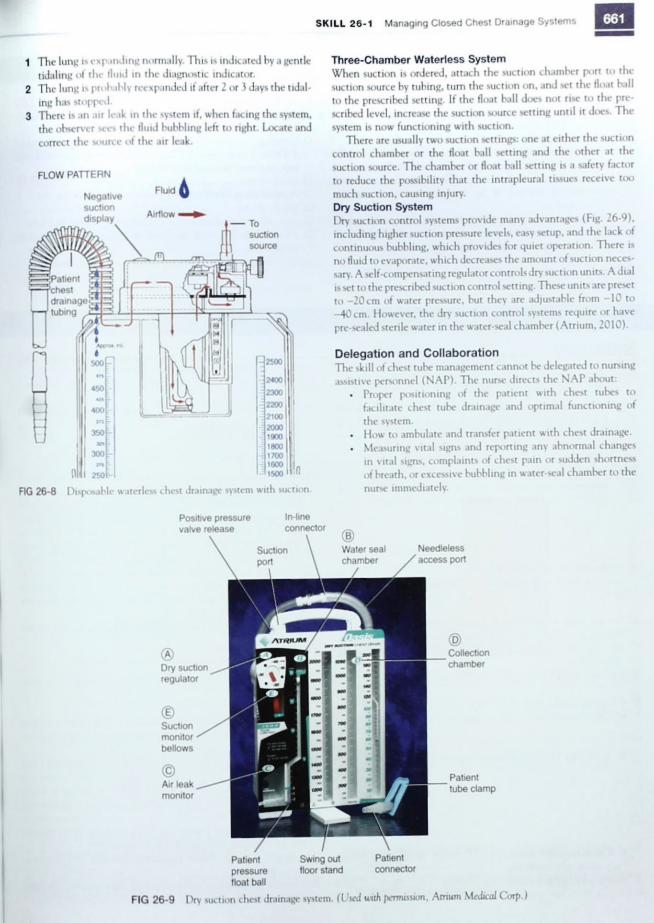
1 The lung is expanding normally. This is indicated by a gentle tidaling of the fluid in the diagnostic indicator.
2 The lung is pn ihahly reexpanded if after 2 or 3 days the tidaling has stopped.
3 There is an air leak in the system if, when facing the system, the observer sees the fluid bubbling left to right. Locate and correct the source of the air leak.
Three-Chamber Waterless System When suction is ordered, attach the suction chamber port to the suction source hy tubing, turn the suction on, and set the float hall to the prescribed setting. If the float hall does not rise to the prescribed level, increase the suction source setting until it does. The system is now functioning with suction.
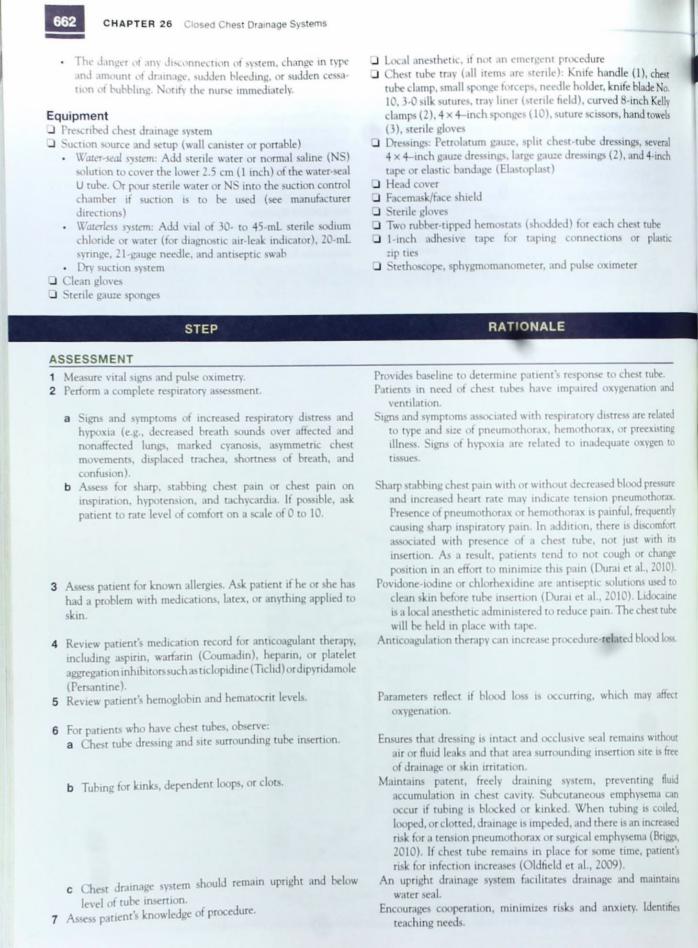
There are usually two suction settings: one at either the suction control chamber or the float hall setting and the other at the suction source. The chamber or float hall setting is a safety factor to reduce the possibility that the intrapleural tissues receive too much suction, causing injury. Dry Suction System Dry suction control systems provide many advantages (Fig. 26-9), including higher suction pressure levels, easy setup, and the lack of continuous bubbling, which provides for quiet operation. There is no fluid to evaporate, which decreases the amount of suction necessary. A self-compensating regulator controls dry suction units. A dial is set to the prescribed suction control setting. These units are preset to —20 cm of water pressure, but they are adjustable from —10 to -40 cm. However, the dry suction control systems require or have pre-sealed sterile water in the water-seal chamber (Atrium, 2010).
Delegation and Collaboration The skill of chest tube management cannot be delegated to nursing assistive personnel (NAP). The nurse directs the NAP about:
• Proper positioning of the patient with chest tubes to facilitate chest tube drainage and optimal functioning of the system.
• How to ambulate and transfer patient with chest drainage. • Measuring vital signs and reporting any abnormal changes
in vital signs, complaints of chest pain or sudden shortness of breath, or excessive bubbling in water-seal chamber to the nurse immediately.
To suction source
FIG 26-8 Disposable waterless chest drainage system with suction.
FLOW PATTERN
Negative suction display
Fluid
Airflow
Patient Swing out Patient pressure floor stand connector float ball
Positive pressure valve release
In-line connector
Water seal Needleless access port
© Collection chamber
Patient tube clamp
® Dry suction regulator
© Suction monitor bellows
© Air leak monitor
662 C H A P T E R 2 6 Closed Chest Drainage Systems
• The danger ot any disconnection of system, change in type and amount of drainage, sudden bleeding, or sudden cessation of bubbling. Notify the nurse immediately.
Equipment • Prescribed chest drainage system Q Suction source and setup (wall canister or portable)
• Water-seal system: Add sterile water or normal saline (NS) solution to cover the lower 2.5 cm (1 inch) of the water-seal U tube. Or pour sterile water or NS into the suction control chamber if suction is to be used (see manufacturer directions)
. Waterless system: Add vial of 30- to 45-mL sterile sodium chloride or water (for diagnostic air-leak indicator), 20-mL syringe, 21 -gauge needle, and antiseptic swab
• Dry suction system • Clean gloves • Sterile gauze sponges
STEP
ASSESSMENT 1 Measure vital signs and pulse oximetry. 2 Perform a complete respiratory assessment.
a Signs and symptoms of increased respiratory distress and hypoxia (e.g., decreased breath sounds over affected and nonaffected lungs, marked cyanosis, asymmetric chest movements, displaced trachea, shortness of breath, and confusion).
b Assess for sharp, stabbing chest pain or chest pain on inspiration, hypotension, and tachycardia. If possible, ask patient to rate level of comfort on a scale of 0 to 10.
3 Assess patient for known allergies. Ask patient if he or she has had a problem with medications, latex, or anything applied to
skin.
4 Review patient's medication record for anticoagulant therapy, including aspirin, warfarin (Coumadin), heparin, or platelet aggregation inhibitors such as ticlopidine (Ticlid) or dipyridamole
(Persantine). 5 Review patient's hemoglobin and hematocrit levels.
6 For patients who have chest tubes, observe: a Chest tube dressing and site surrounding tube insertion.
b Tubing for kinks, dependent loops, or clots.
c Chest drainage system should remain upright and below
level of tube insertion. 7 Assess patient's knowledge of procedure.
J Local anesthetic, if not an emergent procedure _) Chest tube tray (all items are sterile): Knife handle (1), chest
tube clamp, small sponge forceps, needle holder, knife blade No. 10, 3-0 silk sutures, tray liner (sterile held), curved 8-inch Kelly clamps (2), 4 x 4-inch sponges (10), suture scissors, hand towels (3), sterile gloves
• Dressings: Petrolatum gauze, split chest-tube dressings, several 4 x 4-inch gauze dressings, large gauze dressings (2), and 4-inch tape or elastic bandage (Elastoplast)
• Head cover • Facemask/face shield • Sterile gloves • Two rubber-tipped hemostats (shodded) for each chest tube • 1-inch adhesive tape for taping connections or plastic
zip ties • Stethoscope, sphygmomanometer, and pulse oximeter
RATIONALE
Provides baseline to determine patient's response to chest tube. Patients in need of chest tubes have impaired oxygenation and
ventilation. Signs and symptoms associated with respiratory distress are related
to type and size of pneumothorax, hemothorax, or preexisting illness. Signs of hypoxia are related to inadequate oxygen to tissues.
Sharp stabbing chest pain with or without decreased blood pressure and increased heart rate may indicate tension pneumothorax. Presence of pneumothorax or hemothorax is painful, frequently causing sharp inspiratory pain. In addition, there is discomfort associated with presence of a chest tube, not just with its insertion. As a result, patients tend to not cough or change position in an effort to minimize this pain (Durai et al., 2010).
Povidone-iodine or chlorhexidine are antiseptic solutions used to clean skin before tube insertion (Durai et al., 2010). Lidocaine is a local anesthetic administered to reduce pain. The chest tube will be held in place with tape.
Anticoagulation therapy can increase procedure-related blood loss.
Parameters reflect if blood loss is occurring, which may affect oxygenation.
Ensures that dressing is intact and occlusive seal remains without air or fluid leaks and that area surrounding insertion site is free of drainage or skin irritation.
Maintains patent, freely draining system, preventing fluid accumulation in chest cavity. Subcutaneous emphysema can occur if tubing is blocked or kinked. When tubing is coiled, looped, or clotted, drainage is impeded, and there is an increased risk for a tension pneumothorax or surgical emphysema (Briggs, 2010). If chest tube remains in place for some time, patient's risk for infection increases (Oldfield et al., 2009).
An upright drainage system facilitates drainage and maintains water seal.
Encourages cooperation, minimizes risks and anxiety. Identifies teaching needs.
SKILL 26-1 Managing Closed Chest Drainage Systems 663
STEP RATIONALE
NURSING DIAGNOSES Impaired gas exchange . Anxiety
. Acute pain
Related factors are individualized based on fmcient's condition or needs.
• Risk for Infection
PLANNING Expected outcomes following completion of procedure: . Patient is oriented and less anxious. • Vital signs are stable. . Patient reports no chest pain. . Breath sounds are auscultated in all lobes. Lung expansion
is symmetric, pulse oximetry (SpOi) is stable or improved, and respirations are nonlabored.
• Chest tube remains in place, and chest drainage system remains airtight.
• Gentle tidaling (fluctuations or rocking) is evident in water-seal or diagnostic indicator.
Hypoxia is relieved. Decreased hypoxia improves vital sign measures. Reexpansion of lung reduces chest pain. Reexpansion of lung promotes normal respirations.
Indicates correct placement and patency of chest tube drainage system.
Indicates that system is functioning normally. Reflects changes in intrapleural pressure.
IMPLEMENTATION 1 Identify patient using two identifiers (i.e., name and birthday
or name and account number) according to agency policy. Compare identifiers with information on patient's identification bracelet.
2 Check agency policy and determine whether informed consent is needed.
3 Review health care provider's order for chest tube placement. 4 Perform hand hygiene.
5 Set up water-seal system (or dry system with suction); see manufacturer guidelines. a Obtain chest drainage system. Remove wrappers and
prepare to set up a two- or three-chamber system, b While maintaining sterility of drainage tubing, stand system
upright and add sterile water or NS to appropriate compartments. (1) Two-chamber system (without suction): Add sterile
solution to water-seal chamber (second chamber), bringing fluid to required level as indicated.
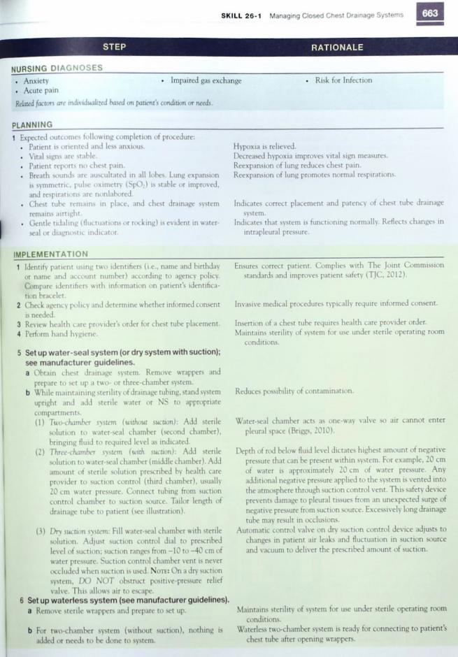
(2) Three-chamber system (uith suction): Add sterile solution to water-seal chamber (middle chamber). Add amount of sterile solution prescribed by health care provider to suction control (third chamber), usually 20 cm water pressure. Connect tubing from suction control chamber to suction source. Tailor length of drainage tube to patient (see illustration).
(3) Dr> suction system: Fill water-seal chamber with sterile solution. Adjust suction control dial to prescribed level of suction; suction ranges from -10 to -40 cm of water pressure. Suction control chamber vent is never occluded when suction is used. NOTE: On a dry suction system, DO NOT obstruct positive-pressure relief valve. This allows air to escape.
6 Set up waterless system (see manufacturer guidelines), a Remove sterile wrappers and prepare to set up.
b For two-chamber system (without suction), nothing is added or needs to be done to system.
Ensures correct patient. Complies with The Joint Commission standards and improves patient safety (TJC, 2012).
Invasive medical procedures typically require informed consent.
Insertion of a chest tube requires health care provider order. Maintains sterility of system for use under sterile operating room
conditions.
Reduces possibility of contamination.
Water-seal chamber acts as one-way valve so air cannot enter pleural space (Briggs, 2010).
Depth of rod below fluid level dictates highest amount of negative pressure that can be present within system. For example, 20 cm of water is approximately 20 cm of water pressure. Any additional negative pressure applied to the system is vented into the atmosphere through suction control vent. This safety device prevents damage to pleural tissues from an unexpected surge of negative pressure from suction source. Excessively long drainage tube may result in occlusions.
Automatic control valve on dry suction control device adjusts to changes in patient air leaks and fluctuation in suction source and vacuum to deliver the prescribed amount of suction.
Maintains sterility of system for use under sterile operating room conditions.
Waterless two-chamber system is ready for connecting to patient's chest tube after opening wrappers.
564
S T E P
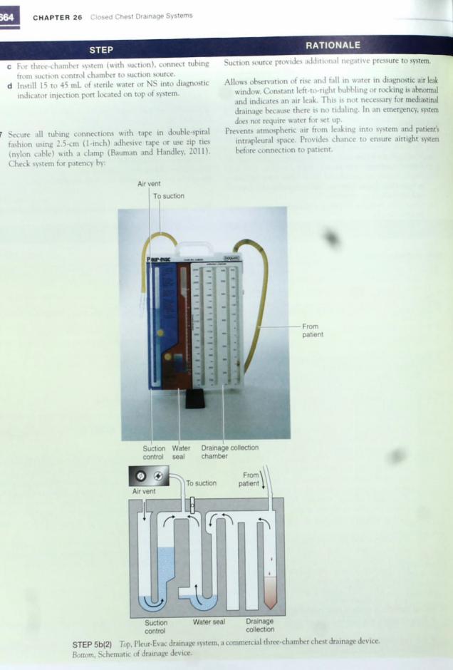
c For three-chamber system (with suction), connect tubing tram suction control chamber to suction source,
d Instill 15 to 45 mL of sterile water or NS into diagnostic indicator injection port located on top of system.
Secure all tubing connections with tape in double-spiral fashion using 2.5-cm (1-inch) adhesive tape or use zip ties (nylon cable) with a clamp (Bauman and Handley, 2011). Check system for patency by:
R A T I O N A L E
Suction source provides additional negative pressure to system.
Allows observation of rise and fall in water in diagnostic air leak window. Constant left-to-right bubbling or rocking is abnormal and indicates an air leak. This is not necessary for mediastinal drainage because there is no tidaling. In an emergency, system does not require water for set up.
Prevents atmospheric air from leaking into system and patient's intrapleural space. Provides chance to ensure airtight system before connection to patient.
Air vent
To suction
From patient
Suction Water Drainage collection control seal chamber
Air vent
Pfl To suction
FronO patient
m rm\ V nA
Suction control
Water seal Drainage collection
STEP 5b(2) Top, Pleur-Evac drainage system, a commercial three-chamber chest drainage device.
Bottom, Schematic of drainage device.
SKILL 26-1 Managing Closed Chest Drainage Systems
RATIONALE STEP
a Clamping drainage tubing that will connect to patient's chest tube.
b Connecting tubing from float ball chamber to suction
source. c Turning on suction to prescribed level.
8 Turn off suction source and unclamp drainage tubing before connecting patient to system. Make a second check to be sure that drainage tubing is not excessively long. Suction source is turned on again after patient is connected.
9 Administer premedication such as sedatives or analgesics as
ordered.
Having patient connected to suction when it is initiated could damage pleural tissues from sudden increase in negative pressure. Tubing that is coiled or looped may become clotted and cause a tension pneumothorax.
Reduces patient anxiety and pain during procedure.
Clinical Decision Point During procedure carefully monitor patient for changes in level of sedation.
10 Provide psychological support to patient (Durai et al., 2010). a Reinforce preprocedure explanation. b Coach and support patient throughout procedure.
11 Perform hand hygiene and apply clean gloves. Position patient for tube insertion so side in which tube is to be inserted is accessible to health care provider.
12 Help health care provider with chest tube insertion by providing needed equipment and local analgesic. Health care provider will anesthetize skin over insertion site, make a small skin incision, insert a clamped tube, suture it in place, and apply occlusive dressing.
13 Help health care provider attach drainage tube to chest tube; remove clamp. Turn on suction to prescribed level.
14 Tape or zip-tie all connections between chest tube and drainage tube. (NOTE: Chest tube is usually taped by health care provider at time of tube placement; check agency policy).
15 Check systems for proper functioning. Health care provider will order a chest x-ray film.
16 After tube placement position patient: a Use semi-Fowler's or high-Fowler's position to evacuate air
(pneumothorax), b Use high-Fowler's position to drain fluid (hemothorax).
17 Check patency of air vents in system. a Water-seal vent must have no occlusion, b Suction control chamber vent is not occluded when suction
is used. C Waterless systems have relief valves without caps.
18 Position excess tubing horizontally on mattress next to patient. Secure with clamp provided so it does not obstruct tubing.
19 Adjust tubing to hang in straight line from chest tube to
drainage chamber.
Reduces patient anxiety and helps complete procedure efficiently.
Reduces transmission of microorganisms.
Ensures smooth insertion.
Connects drainage system and suction (if ordered) to chest tube.
Secures chest tube to drainage system and reduces risk for air leak that causes breaks in airtight system.
Verifies intrapleural placement of tube.
Permits optimum drainage of fluid and/or air.
Permits displaced air to pass into atmosphere. Provides safety factor of releasing excess negative pressure into
atmosphere. Provides safety factor of releasing excess negative pressure. Prevents excess tubing from hanging over edge of mattress in
dependent loop. Drainage collected in loop can occlude drainage system, which predisposes patient to a tension pneumothorax
(Briggs, 2010). Promotes drainage and prevents fluid or blood from accumulating
in pleural cavity.
Clinical Decision Point Frequent gentle lifting of the drain allows gravity to assist blood and other viscous material to move to the drainage bottle.
Patients with recent chest surgery or trauma need to have the chest drain lifted based on assessment of the amount of drainage; some patients might need
chest tube drains lifted every 5 to 10 mmutes until drainage volume decreases. However, when coiled or dependent looping of tubing is unavoidable, the
tubing is lifted every 15 minutes at a minimum to promote drainage (Briggs, 2010).
20 Place two rubber-tipped hemostats (for each chest tube) in an easily accessible position (e.g., taped to top of patient's headboard). These should remain with patient when ambulating.
Chest tubes are double clamped under specific circumstances: (1) to assess for an air leak (Table 26-3), (2) to empty or quickly change disposable systems, or (3) to assess if patient is ready to
have tube removed.
Clinical Decision Point In the event of a chest tube disconnection and risk of contamination, submerge the tube 2 to 4 cm (I to 2 inches) below
the surface of a 250-mL bottle of sterile water or NS until a new chest tube unit can be set up (Bauman and Haiulley, 2011).
ccc OOO
S T E P
21 Dispose of sharp- in proper container, dispose of used supplies, then perform hand hygiene.
22 Care of patient after chest tube insertion: a Perform hand hygiene and apply clean gloves. Assess vital
signs, oxygen saturation; skin color, breath sounds; rate, depth, and ease of respirations; and insertion site every 15 minutes for the first 2 hours, then at least every shift (see agency policy).
b Monitor color, consistency, and amount of chest tube drainage every 15 minutes for first 2 hours. Indicate level of drainage fluid, date, and time on write-on surface of chamber.
(1) Expect less than 100 mL/hr from a mediastinal tube immediately after surgery and no more than 500 mL in first 24 hours.
(2) Expect between 100 and 300 mL in the first 3 hours after insertion of posterior chest tube, with total of 500 to 1000 mL expected in first 24 hours. Drainage is grossly bloody during first several hours after surgery and changes to serous.
(3) Expect little or no output from anterior chest tube that is inserted for a pneumothorax.
c Observe chest dressing for drainage. d Palpate around tube for swelling and crepitus (subcutaneous
emphysema) as noted by crackling.
e Check tubing to ensure that it is free of kinks and dependent loops.
f Observe for fluctuation of drainage in tubing and water-seal chamber during inspiration and expiration. Observe for clots or debris in tubing.
g Keep drainage system upright and below level of patient's chest.
h Check for air leaks by monitoring bubbling in water-seal chamber: Intermittent bubbling is normal during expiration when air is being evacuated from pleural cavity, but continuous bubbling during both inspiration and expiration indicates leak in system,
i Remove gloves and dispose of used soiled equipment in appropriate biohazard container. Perform hand hygiene.
R A T I O N A L E
Reduces transmission of microorganisms.
Provides immediate information about procedure-related complications such as respiratory distress and leakage.
Provides baseline for continuous assessment of type and quantity of drainage. Ensures early detection of complications.
Sudden gush of drainage may result from coughing or changing patient's position, releasing pooled/collected blood rather than indicating active bleeding.
Acute bleeding indicates hemorrhage. Health care provider should be notified if there is more than 250 mL of bloody drainage in an hour (Durai et al., 2010).
Drainage around tube may indicate blockage. Indicates presence of air trapping in subcutaneous tissues. Small
amounts are commonly absorbed. Large amounts are potentially dangerous.
Promotes drainage.
If fluctuation or tidaling stops, it means that either the lung is fully expanded or system is obstructed (Bauman and Handley, 2011). In nonmechanically ventilated patient, fluid rises in water-seal or diagnostic indicator (waterless system) with inspiration and falls with expiration. The opposite occurs in patient who is mechanically ventilated. This indicates that system is functioning properly (Lewis et al., 2011).
Promotes gravity drainage and prevents backflow of fluid and air into pleural space.
Absence of bubbling may indicate that lung is fully expanded in patient with a pneumothorax. Check all connections and locate sources of air leak as described in Table 26-3.
Prevents accidents involving contaminated equipment.
E V A L U A T I O N
1 Evaluate patient for decreased respiratory distress and chest pain. Auscultate patient's lungs and observe chest expansion.
2 Monitor vital signs and pulse oximetry. 3 Reassess patient's level of comfort on scale of 0 to 10, comparing
level with comfort before chest tube insertion.
4 Evaluate patient's ability to use deep-breathing exercises while-maintaining comfort.
5 Monitor continued functioning of system as indicated by reduction in amount of drainage, resolution of air leak, and complete reexpansion of the lung.
Determines status of lung expansion.
Determines if level of oxygenation has improved. Indicates need for analgesia. Patient with chest tube discomfort
hesitates to take deep breaths and as a result is at risk for pneumonia and atelectasis.
Indicates patient's ability to promote lung expansion and prevent complications.
Detects early signs of system complications or indicates possible removal of chest tube.

SKILL 26-1 Managing Closed Chest Drainage Systems 667
TABLE 26-3 Problem Solving with Chest Tubes
Assessment Intervention
Air leak can occur at insertion site, at connection between tube and drainage device, or within drainage device itself. Determine when air leak occurs during respiratory cycle (e.g., inspiration or expiration). Continuous bubbling is noted in water-seal chamber that is attached to suction (Briggs. 2010).
If water-seal unit is not attached to suction, a total absence of bubbling and drainage would indicate a leak.
Check all connections between chest tube and drainage system. Locate leak by clamping tube at different intervals along the tube. Leaks are corrected when constant bubbling stops. If present on a chest drainage system such as the Sahara S 1100a
Pleur-Evac, observe the air-leak meter to determine the size of the leak.
Assess for location of leak by clamping chest tube with two rubber-shod or toothless clamps close to chest wall. If bubbling stops, air leak is inside patient's thorax or at chest insertion site.
Unclamp tube, reinforce chest dressing, and notify health care provider immediately. Leaving chest tube clamped can cause collapse of lung, mediastinal shift, and eventual collapse of other lung from buildup of air pressure within the pleural cavity.
If bubbling continues with the clamps near the chest wall, gradually move one clamp at a time down drainage tubing away from patient and toward suction control chamber. When bubbling stops, leak is in section of tubing or connection between clamps.
Replace tubing or secure connection and release clamps.
If bubbling still continues, it indicates that leak is in the drainage system.
Change the drainage system.
Assess for tension pneumothorax: • Severe respiratory distress • Low oxygen saturation • Chest pain • Absence of breath sounds on affected side • Tracheal shift to unaffected side • Hypotension and signs of shock • Tachycardia
Make sure that chest tubes are patent: remove clamps, eliminate kinks, or eliminate occlusion.
Notify health care provider immediately and prepare for another chest tube insertion.
A one-way flutter (Heimlich) valve or large-gauge needle may be used for short-term emergency release of pressure in the intrapleural space.
Have emergency equipment, oxygen, and code cart available because condition is life threatening.
Water-seal tube is no longer submerged in sterile fluid because of evaporation.
Add sterile water to water-seal chamber until distal tip is 2 cm (1 inch) under surface level.
Unexpected Outcomes 1 Patient develops respiratory distress. Chest pain, a decrease in breath
sounds over affected and nonaffected lungs, marked cyanosis, asymmetric chest movements, presence of subcutaneous emphysema around tube insertion site or neck, hypotension, tachycardia, and/or mediastinal shift, are critical and indicate a severe change in patient status such as excessive blood loss or tension pneumothorax.
2 Air leak is unrelated to patient's respirations.
3 There is no chest tube drainage.
4 Chest tube is dislodged.
5 Substantial increase in bright red drainage is observed.
Related Interventions • Notify health care provider immediately. • Collect set of vital signs and pulse oximetry. • Prepare for chest x-ray. • Provide oxygen as ordered.
• See Table 26-3 for determining source of an air leak and problem solving. • Notify health care provider.
• Observe for kink in chest drainage system. • Observe for possible clot in chest drainage system. • Observe for mediastinal shift or respiratory distress (medical
emergency). • Notify health care provider.
• Immediately apply pressure over chest tube insertion site. • Have assistant obtain sterile petroleum gauze dressing. Apply as patient
exhales. Secure dressing with tight seal. • Notify health care provider.
• Obtain vital signs. • Monitor drainage. • Assess patient's cardiopulmonary status. • Notify health care provider.
668 C H A P T E R 2 6 Closed Chest Drainage Systems
Recording and Reporting • Record respiratory assessment, type ot drainage device, amount
of suction it used, amount of drainage in chamber, and presence of absence of an air leak. Record patient teaching and validation of understanding in nurses' notes and electronic health record (EHR).
• Record level of patient comfort and baseline vital signs, including oxygen saturation. If postoperative patient, record vital signs and oxygen saturation every 15 minutes for at least 2 hours after surgery.
• Record integrity of dressing and presence of drainage on dressing.
• Report any unexpected outcomes immediately to nurse in charge or health care provider.
Special Considerations Teaching • Instruct patient and family caregivers regarding proper func
tioning of chest tube and drainage system. • Inform patient to remain in bed if chest tube is attached to
suction (Maliakal, 2011). • Instruct patient to not lie on the tubing or allow it to get kinked
to promote drainage (Maliakal, 2011). • Instruct patient to immediately report any changes in chest
comfort. Pediatric • If possible, using pictures and special dolls, familiarize child and
family with equipment before inserting chest drainage system (Hockenberry and Wilson, 2011).
• Chest tube drainage greater than 1 mL/kg/hr for more than 3 consecutive hours is excessive and may indicate postoperative hemorrhage. Notify the health care provider immediately (Hockenberry and Wilson, 2011).
Gerontologic • Fragility of the older adult's skin requires special care and plan
ning for management of chest tube dressing. Frequently assess surrounding skin for signs of skin breakdown.
Home Care • Patients with chronic conditions (e.g., uncomplicated pneumo
thorax, effusions, empyema) that require long-term chest tube may be discharged with smaller mobile drains (Varela et al., 2010).
• Instruct patient how to ambulate and remain active with a mobile chest tube drainage system.
• Instruct patient and family caregivers about when to contact health care provider regarding changes in the drainage system (e.g., chest pain, breathlessness, change in color or amount of drainage, leakage on the dressing around the chest tube).
• Provide patient and family caregiver information specific to the type of drain; when possible |have patient demonstrate proper maintenance of the mobile drainage system. Most of these systems do not have a suction control chamber and use a mechanical one-way valve instead of a water-seal chamber (Varela et al., 2010).
SKILL 26-2 Assisting with Removal of Chest Tubes CJ NSO] Chest Tubes Module / Lesson 4
Actual removal of a chest tube is most often the function of a physician or health care provider such as a physician's assistant or nurse practitioner. If you are to remove a chest tube, this procedure must be part of agency policy and procedure standards, and you must be competent in the skill. You will complete a designated number of removals under the observation of the health care provider. This skill details nursing responsibilities and health care provider actions for chest tube removal (Briggs, 2010).
Prepare a patient for chest tube removal by assessing the need for pre-removal analgesia, obtaining the required medication orders, and instructing a patient about the process and what will be requested of him or her. During removal of the chest tube, it is important to instruct a patient to take a deep breath and hold it (Valsalva maneuver) until the tube is removed. This maneuver prevents air from being sucked into the chest as the tube is pulled out and an occlusive dressing is applied (Bauman and Handley, 2011; Briggs, 2010; Durai et al., 2010).
Delegation and Collaboration The skill of assisting with removal of chest tubes cannot be delegated to nursing assistive personnel (NAP). The nurse directs the NAP to:
• Immediately report to the nurse any patient sensations of shortness of breath, increased chest pain, dizziness, or increased anxiety.
• Report to the nurse any drainage on the dressing placed over the chest tube site.
Equipment • Suture set • Sterile scissors • Sterile forceps • Clean gloves • Sterile gloves J Facemask/face shield • Prepared sterile dressing: petrolatum-impregnated gauze, 4 x
4 inch-gauze dressings, and large dressings • 4-inch adhesive tape or elastic bandage (Elastoplast) cut into
strips • Stethoscope, sphygmomanometer, pulse oximeter • Disposable bed pad
SKILL 26-2 Assisting with Removal of Chest Tubes 669
S T E P R A T I O N A L E
A S S E S S M E N T
1 Assess status of patient's lung re-expansion. a Provide health care provider with results of chest x-ray film.
b Note trend in water-seal fluctuation over last 24 hours. Determine if bubbling is present.
c Confirm that drainage has decreased to less than 100-to-150 mL/day (Briggs, 2010; Hunter, 2008).
d Percuss lung tor resonance (see Chapter 6). e Auscultate lung sounds.
2 Assess patient's level of comfort using a scale of 0 to 10 and determine when last analgesic medication was given.
3 Determine patient's understanding of chest tube removal procedure.
4 Do not clamp chest tube before removal. Assess for changes in vital signs, oxygen saturation, chest pain, apprehension, and symptoms of tension pneumothorax.
N U R S I N G D I A G N O S E S
• Acute pain • Anxiety
Related factors are individualized based on patient's condition or needs.
Reveals position of lung tissue in chest cavity and whether sufficient lung reexpansion has occurred (Briggs, 2010; Durai et al., 2010).
Pleura of expanded lung seals holes on internal tip of chest tube, halting fluctuation in water seal. Halt in fluctuation for 24 hours indicates that lung is expanded. When bubbling is present, it usually indicates that lung has not fully expanded (Briggs, 2010; Durai et al., 2010).
Pleural drainage was removed, allowing lung to reexpand.
Normal resonance occurs with reexpansion. Normal breath sounds are heard bilaterally with reexpansion. Chest tube removal is painful; additional analgesia or breathing
exercises are often necessary (Briggs, 2010; Durai et al., 2010). Encourages cooperation and minimizes risks and anxiety. Identifies
teaching needs. Clamping chest tube before removal to assess patient's tolerance is
no longer recommended because there is no benefit to the practice. If a chest tube that was continuing to bubble is clamped, a tension pneumothorax may occur (Briggs, 2010; Hunter, 2008).
• Risk for impaired gas exchange
P L A N N I N G
1 Expected outcomes following completion of procedure: • Lung reexpansion is maintained. • Patient does not experience discomfort. • Spontaneous healing of chest tube insertion site occurs after
removal of tube without infection or other complications. 2 Explain procedure to patient.
Source of air or fluid loss is sealed or has healed. Pain management is achieved. Large, nonporous occlusive dressing at puncture site promotes
uncomplicated healing. Reduces anxiety and promotes patient cooperation.
I M P L E M E N T A T I O N
1 Identify patient using two identifiers (i.e., name and birthday or name and account number) according to agency policy. Compare identifiers with information on patient's identification
bracelet. 2 Administer prescribed medication for pain relief about 30
minutes before procedure.
3 Perform hand hygiene and apply clean gloves and face shield if needed.
4 Help patient to sitting position on edge of bed, lying supine or on side without chest tubes. Place pad under chest tube site.
5 Health care provider prepares an occlusive dressing of petrolatum-impregnated gauze on a pressure dressing, sets it aside on sterile field, and applies sterile gloves.
6 Support patient physically and emotionally while health care provider removes dressing and clips sutures.
Ensures correct patient. Complies with The Joint Commission standards and improves patient safety (TJC, 2012).
Reduces discomfort and relaxes patient. Medication reaches peak effect at time of tube removal. Patients report sensations ranging from pain to pulling when chest tube is removed (Durai et al., 2010; Hunter, 2008).
Reduces transmission of microorganisms.
Health care provider prescribes patients position to facilitate tube removal. Pad absorbs any drainage associated with tube removal.
Essential to prepare in advance for quick application to wound on tube withdrawal.
Patients state that, when they know that the tube is being pulled, they can mentally prepare themselves for the procedure. Support from health care team reduces anxiety and promotes
cooperation.

670
STEP
7 Health care provider asks patient to perform Valsalva maneuver (take deep breath and hold it) or exhale completely and
hold it.
8 Health care provider quickly pulls out chest tube and tightens and ties purse-string suture if present, after which patient is instructed to breathe normally.
9 Health care provider applies sterile occlusive dressing over wound and firmly secures it in position with elastic bandage (Elastoplast) or wide tape.
10 Help patient to upright position supported by pillows.
11 Remove used equipment from bedside. Dispose in appropriate receptacle.
12 Remove gloves and perform hand hygiene.
RATIONALE
Prevents air from being sucked into chest as tube is removed (Bauman and Handley, 2011; Durai et al., 2010). A complication associated with removal of chest tubes is recurrent pneumothorax, which results from atmospheric air reentering pleural cavity. This occurs when patient inhales during tube removal.
This forms an airtight seal and prevents entry of air through chest wound. Sutures aid in skin closure (Briggs, 2010; Durai et al., 2010; Hunter, 2008).
Keeps wound aseptic. Prevents entry of air into chest. Wound closure occurs spontaneously.
Restores patient's comfort. Patients report that proper positioning following chest tube removal helps to relieve procedure-related sensations of pain and pulling (Hunter, 2008).
Prevents spread of microorganisms.
Reduces transmission of microorganisms.
EVALUATION 1 Auscultate lung sounds. 2 Palpate skin over area where tube was inserted for subcutaneous
emphysema.
3 Evaluate for signs of respiratory distress immediately after tube removal and during first few hours after removal.
Helps to confirm lung remains expanded. Subcutaneous emphysema results from entrance of air into
subcutaneous space. It is painful, and as a result patients may not take full lung expansion (Briggs, 2010; Durai et al., 2010).
Provides for early notification of health care provider if adverse symptoms occur. Chest tubes may need reinsertion.
4 Evaluate patient's vital signs, oxygen saturation, pulmonary status, and psychological status.
5 Review chest x-ray film.
Critical Decision Point If ah is heard escaping from the chest tube site, reinforce the occlusive dressing and immediately notify health care provider.
Detects early signs and symptoms of complications.
Identifies early signs of incomplete lung expansion. If complications are noted, a computed tomography (CT) scan, which is better at identifying chest tube location, should he recommended (Lewis et al., 2011).
Indicates that wound did not close well. Determines patient's
tolerance of procedure.
Ensures occlusion and proper healing of chest wound.
Ask about patient's level of pain or comfort. Observe for nonverbal cues of pain and assess level of discomfort on scale
of 0 to 10. Check chest dressing for drainage and patency. When changing dressing, note wound for signs of healing.
Unexpected Outcomes 1 Dyspnea and labored respirations noted after chest tube removal; poten
tial recurrence of pneumothorax, hemothorax, or effusion.
2 Infection is noted at insertion site.
Related Interventions • Notify health care provider. • Obtain vital signs and oxygen saturation. • Stay with patient.
• Prepare for possible chest tube reinsertion. • Assess patient's vital signs for elevated temperature, tachypnea, and
tachycardia. Assess wound for drainage, odor, erythema, or increased pain.
Recording and Reporting • Record removal of tube, amount and appearance of drainage in
the collection bottle, appearance of wound and dressing, and patient's response in nurses' notes and EHR.
• Record vital signs and respiratory assessment on flow
sheet. • Record patient teaching and validation of understanding in
nurses' notes and EHR.
• Report unexpected outcomes to nurse in charge or health care provider.
Special Considerations Teaching • Instruct patient and family caregiver to immediately report signs
of chest pain, shortness of breath, or sensations of chest
discomfort.
SKILL 26-3 Autotransfusion of Chest Tube Drainage 671
Pediatric • Pediatric patients usually require analgesia (e.g., morphine
sulfate O.l mg/kg in combination with midazolam [Versed]) before chest tube removal (Hockenberry and Wilson, 2011).
EMLA (locally applied anesthetic patch) placed under the occlusive dressing at the chest tube insertion site 1 hour before tube removal reduces pain of procedure. However, child may still feel the "pulling" sensation (Hockenberry and Wilson, 2011).
SKILL 26-3 Autotransfusion of Chest Tube Drainage
In autotransfusion blood lost from trauma, injury, or surgery is infused back into a patient's circulatory system. When reinfusion is linked with chest drainage, it is a relatively risk-free, inexpensive, and easy method of replacing blood. Benefits of autotransfusion include an immediate blood supply, no risk of transfusion reaction, and more oxygen supplied to vital organs (Bauman and Handley, 2011). Patients must also have a patent intravenous (IV) line in place (see Chapter 28). Reinfusion is contraindicated in patients with coagulation disorders, infections, cancer, and preexisting liver or kidney dysfunction (Atrium, 2010; Bauman and Handley, 2011).
Delegation and Collaboration The skill of reinfusion of chest tube drainage cannot be delegated to nursing assistive personnel (NAP). The nurse directs the NAP to:
• Immediately inform nurse of changes in patient's vital signs or pulse oximetry (Sp02) levels.
• Immediately inform nurse about increased or decreased drainage from chest tube.
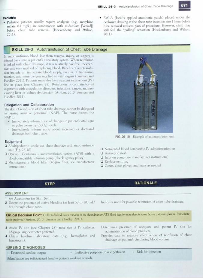
Equipment • Adult/pediatric single-use chest drainage and autotransfusion
unit (Fig. 26-10) • Optional: Continuous autotransfusion system (ATS) with a
blood-compatible infusion pump (check agency policy) • Microaggregate blood filter (40-pm filter, see manufacturer
instructions)
IFH Wi u ft CM. Mr r,„
-r-
:
; .« • II II ill '1
FIG 26-10 Example of autotransfusion unit.
• Nonvented blood-compatible IV administration set • Antiseptic swab • Infusion pump (see manufacturer instructions) • Replacement bag • Gown, clean gloves, and mask as needed
STEP RATIONALE
ASSESSMENT 1 See Assessment for Skill 26-1. 2 Determine presence of active bleeding (at least 50-to-100 mL/ Indicates need for possible reinfusion of chest tube drainage,
hr), through chest tube.
Clinical Decision Point Collected blood never remains in the chest drain or ATS blood bag for more than 6 hours before autotransfusion. Immediate
use is preferred (Atrium, 2010; Bauman and Handley, 2011).
3 Assess IV site (see Chapter 28); note size of IV catheter. Determines presence of adequate and patent IV site for 18-gauge angiocatheter preferred. administration of blood products.
4 Obtain baseline laboratory data (e.g., hemoglobin and Provides data to measure effectiveness of reinfusion of chest hematocrit). drainage on patient's circulating blood volume.
NURSING DIAGNOSES • Decreased cardiac output • Ineffective peripheral tissue perfusion • Risk for infection
Related factors are individualized based on patient's condition or needs.
Related Documents