DOI: 10.1126/scitranslmed.3008177 , 233ra54 (2014); 6 Sci Transl Med et al. Jeremy L. Pinyon Electrode Array Enhances the Bionic Ear Close-Field Electroporation Gene Delivery Using the Cochlear Implant Editor's Summary improve the quality of hearing in deaf individuals. further optimization of electroporation, it is possible that including gene therapy could restore hearing loss and model (animals with hair cells destroyed). Because cochlear implants are already routinely used in patients, with delivery drove regeneration of neurites in the cochlea, leading to restoration of hearing in a deafened guinea pig gene BDNF (BDNF), a protein in humans that is considered to be a nerve growth factor. Electroporation-mediated lining the scala tympani. Once inside the cells' nuclei, the gene construct produced brain-derived neurotrophic factor which allowed passage of genetic material into the mesenchymal cells -- temporary opening of the cell membrane basically, a -- guinea pigs delivered brief, intense electrical pulses to surrounding tissue. This led to electroporation The authors coined their approach ''close-field electroporation.'' An electrode array inserted into the cochlea of neurotrophic factor that can regenerate auditory nerves and, in turn, restore hearing. this issue, Pinyon and colleagues worked to improve upon existing implants by simultaneously delivering a however, improvements in sound quality and range of environments where the implant can be used are desired. In process that allows you to hear. Cochlear implants, or ''bionic ears,'' have been incredibly useful for deaf individuals; specialized cells in the inner ear that help convert acoustic vibrations into nerve impulses, a -- cochlear hair cells Hearing loss affects millions worldwide. Sensorineural hearing loss, in particular, typically follows the loss of Can You Hear Me Now? http://stm.sciencemag.org/content/6/233/233ra54.full.html can be found at: and other services, including high-resolution figures, A complete electronic version of this article http://stm.sciencemag.org/content/suppl/2014/04/21/6.233.233ra54.DC1.html can be found in the online version of this article at: Supplementary Material http://www.sciencemag.org/content/sci/344/6184/1241062.full.html http://stm.sciencemag.org/content/scitransmed/5/210/210ps16.full.html http://stm.sciencemag.org/content/scitransmed/5/210/210rv2.full.html http://stm.sciencemag.org/content/scitransmed/3/82/82cm12.full.html http://stm.sciencemag.org/content/scitransmed/2/21/21ra16.full.html can be found online at: Related Resources for this article http://www.sciencemag.org/about/permissions.dtl in whole or in part can be found at: article permission to reproduce this of this article or about obtaining reprints Information about obtaining is a registered trademark of AAAS. Science Translational Medicine rights reserved. The title NW, Washington, DC 20005. Copyright 2014 by the American Association for the Advancement of Science; all last week in December, by the American Association for the Advancement of Science, 1200 New York Avenue (print ISSN 1946-6234; online ISSN 1946-6242) is published weekly, except the Science Translational Medicine on May 15, 2014 stm.sciencemag.org Downloaded from on May 15, 2014 stm.sciencemag.org Downloaded from

Welcome message from author
This document is posted to help you gain knowledge. Please leave a comment to let me know what you think about it! Share it to your friends and learn new things together.
Transcript

DOI: 10.1126/scitranslmed.3008177, 233ra54 (2014);6 Sci Transl Med et al.Jeremy L. Pinyon
Electrode Array Enhances the Bionic EarClose-Field Electroporation Gene Delivery Using the Cochlear Implant
Editor's Summary
improve the quality of hearing in deaf individuals.further optimization of electroporation, it is possible that including gene therapy could restore hearing loss and model (animals with hair cells destroyed). Because cochlear implants are already routinely used in patients, withdelivery drove regeneration of neurites in the cochlea, leading to restoration of hearing in a deafened guinea pig
geneBDNF(BDNF), a protein in humans that is considered to be a nerve growth factor. Electroporation-mediated lining the scala tympani. Once inside the cells' nuclei, the gene construct produced brain-derived neurotrophic factor
which allowed passage of genetic material into the mesenchymal cells−−temporary opening of the cell membrane basically, a−−guinea pigs delivered brief, intense electrical pulses to surrounding tissue. This led to electroporation
The authors coined their approach ''close-field electroporation.'' An electrode array inserted into the cochlea of
neurotrophic factor that can regenerate auditory nerves and, in turn, restore hearing.this issue, Pinyon and colleagues worked to improve upon existing implants by simultaneously delivering a however, improvements in sound quality and range of environments where the implant can be used are desired. Inprocess that allows you to hear. Cochlear implants, or ''bionic ears,'' have been incredibly useful for deaf individuals;
specialized cells in the inner ear that help convert acoustic vibrations into nerve impulses, a−−cochlear hair cells Hearing loss affects millions worldwide. Sensorineural hearing loss, in particular, typically follows the loss of
Can You Hear Me Now?
http://stm.sciencemag.org/content/6/233/233ra54.full.htmlcan be found at:
and other services, including high-resolution figures,A complete electronic version of this article
http://stm.sciencemag.org/content/suppl/2014/04/21/6.233.233ra54.DC1.html can be found in the online version of this article at: Supplementary Material
http://www.sciencemag.org/content/sci/344/6184/1241062.full.html http://stm.sciencemag.org/content/scitransmed/5/210/210ps16.full.html
http://stm.sciencemag.org/content/scitransmed/5/210/210rv2.full.html http://stm.sciencemag.org/content/scitransmed/3/82/82cm12.full.html
http://stm.sciencemag.org/content/scitransmed/2/21/21ra16.full.html can be found online at:Related Resources for this article
http://www.sciencemag.org/about/permissions.dtl in whole or in part can be found at: article
permission to reproduce this of this article or about obtaining reprintsInformation about obtaining
is a registered trademark of AAAS. Science Translational Medicinerights reserved. The title NW, Washington, DC 20005. Copyright 2014 by the American Association for the Advancement of Science; alllast week in December, by the American Association for the Advancement of Science, 1200 New York Avenue
(print ISSN 1946-6234; online ISSN 1946-6242) is published weekly, except theScience Translational Medicine
on
May
15,
201
4st
m.s
cien
cem
ag.o
rgD
ownl
oade
d fr
om
on
May
15,
201
4st
m.s
cien
cem
ag.o
rgD
ownl
oade
d fr
om

R E S EARCH ART I C L E
GENE THERAPY
Close-Field Electroporation Gene Delivery Using theCochlear Implant Electrode Array Enhances theBionic EarJeremy L. Pinyon, Sherif F. Tadros, Kristina E. Froud, Ann C. Y. Wong, Isabella T. Tompson,Edward N. Crawford, Myungseo Ko, Renée Morris, Matthias Klugmann, Gary D. Housley*
on
May
15,
201
4m
ag.o
rg
The cochlear implant is the most successful bionic prosthesis and has transformed the lives of people with profoundhearing loss. However, the performance of the “bionic ear” is still largely constrained by the neural interface itself.Current spread inherent to broad monopolar stimulation of the spiral ganglion neuron somata obviates the intrinsictonotopic mapping of the cochlear nerve. We show in the guinea pig that neurotrophin gene therapy integrated intothe cochlear implant improves its performance by stimulating spiral ganglion neurite regeneration. We used the co-chlear implant electrode array for novel “close-field” electroporation to transduce mesenchymal cells lining the co-chlear perilymphatic canals with a naked complementary DNA gene construct driving expression of brain-derivedneurotrophic factor (BDNF) and a green fluorescent protein (GFP) reporter. The focusing of electric fields by particularcochlear implant electrode configurations led to surprisingly efficient gene delivery to adjacent mesenchymal cells.The resulting BDNF expression stimulated regeneration of spiral ganglion neurites, which had atrophied 2weeks afterototoxic treatment, in a bilateral sensorineural deafness model. In this model, delivery of a control GFP-only vectorfailed to restore neuron structure, with atrophied neurons indistinguishable from unimplanted cochleae. With BDNFtherapy, the regenerated spiral ganglion neurites extended close to the cochlear implant electrodes, with localizedectopic branching. This neural remodeling enabled bipolar stimulation via the cochlear implant array, with low stim-ulus thresholds and expanded dynamic range of the cochlear nerve, determined via electrically evoked auditorybrainstem responses. This development may broadly improve neural interfaces and extend molecular medicineapplications.
ce
stm.s
cien
Dow
nloa
ded
from
INTRODUCTION
Hearing loss is the most prevalent sensory disability, stemming fromgenetics, environmental factors, particularly noise, ototoxicity, andaging (1–3). The cochlear implant bionic prosthesis has helped peoplesuffering profound hearing loss and, more broadly, has fostered devel-opment of the field of implantable bionic interfaces. Cochlear implantsuse real-time processing of sound into frequency components, whichare mapped to particular electrodes along the cochlear implant arrayas brief current pulses that recruit subpopulations of spiral ganglionneurons (SGNs) that form the auditory nerve. Advances in cochlear im-plant performance have benefitted from innovative engineering andcomputational capabilities of the peripheral components (4). However,the neural interface itself, typically consisting of an array of smallplatinum electrodes (each ~500 mm in diameter), has remained largelyunchanged (5).
In normal-hearing subjects, the cochlea is tonotopically mapped sothat the sensory hair cells at the base of the cochlea encode the highest-frequency sounds and progressively lower frequencies are detectedtoward the apex. With sensorineural hearing loss, the sensory hair cellsand their supporting cells in the organ of Corti are lost, and the SGNsatrophy. The dieback of the auditory nerve is attributed to the loss ofneurotrophin support, principally brain-derived neurotrophic factor(BDNF) and neurotrophin 3 (NT3), from the organ of Corti, signaledvia tyrosine kinase receptors (Trks) expressed by the SGNs (6–8). The
Translational Neuroscience Facility and Department of Physiology, School of MedicalSciences, University of New South Wales, UNSW Australia, Sydney, New South Wales 2052,Australia.*Corresponding author: E-mail: [email protected]
www.Scie
placement of a line of electrodes within scala tympani, a fluid chamberin the cochlea, enables the cochlear implant to electrically stimulate theSGNs and restore a facsimile of hearing. The number of electrodes isconstrained by current spread associated with stimulation of theatrophied SGN somata localized behind the boney modiolar wall ofthe cochlea (5). Thus, a neural gap exists between the cochlear implantand the SGN somata, which necessitates the use of monopolar stimu-lation with current return via an electrode external to the cochlea. Thissubverts the tonotopic mapping of cochlear sound coding. Thus, co-chlear implant recipients rely on the plasticity of the central auditoryprocessing in the brain to translate the broad neuronal recruitment intospeech perception, with concomitantly poor pitch perception (4). Closingthe neural gap between the cochlear implant and the target sensoryneurons offers the opportunity to surmount current biophysical lim-itations of SGN recruitment (5).
Proof-of-principle studies using viral vector–based gene therapy totreat deafness have revealed considerable potential for restoring hearingfunction in animal models. Hearing loss of genetic origin has beenreversed in the vesicular glutamate transporter-3–null mouse model,with adeno-associated virusVGlut3 gene therapy targeting the neonatalinner hair cell synapses (9). Cochlear expression of the transcriptionalregulator Atoh1 (also known as Math1) has driven transfection ofsupporting cells in the organ of Corti into new hair cells (10), whichare innervated de novo by spiral ganglion neurites, to recover hearingfunction (11). Restoration of hair cells via transdifferentiation is not pos-sible with profound hearing loss owing to degeneration of supportingcells. However, neuronal atrophy has been reversed and peripheral spi-ral ganglion neurites have been restored, resulting in better cochlearimplant performance in guinea pigs using BDNF and NT3 therapy
nceTranslationalMedicine.org 23 April 2014 Vol 6 Issue 233 233ra54 1
R E S EARCH ART I C L E
(12–14). Although viral vector gene therapy approaches show prom-ise, clinical translation will need to address delivery, targeting, andsafety constraints (2).
Electroporation offers an alternative pathway for cochlear gene de-livery, but in the conventional “open-field” configuration, the need toplace separate electrodes across the boney cochlear compartments pre-cludes effective cell transfection. Such open-field electroporation haspreviously been directed to studying cell signaling in cochlear neurode-velopment. For example, transfection of murine cochlear cells has beenachieved using paddle-style electrodes and injection of an Atoh1 geneconstruct into the mouse embryo otic placode in utero (15). Open-field
on
May
15,
201
4st
m.s
cien
cem
ag.o
rgow
nloa
ded
from
electroporation has also been used for invitro transfection of rodent embryonicand neonatal cochlear sensory epitheliumexplant tissue (16–18).
Here, we investigated whether the co-chlear implant electrode array could beused to generate localized high electricfields for electroporation-mediated genedelivery in a deafened adult guinea pigcochlea. This “close-field” electropora-tion (CFE), using local current paths be-tween adjacent nodes of an array ofelectrodes, achieved high-efficiencyBDNFgene delivery in a spatially prescribedzone. The resultant BDNF expressiondrove regeneration of the SGN neurites toclose apposition with the cochlear implantelectrodes. The nerve fibers were then ableto be recruited by local bipolar stimulation,withminimal current spread. The findingstherefore pave the way for integration ofgene therapy into the cochlear implantationsurgery, where a neurotrophin-encodingplasmid DNA can be introduced via a fewbrief electrical pulses, to produce directedregeneration of the auditory nerve thatimproves the functionality of the bionicinterface.
D
RESULTS
Ex vivo cochlear gene deliveryAnexvivomodelwasdeveloped to establishthe potential of cochlear implant–mediatedCFEgenedelivery.Wedevelopedabicistronicgene cassette (CMVp-BDNF-IRES-GFPnls)using the cytomegalovirus (CMV)promoterto drive expression of FLAG-tagged BDNFand, via an internal ribosome entry site(IRES) element, a green fluorescent protein(GFP) reporter with a nuclear localizationsite sequence (Fig. 1A). This circular com-plementary DNA (cDNA) plasmid wasmicroinjected into the perilymphatic fluidspaces (scala tympani and scala vestibuli)of normal intact guinea pig cochleae. An
www.Scie
eight-node cochlear implant electrode array was then inserted into thebasal turn of the cochlea via the round window (Fig. 1, B and C). Thearray was configured as either an “alternating” configuration of anodes(+) and cathodes (−) or a “tandem” arraywhere four adjacent nodeswerewired in parallel as the anode (distal end of the array), and the othernodes provided a common cathode (Fig. 1D).
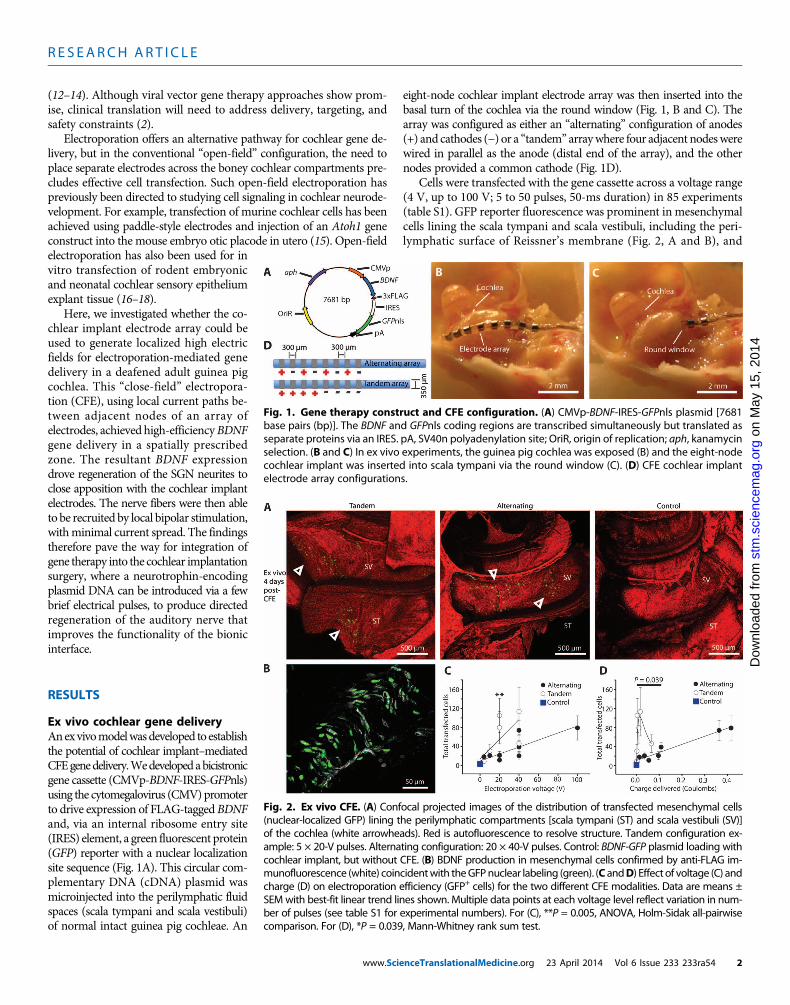
Cells were transfected with the gene cassette across a voltage range(4 V, up to 100 V; 5 to 50 pulses, 50-ms duration) in 85 experiments(table S1). GFP reporter fluorescence was prominent in mesenchymalcells lining the scala tympani and scala vestibuli, including the peri-lymphatic surface of Reissner’s membrane (Fig. 2, A and B), and
Fig. 1. Gene therapy construct and CFE configuration. (A) CMVp-BDNF-IRES-GFPnls plasmid [7681base pairs (bp)]. The BDNF and GFPnls coding regions are transcribed simultaneously but translated as
separate proteins via an IRES. pA, SV40n polyadenylation site; OriR, origin of replication; aph, kanamycinselection. (B and C) In ex vivo experiments, the guinea pig cochlea was exposed (B) and the eight-nodecochlear implant was inserted into scala tympani via the round window (C). (D) CFE cochlear implantelectrode array configurations.Fig. 2. Ex vivo CFE. (A) Confocal projected images of the distribution of transfected mesenchymal cells(nuclear-localized GFP) lining the perilymphatic compartments [scala tympani (ST) and scala vestibuli (SV)]
of the cochlea (white arrowheads). Red is autofluorescence to resolve structure. Tandem configuration ex-ample: 5 × 20-V pulses. Alternating configuration: 20 × 40-V pulses. Control: BDNF-GFP plasmid loading withcochlear implant, but without CFE. (B) BDNF production in mesenchymal cells confirmed by anti-FLAG im-munofluorescence (white) coincidentwith theGFPnuclear labeling (green). (C andD) Effect of voltage (C) andcharge (D) on electroporation efficiency (GFP+ cells) for the two different CFE modalities. Data are means ±SEMwith best-fit linear trend lines shown. Multiple data points at each voltage level reflect variation in num-ber of pulses (see table S1 for experimental numbers). For (C), **P = 0.005, ANOVA, Holm-Sidak all-pairwisecomparison. For (D), *P = 0.039, Mann-Whitney rank sum test.nceTranslationalMedicine.org 23 April 2014 Vol 6 Issue 233 233ra54 2
R E S EARCH ART I C L E
was maintained for the duration of organotypic cultures (3 to 4 days).The GFP-positive cells were confined to the basal turn region, within~1 mm of the array, consistent with spatially constrained electropora-tionof the cells. CFEwas effective from as low as 4V (combinedmean ±SEM=9.5±1.8 cells per cochlea,n=8;P=0.017, unpaired t test) (tableS1).In comparison, control experiments where free DNA was introducedinto the cochleawithout electroporation yieldedminimalGFP-positivecell counts (mean ± SEM = 2.8 ± 0.5 cells per cochlea; n = 5 cochleae)(Fig. 2C). Expression of BDNF by transfected cells was confirmed byanti-FLAG immunolabeling (Fig. 2B).
The tandem electrode array configuration demonstrated greatergene delivery efficiency than the alternating array configuration, withan asymmetric optimum in the lower range of applied voltages (~20 V)(Fig. 2C) and, from resistance measurements during the voltage steps,a minimum effective charge delivery of ~0.04 C (Fig. 2D). Up to 310GFP+ cells were achieved (mean = 105.4 ± 35.7 cells per cochlea; n = 8),with five 20 V pulses lasting 50 ms each, with no increase with addi-
www.Scie
tional pulses. This equated to a charge density per pulse of ~60mC/cm2
on the four platinum ring anodes (table S1). In contrast, the alternatingconfiguration provided a linear increase in transfection efficiency withincreasing voltage to the highest test level and charge delivery, for ex-ample, 20.8 ± 8.4 cells at 20V, 20 pulses (n= 6) to 79.0 ± 25.5 cells (n= 3)at 100 V, 5 pulses (0.42 C total charge). Overall, there was a significantlygreater transfection efficiency with the tandem versus alternating arrayconfigurations between 0.002 and 0.109 C [P = 0.017, ranked one-wayanalysis of variance (ANOVA)] (Fig. 2D).
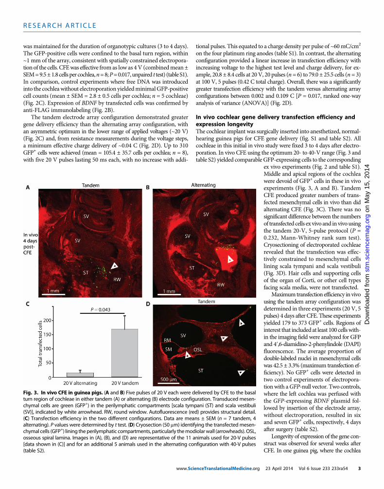
In vivo cochlear gene delivery transfection efficiency andexpression longevityThe cochlear implant was surgically inserted into anesthetized, normal-hearing guinea pigs for CFE gene delivery (fig. S1 and table S2). Allcochleae in this initial in vivo study were fixed 3 to 4 days after electro-poration. In vivo CFE using the optimum 20- to 40-V range (Fig. 3 andtable S2) yielded comparable GFP-expressing cells to the corresponding
nceTranslationalMedicine.org
on
May
15,
201
4st
m.s
cien
cem
ag.o
rgD
ownl
oade
d fr
om
ex vivo experiments (Fig. 2 and table S1).Middle and apical regions of the cochleawere devoid of GFP+ cells in these in vivoexperiments (Fig. 3, A and B). TandemCFE produced greater numbers of trans-fected mesenchymal cells in vivo than didalternating CFE (Fig. 3C). There was nosignificant difference between the numbersof transfected cells ex vivo and in vivousingthe tandem 20-V, 5-pulse protocol (P =0.232, Mann-Whitney rank sum test).Cryosectioning of electroporated cochleaerevealed that the transfection was effec-tively constrained to mesenchymal cellslining scala tympani and scala vestibuli(Fig. 3D). Hair cells and supporting cellsof the organ of Corti, or other cell typesfacing scala media, were not transfected.
Maximum transfection efficiency in vivousing the tandem array configuration wasdetermined in three experiments (20 V, 5pulses) 4 days after CFE. These experimentsyielded 179 to 373 GFP+ cells. Regions ofinterest that included at least 100 cellswith-in the imaging field were analyzed for GFPand 4′,6-diamidino-2-phenylindole (DAPI)fluorescence. The average proportion ofdouble-labeled nuclei in mesenchymal cellswas 42.5 ± 3.3% (maximum transfection ef-ficiency). No GFP+ cells were detected intwo control experiments of electropora-tionwith aGFP-null vector. Two controls,where the left cochlea was perfused withthe GFP-expressing BDNF plasmid fol-lowed by insertion of the electrode array,without electroporation, resulted in sixand seven GFP+ cells, respectively, 4 daysafter surgery (table S2).
Longevity of expression of the gene con-struct was observed for several weeks afterCFE. In one guinea pig, where the cochlea
Fig. 3. In vivo CFE in guinea pigs. (A and B) Five pulses of 20 V each were delivered by CFE to the basalturn region of cochleae in either tandem (A) or alternating (B) electrode configuration. Transduced mesen-
chymal cells are green (GFP+) in the perilymphatic compartments [scala tympani (ST) and scala vestibuli(SV)], indicated by white arrowhead. RW, round window. Autofluorescence (red) provides structural detail.(C) Transfection efficiency in the two different configurations. Data are means ± SEM (n = 7 tandem, 4alternating). P values were determined by t test. (D) Cryosection (50 mm) identifying the transfectedmesen-chymal cells (GFP+) lining theperilymphatic compartments, particularly themodiolarwall (arrowheads). OSL,osseous spiral lamina. Images in (A), (B), and (D) are representative of the 11 animals used for 20-V pulses[data shown in (C)] and for an additional 5 animals used in the alternating configuration with 40-V pulses(table S2).23 April 2014 Vol 6 Issue 233 233ra54 3
R E S EARCH ART I C L E
was analyzed 3 weeks after gene delivery, a total of 11 GFP+mesenchymalcells in seven 50-mm sections were localized to the basal turn region withinscala tympani. No quantitative analysis was undertaken with additionalanimals at this time point, but the drop-off in expression around this timewas confirmed in two cochleae examined 6weeks after gene delivery (eightGFP+cells evident inonly onehemisectioned cochlea), andnoGFPexpres-sion was detected in cochleae from four animals at 10 weeks after genedelivery. GFP signal extending for up to 6 weeks suggests mRNAtranslation beyond the immediate gene delivery period.
BDNF gene therapy in a deafened guinea pig modelFurther in vivo experiments were carried out in a deafened guinea pigmodel. Animals were deafened using a kanamycin-furosemide ototoxic
www.Scie
treatment to selectively remove the sensory hair cells, where theperipheral neurites of the SGN showed substantial degeneration withinthe osseous spiral lamina, and the SGN somata undergo atrophy (figs. S2and S3). The deafened guinea pig organ of Corti typically exhibited pro-gressive flattening of the sensory epithelium due to the loss of haircells and supporting cells (fig. S2). Although interanimal variabilityin progression of SGN atrophy and loss has been reported with thisototoxic deafening procedure (19), our data were consistent with re-gard to substantial loss of neurites in the osseous spiral lamina at the2-week post-deafening time point. At this time point, therapeuticBDNFcDNA was delivered via the cochlear implant array.
BDNF gene therapy led to regeneration of neurite processes thatextended out through the osseous spiral lamina, with side branching
on
May
15,
201
4st
m.s
cien
cem
ag.o
rgD
ownl
oade
d fr
om
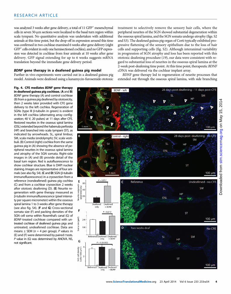
Fig. 4. CFE mediates BDNF gene therapyin deafened guinea pig cochleae. (A and B)
BDNF gene therapy (A) and control cochleae(B) fromaguineapigdeafenedbyototoxicity,then 2 weeks later provided with CFE genedelivery to the left cochlea. Regeneration ofSGNs (type III b-tubulin in green) is evidentin the left cochlea (alternating array config-uration; 40 V, 20 pulses) at 11 days after CFE.Restored neurites in the osseous spiral lamina(OSL)extendedbeyond thehabenulaperforata(HP) and branched into scala tympani (ST), asindicated by arrowheads. SL, spiral limbus;SM, scala media (endolymph); SV, scala vesti-buli. (B) Control (right) cochlea from the sameguinea pig in (A) showing the absence of pe-ripheral neurites in the osseous spiral laminaand atrophy of the SGN somata. Right-sideimages in (A) and (B) provide detail of thebasal turn region. Red is autofluorescence toshow cochlear structure. Blue is DAPI nuclearstaining. Images are representative of four ani-mals (see also fig. S4). (C andD) SGN (b-tubulinimmunofluorescence) in a cryosection from areference (nondeafened) guinea pig cochlea(C) and from a cochlear cryosection 2 weeksafter ototoxic deafening (D). (E) Neurite re-generation with gene therapy measured asb-tubulin immunofluorescence (pixel intensi-ty per squaremicrometer) within the osseousspiral lamina 1 to 3 weeks after gene therapy(see also fig. S4). (F and G) Cross-sectionalsomata size (F) and packing densities of theSGN cell soma within Rosenthal’s canal (G) ofBDNF-treated cochleae compared with un-treated cochleae of deafened guinea pigs anduntreated, undeafened cochleae. Data aremeans ± SEM (n = 4 per group). P values in(E) and (F) were determined by paired t tests.P value in (G) was determined by ANOVA. NS,not significant.nceTranslationalMedicine.org 23 April 2014 Vol 6 Issue 233 233ra54 4

R E S EARCH ART I C L E
2014
through the modiolar wall via micropores (canaliculi perforantes), withmost fibers reaching the habenula perforata, before descending throughthe basilar membrane into the scala tympani region (Fig. 4A). Neuriteregeneration occurred from the basal turn (the site of mesenchymal celltransfection) to the mid-turn region of the BDNF-treated cochleae (Fig.4A). This likely reflects diffusion of the secreted recombinant BDNFwithin the perilymph. Both alternating (40 V, 20 × 50-ms pulses; n =3) and tandem configurationCFE (20V, 5 × 50-ms pulses; n= 1)BDNFgene delivery to the left cochlea led to regeneration of the peripheralspiral ganglionneurites and increased the size of SGN somata comparedwith the untreated (right) cochleae of deafened guinea pigs (Fig. 4, Aand B, and fig. S4). This reversal of SGN atrophy by gene therapywas evident by comparison of the reference SGN state before thedeafening procedure (Fig. 4C) with the atrophy evident 2 weeks afterdeafening (Fig. 4D), the point when gene delivery was undertaken.Images of immunolabeled cryosectionswere analyzed from1 to 3weeksafter gene delivery, corresponding to 3 to 5 weeks after deafening.
Neuronal-specific type III b-tubulin immunofluorescence within theosseous spiral lamina region (basal turn) indicated significantly
www.Scie
on
May
15,
st
m.s
cien
cem
ag.o
rgD
ownl
oade
d fr
om
greater neurite outgrowth in the BDNF-treated animals (Fig. 4E andfig. S4). In addition, the average SGN soma size was about 40% largerin the BDNF gene therapy–treated versus untreated cochleae of thedeafened animals (Fig. 4F). There was no difference in SGN densitybetween either BDNF-treated and untreated cochleae in the deafenedguinea pigs or the undeafened reference cochleae, suggesting that SGNsurvival was not significantly affected within the first 5 weeks afterdeafening (P = 0.184, paired t test, comparing SGN cell density be-tween left and right cochleae; P = 0.26, ANOVA across groups includ-ing the reference) (Fig. 4G).
Ectopic branching extended to the inner sulcus and outer sulcus–spiral ligament region, within scala media, with some variation in fiberextension into scala tympani (Fig. 5A) compared with extension alongthe basilar membrane to the spiral ligament (Fig. 5B). Extension of neu-rite outgrowth also occurred toward the spiral limbus, across the inner sul-cus (Fig. 6,A andB). The control (right cochleae) showedno remnant fibersbeyond the habenula perforata (Fig. 6C).
The presence of the Schwann cell marker aspartoacylase (ASPA)(20) indicated remyelination of regenerated neurites up to 12 weeks afterdeafening (10 weeks after BDNF delivery), including the ectopic processeswithin scala tympani and scalamedia (Fig. 6D). Anti-ASPA immunofluo-rescence was also evident in the osseous spiral lamina of the untreatedcochleae, consistent with preservation of the glial satellite cells and re-tention of residual SGN fibers (Fig. 6D). These glial cells are responsible forthe remyelinationof theSGNfibers anddemonstratemyelination to the tipsof the ectopic neurite branches.
Enhanced cochlear implant performanceThe functional effect of regeneration of the SGN neurites from CFE-mediated BDNF gene therapy was assessed in separate in vivo studieswith the deafened guinea pig model. Electrically evoked auditory brain-stem responses (eABRs) were measured 2 weeks after BDNF-GFP orGFP-only (control) gene delivery via CFE using the tandem array con-figuration (20V, 5× 50-ms pulses;n=5 for each), whichwas 4weeks afterthe ototoxic treatment.With bipolar stimulationof the SGNvia the tandemarrayconfiguration, the improvement in auditory brainstempathway neu-ral recruitmentdue to theBDNFgene therapy was evident from the lowercurrent amplitudes needed to elicit threshold eABRs (Fig. 7A). Themean eABR stimulus threshold for the BDNF gene therapy groupwas <50% of the control group, with no overlap in threshold data(Fig. 7B). This improvement in cochlear implant performance canbe assessed against the reference group, where cochlear implant stimu-lation was applied to normal-hearing guinea pigs (with auditory nerveand the sensory hair cells intact). This produced amean eABR stimulusthreshold (Fig. 7B) thatwas not significantly different from the deafenedBDNF gene therapy group. The result suggests nominally optimum re-covery of threshold, yet discounting possible augmentation of the re-sponse in this reference group due to electrophonic inner hair cellstimulation.
Baseline eABR thresholds were also determined 2 weeks after deafen-ing, just before gene therapy (Fig. 7B). This was significantly higher thanthe BDNF gene therapy group 2 weeks later, after BDNF gene ther-apy, but significantly less than the GFP-only vector control groupafter 2-week expression of the GFP-only gene construct. These dataindicate progressive deterioration in SGN function over time in theabsence of BDNF gene therapy, consistent with the exacerbation ofsensorineural hearing loss in this deafness model in the absence ofneurotrophin treatment.
Fig. 5. Regeneration of the SGN peripheral neurites in vivo after alter-nating configuration CFE BDNF gene therapy in deafened guinea pigs.
Animals were subjected to 20 pulses at 40 V. Confocal projected image recon-structions are of type III b-tubulin immunofluorescence in cochlear cryosections.(A and B) Detail variations in outgrowth of fibers (arrowheads) in the osseousspiral lamina (OSL), with ectopic branching of fibers into scala tympani (ST),both at the habenula perforata (HP) and through the modiolar wall via micro-pores called the canaliculi perforantes (A). Regenerated spiral ganglion neuritesalso projected past the remnant organ of Corti to the outer sulcus (OS) regionand, in the example shown in (B), medially to the inner sulcus (IS). Insets in thebottom right corners show control (no CFE) cochleae from the same animals.SL, spiral limbus; SLg, spiral ligament; SM, scala media; SV, scala vestibuli.nceTranslationalMedicine.org 23 April 2014 Vol 6 Issue 233 233ra54 5

R E S EARCH ART I C L E
Complementing this, the dynamic range (growth function) of theeABR was significantly increased in the BDNF gene therapy treat-ment group (Fig. 7, C and D), with a considerable extension of therange of stimulus eliciting growth of the eABR compared with theGFP vector control group. Thus, BDNF gene therapy restored the co-chlear nerve recruitment toward pretreatment levels. Overall, theseresults demonstrate a functional enhancement of the cochlear implant–neural interface arising from restoration of the peripheral SGN neuritesafter 2 weeks of CFE-mediated BDNF gene therapy.
DISCUSSION
The development of the cochlear implant is currently limited by theneural gap between the atrophied SGNs and the electrode array. Deliv-
www.Scie
ery of therapeutics that may confer protection to the auditory nervefibers during the implantation procedure, or indeed drive regenera-tion of the nerve fibers toward the electrodes, is necessary to improvethe bionic ear. This study describes the development of CFE that usesmultipolar configurations of the cochlear implant electrode array togenerate focused electric fields for localized neurotrophin gene deliv-ery in a guinea pig model. This integration of gene therapy into thecochlear implant procedure achieved focused expression of plasmidDNA encoding the neurotrophin BDNF. Production of BDNF pro-moted rapid directed regeneration of the auditory nerve fibers intoclose proximity with the cochlear implant electrodes, thereby closingthe neural gap in the bionic interface and eliciting improved hearingsensitivity and dynamic range.
We used an ex vivo, organotypic culture model to establish theoptimum conditions for CFE gene delivery before proceeding to the
nceTranslationalMedicine.org
on
May
15,
201
4st
m.s
cien
cem
ag.o
rgD
ownl
oade
d fr
om
in vivo proof-of-principle studies. Elec-troporation is a well-established methodfor delivering drugs, proteins, and nucleicacids into cells (21, 22). DNA migrationacross a cell membrane involves the elec-troporation of the plasma membrane byhigh electric fields, with an electrophoreticaction for driving the negatively chargedmolecules across (23–25). Once insidethe cell, DNA can migrate to the nucleus,where, in the case of circularized plasmidDNA, it remains extrachromosomal (epi-somal) and supports gene expressionwith-out the risk of mutagenesis that arises withintegration. Here, we show that with elec-trode arrays, fields of sufficient intensityto transduce cells can be generated if cur-rent is passed between electrodes that arecontiguous within an array. This provideshighly localized transduction of the cellsin the immediate vicinity of the array, asevidenced by GFP fluorescence within24 hours. The ex vivo experiments dem-onstrated that the configuration of theelectrodes within the array affected celltransfection, with the tandem configura-tion being most efficient. Amplitudes aslow as 4 V yielded significant numbersof transfected cells. These studies also val-idated the production of the recombinantBDNF by the target mesenchymal cells.The selective transduction of mesenchymalcells is likely due to the limited diffusion ofDNA from the perilymphatic compartmentin the few minutes required for DNAloading and CFE.
The in vivoCFE experimentsmigratedthe study from demonstration of targetedgene delivery and BDNF expression (bythe mesenchymal cells adjacent to theelectrodes) to confirmation of peripheralauditory nerve fiber regeneration, reversalof SGNsomata atrophy, and remyelination
Fig. 6. Neurite regeneration and remyelination after tandem configuration CFE BDNF gene therapyin deafened guinea pigs. Animals were administered 5 pulses at 20 V each to deliver BDNF. (A) Type III b-
tubulin immunofluorescence (green) of a hemisectioned cochlea 2 weeks after CFE (n = 3). Confocal three-dimensional reconstruction shows the SGN regenerationwithin the osseous spiral lamina (OSL), through thehabenula perforata (HP) and extending to the inner sulcus (IS) region, under the lateral prominence of thespiral limbus (SL). Cochlear structure shown by autofluorescence (red). (B) Ectopic branching of the regen-erated neurites bordering the spiral limbus within scala media from tissue shown in (A). Nuclear labeling(DAPI) is in blue. (C) Control (deafened, no gene therapy) cochlea from the same guinea pig. (D) Asparto-acylase immunofluorescence (orange) showing myelination of the regenerated neurites and ectopicbranching 6 (n = 2) to 10weeks (n = 4) after CFE. Images have transmitted light overlays for structural detail.DAPI-labeled nuclei show location of the satellite cells (scn) in the 10-week post-CFE image.23 April 2014 Vol 6 Issue 233 233ra54 6

R E S EARCH ART I C L E
14
of the distal neurite processes. The extensive ectopic branching of theSGN neurites after BDNF therapy was reminiscent of the initial neuriteextension that occurs when the SGN afferent fibers first innervate thecochlea searching for target hair cell synapses during development (26).The eABR assessment demonstrated that this auditory nerve regenera-tion lends restorationof nerve fiber recruitment thresholds to pre-deafnesslevels. The halving in eABR threshold achieved by BDNF gene therapyequates to the improved cochlear implant performance achieved in anearlier study in guinea pigs usingdirect BDNFdrugdelivery (27), whereasour absolute thresholds (hearing sensitivity) were about threefold lesswith bipolar stimulation.
Cochlear implants are typically enabled in monopolar stimulationmode to achieve the best auditory function, where individual electrodeswithin the implanted array are drivenwith current return via a commonelectrode located external to the cochlea (5). This provides the best re-cruitment of auditory neurons in the implant recipients because ofthe convoluted current path to the atrophied auditory neurons. Inour studies, bipolar stimulation from adjacent nodes of the cochlear
www.Scie
implant provided sensitive localized recruitment of the repaired audi-tory neurons. Translation of this outcome to a clinical setting wouldlikely facilitate better sound perception in cochlear implant recipientsvia recruitment of smaller subpopulations of SGNs, as previously sug-gested (5). Currently, appreciation of tonal quality (for example, in mu-sic) is very limited, and the potential benefit that may accrue frommorediscrete recruitment of SGNs that we demonstrate here potentiallymakes tonal color and music appreciation a major potential gain (28).
The straightforward nature of theCFE gene delivery established hereprompts consideration of whether neurotrophin gene therapy can beintegrated into otological practice to enhance the performance of the“bionic ear.” The eight-node array design used in the present study wasdeveloped for animal models. Given that the cochlear implant electrodearrays used clinically currently have up to 22 electrodes, the scalability ofthe electric field focusingwill need to be pursued. Evaluation should alsoconsider the current trend toward hybrid acoustic and electrode arrayimplant devices, where residual low-frequency hearing is preserved andused (29). The incorporation of BDNF therapy into the cochlear im-
nceTranslationalMedicine.org
on
May
15,
20
stm
.sci
ence
mag
.org
Dow
nloa
ded
from
plantation procedure via CFEmay protectandpreserve this residual sound transduc-tion in the apical region of the cochlea.Alternative trophic factors may also proveuseful in promoting directionality to neu-ral outgrowth, which wouldmaximize thesensitivity and selectivity of auditory neu-ron recruitment. For example, it has beenshown that cochlear delivery of BDNFand NT3 proteins in combination is moreeffective in preserving SGN in sensori-neural hearing loss models than eitherfactor alone (30).
On the basis of the current studies, it ispossible to envisage the ready integrationof CFE-mediated BDNF therapy into co-chlear implantation procedures, where anotologist could inject the naked cDNAgene construct into scala tympani justbefore insertion of the cochlear implantarray, and then trigger a fewbrief electricalpulses via the array to transfect themesen-chymal cells close to the electrodes.Currentcochlear implant devices have software-programmed current and pulse durationlimitations as a safety constraint to preventplatinum electrode–based electrolysis withconstant stimulation, typically <2-mA cur-rent and <1-ms stimulus duration. Never-theless, the cochlear implant current supplyand control circuitry are capable of levelsand pulse durations approaching thoseused in the present study for effective di-rected genedelivery. ForCFE, theminimumeffective charge density on the platinumelectrodeswas~25mC/cm2,withno rough-ness factor. Although this exceeds the safetylimit of 210 mC/cm2 nominally recom-mended for continuous stimulation, re-flecting the pseudocapacity of the platinum
Fig. 7. Enhanced cochlear implant performance 2 weeks after BDNF gene therapy in deafenedguinea pigs. Animals were deaf for 2 weeks before tandem configuration CFE, 5 pulses at 20 V. (A) eABR
traces after BDNF gene therapy or CFE using a gutted plasmid (GFP control). Red and blue arrowheads showcurrent stimulus needed to elicit a threshold response. Traces start after stimulus artifact. Representative full-length (10-ms) traces for the maximum stimulus levels are shown in the inset (S, stimulus artifact). (B) eABRthresholds of deafenedguineapigs comparedwith theBDNF treatment (n=5) and control groups, includingGFP control vector (n = 5). The reference group (Ref.) was implanted in normal-hearing guinea pigs (n = 5).The “Deaf 2wk” group was guinea pigs 2 weeks after chemical deafening (n = 10). Box plot solid lines showmedian, dashed lines show mean, and frame defines 25th and 75th percentiles. Data are individual animalhearing thresholds. P values were determined by ranked ANOVA with Holm-Sidak multiple pairwise com-parisons. NS, not significant. (C) Individual eABR input-output functions for BDNF-treated deafened guineapigs versus GFP vector–treated deafened controls. Data show the progressive growth in amplitude of theABR p1-n1 wave as the stimulus current through the cochlear implant array is increased. Different symbolsindicate different animals (n = 5 for each group). (D) Average eABR input-output growth functions from thedata shown in (C) (means ± SEM). Linear best-fit trend lines are shown. P valuewas determined by repeated-measures two-way ANOVA for stimulus levels between 250 and 397 mA.23 April 2014 Vol 6 Issue 233 233ra54 7
R E S EARCH ART I C L E
on
May
15,
201
4st
m.s
cien
cem
ag.o
rgD
ownl
oade
d fr
om
electrodes for reversible charge delivery (31), this upper limit for clinicalusewith continuous stimulation is not relevant toCFE,where only a fewbrief pulses over a period of seconds provides the gene delivery treat-ment. Hence, future technical development of the cochlear implant,or indeed other array-type interfaces, could certainly enable this genedelivery platform.
It was evident that BDNF expression in the cochlea waned 6 to 10weeks after CFE. Thismay be due to the cDNA coding, includingmeth-ylation of the CMV promoter, and future studies may consider the de-sirability of long-term episomal expression of plasmids that could beachieved using alternative hybrid promoters (32) and incorporationof viral coding elements, which have been shown to enable sustainedexpression of naked plasmid gene cassettes formanymonths (33). Thatsaid, the expression of the naked plasmid would still depend on thelongevity of the mesenchymal cells, which is unknown. In this context,histological analysis showed no evidence of tissue damage associatedwith CFE, and fibrotic infiltration was evident without CFE. WithCFE BDNF gene delivery, the fibrosis provided a scaffold that facilitatedmigration of the SGN neurites toward the electrodes, aiding in the re-duction in the neural gap. This local gene therapy, if undertaken in con-junction with ongoing low-level electrical stimulation of the implantarray, which is known to support SGNs (34), could ensure that the en-hanced neural interface is sustained.
BDNF neurotrophin gene therapy has broad applicability to neuralrepair (35), but the electrode array–based CFE gene delivery developedhere is not limited by particular gene constructs and overcomes con-straints of other platforms. In this context, the charge delivery is consid-erably reduced over conventional open-field electroporation platforms,with the added benefit of minimal extraneous electrical stimulation ofexcitable tissues while achieving highly localized cell transfection. Safetyconcerns are considerably mitigated in comparison with viral vector–based gene therapy approaches, where uncontrolled spread of the viruscan lead to undesirable off-target effects and immunological reactionsmay prove a barrier to implementation (36). CFE gene therapy alsobenefits from using naked DNA and lacks the packaging constraints as-sociated with viral vectors so that plasmids well in excess of 7 kb can beused (our bicistronic BDNF-GFP gene cassette was 7.7 kb).
More broadly, the development of electrode array–based CFE genedelivery may not only improve the hearing of cochlear implant recipi-ents but also find broader therapeutic applications, such as in conjunc-tion with deep brain stimulation, which uses electrode arrays similar tothe cochlear implant, to treat a range of neurological disorders, fromParkinson’s disease to psychiatric disorders (37).
MATERIALS AND METHODS
Study designThe aim of the studywas to determine how localized neurotrophin genedelivery could be achieved in the deafened guinea pig cochlea by gen-erating focused electric fields using a cochlear implant electrode array ofcomparable design to the human clinical devices and to evaluate thefunctional impact of consequent sensory fiber regeneration. Three ex-perimental series were performed: (i) ex vivo analysis of mesenchymalcell transfection where the cochlear tissue was placed into organotypicculture after in situ CFE in isolated cochleae, (ii) in vivo studies innormal-hearing guinea pigs to compare transfection efficiency with ini-tial ex vivo data, and (iii) in vivo studies in deafened guinea pigs to de-
www.Scie
termine efficacy of recombinant BDNF-mediated SGN regenerationand impact on cochlear implant performance. Animals were randomlyassigned to treatment groups without blinding. Experimental numberswere based onpower calculations fromANOVAwithHolm-Sidak pair-wise comparison that established differences between treatments, forexample, electrode configurations in mid-range electrical pulse param-eters for the ex vivo study. Procedures were approved by the Universityof New South Wales Animal Care and Ethics Committee and are de-scribed in the Supplementary Materials.
Cochlear implant electrode arrayThis study used the cochlear implant electrode array to generate locallyfocused electric fields sufficient to electroporate cells adjacent to thearray for DNA uptake. We define this variant of electroporation asCFE to differentiate this use of contiguous electrodes within an arrayfrom conventional open-field electroporation where the electrodes areseparated by the target tissue or cell suspension. The electrode arrayswere eight-node platinum ring arrays (part no. z60274, CochlearLtd.). The voltage pulses were generatedwith a commercial electropora-tor (CUY21, Nepa Gene). The eight nodes of the array were wired withalternative anodes (+) and cathodes (−) (alternating configuration) orwith the distal four nodes as anodes and the proximal four nodes ascathodes (tandem configuration) (Fig. 1D).
Vector preparationThe plasmid DNA was a bicistronic construct consisting of an expres-sion cassette with a 3× FLAG-taggedBDNF element and aGFP reporterwith a nuclear localization signal, driven by a CMV promoter (Fig. 1A).Details are given in the Supplementary Materials.
Ex vivo and in vivo CFEThe plasmid DNA was pumped into the perilymphatic spaces (scalatympani and scala vestibuli) via the round window of the cochlea(20ml at 2mg/ml over ~40 s). The cochlear implant arraywas then insertedinto scala tympani via the round window, and the electroporation pro-ceeded with a limited number of 50-ms pulses (up to 50 at 1/s). Expres-sion was analyzed by confocal laser scanning microscopy to detect GFPfluorescence from 16 hours onward. Immunofluorescence determinedBDNF expression (anti-FLAG), neural regeneration (anti–b-tubulin),and remyelination (anti-aspartoacylase). Further details, including tissueprocessing, immunohistochemistry, and imaging, are described in theSupplementary Materials.
Gene therapy in chronically implanted, deafened cochleaeGuinea pigs were deafened using a single combined ototoxic treatmentof an intravenous infusion of a loop diuretic and subcutaneous admin-istration of an aminoglycoside antibiotic [after (38, 39)]. Under anesthe-sia as described above, furosemide (100 mg/kg, Frusemide, TroyLaboratories) was infused via the external jugular vein (in 3-ml normalsaline over 6 min). This was followed by a subcutaneous injection ofkanamycin sulfate (400mg/kg, Sigma-Aldrich). After a 2-week recoveryperiod, by which time there was substantial sensorineural hearing loss,the cochlear implant surgery was performed under isoflurane anesthe-sia, including CFE-mediated delivery of the BDNF-GFP, or controlGFPplasmids as described. The connector for the cochlear implant was thensealed with paraffin film and sutured into a fistula located between thescapulae. Deafness was confirmed using acoustically driven ABR anddistortion product otoacoustic emission (DPOAE) measurements via
nceTranslationalMedicine.org 23 April 2014 Vol 6 Issue 233 233ra54 8
R E S EARCH ART I C L E
on
May
15,
201
4st
m.s
cien
cem
ag.o
rgD
ownl
oade
d fr
om
a TDT systems III evoked potential workstation, including a Medusaoptically coupled headstage (Tucker-Davis Technologies) within asound-attenuating chamber (Sonora Technology) (40). Testing wasperformed before deafening and again 1 to 2 weeks after ototoxic treat-ment. ABR was determined at 2-, 4-, 8-, 16-, 24-, and 32-kHz tone pips(5 ms) and clicks (100 ms, alternating polarity), whereas DPOAE wastested at 6, 8, 16, 20, 24, and 28 kHz. ABR thresholds were tested to amaximum level of 90-dB sound pressure level (SPL), whereas DPOAEwas evaluated to 70-dB SPL. Only animals lacking detectable ABR andDPOAE thresholds were used in the gene therapy experiments.
Functional evaluation of cochlear implant performance used electrically-evoked ABR under anesthesia, as described in the SupplementaryMaterials.
Statistical analysisData in treatment groups were tested for normality (SigmaPlot 11,Systat Software Inc.). In cases where this test failed, the data were trans-formed by ranking which established normal distribution before anal-ysis by ANOVA. Holm-Sidak post hoc multiple pairwise comparisonswere performed, which included adjustment of the a value below 0.05depending on the number of comparisons. For single comparisons be-tween left (treated) and right (untreated) cochleae in the same animal,two-tailed paired t tests were undertaken. Where indicated, nonpara-metric analysis used theMann-Whitney rank sum test. In graphs, errorbars indicate ±SEM.
SUPPLEMENTARY MATERIALSwww.sciencetranslationalmedicine.org/cgi/content/full/6/233/233ra54/DC1Materials and MethodsFig. S1. Micro–computed tomography of the guinea pig skull showing the cochlear implantarray.Fig. S2. Characterization of the guinea pig cochlear sensory epithelium degeneration afterototoxic treatment.Fig. S3. Acoustically evoked ABRs to click stimuli in guinea pigs before and after ototoxic treat-ment.Fig. S4. b-Tubulin immunofluorescence in cryosections demonstrating BDNF-mediated regen-eration of spiral ganglion neurites in deafened guinea pig cochleae.Table S1. Nuclear GFP-labeled mesenchymal cell counts arising from ex vivo cochlear CFEgene delivery.Table S2. GFP+ transfected mesenchymal cells after CFE in vivo.Reference (41)
REFERENCES AND NOTES
1. D. I. Nelson, R. Y. Nelson, M. Concha-Barrientos, M. Fingerhut, The global burden of occu-pational noise-induced hearing loss. Am. J. Ind. Med. 48, 446–458 (2005).
2. J. R. Holt, L. H. Vandenberghe, Gene therapy for deaf mice goes viral.Mol. Ther. 20, 1836–1837(2012).
3. D. Yan, Y. Zhu, T. Walsh, D. Xie, H. Yuan, A. Sirmaci, T. Fujikawa, A. C. Wong, T. L. Loh, L. Du,M. Grati, S. M. Vlajkovic, S. Blanton, A. F. Ryan, Z. Y. Chen, P. R. Thorne, B. Kachar, M. Tekin,H. B. Zhao, G. D. Housley, M. C. King, X. Z. Liu, Mutation of the ATP-gated P2X2 receptorleads to progressive hearing loss and increased susceptibility to noise. Proc. Natl. Acad.Sci. U.S.A. 110, 2228–2233 (2013).
4. X. Li, K. Nie, N. S. Imennov, J. T. Rubinstein, L. E. Atlas, Improved perception of music with aharmonic based algorithm for cochlear implants. IEEE Trans. Neural Syst. Rehabil. Eng. 21,684–694 (2013).
5. S. J. O’Leary, R. R. Richardson, H. J. McDermott, Principles of design and biologicalapproaches for improving the selectivity of cochlear implant electrodes. J. Neural Eng.6, 055002 (2009).
6. D. Ramekers, H. Versnel, W. Grolman, S. F. Klis, Neurotrophins and their role in the cochlea.Hear. Res. 288, 19–33 (2012).
7. S. H. Green, E. Bailey, Q. Wang, R. L. Davis, The Trk A, B, C’s of neurotrophins in the cochlea.Anat. Rec. 295, 1877–1895 (2012).
www.Scie
8. Q. Yu, Q. Chang, X. Liu, Y. Wang, H. Li, S. Gong, K. Ye, X. Lin, Protection of spiral ganglionneurons from degeneration using small-molecule TrkB receptor agonists. J. Neurosci. 33,13042–13052 (2013).
9. O. Akil, R. P. Seal, K. Burke, C. Wang, A. Alemi, M. During, R. H. Edwards, L. R. Lustig, Res-toration of hearing in the VGLUT3 knockout mouse using virally mediated gene therapy.Neuron 75, 283–293 (2012).
10. K. Kawamoto, S. Ishimoto, R. Minoda, D. E. Brough, Y. Raphael, Math1 gene transfer gen-erates new cochlear hair cells in mature guinea pigs in vivo. J. Neurosci. 23, 4395–4400(2003).
11. M. Izumikawa, R. Minoda, K. Kawamoto, K. A. Abrashkin, D. L. Swiderski, D. F. Dolan, D. E. Brough,Y. Raphael, Auditory hair cell replacement and hearing improvement by Atoh1 gene therapy indeaf mammals. Nat. Med. 11, 271–276 (2005).
12. A. K. Wise, C. R. Hume, B. O. Flynn, Y. S. Jeelall, C. L. Suhr, B. E. Sgro, S. J. O’Leary, R. K. Shepherd,R. T. Richardson, Effects of localized neurotrophin gene expression on spiral ganglion neuronresprouting in the deafened cochlea. Mol. Ther. 18, 1111–1122 (2010).
13. A. K. Wise, T. Tu, P. J. Atkinson, B. O. Flynn, B. E. Sgro, C. Hume, S. J. O’Leary, R. K. Shepherd,R. T. Richardson, The effect of deafness duration on neurotrophin gene therapy for spiralganglion neuron protection. Hear. Res. 278, 69–76 (2011).
14. P. J. Atkinson, A. K. Wise, B. O. Flynn, B. A. Nayagam, C. R. Hume, S. J. O’Leary, R. K. Shepherd,R. T. Richardson, Neurotrophin gene therapy for sustained neural preservation after deaf-ness. PLOS One 7, e52338 (2012).
15. S. P. Gubbels, D. W. Woessner, J. C. Mitchell, A. J. Ricci, J. V. Brigande, Functional auditory haircells produced in the mammalian cochlea by in utero gene transfer. Nature 455, 537–541(2008).
16. M. Parker, A. Brugeaud, A. S. B. Edge, Primary culture and plasmid electroporation of themurine organ of Corti. J. Vis. Exp. 36, 1685 (2010).
17. J. M. Jones, M. Montcouquiol, A. Dabdoub, C. Woods, M. W. Kelley, Inhibitors of differen-tiation and DNA binding (Ids) regulate Math1 and hair cell formation during the develop-ment of the organ of Corti. J. Neurosci. 26, 550–558 (2006).
18. M. Masuda, K. Pak, E. Chavez, A. F. Ryan, TFE2 and GATA3 enhance induction of POU4F3and myosin VIIa positive cells in nonsensory cochlear epithelium by ATOH1. Dev. Biol. 372,68–80 (2012).
19. H. Versnel, M. J. Agterberg, J. C. de Groot, G. F. Smoorenburg, S. F. Klis, Time course ofcochlear electrophysiology and morphology after combined administration of kanamycinand furosemide. Hear. Res. 231, 1–12 (2007).
20. N. Mersmann, D. Tkachev, R. Jelinek, P. T. Röth, W. Möbius, T. Ruhwedel, S. Rühle, W. Weber-Fahr,A. Sartorius, M. Klugmann, Aspartoacylase-lacZ knockin mice: An engineered model of Canavandisease. PLOS One 6, e20336 (2011).
21. J. Gehl, Electroporation: Theory and methods, perspectives for drug delivery, gene therapyand research. Acta Physiol. Scan. 177, 437–447 (1993).
22. S. Orlowski, L. M. Mir, Cell electropermeabilization: A new tool for biochemical and phar-macological studies. Biochim. Biophys. Acta 1154, 51–63 (1993).
23. J. Teissie, M. Golzio, M. P. Rols, Mechanisms of cell membrane electropermeabilization: A mini-review of our present (lack of ?) knowledge. Biochim. Biophys. Acta 1724, 270–280 (2005).
24. M. P. Rols, Electropermeabilization, a physical method for the delivery of therapeutic mol-ecules into cells. Biochim. Biophys. Acta 1758, 423–428 (2006).
25. S. I. Sukharev, V. A. Klenchin, S. M. Serov, L. V. Chernomordik, A. Chizmadzhev Yu, Electro-poration and electrophoretic DNA transfer into cells. The effect of DNA interaction withelectropores. Biophys. J. 63, 1320–1327 (1992).
26. D. Greenwood, D. J. Jagger, L. Huang, N. Hoya, P. R. Thorne, S. S. Wildman, B. F. King, K. Pak,A. F. Ryan, G. D. Housley, P2X receptor signaling inhibits BDNF-mediated spiral ganglionneuron development in the neonatal rat cochlea. Development 134, 1407–1417 (2007).
27. T. G. Landry, A. K. Wise, J. B. Fallon, R. K. Shepherd, Spiral ganglion neuron survival andfunction in the deafened cochlea following chronic neurotrophic treatment. Hear. Res.282, 303–313 (2011).
28. J. M. Miller, C. G. Le Prell, D. M. Prieskorn, N. L. Wys, R. A. Altschuler, Delayed neurotrophintreatment following deafness rescues spiral ganglion cells from death and promotes re-growth of auditory nerve peripheral processes: Effects of brain-derived neurotrophicfactor and fibroblast growth factor. J. Neurosci. Res. 85, 1959–1969 (2007).
29. L. S. Kim, S. W. Jeong, Y. M. Lee, J. S. Kim, Cochlear implantation in children. Auris NasusLarynx 37, 6–17 (2010).
30. B. C. Thompson, R. T. Richardson, S. E. Moulton, A. J. Evans, S. O’Leary, G. M. Clark, G. G. Wallace,Conducting polymers, dual neurotrophins and pulsed electrical stimulation—Dramatic effectson neurite outgrowth. J. Control. Release 141, 161–167 (2010).
31. D. R. Merrill, M. Bikson, J. G. Jefferys, Electrical stimulation of excitable tissue: Design ofefficacious and safe protocols. J. Neurosci. Methods 141, 171–198 (2005).
32. H. L. Fitzsimons, R. J. Bland, M. J. During, Promoters and regulatory elements that improveadeno-associated virus transgene expression in the brain. Methods 28, 227–236 (2002).
33. S. M. Stoll, C. R. Sclimenti, E. J. Baba, L. Meuse, M. A. Kay, M. P. Calos, Epstein–Barr virus/human vector provides high-level, long-term expression of a1-antitrypsin in mice. Mol.Ther. 4, 122–129 (2001).
nceTranslationalMedicine.org 23 April 2014 Vol 6 Issue 233 233ra54 9

R E S EARCH ART I C L E
y 15
, 201
4
34. M. J. Agterberg, H. Versnel, L. M. van Dijk, J. C. de Groot, S. F. Klis, Enhanced survival ofspiral ganglion cells after cessation of treatment with brain-derived neurotrophic factor indeafened guinea pigs. J. Assoc. Res. Otolaryngol. 10, 355–367 (2009).
35. A. H. Nagahara, M. H. Tuszynski, Potential therapeutic uses of BDNF in neurological andpsychiatric disorders. Nat. Rev. Drug Discov. 10, 209–219 (2011).
36. M. S. Hildebrand, S. S. Newton, S. P. Gubbels, A. M. Sheffield, A. Kochhar, M. G. de Silva,H. H. Dahl, S. D. Rose, M. A. Behlke, R. J. Smith, Advances in molecular and cellular therapies forhearing loss. Mol. Ther. 16, 224–236 (2008).
37. C. Hamani, Y. Temel, Deep brain stimulation for psychiatric disease: Contributions andvalidity of animal models. Sci. Transl. Med. 4, 142rv8 (2012).
38. B. A. West, R. E. Brummett, D. L. Himes, Interaction of kanamycin and ethacrynic acid.Severe cochlear damage in guinea pigs. Arch. Otolaryngol. 98, 32–37 (1973).
39. R. T. Richardson, A. K. Wise, B. C. Thompson, B. O. Flynn, P. J. Atkinson, N. J. Fretwell, J. B. Fallon,G. G. Wallace, R. K. Shepherd, G. M. Clark, S. J. O’Leary, Polypyrrole-coated electrodes for thedelivery of charge and neurotrophins to cochlear neurons. Biomaterials 30, 2614–2624 (2009).
40. J. M. Cederholm, K. E. Froud, A. C. Wong, M. Ko, A. F. Ryan, G. D. Housley, Differentialactions of isoflurane and ketamine-based anaesthetics on cochlear function in the mouse.Hear. Res. 292, 71–79 (2012).
41. H. Aihara, J. Miyazaki, Gene transfer into muscle by electroporation in vivo. Nat. Biotechnol.16, 867–870 (1998).
Acknowledgments: We thank J. Patrick and P. Carter from Cochlear Ltd. for advice on the project,and T.-T. Hung and A. Kwek from the University of New South Wales Biological Resources ImagingLaboratory and theNational Imaging Facility of Australia for supporting themicro–computed tomog-raphy imaging of the cochlear implant electrode array. Funding: The work was supported byAustralian Research Council Linkage grant LP0992098, which included co-funding by Cochlear
www.Scien
Ltd. (Australia). Cochlear Ltd. also supported the study with materials, including the cochlear im-plant electrode arrays. J.L.P. was supported by an Australian Postgraduate Award. Author contri-butions: J.L.P. contributed to all parts of the study design, experimental work, data analysis, andmanuscript preparation. S.F.T. contributed particularly to the cochlear implant surgeries,deafening model, and histology. S.F.T., K.E.F., A.C.Y.W., I.T.T., E.N.C., and M. Ko contributed to thedevelopment of the CFE and cochlear implant experiments. R.M. andM. Klugmann contributed tothe gene construct development, experimental design, analysis, and manuscript preparation.G.D.H. led the project and the manuscript production. Competing interests: Two patent filings areassociated with this research: (i) Title: Method of providing agents to the cochlea. Inventor:G. D. Housley. Filing status: National Phase Examination—Europe (application no. 10799287.7; filingdate 5 July 2010), National Phase—United States (application no. 13/384020; filing date 5 July 2010).Assignee: NewSouth Innovations Pty Limited. (ii) Title: Method and apparatus for close-field elec-troporation. Inventor: G. D. Housley, M. Klugmann, J. Pinyon. Filing status: Provisional, Australia(application no. 2013902263; filed 21 June 2013). Assignee: NewSouth Innovations Pty Limited.Dataandmaterials availability: For transfer of plasmid-based materials, a materials transfer agreementis required.
Submitted 2 December 2013Accepted 7 March 2014Published 23 April 201410.1126/scitranslmed.3008177
Citation: J. L. Pinyon, S. F. Tadros, K. E. Froud, A. C. Y. Wong, I. T. Tompson, E. N. Crawford,M. Ko, R. Morris, M. Klugmann, G. D. Housley, Close-field electroporation gene delivery usingthe cochlear implant electrode array enhances the bionic ear. Sci. Transl. Med. 6, 233ra54(2014).
a
ceTranslationalMedicine.org 23 April 2014 Vol 6 Issue 233 233ra54 10
on
Mst
m.s
cien
cem
ag.o
rgD
ownl
oade
d fr
om
Related Documents