Journal of Experimental Botany, Vol. 61, No. 14, pp. 3935–3945, 2010 doi:10.1093/jxb/erq210 Advance Access publication 27 July, 2010 This paper is available online free of all access charges (see http://jxb.oxfordjournals.org/open_access.html for further details) RESEARCH PAPER Cloning, functional characterization, and co-expression studies of a novel aquaporin (FaPIP2;1) of strawberry fruit Karina Alleva 1,† , Mercedes Marquez 1,† , Natalia Villarreal 2 , Paula Mut 1 , Claudia Bustamante 2 , Jorge Bellati 1 , Gustavo Martı´nez 2 , Marcos Civello 2 and Gabriela Amodeo 1,3, * 1 Laboratorio de Biomembranas, Departamento de Fisiologı´a, Facultad de Medicina, Universidad de Buenos Aires, Argentina 2 IIB-INTECH, UNSAM-CONICET, Chascomu ´ s, Argentina 3 Departamento de Biodiversidad y Biologı ´a Experimental, Facultad de Ciencias Exactas y Naturales, Universidad de Buenos Aires, Ciudad Universitaria, Pabello ´ n II, 4° Piso, C1428EHA Buenos Aires, Argentina y These authors contributed equally to this work. * To whom correspondence should be addressed: E-mail: [email protected] Received 22 December 2009; Revised 11 June 2010; Accepted 15 June 2010 Abstract In strawberry, the putative participation of aquaporins should be considered during fruit ripening. Furthermore, the availability of different firmness cultivars in this non-climacteric fruit is a very useful tool to determine their involvement in softening. In a previous work, the cloning of a strawberry fruit-specific aquaporin, FaPIP1;1, which showed an expression profile associated with fruit ripening was reported. Here, FaPIP2;1, an aquaporin subtype of PIP2 was cloned and its functional characterization in Xenopus oocytes determined. The FaPIP2;1 gene encodes a water channel with high water permeability (P f ) that is regulated by cytosolic pH. Interestingly, the co-expression of both FaPIP subtypes resulted in an enhancement of water permeability, showing P f values that exceeds their individual contribution. The expression pattern of both aquaporin subtypes in two cultivars with contrasting fruit firmness showed that the firmer cultivar (Camarosa) has a higher accumulation of FaPIP1 and FaPIP2 mRNAs during fruit ripening when compared with the softer cultivar (Toyonoka). In conclusion, not only FaPIP aquaporins showed an expression pattern associated with fruit firmness but it was also shown that the enhancement of water transfer through the plasma membrane is coupled to the presence/absence of the co-expression of both subtypes. Key words: Aquaporin, fruit ripening, PIP, strawberry, water transport. Introduction In recent years, more attention has been paid to the possible role played by aquaporins in fruit water status and its relevance for fruit physiology. Water movements are crucial during ripening for at least two key events: (i) the rapid expansion of fruit achieved by the accumulation of large amounts of water during the developmental process (Coombe, 1976), and (ii) the loss of turgor associated with fruit ripening after solute accumula- tion in the apoplast (Wada et al., 2008, 2009). Knowledge about the participation of water movements in these events gives good reasons for the current investigation on the specific involvement of aquaporins in ripening (Chervin et al., 2008; Fouquet et al., 2008; Mut et al., 2008). The relevance of water channels in cell physiology arise from the fact that aquaporins can give to the cell rapid and reversible changes in its hydraulic conductance by modulating mem- brane water permeability. For instance, anoxia modifies cytosolic pH with the consequent closure of plasma membrane aquaporin activity, causing a fast reduction of the hydraulic conductance of Arabidopsis roots (Tournaire- Roux et al., 2003). The plasma membrane water permeabil- ity is likely to allow cells to equilibrate within seconds in response to changes in apoplastic water potential, in contrast to a fruit development process, that takes days to weeks. However, it has been proposed that, at the tissue and organ levels, many non-steady-state physiological processes ª 2010 The Author(s). This is an Open Access article distributed under the terms of the Creative Commons Attribution Non-Commercial License (http://creativecommons.org/licenses/by- nc/2.5), which permits unrestricted non-commercial use, distribution, and reproduction in any medium, provided the original work is properly cited. by guest on September 20, 2010 jxb.oxfordjournals.org Downloaded from

Welcome message from author
This document is posted to help you gain knowledge. Please leave a comment to let me know what you think about it! Share it to your friends and learn new things together.
Transcript
Journal of Experimental Botany, Vol. 61, No. 14, pp. 3935–3945, 2010doi:10.1093/jxb/erq210 Advance Access publication 27 July, 2010This paper is available online free of all access charges (see http://jxb.oxfordjournals.org/open_access.html for further details)
RESEARCH PAPER
Cloning, functional characterization, and co-expressionstudies of a novel aquaporin (FaPIP2;1) of strawberry fruit
Karina Alleva1,†, Mercedes Marquez1,†, Natalia Villarreal2, Paula Mut1, Claudia Bustamante2, Jorge Bellati1,
Gustavo Martınez2, Marcos Civello2 and Gabriela Amodeo1,3,*
1 Laboratorio de Biomembranas, Departamento de Fisiologıa, Facultad de Medicina, Universidad de Buenos Aires, Argentina2 IIB-INTECH, UNSAM-CONICET, Chascomus, Argentina3 Departamento de Biodiversidad y Biologıa Experimental, Facultad de Ciencias Exactas y Naturales, Universidad de Buenos Aires,Ciudad Universitaria, Pabellon II, 4� Piso, C1428EHA Buenos Aires, Argentina
y These authors contributed equally to this work.* To whom correspondence should be addressed: E-mail: [email protected]
Received 22 December 2009; Revised 11 June 2010; Accepted 15 June 2010
Abstract
In strawberry, the putative participation of aquaporins should be considered during fruit ripening. Furthermore, the
availability of different firmness cultivars in this non-climacteric fruit is a very useful tool to determine their
involvement in softening. In a previous work, the cloning of a strawberry fruit-specific aquaporin, FaPIP1;1, whichshowed an expression profile associated with fruit ripening was reported. Here, FaPIP2;1, an aquaporin subtype of
PIP2 was cloned and its functional characterization in Xenopus oocytes determined. The FaPIP2;1 gene encodes
a water channel with high water permeability (Pf) that is regulated by cytosolic pH. Interestingly, the co-expression
of both FaPIP subtypes resulted in an enhancement of water permeability, showing Pf values that exceeds their
individual contribution. The expression pattern of both aquaporin subtypes in two cultivars with contrasting fruit
firmness showed that the firmer cultivar (Camarosa) has a higher accumulation of FaPIP1 and FaPIP2 mRNAs during
fruit ripening when compared with the softer cultivar (Toyonoka). In conclusion, not only FaPIP aquaporins showed
an expression pattern associated with fruit firmness but it was also shown that the enhancement of water transferthrough the plasma membrane is coupled to the presence/absence of the co-expression of both subtypes.
Key words: Aquaporin, fruit ripening, PIP, strawberry, water transport.
Introduction
In recent years, more attention has been paid to the possiblerole played by aquaporins in fruit water status and its
relevance for fruit physiology.
Water movements are crucial during ripening for at least
two key events: (i) the rapid expansion of fruit achieved by
the accumulation of large amounts of water during the
developmental process (Coombe, 1976), and (ii) the loss of
turgor associated with fruit ripening after solute accumula-
tion in the apoplast (Wada et al., 2008, 2009). Knowledgeabout the participation of water movements in these events
gives good reasons for the current investigation on the
specific involvement of aquaporins in ripening (Chervin
et al., 2008; Fouquet et al., 2008; Mut et al., 2008). The
relevance of water channels in cell physiology arise from thefact that aquaporins can give to the cell rapid and reversible
changes in its hydraulic conductance by modulating mem-
brane water permeability. For instance, anoxia modifies
cytosolic pH with the consequent closure of plasma
membrane aquaporin activity, causing a fast reduction of
the hydraulic conductance of Arabidopsis roots (Tournaire-
Roux et al., 2003). The plasma membrane water permeabil-
ity is likely to allow cells to equilibrate within seconds inresponse to changes in apoplastic water potential, in
contrast to a fruit development process, that takes days to
weeks. However, it has been proposed that, at the tissue and
organ levels, many non-steady-state physiological processes
ª 2010 The Author(s).
This is an Open Access article distributed under the terms of the Creative Commons Attribution Non-Commercial License (http://creativecommons.org/licenses/by-nc/2.5), which permits unrestricted non-commercial use, distribution, and reproduction in any medium, provided the original work is properly cited.
by guest on Septem
ber 20, 2010jxb.oxfordjournals.org
Dow
nloaded from
involve water transport through membranes and that even
when the kinetics of water equilibration of whole organs
can be of the order of hours or days, these time constants
would be even longer if aquaporins did not contribute to
transmembrane water flow (Tyerman et al., 1999).
Many studies on fruit ripening have focused on the analysis
of aquaporin gene expression and their participation at different
stages of fruit development has been suggested (Hu et al., 2003;Chervin et al., 2008; Fouquet et al., 2008). However, the
functional characterization of fruit aquaporins is still scarce in
comparison with data available from gene expression analysis.
In a previous work, a full-length sequence of a PIP1
subtype aquaporin was cloned (FaPIP1;1, accession number
GQ390798.1) whose expression increased during strawberry
fruit ripening and was negatively regulated by auxins (Mut
et al., 2008). However, overexpression of FaPIP1;1 inXenopus oocytes failed to contribute to water transport
through the plasma membrane unless it was co-expressed
with AtPIP2;3 (Mut et al., 2008). This particular result
could be explained by the mechanism of PIP trafficking
(Chaumont et al., 2000; Zelazny et al., 2007, 2009). Two
different aquaporin subtypes are involved in water transport
through the plasma membrane: PIP1 and PIP2. These
subtypes displayed different transport activity whenexpressed alone in Xenopus oocytes: while PIP2 increased
oocyte membrane water permeability, PIP1 displayed no or
low water permeability (Chaumont et al., 2000). This
differential behaviour seems to be related to PIP trafficking,
PIP2 is able to reach the plasma membrane whereas PIP1
seems to be retained in the endoplasmic reticulum (Zelazny
et al., 2007, 2009). These functional features of PIP1 and
PIP2 seem to be shared by all plant PIPs studied to date,with the exception of some Arabidopsis PIP1s that show
moderate water transport (Tournaire-Roux et al., 2003) and
a few PIP2s that seems to be unable to increase water
permeability when expressed alone in the oocyte plasma
membrane (Zhou et al., 2007; Azad et al., 2008).
The fact that FaPIP1;1 is able to reach the oocyte
membrane only if a PIP2 aquaporin (in our case AtPIP2;3) is
also present (Mut et al., 2008), suggested that a FaPIP2 shouldbe expressed in the same tissue as FaPIP1;1. Unfortunately,
the Fragaria3ananassa complete genome sequence is not
available yet, so the full number and types of aquaporins
present in strawberry are still unknown. EST libraries show
that PIP2 aquaporins are expected to be expressed in
strawberry fruit (http://www.bioinfo.wsu.edu/gdr/index.php).
In this work, the cloning of the first-full length sequence
of a PIP2 from strawberry fruit is reported, and theexpression pattern of FaPIP subtypes during ripening of
two cultivars with contrasting softening rate has been
analysed. The study also includes the functional character-
ization of FaPIP2;1 and its interaction with FaPIP1;1.
Materials and methods
Plant material
Strawberry (Fragaria3ananassa, Duch.) fruit were obtained fromlocal producers (La Plata, Buenos Aires Province, Argentina).
Fruit from Camarosa (high firmness) and Toyonoka (low firmness)cultivars were harvested at different ripening stages and classifiedaccording to the external coloration degree: large green (LG),white (W), 25% red (25% R), 50% red (50% R), and 100% red(100% R). Fruits were washed, drained, and after calyx andpeduncle removal, they were cut apart, frozen in liquid nitrogen,and stored at –20 �C until use.
Cloning of FaPIP2;1
The first cloning step of FaPIP2;1 was made with degeneratedprimers designed by means of CODEHOP online program (Roseet al., 1998) using PIP2 subtype aquaporin sequences available inthe GenBank. A cDNA library (Stratagene, La Jolla, CA, USA)constructed from 25–75% R strawberry fruit (cv. Chandler) wasused as a template (Civello et al., 1999). Amplification productswere cloned into pGEM-T Easy vector (Promega) according to themanufacturer’s instructions and sequenced on both strands(Macrogen, Inc., Seoul, Korea). Specific primers designed withinthe 5# and 3# end of cDNAs corresponding to several PIP2 wereused in combination with T3 and T7 primers, respectively. PCRproducts were cloned into pGEM-T Easy vector and sequencedto obtain the full PIP2 sequence. Finally, the specific primers(5#-GGGAGATCTATGGCGAAAGACGTTG-3#) and (5#-GG-GACTAGTTTAAGCATTGCTCCTGAAAGACC-3#), both in-cluding BglII and SpeI restriction sites, were used for the lastcloning step, and the ORF of FaPIP2;1 was inserted into the BglIIand SpeI sites of a pT7Ts derived vector carrying 5# and 3#untranslated sequences of a b-globin gene from Xenopus (Agreet al., 1999). The FaPIP2;1 sequence was deposited in GenBankunder the accession number GQ390799.
Sequence analysis
Analysis of FaPIP2;1 sequence and its comparison with knownsequences was carried out using NCBI Blast server (Altschul et al.,1997). The ClustalW program (Thompson et al., 1997) was usedfor sequence alignment. ESPript was used to generate a PostScriptoutput from aligned sequences (Gouet et al., 1999). Phylogeneticanalyses and trees were done using MEGA version 3.0 (Tamuraet al., 2007).
In vitro synthesis and translation
Capped complementary RNAs (cRNA) encoding for FaPIP2;1and FaPIP1;1 were synthesized in vitro using the mMESSAGEmMACHINE T7 High Yield Capped RNA Transcription Kit(Ambion, Austin, Texas, USA) and using EcoRI linearized pT7Tsderived vector carrying the corresponding PIP as the template.AtPIP2;3 (Daniels et al., 1994) was synthesized using mMESSAGEmMACHINE T3 Kit (Ambion Austin, Texas, USA). The synthe-sized products were suspended in RNAse-free water for perform-ing the oocyte microinjection. The quantification of cRNA wasdone by means of ethidium bromide staining after 0.8% agarosegel electrophoresis. Comparison of band intensities was performedwith a marker previously measured by spectrophotometry. Theabsence of unincorporated nucleotides in all cRNA was alsochecked by agarose gel electrophoresis and ethidium bromidestaining.
Oocyte transport studies
Pf assays: Defollicled Xenopus oocytes were injected with 3–50 ngof cRNA dissolved in RNAase free water. Injected oocytes wereincubated for 3 d at 18 �C in ND96 medium (96 mM NaCl, 2 mMKCl, 1 mM MgCl2, 1.8 mM CaCl2, and 5 mM HEPES pH 7.5;;200 mOsmol kg�1 H20) supplemented with 1 lg ml�1 gentamicinsulphate. Osmotic water permeability (Pf) was determined by mea-suring the rate of oocyte swelling induced by a hypo-osmotic shockof 160 mOsm kg�1. Changes in cell volume were video-monitored
3936 | Alleva et al. by guest on S
eptember 20, 2010
jxb.oxfordjournals.orgD
ownloaded from
by a VX-6000 colour video-camera (Microsoft, CA, USA)attached to a zoom stereo-microscope (Olympus SZ40, OlympusCo., Tokyo, Japan).The cell swelling was video-captured in still images (each 20 s
during 180 s) using the AMCaP version 9.20 (http://noeld.com/programs.asp?cat¼video#AMCap) and then the images wereanalysed by treating each oocyte image as a growing sphere whosevolume could be inferred from its cross-sectional area (Image Toolversion 3, http://ddsdx.uthscsa.edu/dig/itdesc.html). The osmoticwater permeability (Pf) was calculated according to Zhang andVerkman (1991) and Agre et al. (1999).As a negative control, non-injected oocytes were used after
checking that they did not show significant differences with water-injected oocytes (data not shown). AtPIP2;3 was used as thepositive control in every experiment.
Pf inhibition assays: For pH inhibition experiments, the oocyteinternal (cytosolic) or external pH was modified as previouslydescribed (Tournaire-Roux et al., 2003). Briefly, the oocyteinternal or external pH was acidified by pre-incubating them for15 min in 50 mM sodium acetate (for internal pH modification) orNaCl (for only external pH modification), 20 mM MES pH 6.0and mannitol until the desired osmolality was achieved (;200mOsmol kg�1 H2O). In order to reach pH 7.5, MES was replacedby HEPES in the solution described above. The swelling responsewas performed by transferring the oocyte to the same solutiondiluted 5-fold with distilled water.In all treatments, negative controls were performed by sub-
mitting non-injected oocytes to the same protocol and thepercentage inhibition was calculated using the formula:
Inhð%Þ¼�1�
�Treatment Pf �NI Pf
�=�Control Pf �NI Pf
��3100
Solute transport measurement: Solute transport measurement wasperformed as previously described in Soto et al. (2008). Briefly,Xenopus oocytes were transferred from ND96 solution (200mosmol kg�1) to a 5-fold diluted medium supplemented with thecorresponding solute to be tested (boric acid, glycerol, ammonia orurea) up to 200 mOsmol kg�1 (Hansen et al., 2002; Beitz et al.,2004). Thus, this final solution behaves as an isotonic solutionunless the aquaporin is permeable to the solute to be tested. Underthese conditions, the increase in oocyte volume is expected to bea consequence of water influx driven by the osmotic gradientcaused by the initial solute uptake (chemical gradient).Solute permeabilities were compared by analysing the initial
swelling rates (d(V/V0)/dt). As a control, the correct expression andfunctional activity of the aquaporin (as a water channel) waspreviously tested in the same batch of oocytes used in theexperiments described above.
RNA isolation and Northern blotting assays
Approximately 30 fruits at each ripening stage were harvested, cutin quarters, and immediately frozen. Two independent isolateswere made of total RNA from each pool of frozen fruits using thehot borate method (Wan and Wilkins, 1994). For Northernblotting assays, total RNA (10 lg) was electrophoresed on 1.2%(w/v) formaldehyde denaturing agarose gel. To ensure that equalamounts of RNA per lane were loaded, samples were stained withethidium bromide and individual lanes were evaluated for compa-rable fluorescence levels upon exposure to a UV light source. Afterrunning, the RNA was transferred to Hybond-N+ nylon mem-brane (Amersham-Pharmacia Biotech, UK), and cross-linked withan UV-Stratalinker Model 1800 (Stratagene, Texas, USA). Mem-branes were prehybridized with 25 ml of a solution containing 50%(v/v) formamide, SSPE buffer (50 mM NaCl, 12 mM NaH2PO4,1 mM EDTA, pH 7.4), 53 Denhart’s solution, 150 lg ml�1
denatured salmon sperm DNA, and 0.5% (w/v) SDS at 42 �C for4 h and then hybridized overnight at 42 �C with the denatured
radiolabelled probe. The membranes were washed once at 42 �Cand twice at 50 �C for 30 min in 25 ml of SSC buffer (15 mMsodium citrate, 150 mM NaCl, pH 7.0) with 0.1% (w/v) SDS. Theblot was exposed to X-ray film (X-OMAT AR, Kodak) with anintensifying screen at –80 �C.Bands corresponding to FaPIP1 and FaPIP2 expression from
each ripening stage of both cultivars were analysed by densitom-etry (Gel Pro Analizer v 3.0). Relative expression (RE) from twoindependent samples was analysed as follows: RE¼ [(FaPIP1 BIX/rRNA BIX)/(FaPIP1 BITLG/rRNA BITLG)] and [(FaPIP2 BIX/rRNA BIX)/(FaPIP2 BITLG/rRNA BITLG)], where BIX repre-sents the band intensity for each ripening stage, and BITLGrepresents the band intensity for the Toyonoka LG ripening stage.
Probe preparation
Probes were prepared by restriction of plasmids T7Ts containingFaPIP1;1 and FaPIP2;1 ORFs with endonucleases BglII and SpeI(Promega, USA). After restriction, the inserts (873 bp and 858 bpfor FaPIP1;1 and FaPIP2;1, respectively) were used as templates ina random primer labelling reaction using [32P] dATP. Afterchecking probe specificity by dot-blot assays (see SupplementaryFig. S1 at JXB online), they were used for Northern blottingexperiments.
General analytical methods
Osmolarities of all solutions were determined using a vapourpressure osmometer (5520C Wescor, Logan, UT). Chemicals werepurchased from Sigma (St Louis, MI, USA) unless otherwiseindicated.
Statistical analysis
Data for Pf values were analysed by Student’s t test at a significancelevel of 0.05. The heterologous expression of FaPIP1;1 andFaPIP2;1 was performed at least three times with independentbatches of oocytes. The figures show one typical experiment,indicating in the legend the number of oocytes employed for eachtreatment.
Results
Cloning and analysis of FaPIP2;1
An aquaporin was cloned from a library of Fragaria3
ananassa fruit and named FaPIP2;1. Its nucleotide seq-
uence (858 bp) code a protein of 285 amino acids with
theoretical PI and MW of 8.6 and 30.2 kDa, respectively,
similar to PIP aquaporins from other species (Gomes et al.,
2009).
The cloned aquaporin was classified as a PIP2 based onBLASTN sequence analysis (http://blast.ncbi.nlm.nih.gov
/Blast.cgi) and named FaPIP2;1 following the nomenclature
proposed for plant aquaporins (Johanson et al., 2001).
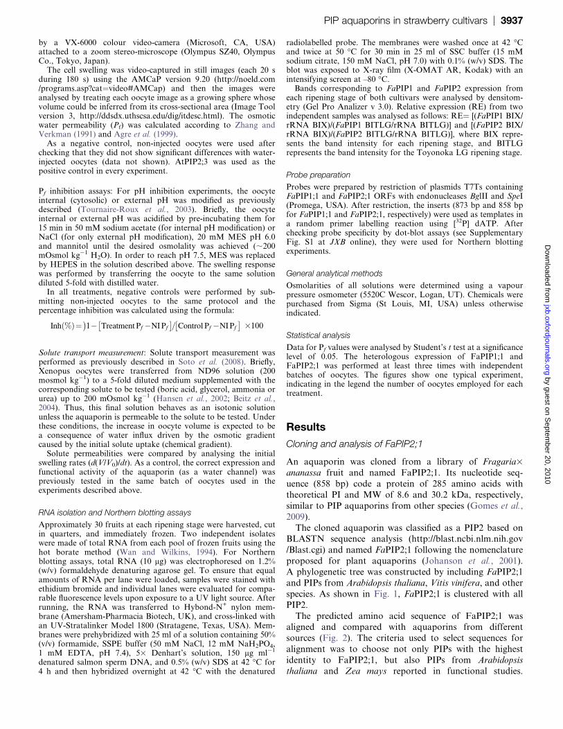
A phylogenetic tree was constructed by including FaPIP2;1
and PIPs from Arabidopsis thaliana, Vitis vinifera, and other
species. As shown in Fig. 1, FaPIP2;1 is clustered with all
PIP2.
The predicted amino acid sequence of FaPIP2;1 wasaligned and compared with aquaporins from different
sources (Fig. 2). The criteria used to select sequences for
alignment was to choose not only PIPs with the highest
identity to FaPIP2;1, but also PIPs from Arabidopsis
thaliana and Zea mays reported in functional studies.
PIP aquaporins in strawberry cultivars | 3937 by guest on S
eptember 20, 2010
jxb.oxfordjournals.orgD
ownloaded from
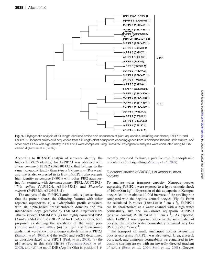
According to BLASTP analysis of sequence identity, the
higher hit (91% identity) for FaPIP2;1 was obtained withPyrus communis PIP2;2 (BAB40143.1), that belongs to the
same taxonomic family than Fragaria3ananassa (Rosaceae)
and that is also expressed in its fruit. FaPIP2;1 also presents
high identity percentage (>89%) with other PIP2 aquapor-
ins, for example, with Samanea saman (PIP2, AC17529.1),
Vitis vinifera (VvPIP2;4, ABN14353.1), and Phaseolus
vulgaris (PvPIP2;3, ABU94631.1).
The analysis of the FaPIP2;1 amino acid sequence showsthat the protein shares the following features with other
reported aquaporins: (i) a hydrophobic profile consistent
with six alpha-helical transmembrane domains and five
inter-helical loops (predicted by TMHMM, http://www.cbs
.dtu.dk/services/TMHMM/), (ii) two highly conserved NPA
(Asn-Pro-Ala) and the ar/R (Phe-His-Thr-Arg) motifs, both
proposed as defining the specificity of the water pore
(Forrest and Bhave, 2007), (iii) the Lys3 and Glu6 aminoacids, that were shown to undergo methylation in AtPIP2;1
(Santoni et al., 2006), (iv) the Ser280 and Ser283 determined
as phosphorylated in AtPIP2;1 (Prak et al., 2008), (v) the
pH sensor, in this case His199 (Tournaire-Roux et al.,
2003), and (vi) the motif DIE (Asp-Ile-Glu) in position 4–6,
recently proposed to have a putative role in endoplasmic
reticulum export signalling (Zelazny et al., 2009).
Functional studies of FaPIP2;1 in Xenopus laevisoocytes
To analyse water transport capacity, Xenopus oocytes
expressing FaPIP2;1 were exposed to a hypo-osmotic shock
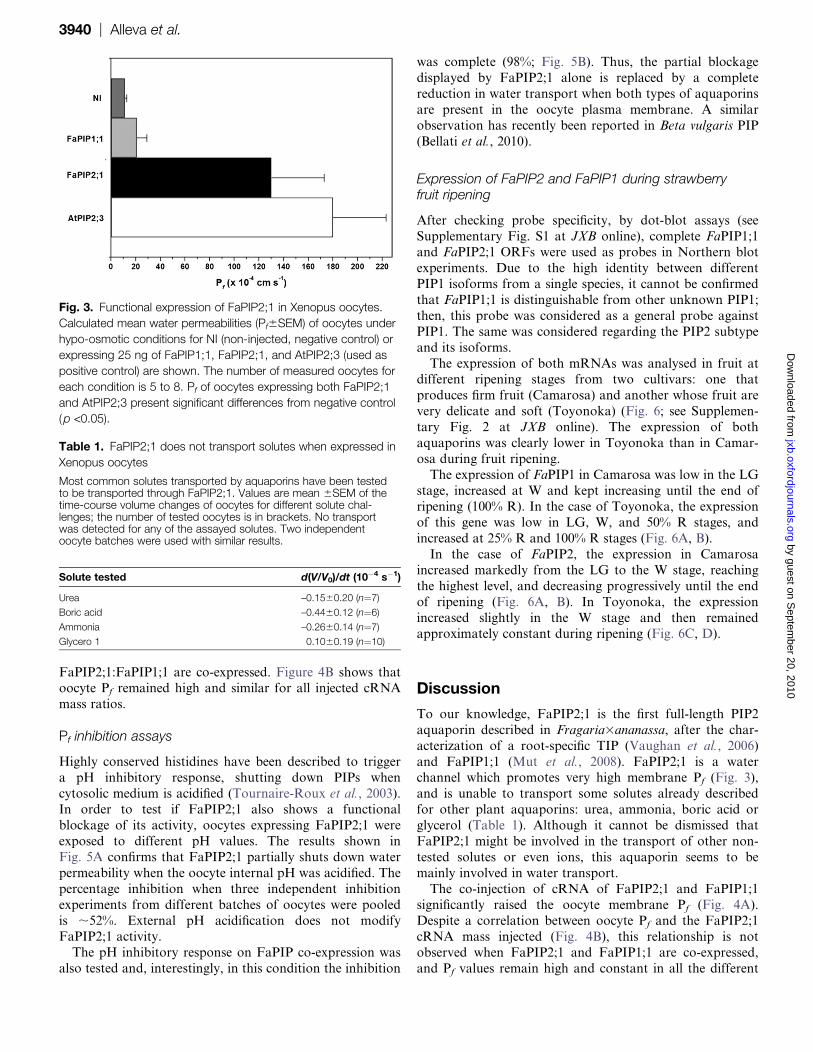
of 160 mOsm kg�1. Expression of this aquaporin in Xenopus
oocytes led to an almost 10-fold increase of the swelling ratecompared with the negative control oocytes (Fig. 3). From
the calculated Pf values (130643310�4 cm s�1), FaPIP2;1
can be characterized as a water channel with a high water
permeability, like the well-known aquaporin AtPIP2;3
(positive control; Pf 180643310�4 cm s�1). As expected,
when FaPIP1;1 was expressed alone in the same batch of
oocytes, the osmotic water permeability remained very low
(Pf 2168310�4 cm s�1).The transport of small, uncharged solutes across the
oocytes expressing FaPIP2;1 was also tested. Urea, glycerol,
boric acid, and ammonia permeability were analysed in iso-
osmotic swelling assays with an inwardly directed gradient
of solute (Beitz et al., 2004; Soto et al., 2008). Oocytes
Fig. 1. Phylogenetic analysis of full-length deduced amino acid sequences of plant aquaporins, including our clones, FaPIP2;1 and
FaPIP1;1. Deduced amino acid sequences from full-length plant aquaporins encoding genes from Arabidopsis thaliana, Vitis vinifera, and
other plant PIP2s with high identity to FaPIP2;1 were compared using Clustal W. Phylogenetic analyses were conducted using MEGA
version 4 (Tamura et al., 2007).
3938 | Alleva et al. by guest on S
eptember 20, 2010
jxb.oxfordjournals.orgD
ownloaded from

injected with 25 ng of FaPIP2;1 cRNA showed no signifi-
cant swelling rates for any of the solutes tested (Table 1).
Co-expression of FaPIP in Xenopus laevis oocytes
Our next step was to test if FaPIP co-expression enhances
water permeability. This approach requires a small amount
of the PIP with high Pf (in our case FaPIP2;1) to be co-
injected with a larger amount of the PIP1. Figure 4A clearly
shows that small amounts of FaPIP2;1 cRNA co-injected
with FaPIP1;1 cRNA (mass ratio 1:4, in mass values
3 ng:12 ng of FaPIP2;1:FaPIP1;1) increased Pf to181623310�4 cm s�1. This result represents an increment
of 79% compared with the Pf obtained by the injection of
3 ng of FaPIP2;1 alone (39611310�4 cm s�1).
It is interesting to note that, while there is a correla-
tion between the cRNA mass of FaPIP2;1 injected alone
and the resulting oocyte Pf, this correlation is lost when
Fig. 2. Alignment of predicted amino acid sequence of Fragaria3ananassa aquaporin (FaPIP2;1) with other aquaporins (ClustalX). The
predicted amino acid sequence of FaPIP2;1 was compared with aquaporins from different sources (aquaporins with the higher identity
with FaPIP2;1 or very well studied in the literature). Transmembrane domains are shown with a dashed line below the alignment; triangles
indicate a potential diacidic motif (putative ER signal); circles indicate the NPA selectivity filter; a star indicates His199, and inverted
triangles putative phosphorylated Ser residues.
PIP aquaporins in strawberry cultivars | 3939 by guest on S
eptember 20, 2010
jxb.oxfordjournals.orgD
ownloaded from
FaPIP2;1:FaPIP1;1 are co-expressed. Figure 4B shows thatoocyte Pf remained high and similar for all injected cRNA
mass ratios.
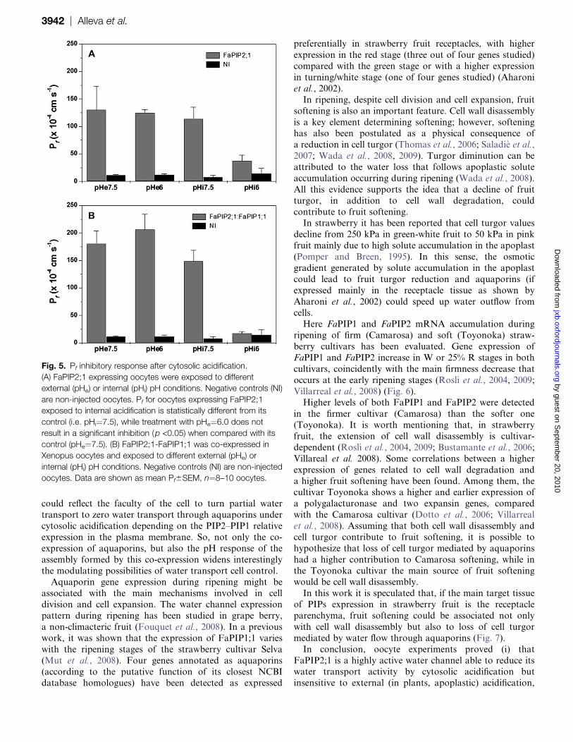
Pf inhibition assays
Highly conserved histidines have been described to trigger
a pH inhibitory response, shutting down PIPs when
cytosolic medium is acidified (Tournaire-Roux et al., 2003).In order to test if FaPIP2;1 also shows a functional
blockage of its activity, oocytes expressing FaPIP2;1 were
exposed to different pH values. The results shown in
Fig. 5A confirms that FaPIP2;1 partially shuts down water
permeability when the oocyte internal pH was acidified. The
percentage inhibition when three independent inhibition
experiments from different batches of oocytes were pooled
is ;52%. External pH acidification does not modifyFaPIP2;1 activity.
The pH inhibitory response on FaPIP co-expression was
also tested and, interestingly, in this condition the inhibition
was complete (98%; Fig. 5B). Thus, the partial blockage
displayed by FaPIP2;1 alone is replaced by a complete
reduction in water transport when both types of aquaporins
are present in the oocyte plasma membrane. A similar
observation has recently been reported in Beta vulgaris PIP
(Bellati et al., 2010).
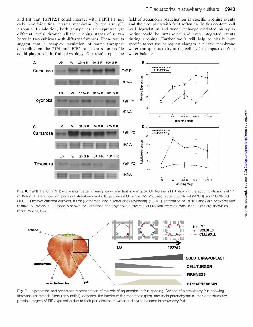
Expression of FaPIP2 and FaPIP1 during strawberryfruit ripening
After checking probe specificity, by dot-blot assays (see
Supplementary Fig. S1 at JXB online), complete FaPIP1;1and FaPIP2;1 ORFs were used as probes in Northern blot
experiments. Due to the high identity between different
PIP1 isoforms from a single species, it cannot be confirmed
that FaPIP1;1 is distinguishable from other unknown PIP1;
then, this probe was considered as a general probe against
PIP1. The same was considered regarding the PIP2 subtype
and its isoforms.
The expression of both mRNAs was analysed in fruit atdifferent ripening stages from two cultivars: one that
produces firm fruit (Camarosa) and another whose fruit are
very delicate and soft (Toyonoka) (Fig. 6; see Supplemen-
tary Fig. 2 at JXB online). The expression of both
aquaporins was clearly lower in Toyonoka than in Camar-
osa during fruit ripening.
The expression of FaPIP1 in Camarosa was low in the LG
stage, increased at W and kept increasing until the end ofripening (100% R). In the case of Toyonoka, the expression
of this gene was low in LG, W, and 50% R stages, and
increased at 25% R and 100% R stages (Fig. 6A, B).
In the case of FaPIP2, the expression in Camarosa
increased markedly from the LG to the W stage, reaching
the highest level, and decreasing progressively until the end
of ripening (Fig. 6A, B). In Toyonoka, the expression
increased slightly in the W stage and then remainedapproximately constant during ripening (Fig. 6C, D).
Discussion
To our knowledge, FaPIP2;1 is the first full-length PIP2
aquaporin described in Fragaria3ananassa, after the char-
acterization of a root-specific TIP (Vaughan et al., 2006)
and FaPIP1;1 (Mut et al., 2008). FaPIP2;1 is a waterchannel which promotes very high membrane Pf (Fig. 3),
and is unable to transport some solutes already described
for other plant aquaporins: urea, ammonia, boric acid or
glycerol (Table 1). Although it cannot be dismissed that
FaPIP2;1 might be involved in the transport of other non-
tested solutes or even ions, this aquaporin seems to be
mainly involved in water transport.
The co-injection of cRNA of FaPIP2;1 and FaPIP1;1significantly raised the oocyte membrane Pf (Fig. 4A).
Despite a correlation between oocyte Pf and the FaPIP2;1
cRNA mass injected (Fig. 4B), this relationship is not
observed when FaPIP2;1 and FaPIP1;1 are co-expressed,
and Pf values remain high and constant in all the different
Fig. 3. Functional expression of FaPIP2;1 in Xenopus oocytes.
Calculated mean water permeabilities (Pf6SEM) of oocytes under
hypo-osmotic conditions for NI (non-injected, negative control) or
expressing 25 ng of FaPIP1;1, FaPIP2;1, and AtPIP2;3 (used as
positive control) are shown. The number of measured oocytes for
each condition is 5 to 8. Pf of oocytes expressing both FaPIP2;1
and AtPIP2;3 present significant differences from negative control
(p <0.05).
Table 1. FaPIP2;1 does not transport solutes when expressed in
Xenopus oocytes
Most common solutes transported by aquaporins have been testedto be transported through FaPIP2;1. Values are mean 6SEM of thetime-course volume changes of oocytes for different solute chal-lenges; the number of tested oocytes is in brackets. No transportwas detected for any of the assayed solutes. Two independentoocyte batches were used with similar results.
Solute tested d(V/V0)/dt (10�4 s�1)
Urea –0.1560.20 (n¼7)
Boric acid –0.4460.12 (n¼6)
Ammonia –0.2660.14 (n¼7)
Glycero 1 0.1060.19 (n¼10)
3940 | Alleva et al. by guest on S
eptember 20, 2010
jxb.oxfordjournals.orgD
ownloaded from

mass ratios assayed. This result could be reflecting ER
processing of aquaporins when their cRNA are co-injected
in the same oocyte (Zelazny et al., 2007). Co-expressed
aquaporins from grape berries show a similar response to
the one reported here (Vandeleur et al., 2009).PIP1–PIP2 interaction has been reported both in Xenopus
oocytes and in living plant cells (Fetter et al., 2004; Zelazny
et al., 2007) and as a consequence, if PIP2 is not expressed,
PIP1 alone cannot enhance water permeability. It is
therefore expected that the modification of FaPIP1;1 and
FaPIP2;1 expression profiles during ripening reported here
could be another mechanism of controlling plasma mem-
brane water transport. That is, high levels of FaPIP1;1 atthe red stage (as in the case of Camarosa) does not
guarantee the enhancement of water transport unless
FaPIP2;1 is present. On the other hand, although FaPIP2;1
can concede high water permeability to the membrane, the
presence of FaPIP1;1 can co-operatively trigger much higher
values. Isolated vesicles of an enriched fraction of plasma
membrane from the 100% red stage fruit show very high
water permeability values (Mut et al., 2008), which supports
our hypothesis.When oocytes expressing FaPIP2;1 are subjected to
cytosolic acidification, membrane Pf is partially shut down
(Fig. 5A). These results are in accordance with the presence
of the highly conserved His199 in the FaPIP2;1 sequence,
a residue shown to be responsible for water transport
blockage under cytosolic acidification in other PIP2 aqua-
porins (Tournaire-Roux et al., 2003). It is interesting to
analyse the reduction of water transport under cytosolicacidification detected for FaPIP2;1 (partial inhibition)
compared with FaPIP2;1-FaPIP1;1 co-expression (total in-
hibition). This differential response of water channels, also
found for Beta vulgaris aquaporins (Bellati et al., 2010),
Fig. 4. Co-expression of FaPIP2;1 and FaPIP1;1. (A) Co-expression of 3 ng of cRNA of FaPIP2;1 with 12 ng of cRNA of FaPIP1;1 is
shown (white bar). As a control, 3 ng of FaPIP2;1 (black bar) and 12 ng of FaPIP1;1 (light grey bar) were injected separately. The
co-expression increased significantly the water permeability six times compared with the expression of FaPIP2;1 alone (p <0.05). Data are
expressed as mean Pf6SEM, n¼5 or 6 oocytes. NI are non-injected oocytes (grey bar). (B) Increasing cRNA mass of FaPIP2;1 injected
alone in oocytes (from 6 ng to 18 ng) shows a increasing Pf. This relationship is not observed in FaPIP2;1-FaPIP1;1 co-expressing
oocytes. Pf remains high but constant no matter the cRNA mass ratio injected. Data are shown as mean Pf 6SEM, n¼6–8 oocytes.
PIP aquaporins in strawberry cultivars | 3941 by guest on S
eptember 20, 2010
jxb.oxfordjournals.orgD
ownloaded from
could reflect the faculty of the cell to turn partial water
transport to zero water transport through aquaporins under
cytosolic acidification depending on the PIP2–PIP1 relative
expression in the plasma membrane. So, not only the co-
expression of aquaporins, but also the pH response of the
assembly formed by this co-expression widens interestingly
the modulating possibilities of water transport cell control.
Aquaporin gene expression during ripening might beassociated with the main mechanisms involved in cell
division and cell expansion. The water channel expression
pattern during ripening has been studied in grape berry,
a non-climacteric fruit (Fouquet et al., 2008). In a previous
work, it was shown that the expression of FaPIP1;1 varies
with the ripening stages of the strawberry cultivar Selva
(Mut et al., 2008). Four genes annotated as aquaporins
(according to the putative function of its closest NCBIdatabase homologues) have been detected as expressed
preferentially in strawberry fruit receptacles, with higher
expression in the red stage (three out of four genes studied)
compared with the green stage or with a higher expression
in turning/white stage (one of four genes studied) (Aharoni
et al., 2002).
In ripening, despite cell division and cell expansion, fruit
softening is also an important feature. Cell wall disassembly
is a key element determining softening; however, softeninghas also been postulated as a physical consequence of
a reduction in cell turgor (Thomas et al., 2006; Saladie et al.,
2007; Wada et al., 2008, 2009). Turgor diminution can be
attributed to the water loss that follows apoplastic solute
accumulation occurring during ripening (Wada et al., 2008).
All this evidence supports the idea that a decline of fruit
turgor, in addition to cell wall degradation, could
contribute to fruit softening.In strawberry it has been reported that cell turgor values
decline from 250 kPa in green-white fruit to 50 kPa in pink
fruit mainly due to high solute accumulation in the apoplast
(Pomper and Breen, 1995). In this sense, the osmotic
gradient generated by solute accumulation in the apoplast
could lead to fruit turgor reduction and aquaporins (if
expressed mainly in the receptacle tissue as shown by
Aharoni et al., 2002) could speed up water outflow fromcells.
Here FaPIP1 and FaPIP2 mRNA accumulation during
ripening of firm (Camarosa) and soft (Toyonoka) straw-
berry cultivars has been evaluated. Gene expression of
FaPIP1 and FaPIP2 increase in W or 25% R stages in both
cultivars, coincidently with the main firmness decrease that
occurs at the early ripening stages (Rosli et al., 2004, 2009;
Villarreal et al., 2008) (Fig. 6).Higher levels of both FaPIP1 and FaPIP2 were detected
in the firmer cultivar (Camarosa) than the softer one
(Toyonoka). It is worth mentioning that, in strawberry
fruit, the extension of cell wall disassembly is cultivar-
dependent (Rosli et al., 2004, 2009; Bustamante et al., 2006;
Villareal et al. 2008). Some correlations between a higher
expression of genes related to cell wall degradation and
a higher fruit softening have been found. Among them, thecultivar Toyonoka shows a higher and earlier expression of
a polygalacturonase and two expansin genes, compared
with the Camarosa cultivar (Dotto et al., 2006; Villarreal
et al., 2008). Assuming that both cell wall disassembly and
cell turgor contribute to fruit softening, it is possible to
hypothesize that loss of cell turgor mediated by aquaporins
had a higher contribution to Camarosa softening, while in
the Toyonoka cultivar the main source of fruit softeningwould be cell wall disassembly.
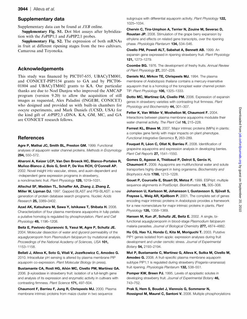
In this work it is speculated that, if the main target tissue
of PIPs expression in strawberry fruit is the receptacle
parenchyma, fruit softening could be associated not only
with cell wall disassembly but also to loss of cell turgor
mediated by water flow through aquaporins (Fig. 7).
In conclusion, oocyte experiments proved (i) that
FaPIP2;1 is a highly active water channel able to reduce itswater transport activity by cytosolic acidification but
insensitive to external (in plants, apoplastic) acidification,
Fig. 5. Pf inhibitory response after cytosolic acidification.
(A) FaPIP2;1 expressing oocytes were exposed to different
external (pHe) or internal (pHi) pH conditions. Negative controls (NI)
are non-injected oocytes. Pf for oocytes expressing FaPIP2;1
exposed to internal acidification is statistically different from its
control (i.e. pHi¼7.5), while treatment with pHe¼6.0 does not
result in a significant inhibition (p <0.05) when compared with its
control (pHe¼7.5). (B) FaPIP2;1-FaPIP1;1 was co-expressed in
Xenopus oocytes and exposed to different external (pHe) or
internal (pHi) pH conditions. Negative controls (NI) are non-injected
oocytes. Data are shown as mean Pf6SEM, n¼8–10 oocytes.
3942 | Alleva et al. by guest on S
eptember 20, 2010
jxb.oxfordjournals.orgD
ownloaded from
and (ii) that FaPIP2;1 could interact with FaPIP1;1 not
only modifying final plasma membrane Pf but also pH
response. In addition, both aquaporins are expressed (at
different levels) through all the ripening stages of straw-
berry in two cultivars with different firmness. These results
suggest that a complex regulation of water transport
depending on the PIP1 and PIP2 rate expression profile
could play a role in fruit physiology. Our results open the
field of aquaporin participation in specific ripening events
and their coupling with fruit softening. In this context, cell
wall degradation and water exchange mediated by aqua-
porins could be juxtaposed and even integrated events
during ripening. Further work will help to clarify how
specific target tissues request changes in plasma membrane
water transport activity at the cell level to impact on fruit
water balance.
Fig. 6. FaPIP1 and FaPIP2 expression pattern during strawberry fruit ripening. (A, C). Northern blot showing the accumulation of FaPIP
mRNA in different ripening stages of strawberry fruits: large green (LG), white (W), 25% red (25%R), 50% red (50%R), and 100% red
(100%R) for two different cultivars, a firm (Camarosa) and a softer one (Toyonoka). (B, D) Quantification of FaPIP1 and FaPIP2 expression
relative to Toyonoka LG stage is shown for Camarosa and Toyonoka cultivars (Gel Pro Analizer v 3.0 was used). Data are shown as
mean 6SEM, n¼2.
Fig. 7. Hypothetical and schematic representation of the role of aquaporins in fruit ripening. Section of a strawberry fruit showing
fibrovascular strands (vascular bundles), achenes, the interior of the receptacle (pith), and main parenchyma; all marked tissues are
possible targets of PIP expression due to their participation in water and solute balance in strawberry fruit.
PIP aquaporins in strawberry cultivars | 3943 by guest on S
eptember 20, 2010
jxb.oxfordjournals.orgD
ownloaded from
Supplementary data
Supplementary data can be found at JXB online.
Supplementary Fig. S1. Dot blot assays after hybridiza-
tion with the FaPIP1;1 and FaPIP2;1 probes.
Supplementary Fig. S2. The expression of both mRNAs
in fruit at different ripening stages from the two cultivars,Camarosa and Toyonoka.
Acknowledgements
This study was financed by PICT07-655, UBACyTM001,
and CONICET-PIP5154 grants to GA and by PICT06-01804 and UBACyTM602 grants to KA. Our particular
thanks are due to Noel Danjou who improved the AMCAP
program (version 9.20) to allow the acquisition of still
images as requested, Alex Paladini (INGEBI, CONICET)
who designed and provided us with built-in chambers for
oocyte experiments, and Mark Daniels (UCSD, USA) for
the kind gift of AtPIP2;3 cDNA. KA, GM, MC, and GA
are CONICET research fellows.
References
Agre P, Mathai JC, Smith BL, Preston GM. 1999. Functional
analyses of aquaporin water channel proteins. Methods in Enzymology
294, 550–572.
Aharoni A, Keizer LCP, Van Den Broeck HC, Blanco-Portales R,
Munoz-Blanco J, Bois G, Smit P, De Vos RCH, O’Connell AP.
2002. Novel insight into vascular, stress, and auxin-dependent and
-independent gene expression programs in strawberry,
a nonclimacteric fruit. Plant Physiology 129, 1019–1031.
Altschul SF, Madden TL, Schaffer AA, Zhang J, Zhang Z,
Miller W, Lipman DJ. 1997. Gapped BLAST and PSI-BLAST: a new
generation of protein database search programs. Nucleic Acids
Research 25, 3389–3402.
Azad AK, Katsuhara M, Sawa Y, Ishikawa T, Shibata H. 2008.
Characterization of four plasma membrane aquaporins in tulip petals:
a putative homolog is regulated by phosphorylation. Plant and Cell
Physiology 49, 1196–1208.
Beitz E, Pavlovic-Djuranovic S, Yasui M, Agre P, Schultz JE.
2004. Molecular dissection of water and glycerol permeability of the
aquaglyceroporin from Plasmodium falciparum by mutational analysis.
Proceedings of the National Academy of Sciences, USA 101,
1153–1158.
Bellati J, Alleva K, Soto G, Vitali V, Jozefkowicz C, Amodeo G.
2010. Intracellular pH sensing is altered by plasma membrane PIP
aquaporin co-expression. Plant Molecular Biology (in press).
Bustamante CA, Rosli HG, Anon MC, Civello PM, Martınez GA.
2006. b-xylosidase in strawberry fruit: isolation of a full-length gene
and analysis of its expression and enzymatic activity in cultivars with
contrasting firmness. Plant Science 171, 497–504.
Chaumont F, Barrieu F, Jung R, Chrispeels MJ. 2000. Plasma
membrane intrinsic proteins from maize cluster in two sequence
subgroups with differential aquaporin activity. Plant Physiology 122,
1025–1034.
Chervin C, Tira-Umphon A, Terrier N, Zouine M, Severac D,
Roustan JP. 2008. Stimulation of the grape berry expansion by
ethylene and effects on related gene transcripts, over the ripening
phase. Physiologia Plantarum 134, 534–546.
Civello PM, Powell ALT, Sabehat A, Bennett AB. 1999. An
expansin gene expressed in ripening strawberry fruit. Plant Physiology
121, 1273–1279.
Coombe BG. 1976. The development of freshy fruits. Annual Review
of Plant Physiology 27, 207–228.
Daniels MJ, Mirkov TE, Chrispeels MJ. 1994. The plasma
membrane of Arabidopsis thaliana contains a mercury-insensitive
aquaporin that is a homolog of the tonoplast water channel protein
TIP. Plant Physiology 106, 1325–1333.
Dotto MC, Martınez GA, Civello PM. 2006. Expression of expansin
genes in strawberry varieties with contrasting fruit firmness. Plant
Physiology and Biochemistry 44, 301–307.
Fetter K, Van Wilder V, Moshelion M, Chaumont F. 2004.
Interactions between plasma membrane aquaporins modulate their
water channel activity. The Plant Cell 16, 215–228.
Forrest KL, Bhave M. 2007. Major intrinsic proteins (MIPs) in plants:
a complex gene family with major impacts on plant phenotype.
Functional Integrative Genomics 7, 263–289.
Fouquet R, Leon C, Ollat N, Barrieu F. 2008. Identification of
grapevine aquaporins and expression analysis in developing berries.
Plant Cell Reports 27, 1541–1550.
Gomes D, Agasse A, Thiebaud P, Delrot S, Geros H,
Chaumont F. 2009. Aquaporins are multifunctional water and solute
transporters highly divergent in living organisms. Biochemistry and
Biophysics Acta 1788, 1213–1228.
Gouet P, Courcelle E, Stuart DI, Metoz F. 1999. ESPript: multiple
sequence alignments in PostScript. Bioinformatics 15, 305–308.
Johanson U, Karlsson M, Johansson I, Gustavsson S, Sjovall S,
Fraysse L, Weig AR, Kjellbom P. 2001. The complete set of genes
encoding major intrinsic proteins in Arabidopsis provides a framework
for a new nomenclature for major intrinsic proteins in plants. Plant
Physiology 126, 1358–1369.
Hansen M, Kun JF, Schultz JE, Beitz E. 2002. A single, bi-
functional aquaglyceroporin in blood-stage Plasmodium falciparum
malaria parasites. Journal of Biological Chemistry 277, 4874–4882.
Hu CG, Hao YJ, Honda C, Kita M, Moriguchi T. 2003. Putative
PIP1 genes isolated from apple: expression analyses during fruit
development and under osmotic stress. Journal of Experimental
Botany 54, 2193–2194.
Mut P, Bustamante C, Martınez G, Alleva K, Sutka M, Civello M,
Amodeo G. 2008. A fruit-specific plasma membrane aquaporin
subtype PIP1;1 is regulated during strawberry (Fragaria3ananassa)
fruit ripening. Physiologia Plantarum 132, 538–551.
Pomper KW, Breen PJ. 1995. Levels of apoplastic solutes in
developing strawberry fruit. Journal of Experimental Botany 46,
743–752.
Prak S, Hem S, Boudet J, Viennois G, Sommerer N,
Rossignol M, Maurel C, Santoni V. 2008. Multiple phosphorylations
3944 | Alleva et al. by guest on S
eptember 20, 2010
jxb.oxfordjournals.orgD
ownloaded from
in the C-terminal tail of plant plasma membrane aquaporins: role in
subcellular trafficking of AtPIP2;1 in response to salt stress. Molecular
Cell Proteomics 7, 1019–1030.
Rose T, Schultz E, Henikoff J, Pietrokovski S, McCallum C,
Henikoff S. 1998. Consensus-degenerate hybrid oligonucleotide
primers for amplification of distantly related sequences. Nucleic Acids
Research 26, 1628–1635.
Rosli HG, Civello PM, Martınez GA. 2004. Changes in cell wall
composition of three Fragaria3ananassa cultivars with different softening
rate during ripening. Plant Physiology and Biochemistry 42, 823–831.
Rosli HG, Civello PM, Martınez GA. 2009. Alpha-l-
arabinofuranosidase from strawberry fruit: cloning of three cDNAs,
characterization of their expression and analysis of enzymatic activity in
cultivars with contrasting firmness. Plant Physiology and Biochemistry
47, 272–281.
Saladie M, Matas AJ, Isaacson T, et al. 2007. A reevaluation of the
key factors that influence tomato fruit softening and integrity. Plant
Physiology 144, 1012–1028.
Santoni V, Verdoucq L, Sommerer N, Vinh J, Pflieger D,
Maurel C. 2006. Methylation of aquaporins in plant plasma
membrane. Biochemistry Journal 400, 189–197.
Soto G, Alleva K, Mazzella MA, Amodeo G, Muschietti JP. 2008.
AtTIP1;3 and AtTIP5;1, the only highly expressed Arabidopsis pollen-
specific aquaporins, transport water and urea. FEBS Letters 582,
4077–4082.
Tamura K, Dudley J, Nei M, Kumar S. 2007. MEGA4: Molecular
Evolutionary Genetics Analysis (MEGA) software version 4.0. Molecular
Biology and Evolution 24, 1596–1599.
Thomas TR, Matthews MA, Shackel KA. 2006. Direct in situ
measurement of cell turgor in grape (Vitis vinifera L.) berries during
development and in response to plant water deficits. Plant, Cell, and
Environment 29, 993–1001.
Thompson JD, Gibson TJ, Plewniak F, Jeanmougin F,
Higgins DG. 1997. The CLUSTAL_X windows interface: flexible
strategies for multiple sequence alignment aided by quality analysis
tools. Nucleic Acids Research 25, 4876–4882.
Tournaire-Roux C, Sutka M, Javot H, Gout E, Gerbeau P,
Luu DT, Bligny R, Maurel C. 2003. Cytosolic pH regulates root
water transport during anoxic stress through gating of aquaporins.
Nature 25, 393–397.
Tyerman SD, Bohnert HJ, Maurel C, Steudle E, Smith JAC.
1999. Plant aquaporins: their molecular biology, biophysics and
significance for water relations. Journal of Experimental Botany 50,
1055–1071.
Vandeleur RK, Mayo G, Shelden MC, Gilliham M, Kaiser BN,
Tyerman SD. 2009. The role of plasma membrane intrinsic protein
aquaporins in water transport through roots: diurnal and drought
stress responses reveal different strategies between isohydric and
anisohydric cultivars of grapevine. Plant Physiology 149, 445–460.
Vaughan SP, James DJ, Lindsey K, Massiah AJ. 2006.
Characterization of FaRB7, a near root-specific gene from strawberry
(Fragaria3ananassa Duch.) and promoter activity analysis in
homologous and heterologous hosts. Journal of Experimental Botany
57, 3901–3910.
Villarreal NM, Rosli HG, Martınez GA, Civello PM. 2008.
Polygalacturonase activity and expression of related genes during
ripening of strawberry cultivars with contrasting fruit firmness.
Postharvest Biology and Technology 47, 141–150.
Wada H, Shackel K, Matthews M. 2008. Fruit ripening in Vitis
vinifera: apoplastic solute accumulation accounts for pre-veraison
turgor loss in berries. Planta 227, 1351–1361.
Wada H, Matthews M, Shackel K. 2009. Seasonal pattern of
apoplastic solute accumulation and loss of cell turgor during ripening
of Vitis vinifera fruit under field conditions. Journal of Experimental
Botany 60, 1773–1781.
Wan CY, Wilkins TA. 1994. A modified hot borate method
significantly enhances the yield of high-quality RNA from cotton
(Gossypium hirsutum L.). Analytical Biochemistry 15, 7–12.
Zelazny E, Borst JW, Muylaert M, Batoko H, Hemminga MA,
Chaumont F. 2007. FRET imaging in living maize cells reveals that
plasma membrane aquaporins interact to regulate their subcellular
localization. Proceedings of National Academy of Sciences, USA 104,
12359–12364.
Zelazny E, Miecielica U, Borst JW, Hemminga MA, Chaumont F.
2009. An N-terminal diacidic motif is required for the trafficking of
maize aquaporins ZmPIP2;4 and ZmPIP2;5 to the plasma membrane.
The Plant Journal 57, 346–355.
Zhang RB, Verkman AS. 1991. Water and urea permeability
properties of Xenopus oocytes: expression of mRNA from toad urinary
bladder. American Journal of Physiology 260, C26–C34.
Zhou Y, Setz N, Niemietz C, Qu H, Offler CE, Tyerman SD,
Patrick JW. 2007. Aquaporins and unloading of phloem-imported
water in coats of developing bean seeds. Plant, Cell and Environment
30, 1566–1577.
PIP aquaporins in strawberry cultivars | 3945 by guest on S
eptember 20, 2010
jxb.oxfordjournals.orgD
ownloaded from
Related Documents