Cloning and Sequence Analysis of Novel DNA Polymerases from Thermophilic Geobacillus Species Isolated from Hot Springs in Turkey: Characterization of a DNA Polymerase I from Geobacillus kaue Strain NB Melike Çağlayan & Neş’e Bilgin Received: 17 March 2011 /Accepted: 2 August 2011 / Published online: 26 August 2011 # Springer Science+Business Media, LLC 2011 Abstract The complete coding sequences of the polA genes from seven thermophilic Geobacillus species, isolated from hot springs of Gönen and Hisaralan in Turkey, were cloned and sequenced. The polA genes of these Geobacillus species contain a long open reading frame of 2,637 bp encoding DNA polymerase I with a calculated molecular mass of 99 kDa. Amino acid sequences of these Geobacillus DNA polymerases are closely related. The multiple sequence alignments show all include the conserved amino acids in the polymerase and 5′-3′ exonuclease domains, but the catalytic residues varied in 3′-5′ exonuclease domain of these Geobacillus DNA polymerases. One of them, DNA polymerase I from Geobacillus kaue strain NB (Gkaue polI) is purified to homogeneity and biochemically characterized in vitro. The optimum temperature for enzymatic activity of Gkaue polI is 70 °C at pH 7.5–8.5 in the presence of 8 mM Mg 2+ and 80–100 mM of monovalent ions. The addition of polyamines stimulates the polymerization activity of the enzyme. Three-dimensional structure of Gkaue polI predicted using homology modeling confirmed the conservation of all the functionally important regions in the polymerase active site. Keywords Geobacillus sp. . DNA polymerase I . Geobacillus kaue strain NB polI . DNA polymerization in vitro . Homology modeling Introduction DNA polymerases are the essential enzymes in all organisms mainly responsible for the replication and repair of DNA [1]. DNA polymerases are classified into five families: A, B, C, X, and a newly identified Y family [2]. Family A DNA polymerases, including human DNA polA and Escherichia coli DNA polymerase I, share fundamental structural features in their polymerase active sites [3]. DNA polymerase I (DNA deoxynucleotidyltransferase, EC 2.7.7.7) is encoded by the polA gene, which consists of a single polypeptide chain comprising three distinct domains. Appl Biochem Biotechnol (2011) 165:1188–1200 DOI 10.1007/s12010-011-9337-5 M. Çağlayan : N. Bilgin (*) Department of Molecular Biology and Genetics, Bogazici University, Bebek, 34342 Istanbul, Turkey e-mail: [email protected]

Welcome message from author
This document is posted to help you gain knowledge. Please leave a comment to let me know what you think about it! Share it to your friends and learn new things together.
Transcript

Cloning and Sequence Analysis of Novel DNAPolymerases from Thermophilic Geobacillus SpeciesIsolated from Hot Springs in Turkey: Characterizationof a DNA Polymerase I from Geobacillus kaue Strain NB
Melike Çağlayan & Neş’e Bilgin
Received: 17 March 2011 /Accepted: 2 August 2011 /Published online: 26 August 2011# Springer Science+Business Media, LLC 2011
Abstract The complete coding sequences of the polA genes from seven thermophilicGeobacillus species, isolated from hot springs of Gönen and Hisaralan in Turkey, were clonedand sequenced. The polA genes of these Geobacillus species contain a long open readingframe of 2,637 bp encoding DNA polymerase I with a calculated molecular mass of 99 kDa.Amino acid sequences of these Geobacillus DNA polymerases are closely related. Themultiple sequence alignments show all include the conserved amino acids in the polymeraseand 5′-3′ exonuclease domains, but the catalytic residues varied in 3′-5′ exonuclease domainof these Geobacillus DNA polymerases. One of them, DNA polymerase I from Geobacilluskaue strain NB (Gkaue polI) is purified to homogeneity and biochemically characterized invitro. The optimum temperature for enzymatic activity of Gkaue polI is 70 °C at pH 7.5–8.5in the presence of 8 mM Mg2+ and 80–100 mM of monovalent ions. The addition ofpolyamines stimulates the polymerization activity of the enzyme. Three-dimensional structureof Gkaue polI predicted using homology modeling confirmed the conservation of all thefunctionally important regions in the polymerase active site.
Keywords Geobacillus sp. . DNA polymerase I .Geobacillus kaue strain NB polI . DNApolymerization in vitro . Homology modeling
Introduction
DNA polymerases are the essential enzymes in all organisms mainly responsible for thereplication and repair of DNA [1]. DNA polymerases are classified into five families: A, B, C,X, and a newly identified Y family [2]. Family A DNA polymerases, including human DNApolA and Escherichia coli DNA polymerase I, share fundamental structural features in theirpolymerase active sites [3].
DNA polymerase I (DNA deoxynucleotidyltransferase, EC 2.7.7.7) is encoded by thepolA gene, which consists of a single polypeptide chain comprising three distinct domains.
Appl Biochem Biotechnol (2011) 165:1188–1200DOI 10.1007/s12010-011-9337-5
M. Çağlayan : N. Bilgin (*)Department of Molecular Biology and Genetics, Bogazici University, Bebek, 34342 Istanbul, Turkeye-mail: [email protected]

These three domains are the C-terminal domain with DNA polymerase activity, the centraldomain with 3′-5′ exonuclease (or proofreading) activity and the N-terminal domain with 5′-3′exonuclease activity [4]. The C-terminal domain includes the nucleotide and amino acidconservation in the three highly conserved regions (known asmotifs A, B, andC) residing in threedistinct subdomains designated as thumb, palm, and fingers [5]. The palm subdomain has thecatalytic center and contains the conserved carboxylate residues, while the thumb and fingerssubdomains are responsible for the binding of DNA and the incoming dNTP, respectively [6].
A few moderately thermostable DNA polymerase I have been purified and characterizedfrom the thermophilic members of the family Bacillaceae [7], especially from the genusBacillus and from a recently identified new genus Geobacillus: Bacillus stearothermophilus[8–10], Bacillus caldotenax [11, 12], Geobacillus sp. MKK [13], and Geobacilluscaldoxylolyticus TK4 [14]. The crystal structure of a family A member DNA polymeraseI from the genus Bacillus has only been identified for B. stearothermophilus DNApolymerase large fragment, known as Bacillus fragment (BF) [15, 16]. BF is a segment ofthe polymerase gene that encodes the C-terminal 592 amino acids of the full-length B.stearothermophilus DNA polymerase I and has the polymerase activity but lacks the 3′-5′exonuclease and the 5′-3′ exonuclease activities of the full-length protein [17, 18].
In this study, we report seven new members of family A DNA polymerases fromthermophilicGeobacillus species, collected from Gönen and Hisaralan hot springs in Turkey.DNA polymerase I sequences from seven thermophilic Geobacillus species were determinedusing PCR-based cloning and subsequent sequencing of the polA genes. One of them, DNApolymerase I from Geobacillus kaue strain NB (Gkaue polI) was expressed in E. coli andpurified and characterized biochemically in vitro. The three-dimensional structure of GkauepolI were predicted via homology modeling using the crystal structure of BF as a template.
Materials and Methods
Growth Conditions
Geobacillus species were cultivated at 70 °C in a growth medium containing 4 g yeast extract,8 g tryptone, 3 g NaCl, 0.1 g nitrilotriacetic acid, 0.06 g CaSO4·2H2O, 0.1 g MgSO4·7H2O,0.1 g KNO3, 0.69 g NaNO3, 0.1 g Na2HPO4, 0.28 mg FeCl3, 2.2 mg MnSO4·H2O, 0.5 mgZnSO4·7H2O, 0.016 mg CuSO4, 0.025 mg Na2MoO4·2H2O, 0.046 mg CoCl2·6H2O, and0.005 ml concentrated H2SO4 in 1 l at pH 7.5. G. kaue strain NB, whose DNA polymerase Iwas characterized in this study, was isolated from a hot spring in Gönen, and deposited in theBacillus Stock Center at the University of Ohio (accession no. BGSC105A2).
Cloning and Amino Acid Sequence Analysis of the polA Genes
We designed degenerate primers (Table 1) in order to amplify the previously unknown polAgenes from seven Geobacillus species based on the most conserved regions of previouslyknown polA genes from the Bacillus species [8–12]. The full-length polA gene sequenceswere determined by plasmid DNA sequencing of the genes after their PCR-based cloninginto pETM-20 expression vector (Novagen). For this purpose, the oligonucleotide primersF-NcoI and R-KpnI (Table 1) were designed. PCR reactions were in 25-μl reaction volumecontaining 1X Pfu buffer, 1.5 mM of MgCl2, 0.2 mM of each dNTP, 1 μM of each primer,50 ng of genomic DNA, and 0.25 unit of Pfu DNA polymerase. The thermal cyclingconditions were as follows: 2 min and 30 s at 94 °C for initial denaturation, followed by 30
Appl Biochem Biotechnol (2011) 165:1188–1200 1189

cycles of 30 s at 95 °C for denaturation, 1 min and 30 s at 55 °C for annealing, and 3 min at72 °C for extension. The amplified PCR product was inserted between the NcoI and KpnIrestriction enzyme cleavage sites into the multiple cloning region of pETM-20 vector; 1 μgPCR product and pETM-20 vector plasmid DNAwere firstly digested with 1 unit of NcoI at37 °C for 1.5 h and subsequently digested by 1 unit of KpnI at 37 °C for an additional 1.5 h.Double-digested PCR product and pETM-20 vector were ligated overnight at 4 °C by T4DNA ligase. Ligation mixture was then used for the transformation of E. coli TOP10F'cells. Purified plasmids harboring correctly inserted polA genes were then confirmed byrestriction enzyme digestion and PCR amplification using pETM-20-specific primer pair F-T7and R-pRSET (Table 1). The full-length polA gene sequences were determined by plasmidDNA sequencing using a set of primers (Table 1). The name of the plasmids carrying thecloned DNA polymerase I genes from seven Geobacillus species were given in Table 2. Thenucleotide sequence analysis and homology search of the polA genes were performed usingBasic Local Alignment Search Tool. For sequence alignments, multiple sequence alignmenttool CLUSTAL W was used [19]. The phylogenetic and evolutionary analyses wereconducted using Molecular Evolutionary Genetics Analysis (MEGA) version 4.0 [20] byneighbor-joining strategy [21].
Structural Homology Modeling
The three-dimensional (3D) models for the polymerase domain of Gkaue polI weregenerated using SWISS-MODEL [22] via homology modeling [23]. The crystal structure ofDNA polymerase I large fragment from B. stearothermophilus (BF, pdb code 1XWL) wasused as the template structure [15, 16]. The predicted models were visualized using PyMOL(DeLano Scientific).
Table 1 Oligonucleotide primers used for PCR amplification and sequencing of the polA genes
Primer 5′-Sequencea
F-40 AGATTGAAGAAAAAACTCGT
F-272 GCRTGGTACAATAGRACAAGG A
F-339 GAYGGMARCAGYSTGGCDTA
F-639 GAAGCGGGACGAYATTAT
F-1300 ATG CCC CGATTGTCGGAATC
F-2175 GADCCCBAACYTGCACGARCTSATT
R-126 ACCAGCAAGTTGGGTCGGC
R-780 CGGTGATCCCTTTTTTCGTA
R-2030 TTCGACGATTTCATGGTGGG
R-2605 ACRTABCCTTTYTGTTTY
R-2950 TYTTATTTSGCRTCRCRTACCAY
F-poll GCRTGGTACAATAGRACAAGGA
R-poll TYTTATTTSGGRTCRTACCAY
F-NcoI ATCACCATGGGAATGAGATTGAAGAAAAACTCGT
R-KpnI ATTAGGTACCTTATTTGGGATCGTACCACGTCGG
F-T7 TAATACGACTCACTATAGGG
R-pRSET TAGTTATTGCTCAGCGGTGG
a R=A/G, Y=T/C, M=A/C, S=G/C, N=A/G/T/C
1190 Appl Biochem Biotechnol (2011) 165:1188–1200

Expression and Purification
Gkaue polI was expressed in E. coli JM109(DE3) cells. E. coli cells harboring pGkaueNBpolIwere grown at 37 °C in LB medium containing 150 μg/ml ampicillin. When OD600 reached0.6, the cells were induced by the addition of isopropyl-β-D-thiogalactopyranoside (IPTG) toa final concentration of 1 mM and growthwas continued for another 4 h. Cells were harvested at7,800×g at 4 °C for 15 min and stored at −80 °C until use. For protein purification, cells weresuspended in 15 ml of ice-cold Ni2+-affinity buffer (20 mM Na-phosphate buffer (pH 7.4),0.5 M NaCl, 0.1 mM PMSF, 20 mM imidazole, 1 mM DTE) and disrupted using a bead-beater (three times for 30 s). After centrifugation at 30,000×g for 30 min at 4 °C to get rid ofthe cell debris, the clear supernatant was loaded directly to a 5 ml HisTrap FF column (GEHealthcare) equilibrated with Ni2+-affinity buffer. A linear gradient from 20 to 500 mMimidazole was applied. Column fractions were analyzed by 10% sodium dodecyl sulfate-polyacrylamide gel electrophoresis (SDS-PAGE). The fractions containing the fusion proteinwere pooled and digested with TEV protease (1:300, TEV protease/fusion protein) overnightat 4 °C during dialysis against TEV protease digestion buffer (50 mM Tris–HCl (pH 7.9),1 mM MgCl2, 1 mM DTE, and 300 mM KCl). After digestion, the sample was reapplied to asecond HisTrap FF column and the purified Gkaue polI was collected in the flowthrough. Thepurified protein was concentrated, dialyzed overnight against polymerase storage buffer(20 mM Tris–HCl (pH 7.9), 100 mM KCl, 0.1 mM EDTA, 1 mM DTE, 50% glycerol, and0.1% Triton-X 100), and stored at −20 °C. Protein concentration was determined using theBradford assay [24].
Biochemical Characterization Assays
The polymerase activity of Gkaue polI was measured under standard DNA polymerizingassay conditions: The reaction mixture (in 50 μl) contained 0.2 mM of each dCTP, dTTP,and dGTP; 0.2 mM [3H]dATP (specific activity of 400 cpm/pmol); and 0.5 μg activated calfthymus DNA (having 65% of single-stranded form) in polymerase reaction buffer 920 mMTris–HCl (pH 8.0), 50 mM KCl) including 1.5 mM MgCl2. Reactions were started by theaddition of 2 pmol of Gkaue polI into prewarmed reaction mixture at 70 °C. Reactions wereterminated by the addition of ice-cold 10% TCA containing 10 mM sodium pyrophosphateand the samples were left on ice until filtration. Precipitates were collected on glass fiberfilters (Whatman, GF/C), washed three times with ice-cold 10% TCA then with ice-coldisopropanol, dried, and counted in a liquid scintillation counter.
The optimum temperature of Gkaue polI was determined by measuring the polymeraseactivity at a temperature range from 40 to 100 °C for 15 min under standard DNA polymerizing
Table 2 The name of the plasmids carrying the polA genes of Geobacillus species identified in this work
Source Geobacillus species Name of the plasmid
Geobacillus anatolicus pGanapolI
G. anatolicus strain C4 pGanaC4polI
Geobacillus bogazici pGbogazicipolI
Anoxybacillus sp. NB pAnoxypolI
Geobacillus kaue strain NB pGkaueNBpolI
G. kaue strain MC pGkaueMCpolI
G. kaue strain E1 pGkaueE1polI
Appl Biochem Biotechnol (2011) 165:1188–1200 1191

assay conditions. In order to test thermostability, the enzyme was portioned and incubated at atemperature range from 50 to 90 °C for time intervals from 1 to 60 min. After this incubation,the samples were left on ice until each sample was prewarmed to 70 °C and their polymeraseactivity was assayed at 70 °C for 15 min under standard DNA polymerizing assay conditions.When added, 0.1 mg/ml BSA was present in the reaction mixtures as a stabilizer.
The pH optimum of Gkaue polI was determined by assaying polymerase activity at 70 °Cfor 15 min in a triple buffer system containing 50 mM of each of Bis-Tris propane, N-cyclohexyl-2-aminoethanesulfonic acid (CHES), and 2-(N-morpholino)ethanesulfonic acid(MES). All three of these buffers were mixed together before adjusting the pH to a series ofpH values between 5.0 to 9.5 (with HCl) so that changes in the activity of Gkaue polI thatmight be caused by different buffer components were eliminated.
The optimum concentrations of divalent ions (0 to15 mM of MgCl2 or MnCl2) andmonovalent ions (0 to 300 mM of KCl, NaCl, or NH4Cl) were determined under standardDNA polymerizing assay conditions at 70 °C for 15 min. When indicated, polymix buffer(5 mM potassium phosphate (pH 7.5), 95 mM KCl, 5 mM NH4Cl, 5 mM Mg2+-acetate,0.5 mM CaCl2, 8 mM putresine, 1 mM spermidine, and 1 mM dithioerythritol) was used asthe polymerization reaction buffer. Polymix buffer or DNA polymerization buffer wereprepared without Mg2+ when the enzyme activity was compared in two buffer systems as afunction of Mg2+ concentrations (from 1 to 10 mM).
Nucleotide Sequence Accession Numbers
The GenBank accession numbers for the polA genes from seven Geobacillus speciesidentified in this work are presented in Table 3.
Results and Discussion
Sequence Analysis of Geobacillus DNA Polymerase I
In this study, we report new members of family A DNA polymerases from seven recentlyidentified thermophilic Geobacillus species. After PCR-based cloning, the entire nucleotidesequences of the polA genes, including the Shine–Dalgarno sequence, translational start andstop sites of the proteins were identified and submitted to GenBank at NCBI (Table 3). Thenucleotide sequences of the polA genes from all seven Gebacillus species contain 2,637 bp
Table 3 The list of Geobacillus species with their GenBank accession numbers for the polA genes identifiedin this work
Geobacillus species Source GenBank accession numbersof polA genes
Geobacillus anatolicus Hisaralan, 98°C DG810290
G. anatolicus strain C4 Hisaralan, 80°C FJ215760
Geobacillus bogazici Hisaralan, 96°C FJ215758
Anoxybacillus sp. NB Hisaralan, 85°C FJ215762
Geobacillus kaue strain NB Gönen, 68°C FJ215757
G. kaue strain MC Gönen, 68°C FJ215759
G. kaue strain E1 Gönen, 77°C FJ215761
1192 Appl Biochem Biotechnol (2011) 165:1188–1200
coding a protein with 877 amino acids (excluding the stop codon) with a calculatedmolecular mass of 99.3 kDa.
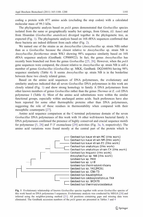
The phylogenetic analysis based on polA genes demonstrated that Geobacillus speciesisolated from the same or geographically nearby hot springs, from Gönen, (G. kaue) andfrom Hisaralan (Geobacillus anatolicus) diverged together in the phylogenetic tree, asexpected (Fig. 1). The phylogenetic analysis based on 16S rRNA sequences confirmed thatthese bacteria are indeed different from each other (Fig. 2).
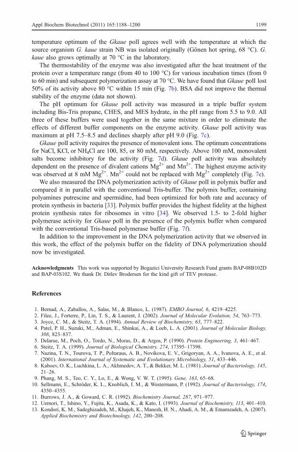
We named one of the strains as an Anoxybacillus (Anoxybacillus sp. strain NB) ratherthan as a Geobacillus because the closest relative to Anoxybacillus sp. strain NB isAnoxybacillus flavithermus strain WK1 showing 98% sequence similarity based on 16SrRNA sequence analysis (GenBank: CP000922). In fact, the genus Anoxybacillus hasrecently been branched out from the genus Geobacillus [25, 26]. However, when the polAgene sequences were compared, the closest relative to Anoxybacillus sp. strain NB is still amember of genus Geobacillus (Geobacillus sp. MKK, GenBank: DQ244056) having 98%sequence similarity (Table 4). It seems Anoxybacillus sp. strain NB is in the borderlinebetween these two closely related genus.
Based on the amino acid sequences of DNA polymerases, the evolutionary andsimilarity analyses indicated that all seven Geobacillus DNA polymerases in this work areclosely related (Fig. 1) and show strong homology to family A DNA polymerases fromother known members of genus Geobacillus rather than the genus Thermus or E. coli DNApolymerase I (Table 4). Most of the amino acid substitutions were within the similarfunctional groups, especially within uncharged amino acids. Such preferences have alsobeen reported for some other thermophilic proteins other than DNA polymerases,suggesting the role of these residues in thermostability when compared with theirmesophilic counterparts [27].
Amino acid sequence comparison at the C-terminal polymerase domain of the sevenGeobacillus DNA polymerases of this work with 16 other well-known bacterial family ADNA polymerases confirmed the presence of highly conserved and crucial sequence motifsfor polymerase [5, 28] and 5′-3′ exonuclease [29] activities (Fig. 3a, b, respectively). Theamino acid variations were found mostly at the central part of the protein which is
Fig. 1 Evolutionary relationship of known Geobacillus species together with seven Geobacillus species ofthis work based on DNA polymerase I sequences. Evolutionary analysis was conducted by MEGA [20] andinferred using the neighbor-joining method [21]. All positions containing gaps and missing data wereeliminated. The GenBank accession numbers of the polA genes are presented in Tables 3 and 4
Appl Biochem Biotechnol (2011) 165:1188–1200 1193
responsible for 3′-5′ exonuclease (proofreading) activity of the enzyme [30, 31]. The rolesof the carboxylate groups (Asp355, Glu357, Asp424, and Asp501) in 3′-5′ exonuclease activitythat are coordinated by divalent metal ions were reported before in E. coli DNA polymeraseI [30]. These residues correspond to Val321, Glu323, Ala378, and Lys452, respectively, in all
Fig. 2 Evolutionary relationship of some Geobacillus species together with seven Geobacillus species ofthis work based on 16S rRNA sequences. The GenBank accession numbers are presented in parenthesis.Evolutionary analysis was conducted similarly as depicted in Fig. 1
Table 4 Similarity analysis of Gkaue polI with 16 well-known members of family A DNA polymerases
Species Similarity to Gkaue polI (%) Genbank accession numbers
Escherichia coli 55 V00317
Thermus thermophilus 59 D28878
Thermus aquaticus 59 D32013
Thermus filigormis 57 AF030320
Bacillus caldotenax 94 D12982
Bacillus stearothermophilus 93 BSU33536
Bacillus subtilis 82 U86402
Geobacillus sp. MKK 98 DQ244056
Geobacillus sp. LH8 98 DQ392964
Geobacillus thermodenitrificans 96 Z26928
Geobacillus sp Y412MC61 95 ACX77456
Geobacillus sp C56-T3 94 YP003670385
Geobacillus kaustophilus 94 YP148583
Geobacillus caldoxylosilyticus TK4 89 DQ340803
Geobacillus sp. WCH70 89 YP002950632
Geobacillus thermoglucosidasius 88 ZP06809476
Homology search was performed using Basic Local Alignment Search Tool based on amino acid sequencesof the polA genes
1194 Appl Biochem Biotechnol (2011) 165:1188–1200

Geobacillus DNA polymerases described in this work, indicating that three of the fourcritical metal-binding residues are different in these proteins (Fig. 3c). Due to these changesin metal-binding residues, the potential DNA and nucleotide-binding sites were altered,resulting in the loss of 3′-5′ exonuclease activity. Similar observations had been reportedearlier for other thermophilic DNA polymerases from B. stearothermophilus [15] and fromThermus aquaticus [32].
Since Geobacillus species identified in this work were closely related, we onlycharacterized one of them, G. kaue strain NB DNA polymerase I (Gkaue polI, GenBank:FJ215757), biochemically in vitro.
Structure of G. kaue DNA Polymerase I
The nucleotide sequence and deduced amino acid sequence of Gkaue polI waspresented in Fig. 4. The 3D structure of Gkaue polI was predicted by homology modelingusing BF as a template structure [15, 16] since it has a high sequence similarity (90%)with the corresponding region (between residues 299–878) of the full-length Gkaue polI.The structure for DNA polymerase domain (residues 471–878) of Gkaue polI wasdepicted resembling a right hand with fingers, palm, and thumb subdomains (Fig. 5a).These three subdomains define a common structural feature even in distantly relatedDNA polymerases and predicted to bind duplex DNA [4]. Root-mean-square deviation(RMSD) of equivalent α-carbon positions of Gkaue polI with respect to the templateprotein (BF) is 0.73 Å.
Fig. 3 The amino acid sequence alignment of 16 well-known members of family A DNA polymerasestogether with the consensus sequences of seven Geobacillus species in this work. The conserved amino acidresidues involved in a 5′-3′ exonuclease, b polymerase, and c 3′-5′ exonuclease domains. The GenBankaccession numbers of the polA genes and abbreviations of the species are as presented in Tables 3 and 4. Theabbreviations of the species are as follows: Escherichia coli (Ecoli), Thermus thermophilus (Tth), Thermusaquaticus (Taq), Thermus filiformis (Tfli), Bacillus caldotenax (Bcal), Bacillus stearothermophilus (Bstr),Bacillus subtilis (Bsub), Geobacillus sp. MKK (GMKK), Geobacillus sp. LH8 (GLH8), Geobacillusthermodenitrificans (GTHRdnt), Geobacillus sp. Y412MC61 (GY412), Geobacillus sp. C56-T3 (GYC56),Geobacillus kaustophilus (Gkau), Geobacillus caldoxylosilyticus TK4 (GcalTK4), Geobacillus sp. WCH70(GWCH70) Geobacillus thermoglucosidasius (GTHRglu)
Appl Biochem Biotechnol (2011) 165:1188–1200 1195

Furthermore, the polymerase active site residing on the palm subdomain at C-terminalpolymerase domain of Gkaue polI (residues 617–834) revealed highly conserved motifs A,B, and C together with strictly conserved residues (Fig. 5b). For example, Tyr714 (Tyr716 inGkaue polI) is positioned at the first nucleotide of 5′-template overhang and interacts withthe template base. The location of the 3'-OH terminus is positioned for catalysis, whichforms hydrogen bond to Asp830 (Asp832 in Gkaue polI), an essential and invariant residue inall polymerases.
Expression and Purification of G. kaue DNA Polymerase I
Gkaue polI was expressed into E. coli JM109(DE3) cells (Fig. 6a). The fusion protein,including the additional region (six histidine residues, thioredoxin (TrxA) coding sequence
Fig. 4 Nucleotide sequence and deduced amino acid sequence of Gkaue polI. The predicted Shine–Dalgarnoregion located upstream from the ATG start codon (M) and −10 region prokaryotic promoter motif areunderlined. The putative transcription start point is indicated by a bent arrow. The translation stop codon(UAA) is indicated by an asterisk
1196 Appl Biochem Biotechnol (2011) 165:1188–1200

and a TEV protease cleavage site), has a total molecular mass of 113.6 kDa. The fusionGkaue polI was purified by Ni2+-affinity chromatography and then digested with TEVprotease. A second Ni2+-affinity chromatography after TEV digestion enabled an efficientremoval of the histidine-tag, as well as the contaminating cellular proteins carried over fromthe first chromatography step. The purified Gkaue polI (99.3 kDa) was homogeneous asjudged by SDS-PAGE analysis (Fig. 6b); 2.6 mg of purified enzyme was obtained from2.5 l of cell culture (Table 5).
Fig. 5 Three-dimensional structures of Gkaue polI. a The predicted model for the C-terminal DNApolymerase domain of Gkaue polI. Helix, sheet, and loop were colored red, yellow, and green, respectively. bThe predicted model for the active site of Gkaue polI. The highly conserved amino acid residues (motifs A,B, and C) in polymerase domains (shown in Fig. 2b) were colored pink. The positions of amino acid residueswere depicted based on BF structure [15, 16]
Fig. 6 SDS-PAGE analysis of the expression and the purification of Gkaue polI. a Analysis of the proteinexpression before and after IPTG induction. Lane M, molecular weight standard. b Analysis of the proteinpurification. Lane 1, undigested fusion protein; lane 2, TEV protease digested fusion protein; lane 3, purifiedenzyme after TEV protease digestion and after second Ni2+-affinity chromatography
Appl Biochem Biotechnol (2011) 165:1188–1200 1197

Biochemical Properties of Geobacillus DNA Polymerase I
This is the second study, to our knowledge, a thermophilic family A DNA polymerase Ifrom the genus Geobacillus was characterized biochemically. The first one was from DNApolymerase I from Geobacillus caldoxylosilyticus TK4 [14].
Optimum conditions for the enzymatic activity of Gkaue polI were measured in vitrousing [3H]dATP incorporation to a partially single-stranded DNA substrate (see “Materialsand Methods”). The maximum polymerization activity was observed at 70 °C (Fig. 7a). The
Table 5 Purification scheme of Gkaue polI
Step Total protein (mg) Total activity (U) Specific activity (U/mg) Yield (%)
Ni2+-column purification 36 766 21 100
TEV digestion 13 350 27 46
Dialysis 2.7 100 37 14
Fig. 7 The optimum conditions of Gkaue polI. a Optimum temperature. b Thermostability. c Optimum pH.Optimum concentrations for d monovalent ions and e divalent ions. f Optimum Mg2+ concentration in thepresence of standard polymerase reaction buffer (empty diamonds) and polymix buffer (empty circles)
1198 Appl Biochem Biotechnol (2011) 165:1188–1200
temperature optimum of the Gkaue polI agrees well with the temperature at which thesource organism G. kaue strain NB was isolated originally (Gönen hot spring, 68 °C). G.kaue also grows optimally at 70 °C in the laboratory.
The thermostability of the enzyme was also investigated after the heat treatment of theprotein over a temperature range (from 40 to 100 °C) for various incubation times (from 0to 60 min) and subsequent polymerization assay at 70 °C. We have found that Gkaue polI lost50% of its activity above 80 °C within 15 min (Fig. 7b). BSA did not improve the thermalstability of the enzyme (data not shown).
The pH optimum for Gkaue polI activity was measured in a triple buffer systemincluding Bis-Tris propane, CHES, and MES hydrate, in the pH range from 5.5 to 9.0. Allthree of these buffers were used together in the same mixture in order to eliminate theeffects of different buffer components on the enzyme activity. Gkaue polI activity wasmaximum at pH 7.5–8.5 and declines sharply after pH 9.0 (Fig. 7c).
Gkaue polI activity requires the presence of monovalent ions. The optimum concentrationsfor NaCl, KCl, or NH4Cl are 100, 85, or 80 mM, respectively. Above 100 mM, monovalentsalts become inhibitory for the activity (Fig. 7d). Gkaue polI activity was absolutelydependent on the presence of divalent cations Mg2+ and Mn2+. The highest enzyme activitywas observed at 8 mM Mg2+. Mn2+ could not be replaced with Mg2+ completely (Fig. 7e).
We also measured the DNA polymerization activity of Gkaue polI in polymix buffer andcompared it in parallel with the conventional Tris-buffer. The polymix buffer, containingpolyamines putrescine and spermidine, had been optimized for both rate and accuracy ofprotein synthesis in bacteria [33]. Polymix buffer provides the highest fidelity at the highestprotein synthesis rates for ribosomes in vitro [34]. We observed 1.5- to 2-fold higherpolymerase activity for Gkaue polI in the presence of the polymix buffer when comparedwith the conventional Tris-based polymerase buffer (Fig. 7f).
In addition to the improvement in the DNA polymerization activity that we observed inthis work, the effect of the polymix buffer on the fidelity of DNA polymerization shouldnow be investigated.
Acknowledgments This work was supported by Bogazici University Research Fund grants BAP-08B102Dand BAP-03S102. We thank Dr. Ditlev Brodersen for the kind gift of TEV protease.
References
1. Bernad, A., Zaballos, A., Salas, M., & Blanco, L. (1987). EMBO Journal, 6, 4219–4225.2. Filee, J., Forterre, P., Lin, T. S., & Laurent, J. (2002). Journal of Molecular Evolution, 54, 763–773.3. Joyce, C. M., & Steitz, T. A. (1994). Annual Review of Biochemistry, 63, 777–822.4. Patel, P. H., Suzuki, M., Adman, E., Shinkai, A., & Loeb, L. A. (2001). Journal of Molecular Biology,
308, 823–837.5. Delarue, M., Poch, O., Tordo, N., Moras, D., & Argos, P. (1990). Protein Engineering, 3, 461–467.6. Steitz, T. A. (1999). Journal of Biological Chemistry, 274, 17395–17398.7. Nazina, T. N., Tourova, T. P., Poltaraus, A. B., Novikova, E. V., Grigoryan, A. A., Ivanova, A. E., et al.
(2001). International Journal of Systematic and Evolutionary Microbiology, 51, 433–446.8. Kaboev, O. K., Luchkina, L. A., Akhmedov, A. T., & Bekker, M. L. (1981). Journal of Bacteriology, 145,
21–26.9. Phang, M. S., Teo, C. Y., Lo, E., & Wong, V. W. T. (1995). Gene, 163, 65–68.10. Sellmann, E., Schröder, K. L., Knoblich, I. M., & Westermann, P. (1992). Journal of Bacteriology, 174,
4350–4355.11. Burrows, J. A., & Goward, C. R. (1992). Biochemistry Journal, 287, 971–977.12. Uemori, T., Ishino, Y., Fujita, K., Asada, K., & Kato, I. (1993). Journal of Biochemistry, 113, 401–410.13. Kondori, K. M., Sadeghizadeh, M., Khajeh, K., Manesh, H. N., Ahadi, A. M., & Emamzadeh, A. (2007).
Applied Biochemistry and Biotechnology, 142, 200–208.
Appl Biochem Biotechnol (2011) 165:1188–1200 1199

14. Sandalli, C., Singh, K., Modak, M. J., Kethar, A., Canakci, S., Demir, I., et al. (2009). AppliedMicrobiology and Biotechnology, 84, 105–117.
15. Kiefer, J. R., Mao, C., Hansen, C. J., Basehore, S. L., Hogrefe, H. H., Braman, J. C., et al. (1997). Structure, 5,95–108.
16. Kiefer, J. R., Mao, C., Braman, J. C., & Beese, L. S. (1998). Nature, 391, 304–307.17. Aliota, J. M., Pelletier, J. J., Ware, J. L., Moran, L. S., Benner, J. S., & Kong, H. (1996).Genetic Analysis, 12,
185–195.18. Lu, Y. Y., Ye, S. Y., & Hong, G. F. (1991). Biotechniques, 11, 464–466.19. Thompson, J. D., Higgins, G., & Gipson, T. J. (1995). Nucleic Acids Research, 22, 4673–4680.20. Tamura, K., Dudley, J., Nei, M., & Kumar, S. (2007). Molecular Biology and Evolution, 24, 1596–1599.21. Saitou, N., & Nei, M. (1987). Molecular Biology and Evolution, 4, 406–425.22. Schwede, T., Koop, J., Guex, N., & Peitsch, M. C. (2003). Nucleic Acids Research, 31, 3381–3385.23. Nayeem, A., Sitkoff, D., & Krystek, S. (2007). Protein Science, 15, 808–824.24. Bradford, M. M. (1976). Analytical Biochemistry, 72, 248–254.25. Pikuta, E., Lysenko, A., Chuvilskaya, N., Mendrock, U., Hippe, H., Suzina, N., et al. (2000).
International Journal of Systematic and Evolutionary Microbiology, 50, 2109–2117.26. Saw, J. H., Mountain, B. W., Feng, L., Omelchenko, M. V., Hou, S., Saito, J. A., et al. (2008). Genome
Biology, 9, R161.27. Jaenicke, R., & Böhm, G. (1998). Current Opinion of Structural Biology, 8, 738–748.28. Polesky, A. H., Steitz, T. A., Grindley, N. D., & Joyce, C. M. (1990). Journal of Biological Chemistry, 265,
14579–14591.29. Gutman, P. D., & Minton, K. W. (1993). Nucleic Acids Research, 21, 4406–4407.30. Beese, L. S., & Steitz, T. A. (1991). EMBO Journal, 10, 25–33.31. Derbyshire, V., Grindley, N. D., & Joyce, C. M. (1991). EMBO Journal, 10, 17–24.32. Korolev, S., Nayal, M., Barnes, W. M., DiCera, E., & Waksman, G. (1995). Proceedings of the National
Academy of Sciences, 92, 9264–9268.33. Jelenc, P. C., & Kurland, C. G. (1979). Proceedings of the National Academy of Sciences, 76, 3174–3178.34. Ehrenberg, M., Bilgin, N., & Kurland, C. G. (1989). In Spedding G. (Ed), Ribosomes and Protein
Synthesis (pp. 101–129). Oxford: IRL Press.
1200 Appl Biochem Biotechnol (2011) 165:1188–1200
Related Documents