Cloning and expression analysis of two ROR-g homologues (ROR-ga1 and ROR-ga2) in rainbow trout Oncorhynchus mykiss Milena M. Monte a, * , Tiehui Wang a , Maria M. Costa a, b , Nor Omaima Harun a, 1 , Chris J. Secombes a a Scottish Fish Immunology Research Centre, School of Biological Sciences, University of Aberdeen, Zoology Building, Tillydrone Avenue, Aberdeen AB24 2TZ, Scotland, UK b Instituto de Investigaciones Marinas, Consejo Superior de Investigaciones Científicas (CSIC), Eduardo Cabello 6, Vigo, Spain article info Article history: Received 25 January 2012 Received in revised form 11 May 2012 Accepted 14 May 2012 Available online 24 May 2012 Keywords: Oncorhynchus mykiss ROR-g Bacterial infection Vaccination Viral infection abstract This paper describes the cloning and characterisation of two retinoid-related orphan receptor (ROR)-g homologues (ROR-ga1 and -ga2) in rainbow trout (Oncorhynchus mykiss). The coding region predicted for both homologues consists of 1410 base pairs (bp), which translate into two 469 amino acid (aa) proteins. The trout ROR-gs revealed a high conservation of both DNA- and ligand-binding domains (functional regions of the nuclear receptor family), and shared a high homology to mammalian ROR-gt. A phylo- genetic tree containing ROR family members confirmed that both trout homologues clustered within the ROR-g group. Both results suggested that these molecules are likely to be ROR-g homologues, more similar to the mammalian splice variant ROR-gt than the full length ROR-g. Expression analysis of tissues obtained from healthy fish revealed highest constitutive expression of trout ROR-g in muscle, followed by the brain, heart and skin. This suggests that these genes may play an important role in such tissues. In vitro studies, using trout cell lines, demonstrated that ROR-g is induced significantly by LPS and down- regulated by the presence of PolyI:C and recombinant interferon (IFN)-g. Moreover, analysis of this gene in head kidney macrophages and mixed primary leucocyte cultures indicated that differences were apparent between the different cell types/sources used, indicating that its expression may be cell-type dependent. Additional studies to investigate the regulation of this gene in vivo demonstrated that its expression was significantly higher in vaccinated vs unvaccinated fish following bacterial (Yersinia ruckeri) challenge but it was down-regulated after a viral (VHSV) infection. This suggests a potential role of trout ROR-g, a putative T H 17 transcription factor, in protection against extracellular bacteria. Ó 2012 Elsevier Ltd. All rights reserved. 1. Introduction Nuclear receptors (NRs) constitute one of the largest super- families of eukaryotic transcription factors, with over 60 family members known that share structural similarities [1,2]. They regulate the expression of genes involved in key cellular processes, such as cell growth, differentiation and apoptosis. These receptors function by promoting a link between signalling molecules and the transcriptional response upon binding of a variety of extracellular ligands [1e3]. The lipophilic ligands to which these receptors bind include a range of known hormones, from steroids (e.g. oestrogens and progesterone) to thyroid hormones [3,4]. Since the activity of these receptors can be controlled by the direct action of natural and synthetic compounds, with their dysfunction resulting in disease, NRs are considered to be good targets for drug research [2,3]. Orphan nuclear receptors include the retinoid-related orphan receptors (RORs), whose ligands were unknown at the time they were identified and hence the reason they were named as “orphan” [2,3,5]. The ROR subfamily can be further divided into a, b and g isotopes, also referred to as NR1F1-3 (Nuclear Receptor Nomen- clature Committee), or RORA-C (Human Gene Nomenclature Committee) [6,7]. Members of this subfamily have been identified in several mammalian species, as well as in early vertebrates, such as bony fish [7,8]. In terms of structural organisation, ROR mole- cules share a similar structure with other members of the NR family, and contain the following functional regions: an N-terminal domain (A/B), a highly conserved DNA-binding domain (DBD), a hinge region and a C-terminal ligand-binding domain (LBD) [7e9]. The highly variable N-terminal domain is present in all NRs. It contains a transcriptional activation function region, known as AF- 1, which is recognized by coactivators or other transcription factors, acting in a ligand-independent manner [1,10]. The DNA-binding * Corresponding author. Tel.: þ44 1224 272870; fax: þ44 1224 272396. E-mail address: [email protected] (M.M. Monte). 1 Current address: Department of Biological Sciences, Faculty of Science and Technology, Universiti Malaysia Terengganu, 21030 Kuala Terengganu, Terengganu Darul Iman, Malaysia. Contents lists available at SciVerse ScienceDirect Fish & Shellfish Immunology journal homepage: www.elsevier.com/locate/fsi 1050-4648/$ e see front matter Ó 2012 Elsevier Ltd. All rights reserved. doi:10.1016/j.fsi.2012.05.023 Fish & Shellfish Immunology 33 (2012) 365e374

Welcome message from author
This document is posted to help you gain knowledge. Please leave a comment to let me know what you think about it! Share it to your friends and learn new things together.
Transcript

at SciVerse ScienceDirect
Fish & Shellfish Immunology 33 (2012) 365e374
Contents lists available
Fish & Shellfish Immunology
journal homepage: www.elsevier .com/locate / fs i
Cloning and expression analysis of two ROR-g homologues (ROR-ga1and ROR-ga2) in rainbow trout Oncorhynchus mykiss
Milena M. Monte a,*, Tiehui Wang a, Maria M. Costa a,b, Nor Omaima Harun a,1, Chris J. Secombes a
a Scottish Fish Immunology Research Centre, School of Biological Sciences, University of Aberdeen, Zoology Building, Tillydrone Avenue, Aberdeen AB24 2TZ, Scotland, UKb Instituto de Investigaciones Marinas, Consejo Superior de Investigaciones Científicas (CSIC), Eduardo Cabello 6, Vigo, Spain
a r t i c l e i n f o
Article history:Received 25 January 2012Received in revised form11 May 2012Accepted 14 May 2012Available online 24 May 2012
Keywords:Oncorhynchus mykissROR-gBacterial infectionVaccinationViral infection
* Corresponding author. Tel.: þ44 1224 272870; faxE-mail address: [email protected] (M.M. Mont
1 Current address: Department of Biological ScienTechnology, Universiti Malaysia Terengganu, 21030 KDarul Iman, Malaysia.
1050-4648/$ e see front matter � 2012 Elsevier Ltd.doi:10.1016/j.fsi.2012.05.023
a b s t r a c t
This paper describes the cloning and characterisation of two retinoid-related orphan receptor (ROR)-ghomologues (ROR-ga1 and -ga2) in rainbow trout (Oncorhynchus mykiss). The coding region predicted forboth homologues consists of 1410 base pairs (bp), which translate into two 469 amino acid (aa) proteins.The trout ROR-gs revealed a high conservation of both DNA- and ligand-binding domains (functionalregions of the nuclear receptor family), and shared a high homology to mammalian ROR-gt. A phylo-genetic tree containing ROR family members confirmed that both trout homologues clustered within theROR-g group. Both results suggested that these molecules are likely to be ROR-g homologues, moresimilar to the mammalian splice variant ROR-gt than the full length ROR-g. Expression analysis of tissuesobtained from healthy fish revealed highest constitutive expression of trout ROR-g in muscle, followedby the brain, heart and skin. This suggests that these genes may play an important role in such tissues.In vitro studies, using trout cell lines, demonstrated that ROR-g is induced significantly by LPS and down-regulated by the presence of PolyI:C and recombinant interferon (IFN)-g. Moreover, analysis of this genein head kidney macrophages and mixed primary leucocyte cultures indicated that differences wereapparent between the different cell types/sources used, indicating that its expression may be cell-typedependent. Additional studies to investigate the regulation of this gene in vivo demonstrated that itsexpression was significantly higher in vaccinated vs unvaccinated fish following bacterial (Yersiniaruckeri) challenge but it was down-regulated after a viral (VHSV) infection. This suggests a potential roleof trout ROR-g, a putative TH17 transcription factor, in protection against extracellular bacteria.
� 2012 Elsevier Ltd. All rights reserved.
1. Introduction
Nuclear receptors (NRs) constitute one of the largest super-families of eukaryotic transcription factors, with over 60 familymembers known that share structural similarities [1,2]. Theyregulate the expression of genes involved in key cellular processes,such as cell growth, differentiation and apoptosis. These receptorsfunction by promoting a link between signalling molecules and thetranscriptional response upon binding of a variety of extracellularligands [1e3]. The lipophilic ligands to which these receptors bindinclude a range of known hormones, from steroids (e.g. oestrogensand progesterone) to thyroid hormones [3,4]. Since the activity ofthese receptors can be controlled by the direct action of natural and
: þ44 1224 272396.e).ces, Faculty of Science and
uala Terengganu, Terengganu
All rights reserved.
synthetic compounds, with their dysfunction resulting in disease,NRs are considered to be good targets for drug research [2,3].Orphan nuclear receptors include the retinoid-related orphanreceptors (RORs), whose ligands were unknown at the time theywere identified and hence the reason they were named as “orphan”[2,3,5]. The ROR subfamily can be further divided into a, b and gisotopes, also referred to as NR1F1-3 (Nuclear Receptor Nomen-clature Committee), or RORA-C (Human Gene NomenclatureCommittee) [6,7]. Members of this subfamily have been identifiedin several mammalian species, as well as in early vertebrates, suchas bony fish [7,8]. In terms of structural organisation, ROR mole-cules share a similar structure with other members of the NRfamily, and contain the following functional regions: an N-terminaldomain (A/B), a highly conserved DNA-binding domain (DBD),a hinge region and a C-terminal ligand-binding domain (LBD)[7e9].
The highly variable N-terminal domain is present in all NRs. Itcontains a transcriptional activation function region, known as AF-1, which is recognized by coactivators or other transcription factors,acting in a ligand-independent manner [1,10]. The DNA-binding

Table 1Oligonucleotide primers used for cloning and expression analysis.
Primer name Primer sequence (50 / 30) Primers used
ROR-g F1 CTGTGGGGACAAGTCATCAGG 30-RACEROR-g F2 CCAATCGGAATCGCTGCCROR-g R1 GAGTGGCGATGTCGTCAAGGTC 50-RACEROR-g R2 GGCCATGACCTTCGACCTCROR-g F ACAGACCTTCAAAGCTCTTGGTTGTG Real-time PCRROR-g R GGGAAGCTTGGACACCATCTTTGROR-g F3 AATACAGCAAGAAAATGAGAGCACAAA Real-time PCRROR-g R3 GGACATGCGGCCAAACTTTACEF-1a F CAAGGATATCCGTCGTGGCA Real-time PCREF-1a R ACAGCGAAACGACCAAGAGG
M.M. Monte et al. / Fish & Shellfish Immunology 33 (2012) 365e374366
domain, on the other hand, is highly conserved among the familymembers, and contains two C4 zinc-finger motifs involved in therecognition of ROREs in the promoter region of the targeted genes[2,9]. ROR-g receptors are known to bind preferably as monomersto these specific response elements that consist of an AGGTCAmotifpreceded by an A/T rich-region [2,11]. This binding allows thereceptors to perform their various roles through the communica-tion between the receptor and its intracellular environment,regulating transcription [4,8]. DNA- and ligand-binding domainsare connected together by a hinge region which is highly variableand flexible [2,9]. The ligand-binding domain is moderatelyconserved between members of the NR family and containsa second activation function region, described as AF-2. This regioncontains a characteristic motif, FFXE/DFF, where F correspondsto a hydrophobic residue and X can be any amino acid (aa), and actsas a ligand-dependent transcription factor [2,9].
In mammals, two isoforms of ROR-g have been identified, andnamed ROR-g1 and -g2. The latter, commonly referred to as ROR-gt,lacks the A/B domain and thus consists of a truncated form of ROR-g1, the result of alternative RNA splicing of a common transcript[9,11e13]. They display different tissue distribution patterns andtherefore regulate distinct physiological processes. A higherconstitutive expression of ROR-g1 was found in liver, muscle,thymus and kidney tissues [11,12,14]. In contrast, ROR-gt expressionis restricted to a limited number of cell populations, such asdeveloping thymocytes and T cells in secondary lymphoid tissues[7,13,15]. More recently this isoform was identified as the master Thelper (TH) 17 transcription factor, crucial for the differentiation ofnaïve CD4þ T cells into activated TH17 cells [16,17]. Most recentlystudies have revealed that ROR-gt is also expressed in a novelmucosal lymphocyte population in the intestinal lamina, whichsecretes IL-22 and co-expresses natural killer (NK) cell markers[8,19]. In mice and humans, ROR-g molecules encode proteins of516 and 518 aa, respectively, and share 88% aa homology [11,12,14].In contrast, ROR-gt encodes an open reading frame of 495 aa inmice and 497 in humans [12,13]. In fish, ROR-g homologues havebeen identified in zebrafish [8], with the characterisation of twoisotypes of ROR-g (ROR-ga and -gb). In the present study we reporton the identification and characterisation of two ROR-g homo-logues (ROR-ga1 and -ga2) in rainbow trout, in order to gaina better insight into these molecules and their role in the immunesystem of early vertebrate species.
2. Material and methods
2.1. Fish
Rainbow trout (Oncorhynchus mykiss), weighing approximately100 g, were purchased from the Mill of Elrich Trout Fishery(Aberdeenshire, UK) and maintained in 1-m-diameter aeratedfibreglass tanks supplied with a continuous flow of recirculatingfreshwater at 15 � 1 �C. Fish were fed twice daily on standardcommercial pellets (EWOS), and were given a 2-week acclimati-sation period prior to treatment.
2.2. Cloning and sequencing of ROR-g
For the sequencing of trout ROR-g homologues, a TBLASTNsearch (http://blast.ncbi.nlm.nih.gov/Blast.cgi) [20] was performed,identifying a rainbow trout expressed sequence tag (EST, GenBankaccession number: CX006390), which shared 83% identity with theN-terminus of zebrafish (Danio rerio) ROR-g [8]. The full-lengthcDNA sequence was obtained by RACE using SMART cDNA asdescribed previously [21]. A single band was amplified by 50-RACEPCR, using primers ROR-gR1 and -gR2, and cDNA samples obtained
from spleen and head kidney. Sequence analysis revealed that thePCR products obtained from both tissues were identical, with sizesof approximately 664 bp, being 21 bp larger than the EST at the 50-end. Amplification of spleen cDNA by 30-RACE PCR, using ROR-gF1and -gF2 primers, detected the presence of two bands. Homologyanalysis revealed that both products shared around 98% identity inthe coding region, but only 78% in the 30-untranslated region (UTR).This suggests that they may have arisen from two differentparalogues, which are more closely related to the zebrafish ROR-ga,and were therefore designated as ROR-ga1 and ROR-ga2. Theirnucleotide sequences were submitted to the EMBL/DDBJ/GenBanknucleotide sequence database under the accession numbers:FM883712 and FM883713, respectively. The sequences of allprimers used are given in Table 1.
2.3. Bioinformatics
The generated sequences were analysed for similarity withother known molecules, and also used to identify homologoussequences in GenBank using the BLAST search [20]. Comparisonsbetween more than two sequences were performed using theCLUSTAL W multiple sequence alignment package (http://align.genome.jp) [22] and conserved residues were shaded using theBOXSHADE server (version 3.21) (http://www.ch.embnet.org/software/BOX_form.html). Amino acid homology comparisonswere performed using the MatGAT package (version 2.02) [23].Phylogenetic trees were constructed from CLUSTAL W generatedalignments using the NeighboureJoining (NeJ) method within theMEGA (version 4.1) package [24]. In addition, the domain structurewas predicted using the SMART 6 program (http://smart.embl-heidelberg.de) [25].
2.4. Expression analysis
2.4.1. Tissue distributionSix rainbow trout were killed and 14 tissues (liver, caudal
kidney, spleen, heart, head kidney, skin, thymus, scales, brain,muscle, gonad, gills, tail fins and intestine) collected for RNAextraction using TRI Reagent� (Applied Biosciences) following themanufacturer’s instructions. The first strand cDNAwas synthesisedusing BioScript� (Bioline, UK), diluted in TE buffer (10 mMTriseHCl, 1 mM EDTA, pH 8.0) and stored at �20 �C. Real-time PCRwas performed using IMMOLASE (Bioline) and SYBR Green fluo-rescent tag (Invitrogen) in a LightCycler� 480 System (RocheApplied Science, UK). The two trout ROR-g molecules share highsequence identity in the coding region that prevents the design ofgene-specific primers for real-time PCR analysis. Thus a single pairof primers was designed in the coding region in order to detect theexpression of both genes. To investigate if the transcript detected bythose primers is the major product of trout ROR-g, primers F3 andR3 were designed in the 50-end of the ROR-g transcript. Real-time

M.M. Monte et al. / Fish & Shellfish Immunology 33 (2012) 365e374 367
PCR analysis was performed in selected tissues (thymus, muscle,heart and brain). The relative expression of ROR-gwas calculated asarbitrary units and normalised against the expression level ofrainbow trout elongation factor (EF)-1a, a housekeeping gene.Primer sequences are listed in Table 1.
2.4.2. Maintenance of cell linesFour rainbow trout cell lines derived from different tissues were
used for this work: monocyte/macrophage-like RTS-11 cells fromspleen [26], fibroblast-like RTG-2 cells from gonad [27], RTL fromliver [28] and RTGILL from gill [29]. The latter was kindly suppliedby the Virology group at Marine Scotland Science (Aberdeen). Thecells were grown andmaintained at 20 �C in Leibovitz L-15medium(Gibco) supplemented with penicillin/streptomycin (P/S; 100 units/mL and 100 mg/mL, respectively; Gibco) and 30% foetal bovineserum (FBS; Biosera) for RTS-11, or 10% for all the other cell lines.Cells were counted using trypan blue (SigmaeAldrich) and seededat a concentration of 5e10 � 105 cells/mL into 25-cm2
flasks(Sarstedt).
2.4.3. Preparation of primary leucocyte culturesHead kidneys were collected aseptically from four fish, and cells
were pushed through a 100-mm nylon mesh (John Stanier) withincomplete L-15 medium, i.e. L-15 supplemented with P/S, 0.5% FBSand 10 U/mL heparin (SigmaeAldrich). The suspensions were thencentrifuged at 200 g for 5 min, and washed once with completemedium (same constituents as incomplete medium but with 10%FBS). Cells were counted and 5 mL of 1e1.5 � 106 cells/ml wereseeded into 25-cm2
flasks ready for treatment.
2.4.4. Preparation of macrophage primary culturesTo obtain trout primary macrophage cultures, head kidney
leucocyte suspensions, isolated as described previously, wereresuspended in L-15 mediumwith 0.1% FBS and P/S. Five millilitresof cells were added into 25-cm2
flasks, at a final concentration of2 � 106 cells/mL. In order to allow the adherence of macrophages,the cells were left in an incubator at 20 �C for 2 h. After incubation,the non adherent cells were removed by washing twice with L-15medium in the same conditions. After the washing steps, theprimary cultures were then incubatedwith completemedium (L-15medium supplemented with 10% FBS and P/S) at the sametemperature. After 3 days any non adherent cells were againremoved before use.
2.5. Induced expression in vitro
2.5.1. Primary culturesHead kidney primary cultures, prepared as described above,
were incubated with known stimulatory concentrations [30] ofEscherichia coli lipopolysaccharide (LPS; 25 mg/mL), poly-inosinic:polycytidylic acid (PolyI:C; 50 mg/mL), phorbol 12-myristate 13-acetate (PMA; 100 ng/mL), calcium ionophore (CI;0.5 mg/mL), phytohaemagglutinin (PHA; 10 mg/mL), PMA (100 ng/mL) plus CI (0.5 mg/mL), dexamethasone (DM; 0.5 mg/mL), or leftuntreated (control), for 4, 8 and 24 h. All chemicals were purchasedfrom SigmaeAldrich. After the incubation periods, cells attached tothe flasks and in suspension, were dissolved in TRI Reagent�. TheRNA extraction and cDNA synthesis was performed as describedabove. Data were normalised to EF-1a gene expression and ana-lysed against controls using the Pfaffl method [31].
2.5.2. MacrophagesFour-day old head kidney primary macrophage cultures,
prepared as above, were incubated with LPS (25 mg/mL), PolyI:C(50 mg/mL), and the pro-inflammatory cytokine rIFN-g (20 ng/mL)
[32]. After incubation for 4, 8 and 24 h the cells were resuspendedin TRI Reagent� for RNA extraction and cDNA synthesis as describedabove.
2.5.3. Cell linesAfter an overnight incubation post-passaging, the cell lines
(RTGILL, RTG-2, RTL and RTS-11) were incubated with LPS, PolyI:Cand rIFN-g at the same concentrations used for the primarymacrophage cultures or with medium alone for controls. Followingincubation for 4, 8 and 24 h the cells were dissolved in TRI Reagent�
and the RNA extracted.
2.6. Modulation of ROR-g expression in vivo
2.6.1. Viral infectionTo further investigate the regulation of trout ROR-g upon viral
exposure, the expression of this molecule was analysed in fishinfected with the pathogenic viral hemorrhagic septicaemia virus(VHSV), strain DK-F1. The challenge was performed as described byCampbell et al. [33]. Briefly, four fish were killed at 1, 2, 3, 4 and 6days post-infection with DK-F1 (1 � 108 TCID50/fish) or injectionwith control media as control. The head kidneys were collected forRNA extraction and cDNA synthesis. The expression level of ROR-gwas analysed by real-time PCR, as described above.
2.6.2. Vaccination and bacterial infectionVaccinationandbacterial challengewereperformedasdescribed
by Harun et al. [34]. Briefly, trout were vaccinated with Aquavac�against enteric redmouth disease (ERM) by intraperitoneal injec-tion. The control group was handled in a similar way, but withoutvaccination. The fish were kept for 60 days in aerated freshwatertanks until theywere challengedwith a pathogenic strain (MT3072)of Yersinia ruckeri (0.5 mL per fish, 2 � 106 cfu/mL). Six fish werekilled at 6 h, 1, 2 and 3 days post-infection and spleen and gill tissuewas collected for total RNAextraction. The expression level of ROR-gwas analysed by real-time PCR, as described above.
2.7. Statistical analysis
Data were analysed statistically using the student T-test andone-way analysis of variance (ANOVA), with the LSD post hoc testused for comparison of means when appropriate, within the SPSSpackage 18.0 (SPSS Inc., Chicago). Differences were consideredstatistically significant when P < 0.05.
3. Results
3.1. Cloning and sequencing of ROR-g
Two trout ROR-g cDNA sequences, ROR-ga1 (EMBL accessionnumber: FM883712) and ROR-ga2 (EMBL accession number:FM883713), were identified and shared a nucleotide similarity of91.6% (Supplementary Figure). Both sequences contain a 50-UTR of303 bp and a coding region of 1410 bp which translates intoa 469 aa protein with predicted molecular mass of 52.8 and 52.9kilodalton (kDa), respectively. In the 30-UTR, two mRNA instabilitymotifs (ATTTA) and three putative polyadenylation signal motifs(AATAAA) were also identified in both sequences. The second pol-yadenylation signal in the present transcripts is located 15 bpupstream of the poly A tail, suggesting that this is an authentic site.
Multiple alignment of the predicted amino acid sequences ofvertebrate ROR-g molecules revealed that the sequences are wellconserved among different vertebrate species (Fig. 1A). The pres-ence of two highly conserved regions was identified in the troutsequences; a DNA-binding domain of 71 aa, which contains two C4

A
B
DNA-binding domain
Trout_RORγ a1 1 ---------------------MRAQIEVIPCKICGDKSSGIHYGVITCEGCKGFFRRSQQNNAMYSCSRQRNCLIDRTNRTrout_RORγ a2 1 ---------------------MRAQIEVIPCKICGDKSSGIHYGVITCEGCKGFFRRSQQNNAMYSCSRQRNCLIDRTNRZebrafish_RORγ a 1 ---------------------MRAQIEVIPCKICGDKSSGIHYGVITCEGCKGFFRRSQQNNAMYSCSRQRNCIIDRTNRZebrafish_RORγ b 1 ---------------------MRAQIEVIPCKICGDKSSGIHYGVITCEGCKGFFRRSQQNNAMYSCSRQRNCLIDRTNRMouse_RORγ t 1 ---------------------MRTQIEVIPCKICGDKSSGIHYGVITCEGCKGFFRRSQQCNVAYSCTRQQNCPIDRTSRHuman_RORγ t 1 ---------------------MRTQIEVIPCKICGDKSSGIHYGVITCEGCKGFFRRSQRCNAAYSCTRQQNCPIDRTSRMouse_RORγ 1 MDRAPQRHHRTSRELLAAKKTHTSQIEVIPCKICGDKSSGIHYGVITCEGCKGFFRRSQQCNVAYSCTRQQNCPIDRTSRHuman_RORγ 1 MDRAPQRQHRASRELLAAKKTHTSQIEVIPCKICGDKSSGIHYGVITCEGCKGFFRRSQRCNAAYSCTRQQNCPIDRTSRconsensus 1 ...***********************************. *. ***.** **.**** *
Trout_RORγ a1 60 NRCQHCRLQKCLALGMSRDAVKFGRMSKKQRDSLYAEVQKHQASQE---------LAAAR----------EVGAEVEGHGTrout_RORγ a2 60 NRCQHCRLQKCLALGMSRDAVKFGRMSKKQRDSLYAEVQKHQASQE---------LTAAR----------EVGAEVEGHGZebrafish_RORγ a 60 NRCQHCRLQKCLALGMSRDAVKFGRMSKKQRDSLYAEVQKHQQSQE---------RAGGLGNGISSHTGDEAGENGNGHSZebrafish_RORγ b 60 NRCQHCRLQKCLALGMSRDAVKFGRMSKKQRDSLYAEVQRHQQLSQDCLAG---------------LTGRDEAEDGAHSRMouse_RORγ t 60 NRCQHCRLQKCLALGMSRDAVKFGRMSKKQRDSLHAEVQKQLQQ--QQQQEQVAKTPPAGSRGADTLTYTLGLSDGQLPLHuman_RORγ t 60 NRCQHCRLQKCLALGMSRDAVKFGRMSKKQRDSLHAEVQKQLQQRQQQQQEPVVKTPPAGAQGADTLTYTLGLPDGQLPLMouse_RORγ 81 NRCQHCRLQKCLALGMSRDAVKFGRMSKKQRDSLHAEVQKQLQQ--QQQQEQVAKTPPAGSRGADTLTYTLGLSDGQLPLHuman_RORγ 81 NRCQHCRLQKCLALGMSRDAVKFGRMSKKQRDSLHAEVQKQLQQRQQQQQEPVVKTPPAGAQGADTLTYTLGLPDGQLPLconsensus 81 ********************************** ****. .. ..... . ...... ........... ......
Trout_RORγ a1 121 RAYSRGSSAALSDLDDIATLPDGLLFDLPLTPEGAEREYCNLEMIG-------GSS---SSSQSSPEQNGLDFRDGNHHITrout_RORγ a2 121 RAYSRGSSAALSDLDDIATLPDGLLFDLPLTPEGAEREYCNLEMIG-------GSS---SSSQSSPEQNGLDFRDGNHHIZebrafish_RORγ a 131 RAYSRGSSTTLSDLDDITTLPDGLLFDLPLTPEEAA-DYCSLELLG-------GSSGNSSSSQSSPESNRQEFG-DVTHIZebrafish_RORγ b 125 PYSSGGSSSTLSDLDDIATLPDGLLFDLPLTPEEAG-EYCALEMLSS-GTGSGGSGSGSSSSSQSSPEPSAVDLADAVRVMouse_RORγ t 138 GASPDLPEASACPPGLLRASGSGPPYSNTLAKTEVQGASCHLEYSPERGKAEGRDSIYSTDGQLTLGRCGLRFEETRHPEHuman_RORγ t 140 GSSPDLPEASACPPGLLKASGSGPSYSNNLAKAGLNGASCHLEYSPERGKAEGRESFYSTGSQLTPDRCGLRFEEHRHPGMouse_RORγ 159 GASPDLPEASACPPGLLRASGSGPPYSNTLAKTEVQGASCHLEYSPERGKAEGRDSIYSTDGQLTLGRCGLRFEETRHPEHuman_RORγ 161 GSSPDLPEASACPPGLLKASGSGPSYSNNLAKAGLNGASCHLEYSPERGKAEGRESFYSTGSQLTPDRCGLRFEEHRHPGconsensus 161 ... . .. .. * .. .* . .... *.**.......... .. .................. ....
Trout_RORγ a1 191 KHEYQLLHESSLFTHTLLNTLPEGFSILEIERITASVVKSHMETSQHGSEELKRLVWSLYTPEETRNFQSKSAEVMWQHCTrout_RORγ a2 191 KHEYQLLHESSLFTHTLLNTLPEGFSILEIERITASVVKSHMETSQHGSEELKRLVWSLYTPEETRNFQSRSAEVMWQQCZebrafish_RORγ a 202 KHEYMSPHESGLFTRSILN-PPEGCSLMEIERITQNVIKSHIETSQYSTEELKRLAWTLYTPEEIRVYQNKPTEMLWQQCZebrafish_RORγ b 203 KHEYIMPDNS-RLTHSLLGSIPDNCSLHDIERITQNVVKSHLETCQYSTEELKKHTWSLYTPEETRSFQLKSAEWMWQQCMouse_RORγ t 218 LGEPEQGPDSHCIPSFCSAPEVPYASLTDIEYLVQNVCKSFRETCQLRLEDLLRQRTNLFSREEVTSYQRKSMWEMWERCHuman_RORγ t 220 LGELGQGPDSYGSPSFRSTPEAPYASLTEIEHLVQSVCKSYRETCQLRLEDLLRQRSNIFSREEVTGYQRKSMWEMWERCMouse_RORγ 239 LGEPEQGPDSHCIPSFCSAPEVPYASLTDIEYLVQNVCKSFRETCQLRLEDLLRQRTNLFSREEVTSYQRKSMWEMWERCHuman_RORγ 241 LGELGQGPDSYGSPSFRSTPEAPYASLTEIEHLVQSVCKSYRETCQLRLEDLLRQRSNIFSREEVTGYQRKSMWEMWERCconsensus 241 *. ....* ... ......*...**.. . *.**..**.*...*.* ........ **. .*.... ..* .*
Ligand-binding domain
Trout_RORγ a1 271 AIHLTNAIQYVVEFAKRITGFLDLCQNDQIILLKAGCLDVLLIRMCRAYNPINNTVLFDGKFASAQTFKALGCDDLVGAVTrout_RORγ a2 271 AIHLTNAIQYVVEFAKRITGFLDLCQNDQVILLKAGCLDVLLIRMCRAYNPINNTVLFDGKFASAQTFKALGCDDLVGAVZebrafish_RORγ a 281 AVQLTNAIQYVVEFAKRISGFMDLSQNDQIILLKAGCLDVLLIRMCRAYNPINNTLLFDGKFASPQLFKALGCDDLVSAVZebrafish_RORγ b 282 ALQITNAIQYVVEFAKRISGFMELCQNDQIILLKAGCLEVLLIRMCRAYNSSNNTMFFDGKFASPQLFKALGCDDLVNAVMouse_RORγ t 298 AHHLTEAIQYVVEFAKRLSGFMELCQNDQIILLTAGAMEVVLVRMCRAYNANNHTVFFEGKYGGVELFRALGCSELISSIHuman_RORγ t 300 AHHLTEAIQYVVEFAKRLSGFMELCQNDQIVLLKAGAMEVVLVRMCRAYNADNRTVFFEGKYGGMELFRALGCSELISSIMouse_RORγ 319 AHHLTEAIQYVVEFAKRLSGFMELCQNDQIILLKAGAMEVVLVRMCRAYNANNHTVFFEGKYGGVELFRALGCSELISSIHuman_RORγ 321 AHHLTEAIQYVVEFAKRLSGFMELCQNDQIVLLKAGAMEVVLVRMCRAYNADNRTVFFEGKYGGMELFRALGCSELISSIconsensus 321 *...* ***********..**..*.****..**.** ..*.*.*******. *.*..*.**.. . .*.**** .*.. .
Trout_RORγ a1 351 FDLAKSLSCIQLSEEEMALFSAAVLLSPDRPWLKDTQQVQKLQEKVYVSLQHCLHRCGSSEEKLAKMVSKLPMMKSICNLTrout_RORγ a2 351 FDLAKSLSCIQLSEEEMALFSAAVLLSPDRPWLKDTQQVQKLQEKVYLSLQHCLHRCGSSEEKLAKMVSKLPMMKSICNLZebrafish_RORγ a 361 FEMAKTLSRLQLSGEEMALFTATVLLSPDRPWLTDAQKVQKLQEKVYVALQHYLHKSGVHEEKLARMVSKLPMMKSICNLZebrafish_RORγ b 362 FELAKSLSRLQLSEEEMALYSAAVLLAPDRPWLTESQQVQKLQEKVYVALQHSLHMNGATIEKLDKMVSKLPQMKSICNLMouse_RORγ t 378 FDFSHFLSALCFSEDEIALYTALVLINANRPGLQEKRRVEHLQYNLELAFHHHLCKTHR--QGLLAKLPPKGKLRSLCSQHuman_RORγ t 380 FDFSHSLSALHFSEDEIALYTALVLINAHRPGLQEKRKVEQLQYNLELAFHHHLCKTHR--QSILAKLPPKGKLRSLCSQMouse_RORγ 399 FDFSHFLSALCFSEDEIALYTALVLINANRPGLQEKRRVEHLQYNLELAFHHHLCKTHR--QGLLAKLPPKGKLRSLCSQHuman_RORγ 401 FDFSHSLSALHFSEDEIALYTALVLINAHRPGLQEKRKVEQLQYNLELAFHHHLCKTHR--QSILAKLPPKGKLRSLCSQconsensus 401 *.. ..**... *..*.**..*.**.. .** *... .* .** . .. *.* .. . .... . ...*.*
Similarity IdentityTrout_RORγ a1 431 HIDKLKFFRLLHPETAYN-FPPLYREVFSSEITFPDSTMGTrout_RORγ a2 431 HIDKLEFFRLLHPETAFN-FPPLYREVFSSEITFPDSTMD 99.6 98.3 Zebrafish_RORγ a 441 HIDKLEFFRLLHPETAYN-FPALYREVFCSEITFPDSTEG 86.4 78.4Zebrafish_RORγ b 442 HIDKLEFFRLVHPETAYS-FPPLYREVFGSEINFPDSTNS 85.6 75.4Mouse_RORγ t 456 HVEKLQIFQHLHPIVVQAAFPPLYKELFSTDVESPEGLSK 65.1 46.7Human_RORγ t 458 HVERLQIFQHLHPIVVQAAFPPLYKELFSTETESPVGLSK 65.6 47Mouse_RORγ 477 HVEKLQIFQHLHPIVVQAAFPPLYKELFSTDVESPEGLSK 62.8 45.2Human_RORγ 479 HVERLQIFQHLHPIVVQAAFPPLYKELFSTETESPVGLSK 63.1 45.3consensus 481 *...*. * .** ...**.**.*.*..... *. ..
Zebrafish ROR-- b
Human ROR--
40 30 86 142 513 122 133 108 111 110 162
Zebrafish ROR-- a
7 86 142 456 122 133 108 111 162113
7 86 142 459 122 133 108 111 113 162
Human ROR-- t
7 86 142 513 122 133 108 111 110 162
M.M. Monte et al. / Fish & Shellfish Immunology 33 (2012) 365e374368
M.M. Monte et al. / Fish & Shellfish Immunology 33 (2012) 365e374 369
zinc-finger motifs, and a ligand-binding domain of 159 aa. TroutROR-g homologues showed a conservation of fourteen cysteine(Cys) residues amongst the vertebrate species selected, nine ofthem located in the DNA-binding domain. A high conservation ofthe AF-2 motif was also observed among all the ROR-g moleculesanalysed, which in the trout and zebrafish homologues was char-acterised by the sequence LYREVF. In terms of homology, the troutproteins showed high amino acid similarity with zebrafish ROR-ghomologues, with percentages of aa similarity >85%, followed byhuman andmouse ROR-gt (65.6 and 65.1%, respectively) and ROR-g(63.1 and 62.8%, respectively). It was clear from the alignment thatthe N-terminal was more similar to ROR-gt than ROR-g, and thiswas also born out by investigation of the genomic organisation ofthe human and zebrafish ROR-gmolecules. It was possible to detecta similar intron/exon organisation between human ROR-gt and thezebrafish homologues, with the presence of 10 exons, 8 of whichwere identical in terms of protein encoding nucleotides thatincluded critically the 50-end exons (Fig. 1B). To further analyse therelationship of both trout sequences with other vertebrate mole-cules within the ROR family, a phylogenetic tree was constructed(Fig. 2). The rainbow trout sequences clustered together with otherknown ROR-g molecules forming a separate clade which wassupported by a high bootstrap value of 97. Within the piscinecluster, they grouped more closely with zebrafish ROR-ga, sug-gesting that the nomenclature is correct, and hence why we termthem ROR-ga1 and ROR-ga2.
3.2. Tissue distribution of trout ROR-g
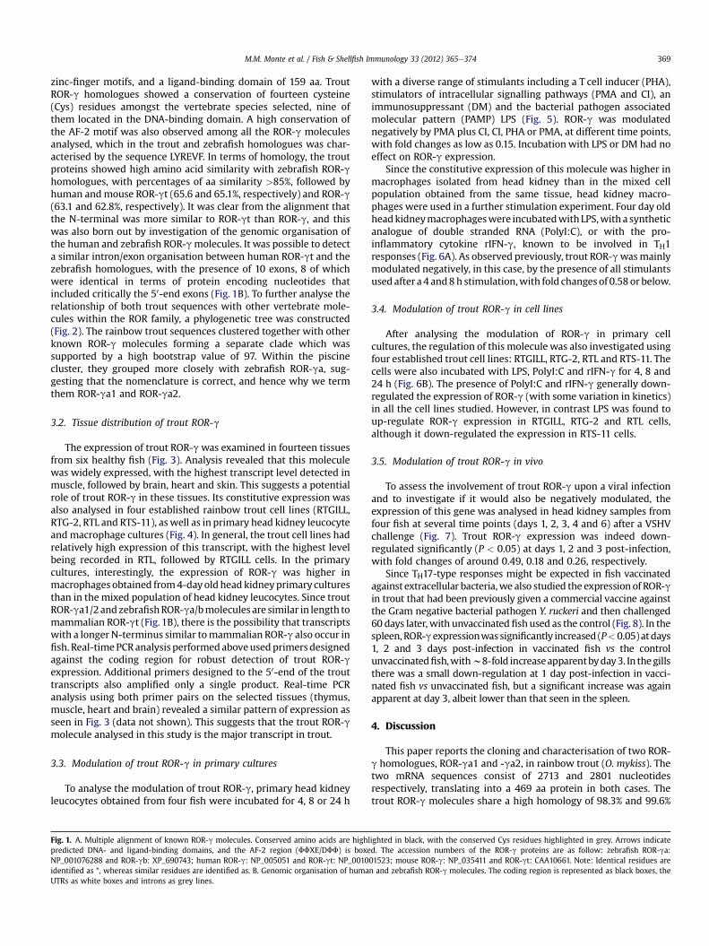
The expression of trout ROR-gwas examined in fourteen tissuesfrom six healthy fish (Fig. 3). Analysis revealed that this moleculewas widely expressed, with the highest transcript level detected inmuscle, followed by brain, heart and skin. This suggests a potentialrole of trout ROR-g in these tissues. Its constitutive expression wasalso analysed in four established rainbow trout cell lines (RTGILL,RTG-2, RTL and RTS-11), as well as in primary head kidney leucocyteand macrophage cultures (Fig. 4). In general, the trout cell lines hadrelatively high expression of this transcript, with the highest levelbeing recorded in RTL, followed by RTGILL cells. In the primarycultures, interestingly, the expression of ROR-g was higher inmacrophages obtained from4-dayold head kidneyprimaryculturesthan in themixed population of head kidney leucocytes. Since troutROR-ga1/2andzebrafishROR-ga/bmolecules are similar in length tomammalian ROR-gt (Fig. 1B), there is the possibility that transcriptswith a longer N-terminus similar tomammalian ROR-g also occur infish. Real-timePCRanalysis performedaboveusedprimersdesignedagainst the coding region for robust detection of trout ROR-gexpression. Additional primers designed to the 50-end of the trouttranscripts also amplified only a single product. Real-time PCRanalysis using both primer pairs on the selected tissues (thymus,muscle, heart and brain) revealed a similar pattern of expression asseen in Fig. 3 (data not shown). This suggests that the trout ROR-gmolecule analysed in this study is the major transcript in trout.
3.3. Modulation of trout ROR-g in primary cultures
To analyse the modulation of trout ROR-g, primary head kidneyleucocytes obtained from four fish were incubated for 4, 8 or 24 h
Fig. 1. A. Multiple alignment of known ROR-g molecules. Conserved amino acids are highlpredicted DNA- and ligand-binding domains, and the AF-2 region (FFXE/DFF) is boxeNP_001076288 and ROR-gb: XP_690743; human ROR-g: NP_005051 and ROR-gt: NP_0010identified as *, whereas similar residues are identified as. B. Genomic organisation of humaUTRs as white boxes and introns as grey lines.
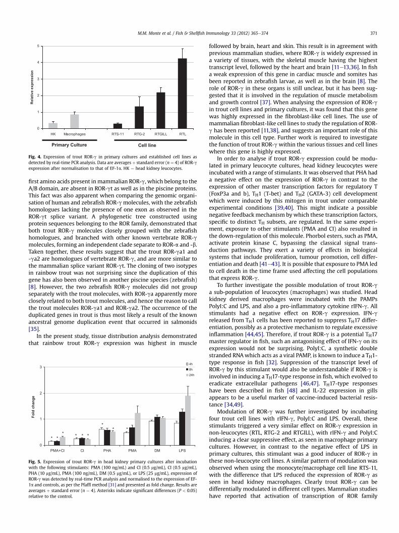
with a diverse range of stimulants including a T cell inducer (PHA),stimulators of intracellular signalling pathways (PMA and CI), animmunosuppressant (DM) and the bacterial pathogen associatedmolecular pattern (PAMP) LPS (Fig. 5). ROR-g was modulatednegatively by PMA plus CI, CI, PHA or PMA, at different time points,with fold changes as low as 0.15. Incubationwith LPS or DM had noeffect on ROR-g expression.
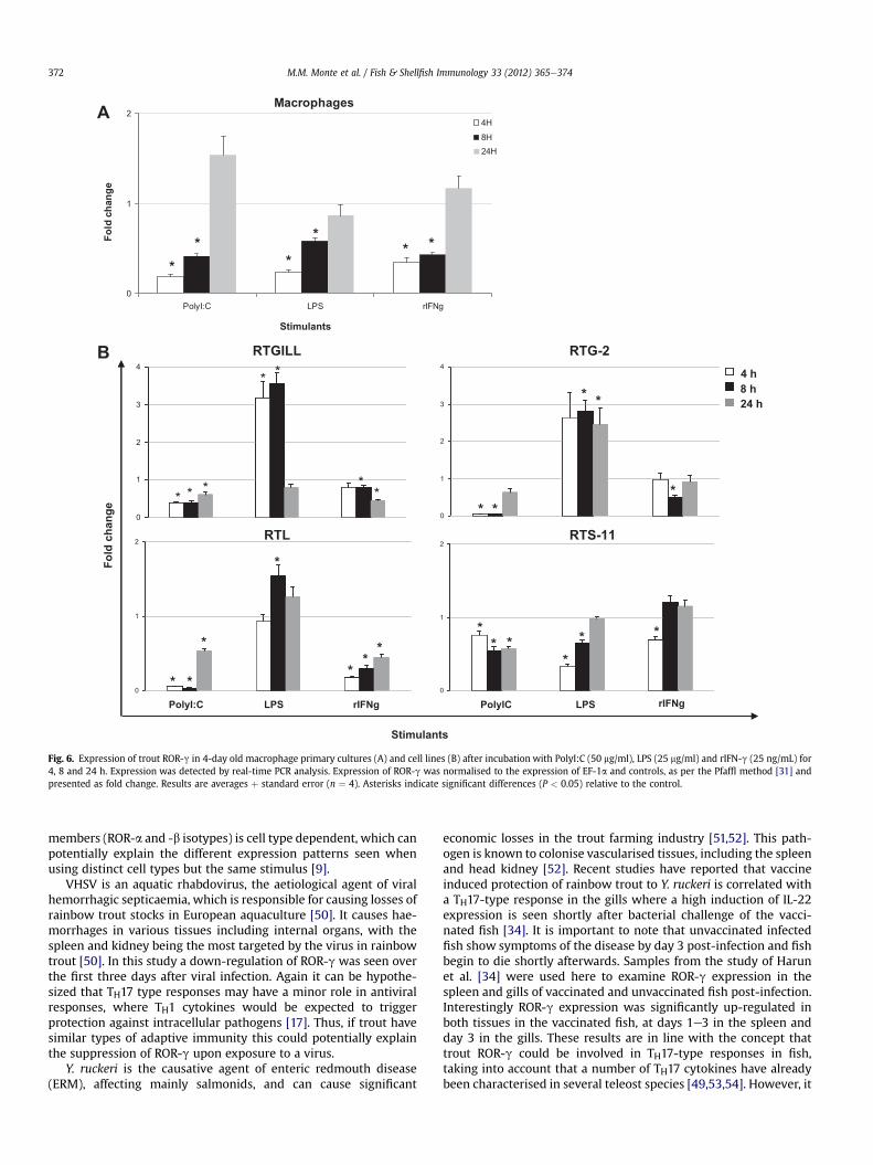
Since the constitutive expression of this molecule was higher inmacrophages isolated from head kidney than in the mixed cellpopulation obtained from the same tissue, head kidney macro-phages were used in a further stimulation experiment. Four day oldheadkidneymacrophageswere incubatedwith LPS,with a syntheticanalogue of double stranded RNA (PolyI:C), or with the pro-inflammatory cytokine rIFN-g, known to be involved in TH1responses (Fig. 6A). As observed previously, trout ROR-gwasmainlymodulated negatively, in this case, by the presence of all stimulantsused after a 4 and8h stimulation,with fold changes of 0.58 or below.
3.4. Modulation of trout ROR-g in cell lines
After analysing the modulation of ROR-g in primary cellcultures, the regulation of this molecule was also investigated usingfour established trout cell lines: RTGILL, RTG-2, RTL and RTS-11. Thecells were also incubated with LPS, PolyI:C and rIFN-g for 4, 8 and24 h (Fig. 6B). The presence of PolyI:C and rIFN-g generally down-regulated the expression of ROR-g (with some variation in kinetics)in all the cell lines studied. However, in contrast LPS was found toup-regulate ROR-g expression in RTGILL, RTG-2 and RTL cells,although it down-regulated the expression in RTS-11 cells.
3.5. Modulation of trout ROR-g in vivo
To assess the involvement of trout ROR-g upon a viral infectionand to investigate if it would also be negatively modulated, theexpression of this gene was analysed in head kidney samples fromfour fish at several time points (days 1, 2, 3, 4 and 6) after a VSHVchallenge (Fig. 7). Trout ROR-g expression was indeed down-regulated significantly (P < 0.05) at days 1, 2 and 3 post-infection,with fold changes of around 0.49, 0.18 and 0.26, respectively.
Since TH17-type responses might be expected in fish vaccinatedagainst extracellular bacteria,we also studied the expression of ROR-gin trout that had been previously given a commercial vaccine againstthe Gram negative bacterial pathogen Y. ruckeri and then challenged60 days later, with unvaccinated fish used as the control (Fig. 8). In thespleen, ROR-gexpressionwassignificantly increased (P<0.05)atdays1, 2 and 3 days post-infection in vaccinated fish vs the controlunvaccinatedfish,withw8-fold increaseapparentbyday3. In thegillsthere was a small down-regulation at 1 day post-infection in vacci-nated fish vs unvaccinated fish, but a significant increase was againapparent at day 3, albeit lower than that seen in the spleen.
4. Discussion
This paper reports the cloning and characterisation of two ROR-g homologues, ROR-ga1 and -ga2, in rainbow trout (O. mykiss). Thetwo mRNA sequences consist of 2713 and 2801 nucleotidesrespectively, translating into a 469 aa protein in both cases. Thetrout ROR-g molecules share a high homology of 98.3% and 99.6%
ighted in black, with the conserved Cys residues highlighted in grey. Arrows indicated. The accession numbers of the ROR-g proteins are as follow: zebrafish ROR-ga:01523; mouse ROR-g: NP_035411 and ROR-gt: CAA10661. Note: Identical residues aren and zebrafish ROR-g molecules. The coding region is represented as black boxes, the

ROR-α human
ROR-α cow
ROR-α mouse
ROR-α chicken
ROR-α xenopus
ROR-α zebrafish
ROR-α fugu
ROR-β zebrafish
ROR-β chicken
ROR-β human
ROR-β horseROR-β cow
ROR-β mouse
ROR-γa1 trout
ROR-γa2 trout
ROR-γa zebrafish
ROR-γb zebrafish
ROR-γ mouse
ROR-γ cow
ROR-γt human
ROR-γ human100
61
100
100
96
100
97
68
50
100
100
100
100
100
51
100
98
58
0.1
ROR-α
ROR-β
ROR-γ
Fig. 2. Phylogenetic tree showing the relationship of both trout ROR-g amino acid sequences with other known members of the ROR family. The amino acid sequences were alignedusing CLUSTAL W and the tree constructed by the Neighbor-Joining (N-J) method supported with 1000 bootstrap replications using MEGA 4.1 software. The accession numbers ofthe sequences used for the analysis are: ROR-a: human: NP_599023; cow: NP_001179790; mouse: NP_038674; chicken: XP_413763; xenopus: NP_001072663; zebrafish:NP_001103637 and fugu: ENSTRUP00000026635. ROR-b: zebrafish: ABO15413; chicken: Q98934; human: BAH02286; horse: XP_001488198; cow: NP_001179587 and mouse:NP_666207. ROR-g: zebrafish homologues a: NP_001076288 and b: XP_690743; mouse: NP_035411; cow: NP_001076920; human: NP_005051 and human homologue t:NP_001001523. Trout ROR-g proteins are in bold and underlined.
M.M. Monte et al. / Fish & Shellfish Immunology 33 (2012) 365e374370
identity and similarity, respectively. A multiple aa alignment of thedifferent vertebrate ROR-g molecules revealed a high conservationof DNA- and ligand-binding domains, which are characteristicfeatures of the nuclear receptor family [3,9]. The present resultsalso indicated that the AF-2 motif displayed a high conservationacross vertebrates. This was not unexpected since this motif was
0
1
10
100
1,000
Tail fins Headkidney
Gonad Thymus Spleen Caudalkidney
Scal
Re
lativ
e e
xp
re
ss
ion
Fig. 3. Expression of trout ROR-g in tissues (tail fins, head kidney, gonad, thymus, spleen, cauas detected by real-time PCR analysis. Data are averages þ standard error (n ¼ 6) of ROR-g
reported to be highly conserved amongst all ROR family members[6e8]. In terms of homology with other vertebrate species, troutROR-g shared the highest similarity with zebrafish proteins (over85%). A slightly higher homology to mammalian ROR-gt (over 65%)vs ROR-g (62.8e63.1%) was also observed, and reflects the simi-larity of protein lengths between fish ROR-g and ROR-gt. Thus, the
es Intestine Gills Liver Skin Heart Brain Muscle
dal kidney, scales, intestine, gills, liver, skin, heart, brain and muscle) from healthy fish,expression after being normalised to that of elongation factor (EF)-1a.
0
1
2
3
4
5
HK Macrophages RTS-11 RTG-2 RTGILL RTL
Re
lativ
e e
xp
re
ss
ion
Primary Culture Cell line
Fig. 4. Expression of trout ROR-g in primary cultures and established cell lines asdetected by real-time PCR analysis. Data are averages þ standard error (n ¼ 4) of ROR-gexpression after normalisation to that of EF-1a. HK ¼ head kidney leucocytes.
M.M. Monte et al. / Fish & Shellfish Immunology 33 (2012) 365e374 371
first amino acids present in mammalian ROR-g, which belong to theA/B domain, are absent in ROR-gt as well as in the piscine proteins.This fact was also apparent when comparing the genomic organi-sation of human and zebrafish ROR-gmolecules, with the zebrafishhomologues lacking the presence of one exon as observed in theROR-gt splice variant. A phylogenetic tree constructed usingprotein sequences belonging to the ROR family, demonstrated thatboth trout ROR-g molecules closely grouped with the zebrafishhomologues, and branched with other known vertebrate ROR-gmolecules, forming an independent clade separate to ROR-a and -b.Taken together, these results suggest that the trout ROR-ga1 and-ga2 are homologues of vertebrate ROR-g, and are more similar tothe mammalian splice variant ROR-gt. The cloning of two isotypesin rainbow trout was not surprising since the duplication of thisgene has also been observed in another piscine species (zebrafish)[8]. However, the two zebrafish ROR-g molecules did not groupseparately with the trout molecules, with ROR-ga apparently moreclosely related to both trout molecules, and hence the reason to callthe trout molecules ROR-ga1 and ROR-ga2. The occurrence of theduplicated genes in trout is thus most likely a result of the knownancestral genome duplication event that occurred in salmonids[35].
In the present study, tissue distribution analysis demonstratedthat rainbow trout ROR-g expression was highest in muscle
0
1
2
3
PMA+CI CI PHA PMA DM LPS
Fo
ld
ch
an
ge
4h
8h
24h
* ** * * *
** *
Fig. 5. Expression of trout ROR-g in head kidney primary cultures after incubationwith the following stimulants: PMA (100 ng/mL) and CI (0.5 mg/mL), CI (0.5 mg/mL),PHA (10 mg/mL), PMA (100 ng/mL), DM (0.5 mg/mL), or LPS (25 mg/mL), expression ofROR-g was detected by real-time PCR analysis and normalised to the expression of EF-1a and controls, as per the Pfaffl method [31] and presented as fold change. Results areaverages þ standard error (n ¼ 4). Asterisks indicate significant differences (P < 0.05)relative to the control.
followed by brain, heart and skin. This result is in agreement withprevious mammalian studies, where ROR-g is widely expressed ina variety of tissues, with the skeletal muscle having the highesttranscript level, followed by the heart and brain [11e13,36]. In fisha weak expression of this gene in cardiac muscle and somites hasbeen reported in zebrafish larvae, as well as in the brain [8]. Therole of ROR-g in these organs is still unclear, but it has been sug-gested that it is involved in the regulation of muscle metabolismand growth control [37]. When analysing the expression of ROR-gin trout cell lines and primary cultures, it was found that this genewas highly expressed in the fibroblast-like cell lines. The use ofmammalian fibroblast-like cell lines to study the regulation of ROR-g has been reported [11,38], and suggests an important role of thismolecule in this cell type. Further work is required to investigatethe function of trout ROR-gwithin the various tissues and cell lineswhere this gene is highly expressed.
In order to analyse if trout ROR-g expression could be modu-lated in primary leucocyte cultures, head kidney leucocytes wereincubated with a range of stimulants. It was observed that PHA hada negative effect on the expression of ROR-g in contrast to theexpression of other master transcription factors for regulatory T(FoxP3a and b), TH1 (T-bet) and TH2 (GATA-3) cell developmentwhich were induced by this mitogen in trout under comparableexperimental conditions [39,40]. This might indicate a possiblenegative feedback mechanism bywhich these transcription factors,specific to distinct TH subsets, are regulated. In the same experi-ment, exposure to other stimulants (PMA and CI) also resulted inthe down-regulation of this molecule. Phorbol esters, such as PMA,activate protein kinase C, bypassing the classical signal trans-duction pathways. They exert a variety of effects in biologicalsystems that include proliferation, tumour promotion, cell differ-entiation and death [41e43]. It is possible that exposure to PMA ledto cell death in the time frame used affecting the cell populationsthat express ROR-g.
To further investigate the possible modulation of trout ROR-ga sub-population of leucocytes (macrophages) was studied. Headkidney derived macrophages were incubated with the PAMPsPolyI:C and LPS, and also a pro-inflammatory cytokine rIFN-g. Allstimulants had a negative effect on ROR-g expression. IFN-greleased from TH1 cells has been reported to suppress TH17 differ-entiation, possibly as a protective mechanism to regulate excessiveinflammation [44,45]. Therefore, if trout ROR-g is a potential TH17master regulator in fish, such an antagonising effect of IFN-g on itsexpression would not be surprising. PolyI:C, a synthetic doublestranded RNAwhich acts as a viral PAMP, is known to induce a TH1-type response in fish [32]. Suppression of the transcript level ofROR-g by this stimulant would also be understandable if ROR-g isinvolved in inducing a TH17-type response in fish, which evolved toeradicate extracellular pathogens [46,47]. TH17-type responseshave been described in fish [48] and IL-22 expression in gillsappears to be a useful marker of vaccine-induced bacterial resis-tance [34,49].
Modulation of ROR-g was further investigated by incubatingfour trout cell lines with rIFN-g, PolyI:C and LPS. Overall, thesestimulants triggered a very similar effect on ROR-g expression innon-leucocytes (RTL, RTG-2 and RTGILL), with rIFN-g and PolyI:Cinducing a clear suppressive effect, as seen in macrophage primarycultures. However, in contrast to the negative effect of LPS inprimary cultures, this stimulant was a good inducer of ROR-g inthese non-leucocyte cell lines. A similar pattern of modulation wasobserved when using the monocyte/macrophage cell line RTS-11,with the difference that LPS reduced the expression of ROR-g asseen in head kidney macrophages. Clearly trout ROR-g can bedifferentially modulated in different cell types. Mammalian studieshave reported that activation of transcription of ROR family
A
B
0
1
2
PolyI:C LPS rIFNg
4H8H24H
*
*
*
*
**
Fo
ld
ch
an
ge
Stimulants
Macrophages
0
1
2
3
4
* *
*
*
*
**
0
1
2
3
4
* *
**
*
0
1
2
* *
*
*
*
*
*
RTG-2RTGILL
RTS-11RTL
Fo
ld
c
ha
ng
e
Stimulants
4 h
8 h
24 h
PolyI:C rIFNgLPS
0
1
2
*
* *
*
**
PolyIC LPS rIFNg
Fig. 6. Expression of trout ROR-g in 4-day old macrophage primary cultures (A) and cell lines (B) after incubation with PolyI:C (50 mg/ml), LPS (25 mg/ml) and rIFN-g (25 ng/mL) for4, 8 and 24 h. Expression was detected by real-time PCR analysis. Expression of ROR-g was normalised to the expression of EF-1a and controls, as per the Pfaffl method [31] andpresented as fold change. Results are averages þ standard error (n ¼ 4). Asterisks indicate significant differences (P < 0.05) relative to the control.
M.M. Monte et al. / Fish & Shellfish Immunology 33 (2012) 365e374372
members (ROR-a and -b isotypes) is cell type dependent, which canpotentially explain the different expression patterns seen whenusing distinct cell types but the same stimulus [9].
VHSV is an aquatic rhabdovirus, the aetiological agent of viralhemorrhagic septicaemia, which is responsible for causing losses ofrainbow trout stocks in European aquaculture [50]. It causes hae-morrhages in various tissues including internal organs, with thespleen and kidney being the most targeted by the virus in rainbowtrout [50]. In this study a down-regulation of ROR-g was seen overthe first three days after viral infection. Again it can be hypothe-sized that TH17 type responses may have a minor role in antiviralresponses, where TH1 cytokines would be expected to triggerprotection against intracellular pathogens [17]. Thus, if trout havesimilar types of adaptive immunity this could potentially explainthe suppression of ROR-g upon exposure to a virus.
Y. ruckeri is the causative agent of enteric redmouth disease(ERM), affecting mainly salmonids, and can cause significant
economic losses in the trout farming industry [51,52]. This path-ogen is known to colonise vascularised tissues, including the spleenand head kidney [52]. Recent studies have reported that vaccineinduced protection of rainbow trout to Y. ruckeri is correlated witha TH17-type response in the gills where a high induction of IL-22expression is seen shortly after bacterial challenge of the vacci-nated fish [34]. It is important to note that unvaccinated infectedfish show symptoms of the disease by day 3 post-infection and fishbegin to die shortly afterwards. Samples from the study of Harunet al. [34] were used here to examine ROR-g expression in thespleen and gills of vaccinated and unvaccinated fish post-infection.Interestingly ROR-g expression was significantly up-regulated inboth tissues in the vaccinated fish, at days 1e3 in the spleen andday 3 in the gills. These results are in line with the concept thattrout ROR-g could be involved in TH17-type responses in fish,taking into account that a number of TH17 cytokines have alreadybeen characterised in several teleost species [49,53,54]. However, it

0
1
2
Day 1 Day 2 Day 3 Day 4 Day 6
Fo
ld
ch
an
ge
*
**
Fig. 7. Expression of trout ROR-g during a viral infection. Rainbow trout were injectedintraperitoneally with VHSV (strain DK-F1; 1 � 108 TCID50/mL) or control media. Headkidney tissue was collected at days 1, 2, 3, 4 and 6 after challenge, and RNA extractedfor real-time PCR analysis. Expression of ROR-g was normalised to the expression ofEF-1a and controls, as per the Pfaffl method [31] and presented as fold change. Resultsare averages þ standard error (n ¼ 4). Asterisks indicate significant differences(P < 0.05) relative to the control.
M.M. Monte et al. / Fish & Shellfish Immunology 33 (2012) 365e374 373
is also important to note that TH17 cells are not the only cell typethat can express ROR-gt and secrete IL-17 and/or IL-22. Recentstudies have reported that a sub-population of mucosal NK cellsand NKT cells also express these molecules [18,19,55].
In summary, this report describes the cloning and character-isation of two trout ROR-g homologues for the first time insalmonids, and their modulation upon treatment with differentstimuli. The high expression of this gene in muscle and brain,obtained from healthy fish, and in non-leucocyte cell lines, indi-cates a potential role of ROR-g in these tissues/cell types. Theexpression of ROR-g in these cells was induced by LPS but sup-pressed by PolyI:C and rIFN-g. Similarly, in virus challenged fishROR-g expression was suppressed. However, in vaccinated fish thatare subsequently challenged with the homologous bacteria theexpression of ROR-g was induced 1e3 days post-infection. Overall,
0
2
4
6
8
10
12
6h Day 1 Day 2 Day 3
Fo
ld
ch
an
ge
Gill
Spleen
*
*
*
*
*
Fig. 8. Expression of trout ROR-g following bacterial infection of vaccinated (60 daysearlier with Aquavac�) and unvaccinated fish. Rainbow trout were injected intraper-itoneally with Yersinia ruckeri (0.5 mL, 1 � 106 cfu/fish) and spleen and gill tissuecollected at 6 h, 1, 2 and 3 days after challenge. RNA was extracted for real-time PCRanalysis, and data were analysed using the Pfaffl method [31] and expressed as foldchange relative to the unvaccinated fish. Results are averages þ standard error (n ¼ 5),and asterisks indicate significant differences (P < 0.05) relative to the unvaccinatedfish.
the sequence and expression analysis lead us to speculate that troutROR-g could function in a similar way to mammalian ROR-gt, atleast in terms of having a role in responses to extracellular micro-bial infections. In addition, since particular stimulants triggereddistinct effects on ROR-g expression according to the cell typestudied, suggests that the regulatory mechanism(s) that govern theexpression of this gene are most likely cell-type dependent.
Acknowledgements
This work was supported financially by a PhD studentship fromthe FCT (Foundation for Science and Technology, Portugal) andPOPH/FSE (Grant no. SFRH/BD/38236/2007) to Milena M. Monte,and the European Commission Contract No. 007103 (IMAQUANIM-Improved immunity of aquacultured animals). Maria M. Costareceived funding from the Consejo Superior de InvestigacionesCientíficas (CSIC, Spain) and the Xunta de Galicia for her “ÁngelesAlvariño” postdoctoral contract. Nor O. Harun received fundingfrom the Ministry of Higher Education (Malaysia) and the Univer-sity Malaysia Terengganu for her PhD studies. We thank MarineScotland staff for providing samples from VHSV-infected rainbowtrout generated within the Scottish Government funded researchproject FC1996.
Appendix A. Supplementary Material
Supplementary Material related to this article can be foundonline at doi:10.1016/j.fsi.2012.05.023.
References
[1] Laudet V. Evolution of the nuclear receptor superfamily: early diversificationfrom an ancestral orphan receptor. J Mol Endocrinol 1997;19(3):207e26.
[2] Giguère V. Orphan nuclear receptors: from gene to function. Endocr Rev 1999;20(5):689e725.
[3] Zaveri NT, Murphy BJ. Nuclear hormone receptors. In: Taylor J, Triggle D,editors. Comprehensive medicinal chemistry; 2007. p. 993e1036.
[4] Lodish H, Berk A, Kaiser CA, Krieger M, Scott MP, Bretscher A, et al. Molecularcell biology. 6th ed. Freeman and Company; 2008.
[5] Mangelsdorf DJ, Thummel C, Beato M, Herrlich P, Schutz G, Umesono K, et al.The nuclear receptor superfamily: the second decade. Cell 1995;83(6):835e9.
[6] Jetten AM, Joo JH. Retinoid-related orphan receptors (RORs): roles in cellulardifferentiation and development. Adv Dev Biol 2006;16:313e55.
[7] Jetten AM. Retinoid-related orphan receptors (RORs): critical roles in devel-opment, immunity, circadian rhythm, and cellular metabolism. Nucl ReceptSignal 2009;7:1e32.
[8] Flores MV, Hall C, Jury A, Crosier K, Crosier P. The zebrafish retinoid-relatedorphan receptor (ROR) gene family. Gene Expr Patterns 2007;7(5):535e43.
[9] Jetten AM, Kurebayashi S, Ueda E. The ROR nuclear orphan receptorsubfamily: critical regulators of multiple biological processes. Prog NucleicAcid Res Mol Biol 2001;69:205e47.
[10] Gronemeyer H, Gustafsson JÅ, Laudet V. Principles for modulation of thenuclear receptor superfamily. Nat Rev Drug Discov 2004;3(11):950e64.
[11] Medvedev A, Yan ZH, Hirose T, Giguère V, Jetten AM. Cloning of a cDNAencoding the murine orphan receptor RZR/RORg and characterization of itsresponse element. Gene 1996;181(1e2):199e206.
[12] Hirose T, Smith RJ, Jetten AM. ROR-gamma: the third member of ROR/RZRorphan receptor subfamily that is highly expressed in skeletal muscle. Bio-chem Biophys Res Commun 1994;205(3):1976e83.
[13] He YW, Deftos ML, Ojala EW, Bevan MJ. RORgt, a novel isoform of an orphanreceptor, negatively regulates fas ligand expression and IL-2 production in Tcells. Immunity 1998;9(6):797e806.
[14] Ortiz MA, Piedrafita FJ, Pfahl M, Maki RTOR. A new orphan receptor expressedin the thymus that can modulate retinoid and thyroid hormone signals. MolEndocrinol 1995;9(12):1679e91.
[15] Kurebayashi S, Ueda E, Sakaue M, Patel DD, Medvedev A, Zhang F, et al.Retinoid-related orphan receptor (ROR) is essential for lymphoid organo-genesis and controls apoptosis during thymopoiesis. Proc Natl Acad Sci USA2000;97(18):10132e7.
[16] Ivanov II, McKenzie BS, Zhou L, Tadokoro CE, Lepelley A, Lafaille JJ, et al. Theorphan nuclear receptor ROR gt directs the differentiation program ofproinflammatory IL-17 T helper cells. Cell 2006;126(6):1121e33.
[17] Weaver CT, Hatton RD, Mangan PR, Harrington LE. IL-17 family cytokines andthe expanding diversity of effector T cell lineages. Annu Rev Immunol 2007;25:821e52.

M.M. Monte et al. / Fish & Shellfish Immunology 33 (2012) 365e374374
[18] Luci C, Reynders A, Ivanov II, Cognet C, Chiche L, Chasson L, et al. Influence ofthe transcription factor RORgt on the development of NKp46 cell populationsin gut and skin. Nat Immunol 2008;10:75e82.
[19] Sanos SL, Bui VL, Mortha A, Oberle K, Heners C, Johner C, et al. RORgt andcommensal microflora are required for the differentiation of mucosal inter-leukin 22eproducing NKp46 cells. Nat Immunol 2008;10:83e91.
[20] Altschul SF, Madden TL, Schaffer AA, Zhang J, Zhang Z, Miller W, et al. GappedBLAST and PSI-BLAST: a new generation of protein database search programs.Nucleic Acid Res 1997;25(17):3389e402.
[21] Wang T, Secombes CJ. Complete sequencing and expression of threecomplement components, C1r, C4 and C1 inhibitor, of the classical activationpathway of the complement system in rainbow trout Oncorhynchus mykiss.Immunogenetics 2003;55(9):615e28.
[22] Chenna R, Sugawara H, Koike T, Lopez R, Gibson TJ, Higgins DG, et al. Multiplesequence alignment with the clustal series of programs. Nucleic Acids Res2003;31(13):3497e500.
[23] Campanella JJ, Bitincka L, Smalley J. MatGAT: an application that generatessimilarity/identity matrices using protein or DNA sequences. BMC Bioinform2003;4:29.
[24] Tamura K, Dudley J, Nei M, Kumar S. MEGA4: molecular evolutionary geneticsanalysis (MEGA) software version 4.0. Mol Biol Evol 2007;24(8):1596e9.
[25] Letunic I, Doerks T, Bork P. SMART 6: recent updates and new developments.Nucleic Acids Res 2009;37:229e32.
[26] Ganassin RC, Bols NC. Development of a monocyte/macrophage-like cell line,RTS11, from rainbow trout spleen. Fish Shellfish Immunol 1998;8(6):457e76.
[27] Wolf K, Quimby MC. Established eurythermic line of fish cells in vitro. Science1962;135:1065e6.
[28] Lee LEJ, Clemons JH, Bechtel DG, Caldwell SJ, Han KB, Pasitschniak-Arts M,et al. Development and characterization of a rainbow trout liver cell lineexpressing cytochrome P450-dependent monooxygenase activity. Cell BiolToxicol 1993;9(3):279e94.
[29] Bols N, Barlian A, Chirino-Trejo M, Caldwell S, Goegan P, Lee L. Developmentof a cell line from primary cultures of rainbow trout, Oncorhynchus mykiss(Walbaum), gills. J Fish Dis 1994;17(6):601e11.
[30] Wang T, Diaz-Rosales P, Costa MM, Campbell S, Snow M, Collet B, et al.Functional characterization of a nonmammalian IL-21: rainbow trout Onco-rhynchus mykiss IL-21 upregulates the expression of the TH cell signaturecytokines IFN-g, IL-10, and IL-22. J Immunol 2011;186(2):708e21.
[31] Pfaffl MW. A new mathematical model for relative quantification in real-timeRT-PCR. Nucleic Acids Res 2001;29(9):e45.
[32] Zou J, CarringtonA,Collet B,Dijkstra JM,YoshiuraY,BolsN, et al. Identificationandbioactivities of IFN-g in rainbow trout Oncorhynchus mykiss: the first Th1-typecytokine characterized functionally in fish. J Immunol 2005;175(4):2484e94.
[33] Campbell S, McBeath A, Secombes C, Snow M, Collet B. Interferon responsefollowing infection with genetically similar isolates of viral haemorrhagicsepticaemia virus (VHSV) exhibiting contrasting virulence in rainbow trout.Fish Shellfish Immunol 2011;30(1):287e94.
[34] Harun NO, Wang T, Secombes CJ. Gene expression profiling in naïve and vacci-nated rainbow trout after Yersinia ruckeri infection: insights into the mecha-nisms of protection seen in vaccinated fish. Vaccine 2011;29(26):4388e99.
[35] Koop BF, von Schalburg KR, Leong J, Walker N, Lieph R, Cooper GA, et al.A salmonid EST genomic study: genes, duplications, phylogeny and micro-arrays. BMC Genomics 2008;9:545.
[36] Kang HS, Angers M, Beak JY, Wu X, Gimble JM, Wada T, et al. Gene expressionprofiling reveals a regulatory role for RORa and RORg in phase I and phase IImetabolism. Physiol Genomics 2007;31:281e94.
[37] Raichur S, Lau P, Staels B, Muscat GEO. Retinoid-related orphan receptor gregulates several genes that control metabolism in skeletal muscle cells: linksto modulation of reactive oxygen species production. J Mol Endocrinol 2007;39(1):29e44.
[38] Austin S, Medvedev A, Yan ZH, Adachi H, Hirose T, Jetten AM. Induction of thenuclear orphan receptor RORgamma during adipocyte differentiation of D1and 3T3-L1 cells. Cell Growth Differ 1998;9(3):267e76.
[39] Wang T, Holland JW, Martin SAM, Secombes CJ. Sequence and expressionanalysis of two T helper master transcription factors, T-bet and GATA3, inrainbow trout Oncorhynchus mykiss and analysis of their expression duringbacterial and parasitic infection. Fish Shellfish Immunol 2010;29(5):705e15.
[40] Wang T, Monte MM, Huang W, Boudinot P, Martin SAM, Secombes CJ. Iden-tification of two FoxP3 genes in rainbow trout (Oncorhynchus mykiss) withdifferential induction patterns. Mol Immunol 2010;47(16):2563e74.
[41] Niedel JE, Kuhn LJ, Vandenbark G. Phorbol diester receptor copurifies withprotein kinase C. Proc Natl Acad Sci USA 1983;80(1):36.
[42] Leach KL, Blumberg PM. Modulation of protein kinase C activity and [3H]phorbol 12, 13-dibutyrate binding by various tumor promoters in mousebrain cytosol. Cancer Res 1985;45(5):1958e63.
[43] Blumberg PM. Protein kinase C as the receptor for the phorbol ester tumorpromoters: sixth rhoads memorial award lecture. Cancer Res 1988;48(1):1e8.
[44] Harrington LE, Hatton RD, Mangan PR, Turner H, Murphy TL, Murphy KM,et al. Interleukin 17eproducing CD4 effector T cells develop via a lineagedistinct from the T helper type 1 and 2 lineages. Nat Immunol 2005;6(11):1123e32.
[45] Park H, Li Z, Yang XO, Chang SH, Nurieva R, Wang YH, et al. A distinct lineageof CD4 T cells regulates tissue inflammation by producing interleukin 17. NatImmunol 2005;6(11):1133e41.
[46] Bettelli E, Oukka M, Kuchroo VK. TH-17 cells in the circle of immunity andautoimmunity. Nat Immunol 2007;8(4):345e50.
[47] Wilson NJ, Boniface K, Chan JR, McKenzie BS, Blumenschei WM, Mattson JD,et al. Development, cytokine profile and function of human interleukin17eproducing helper T cells. Nat Immunol 2007;8(9):950e7.
[48] Ribeiro CMS, Pontes MJSL, Bird S, Chadzinska M, Scheer M, Verburg-vanKemenade BML, et al. Trypanosomiasis-induced Th17-like immune responsesin carp. Plos ONE 2010;5(9):e13012.
[49] Corripio-Miyar Y, Zou J, Richmond H, Secombes CJ. Identification ofinterleukin-22 in gadoids and examination of its expression level in vacci-nated fish. Mol Immunol 2009;46(10):2098e106.
[50] Skall HF, Olesen NJ, Mellergaard S. Viral haemorrhagic septicaemia virus inmarine fish and its implications for fish farmingea review. J Fish Dis 2005;28(9):509e29.
[51] Fernández L, Méndez J, Guijarro JA. Molecular virulence mechanisms of thefish pathogen Yersinia ruckeri. Vet Microbiol 2007;125(1e2):1e10.
[52] Tobback E, Decostere A, Hermans K, Haesebrouck F, Chiers K. Yersinia ruckeriinfections in Salmonid fish. J Fish Dis 2007;30(5):257e68.
[53] Gunimaladevi I, Savan R, Sakai M. Identification, cloning and characterizationof interleukin-17 and its family from zebrafish. Fish Shellfish Immunol 2006;21(4):393e403.
[54] Monte MM, Zou J, Wang T, Carrington A, Secombes CJ. Cloning, expressionanalysis and bioactivity studies of rainbow trout (Oncorhynchus mykiss)interleukin-22. Cytokine 2011;55(1):62e73.
[55] Rachitskaya AV, Hansen AM, Horai R, Li Z, Villasmil R, Luger D, et al. Cuttingedge: NKT cells constitutively express IL-23 receptor and RORgammat andrapidly produce IL-17 upon receptor ligation in an IL-6-independent fashion.J Immunol 2008;180(8):5167e71.
Related Documents


