Cloning and Characterization of GLOSSY1, a Maize Gene Involved in Cuticle Membrane and Wax Production 1[w] Monica Sturaro, Hans Hartings, Elmon Schmelzer, Riccardo Velasco, Francesco Salamini, and Mario Motto* Istituto Sperimentale per la Cerealicoltura, Sezione di Bergamo, 24126 Bergamo, Italy (M.S., H.H., M.M.); and Max-Planck Institut fu ¨r Zu ¨chtungsforschung, D–50829 Cologne, Germany (E.S., R.V., F.S.) The cuticle covering the aerial organs of land plants plays a protective role against several biotic and abiotic stresses and, in addition, participates in a variety of plant-insect interactions. Here, we describe the molecular cloning and characterization of the maize (Zea mays) GLOSSY1 (GL1) gene, a component of the pathway leading to cuticular wax biosynthesis in seedling leaves. The genomic and cDNA sequences we isolated differ significantly in length and in most of the coding region from those previously identified. The predicted GL1 protein includes three histidine-rich domains, the landmark of a family of membrane- bound desaturases/hydroxylases, including fatty acid-modifying enzymes. GL1 expression is not restricted to the juvenile developmental stage of the maize plant, pointing to a broader function of the gene product than anticipated on the basis of the mutant phenotype. Indeed, in addition to affecting cuticular wax biosynthesis, gl1 mutations have a pleiotropic effect on epidermis development, altering trichome size and impairing cutin structure. Of the many wax biosynthetic genes identified so far, only a few from Arabidopsis (Arabidopsis thaliana) were found to be essential for normal cutin formation. Among these is WAX2, which shares 62% identity with GL1 at the protein level. In wax2-defective plants, cutin alterations induce postgenital organ fusion. This trait is not displayed by gl1 mutants, suggesting a different role of the maize and Arabidopsis cuticle in plant development. The cuticle forms the outermost layer of the above- ground parts of most plants. The physical and chem- ical properties of this structure support vital functions such as prevention of nonstomatal water loss, pro- tection against UV irradiation, and reduction of de- position of dust, pollen, and air pollutants. In addition, it plays a critical role in plant defense against bacterial and fungal pathogens and participates in a variety of plant-insect interactions (Post-Beittenmiller, 1996). The cuticle is synthesized by the epidermal cells and consists of an outer layer of epicuticular waxes over- laying the cuticle membrane, which is composed of a network of interesterified hydroxy and epoxy- hydroxy fatty acids of mainly 16 and 18 atoms in length (cutin) interspersed by intracuticular waxes (Walton, 1990). Cuticular waxes consist primarily of complex mixtures of aliphatic molecules of mainly 16 to 34 carbon atoms in length that occur as free fatty acids, aldehydes, primary alcohols, alkanes, and esters. Their production is a biologically complex process involving a host of synthetic and transport mechanisms. The composition of cuticular waxes differs among plant species, organs, and tissues, and during development. Wax deposition on the leaf surface is also regulated by environmental signals such as light, moisture, and temperature (Kolattukudy, 1996). Although advances have been made in the under- standing of the biosynthesis of specific cutin and wax constituents, many questions pertaining to the orga- nization and regulation of the concerned biochemical pathways remain unanswered. The availability of mutants deficient in cuticular wax accumulation in a variety of species and the isolation of the corre- sponding genes reveal helpful information to eluci- date cuticular wax biosynthesis and to characterize molecular aspects of regulatory control (Kunst and Samuels, 2003). In maize (Zea mays), at least 18 loci (the GLOSSY or GL loci) have been found to affect the quantity and/or the composition of cuticular waxes on the surface of seedling leaves (Neuffer et al., 1997). From genetic and biochemical analyses of maize plants carrying differ- ent gl mutations, a preliminary model predicting two distinct pathways for cuticular wax biosynthesis has been set forth (Bianchi et al., 1985). One pathway would be responsible for wax synthesis in the first five or six juvenile leaves, whereas the other would pro- duce waxes during the whole life cycle of the maize plant. The products of these two pathways can be distinguished by their chemical composition: Approx- imately 80% of the juvenile waxes are very-long-chain alcohols and aldehydes, whereas approximately 70% of the waxes produced throughout the life of a maize plant consist of esters (Bianchi et al., 1985). These ontogenetic differences in wax composition lead to different phenotypes of the maize leaves; juvenile 1 This work was supported by grants from the Ministero delle Politiche Agricole e Forestali, Rome. * Corresponding author; e-mail [email protected]; fax 39–035–31– 60–54. [w] The online version of this article contains Web-only data. Article, publication date, and citation information can be found at www.plantphysiol.org/cgi/doi/10.1104/pp.104.058164. 478 Plant Physiology, May 2005, Vol. 138, pp. 478–489, www.plantphysiol.org Ó 2005 American Society of Plant Biologists

Welcome message from author
This document is posted to help you gain knowledge. Please leave a comment to let me know what you think about it! Share it to your friends and learn new things together.
Transcript

Cloning and Characterization of GLOSSY1, a Maize GeneInvolved in Cuticle Membrane and Wax Production1[w]
Monica Sturaro, Hans Hartings, Elmon Schmelzer, Riccardo Velasco, Francesco Salamini, and Mario Motto*
Istituto Sperimentale per la Cerealicoltura, Sezione di Bergamo, 24126 Bergamo, Italy (M.S., H.H., M.M.);and Max-Planck Institut fur Zuchtungsforschung, D–50829 Cologne, Germany (E.S., R.V., F.S.)
The cuticle covering the aerial organs of land plants plays a protective role against several biotic and abiotic stresses and, inaddition, participates in a variety of plant-insect interactions. Here, we describe the molecular cloning and characterization ofthe maize (Zea mays) GLOSSY1 (GL1) gene, a component of the pathway leading to cuticular wax biosynthesis in seedlingleaves. The genomic and cDNA sequences we isolated differ significantly in length and in most of the coding region from thosepreviously identified. The predicted GL1 protein includes three histidine-rich domains, the landmark of a family of membrane-bound desaturases/hydroxylases, including fatty acid-modifying enzymes. GL1 expression is not restricted to the juveniledevelopmental stage of the maize plant, pointing to a broader function of the gene product than anticipated on the basis of themutant phenotype. Indeed, in addition to affecting cuticular wax biosynthesis, gl1 mutations have a pleiotropic effect onepidermis development, altering trichome size and impairing cutin structure. Of the many wax biosynthetic genes identified sofar, only a few from Arabidopsis (Arabidopsis thaliana) were found to be essential for normal cutin formation. Among these isWAX2, which shares 62% identity with GL1 at the protein level. In wax2-defective plants, cutin alterations induce postgenitalorgan fusion. This trait is not displayed by gl1 mutants, suggesting a different role of the maize and Arabidopsis cuticle in plantdevelopment.
The cuticle forms the outermost layer of the above-ground parts of most plants. The physical and chem-ical properties of this structure support vital functionssuch as prevention of nonstomatal water loss, pro-tection against UV irradiation, and reduction of de-position of dust, pollen, and air pollutants. In addition,it plays a critical role in plant defense against bacterialand fungal pathogens and participates in a variety ofplant-insect interactions (Post-Beittenmiller, 1996).
The cuticle is synthesized by the epidermal cells andconsists of an outer layer of epicuticular waxes over-laying the cuticle membrane, which is composed ofa network of interesterified hydroxy and epoxy-hydroxy fatty acids of mainly 16 and 18 atoms in length(cutin) interspersed by intracuticular waxes (Walton,1990). Cuticular waxes consist primarily of complexmixtures of aliphatic molecules of mainly 16 to 34carbon atoms in length that occur as free fatty acids,aldehydes, primary alcohols, alkanes, and esters. Theirproduction is a biologically complex process involvinga host of synthetic and transport mechanisms. Thecomposition of cuticular waxes differs among plantspecies, organs, and tissues, and during development.Wax deposition on the leaf surface is also regulated by
environmental signals such as light, moisture, andtemperature (Kolattukudy, 1996).
Although advances have been made in the under-standing of the biosynthesis of specific cutin and waxconstituents, many questions pertaining to the orga-nization and regulation of the concerned biochemicalpathways remain unanswered. The availability ofmutants deficient in cuticular wax accumulation ina variety of species and the isolation of the corre-sponding genes reveal helpful information to eluci-date cuticular wax biosynthesis and to characterizemolecular aspects of regulatory control (Kunst andSamuels, 2003).
In maize (Zea mays), at least 18 loci (the GLOSSY orGL loci) have been found to affect the quantity and/orthe composition of cuticular waxes on the surface ofseedling leaves (Neuffer et al., 1997). From genetic andbiochemical analyses of maize plants carrying differ-ent gl mutations, a preliminary model predicting twodistinct pathways for cuticular wax biosynthesis hasbeen set forth (Bianchi et al., 1985). One pathwaywould be responsible for wax synthesis in the first fiveor six juvenile leaves, whereas the other would pro-duce waxes during the whole life cycle of the maizeplant. The products of these two pathways can bedistinguished by their chemical composition: Approx-imately 80% of the juvenile waxes are very-long-chainalcohols and aldehydes, whereas approximately 70%of the waxes produced throughout the life of a maizeplant consist of esters (Bianchi et al., 1985). Theseontogenetic differences in wax composition lead todifferent phenotypes of the maize leaves; juvenile
1 This work was supported by grants from the Ministero dellePolitiche Agricole e Forestali, Rome.
* Corresponding author; e-mail [email protected]; fax 39–035–31–60–54.
[w] The online version of this article contains Web-only data.Article, publication date, and citation information can be found at
www.plantphysiol.org/cgi/doi/10.1104/pp.104.058164.
478 Plant Physiology, May 2005, Vol. 138, pp. 478–489, www.plantphysiol.org � 2005 American Society of Plant Biologists

leaves of wild-type maize plants have a glaucoussurface appearance, whereas all leaves appearing laterin plant development have a glossy surface. Mutationsimpairing the juvenile wax pathway also confer aglossy appearance to the juvenile leaves. Because oftheir phenotypic appearance, such mutants were de-signated glossy. The different composition of the waxlayer of epidermal cells of juvenile and adult leaves isone of the traits that defines the juvenile-to-adultphase transition in wild-type maize plants (Lawsonand Poethig, 1995, and refs. therein).
Over the past years, various maize GLOSSY genesinvolved in cuticular wax production have been cloned(Moose and Sisco, 1994; Tacke et al., 1995; Hansen et al.,1997; Xu et al., 1997). Based on evidence presented byXu et al. (2002), GL8 encodes as a b-ketoacyl reductaseof the fatty acid elongase complex involved in waxproduction. GL2 is apparently involved in acyl chainelongation from C30 to C32. However, comparisons ofthe predicted GL2 sequence with those in proteindatabases revealed no similarities to any known fattyacyl synthases, as may be expected for a component ofthe acyl elongation pathway (Tacke et al., 1995). TheGL15 locus is a developmental gene belonging tothe APETALA2 family of regulatory genes involved inthe transition from juvenile to adult leaves (Moose andSisco, 1996). The glossy phenotype of gl15 mutants issecondary to the primary mutant defect supporting theprecocious development of adult leaves.
Mutation at the GL1 locus causes dramatic alter-ations in the amount, composition, and crystallizationpatterns of juvenile cuticular waxes (Bianchi et al.,1985). The most conspicuous property of gl1 waxes, inaddition to a large reduction of aldehydes and alco-hols, concerns the predominant chain length of long-chain aldehydes and the corresponding free andesterified alcohols, which turn out to be reduced bytwo carbon atoms. In this context, it has been hypoth-esized that the GL1 locus is either required for anelongation step in cuticular wax biosynthesis or isaffecting the supply of wax precursors. However, thespecific role of GL1 that emerged from these chemicalstudies was neither precise nor definitive.
In an effort to characterize the Gl1 gene, we per-formed transposon-tagging experiments with theEnhancer/Suppressor mutation (En/Spm) element,which led to the tagging of the GL1 locus (Maddaloniet al., 1990). In this article, we describe the cloningand molecular characterization of this gene. The geno-mic and cDNA sequences we have isolated differ inmost of the coding region from the putative GL1 geneand transcript previously identified by Hansen et al.(1997). The protein encoded by GL1 shows significanthomology with the entire sequence of the WAX2 geneproduct of Arabidopsis (Arabidopsis thaliana) involvedin both cutin development and cuticular wax pro-duction (Chen et al., 2003). Similarly, the gl1 mutantdisplays a reduction in cuticular wax deposition andan alteration in cuticle membrane and plant hair(trichome) morphology.
RESULTS
Isolation of Transposon-Tagged Alleles of theGL1 Locus
From the cross outlined in ‘‘Materials and Meth-ods,’’ nine glossy seedlings with revertant nonglossysectors were identified out of approximately 90,000 F1seedlings. The new alleles were designated gl1-m1through gl1-m9. Genetic analyses of the new mutablealleles are described by Maddaloni et al. (1990). Sevenalleles, gl1-m1, 2, 3, 5, 7, 8, and 9, are due to theinsertion of an element behaving autonomously but,contrary to expectation, are different from Activator(Ac). The alleles gl1-m4 and -m6 carried a nonautono-mous element.
Plants carrying the gl1-m5 allele were tested for thefunctional presence in their genome of the trans-posable element En/Spm. Plants heterozygous for thegl1-m5 allele and for the stable recessive referenceallele gl1-ref were crossed with the En/Spm testerstrains described in ‘‘Materials and Methods.’’ Threeindependent test crosses were performed. F1 plants ofeach test cross were selfed, and the resulting earswere scored for F2 kernels displaying reversions ofthe a1-m1 or a1-m(r) tester allele. All three groups of F1plants gave rise to segregating and nonsegregatingears, with a percentage of the former near to 75%, sug-gesting the presence, in the gl1-m5 progenitor, of twounlinked copies of an active En/Spm element. F2kernels of both groups of ears were grown to seed-lings, and these were scored for leaf variegation due tothe gl1-m5 allele. The results of these experiments aresummarized in Table I. Approximately one-half of theears that segregated for variegated kernels also segre-gated gl1-m5 variegated seedlings, while ears withoutvariegated seeds did not originate variegated seed-lings. This result indicates that the gl1-m5 allele islikely to be caused by the insertion of an autonomousactive En/Spm element.
Phenotypic Analysis of gl1 Mutants
Morphology of epicuticular waxes of wild-type andgl1 plants was previously examined with scanningelectron microscopy (SEM) by Lorenzoni and Salamini(1975). This study showed that epidermal cells of gl1seedling leaves are almost waxless, except for stomata
Table I. Genetic test for the presence of En/Spm in the gl1-m5 strain
Segregation
Cross
F2 Ears with Variegated
Kernels
F2 Ears without Variegated
Kernels
gl1-m5 gl1-ref gl1-m5 gl1-ref1 4 3 0 32 5 4 0 43 5 6 0 4Total 14 13 0 11
GLOSSY1 Characterization
Plant Physiol. Vol. 138, 2005 479

subsidiary cells and cells of the leaf borders. Waxextrusion on gl1 leaves is reduced in size and hasa round shape in contrast to the crystalline micro-structure of wild-type epicuticular wax.
The wax phenotypes of a GL1 wild-type allele, of therecessive gl1-ref allele and of the unstable gl1-m5 alleleare shown in Figure 1, A to C, as revealed by visual andmicroscopic inspections. The gl1-m5 allele shows clearsomatic instability that is visible as sectors of wild-typetissue in a mutant background (Fig. 1D). The revertantsectors can cover small or large parts of a leaf (up to one-half) or are restricted to single epidermal cells. Thisfinding shows that GL1, like other GL genes, acts cellautonomously during juvenile leaf development(Moose and Sisco, 1994; Tacke et al., 1995).
Epidermal cells of maize leaves are arranged in cellrows that extend longitudinally parallel with the veinsand show a gradient of cell differentiation from theleaf base to the leaf tip. Selected cell rows are enrichedwith stomata, others with trichomes, while still othersare devoid of both types of specialized epidermal cells.
gl1 mutant trichomes are smaller and more closelyspaced in comparison to the wild type. In the apicalregion of fully developed juvenile leaves, their size isabout one-half that of the wild type; on the leafmargin, mean distance between mutant and wild-type trichomes is 124 6 14 mm and 171 6 28 mm,respectively (Fig. 1, E and F). Stomata distribution ongl1 leaves does not differ from the wild type.
Ultrastuctural analysis of the leaf cuticle with trans-mission electron microscopy (TEM) indicates that thewild-type cuticle membrane appears to be dividedinto an outermost translucent layer (the cuticle proper)and an innermost opaque layer (the reticulated cutic-ular layer; Fig. 1G). In the gl1 mutant, cuticle mem-brane thickness is clearly reduced by about 50% andthe cuticle proper appears almost absent (Fig. 1H). Nodifferences in permeability to chlorophyll were de-tected in gl1 leaves compared to the wild type, asdetermined with extraction in 80% ethanol, with orwithout prior removal of cuticular waxes (data notshown). gl1 plants do not show reduction in pollen
Figure 1. The leaf surface in wild-typeand gl1 alleles. Phenotype of a wild-type allele at the GL1 locus (A) com-pared to a stable recessive (B) anda mutable (C) allele, as seen whenunder water. D, Phenotype seen on theabaxial surface of the second leafviewed with SEM: The reverted cellsappear white in a dark background.SEM analysis of wild-type (E) and mu-tant (F) leaf surfaces. Insets, Close-upview of upper surface showing de-tails of trichome morphology anddensity (all images are at 1003 en-largement). TEM analysis of wild-type(G) and mutant (H) cuticle membranesis shown. CU, Cuticle; CW, cell wall.
Sturaro et al.
480 Plant Physiol. Vol. 138, 2005

fertility, probably because waxes affected by the mu-tation are those found on juvenile leaves.
Isolation and Characterization of the gl1-m5 Allele
The internal EcoRI/BamHI fragment of the En/Spmelement was used as a hybridization probe in DNAgel-blot analyses of families segregating for gl1-m5. An8.3-kb HindIII fragment that cosegregated with the gl1-m5 mutant phenotype was identified. Representativehomozygous (lanes 4 and 5) and heterozygous (lanes 6and 7) gl1-m5 plants with the 8.3-kb En/Spm hybrid-izing fragment are shown in Figure 2A. Evidence thatthe HindIII fragment represents an En/Spm inser-tion in the GL1 gene came from the absence of thisfragment in plants homozygous for germinal revertedalleles derived from gl1-m5 (Fig. 2A, lanes 2 and 3). Noother En/Spm hybridizing fragments from gl1-m5plants were consistently found to be missing fromthese derivatives.
A size-fractionated subgenomic library of HindIIIfragments from heterozygous gl1-m5 plants was con-structed in the lEMBL3 vector, and the 8.3-kb HindIIIfragment was isolated in the clone l-09 using theEcoRI/BamHI probe of the En/Spm element. The re-striction map of the cloned HindIII fragment indicatedthe presence of an En/Spm element flanked by non-En/Spm sequences. A restriction fragment (0.95-kbHindIII-XhoI) carried by the non-En/Spm sequencewas used as a hybridization probe to the same DNAgel blot shown in Figure 2A. The resulting hybridi-zation pattern is shown in Figure 2B. Homozygousgl1-m5 plants, which exhibit an unstable phenotype inthe presence of the autonomous En/Spm, showed theexpected 8.3-kb HindIII fragment and a low-intensity6.0-kb fragment (lanes 4 and 5). The 6.0-kb frag-ment was correlated with the generation of somaticrevertant sectors from gl1-m5 and thus representedthe original progenitor allele. Proof that the 0.95-kbHindIII-XhoI fragment represents part of the GL1 genecame from comparing the two independent germinalrevertant derivatives of gl1-m5 (lanes 2 and 3) withtheir mutable siblings (lanes 4–7). The homozygousrevertant plants contained only a 6.0-kb HindIII hy-bridizing fragment, whereas their mutable siblingsheterozygous for the gl1-m5 and gl1-ref alleles, con-tained the 8.3- and 6.0-kb fragments (lanes 6 and 7).The size difference between the restriction fragmentrepresenting gl1-m5 and its somatic and germinal re-vertant derivatives was consistent with the En/Spminsertion observed within the cloned 8.3-kb HindIIIfragment (Fig. 2B).
Although these results indicated that l-09 containsa DNA fragment cosegregating with the gl1-m5 allele,they did not unambiguously establish that this clonewas derived from the GL1 locus. To establish whetherthe DNA fragment in clone l-09 indeed representedthe GL1 locus, the 0.95-kb HindIII-XhoI fragment ofthis clone was used as a hybridization probe in allelic
cross-reference experiments. The rationale for theseexperiments was that if this probe derives from theGL1 locus, then it should detect RFLPs between un-stable gl1 mutants and their respective wild-typealleles. Southern-blot analyses were performed onDNA from seven independent gl1 mutable alleles
Figure 2. Southern analysis of the gl1-m5 locus and other mutablealleles. Maize DNA cleaved with HindIII was subjected to Southernhybridization. The EcoRI/BamI fragment spanning the region betweenpositions 2,518 to 4,459 of the En/Spm element and the 0.95-kbHindIII-XhoI gl1-m5-derived fragments were used as a probe in A andB, respectively. Alleles are designed as follows: (1) wild-type allele fromthe inbred line WF9; (2) GL1-Rev2 and (3) GL1-Rev3; revertant allelesobtained from gl1-m5; (4 and 5) homozygous gl1-m5 plants; (6 and 7)plants heterozygous for gl1-m5 and gl1-ref showing a gl1 mutablephenotype; and (8) homozygous gl1-ref plants. A, The 8.3-kb bandcharacteristic of the gl1-m5 allele is absent in revertant derivatives ofgl1-m5. B, The blot in A was stripped and rehybridized with an HindIII-XhoI probe derived from gl1-m5. The 8.3-kb HindIII fragment from thegl1-m5 allele and the 6.0-kb fragment from the GL1-Wf9 and eithergl1-m5 revertant or the gl1-ref alleles are indicated. C, Allelic cross-referencing experiments. DNA isolated from plants that carried theseven unstable mutant alleles gl1-m1 (1), -m2 (2), -m3 (3), -m5 (4), -m7(5), -m8 (6), and -m9 (7) was digested with HindIII, electrophoresed onagarose gel, and transferred to a nylon membrane. Hybridization withthe 0.95-kb HindIII-XhoI fragment of l-09 revealed polymorphismassociated with the gl1-m5 allele relative to other gl1 En/Spm mutablealleles. Molecular sizes (kb) are indicated.
GLOSSY1 Characterization
Plant Physiol. Vol. 138, 2005 481

(each of which carried an independently derivedgl1 En/Spm allele). Hybridization with the 0.95-kbHindIII-XhoI fragment from l-09 revealed a fragmentof different size according to the specific allelic state ofthe GL1 locus (Fig. 2C). All indications obtained fromthe Southern experiments strongly suggested that theprobe used for hybridization was able to recognizeallelic states modifying, at the molecular level, the GL1locus. It was concluded that the 0.95-kb HindIII-XhoIDNA sequences isolated from clone l-09 mark a spe-cific tract of the maize genome that corresponds to theGL1 locus.
Sequence Analysis of the GL1 Locus
The HindIII-XhoI fragment derived from clone l-09was used as a molecular probe in hybridization experi-ments on a maize bacterial artificial chromosome(BAC) library derived from inbred line F2. Twenty-three independent clones were identified, eight ofwhich were subsequently analyzed by restrictionmapping. Two adjacent HindIII fragments, one ofwhich hybridized with the molecular probe derivedfrom clone l-09, could be identified in all clonesconsidered and were used to obtain the nucleotidesequence of the entire GL1 locus together with a 2.1-kbpromoter fragment.
Computer-aided analysis of the genomic sequenceobtained identified the putative exons encompassingthe GL1 transcript. A database search for proteinshomologous to the deduced GL1 polypeptide bol-stered the postulated mRNA sequence. On the basis ofthese data, two primers were designed to isolate thefull-length coding sequence of GL1 by reverse tran-scription (RT)-PCR. A single 2,056-bp fragment, in-cluding a 1,866-nucleotide-long open reading frame(ORF), was amplified from RNA extracted from wild-type seedling leaves of the inbred Wf9. From a partialcDNA clone isolated from a seedling cDNA library, wededuced that the GL1 transcript contains a 240-bp-longuntranslated region (UTR) at its 3# end (data not
shown). The 5# UTR was previously found to be185 bp long. Taken together, these data suggest an ap-proximate length of 2,291 bp for the GL1 transcript. Anin-frame stop codon was present 87 bp upstream of theATG start codon of the main ORF. No alternativetranslation start sites were present, indicating thatthe amplified fragment included the complete codingregion. Putative CAAT- and TATA-box motifs werefound in the promoter sequence 200 and 146 bpupstream of the ATG start codon, respectively, whilea putative polyadenylation site was present 312 bpdownstream of the translation stop codon.
Alignment of cDNA and genomic sequences re-vealed the presence of eight noncontiguous stretchesof homology, interspersed with seven intron se-quences ranging in size from 95 to 2,025 bp (Fig. 3).The deduced coding sequence displays three singlebase differences with respect to the genomic sequenceconsidered. This discrepancy is due to polymorphismbetween the two strains used for the isolation of ge-nomic and cDNA clones, as confirmed by sequenceanalysis of the corresponding genomic regions of theWf9 inbred line (data not shown).
Mapping of En/Spm Insertion Sites
The available unstable gl1 alleles were examined bySouthern analysis using different fragments of the GL1gene as probes. All unstable alleles were found to becaused by independent insertions of members of theEn/Spm transposable element family. The approxi-mate insertion site and the orientation of the elementswith respect to the GL1 gene were determined for allseven unstable alleles. In four cases (gl1-m1, 2, 5, and8), the transposable element was inserted distal to theHindIII restriction site present in the fourth intron ofthe GL1 locus, which has been used previously todelimit the end of the GL1 gene (Fig. 3; Hansen et al.,1997).
Figure 3. Molecular map of the GL1 locus. Exon sequences (top line) together with interspersing intron sequences (middle andbottom lines) are reported. The approximate position of insertion sites of En/Spm transposable element sequences present in gl1mutable alleles (m1–m9) is indicated with arrowheads indicating transposable element orientation. ATG start and TGA stopcodons, as well as restriction sites of the ApaI (A), BamHI (B), HindIII (H), KpnI (K), SalI (Sa), SpeI (Sp), and SstI (Ss) endonucleasesare reported. DNA segments used as molecular probes in Southern analyses are indicated and marked I, III, and IV. Probe II,consisting of an SstI (intron 3)-SstI (intron 4) fragment, is not indicated.
Sturaro et al.
482 Plant Physiol. Vol. 138, 2005
The identification of the transposable elements pres-ent at the different unstable gl1 alleles and the locationof the insertion point in the known sequence of the GL1gene made possible a PCR-mediated amplification ofspecific fragments spanning the 5# and 3# junctionsbetween the gl1 and terminal transposable elementsequences. GL1- and En/Spm-specific primers wereemployed in PCR amplification reactions (see Sup-plemental Tables II and III) generating amplifiedfragments of the expected lengths. All amplification pro-ducts were sequenced to determine the precise insertionpoint of the transposable element present in each of theunstable gl1 alleles (Fig. 3). The sevenEn/Spm insertionsare all placed within the genomic region encompassedby the cDNA sequence. In all cases, the characteristictarget site duplication of three nucleotides for En/Spmwas observed (data not shown).
Characterization of the Predicted GL1 Protein
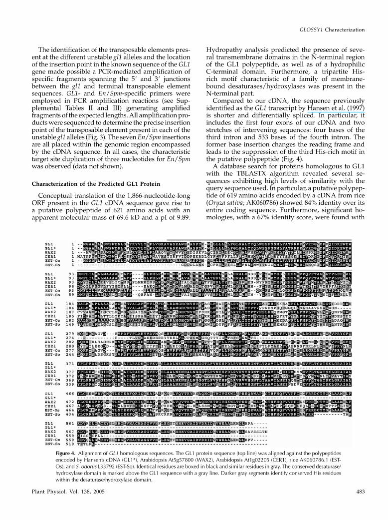
Conceptual translation of the 1,866-nucleotide-longORF present in the GL1 cDNA sequence gave rise toa putative polypeptide of 621 amino acids with anapparent molecular mass of 69.6 kD and a pI of 9.89.
Hydropathy analysis predicted the presence of seve-ral transmembrane domains in the N-terminal regionof the GL1 polypeptide, as well as of a hydrophilicC-terminal domain. Furthermore, a tripartite His-rich motif characteristic of a family of membrane-bound desaturases/hydroxylases was present in theN-terminal part.
Compared to our cDNA, the sequence previouslyidentified as the GL1 transcript by Hansen et al. (1997)is shorter and differentially spliced. In particular, itincludes the first four exons of our cDNA and twostretches of intervening sequences: four bases of thethird intron and 533 bases of the fourth intron. Theformer base insertion changes the reading frame andleads to the suppression of the third His-rich motif inthe putative polypeptide (Fig. 4).
A database search for proteins homologous to GL1with the TBLASTX algorithm revealed several se-quences exhibiting high levels of similarity with thequery sequence used. In particular, a putative polypep-tide of 619 amino acids encoded by a cDNA from rice(Oryza sativa; AK060786) showed 84% identity over itsentire coding sequence. Furthermore, significant ho-mologies, with a 67% identity score, were found with
Figure 4. Alignment of GL1 homologous sequences. The GL1 protein sequence (top line) was aligned against the polypeptidesencoded by Hansen’s cDNA (GL1*), Arabidopsis At5g57800 (WAX2), Arabidopsis At1g02205 (CER1), rice AK060786.1 (EST-Os), and S. odorus L33792 (EST-So). Identical residues are boxed in black and similar residues in gray. The conserved desaturase/hydroxylase domain is marked above the GL1 sequence with a gray line. Darker gray segments identify conserved His residueswithin the desaturase/hydroxylase domain.
GLOSSY1 Characterization
Plant Physiol. Vol. 138, 2005 483

the products of two other rice cDNAs (AK066569 andAK070469), with the WAX2 locus of Arabidopsisencoding a protein involved in cuticle synthesis (62%identity), and a partial polypeptide (L33792) derivedfrom Senecio odorus (55% identity). The alignment ofthe deduced GL1 amino acid sequence and deducedprotein sequences exhibiting high similarity scores isdepicted in Figure 4. The highest degree of homologyconsistently regards the C-terminal part of the de-duced proteins.
A comparison of the deduced GL1 protein sequenceand the product of the Arabidopsis ECERIFERUM1(CER1) locus, a putative aldehyde decarbonylase ac-tive in the cuticular wax biosynthesis pathway,reveals an overall identity of 35%. This similarity scorewas significantly lower than the degree of similarityencountered between the putative GL1 and Arabi-dopsis WAX2 proteins (62%). Since previous re-sults attributed to the maize GL1 locus a role as anArabidopsis CER1 ortholog, we investigated aminoacid sequence similarities among a restricted groupof GL1 homolog sequences by means of phylogeneticanalysis (Fig. 5). These analyses suggested the pres-ence of two groups of protein sequences, the formercontaining the CER1 protein as a founder sequence,the latter including the WAX2 sequence. Interestingly,the GL1 sequence showed a high level of homologywith the members of the WAX2 group, while a secondmaize sequence (GenBank AY104752) was locatedwithin the CER1 group with which it shares 55%amino acid identity. Thus, phylogenetic analysis in-dicated that Gl1-related sequences can be dividedinto two subgroups, each comprising genes from
at least three species: maize, rice, and Arabidopsis(Fig. 5).
GL1 Transcription Analysis
The 3# end of the GL1 cDNA was used as a probe innorthern-blot experiments performed with total RNAextracted from different tissues of wild-type plantsand from leaf tissue homozygous for the gl1-ref allele.As shown in Figure 6A, the RNA extracted from wild-type seedlings showed a transcript with an estimatedsize of 2,300 residues, in accordance with the expectedlength of the GL1 mRNA (lane 1). The accumulationof this RNA was dramatically reduced in the gl1-refmutant (lane 2) and was completely blocked in theroot where, instead, a transcript of greater size wasdetected (lane 3). GL1 expression was evident also inadult leaves (lane 4) and in floral organs (silks andanthers; lanes 5 and 6, respectively), suggesting thatGL1 activity was not restricted to the juvenile de-velopmental phase of the maize plant. The samepattern of hybridization was observed using thecomplete Gl1 cDNA as a probe (data not shown). Tocheck the amount of RNA loading, the filters werestripped and reprobed with a maize cytosolic GAPDHclone (Fig. 6B). The GL1 transcript was further studiedby RT-PCR analysis using the samples describedabove (Fig. 6C). The use of forward and reverse GL1primers allowed the amplification of a fragment of theexpected size from RNA samples obtained from wild-type seedlings (lane 2) and, at low abundance, fromgl1-ref mutant leaf (lane 3), mature leaf (lane 5), andanther tissue (lane 7). PCR amplification with primersagainst cytosolic GAPDH was used to verify the integ-rity of the samples (Fig. 6D).
Figure 5. Phylogenesis of GL1-likesequences. The deduced amino acidsequence of the GL1 locus was alignedagainst 12 deduced sequences ob-tained from homologous loci, as avail-able in GenBank. Neighbor-joininganalysis was used to obtain a phylo-genetic tree, which was bootstrappedover 1,000 cycles. Significance valuesabove a 50% cutoff threshold are in-dicated near the relative branches. Twodistinct groupings are boxed: CER1-related sequences (I), including riceAK066386, maize AY104752, riceAK100751, rice AK068166, Arabi-dopsis At1g02205 (CER1), Arabi-dopsis At2g37700, and ArabidopsisAt1g02190; and WAX2-related se-quences (II), including ArabidopsisAt5g57800 (WAX2), S. odorus L33792,maize AY505017 (Gl1, this study), riceAK060786, rice AK066569, and riceAK070469. Scale bar, Similarity coeffi-cient.
Sturaro et al.
484 Plant Physiol. Vol. 138, 2005

As can be seen in Figure 6C, from the root (lane 3)and silk (lane 5) extracts no RNA amplification wasobtained by RT-PCR. In this respect, we identified anincomplete Gl1-related clone by screening a silk cDNAlibrary using Gl1 as a probe (H. Hartings, R. Velasco,and M. Motto, unpublished data). This silk cDNAshows 78% identity with Gl1; northern experimentsperformed with the same samples as those in Figure6A give a similar hybridization pattern but witha higher intensity in the silk extract (see SupplementalFig. 7). This was taken as evidence that a Gl1-relatedgene is expressed mainly in the silk tissue and givesrise to an mRNA cross-hybridizing to the Gl1 probe.
As concerns the band in lane 3 of Figure 6A, this mightbe either a root-specific transcript related in sequenceto Gl1 or an unspliced version of Gl1 not amplified byRT-PCR with the conditions used. However, usingdifferent combinations of Gl1-specific primers aimedat identifying the presence of intron sequences in theGl1 transcript, we had no indication of the occurrenceof an unspliced version of the Gl1 mRNA in the rootextract (data not shown). Accordingly, this band islikely to be the result of unspecific cross-hybridization.
DISCUSSION
Molecular Cloning and Characterization of theGL1 Locus
To obtain molecular insights into the nature of thegenetic lesion that gives rise to the gl1 phenotype,a collection of unstable gl1 mutations induced byautonomous elements of the En/Spm family wasgenerated (Maddaloni et al., 1990). From one of thesemutable alleles, a partial sequence of Gl1 was iden-tified and molecularly cloned, as confirmed by alleliccross-referencing and northern-blot experiments, andused to recover the complete gene from a maizegenomic library. GL1 is a single-copy gene, whichgives rise to a transcript carrying a 1,866-bp ORF andspanning eight noncontiguous genomic stretches.The genomic region encompassed by the cDNAsequence includes all the En insertion sites foundin the unstable gl1 alleles.
Together, our results indicate that we have clonedthe GL1 genomic sequence and the complete codingregion of its major transcript. Hansen et al. (1997),who performed similar experiments to characterizethe GL1 gene, isolated a genomic and a cDNA clonethat, according to our results, represent a part of theGL1 locus and a differently spliced/partial unpro-cessed mRNA, respectively. Our conclusions arebased on several lines of evidence. First, an entirecollection of seven independent En/Spm insertionswas analyzed to define the position of the trans-posable element in the unstable gl1 alleles. In fourcases, the En/Spm insertion sequence is locateddownstream of the coding region of the GL1 geneas described by Hansen et al. (1997). Second, thecDNA isolated by Hansen et al. is 1,585 bp long,includes only the first four exons of our cDNA, andcodes for a polypeptide of 319 amino acids withhomology to CER1 and related proteins limited tothe N-terminal region. The C-terminal domain hasno counterparts in any of the GL1 homologs identi-fied so far. Moreover, this polypeptide includes onlythe first two His-rich motifs. Third, Hansen’s cloneexactly matches the 5# region of our genomic se-quence. Because GL1 is a single-copy gene, it isconcluded that both sequences derive from the samelocus. However, the former was isolated by screen-ing a genomic library constructed with HindIII-digested DNA. It is likely that the availability ofa short GL1 transcript and the concomitant molec-ular situation at the GL1 locus presenting a HindIIIsite in the fourth, relatively lengthy, intron havesuggested (Hansen et al., 1997) that the HindIII sitewas located beyond the end of the GL1 gene. Inconclusion, our data regarding the GL1 transcript,its coding region, and the distribution of En/Spmelement insertion sites within the locus suggest thatthe polypeptide described by Hansen et al. isnot sufficient to perform the function of the GL1gene.
Figure 6. Northern analysis and RT-PCR analysis of GL1 transcription.A, mRNA extracted from young wild-type leaf (1), young gl1-Ref leaf(2), root (3), old wild-type leaf (4), silk (5), and anther (6) tissue washybridized with the 3# end of the GL1 cDNA. B, The same blot in Awas stripped and rehybridized with a GAPDH probe. C and D, Thepresence of GL1 (C) and GAPDH (D) transcripts was assayed by RT-PCRin young wild-type leaf (2), young gl1-ref leaf (3), root (4), old wild-typeleaf (5), silk (6), and anther (7) tissue. Size markers present in lane 1were used to deduce the sizes of amplification products as indicated onthe right.
GLOSSY1 Characterization
Plant Physiol. Vol. 138, 2005 485

The GL1 Gene Encodes a WAX2-Related Protein
Displaying Transmembrane Domains
The putative protein encoded by GL1 is 621 aminoacids long and is related in length and sequence tothose coded by a number of loci from different plantspecies. These polypeptide sequences display severalpredicted transmembrane domains in the N-terminalregion and a globular domain in the C-terminal part.A common feature shared by these proteins is thepresence of eight conserved His motifs in the tripartitedomain H-X2–4-H, H-X2–3-H-H, (H/Q)-X2–3-H-H,which form a di-iron-binding site essential for catalyticactivity in a large family of integral membrane en-zymes, such as acyl desaturases, alkyl-hydroxylases,epoxydases, acetylenases, methyl oxidases, ketolases,and decarbonylases, activities found in prokaryotesand eukaryotes (Shanklin and Cahoon, 1998, and refs.therein). The GL1-related proteins identified by meansof sequence comparisons include the WAX2 and CER1gene products of Arabidopsis, both of which areinvolved in cuticular wax production. Interestingly,in the GL1, WAX2, and CER1 genes, intron positionsare conserved (see Supplemental Fig. 8), suggestinga common origin from the same ancestor gene. How-ever, GL1 shows 62% overall amino acid identity toWAX2, which rises to 78% when the C-terminal regionis considered, compared to 35% homology with CER1.Therefore, GL1 is probably the WAX2 ortholog frommaize. In addition, sequence comparison clearly in-dicates the presence of two distinct subgroups of GL1-related genes in rice, maize, and Arabidopsis. Theformer, including GL1 itself, is related to the WAX2gene of Arabidopsis, while the latter comprises se-quences more similar to CER1, among which is a maizeexpressed sequence tag (EST; ID AY104752; see Fig. 5),which could be the true CER1 ortholog of maize.
Mutations of GL1, CER1, and WAX2 cause dramaticalterations in composition and crystallization patternsof cuticular waxes (Lorenzoni and Salamini, 1975;Bianchi et al., 1985; Jenks et al., 1995; Chen et al.,2003). Cuticular wax of juvenile maize leaves iscomposed of primary alcohols (63%), aldehydes(20%), and esters (16%) mainly derived from C32acyl moieties, whereas on Arabidopsis stems themain constituents are alkanes (53%) and ketones(23%), with primary alcohols and aldehydes reducedto 11% and 4%, respectively. Arabidopsis leaves arealmost devoid of ketones, and alkanes represent 72%of total waxes. Because of these differences in waxcomposition, comparing the biochemical effect of waxgene mutations in maize and Arabidopsis is notconclusive in defining the homology in gene functions.
CER1 was suggested to be an aldehyde decarbon-ylase because the mutant shows an increase in alde-hydes and a reduction of the products of aldehydedecarbonylation, namely, alkane, secondary alcohols,and ketones (Aarts et al., 1995). In maize, these lattercompounds represent only 1% of total cuticular waxes;therefore, mutations in a putative aldehyde decarbon-
ylase are not likely to be identified based only onvisual screenings.
In wax2 mutants, total wax load is diminished byabout 80% because of the reduced accumulation of allthe prevalent wax constituents, including aldehydes,with the exception of C30 primary alcohols, which areincreased on wax2 stems.
Wax load on gl1 juvenile leaves is reduced by 73%compared to the wild type due to a decreased accu-mulation of both aldehydes and primary alcohols,while the amount of esters does not change. Themutation has a pronounced effect on the synthesis oflong-chain wax compounds (C32), whereas those withshorter acyl chains are less affected or even increased(Bianchi et al., 1977). The overall change in wax com-position on gl1 seedlings is similar to that observed onwax2 mutant leaves. In conclusion, these biochemicaldata on composition of mutant waxes also support theconclusion that GL1 is more closely related to WAX2than to CER1.
The gl1 Mutation Affects Cuticular Wax Accumulationand Other Cuticular Traits
The Arabidopsis WAX2/YRE gene described byChen et al. (2003) and Kurata et al. (2003) has a broadrole in cuticle biosynthesis. Its mutation alters bothcutin morphology and wax production. In addition,this mutation affects other cuticular and plant traits,including trichome development, leaf transpiration,pollen fertility, and postgenital organ fusion duringearly organ development. Similarly to wax2-defectiveplants, gl1-ref mutants display a reduction in cuticularwax deposition and alterations in cuticle membranestructure and trichome development. However, post-genital organ fusion was not observed in the gl1-refmutant. As a possible explanation for these discrep-ancies, the gl1-ref allele might condition a leaky muta-tion impairing only some of the GL1 functions.However, a null-transcript gl1 mutant shows normaldevelopment and the absence of organ fusion (data notshown). A second possible explanation is the differenteffect of the two mutations on cutin morphology,which could be of functional importance: In wax2mutants, cuticle membrane thickness is increased,although its weight is reduced.
Alternatively, different roles on plant developmentmay be ascribed to maize and Arabidopsis cuticles. Inaddition to wax2, the abnormal leaf shape1 (ale1) andlacerata (lcr) Arabidopsis mutants are altered in cuticlemembrane morphology and display postgenital organfusion (Tanaka et al., 2001; Wellesen et al., 2001).Instead, the long-chain acyl-CoA synthetase2 (lacs2) mu-tant shows a reduction in cutin thickness (240%) andseveral phenotypic alterations normally associatedwith defective cutin structure but not organ fusion,suggesting that this trait is very likely linked to moredrastic cutin impairments (Schnurr et al., 2004). Thecausal relationship between cuticle membrane defectsand postgenital organ fusion in Arabidopsis is
Sturaro et al.
486 Plant Physiol. Vol. 138, 2005

strengthened by the finding that degradation of thecutin layer in transgenic Arabidopsis plants express-ing a cutinase gene leads to adhesion of different adultorgans (Sieber et al., 2000).
A similar correlation is not observed in maize andother monocots. Maize mutants with adhesion-competent epidermal cells include crinkly4 (cr4) andadherent1 (ad1). The cr4 mutation has a broad effecton epidermal cell morphology, not restricted to thecuticular layer (Becraft et al., 1996). ad1 mutants ex-hibit alteration in cell wall structure and epicuticularwax deposition in the epidermal layer, while the cu-ticle membrane in adherent regions appears intact(Sinha and Lynch, 1998). By contrast, the sorghumbm-22 mutant with a severely reduced cuticle mem-brane displays normal plant structure and develop-ment (Jenks et al., 1994). Moreover, maize seedlingstreated with an inhibitor of cutin synthesis have novisual phenotypic alterations (Lequeu et al., 2003). Col-lectively, these results point to a different role ofmaize and Arabidopsis cuticles in plant development,namely, in the prevention of postgenital organ fusion.
Expression of the GL1 Gene Is Tissue and Organ
Specific and Developmentally Regulated
As far as GL1 function is concerned, analysis of themutant phenotype indicates that GL1, like the otherGLOSSY genes, is an essential component of thejuvenile wax layer biosynthetic route (Bianchi et al.,1985). Young gl1 leaves have the chemical wax com-position of wild-type adult leaves. It can be concludedthat the regulation of GL1 expression is an integral partof the cell response to age-related events of differen-tiation. It was shown that in maize the juvenile-to-adult transition is under genetic control, with a seriesof independent mutations—CORN-GRASS1 (CG1),TEOPOD1 (TP1), TEOPOD2 (TP2), HAIRY-SHEATH-FRAYED1 (HSF1; Poethig, 1988, and refs. therein), andGL15 (Moose and Sisco, 1996)—altering the transitionfrom juvenile to adult vegetative growth. The interac-tion of GL15 with TP1 and TP2 indicates that GL15 actsdownstream of these genes and that it is required fortheir effect during epidermis development (Evanset al., 1994). The GL15 gene is a transcriptional activa-tor of the APETALA2 family of regulatory genes(Moose and Sisco, 1996), which is expected to interactdirectly with the promoters of structural genes neededfor the accumulation of cuticular waxes, a predictionthat can be experimentally tested now that GL1, GL2,and GL8 are listed among the cloned genes contribut-ing to wax deposition.
From the expression profile experiments, it can beargued that the regulation of the expression of thecharacterized GLOSSY genes is more complex thanpredicted only on the basis of seedling phenotypes.GL8 turns out to be expressed in different organs of theadult plant, including the roots, although to a lesserextent than in seedling leaves (Xu et al., 1997). GL1 isnot expressed in the root but is detectable in anthers
and, to a minor extent, also in adult leaves, similar tothe GL2 transcript that, besides being characteristicallyproduced in young leaves, is also detectable in the partof the maize shoot contributing to the development ofthe female inflorescence (Velasco et al., 2002). Theseresults demonstrate that the GL1 protein, like theproducts of other GLOSSY genes, is not merely in-volved in the cuticle formation of the green part of themaize seedling. Together, these findings indicate thatfactors conditioning tissue juvenility may be reacti-vated after the formation of the lateral meristem(Uhrig et al., 1997). Alternatively, a different controlmechanism affecting wax biosynthetic genes shouldexist in adult plant tissues.
MATERIALS AND METHODS
Plant Material
The origin and maintenance of the wx-m7 transposon stock, the gl1-ref
allele used in this study, and the recovery of the gl1-mutable strains have been
described previously (Maddaloni et al., 1990). Briefly, gl1 alleles were
identified in the F1 generation of a cross between a strain homozygous for
the unstable allele wx-m7 of the WAXY locus and for a dominant GL1 allele
(used as the male parent) and a female parent strain homozygous for the
stable recessive gl1-ref allele. Both strains were in the Wf9 inbred line genetic
background. The instability of the wx-m7 allele is due to the transposable
element Ac (McClintock, 1962; Behrens et al., 1984). In our tagging experi-
ments, nine unstable alleles were generated at the GL1 locus. Seven, gl1-m1, 2,
3, 5, 7, 8, and 9, were due to the insertion of an autonomous element, while
gl1-m4 and gl1-m6 were caused by a nonautonomous element of a family
of transposable elements different from Ac/Ds (Maddaloni et al., 1990).
The wx-m7 stock also contains copies of active autonomous En/Spm
elements (Michel et al., 1995). To test for the presence in gl1-m5 in stable
mutants of En/Spm elements, crosses with two tester strains were generated.
The resulting F1 plants were selfed and the F2 seeds were checked for
mutability of either of the two tester alleles, a1-m(r) or a1-m1, both due to the
insertion of a defective En/Spm element (I/dSpm) and showing somatic
instability only in the presence of an active En/Spm element. In its absence, the
a1-m1 allele gives rise to kernels with a pale color, while a1-m1(r) is colorless.
In the presence of an active En/Spm element, colored spots are produced on
the pale (a1-m1) or colorless [a1m(r)] background. Crosses and selfings were
according to standard procedures.
Genomic Cloning and Southern Analysis
Maize (Zea mays) DNA was isolated from leaves of flowering plants or
from seedlings as described (Michel et al., 1995). Southern hybridizations
and radioactive labeling of probes were according to standard procedures
(Sambrook et al., 1989). A plasmid clone carrying the complete En/Spm
element isolated from the wx-844 allele was provided by Dr. Alfons Gierl
(Munich). An EcoRI/BamHI restriction fragment that covered the internal
region of the En/Spm element between positions 2,518 to 4,459 (Pereira et al.,
1986) was used as a molecular probe.
Southern analysis was performed to map the position of the En element in
seven unstable gl1 alleles. For this purpose, genomic DNA from homozygous
mutant plants was digested with the restriction enzymes BamHI, HindIII,
ApaI, SstI, SpeI, and KpnI. Blots were probed with four PCR-derived frag-
ments of the GL1 allele covering regions (base positions are indicated relative
to the translation start site) from 22,302 to 2648 (probe I), from 11,213 to
12,048 (probe II), from 12,426 to 13,581 (probe III), and from14,131 to 14,745
(probe IV).
HindIII-digested genomic fragments from 7 to 11 kb from plants carrying
the gl1-m5 allele were cloned into the lEMBL3 vector, after separation and
purification from agarose gels with the QIAEX gel extraction kit (Qiagen,
Valencia, CA). Nine clones hybridizing to a fragment covering the En/Spm
sequence between the EcoRI sites at positions 5,836 and 8,278 were identified
within approximately 100,000 recombinant phages. Restriction mapping of
GLOSSY1 Characterization
Plant Physiol. Vol. 138, 2005 487

the clones identified a single clone, designated l-09, carrying a HindIII insert
of approximately 8.3 kb, which was chosen for further analysis.
Recombinant clones carrying a wild-type GL1 allele were recovered from
a maize BAC library derived from DNA extracted from the inbred F2, kindly
provided by Dr. Keith Edwards (University of Bristol, UK).
Northern and RT-PCR Analysis
Total RNA was isolated from the following organs and tissues of the inbred
Wf9: second and third leaf of wild-type and gl1-ref mutant seedlings, wild-
type roots of 1-week-old seedlings, wild-type adult leaf (top leaf, surrounding
the tassel), wild-type silks, and wild-type anthers (both immature and pollen-
shedding, from the same tassel). Extractions were performed using TRizol
(Invitrogen, Carlsbad, CA) according to the manufacturer’s instructions. For
northern experiments, 20 mg of total RNA samples were fractionated on
denaturing gels, capillary blotted onto nylon membranes (Hybond N1;
Amersham, Little Chalfont, UK), and hybridized at 45�C in Ultrahyb solu-
tion (Ambion, Austin, TX). Filters were washed at 55�C in 23 SSC/0.1%
SDS (15 min), in 13 SSC/0.1%SDS (30 min), and in 0.13 SSC/0.1% SDS
(30 min). RNA markers from 0.2 to 10 kb (Sigma-Aldrich, St. Louis) were used
as size standards. Probes were the full-length GL1 cDNA and the 400-bp
3# end, including the complete 3# UTR, of a partial GL1 clone isolated from a
seedling cDNA library. To check the amount of RNA loading, filters were
rehybridized with a probe derived from maize cytosolic GAPDH cDNA.
For RT-PCR, 5 mg of total RNA were reverse transcribed using Super-
ScriptII reverse transcriptase (Invitrogen) according to manufacturer’s
instructions. One-twentieth of the final reaction product was amplified by
PCR with the following primers: forward, 5#-ATCGAATTCACGTACGG-
CACAGTTGCTAGC-3#; reverse, 5#-CGCTCTAGACCACCAATTCACACTC-
GACG-3#.The forward primer annealed to the region of the GL1 cDNA starting 70 bp
upstream of the ATG start codon, while the reverse primer annealed to the
region starting 101 bp downstream of the stop codon. To avoid formation of
secondary structures, PCR reactions were performed in the presence of 10%
DMSO (final concentration). The 5# end of the forward and reverse primers
included, respectively, EcoRI and XbaI restriction sites (indicated in italics in
the above sequences), which were used to subclone the GL1 cDNA from leaves
of the inbred Wf9 into the pBluescriptSKII vector (Stratagene, La Jolla, CA)
prior to sequencing. Five independent clones were sequenced on both strands
to determine the sequence of the GL1 cDNA.
DNA Sequencing
DNA sequencing was carried out with an automatic sequencer (CEQ 8000;
Beckman-Coulter, Fullerton, CA). Genomic and cDNA sequences were de-
termined on both strands.
Microscopic Inspection
SEM was used to study adaxial surfaces of primary leaves of gl1 mutant
and wild-type plants grown for 2 to 3 weeks in a phytochamber at 26�C/19�C(day/night) and 40% humidity with a 16/8-h light/dark rhythm, and a light
intensity of 1,900 mE m22 s21. Segments of the middle part of the leaf blade
were fixed to a specimen holder by tissue tek and shock frozen with liquid
nitrogen within a high vacuum cryo preparation stage. Samples were trans-
ferred under vacuum to a cryo preparation chamber where they were sputter
coated with gold and examined on the cold stage of a Zeiss DSM 940 SEM
(Carl Zeiss NTS GmbH, Oberkochen, Germany). For TEM investigation of the
cuticle, small pieces (2–3 mm2) of primary leaf blades were fixed for 2 h at
room temperature in 2.5% (v/v) glutaraldehyde and 2% (v/v) formaldehyde
in 0.05 M phosphate buffer (PB), pH 6.8. After washing in PB, samples were
postfixed for 1 h in 2% (v/v) osmium tetroxide in PB, washed again, and
dehydrated through a graded series of ethanol. Samples were then infiltrated
with LR White resin (Plano, Marburg, Germany) and polymerized for 48 h at
60�C. Ultrathin cross-sections were prepared and mounted on carbon-coated
Formvar copper grids (200 mesh; Plano). After staining with 2% uranyl acetate
for 2 h, sections were inspected with a Zeiss EM 10 TEM (Carl Zeiss NTS
GmbH).
Statistical Analysis
Multiple DNA and protein sequence alignments were performed using
ClustalW (Thompson et al., 1994), while phylogenetic analysis was performed
according to the MEGA version 2.1 software package (Kumar et al., 2001). For
tree construction based on aligned amino acid sequences, the neighbor-joining
tree-building method was utilized. Bootstrap analysis (1,000 replicates) was
used to assign a consensus tree at a 50% cutoff value.
Sequence data from this article have been deposited with the EMBL/
GenBank data libraries under accession numbers AY505017 and AY505498.
ACKNOWLEDGMENT
We thank Dr. Keith Edwards, University of Bristol, UK, for providing
a recombinant BAC clone carrying a wild-type GL1 gene.
Received December 15, 2004; returned for revision February 9, 2005; accepted
February 9, 2005.
LITERATURE CITED
Aarts MGM, Keijzer CJ, Stiekema WJ, Pereira A (1995) Molecular
characterization of the CER1 gene of Arabidopsis involved in epicutic-
ular wax biosynthesis and pollen fertility. Plant Cell 7: 2115–2127
Becraft PW, Stinard PS, McCarty DR (1996) CRINKLY4: a TNFR-like
receptor kinase involved in maize epidermal differentiation. Science
273: 1406–1409
Behrens U, Federoff N, Laird A, Muller-NeumannM, Starlinger P, Yoder J
(1984) Cloning of the Zea mays controlling element Ac from the wx-m7
allele. Mol Gen Genet 194: 346–347
Bianchi A, Bianchi G, Avato P, Salamini F (1985) Biosynthetic pathways of
epicuticular wax of maize as assessed by mutation, light, plant age and
inhibitor studies. Maydica 30: 179–198
Bianchi G, Avato P, Salamini F (1977) glossy mutants of maize. VII.
Chemistry of glossy1, glossy3 and glossy7 epicuticular waxes. Maydica
22: 9–17
Chen X, Goodwin SM, Boroff VL, Liu X, Jenks MA (2003) Cloning and
characterization of the WAX2 gene of Arabidopsis involved in cuticle
membrane and wax production. Plant Cell 15: 1170–1185
Evans M, Passas HJ, Poethig RS (1994) Heterochronic effects of glossy15
mutations on epidermal cell identity in maize. Development 120:
1971–1981
Hansen JD, Pyee J, Xia Y, Wen T-J, Robertson DS, Kolattukudy PE,
Nikolau BJ, Schnable PS (1997) The glossy1 locus of maize and an
epidermis-specific cDNA from Klenia odora define a class of receptor-like
proteins required for the normal accumulation of cuticular waxes. Plant
Physiol 113: 1091–1100
Jenks MA, Joly RJ, Peters PJ, Rich PJ, Axtell JD, Ashworth EN (1994)
Chemically induced cuticle mutation affecting epidermal conductance
to water vapor and disease susceptibility in Sorghum bicolor (L.) Moench.
Plant Physiol 105: 1239–1245
Jenks MA, Tuttle HA, Eigenbrode SD, Feldmann KA (1995) Leaf epicu-
ticular waxes of the eceriferum mutants in Arabidopsis. Plant Physiol
108: 369–377
Kolattukudy PE (1996) Biosynthetic pathways of cutin and waxes and their
sensitivity to environmental stresses. In G Kerstiens, ed, Plant Cuticles.
BIOS Scientific Publishers, Oxford, pp 83–108
Kumar S, Tamura K, Jakobsen IB, Nei M (2001) MEGA2: molecular
evolutionary genetics analysis software. Bioinformatics 17: 1244–1245
Kunst L, Samuels AL (2003) Biosynthesis and secretion of plant cuticular
waxes. Prog Lipid Res 42: 51–80
Kurata T, Kawabata-Awai C, Sakuradani E, Shimizu S, Okada K, Wada T
(2003) The YORE-YORE gene regulates multiple aspects of epidermal
cell differentiation in Arabidopsis. Plant J 36: 55–56
Lawson EJR, Poethig RS (1995) Shoot development in plants: time for
a change. Trends Genet 11: 263–268
Lequeu J, Fauconnier M-L, Chammai A, Bronner R, Blee E (2003)
Formation of plant cuticle: evidence for the occurrence of the peroxy-
genase pathway. Plant J 36: 155–164
Sturaro et al.
488 Plant Physiol. Vol. 138, 2005

Lorenzoni C, Salamini F (1975) glossy mutants of maize. V. Morphology of
the epicuticular waxes. Maydica 20: 5–19
Maddaloni M, Bossinger G, Di Fonzo N, Motto M, Salamini F, Bianchi A
(1990) Unstable alleles of the Glossy1 locus of maize show a light-
dependent variation in the pattern of somatic reversion. Maydica 35:
409–420
McClintock B (1962) Aspects of gene regulation in maize. Carnegie Inst
Wash Year Book 63: 592–603
Michel D, Hartings H, Lanzini S, Michel M, Motto M, Riboldi GR,
Salamini F, Doring H-P (1995) Insertion mutations at the maize Opaque2
locus induced by transposable element families Ac, En/Spm, and Bg. Mol
Gen Genet 248: 287–292
Moose SP, Sisco PH (1994) Glossy15 controls the epidermal juvenile-to-
adult phase transition in maize. Plant Cell 6: 1343–1355
Moose SP, Sisco PH (1996) Glossy15, an APETALA2-like gene from maize
that regulates leaf epidermal cell identity. Genes Dev 10: 3018–3027
Neuffer M, Coe E, Wessler SR (1997) Mutants in Maize. Cold Spring
Harbor Laboratory Press, Cold Spring Harbor, NY
Pereira A, Cuypers H, Gierl A, Schwarz-Sommer Z, Saedler H (1986)
Molecular analysis of the En/Spm transposable element system of Zea
mays. EMBO J 5: 835–841
Poethig RS (1988) Heterochronic mutations affecting shoot development in
maize. Genetics 119: 959–973
Post-Beittenmiller D (1996) Biochemistry and molecular biology of wax
production in plants. Annu Rev Plant Physiol Plant Mol Biol 47: 405–430
Sambrook J, Fritsch EF, Maniatis T (1989) Molecular Cloning: A Labora-
tory Manual, Ed 2. Cold Spring Harbor Laboratory Press, Cold Spring
Harbor, NY
Schnurr J, Shockey J, Browse J (2004) The acyl-CoA synthetase encoded by
LACS2 is essential for normal cuticle development in Arabidopsis. Plant
Cell 16: 629–642
Shanklin J, Cahoon EB (1998) Desaturation and related modifications of
fatty acids. Annu Rev Plant Physiol Plant Mol Biol 49: 611–641
Sieber P, Schorderet M, Ryser U, Buchala A, Kolattukudy P, Metraux JP,
Nawrath C (2000) Transgenic Arabidopsis plants expressing a fungal
cutinase show alterations in the structure and properties of the cuticle
and postgenital organ fusions. Plant Cell 12: 721–737
Sinha N, Lynch M (1998) Fused organs in the adherent1 mutation in maize
show altered epidermal walls with no perturbations in tissue identities.
Planta 206: 184–195
Tacke E, Korfhage C, Michel D, Maddaloni M, Motto M, Lanzini S,
Salamini F, Doring H-P (1995) Transposon tagging of the maize
Glossy2 locus with the transposable element En/Spm. Plant J 8: 907–917
Tanaka H, Onouchi H, Kondo M, Hara-Nishimura I, Nishimura M,
Machida C, Machida Y (2001) A subtilisin-like serine protease is
required for epidermal surface formation in Arabidopsis embryos and
juvenile plants. Development 128: 4681–4689
Thompson JD, Higgins DG, Gibson TJ (1994) CLUSTAL W: improving
the sensitivity of progressive multiple sequence alignment through
sequence weighting, position-specific gap penalties and weight matrix
choice. Nucleic Acids Res 22: 4673–4680
Uhrig H, Marocco A, Doring H-P, Salamini F (1997) The clonal origin of
the lateral meristem generating the ear shoot of maize. Planta 201:
9–17
Velasco R, Korfhage C, Salamini A, Tacke E, Schmitz J, Motto M,
Salamini F, Doring H-P (2002) Expression of the glossy2 gene of maize
during plant development. Maydica 47: 71–81
Walton TJ (1990) Waxes, cutin and suberin. In JL Harwood, JR Bowyer, eds,
Methods in Plant Biochemistry: Lipids, Membranes and Aspects of
Photobiology. Academic Press, San Diego, pp 105–158
Wellesen K, Durst F, Pinot F, Benveniste I, Nettesheim K, Wisman E,
Steiner-Lange S, Saedler H, Yephremov A (2001) Functional analysis of
the LACERATA gene of Arabidopsis provides evidence for different
roles of fatty acid v-hydroxylation in development. Proc Natl Acad Sci
USA 98: 9694–9699
Xu X, Dietrich CR, Delledonne M, Xia Y, Wen T-J, Robertson DS,
Nikolau BJ, Schnable PS (1997) Sequence analysis of the cloned glossy8
gene of maize suggests that it may code for a b-ketoacyl reductase
required for the biosynthesis of cuticular waxes. Plant Physiol 115:
501–510
Xu X, Dietrich CR, Lessire R, Nikolau BJ, Schnable PS (2002) The
endoplasmic reticulum-associated maize GL8 protein is a component
of the acyl-coenzyme A elongase involved in the production of cuticular
waxes. Plant Physiol 128: 924–934
GLOSSY1 Characterization
Plant Physiol. Vol. 138, 2005 489
Related Documents