Current Biology 23, 1–6, June 17, 2013 ª2013 Elsevier Ltd All rights reserved http://dx.doi.org/10.1016/j.cub.2013.05.007 Report Cloning and Characterization of Four Novel Coral Acid-Rich Proteins that Precipitate Carbonates In Vitro Tali Mass, 1 Jeana L. Drake, 1 Liti Haramaty, 1 J. Dongun Kim, 1,3 Ehud Zelzion, 2 Debashish Bhattacharya, 2 and Paul G. Falkowski 1,3,4, * 1 Environmental Biophysics and Molecular Ecology Program, Institute of Marine and Coastal Sciences 2 Department of Ecology, Evolution, and Natural Resources Rutgers University, New Brunswick, NJ 08901, USA 3 Department of Chemistry and Chemical Biology 4 Department of Earth and Planetary Sciences Rutgers University, Piscataway, NJ 08854, USA Summary Biomineralization is a widely dispersed and highly regulated but poorly understood process by which organisms precip- itate minerals from a wide variety of elements [1]. For many years, it has been hypothesized that the biological precipita- tion of carbonates is catalyzed by and organized on an extracellular organic matrix containing a suite of proteins, lipids, and polysaccharides [2, 3]. The structures of these molecules, their evolutionary history, and the biophysical mechanisms responsible for calcification remain enigmatic. Despite the recognition that mineralized tissues contain pro- teins that are unusually rich in aspartic and glutamic acids [4–6], the role of these proteins in biomineralization remains elusive [5, 6]. Here we report, for the first time, the identifica- tion, cloning, amino acid sequence, and characterization of four highly acidic proteins, derived from expression of genes obtained from the common stony coral, Stylophora pistillata. Each of these four proteins can spontaneously catalyze the precipitation of calcium carbonate in vitro. Our results demonstrate that coral acid-rich proteins (CARPs) not only bind Ca 2+ stoichiometrically but also precipitate aragonite in vitro in seawater at pH 8.2 and 7.6, via an electrostatic interaction with protons on bicarbonate anions. Phyloge- netic analysis suggests that at least one of the CARPs arose from a gene fusion. Similar, highly acidic proteins appear to have evolved several times independently in metazoans through convergence. Based purely on thermodynamic grounds, the predicted change in surface ocean pH in the next decades would appear to have minimal effect on the ca- pacity of these acid-rich proteins to precipitate carbonates. Results and Discussion Despite the broad interest in coral calcification and the poten- tial for climate-driven adverse effects, the molecules and biophysical mechanism responsible for the precipitation of carbonates in corals are poorly understood. To date, we lack both a characterization of molecules involved in calcification and a mechanistic understanding of processes that lead to and control calcification. This lack of knowledge limits our abil- ity to predict the response of corals to increasing atmospheric CO 2 . To date, the best-characterized highly acidic proteins that catalyze the precipitation of carbonates are from mollusk shells and an echinoderm [4, 7–9]. However, in spite of the fact that stony corals are among the most abundant bio- mineralizing metazoans on Earth, surprisingly little is known about their skeletal organic matrix (SOM) proteins or how they regulate crystal formation. Furthermore, despite the fact that the amino acid composition of coral SOM proteins is char- acterized by a predominance of acidic amino acids [6, 10, 11], the only coral SOM protein fully characterized, i.e., galaxin, which is isolated from the stony coral Galaxea fascicularis, possesses neither acidic regions nor obvious Ca 2+ -binding domains [12]. Recently, Drake et al. [13] identified 36 SOM pro- teins from a stony coral, of which two are highly acidic. They suggested, however, that their approach, based on liquid chromatography-tandem mass spectrometry (LC-MS/MS) protein sequencing, might not be the most efficient method for identifying these molecules, due to highly redundant se- quences. Although a number of N-terminal SOM protein se- quences have been reported both in scleratinian and alcyonian corals, none of these share strong sequence similarity to other known proteins or to each other, and only one exhibits a highly acidic sequence [14–16]. A group of hypothetical, soluble acidic proteins (SAPs) has been identified in the Acropora dig- itifera genome, a transcriptome from A. millepora, and in ex- pressed sequence tag (EST) libraries [17, 18]; however, the role of these hypothetical proteins in calcification was not examined, and the structure of the encoded genes was not validated using RT-PCR. To better characterize the proteins responsible for coral calcification, we generated a draft genome assembly from the cnidarian host cells of the Indo-Pacific stony coral, Stylophora pistillata (see Supplemental Experimental Proce- dures available online). In combination with transcriptomic data from EST libraries and RT-PCR, we identified and charac- terized four coral acid-rich proteins (CARPs 1–4; GenBank accession numbers KC148537–KC148539 and KC493647) (Figure S1 and Table S1). Three of these, CARPs 1–3, contain a secretory signal peptide, suggesting that they are dis- charged from the cells. CARP1 also contains an EF hand Ca 2+ -binding domain. CARPs 2 and 3 contain an isoleucine- proline-valine (IPV)-like motif following the signal peptide that has previously been suggested to assist in the trafficking of secreted, acidic, calcium-binding proteins out of the rough endoplasmic reticulum in metazoans [19](Figure S1). An addi- tional gene encodes a protein, CARP4, which lacks a targeting sequence but contains regions that have previously been iden- tified in proteins extracted from the S. pistillata aragonite skel- etal matrix [13, 14] and is therefore highly likely to be a compo- nent of the SOM. The calculated isoelectric points (pI) of CARPs 1–4 are 4.23, 4.78, 3.04, and 3.99, respectively (Table S1). Isolated SOM proteins from corals and other marine inver- tebrates are often posttranslationally modified [20, 21]; anal- ysis of the amino acid sequence of the four CARPs reveals several potential sites of posttranslational modification, including phosphorylation and O- and N-glycosylation sites (Figure S1). Indeed, glycoproteins were recently reported to be present in the SOM of S. pistillata [13]. *Correspondence: [email protected] Please cite this article in press as: Mass et al., Cloning and Characterization of Four Novel Coral Acid-Rich Proteins that Precipitate Carbonates In Vitro, Current Biology (2013), http://dx.doi.org/10.1016/j.cub.2013.05.007

Welcome message from author
This document is posted to help you gain knowledge. Please leave a comment to let me know what you think about it! Share it to your friends and learn new things together.
Transcript
Please cite this article in press as: Mass et al., Cloning and Characterization of Four Novel Coral Acid-Rich Proteins that PrecipitateCarbonates In Vitro, Current Biology (2013), http://dx.doi.org/10.1016/j.cub.2013.05.007
Cloning and Characterization
Current Biology 23, 1–6, June 17, 2013 ª2013 Elsevier Ltd All rights reserved http://dx.doi.org/10.1016/j.cub.2013.05.007
Report
of Four Novel Coral Acid-Rich Proteinsthat Precipitate Carbonates In Vitro
Tali Mass,1 Jeana L. Drake,1 Liti Haramaty,1
J. Dongun Kim,1,3 Ehud Zelzion,2 Debashish Bhattacharya,2
and Paul G. Falkowski1,3,4,*1Environmental Biophysics and Molecular Ecology Program,Institute of Marine and Coastal Sciences2Department of Ecology, Evolution, and Natural ResourcesRutgers University, New Brunswick, NJ 08901, USA3Department of Chemistry and Chemical Biology4Department of Earth and Planetary SciencesRutgers University, Piscataway, NJ 08854, USA
Summary
Biomineralization is a widely dispersed and highly regulated
but poorly understood process by which organisms precip-itate minerals from a wide variety of elements [1]. For many
years, it has been hypothesized that the biological precipita-tion of carbonates is catalyzed by and organized on an
extracellular organic matrix containing a suite of proteins,lipids, and polysaccharides [2, 3]. The structures of these
molecules, their evolutionary history, and the biophysicalmechanisms responsible for calcification remain enigmatic.
Despite the recognition thatmineralized tissues contain pro-teins that are unusually rich in aspartic and glutamic acids
[4–6], the role of these proteins in biomineralization remainselusive [5, 6]. Here we report, for the first time, the identifica-
tion, cloning, amino acid sequence, and characterization of
four highly acidic proteins, derived fromexpressionof genesobtained from the commonstony coral,Stylophora pistillata.
Each of these four proteins can spontaneously catalyze theprecipitation of calcium carbonate in vitro. Our results
demonstrate that coral acid-rich proteins (CARPs) not onlybind Ca2+ stoichiometrically but also precipitate aragonite
in vitro in seawater at pH 8.2 and 7.6, via an electrostaticinteraction with protons on bicarbonate anions. Phyloge-
netic analysis suggests that at least one of the CARPs arosefrom a gene fusion. Similar, highly acidic proteins appear to
have evolved several times independently in metazoansthrough convergence. Based purely on thermodynamic
grounds, the predicted change in surface ocean pH in thenext decades would appear to have minimal effect on the ca-
pacity of these acid-rich proteins to precipitate carbonates.
Results and Discussion
Despite the broad interest in coral calcification and the poten-tial for climate-driven adverse effects, the molecules andbiophysical mechanism responsible for the precipitation ofcarbonates in corals are poorly understood. To date, we lackboth a characterization of molecules involved in calcificationand a mechanistic understanding of processes that lead toand control calcification. This lack of knowledge limits our abil-ity to predict the response of corals to increasing atmosphericCO2.
*Correspondence: [email protected]
To date, the best-characterized highly acidic proteins thatcatalyze the precipitation of carbonates are from molluskshells and an echinoderm [4, 7–9]. However, in spite of thefact that stony corals are among the most abundant bio-mineralizing metazoans on Earth, surprisingly little is knownabout their skeletal organic matrix (SOM) proteins or howthey regulate crystal formation. Furthermore, despite the factthat the amino acid composition of coral SOMproteins is char-acterized by a predominance of acidic amino acids [6, 10, 11],the only coral SOM protein fully characterized, i.e., galaxin,which is isolated from the stony coral Galaxea fascicularis,possesses neither acidic regions nor obvious Ca2+-bindingdomains [12]. Recently, Drake et al. [13] identified 36 SOMpro-teins from a stony coral, of which two are highly acidic. Theysuggested, however, that their approach, based on liquidchromatography-tandem mass spectrometry (LC-MS/MS)protein sequencing, might not be the most efficient methodfor identifying these molecules, due to highly redundant se-quences. Although a number of N-terminal SOM protein se-quences have been reported both in scleratinian and alcyoniancorals, none of these share strong sequence similarity to otherknown proteins or to each other, and only one exhibits a highlyacidic sequence [14–16]. A group of hypothetical, solubleacidic proteins (SAPs) has been identified in the Acropora dig-itifera genome, a transcriptome from A. millepora, and in ex-pressed sequence tag (EST) libraries [17, 18]; however, therole of these hypothetical proteins in calcification was notexamined, and the structure of the encoded genes was notvalidated using RT-PCR.To better characterize the proteins responsible for coral
calcification, we generated a draft genome assembly fromthe cnidarian host cells of the Indo-Pacific stony coral,Stylophora pistillata (see Supplemental Experimental Proce-dures available online). In combination with transcriptomicdata from EST libraries and RT-PCR, we identified and charac-terized four coral acid-rich proteins (CARPs 1–4; GenBankaccession numbers KC148537–KC148539 and KC493647)(Figure S1 and Table S1). Three of these, CARPs 1–3, containa secretory signal peptide, suggesting that they are dis-charged from the cells. CARP1 also contains an EF handCa2+-binding domain. CARPs 2 and 3 contain an isoleucine-proline-valine (IPV)-like motif following the signal peptidethat has previously been suggested to assist in the traffickingof secreted, acidic, calcium-binding proteins out of the roughendoplasmic reticulum in metazoans [19] (Figure S1). An addi-tional gene encodes a protein, CARP4, which lacks a targetingsequence but contains regions that have previously been iden-tified in proteins extracted from the S. pistillata aragonite skel-etal matrix [13, 14] and is therefore highly likely to be a compo-nent of the SOM. The calculated isoelectric points (pI) ofCARPs 1–4 are 4.23, 4.78, 3.04, and 3.99, respectively (TableS1). Isolated SOMproteins from corals and other marine inver-tebrates are often posttranslationally modified [20, 21]; anal-ysis of the amino acid sequence of the four CARPs revealsseveral potential sites of posttranslational modification,including phosphorylation and O- and N-glycosylation sites(Figure S1). Indeed, glycoproteins were recently reported tobe present in the SOM of S. pistillata [13].

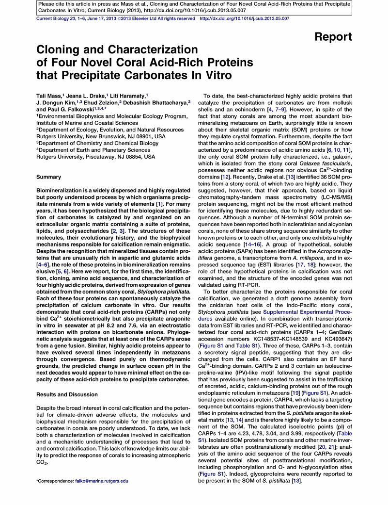
Figure 1. Scanning Electron Microscope Images and Elemental Composition of CaCO3 Crystals Grown in Artificial Seawater
Scanning electron microscope (SEM) images and elemental composition of CaCO3 crystals grown in artificial seawater containing 0.1 mM of CARPs 1–4 at
pH 8.2 and 7.6 (A). In both pH treatments, a reticulate pattern is clearly visible (insets in A). EDS and SEM images (inset) of salts precipitated in protein-free
artificial seawater (B) and in artificial seawater containing BSA (C) confirm that calcium carbonate does not precipitate in the absence of CARPs. Elemental
composition of the crystals grown in artificial seawater containing CARPs 1–4 in both pH treatments reveal their calcium and carbonate composition (D). The
Au and Pd peaks derive from the gold coating of the sample, and the Si peak derives from the silica wafer base.
Current Biology Vol 23 No 122
Please cite this article in press as: Mass et al., Cloning and Characterization of Four Novel Coral Acid-Rich Proteins that PrecipitateCarbonates In Vitro, Current Biology (2013), http://dx.doi.org/10.1016/j.cub.2013.05.007
To examine the effect of CARPs on CaCO3 precipitation,we expressed and purified the acidic domain (excludingthe putative signal peptide) from CARPs 1 and 2 and theentirety of CARPs 3 and 4. The recombinant His-taggedproteins were visualized, and their identities were verified bywestern blot analysis using polyclonal antibodies generatedagainst each recombinant protein (Figure S2). In addition, thepredicted amino acid composition of the pure proteins wasverified by high performance liquid chromatography (HPLC).The molecular masses, estimated from SDS-PAGE for theseproteins, are slightly higher than predicted from their
sequences (14, 20, 18, and 37 kDa, respectively), probablybecause of the high levels of negatively charged aminoacids [8].To determine the stoichiometry of acidic residues involved
in binding calcium ions, we developed a 45Ca2+ assay, in which20 mCi of the chloride salt of the radiotracer, diluted in 20mM 40
CaCl2, was overlaid on a polyvinylidene fluoride membranecontaining various dilutions of pure CARPs 1 and 3 (Supple-mental Experimental Procedures). Ca2+ binds to CARPs 1–4(Figure S2). Based on the binding affinity, we calculated themolar ratio between acidic amino acid residues and Ca2+ in

Nematostella vectensis A7SND1Nematostella vectensis A7T654
Nematostella vectensis gi_156368855Nematostella vectensis A7SKE6
Stylophora pistillata calumenin-relatedPocillopora damicornis bu_91849.1_c62668Pocillopora damicornis bu_91849.1_c1120
Pocillopora damicornis bu_91849.1_c56279Pocillopora damicornis bu_91849.1_c62798
Pinctada fucata pfu_aug1.0_18782.1_54519
Acropora digitifera aug_v2a.23189
Nematostella vectensis gi_156354160
Stylophora pistillata CARP1Acidicdomain EF-hand
Crotalus adamanteus gi_387018050
Bos taurus gi_115497628Ailuropoda melanoleuca gi_301786683 Reticulocalbin 2
Acidic domain extension
Nematostella vectensis A7SG32
Nematostella vectensis gi_156408676Nematostella vectensis A7RFA2
Stylophora pistillata CARP1 gene
A
B
2000 3000 4000 bp
- Calumenin-B
Calumenin
0
0
67x
152x
DNA coverage
RNAseq coverage
Danio rerio gi_68398601Cavia porcellus gi_348557452
Tetraodon nigroviridis gi_47224124100
96
71Saccoglossus kowalevskii gi_291228220Porites astreoides isotig04879
Acropora digitifera tr_07367_10739Acropora digitifera tr_21940_4551
Porites astreoides isotig04312100
Tribolium castaneum gi_91081545Drosophila ananassae gi_194748539
Daphnia pulex gi_32145672588
70
83Hydra magnipapillata gi_221116813
Acropora millepora gi_89139688Acropora millepora gi_89134996100
100Acropora millepora tr_05162_8403Acropora millepora tr_GO003804Acropora millepora tr_GO002033
Porites astreoides isotig01521Porites astreoides isotig01520
99
100
5895
95
92
100
95
100
Crassostrea gigas gi_405952353
100
100Acropora millepora DY580502Acropora millepora DY584521100
95
100
Acropora millepora isotig 0919100
100
100
Acropora millepora DY577504Porites astreoides isotig10486
Porites astreoides isotig0799765
100
76
97
97
96
0.2 substitutions/site
Reticulocalbin 1
Figure 2. Evolutionary Analysis of the CARP1
Gene in Stylophora pistillata
(A) Genome region that encodes the CARP1
gene, showing the intron/exon structure, tran-
script coverage using mRNA-seq, and genome
coverage in the draft assembly. Note the strong
support for the N-terminal exon that encodes
the acidic domain (shown with the purple box).
The EF hand-encoding exons are shown with
the blue boxes.
(B) Maximum likelihood (RAxML) tree showing
the phylogenetic position of CARP1 among other
calumenin-related homologs in corals and other
taxa. The S. pistillata genes are in green text,
other corals are in red, sea anemone is in blue,
and other taxa are in black. RAxML bootstrap
values, when greater than 50%, are shown at
the nodes. Themonophyletic clade of coral genes
that contains the N-terminal extension is marked
by the vertical bar.
Coral Biomineralization3
Please cite this article in press as: Mass et al., Cloning and Characterization of Four Novel Coral Acid-Rich Proteins that PrecipitateCarbonates In Vitro, Current Biology (2013), http://dx.doi.org/10.1016/j.cub.2013.05.007
CARPs 1 and 3 to be 1.6 and 2.5 with coefficients of determi-nation of 0.98 and 0.91, respectively.
To assess whether the CARPs catalyze precipitation ofCaCO3, we conducted in vitro experiments in which the fourpurified proteins were incubated in artificial seawater adjustedto pH 8.2 and 7.6. Given the measured alkalinity of 2,221 and2,114 mmol kg-1, the calculated Uarag for these conditionswas 4.15 and 1.27, respectively. These experiments weredesigned to simulate contemporary and predicted pH condi-tions in the upper ocean in the coming century [22]. In addition,in vitro aragonite precipitation has been previously reported inthe presence of primary cell culture in enriched seawater [11].Figure 1 shows Scanning electron microscope (SEM) imagesof CaCO3 crystals grown in the presence of CARPs 1–4, atpH 8.2 and 7.6. In all cases, a typical reticulate crystallizationpattern is visible at nanometer length scales (Figure 1, insets).The mineral phases of biominerals, regardless of their origin,always show a reticulate structure that is different from thetypical crystalline organization. This structure occurs due tothe presence of organic remains surrounding the crystallized
units [23]. In control experiments, eitherwith no addition of CARPs, with BSA, orwith His-tagged glycosyl hydrolase 2(E. coli M863), only NaCl crystals wereobserved (Figures 1B and 1C). Thechemical composition of the crystalsprecipitated in the presence of CARPs,determined by energy-dispersive X-rayspectroscopy (EDS), confirmed thatthey are calcium carbonate (Figure 1D).A typical X-ray spectrum of the crystalsfrom each CARP treatment reveals asimilar elemental composition, as previ-ously reported [11], with a sulfur peak inaddition to the prominent calcium andcarbon peaks (Figure 1D).
In order to identify the carbonatemineral, we used both Feigl’s stain (Sup-plemental Experimental Procedures)and high-resolution inductively coupledplasma mass spectrometry (ICP-MS).The positive stain (Figure S2) and theSr/Ca ratio (7.83 6 1.53 mmol mol-1),
measured by ICP-MS, suggest that the crystals are indeedaragonite, similar to the results of Helman et al. [24]. Althoughthe specific form of the carbonate mineral is not critical to therole of CARPs in the precipitation process, the thermodynam-ically favorable form is aragonite in modern seawater [25].The wide distribution of calcification in protists and meta-
zoans [26] leads to the question of the evolutionary history ofthis process. To address this question, we looked for putativehomologs of CARPs 1–4 in our comprehensive local databasederived from NCBI Reference Sequences (RefSeq) v51. Thisdatabase also included 13 genomes or EST data sets from bio-mineralizing metazoans that are not in RefSeq (listed in TableS2). The sequence comparisons showed that CARP1 is closelyrelated to two calumenin-related proteins widely distributed inmetazoans (Figure 2).Analysis of the (noncalcifying) sea anemone Anemonia
viridis shows calumenin to be the most strongly upregulatedprotein when symbiotic dinoflagellates (i.e., Symbiodiniumspecies) are present in animal cells [27]. In S. pistillata,CARP1 is comprised of five exons, with the N-terminal exon

M FH SWWMT L L I L G ST V S F V F T EGDH L K PGH S ED EHD ED EHD - - - E EMADHAD EQN PAD E E ET ED E EKDDD KM EDD SDDD EM SRWWW I A L F I SV AV C L SAAV SHDN L P PGH S ED EHD ED P EDQ LD EQD SD T E EKX X X X X X X X X X X X X X X X X X X X X X X X X X XM SRWWW I A L F I SV AV C L SAAV SHDN L P PGH S ED EHD ED P EDQ LD EQD SD T E EK E ED E EK E ET DD KN EDD EDD EGD E EDGE
ED E SQGDD EGED ENDQ SH L EHD A F L GDN Y T E F KGL S P ED AKT K L AQ L I KD EVD LN KDGV L T ED E I RQR FH V T T K EY RKK EX X X SQGD - EG- D ET DN T N I EHD A F L GDN Y T E F KGL S P E EAQT K L I Q L I KD E I DQN KDGY L T ED E I RAR FH V T T K EY RRK ED E - SQGD - EG- D ET DN T N I EHD A F L GDN Y T E F KGL S P E EAQT K L I Q L I KD E I DQN KDGY L T ED E I RAR FH V T T K EY RRK E
VM ETMKQHD ED KDGKV SWE E F KKGH F SDDGKD ED AK EQMK ED E EK F K F AD EDGDGK LD L E EYMA F YH PGDN PRMT E F T I EVM ETMKQHD ED KDGKV SWE E F KKGH F T DDGKD ED SK EQMK ED E EK F K F AD EDGDGK LD L E EY L A F YH PGDN L RMAG F T I QVM ETMKQHD ED KDGKV SWE E F KKGH F T DDGKD ED SK EQMK ED E EK F K F AD EDGDGK LD L E EY L A F YH PGDN L RMAG F T I Q
D S L KKHD KD KDGQV SKK E F L AT F SD VNDD AK E EM EKD FNNN FD KD KNGR LN K E EMK SWL F PDDD F ST E E PKT L I K EAD EDD S L KKHD KD KDGQ I SKK E F L AT F SD VNDD AK E EM EKD FNNN FD KD S SGK LD KD EMK LWL F PDDD F ST E E PKT L I K EAD EDD S L KKHD KD KDGQ I SKK E F L AT F SD VNDD AK E EM EKD FNNN FD KD S SGK LD KD EMK LWL F PDDD F ST E E PKT L I K EAD ED
KDGK L TMD E I MKN Y KV F I ED E P ED S SHD E LKDGK L SMD E I KKN Y KV F V ED EADD SNHD E LKDGK L SMD E I KKN Y KV F V ED EADD SNHD E L
Stylophora pistillata - CARP1Acropora digitifera - aug_v2a.23189Acropora millepora - gi89132448
Stylophora pistillata - CARP1Acropora digitifera - aug_v2a.23189Acropora millepora - gi89132448
Stylophora pistillata - CARP1Acropora digitifera - aug_v2a.23189Acropora millepora - gi89132448
Stylophora pistillata - CARP1Acropora digitifera - aug_v2a.23189Acropora millepora - gi89132448
Stylophora pistillata - CARP1Acropora digitifera - aug_v2a.23189Acropora millepora - gi89132448
* *
*
Figure 3. Alignment of CARP1 and Coral Homologs Containing the Acidic Domain
Note the three blocks denoted by the black, red, and green asterisks, which represent the secretion signal, acidic domain, and EF hand domain, respectively.
Current Biology Vol 23 No 124
Please cite this article in press as: Mass et al., Cloning and Characterization of Four Novel Coral Acid-Rich Proteins that PrecipitateCarbonates In Vitro, Current Biology (2013), http://dx.doi.org/10.1016/j.cub.2013.05.007
containing the acid-rich domain described above that is ab-sent in other calumenin-related proteins. The second and thirdexons encode the EF hand Ca2+-binding domain (Figure 2A)and are similar to calumenin-related proteins. The exon-intronstructure of CARP1 is strongly supported by mRNA-seq data,and independent assembly using transcriptome data recov-ered the intact cDNA that includes the five exons shown in Fig-ure 2A. Phylogenetic analysis of CARP1 (Figure 2B) revealsthat the N-terminal acid-rich domain is coral specific (Figures2A and 3). The observation that neither calumenin nor otherrelated nonacidic Ca2+-binding proteins are known to takepart in the calcifying process, together with the fact thatCARP1 contains an N-terminal acid-rich domain capable ofprecipitating CaCO3, is consistent with the idea that CARP1 re-sults from a gene fusion. Fusion of EF hand domains to heter-ologous genes, leading to novel functions, has been previouslyobserved (e.g., the NEFA and nucleobindin genes [28]) and,in general, allows the translation of a regulatory signal (i.e.,Ca2+-binding) to different functional responses [29]. These re-sults demonstrate that CARP1, while widely dispersed amongmetazoans, evolved independently [26] but converged in func-tion to other metazoan calcifying proteins. The search also re-vealed a high similarity of CARP2 (Figure 4A) and CARP4 (seeFigure S3 in Drake et al. [13]) only to other acidic scleractinianproteins. The fact that CARP2 and CARP4 exhibit a high simi-larity only to other acidic scleractinian proteins supports theargument suggested by Drake et al. [13], that CARP4 andpotentially CARP2 belong to highly acidic subfamilies ofproteins that are well conserved across order Scleractinia.Interestingly, CARP3 revealed high similarity both to acidicscleractinian proteins and to the prismatic shell matrix proteinfamily of the bivalve Atrina rigida, Asprich [4] (Figure 4B).
The convergent sequence evolution of highly acidic proteinsin all calcium carbonate-precipitating organisms identified to
date suggests a common underlying mechanism for biominer-alization. Calcium is an s block element that forms a metal-ligand complex via coordinate bonds, typically with oxygenatoms from carboxylates in either a bidentate or syn/antimonodentate mode [30]. We propose that the acid-rich re-gions in CARPs effectively localize calcium ions, therebyincreasing the ionic strength in the microenvironment andleading to an increased local dielectric constant, which, inturn, decreases the pKa of the microenvironment [31] (Fig-ure S3A). The coordination bond strength of calcium with car-boxylates is not as strong as for transitionmetals, such as zinc;the carboxylates are readily replaced by a stronger Lewis base[32]. The net negative charge on the oxygen atoms in thecarboxyl groups leads to electrostatic displacement of pro-tons from bicarbonate anions, thereby allowing the precipita-tion of inorganic carbonates on an organic scaffold [33](Figure S3A).A b sheet conformation has been shown to accelerate
calcification [34], but acidic protein domains of CARPs 1–4form neither a helices nor b sheets (Figure S3B). Indeed,aspartic acid favors neither a helices nor b sheets but hasa relatively higher propensity to form aL conformationalstates compared to other amino acid residues [35]. The elec-trostatic interactions between calcium and acidic residuesmay confer an aL conformation in a number of residuesof the experimentally determined acidic domains. Forexample, in a catgrip Ca2+-binding motif, the backboneresidues have alternating aL and aR conformations, which isa characteristic of an a sheet structure [36]. We proposethat calcium carbonate precipitation and its higher-order as-sembly might be a consequence of a sheet-mediated proteinaggregation. This reaction is almost certainly controlled byother, nonmineralizing proteins, such as collagens [13], whichhelp guide the arrangement of the crystals to achieve the

MRN F L I A L A L I A I F AAVQ SM PAD T H ED KARN Y V P E S- - - - AN - AT D PAV A E P S EA END PAQ S ET E - PAA E EA ST D AA SD TMKN V LM I L A L I A I V AAVQ SM PAD T QD AKARN Y V P E S- - - - AN - AT D PGAA E P S EADND ST Q S E P E - PT S E EA S SD V - T E PMKN L V I A L A L L AV F AAVQ SM PA - - - E PKARN F V P E S- - - - ANNAT D L AAA E P S EA E SD PAQ S EA E - PAAD EAG SD A - T E PMKN L V I A L A L L AV F AAVQ SM PA - - - E PKARN F V P E S- - - - ANNAT D L AAA E P S EA E SD PAQ S EA E - PAAD EAG SD A - T E PMKN L V I A L A L L AV F AAVQ SM PA - - - E PKARN F V P E S- - - - ANNAT D L AAA E P S EA E SD PAQ S EA E - PAAD EAA SD A - T E PMKGL A I L I A I AA L L AV SH PK PV F - - - - - KR S L SD P SDDGGAND V ADD V EAD AAD L E ED VDQD VD END VDD E ED ADD EADG
K EDD SAAADD S SDDD - LDDD SVD END EDD EDD EDD EDD EDD EDD EDD EDD EDD SDD SDD - - - - - - - - - - - - - - - - - - - - -K ED ET AV ADD SNDDD - LDDD SVD END EDD EDD EDD EDD EDD EDD E - - - - - - - - - - - - - - - - - - - - - - - - - - - - - - - - - - -K EDD SAADDD S SDDDD LDDD SVDGDD XQX X X X X X X XQX - - - - - - - - - - - - - - - - - - - - - - - - - - - - - - - - - - - - - - - - - -K EDD SAADDD S SDDDD LDDD SVDGDD XQX X X X X X X XQX - - - - - - - - - - - - - - - - - - - - - - - - - - - - - - - - - - - - - - - - - -K EDD SAADDD S SDDDD LDDD SVDGDD ED E EDD EDD ED E EDD EDD ADD EDD E ED S EDGDD E ED SD EGDD EAD S ED SDDQT DGDDD A SDGEDDDDGD SA E ED EDNGDD SGADDGEDD SADDD S EDDDDDDDDDDDDDDDDDDN E E EDDGGDDD A SAD EAD EA
GDDGDD ENDGDD EDDGDD EDDGDD - - - E - - - - - - - -- - - - - - - - - - - - - - - - - - - - - - - - - - - - - - - - - - - -- - - - - - - - - - - - - - - - - - - - - - - - - - - - - - - - - - - -- - - - - - - - - - - - - - - - - - - - - - - - - - - - - - - - - - - -SDDGDD ET D SDDGDD KDDGED SAD - - - E - - - - - - - -D AD EAD AD EAD ADND AAD ET D AAD V GT EA ED V ADD E
Stylophora pistillata - CARP3Porites astreoides - 19472_4Acropora hyacinthus - isotig07357Acropora tenuis - isotig36709Acropora millepora - isotig13947Atrina rigida - Asprich_a
Stylophora pistillata - CARP3Porites astreoides - 19472_4Acropora hyacinthus - isotig07357Acropora tenuis - isotig36709Acropora millepora - isotig13947Atrina rigida - Asprich_a
Stylophora pistillata - CARP3Porites astreoides - 19472_4Acropora hyacinthus - isotig07357Acropora tenuis - isotig36709Acropora millepora - isotig13947Atrina rigida - Asprich_a
* *
MV L V L I QAT H L L C SV L I L V S SA PV EN E I R I RGPK L ED E E EGN F P P I M PAQ L E L K ER E F PKK E E ERK EAK ED ENM L R E E L K- - - - - - - - - - - - - - - - - - - - - - - - - - - - - - - - - - - - - - - - - - - - - - - - - - - - - - - - - - - - - - - - - - - - - - - - - - - - - - - -
H F RD E E S L KN V I T R L ER E L A F EKT ER E EN R ET ED L SN E E L V ER E L P E EVD E I P E EKGAR E L K E ENGL EM F Y RN LQRK L K E- - - - - - - - - - - - - - - - - - - - - - - - - - - - - - - - - - - - - - - - - - - - - - - - - - - - - - - - - - - - - - - - - - - - - - - - - - - - - - - -
KQ ERDM PV K EM EY E S P EDQ E E EMQ ER E LD E E F K EK SKR E L E E ED L E ET GA E ER ED KR E L A E EV S SR E E L E EN E E E L A L KR- - - - - - - - - - - - - - - - - - - - - EX S ER E LD E E L E EN SKR E L EA E E L E ET V A E ER ED KR E L A ED ER SR E E L E EN E E E SA L KR
KRGE ENMAT EWE I P E SV EH YD EN KR SKH P PKHMR ER EA ER ER ER FDDHGH K ER ER E - - E F R ERQR E L A L SNGGK LH ER E LKRGK EDMAT EWE I P E S E ER FGHD KR SKH P PKHMR ER EA ER ER ER FDDHGH K ER ER ER E E F R ERQR ERA L VMA EK LH ER EM
EGRKQRQ E I GLHGV RR E E S ER F R F RV RGEE ERKQRQ E I GPHGV RR E EX ER F R F RARGE
Stylophora pistillata - CARP2Pocillopora damicornis - g28403
Stylophora pistillata - CARP2Pocillopora damicornis - g28403
Stylophora pistillata - CARP2Pocillopora damicornis - g28403
Stylophora pistillata - CARP2Pocillopora damicornis - g28403
Stylophora pistillata - CARP2Pocillopora damicornis - g28403
* *A
B
Figure 4. Alignment of CARP2 with Coral Homologs and CARP3 with Coral and Bivalve Homologs
(A) Alignment of CARP2 with coral homologs.
(B) Alignment of CARP3 with coral and bivalve homologs.
Note the block denoted by the black and blue asterisks, which represent the secretion signal and the IPV-like motif, respectively.
Coral Biomineralization5
Please cite this article in press as: Mass et al., Cloning and Characterization of Four Novel Coral Acid-Rich Proteins that PrecipitateCarbonates In Vitro, Current Biology (2013), http://dx.doi.org/10.1016/j.cub.2013.05.007
macroscopic architecture characteristic of each speciesof coral.
ConclusionsOur research has identified and characterized four unique,acidic proteins in corals that catalyze the precipitation ofCaCO3 in vitro and opens an approach not only for under-standing how the process is controlled in vivo but also forin vitro applications in novel biomaterials. The precipitation re-action is likely to be driven by an electrostatic interaction ofCa2+ ions with the carboxylate groups on the proteins, fol-lowed by dehydration and precipitation of carbonate.Whereascalcification is widely dispersed across metazoan phyla [26],cnidarians (corals in particular) appear to be themost ancientlydiverged extant phylumpossessing this trait [26]. To the extentthat the animal can buffer the reaction from the external pH ofthe bulk fluid [37], the calculated pI values strongly suggestthat these proteins will continue to catalyze calcification reac-tions at ocean pH values projected in the coming century,assuming that the animals can physiologically acclimateand/or genetically adapt to these changing conditions.
Experimental Procedures
DNA and RNA Purification and Complementary DNA Synthesis
Total genomic DNA that was free of cells from the dinoflagellate symbiont
Symbiodinium species was extracted from harvested cells using a blood
and cell culture DNA Mini Kit (QIAGEN) with small modifications. Total
RNA was extracted using TRIzol Reagent (Life Technologies) following the
manufacturer’s protocol with small modifications. Further details are
described in Supplemental Experimental Procedures.
Stylophora pistillata Draft Genome and Gene Model
Genomic DNA that was free of cells from the dinoflagellate symbiont
Symbiodinium species and total RNA were used to produce a draft genome
as well as transcriptome data using single-read and paired-end protocols
on an Illumina Genome Analyzer IIx. Further details are described in Supple-
mental Experimental Procedures.
45Ca2+ Overlay Assay
SDS-polyacrylamide gel was transferred to polyvinylidene fluoride (PVDF)
membranes. CARPs 1–4 and control proteins (glycosyl hydrolase 2 [E. coli
M863] and BSA) blotted on PVDF membrane were labeled with 45Ca, as
described previously [38]. Further details are described in Supplemental
Experimental Procedures.
In Vitro CaCO3 Precipitation
Calcium carbonate precipitation experiments were carried out by adding
0.1 mM of CARPs 1–4 to 1 ml of artificial seawater (Instant Ocean sea salt,
Aquarium Systems; salinity = 34). Further details are described in Supple-
mental Experimental Procedures.
Supplemental Information
Supplemental Information includes four figures, three tables, and Supple-
mental Experimental Procedures and can be found with this article online
at http://dx.doi.org/10.1016/j.cub.2013.05.007.

Current Biology Vol 23 No 126
Please cite this article in press as: Mass et al., Cloning and Characterization of Four Novel Coral Acid-Rich Proteins that PrecipitateCarbonates In Vitro, Current Biology (2013), http://dx.doi.org/10.1016/j.cub.2013.05.007
Acknowledgments
This research was supported by National Science Foundation Grant
EF1041143 to P.G.F. We are grateful to J. Yaiullo of the Long Island Aquar-
ium and F. Natale of the Institute of Marine and Coastal Sciences for
providing and maintaining the corals used in this study. We thank members
of the Genome Cooperative at Rutgers University for aid in generating and
analyzing the draft genome data. We thank L. Fisher, K. Thamatrakoln, L.
Mintz, K. Wyman, J. Kalansky, S. Murali, and V. Yamazaki for advice and
technical support.
Received: March 19, 2013
Revised: April 17, 2013
Accepted: May 7, 2013
Published: June 6, 2013
References
1. Lowenstam, H.A. (1981). Minerals formed by organisms. Science 211,
1126–1131.
2. Mann, S. (2001). Biomineralization: Principles and Concepts in
Bioinorganic Materials Chemistry (New York: Oxford University Press).
3. Watanabe, T., Fukuda, I., China, K., and Isa, Y. (2003). Molecular ana-
lyses of protein components of the organic matrix in the exoskeleton
of two scleractinian coral species. Comp. Biochem. Physiol. B
Biochem. Mol. Biol. 136, 767–774.
4. Gotliv, B.A., Kessler, N., Sumerel, J.L., Morse, D.E., Tuross, N., Addadi,
L., and Weiner, S. (2005). Asprich: A novel aspartic acid-rich protein
family from the prismatic shell matrix of the bivalve Atrina rigida.
ChemBioChem 6, 304–314.
5. Weiner, S. (1979). Aspartic acid-rich proteins: major components of the
soluble organic matrix of mollusk shells. Calcif. Tissue Int. 29, 163–167.
6. Mitterer, R.M. (1978). Amino acid composition and metal binding capa-
bility of the skeletal protein of corals. Bull. Mar. Sci. 28, 173–180.
7. Suzuki, M., Saruwatari, K., Kogure, T., Yamamoto, Y., Nishimura, T.,
Kato, T., and Nagasawa, H. (2009). An acidic matrix protein, Pif, is a
key macromolecule for nacre formation. Science 325, 1388–1390.
8. Takeuchi, T., Sarashina, I., Iijima, M., and Endo, K. (2008). In vitro regu-
lation of CaCO(3) crystal polymorphism by the highly acidic molluscan
shell protein Aspein. FEBS Lett. 582, 591–596.
9. Costa, C., Karakostis, K., Zito, F., andMatranga, V. (2012). Phylogenetic
analysis and expression patterns of p16 and p19 in Paracentrotus
lividus embryos. Dev. Genes Evol. 222, 245–251.
10. Constantz, B., and Weiner, S. (1988). Acidic macromolecules associ-
ated with the mineral phase of scleractinian coral skeletons. J. Exp.
Zool. 248, 253–258.
11. Mass, T., Drake, J.L., Haramaty, L., Rosenthal, Y., Schofield, O.M.E.,
Sherrell, R.M., and Falkowski, P.G. (2012). Aragonite precipitation by
‘‘proto-polyps’’ in coral cell cultures. PLoS ONE 7, e35049.
12. Fukuda, I., Ooki, S., Fujita, T., Murayama, E., Nagasawa, H., Isa, Y., and
Watanabe, T. (2003). Molecular cloning of a cDNA encoding a soluble
protein in the coral exoskeleton. Biochem. Biophys. Res. Commun.
304, 11–17.
13. Drake, J.L., Mass, T., Haramaty, L., Zelzion, E., Bhattacharya, D., and
Falkowski, P.G. (2013). Proteomic analysis of skeletal organic matrix
from the stony coral Stylophora pistillata. Proc. Natl. Acad. Sci. USA
110, 3788–3793.
14. Puverel, S., Tambutte, E., Pereira-Mouries, L., Zoccola, D., Allemand, D.,
and Tambutte, S. (2005). Soluble organic matrix of two Scleractinian
corals: partial and comparative analysis. Comp. Biochem. Physiol. B
Biochem. Mol. Biol. 141, 480–487.
15. Rahman, M.A., Isa, Y., and Uehara, T. (2005). Proteins of calcified endo-
skeleton: II partial amino acid sequences of endoskeletal proteins and
the characterization of proteinaceous organic matrix of spicules from
the alcyonarian, Synularia polydactyla. Proteomics 5, 885–893.
16. Rahman, M.A., Oomori, T., andWorheide, G. (2011). Calcite formation in
soft coral sclerites is determined by a single reactive extracellular pro-
tein. J. Biol. Chem. 286, 31638–31649.
17. Moya, A., Huisman, L., Ball, E.E., Hayward, D.C., Grasso, L.C., Chua,
C.M., Woo, H.N., Gattuso, J.P., Foret, S., and Miller, D.J. (2012).
Whole transcriptome analysis of the coral Acropora millepora reveals
complex responses to CO2-driven acidification during the initiation of
calcification. Mol. Ecol. 21, 2440–2454.
18. Shinzato, C., Shoguchi, E., Kawashima, T., Hamada, M., Hisata, K.,
Tanaka, M., Fujie, M., Fujiwara, M., Koyanagi, R., Ikuta, T., et al.
(2011). Using the Acropora digitifera genome to understand coral re-
sponses to environmental change. Nature 476, 320–323.
19. von Marschall, Z., Mok, S., Phillips, M.D., McKnight, D.A., and Fisher,
L.W. (2012). Rough endoplasmic reticulum trafficking errors by different
classes of mutant dentin sialophosphoprotein (DSPP) cause dominant
negative effects in both dentinogenesis imperfecta and dentin dysplasia
by entrapping normal DSPP. J. Bone Miner. Res. 27, 1309–1321.
20. Hecker, A., Testeniere, O., Marin, F., and Luquet, G. (2003).
Phosphorylation of serine residues is fundamental for the calcium-bind-
ing ability of Orchestin, a soluble matrix protein from crustacean cal-
cium storage structures. FEBS Lett. 535, 49–54.
21. Mann, K., Poustka, A.J., and Mann, M. (2010). Phosphoproteomes of
Strongylocentrotus purpuratus shell and tooth matrix: identification of
a major acidic sea urchin tooth phosphoprotein, phosphodontin.
Proteome Sci. 8, 6.
22. Hughes, T.P., Baird, A.H., Bellwood, D.R., Card, M., Connolly, S.R.,
Folke, C., Grosberg, R., Hoegh-Guldberg, O., Jackson, J.B.C.,
Kleypas, J., et al. (2003). Climate change, human impacts, and the resil-
ience of coral reefs. Science 301, 929–933.
23. Cuif, J.-P., Dauphin, Y., Nehrke, G., Nouet, J., and Perez-Huerta, A.
(2012). Layered growth and crystallization in calcareous biominerals:
Impact of structural and chemical evidence on two major concepts in
invertebrate biomineralization studies. Minerals 2, 11–39.
24. Helman, Y., Natale, F., Sherrell, R.M., Lavigne, M., Starovoytov, V.,
Gorbunov, M.Y., and Falkowski, P.G. (2008). Extracellular matrix pro-
duction and calcium carbonate precipitation by coral cells in vitro.
Proc. Natl. Acad. Sci. USA 105, 54–58.
25. Flugel, E., and Senowbari-Daryan, B. (2001). Triassic reefs of the Tethys.
In The History and Sedimentology of Ancient Reef Systems, G.D.
Stanley, Jr., ed. (New York: Kluwer Academic Publishing/Plenum),
pp. 217–249.
26. Knoll, A.H. (2003). Biomineralization and evolutionary history. Rev.
Mineral. Geochem. 54, 329–356.
27. Ganot, P., Moya, A., Magnone, V., Allemand, D., Furla, P., and
Sabourault, C. (2011). Adaptations to endosymbiosis in a cnidarian-
dinoflagellate association: differential gene expression and specific
gene duplications. PLoS Genet. 7, e1002187.
28. Karabinos, A., Bhattacharya, D., Morys-Wortmann, C., Kroll, K.,
Hirschfeld, G., Kratzin, H.D., Barnikol-Watanabe, S., and Hilschmann,
N. (1996). The divergent domains of the NEFA and nucleobindin proteins
are derived from an EF-hand ancestor. Mol. Biol. Evol. 13, 990–998.
29. Frenkel-Morgenstern, M., and Valencia, A. (2012). Novel domain combi-
nations in proteins encoded by chimeric transcripts. Bioinformatics 28,
i67–i74.
30. Holm, R.H., Kennepohl, P., and Solomon, E.I. (1996). Structural and
functional aspects of metal sites in biology. Chem. Rev. 96, 2239–2314.
31. Bashford, D., and Karplus, M. (1990). pKa’s of ionizable groups in pro-
teins: atomic detail from a continuum electrostatic model.
Biochemistry 29, 10219–10225.
32. Da Silva, J.F., and Williams, R.J.P. (2001). The Biological Chemistry of
the Elements: The Inorganic Chemistry of Life (New York: Oxford
University Press), p. 271.
33. Greenfield, E.D., Wilson, D.C., and Crenshaw, M.A. (1984). Ionotropic
nucleation of calcium carbonate by molluscan matrix. Amer. Zool. 24,
925–932.
34. Addadi, L., Moradian, J., Shay, E., Maroudas, N.G., and Weiner, S.
(1987). A chemical model for the cooperation of sulfates and carboxyl-
ates in calcite crystal nucleation: Relevance to biomineralization.
Proc. Natl. Acad. Sci. USA 84, 2732–2736.
35. Annavarapu, S., and Nanda, V. (2009). Mirrors in the PDB: left-handed
alpha-turns guide design with D-amino acids. BMC Struct. Biol. 9, 61.
36. Watson, J.D., and Milner-White, E.J. (2002). The conformations of poly-
peptide chains where the main-chain parts of successive residues are
enantiomeric. Their occurrence in cation and anion-binding regions of
proteins. J. Mol. Biol. 315, 183–191.
37. McCulloch, M., Falter, J., Trotter, J., and Montagna, P. (2012). Coral re-
silience to ocean acidification and global warming through pH up-regu-
lation. Nat. Clim. Chang. 2, 623–627.
38. Maruyama, K., Mikawa, T., and Ebashi, S. (1984). Detection of calcium
binding proteins by 45Ca autoradiography on nitrocellulose mem-
brane after sodium dodecyl sulfate gel electrophoresis. J. Biochem.
95, 511–519.
Related Documents