Citation: Böttcher, M.; Böttcher-Loschinski, R.; Kahlfuss, S.; Aigner, M.; Gießl, A.; Mackensen, A.; Schlötzer-Schrehardt, U.; Tüting, T.; Bruns, H.; Mougiakakos, D. CLL-Derived Extracellular Vesicles Impair T-Cell Activation and Foster T-Cell Exhaustion via Multiple Immunological Checkpoints. Cells 2022, 11, 2176. https://doi.org/ 10.3390/cells11142176 Academic Editor: Aksam J. Merched Received: 31 May 2022 Accepted: 11 July 2022 Published: 12 July 2022 Publisher’s Note: MDPI stays neutral with regard to jurisdictional claims in published maps and institutional affil- iations. Copyright: © 2022 by the authors. Licensee MDPI, Basel, Switzerland. This article is an open access article distributed under the terms and conditions of the Creative Commons Attribution (CC BY) license (https:// creativecommons.org/licenses/by/ 4.0/). cells Article CLL-Derived Extracellular Vesicles Impair T-Cell Activation and Foster T-Cell Exhaustion via Multiple Immunological Checkpoints Martin Böttcher 1, * , Romy Böttcher-Loschinski 1 , Sascha Kahlfuss 2,3,4,5 , Michael Aigner 6 , Andreas Gießl 7 , Andreas Mackensen 6 , Ursula Schlötzer-Schrehardt 7 , Thomas Tüting 3,8 , Heiko Bruns 6 and Dimitrios Mougiakakos 1,3,6, * 1 Department of Hematology and Oncology, University Hospital Magdeburg, Otto-von-Guericke University Magdeburg, 39120 Magdeburg, Germany; [email protected] 2 Institute of Molecular and Clinical Immunology, Medical Faculty, Otto-von-Guericke University Magdeburg, 39120 Magdeburg, Germany; [email protected] 3 Health Campus Immunology, Infectiology, and Inflammation (GCI3), Medical Center, Otto-von-Guericke University Magdeburg, 39120 Magdeburg, Germany; [email protected] 4 Institute of Medical Microbiology and Hospital Hygiene, Medical Faculty, Otto-von-Guericke University Magdeburg, 39120 Magdeburg, Germany 5 ChaMP, Center for Health and Medical Prevention, Otto-von-Guericke University Magdeburg, 39120 Magdeburg, Germany 6 Medical Department 5–Hematology and Oncology, University Hospital Erlangen, Friedrich-Alexander-University of Erlangen-Nürnberg, 91054 Erlangen, Germany; [email protected] (M.A.); [email protected] (A.M.); [email protected] (H.B.) 7 Department of Ophthalmology, University Hospital Erlangen, Friedrich-Alexander-University of Erlangen-Nürnberg, 91054 Erlangen, Germany; [email protected] (A.G.); [email protected] (U.S.-S.) 8 Laboratory for Experimental Dermatology, Department of Dermatology, Medical Faculty, Otto-von-Guericke University Magdeburg, 39120 Magdeburg, Germany * Correspondence: [email protected] (M.B.); [email protected] (D.M.) Abstract: Background: Chronic lymphocytic leukemia (CLL) is characterized by the clonal expansion of malignant B-cells and multiple immune defects. This leads, among others, to severe infectious complications and inefficient immune surveillance. T-cell deficiencies in CLL include enhanced immune(-metabolic) exhaustion, impaired activation and cytokine production, and immunological synapse malformation. Several studies have meanwhile reported CLL-cell–T-cell interactions that culminate in T-cell dysfunction. However, the complex entirety of their interplay is incompletely understood. Here, we focused on the impact of CLL cell-derived vesicles (EVs), which are known to exert immunoregulatory effects, on T-cell function. Methods: We characterized EVs secreted by CLL-cells and determined their influence on T-cells in terms of survival, activation, (metabolic) fitness, and function. Results: We found that CLL-EVs hamper T-cell viability, proliferation, activation, and metabolism while fostering their exhaustion and formation of regulatory T-cell subsets. A detailed analysis of the CLL-EV cargo revealed an abundance of immunological checkpoints (ICs) that could explain the detected T-cell dysregulations. Conclusions: The identification of a variety of ICs loaded on CLL-EVs may account for T-cell defects in CLL patients and could represent a barrier for immunotherapies such as IC blockade or adoptive T-cell transfer. Our findings could pave way for improving antitumor immunity by simultaneously targeting EV formation or multiple ICs. Keywords: chronic lymphocytic leukemia; extracellular vesicles; T-cells; immune evasion; immune checkpoints; cellular therapy Cells 2022, 11, 2176. https://doi.org/10.3390/cells11142176 https://www.mdpi.com/journal/cells

Welcome message from author
This document is posted to help you gain knowledge. Please leave a comment to let me know what you think about it! Share it to your friends and learn new things together.
Transcript
Citation: Böttcher, M.;
Böttcher-Loschinski, R.; Kahlfuss, S.;
Aigner, M.; Gießl, A.; Mackensen, A.;
Schlötzer-Schrehardt, U.; Tüting, T.;
Bruns, H.; Mougiakakos, D.
CLL-Derived Extracellular Vesicles
Impair T-Cell Activation and Foster
T-Cell Exhaustion via Multiple
Immunological Checkpoints. Cells
2022, 11, 2176. https://doi.org/
10.3390/cells11142176
Academic Editor: Aksam J. Merched
Received: 31 May 2022
Accepted: 11 July 2022
Published: 12 July 2022
Publisher’s Note: MDPI stays neutral
with regard to jurisdictional claims in
published maps and institutional affil-
iations.
Copyright: © 2022 by the authors.
Licensee MDPI, Basel, Switzerland.
This article is an open access article
distributed under the terms and
conditions of the Creative Commons
Attribution (CC BY) license (https://
creativecommons.org/licenses/by/
4.0/).
cells
Article
CLL-Derived Extracellular Vesicles Impair T-Cell Activationand Foster T-Cell Exhaustion via MultipleImmunological CheckpointsMartin Böttcher 1,* , Romy Böttcher-Loschinski 1, Sascha Kahlfuss 2,3,4,5 , Michael Aigner 6, Andreas Gießl 7 ,Andreas Mackensen 6, Ursula Schlötzer-Schrehardt 7, Thomas Tüting 3,8, Heiko Bruns 6
and Dimitrios Mougiakakos 1,3,6,*
1 Department of Hematology and Oncology, University Hospital Magdeburg,Otto-von-Guericke University Magdeburg, 39120 Magdeburg, Germany;[email protected]
2 Institute of Molecular and Clinical Immunology, Medical Faculty, Otto-von-Guericke University Magdeburg,39120 Magdeburg, Germany; [email protected]
3 Health Campus Immunology, Infectiology, and Inflammation (GCI3), Medical Center,Otto-von-Guericke University Magdeburg, 39120 Magdeburg, Germany; [email protected]
4 Institute of Medical Microbiology and Hospital Hygiene, Medical Faculty,Otto-von-Guericke University Magdeburg, 39120 Magdeburg, Germany
5 ChaMP, Center for Health and Medical Prevention, Otto-von-Guericke University Magdeburg,39120 Magdeburg, Germany
6 Medical Department 5–Hematology and Oncology, University Hospital Erlangen,Friedrich-Alexander-University of Erlangen-Nürnberg, 91054 Erlangen, Germany;[email protected] (M.A.); [email protected] (A.M.);[email protected] (H.B.)
7 Department of Ophthalmology, University Hospital Erlangen,Friedrich-Alexander-University of Erlangen-Nürnberg, 91054 Erlangen, Germany;[email protected] (A.G.); [email protected] (U.S.-S.)
8 Laboratory for Experimental Dermatology, Department of Dermatology, Medical Faculty,Otto-von-Guericke University Magdeburg, 39120 Magdeburg, Germany
* Correspondence: [email protected] (M.B.); [email protected] (D.M.)
Abstract: Background: Chronic lymphocytic leukemia (CLL) is characterized by the clonal expansionof malignant B-cells and multiple immune defects. This leads, among others, to severe infectiouscomplications and inefficient immune surveillance. T-cell deficiencies in CLL include enhancedimmune(-metabolic) exhaustion, impaired activation and cytokine production, and immunologicalsynapse malformation. Several studies have meanwhile reported CLL-cell–T-cell interactions thatculminate in T-cell dysfunction. However, the complex entirety of their interplay is incompletelyunderstood. Here, we focused on the impact of CLL cell-derived vesicles (EVs), which are knownto exert immunoregulatory effects, on T-cell function. Methods: We characterized EVs secreted byCLL-cells and determined their influence on T-cells in terms of survival, activation, (metabolic) fitness,and function. Results: We found that CLL-EVs hamper T-cell viability, proliferation, activation, andmetabolism while fostering their exhaustion and formation of regulatory T-cell subsets. A detailedanalysis of the CLL-EV cargo revealed an abundance of immunological checkpoints (ICs) thatcould explain the detected T-cell dysregulations. Conclusions: The identification of a variety of ICsloaded on CLL-EVs may account for T-cell defects in CLL patients and could represent a barrier forimmunotherapies such as IC blockade or adoptive T-cell transfer. Our findings could pave way forimproving antitumor immunity by simultaneously targeting EV formation or multiple ICs.
Keywords: chronic lymphocytic leukemia; extracellular vesicles; T-cells; immune evasion; immunecheckpoints; cellular therapy
Cells 2022, 11, 2176. https://doi.org/10.3390/cells11142176 https://www.mdpi.com/journal/cells
Cells 2022, 11, 2176 2 of 20
1. Introduction
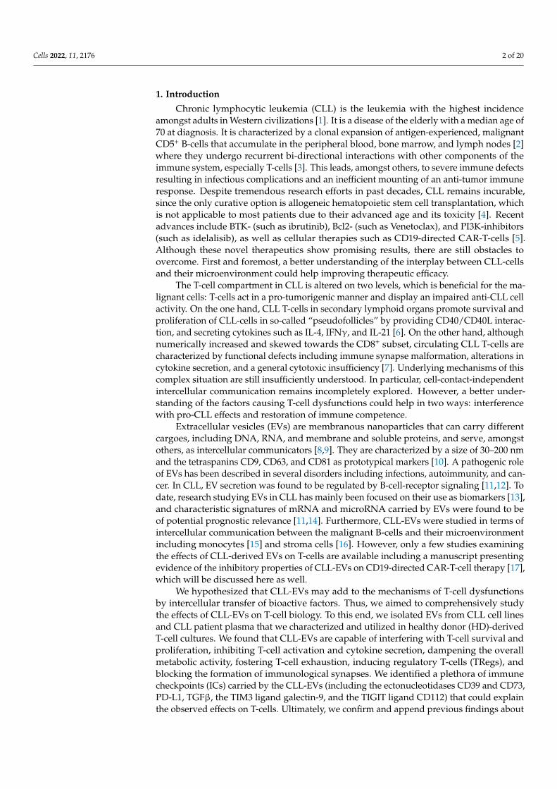
Chronic lymphocytic leukemia (CLL) is the leukemia with the highest incidenceamongst adults in Western civilizations [1]. It is a disease of the elderly with a median age of70 at diagnosis. It is characterized by a clonal expansion of antigen-experienced, malignantCD5+ B-cells that accumulate in the peripheral blood, bone marrow, and lymph nodes [2]where they undergo recurrent bi-directional interactions with other components of theimmune system, especially T-cells [3]. This leads, amongst others, to severe immune defectsresulting in infectious complications and an inefficient mounting of an anti-tumor immuneresponse. Despite tremendous research efforts in past decades, CLL remains incurable,since the only curative option is allogeneic hematopoietic stem cell transplantation, whichis not applicable to most patients due to their advanced age and its toxicity [4]. Recentadvances include BTK- (such as ibrutinib), Bcl2- (such as Venetoclax), and PI3K-inhibitors(such as idelalisib), as well as cellular therapies such as CD19-directed CAR-T-cells [5].Although these novel therapeutics show promising results, there are still obstacles toovercome. First and foremost, a better understanding of the interplay between CLL-cellsand their microenvironment could help improving therapeutic efficacy.
The T-cell compartment in CLL is altered on two levels, which is beneficial for the ma-lignant cells: T-cells act in a pro-tumorigenic manner and display an impaired anti-CLL cellactivity. On the one hand, CLL T-cells in secondary lymphoid organs promote survival andproliferation of CLL-cells in so-called “pseudofollicles” by providing CD40/CD40L interac-tion, and secreting cytokines such as IL-4, IFNγ, and IL-21 [6]. On the other hand, althoughnumerically increased and skewed towards the CD8+ subset, circulating CLL T-cells arecharacterized by functional defects including immune synapse malformation, alterations incytokine secretion, and a general cytotoxic insufficiency [7]. Underlying mechanisms of thiscomplex situation are still insufficiently understood. In particular, cell-contact-independentintercellular communication remains incompletely explored. However, a better under-standing of the factors causing T-cell dysfunctions could help in two ways: interferencewith pro-CLL effects and restoration of immune competence.
Extracellular vesicles (EVs) are membranous nanoparticles that can carry differentcargoes, including DNA, RNA, and membrane and soluble proteins, and serve, amongstothers, as intercellular communicators [8,9]. They are characterized by a size of 30–200 nmand the tetraspanins CD9, CD63, and CD81 as prototypical markers [10]. A pathogenic roleof EVs has been described in several disorders including infections, autoimmunity, and can-cer. In CLL, EV secretion was found to be regulated by B-cell-receptor signaling [11,12]. Todate, research studying EVs in CLL has mainly been focused on their use as biomarkers [13],and characteristic signatures of mRNA and microRNA carried by EVs were found to beof potential prognostic relevance [11,14]. Furthermore, CLL-EVs were studied in terms ofintercellular communication between the malignant B-cells and their microenvironmentincluding monocytes [15] and stroma cells [16]. However, only a few studies examiningthe effects of CLL-derived EVs on T-cells are available including a manuscript presentingevidence of the inhibitory properties of CLL-EVs on CD19-directed CAR-T-cell therapy [17],which will be discussed here as well.
We hypothesized that CLL-EVs may add to the mechanisms of T-cell dysfunctionsby intercellular transfer of bioactive factors. Thus, we aimed to comprehensively studythe effects of CLL-EVs on T-cell biology. To this end, we isolated EVs from CLL cell linesand CLL patient plasma that we characterized and utilized in healthy donor (HD)-derivedT-cell cultures. We found that CLL-EVs are capable of interfering with T-cell survival andproliferation, inhibiting T-cell activation and cytokine secretion, dampening the overallmetabolic activity, fostering T-cell exhaustion, inducing regulatory T-cells (TRegs), andblocking the formation of immunological synapses. We identified a plethora of immunecheckpoints (ICs) carried by the CLL-EVs (including the ectonucleotidases CD39 and CD73,PD-L1, TGFβ, the TIM3 ligand galectin-9, and the TIGIT ligand CD112) that could explainthe observed effects on T-cells. Ultimately, we confirm and append previous findings about
Cells 2022, 11, 2176 3 of 20
the detrimental influence of CLL-EVs on CAR-T-cell function [17]. Our novel findings onCLL-EVs could contribute to developing the means for restoring T-cell immunity in CLL.
2. Materials and Methods2.1. Cell Lines and Cells
Peripheral blood and plasma samples were collected upon approval by the localethics committee (approval numbers: 291_14B, 289_16B) and patients’ written informedconsent. Human CLL cell lines MEC-1, HG-3, EHEB, and PGA1, and the cell lines Raji andJurkat were purchased from DSMZ (Braunschweig, Germany). EBV-LCL lines were kindlyprovided by Dr. Regina Gary and Dr. Sascha Kretschmann (Erlangen, Germany).
2.2. Extracellular Vesicles
Isolation: CLL cell culture supernatants were harvested, sequentially centrifuged, andfiltered (0.22 µm mesh) to remove large vesicles, apoptotic bodies, and debris. Subsequently,EVs were isolated by ultracentrifugation (70 min, 110,000× g, 4 ◦C), washed once with PBS,and stored in PBS at −80 ◦C until further use. EVs from treatment-naïve CLL patient plasma(n = 8) were similarly isolated by sequential centrifugation and filtration (0.22 µm mesh)for removal of large vesicles, apoptotic bodies, and debris, and subsequently harvested byultracentrifugation at 110.000× g once for 2 h and twice for 70 min. For quantification, totalprotein content was used and analyzed by a Pierce BCA Assay kit (ThermoFisher Scientific,Waltham, MA, USA).
Electron microscopy: Fixed EV solutions (4% PFA in PBS mixed 1:1 with EV) wereplaced onto 10 min UV irradiated 300-mesh formvar/carbon-coated grids and allowedto absorb to the formvar for 5 min. Grids were incubated 5 min in a dry environment,rinsed one time with PBS and placed in 1% glutaraldehyde (in PBS) for 5 min. After rinsingin distilled water (7 times), the grids were stained for contrast using a 2% uranyl oxalatesolution (pH 7 for 5 min in dark). Afterwards the grids were incubated in drops of methylcellulose–uranyl acetate (8 parts 2% methyl cellulose (in water), 1 part ddH2O, 1 part 4%uranyl acetate (in water), pH 4, sterile filtered) for 10 min on ice (dark) [18]. After that, gridswere removed with stainless steel loops and excess fluid was blotted by gentle pushingon Whatman filter paper. After air-drying, the samples were examined and photographedwith a Zeiss EM10 electron microscope (Zeiss, Jena, Germany) and a Gatan SC1000 Orius™CCD camera (GATAN, Munich, Germany) in combination with the DigitalMicrograph™software 3.1 (GATAN, Pleasanton, CA, USA). Images were adjusted for contrast andbrightness using Adobe Photoshop CC 2018 (Adobe Systems, San José, CA, USA).
Fluorescent labeling: Cells or EVs were labeled for uptake experiments using the PKH26Red Fluorescent Cell Linker Mini kit (Sigma-Aldrich, St. Louis, MO, USA) according to themanufacturer’s instructions. Excess dye was quenched by adding 2 mL 10% BSA in PBS anda subsequent washing step with PBS by ultracentrifugation (70 min, 100,000× g, 4 ◦C).
2.3. Cell Culture
Human CLL cell lines MEC-1, HG-3, PGA1, and EHEB were grown as high-densitycell culture in CELLine classic bioreactor flasks (Sigma-Aldrich) in RPMI-1640 (Sigma-Aldrich) supplemented with 2 mM L-glutamine (Sigma-Aldrich), 10% exosome-depletedFCS (c.c.pro, Vogtei, Germany), and 40 U/mL penicillin–streptomycin (ThermoFisherScientific) at 37 ◦C, 5% CO2. Jurkat and Raji cells were cultured in AIM V (ThermoFisherScientific) in conventional cell culture flasks at 37 ◦C, 5% CO2. Cell line cultures were splittwice per week.
Healthy donor peripheral blood mononuclear cells (PBMCs) were obtained froma total of 50 volunteers using Ficoll-Paque (GE Healthcare, Chalfont St. Giles, UK). T-cellswere isolated from human PBMCs using the Pan T cell isolation kit, human (MiltenyiBiotec, Bergisch-Gladbach, Germany) according to manufacturer’s guidelines. T-cells orCAR-T-cells were seeded at a density of 106/mL in AIM V or AIM V with 50 U/mLIL-2 (Miltenyi Biotec), respectively, and pretreated with 30 µg/mL CLL-EVs for 3 days.
Cells 2022, 11, 2176 4 of 20
Afterwards, cells were re-seeded at the same density with fresh EVs in the presence orabsence of anti-CD2/CD3/CD28-coated beads (Miltenyi Biotec) for 24–96 h depending onthe subsequent analysis.
Raji and Jurkat cells were co-cultured at a ratio of 1:4 in AIM V. Raji cells were pre-incubated with 30 ng/mL SEA for 45 min. The co-culture was incubated for 24 h in theabsence/presence of 30 µg/mL CLL-EVs. For selected experiments EVs at a density of800 µg/mL were pre-incubated with 10 µg/mL anti-PDL1 antibody (ThermoFisher Scientific)or the corresponding isotype control for 30 min. Secreted IL-2 was quantified with a pre-madeELISA kit (Biolegend, San Diego, CA, USA) according to the manufacturer’s instructions.
The inhibitors used in all experiment are listed in Supplementary Table S1.
2.4. Flow Cytometry
Samples were recorded on a FACS Canto II (BD Biosciences, Franklin Lakes, NJ, USA)and data was analyzed using FlowJo V 10 (BD Biosciences).
Multiplex assay: Common EV markers were analyzed by flow cytometry usinga commercial MACSPlex exosome kit (Miltenyi Biotec) according to the manufacturer’sinstructions.
EV flow cytometry: To analyze membrane and soluble proteins carried by EVs, theEVs or their lysates were coated on 4 µm latex beads (ThermoFisher Scientific) at 4 ◦Covernight, free binding sites were saturated by 1 M glycine (Sigma-Aldrich), and beadswere washed with PBS/5% BSA. Then, CLL-EV-coated beads were incubated with humanIgG (Gamunex, Grifols, Barcelona, Spain) and stained using fluorochrome-conjugatedantibodies and the respective isotype controls (see Supplementary Table S2).
PKH26 EV-uptake assay: PKH26-labeled CLL-cells were incubated with activated andnon-activated healthy donor T-cells in the presence and absence of EV-secretion inhibitors(Nexinhib20, GW4869) at indicated ratios for 24 h.
PKH26-labeled EVs were incubated with activated and non-activated healthy donorT-cells in the presence and absence of EV uptake inhibitors (EIPA, cytochalasin D, dynasore,and heparin). Additionally, after incubation with labeled EVs, T-cells were treated withtrypsin prior to analysis to distinguish between surface-bound and internalized EVs.
Proliferation: For proliferation analysis, cells were stained with violet proliferation dye450 (BD Biosciences) prior to the culture according to the manufacturer’s recommendation.Dye dilution was recorded and analyzed using the proliferation tool in FlowJo.
Surface staining: Samples were stained with Zombie Aqua (Biolegend) for dead celldiscrimination, blocked with human IgG (Gamunex), and stained with fluorochrome-conjugated antibodies (see Supplementary Table S2) against surface proteins according tothe manufacturer’s instruction.
Intracellular staining: Intracellular proteins were stained subsequent to surface stainingusing the FoxP3 transcription factor staining Kit (Biolegend) and fluorochrome-conjugatedantibodies (see Supplementary Table S2) according to the manufacturer’s manual.
PhosFlow: Intracellular phospho-proteins were stained using the PhosFlowTM Fix bufferI together with the PhosFlowTM Perm buffer III (both BD Biosciences) and fluorochrome-conjugated phospho-specific antibodies (see Supplementary Table S2) according to themanufacturer’s recommendations.
Glucose uptake: Glucose uptake was analyzed by the use of 6-NBDG (6-(N-(7-nitrobenz-2-oxa-1,3-diazol-4-yl)amino)-6-deoxyglucose; ThermoFisher Scientific). Cells were washedin PBS and glucose-free medium by centrifugation (300× g, 5 min, 4 ◦C) and resuspendedin glucose-free medium containing 0.3 mM 6-NBDG. Samples were incubated for 15 min at37 ◦C, 5% CO2 and subsequently recorded after two washing steps.
Fatty acid uptake: Uptake of long-chain fatty acids was determined by staining withBodipyTM FL C16 (ThermoFisher Scientific) according to the product’s manual.
Mitochondrial parameters: Mitochondrial biomass and membrane potential were de-termined with MitoTrackerTM Green and tetramethylrhodamine ethyl ester (TMRE) (bothThermoFisher Scientific) according to the manufacturer’s manual.
Cells 2022, 11, 2176 5 of 20
Reactive oxygen species: Cellular reactive oxygen species and mitochondrial superox-ides were analyzed using CellROX Deep Red reagent and MitoSOX Red (both ThermoFisherScientific) according to the manufacturer’s manual.
2.5. Fluorescent Microscopy
Isolated T-cells were activated with plate-bound CD3/soluble CD28 (both Ther-moFisher Scientific) in the presence of PKH26-labeled CLL-EVs for 48 h at 37 ◦C, 5% CO2.Cells were harvested and coated onto poly-D-lysine microscopic slides (ThermoFisherScientific). Slides were washed with PBS and cells were fixed with 4% paraformaldehydesolution. After washing, slides were blocked with ratIgG, incubated with anti-CD4-AF488or anti-CD8-AF488 antibodies, and counter-stained with DAPI. Samples were recorded ona Leica laser-scanning microscope and images were analyzed using LAS X Core softwareversion 3 (Leica, Wetzlar, Germany).
2.6. Metabolic Analyses
Glucose/lactate: Glucose and lactate concentrations in cell culture supernatantswere quantified using a SuperGLcompact (Hitado, Möhnesee, Germany) according to thedevice’s manual.
Extracellular flux analysis: Bioenergetics of glycolysis and mitochondrial respirationwere analyzed on the extracellular flux analyzer Seahorse XFe 96 (Agilent, Santa Clara, CA,USA), as previously described [19].
Scenith: Scenith analyses and calculations were performed according to theoriginal publication [20].
ATP/Adenosine assay: ATP concentrations in supernatants were quantified with a flu-orometric ATP Assay Kit (Sigma-Aldrich). Adenosine concentrations in supernatants werequantified with a fluorometric Adenosine Assay Kit (PromoCell, Heidelberg, Germany).
2.7. Imaging Cytometry
Imaging cytometry was performed on a FlowSight (Luminex, Austin, TX, USA) anddata were analyzed using IDEAS 6.3 including a machine-learning module (Luminex).
For immune synapse staining HD-derived, isolated T-cells were pre-stimulated withanti-CD2/CD3/CD28-coated beads (Miltenyi Biotec) in the absence/presence of 30 µg/mLCLL-EVs for 48 h at 37 ◦C, 5% CO2. Raji cells were labeled with CFSE (ThermoFisherScientific) and loaded with 1 µg/mL SEA and SEB (both Sigma-Aldrich). T-cells andRaji cells were mixed at a ratio of 1:1, CLL-EVs were added at 60 µg/mL, and sam-ples were incubated for 7 min at 37 ◦C. Samples were fixed with 1.5% PFA and per-meabilized with PBS/2%FCS/0.1% Triton-X. They were stained with surface antibodiesand Biotin-XX-Phalloidin (ThermoFisher Scientific), as well as Streptavidin-PE/TexasRed(ThermoFisher Scientific). Staining of nuclei was done using either 7-AAD (Biolegend) orSyto13 (ThermoFisher Scientific). Synapse staining of CAR-T-cells was similarly performedusing Mec-1 cells as the target instead of Raji with a CD19 surface staining for detection.
2.8. Statistics
Outliers were determined using the ROUT test. Differences in means were evaluatedwith parametric (paired/unpaired t-test, one-way ANOVA) or nonparametric (unpairedMann–Whitney, paired Wilcoxon, unpaired Kruskal–Wallis) tests based on the number ofcomparisons (two or more than two) and distribution levels (as determined by Shapiro–Wilk and Kolmogorov–Smirnov). All statistical analyses were performed using GraphPadPrism Version 7 or 8 (GraphPad Software Inc., San Diego, CA, USA) at a significancelevel of p < 0.05.
Cells 2022, 11, 2176 6 of 20
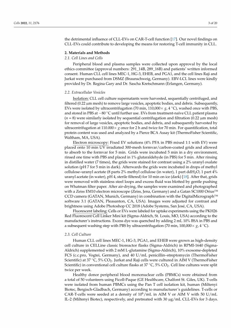
3. Results3.1. CLL-Cells Secrete EVs That Interact with T-Cells
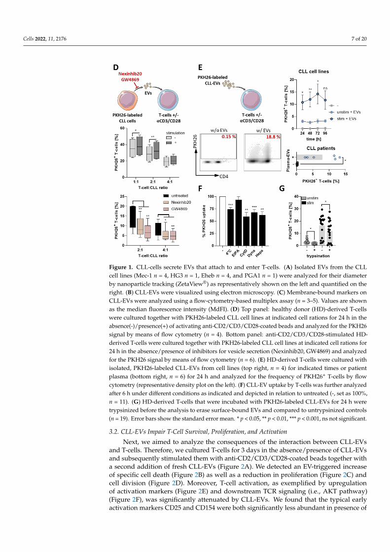
T-cells are central to tumor immune surveillance. Thus, we wanted to further explorethe contact-independent cross-talk between CLL-cells and T-cells, with a focus on EVs. First,we isolated EVs from different CLL cell lines (Mec-1, HG3, PGA1, and Eheb) as well as CLL-patient plasma by sequential (ultra-)centrifugation. We confirmed typical characteristics ofthe EVs, such as a diameter of 100–150 nm, shape, and EV-specific protein markers, usinga nanoparticle tracking analyzer (ZetaView®, Figure 1A), electron microscopy (Figure 1B),and a flow cytometry-based multiplex assay (Figure 1C), respectively. The latter showed,amongst others, the presence of typical EV-markers such as CD9, CD63, and CD81, as wellas markers of the parental cell type such as CD19, CD20, and CD40, some of which wecould confirm on EVs obtained from the CLL-patient plasma (Supplementary Figure S1A).
To investigate a potential EV-mediated communication between CLL-cells and T-cells,we co-cultured PKH26-labeled CLL cell lines (that also secrete PKH26+ EVs, data notshown) together with HD-derived T-cells in the absence/presence of a T-cell stimulatorysignal (via anti-CD2/CD3/CD28-coated beads). We found that T-cells obtained a PKH26signal after 24 h of co-culture, which was significantly stronger upon T-cell stimulation.However, the PKH26 signal was significantly reduced by inhibitors of EV secretion usedduring the co-culture (i.e., Nexinhib20 and GW4869) indicating a transfer of EVs from CLL-cells to T-cells (Figure 1D). Isolated cell-line- and patient-plasma-derived PKH26-labeledCLL-EVs were efficiently taken up by or bound to T-cells, which was most prominent uponT-cell stimulation (Figure 1E). Notably, CD4+ and CD8+ T-cells were similarly capable ofinteracting with CLL-EVs (Supplementary Figure S1B). The analysis of CLL-EV uptakeby T-cells under different conditions including low temperature and several inhibitors ofuptake routes [21] revealed that interaction of T-cells with CLL-EVs was only partly energy-dependent and mainly occurred via endocytosis (Figure 1F). Moreover, labeled CLL-EVsare not only found inside, but also as mentioned previously, on the outer cell membrane ofT-cells (Supplementary Figure S1C). To estimate the amount of surface-bound CLL-EVs,we trypsinized the T-cells after culture in presence of CLL-EVs to detach surface-boundEVs and detected a significant reduction of the PKH26 signal (Figure 1G).
Together, these data show that CLL-cells secrete EVs that potentially bind to the T-cellsurface and/or enter T-cells.
Cells 2022, 11, x FOR PEER REVIEW 7 of 23
Figure 1. CLL-cells secrete EVs that attach to and enter T-cells. (A) Isolated EVs from the CLL cell lines (Mec-1 n = 4, HG3 n = 1, Eheb n = 4, and PGA1 n = 1) were analyzed for their diameter by nanoparticle tracking (ZetaView®) as representatively shown on the left and quantified on the right. (B) CLL-EVs were visualized using electron microscopy. (C) Membrane-bound markers on CLL-EVs were analyzed using a flow-cytometry-based multiplex assay (n = 3–5). Values are shown as the median fluorescence intensity (MdFI). (D) Top panel: healthy donor (HD)-derived T-cells were cultured together with PKH26-labeled CLL cell lines at indicated cell rations for 24 h in the ab-sence(-)/presence(+) of activating anti-CD2/CD3/CD28-coated beads and analyzed for the PKH26 signal by means of flow cytometry (n = 4). Bottom panel: anti-CD2/CD3/CD28-stimulated HD-de-rived T-cells were cultured together with PKH26-labeled CLL cell lines at indicated cell rations for 24 h in the absence/presence of inhibitors for vesicle secretion (Nexinhib20, GW4869) and analyzed for the PKH26 signal by means of flow cytometry (n = 6). (E) HD-derived T-cells were cultured with isolated, PKH26-labeled CLL-EVs from cell lines (top right, n = 4) for indicated times or patient plasma (bottom right, n = 6) for 24 h and analyzed for the frequency of PKH26+ T-cells by flow cytometry (representative density plot on the left). (F) CLL-EV uptake by T-cells was further ana-lyzed after 6 h under different conditions as indicated and depicted in relation to untreated (-, set as
Figure 1. Cont.
Cells 2022, 11, 2176 7 of 20
Cells 2022, 11, x FOR PEER REVIEW 7 of 23
Figure 1. CLL-cells secrete EVs that attach to and enter T-cells. (A) Isolated EVs from the CLL cell lines (Mec-1 n = 4, HG3 n = 1, Eheb n = 4, and PGA1 n = 1) were analyzed for their diameter by nanoparticle tracking (ZetaView®) as representatively shown on the left and quantified on the right. (B) CLL-EVs were visualized using electron microscopy. (C) Membrane-bound markers on CLL-EVs were analyzed using a flow-cytometry-based multiplex assay (n = 3–5). Values are shown as the median fluorescence intensity (MdFI). (D) Top panel: healthy donor (HD)-derived T-cells were cultured together with PKH26-labeled CLL cell lines at indicated cell rations for 24 h in the ab-sence(-)/presence(+) of activating anti-CD2/CD3/CD28-coated beads and analyzed for the PKH26 signal by means of flow cytometry (n = 4). Bottom panel: anti-CD2/CD3/CD28-stimulated HD-de-rived T-cells were cultured together with PKH26-labeled CLL cell lines at indicated cell rations for 24 h in the absence/presence of inhibitors for vesicle secretion (Nexinhib20, GW4869) and analyzed for the PKH26 signal by means of flow cytometry (n = 6). (E) HD-derived T-cells were cultured with isolated, PKH26-labeled CLL-EVs from cell lines (top right, n = 4) for indicated times or patient plasma (bottom right, n = 6) for 24 h and analyzed for the frequency of PKH26+ T-cells by flow cytometry (representative density plot on the left). (F) CLL-EV uptake by T-cells was further ana-lyzed after 6 h under different conditions as indicated and depicted in relation to untreated (-, set as
Figure 1. CLL-cells secrete EVs that attach to and enter T-cells. (A) Isolated EVs from the CLLcell lines (Mec-1 n = 4, HG3 n = 1, Eheb n = 4, and PGA1 n = 1) were analyzed for their diameterby nanoparticle tracking (ZetaView®) as representatively shown on the left and quantified on theright. (B) CLL-EVs were visualized using electron microscopy. (C) Membrane-bound markers onCLL-EVs were analyzed using a flow-cytometry-based multiplex assay (n = 3–5). Values are shownas the median fluorescence intensity (MdFI). (D) Top panel: healthy donor (HD)-derived T-cellswere cultured together with PKH26-labeled CLL cell lines at indicated cell rations for 24 h in theabsence(-)/presence(+) of activating anti-CD2/CD3/CD28-coated beads and analyzed for the PKH26signal by means of flow cytometry (n = 4). Bottom panel: anti-CD2/CD3/CD28-stimulated HD-derived T-cells were cultured together with PKH26-labeled CLL cell lines at indicated cell rations for24 h in the absence/presence of inhibitors for vesicle secretion (Nexinhib20, GW4869) and analyzedfor the PKH26 signal by means of flow cytometry (n = 6). (E) HD-derived T-cells were cultured withisolated, PKH26-labeled CLL-EVs from cell lines (top right, n = 4) for indicated times or patientplasma (bottom right, n = 6) for 24 h and analyzed for the frequency of PKH26+ T-cells by flowcytometry (representative density plot on the left). (F) CLL-EV uptake by T-cells was further analyzedafter 6 h under different conditions as indicated and depicted in relation to untreated (-, set as 100%,n = 11). (G) HD-derived T-cells that were incubated with PKH26-labeled CLL-EVs for 24 h weretrypsinized before the analysis to erase surface-bound EVs and compared to untrypsinized controls(n = 19). Error bars show the standard error mean. * p < 0.05, ** p < 0.01, *** p < 0.001, ns not significant.
3.2. CLL-EVs Impair T-Cell Survival, Proliferation, and Activation
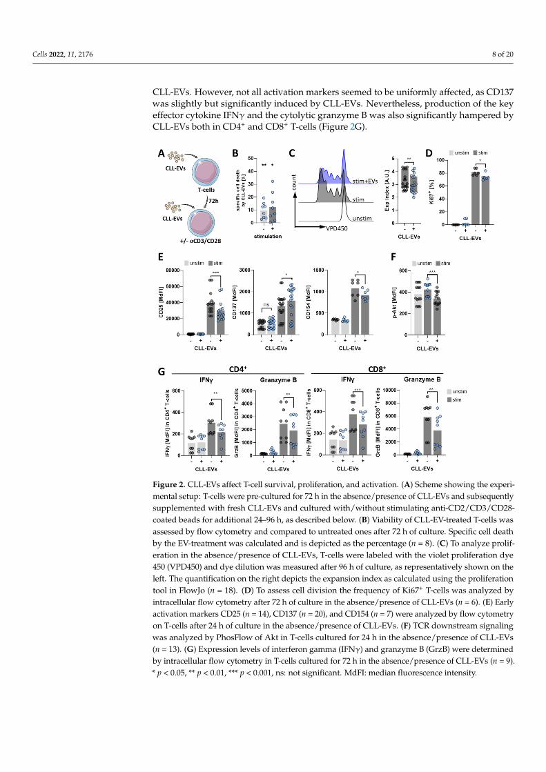
Next, we aimed to analyze the consequences of the interaction between CLL-EVsand T-cells. Therefore, we cultured T-cells for 3 days in the absence/presence of CLL-EVsand subsequently stimulated them with anti-CD2/CD3/CD28-coated beads together witha second addition of fresh CLL-EVs (Figure 2A). We detected an EV-triggered increaseof specific cell death (Figure 2B) as well as a reduction in proliferation (Figure 2C) andcell division (Figure 2D). Moreover, T-cell activation, as exemplified by upregulationof activation markers (Figure 2E) and downstream TCR signaling (i.e., AKT pathway)(Figure 2F), was significantly attenuated by CLL-EVs. We found that the typical earlyactivation markers CD25 and CD154 were both significantly less abundant in presence of
Cells 2022, 11, 2176 8 of 20
CLL-EVs. However, not all activation markers seemed to be uniformly affected, as CD137was slightly but significantly induced by CLL-EVs. Nevertheless, production of the keyeffector cytokine IFNγ and the cytolytic granzyme B was also significantly hampered byCLL-EVs both in CD4+ and CD8+ T-cells (Figure 2G).
Cells 2022, 11, x FOR PEER REVIEW 9 of 23
Figure 2. CLL-EVs affect T-cell survival, proliferation, and activation. (A) Scheme showing the ex-perimental setup: T-cells were pre-cultured for 72 h in the absence/presence of CLL-EVs and subse-quently supplemented with fresh CLL-EVs and cultured with/without stimulating anti-CD2/CD3/CD28-coated beads for additional 24–96 h, as described below. (B) Viability of CLL-EV-treated T-cells was assessed by flow cytometry and compared to untreated ones after 72 h of culture. Specific cell death by the EV-treatment was calculated and is depicted as the percentage (n = 8). (C) To analyze proliferation in the absence/presence of CLL-EVs, T-cells were labeled with the violet proliferation dye 450 (VPD450) and dye dilution was measured after 96 h of culture, as representa-tively shown on the left. The quantification on the right depicts the expansion index as calculated using the proliferation tool in FlowJo (n = 18). (D) To assess cell division the frequency of Ki67+ T-cells was analyzed by intracellular flow cytometry after 72 h of culture in the absence/presence of CLL-EVs (n = 6). (E) Early activation markers CD25 (n = 14), CD137 (n = 20), and CD154 (n = 7) were analyzed by flow cytometry on T-cells after 24 h of culture in the absence/presence of CLL-EVs. (F) TCR downstream signaling was analyzed by PhosFlow of Akt in T-cells cultured for 24 h in the absence/presence of CLL-EVs (n = 13). (G) Expression levels of interferon gamma (IFNγ) and granzyme B (GrzB) were determined by intracellular flow cytometry in T-cells cultured for 72 h in the absence/presence of CLL-EVs (n = 9). * p < 0.05, ** p < 0.01, *** p < 0.001, ns: not significant. MdFI: median fluorescence intensity.
3.3. CLL-EVs Alter T-Cell Metabolism and Skew Their Subset Composition The prominent role of metabolism in governing the growth, activation, differentia-
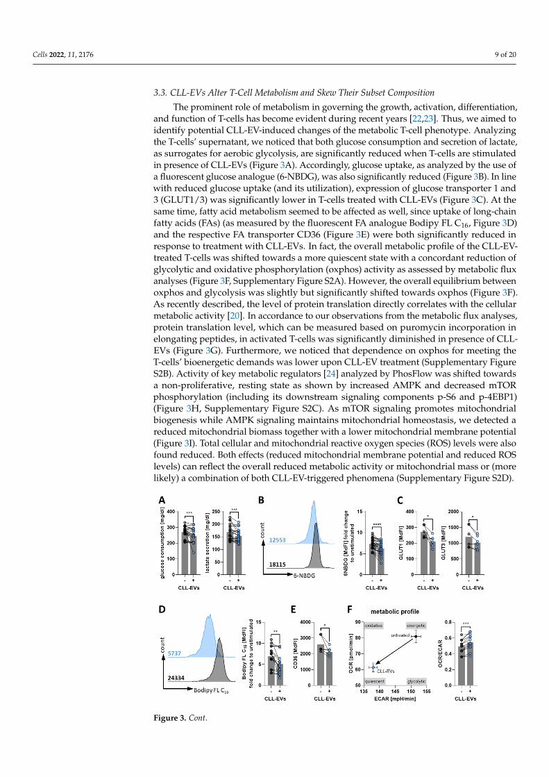
tion, and function of T-cells has become evident during recent years [22,23]. Thus, we aimed to identify potential CLL-EV-induced changes of the metabolic T-cell phenotype. Analyzing the T-cells’ supernatant, we noticed that both glucose consumption and secre-tion of lactate, as surrogates for aerobic glycolysis, are significantly reduced when T-cells
Figure 2. CLL-EVs affect T-cell survival, proliferation, and activation. (A) Scheme showing the experi-mental setup: T-cells were pre-cultured for 72 h in the absence/presence of CLL-EVs and subsequentlysupplemented with fresh CLL-EVs and cultured with/without stimulating anti-CD2/CD3/CD28-coated beads for additional 24–96 h, as described below. (B) Viability of CLL-EV-treated T-cells wasassessed by flow cytometry and compared to untreated ones after 72 h of culture. Specific cell deathby the EV-treatment was calculated and is depicted as the percentage (n = 8). (C) To analyze prolif-eration in the absence/presence of CLL-EVs, T-cells were labeled with the violet proliferation dye450 (VPD450) and dye dilution was measured after 96 h of culture, as representatively shown on theleft. The quantification on the right depicts the expansion index as calculated using the proliferationtool in FlowJo (n = 18). (D) To assess cell division the frequency of Ki67+ T-cells was analyzed byintracellular flow cytometry after 72 h of culture in the absence/presence of CLL-EVs (n = 6). (E) Earlyactivation markers CD25 (n = 14), CD137 (n = 20), and CD154 (n = 7) were analyzed by flow cytometryon T-cells after 24 h of culture in the absence/presence of CLL-EVs. (F) TCR downstream signalingwas analyzed by PhosFlow of Akt in T-cells cultured for 24 h in the absence/presence of CLL-EVs(n = 13). (G) Expression levels of interferon gamma (IFNγ) and granzyme B (GrzB) were determinedby intracellular flow cytometry in T-cells cultured for 72 h in the absence/presence of CLL-EVs (n = 9).* p < 0.05, ** p < 0.01, *** p < 0.001, ns: not significant. MdFI: median fluorescence intensity.
Cells 2022, 11, 2176 9 of 20
3.3. CLL-EVs Alter T-Cell Metabolism and Skew Their Subset Composition
The prominent role of metabolism in governing the growth, activation, differentiation,and function of T-cells has become evident during recent years [22,23]. Thus, we aimed toidentify potential CLL-EV-induced changes of the metabolic T-cell phenotype. Analyzingthe T-cells’ supernatant, we noticed that both glucose consumption and secretion of lactate,as surrogates for aerobic glycolysis, are significantly reduced when T-cells are stimulatedin presence of CLL-EVs (Figure 3A). Accordingly, glucose uptake, as analyzed by the use ofa fluorescent glucose analogue (6-NBDG), was also significantly reduced (Figure 3B). In linewith reduced glucose uptake (and its utilization), expression of glucose transporter 1 and3 (GLUT1/3) was significantly lower in T-cells treated with CLL-EVs (Figure 3C). At thesame time, fatty acid metabolism seemed to be affected as well, since uptake of long-chainfatty acids (FAs) (as measured by the fluorescent FA analogue Bodipy FL C16, Figure 3D)and the respective FA transporter CD36 (Figure 3E) were both significantly reduced inresponse to treatment with CLL-EVs. In fact, the overall metabolic profile of the CLL-EV-treated T-cells was shifted towards a more quiescent state with a concordant reduction ofglycolytic and oxidative phosphorylation (oxphos) activity as assessed by metabolic fluxanalyses (Figure 3F, Supplementary Figure S2A). However, the overall equilibrium betweenoxphos and glycolysis was slightly but significantly shifted towards oxphos (Figure 3F).As recently described, the level of protein translation directly correlates with the cellularmetabolic activity [20]. In accordance to our observations from the metabolic flux analyses,protein translation level, which can be measured based on puromycin incorporation inelongating peptides, in activated T-cells was significantly diminished in presence of CLL-EVs (Figure 3G). Furthermore, we noticed that dependence on oxphos for meeting theT-cells’ bioenergetic demands was lower upon CLL-EV treatment (Supplementary FigureS2B). Activity of key metabolic regulators [24] analyzed by PhosFlow was shifted towardsa non-proliferative, resting state as shown by increased AMPK and decreased mTORphosphorylation (including its downstream signaling components p-S6 and p-4EBP1)(Figure 3H, Supplementary Figure S2C). As mTOR signaling promotes mitochondrialbiogenesis while AMPK signaling maintains mitochondrial homeostasis, we detected areduced mitochondrial biomass together with a lower mitochondrial membrane potential(Figure 3I). Total cellular and mitochondrial reactive oxygen species (ROS) levels were alsofound reduced. Both effects (reduced mitochondrial membrane potential and reduced ROSlevels) can reflect the overall reduced metabolic activity or mitochondrial mass or (morelikely) a combination of both CLL-EV-triggered phenomena (Supplementary Figure S2D).
Cells 2022, 11, x FOR PEER REVIEW 11 of 23
Figure 3. CLL-EVs modulate T-cell metabolism. (A) Consumption of glucose and secretion of lactate in the supernatant of activated T-cells cultured for 72 h in the absence/presence of CLL-EVs was quantified (n = 21). (B) Glucose uptake of activated T-cells cultured for 72 h in the absence/presence of CLL-EVs was semi-quantified by flow cytometry using the fluorescent glucose analogue 6-NBDG and depicted as the median fluorescence intensity as representatively shown on the left and sum-marized on the right (n = 30). (C) Surface levels of glucose transporters 1 and 3 were analyzed by flow cytometry on activated T-cell culture for 72 h in the absence/presence of CLL-EVs (n = 6). (D) Long-chain fatty acid (LC-FA) uptake of activated T-cells cultured for 72 h in the absence/presence of CLL-EVs was semi-quantified by flow cytometry using the fluorescent LC-FA analogue Bodipy® FL C16 and depicted as the median fluorescence intensity as representatively shown on the left and summarized on the right (n = 16). (E) Surface levels of fatty acid transporter CD36 was analyzed by flow cytometry on activated T-cell culture for 72 h in the absence/presence of CLL-EVs (n = 6). (F) The metabolic profile of activated T-cell culture for 72 h in the absence/presence of CLL-EVs was recorded by metabolic flux analysis. Extracellular acidification rate (ECAR, as a surrogate for aero-bic glycolysis) and oxygen consumption rate (OCR, as a surrogate for oxphos) were determined in real-time and plotted as depicted in the left graph. The balance between oxphos and glycolysis is shown as the ratio of OCR/ECAR in the right graph (n = 9). (G) The level of translation as an indica-tor of metabolic activity/energy production was measured by flow cytometry in activated T-cells cultured for 72 h in the absence/presence of CLL-EVs (n = 12). (H) AMPK (n = 6) and mTOR (n = 11)
Figure 3. Cont.
Cells 2022, 11, 2176 10 of 20
Cells 2022, 11, x FOR PEER REVIEW 11 of 23
Figure 3. CLL-EVs modulate T-cell metabolism. (A) Consumption of glucose and secretion of lactate in the supernatant of activated T-cells cultured for 72 h in the absence/presence of CLL-EVs was quantified (n = 21). (B) Glucose uptake of activated T-cells cultured for 72 h in the absence/presence of CLL-EVs was semi-quantified by flow cytometry using the fluorescent glucose analogue 6-NBDG and depicted as the median fluorescence intensity as representatively shown on the left and sum-marized on the right (n = 30). (C) Surface levels of glucose transporters 1 and 3 were analyzed by flow cytometry on activated T-cell culture for 72 h in the absence/presence of CLL-EVs (n = 6). (D) Long-chain fatty acid (LC-FA) uptake of activated T-cells cultured for 72 h in the absence/presence of CLL-EVs was semi-quantified by flow cytometry using the fluorescent LC-FA analogue Bodipy® FL C16 and depicted as the median fluorescence intensity as representatively shown on the left and summarized on the right (n = 16). (E) Surface levels of fatty acid transporter CD36 was analyzed by flow cytometry on activated T-cell culture for 72 h in the absence/presence of CLL-EVs (n = 6). (F) The metabolic profile of activated T-cell culture for 72 h in the absence/presence of CLL-EVs was recorded by metabolic flux analysis. Extracellular acidification rate (ECAR, as a surrogate for aero-bic glycolysis) and oxygen consumption rate (OCR, as a surrogate for oxphos) were determined in real-time and plotted as depicted in the left graph. The balance between oxphos and glycolysis is shown as the ratio of OCR/ECAR in the right graph (n = 9). (G) The level of translation as an indica-tor of metabolic activity/energy production was measured by flow cytometry in activated T-cells cultured for 72 h in the absence/presence of CLL-EVs (n = 12). (H) AMPK (n = 6) and mTOR (n = 11)
Figure 3. CLL-EVs modulate T-cell metabolism. (A) Consumption of glucose and secretion of lactatein the supernatant of activated T-cells cultured for 72 h in the absence/presence of CLL-EVs wasquantified (n = 21). (B) Glucose uptake of activated T-cells cultured for 72 h in the absence/presence ofCLL-EVs was semi-quantified by flow cytometry using the fluorescent glucose analogue 6-NBDG anddepicted as the median fluorescence intensity as representatively shown on the left and summarizedon the right (n = 30). (C) Surface levels of glucose transporters 1 and 3 were analyzed by flowcytometry on activated T-cell culture for 72 h in the absence/presence of CLL-EVs (n = 6). (D) Long-chain fatty acid (LC-FA) uptake of activated T-cells cultured for 72 h in the absence/presence ofCLL-EVs was semi-quantified by flow cytometry using the fluorescent LC-FA analogue Bodipy®
FL C16 and depicted as the median fluorescence intensity as representatively shown on the left andsummarized on the right (n = 16). (E) Surface levels of fatty acid transporter CD36 was analyzed byflow cytometry on activated T-cell culture for 72 h in the absence/presence of CLL-EVs (n = 6). (F) Themetabolic profile of activated T-cell culture for 72 h in the absence/presence of CLL-EVs was recordedby metabolic flux analysis. Extracellular acidification rate (ECAR, as a surrogate for aerobic glycolysis)and oxygen consumption rate (OCR, as a surrogate for oxphos) were determined in real-time andplotted as depicted in the left graph. The balance between oxphos and glycolysis is shown as the ratioof OCR/ECAR in the right graph (n = 9). (G) The level of translation as an indicator of metabolicactivity/energy production was measured by flow cytometry in activated T-cells cultured for 72 hin the absence/presence of CLL-EVs (n = 12). (H) AMPK (n = 6) and mTOR (n = 11) signaling wereanalyzed by PhosFlow in activated T-cells cultured for 24 h in the absence/presence of CLL-EVs.(I) Mitochondrial biomass (MitoTrackerTM, n = 7) and mitochondrial membrane potential (TMRE,n = 8) were measured in activated T-cells after 72 h of culture in the absence/presence of CLL-EVs byflow cytometry. (J) T-cell-subset frequencies of naïve (Tn), central memory (TCM), effector memory(TEM), and terminally differentiated (TEMRA) ones were analyzed in activated CD4+ and CD8+ T-cellsby flow cytometry after 72 h of culture in the absence/presence of CLL-EVs (n = 5–6). Error barsshow the standard error mean. * p < 0.05, ** p < 0.01, *** p < 0.001, **** p < 0.0001. MdFI: medianfluorescence intensity.
It is well-established that T-cell subsets display different metabolic phenotypes [25].Thus, we determined the frequencies of naïve (n; CD45RO− CCR7+), central memory (CM;CD45RO+ CCR7+), effector memory (EM; CD45RO+ CCR7−), and terminally differentiatedeffector T-cells (EMRA; CD45RO− CCR7−) [26] (gating strategy see Supplementary FigureS2E) in absence or presence of CLL-EVs. It is known that central memory T-cells are lessproliferative and metabolically active with a preference for oxphos while effector memoryT-cells rapidly proliferate and display an active glycolytic metabolism [25]. Thus, it is
Cells 2022, 11, 2176 11 of 20
not surprising that we found a skewing of effector/memory distribution towards centralmemory cells at the expanse of effector memory cells by the presence of CLL-EVs (Figure 3J).
Taken together, these data reveal a substantial modulation of the T-cell bioenergeticsby CLL-EVs.
3.4. CLL-EVs Promote T-Cell Exhaustion and Regulatory T-Cell Formation
Bioenergetic alterations have been shown to not only directly act in a T-cell suppressivemanner but to also favor a tolerogenic milieu by promoting T-cell exhaustion [27] and/orTreg induction [28].
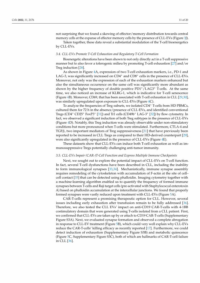
As shown in Figure 4A, expression of two T-cell exhaustion markers, i.e., PD-1 andLAG-3, was significantly increased on CD4+ and CD8+ cells in the presence of CLL-EVs.Moreover, not only was the expression of each of the exhaustion markers enhanced butalso the simultaneous occurrence on the same cell was significantly more abundant asshown by the higher frequency of double positive PD1+/LAG3+ T-cells. At the sametime, we also noticed an increase of KLRG-1, which is indicative for T-cell senescence(Figure 4B). Moreover, CD69, that has been associated with T-cell exhaustion in CLL [29,30],was similarly upregulated upon exposure to CLL-EVs (Figure 4C).
To analyze the frequencies of Treg subsets, we isolated CD4+ T-cells from HD PBMCs,cultured them for 72 h in the absence/presence of CLL-EVs, and identified conventionalTregs (CD4+ CD25+ FoxP3+ [31]) and Tr1 cells (CD49b+ LAG-3+ [32]) by flow cytometry. Infact, we observed a significant induction of both Treg subtypes in the presence of CLL-EVs(Figure 4D). Notably, this Treg induction was already observable under non-stimulatoryconditions but more pronounced when T-cells were stimulated. Furthermore, CTLA-4 andTGFβ, two important mediators of Treg suppressiveness [31] that have previously beenreported to be increased in CLL Tregs as compared to their HD-derived counterpart [29],were also significantly upregulated in the presence of CLL-EVs (Figure 4E).
These datasets show that CLL-EVs can induce both T-cell exhaustion as well as im-munosuppressive Tregs potentially challenging anti-tumor immunity.
3.5. CLL-EVs Impair (CAR-)T-Cell Function and Express Multiple Immune Checkpoints
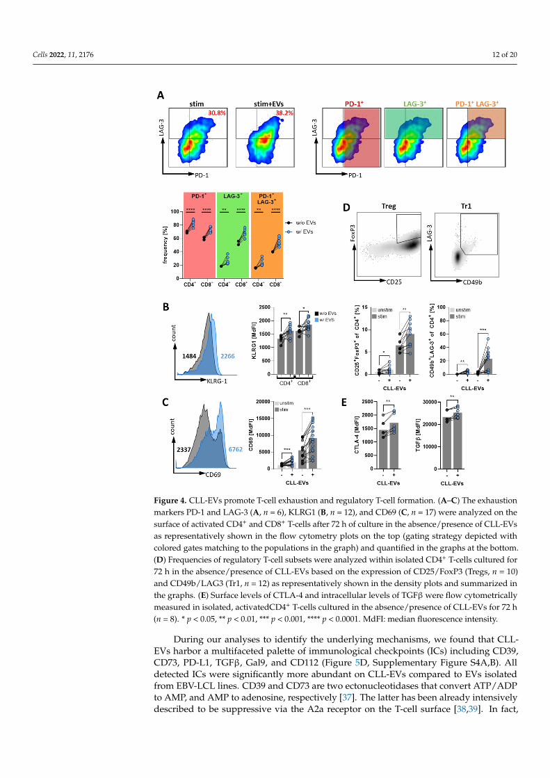
Next, we sought out to explore the potential impact of CLL-EVs on T-cell function.In fact, several T-cell dysfunctions have been described in CLL, including the inabilityto form immunological synapses [33,34]. Mechanistically, immune synapse assemblyrequires remodeling of the cytoskeleton with accumulation of F-actin at the site of cell–cell contact [35] that can be detected using phalloidin. Imaging cytometry together witha machine-learning algorithm enabled us to quantify the frequency of formed immunesynapses between T-cells and Raji target cells (pre-activated with Staphylococcal enterotoxinA) based on phalloidin accumulation at the intercellular junctions. We found that properlyformed synapses were vastly reduced upon treatment with CLL-EVs (Figure 5A).
CAR-T-cells represent a promising therapeutic option for CLL. However, severalissues including early exhaustion after transfusion remain to be fully addressed [36].Therefore, we also tested the CLL EVs’ impact on anti-CD19 CAR-T-cells with 4-1BBcostimulatory domain that were generated using T-cells isolated from a CLL patient. First,we confirmed that CLL-EVs are taken up by or attach to CD19 CAR-T-cells (SupplementaryFigure S3A). Next, we evaluated synapse formation and observed a complete abrogationin response to CLL-EV treatment (Figure 5B), which could very well explain why CLL-EVsreduce the CAR-T-cells’ killing efficacy as recently reported [17]. Furthermore, we coulddetect induction of exhaustion (Supplementary Figure S3B) and metabolic quiescence(Figure 5C, Supplementary Figure S3C), both of which are hallmarks of CAR-T-cell failurein CLL [36].
Cells 2022, 11, 2176 12 of 20Cells 2022, 11, x FOR PEER REVIEW 13 of 23
Figure 4. CLL-EVs promote T-cell exhaustion and regulatory T-cell formation. (A–C) The exhaus-tion markers PD-1 and LAG-3 (A, n = 6), KLRG1 (B, n = 12), and CD69 (C, n = 17) were analyzed on the surface of activated CD4+ and CD8+ T-cells after 72 h of culture in the absence/presence of CLL-EVs as representatively shown in the flow cytometry plots on the top (gating strategy depicted with colored gates matching to the populations in the graph) and quantified in the graphs at the bottom. (D) Frequencies of regulatory T-cell subsets were analyzed within isolated CD4+ T-cells cultured for 72 h in the absence/presence of CLL-EVs based on the expression of CD25/FoxP3 (Tregs, n = 10) and CD49b/LAG3 (Tr1, n = 12) as representatively shown in the density plots and summarized in the graphs. (E) Surface levels of CTLA-4 and intracellular levels of TGFβ were flow cytometrically meas-ured in isolated, activatedCD4+ T-cells cultured in the absence/presence of CLL-EVs for 72 h (n = 8). * p < 0.05, ** p < 0.01, *** p < 0.001, **** p < 0.0001. MdFI: median fluorescence intensity.
To analyze the frequencies of Treg subsets, we isolated CD4+ T-cells from HD PBMCs, cultured them for 72 h in the absence/presence of CLL-EVs, and identified conventional Tregs (CD4+ CD25+ FoxP3+ [31]) and Tr1 cells (CD49b+ LAG-3+ [32]) by flow cytometry. In fact, we observed a significant induction of both Treg subtypes in the presence of CLL-EVs (Figure 4D). Notably, this Treg induction was already observable under non-stimu-latory conditions but more pronounced when T-cells were stimulated. Furthermore, CTLA-4 and TGFβ, two important mediators of Treg suppressiveness [31] that have pre-viously been reported to be increased in CLL Tregs as compared to their HD-derived
Figure 4. CLL-EVs promote T-cell exhaustion and regulatory T-cell formation. (A–C) The exhaustionmarkers PD-1 and LAG-3 (A, n = 6), KLRG1 (B, n = 12), and CD69 (C, n = 17) were analyzed on thesurface of activated CD4+ and CD8+ T-cells after 72 h of culture in the absence/presence of CLL-EVsas representatively shown in the flow cytometry plots on the top (gating strategy depicted withcolored gates matching to the populations in the graph) and quantified in the graphs at the bottom.(D) Frequencies of regulatory T-cell subsets were analyzed within isolated CD4+ T-cells cultured for72 h in the absence/presence of CLL-EVs based on the expression of CD25/FoxP3 (Tregs, n = 10)and CD49b/LAG3 (Tr1, n = 12) as representatively shown in the density plots and summarized inthe graphs. (E) Surface levels of CTLA-4 and intracellular levels of TGFβ were flow cytometricallymeasured in isolated, activatedCD4+ T-cells cultured in the absence/presence of CLL-EVs for 72 h(n = 8). * p < 0.05, ** p < 0.01, *** p < 0.001, **** p < 0.0001. MdFI: median fluorescence intensity.
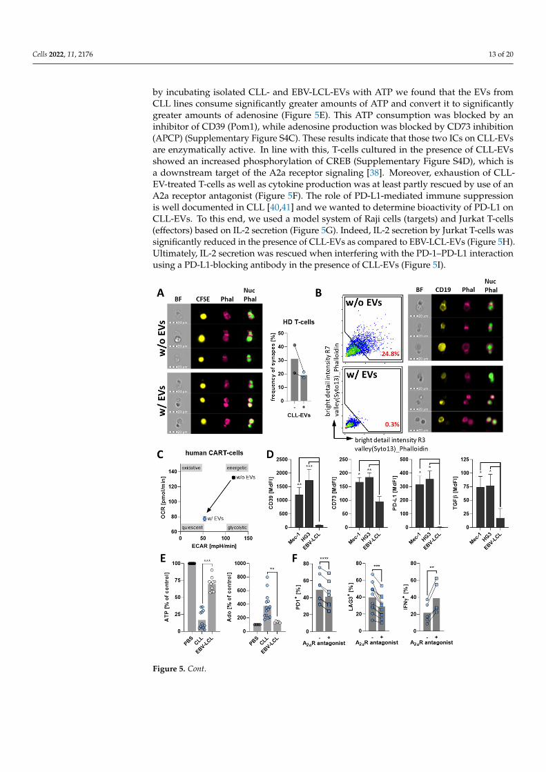
During our analyses to identify the underlying mechanisms, we found that CLL-EVs harbor a multifaceted palette of immunological checkpoints (ICs) including CD39,CD73, PD-L1, TGFβ, Gal9, and CD112 (Figure 5D, Supplementary Figure S4A,B). Alldetected ICs were significantly more abundant on CLL-EVs compared to EVs isolatedfrom EBV-LCL lines. CD39 and CD73 are two ectonucleotidases that convert ATP/ADPto AMP, and AMP to adenosine, respectively [37]. The latter has been already intensivelydescribed to be suppressive via the A2a receptor on the T-cell surface [38,39]. In fact,
Cells 2022, 11, 2176 13 of 20
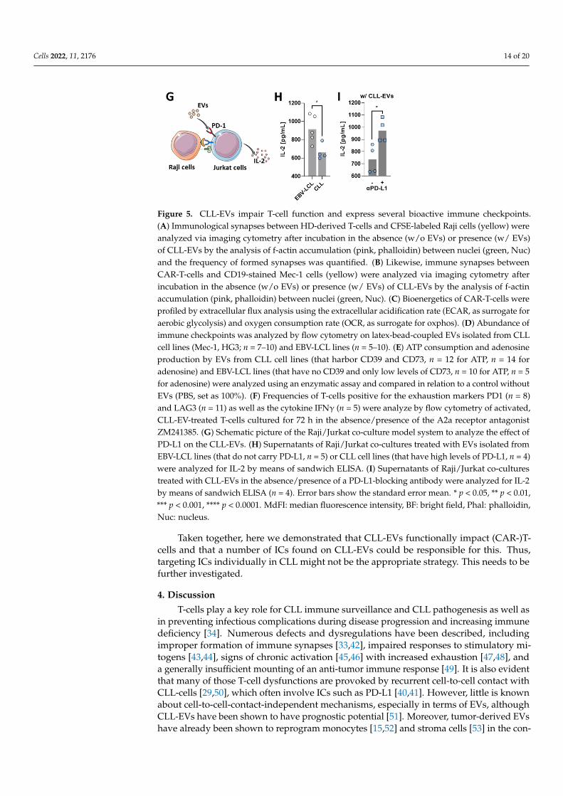
by incubating isolated CLL- and EBV-LCL-EVs with ATP we found that the EVs fromCLL lines consume significantly greater amounts of ATP and convert it to significantlygreater amounts of adenosine (Figure 5E). This ATP consumption was blocked by aninhibitor of CD39 (Pom1), while adenosine production was blocked by CD73 inhibition(APCP) (Supplementary Figure S4C). These results indicate that those two ICs on CLL-EVsare enzymatically active. In line with this, T-cells cultured in the presence of CLL-EVsshowed an increased phosphorylation of CREB (Supplementary Figure S4D), which isa downstream target of the A2a receptor signaling [38]. Moreover, exhaustion of CLL-EV-treated T-cells as well as cytokine production was at least partly rescued by use of anA2a receptor antagonist (Figure 5F). The role of PD-L1-mediated immune suppressionis well documented in CLL [40,41] and we wanted to determine bioactivity of PD-L1 onCLL-EVs. To this end, we used a model system of Raji cells (targets) and Jurkat T-cells(effectors) based on IL-2 secretion (Figure 5G). Indeed, IL-2 secretion by Jurkat T-cells wassignificantly reduced in the presence of CLL-EVs as compared to EBV-LCL-EVs (Figure 5H).Ultimately, IL-2 secretion was rescued when interfering with the PD-1–PD-L1 interactionusing a PD-L1-blocking antibody in the presence of CLL-EVs (Figure 5I).
Cells 2022, 11, x FOR PEER REVIEW 15 of 23
Figure 5. CLL-EVs impair T-cell function and express several bioactive immune checkpoints. (A) Immunological synapses between HD-derived T-cells and CFSE-labeled Raji cells (yellow) were an-alyzed via imaging cytometry after incubation in the absence (w/o EVs) or presence (w/ EVs) of CLL-EVs by the analysis of f-actin accumulation (pink, phalloidin) between nuclei (green, Nuc) and the frequency of formed synapses was quantified. (B) Likewise, immune synapses between CAR-T-cells and CD19-stained Mec-1 cells (yellow) were analyzed via imaging cytometry after incubation in the absence (w/o EVs) or presence (w/ EVs) of CLL-EVs by the analysis of f-actin accumulation (pink, phalloidin) between nuclei (green, Nuc). (C) Bioenergetics of CAR-T-cells were profiled by extracellular flux analysis using the extracellular acidification rate (ECAR, as surrogate for aerobic glycolysis) and oxygen consumption rate (OCR, as surrogate for oxphos). (D) Abundance of im-mune checkpoints was analyzed by flow cytometry on latex-bead-coupled EVs isolated from CLL cell lines (Mec-1, HG3; n = 7–10) and EBV-LCL lines (n = 5–10). (E) ATP consumption and adenosine production by EVs from CLL cell lines (that harbor CD39 and CD73, n = 12 for ATP, n = 14 for adenosine) and EBV-LCL lines (that have no CD39 and only low levels of CD73, n = 10 for ATP, n = 5 for adenosine) were analyzed using an enzymatic assay and compared in relation to a control
Figure 5. Cont.
Cells 2022, 11, 2176 14 of 20
Cells 2022, 11, x FOR PEER REVIEW 15 of 23
Figure 5. CLL-EVs impair T-cell function and express several bioactive immune checkpoints. (A) Immunological synapses between HD-derived T-cells and CFSE-labeled Raji cells (yellow) were an-alyzed via imaging cytometry after incubation in the absence (w/o EVs) or presence (w/ EVs) of CLL-EVs by the analysis of f-actin accumulation (pink, phalloidin) between nuclei (green, Nuc) and the frequency of formed synapses was quantified. (B) Likewise, immune synapses between CAR-T-cells and CD19-stained Mec-1 cells (yellow) were analyzed via imaging cytometry after incubation in the absence (w/o EVs) or presence (w/ EVs) of CLL-EVs by the analysis of f-actin accumulation (pink, phalloidin) between nuclei (green, Nuc). (C) Bioenergetics of CAR-T-cells were profiled by extracellular flux analysis using the extracellular acidification rate (ECAR, as surrogate for aerobic glycolysis) and oxygen consumption rate (OCR, as surrogate for oxphos). (D) Abundance of im-mune checkpoints was analyzed by flow cytometry on latex-bead-coupled EVs isolated from CLL cell lines (Mec-1, HG3; n = 7–10) and EBV-LCL lines (n = 5–10). (E) ATP consumption and adenosine production by EVs from CLL cell lines (that harbor CD39 and CD73, n = 12 for ATP, n = 14 for adenosine) and EBV-LCL lines (that have no CD39 and only low levels of CD73, n = 10 for ATP, n = 5 for adenosine) were analyzed using an enzymatic assay and compared in relation to a control
Figure 5. CLL-EVs impair T-cell function and express several bioactive immune checkpoints.(A) Immunological synapses between HD-derived T-cells and CFSE-labeled Raji cells (yellow) wereanalyzed via imaging cytometry after incubation in the absence (w/o EVs) or presence (w/ EVs)of CLL-EVs by the analysis of f-actin accumulation (pink, phalloidin) between nuclei (green, Nuc)and the frequency of formed synapses was quantified. (B) Likewise, immune synapses betweenCAR-T-cells and CD19-stained Mec-1 cells (yellow) were analyzed via imaging cytometry afterincubation in the absence (w/o EVs) or presence (w/ EVs) of CLL-EVs by the analysis of f-actinaccumulation (pink, phalloidin) between nuclei (green, Nuc). (C) Bioenergetics of CAR-T-cells wereprofiled by extracellular flux analysis using the extracellular acidification rate (ECAR, as surrogate foraerobic glycolysis) and oxygen consumption rate (OCR, as surrogate for oxphos). (D) Abundance ofimmune checkpoints was analyzed by flow cytometry on latex-bead-coupled EVs isolated from CLLcell lines (Mec-1, HG3; n = 7–10) and EBV-LCL lines (n = 5–10). (E) ATP consumption and adenosineproduction by EVs from CLL cell lines (that harbor CD39 and CD73, n = 12 for ATP, n = 14 foradenosine) and EBV-LCL lines (that have no CD39 and only low levels of CD73, n = 10 for ATP, n = 5for adenosine) were analyzed using an enzymatic assay and compared in relation to a control withoutEVs (PBS, set as 100%). (F) Frequencies of T-cells positive for the exhaustion markers PD1 (n = 8)and LAG3 (n = 11) as well as the cytokine IFNγ (n = 5) were analyze by flow cytometry of activated,CLL-EV-treated T-cells cultured for 72 h in the absence/presence of the A2a receptor antagonistZM241385. (G) Schematic picture of the Raji/Jurkat co-culture model system to analyze the effect ofPD-L1 on the CLL-EVs. (H) Supernatants of Raji/Jurkat co-cultures treated with EVs isolated fromEBV-LCL lines (that do not carry PD-L1, n = 5) or CLL cell lines (that have high levels of PD-L1, n = 4)were analyzed for IL-2 by means of sandwich ELISA. (I) Supernatants of Raji/Jurkat co-culturestreated with CLL-EVs in the absence/presence of a PD-L1-blocking antibody were analyzed for IL-2by means of sandwich ELISA (n = 4). Error bars show the standard error mean. * p < 0.05, ** p < 0.01,*** p < 0.001, **** p < 0.0001. MdFI: median fluorescence intensity, BF: bright field, Phal: phalloidin,Nuc: nucleus.
Taken together, here we demonstrated that CLL-EVs functionally impact (CAR-)T-cells and that a number of ICs found on CLL-EVs could be responsible for this. Thus,targeting ICs individually in CLL might not be the appropriate strategy. This needs to befurther investigated.
4. Discussion
T-cells play a key role for CLL immune surveillance and CLL pathogenesis as well asin preventing infectious complications during disease progression and increasing immunedeficiency [34]. Numerous defects and dysregulations have been described, includingimproper formation of immune synapses [33,42], impaired responses to stimulatory mi-togens [43,44], signs of chronic activation [45,46] with increased exhaustion [47,48], anda generally insufficient mounting of an anti-tumor immune response [49]. It is also evidentthat many of those T-cell dysfunctions are provoked by recurrent cell-to-cell contact withCLL-cells [29,50], which often involve ICs such as PD-L1 [40,41]. However, little is knownabout cell-to-cell-contact-independent mechanisms, especially in terms of EVs, althoughCLL-EVs have been shown to have prognostic potential [51]. Moreover, tumor-derived EVshave already been shown to reprogram monocytes [15,52] and stroma cells [53] in the con-
Cells 2022, 11, 2176 15 of 20
text of CLL. Especially in light of the emerging relevance of applying immunotherapies suchas CAR-T-cells in CLL [36], it is of utmost importance to further improve our understandingof the immunomodulatory role of CLL-EVs. Therefore, we performed a comprehensiveanalysis of effects mediated by CLL-EVs on T-cells, their functional consequences, and thepotential involvement of CLL-EV-carried ICs.
First, we confirmed the capability of T-cells to bind and to readily take up CLL-EVs, asalready indicated in earlier studies [12]. Although previous experiments by others concludethe importance of CLL-EV uptake [12], our data suggest that not only uptake but alsosurface interaction (possibly via ICs receptors, see below) is occurring. Unfortunately, wewere not able to delineate the balance between surface ligation and uptake of CLL-EVs onthe observed T-cell dysfunctions because available inhibitors for EV uptake are cytotoxic toT-cells over longer in vitro culture periods. Nevertheless, we were still able to show thata substantial amount of CLL-EVs was engulfed via endocytosis because 1) the frequencyof EV+ T-cells was reduced when including endocytotic inhibitors, and 2) the majority ofT-cells incubated with fluorescently labeled CLL-EVs still retained a strong fluorescentsignal even when surface-bound EVs were removed with trypsin prior to the analysis.Notably, EV attachment/uptake was most prominent when using activated T-cells, whichmight be caused by a general upregulation of surface receptors upon TCR stimulation andrequires further investigation.
The dual role of T-cells for CLL pathogenesis and development has been intensivelyreviewed recently [29,34]. T-cells in the tumor microenvironment seem to provide nurturingand/or survival signals to the CLL-cells, and their ability to mount an anti-tumor responseis limited. However, underlying mechanisms remain largely inconclusive. Our analysesreveal the ability of CLL-EVs to interfere with T-cell function on several levels similarto effects observed by EVs from other models/tumor entities (as recently reviewed [54]):(1) the viability and proliferation of T-cells are drastically reduced, (2) T-cell activationand effector cytokine secretion are diminished, (3) T-cell metabolism is shifted towardsa quiescent state with an according shift of the effector/memory subsets, and (4) T-cellexhaustion is fostered. Another hallmark in T-cell dysregulation of CLL is the accumulationof Treg subsets that correlate with poor prognosis of CLL patients [55,56]. Again, CLL-EVsmay be a driving factor as we showed an increase in regulatory T-cell subsets togetherwith the increased expression of inhibitory mediators upon presence of CLL-EVs. Whetherthe increased frequencies are a result of Treg induction or rely more on the preferentialcytotoxic effect on conventional T-cells remains speculative. However, CD4+ and CD8+
T-cells are similarly affected in terms of reduced viability by CLL-EVs, and CLL-EVs carrylarge amounts of TGFβ, which is known to induce Tregs [57]. Both findings favor thehypothesis of an active induction of Tregs by CLL-EVs, which certainly needs intensifiedinvestigation. Ultimately, we identified CLL-EVs as one factor causing improper formationof immune synapses, which is a known contributor to CLL immune escape [33].
Impaired immune surveillance, as observed in CLL, can be attributed to the aberrantexpression of IC receptors on CLL T-cells and the abundant expression of the correspondingligands on the CLL-cells [7]. In accordance with previous findings of the increased expres-sion of IC on CLL-cells, we found multiples of those ICs being carried by the CLL- EVs,which could explain the observed T-cell dysfunctionalities. Interestingly, ICs were previ-ously identified as a relevant promoter of immune synapse malformation [42]. In the latterstudy by Ramsay and colleagues and in contrast to our data, cell-to-cell contact betweenCLL-cells and T-cells was a prerequisite for dysfunctional immune synapses. However,since we measured large amounts of ICs on CLL-EVs it appears likely to us that CLL-EVscan mimic the parental CLL-cell surface in this regard resulting in comparable synapseanomalies. Additionally, the ectonucleotidases CD39 and CD73, which convert ATP/ADPto AMP and further to adenosine, are both highly expressed by CLL-cells, especially in theproliferating compartment [37]. Similarly, we found a high abundance of those two check-points on the CLL-EVs and could confirm their enzymatic activity. This is of particularinterest as adenosine in CLL has a dual-role: (1) it supports and protects the CLL-cells [58]
Cells 2022, 11, 2176 16 of 20
and (2) it impairs T-cell function [59]. In the latter case, different modes of action werefound, including reduced cytokine expression [60], impaired T-cell metabolism [38], induc-tion of Tregs [61], and induction of T-cell exhaustion [62]. Thus, it is not surprising that allthe phenomena that were previously observed for adenosine on T-cells were also evidentin T-cells that we treated with CLL-EVs. Additionally, the role of PD-L1 as an IC in thecontext of T-cell inhibition and anti-tumor therapy is indisputable [63,64]. PD-L1 expressedby CLL-cells can inhibit T-cell activation [41], suppress the secretion of effector cytokines byT-cells [65], enhance T-cell exhaustion, and impair the formation of immune synapses [40].Again, those effects could also be observed in the presence of PD-L1-harboring CLL-EVs.However, to date, single anti-PD1/PD-L1 treatment in CLL therapy has an unsuccessfulhistory [66,67]. So far, the reason for the unresponsiveness towards a blockade of thePD-1/PD-L1 axis in CLL, albeit promising results from preclinical models [40,65], is vastlyunknown. In fact, our data, which of course requires experimental verification, couldcontribute to speculation regarding the possible causes: the great variance of ICs such asthe aforementioned CD39/CD73 and TGFβ but also galectin-9 (a ligand for TIM-3 andPD-1 [68]) and CD112 (a ligand for TIGIT [69]) may compensate for the blockade/inhibitionof one single IC (such as PD-L1). Many downstream effects of those co-inhibitory signalsare shared and their concerted action may act in an additive or even synergistic manner.This notion is corroborated by studies demonstrating that the dual blockade of PD-1 andLAG3 ICs could limit CLL development and restore immune competence while an in-dividual IC-targeting was inefficient [70]. Moreover, the high abundance and constantsecretion of EVs (that harbor PD-L1) in CLL may simply outcompete the blocking activityof anti-PD-1/PD-L1 antibodies. In fact, acquired resistance to anti-PD-1/PD-L1 therapy byPD-L1-carrying EVs has already been demonstrated in several solid-tumor entities [71] andit is speculated that PD-L1 presented by EVs may preferentially bind immunotherapeuticantibodies and thereby act as a “sink” to divert them away from the tumor cells [72]. Thisargument is strengthened by the fact that the Bruton tyrosine kinase inhibitor ibrutinibis able to reduce secretion of CLL-EVs [11] and at the same time to increase efficacy ofPD-1/PD-L1 blockade [73,74]. Currently a combination of ibrutinib and PD-1 blockade isbeing evaluated within a phase II clinical trial in CLL patients [75].
Importantly, although CD19-targeting CAR-T-cell therapies in CLL show promisingresponse rates [76], long-term results remain limited [77,78]. Key mechanisms responsiblefor insufficient CAR-T-cell efficacy include loss of the target antigen [79], T-cell defects [80],and an immunosuppressive tumor microenvironment [81]. In addition, CAR-T-cells pro-duced from CLL patients show increased signs of exhaustion compared to those derivedfrom healthy donors before infusion [82]. In particular, those patients whose CAR-T-cells expressed multiple inhibitory receptors such as PD-1, TIM-3, and LAG-3 presentedlower responses [78]. A recent study identified EVs from CLL patient plasma to be ca-pable of impairing antigen-specific proliferation and killing, and of inducing CAR-T-cellexhaustion [17], which we could confirm as well. Moreover, our analyses append theCLL-EV-mediated repertoire of CAR-T-cell dysfunction by the almost absent formation ofimmune synapses and the drastically reduced cellular metabolism. To sum up, CAR-T-celltherapy in CLL could be affected by CLL-EVs in two ways: (1) the CLL T-cells’ lack offunctional competence has a substantial impact on the final CAR-T-cell product [82], and(2) re-infused CAR-T-cells again have to face immunosuppressive CLL-EVs.
5. Conclusions
In conclusion, we showed that CLL-EVs can be a mediator of T-cell dysfunctionin CLL. In fact, CLL-EVs carry multiple ICs as a potential further tool for promotingan immune-privileged environment mediated by CLL. Moreover, various studies indi-cate that targeting individual ICs in CLL might not be sufficient to reach meaningfulclinical effects. The numerous ICs on CLL-EVs could be one of the underlying causes(over-)compensating the blockade of a single IC by their coordinated and combined activity.Therefore, our findings could pave the way for novel therapeutic strategies in CLL, such as
Cells 2022, 11, 2176 17 of 20
the simultaneous blockade of multiple ICs or the targeting of EV production and secretionby CLL-cells. A better understanding of immune-suppressive strategies could help to fosterintrinsic anti-tumor immunity and increase the efficacy of adoptive cell therapies, mostimportantly CAR-T-cells.
Supplementary Materials: The following supporting information can be downloaded at: https://www.mdpi.com/article/10.3390/cells11142176/s1. Figure S1: Supportive data for Figure 1.Figure S2: Bioenergetics of CLL-EV-treated T-cells. Figure S3: Effects of CLL-EVs on CAR-T-cells.Figure S4: Immune checkpoint activity on CLL-EVs. Table S1: Experimental inhibitors. Table S2:Antibodies for flow cytometry and fluorescence microscopy.
Author Contributions: Conceptualization, D.M.; methodology, M.B., R.B.-L., A.G., U.S.-S., S.K., M.A.,A.M. and H.B.; formal analysis, M.B. and R.B.-L.; investigation, M.B., R.B.-L. and A.G.; writing—original draft preparation, M.B.; writing—review and editing, D.M. and T.T.; visualization, M.B. andD.M.; project administration, M.B. and R.B.-L.; funding acquisition, D.M. All authors have read andagreed to the published version of the manuscript.
Funding: This research was funded by Deutsche Forschungsgemeinschaft (DFG), grant number404074532.
Institutional Review Board Statement: The study was conducted in accordance with the Declarationof Helsinki, and approved by the Ethics Committee of the Friedrich-Alexander University Erlangen-Nuremberg (approval number 291_14B and 289_16B).
Informed Consent Statement: Informed consent was obtained from all subjects involved in the study.
Data Availability Statement: Original data will be made available by the corresponding author uponreasonable request.
Acknowledgments: We thank Domenica Saul, Kristin Mentz, Samantha-Josefine Popp, and StephanieAdam-Frey for excellent technical support. The graphical abstract was prepared with biorender.com.
Conflicts of Interest: The authors declare no conflict of interest.
References1. Hallek, M. Chronic lymphocytic leukemia: 2020 update on diagnosis, risk stratification and treatment. Am. J. Hematol. 2019, 94,
1266–1287. [CrossRef] [PubMed]2. Chiorazzi, N.; Rai, K.R.; Ferrarini, M. Chronic lymphocytic leukemia. N. Engl. J. Med. 2005, 352, 804–815. [CrossRef] [PubMed]3. Ten Hacken, E.; Burger, J.A. Microenvironment interactions and B-cell receptor signaling in Chronic Lymphocytic Leukemia:
Implications for disease pathogenesis and treatment. Biochim. Biophys. Acta 2016, 1863, 401–413. [CrossRef] [PubMed]4. Bewarder, M.; Stilgenbauer, S.; Thurner, L.; Kaddu-Mulindwa, D. Current treatment options in CLL. Cancers 2021, 13, 2468.
[CrossRef] [PubMed]5. Wiedmeier-Nutor, J.; Leis, J. Chronic lymphocytic leukemia: Chemotherapy free and other novel therapies including CAR T.
Curr. Treat. Options Oncol. 2022, 23, 904–919. [CrossRef]6. Os, A.; Burgler, S.; Ribes, A.P.; Funderud, A.; Wang, D.; Thompson, K.M.; Tjonnfjord, G.E.; Bogen, B.; Munthe, L.A. Chronic
lymphocytic leukemia cells are activated and proliferate in response to specific T helper cells. Cell Rep. 2013, 4, 566–577. [CrossRef]7. Griggio, V.; Perutelli, F.; Salvetti, C.; Boccellato, E.; Boccadoro, M.; Vitale, C.; Coscia, M. Immune dysfunctions and immune-based
therapeutic interventions in chronic lymphocytic leukemia. Front. Immunol. 2020, 11, 594556. [CrossRef]8. Abels, E.R.; Breakefield, X.O. Introduction to extracellular vesicles: Biogenesis, RNA cargo selection, content, release, and uptake.
Cell Mol. Neurobiol. 2016, 36, 301–312. [CrossRef]9. Robbins, P.D.; Morelli, A.E. Regulation of immune responses by extracellular vesicles. Nat. Rev. Immunol. 2014, 14, 195–208. [CrossRef]10. Lotvall, J.; Hill, A.F.; Hochberg, F.; Buzas, E.I.; Di Vizio, D.; Gardiner, C.; Gho, Y.S.; Kurochkin, I.V.; Mathivanan, S.; Quesenberry,
P.; et al. Minimal experimental requirements for definition of extracellular vesicles and their functions: A position statement fromthe International Society for Extracellular Vesicles. J. Extracell Vesicles 2014, 3, 26913. [CrossRef]
11. Yeh, Y.Y.; Ozer, H.G.; Lehman, A.M.; Maddocks, K.; Yu, L.; Johnson, A.J.; Byrd, J.C. Characterization of CLL exosomes revealsa distinct microRNA signature and enhanced secretion by activation of BCR signaling. Blood 2015, 125, 3297–3305. [CrossRef]
12. Smallwood, D.T.; Apollonio, B.; Willimott, S.; Lezina, L.; Alharthi, A.; Ambrose, A.R.; De Rossi, G.; Ramsay, A.G.; Wagner, S.D.Extracellular vesicles released by CD40/IL-4-stimulated CLL cells confer altered functional properties to CD4+ T cells. Blood 2016,128, 542–552. [CrossRef] [PubMed]
13. Trino, S.; Lamorte, D.; Caivano, A.; De Luca, L.; Sgambato, A.; Laurenzana, I. Clinical relevance of extracellular vesicles inhematological neoplasms: From liquid biopsy to cell biopsy. Leukemia 2021, 35, 661–678. [CrossRef] [PubMed]
Cells 2022, 11, 2176 18 of 20
14. Reiners, K.S.; Shatnyeva, O.; Vasyutina, E.; Bosl, T.; Hansen, H.P.; Hallek, M.; Herling, M.; von Strandmann, E.P. Extracellularvesicles released from chronic lymphocytic leukemia cells exhibit a disease relevant mRNA signature and transfer mRNA tobystander cells. Haematologica 2017, 102, e100–e103. [CrossRef] [PubMed]
15. Bruns, H.; Bottcher, M.; Qorraj, M.; Fabri, M.; Jitschin, S.; Dindorf, J.; Busch, L.; Jitschin, R.; Mackensen, A.; Mougiakakos, D. CLL-cell-mediated MDSC induction by exosomal miR-155 transfer is disrupted by vitamin D. Leukemia 2017, 31, 985–988. [CrossRef]
16. Dubois, N.; Crompot, E.; Meuleman, N.; Bron, D.; Lagneaux, L.; Stamatopoulos, B. Importance of crosstalk between chroniclymphocytic leukemia cells and the stromal microenvironment: Direct contact, soluble factors, and extracellular vesicles.Front. Oncol. 2020, 10, 1422. [CrossRef]
17. Cox, M.J.; Lucien, F.; Sakemura, R.; Boysen, J.C.; Kim, Y.; Horvei, P.; Manriquez Roman, C.; Hansen, M.J.; Tapper, E.E.;Siegler, E.L.; et al. Leukemic extracellular vesicles induce chimeric antigen receptor T cell dysfunction in chronic lymphocyticleukemia. Mol. Ther. 2021, 29, 1529–1540. [CrossRef]
18. Thery, C.; Amigorena, S.; Raposo, G.; Clayton, A. Isolation and characterization of exosomes from cell culture supernatants andbiological fluids. Curr. Protoc. Cell Biol. 2006, 30, 3.22.1–3.22.29. [CrossRef]
19. Bottcher, M.; Renner, K.; Berger, R.; Mentz, K.; Thomas, S.; Cardenas-Conejo, Z.E.; Dettmer, K.; Oefner, P.J.; Mackensen, A.;Kreutz, M.; et al. D-2-hydroxyglutarate interferes with HIF-1alpha stability skewing T-cell metabolism towards oxidativephosphorylation and impairing Th17 polarization. Oncoimmunology 2018, 7, e1445454. [CrossRef]
20. Arguello, R.J.; Combes, A.J.; Char, R.; Gigan, J.P.; Baaziz, A.I.; Bousiquot, E.; Camosseto, V.; Samad, B.; Tsui, J.; Yan, P.; et al.SCENITH: A flow cytometry-based method to functionally profile energy metabolism with single-cell resolution. Cell Metab.2020, 32, 1063–1075.e7. [CrossRef]
21. Mulcahy, L.A.; Pink, R.C.; Carter, D.R. Routes and mechanisms of extracellular vesicle uptake. J. Extracell Vesicles 2014, 3,24641. [CrossRef]
22. Buck, M.D.; O’Sullivan, D.; Pearce, E.L. T cell metabolism drives immunity. J. Exp. Med. 2015, 212, 1345–1360. [CrossRef] [PubMed]23. Geltink, R.I.K.; Kyle, R.L.; Pearce, E.L. Unraveling the complex interplay between T cell metabolism and function. Annu. Rev.
Immunol. 2018, 36, 461–488. [CrossRef]24. Saravia, J.; Raynor, J.L.; Chapman, N.M.; Lim, S.A.; Chi, H. Signaling networks in immunometabolism. Cell Res. 2020, 30, 328–342.
[CrossRef] [PubMed]25. O’Sullivan, D. The metabolic spectrum of memory T cells. Immunol. Cell Biol. 2019, 97, 636–646. [CrossRef] [PubMed]26. Mahnke, Y.D.; Brodie, T.M.; Sallusto, F.; Roederer, M.; Lugli, E. The who’s who of T-cell differentiation: Human memory T-cell
subsets. Eur. J. Immunol. 2013, 43, 2797–2809. [CrossRef]27. Franco, F.; Jaccard, A.; Romero, P.; Yu, Y.R.; Ho, P.C. Metabolic and epigenetic regulation of T-cell exhaustion. Nat. Metab. 2020, 2,
1001–1012. [CrossRef]28. Coe, D.J.; Kishore, M.; Marelli-Berg, F. Metabolic regulation of regulatory T cell development and function. Front. Immunol. 2014,
5, 590. [CrossRef]29. Roessner, P.M.; Seiffert, M. T-cells in chronic lymphocytic leukemia: Guardians or drivers of disease? Leukemia 2020, 34,
2012–2024. [CrossRef]30. Beltra, J.C.; Manne, S.; Abdel-Hakeem, M.S.; Kurachi, M.; Giles, J.R.; Chen, Z.; Casella, V.; Ngiow, S.F.; Khan, O.; Huang, Y.J.; et al.
Developmental relationships of four exhausted CD8(+) T cell subsets reveals underlying transcriptional and epigenetic landscapecontrol mechanisms. Immunity 2020, 52, 825–841. [CrossRef]
31. Gao, R.; Shi, G.P.; Wang, J. Functional diversities of regulatory T cells in the context of cancer immunotherapy. Front. Immunol.2022, 13, 833667. [CrossRef] [PubMed]
32. Gagliani, N.; Magnani, C.F.; Huber, S.; Gianolini, M.E.; Pala, M.; Licona-Limon, P.; Guo, B.; Herbert, D.R.; Bulfone, A.; Trentini, F.;et al. Coexpression of CD49b and LAG-3 identifies human and mouse T regulatory type 1 cells. Nat. Med. 2013, 19, 739–746.[CrossRef] [PubMed]
33. Ramsay, A.G.; Johnson, A.J.; Lee, A.M.; Gorgun, G.; Le Dieu, R.; Blum, W.; Byrd, J.C.; Gribben, J.G. Chronic lymphocytic leukemiaT cells show impaired immunological synapse formation that can be reversed with an immunomodulating drug. J. Clin. Investig.2008, 118, 2427–2437. [CrossRef]
34. Vlachonikola, E.; Stamatopoulos, K.; Chatzidimitriou, A. T cells in chronic lymphocytic leukemia: A two-edged sword.Front. Immunol. 2020, 11, 612244. [CrossRef] [PubMed]
35. Dustin, M.L.; Cooper, J.A. The immunological synapse and the actin cytoskeleton: Molecular hardware for T cell signaling.Nat. Immunol. 2000, 1, 23–29. [CrossRef]
36. Mancikova, V.; Smida, M. Current State of CAR T-cell therapy in chronic lymphocytic leukemia. Int. J. Mol. Sci. 2021, 22,5536. [CrossRef]
37. Vaisitti, T.; Arruga, F.; Guerra, G.; Deaglio, S. Ectonucleotidases in blood malignancies: A tale of surface markers and therapeutictargets. Front. Immunol. 2019, 10, 2301. [CrossRef]
38. Mastelic-Gavillet, B.; Navarro Rodrigo, B.; Decombaz, L.; Wang, H.; Ercolano, G.; Ahmed, R.; Lozano, L.E.; Ianaro, A.; Derre, L.;Valerio, M.; et al. Adenosine mediates functional and metabolic suppression of peripheral and tumor-infiltrating CD8(+) T cells.J. Immunother. Cancer 2019, 7, 257. [CrossRef]
39. Allard, B.; Allard, D.; Buisseret, L.; Stagg, J. The adenosine pathway in immuno-oncology. Nat. Rev. Clin. Oncol. 2020, 17,611–629. [CrossRef]
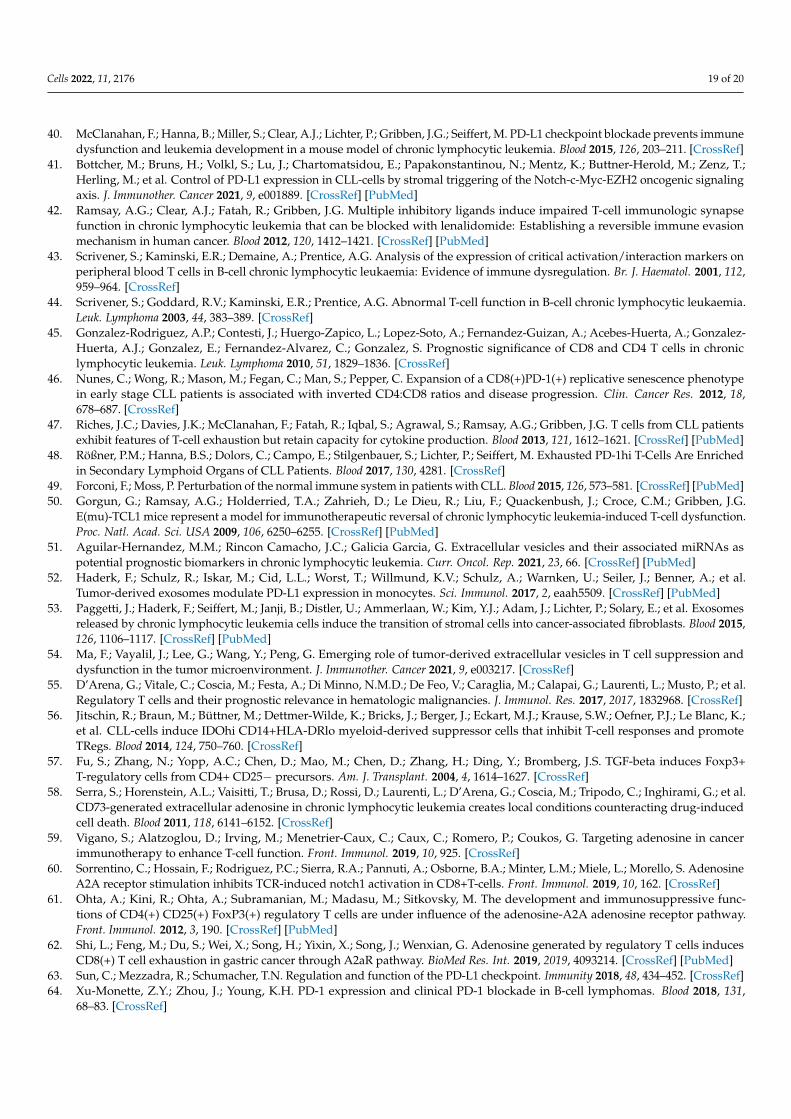
Cells 2022, 11, 2176 19 of 20
40. McClanahan, F.; Hanna, B.; Miller, S.; Clear, A.J.; Lichter, P.; Gribben, J.G.; Seiffert, M. PD-L1 checkpoint blockade prevents immunedysfunction and leukemia development in a mouse model of chronic lymphocytic leukemia. Blood 2015, 126, 203–211. [CrossRef]
41. Bottcher, M.; Bruns, H.; Volkl, S.; Lu, J.; Chartomatsidou, E.; Papakonstantinou, N.; Mentz, K.; Buttner-Herold, M.; Zenz, T.;Herling, M.; et al. Control of PD-L1 expression in CLL-cells by stromal triggering of the Notch-c-Myc-EZH2 oncogenic signalingaxis. J. Immunother. Cancer 2021, 9, e001889. [CrossRef] [PubMed]
42. Ramsay, A.G.; Clear, A.J.; Fatah, R.; Gribben, J.G. Multiple inhibitory ligands induce impaired T-cell immunologic synapsefunction in chronic lymphocytic leukemia that can be blocked with lenalidomide: Establishing a reversible immune evasionmechanism in human cancer. Blood 2012, 120, 1412–1421. [CrossRef] [PubMed]
43. Scrivener, S.; Kaminski, E.R.; Demaine, A.; Prentice, A.G. Analysis of the expression of critical activation/interaction markers onperipheral blood T cells in B-cell chronic lymphocytic leukaemia: Evidence of immune dysregulation. Br. J. Haematol. 2001, 112,959–964. [CrossRef]
44. Scrivener, S.; Goddard, R.V.; Kaminski, E.R.; Prentice, A.G. Abnormal T-cell function in B-cell chronic lymphocytic leukaemia.Leuk. Lymphoma 2003, 44, 383–389. [CrossRef]
45. Gonzalez-Rodriguez, A.P.; Contesti, J.; Huergo-Zapico, L.; Lopez-Soto, A.; Fernandez-Guizan, A.; Acebes-Huerta, A.; Gonzalez-Huerta, A.J.; Gonzalez, E.; Fernandez-Alvarez, C.; Gonzalez, S. Prognostic significance of CD8 and CD4 T cells in chroniclymphocytic leukemia. Leuk. Lymphoma 2010, 51, 1829–1836. [CrossRef]
46. Nunes, C.; Wong, R.; Mason, M.; Fegan, C.; Man, S.; Pepper, C. Expansion of a CD8(+)PD-1(+) replicative senescence phenotypein early stage CLL patients is associated with inverted CD4:CD8 ratios and disease progression. Clin. Cancer Res. 2012, 18,678–687. [CrossRef]
47. Riches, J.C.; Davies, J.K.; McClanahan, F.; Fatah, R.; Iqbal, S.; Agrawal, S.; Ramsay, A.G.; Gribben, J.G. T cells from CLL patientsexhibit features of T-cell exhaustion but retain capacity for cytokine production. Blood 2013, 121, 1612–1621. [CrossRef] [PubMed]
48. Rößner, P.M.; Hanna, B.S.; Dolors, C.; Campo, E.; Stilgenbauer, S.; Lichter, P.; Seiffert, M. Exhausted PD-1hi T-Cells Are Enrichedin Secondary Lymphoid Organs of CLL Patients. Blood 2017, 130, 4281. [CrossRef]
49. Forconi, F.; Moss, P. Perturbation of the normal immune system in patients with CLL. Blood 2015, 126, 573–581. [CrossRef] [PubMed]50. Gorgun, G.; Ramsay, A.G.; Holderried, T.A.; Zahrieh, D.; Le Dieu, R.; Liu, F.; Quackenbush, J.; Croce, C.M.; Gribben, J.G.
E(mu)-TCL1 mice represent a model for immunotherapeutic reversal of chronic lymphocytic leukemia-induced T-cell dysfunction.Proc. Natl. Acad. Sci. USA 2009, 106, 6250–6255. [CrossRef] [PubMed]
51. Aguilar-Hernandez, M.M.; Rincon Camacho, J.C.; Galicia Garcia, G. Extracellular vesicles and their associated miRNAs aspotential prognostic biomarkers in chronic lymphocytic leukemia. Curr. Oncol. Rep. 2021, 23, 66. [CrossRef] [PubMed]
52. Haderk, F.; Schulz, R.; Iskar, M.; Cid, L.L.; Worst, T.; Willmund, K.V.; Schulz, A.; Warnken, U.; Seiler, J.; Benner, A.; et al.Tumor-derived exosomes modulate PD-L1 expression in monocytes. Sci. Immunol. 2017, 2, eaah5509. [CrossRef] [PubMed]
53. Paggetti, J.; Haderk, F.; Seiffert, M.; Janji, B.; Distler, U.; Ammerlaan, W.; Kim, Y.J.; Adam, J.; Lichter, P.; Solary, E.; et al. Exosomesreleased by chronic lymphocytic leukemia cells induce the transition of stromal cells into cancer-associated fibroblasts. Blood 2015,126, 1106–1117. [CrossRef] [PubMed]
54. Ma, F.; Vayalil, J.; Lee, G.; Wang, Y.; Peng, G. Emerging role of tumor-derived extracellular vesicles in T cell suppression anddysfunction in the tumor microenvironment. J. Immunother. Cancer 2021, 9, e003217. [CrossRef]
55. D’Arena, G.; Vitale, C.; Coscia, M.; Festa, A.; Di Minno, N.M.D.; De Feo, V.; Caraglia, M.; Calapai, G.; Laurenti, L.; Musto, P.; et al.Regulatory T cells and their prognostic relevance in hematologic malignancies. J. Immunol. Res. 2017, 2017, 1832968. [CrossRef]
56. Jitschin, R.; Braun, M.; Büttner, M.; Dettmer-Wilde, K.; Bricks, J.; Berger, J.; Eckart, M.J.; Krause, S.W.; Oefner, P.J.; Le Blanc, K.;et al. CLL-cells induce IDOhi CD14+HLA-DRlo myeloid-derived suppressor cells that inhibit T-cell responses and promoteTRegs. Blood 2014, 124, 750–760. [CrossRef]
57. Fu, S.; Zhang, N.; Yopp, A.C.; Chen, D.; Mao, M.; Chen, D.; Zhang, H.; Ding, Y.; Bromberg, J.S. TGF-beta induces Foxp3+T-regulatory cells from CD4+ CD25− precursors. Am. J. Transplant. 2004, 4, 1614–1627. [CrossRef]
58. Serra, S.; Horenstein, A.L.; Vaisitti, T.; Brusa, D.; Rossi, D.; Laurenti, L.; D’Arena, G.; Coscia, M.; Tripodo, C.; Inghirami, G.; et al.CD73-generated extracellular adenosine in chronic lymphocytic leukemia creates local conditions counteracting drug-inducedcell death. Blood 2011, 118, 6141–6152. [CrossRef]
59. Vigano, S.; Alatzoglou, D.; Irving, M.; Menetrier-Caux, C.; Caux, C.; Romero, P.; Coukos, G. Targeting adenosine in cancerimmunotherapy to enhance T-cell function. Front. Immunol. 2019, 10, 925. [CrossRef]
60. Sorrentino, C.; Hossain, F.; Rodriguez, P.C.; Sierra, R.A.; Pannuti, A.; Osborne, B.A.; Minter, L.M.; Miele, L.; Morello, S. AdenosineA2A receptor stimulation inhibits TCR-induced notch1 activation in CD8+T-cells. Front. Immunol. 2019, 10, 162. [CrossRef]
61. Ohta, A.; Kini, R.; Ohta, A.; Subramanian, M.; Madasu, M.; Sitkovsky, M. The development and immunosuppressive func-tions of CD4(+) CD25(+) FoxP3(+) regulatory T cells are under influence of the adenosine-A2A adenosine receptor pathway.Front. Immunol. 2012, 3, 190. [CrossRef] [PubMed]
62. Shi, L.; Feng, M.; Du, S.; Wei, X.; Song, H.; Yixin, X.; Song, J.; Wenxian, G. Adenosine generated by regulatory T cells inducesCD8(+) T cell exhaustion in gastric cancer through A2aR pathway. BioMed Res. Int. 2019, 2019, 4093214. [CrossRef] [PubMed]
63. Sun, C.; Mezzadra, R.; Schumacher, T.N. Regulation and function of the PD-L1 checkpoint. Immunity 2018, 48, 434–452. [CrossRef]64. Xu-Monette, Z.Y.; Zhou, J.; Young, K.H. PD-1 expression and clinical PD-1 blockade in B-cell lymphomas. Blood 2018, 131,
68–83. [CrossRef]
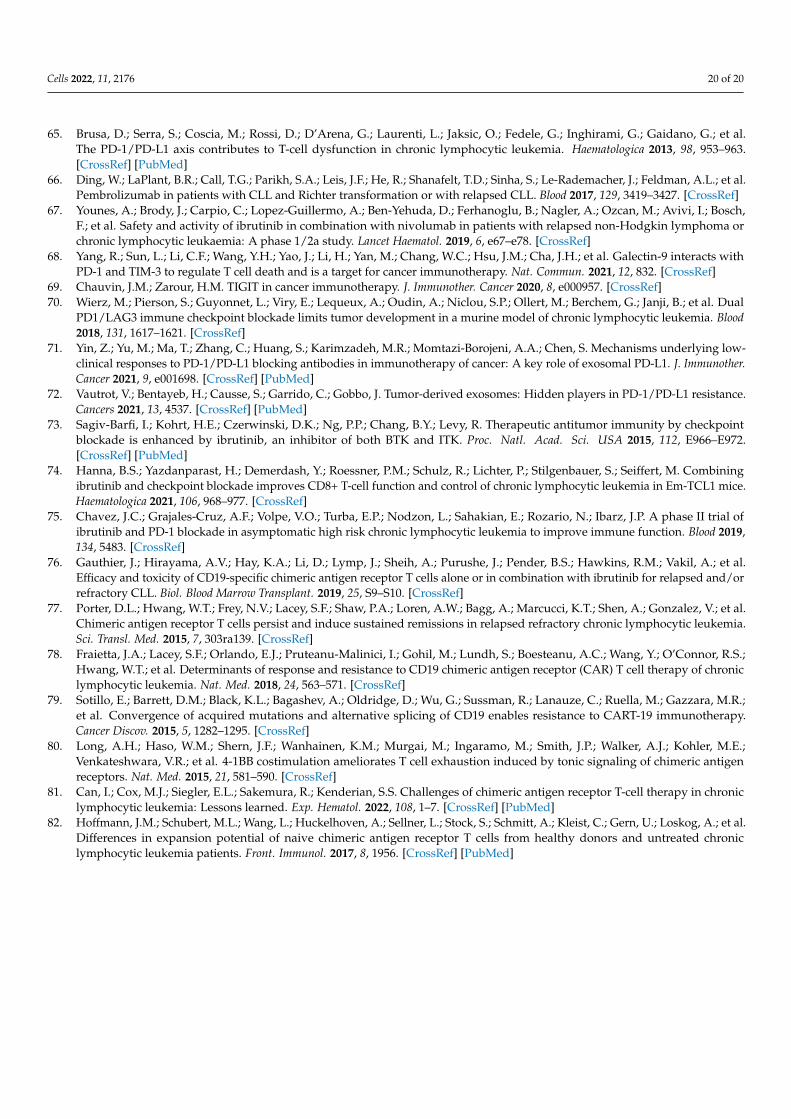
Cells 2022, 11, 2176 20 of 20
65. Brusa, D.; Serra, S.; Coscia, M.; Rossi, D.; D’Arena, G.; Laurenti, L.; Jaksic, O.; Fedele, G.; Inghirami, G.; Gaidano, G.; et al.The PD-1/PD-L1 axis contributes to T-cell dysfunction in chronic lymphocytic leukemia. Haematologica 2013, 98, 953–963.[CrossRef] [PubMed]
66. Ding, W.; LaPlant, B.R.; Call, T.G.; Parikh, S.A.; Leis, J.F.; He, R.; Shanafelt, T.D.; Sinha, S.; Le-Rademacher, J.; Feldman, A.L.; et al.Pembrolizumab in patients with CLL and Richter transformation or with relapsed CLL. Blood 2017, 129, 3419–3427. [CrossRef]
67. Younes, A.; Brody, J.; Carpio, C.; Lopez-Guillermo, A.; Ben-Yehuda, D.; Ferhanoglu, B.; Nagler, A.; Ozcan, M.; Avivi, I.; Bosch,F.; et al. Safety and activity of ibrutinib in combination with nivolumab in patients with relapsed non-Hodgkin lymphoma orchronic lymphocytic leukaemia: A phase 1/2a study. Lancet Haematol. 2019, 6, e67–e78. [CrossRef]
68. Yang, R.; Sun, L.; Li, C.F.; Wang, Y.H.; Yao, J.; Li, H.; Yan, M.; Chang, W.C.; Hsu, J.M.; Cha, J.H.; et al. Galectin-9 interacts withPD-1 and TIM-3 to regulate T cell death and is a target for cancer immunotherapy. Nat. Commun. 2021, 12, 832. [CrossRef]
69. Chauvin, J.M.; Zarour, H.M. TIGIT in cancer immunotherapy. J. Immunother. Cancer 2020, 8, e000957. [CrossRef]70. Wierz, M.; Pierson, S.; Guyonnet, L.; Viry, E.; Lequeux, A.; Oudin, A.; Niclou, S.P.; Ollert, M.; Berchem, G.; Janji, B.; et al. Dual
PD1/LAG3 immune checkpoint blockade limits tumor development in a murine model of chronic lymphocytic leukemia. Blood2018, 131, 1617–1621. [CrossRef]
71. Yin, Z.; Yu, M.; Ma, T.; Zhang, C.; Huang, S.; Karimzadeh, M.R.; Momtazi-Borojeni, A.A.; Chen, S. Mechanisms underlying low-clinical responses to PD-1/PD-L1 blocking antibodies in immunotherapy of cancer: A key role of exosomal PD-L1. J. Immunother.Cancer 2021, 9, e001698. [CrossRef] [PubMed]
72. Vautrot, V.; Bentayeb, H.; Causse, S.; Garrido, C.; Gobbo, J. Tumor-derived exosomes: Hidden players in PD-1/PD-L1 resistance.Cancers 2021, 13, 4537. [CrossRef] [PubMed]
73. Sagiv-Barfi, I.; Kohrt, H.E.; Czerwinski, D.K.; Ng, P.P.; Chang, B.Y.; Levy, R. Therapeutic antitumor immunity by checkpointblockade is enhanced by ibrutinib, an inhibitor of both BTK and ITK. Proc. Natl. Acad. Sci. USA 2015, 112, E966–E972.[CrossRef] [PubMed]
74. Hanna, B.S.; Yazdanparast, H.; Demerdash, Y.; Roessner, P.M.; Schulz, R.; Lichter, P.; Stilgenbauer, S.; Seiffert, M. Combiningibrutinib and checkpoint blockade improves CD8+ T-cell function and control of chronic lymphocytic leukemia in Em-TCL1 mice.Haematologica 2021, 106, 968–977. [CrossRef]
75. Chavez, J.C.; Grajales-Cruz, A.F.; Volpe, V.O.; Turba, E.P.; Nodzon, L.; Sahakian, E.; Rozario, N.; Ibarz, J.P. A phase II trial ofibrutinib and PD-1 blockade in asymptomatic high risk chronic lymphocytic leukemia to improve immune function. Blood 2019,134, 5483. [CrossRef]
76. Gauthier, J.; Hirayama, A.V.; Hay, K.A.; Li, D.; Lymp, J.; Sheih, A.; Purushe, J.; Pender, B.S.; Hawkins, R.M.; Vakil, A.; et al.Efficacy and toxicity of CD19-specific chimeric antigen receptor T cells alone or in combination with ibrutinib for relapsed and/orrefractory CLL. Biol. Blood Marrow Transplant. 2019, 25, S9–S10. [CrossRef]
77. Porter, D.L.; Hwang, W.T.; Frey, N.V.; Lacey, S.F.; Shaw, P.A.; Loren, A.W.; Bagg, A.; Marcucci, K.T.; Shen, A.; Gonzalez, V.; et al.Chimeric antigen receptor T cells persist and induce sustained remissions in relapsed refractory chronic lymphocytic leukemia.Sci. Transl. Med. 2015, 7, 303ra139. [CrossRef]
78. Fraietta, J.A.; Lacey, S.F.; Orlando, E.J.; Pruteanu-Malinici, I.; Gohil, M.; Lundh, S.; Boesteanu, A.C.; Wang, Y.; O’Connor, R.S.;Hwang, W.T.; et al. Determinants of response and resistance to CD19 chimeric antigen receptor (CAR) T cell therapy of chroniclymphocytic leukemia. Nat. Med. 2018, 24, 563–571. [CrossRef]
79. Sotillo, E.; Barrett, D.M.; Black, K.L.; Bagashev, A.; Oldridge, D.; Wu, G.; Sussman, R.; Lanauze, C.; Ruella, M.; Gazzara, M.R.;et al. Convergence of acquired mutations and alternative splicing of CD19 enables resistance to CART-19 immunotherapy.Cancer Discov. 2015, 5, 1282–1295. [CrossRef]
80. Long, A.H.; Haso, W.M.; Shern, J.F.; Wanhainen, K.M.; Murgai, M.; Ingaramo, M.; Smith, J.P.; Walker, A.J.; Kohler, M.E.;Venkateshwara, V.R.; et al. 4-1BB costimulation ameliorates T cell exhaustion induced by tonic signaling of chimeric antigenreceptors. Nat. Med. 2015, 21, 581–590. [CrossRef]
81. Can, I.; Cox, M.J.; Siegler, E.L.; Sakemura, R.; Kenderian, S.S. Challenges of chimeric antigen receptor T-cell therapy in chroniclymphocytic leukemia: Lessons learned. Exp. Hematol. 2022, 108, 1–7. [CrossRef] [PubMed]
82. Hoffmann, J.M.; Schubert, M.L.; Wang, L.; Huckelhoven, A.; Sellner, L.; Stock, S.; Schmitt, A.; Kleist, C.; Gern, U.; Loskog, A.; et al.Differences in expansion potential of naive chimeric antigen receptor T cells from healthy donors and untreated chroniclymphocytic leukemia patients. Front. Immunol. 2017, 8, 1956. [CrossRef] [PubMed]
Related Documents