Hindawi Publishing Corporation International Journal of Surgical Oncology Volume 2012, Article ID 406568, 12 pages doi:10.1155/2012/406568 Research Article Clinicopathologic Comparison of High-Dose-Rate Endorectal Brachytherapy versus Conventional Chemoradiotherapy in the Neoadjuvant Setting for Resectable Stages II and III Low Rectal Cancer Jessica A. Smith, 1 Aaron T. Wild, 1 Aatur Singhi, 2 Siva P. Raman, 3 Haoming Qiu, 1 Rachit Kumar, 1 Amy Hacker-Prietz, 1 Ralph H. Hruban, 2 Ihab R. Kamel, 3 Jonathan Efron, 4 Elizabeth C. Wick, 4 Nilofer S. Azad, 5 Luis A. Diaz Jr., 5 Yi Le, 1 Elwood P. Armour, 1 Susan L. Gearhart, 4 and Joseph M. Herman 1 1 Department of Radiation Oncology & Molecular Radiation Sciences, Sidney Kimmel Comprehensive Cancer Center, Johns Hopkins University School of Medicine, Baltimore, MD 21205-2196, USA 2 Department of Pathology, Sidney Kimmel Comprehensive Cancer Center, Johns Hopkins University School of Medicine, Baltimore, MD 21205-2196, USA 3 Department of Radiology, Johns Hopkins University School of Medicine, Baltimore, MD 21205-2196, USA 4 Department of Surgery, Sidney Kimmel Comprehensive Cancer Center, Johns Hopkins University School of Medicine, Baltimore, MD 21205-2196, USA 5 Department of Oncology, Sidney Kimmel Comprehensive Cancer Center, Johns Hopkins University School of Medicine, Baltimore, MD 21205-2196, USA Correspondence should be addressed to Joseph M. Herman, [email protected] Received 16 April 2012; Accepted 15 May 2012 Academic Editor: Nikolaos Touroutoglou Copyright © 2012 Jessica A. Smith et al. This is an open access article distributed under the Creative Commons Attribution License, which permits unrestricted use, distribution, and reproduction in any medium, provided the original work is properly cited. Purpose. To assess for differences in clinical, radiologic, and pathologic outcomes between patients with stage II-III rectal adenocarcinoma treated neoadjuvantly with conventional external beam radiotherapy (3D conformal radiotherapy (3DRT) or intensity-modulated radiotherapy (IMRT)) versus high-dose-rate endorectal brachytherapy (EBT). Methods. Patients undergoing neoadjuvant EBT received 4 consecutive daily 6.5Gy fractions without chemotherapy, while those undergoing 3DRT or IMRT received 28 daily 1.8Gy fractions with concurrent 5-fluorouracil. Data was collected prospectively for 7 EBT patients and retrospectively for 25 historical 3DRT/IMRT controls. Results. Time to surgery was less for EBT compared to 3DRT and IMRT (P< 0.001). There was a trend towards higher rate of pathologic CR for EBT (P = 0.06). Rates of margin and lymph node positivity at resection were similar for all groups. Acute toxicity was less for EBT compared to 3DRT and IMRT (P = 0.025). Overall and progression-free survival were noninferior for EBT. On MRI, EBT achieved similar complete response rate and reduction in tumor volume as 3DRT and IMRT. Histopathologic comparison showed that EBT resulted in more localized treatment effects and fewer serosal adhesions. Conclusions. EBT offers several practical benefits over conventional radiotherapy techniques and appears to be at least as effective against low rectal cancer as measured by short-term outcomes. 1. Introduction Colorectal cancer is the 3rd most common malignancy among both men and women in the United States [1]. Approximately thirty percent of colorectal adenocarcinomas occur in the rectum, totaling approximately 40,290 newly diagnosed cases per year [2]. There are two main goals of treatment for rectal adenocarcinoma, with the first being

Welcome message from author
This document is posted to help you gain knowledge. Please leave a comment to let me know what you think about it! Share it to your friends and learn new things together.
Transcript
Hindawi Publishing CorporationInternational Journal of Surgical OncologyVolume 2012, Article ID 406568, 12 pagesdoi:10.1155/2012/406568
Research Article
Clinicopathologic Comparison of High-Dose-RateEndorectal Brachytherapy versus ConventionalChemoradiotherapy in the Neoadjuvant Setting forResectable Stages II and III Low Rectal Cancer
Jessica A. Smith,1 Aaron T. Wild,1 Aatur Singhi,2 Siva P. Raman,3 Haoming Qiu,1
Rachit Kumar,1 Amy Hacker-Prietz,1 Ralph H. Hruban,2 Ihab R. Kamel,3 Jonathan Efron,4
Elizabeth C. Wick,4 Nilofer S. Azad,5 Luis A. Diaz Jr.,5 Yi Le,1 Elwood P. Armour,1
Susan L. Gearhart,4 and Joseph M. Herman1
1 Department of Radiation Oncology & Molecular Radiation Sciences, Sidney Kimmel Comprehensive Cancer Center,Johns Hopkins University School of Medicine, Baltimore, MD 21205-2196, USA
2 Department of Pathology, Sidney Kimmel Comprehensive Cancer Center, Johns Hopkins University School of Medicine,Baltimore, MD 21205-2196, USA
3 Department of Radiology, Johns Hopkins University School of Medicine, Baltimore, MD 21205-2196, USA4 Department of Surgery, Sidney Kimmel Comprehensive Cancer Center, Johns Hopkins University School of Medicine,Baltimore, MD 21205-2196, USA
5 Department of Oncology, Sidney Kimmel Comprehensive Cancer Center, Johns Hopkins University School of Medicine,Baltimore, MD 21205-2196, USA
Correspondence should be addressed to Joseph M. Herman, [email protected]
Received 16 April 2012; Accepted 15 May 2012
Academic Editor: Nikolaos Touroutoglou
Copyright © 2012 Jessica A. Smith et al. This is an open access article distributed under the Creative Commons AttributionLicense, which permits unrestricted use, distribution, and reproduction in any medium, provided the original work is properlycited.
Purpose. To assess for differences in clinical, radiologic, and pathologic outcomes between patients with stage II-III rectaladenocarcinoma treated neoadjuvantly with conventional external beam radiotherapy (3D conformal radiotherapy (3DRT) orintensity-modulated radiotherapy (IMRT)) versus high-dose-rate endorectal brachytherapy (EBT). Methods. Patients undergoingneoadjuvant EBT received 4 consecutive daily 6.5 Gy fractions without chemotherapy, while those undergoing 3DRT or IMRTreceived 28 daily 1.8 Gy fractions with concurrent 5-fluorouracil. Data was collected prospectively for 7 EBT patients andretrospectively for 25 historical 3DRT/IMRT controls. Results. Time to surgery was less for EBT compared to 3DRT and IMRT(P < 0.001). There was a trend towards higher rate of pathologic CR for EBT (P = 0.06). Rates of margin and lymph node positivityat resection were similar for all groups. Acute toxicity was less for EBT compared to 3DRT and IMRT (P = 0.025). Overall andprogression-free survival were noninferior for EBT. On MRI, EBT achieved similar complete response rate and reduction in tumorvolume as 3DRT and IMRT. Histopathologic comparison showed that EBT resulted in more localized treatment effects and fewerserosal adhesions. Conclusions. EBT offers several practical benefits over conventional radiotherapy techniques and appears to be atleast as effective against low rectal cancer as measured by short-term outcomes.
1. Introduction
Colorectal cancer is the 3rd most common malignancyamong both men and women in the United States [1].
Approximately thirty percent of colorectal adenocarcinomasoccur in the rectum, totaling approximately 40,290 newlydiagnosed cases per year [2]. There are two main goals oftreatment for rectal adenocarcinoma, with the first being
2 International Journal of Surgical Oncology
complete resection to minimize the risk of recurrence andthe second being sphincter preservation in order to maintainnormal evacuative function. The current standard of carefor patients with stage II-III resectable adenocarcinoma ofthe rectum is neoadjuvant chemoradiation consisting of5-fluorouracil- (5-FU-) based chemotherapy and externalbeam radiation using intensity modulated (IMRT) or 3Dconformal (3DRT) radiotherapy techniques. Chemoradia-tion is followed by total mesorectal excision (TME) witheither a lower anterior resection (LAR; sphincter preserving)or an abdominoperineal resection (APR; nonsphincter pre-serving) and adjuvant FOLFOX chemotherapy [3]. The timeframe of conventional neoadjuvant therapy is 5-6 weeks ofconcurrent chemoradiation followed by a 6–8 week recoverywindow, then surgical resection. 3DRT or IMRT techniquesare considered the standard of care, with a total dose of50.4 Gy given over 28 fractions.
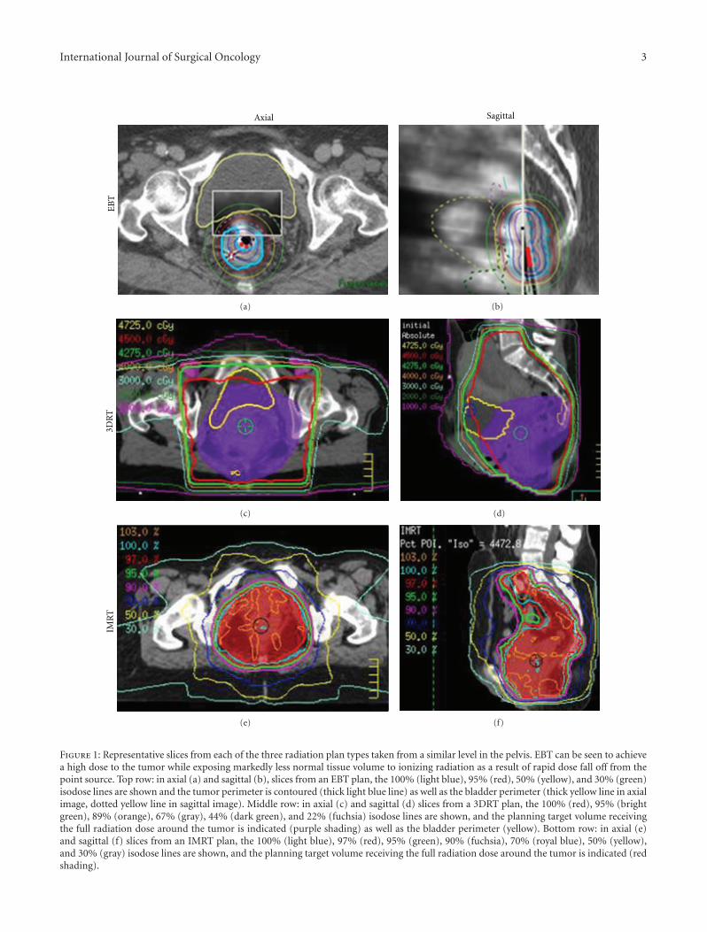
Preoperative external beam radiation has been shown toincrease pathological response rates and reduce the risk oflocal recurrence [3], but it is also associated with an increasedrisk of therapy-induced side effects and increased morbidity[4]. These acute toxicity events may lead to treatment breaks,compromising the efficacy of treatment and delaying surgery[3]. In an attempt to reduce treatment-related toxicity, high-dose-rate endorectal brachytherapy (EBT) was developed asan alternative neoadjuvant option for locally advanced lowrectal cancer. This technique has been previously describedas monotherapy for the treatment of prostate, cervical,esophageal, and buccal mucosal tumors [5–7]. EBT is aradiotherapy technique that allows for delivery of a focusedhigh dose of ionizing radiation at the mucosal surface directlyoverlying the tumor while avoiding injury to surroundingnormal tissues. Rapid dose fall off from the iridium-192point source and the lack of external radiation beams thatmust pass through the normal pelvic tissues to reach thetumor combine to minimize dose to normal surroundingstructures such as the femoral heads, bowel, bladder, andreproductive organs compared to conventional radiotherapytechniques (Figure 1). In high-dose-rate brachytherapy, theradioisotope is inserted for a brief period of time (approx-imately 15 minutes for EBT) to deliver the required dose andthen withdrawn from the body, as opposed to low-dose-ratebrachytherapy where a radioactive source is left implantedin the patient. EBT is given in 4 fractions of 6.5 Gy, fora total dose of 26 Gy. The treatment consists of 4 days ofradiation treatment alone followed by a 6–8 week recoverywindow, then surgical resection and adjuvant chemotherapy.Investigators at McGill University have achieved excellenttumor regression with over 29% of patients having acomplete pathologic response at surgery [4, 6, 8]. Theresponse rates of EBT are similar if not superior to thoseachieved with conventional neoadjuvant chemoradiation,for which the associated pathologic CR rate ranges from8% to 19% [9–14]. The potential benefits of EBT forpatients include the short duration of therapy, the seeminglyhigh response rate reported thus far, and the avoidanceof concurrent neoadjuvant systemic chemotherapy and itsassociated toxicity.
At this point, only one group has published data onpatients with resectable rectal adenocarcinoma who weretreated with EBT. Herein, we analyze the preliminary out-comes obtained with EBT and compare them to patientsreceiving conventional neoadjuvant chemoradiation (3DRT/IMRT) at our institution. The primary goal of this study,therefore, is to compare radiologic, pathologic, and short-term clinical outcomes between EBT and conventional radi-ation techniques in the neoadjuvant setting.
2. Materials and Methods
2.1. Patient Selection. From 2010–2012, 7 patients with local-ly advanced low rectal adenocarcinoma (within 12 cm ofthe anal verge) were enrolled in a prospective study of neo-adjuvant EBT (NCT01226979) at Johns Hopkins Hospital.Patients were required to meet the following inclusioncriteria: greater than 18 years of age, histologically confirmedadenocarcinoma of the rectum, able to undergo local stagingby MRI and/or EUS demonstrating a T2N1 or T3N0-1tumor, and ECOG performance status of 0 or 1. Patientswere excluded if they had tumors greater than 12 cm fromthe anal verge, metastatic disease at time of enrollment,positive inguinal or iliac lymph nodes on MRI, PET, orEUS, concurrent malignancy, bulky tumors that would notallow application of the endorectal probe, or previouspelvic irradiation. For comparison, historical controls wereobtained by identifying all patients with stage II-III rectaladenocarcinoma who received conventional neoadjuvantchemoradiation with IMRT or 3DRT at our institution from2008–2012 and went on to surgical resection.
2.2. Clinical Outcomes. Clinical data for patients treated withEBT was gathered prospectively as part of the trial protocol.To gather data for historical controls, retrospective chartreview was performed using the electronic patient record(EPR) system after approval by the Institutional ReviewBoard. For all patients, toxicity was evaluated using the Na-tional Cancer Institute (NCI) Common Terminology Crite-ria for Adverse Events (CTCAE) version 4.0.
2.3. Neoadjuvant Therapy. Patients in the EBT group weretreated with 4 consecutive daily fractions of 6.5 Gy targeted tothe tumor and mesorectal lymph nodes without concurrentchemotherapy. Each fraction was delivered over approxi-mately 15 minutes using a flexible silicone intracavitaryapplicator (OncoSmart, Nucletron, Veenendaal, The Nether-lands) positioned in the rectum using fluoroscopic guidanceand a microSelectron high-dose-rate iridium-192 remoteafterloading system (Nucletron) as described by Vuong et al.[4]. Treatment planning was performed using the Oncentrabrachytherapy planning system (Nucletron). Patients in the3DRT and IMRT groups received 28 daily (Monday throughFriday) fractions of 1.8 Gy over a period of 5 to 6 weeks (totaldose of 50.4 Gy) with concurrent oral 5-FU.
2.4. Surgical Resection. At the initial assessment for allpatients, surgery was preplanned according to the standardof care to take place from 6 to 8 weeks following completion
International Journal of Surgical Oncology 3
AxialE
BT
(a)
Sagittal
(b)
3DR
T
(c) (d)
IMR
T
(e) (f)
Figure 1: Representative slices from each of the three radiation plan types taken from a similar level in the pelvis. EBT can be seen to achievea high dose to the tumor while exposing markedly less normal tissue volume to ionizing radiation as a result of rapid dose fall off from thepoint source. Top row: in axial (a) and sagittal (b), slices from an EBT plan, the 100% (light blue), 95% (red), 50% (yellow), and 30% (green)isodose lines are shown and the tumor perimeter is contoured (thick light blue line) as well as the bladder perimeter (thick yellow line in axialimage, dotted yellow line in sagittal image). Middle row: in axial (c) and sagittal (d) slices from a 3DRT plan, the 100% (red), 95% (brightgreen), 89% (orange), 67% (gray), 44% (dark green), and 22% (fuchsia) isodose lines are shown, and the planning target volume receivingthe full radiation dose around the tumor is indicated (purple shading) as well as the bladder perimeter (yellow). Bottom row: in axial (e)and sagittal (f) slices from an IMRT plan, the 100% (light blue), 97% (red), 95% (green), 90% (fuchsia), 70% (royal blue), 50% (yellow),and 30% (gray) isodose lines are shown, and the planning target volume receiving the full radiation dose around the tumor is indicated (redshading).
4 International Journal of Surgical Oncology
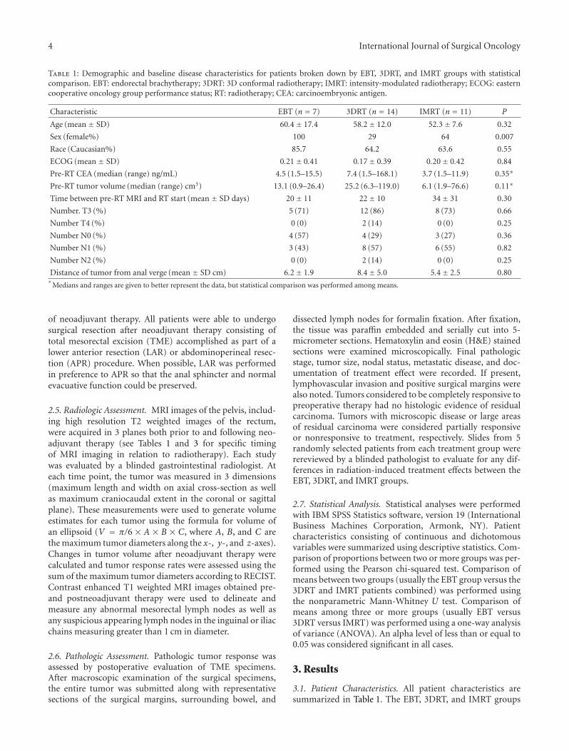
Table 1: Demographic and baseline disease characteristics for patients broken down by EBT, 3DRT, and IMRT groups with statisticalcomparison. EBT: endorectal brachytherapy; 3DRT: 3D conformal radiotherapy; IMRT: intensity-modulated radiotherapy; ECOG: easterncooperative oncology group performance status; RT: radiotherapy; CEA: carcinoembryonic antigen.
Characteristic EBT (n = 7) 3DRT (n = 14) IMRT (n = 11) P
Age (mean ± SD) 60.4± 17.4 58.2± 12.0 52.3± 7.6 0.32
Sex (female%) 100 29 64 0.007
Race (Caucasian%) 85.7 64.2 63.6 0.55
ECOG (mean ± SD) 0.21± 0.41 0.17± 0.39 0.20± 0.42 0.84
Pre-RT CEA (median (range) ng/mL) 4.5 (1.5–15.5) 7.4 (1.5–168.1) 3.7 (1.5–11.9) 0.35∗
Pre-RT tumor volume (median (range) cm3) 13.1 (0.9–26.4) 25.2 (6.3–119.0) 6.1 (1.9–76.6) 0.11∗
Time between pre-RT MRI and RT start (mean ± SD days) 20± 11 22± 10 34± 31 0.30
Number. T3 (%) 5 (71) 12 (86) 8 (73) 0.66
Number T4 (%) 0 (0) 2 (14) 0 (0) 0.25
Number N0 (%) 4 (57) 4 (29) 3 (27) 0.36
Number N1 (%) 3 (43) 8 (57) 6 (55) 0.82
Number N2 (%) 0 (0) 2 (14) 0 (0) 0.25
Distance of tumor from anal verge (mean ± SD cm) 6.2± 1.9 8.4± 5.0 5.4± 2.5 0.80∗
Medians and ranges are given to better represent the data, but statistical comparison was performed among means.
of neoadjuvant therapy. All patients were able to undergosurgical resection after neoadjuvant therapy consisting oftotal mesorectal excision (TME) accomplished as part of alower anterior resection (LAR) or abdominoperineal resec-tion (APR) procedure. When possible, LAR was performedin preference to APR so that the anal sphincter and normalevacuative function could be preserved.
2.5. Radiologic Assessment. MRI images of the pelvis, includ-ing high resolution T2 weighted images of the rectum,were acquired in 3 planes both prior to and following neo-adjuvant therapy (see Tables 1 and 3 for specific timingof MRI imaging in relation to radiotherapy). Each studywas evaluated by a blinded gastrointestinal radiologist. Ateach time point, the tumor was measured in 3 dimensions(maximum length and width on axial cross-section as wellas maximum craniocaudal extent in the coronal or sagittalplane). These measurements were used to generate volumeestimates for each tumor using the formula for volume ofan ellipsoid (V = π/6 × A × B × C, where A, B, and C arethe maximum tumor diameters along the x-, y-, and z-axes).Changes in tumor volume after neoadjuvant therapy werecalculated and tumor response rates were assessed using thesum of the maximum tumor diameters according to RECIST.Contrast enhanced T1 weighted MRI images obtained pre-and postneoadjuvant therapy were used to delineate andmeasure any abnormal mesorectal lymph nodes as well asany suspicious appearing lymph nodes in the inguinal or iliacchains measuring greater than 1 cm in diameter.
2.6. Pathologic Assessment. Pathologic tumor response wasassessed by postoperative evaluation of TME specimens.After macroscopic examination of the surgical specimens,the entire tumor was submitted along with representativesections of the surgical margins, surrounding bowel, and
dissected lymph nodes for formalin fixation. After fixation,the tissue was paraffin embedded and serially cut into 5-micrometer sections. Hematoxylin and eosin (H&E) stainedsections were examined microscopically. Final pathologicstage, tumor size, nodal status, metastatic disease, and doc-umentation of treatment effect were recorded. If present,lymphovascular invasion and positive surgical margins werealso noted. Tumors considered to be completely responsive topreoperative therapy had no histologic evidence of residualcarcinoma. Tumors with microscopic disease or large areasof residual carcinoma were considered partially responsiveor nonresponsive to treatment, respectively. Slides from 5randomly selected patients from each treatment group wererereviewed by a blinded pathologist to evaluate for any dif-ferences in radiation-induced treatment effects between theEBT, 3DRT, and IMRT groups.
2.7. Statistical Analysis. Statistical analyses were performedwith IBM SPSS Statistics software, version 19 (InternationalBusiness Machines Corporation, Armonk, NY). Patientcharacteristics consisting of continuous and dichotomousvariables were summarized using descriptive statistics. Com-parison of proportions between two or more groups was per-formed using the Pearson chi-squared test. Comparison ofmeans between two groups (usually the EBT group versus the3DRT and IMRT patients combined) was performed usingthe nonparametric Mann-Whitney U test. Comparison ofmeans among three or more groups (usually EBT versus3DRT versus IMRT) was performed using a one-way analysisof variance (ANOVA). An alpha level of less than or equal to0.05 was considered significant in all cases.
3. Results
3.1. Patient Characteristics. All patient characteristics aresummarized in Table 1. The EBT, 3DRT, and IMRT groups

International Journal of Surgical Oncology 5
Table 2: Clinical outcomes broken down by EBT, 3DRT, and IMRT groups with statistical comparison. EBT: endorectal brachytherapy;3DRT: 3D conformal radiotherapy; IMRT: intensity-modulated radiotherapy; RT: radiotherapy; CEA: carcinoembryonic antigen.
Clinical outcome EBT (n = 7) 3DRT (n = 14) IMRT (n = 14) P
Time to surgery from RT start (mean ± SD days) 53± 8 104± 21 119± 51 <0.001
Time to surgery from RT end (mean ± SD days) 50± 8 65± 20 79± 51 0.038
Post-RT CEA (median (range) ng/mL) 3.2 (1.1–18.3) 3.9 (1.4–67.8) 1.9 (0.5–12.3) 0.41∗
Change in CEA pre-RT → post-RT (median (range) %) −20 (−45 to +18) −40 (−83 to +300) −12 (−91 to +20) 0.36∗
No. with grade 1 toxicity (%) 4 (57) 14 (100) 9 (82) 0.025
No. with grade 2 toxicity (%) 1 (14) 8 (57) 2 (18) 0.056
No. with grade 3 toxicity (%) 1 (14) 1 (7) 1 (9) 0.87
No. who underwent sphincter-preserving surgery (%) 6 (86) 13 (93) 10 (91) 0.87
No. with postoperative complications (%) 4 (29) 4 (36) 2/7 (29) 0.90
No. alive at 6 months post-RT/total (%) 7 (100) 14 (100) 11 (100) 1.0
No. with local recurrence at 6 months post-RT (%) 0 (0) 0 (0) 0 (0) 1.0
No. with distant metastasis at 6 months post-RT (%) 0 (0/7) 1 (7) 1 (9) 0.73∗
Medians and ranges are given to better represent the data, but statistical comparison was performed among means.
Table 3: Radiologic outcomes broken down by EBT, 3DRT, and IMRT groups with statistical comparison. EBT: endorectal brachytherapy;3DRT: 3D conformal radiotherapy; IMRT: intensity-modulated radiotherapy; RT: radiotherapy; CR: complete response; PR: partialresponse; SD: stable disease; PD: progressive disease; LN: lymph nodes.
Radiologic outcome EBT (n = 7) 3DRT (n = 10) IMRT (n = 7) P
Post-RT tumor volume (median (range) cm3) 1.0 (0.0–3.6) 3.8 (0.7–26.3) 0.4 (0.0–5.5) 0.16∗
Time between RT end and post-RT MRI (mean ± SD days) 35± 3 35± 7 36± 9 0.98
% decrease in tumor volume pre-RT → post-RT (median (range)) 89 (38–100) 87 (16–96) 93 (66–100) 0.78
No. CR (%) 1 (14) 0 (0) 1 (14) 0.46
No. PR (%) 4 (57) 9 (90) 6 (86) 0.23
No. SD (%) 2 (29) 1 (10) 0 (0) 0.26
No. PD (%) 0 (0) 0 (0) 0 (0) 1.0
No. with clinically significant mesorectal LN before treatment (%) → no. aftertreatment (%)
1 (14) → 0 (0) 6 (43) → 3 (21) 1 (14) → 1 (14) —
No. with clinically significant pelvic LN before treatment (%) → no. aftertreatment (%)
0 (0) → 0 (0) 4 (29) → 4 (29) 1 (14) → 0 (0) —
∗Medians and ranges are given to better represent the data, but statistical comparison was performed among means.
consisted of 7, 14, and 11 patients, respectively. Medianlengths of followup were 7 months for EBT, 15 months for3DRT, and 12 months for IMRT. Demographic and baselinedisease characteristics, including age, race, ECOG perfor-mance status, pre-RT carcinoembryonic antigen (CEA) level,pre-RT tumor volume, T stage, N stage, and tumor distancefrom the anal verge, were similar among the 3 groups (allP > 0.05; Table 1). There was, however, a difference in gen-der distribution between the 3 groups, with 100% of EBTpatients being female compared to only 29% and 64% of the3DRT and IMRT groups, respectively (P = 0.007).
3.2. Clinical Outcomes. Clinical outcomes of interest includ-ed time to surgical resection, change in CEA level after neo-adjuvant therapy, acute toxicity, sphincter preservation, andpostoperative complications; these outcomes are summa-rized in Table 2. Time elapsed from the start of neoadjuvanttherapy to surgical resection was reduced by nearly half inpatients who underwent EBT as opposed to 3DRT or IMRT
(P < 0.001). This reduction is not unexpected given theshorter time course of EBT (4 days) compared to 3DRT orIMRT (5-6 weeks). More interestingly, however, the timeelapsed from conclusion of neoadjuvant therapy to surgicalresection was also reduced for patients who underwent EBT(P = 0.038), despite the fact that all surgeries were similarlyplanned to take place 6–8 weeks following completion ofneoadjuvant therapy. All 3 groups demonstrated similarmedian reductions in CEA levels after neoadjuvant therapy(P = 0.36). Fewer patients experienced grade 1 or 2 acutetoxicity in the EBT group than in the external beam group(P = 0.025). Grade 3 toxicity was rare, occurring in onepatient from each of the 3 groups with all 3 incidents takingthe form of proctitis. No grade 4 toxicity was reported. Ratesof sphincter preservation and postoperative complicationswere similar among the 3 groups. Given the natural history ofrectal adenocarcinoma, length of followup was not sufficientto perform informative analyses of survival and disease pro-gression; however, preliminary results are given here to allow

6 International Journal of Surgical Oncology
Table 4: Pathologic outcomes broken down by EBT, 3DRT, and IMRT groups with statistical comparison. EBT: endorectal brachytherapy;3DRT: 3D conformal radiotherapy; IMRT: intensity-modulated radiotherapy; RT: radiotherapy; CR: complete response; LN: lymph nodes.
Pathologic outcome ERBT (n = 7) 3DRT (n = 14) IMRT (n = 11) P
No. with pathologic CR of primary tumor at surgery (%) 3 (43) 1 (7) 2 (18) 0.06
No. with positive margins at surgery (%) 0 (0) 1 (7) 0 (0) 0.47
No. with LN involvement at surgery (%) 3 (43) 8 (57) 4 (36) 0.57
No. with lymphovascular invasion (%) 1 (14) 1 (7) 2 (18) 0.60
for comparison of EBT to conventional neoadjuvant therapyat the current length of followup. The rates of overall survivaland local recurrence free survival at 6 months were 100% inall 3 groups. The rate of distant metastasis at 6 months was0% for EBT, 7% for 3DRT, and 9% for IMRT. Thus, at a 6-month time point, EBT appears noninferior to conventionalneoadjuvant chemoradiation using 3DRT or IMRT.
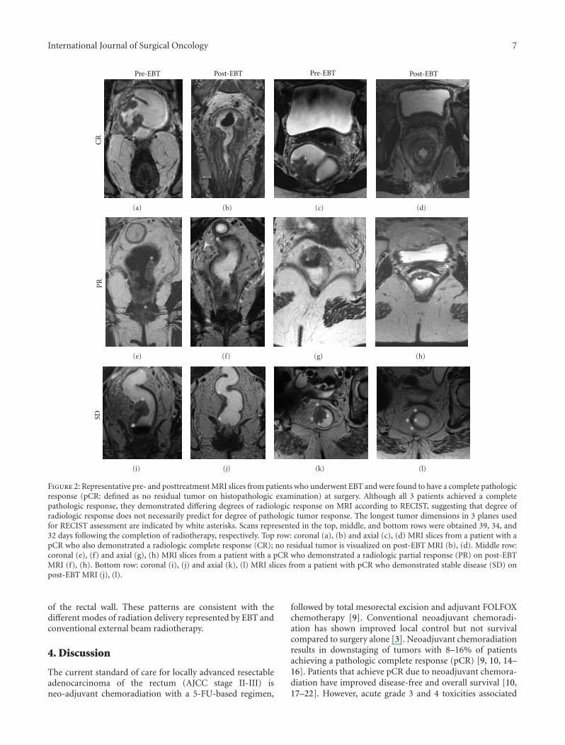
3.3. Radiologic Outcomes. Radiologic outcomes of interestincluded change in tumor volume, tumor response ratesanalyzed according to RECIST, and change in mesorectal andpelvic nodal disease status over the course of neoadjuvanttherapy. These outcomes are summarized in Table 3 for allpatients in each group who had pre- and posttreatment MRIstudies available (n = 7 for EBT, n = 10 for 3DRT, n =7 for IMRT). All 3 groups showed a striking response toneoadjuvant therapy, with similarly marked reductions intumor volume. Thus, EBT achieved a comparable degree ofreduction in tumor volume as measured on MRI after only 4days of treatment without chemotherapy as 3DRT and IMRTachieved over 5 to 6 weeks with concurrent 5-FU. Tumorresponse rates according to RECIST were also similar to the 3treatment techniques (P > 0.05 for rates of CR, PR, SD, andPD, as summarized in Table 3). Identification of clinicallysignificant (≥1.0 cm in longest dimension) lymph nodes onpre- and posttreatment MRI showed that the proportionof patients with radiologic mesorectal nodal involvementdecreased over the course of neoadjuvant therapy in the EBTand 3DRT groups, but remained stable in the IMRT group.Clinically significant pelvic lymph nodes were identified onpretreatment MRI in 4 patients in the 3DRT group; in all 4patients, these nodes remained clinically significant follow-ing treatment. One patient in the IMRT group was observedto have a pelvic lymph node prior to treatment, whichsubsequently resolved after chemoradiation. No patient inany group developed new pelvic nodal involvement overthe course of neoadjuvant therapy. The degree of radiologictumor response, however, did not appear to correlate withpathologic complete response (pCR), with more patientsmanifesting a pCR at surgery than were observed to have acomplete radiologic response on post-EBT MRI (Figure 2).
3.4. Pathologic Outcomes. Pathologic outcomes of interestincluded tumor complete response rate, surgical margin sta-tus, lymph node involvement, and lymphovascular invasion;these outcomes are summarized in Table 4. There was atrend towards higher rate of complete pathologic responsein patients who underwent EBT (43%) compared to the
external beam group consisting of 3DRT and IMRT patientscombined (12%) (P = 0.06). Rates of margin positivity,lymph node involvement, and lymphovascular invasion weresimilar among the three treatment groups.
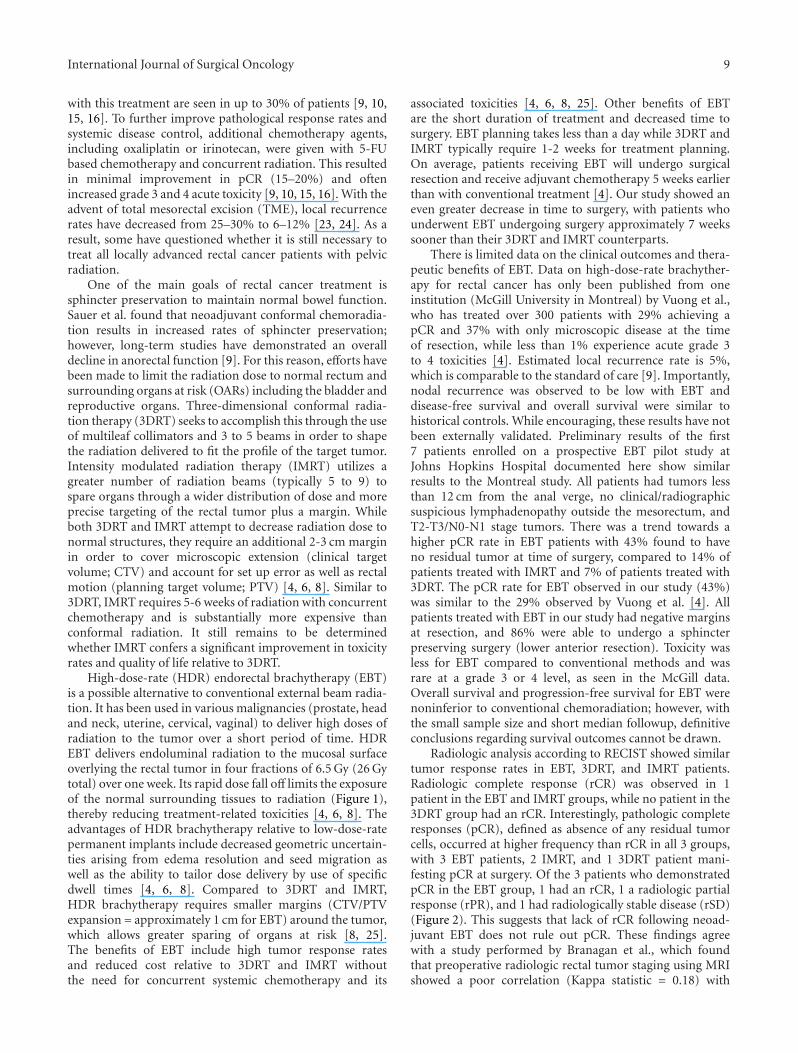
Surgical pathology slides from 5 cases in each of the 3treatment groups (15 cases total) were randomly selected forrereview by a blinded pathologist to assess for qualitativedifferences in microscopic treatment effects. The surgicalresection specimens showed disruption of tumor architec-ture (regions of necrosis and the presence of mucinous poolswith sparse floating tumor cells) and radiation treatmenteffects for all 3 radiotherapy techniques. However, the distri-bution and degree of radiation-induced changes throughoutthe layers of the rectal wall were different for EBT versusconventional 3DRT/IMRT (since radiation treatment effectswere found to be virtually identical between specimens fromthe 3DRT and IMRT groups, these groups will be collectivelyreferred to as the conventional external beam group from thispoint forward). Compared to the conventional external beamgroup, specimens from EBT patients demonstrated morepronounced radiation-induced changes to the superficiallayers of the rectal wall (mucosa, lamina propria, submucosa,and muscularis interna) in the region where the tumor waslocated (Figure 3). The mucosa overlying the tumor wasobserved to be ulcerated and the underlying lamina propriamanifested extensive fibrosis and hyalinization (Figure 3(a)).The vessels in the submucosa were seen to have a thickenedand sclerosed vascular smooth muscle layer (Figure 3(b)),but deeper vessels located in the subserosa were largelyspared (Figure 3(d)). Atrophy, disorganization, and degen-eration were evident in the muscularis propria interna, butto a lesser degree in the deeper muscularis propria externa(Figure 3(c)). Few serosal adhesions were observed.
In specimens from patients treated with conventionalexternal beam radiation, pathologic findings were similar innature, but opposite in distribution with deeper layers ofthe rectal wall being more prominently affected than moresuperficial layers (Figure 3). Ulceration of the mucosal sur-face overlying the tumor was milder (Figure 3(f)); the mus-cularis propria externa rather than interna exhibited moreextensive degeneration (Figure 3(h)); deeper vessels near theserosa (Figure 3(d)) were more affected than submucosalvessels (Figure 3(g)); more numerous serosal adhesions werepresent (Figure 3(j)). These contrasting distributions oftreatment effect suggest that EBT imparts a more intenseablative effect to the tumor and rectal tissue immediatelysurrounding it, while conventional external beam treatmentgenerates more diffuse ablative effects throughout all layers
International Journal of Surgical Oncology 7
Pre-EBT
CR
∗
∗
(a)
Post-EBT
(b)
Pre-EBT
∗∗∗ ∗
(c)
Post-EBT
(d)
PR
∗
∗
(e)
∗
∗
(f)
∗∗∗ ∗
(g)
∗ ∗∗∗
(h)
SD
∗
∗
(i)
∗
∗
(j)
∗∗∗ ∗
(k)
∗∗∗∗
(l)
Figure 2: Representative pre- and posttreatment MRI slices from patients who underwent EBT and were found to have a complete pathologicresponse (pCR: defined as no residual tumor on histopathologic examination) at surgery. Although all 3 patients achieved a completepathologic response, they demonstrated differing degrees of radiologic response on MRI according to RECIST, suggesting that degree ofradiologic response does not necessarily predict for degree of pathologic tumor response. The longest tumor dimensions in 3 planes usedfor RECIST assessment are indicated by white asterisks. Scans represented in the top, middle, and bottom rows were obtained 39, 34, and32 days following the completion of radiotherapy, respectively. Top row: coronal (a), (b) and axial (c), (d) MRI slices from a patient with apCR who also demonstrated a radiologic complete response (CR); no residual tumor is visualized on post-EBT MRI (b), (d). Middle row:coronal (e), (f) and axial (g), (h) MRI slices from a patient with a pCR who demonstrated a radiologic partial response (PR) on post-EBTMRI (f), (h). Bottom row: coronal (i), (j) and axial (k), (l) MRI slices from a patient with pCR who demonstrated stable disease (SD) onpost-EBT MRI (j), (l).
of the rectal wall. These patterns are consistent with thedifferent modes of radiation delivery represented by EBT andconventional external beam radiotherapy.
4. Discussion
The current standard of care for locally advanced resectableadenocarcinoma of the rectum (AJCC stage II-III) isneo-adjuvant chemoradiation with a 5-FU-based regimen,
followed by total mesorectal excision and adjuvant FOLFOXchemotherapy [9]. Conventional neoadjuvant chemoradi-ation has shown improved local control but not survivalcompared to surgery alone [3]. Neoadjuvant chemoradiationresults in downstaging of tumors with 8–16% of patientsachieving a pathologic complete response (pCR) [9, 10, 14–16]. Patients that achieve pCR due to neoadjuvant chemora-diation have improved disease-free and overall survival [10,17–22]. However, acute grade 3 and 4 toxicities associated

8 International Journal of Surgical Oncology
EBT
Mu
cosa
an
dla
min
a pr
opri
aSu
bmu
cosa
Mu
scu
lari
spr
opri
aSu
bser
osa
Sero
sa
(d)
(e)
(g)
(h)
(i)
(j)
(a)
(b)
Externa
Interna
(c)
Externa
Interna
Conventional CRT
(f)
∗
∗
∗
∗
∗∗∗
Figure 3: Representative H&E stained histopathologic sections at 4x magnification from patients who exhibited a complete pathologicresponse to EBT (a)–(e) and conventional external beam radiation (f)–(j). All images are taken from the region of the rectal wall where thetumor was located prior to neoadjuvant therapy. All insets are shown at 20x magnification. First row: at the mucosa, extensive ulceration(solid arrows) is apparent after EBT (a), while the mucosa remains intact (solid arrows) after conventional CRT (f). Hyalinization of thelamina propria (asterisks) is also evident after EBT (a). Second row: in the submucosa, marked hypertrophy and sclerosis of vessel walls canbe seen following EBT (b), while only slight hypertrophy of vessel walls is seen after conventional CRT (g). Third row: within the muscularispropria, the more superficial interna layer can be seen to exhibit degeneration and atrophy after EBT while the externa layer remains largelyintact (c); in a contrary fashion, following conventional CRT, it is the externa layer that exhibits more prominent degeneration comparedto the interna (h). fourth row: at the level of the subserosa, vessel walls appear normal in patients treated with EBT (d), but distinctlyhypertrophied in patients treated with conventional CRT (i). Fifth row: the serosa demonstrates few adhesions (asterisk) after treatmentwith EBT (e), in contrast to the extensive adhesions (asterisks) present after treatment with conventional CRT (j).
International Journal of Surgical Oncology 9
with this treatment are seen in up to 30% of patients [9, 10,15, 16]. To further improve pathological response rates andsystemic disease control, additional chemotherapy agents,including oxaliplatin or irinotecan, were given with 5-FUbased chemotherapy and concurrent radiation. This resultedin minimal improvement in pCR (15–20%) and oftenincreased grade 3 and 4 acute toxicity [9, 10, 15, 16]. With theadvent of total mesorectal excision (TME), local recurrencerates have decreased from 25–30% to 6–12% [23, 24]. As aresult, some have questioned whether it is still necessary totreat all locally advanced rectal cancer patients with pelvicradiation.
One of the main goals of rectal cancer treatment issphincter preservation to maintain normal bowel function.Sauer et al. found that neoadjuvant conformal chemoradia-tion results in increased rates of sphincter preservation;however, long-term studies have demonstrated an overalldecline in anorectal function [9]. For this reason, efforts havebeen made to limit the radiation dose to normal rectum andsurrounding organs at risk (OARs) including the bladder andreproductive organs. Three-dimensional conformal radia-tion therapy (3DRT) seeks to accomplish this through the useof multileaf collimators and 3 to 5 beams in order to shapethe radiation delivered to fit the profile of the target tumor.Intensity modulated radiation therapy (IMRT) utilizes agreater number of radiation beams (typically 5 to 9) tospare organs through a wider distribution of dose and moreprecise targeting of the rectal tumor plus a margin. Whileboth 3DRT and IMRT attempt to decrease radiation dose tonormal structures, they require an additional 2-3 cm marginin order to cover microscopic extension (clinical targetvolume; CTV) and account for set up error as well as rectalmotion (planning target volume; PTV) [4, 6, 8]. Similar to3DRT, IMRT requires 5-6 weeks of radiation with concurrentchemotherapy and is substantially more expensive thanconformal radiation. It still remains to be determinedwhether IMRT confers a significant improvement in toxicityrates and quality of life relative to 3DRT.
High-dose-rate (HDR) endorectal brachytherapy (EBT)is a possible alternative to conventional external beam radia-tion. It has been used in various malignancies (prostate, headand neck, uterine, cervical, vaginal) to deliver high doses ofradiation to the tumor over a short period of time. HDREBT delivers endoluminal radiation to the mucosal surfaceoverlying the rectal tumor in four fractions of 6.5 Gy (26 Gytotal) over one week. Its rapid dose fall off limits the exposureof the normal surrounding tissues to radiation (Figure 1),thereby reducing treatment-related toxicities [4, 6, 8]. Theadvantages of HDR brachytherapy relative to low-dose-ratepermanent implants include decreased geometric uncertain-ties arising from edema resolution and seed migration aswell as the ability to tailor dose delivery by use of specificdwell times [4, 6, 8]. Compared to 3DRT and IMRT,HDR brachytherapy requires smaller margins (CTV/PTVexpansion = approximately 1 cm for EBT) around the tumor,which allows greater sparing of organs at risk [8, 25].The benefits of EBT include high tumor response ratesand reduced cost relative to 3DRT and IMRT withoutthe need for concurrent systemic chemotherapy and its
associated toxicities [4, 6, 8, 25]. Other benefits of EBTare the short duration of treatment and decreased time tosurgery. EBT planning takes less than a day while 3DRT andIMRT typically require 1-2 weeks for treatment planning.On average, patients receiving EBT will undergo surgicalresection and receive adjuvant chemotherapy 5 weeks earlierthan with conventional treatment [4]. Our study showed aneven greater decrease in time to surgery, with patients whounderwent EBT undergoing surgery approximately 7 weekssooner than their 3DRT and IMRT counterparts.
There is limited data on the clinical outcomes and thera-peutic benefits of EBT. Data on high-dose-rate brachyther-apy for rectal cancer has only been published from oneinstitution (McGill University in Montreal) by Vuong et al.,who has treated over 300 patients with 29% achieving apCR and 37% with only microscopic disease at the timeof resection, while less than 1% experience acute grade 3to 4 toxicities [4]. Estimated local recurrence rate is 5%,which is comparable to the standard of care [9]. Importantly,nodal recurrence was observed to be low with EBT anddisease-free survival and overall survival were similar tohistorical controls. While encouraging, these results have notbeen externally validated. Preliminary results of the first7 patients enrolled on a prospective EBT pilot study atJohns Hopkins Hospital documented here show similarresults to the Montreal study. All patients had tumors lessthan 12 cm from the anal verge, no clinical/radiographicsuspicious lymphadenopathy outside the mesorectum, andT2-T3/N0-N1 stage tumors. There was a trend towards ahigher pCR rate in EBT patients with 43% found to haveno residual tumor at time of surgery, compared to 14% ofpatients treated with IMRT and 7% of patients treated with3DRT. The pCR rate for EBT observed in our study (43%)was similar to the 29% observed by Vuong et al. [4]. Allpatients treated with EBT in our study had negative marginsat resection, and 86% were able to undergo a sphincterpreserving surgery (lower anterior resection). Toxicity wasless for EBT compared to conventional methods and wasrare at a grade 3 or 4 level, as seen in the McGill data.Overall survival and progression-free survival for EBT werenoninferior to conventional chemoradiation; however, withthe small sample size and short median followup, definitiveconclusions regarding survival outcomes cannot be drawn.
Radiologic analysis according to RECIST showed similartumor response rates in EBT, 3DRT, and IMRT patients.Radiologic complete response (rCR) was observed in 1patient in the EBT and IMRT groups, while no patient in the3DRT group had an rCR. Interestingly, pathologic completeresponses (pCR), defined as absence of any residual tumorcells, occurred at higher frequency than rCR in all 3 groups,with 3 EBT patients, 2 IMRT, and 1 3DRT patient mani-festing pCR at surgery. Of the 3 patients who demonstratedpCR in the EBT group, 1 had an rCR, 1 a radiologic partialresponse (rPR), and 1 had radiologically stable disease (rSD)(Figure 2). This suggests that lack of rCR following neoad-juvant EBT does not rule out pCR. These findings agreewith a study performed by Branagan et al., which foundthat preoperative radiologic rectal tumor staging using MRIshowed a poor correlation (Kappa statistic = 0.18) with

10 International Journal of Surgical Oncology
pathologic tumor stage of the resected specimen [26]. Asmicroscopic pathologic examination of the TME specimenis the gold standard for assessment of tumor response, ourdata indicate that the radiologic response on preoperativeMRI cannot be reliably used to predict degree of tumorresponse to EBT because even rSD can correlate with a pCRat resection. Positron emission tomography (PET) imagingmay represent a more effective way to radiologically eval-uate tumor response prior to surgery. Although a studyperformed at Stanford University showed that changes seenon PET have limited value in predicting for pathologicresponse of rectal cancer after conventional neoadjuvantchemoradiation, the utility of PET has not yet been examinedin assessing tumor response to EBT [27].
One concern in treating rectal tumors with EBT insteadof conventional external beam radiation is lack of steriliza-tion of pelvic lymph nodes as a result of the rapid dose falloffassociated with EBT, which covers only the mesorectal lymphnodes with little to no coverage of pelvic nodes. Thus, it isconceivable that EBT may lead to higher lymph node metas-tasis rates and local recurrence. For that reason, patients areselected for EBT based on pathologic lymph node statusby imaging. Patients are excluded if positive lymph nodesare identified in the pelvis outside the mesorectum prior totreatment. It is encouraging that in our study preoperativeMRI showed no development of pelvic node involvement forany of the 7 patients who received EBT. Furthermore, theone patient in the EBT group with radiologic involvementof mesorectal lymph nodes on pretreatment MRI exhibitedcomplete resolution of nodal involvement on post-EBTimaging. Although limited in their generalizability by thesmall sample size, these findings suggest that when patientsare carefully selected for neoadjuvant EBT (i.e., N0-N1patients only), there is a low likelihood that they willdevelop radiologic evidence of N2 disease prior to surgery.This evidence confirms data presented by Vuong et al.documenting a 5-year local recurrence rate of 5% in N0-N1 patients treated with EBT, which likely indicates a 5%or lower rate of spread to pelvic lymph nodes prior tosurgery [4]. However, longer followup and a greater numberof patients are needed in our study before radiologic resultsregarding development of N2 disease prior to surgery can becorrelated with local recurrence rates.
Radiation-induced injury to the rectum is well docu-mented and characteristic histologic changes include archi-tectural disruption and atrophy, goblet cell loss, shortenedcrypts, a thickened and distorted muscularis, intestinal wallfibrosis, serosal thickening, and vascular sclerosis [28, 29].A study in mice that documented the histopathologic char-acteristics of radiation injury to intestinal tissue observedsimilar findings as those listed above [30]. Mice that receivedexternal beam radiotherapy showed mucosal ulcerations,fibrotic changes, serosal thickening, and marked vascularsclerosis. Effects on rectal tissue due to high-dose-rate (HDR)brachytherapy have not yet been published. However, anautopsy study evaluating the histological findings in prostatetissue treated with low-dose-rate brachytherapy showedsimilar results to our study [31]. The prostate specimens
showed distorted glandular architecture, extensive fibrosis,and hyalinization of the blood vessels.
Our study is the first to describe the histologic differencesin treatment effect of EBT compared to conventional externalbeam radiation seen on pathologic examination of rectaladenocarcinoma resection specimens. In general, the types ofhistologic changes induced by EBT and conventional exter-nal beam radiation were similar, consisting of mucosalulceration, fibrosis and hyalinization of the lamina propria,degeneration of the muscularis propria, and vessel wallhypertrophy and sclerosis as well as formation of serosaladhesions. Notably, however, the distribution and degree ofthese changes throughout the layers of the rectal wall weredistinct for EBT. TME specimens from patients who receivedconventional external beam radiation demonstrated moder-ate radiation-induced changes diffusely throughout the rectalwall. Specimens from patients treated with EBT, on the otherhand, displayed these changes along a gradient, with intensetreatment effect apparent in the superficial layers of the rectalwall (mucosa, lamina propria, submucosa, and muscularispropria interna), but progressively reduced treatment effectin each of the deeper layers (muscularis propria externa,subserosa, and serosa). These contrasting distributions oftreatment effect suggest that EBT may achieve a more potentlocalized ablative effect on the tumor and immediately sur-rounding rectal tissue than does conventional external beamradiation, but may not be as effective in sterilizing the serosa.
It follows that careful patient selection is critical for suc-cessful implementation of EBT. Patients with T1–T3 lesionsmay derive considerable benefit from the high ablativepotential of EBT and would be considered viable candidatesbecause their tumors can be adequately covered with EBTwithout extreme doses to the rectal wall. If tumors aremore than 3-4 cm from the rectal wall, EBT may causeincreased proctitis. Our results, as well as the data reportedby Vuong et al. [4], indicate that EBT likely achieves higherpCR rates than conventional external beam radiation. Agrowing body of evidence supports the notion that patientswith pCR after neoadjuvant therapy have more favorablelong-term outcomes compared to patients with lesser or nopathologic response [10, 17–22]. Thus, it may be possibleto improve outcomes in patients with T1–T3 rectal tumorsby treatment with EBT rather than conventional externalbeam radiation. Patients with T4 lesions, however, may haveportions of tumor that extend beyond the effective rangeof the radioisotope used in EBT and are likely better suitedto conventional external beam radiation, which we haveobserved in this study to affect all layers of the rectal wall,including the outermost serosa.
Modern staging performed with endorectal ultrasound(EUS) and pelvic MRI has been shown to attain a high degreeof accuracy in determining the T stage of rectal tumors. EUShas demonstrated a sensitivity of 90% and specificity of 75%for identifying T3 tumors, while MRI has a sensitivity of80–86% and specificity of 71–76% [32–35]. In predictingadjacent organ invasion (T4 tumor stage), EUS and MRIhave demonstrated sensitivities of 70% and 74%, respec-tively, and high specificity at 97% and 96% [33]. The use ofthese staging modalities has become routine in recent years

International Journal of Surgical Oncology 11
as part of the workup for rectal cancer and can be utilizedto discern patients well suited to EBT versus conventionalexternal beam radiation in the clinical setting.
Finally, the limited range at which radiation treatmenteffects were observed for EBT on histopathologic examina-tion in our study provides a rationale for the lesser degreeof toxicity experienced by patients in the EBT group. Saueret al. reported a 27% incidence of grade 3 to 4 acute toxicityas a result of neoadjuvant chemoradiation for rectal cancer[9]. More recent studies involving the addition of agents suchas oxaliplatin to neoadjuvant chemoradiation regimens havebeen associated with rates of grade 3 to 4 acute toxicity ashigh as 36% [10–13]. The rate of acute toxicity in our studywas well below this, with only one patient in the EBT group(14%) experiencing grade 3 proctitis. Thus, in addition toincreasing the likelihood of achieving a pathologic completeresponse, EBT may also provide a less toxic mode ofneoadjuvant therapy that appears at least as effective as long-course conventional chemoradiation as measured by short-term outcomes.
Study Limitations. Our study was primarily limited by asmall number of patients and a short period of followup.These limitations precluded definitive survival analysis, butdid not hinder evaluation of several clinical, radiologic, andpathologic outcomes of interest. The fact that only data onEBT patients was collected prospectively, while data on 3DRTand IMRT patients was collected retrospectively, introducesthe biases inherent in retrospective studies to our analysis.It is also possible, though unlikely, that the difference ingender distribution between the EBT, 3DRT, and IMRTgroups could confound our analyses, especially in regard totoxicity considering the different organs at risk in the pelvicregion between males and females. Further followup of theEBT patients included in this study, as well as future EBTpatients (trial enrollment goal is 30 patients), will be neededto determine median overall survival and thus estimate theimpact of EBT on this primary oncologic outcome.
5. Conclusions
Comparison of preliminary EBT trial data to historical con-trols treated with conventional external beam radiationreveals that patients treated with EBT experience less toxicityand shorter time to surgery without compromising marginor lymph node status at resection. Followup was not suffi-cient for survival analysis, but EBT appears noninferior to3DRT and IMRT at 6 months. EBT alone administered over4 days achieves similar radiologic and favorable pathologictumor response rates when compared to 5-6 weeks of con-ventional chemoradiation. EBT showed a more intense localablative effect on histopathologic examination, suggesting agreater likelihood of achieving pathologic complete responseand, consequently, improved long-term outcomes. Further-more, radiation-induced changes due to EBT were tightlylocalized to the area of the tumor with greater sparing ofnormal tissues including small bowel, likely explaining thelower rate of toxicity observed in comparison to 3DRTand IMRT. Careful patient selection using EUS and MRI
is necessary to ensure that patients with T4 tumors thatextend beyond the range of the radioisotope used for EBTare not offered this therapy. In summary, EBT appears tobe a promising mode of neoadjuvant treatment for lowlying rectal adenocarcinoma. Longer followup and a largermulticenter study are needed to conclusively evaluate thepotential of EBT to produce a survival benefit in this patientpopulation.
Authors’ Contribution
The authors contributed equally to this work.
Acknowledgments
The authors acknowledge the generous support of the Clau-dio X. Gonzalez Family Foundation for assistance with thisproject. The authors Jessica A. Smith and Aaron T. Wild areco-first authors and contributed equally to this work.
References
[1] W. M. Mendenhall, R. A. Zlotecki, F. E. Snead et al., “Radio-therapy in the treatment of resectable rectal adenocarcinoma,”American Journal of Clinical Oncology, vol. 32, no. 6, pp. 629–638, 2009.
[2] R. Siegel, D. Naishadham, and A. Jemal, “Cancer statistics,2012,” Cancer Journal for Clinicians, vol. 62, no. 1, pp. 10–29,2012.
[3] J. Meyer, G. Balch, C. Willett, and B. Czito, “Update on treat-ment advances in combined-modality therapy for anal andrectal carcinomas,” Current Oncology Reports, vol. 13, no. 3,pp. 177–185, 2011.
[4] T. Vuong, S. Devic, and E. Podgorsak, “High dose rate endo-rectal brachytherapy as a neoadjuvant treatment for patientswith resectable rectal cancer,” Clinical Oncology, vol. 19, no. 9,pp. 701–705, 2007.
[5] R. Martınez-Monge, A. Gomez-Iturriaga, M. Cambeiro et al.,“Phase I-II trial of perioperative high-dose-rate brachytherapyin oral cavity and oropharyngeal cancer,” Brachytherapy, vol. 8,no. 1, pp. 26–33, 2009.
[6] T. Vuong, P. Szego, M. David et al., “The safety and usefulnessof high-dose-rate endoluminal brachytherapy as a boost in thetreatment of patients with esophageal cancer with externalbeam radiation with or without chemotherapy,” InternationalJournal of Radiation Oncology Biology Physics, vol. 63, no. 3,pp. 758–764, 2005.
[7] W. Harms, H. D. Becker, R. Krempien, and M. Wannen-macher, “Contemporary role of modern brachytherapy tech-niques in the management of malignant thoracic tumors,”Seminars in Surgical Oncology, vol. 20, no. 1, pp. 57–65, 2001.
[8] T. Vuong, P. J. Belliveau, R. P. Michel et al., “Conformalpreoperative endorectal brachytherapy treatment for locallyadvanced rectal cancer: early results of a phase I/II study,” Dis-eases of the Colon and Rectum, vol. 45, no. 11, pp. 1486–1495,2002.
[9] R. Sauer, H. Becker, W. Hohenberger et al., “Preoperative ver-sus postoperative chemoradiotherapy for rectal cancer,” TheNew England Journal of Medicine, vol. 351, no. 17, pp. 1731–1740, 2004.
12 International Journal of Surgical Oncology
[10] C. Rodel, P. Martus, T. Papadoupolos et al., “Prognosticsignificance of tumor regression after preoperative chemora-diotherapy for rectal cancer,” Journal of Clinical Oncology, vol.23, no. 34, pp. 8688–8696, 2005.
[11] R. Glynne-Jones, D. Sebag-Montefiore, T. S. Maughan, S. J.Falk, and A. C. McDonald, “A phase I dose escalation study ofcontinuous oral capecitabine in combination with oxaliplatinand pelvic radiation (XELOX-RT) in patients with locallyadvanced rectal cancer,” Annals of Oncology, vol. 17, no. 1, pp.50–56, 2006.
[12] J. P. Machiels, L. Duck, B. Honhon et al., “Phase II study of pre-operative oxaliplatin, capecitabine and external beam radio-therapy in patients with rectal cancer: the RadiOxCape study,”Annals of Oncology, vol. 16, no. 12, pp. 1898–1905, 2005.
[13] H. Rutten, D. Sebag Montefiore, R. Glynne-Jones et al., “Cape-citabine, oxaliplatin, radiotherapy, and excision (CORE) inpatients with MRI-defined locally advanced rectal adenocarci-noma: results of an international multicenter phase II study,”Journal of Clinical Oncology, vol. 24, no. 18S, supplement,2006.
[14] J. P. Gerard, T. Conroy, F. Bonnetain et al., “Preoperativeradiotherapy with or without concurrent fluorouracil and leu-covorin in T3-4 rectal cancers: results of FFCD 9203,” Journalof Clinical Oncology, vol. 24, no. 28, pp. 4620–4625, 2006.
[15] J. P. Gerard, F. Bonnetain, and T. Conroy, “Preoperative(perop) radiotherapy (RT) (+/−) 5 FU/folinic acid (FA) in T3-4 rectal cancers: results of the FFCD 9203 randomized trial,”Journal of Clinical Oncology, vol. 23, no. 247s, supplement,2005.
[16] R. Fietkau, M. Barten, G. Klautke et al., “Postoperative chemo-therapy may not be necessary for patients with ypN0-categoryafter neoadjuvant chemoradiotherapy of rectal cancer,” Dis-eases of the Colon and Rectum, vol. 49, no. 9, pp. 1284–1292,2006.
[17] M. Maas, P. J. Nelemans, V. Valentini et al., “Long-term out-come in patients with a pathological complete response afterchemoradiation for rectal cancer: a pooled analysis of individ-ual patient data,” The Lancet Oncology, vol. 11, no. 9, pp. 835–844, 2010.
[18] F. Stipa, D. B. Chessin, J. Shia et al., “A pathologic completeresponse of rectal cancer to preoperative combined-modalitytherapy results in improved oncological outcome comparedwith those who achieve no downstaging on the basis ofpreoperative endorectal ultrasonography,” Annals of SurgicalOncology, vol. 13, no. 8, pp. 1047–1053, 2006.
[19] C. Capirci, V. Valentini, L. Cionini et al., “Prognostic valueof pathologic complete response after neoadjuvant therapy inlocally advanced rectal cancer: long-term analysis of 566 ypCRpatients,” International Journal of Radiation Oncology BiologyPhysics, vol. 72, no. 1, pp. 99–107, 2008.
[20] L. F. de Campos-Lobato, L. Stocchi, A. da Luz Moreira et al.,“Pathologic complete response after neoadjuvant treatmentfor rectal cancer decreases distant recurrence and could erad-icate local recurrence,” Annals of Surgical Oncology, vol. 18, no.6, pp. 1590–1598, 2011.
[21] A. M. Wolthuis, F. Penninckx, K. Haustermans, N. Ectors, E.Van Cutsem, and A. D’Hoore, “Outcome standards for anorgan preservation strategy in stage II and III rectal adenocar-cinoma after neoadjuvant chemoradiation,” Annals of SurgicalOncology, vol. 18, no. 3, pp. 684–690, 2011.
[22] C. Belluco, A. De Paoli, V. Canzonieri et al., “Long-termoutcome of patients with complete pathologic response after
neoadjuvant chemoradiation for cT3 rectal cancer: implica-tions for local excision surgical strategies,” Annals of SurgicalOncology, vol. 18, no. 13, pp. 3686–3693, 2011.
[23] E. Kapiteijn, C. A. M. Marijnen, I. D. Nagtegaal et al., “Pre-operative radiotherapy combined with total mesorectal exci-sion for resectable rectal cancer,” The New England Journal ofMedicine, vol. 345, no. 9, pp. 638–646, 2001.
[24] E. Kapiteijn and C. J. H. van De Velde, “European trials withtotal mesorectal excision,” Seminars in Surgical Oncology, vol.19, no. 4, pp. 350–357, 2000.
[25] L. Arbea, L. I. Ramos, R. Martınez-Monge, M. Moreno, and J.Aristu, “Intensity-modulated radiation therapy (IMRT) vs. 3Dconformal radiotherapy (3DCRT) in locally advanced rectalcancer (LARC): dosimetric comparison and clinical impli-cations,” Radiation Oncology, vol. 5, no. 1, article 17, 2010.
[26] G. Branagan, H. Chave, C. Fuller, S. McGee, and D. Finnis,“Can magnetic resonance imaging predict circumferentialmargins and TNM stage in rectal cancer?” Diseases of the Colonand Rectum, vol. 47, no. 8, pp. 1317–1322, 2004.
[27] S. K. Chennupati, A. Quon, A. Kamaya et al., “Positronemission tomography for predicting pathologic response afterneoadjuvant chemoradiotherapy for locally advanced rectalcancer,” American Journal of Clinical Oncology, 2011. In press.
[28] C. M. Fenoglio-Preiser, A. Noffsing, G. Stennernan, P. Lantz,and P. G. Isaacson, “Epithelial neoplasms of the colon,” inGastrointestinal Pathology: An Atlas and Text, pp. 796–800,Lippincott, Williams, & Wilkins, Philadelphia, Pa, USA, 3rdedition, 2008.
[29] M. Hauer-Jensen, “Late radiation injury of the small intes-tine. Clinical, pathophysiologic and radiobiologic aspects. Areview,” Acta Oncologica, vol. 29, no. 4, pp. 401–415, 1990.
[30] C. W. Langberg and M. Hauer-Jensen, “Influence of fractionsize on the development of late radiation enteropathy: an ex-perimental study in the rat,” Acta Oncologica, vol. 35, no. 1, pp.89–94, 1996.
[31] A. Shapiro, O. Shapiro, N. B. Delongchamps, J. A. Bogart, G.P. Haas, and G. De La Rosa, “Autopsy evaluation of a prostatecancer case treated with brachytherapy,” Anticancer Research,vol. 28, no. 6, pp. 3909–3912, 2008.
[32] T. Akasu, H. Kondo, Y. Moriya et al., “Endorectal ultrasonog-raphy and treatment of early stage rectal cancer,” World Journalof Surgery, vol. 24, no. 9, pp. 1061–1068, 2000.
[33] S. Bipat, A. S. Glas, F. J. M. Slors, A. H. Zwinderman, P. M.M. Bossuyt, and J. Stoker, “Rectal cancer: local staging andassessment of lymph node involvement with endoluminal US,CT, and MR imaging—a meta-analysis,” Radiology, vol. 232,no. 3, pp. 773–783, 2004.
[34] F. Marusch, H. Lippert, A. Koch et al., “Routine use of tran-srectal ultrasound in rectal carcinoma: results of a prospectivemulticenter study,” Endoscopy, vol. 34, no. 5, pp. 385–390,2002.
[35] H. Ptok, F. Marusch, F. Meyer et al., “Feasibility and accuracyof TRUS in the pre-treatment staging for rectal carcinoma ingeneral practice,” European Journal of Surgical Oncology, vol.32, no. 4, pp. 420–425, 2006.
Related Documents