PARASITOLOGY Clinical Textbook for Veterinary Technicians ,4 th Edition, Dennis M. McCurnin, Saunders Pathology & Parasitology for Veterinary Technicians Leland S. Sharpiro, Thomson, Delmar Learning

Clinical Textbook for Veterinary Technicians,4 th Edition, Dennis M. McCurnin, Saunders Pathology & Parasitology for Veterinary Technicians Leland S. Sharpiro,
Dec 14, 2015
Welcome message from author
This document is posted to help you gain knowledge. Please leave a comment to let me know what you think about it! Share it to your friends and learn new things together.
Transcript

PARASITOLOGY
Clinical Textbook for Veterinary Technicians ,4th Edition, Dennis M. McCurnin, SaundersPathology & Parasitology for Veterinary Technicians Leland S. Sharpiro, Thomson, Delmar Learning

Most parasites are capable of causing significant damage to the host.
The potential may be a function of :• the number of parasites present in some cases• in the case of other parasites, location within the host• production of toxins•Interference with normal physiological processes

Ectoparasites (external parasites) :•mites, ticks, lice fleas, chiggers, and myiasis-inducing flies (an infestation of the skin by developing larvae i.e. maggots)
ANDEndoparasites (internal parasites):•protozoa, trematodes, tape-worms and nematodes
Have representative parasites on or in all animals and in every organ or tissue.
Some parasites are host specific, whereas other parasites are capable of infesting or infecting a broad range of animal hosts.
Modes of transmission vary considerably from simple, direct transmission to an extremely complex life cycle, involving the use of an intermediate host or specific environmental conditions.

The diagnosis of parasitism is not difficult, but timing, choice of technique, and interpretation of the results are often crucial for effective treatment and control.
ENDOPARASITESParasites of the Dog and Cat
ROUNDWORMS (Ascarids)
Toxocara canis, Toxocara cati, and Toxascaris leonina : are ascarids that are thick, white to cream-colored nematodes.
Nematode: simple roundworms, colorless, unsegmented, and lacking appendages
All three species are common in most geographical areas of the Unites States.
Eggs are highly resistant to adverse conditions and under ideal environmental conditions become infective in about 2 weeks.

Eggs are ingested
Eggs hatch in small intestine
Penetrate the
mucosa
Migrate through the liverPass
through the heart
Enter the lungs
where they develop
Larvae are coughed
and swallowed
Mature in the small
intestine within 4-6
weeks
Round worm life cycle
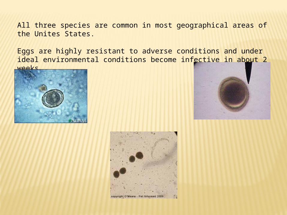
Hookworms are all short, thick parasites.
All hookworm species have a similar life cycle.
Hookworms are capable of establishing in the host after ingestion, but the normal mode of infection is skin penetration.

Larvae penetrate the skin
Larvae enters
the venous
circulation
Ultimately end up in the lungs
where they
develop in a short period
Worms are
coughed up and
swallowed
Enter the small
intestine and
mature Hookworm life cycle

Intestinal Threadworm (strongyloides stercoralis)
A nematode that is a parasite of dogs, cats, foxes, humans, primate, and possibly other wild carnivores.
Only the female nematode is parasitic, and she reproduces parthenogenetically (without fertilization).
The primary mode of infection is by skin penetration.

Tapeworms
The species of tapeworms found in dogs and cats is dependent on their geographic location and the amount of free-ranging activity the animals are given.
All tapeworms have an intermediate host in which the larval stage develops.
The larval stage is a rather large, fluid filled bladder called a hydatid cyst that is easily recognized by; (1) its large size, (2) the presence of numerous small pieces of larval tapeworms
on the inner surface(3) and the presence of compartments within the body of the
cyst in which daughter cysts have grown and fused together.

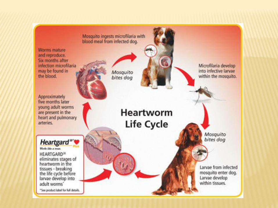
Heartworm: one of the most dangerous nematode parasites of a dog.
Dirofilaria immitis - long, slender worms that lice in the host’s pulmonary artery.
In heavy infections, they migrate to the right ventricle of the heart. (Cats rarely get heartworms; when they do, it is a serious disease.)
In heavy infections, the worms in the pulmonary artery may block the flow of blood to the lungs. Because the heart must work harder to get the flow of blood to the lungs, heart failure may occur.
Signs of heartworm infection in dogs include listlessness, breathing difficulty after exercise, and coughing.
Veterinarians confirm heartworm infections by finding the microfiliaria in the blood, but sometimes microfilariae are not visible in the infected dog’s blood.
(microfilaria – embryos released by female heartworms)

Microfilaria
Heartworms
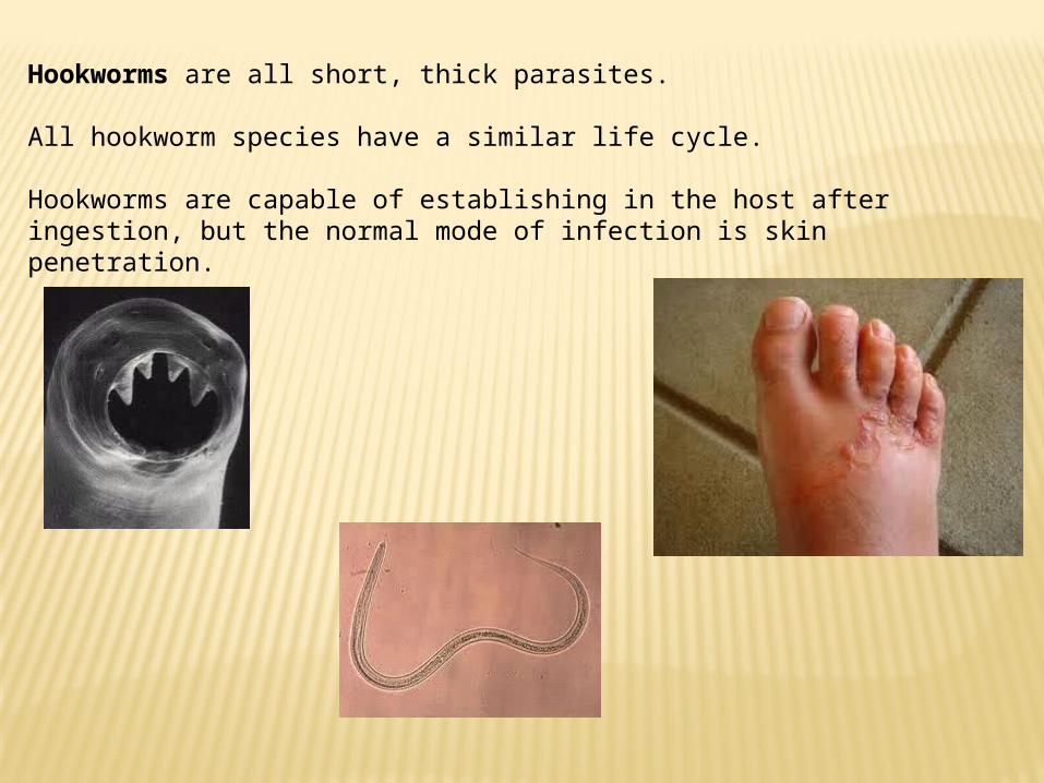
Female worms release
embryos (microfilaria)
into the blood Microfilariae are taken up by and
develop into first-stage larvae
in a mosquito
The larvae molt twice and
move to the mosquito’s mouthparts
The mosquito bites the dog and
the third stage larvae enters the
animals skin
In the tissues under the skin, the larvae molt
twice over several month
The young adult worms
then move into veins and travel
to the heart
In the pulmonary artery, they
grow into full-sized adults.

Treating heartworm infection is very difficult. Some drugs do kill the adult worms, but dead worms can block the blood vessels of the lungs.
Heartworms may also be removed by surgery.
After the adult worms are killed, the microfilaria must be killed.
Control of heartworm is best achieved by the use of prophylactic drugs given during the mosquito season to kill the third and fourth stage larvae before they get to the heart.
(prophylactic - something that prevents or protects)

EctoparasitesParasites of Domesticated Animals
The ectoparasites of domesticated animals generally are members of the phylum Arthropoda.
Diagnosis is generally based on the external morphological appearance.
There are a number of treatments.
Control is very difficult, sometimes making it necessary to treat the premises and preventing interaction with infected animals.
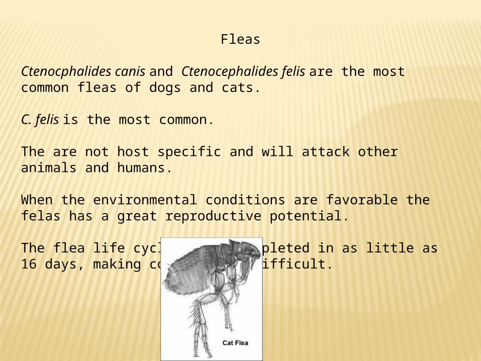
Fleas
Ctenocphalides canis and Ctenocephalides felis are the most common fleas of dogs and cats.
C. felis is the most common.
The are not host specific and will attack other animals and humans.
When the environmental conditions are favorable the felas has a great reproductive potential.
The flea life cycle can be completed in as little as 16 days, making control very difficult.
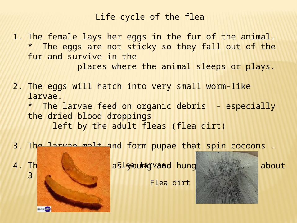
Life cycle of the flea
1. The female lays her eggs in the fur of the animal. * The eggs are not sticky so they fall out of the fur and survive in the
places where the animal sleeps or plays.
2. The eggs will hatch into very small worm-like larvae. * The larvae feed on organic debris - especially the dried blood droppings left by the adult fleas (flea dirt)
3. The larvae molt and form pupae that spin cocoons .
4. They then emerge as young and hungry adults in about 3 weeks. Flea larvae
Flea dirt


Once fleas have had a chance to establish the life cycle in a house and yard environment, no control program will be successful that does not emphasize environmental control.
Mechanical cleaning of the house and yard environment should precede any application of insecticides.
Flea bites

Mites
Most mites are host specific and even though they are morphologically similar, they will not cross-infest other hosts.
Mites live on the host continuously and infect other animals by contact.
The life cycles of these mites are all slightly different because some burrow and others live on the surface of the skin and deposit eggs. Some even burrow in the skin and deposit eggs.
The eggs hatch into six-legged nymphs, which develop and molt into adults.
The entire cycle requires 9 to 17 days.
Diagnosis of mites is based on the morphological appearance of the adult, and it generally requires a thorough skin scraping.

Demodex mites Ear mite
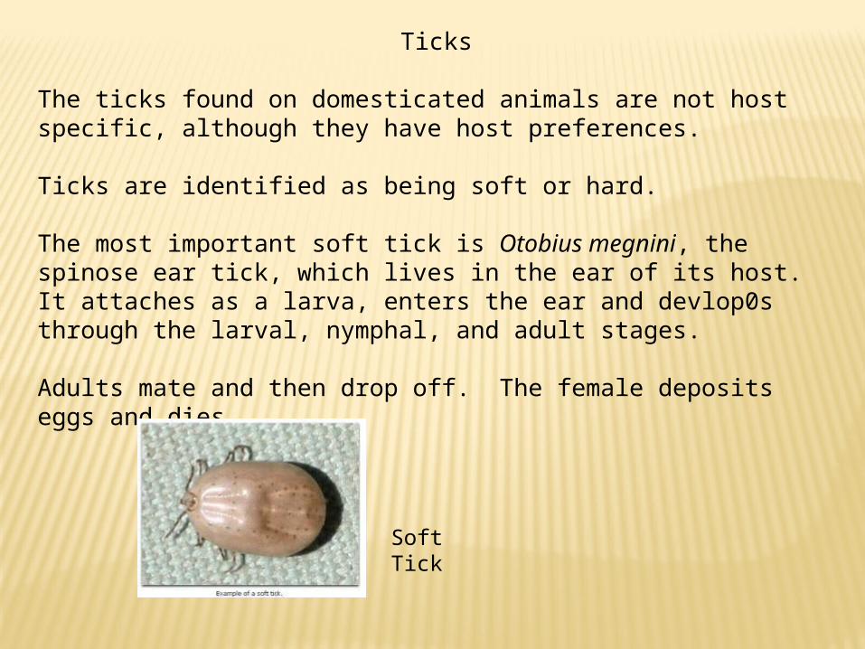
Ticks
The ticks found on domesticated animals are not host specific, although they have host preferences.
Ticks are identified as being soft or hard.
The most important soft tick is Otobius megnini, the spinose ear tick, which lives in the ear of its host. It attaches as a larva, enters the ear and devlop0s through the larval, nymphal, and adult stages.
Adults mate and then drop off. The female deposits eggs and dies.
Soft Tick
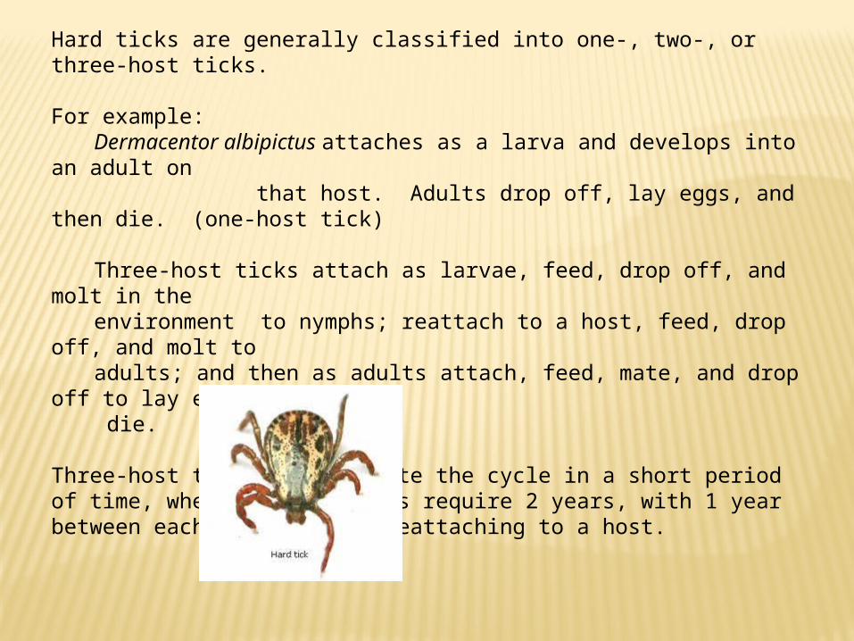
Hard ticks are generally classified into one-, two-, or three-host ticks.
For example: Dermacentor albipictus attaches as a larva and develops into an adult on that host. Adults drop off, lay eggs, and then die. (one-host tick)
Three-host ticks attach as larvae, feed, drop off, and molt in the
environment to nymphs; reattach to a host, feed, drop off, and molt to
adults; and then as adults attach, feed, mate, and drop off to lay eggs and
die.
Three-host ticks may complete the cycle in a short period of time, whereas other ticks require 2 years, with 1 year between each stage before reattaching to a host.
Diagnostic Procedures
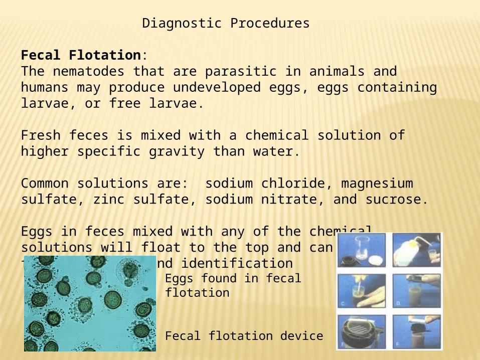
Fecal Flotation:The nematodes that are parasitic in animals and humans may produce undeveloped eggs, eggs containing larvae, or free larvae.
Fresh feces is mixed with a chemical solution of higher specific gravity than water.
Common solutions are: sodium chloride, magnesium sulfate, zinc sulfate, sodium nitrate, and sucrose.
Eggs in feces mixed with any of the chemical solutions will float to the top and can be removed for examination and identification
Eggs found in fecal flotation
Fecal flotation device
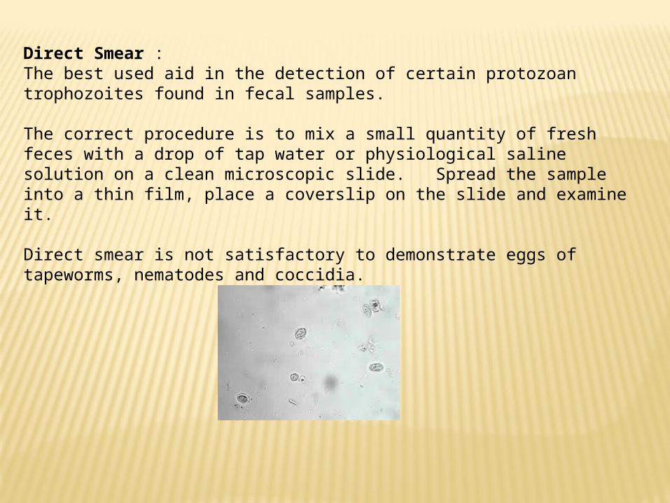
Direct Smear : The best used aid in the detection of certain protozoan trophozoites found in fecal samples.
The correct procedure is to mix a small quantity of fresh feces with a drop of tap water or physiological saline solution on a clean microscopic slide. Spread the sample into a thin film, place a coverslip on the slide and examine it.
Direct smear is not satisfactory to demonstrate eggs of tapeworms, nematodes and coccidia.

The Willis Technique
Equipment needed: sputum vial, glass microscope slide, coverslip and tongue depressor.
Procedure: 1. A small amount of feces (about the size of a pea) is placed in
the vial. 2. A sufficient flotation solution is added to cover the feces3. The feces is then macerated and mixed4. Solution is added until the vial is about half full and the
material is mixed again5. The vial is filled with flotation solution until the meniscus
bulges slightly6. Place a clean microscope slide over the vial – leave in place 10
minutes7. Lift microscope slide straight up and turn over8. A coverslip is then affixed.
Much of the liquid will drain from the slide, but the eggs remain firmly attached if the glass is clean.
Related Documents