Clinical Surgery Pearls

Welcome message from author
This document is posted to help you gain knowledge. Please leave a comment to let me know what you think about it! Share it to your friends and learn new things together.
Transcript

Clinical Surgery Pearls
Clinical Surgery Pearls
R Dayananda Babu MS MNAMS FAES
Professor of SurgeryPushpagiri Medical College
Tiruvalla, KeralaIndia
JAYPEE BROTHERS MEDICAL PUBLISHERS (P) LTDKochi • St Louis (USA) • Panama City (Panama) • London (UK) • New Delhi • Ahmedabad
• Bengaluru • Chennai • Hyderabad • Kolkata • Lucknow • Mumbai • Nagpur
®
Published by dr-notes.com
Published byJitendar P VijJaypee Brothers Medical Publishers (P) Ltd
Corporate Office4838/24 Ansari Road, Daryaganj, New Delhi - 110002, India, Phone: +91-11-43574357 Fax: +91-11-43574314
Registered OfficeB-3 EMCA House, 23/23B Ansari Road, Daryaganj, New Delhi - 110 002, IndiaPhones: +91-11-23272143, +91-11-23272703, +91-11-23282021+91-11-23245672, Rel: +91-11-32558559, Fax: +91-11-23276490, +91-11-23245683e-mail: [email protected], Website: www.jaypeebrothers.com
Offices in India• Ahmedabad, Phone: Rel: +91-79-32988717, e-mail: [email protected]• Bengaluru, Phone: Rel: +91-80-32714073, e-mail: [email protected]• Chennai, Phone: Rel: +91-44-32972089, e-mail: [email protected]• Hyderabad, Phone: Rel:+91-40-32940929, e-mail: [email protected]• Kochi, Phone: +91-484-2395740, e-mail: [email protected]• Kolkata, Phone: +91-33-22276415, e-mail: [email protected]• Lucknow, Phone: +91-522-3040554, e-mail: [email protected]• Mumbai, Phone: Rel: +91-22-32926896, e-mail: [email protected]• Nagpur, Phone: Rel: +91-712-3245220, e-mail: [email protected]
Overseas Offices
• North America Office, USA, Ph: 001-636-6279734e-mail: [email protected], [email protected]
• Central America Office, Panama City, Panama, Ph: 001-507-317-0160e-mail: [email protected]: www.jphmedical.com
• Europe Office, UK, Ph: +44 (0) 2031708910, e-mail: [email protected]
Clinical Surgery Pearls© 2010, Jaypee Brothers Medical Publishers
All rights reserved. No part of this publication should be reproduced, stored in a retrieval system, or transmitted in any form or by any means:electronic, mechanical, photocopying, recording, or otherwise, without the prior written permission of the author and the publisher.
This book has been published in good faith that the material provided by author is original. Every effort is made to ensure accuracy ofmaterial, but the publisher, printer and author will not be held responsible for any inadvertent error(s). In case of any dispute, all legal mattersto be settled under Delhi jurisdiction only.
First Edition: 2010ISBN 978-81-8448-922-4
Typeset at JPBMP typesetting unitPrinted at
Dedicated to
My late parents for their love and affection –
Mr Raghavan and Mrs Mallakshy
My only sister – the late Ms Damayanthy
My wife – Professor (Dr) Geetha Bhai and to my beloved
son Deepak D Babu for their moral support
My teachers for their wisdom
My patients for their trust and support
My students for their assistance
Published by dr-notes.com
FOREWORD
Professor R Dayananda Babu is known to me for the past forty years. I have great admiration forhis wealth of knowledge in the subject of surgery.
He has written the book Clinical Surgery Pearls with careful and persistent effort. The overridinggoal has been the mobilization of information relative to the science and skills of surgery. In additionto defining the frontiers of surgical knowledge it affords the student to assimilate the fundamentalsin an easy way.
This book will be an enormous help to those who are studying surgery at both undergraduateand postgraduate levels.
I wish the book a great success.
Professor (Dr) Mathew VargheseMS FRCS Ed.
Emeritus Professor of SurgeryGovernment Medical College
KottayamKerala, India
PREFACE
This book is the final result of my continuous teaching and learning process with my undergraduateand postgraduate students in surgery. Whenever I interact with my students, I realize their problemsand deficiencies and find out the solutions, so that it reaches them. Whenever I read a chapter, aseries of questions will come to my mind and then I will try to answer those questions. That is exactlythe reason why this book is in question-answer format. The flow charts and tables in this book areevolved in the classrooms and bedside teaching area.
Whenever I read a topic, I try to define the condition. I feel that when you define something,half the problem is solved and therefore the first section is devoted to definitions. There are more than100 definitions in this book.
Another important aspect of any learning process is to find out the concepts behind the diseaseprocess and management. These concepts are converted to an easily digestible capsule form in thisbook for the students. As an examiner at undergraduate and postgraduate level, I realized thatmost of the time the students miss many important clinical points during case presentation, notbecause they do not know them but because they do not have a checklist. Therefore I have givenchecklist for all clinical cases. The questions for the PG students are marked as PG in brackets sothat the undergraduate students can skip them if they feel so.
More than fifty clinical cases are discussed in this book (both long ones and short ones). Eachcase starts with a clinical capsule and questions are formulated based on the clinical capsule. Thereis a separate section for radiology and imaging and about 32 skiagrams are discussed. Importanttables and charts are included as a separate section for ready reference.
This is a clinical book of definitions, checklists, tables, flow charts, questions and answers. Allmy classes are distilled into a book and the title is “Clinical Surgery Pearls”. The preparation ofthis book took seven long years of hard work, and I completed this book single handedly. All theclinical photographs are taken by me with a small Kodak digital camera. The highlighted boxesand charts in this book will make it easily readable. I am sure the unique style and the student-oriented approach will make the learning process a pleasant experience.
R Dayananda Babu
Published by dr-notes.com

ACKNOWLEDGEMENTS
I thankfully acknowledge my patients for permitting me to take clinical photographs. The medicalillustrations are drawn in Adobe photoshop by my favorite student Dr Suraj Rajan, who is nowworking in the US. Dr Suraj also read the first “raw copy” and gave suggestions from the “studentpoint of view”, which is incorporated as student review. I am short of words to thank him. I amgrateful to all my Professors and teachers in surgery. I remember my great teachers like Prof CKPMenon, Prof KJ Jacob, Prof Mathew Varghese, Prof Balsalam, Prof Mohankumar, Prof KY Roy andProf CK Bahuleyan. My wife Dr Geetha Bhai helped me in proofreading and editing this book andwithout her help this could not have been possible. I also thank all my postgraduate andundergraduate students in surgery. I thank Shri Jitendar P Vij, Chairman and Managing Director,Jaypee Brothers Medical Publishers (P) Ltd. and appreciate the work of Mr Tarun Duneja (Director-Publishing, Jaypee Brothers), Mr John Paul, (author co-ordinator, Jaypee Brothers, Kochi) andMr Jagadish (Marketing Manager, Jaypee Brothers) and all the staff of Kochi branch for bringingout this book in time. Finally I am thankful to Mr Subramanian for spending time with me anddoing the DTP work of this book.
xiii
Contents
SECTION 1: Definitions1. Definitions 3
SECTION 2: Long Cases2. Toxic Goiter 233. Solitary Thyroid Nodule (STN-Nontoxic) 494. Papillary Carcinoma Thyroid with Lymph Node Metastasis 565. Multinodular Goiter 726. Early Breast Cancer 777. Locally Advanced Breast Cancer 968. Epigastric Lump 1079. Right Hypochondrial Lump Without Jaundice 121
10. Right Iliac Fossa Mass (suspected ileocecal tuberculosis) 12911. Suspected Carcinoma of the Cecum 13612. Appendicular Mass 14913. Obstructive Jaundice 15614. Varicose Veins 17215. Peripheral Occlusive Vascular Disease 19316. Lymphoma 21517. Renal Swelling 23418. Pseudocyst of Pancreas 24719. Retroperitoneal Tumor 25420. Testicular Malignancy 26221. Portal Hypertension 27722. Mesenteric Cyst 296
SECTION 3: Short Cases23. Non-thyroid Neck Swelling 30324. Tuberculous Cervical Lymph Node 307
CONTENTS
Published by dr-notes.com
xiv
Clinical Surgery Pearls
25. Cervical Metastatic Lymph Node and Neck Dissections 31626. Carcinoma Tongue with Submandibular Lymph Node 32927. Carcinoma of Gingivobuccal Complex (Indian Oral Cancer) 34028. Parotid Swelling 34629. Submandibular Sialadenitis 35830. Ranula, Plunging Ranula, Sublingual Dermoid and Mucous Cyst 36331. Thyroglossal Cyst, Lingual Thyroid, Ectopic Thyroid, Subhyoid
Bursa and Carcinoma Arising in Thyroglossal Cyst 36732. Branchial Cyst, Branchial Fistula, Cystic Hygroma 37333. Soft Tissue Sarcoma 37934. Neurofibroma, von Recklinghausen’s Disease 38735. Lipoma (Universal Tumor) 39436. Sebaceous Cyst/Epidermoid Cyst/Wen/Dermoid Cyst 39737. Ulcer 40238. Malignant Melanoma 41339. Basal Cell Carcinoma/Rodent Ulcer 42840. Squamous Cell Carcinoma—SCC (Epithelioma) 43341. Carcinoma Penis 44142. Congenital AV Fistula/Hemangioma/Compressible Swelling 44943. Unilateral Lower Limb Edema 45944. Hydrocele of Tunica Vaginalis Sac (Epididymal Cyst, Spermatocele,
Varicocele, Hematocele, Chylocele, etc.) 46845. Inguinal Hernia/Femoral Hernia 47846. Incisional Hernia (Ventral Hernia, Postoperative Hernia) 49947. Epigastric Hernia (Fatty Hernia of the Linea Alba) 50448. Paraumbilical Hernia, Umbilical Hernia in Adults and Children 50749. Desmoid Tumor, Interparietal Hernia (Interstitial), Spigelian Hernia 51450. Gynecomastia/Male Breast Carcinoma 51751. Fibroadenoma/Cystosarcoma/Breast Cyst/Fibroadenosis/
Fibrocystic Disease/ Mastalgia/Mastopathy/Chronic Mastitis 526
xv
Contents
SECTION 4: Radiology and Imaging52. Radiology Questions and Answers 535
SECTION 5: Important Tables and Charts53. General 59754. Trauma 60555. Burns 61256. Neck 61657. Breast 61758. Abdomen 62159. Vascular 63260. Limbs 63761. Anorectal 642
Index 645
Published by dr-notes.com

3
Definitions
1. ABDOMINAL APOPLEXY
Spontaneous hemorrhage into the peritonealcavity:
Causes:a. Tumors – Hepatoma
– Spleen– Other organs
b. Arteriosclerotic lesion in older individuals– Superior mesenteric artery —
Mesenteric apoplexy (spon-taneous rupture)
– Right colic artery– Branches of celiac.
c. Hemorrhage from congenital aneurysm inyoung patients — Bleeding from splenicartery aneurysm in pregnancy.
2. ABSCESS, COLD ABSCESS
Abscess: It is a localized collection of pus in apathological space lined by granulation tissue.Cold Abscess: Soft fluctuant swelling withoutsigns of inflammation, which is mistaken for acyst. This is lined by granulation tissue andcaseous material. It is due to tuberculousinfection and contains tubercle bacilli. It is not
hot. Brawny induration, edema and tendernessare absent.
3. ACUTE ABDOMEN
Any sudden spontaneous nontraumaticdisorder affecting the abdomen for whichurgent operation may be necessary and unduedelay in diagnosis may adversely affect theoutcome.
4. ADL (ACTIVITIES OF DAILY LIVING)
It is critical to assess the functional status of theprospective older candidate for surgery prior toscheduling an operation.The activities are—1. Feeding oneself2. Bathing3. Toileting (continence)4. Transferring from bed to chair5. Dressing6. Grooming.
Instrumental ADLs are more complex —a. Food preparationb. Shoppingc. Balancing.
Definitions
4
Clinical Surgery Pearls
5. AGENESIS / ATRESIA
Agenesis: Failure of development of an organor structure.Atresia: Failure to canalize a viscera.
6. AMYLASE
Amylase: A serum amylase level four times abovethe normal is indicative of acute pancreatitis.
7. ANKYLOGLOSSIA
Inability to protrude the tongue due to involve-ment of the muscles of tongue by carcinoma.The tongue deviates to the affected side.
8. APATHETIC HYPERTHYROIDISM
Asymptomatic mild hyperthyroidism occurringin the elderly recognized only by laboratoryfindings.
9. ARC OF RIOLAN (MEANDERINGMESENTERIC ARTERY)
The left colic artery near the splenic flexurebifurcates, one of the branches passes to the rightin the transverse mesocolon to anastomose witha similar branch of middle colic artery to formthe Arc of Riolan. This has got important role insupplementing the marginal artery (Fig. 10.1).
10. BACTEREMIA, PYEMIA, SEPTICEMIA
Bacteremia: Circulating bacteria in the bloodwithout producing disease.Pyemia: Circulating infective emboli composed
of masses of organisms, vegetations and infectedclots in the blood stream.Septicemia: Circulation of bacteria in bloodproducing disease.
11. BARRETT’S ESOPHAGUS
It is a metaplasia of the lower esophagealmucosa due to replacement of the squamousepithelium, by columnar epithelium,endoscopically having salmon pink appearancereplacing the whitish squamous epitheliumpathologically showing intestinal type ofepithelium with goblet cells.
12. BILIARY COLIC, CHOLECYSTITIS
The term colic is inaccurate for gallbladder. Itproduces constant pain in most cases as a resultof obstruction to cystic duct. The pain last for1–5 hours, and rarely shorter than 1 hourduration (Right upper quadrant pain radiatingto right upper back, right scapula or betweenthe scapula). Pain lasting beyond 24 hourssuggest acute inflammation – Cholecystitis.
13. BOIL, FURUNCLE, FURUNCULOSIS,FOLLICULITIS, CARBUNCLE
Folliculitis: Affection of the root of one hairfollicle alone by staphylococcus is calledfolliculitis.
Boil/Furuncle: Infection of the root of the hairfollicle with perifolliculitis caused byStaphylococcus is called Boil/Furuncle.
Published by dr-notes.com
5
Definitions
Furunculosis: Multiple boils with interveningnormal tissue is called furunculosis.
Carbuncle: Infective gangrene of skin andsubcutaneous tissue caused by staphylococcus(Multiple boils with involvement of interveningtissue also).
14. BREAST CARCINOMA—DEFINITIONS
Skin tethering and fixity: The skin tethering isdue to early involvement of ligaments ofCooper.
Manifested as puckering of the skin. Theunderling lump can be moved independentlyof the skin to some extent.
Skin fixity: It is due to invasion of carcinomaalong the ligaments of Cooper to the skin.
The lump and the skin cannot be movedseperately.
Retraction (Recent) of nipple: Extension ofgrowth along the lactiferous duct andsubsequent fibrosis.
Peau d’ Orange appearance is due to blockageof the lymphatics draining the skin – cutaneouslymphedema. The hair follicles are more firmlyfixed to the subcutaneous tissue than the rest ofthe skin. The hair follicles appear to be retractedand the between areas swell giving the orangepeel appearance.
TDLU: (Terminal Duct Lobular Unit):Thefunctional unit of the breast is the terminal ductlobular unit. All cancers of the breast and mostbenign conditions arise with in TDLU (Fig. 5.4).
Skin Involvement – T4b
Edema (including peau d’ orange) or ulcerationof the skin of the breast or satellite skin nodulesconfined to the breast. Dimpling of the skin andnipple retraction are not considered skininvolvement.
Inflammatory carcinoma breast: It is aclinicopathological entity characterized bydiffuse erythema and edema (peau d’ orange)of the breast without an underlying palpablemass, involving the majority of the skin of thebreast. This is due to tumor emboli withindermal lymphatics. The biopsy shoulddemonstrate cancer within the dermallymphatic or in breast parenchyma itself.Neglected LABC (locally advanced breastcancer) is not inflammatory Ca.
Extensive in situ component: If more than 25%of the main tumor mass contain in situ diseaseand there is in situ cancer in the surroundingbreast tissue, the cancer is classified as havingan extensive in situ component.
Chest wall infiltration: Chest wall includesRibs, intercostals muscles and serratus anteriormuscle but not the pectoral muscle.
Supraclavicular nodes: These are seen in atriangle defined by the omohyoid muscle andtendon, internal jugular vein (medial border)and the clavicle and subclavian vein (lowerborder). Adjacent nodes outside this triangle areconsidered to be lower cervical nodes (M1).
Multifocal: Tumor foci in the same quadrant iscalled multifocal.
6
Clinical Surgery Pearls
Multicentric: Tumor foci in different quadrantis called multicentric.
Microinvasion (Ti mic): Microinvasion of 0.1 cmor less in greatest dimension.
Micrometastasis: Tumor deposits greater than0.2 mm, but not greater than 2 mm in largestdimension having histologic evidence ofmalignant activity namely proliferation orstromal reaction.
Isolated tumor cells: Single cell or small clustersof cells not greater than 0.2 mm in largestdimension with no histologic evidence ofmalignant activity.
15. BRUIT
It is the sound produced by the turbulent bloodflow through a stenotic arterial segment which istransmitted distally along the course of the artery.When a bruit is heard over the peripheral vessel,stenosis is present at or proximal to that level.
It is heard loudest during systole and withgreater stenosis may extend into diastole. Thepitch of the bruit rises as the stenosis becomesmore marked. Absence of bruit does notindicated absence of occlusion. When thevessel become completely occluded, the bruitmay be disappeared.
16. BURNS, SCALD, FAT BURN
Burns: Injury by dry heat.Scald: Injury by moist heat.Fat burn: Injury by boiling oil.
17. BURSAE: BUNION, CLERGYMAN’SKNEE, GOLFER’S ELBOW, STUDENTSELBOW, HOUSEMAID’S KNEE, TENNIS
ELBOW
Bursae: These are fluid-filled cavities lined withflattened endothelium similar to synovium.Usually seen in relation to joints. When theydevelop over pressure points, they are calledadventitious bursae (see examples). Theyprevent friction during movement. Fluctuation,fluid thrill and transillumination are positive.
Housemaid’s knee: It is a subcutaneous bursaebetween patella and skin.
Clergyman’s knee: It is a subcutaneous bursabetween skin and ligamentum patella.
Students elbow: It is a subcutaneous bursaebetween skin and olecranon.
Golfer’s elbow: It is medial epicondylitisTenderness can be elicited at the medialepicondyle at the common flexor origin.Tennis elbow: It is lateral epicondylitis(Common extensor origin at the lateralepicondyle is affected).
Bunion: Sub cutaneous bursa between skin andhead of 1st metatarsal bone.
18. CARBUNCLE
Read boil.
19. CELLULITIS, ERYSIPELAS
Cellulitis: Spreading inflammation ofsubcutaneous and fascial tissue caused by
Published by dr-notes.com
7
Definitions
Streptococcus pyogenes. Commences in a trivialinfected wound. It has “No edge, No fluctuation,No pus and No limit”.
Morison’s aphorism: “Cellulitis occurring inChildren is never primary in the cellular tissue,but secondary to an underlying bone infection”.
Cellulitis of the scrotum: Always rule outextravasation of urine.
Erysipelas: It is cuticular lymphangitis.
Milian’s ear sign: Facial erysipelas spreads andinvolves the pinna because it is cuticularlymphangitis. Subcutaneous inflammationsstop short for the pinna because of closeadherence of the skin to the cartilage.
20. CLAUDICATION, REST PAIN
Claudication: (I limp). Claudication is the cramplike muscle pain which appears followingexercise when there is an inadequate arterialblood flow.It must fulfill three criteria1. It is a cramp like muscle pain (usually the
calf)2. Pain develops only when the muscle is
exercised.3. The pain disappears when the exercise
stops.
Rest pain: It is the continues pain caused bysevere ischemia. This pain is present at restthrough out the day and the night. The pain isrelieved by putting the leg below the level ofthe heart.
21. CLERGYMAN’S KNEE
Read bursae.
22. COLD ABSCESS
Read abscess.
23. COMPRESSIBILITY, REDUCIBILITY
Compressibility: When the contents of aswelling can be emptied by squeezing but theswelling reappear spontaneously on release ofpressure.
Reducibility: When the contents of a swelling canbe emptied by squeezing but does not returnspontaneously. This requires additional forcesuch as cough or effect of gravity. E.g: Hernia.
24. COMPOUND PALMAR GANGLION
Compound palmar ganglion: It is a tuberculousaffection of ulnar bursae, with a swelling in thehollow of the palm, extending to the lower forearm. Cross fluctuation can be elicited betweenthe palm and lower forearm.
25. CONSTIPATION, OBSTIPATION
Constipation: A bowel frequency of less thanone every 3 days. (Fewer than two per week).Obstipation: (Absolute constipation): Absenceof passage of both stool and flatus.
26. COUGH IMPULSE
Cough Impulse: Expansile impulse seen or felt overa swelling when the patient coughs, cries or strains.
8
Clinical Surgery Pearls
27. CREPITUS
Crepitus: (Grating or crackling sensationimparted to the examining fingers) may bepresent when the joint contain loose bodies. Maycommunicate with joint. It is also seen in thefollowing conditions.
• Subcutaneous emphysema (surgical emphy-sema) – gas is present in the subcutaneoustissue.
Four types:a. Traumatic – Fracture ribs, injury to nasal
fossa, breach of continuity of larynx,tacheostomy, fracture skull involvingsinuses.
b. Infective – Gas gangrene.c. Extraneous – After fluid administration,
closure of surgical wound, etc.d. Complicating rupture of esophagus.
• Fracture of bones• Extravasation of gas in pneumoperitoneum• Pseudo gas gangrene (air entrapped in the
subcutaneous tissue after laparotomy).
28. CYST
Cyst: It is a pathological fluid-filled sac boundby a wall. It may be true or false. Congenital oracquired.True cyst: It is one in which the sac is lined withcells of epithelial origin.False cyst: It is a walled off fluid collection notlined by epithelium. False cyst may beinflammatory or degenerative.
Examples of false cyst:
• Dental / Radicular cyst• Encysted pleural effusion• Pseudocyst of pancreas• Cystic degeneration of tumors• Brain cyst
29. DERMOID
Dermoid: Cyst formation due to sequestrationof epithelium deep to the skin surface.
30. DIETL’S CRISES
Dietl’s crises: This is due to intermittenthydronephrosis. After an attack of acute renalpain, a swelling is found in the loin due to thehydronephrosis. Following the passage of largevolume of urine some hours later, the pain isrelieved and the swelling will disappear.
31. DIVERTICULUM, DIVERTICULOSIS
Diverticulum: Abnormal external projectionfrom a hollow viscus external to the serosa iscalled diverticulum. It may be true or false,congenital or acquired. Congenital is true andacquired is false (one meaning of diverticulumis a wayside house of ill-fame).
True diverticulum: Containing all the layers ofthe bowel wall.
False diverticulum: There is no muscle coat, butall other layers (herniation of mucosa orsubmucosa through the muscular coat).
Published by dr-notes.com
9
Definitions
Pulsion diverticulum: The diverticulum ispushed out by intraluminal pressure.
Traction diverticulum: Diverticulum developsas a result of external traction.
Diverticulosis: Presence of multiple falsediverticula.
32. DIARRHEA
Diarrhea: If stools contain more than 300mlfluid daily.
33. EDEMA
Edema: It is an imbalance between capillaryfiltration and lymphatic drainage (this does notmean that all edemas are lymphedemas). Thiswill occur only when the lymphatic system failsto drain the tissue fluid produced by normalcapillary filtration.
34. EMPYEMA
Empyema: Collection of pus in a physiologicalspace.
35. ERYSIPELAS (READ CELLULITIS)
Erysipelas: Spreading cuticular lymphangitiscaused by Streptococcus pyogenes. It has asharply defined margin unlike cellulitis. Thevesicles contain serum. Milian’s ear sign –Erysipelas can spread to the pinna.
36. ERYTHROPLAKIA, LEUKOPLAKIA
Erythroplakia: Any lesion of the oral mucosathat presents as bright red velvety plaqueswhich can not be characterized clinically orpathologically as any other recognizable condition.Leukoplakia: Any white patch or plaque thatcan not be characterized clinically or patho-logically as any other disease.
37. EXOTOXIN, ENDOTOXIN
Exotoxin: Toxin liberated by living bacteria.Endotoxin: Toxin liberated after death ofbacteria, being a part of the organism itself.
38. EVIDENCE — LEVELS
Levels of evidences: Agency for health carepolicy and research grading system for evidenceand recommendation.
Evidence Description
I a Evidence from meta analysis of randomized controlled trials RCTI b Evidence from at least one RCTII a Evidence from at least one controlled study without randomizationII b Evidence from at least one other type of quasi – experimental study.III Evidence from non experimental descriptive studies, such as comparative studies and case
control studies. IV Evidence from expert committee reports or opinions or clinical experience of respected
authorities or both.
10
Clinical Surgery Pearls
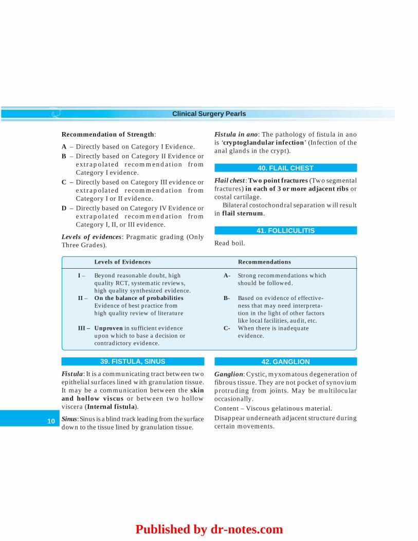
Recommendation of Strength:
A – Directly based on Category I Evidence.B – Directly based on Category II Evidence or
extrapolated recommendation fromCategory I evidence.
C – Directly based on Category III evidence orextrapolated recommendation fromCategory I or II evidence.
D – Directly based on Category IV Evidence orextrapolated recommendation fromCategory I, II, or III evidence.
Levels of evidences: Pragmatic grading (OnlyThree Grades).
Levels of Evidences Recommendations
I – Beyond reasonable doubt, high A- Strong recommendations whichquality RCT, systematic reviews, should be followed.high quality synthesized evidence.
II – On the balance of probabilities B- Based on evidence of effective-Evidence of best practice from ness that may need interpreta-high quality review of literature tion in the light of other factors
like local facilities, audit, etc. III – Unproven in sufficient evidence C- When there is inadequate
upon which to base a decision or evidence.contradictory evidence.
39. FISTULA, SINUS
Fistula: It is a communicating tract between twoepithelial surfaces lined with granulation tissue.It may be a communication between the skinand hollow viscus or between two hollowviscera (Internal fistula).
Sinus: Sinus is a blind track leading from the surfacedown to the tissue lined by granulation tissue.
Fistula in ano: The pathology of fistula in anois ‘cryptoglandular infection’ (Infection of theanal glands in the crypt).
40. FLAIL CHEST
Flail chest: Two point fractures (Two segmentalfractures) in each of 3 or more adjacent ribs orcostal cartilage.
Bilateral costochondral separation will resultin flail sternum.
41. FOLLICULITIS
Read boil.
42. GANGLION
Ganglion: Cystic, myxomatous degeneration offibrous tissue. They are not pocket of synoviumprotruding from joints. May be multilocularoccasionally.Content – Viscous gelatinous material.Disappear underneath adjacent structure duringcertain movements.
Published by dr-notes.com
11
Definitions
Fluctuation is present if not tense.
43. GANGRENE, NECROSIS, INFARCTION,SLOUGH
Gangrene: Macroscopic death of tissue withputrefaction.
Necrosis: Microscopic death of tissue.
Infarction: Ischemic necrosis is called infarction.
Slough: A piece of dead tissue separated fromliving tissue.
44. EARLY GASTRIC CANCER
Early gastric cancer: Cancer of the stomachconfined to the mucosa and submucosairrespective of the nodal status.
45. GASTRINOMA
Gastrinoma: A basal gastric acid output morethan 15 m mol/ HR and a fasting gastrin levelof more than 200 pg/ ml is strongly supportingthe diagnosis.
46. GASTRINOMA TRIANGLE(PSAROS TRIANGLE)
Gastrinoma triangle: The three points formingthe triangle are:1. Junction between the head and neck of the
pancreas.2. Junction of Cystic duct with CBD.3. Junction between 2nd and 3rd parts of the
duodenum.
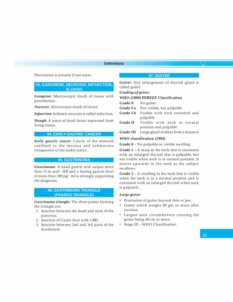
47. GOITER
Goiter: Any enlargement of thyroid gland iscalled goiter.Grading of goiter:WHO (1990) PEREZZ ClassificationGrade 0 No goiterGrade I a Not visible, but palpableGrade I b Visible with neck extended and
palpableGrade II Visible with neck in normal
position and palpableGrade III Large gland evident from a distance
WHO classification (1994)
Grade 0 – No palpable or visible swellingGrade 1 – A mass in the neck that is consistentwith an enlarged thyroid that is palpable, butnot visible when neck is in normal position. Itmoves upwards in the neck as the subjectswallowsGrade 2 – A swelling in the neck that is visiblewhen the neck is in a normal position and isconsistent with an enlarged thyroid when neckis palpated.
Large goiter:
• Protrusion of goiter beyond chin or jaw.• Goiter which weighs 80 gm or more after
excision.• Largest neck circumference crossing the
goiter being 40 cm or more.• Stage III – WHO Classification.
12
Clinical Surgery Pearls
48. GRANULOMA
Granuloma: Tumor-like mass formed in chronicinflammatory tissue.
49. HAMARTOMA, TERATOMA
Hamartoma: A tumor-like formation of tissuesindigenous to the site due to developmentalaberration.
Teratoma: Tumor-like proliferation of tissues,not indigenous in origin, containing more thanone germinal layer.
50. HEMATEMESIS, MELEMESIS, MELENA,HEMATOCHEZIA
Hematemesis: Vomiting of bright red or darkblood.
Melemesis: Vomiting of altered blood is calledmelemesis. Coffee ground vomitus is due tovomiting of blood that has been in the stomachlong enough for gastric acid to convert Hb tomethemoglobin.
Melena: Passage of black or tarry sticky,semisolid, stools due to the presence of alteredblood. It can be produced by blood entering thebowel at any point from mouth to cecum. Theblack color is due the Hematin (from Heme). 50to 100ml of blood in stomach can producemelena. 1 liter of blood in stomach will producemelena for 3–5 days.
Hematochezia: Passage of bright red bloodfrom the rectum (Colon, rectum, and anus)
is called hematochezia. Brisk bleeding fromupper intestine with rapid transit can alsoproduce it.
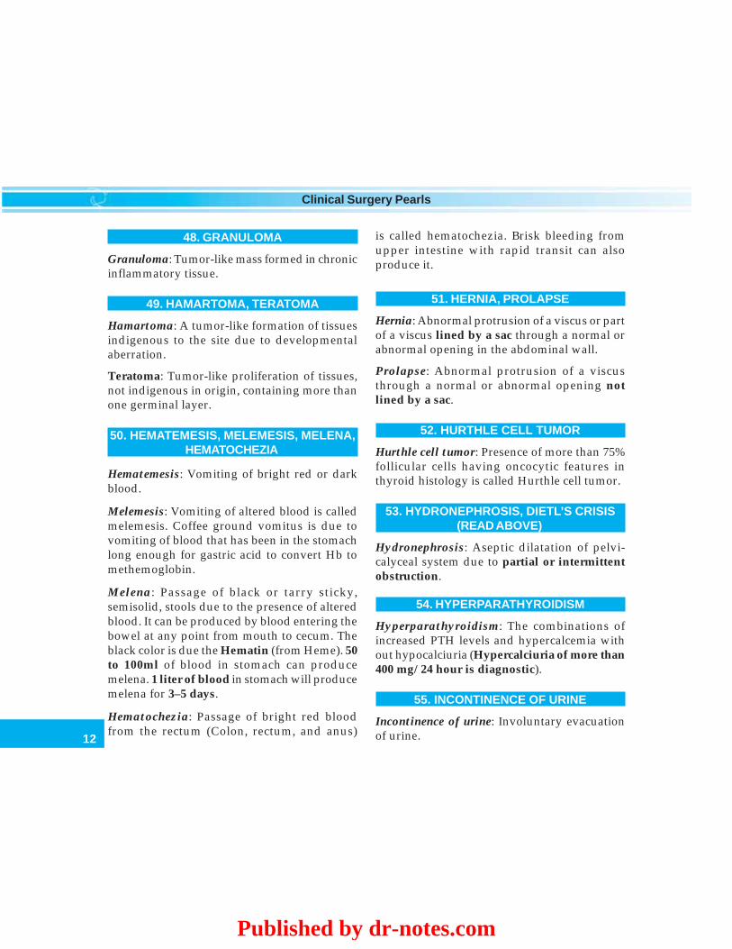
51. HERNIA, PROLAPSE
Hernia: Abnormal protrusion of a viscus or partof a viscus lined by a sac through a normal orabnormal opening in the abdominal wall.
Prolapse: Abnormal protrusion of a viscusthrough a normal or abnormal opening notlined by a sac.
52. HURTHLE CELL TUMOR
Hurthle cell tumor: Presence of more than 75%follicular cells having oncocytic features inthyroid histology is called Hurthle cell tumor.
53. HYDRONEPHROSIS, DIETL’S CRISIS(READ ABOVE)
Hydronephrosis: Aseptic dilatation of pelvi-calyceal system due to partial or intermittentobstruction.
54. HYPERPARATHYROIDISM
Hyperparathyroidism: The combinations ofincreased PTH levels and hypercalcemia without hypocalciuria (Hypercalciuria of more than400 mg/ 24 hour is diagnostic).
55. INCONTINENCE OF URINE
Incontinence of urine: Involuntary evacuationof urine.
Published by dr-notes.com
13
Definitions
56. INCONTINENCE OF STOOL
Incontinence of stool: Involuntary evacuationof stool.3. Typesa. Incontinence for solid facesb. Incontinence for liquid facesc. Incontinence for gas.
57. INFARCTION
Read gangrene.
58. INGUINAL CANAL
Inguinal canal: It is an intermuscular slitsituated between the superficial inguinal ringand deep inguinal ring.
59. INTUSSUSCEPTION
Intussusception: Telescoping of proximalintestine to the distal intestine.Retrograde intussusception: Telescoping of distalintestine into the proximal intestine (e.g: jejuno-gastric intussusception) after gastro-jeunostomy).
60. JAUNDICE
Jaundice: Yellowish discoloration of skin andmucous membrane due to excessive circulating bile.
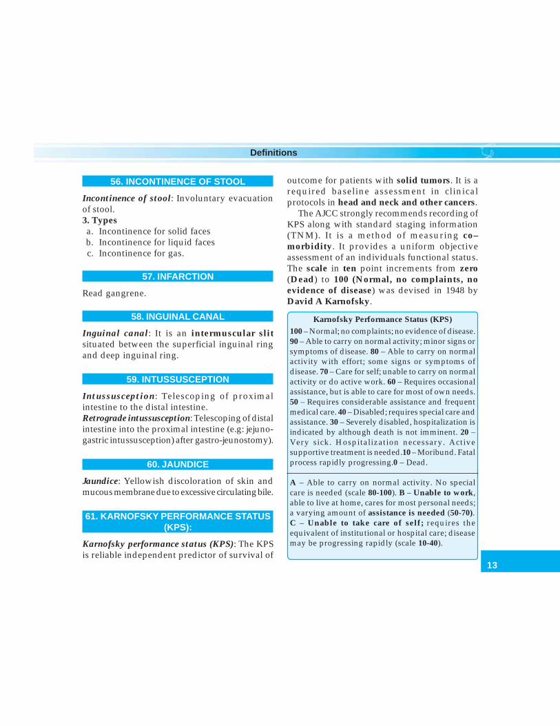
61. KARNOFSKY PERFORMANCE STATUS(KPS):
Karnofsky performance status (KPS): The KPSis reliable independent predictor of survival of
outcome for patients with solid tumors. It is arequired baseline assessment in clinicalprotocols in head and neck and other cancers.
The AJCC strongly recommends recording ofKPS along with standard staging information(TNM). It is a method of measuring co–morbidity. It provides a uniform objectiveassessment of an individuals functional status.The scale in ten point increments from zero(Dead) to 100 (Normal, no complaints, noevidence of disease) was devised in 1948 byDavid A Karnofsky.
Karnofsky Performance Status (KPS)100 – Normal; no complaints; no evidence of disease.90 – Able to carry on normal activity; minor signs orsymptoms of disease. 80 – Able to carry on normalactivity with effort; some signs or symptoms ofdisease. 70 – Care for self; unable to carry on normalactivity or do active work. 60 – Requires occasionalassistance, but is able to care for most of own needs.50 – Requires considerable assistance and frequentmedical care. 40 – Disabled; requires special care andassistance. 30 – Severely disabled, hospitalization isindicated by although death is not imminent. 20 –Very sick. Hospitalization necessary. Activesupportive treatment is needed.10 – Moribund. Fatalprocess rapidly progressing.0 – Dead.
A – Able to carry on normal activity. No specialcare is needed (scale 80-100). B – Unable to work,able to live at home, cares for most personal needs;a varying amount of assistance is needed (50-70).C – Unable to take care of self; requires theequivalent of institutional or hospital care; diseasemay be progressing rapidly (scale 10-40).
14
Clinical Surgery Pearls
62. LINE OF DEMARCATION
Line of demarcation: Zone of demarcationbetween viable and gangrenous tissue indicatedby a band of hyperemia and hyperesthesia onthe surface and separation is achieved by a layerof granulation tissue.
In dry gangrene the line of demarcationappears in a matter of days without infectionand this is called “separation by asepticulceration.”
In moist gangrene the line of demarcation ismore proximal than dry gangrene and the processis called “separation by septic ulceration”.
63. LIPOMA (UNIVERSAL TUMOR)
Lipoma: It is benign tumor from “adult fat cell’.It is called “universal Tumor” or “ubiquitoustumor” and hence the aphorism: “when indoubt hedge on fat”.
64. LOWER GI BLEED, UPPER GI BLEED
Lower GI bleed: It is a bleeding from distal tothe ligament of Treitz.
Upper GI bleed: It is a bleeding from proximalto the ligament of Treitz.
65. MARGINAL ARTERY OF DRUMMOND,ARC OF RIOLAN (READ ABOVE)
Marginal artery of drummond: It is the paracolicvessel of anastomosis between the superiormesenteric and inferior mesenteric arterial system.
66. MASSIVE HEMOTHORAX
Massive hemothorax: When 1500 ml or more ofblood is acutely removed from the pleural space,then it is called massive hemothorax.
67. MASSIVE BLOOD TRANSFUSION
Massive blood transfusion: The term massivetransfusion implies a single transfusion greaterthan 2500 ml or 5000 ml transfused over a periodof 24 hours.
68. MELENA, MELEMESIS
Read hematemesis.
69. MENARCHE—EARLY
Early menarche: Age of menarche before 12 years.
70. MENOPAUSE —LATE
Late menopause: Menopause after 50 years.
71. MESENTERY OF SMALL INTESTINE —ATTACHMENT
Mesentery of small intestine — attachment: Thebase of the mesentery attaches to the posteriorabdominal wall to the left of the second lumbarvertebra and passes obliquely to the right andinferiorly to the right sacroiliac joint crossing 3rdpart of the duodenum, aorta, IVC and right ureter.It is 6 inches (15 cm) in length. Remember thesmall intestine has got 6 meters length (Fig. 2.1).
Published by dr-notes.com
15
Definitions
72. MESENTERY OF SIGMOID —ATTACHMENT
Mesentery of sigmoid - attachment: It is shapedlike an inverted V. The apex of the V is at thebifurcation of left common iliac artery crossingthe brim. The right limb descends to the thirdpiece of the sacrum. The left limb runs alongthe brim of left side of pelvis. (Fig. 10.2).
73. MESENTERY OF THE TRANSVERSECOLON
Mesentery of the transverse colon: It is attachedto the descending part of Duodenum to the headand lower aspect of the body of the pancreasand placed horizontally to the anterior surfaceof the left Kidney.
74. NECROSIS
Read gangrene.
75. OLD AGE
Old age: Above 65 years is old age and above85years is very old age.
76. ORAL CAVITY, BUCCAL MUCOSA,RETROMOLAR TRIGONE, TRISMUS,
ANKYLOGLOSSIA
Oral cavity: Starts at skin vermilion junctionof lip anteriorly to circumvallate papillae oftongue, posterior part of the hard palate, andanterior pillar of tonsil posteriorly. Oral cavityincludes the following.• Lips• Buccal mucosa
• Upper and lower alveolar ridge• Retromolar trigone• Floor of the mouth• Hard palate• Oral tongue
Buccal mucosa: Extends from the upper alveolarridge down to the lower alveolar ridge, and fromthe commissure anteriorly to the mandibularramus and retromolar region posteriorly.
Retromolar trigone: It is defined as the anteriorsurface of the ascending ramus of the mandible.It is triangular in shape with the base beingsuperior behind the third upper molar tooth andthe apex inferior behind the 3rd lower molar.
Trismus: (Spasmodic clenching) is inability toopen the mouth.
Causes for Trismus·• Oral carcinoma – Involvement of pterygoid,
masseter, temporalis and buccinator muscle.• Inflammatory – Parotitis• Tooth abscess (Dental)• Erupting wisdom tooth• Peritonsillar abscess (Quinsy)• Tetanus — (Painful smiling — Risus
Sardonicus)Ankyloglossia (Read above).
77. PANCREATITIS, PANCREATICNECROSIS, PANCREATIC ABSCESS,PANCREATIC ASCITES, PANCREATIC
EFFUSION, PSEUDOCYST, PANCREATICNECROSIS, ACUTE FLUID COLLECTION
Chronic pancreatitis: It is a disease in whichthere is irreversible progressive destruction of
16
Clinical Surgery Pearls
pancreatic tissue. Its clinical course ischaracterized by dynamic progressive fibrosisof the pancreas.
Acute Pancreatitis
Acute fluid collection: It is fluid collection in ornear the pancreas with ill defined wall occurringearly in acute pancreatitis.
Pancreatitis acute pseudocyst: It is a collectionof pancreatic juice enclosed in a wall of fibrousor granulation tissue (Requires 4 weeks).
Pancreatic necrosis: Diffuse or focal area of nonviable pancreatic parenchyma. Associated peripancreatic fat necrosis is present.
Infected pancreatic necrosis: Same as above withinfection.
Pancreatic abscess: Circumscribed intraabdominal collection of pus in proximity topancreas. There is no pancreatic necrosis.
Pancreatic ascites: Chronic generalizedperitoneal enzyme rich effusion associated withpancreatic ductal disruption.
Pancreatic effusion: Encapsulated collection offluid in the pleural cavity.
78. PAPILLOMA (BENIGN PAPILLOMA),POLYP, POLYPOSIS
Benign papillomas: These are hamartomasconsisting of an overgrowth of all skin layersand its appendages having a central core andnormal sensation. They are well-defined,
usually, pedunculated ranging from fewmillimeters to a few centimeters in size,commonly 5 mm across. The surface may begrooved or deeply fissured. The complicationsof papilloma are inflammation, bleedingulceration, pigmentation and keratosis.
Polyp: It is a morphological term and nohistologic diagnosis is implied. They are massesof tissue that project into the lumen of viscera.When the base is broader than the head it iscalled sessile. When the base is narrower thanhead it is called pedunculated. It may be benignor malignant, Mucosal or sub-mucosal ormuscular.
Polyposis: Presence of many polyps.
Classification of polypa. Neoplastic
• Adenoma - Tubular adenoma,- Tubulovillous- Villous adenoma
• Carcinoid• Adenocarcinoma
b. Hamartomatous• Juvenile polyp (associated with
malrotation or Meckel’s diverticulum)• Peutz–Jeghers polyps
c. Inflammatory (Pseudo-polyp)• Benign lymphoid polyp
d. Hyperplastic polyp (Metaplastic polyp)• Diminutive lesions most often found in
left side of the colone. Miscellaneous
• Lipoma• Leiomyoma
Published by dr-notes.com
17
Definitions
79. PARALYTIC ILEUS
Paralytic ileus: Defined as a state in which thereis failure of transmission of peristaltic waves inthe intestine secondary to neuromuscularfailure [in the myenteric (Auerbach) and thesub-mucous (Meissner) plexuses.
80. PARAPHIMOSIS, PHIMOSIS
Phimosis: Inability to retract the foreskin toexpose the glans.
Paraphimosis: Inability to reduce a previouslyretracted foreskin.
81. PEAU D’ ORANGE
Read breast
82. PERFUSION, TRANSFUSION
Perfusion: Artificial passage of fluid throughblood vessel (usually veins).
Transfusion: Intravenous administration ofblood or its components.
83. PROLAPSE – READ HERNIA
Abnormal protrusion of a viscus through anormal or abnormal opening not lined by a sac.
84. PSEUDO THYROTOXICOSIS
Seen in critically ill patients characterized byincreased levels of T4 and decreased levels of T3due to failure of conversion of T4 to T3.
85. PUS
Pus: It is a fluid composed of living and deadbacteria, dead fixed and free cells (the latterrepresenting body’s phagocytic response) andforeign material such as sutures, implants andsplinters.
Color of the pus may give a clue regarding theorganism.• Creamy yellow – Staphylococci.• Watery opalescent – Streptococcus• Blue/Green – Pseudomonas• Purplish brown – Amoebic liver abscess• Yellow granules – Actinomycosis.
86. RENAL ANGLE
Renal angle: Angle between the 12th rib and theedge of the erectorspinae muscle. Normally thisis empty and resonant. There should not be anytenderness.
Rest pain: It is the continues pain caused bysevere ischemia. This pain is present at restthroughout the day and the night. The pain isrelieved by putting the leg below the level ofthe heart.
87. RETENTION OF URINE
Retention of urine: Accumulation of urine in thebladder with inability to void.
Acute retention: Sudden inability to pass urinewith a painful bladder.
Chronic retention: Retention with a painlessbladder.
18
Clinical Surgery Pearls
Size of Urinary CatheterFrench or Charriere’s scaleFr or Ch3 Fr = 1mm outer diameter of catheter
Recall Shakespeare’s ‘Seven Ages ofMan’ from As You Like It.
All the World’s a stage.And all the men and women merely players:They have their exits and their entrances:And one man in his time plays many parts,His acts being seven ages. At first the infant,Mewling and puking in the nurse’s arms.And then the whining school boy, with hissatchel,And shining morning face, creeping like snail,Unwillingly to school. And then the lover,Sighing like furnace, with a woeful balladMade to his mistress’ eyebrow. Then a solider,Full of strange oaths, and bearded like the pard,Jealous in honour, sudden and quick in quarrel,Seeking the bubble reputationEven in the cannon’s mouth. And then thejustice,In fair round belly with good capon lin’d,With eyes severe, and beard of formal cut,Full of wise saws and modern instances;And so he plays his part. The sixth age shiftsInto the lean and slipper’d pantaloon,With spectacles on nose and pouch on side;His youthful hose well say’d a world too wideFor his shrunk shank; and his big manly voice,Turning again towards childish treble, pipesAnd whistle in his sound. Last scene of all,That ends this strange eventful history,Is second childishness, and mere oblivionSans teeth, sans eyes, sans taste, sans everything
Important causes for retention of urine as perthe seven ages are—1. The infant – Posterior urethral valve2. The school boy – Enlarged bladder neck
(Marion’s disease)– Obturation by stone
3. The “lover age” – Retention followingacute urethritis
4. The soldier – Urethral stricture5. The justice – Benign enlargement of
the prostate6. The sixth age – Carcinoma of the
prostate7. The last age – Carcinoma of the
prostate– Benign enlargement of
the prostateThree most important causes for acute retentionin female• Retroverted gravid uterus (Do bimanual
palpation of uterus)• Disseminated sclerosis (CNS examination).• Hysteria.“Bashful bladder” – Cannot pass urine whenanother person is in the vicinity.
88. RETROMOLAR TRIGONE
Read oral cavity
89. RIGIDITY, GUARDING
Reflex contraction of the abdominal wallmuscles secondary to intraperitonealinflammation.
Published by dr-notes.com
19
Definitions
Rigidity: In Rigidity there is contraction evenat rest.
Guarding: In guarding it is secondary toprovocation from the examining hand of thephysician.
90. RUN IN, DISTAL RUN OFF
Distal run off: Patency of the main vesselbeyond an arterial occlusion seen in angiogram.
Run in: Patency of the main vessel proximal tothe site of occlusion in angiogram.
91. SCOLIOSIS
Scoliosis: Rotatolateral deformity of the spine.
92. SCREENING, SURVEILLANCE
Screening: It is defined as testing a group ofpeople considered to be at normal risk for adisease, to discover those at increased risk.
Surveillance: It is defined as testing of a groupknown to be at increased risk for a disease.
93. SINUS
Read fistula
94. STRICTURE, STENOSIS
Stricture: Narrowing of a length of canal orhollow organ.
Stenosis: Narrowing of a segment of canal ororifice.
95. STRANGURY, TENESMUS
Strangury: Painful, frequent, ineffectiveattempts at micturition.
Tenesmus : Painful, frequent, ineffectiveattempts at defecation.
96. TENSION PNEUMOTHORAX
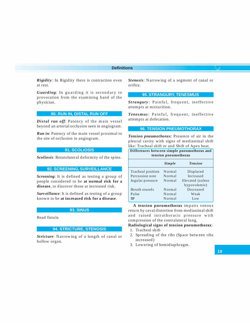
Tension pneumothorax: Presence of air in thepleural cavity with signs of mediastinal shiftlike: Tracheal shift or and Shift of Apex beat.
Differences between simple pneumothorax andtension pneumothorax
Simple Tension
Tracheal position Normal DisplacedPercussion note Normal IncreasedJugular pressure Normal Elevated (unless
hypovolemic)Breath sounds Normal DecreasedPulse Normal WeakBP Normal Low
A tension pneumothorax impairs venousreturn by caval distortion from mediastinal shiftand raised intrathoracic pressure withcompression of the contralateral lung.Radiological signs of tension pneumothorax:1. Tracheal shift2. Spreading of the ribs (Space between ribs
increased)3. Lowering of hemidiaphragm.
20
Clinical Surgery Pearls
97. THIRD DAY FEVER
Third day fever: If a patient is developing feveron the third postoperative day of surgery,suspect septic foci in the IV cannula.
98. TUBERCLE, CASEOUS MATERIAL,TUBERCULOUS PUS
Tubercle: Microscopically consists of an area ofcaseation surrounded by:a. Giant cells (having 20 or more peripherallyarranged nuclei)b. Zone of epithelioid cells around giant cellsc. Zone of inflammatory cells – lymphocytes andplasma cells.
Tubercle is visible to the naked eye towardsthe end of second week.
Caseous material: It is a dry, granular andcheese like material (Granular structurelessmaterial microscopically).
Tuberculous pus: Softening and liquefaction ofthe caseous material result in a thick creamyfluid called tuberculous pus. Liquefaction isassociated with multiplication of bacteria. It ishighly infective. It contains fatty debris inserous fluid with a few necrotic cells (It isusually sterile).
99. ULCER
Ulcer: Abnormal breach in the continuity of theskin or mucous membrane due to moleculardeath of tissue.
100. UPPER G I BLEED
Read Lower GI.
101. VARICOSE VEIN
Varicose vein: (WHO Definition) Abnormallydilated saccular or cylindrical superficial veinswhich can be circumscribed or segmental.
102. VOLVULUS
Volvulus: Axial rotation of a portion of bowelabout its mesentery. Volvulus can occur in thececum, sigmoidcolon and in the stomach.In the stomach, there are two types of volvulus.• Organoaxial – rotation of stomach in
horizontal direction (common).• Mesenteroaxial – rotation of the stomach in
the vertical direction.
103. WEIGHT LOSS
Weight loss: Loss of more than 10% body weightover a period of 6 month.
Published by dr-notes.com
23
Toxic Goiter
1 Toxic Goiter
CASE CAPSULE
A 30-year-old female patient with a thin build, haspresented with diffuse enlargement of thethyroid and palpitation of 6 months duration. Shecomplains of increased appetite and loss ofweight. She is apparently irritable and says she isintolerant to hot weather with excessive sweating.She has a preference for cold weather. She alsocomplains of insomnia and loss of concentrationability. She has diarrhea in addition. She is marriedand has a baby of six months old. She complainsof amenorrhea for the last three months. Onexamination, patient is agitated and nervous.Examination of the palms revealed that they aremoist and sweaty. She has tachycardia, fine andfast tremor, and protruded eyeballs. There isvisible diffuse enlargement of the thyroid. Onauscultation there is a systolic bruit heard in theupper pole of the thyroid. The carotids are felt inthe normal position. The trachea is central. Thereis no evidence of retrosternal extension. Thecervical lymph nodes are not enlarged.
In all goiters or swelling in the neck assessthe following–1. What is the anatomical diagnosis – by
assessing the plane – deep to the deep fasciaand deep to the sternomastoid?
2. What is the pathological diagnosis, E.g.:nodular goiter, solitary thyroid nodule,carcinoma, etc.
3. What is the functional diagnosis – Whetherthe patient is euthyroid, hyperthyroid,hypothyroid.
Checklist for history
• Onset related to puberty, pregnancy• Residence: Endemic area or not• Ingestion of goitrogens• Intolerance to hot/cold temperature• Increased appetite with loss of weight
(Hyperthyroidism)• Gain in weight (Hypothyroidism)• Change in menstrual cycle• Bowel habit — diarrhea (Hyper), constipation
(Hypo)• Difficulty in swallowing• Difficulty in breathing• Hoarseness of voice• Postural cough during sleeping (retrosternal
extension)• History of palpitation/shortness of breath on
exertion• Insomnia, loss of concentration (Hyper)• Irritability/nervousness (Hyper)
24
Clinical Surgery Pearls
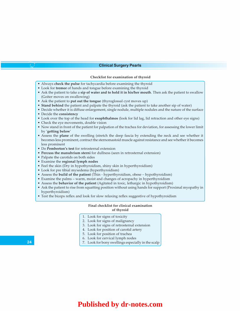
Checklist for examination of thyroid
• Always check the pulse for tachycardia before examining the thyroid• Look for tremor of hands and tongue before examining the thyroid• Ask the patient to take a sip of water and to hold it in his/her mouth. Then ask the patient to swallow
(Goiter moves on swallowing)• Ask the patient to put out the tongue (thyroglossal cyst moves up)• Stand behind the patient and palpate the thyroid (ask the patient to take another sip of water)• Decide whether it is diffuse enlargement, single nodule, multiple nodules and the nature of the surface• Decide the consistency• Look over the top of the head for exophthalmos (look for lid lag, lid retraction and other eye signs)• Check the eye movements, double vision• Now stand in front of the patient for palpation of the trachea for deviation, for assessing the lower limit
by ‘getting below’• Assess the plane of the swelling (stretch the deep fascia by extending the neck and see whether it
becomes less prominent, contract the sternomastoid muscle against resistance and see whether it becomesless prominent
• Do Pemberton’s test for retrosternal extension• Percuss the manubrium sterni for dullness (seen in retrosternal extension)• Palpate the carotids on both sides• Examine the regional lymph nodes• Feel the skin (Dry in hypothyroidism, shiny skin in hyperthyroidism)• Look for pre tibial myxedema (hyperthyroidism)• Assess the build of the patient (Thin - hyperthyroidism, obese – hypothyroidism)• Examine the palms – warm, moist and changes of acropachy in hyperthyroidism• Assess the behavior of the patient (Agitated in toxic, lethargic in hypothyroidism)• Ask the patient to rise from squatting position without using hands for support (Proximal myopathy in
hyperthyroidism)• Test the biceps reflex and look for slow relaxing reflex suggestive of hypothyroidism
Final checklist for clinical examinationof thyroid
1. Look for signs of toxicity2. Look for signs of malignancy3. Look for signs of retrosternal extension4. Look for position of carotid artery5. Look for position of trachea6. Look for cervical lymph nodes7. Look for bony swellings especially in the scalp
Published by dr-notes.com
25
Toxic Goiter
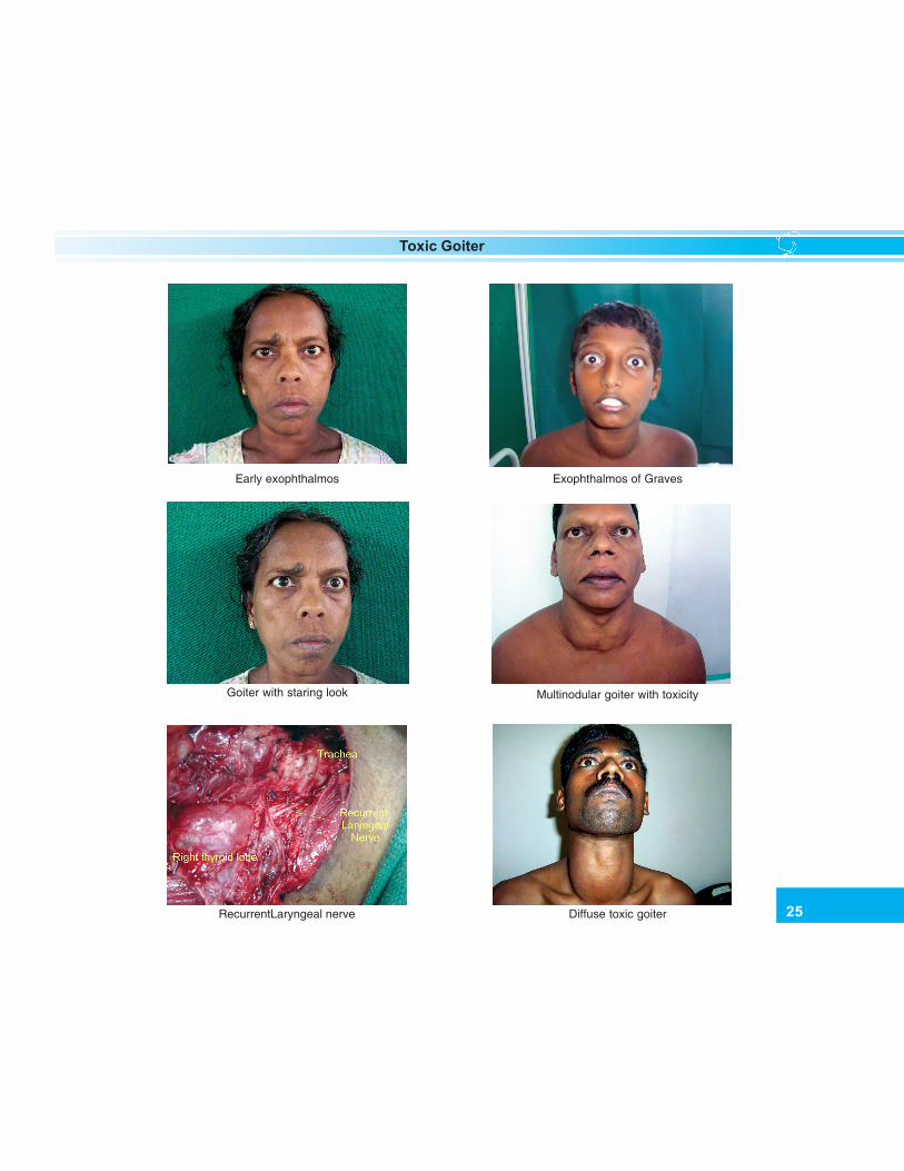
Early exophthalmos Exophthalmos of Graves
Goiter with staring look Multinodular goiter with toxicity
RecurrentLaryngeal nerve Diffuse toxic goiter
26
Clinical Surgery Pearls
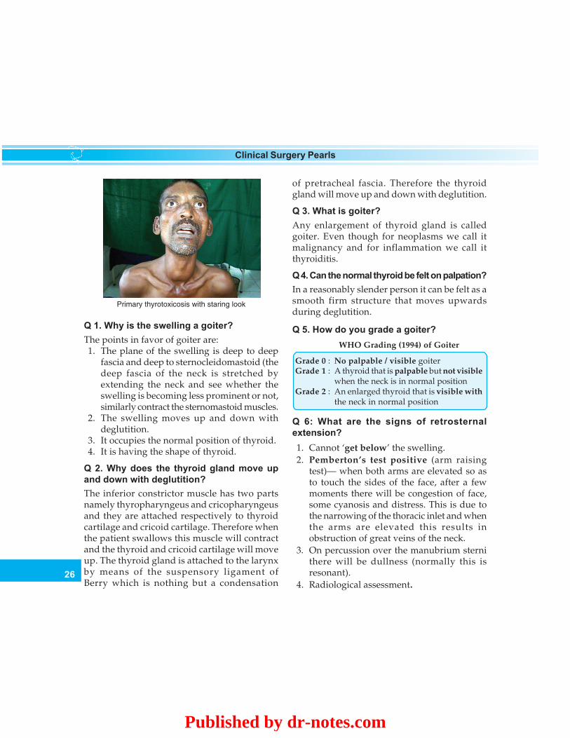
Q 1. Why is the swelling a goiter?The points in favor of goiter are:1. The plane of the swelling is deep to deep
fascia and deep to sternocleidomastoid (thedeep fascia of the neck is stretched byextending the neck and see whether theswelling is becoming less prominent or not,similarly contract the sternomastoid muscles.
2. The swelling moves up and down withdeglutition.
3. It occupies the normal position of thyroid.4. It is having the shape of thyroid.
Q 2. Why does the thyroid gland move upand down with deglutition?The inferior constrictor muscle has two partsnamely thyropharyngeus and cricopharyngeusand they are attached respectively to thyroidcartilage and cricoid cartilage. Therefore whenthe patient swallows this muscle will contractand the thyroid and cricoid cartilage will moveup. The thyroid gland is attached to the larynxby means of the suspensory ligament ofBerry which is nothing but a condensation
of pretracheal fascia. Therefore the thyroidgland will move up and down with deglutition.
Q 3. What is goiter?Any enlargement of thyroid gland is calledgoiter. Even though for neoplasms we call itmalignancy and for inflammation we call itthyroiditis.
Q 4. Can the normal thyroid be felt on palpation?In a reasonably slender person it can be felt as asmooth firm structure that moves upwardsduring deglutition.
Q 5. How do you grade a goiter? WHO Grading (1994) of Goiter
Grade 0 : No palpable / visible goiterGrade 1 : A thyroid that is palpable but not visible
when the neck is in normal positionGrade 2 : An enlarged thyroid that is visible with
the neck in normal position
Q 6: What are the signs of retrosternalextension?1. Cannot ‘get below’ the swelling.2. Pemberton’s test positive (arm raising
test)— when both arms are elevated so asto touch the sides of the face, after a fewmoments there will be congestion of face,some cyanosis and distress. This is due tothe narrowing of the thoracic inlet and whenthe arms are elevated this results inobstruction of great veins of the neck.
3. On percussion over the manubrium sternithere will be dullness (normally this isresonant).
4. Radiological assessment.
Primary thyrotoxicosis with staring look
Published by dr-notes.com
27
Toxic Goiter
Q 7. How will you assess the position oftrachea?The position of trachea can be assessed by:1. Palpation of trachea (this will be difficult in
case of large goiter)2. Auscultation to detect the position of
trachea3. Radiological.
Q 8. In which position you normally palpatea patient with thyroid?The examiner stands behind the patient and willdo the palpation.
Q 9. What is Kocher’s test?Slight compression on the lateral lobes of thyroidproduces stridor. If this test is positive it signifiesthat the patient has an obstructed trachea.
Q 10. What are the conditions in which youget narrowing of the trachea?Narrowing of trachea is found in1. Carcinoma of the thyroid2. Retrosternal goiters3. “Scabbard” trachea of long standing
multinodular goiter4. Riedel’s thyroiditis.
Q 11 What is plunging goiter?In this condition the whole of the enlargedthyroid lies in the superior mediastinum andthere is no palpable thyroid gland in the neck.When the intrathoracic pressure rises as incoughing, the goiter will be seen in the neck,this is called plunging goiter.
Q 12. What is Berry’s sign?
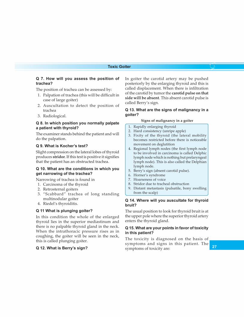
In goiter the carotid artery may be pushedposteriorly by the enlarging thyroid and this iscalled displacement. When there is infiltrationof the carotid by tumor the carotid pulse on thatside will be absent. This absent carotid pulse iscalled Berry’s sign.
Q 13. What are the signs of malignancy in agoiter?
Signs of malignancy in a goiter
1. Rapidly enlarging thyroid2. Hard consistency (unripe apple)3. Fixity of the thyroid (the lateral mobility
becomes restricted before there is noticeablemovement on deglutition
4. Regional lymph nodes (the first lymph nodeto be involved in carcinoma is called Delphiclymph node which is nothing but prelaryngeallymph node). This is also called the Delphianlymph node.
5. Berry’s sign (absent carotid pulse).6. Horner’s syndrome7. Hoarseness of voice8. Stridor due to tracheal obstruction9. Distant metastasis (pulsatile, bony swelling
from the scalp)
Q 14. Where will you auscultate for thyroidbruit?The usual position to look for thyroid bruit is atthe upper pole where the superior thyroid arteryenters the thyroid gland.
Q 15. What are your points in favor of toxicityin this patient?The toxicity is diagnosed on the basis ofsymptoms and signs in this patient. Thesymptoms of toxicity are:
28
Clinical Surgery Pearls
Symptoms of toxicity
System Symptoms
• Nervous system Nervousness, agitation, irrita-bility, insomnia, nervousinstability, tremor of the handsand tongue
• Cardiovascular Palpitation, dyspnea onsystem exertion, chest pain, etc.
• Metabolic and Increase in appetite and loss ofalimentary weight, change in bowel habit,system usually diarrhea; preference for
cold weather; excessivesweating; intolerance to hotweather
• Menstrual Usually amenorrhea or oligo-changes menorrhea
• Musculoskeletal Generalized weight loss;wasting and weakness of smallmuscles of the hand, shoulderand face.
Signs of toxicity
1. Uniform, smooth, soft or firm enlargement ofthyroid in Graves’ disease (primary) Bosselatedswelling or solitary nodule in case of secondary
2. The gland is vascular as evidenced by bruit3. Tremor of the outstretched hands (fine, fast) and
tongue4. Warm and moist hands5. Tachycardia6. Extra systoles, atrial fibrillation, and cardiac
failure7. Eye signs8. Myopathy — weakness of the proximal limb
muscle is commonly found. Severe muscularweakness resembling myasthenia gravis occursoccasionally.
Q 16. What are the eye signs of thyrotoxico-sis?
Eye signs
1. Lid retraction – this sign is caused by overactivity of involuntary smooth muscle part of thelevator palpebrae superioris muscle. If the uppereye lid is higher than normal and the lower lidis in correct position, the patient has gotlidretraction (this is not exophthalmos)
2. Lid lag (von Graefe’s sign) – when the upperlid does not keep pace with the eyeball as itfollows a finger moving from above downwards,it is lid lag
3. Exophthalmos – here both the eyelids are movedaway from center with sclera visible below orall around. Here the eyeball is pushed forwardsby increase in retroorbital fat, edema, andcellular infiltration (sclera should be alwaysvisible below the lower edge of eyes inexophthalmos)
4. The other eye signs are –a. Widening of the palpable fissure (Stellwag’s
sign) this is due to lid retractionb. Joffroy’s sign – absence of wrinkling of the
forehead when the head is bent downc. Möbius’s sign – difficulty in convergence
when the patient is asked to look at nearobjects
5. Severe exophthalmos – Intraorbital edema issuper added to the increased deposition of intraorbital fat. It comprises of;a. Intraorbital congestion – watering of eyes,
dilated blood vessels in lateral conjunctivab. Increased intraocular tensionc. Muscle paralysis (Ophthalmoplegia) –
evidenced by double vision, especially wheneye is moved upward end and outwards
Published by dr-notes.com
29
Toxic Goiter
(muscles of elevation and abduction namely,superior rectus and inferior oblique musclesare affected)
d. Chemosis
Q 17. What is pretibial myxedema?This is a misnomer and it is seen in primarytoxicosis (In Graves’ disease with exophthalmosonly). It is usually symmetrical. The earlieststage is a shiny red plaque of thickened skinwith coarse hair, which may be cyanotic whencold. In severe cases the skin of the whole legbelow knee is involved, together with that offoot and the ankle and there may be clubbing ofthe fingers and toes (Thyroid acropachy).
Q 18. What are the three most importantclinical types of toxicity?
Clinical types of thyrotoxicosis
1. Primary thyrotoxicosis/Graves/diffuse toxicgoiter
2. Secondary thyrotoxicosis/Plummer’s Disease/Toxic nodular goiter
3. Toxic nodule/adenoma/autonomous nodule
Q 19. What is the difference between thyro-toxicosis and hyperthyroidism?Thyrotoxicosis refers to the biochemical andphysiological manifestations of excessivethyroid hormone. Hyperthyroidism is a termreserved for disorders that result in the overproduction of hormone by the thyroid gland.Thyrotoxicosis need not be due tohyperthyroidism. In short in hyperthyroidismthe pathology is in the thyroid gland itself. The
causes for hyperthyroidism and toxicosiswithout hyperthyroidism are shown below.
Hyperthyroidism Toxicosis withouthyperthyroidism
1. Graves disease 1. Subacute thyroiditis*2. Toxic nodular 2. Ectopic functioning
goiter thyroid tissue3. Toxic adenoma 3. Silent thyroiditis4. Jod-Basedow’s 4. Struma ovarii
disease5. Metastatic follicular
carcinoma (functioning)6. Trophoblastic tumors7. Postpartum thyroiditis8. Thyrotoxicosis factitia
*Note: In thyroiditis, inflammation of thyroidcauses release of already formed thyroidhormones into the circulation, resulting intoxicosis. In other conditions like struma ovarii,trophoblastic tumors, etc. there is extra thyroidproduction of thyroxin from these tissues.
Q 20. What is Graves’ disease?The essential component of Gravess disease are• Diffuse goiter• Thyrotoxicosis• Autoimmune manifestations like:
– Infiltrative ophthalmopathy– Dermatopathy– Myopathy.
Q 21. What is the essential etiology of Graves’disease?Graves’ disease is an autoimmune disordercaused by thyroid stimulating immunoglobulins
30
Clinical Surgery Pearls
Primary Secondary
1. Enlargement of goiter is diffuse, firm or soft Bosselated or nodular not uniform2. Onset is abrupt Insidious3. Hyperthyroidism is usually severe Hyperthyroidism usually mild4. Cardiac failure is rare Cardiac failure or atrial fibrillation common5. Eye signs common Except lid lag and retraction other eye signs are
not seen6. No pre-existing goiter Pre existing nodular goiter for a long duration7. Usually younger women Usually middle aged or elderly8. The entire gland is overactive Internodular thyroid tissue is overactive, rarely
one or more nodules also may be overactive9. It is due to abnormal thyroid stimulating No such antibodies (it is due to over activity of
antibodies (TSAb) nodules)10. Can be managed by, drugs, radioiodine, and Surgery is the treatment of choice after control of
surgery the toxicity11. Manifestations not due to hyperthyroidism: Not seen
pretibial myxedema may occur
(TSIs) that have been produced against anantigen in the thyroid. This is directed to thethyroid stimulating hormone receptors (TSHR- Ab). This acts like TSH agonist. TSH Ab isfound only in Graves’ disease.
Q 22. What are the precipitating factors forprimary thyrotoxicosis?Remember 3 - S• Sex (puberty, pregnancy)• Sepsis• Psyche (sudden emotional upset).
Q 23. What are the differences betweenprimary thyrotoxicosis and secondary thyro-toxicosis?
Q 24. How will you confirm your diagnosisof toxicity?
Confirmation by:• Thyroid Function Test – T3, T4 and TSH.• Free T3, T4 are more significant and
meaningful. The T3 and T4 are raised and TSHis lowered in hyperthyroidism.Normal values are total T3 80 – 190 ng/dL
T4 5 – 12 mg/dLTSH 0-6 units/ml
Note: The total T3 and T4 hormone level will varydepending upon the amount of thyroid bindingglobulin (TBG).
Q 25. What are the other investigationsrequired?• Antithyroglobulin Antibody: More than
1:100.
• Thyroid Peroxidase (TPO): > 25Units( TPO and TSH antibodies are increased inAutoimmune Thyroiditis).
Published by dr-notes.com
31
Toxic Goiter
• TSH receptor antibodies are difficult toestimate.
• Radioisotope Scintigraphy (RadionuclideScan).
Q 26. What is the role of isotope scanning inthyroid?• The only absolute indication in thyrotoxicosis
for isotope scanning is for the diagnosis ofAutonomous Toxic Nodules.
• Toxicity with nodularity is an indication. Itcan identify hypofunctioning nodule (cold).Cold nodule in Graves is likely to bemalignant.
• It is the only method by which one candefinitely differentiate Primary, Secondaryand Toxic Nodules.
• Isotope scan can also differentiate hyper-
(e.g.: Thyroiditis).Other indications for isotope scan are:
• To identify ectopic thyroid tissue.• To identify recurrence and metastasis in
thyroid carcinoma.
Q 27. What is the isotope of choice fordiagnostic scanning of the thyroid?• 99mTc is the isotope of choice for diagnostic
purposes. It is cheap and the radiation is lessthan radioiodine. Twenty minutes afterintravenous injection of 99mTc, scanning isdone over the thyroid.
• If radio active iodine is used 123I is the isotopeof choice for diagnostic purposes.
Q 28. What is the half life of the variousradioisotopes used in thyroid?
Isotope Half life Route Rays Comment
I123 13 hours Oral Gamma rays Will not detect nodules < 1 cm size
I131 8 days Oral Gamma and beta rays Too much irradiation if used fordiagnostic scanning
I132 2.3 hours Oral Gamma and beta rays Not used for clinical purposes.
Tc99 6 hours IV Gamma rays Commonly used for diagnosticscanning of thyroid
thyroidism from toxicosis due to other causes.(To differentiate hyperthyroid thyrotoxicosisfrom nonhyperthyroid thyrotoxicosis). Theradioactive iodine uptake (RAIU) is increasedin hyperthyroidism where as toxicosis due toextra - thyroidal causes the RAIU is decreased
Q 29. What is the problem with Technetiumscanning?Carcinoma concentrates technetium andtherefore a hot nodule need not necessarily bebenign.
32
Clinical Surgery Pearls
Q 33. What are the toxic situations wherethere is decreased uptake of isotope inthyroid gland?Low uptake is seen in:• Thyroiditis• Postpartum thyrotoxicosis• Struma ovarii• Factitious thyrotoxicosis• Jod-Basedow thyrotoxicosisFig. 1.1: Primary toxicosis
Fig. 1.2: Secondary toxicosis
Fig. 1.3: Toxic nodule
Q 30. What is discordant scan?A nodule which is warm on technetiumscanning and cold on radioiodine scanningis called discordant scan. This is suggestive ofmalignancy.
Q 31. Why technetium is preferred over radio-iodine for diagnostic scanning?It gives small amount of radiation and you getthe image within minutes.
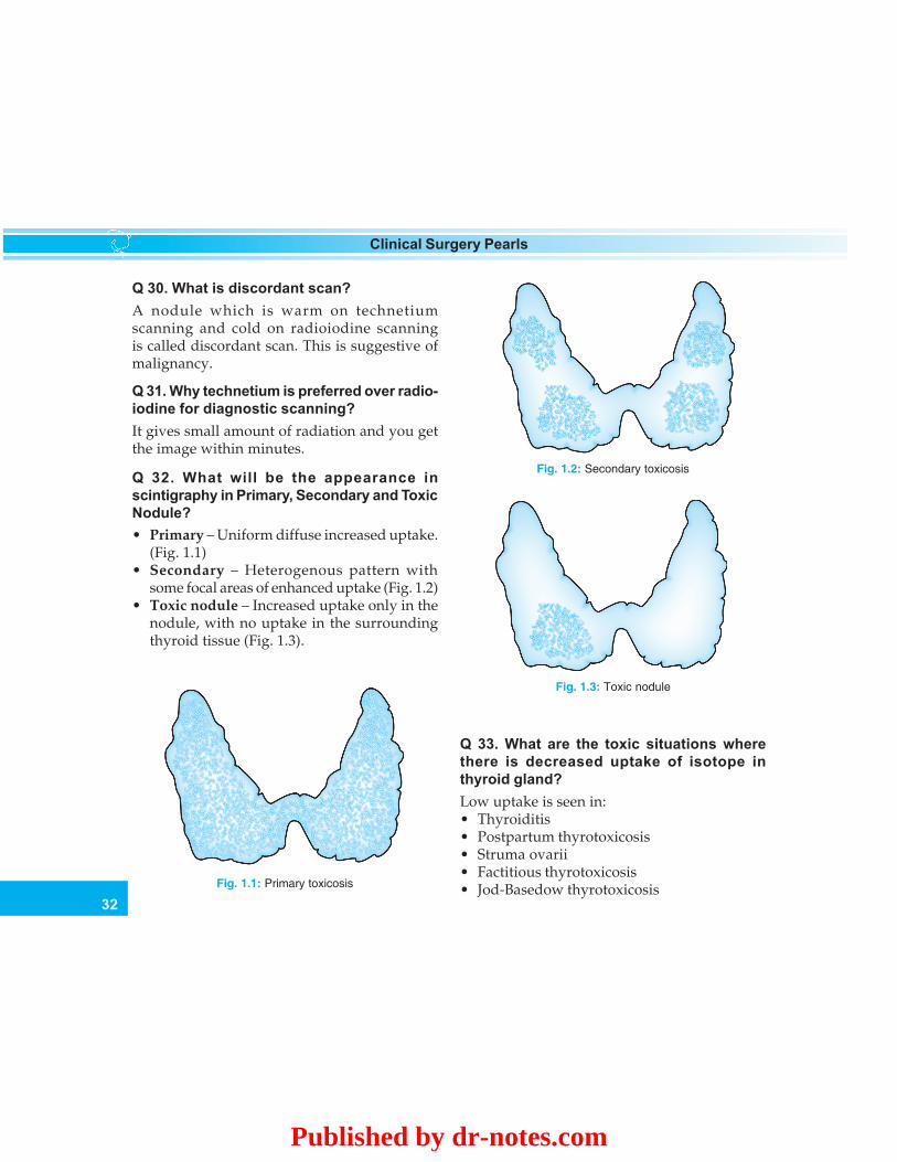
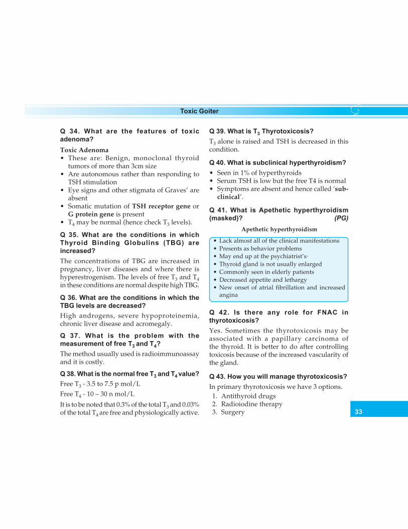
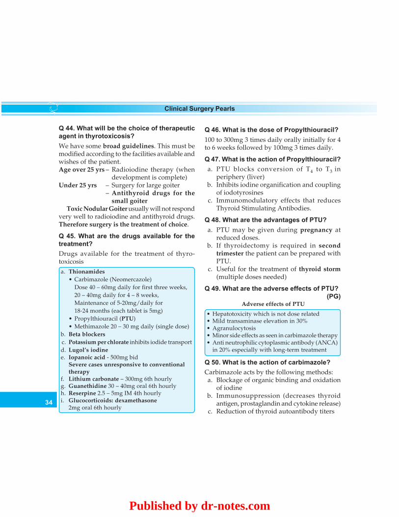
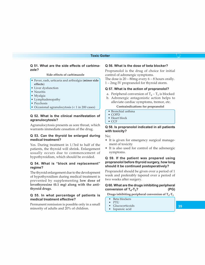
Q 32. What will be the appearance inscintigraphy in Primary, Secondary and ToxicNodule?• Primary – Uniform diffuse increased uptake.
(Fig. 1.1)• Secondary – Heterogenous pattern with
some focal areas of enhanced uptake (Fig. 1.2)• Toxic nodule – Increased uptake only in the
nodule, with no uptake in the surroundingthyroid tissue (Fig. 1.3).
Published by dr-notes.com
33
Toxic Goiter
Q 34. What are the features of toxicadenoma?Toxic Adenoma• These are: Benign, monoclonal thyroid
tumors of more than 3cm size• Are autonomous rather than responding to
TSH stimulation• Eye signs and other stigmata of Graves’ are
absent• Somatic mutation of TSH receptor gene or
G protein gene is present• T4 may be normal (hence check T3 levels).
Q 35. What are the conditions in whichThyroid Binding Globulins (TBG) areincreased?The concentrations of TBG are increased inpregnancy, liver diseases and where there ishyperestrogenism. The levels of free T3 and T4in these conditions are normal despite high TBG.
Q 36. What are the conditions in which theTBG levels are decreased?High androgens, severe hypoproteinemia,chronic liver disease and acromegaly.
Q 37. What is the problem with themeasurement of free T3 and T4?The method usually used is radioimmunoassayand it is costly.
Q 38. What is the normal free T3 and T4 value?Free T3 - 3.5 to 7.5 p mol/LFree T4 - 10 – 30 n mol/L
It is to be noted that 0.3% of the total T3 and 0.03%of the total T4 are free and physiologically active.
Q 39. What is T3 Thyrotoxicosis?T3 alone is raised and TSH is decreased in thiscondition.
Q 40. What is subclinical hyperthyroidism?• Seen in 1% of hyperthyroids• Serum TSH is low but the free T4 is normal• Symptoms are absent and hence called ‘sub-
clinical’.
Q 41. What is Apethetic hyperthyroidism(masked)? (PG)
Apethetic hyperthyroidism
• Lack almost all of the clinical manifestations• Presents as behavior problems• May end up at the psychiatrist’s·• Thyroid gland is not usually enlarged• Commonly seen in elderly patients• Decreased appetite and lethargy• New onset of atrial fibrillation and increased
angina
Q 42. Is there any role for FNAC inthyrotoxicosis?Yes. Sometimes the thyrotoxicosis may beassociated with a papillary carcinoma ofthe thyroid. It is better to do after controllingtoxicosis because of the increased vascularity ofthe gland.
Q 43. How you will manage thyrotoxicosis?In primary thyrotoxicosis we have 3 options.1. Antithyroid drugs2. Radioiodine therapy3. Surgery
34
Clinical Surgery Pearls
Q 44. What will be the choice of therapeuticagent in thyrotoxicosis?We have some broad guidelines. This must bemodified according to the facilities available andwishes of the patient.Age over 25 yrs – Radioiodine therapy (when
development is complete)Under 25 yrs – Surgery for large goiter
– Antithyroid drugs for thesmall goiter
Toxic Nodular Goiter usually will not respondvery well to radioiodine and antithyroid drugs.Therefore surgery is the treatment of choice.
Q 45. What are the drugs available for thetreatment?Drugs available for the treatment of thyro-toxicosisa. Thionamides
• Carbimazole (Neomercazole)Dose 40 – 60mg daily for first three weeks,20 – 40mg daily for 4 – 8 weeks,Maintenance of 5-20mg/daily for18-24 months (each tablet is 5mg)
• Propylthiouracil (PTU)• Methimazole 20 – 30 mg daily (single dose)
b. Beta blockersc. Potassium per chlorate inhibits iodide transportd. Lugol’s iodinee. Iopanoic acid - 500mg bid
Severe cases unresponsive to conventionaltherapy
f. Lithium carbonate – 300mg 6th hourlyg. Guanethidine 30 – 40mg oral 6th hourlyh. Reserpine 2.5 – 5mg IM 4th hourlyi. Glucocorticoids: dexamethasone
2mg oral 6th hourly
Q 46. What is the dose of Propylthiouracil?100 to 300mg 3 times daily orally initially for 4to 6 weeks followed by 100mg 3 times daily.
Q 47. What is the action of Propylthiouracil?a. PTU blocks conversion of T4 to T3 in
periphery (liver)b. Inhibits iodine organification and coupling
of iodotyrosinesc. Immunomodulatory effects that reduces
Thyroid Stimulating Antibodies.
Q 48. What are the advantages of PTU?a. PTU may be given during pregnancy at
reduced doses.b. If thyroidectomy is required in second
trimester the patient can be prepared withPTU.
c. Useful for the treatment of thyroid storm(multiple doses needed)
Q 49. What are the adverse effects of PTU?(PG)
Adverse effects of PTU
• Hepatotoxicity which is not dose related• Mild transaminase elevation in 30%• Agranulocytosis• Minor side effects as seen in carbimazole therapy• Anti neutrophilic cytoplasmic antibody (ANCA)
in 20% especially with long-term treatment
Q 50. What is the action of carbimazole?Carbimazole acts by the following methods:a. Blockage of organic binding and oxidation
of iodineb. Immunosuppression (decreases thyroid
antigen, prostaglandin and cytokine release)c. Reduction of thyroid autoantibody titers
Published by dr-notes.com
35
Toxic Goiter
Q 51. What are the side effects of carbima-zole?
Side effects of carbimazole
• Fever, rash, urticaria and arthralgia (minor sideeffects)
• Liver dysfunction• Neuritis• Myalgia• Lymphadenopathy• Psychosis• Occasional agranulocytosis (< 1 in 200 cases)
Q 52. What is the clinical manifestation ofagranulocytosis?Agranulocytosis presents as sore throat, whichwarrants immediate cessation of the drug.
Q 53. Can the thyroid be enlarged duringmedical treatment?Yes. During treatment in 1/3rd to half of thepatients, the thyroid will shrink. Enlargementusually occurs due to commencement ofhypothyroidism, which should be avoided.
Q 54. What is “block and replacement”regime?The thyroid enlargement due to the developmentof hypothyroidism during medical treatment isprevented by supplementing low dose oflevothyroxine (0.1 mg) along with the anti-thyroid drugs.
Q 55. In what percentage of patients ismedical treatment effective?Permanent remission is possible only in a smallminority of adults and 20% of children.
Q 56. What is the dose of beta blocker?Propranolol is the drug of choice for initialcontrol of adrenergic symptoms.The dose is 20 – 80mg every 6 – 8 hours orally.1 – 2mg IV propranolol for thyroid storm.
Q 57. What is the action of propranolol?a. Peripheral conversion of T4 – T3 is blockedb. Adrenergic antagonistic action helps to
alleviate cardiac symptoms, tremor, etc.Contraindications for propranolol
• Bronchial asthma• COPD• Heart block• CCF
Q 58. Is propranolol indicated in all patientswith toxicity?No;• It is given for emergency surgical manage-
ment of toxicity• It is also used for control of the adrenergic
symptoms.
Q 59. If the patient was prepared usingpropranolol before thyroid surgery, how longshould it be continued postoperatively?Propranolol should be given over a period of 1week and preferably tapered over a period oftwo weeks after surgery.
Q 60. What are the drugs inhibiting peripheralconversion of T4-T3? (PG)
Drugs inhibiting peripheral conversion of T4-T3
• Beta blockers• PTU• Glucocorticoids• Iopanoic acid
36
Clinical Surgery Pearls
Q 61. What is the minimum duration ofmedical treatment required before surgery?Thyroidectomy performed immediately aftercontrol of thyrotoxicosis is associated with riskof thyroid crisis and it is preferable to waitapproximately two months until after a patientis euthyroid.
Q 62. Is there any role for Dexamethasone inthe management of thyrotoxicosis? (PG)• It is used for the management of thyrotoxic
crisis• Dose is 2mg every 6th hourly (injection)• The actions are
a. Inhibits glandular secretion of hormoneb. Inhibits peripheral conversion of T4 to T3c. Immunosuppression
Q 63. What is Lugol’s Iodine and what is itsdose?Five percent iodine in 10% potassium iodide iscalled Lugol’s iodine.
The dose is 10 drops in a glass of water 3 timesdaily for 10 days.
Q 64. What are the actions of Lugol’s iodine?a. Decreases the vascularity of the glandb. Makes the thyroid firm and less friable
(helps in surgical removal)c. Prevents the release of hormone from the
gland – Thyroid constipation.
Q 65. What will happen if Lugol’s iodine isgiven for more than 10 days?After two weeks the effect of Lugol’s iodinetherapy is lost due to the so called thyroidescape from iodine control.
Q 66. What are the indications for radioiodinetherapy?• Radio iodine (I131) is usually given to patients
above 45 years for primary thyrotoxicosis.• Isotope facility must be available.
Q 67. What are the problems of radioiodinetherapy? (PG)
Problems of radioiodine therapy
a. Indefinite follow up is essential as the patientmay develop hypothyroidism (75%)
b. Chance of permanent thyroid failure – 90% (hypois more due to failure of cellular reproduction).
c. Theoretical possibility of genetic damage,leukemia, damage to fetus and carcinoma (noconvincing evidence)
d.Takes two to three months for control ofsymptoms
e. Worsening of ophthalmopathy (especially insmokers) and dermatopathy
f. Mild anterior neck paing. Increased risk of benign tumorsh. Malignant transformation in young patientsi. May induced hyperparathyroidism
Q 68. What are the contraindications of radio-iodine therapy?
Contraindications of radioiodine therapy
• Pregnancy• Lactating mothers• Women desiring pregnancy within 1 year• Children/adolescents (relative)
Q 69. What is the dose of radio iodine? (PG)300 to 600 MBq, if there is no clinical improve-ment after 12 weeks further dose is given. Twoor more doses are necessary in 20 to 30% of cases.
Published by dr-notes.com
37
Toxic Goiter
Q 70. What is the method of radioiodinetreatment for toxicity? (PG)a. Make the patient euthyroid with drugsb. Discontinue drugs for 5 daysc. Administer I131 300-600 MBq (5-10 mCi)d. Start antithyroid drugs after 1 week and
continue for 6 to 8weekse. After 12 weeks, if there is no improvement,
give another dose of radioiodinef. Two or more doses of radioiodine may be
required.
Q 71. What are the indications for surgery inthyrotoxicosis? (PG)
a. Intolerance or non-compliance with antithyroid drugs.
b. Contraindications to radio iodine therapy.c. Graves’ disease in children, adolescents and
those who are under the age of 25 years.d. In women who are potential mothers.e. Large goiter.f. Persistent thyromegaly.g. If antithyroid medication is required for more
than 2 years.h. Graves’ with nodulesi. Ophthalmopathyj. Pressure symptomsk. Toxic MNGl. Substernal goiterm. Amiodarone induced thyrotoxicosis
Q 72. What are drugs used for preparation ofa patient for “urgent thyroidectomy”? (PG)Combination of oral Iopanoic Acid 500mg bid+ Dexamethasone 1mg bid + PTU or MMI andBeta blockers for 5 to 7 days.
Q 73. What are the advantages of surgery?(PG)
Advantages of surgery
a. Surgery is effective in achieving euthyroidstatus in 95-97% of patients
b. Controls hyperthyroidism immediatelyc. Hazards associated with radioiodine therapy
are avoided.d. Surgery will provide tissue for histologye. Surgery will remove occult foci of malignancyf. Childbearing is immediately possibleg. Coexisting parathyroid carcinoma can be
removedh. Is a better treatment for toxicity with
ophthalmopathyi. No need for follow up because nodules are not
left behind.
Q 74. What is the surgical treatment ofprimary thyrotoxicosis?Near total thyroidectomy or subtotalthyroidectomy. Near total thyroidectomy isnow recommended as the treatment of choice.
Q 75. What is the recommended treatment forSecondary Thyrotoxicosis?Surgery is preferred over radioiodine forsecondary thyrotoxicosis because:• It will not respond to radioiodine as most of
the nodules may not take up radioiodine.• Large and repeated doses of radioiodine may
be required.
Q 76. What is the recommended treatment oftoxic nodule?Once the patient is made euthyroid, surgery inthe form of Hemithyroidectomy will givepermanent relief.
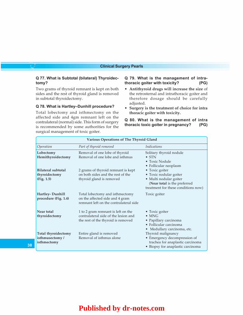
38
Clinical Surgery Pearls
Q 77. What is Subtotal (bilateral) Thyroidec-tomy?Two grams of thyroid remnant is kept on bothsides and the rest of thyroid gland is removedin subtotal thyroidectomy.
Q 78. What is Hartley–Dunhill procedure?Total lobectomy and isthmectomy on theaffected side and 4gm remnant left on thecontralateral (normal) side. This form of surgeryis recommended by some authorities for thesurgical management of toxic goiter.
Various Operations of The Thyroid Gland
Operation Part of thyroid removed Indications
Lobectomy Removal of one lobe of thyroid Solitary thyroid noduleHemithyroidectomy Removal of one lobe and isthmus • STN
• Toxic Nodule• Follicular neoplasm
Bilateral subtotal 2 grams of thyroid remnant is kept • Toxic goiterthyroidectomy on both sides and the rest of the • Toxic nodular goiter(Fig. 1.5) thyroid gland is removed • Multi nodular goiter
(Near total is the preferredtreatment for these conditions now)
Hartley- Dunhill Total lobectomy and isthmectomy Toxic goiterprocedure (Fig. 1.4) on the affected side and 4 gram
remnant left on the contralateral side
Near total 1 to 2 gram remnant is left on the • Toxic goiterthyroidectomy contralateral side of the lesion and • MNG
the rest of the thyroid is removed • Papillary carcinoma• Follicular carcinoma• Medullary carcinoma, etc.
Total thyroidectomy Entire gland is removed Thyroid malignancyisthmusectomy / Removal of isthmus alone • Emergency decompression ofisthmectomy trachea for anaplastic carcinoma
• Biopsy for anaplastic carcinoma
Q 79. What is the management of intra-thoracic goiter with toxicity? (PG)• Antithyroid drugs will increase the size of
the retrosternal and intrathoracic goiter andtherefore dosage should be carefullyadjusted.
• Surgery is the treatment of choice for intrathoracic goiter with toxicity.
Q 80. What is the management of intrathoracic toxic goiter in pregnancy? (PG)
Published by dr-notes.com
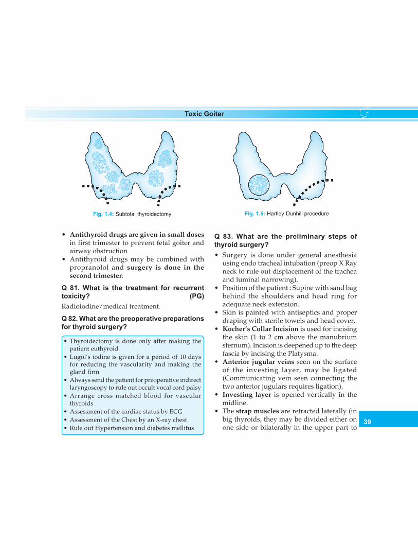
39
Toxic Goiter
• Antithyroid drugs are given in small dosesin first trimester to prevent fetal goiter andairway obstruction
• Antithyroid drugs may be combined withpropranolol and surgery is done in thesecond trimester.
Q 81. What is the treatment for recurrenttoxicity? (PG)Radioiodine/medical treatment.
Q 82. What are the preoperative preparationsfor thyroid surgery?
• Thyroidectomy is done only after making thepatient euthyroid
• Lugol’s iodine is given for a period of 10 daysfor reducing the vascularity and making thegland firm
• Always send the patient for preoperative indirectlaryngoscopy to rule out occult vocal cord palsy
• Arrange cross matched blood for vascularthyroids
• Assessment of the cardiac status by ECG• Assessment of the Chest by an X-ray chest• Rule out Hypertension and diabetes mellitus
Q 83. What are the preliminary steps ofthyroid surgery?• Surgery is done under general anesthesia
using endo tracheal intubation (preop X Rayneck to rule out displacement of the tracheaand luminal narrowing).
• Position of the patient : Supine with sand bagbehind the shoulders and head ring foradequate neck extension.
• Skin is painted with antiseptics and properdraping with sterile towels and head cover.
• Kocher’s Collar Incision is used for incisingthe skin (1 to 2 cm above the manubriumsternum). Incision is deepened up to the deepfascia by incising the Platysma.
• Anterior jugular veins seen on the surfaceof the investing layer, may be ligated(Communicating vein seen connecting thetwo anterior jugulars requires ligation).
• Investing layer is opened vertically in themidline.
• The strap muscles are retracted laterally (inbig thyroids, they may be divided either onone side or bilaterally in the upper part to
Fig. 1.5: Hartley Dunhill procedureFig. 1.4: Subtotal thyroidectomy
40
Clinical Surgery Pearls
save the Ansa Hypoglossi nerves entering thestrap muscles in the lower part)
• The Pretracheal fascia is incised verticallyand the thyroid gland is mobilized byligating the middle thyroid vein (this is thefirst vessel to be ligated).
Q 84. What are the essential steps ofthyroidectomy?A. Identification of parathyroid glands
• Identify the parathyroids before ligatingthe vessels
• Parathyroids have the color of peanutbutter, each of 6 × 3 × 3 mm size and lessthan 40 mg weight.
• The Recurrent Laryngeal Nerve (RLN) –Inferior thyroid artery junction is criticalin identifying the parathyroids (this is acritical area of RLN injury as well).
• The Superior Parathyroid glands areabove and behind this junction.
• The Inferior Parathyroid glands are belowand anterior to this junction havingvariable positions. The inferior glandsmay be situated on the inferior pole of thethyroid, thyrothymic ligament, in thethymus or in the perithymic fat.
B. Identification of the recurrent laryngealnerve• It is preferable to identify the entire
course of the nerve in thyroid surgery• There is no role for the old axiom “ nerve
seen is nerve injured”, which is called the‘ostrich philosophy’.
• The first identification is at the so-calledRiddle’s triangle, which is bound byinferior thyroid artery above, the carotid
artery laterally and the trachea medially• From there, the nerve is traced upwards
to the point of its entry into the larynx atthe greater cornu of the thyroid cartilage.
• Before entry, the nerve may divide intotwo or more extralaryngeal branches.
• When there is difficulty in identifying theRLN, the entry point of the nerve can belocated by identifying Zuckerkandl’stubercle.
• The nerve may cross the inferior thyroidartery usually deep to the artery,sometimes superficial or may even passthrough the fork of the branches of the artery
C. Ligation of vessels• The branches of the Superior Thyroid
vessels are individually skeletonized andligated as close to the superior pole aspossible after identifying the externalbranch of the Superior Laryngeal Nerve
• A medial approach to the superior polevia the avascular space between theCricothyoid muscle and the upper poleof the gland is ideal.
• “Mass Ligation” of the superior pole isto be avoided.
• Capsular ligation of the inferior thyroidartery:a. Inferior thyroid artery is an end artery
to the parathyroids and hence ligationof the trunk of the inferior thyroidartery is not recommended.
b. The small branches entering thecapsule of the gland alone are ligated,thereby preserving the blood supplyto the parathyroids.
c. These small branches are therefore
Published by dr-notes.com
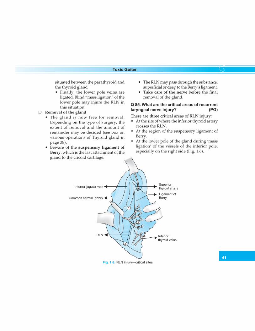
41
Toxic Goiter
situated between the parathyroid andthe thyroid gland• Finally, the lower pole veins are
ligated. Blind “mass ligation” of thelower pole may injure the RLN inthis situation.
D. Removal of the gland• The gland is now free for removal.
Depending on the type of surgery, theextent of removal and the amount ofremainder may be decided (see box onvarious operations of Thyroid gland inpage 38).
• Beware of the suspensory ligament ofBerry, which is the last attachment of thegland to the cricoid cartilage.
• The RLN may pass through the substance,superficial or deep to the Berry’s ligament.
• Take care of the nerve before the finalremoval of the gland.
Q 85. What are the critical areas of recurrentlaryngeal nerve injury? (PG)There are three critical areas of RLN injury:• At the site of where the inferior thyroid artery
crosses the RLN.• At the region of the suspensory ligament of
Berry.• At the lower pole of the gland during ‘mass
ligation’ of the vessels of the inferior pole,especially on the right side (Fig. 1.6).
Fig. 1.6: RLN injury—critical sites
42
Clinical Surgery Pearls
Q 86. What is Zuckerkandl’s tubercle? (PG)• It is the posterior extension of the lateral lobes
of the thyroid gland near the ligament of Berry.• It is found in 14 - 55% of cases.• The RLN runs cranially in a fissure between
the Zuckerkandl’s tubercle and the mainbody of the gland or the tracheal surface
• The RLN may have a sharp angle beneath thetubercle.
• The nerve is so constantly related to thistubercle, that it is often called “an arrowpointing to the RLN”
Q 87. What is nonrecurrent laryngeal nerve?• This is seen on the right side as a result of
failure of development of the 4th aortic arch• The RLN here may arise as a branch from the
vagus at the level of the inferior horn of thethyroid cartilage (instead of going down,curving around the subclavian artery andcoming up).
• The incidence is 0.2 – 0.5 %.
Q 88. What is suspensory ligament of Berry?• It is nothing but a condensation of the
pretracheal fascia attaching the thyroid glandto the cricoid.
• The two ligaments on either side form a slinganchoring the gland to the larynx.
• It must be severed before the gland can beremoved.
• The RLN is in immediate contact with theback of the ligament.
Q 89. What are the complications of thyroi-dectomy?
Complications of thyroidectomycan be classified as
A. Metabolic1. Hypoparathyroidism
• Temporary Hypoparathyroidism• Temporary hypocalcemia without
hypoparathyoidism (‘hungry bone’syndrome)
• Permanent hypoparathyroidism(ischemia and removal of the gland )
• Spurious Hypoparathyroidism (Totalcalcium is decreased but ionizedcalcium is normal)
2. Thyroid Storm (thyrotoxic crisis)3. Hypothyroidism (20 – 40%)
B. Nonmetabolic1. Neural:
• RLN injuries• External laryngeal nerve injuries
2. Nonneural:• Hemorrhage• Hematoma• Stridor and airway obstruction• Skin flap necrosis• Compromise of tracheoesophageal
blood supply• Horner’s syndrome (sympathetic nerve
injury)• Chylous fistula (extremely rare)
Q 90. What are the two most importantcomplications of thyroidectomy?1. Hypoparathyroidism2. Recurrent laryngeal nerve injury.
Note: The incidence of both should be less than1 % for an experienced thyroid surgeon.
Published by dr-notes.com
43
Toxic Goiter
Q 91. What are the clinical manifestations ofhypoparathyroidism?
Clinical manifestations of hypoparathyroidism
• Acral paresthesia• Circumoral tingling numbness and paresthesia• Anxiety• Carpopedal spasm• Laryngeal stridor• Spasm of respiratory muscles: dread of
suffocation• Convulsions in later stages• Blurring of vision due to spasm of intraocular
muscles• Prolonged unrectified tetany can give rise to
cataracts
Note: These clinical manifestations can occur onthe same day of surgery or may be noticed lateron.
Q 92. What will be the biochemical finding inhypoparathyroidism?• Decrease in calcium and increase in
phosphorus is the feature of hypopara-thyroidism.
• Decrease in calcium with a decrease inPhosphorus is suggestive of “hungry bone”syndrome.
Q 93. What is Chvostek sign?With a knee hammer, gently tap the facial nerveas it courses in front of the external auditorymeatus. When tetany (as a result of hyperpara-thyroidism) exists, the tapping of the hyperexcitable nerve provokes a brisk musculartwitch on the same side of the face.
Q 94. What is Trousseau’s sign?A sphygmomanometer cuff is placed aroundthe arm and the pressure is raised to 200 mm ofHg. If tetany is present, in 5 minutes, typicalcontractions of the hand are seen: the fingersare extended except at the metacarpophalangealjoints and the thumb is strongly adducted, thecombined effect of which is to produce the so-called obstetrician’s hand (accoucheur’s hand)
Note: The migratory superficial thrombo-phlebitis is also called Trousseau’s sign (seen invisceral malignancies and TAO).
Q 95. What is carpopedal spasm?Strong adduction of the thumb is always presentin tetany and this when coupled with extensionof the feet is called “Carpopedal spasm.”
Q 96. What is spurious hypocalcemia?(PG)
• Decrease in total calcium, albumin andhematocrit are seen in first two postoperativedays following any surgery including non-thyroid.
• This is as a result of anti diuretic hormonerelease from general stress of the surgery andthe consequent water retention by kidneysand hemodilution.
• The albumin-bound total calcium isdecreased as a result of this.
• The nonprotein bound free (ionized)calcium will be normal in this situation andtherefore estimation of the free calcium isimportant to distinguish spurioushypocalcemia from true hypocalcemia.
44
Clinical Surgery Pearls
Q 97. How can you prevent hypoparathy-roidism?It is prevented by taking the followingprecautions during surgery:• Identification of parathyroids peroperatively• Protection of blood supply to parathyroid
glands.• Capsular ligation of inferior thyroid vessels• Autotransplantation of parathyroids in an
event of inadvertent removal.
Q 98. What is the treatment of hypoparathy-roidism? (PG)A. Emergency: 20 ml of 10% calcium gluconate
IV in 100 ml of D5W over 10 – 15 minutesB. Chronic Patient:
• Calcium alone is not enough for themanagement of hypoparathyroidism.
• Calcium carbonate 500mg oral tablets(1.5 to 2.5 gram per day).
• Vitamin D (Ergocalciferol) 50 – 100,000Units per day or dihydrotachysterol(DHT) 250 – 1000 micrograms daily.
• Magnesium parenteral IV/IM 0.5g(4mEq) and 2 to 4 mEq per Kg bodyweight over 3-5 days. Magnesiumgluconate oral 500mg tablets
Note: If true hypocalcemia is identified, it isbetter to treat it preemptively rather than to waitfor the symptoms.
Q 99. Can the patient get recurrentthyrotoxicosis after surgery? (PG)Yes, in 5% of cases. Cure is possible if the thyroidtissue can be reduced below a critical mass. Thiswill result in reduction of TSAb. When the mass
of thyroid tissue is small it can produce onlylimited hypertrophy and hyperplasia even if thecirculating TSAb is high.
Q 100. What are the causes for stridor post-operatively?
Causes of postthyroidectomy stridor
a. Hematoma (rule out hematoma first by changeof dressing)
b. Laryngeal edema – 3 causes1. Edema due to intubation2. Edema due to hematoma3. Edema due to hypothyroidism as a result
of aggressive antithyroid drug therapy.c. Recurrent laryngeal nerve injury.
Q 101. What are the clinical manifestationsof RLN injury?The commonest manifestation is hoarseness.The other manifestations are :• Dysphonia• Paralytic aphonia• Periodic aspiration• Ineffective cough.
Note:• Unilateral RLN palsy is well-compensated
normally• Normal voice doesn’t mean that the nerve is
intact• All hoarsenesses are not due to nerve injury
either.
Q 102. What is the Indirect Laryngoscopyfinding in Unilateral RLN palsy? (PG)• Paramedian position of the paralyzed cord• Hyperadduction of the normal cord during
phonation as a compensation.
Published by dr-notes.com
45
Toxic Goiter
Q 103. Is there any role for routine post-operative indirect laryngoscopy? (PG)• Early routine indirect laryngoscopy is done
on fourth or fifth day.• Cord paralysis is commoner than supposed.• Asymptomatic bilateral vocal cord paralysis
is possible.
Q 104. What is the treatment of unilateral RLNpalsy? (PG)• Symptomatic improvement is seen within 6
weeks.• Perioperative steroids are given to reduce the
incidence of temporary RLN palsy resultingfrom edema or contusion.
• Steroids should be started within 7 days ofsurgery.
• Prednisolone 15mg tid for 10 days is given.• Gradually taper the dose to zero over the next
10 days.• If there isn’t any recovery within 6 months,
degeneration is to be suspected (recoverymay be delayed for 6 – 12 months; noregeneration after 18 months).
• Speech therapy is instituted if there is norecovery.
• Medialization of the cord by Teflon injectionor some other technique.
Q 105. What are the clinical presentations andtreatment of Bilateral RLN palsy? (PG)• This will cause paralysis in adduction• Clinically this may go undetected for long
periods.• Patient may tolerate minimal airway for
many years.
• May present as inspiratory stridor, dyspnea,or minimal dysphonia.
• Emergency endotracheal intubation may berequired.
• It is better to do tracheostomy and wait forone year (valved tracheostomy tubes areavailable).
• Arytenoidopexy, cordectomy or endoscopiclaser treatment is done after 18 months.
Q 106. What is the innervation of externallaryngeal nerve (a branch of superiorlaryngeal nerve)?The nerve supplies the cricothyroid muscle,which is a tensor of the vocal cord.
Q 107. What is the clinical presentation ofunilateral external laryngeal nerve injury?Clinical presentation• Loss of high pitch for the voice• Voice fatigue• Breathy voice• Frequent throat clearing.
Indirect laryngoscopy will reveal• Shorter and hyperemic vocal cord• The affected vocal cord will be at a lower level• The glottic chink is oblique (rotation of the
posterior commissure to the paralyzed side).
Q 108. What is thyrotoxic crisis (thyroidstorm)?It is a sudden life-threatening exacerbation ofthyrotoxicosis seen in 1 to 2% of patients. Thisis a syndrome manifested by high grade fever,sweating, tachypnea, hyperventilation,tachycardia, palpitation, restlessness, tremor,
46
Clinical Surgery Pearls
psychosis, delirium, diarrhea, dehydration,nausea, vomiting, hypotension and endstagecoma. The causes for crisis are:a. Inadequate preparation prior to surgeryb. Infection in thyrotoxicosisc. Trauma in thyrotoxicosisd. Preeclampsiae. Diabetic ketoacidosisf. Surgical emergencyg. Emotional stressh. Vigorous palpation of the gland.
Q 109. What is Bayley’s symptom complex?Bayley’s symptom complex of thyroid storm
a. Insomniab. Anorexiac. Vomitingd. Diarrheae. Diaphoresisf. Emotional instabilityg. Temperature > 38oCh. Tachycardiai. Accentuated symptoms and signs of toxicosisj. System dysfunction
Q 110. What is the treatment of thyroid storm?Treatment of thyroid storm
1. To control fever:• Acetaminophen is used• Aspirin is never used as it can elevate the
free thyroid hormones• Tepid Sponging• Cooling blankets
2. To correct dehydration and electrolyteimbalance:• IV fluids
3. To control the heart rate:a. Propranalol 1–2 mg IV 6th hourly (40-80 mgQID) orally)b. Esmolol 250 – 500 microgram per Kg bodyweight loading and 50 microgram/Kg/minutemaintenance
4. To inhibit Hormone release:a. Logols’ iodine 10 drops 3 timesb. Sodium iodide 1gm IV over 24 hoursc. Super saturated potassium iodide (SSKI) 10
drops twice daily5. To inhibit new hormone synthesis:
a. Propylthiouracil (50–200 mg)b. Carbimazole (20 mg every 4 hours)In case of adverse reaction to PTU orCarbimazole, use Lithium carbonate 300 mgevery 6th hours
6. To reduce systemic symptoms:a. Hydrocortisone 100mg IV 6th hourlyb. Dexamethasone injection 6 - 8 mg IV or2 mg orally 6th hourly
7. Treatment of CCF8. Antibiotic coverage for infection9. Sedation
10. Dialysis if required
Q 111. What are the methods available toremove T3 and T4 from serum? (PG)a. Oral cholestyramineb. Peritoneal dialysisc. Hemoperfusion.
Q 112. What is the treatment of thyrotoxicosisin pregnancy? (PG)• Radioiodine is absolutely contraindicated
because of the risk to the fetus.• The antithyroid drugs and TSH cross the
placenta and therefore the baby is borngoitrous and hypothyroid.
Published by dr-notes.com
47
Toxic Goiter
• Low dose antithyroid drugs, preferably PTUis the ideal treatment (to keep the free T4 ofpregnant women in the high normal
• Avoid methimazole: Associated with Cutisaplasia, and esophageal and choanal atresia
• The danger of surgery is miscarriage.• Surgery can be carried out in the second
trimester.
Q 113. What is the treatment of hyperthyroi-dism during lactation? (PG)• Thionamides are secreted in breast milk and
this was once considered a contraindication.• PTU at a dose of 750 mg is safe.
Q 114. What is postpartum hyperthyroidism?(PG)
Pregnancy will lead on to exacerbation of autoimmune diseases. This may occur withpreviously diagnosed or undiagnosedhyperthyroidism. There is a strong associationwith HLA – DR3 and HLA DR5 haplotypes.
Q 115. What is thyrotoxicosis factitia?This is usually seen in health cranks as a resultof oral intake of thyroxine. (usually taken toreduce weight)
Q 116. What is Jod – Basedow thyrotoxi-cosis?Large doses of iodide given to hyperplasticendemic goiter which is iodine avid mayproduce temporary hyperthyroidism. This is Jod– Basedow thyrotoxicosis.
Q 117. What is neonatal thyrotoxicosis?This is seen in babies born to hyperthyroidmothers. TSAb can cross the placental barrier.
The hyperthyroidism gradually subsides after3 to 4 weeks.
Q 118. What is thyrocardiac?Severe cardiac damage wholly or partly due tohyperthyroidism. This is usually due tosecondary thyrotoxicosis and is mild. This mustbe rapidly controlled with Propranolol toprevent further cardiac damage.
Q 119. What is struma ovarii?Teratoma of the ovaries may differentiate intothyroid tissue. This thyroid tissue becomeshyperactive resulting in mild thyrotoxicosis. T3and T4 are raised with suppressed TSH. Radioactive iodine uptake (RAIU) in neck is suppressedand higher intake is seen in the pelvis.
Q 120. What is Hashitoxicosis? (PG)• This is due to painless thyroiditis.• This is an early stage of autoimmune
thyroiditis.• FNAC picture is that of Hashimoto’s
thyroiditis.• Thyrotoxicosis in this situation is mild.• Glandular enlargement is seen only in 60%
of cases.• The inflamed gland releases the already
formed thyroid hormones into the bloodstream resulting in toxicosis
Q 121. What is Trophoblastic thyrotoxicosis?(PG)
The hCG from hydatidiform mole, chorio-carcinoma and metastatic embryonal carcinomaexhibit cross specificity to thyroid stimulatinghormone receptor (TSHR). This results inthyroid overactivity.
48
Clinical Surgery Pearls
Q 122. What is the histopathologicalappearance of hyperthyroidism?a. There is hyperplasia of the acini lined by
high columnar epithelium.b. The acini are empty and some of them
contain vacuolated colloid (Scalloping).c. Pseudopapillary formation is seen.
Q 123. What will happen to exophthalmosafter surgery or radioiodine therapy?Both will worsen ophthalmopathy. Thiona-mides will alleviate the eye problem throughimmunosuppression.
Q 124. What is the management ofexophthalmos (Thyroid Associated Ophthal-mopathy)?
Symptom controla. Sleeping with head end elevation.b. 1% methylcellulose eye drops to prevent
corneal ulcerationc. High dose prednisolone orally or
hydrocortisone IV.d. Collimated super voltage radiation to retro
orbital space (needs expertise).
Note: Radioiodine is avoided in ophthalmo-pathy.
Q 125. Is there any role for surgery inexophthalmos?Surgical removal of lateral wall or roof of orbitis done for decompression when optic nerve isin danger.
Published by dr-notes.com
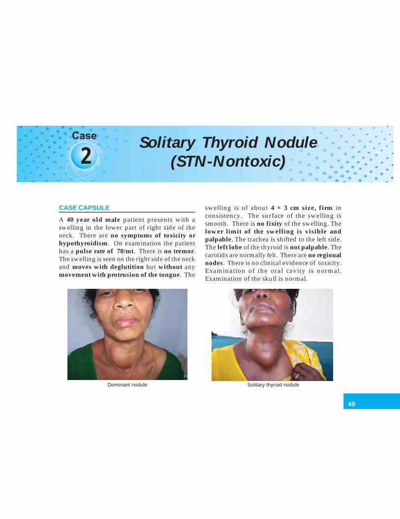
49
Solitary Thyroid Nodule (STN- Nontoxic)
2Solitary Thyroid Nodule
(STN-Nontoxic)
CASE CAPSULE
A 40 year old male patient presents with aswelling in the lower part of right side of theneck. There are no symptoms of toxicity orhypothyroidism. On examination the patienthas a pulse rate of 70/mt. There is no tremor.The swelling is seen on the right side of the neckand moves with deglutition but without anymovement with protrusion of the tongue. The
swelling is of about 4 × 3 cm size, firm inconsistency. The surface of the swelling issmooth. There is no fixity of the swelling. Thelower limit of the swelling is visible andpalpable. The trachea is shifted to the left side.The left lobe of the thyroid is not palpable. Thecarotids are normally felt. There are no regionalnodes. There is no clinical evidence of toxicity.Examination of the oral cavity is normal.Examination of the skull is normal.
Dominant nodule Solitary thyroid nodule
50
Clinical Surgery Pearls
Q 1. What are your points in favor of solitarythyroid nodule?
Points in favor of Solitary Thyroid Nodule (STN):a. There is only a single palpable thyroid
nodule.b. The rest of the gland is impalpable.
Q 2. What is the difference between adominant nodule and solitary thyroidnodule?
A discrete swelling in a gland with clinicalevidence of abnormality in the form of palpablecontralateral lobe or generalized mildnodularity is called dominant nodule. Asmentioned earlier, a discrete swelling in anotherwise impalpable gland is called isolatedor STN. About 70% of the discrete thyroidswellings are STN and 30% are dominantnodules. .
Q 3. What percentage of the so-called STNwill ultimately become a dominant nodule ofnodular goiter?
The true incidence of STN after USG / surgicalexploration comes down to only 50%.
Q 4. What is the importance of STN?
The importance of STN lies in the risk of malig-nancy compared with other thyroid swellings.
Q 5. What percentage of STN prove to bemalignant?
Twenty percentage of STN prove to bemalignant.
Q 6. What are the differential diagnosis of STN?
Differential diagnosis of STN
a. Nodular goiter with dominant nodule – 50%b. Adenoma – 20%c. Cancer – 20%d. Thyroiditis – 5%e. Cyst – 5%
Q 7. What is the most important singleinvestigation for diagnosis in STN?
Because of the risk of neoplasia the single mostimportant investigation of choice is fine needleaspiration cytology (FNAC).
Q 8. Can you differentiate a follicularcarcinoma from follicular adenoma in FNAC?
FNAC cannot differentiate follicular adenomafrom carcinoma. The distinction is dependentnot on cytology but on histological criterianamely capsular and vascular invasion. Thecapsular and vascular invasion can only beidentified on histology, which requires thethyroid tissue taken as paraffin block.
Q 9. What are the new techniques availablefor differentiating follicular carcinoma fromadenoma in FNAC? (PG)
In future FNAC differentiation may be possibleby the following techniques.a. Ploidy study of the DNA material: Polyploidy
for benign and aneuploidy for carcinoma.b. Benign tumors are monoclonal and
malignant tumors are polyclonal(monoclonal antibody MOAB 47)
c. Magnetic resonance spectroscopyd. Thyroimmunoperoxidase estimation.
Published by dr-notes.com
51
Solitary Thyroid Nodule (STN- Nontoxic)
Q 10. What is the definition of ‘adequatesmear’ in FNAC? (PG)
An adequate smear should have at least sixclusters of cells each containing about 20 cells.
Q 11. What are the possible FNAC reports inthyroid?
Possible FNAC reports in thyroid
a. Benign – abundant colloid and typical follicularcells.
b. Malignantc. Indeterminate–little colloid and many follicular
cells or Hurthle cells (Follicular neoplasm/suspicious).
d. Inadequate–cystic lesions, degeneratingadenomas.
Q 12. What is the classification of fine needleaspiration cytology?
Classification of fine needle aspiration cytologyof thyroid
Thy 1 NondiagnosticThy 2 NonneoplasticThy 3 FollicularThy 4 Suspicious of malignancyThy 5 Malignant
Q 13. What is the overall diagnostic accuracyof FNAC?
• The overall diagnostic accuracy is about 95%.• The diagnostic sensitivity of 83% and
specificity of 92%.
Q 14. What are the conditions in which FNACwill give a definite diagnosis?
• Colloid nodule• Thyroiditis
• Papillary carcinoma• Medullary carcinoma• Anaplastic carcinoma• Lymphoma.
Q 15. Which institution was responsible forpioneering the technique of FNAC?
Karolinska Hospital, Sweden (they decide theNobel Prize for Medicine).
Q 16. What is the minimum number of needlepasses required in FNAC?
Minimum 6 passes.
Q 17. What is the approach if the STN iscystic?
FNAC is less reliable in cystic swellings. Afteraspirating cyst fluid a further sample should betaken from the cyst wall for cytology.
Q 18. What will be the course of action if thecyst is recurring after aspiration?
A recurrent cyst should be removed surgically.
Q 19. What is the malignancy rate in cysticlesion?
Cystic lesions are less likely to be malignant.Malignancy rate in complex cysts is 72% andsimple cyst is 7%.
Q 20. What are the indications for true cut-(core) biopsy in thyroid?
It is useful ina. Anaplastic carcinomab. Lymphoma.
In these two situations, true cut biopsy canavoid an operation.
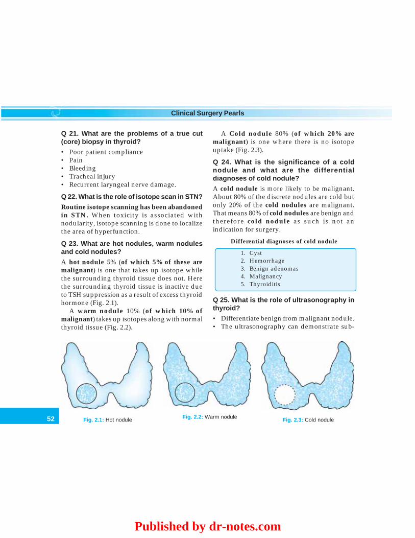
52
Clinical Surgery Pearls
Q 21. What are the problems of a true cut(core) biopsy in thyroid?
• Poor patient compliance• Pain• Bleeding• Tracheal injury• Recurrent laryngeal nerve damage.
Q 22. What is the role of isotope scan in STN?
Routine isotope scanning has been abandonedin STN. When toxicity is associated withnodularity, isotope scanning is done to localizethe area of hyperfunction.
Q 23. What are hot nodules, warm nodulesand cold nodules?
A hot nodule 5% (of which 5% of these aremalignant) is one that takes up isotope whilethe surrounding thyroid tissue does not. Herethe surrounding thyroid tissue is inactive dueto TSH suppression as a result of excess thyroidhormone (Fig. 2.1).
A warm nodule 10% (of which 10% ofmalignant) takes up isotopes along with normalthyroid tissue (Fig. 2.2).
Fig. 2.2: Warm nodule Fig. 2.3: Cold noduleFig. 2.1: Hot nodule
A Cold nodule 80% (of which 20% aremalignant) is one where there is no isotopeuptake (Fig. 2.3).
Q 24. What is the significance of a coldnodule and what are the differentialdiagnoses of cold nodule?
A cold nodule is more likely to be malignant.About 80% of the discrete nodules are cold butonly 20% of the cold nodules are malignant.That means 80% of cold nodules are benign andtherefore cold nodule as such is not anindication for surgery.
Differential diagnoses of cold nodule
1. Cyst2. Hemorrhage3. Benign adenomas4. Malignancy5. Thyroiditis
Q 25. What is the role of ultrasonography inthyroid?
• Differentiate benign from malignant nodule.• The ultrasonography can demonstrate sub-
Published by dr-notes.com
53
Solitary Thyroid Nodule (STN- Nontoxic)
clinical nodularity, can identify deep non-palpable thyroid nodules.
• Size of the nodule can be measured.• It can also differentiate solid from cystic
swellings.• Sono guided FNAC can be done.• Identify cervical lymph nodes.• Identify multicentricity.
Q 26. Can you differentiate benign conditionfrom malignancy in ultrasonography? (PG)
Yes, the difference between benign andmalignant conditions in USG
Benign Malignant
1. Hyperechoic nodule 1. Hypoechoic2. Significant cystic 2. Need not be there
component3. Peripheral egg shell 3. Microcalcifications
like calcification inside4. A sonolucent rim 4. No halo
(halo) around thenodule
5. Well-defined nodule 5. Poorly defined marginmargin
Q 27. What are the differential diagnosis ofthyroid cysts?
a. Colloid degeneration - 50%b. Involution of follicular adenomac. Malignancy – 10-15%d. Papillary carcinoma.
Q 28. What are the indications for surgery inSTN?
Indications for surgery in STN
a. FNAC positive for malignancyb. Follicular neoplasmc. Clinical suspicion – hard texture, fixity,
hoarseness, lymph node, male sex, etc.d. Recurrence of a cyst after aspiratione. Toxic adenomaf. Pressure symptomsg. Cosmesis
Q 29. When the FNAC reports comes asfollicular neoplasm. How will you proceed?
Follicular neoplasm may be follicular adenomaor follicular carcinoma and distinction ispossible only on histology evidenced bycapsular and vascular invasion. Majority (about80%) of the follicular neoplasms are benign.
Q 30. What are the clinical situations in favourof malignancy?
Suspect malignancy in the followingClinical situations
a. Discrete swelling in a male (more likely to bemalignant)
b. Child with a thyroid nodule (50% malignant)c. Extremes of age with discrete swelling (above
50 years and < 20 years teenagers)d. Hard irregular swellinge. Fixityf. Recurrent laryngeal nerve paralysisg. Lymph nodeh. Size > 4cmi. History of head and neck irradiationj. Association with other endocrine neoplasias
(e.g.: Multiple Endocrine Neoplasia - MEN).
54
Clinical Surgery Pearls
Q 31. What are the causes for a hard thyroidnodule?
Causes of a hard thyroid nodule
a. Malignancyb. Calcification - dystrophicc. Thyroiditisd. Haemorrhage into nodules
Q 32. What is the meaning of follicularneoplasia?
• Follicular neoplasia may be benign ormalignant.
• 80% of follicular neoplasia are benign (20%are malignant) and therefore total thyroidec-tomy as a treatment is recommended onlyafter getting the histopathology report.
• The pathologist can make a cytologicaldiagnosis of a carcinoma only in thefollowing situations:1. Papillary thyroid carcinoma (PTC):
orphan Annie eyed nucleus.2. Medullary thyroid carcinoma (MTC):
Cell balls + Amyloid stroma.3. Anaplastic carcinoma (difficulty in
differentiating it from lymphoma) .• Follicular carcinoma cannot be diagnosed by
cytology.
Q 33. What is the minimum surgery for a casewith FNAC report of follicular neoplasia whenthe STN is confined to one lobe of thyroid?
It is preferable to do a hemithyroidectomy onthe side of the STN,. i.e. removal of the affectedlobe along with the isthmus. The minimumsurgery should be a lobectomy.
Q 34. After hemithyroidectomy histopathologyis reported as carcinoma (papillary carcinoma/ follicular carcinoma), how will you proceed?
In this situation Reexploration and completiontotal thyroidectomy is recommended in allpatients so that further postoperative follow upis possible.
Q 35. Why is reexploration and completionthyroidectomy recommended in carcinomathyroid?
a. In follicular carcinoma the main mode ofspread is by blood stream. When a patientdevelops metastases, the treatment ofchoice is radioiodine therapy. If theremaining thyroid is not removed the iodinewill be taken up by the remaining thyroidand metastasis will not take up radioiodine.Therefore it cannot be located and treated.
b. The tumour marker for Differentiatedthyroid cancer (Papillary and follicularcarcinomas) is thyroglobulin. Thethyroglobulin level as a tumour marker issignificant only after total thyroidectomy.The thyroglobulin levels are elevated intumor bed recurrence and metastasisanywhere in the body.
c. Multicentricity and intrathyroid spreadwhich are seen in papillary thyroid cancercan be tackled with reexploration andcompletion thyroidectomy.
Q 36. What is the timing for Completionthyroidectomy? (PG)
• It is better to do it as early as possible.• The ideal timing would be 2 to 3 days after
the initial surgery.
Published by dr-notes.com
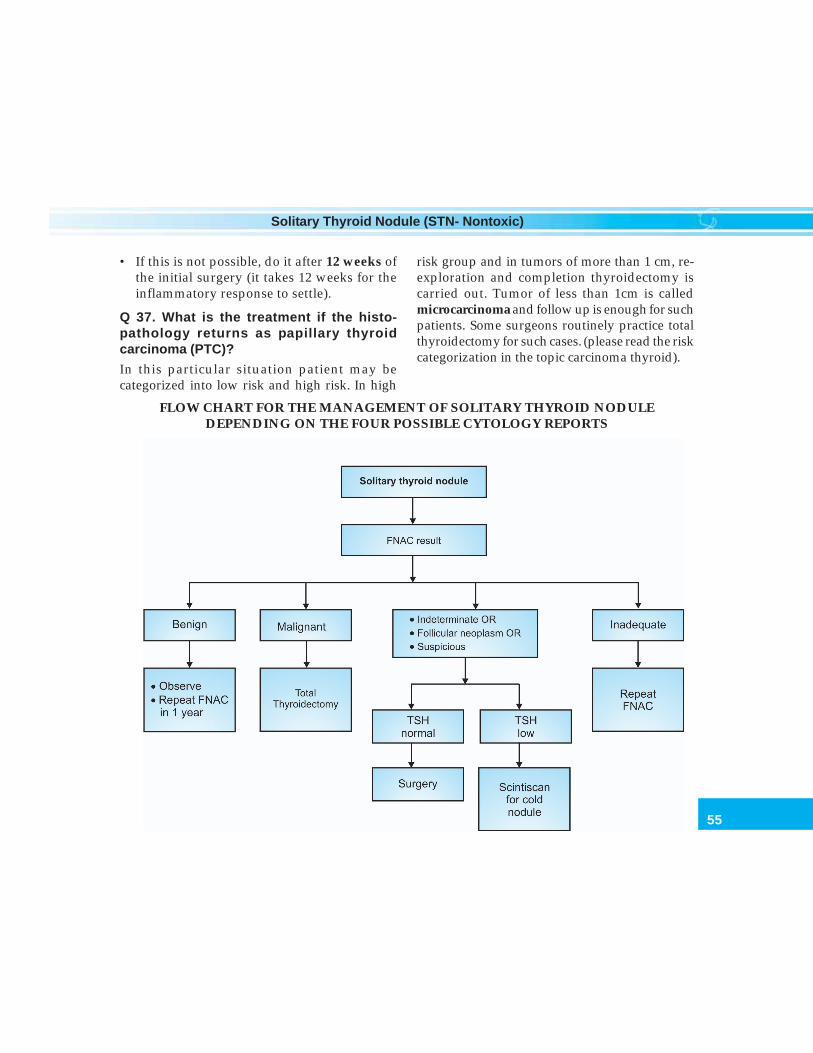
55
Solitary Thyroid Nodule (STN- Nontoxic)
• If this is not possible, do it after 12 weeks ofthe initial surgery (it takes 12 weeks for theinflammatory response to settle).
Q 37. What is the treatment if the histo-pathology returns as papillary thyroidcarcinoma (PTC)?
In this particular situation patient may becategorized into low risk and high risk. In high
risk group and in tumors of more than 1 cm, re-exploration and completion thyroidectomy iscarried out. Tumor of less than 1cm is calledmicrocarcinoma and follow up is enough for suchpatients. Some surgeons routinely practice totalthyroidectomy for such cases. (please read the riskcategorization in the topic carcinoma thyroid).
FLOW CHART FOR THE MANAGEMENT OF SOLITARY THYROID NODULEDEPENDING ON THE FOUR POSSIBLE CYTOLOGY REPORTS
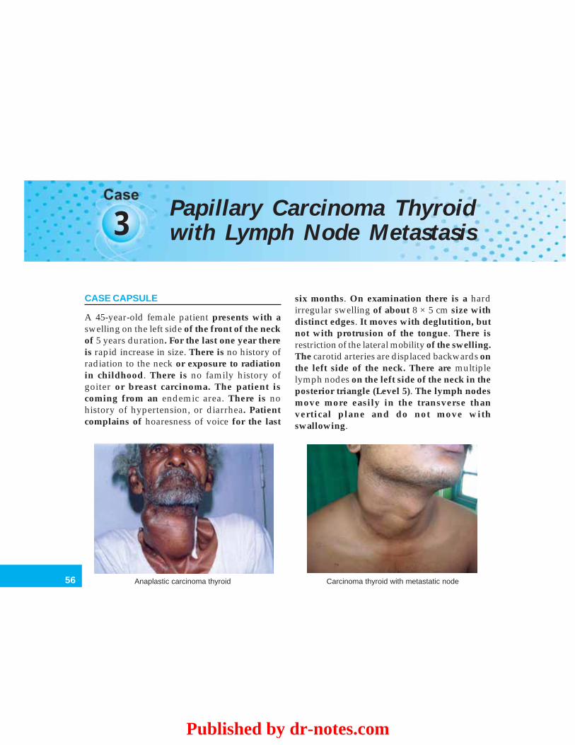
56
Clinical Surgery Pearls
3 Papillary Carcinoma Thyroidwith Lymph Node Metastasis
CASE CAPSULE
A 45-year-old female patient presents with aswelling on the left side of the front of the neckof 5 years duration. For the last one year thereis rapid increase in size. There is no history ofradiation to the neck or exposure to radiationin childhood. There is no family history ofgoiter or breast carcinoma. The patient iscoming from an endemic area. There is nohistory of hypertension, or diarrhea. Patientcomplains of hoaresness of voice for the last
six months. On examination there is a hardirregular swelling of about 8 × 5 cm size withdistinct edges. It moves with deglutition, butnot with protrusion of the tongue. There isrestriction of the lateral mobility of the swelling.The carotid arteries are displaced backwards onthe left side of the neck. There are multiplelymph nodes on the left side of the neck in theposterior triangle (Level 5). The lymph nodesmove more easily in the transverse thanvertical plane and do not move withswallowing.
Anaplastic carcinoma thyroid Carcinoma thyroid with metastatic node
Published by dr-notes.com
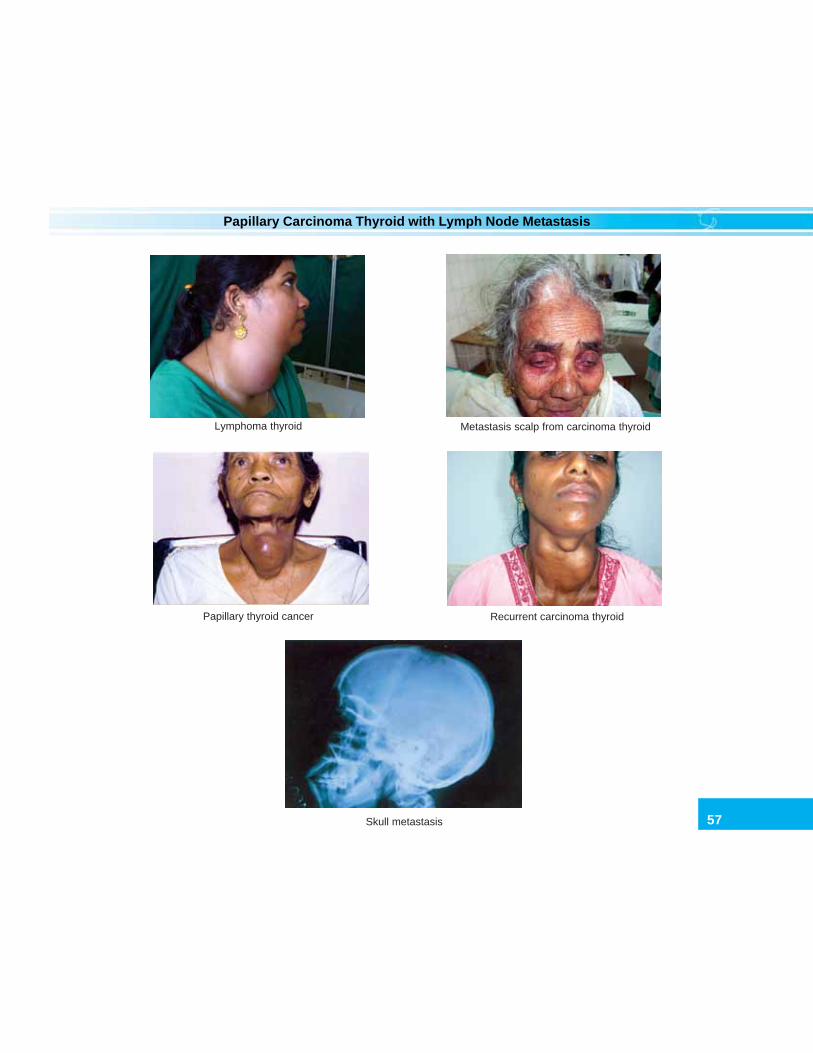
57
Papillary Carcinoma Thyroid with Lymph Node Metastasis
Lymphoma thyroid Metastasis scalp from carcinoma thyroid
Recurrent carcinoma thyroidPapillary thyroid cancer
Skull metastasis
58
Clinical Surgery Pearls
READ THE PRELIMINARY PORTION ANDCHECK LIST OF CASE NO:1.
Q 1. How will you differentiate thyroidswelling from lymph nodes of the neck?
The thyroid swelling will move up and downwith deglutition whereas the lymph nodeswelling will not do so. For all practicalpurposes any swelling which is not moving upand down with deglutition is non thyroidale.g.: may be lymph nodes, may be neurofibromaor other solid and cystic swellings.
Q 2. What are the diagnostic points favoringcarcinoma thyroid in this case?
The diagnostic points are:a. Hard thyroid noduleb. The suspicious lymph nodes in the neckc. Hoarseness of voiced. Restriction of laterial mobility.e. Carotid displaced backwards on left side
(Berry’s Singh).
Q 3. What are the differential diagnoses of ahard thyroid nodule?
Differential diagnoses of a hard thyroid nodule are:a. Calcification of a nodule of a nodular goiterb. Carcinomac. Thyroiditis.
Q 4. How will you identify calcification of athyroid nodule?
Simple plain X-ray of the neck will revealcalcification corresponding to the nodule.
Q 5. What type of calcification do you get insuch cases?
The type of calcification is dystrophic.
Q 6. What is Delphic lymph node?
The Delphic Lymph Node is the first lymph nodeinvolved in carcinoma thyroid. It is nothing butthe enlarged prelaryngeal lymph node.
Q 7. What is the importance of examining thescalp in carcinoma thyroid?
The main mode of spread of follicular carcinomais by blood stream and the commonestmetastasis is bony metastasis which is usuallyseen in the skull bone as a pulsatile bonyswelling.
Q 8. What are the differential diagnoses ofpulsatile bony swelling in the scalp?
The differential diagnoses are:a. Primary malignancy – solitary plasma-
cytoma, telengiectatic variety of osteogenicsarcoma.
b. Metastasis – from papillary carcinoma,Renal cell carcinoma, etc.
Q 9. What are the types of carcinoma thyroidin which you get a lymph node metastasis?
1. Papillary carcinoma thyroid2. Medullary carcinoma thyroid
Q 10. What are the malignancies associatedwith autoimmune thyroiditis?
a. Lymphoma of thyroidb. Papillary carcinoma thyroid.
Q 11. What is the most important investiga-tion in this patient to confirm the diagnosisof carcinoma thyroid?
FNAC from thyroid nodule and FNAC orbiopsy of the lymph node.
Published by dr-notes.com
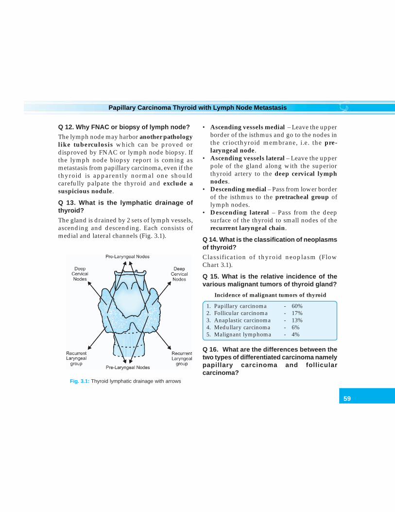
59
Papillary Carcinoma Thyroid with Lymph Node Metastasis
Q 12. Why FNAC or biopsy of lymph node?
The lymph node may harbor another pathologylike tuberculosis which can be proved ordisproved by FNAC or lymph node biopsy. Ifthe lymph node biopsy report is coming asmetastasis from papillary carcinoma, even if thethyroid is apparently normal one shouldcarefully palpate the thyroid and exclude asuspicious nodule.
Q 13. What is the lymphatic drainage ofthyroid?
The gland is drained by 2 sets of lymph vessels,ascending and descending. Each consists ofmedial and lateral channels (Fig. 3.1).
Fig. 3.1: Thyroid lymphatic drainage with arrows
• Ascending vessels medial – Leave the upperborder of the isthmus and go to the nodes inthe criocthyroid membrane, i.e. the pre-laryngeal node.
• Ascending vessels lateral – Leave the upperpole of the gland along with the superiorthyroid artery to the deep cervical lymphnodes.
• Descending medial – Pass from lower borderof the isthmus to the pretracheal group oflymph nodes.
• Descending lateral – Pass from the deepsurface of the thyroid to small nodes of therecurrent laryngeal chain.
Q 14. What is the classification of neoplasmsof thyroid?
Classification of thyroid neoplasm (FlowChart 3.1).
Q 15. What is the relative incidence of thevarious malignant tumors of thyroid gland?
Incidence of malignant tumors of thyroid
1. Papillary carcinoma - 60%2. Follicular carcinoma - 17%3. Anaplastic carcinoma - 13%4. Medullary carcinoma - 6%5. Malignant lymphoma - 4%
Q 16. What are the differences between thetwo types of differentiated carcinoma namelypapillary carcinoma and follicularcarcinoma?
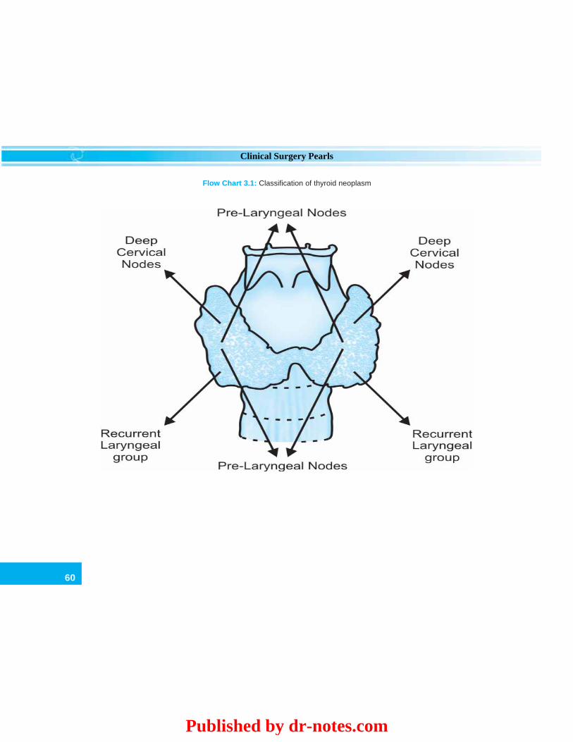
60
Clinical Surgery Pearls
Flow Chart 3.1: Classification of thyroid neoplasm
Published by dr-notes.com
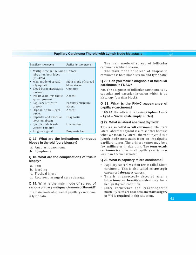
61
Papillary Carcinoma Thyroid with Lymph Node Metastasis
Papillary carcinoma Follicular carcinoma
• Multiple foci in the same Unifocallobe or on both lobes(21- 46%)
• Main mode of spread Main mode of spread– lymphatic bloodstream
• Blood borne metastasis Commonunusual
• Intrathyroid lymphatic Absentspread present
• Papillary structure Papillary structurepresent absent
• Orphan Annie – eyed Absentnuclei
• Capsular and vascular Diagnosticinvasion absent
• Lymph node invol- Uncommonvement common
• Prognosis good Prognosis bad
Q 17. What are the indications for trucutbiopsy in thyroid (core biopsy)?
a. Anaplastic carcinomab. Lymphoma.
Q 18. What are the complications of trucutbiopsy?
a. Painb. Bleedingc. Tracheal injuryd. Recurrent laryngeal nerve damage.
Q 19. What is the main mode of spread ofvarious primary malignant tumors of thyroid?
The main mode of spread of papillary carcinomais lymphatic.
The main mode of spread of follicularcarcinoma is blood stream.
The main mode of spread of anaplasticcarcinoma is both blood stream and lymphatic.
Q 20: Can you make a diagnosis of follicularcarcinoma in FNAC?
No. The diagnosis of follicular carcinoma is bycapsular and vascular invasion which is byhistology (paraffin block).
Q 21. What is the FNAC appearance ofpapillary carcinoma?
In FNAC the cells will be having Orphan Annie– Eyed – Nuclei (pale empty nuclei).
Q 22. What is lateral aberrant thyroid?
This is also called occult carcinoma. The termlateral aberrant thyroid is a misnomer becausewhat we mean by lateral aberrant thyroid is alymph node metastasis from an impalpablepapillary tumor. The primary tumor may be afew millimeter in size only. The term occultcarcinoma is applied to all papillary carcinomasless than 1.5 cm diameter.
Q 23. What is papillary micro carcinoma?
• Papillary cancer less than 1cm is called Microcarcinoma. This is also called microscopiccancer or laboratory cancer.
• This is unexpectedly detected after alobectomy or hemithyroidectomy for abenign thyroid condition.
• Since recurrence and cancer-specificmortality rates are near zero, no more surgeryor 131I is required in this situation.
62
Clinical Surgery Pearls
• Thyroid hormone replacement therapy maybe instituted.
Q 24. What are the etiological factors forcarcinoma thyroid?
1. Accidental exposure to radiation in less than10 yrs of age (Chernobyl Nuclear disaster).May produce papillary carcinoma andfollicular variant of papillary carcinoma.
2. Increased TSH stimulation (Follicularcarcinoma in endemic areas).
3. Autoimmune thyroiditis : Hashimoto’s isassociated with a 70 fold increase inlymphoma .
4. Oncogenes: C – myc, C – erb, Ras Oncogene,etc. (associated with MEN).
5. Irradiation to neck for lymphoma inchildhood and in olden days, for thymoma
6. Genetic: Papillary carcinoma, Cowdensyndrome, APC gene (associated withpapillary carcinoma) and familialmedullary thyroid carcinoma.
Q 25. What is Cowden Syndrome?
• The association of DTC with carcinoma breastand multiple hamartomas.
• The defect is due to a germ line mutation ofPTEN tumor suppressor gene.
Q 26. What are the scoring systems availablefor categorization of patients into low risk andhigh risk in differentiated thyroid cancer?
Many scoring systems are available1. AGES – Age, Grade, Extend and Size
(Mayo clinic-Hay et al)2. AMES – Age, Metastasis, Extend and Size
(Lahey clinic – Cady et al)
3. MACIS– Metastasis, Age, Completeness ofsurgery, Invasion of extra-thyroidal tissue, Size (Hay et al)
4. Sloan– KetteringIn first 3 systems, patients are categorized
into low risk and high risk. In Sloan - Kettering,patients are categorized into 3 groups namelylow risk, intermediate and high risk.Low risk Less than 45 years of age
Less than 4 cm sizeFavorable tumor factors
Intermediate risk Low risk patients with highrisk tumorsHigh risk patients with lowrisk tumours
High risk More than 45 years of ageMore than 4 cm sizeUnfavorable tumor factors.
Q 27. What is logic behind consideringpapillary and follicular carcinoma together formanagement purposes?
They are considered together because, both ofthem are Differentiated Thyroid CancerDerived from follicular cells. DifferentiatedThyroid Cancer (DTC) is a spectrum of diseaserather than a single disease entity. It is not a life-threatening disease and you can have nearnormal life expectancy. It is a tumor withindolent biological behavior with variableaggressiveness.
Q 28. What do you mean by well-differentiatedthyroid cancer?
They constitute 90% of the thyroid cancers:• Papillary thyroid cancer
Published by dr-notes.com
63
Papillary Carcinoma Thyroid with Lymph Node Metastasis
• Follicular cancer• Hurthle cell tumors.
Note: 10% are poorly differentiated, comprisingof anaplastic, medullary cancers andlymphomas.
Q 29. What is Hurthle cell neoplasm?
• It is a variant of follicular neoplasm in whichmore than 75 % of the follicles have oxyphil(Askanazy or Hurthle) cells or oncocyticfeatures.
• The cells are granular with acidophiliccytoplasm.
• Immunostaining for thyroglobulin is positive• Mean age of the disease is 60years.• It is multicentric in 33%.• More likely to produce lymphnode mets.• It will not take up radio iodine.• Distant metastasis is seen in 18%.
Q 30. What are the types of follicularcarcinoma ?
They are classified as minimally invasive andwidely invasive.
Q 31. What are the peculiarities of follicularcarcinoma ?
• They do not invade the lymphatics and hencelack nodal mets.
• Immunostaining for Thyroglobulin ispositive.
• Seen predominantly in females.
Q 32: What is ‘encapsulated variant’ ofpapillary carcinoma? (PG)
• This tumor has a capsule-like adenoma butwith local invasion.
• It may be associated with nodal metastasis.• There is problem in distinguishing this lesion
from a hyperplastic nodule.• The overall prognosis is excellent.
Q 33. What is ‘diffuse sclerosing variant’ ?(PG)
• This is seen primarily in children.• It is highly aggressive (often misdiagnosed
as Hashimoto’s).• Prominent lymphocytic infiltration is present.• Incidence of lymph node mets is 100%.• Prognosis is poor.
Q 34. What is ‘Lindsay tumor’? (PG)
It is a combination of the encapsulated variantand the follicular variant of papillarycarcinoma. It behaves in a very indolent fashionand has good prognosis.
Q 35. What is extrathyroidal spread?
The term extrathyroidal indicates that theprimary tumor has infiltrated through thecapsule of the gland.
Q 36. What is the AJCC staging (postopera-tive) of differentiated thyroid carcinoma?
TNM classification for differentiatedthyroid carcinoma
T1 Tumor diameter 2 cm or smallerT2 Tumor diameter >2 to 4 cmT3 Tumor diameter >4 cm limited to the thyroid
with minimal extrathyroidal extensionT4a Tumor of any size extending beyond the
thyroid capsule to invade subcutaneous softtissue, larynx, trachea, esophagus orrecurrent laryngeal nerve

64
Clinical Surgery Pearls
T4b Tumor invades prevertebral fascia or encasesthe carotid artery or mediastinal vessels.
Tx Primary tumor size unknown, but withoutextrathyroidal invasion
N0 No metastatic nodesN1a Metastasis to level VI (pretracheal,
paratracheal, prelaryngeal / Delphian nodes)N1b Metastasis to unilateral, bilateral, contralateral
cervical or superior mediastinal nodemetastasis
Nx Nodes not assessed at surgeryM0 No distant metastasisM1 Distant metastasisMx Distant metastasis not assessed
Q 37. What is the importance of age indifferentiating thyroid cancer (DTC)?
This is the only human cancer where age isincluded in staging.
Q 38. What is the AJCC staging(postoperative) of Differentiated ThyroidCarcinoma? (PG)
AJCC staging (postoperative) of differentiatedthyroid carcinoma
Age less than 45 years (only 2 stages)Stage I : any T, any N and M0Stage II : any T, any N, M1Age more than 45 years (4 stages)Stage I : T1, N0 and M0Stage II : T2, N0 and M0Stage III : T3, N0, and M0
: T1, N1a, M0: T2, N1a, M0: T3, N1a, M0
StageIV A : T4a, N0, M0(T4a/Lateral node is stage IV A): T4a, N1a, M0
: T1, N1b, M0: T2, N1b, M0: T3, N1b, M0: T4a, N1b,M0
StageIV B : T4b, any N, M0(T4b is Stage IV B)
StageIV C : any T, any N, M1(metastasis is Stage IV C)
*Note: Stage III constitute minimal extrathyroidextension or level VI nodes.
The AJCC staging is for predicting the risk ofdeath. For assessing the risk of recurrence a threelevel stratification is recommended by theAmerican Thyroid Association, which is asfollows: (PG)
After initial surgery and remnant ablation:1. Low risk:
• No local / distant metastasis.• All microscopic tumor resected• No tumor invasion of locoregional
structures• No aggressive histology• No vascular invasion• No 131I uptake outside thyroid bed.
2. Intermediate risk:• Microscopic invasion of tumor to perithyroid
soft tissue• Tumor with aggressive histology• Vascular invasion.
3. High risk:• Macroscopic tumor invasion• Incomplete tumor invasion• Distant metastasis• 131I uptake outside thyroid bed after
remnant ablation.
Published by dr-notes.com
65
Papillary Carcinoma Thyroid with Lymph Node Metastasis
Q 39. What is minimal extrathyroid extension?(PG)
Extension to sternothyroid muscle orperithyroid soft tissue is called minimalextrathyroid invasion.
Q 40. What is the prognosis of differentiatedthyroid cancer (DTC)?
• The 25 year mortality rate of a low-risk DTCis 2%. That means 98% of the patients willsurvive 25 years.
• For the high-risk group the 25 year mortalityis 46%.
• 80 to 90% of the patients come under the low-risk group
• In short 80% of the patients requirelobectomy alone
• 15% require aggressive treatment• 5% die regardless of a treatment.
Q 41. What is the surgical treatment of choicefor a preoperatively proven case of papillarycarcinoma?
If the FNAC is diagnostic of papillary thyroidcancer the treatment of choice is near total or totalthyroidectomy (TT) with central compartmentdissection. Lymph nodes in the jugular chainshould be carefully looked for during surgery.
Q 42. What is the difference between neartotal and total thyroidectomy?
In near total thyroidectomy 1 to 2gm of thyroidtissue is preserved on the contralateral side,which preserves blood supply to one or bothparathyroids.
Q 43. What is central compartment neckdissection?
• It extends from hyoid bone above toinnominate vein below and carotidslaterally.
• Thyroid and the thymus are removed en-blocalong with the paratracheal, tracheoesophageal, pretracheal and pre laryngealnodes.
Q 44. What is the rationale for centralcompartment neck dissection and what arethe problems associated ? (PG)
• Fifty percent node positivity is seen inroutine central compartment dissection.
• Reoperation and nodal dissection in thecentral compartment area are difficult.
• Thymus and thyroid are removed en-bloc inthis dissection
• Central compartment neck dissection is doneas long as low incidence of hypopara-thyroidism can be achieved.
• Lower parathyroids are a greater risk fordamage in a Central Compartment neckdissection.
Q 45. What are the indications for completionthyroidectomy when there is histologicalsurprise of carcinoma after a hemithyroidec-tomy?
Indications for completion thyroidectomy
1. History of radiation2. High-risk factors3. More than 1cm size4. Contralateral thyroid nodule5. Regional or distant metastasis6. Family history of DTC in first degree relatives7. Age > 45 years8. Multifocal tumor
66
Clinical Surgery Pearls
Q 50. What are the features of Columnar cellvariety of papillary cancer? (PG)
• This is seen only in males• All patients will die within 5 years of
diagnosis.
Q 51. What are the indications for totalthyroidectomy in carcinoma?
Indications for total thyroidectomy in carcinoma
1. Bilateral disease2. High-risk category3. Radiated patients4. Patients with large nodal metastasis5. Patients with local invasion6. Patients with distant metastasis7. Patients with occult cancer with node metastasis
Q 52. What is the treatment of lymph nodemetastasis in papillary thyroid cancer?
If lymph nodes are clinically present, afunctional neck dissection (preserving thesternomastoid muscle, accessory nerve andinternal jugular vein) is carried out along withtotal thyroidectomy. Oncologically carcinomathyroid is the only indication for a functionalneck node dissection. If clinically andperoperatively there aren’t any nodes, onlycentral compartment dissection is done.
Q 53. What is the postoperative follow upafter total thyroidectomy?
All patients, after total thyroidectomy shouldreceive the following:a. T4 suppression (300 microgram eltroxine
daily) after scintigraphy for remnantthyroid tissue.
9. Extrathyroid spread10. Major vascular invasion11. Major capsular invasion
Q 46. Is there any role for lobectomy aloneas a treatment for DTC?
Lobectomy is acceptable in a situation where asmall intrathyroid tumor of < 1cm in a low-riskpatient who is node-negative.
Q 47. What is the indication for lateral necknode dissection (functional neck dissection)?
Indication for functional neck dissection
• Lymphadenopathy detected clinically• Node identified by imaging• Biopsy-proven metastatic nodes• Frozen-section node positivity during surgery
Q 48. What are the bad histological subtypesof DTC?
Bad histological subtypes of DTC
• Tall cell variety• Columnar cell• Trabecular• Scirrhous• Solid• Oxyphilic subtype of follicular thyroid cancer• Insular type of follicular thyroid cancer
Q 49. What are the features of tall cell varietyof papillary cancer? (PG)
• This is an aggressive variant• Seen in elderly• Represents 10% of papillary tumors• Extracapsular and vascular invasion in 30 %• Five year Survival rate is < 30%.
Published by dr-notes.com
67
Papillary Carcinoma Thyroid with Lymph Node Metastasis
b. Look for thyroid remnant by scintigraphyat 6 weeks (1 mCi radioiodine for131I cervicalscanning). For optimal scanning, the serumTSH level should be at least 30mIU/L. Ifthe patient is on eltroxine, two injections ofrecombinant human TSH will give thedesired effect.
c. If remnant is detected, Radio RemnantAblation at a dose of 30mCi (RRA) isgiven(131I).
d. At 3 to 6 months, whole body scan with3mCi radioactive iodine for metastasis andultrasound examination is carried out.
e. If negative scanning is obtained, follow upwith Thyroglobulin (Tg) level. Upondetection of increased thyroglobulin level,further whole body scan is indicated.
Q 54. What is the rationale of RRA? (PG)
a. RRA will destroy occult microscopiccarcinoma.
b. Later detection of persistent or recurrentdisease is possible, after distruction of thethyroid remnant.
c. Serum thyroglobulin as a tumor marker isuseful only after complete removal anddestruction of the thyroid. (therefore thereis no role for thyroglobulin assay in thepreoperative period).
Q 55. What is the upper limit of normal levelof thyroglobin (Tg)? (PG)
• When on suppressive therapy: Above 5ng/ml.
• When the patient is hypothyroid: more than10ng/ml.
These are indications of imaging forpersistent, recurrent or metastatic disease.
Q 56. If thyroglobin (Tg) measurement is highand total body scanning reveals metastases,what will be the course of action? (PG)
For metastasis, 100 to 200 mCi radioiodine 131 Iis given (RAI).
Q 57. What are the precautions to be takenbefore RAI therapy? (PG)
Precautions to be taken before RAI therapy
a. Low iodine diet for 10 daysb. Isolation of the patient (when dose is more than
30mci)c. Oral fluid intake to increase urine output so that
bladder injury is reducedd. Lemon sucking to avoid sialadenitise. Avoid pregnancy for 6 monthsf. Treat constipation with cathartics to reduce
gonadal and colonic irradiationg. Sperm count reduction is noticed for several
months
Note: Posttreatment whole body scan should bedone 4 to10 days after RAI, which may detectnew lesions that need further treatment.
Q 58. What is the dose for skeletal meta-stasis? (PG)
250 to 300 mCi (milli Curie) of 131 I
Q 59. What is the further follow up? (PG)
a. Follow up with whole body scan at 6months and yearly thereafter till threeconsecutive negative scans are obtained.
b. Serum Tg level for every 6 months.
68
Clinical Surgery Pearls
c. Life long suppressive therapy with thyroxin(the TSH should be monitored every 6months and dose of thyroxin adjusted).
Dose of radioiodine ( I131 ) in differentiatedcarcinoma thyroid
• Cervical Scan - 1 mCi.• Total Body Scan - 3 mCi• Remnant Ablation - 30 mCi• Treatment of metastasis - 100 – 250 mCi
Flow chart for management of DTC
AFTER Total Thyroidectomy + TSH SUPPRESSION
T4 withdrawal for 3wks OR two IM injections of0.9mg of recombinant TSH
Pretherapy low dose 131I – scanning (1 to 3mCi)before ablation
Radio Remnant Ablation (30-100mCi)
Postablation Whole Body Scan 5 to 8 days after RRA
If low-risk, negative thyroglobulin and normalneck USG, then no whole body scan
If intermediate risk and high-risk with persistentdisease, then 6 to 12 months after RRA do
diagnostic whole body scan
Further follow up• Thyroglobulin : 6 to 12 months• Cervical US ; 6 and 12 months, annually for3 – 5 yrs
MEDULLARY THYROID CARCINOMA
Q 60. What are the peculiarities of medullarythyroid carcinoma? (MTC)
a. They constitute 5 to 10% of all thyroidcancers
b. Are derived from parafollicular cells (C-cells)developed from neural crests.
c. There is a characteristic amyloid stroma.d. High levels of serum calcitonin are
produced (more than 0.08 ng/ml); it is atumor marker for medullary carcinoma
e. Diarrhea is a feature in 30% of casesf. Some tumours are familial (~ 20%)g. It may form part of multiple endocrine
neoplasia (MEN) syndromes : IIa and IIbh. Clinical course is more aggressive then
differentiated thyroid cancersi. Do not take up Radioactive iodinej. Higher recurrence rate and mortality
k. Radiation and chemotherapy are ineffectivel. This is perhaps the only situation where a
surgery based on genetic testing is routinelydone.
m. Once the diagnosis of medullary thyroidcancer is suspected, all patients should bescreened for mutation of the RET proto-oncogene to exclude familial disease.
n. When a genetic defeet is found, all familymembers should be screened.
Q 61. What are the four clinical settings ofMTC? (PG)
a. Sporadic medullary thyroid carcinoma(80%)
b. Familial MEN IIac. Familial MEN IIbd. Familial non-MEN medullary thyroid
carcinoma (FMTC).
Published by dr-notes.com
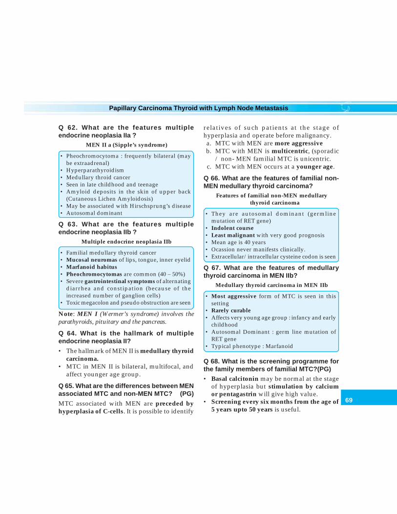
69
Papillary Carcinoma Thyroid with Lymph Node Metastasis
Q 62. What are the features multipleendocrine neoplasia IIa ?
MEN II a (Sipple’s syndrome)
• Pheochromocytoma : frequently bilateral (maybe extraadrenal)
• Hyperparathyroidism• Medullary throid cancer• Seen in late childhood and teenage• Amyloid deposits in the skin of upper back
(Cutaneous Lichen Amyloidosis)• May be associated with Hirschsprung’s disease• Autosomal dominant
Q 63. What are the features multipleendocrine neoplasia IIb ?
Multiple endocrine neoplasia IIb
• Familial medullary thyroid cancer• Mucosal neuromas of lips, tongue, inner eyelid• Marfanoid habitus• Pheochromocytomas are common (40 – 50%)• Severe gastrointestinal symptoms of alternating
diarrhea and constipation (because of theincreased number of ganglion cells)
• Toxic megacolon and pseudo obstruction are seen
Note: MEN I (Wermer’s syndrome) involves theparathyroids, pituitary and the pancreas.
Q 64. What is the hallmark of multipleendocrine neoplasia II?
• The hallmark of MEN II is medullary thyroidcarcinoma.
• MTC in MEN II is bilateral, multifocal, andaffect younger age group.
Q 65. What are the differences between MENassociated MTC and non-MEN MTC? (PG)
MTC associated with MEN are preceded byhyperplasia of C-cells. It is possible to identify
relatives of such patients at the stage ofhyperplasia and operate before malignancy.a. MTC with MEN are more aggressiveb. MTC with MEN is multicentric, (sporadic
/ non- MEN familial MTC is unicentric.c. MTC with MEN occurs at a younger age.
Q 66. What are the features of familial non-MEN medullary thyroid carcinoma?
Features of familial non-MEN medullarythyroid carcinoma
• They are autosomal dominant (germlinemutation of RET gene)
• Indolent course• Least malignant with very good prognosis• Mean age is 40 years• Ocassion never manifests clinically.• Extracellular/intracellular cysteine codon is seen
Q 67. What are the features of medullarythyroid carcinoma in MEN IIb?
Medullary thyroid carcinoma in MEN IIb
• Most aggressive form of MTC is seen in thissetting
• Rarely curable• Affects very young age group : infancy and early
childhood• Autosomal Dominant : germ line mutation of
RET gene• Typical phenotype : Marfanoid
Q 68. What is the screening programme forthe family members of familial MTC?(PG)
• Basal calcitonin may be normal at the stageof hyperplasia but stimulation by calciumor pentagastrin will give high value.
• Screening every six months from the age of5 years upto 50 years is useful.
70
Clinical Surgery Pearls
• Ultrasound of the neck is also useful foridentifying nodal metastasis.
Q 69. Is there any role for prophylacticthyroidectomy in family members. If so, atwhat age ?
• Yes. Prophylactic thyroidectomy can preventtumor occurrence at the stage of C-cellhyperplasia.
• Surgery by 3 years for MEN IIa.• Surgery by 1 year for MEN IIb.
Q 70. What are the clinical presentations ofmedullary thyroid carcinoma?
• Medullary thyroid carcinoma is a greatimitator.
• Lump in the neck• Nodal metastases• Paraneoplastic syndromes: Cushing and
Carcinoid• Hoarseness, stridor and upper airway
obstruction• Diarrhea and flushing.
Q 71. How do you confirm the diagnosis ofmedullary thyroid carcinoma?
• FNAC• Calcitonin : unstimulated serum calcitonin
>100 pg/mL is suggestive of MTC• CEA: 50% of medullary thyroid cancer• USG / CTscan / MRI• Screening for pheochromocytoma by 24 hr
urinary catecholamines in all cases.Note:1. In all suspected MTCs, do USG abdomen
and 24 hr urinary catecholamines to rule outpheochromocytoma.
2. Rule out MTC in all thyroid cases withdiarrhea.
3. Rule out MTC in all thyroid cases withhypertension.
Q 72. What is the treatment of medullarythyroid carcinoma?
• Since they don’t take up RAI, surgery is theonly curative treatment.
• No role of anything less than totalthyroidectomy.
• Routine dissection of central compartmentis a must.
• Sampling of the jugular nodes are done (ifpositive, Modified Neck dissection is done).
• Autotransplantation of parathyroid isrequired (for completeness of thyroidresection) : transplanted to the non-dominantforearm.
• Some centers do a thymectomy with theseregularly (for fear of metastasis in thymus).
• Post operative stimulated calcitonin assay isdone for assessment of the adequacy ofsurgical resection.
Q 73. What is the treatment of anaplasticcarcinoma of the thyroid?
• It is very difficult to differentiate anaplasticcarcinoma from lymphoma of thyroid.
• They are extremely lethal tumors• The treatment of choice is not surgery but
radiotherapy.• The indication for surgery is to relieve
tracheal obstruction by isthmusectomy. Thiswill give tissue for histology.
• Curative resection is attempted when thereis no infiltration through the thyroid capsule.
Published by dr-notes.com
71
Papillary Carcinoma Thyroid with Lymph Node Metastasis
Q 74. What is the management of lymphoma?
• There is no role for surgery.• Radiotherapy and chemotherapy are the
treatment of choice.• For Low grade B cell MALT Lymphoma
radiotherapy alone is enough.• Tracheal obstruction requires urgent
chemotherapy after intubation.• The temptation for a tracheostomy must be
resisted.
Q 75. What is incidentaloma?
When performing imaging for other head andneck problems, small nodule in the thyroid lessthan 1cm is identified by serendipity. It iscalled incidentaloma. They are invariablybenign and what is required is an ultrasoundguided FNAC.
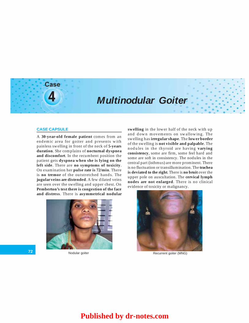
72
Clinical Surgery Pearls
4 Multinodular Goiter
CASE CAPSULE
A 30-year-old female patient comes from anendemic area for goiter and presents withpainless swelling in front of the neck of 5-yearsduration. She complains of nocturnal dyspneaand discomfort. In the recumbent position thepatient gets dyspnea when she is lying on theleft side. There are no symptoms of toxicity.On examination her pulse rate is 72/min. Thereis no tremor of the outstretched hands. Thejugular veins are distended. A few dilated veinsare seen over the swelling and upper chest. OnPemberton’s test there is congestion of the faceand distress. There is asymmetrical nodular
swelling in the lower half of the neck with upand down movements on swallowing. Theswelling has irregular shape. The lower borderof the swelling is not visible and palpable. Thenodules in the thyroid are having varyingconsistency, some are firm, some feel hard andsome are soft in consistency. The nodules in thecentral part (isthmus) are more prominent. Thereis no fluctuation or transillumination. The tracheais deviated to the right. There is no bruit over theupper pole on auscultation. The cervical lymphnodes are not enlarged. There is no clinicalevidence of toxicity or malignancy.
Nodular goiter Recurrent goiter (MNG)
Published by dr-notes.com
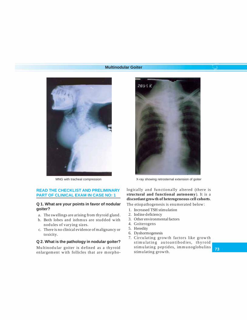
73
Multinodular Goiter
READ THE CHECKLIST AND PRELIMINARYPART OF CLINICAL EXAM IN CASE NO: 1
Q 1. What are your points in favor of nodulargoiter?
a. The swellings are arising from thyroid gland.b. Both lobes and isthmus are studded with
nodules of varying sizes.c. There is no clinical evidence of malignancy or
toxicity.
Q 2. What is the pathology in nodular goiter?
Multinodular goiter is defined as a thyroidenlargement with follicles that are morpho-
logically and functionally altered (there isstructural and functional autonomy). It is adiscordant growth of heterogeneous cell cohorts.The etiopathogenesis is enumerated below:1. Increased TSH stimulation2. Iodine deficiency3. Other environmental factors4. Goiterogens5. Heredity6. Dyshormogenesis7. Circulating growth factors like growth
stimulating autoantibodies, thyroidstimulating peptides, immunoglobulinsstimulating growth.
MNG with tracheal compression X-ray showing retrosternal extension of goiter
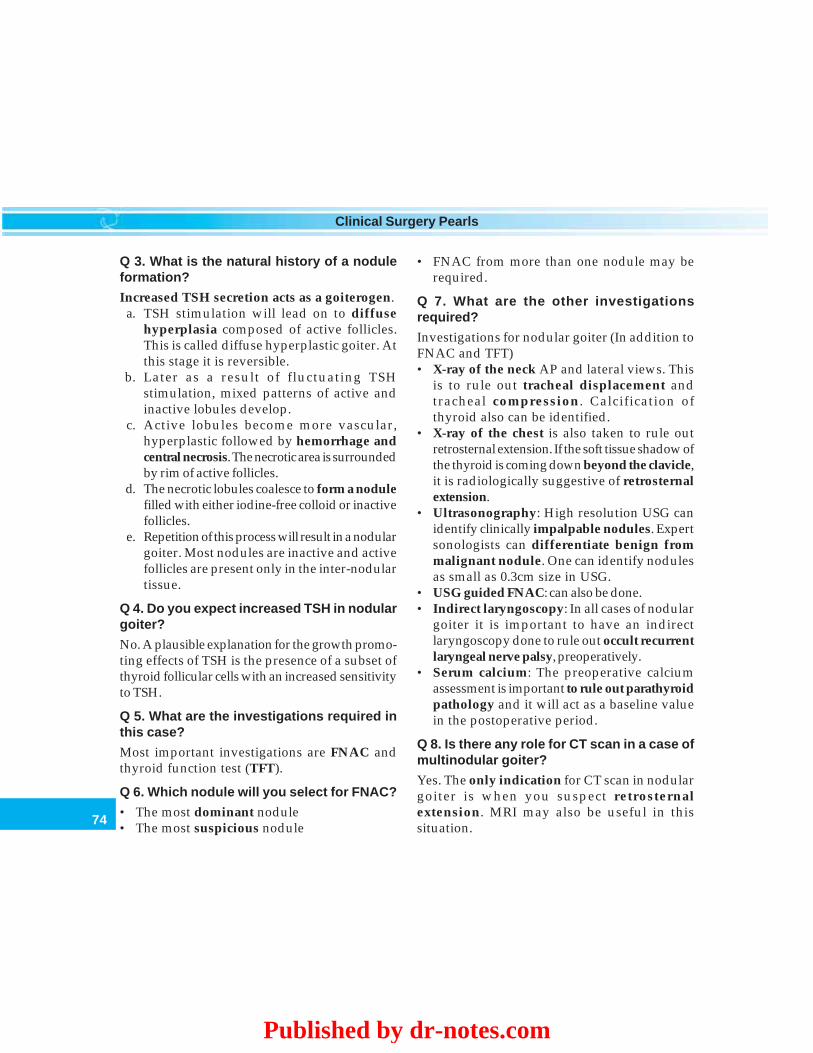
74
Clinical Surgery Pearls
Q 3. What is the natural history of a noduleformation?
Increased TSH secretion acts as a goiterogen.a. TSH stimulation will lead on to diffuse
hyperplasia composed of active follicles.This is called diffuse hyperplastic goiter. Atthis stage it is reversible.
b. Later as a result of fluctuating TSHstimulation, mixed patterns of active andinactive lobules develop.
c. Active lobules become more vascular,hyperplastic followed by hemorrhage andcentral necrosis. The necrotic area is surroundedby rim of active follicles.
d. The necrotic lobules coalesce to form a nodulefilled with either iodine-free colloid or inactivefollicles.
e. Repetition of this process will result in a nodulargoiter. Most nodules are inactive and activefollicles are present only in the inter-nodulartissue.
Q 4. Do you expect increased TSH in nodulargoiter?
No. A plausible explanation for the growth promo-ting effects of TSH is the presence of a subset ofthyroid follicular cells with an increased sensitivityto TSH.
Q 5. What are the investigations required inthis case?
Most important investigations are FNAC andthyroid function test (TFT).
Q 6. Which nodule will you select for FNAC?
• The most dominant nodule• The most suspicious nodule
• FNAC from more than one nodule may berequired.
Q 7. What are the other investigationsrequired?
Investigations for nodular goiter (In addition toFNAC and TFT)• X-ray of the neck AP and lateral views. This
is to rule out tracheal displacement andtracheal compression. Calcification ofthyroid also can be identified.
• X-ray of the chest is also taken to rule outretrosternal extension. If the soft tissue shadow ofthe thyroid is coming down beyond the clavicle,it is radiologically suggestive of retrosternalextension.
• Ultrasonography: High resolution USG canidentify clinically impalpable nodules. Expertsonologists can differentiate benign frommalignant nodule. One can identify nodulesas small as 0.3cm size in USG.
• USG guided FNAC: can also be done.• Indirect laryngoscopy: In all cases of nodular
goiter it is important to have an indirectlaryngoscopy done to rule out occult recurrentlaryngeal nerve palsy, preoperatively.
• Serum calcium: The preoperative calciumassessment is important to rule out parathyroidpathology and it will act as a baseline valuein the postoperative period.
Q 8. Is there any role for CT scan in a case ofmultinodular goiter?
Yes. The only indication for CT scan in nodulargoiter is when you suspect retrosternalextension. MRI may also be useful in thissituation.
Published by dr-notes.com
75
Multinodular Goiter
Q 9. Is there any role for pulmonary functiontest (PFT) in MNG?
Clinically whenever there is large/long-standing goiter and tracheal deviation orcompression, it is better to do a pulmonaryfunction test so that one can identify a patientwho is likely to develop tracheomalacia /scabbard trachea.
Q 10. How will you identify and tackletracheomalacia (weakness of trachealrings)?
After thyroidectomy the surgeon should palpatefor the tracheal rings for its strength. In case ofdoubt, the anesthesiologist may withdraw theendotracheal tube partially to check for weaknessof tracheal rings. When there is suspicion of weaknessof tracheal rings it is better to keep the tube as trachealstent postoperatively for 24 hours. After 24hours extubate and see. If the patient is goingfor trouble do a tracheostomy.
Q 11. Is there any role for thyroid scinti-graphy?
It is not required routinely. It is useful in ahyperthyroid patient with a dominant nodule asit defines the area of hyperactivity, thereby choosingproper surgical therapy.
Q 12. What are the complications of nodulargoiter?
Complications of nodular goiter
1. Toxicity2. Malignancy3. Retrosternal extension4. Pressure effects5. Calcification
Q 13. What type of toxicity do you get in MNG?
1. When the patient develops hypertrophy ofthe nodule he/she gets Plummer’s disease,(secondary).
2. When the internodular tissue is hypertrophiedpatient will get primary thyrotoxicosis.
Q 14. What are the indications for surgery inMNG?
The indications for surgery are:1. Complications (already mentioned)2. Cosmesis.
Q 15. What is the role of suppressive levothy-roxine therapy?
The results of T4 suppression therapy are inconsistentand marginal. Only small goiter would respond andthat too partially. Goiters that respond, do so withina period of six months.
Q 16. What are the disadvantages of suppres-sion therapy?
1. One should consistently suppress the TSH levelto < 0.5mIU/L.
2. Periodic monitoring of TSH is required torule out hyperthyroidism.
3. Large amounts of thyroid tissue are likelyto be hormone insensitive in MNG.
4. Variable TSH dependency by various thyro-cytes resulting in inconsistent response.
5. Indefinite treatment is required and most of thegoiter recur after cessation of therapy.
Q 17. Is there any role for radioiodine therapyin MNG?
Radioiodine therapy is of no value in large MNGwith poorly functioning nodules. The efficacy
76
Clinical Surgery Pearls
of RAI depends on the presence of reasonablegland activity all over the thyroid that is not seenin large MNG. Radiation induced autoimmunethyroiditis and hypothyroidism are other prob-lems. It is of limited value and useful only intwo groups of patient. Patients with small goiterwith reasonable gland function and patientswith substantially increased operative risk.
Q 18. What is the surgical option in a case ofnodular goiter?
The conventional surgery is subtotalthyroidectomy, which is not adequate in a caseof nodular goiter which is involving both lobesof the gland. The ideal option will be to do atotal thyroidectomy, with immediate and lifelong replacement of thyroid hormone. Whenone lobe is more involved than the other lobe,total lobectomy on the more affected side withsubtotal resection on the less affected side will bean ideal option. It is therefore ideal to haveremoval of all the diseased gland, the so-calledadequate thyroidectomy.
Q 19. What is the real problem if you leavebehind diseased nodular portions?
1. It will produce enlargement and recurrenceafter 10-15 years.
2. Reoperation for recurrent nodular goiter is moredifficult and hazardous due to altered anatomyand fibrosis. Therefore it is better to favor totalthyroidectomy in younger patients.
Q 20. What is the timing of recurrence?
It is seen usually after 10 to 15 years.
Q 21. Is there any role for TSH suppressionin preventing recurrence?
If diseased nodules are left behind, TSHsuppression will not help in preventingrecurrence. At present its role is uncertain.However, once total thyroidectomy is donepatient needs replacement therapy to preventhypothyroidism.
Published by dr-notes.com
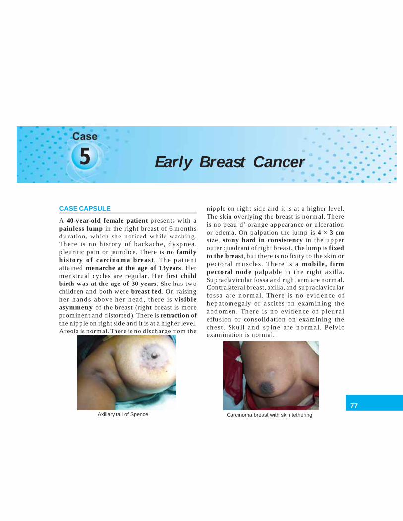
77
Early Breast Cancer
5 Early Breast Cancer
CASE CAPSULE
A 40-year-old female patient presents with apainless lump in the right breast of 6 monthsduration, which she noticed while washing.There is no history of backache, dyspnea,pleuritic pain or jaundice. There is no familyhistory of carcinoma breast. The patientattained menarche at the age of 13years. Hermenstrual cycles are regular. Her first childbirth was at the age of 30-years. She has twochildren and both were breast fed. On raisingher hands above her head, there is visibleasymmetry of the breast (right breast is moreprominent and distorted). There is retraction ofthe nipple on right side and it is at a higher level.Areola is normal. There is no discharge from the
nipple on right side and it is at a higher level.The skin overlying the breast is normal. Thereis no peau d’ orange appearance or ulcerationor edema. On palpation the lump is 4 × 3 cmsize, stony hard in consistency in the upperouter quadrant of right breast. The lump is fixedto the breast, but there is no fixity to the skin orpectoral muscles. There is a mobile, firmpectoral node palpable in the right axilla.Supraclavicular fossa and right arm are normal.Contralateral breast, axilla, and supraclavicularfossa are normal. There is no evidence ofhepatomegaly or ascites on examining theabdomen. There is no evidence of pleuraleffusion or consolidation on examining thechest. Skull and spine are normal. Pelvicexamination is normal.
Axillary tail of Spence Carcinoma breast with skin tethering
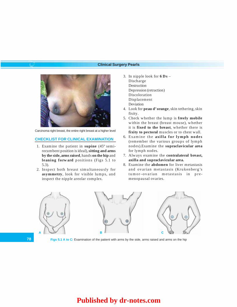
78
Clinical Surgery Pearls
CHECKLIST FOR CLINICAL EXAMINATION
1. Examine the patient in supine (45o semi-recumbent position is ideal), sitting and armsby the side, arms raised, hands on the hip andleaning forward positions (Figs 5.1 to5.3).
2. Inspect both breast simultaneously forasymmetry, look for visible lumps, andinspect the nipple areolar complex.
3. In nipple look for 6 Ds –DischargeDestructionDepression (retraction)DiscolorationDisplacementDeviation
4. Look for peau d’ orange, skin tethering, skinfixity.
5. Check whether the lump is freely mobilewithin the breast (breast mouse), whetherit is fixed to the breast, whether there isfixity to pectoral muscles or to chest wall.
6. Examine the axilla for lymph nodes(remember the various groups of lymphnodes).Examine the supraclavicular areafor lymph nodes.
7. Always examine the contralateral breast,axilla and supraclavicular area.
8. Examine the abdomen for liver metastasisand ovarian metastasis (Krukenberg’stumor–ovarian metastasis in pre-menopausal ovaries.
Figs 5.1 A to C: Examination of the patient with arms by the side, arms raised and arms on the hip
Carcinoma right breast, the entire right breast at a higher level
A B C
Published by dr-notes.com
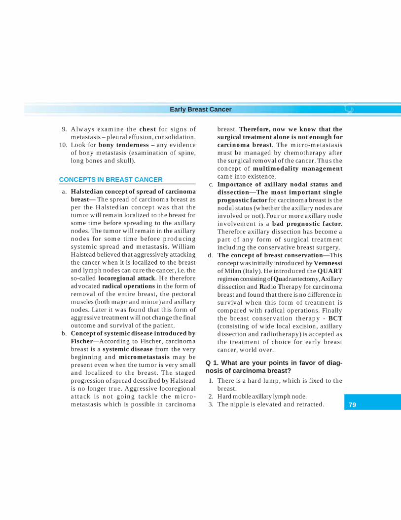
79
Early Breast Cancer
9. Always examine the chest for signs ofmetastasis – pleural effusion, consolidation.
10. Look for bony tenderness – any evidenceof bony metastasis (examination of spine,long bones and skull).
CONCEPTS IN BREAST CANCER
a. Halstedian concept of spread of carcinomabreast— The spread of carcinoma breast asper the Halstedian concept was that thetumor will remain localized to the breast forsome time before spreading to the axillarynodes. The tumor will remain in the axillarynodes for some time before producingsystemic spread and metastasis. WilliamHalstead believed that aggressively attackingthe cancer when it is localized to the breastand lymph nodes can cure the cancer, i.e. theso-called locoregional attack. He thereforeadvocated radical operations in the form ofremoval of the entire breast, the pectoralmuscles (both major and minor) and axillarynodes. Later it was found that this form ofaggressive treatment will not change the finaloutcome and survival of the patient.
b. Concept of systemic disease introduced byFischer—According to Fischer, carcinomabreast is a systemic disease from the verybeginning and micrometastasis may bepresent even when the tumor is very smalland localized to the breast. The stagedprogression of spread described by Halsteadis no longer true. Aggressive locoregionalattack is not going tackle the micro-metastasis which is possible in carcinoma
breast. Therefore, now we know that thesurgical treatment alone is not enough forcarcinoma breast. The micro-metastasismust be managed by chemotherapy afterthe surgical removal of the cancer. Thus theconcept of multimodality managementcame into existence.
c. Importance of axillary nodal status anddissection—The most important singleprognostic factor for carcinoma breast is thenodal status (whether the axillary nodes areinvolved or not). Four or more axillary nodeinvolvement is a bad prognostic factor.Therefore axillary dissection has become apart of any form of surgical treatmentincluding the conservative breast surgery.
d. The concept of breast conservation—Thisconcept was initially introduced by Veronessiof Milan (Italy). He introduced the QUARTregimen consisting of Quadrantectomy, Axillarydissection and Radio Therapy for carcinomabreast and found that there is no difference insurvival when this form of treatment iscompared with radical operations. Finallythe breast conservation therapy - BCT(consisting of wide local excision, axillarydissection and radiotherapy) is accepted asthe treatment of choice for early breastcancer, world over.
Q 1. What are your points in favor of diag-nosis of carcinoma breast?
1. There is a hard lump, which is fixed to thebreast.
2. Hard mobile axillary lymph node.3. The nipple is elevated and retracted.
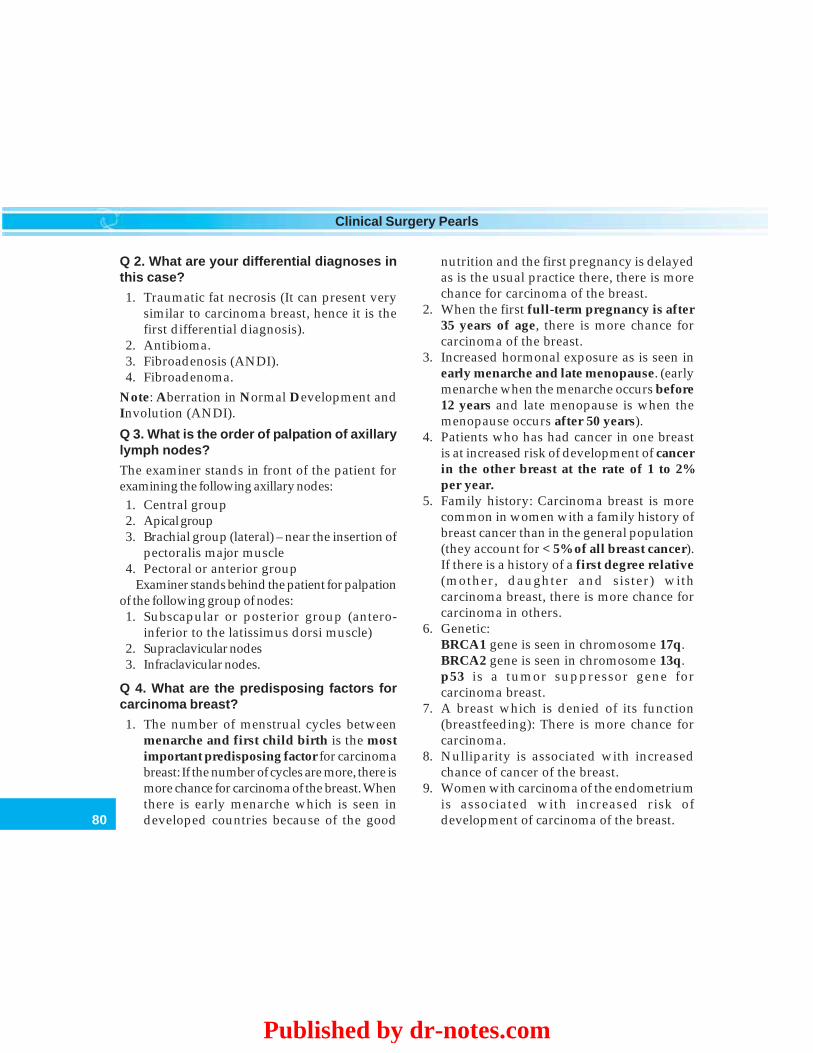
80
Clinical Surgery Pearls
Q 2. What are your differential diagnoses inthis case?
1. Traumatic fat necrosis (It can present verysimilar to carcinoma breast, hence it is thefirst differential diagnosis).
2. Antibioma.3. Fibroadenosis (ANDI).4. Fibroadenoma.
Note: Aberration in Normal Development andInvolution (ANDI).
Q 3. What is the order of palpation of axillarylymph nodes?
The examiner stands in front of the patient forexamining the following axillary nodes:1. Central group2. Apical group3. Brachial group (lateral) – near the insertion of
pectoralis major muscle4. Pectoral or anterior group
Examiner stands behind the patient for palpationof the following group of nodes:1. Subscapular or posterior group (antero-
inferior to the latissimus dorsi muscle)2. Supraclavicular nodes3. Infraclavicular nodes.
Q 4. What are the predisposing factors forcarcinoma breast?
1. The number of menstrual cycles betweenmenarche and first child birth is the mostimportant predisposing factor for carcinomabreast: If the number of cycles are more, there ismore chance for carcinoma of the breast. Whenthere is early menarche which is seen indeveloped countries because of the good
nutrition and the first pregnancy is delayedas is the usual practice there, there is morechance for carcinoma of the breast.
2. When the first full-term pregnancy is after35 years of age, there is more chance forcarcinoma of the breast.
3. Increased hormonal exposure as is seen inearly menarche and late menopause. (earlymenarche when the menarche occurs before12 years and late menopause is when themenopause occurs after 50 years).
4. Patients who has had cancer in one breastis at increased risk of development of cancerin the other breast at the rate of 1 to 2%per year.
5. Family history: Carcinoma breast is morecommon in women with a family history ofbreast cancer than in the general population(they account for < 5% of all breast cancer).If there is a history of a first degree relative(mother, daughter and sister) withcarcinoma breast, there is more chance forcarcinoma in others.
6. Genetic:BRCA1 gene is seen in chromosome 17q.BRCA2 gene is seen in chromosome 13q.p53 is a tumor suppressor gene forcarcinoma breast.
7. A breast which is denied of its function(breastfeeding): There is more chance forcarcinoma.
8. Nulliparity is associated with increasedchance of cancer of the breast.
9. Women with carcinoma of the endometriumis associated with increased risk ofdevelopment of carcinoma of the breast.
Published by dr-notes.com
81
Early Breast Cancer
10. History of exposure to ionizing radiation:History of Mantle radiotherapy forHodgkin’s disease in childhood isassociated with increased incidence ofcarcinoma, especially when the breast isdeveloping. But this will take at least 10years.
11. Postmenopausal obesity: This is due toincreased conversion of steroid hormonesto estradiol in body fat.
12. Diet: Diets low in phytoestrogens and fattyfood is associated with increased risk ofdeveloping breast carcinoma.
13. High intake of alcohol.14. Age: Extremely rare below 20 years but
there after the incidence steadily rises so thatby the age of 90 years nearly 20% of womenare affected.
15. Geographical: Common in the Westernworld, less common in developing countriesand Japan.
16. History of benign proliferative breastdisease is associated with increased chanceso also a breast with previous scars.
17. Environmental: Pesticides, etc.18. Hormone replacement therapy: Combination
of estrogen and progesterone taken for avery long duration is associated withincreased risk for carcinoma breast.Estrogen alone is associated with slightlyincreased risk for carcinoma.
19. Sex: Female breast is more prone forcarcinoma than the male breast.
20. Oral contraceptive pills: Do not appear toincrease the risk of carcinoma breast.
Q 5. How will you proceed to investigate sucha patient?
Triple assessment consisting of clinicalexamination, imaging (mammogram/ultra-sound) and FNAC / Core biopsy. FNAC / Corebiopsy is done first, after clinical examination.
Q 6. Why FNAC is done first?
FNAC will give a cytological diagnosis ofmalignancy or benign nature. It is also cheap,simple and cost-effective.
Q 7. What are the different grades of cyto-logical reporting?
Grade Result
AC0 (Grade 0) No epithelial cells presentAC1 (Grade 1) Scanty benign cellsAC2 (Grade 2) Benign cellsAC3 (Grade 3) Atypical cells present – may
need a biopsy if clinically orradiologically suspicious
AC4 (Grade 4) Highly suspicious of malignancyAC5 (Grade 5) Definitely malignant
Q 8. What are the stains used for stainingFNAC smears?
a. Giemsab. Papanicolaouc. Hematoxylin and eosin.
Q 9. How many needle passes are requiredfor FNAC and what is the needle size?
Usually 23G needle is used.Minimum 6 needle passes are required.
82
Clinical Surgery Pearls
Q 10. If the FNAC is inconclusive can yourepeat the procedure?
Yes, you can repeat FNAC upto 3 times.
Q 11. Still FNAC is negative, how will youproceed?
Core biopsy is the investigation of choice in sucha patient. Usually core biopsy is not recommen-ded for lesions less than 2cm because the smalllesions can be easily excised under local anesthesia.
Q 12. How is core biopsy done?
After infiltrating 1% lignocaine anesthetic intothe skin and down to around the lesion, a smallincision is placed and the lesion is approachedwith the specific core biopsy needle at approxi-mately 45o angle to the breast so that it will notdamage the chest wall.
Q 13. What are the advantages of core biopsy(Tru-cut) over FNAC?
1. FNAC cannot differentiate in situ carcinomafrom infiltrating carcinoma.
2. Receptor assay (Estrogen Receptor andProgesterone Receptor) can be done with corebiopsy specimen.
Note: Therefore core biopsy is the preferred methodfor diagnosis of suspicious breast lump.
Q 14. What are the indications for excisionbiopsy when FNAC is negative?
If a lesion is reported as malignant on cytology andis clinically and mammographically malignant, thepatient does not require further confirmation.
Indications for excision biopsy
a. In lesions < 2cm where core biopsy is not possiblewithout imaging.
b. In lesions between 2 and 4cm where core biopsyis negative.
c. In cystic lesions when the cyst recurs orincreases in size or there is residual mass afteraspiration and FNAC is inconclusive twice.
d. A discrete breast lesion even if it is consideredbenign by all investigation needs excision biopsy.
Q 15. Is there any role for incision biopsy?
For lesions over 4 cm in size where FNAC andcore biopsy are negative, but the lesionis suspicious of malignancy on clinicalexamination and mammography, incisionbiopsy is done (excision biopsy of such a biglump will produce distortion of the breast).
Q 16. If the FNAC is inconclusive, what is theorder of further investigations?
• Less than 2 cm size swelling– wide local excision• 2 – 4 cm size swelling – core biopsy.
If core biopsy fails:• Size of tumor < 4 cm – wide local excision• Size of tumor > 4 cm – incisional biopsy
Q 17. What is the incision planning for breastbiopsy?
• Plan the incision in such a way that if the tumorturns out to be carcinoma, the biopsy scar can beincluded along with the nipple areolar complexwithin the future transverse elliptical incision ofmastectomy.
Published by dr-notes.com
83
Early Breast Cancer
• For lesions within 5 cm of areolar margin,cosmesis is better with circum areolarincision.
• There is no role for radial incision in breastexcept at 3 and 9 O’Clock position.
• Prefer curvi linear incisions.• Follow the lines of resting skin tension/
Langer’s lines.
Q 18. What are the modalities of imagingavailable for breast?
a. Mammographyb. Ultrasoundc. MRI – useful for differentiating scar tissue
from tumor recurrenced. 18 FDG PET scan (not routinely done).
The most frequently used modalities aremammography and ultrasound. Above 35 years ofage mammography is preferred. In youngpatients less than 35 years of age ultrasound ispreferred.
Q 19. Is there any role for mammography inthis patient if it is found to be positive formalignancy in FNAC?
Yes. Mammography is indicated for the samebreast to rule out multicentricity. It is alsoindicated for the contralateral breast to rule outa nonpalpable lesion there.
Q 20. What are the mammography findingsin a case of carcinoma breast?
Mammographic findings in carcinoma breast.Primary signsa. Mass lesion with clustered microcalcification
(dense calcification is suggestive of benigndisease).
b. Speckled mass lesion with ill definedmargin.
c. Architectural distortion of the breastparenchyma (Stellate lesion).
d. Density of the lesion will be more than the restof the breast.
Secondary signsa. Skin changes – retraction, thickening,
dimplingb. Nipple changes – flattening, retraction, etc.c. Increased vascularityd. Axillary lymphadenopathy.
Q 21. What is the commonest malignancy ofthe breast?
Infiltrating duct carcinoma.
Q 22. If the FNAC is reported as infiltratingduct carcinoma, what will be the furthermanagement?
One should proceed to staging work up in sucha patient. Since this case clinically is an earlybreast cancer, the minimum investigationsrequired in this patient are:
Staging work up in carcinoma breast
a. Mammography of the same breast andcontralateral breast
b. Ultrasound of the abdomen to rule out metastasis inthe liver and for Krukenberg’s tumor
c. X-ray of the chest to rule out pulmonarymetastasis
d. Liver function test (LFT)
Q 23. What is the staging of carcinomabreast?
It is divided into 4 stages by the AJCC 6thedition.
84
Clinical Surgery Pearls
Stage I and II are called early breast cancer(EBC).
Stage III a, b, c are called locally advancedbreast cancer (LABC).
Stage IV is metastatic cancer.
Q 24. What is the TNM staging?
Primary tumor (T)
Tx primary tumor cannot be assessed*.T 0 no evidence of primaryTis – carcinoma in situ (Tis DCIS, Tis LCIS, TisPaget’s,Tis (DCIS) – Ductal carcinoma in situTis (LCIS) - Lobular carcinoma in situTis (Paget’s) – Paget’s disease of the nipple withno tumorT1 – tumour less than 2 cmT1 mic – Microinvasion 0.1 cm or less in greatestdimensionT1a – Tumor more than 0.1 cm but not more than0.5 cm in greatest dimensionT1b – Tumor more than 0.5 cm but not more than1 cm in greatest dimension.T1c – Tumor more than 1 cm but not more than 2cm in greatest dimensionT2 – Tumor more than 2 cm but not more than 5 cm.T3 – Tumor more than 5 cm in greatest dimensionT4 – Tumor of any size with direct extension toT4a. Extension to chest wall, not including pectoralismuscleT4b. Skin edema (including peau d’ orange) orulceration of the skin or satellite skin nodules.T4c. Both T4a and T4b.T4d. Inflammatory carcinoma
Note: TX – lump already excised, treatedelsewhere without documentation of the size,or treated with chemotherapy.
The chest wall includes ribs, intercostalmuscles and serratus anterior muscle, but notthe pectoral muscles.
Regional nodes
Nx Lymph nodes cannot be assessedN0 No regional nodesN1 Mobile ipsilateral axillary lymph nodesN2 Fixed ipsilateral/matted or clinically
apparent ipsilateral internal Mammarynodes in the absence of clinically evidentaxillary nodes.
N2a Metastasis in ipsilateral axillary lymphnodes fixed to one another (matted) or toother structures.
N2b Metastasis only in clinically apparentinternal mammary nodes and in theabsence of clinically evident axillary lymphnode metastasis N3 Metastasis in ipsilateralinfraclavicular nodes or metastasis inIpsilateral internal mammary and axillarylymph nodes or metastasis in Ipsilateralsupraclavicular lymph nodes.
N3a Metastasis in ipsilateral infraclavicularlymph nodes
N3b Metastasis in ipsilateral internal mammarylymph node(s) and axillary lymph node(s)
N3c Metastasis in ipsilateral supraclavicularlymph node(s)
Note: Clinically apparent is defined as detectedby imaging studies (excluding lympho-scintigraphy) or by clinical examination orgrossly visible pathologically.
Distant metastasis
Mx Distant metastasis cannot be assessed.M0 No distant metastasisM1 Distant metastasis
Published by dr-notes.com
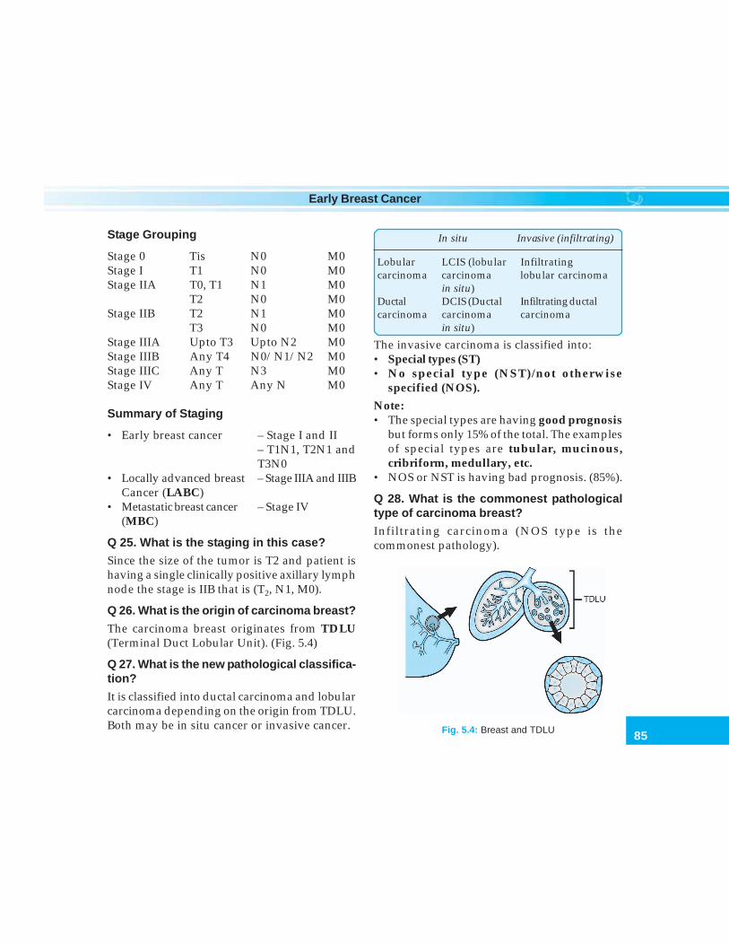
85
Early Breast Cancer
Stage Grouping
Stage 0 Tis N0 M0Stage I T1 N0 M0Stage IIA T0, T1 N1 M0
T2 N0 M0Stage IIB T2 N1 M0
T3 N0 M0Stage IIIA Upto T3 Upto N2 M0Stage IIIB Any T4 N0/N1/N2 M0Stage IIIC Any T N3 M0Stage IV Any T Any N M0
Summary of Staging
• Early breast cancer – Stage I and II– T1N1, T2N1 andT3N0
• Locally advanced breast – Stage IIIA and IIIBCancer (LABC)
• Metastatic breast cancer – Stage IV(MBC)
Q 25. What is the staging in this case?
Since the size of the tumor is T2 and patient ishaving a single clinically positive axillary lymphnode the stage is IIB that is (T2, N1, M0).
Q 26. What is the origin of carcinoma breast?
The carcinoma breast originates from TDLU(Terminal Duct Lobular Unit). (Fig. 5.4)
Q 27. What is the new pathological classifica-tion?
It is classified into ductal carcinoma and lobularcarcinoma depending on the origin from TDLU.Both may be in situ cancer or invasive cancer.
In situ Invasive (infiltrating)
Lobular LCIS (lobular Infiltratingcarcinoma carcinoma lobular carcinoma
in situ)Ductal DCIS (Ductal Infiltrating ductalcarcinoma carcinoma carcinoma
in situ)
The invasive carcinoma is classified into:• Special types (ST)• No special type (NST)/not otherwise
specified (NOS).
Note:• The special types are having good prognosis
but forms only 15% of the total. The examplesof special types are tubular, mucinous,cribriform, medullary, etc.
• NOS or NST is having bad prognosis. (85%).
Q 28. What is the commonest pathologicaltype of carcinoma breast?
Infiltrating carcinoma (NOS type is thecommonest pathology).
Fig. 5.4: Breast and TDLU
86
Clinical Surgery Pearls
Q 29. What is the tumor doubling time forcarcinoma breast?
• One doubling time takes 2 – 9 months.• For reaching 1cm size of the tumor 30
doubling times are required.• The minimum size required for clinical
palpation of a tumor is 1cm.• On an average, it takes five years for a tumor
to become clinically palpable in the breast.
Q 30. What is the grading of the tumor?
The Nottingham combined histological grade(Elston – Ellis modification of Scarff Bloom –Richardson grading system) which is amodified Bloom – Richardson grading is usedfor grading. In this the nuclear pleomorphism,mitotic count and tubule formation are takeninto account giving scores of 1 (favorable) 2-3(unfavorable). A score of 3-5 is grade I, 6-7 isgrade II, 8-9 is grade III.
Q 31. What is the surgical treatment of choicein this patient?
Now the gold standard surgical treatmentoption for stage I and II patients are BCT (Breastconservation therapy). Modified RadicalMastectomy is another option (Flow chart 5.1).
Q 32. What is breast conservation therapy?
Breast conservation consists of wide excisionwith 1cm tumor clearance without excising theoverlying skin, axillary node dissection througha separate incision in the axilla, followed byradiotherapy to the rest of the breast.
Components – of BCT
• Wide excision• Axillary node dissection• Radiotherapy to the remaining breast
Q 33. What are the precautions to be takenduring wide excision in breast conservativesurgery?
Important steps of wide excision forcarcinoma breast
a. The incision should be curvilinear placed alongthe Langer’s lines (skin tension lines).
b. Removal of the skin overlying the lesion isunnecessary.
c. 1 cm tumor clearance is important (macroscopic).d. The skin flaps should not be undermined.e. Pectoral fascia is not excised unless involved.f. If it is involving the muscle, a fillet of muscle
should be removed.g. Specimen should be oriented with sutures or
clips.h. Meticulous hemostasis is secured with diathermy.i. The cavity is lavaged with dilute chlorhexidine
solution and kept for 2minutes.j. The cavity is not obliterated.
k. No drain is put.
Q 34. What are the contraindication for breastconservation?
Contraindication for breast conservation
1. T3, T4 tumors, N2 or M12. Large or central tumors in small breast3. Multifocal/multicentric disease4. Collagen vascular disease5. Extensive in situ component
Published by dr-notes.com
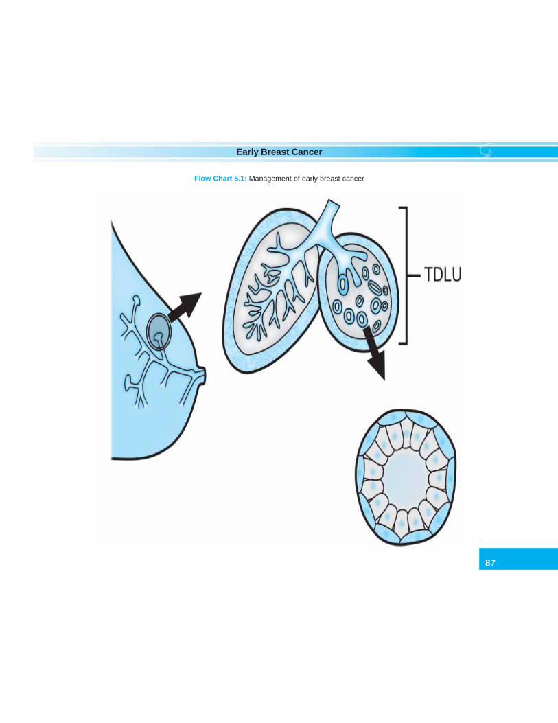
87
Early Breast Cancer
Flow Chart 5.1: Management of early breast cancer
88
Clinical Surgery Pearls
Consider MRM (Modified radical mastectomy)in these cases.
Q 35. What is the rationale for axillarydissection?
The single most important prognostic factor forcarcinoma breast is nodal status (4 or morepathologically + ve axillary node is prognosticallybad). Therefore axillary dissection will form part ofthe surgical management of all types proceduresincluding breast conservation.
Q 36. What is the average number of axillarylymph nodes and internal mammary nodes?
• Average number of axillary nodes is 53• Internal mammary nodes – 3 to 4.
Q 37. What is the rationale for radiotherapyin early breast cancer surgery?
Radiotherapy is given to tackle themulticentricity in the remaining breast.
The patient should be willing to undergoradiotherapy 5 days a week for 20 days andradiotherapy facility should be available.
Q 38. What is the rationale for mastectomyin early breast cancer?
Mastectomy is recommended to remove themulticentricity seen in carcinoma breast.
Q 39. What is the difference betweenmulticentricity and multifocality?
• When tumor foci are found in a differentquadrant it is called multicentricity.
• When tumor foci are found in the same quadrantit is called multifocal.
Q 40. What is the type of mastectomy recom-mended for early breast cancer ?
Modified Radical Mastectomy of Auchinclossis preferred (Case No. 6).
Q 41. What is QUART Regimen of VERONESI(Contemporary Italian Surgeon UmbretoVeronesi from Milan)?
Veronesi of Milan is responsible for bringing aparadigm shift in surgical treatment ofcarcinoma breast from mastectomy toconservation. Now quart regimen is notfollowed. It consists of au adrantectomy,Axillary Dissection and Radio Therapy).
Q 42. What is extensive in situ component(EIC)?
The presence of EIC in an invasive cancer isdefined as the presence of more than 25% DCIScomponent in the main tumor or if DCIS ispresent elsewhere in the surrounding tissue.
Q 43. What is inflammatory carcinoma?
Features of inflammatory carcinoma
• It is a rare and most malignant form of breastcancer, constituting less than 3% of cases.
• It is a clinicopathological entity characterizedby diffuse erythema and edema (peau d’orange) of the breast, often without underlyingpalpable mass.
• The clinical findings should involve themajority of the skin of the breast.
• The term should not be applied to a patient withneglected locally advanced cancer of the breast.
• On imaging there may be detectable mass andthickening of the skin over the breast.
Published by dr-notes.com
89
Early Breast Cancer
• The clinical presentation is due to tumor emboliwithin dermal lymphatics.
• The inflammatory carcinoma is one where there ispathologically evident dermal lymphatic infiltrationby the tumor for more than 1/3rd of the skin ofthe breast (dermal lymphatic infiltration andclinical findings are important).
• This is usually not associated with a palpablelump. It has a very bad prognosis and it isincluded under stage III (classified as T4d).
Q 44. Is there any role for chemotherapy inearly breast cancer surgery?
Yes. All patients with more than 1cm size tumorand < 70years need combination chemotherapyfor 6 cycles – CMF (Cyclophosphamide,Methotrexate, 5 FU) or CAF (Adriamycin-basedregimen) (Flow chart 5.1).
Q 45. What is the rationale for chemotherapy?The new concept about carcinoma breast is thatit is a systemic disease from the very beginning.This is in contrast to the old Halstedian conceptwhere a staged progression of the tumor fromthe breast to the axilla and from their to systemicdissemination was proposed. Therefore fromthe very beginning patient can have micro-metastases which cannot be detected. In orderto tackle the micrometastases systemicchemotherapy is given.
Q 46. Is there any role for hormone therapyin this patient?If the tumor is ER and PR (Estrogen receptorand Progesterone receptor) positive the patient
is given the antiestrogen called Tamoxifen20mg daily for five years (Flow chart 5.1).
Q 47. What are the actions of Tamoxifen?
Actions of Tamoxifen
a. It has antiestrogenic action on the breast andestrogenic action on other tissues
b. Tumoricidalc. Prevent recurrence in the contralateral breast
Note: Tamoxifen is a selective estrogen receptormodulator (SERM) that has antagonistic actionon estrogen receptors in the breast and agonisticaction on ER receptor elsewhere.
The half life of the drug is 7 days and it takes4 weeks for action. The other beneficial effectsof Tamoxifen are:a. Preservation of bone density in postmeno-
pausal womenb. Decrease in cholesterolc. Reduction in cardiovascular morbidity
(These are due to the estrogenic effects ofTamoxifen on extra mammary tissues).
Q 48. What are the side effects of Tamoxifen?
Side effects of tamoxifen
a. Hot flushesb. Thromboembolic complications (deep vein
thrombosis and pulmonary embolism) and strokec. Vaginal dryness and atrophy in premenopausal
womend. Vaginal discharge in postmenopausal womene. Increase risk of endometrial cancer in
postmenopausal womenf. Bone loss in premenopausal womeng. Weight gain
90
Clinical Surgery Pearls
Side effects in post- Side effects in pre-menopausal women menopausal women
• Endometrial cancer • Bone loss• Pulmonary embolism • Decreased vaginal
secretion• Stroke • Atrophy of vaginal
epithelium• Vaginal discharge
Q 49. What is the drug which is likely toreplace Tamoxifen?
Aromatase inhibitors – the production of estrogenrequires the activity of aromatase enzyme.Estrogen production in postmenopausal womenis by peripheral aromatization of androgensproduced from the adrenal gland. Aromataseinhibitors block estrogen production in post-menopausal women and therefore they formadjuvant therapy for postmenopausal women.A number of aromatase inhibitors are availablelike:a. Aminoglutethimide – this also blocks
cortisol.b. Newer aromatase inhibitors,
E.g: Anastrozole, Letrozole
Q 50. What is the source of estrogen in post-menopausal women?
• The circulating estrogen in postmenopausalwomen is about 10% of the level in premeno-pausal. They are synthesized peripherallyprincipally in fat including breast fat, skin,muscle and liver.
• Adrenal gland.
Q 51. Can you avoid axillary dissection incarcinoma breast?
Yes. Axillary dissection can be avoided bysentinel node biopsy.
Q 52. What is sentinel lymph node biopsy?
A sentinel node is a lymph node that primarilydrains the tumor area. Sentinel node biopsy isdone for early breast cancer with clinicallynegative axillary nodes. Injection of radiolabelled(technetium) sulphur colloid and or a blue dye intothe tumour or peritumoral area or the skinoverlying before excision of the primary allowsidentification of the sentinel node in the axillaeither by intraoperative visual inspection orwith the help of gamma probe. For patients withbreast cancer one or two nodes are usuallyidentified.
The concept is that if the identified sentinelnodes are negative for malignancy in frozensection examination, the chances for a higherlevel node to be affected by skip metastasis isremote and therefore an axillary clearance canbe avoided with the consequent morbidity. Ifthe sentinel node is positive the patient willgo in for formal axillary clearance.
Q 53. What is the management of DCIS(Ductal carcinoma in situ)?
DCIS can be classified into widespread type (i.e.more than 4cm size) and localized type. Forwide-spread type, the treatment of choice ismastectomy.The 3 surgical options are—1. Simple mastectomy
Published by dr-notes.com
91
Early Breast Cancer
2. Breast conservation surgery (Lumpectomy)+ Radiotherapy to the breast + Tamoxifenfor 5 years.
3. Lumpectomy alone (without radiotherapy).• There is no need for axillary dissection in DCIS
because of the fact that chance for nodeinvolvement is negligible (4%).
• There is no role for chemotherapy in DCIS.
Q 54. What are the indications formastectomy in DCIS?
1. Women with multicentric DCIS2. Extensive or diffused ductal carcinoma3. Positive margins4. Acceptable cosmesis cannot be achieved5. Radiotherapy is contraindicated (Collagen
vascular disease)6. Pregnancy7. Diffuse malignant appearing micro
calcification8. Patient preference.
Q 55. What are the essential steps oflumpectomy?
Essential steps of lumpectomy
• Complete surgical excision• Careful orientation of the specimen• Negative margin of at least 1mm width• There should be no tumor at the margin
(optimal margin width not known)• Margin status is taken before closure of the
wound• Radiography to confirm excision of micro-
calcification• Postexcision breast imaging to confirm removal
of suspicious areas.• Reexcision as necessary to obtain negative margin
Note: Lumpectomy is followed by radio-therapy in breast conservation treatment forDCIS.
Q 56. Is there any role for hormone therapyin DCIS?
Yes. If they are ER positive (Estrogen ReceptorPositive), Tamoxifen is given for 5 years.
Q 57. What are the indications forlumpectomy alone
• Small lesions < 0.5 cm size• Unicentric lesion• Low grade lesion.
Q 58. What is the follow up of DCIS aftersurgery?
• Physical examination every 6 months for 5years
• Mammogram every 12 months.
Q 59. What is LCIS (Lobular carcinomain situ)?
The present consensus regarding LCIS is that itis a marker of subsequent development ofinvasive cancer rather than a preinvasivecancerous lesion. (It is not a precursor but apredictor for carcinoma. Hence it is called abystander lesion).• LCIS is a rare form of non-invasive breast
cancer. It does not typically form micro-calcifications and therefore not easilydetectable on X-rays and is usually foundincidentally on biopsy.
• Invasive ductal cancer develops inapproximately 25% of patients, which maybe delayed for 10-20years
92
Clinical Surgery Pearls
- 45% develop ipsilateral carcinoma- 35% develop contralateral carcinoma- 15% develop bilateral carcinoma.
• The risk of development of invasive ductalcancer is equal for both breasts.
• LCIS tends to be multifocal in both breasts.• The contralateral breast is affected in 30% of
cases and in the residual breast in upto 70% ofcases. The incidence of nodal involvement is verylow in this cancer.
Q 60. What is the management of LCIS?
Management of LCIS
• History and physical examination every (6-12months)
• Diagnostic bilateral mammogram (every 12months)
• Pathology review• Observation for the development of invasive
cancer (ipsilateral and contralateral breast)• Risk reduction with Tamoxifen for pre-
menopausal women• Risk reduction with Raloxifen for post
menopausal women• Bilateral mastectomy and reconstruction in
special circumstances
Q 61. What are the indications forprophylactic mastectomy?
1. LCIS (Lobular carcinoma in situ)2. BRCA1 and 2.
Q 62. What are the bad prognostic factorsfor carcinoma breast?
Bad prognostic factors for carcinoma breast
1. Axillary nodal status is the most importantsingle prognostic factor
2. Age < 35 years – bad prognosis3. Grade of tumor (Bloom - Richardson grade) –
higher the grade, bad the prognosis.4. Extensive in situ component – bad prognosis.5. Receptor status – ER negative and PR negative
—bad prognosis.6. P53 status (Tumor suppressor gene called
‘Guardian of Genome’) positive – bad prognosis.7. Presence of HER – 2 (Tyrosine kinase receptor)
—bad prognosis.
Q 63. What is Paget’s disease of the breast?
It is nothing but a ductal carcinoma withassociated nipple destruction, seen in 1-2% ofthe patients. In 50% there will be an underlyingmass lesion. 90% of these patients will haveinvasive carcinoma. For the patients withoutmass lesion 30% will be later found to have aninvasive carcinoma. It should be differentiatedfrom eczema. Mammography is indicated insuch patients and also imprint cytology of thelesion. Incisional biopsy is finally required.• If a mass lesion is present, treatment is MRM
(Modified Radical Mastectomy).• Otherwise a wide local excision of the nipple
areolar region, axillary dissection followed byradiotherapy and other adjuvant treatmentis required.
Q 64. What are the difference between Paget’sdisease and eczema?
Published by dr-notes.com
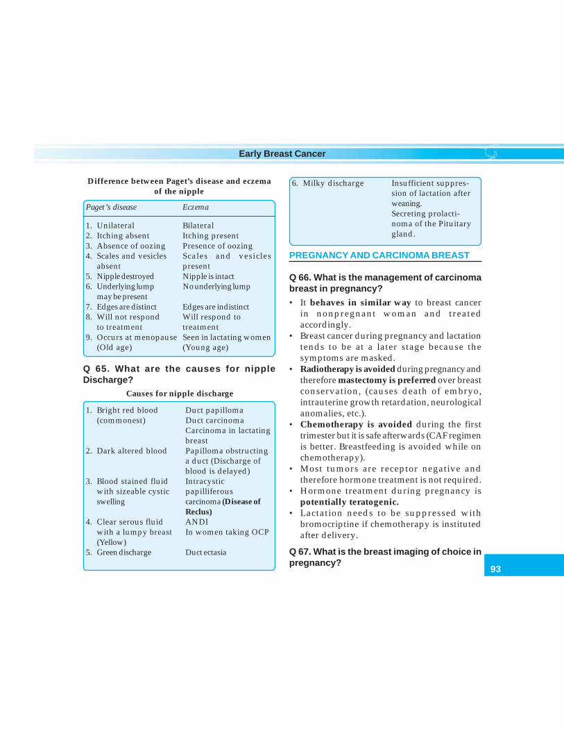
93
Early Breast Cancer
Difference between Paget’s disease and eczemaof the nipple
Paget’s disease Eczema
1. Unilateral Bilateral2. Itching absent Itching present3. Absence of oozing Presence of oozing4. Scales and vesicles Scales and vesicles
absent present5. Nipple destroyed Nipple is intact6. Underlying lump No underlying lump
may be present7. Edges are distinct Edges are indistinct8. Will not respond Will respond to
to treatment treatment9. Occurs at menopause Seen in lactating women
(Old age) (Young age)
Q 65. What are the causes for nippleDischarge?
Causes for nipple discharge
1. Bright red blood Duct papilloma(commonest) Duct carcinoma
Carcinoma in lactatingbreast
2. Dark altered blood Papilloma obstructinga duct (Discharge ofblood is delayed)
3. Blood stained fluid Intracysticwith sizeable cystic papilliferousswelling carcinoma (Disease of
Reclus)4. Clear serous fluid ANDI
with a lumpy breast In women taking OCP(Yellow)
5. Green discharge Duct ectasia
6. Milky discharge Insufficient suppres-sion of lactation afterweaning.Secreting prolacti-noma of the Pituitarygland.
PREGNANCY AND CARCINOMA BREAST
Q 66. What is the management of carcinomabreast in pregnancy?
• It behaves in similar way to breast cancerin nonpregnant woman and treatedaccordingly.
• Breast cancer during pregnancy and lactationtends to be at a later stage because thesymptoms are masked.
• Radiotherapy is avoided during pregnancy andtherefore mastectomy is preferred over breastconservation, (causes death of embryo,intrauterine growth retardation, neurologicalanomalies, etc.).
• Chemotherapy is avoided during the firsttrimester but it is safe afterwards (CAF regimenis better. Breastfeeding is avoided while onchemotherapy).
• Most tumors are receptor negative andtherefore hormone treatment is not required.
• Hormone treatment during pregnancy ispotentially teratogenic.
• Lactation needs to be suppressed withbromocriptine if chemotherapy is institutedafter delivery.
Q 67. What is the breast imaging of choice inpregnancy?
94
Clinical Surgery Pearls
• MRI is the choice• Ultrasound and mammogram are likely to
yield more false-positive results because ofincreased parenchymal density, increasedvascularity, water content and cellularity.
Q 68. What is the staging work up duringpregnancy?
• There is no contraindication for X-ray chest.It can be done with abdominal shielding.
• Alkaline phosphatase is not dependableduring pregnancy (It is elevated inpregnancy).
• Bone scan is delayed until after delivery (MRI issafe for metastasis).
Q 69. Can a patient become pregnant aftertreatment of carcinoma breast?
Women are usually advised to wait for two yearsfor subsequent pregnancy since the maximumrecurrences are seen during the first 2 yrs.
Q 70. What is the management of carcinomabreast during various trimesters ofpregnancy?
First trimester• Modified radical mastectomy and axillary
dissection for the local disease• If found to be node positive, terminate the
pregnancy and give chemotherapy• If it is locally advanced terminate the
pregnancy and treat.
Second trimester• The decision is based on the stage of the
disease and the wish of the patient tocontinue pregnancy
• All stages surgery is recommended.
Third trimester• Do surgery• Wait till delivery and give adjuvant
chemotherapy after delivery.
Q 71. What is the role of therapeutic abortionin carcinoma breast?
• It is indicated if radiotherapy and chemo-therapy cannot be postponed due to the stageof the disease
• There is no advantage as such for therapeuticabortion, but the treatment is made easier.
Q 72. What is bilateral breast cancer?
• It means a separate primary cancer in eachbreast
• It can be synchronous or metachronous.
Q 73. How to differentiate whether the secondtumor is primary or metastatic?
The Choudary Millis criteria used for callingthe second tumor a primary are:1. Demonstration of in situ change in the
contralateral tumor.2. If it is histologically different from the
cancer in the first breast.3. If the degree of histologic differentiation is
distinctly greater than the lesion in the firstbreast.
4. In the absence of histologic difference, thecontralateral is considered to be primary,provided there is no evidence of local,regional or distant metastasis from thecancer in the ipsilateral breast.
Published by dr-notes.com
95
Early Breast Cancer
Q 74. What are the risk factors for thedevelopment of a second primary?
• A woman who has had breast cancer has afive fold increase in risk for second breastcancer
• Multifocal cancer in the original breast• Lobular carcinoma in situ• Family history of breast cancer• Diagnosis of original cancer at an early age.
Q 75. What is the treatment of secondprimary?
Give treatment appropriate for tumordepending on the stage of the disease.
Q 76. What is the mammographic findingsin metastatic cancer of the breast?
Mammographic findings in metastaticcancer of the breast
• Metastatic cancer is less infiltrative (primarytumour is more infiltrative)
• More diffuse• Fewer fine calcifications• Presence of secondary edema
For years and years they told me, Be careful ofyour breasts. Don’t ever squeeze or bruise them.
And give them monthly tests.So I heeded all their warnings, An protected
them by law. Guarded them very carefully, AndI always wore my bra
After 30 years of astute care, My doctor found alump
She ordered up a mammogramTo look inside that bump.
”Stand up very close” she said. As she got myboob in line, ”And tell me when it hurts” she
said. ”Ah yes! There, that’s fine.She stepped upon a pedal. I could not believe
my eyes! A plastic plate pressed down anddown, My boob was in a vise!
My skin was stretched and stretched, From wayup under my chin. My poor boob was being
squashed, To Swedish pancake thin.
Elegy for a Poor Breast
Excruciating pain I felt, Within it’s vice – likegrip. A prisoner in this viscous thing. My poor
defenseless tit!‘Take a deep breath’ she said to me,Who does
she think she’s kidding? My chest is mashed inher machine, And woozy I am getting.
‘There, that was good, ”I heard her sayAs theroom was slowly swaying. ”Now, let’s have a go
at the other one,” Lord have mercy I waspraying.
It squeezed me from up and down. It squeezedme from both sides. I’ll bet she’s never had this
done Not to her tender little hide! If I had noproblem when I came in, I surely have one now.If there had been a cyst in there, It would have
popped, “ker-pow!”This machine was created by a man, Of this, I
have no doubt. I’d like to stick his balls in there,And see how they come out!
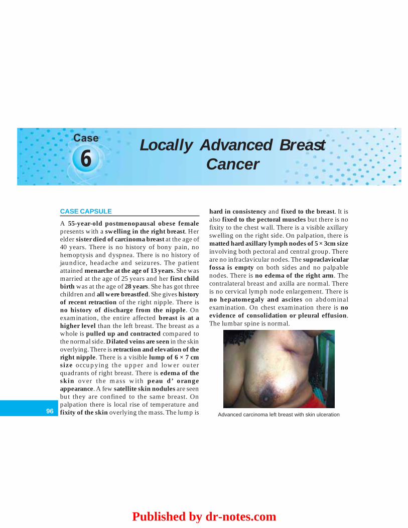
96
Clinical Surgery Pearls
6Locally Advanced Breast
Cancer
CASE CAPSULE
A 55-year-old postmenopausal obese femalepresents with a swelling in the right breast. Herelder sister died of carcinoma breast at the age of40 years. There is no history of bony pain, nohemoptysis and dyspnea. There is no history ofjaundice, headache and seizures. The patientattained menarche at the age of 13 years. She wasmarried at the age of 25 years and her first childbirth was at the age of 28 years. She has got threechildren and all were breastfed. She gives historyof recent retraction of the right nipple. There isno history of discharge from the nipple. Onexamination, the entire affected breast is at ahigher level than the left breast. The breast as awhole is pulled up and contracted compared tothe normal side. Dilated veins are seen in the skinoverlying. There is retraction and elevation of theright nipple. There is a visible lump of 6 × 7 cmsize occupying the upper and lower outerquadrants of right breast. There is edema of theskin over the mass with peau d’ orangeappearance. A few satellite skin nodules are seenbut they are confined to the same breast. Onpalpation there is local rise of temperature andfixity of the skin overlying the mass. The lump is
hard in consistency and fixed to the breast. It isalso fixed to the pectoral muscles but there is nofixity to the chest wall. There is a visible axillaryswelling on the right side. On palpation, there ismatted hard axillary lymph nodes of 5 × 3cm sizeinvolving both pectoral and central group. Thereare no infraclavicular nodes. The supraclavicularfossa is empty on both sides and no palpablenodes. There is no edema of the right arm. Thecontralateral breast and axilla are normal. Thereis no cervical lymph node enlargement. There isno hepatomegaly and ascites on abdominalexamination. On chest examination there is noevidence of consolidation or pleural effusion.The lumbar spine is normal.
Advanced carcinoma left breast with skin ulceration
Published by dr-notes.com
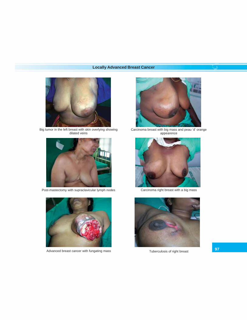
97
Locally Advanced Breast Cancer
Big tumor in the left breast with skin overlying showingdilated veins
Carcinoma breast with big mass and peau ‘d’ orangeappearence
Post-mastectomy with supraclavicular lymph nodes Carcinoma right breast with a big mass
Tuberculosis of right breastAdvanced breast cancer with fungating mass
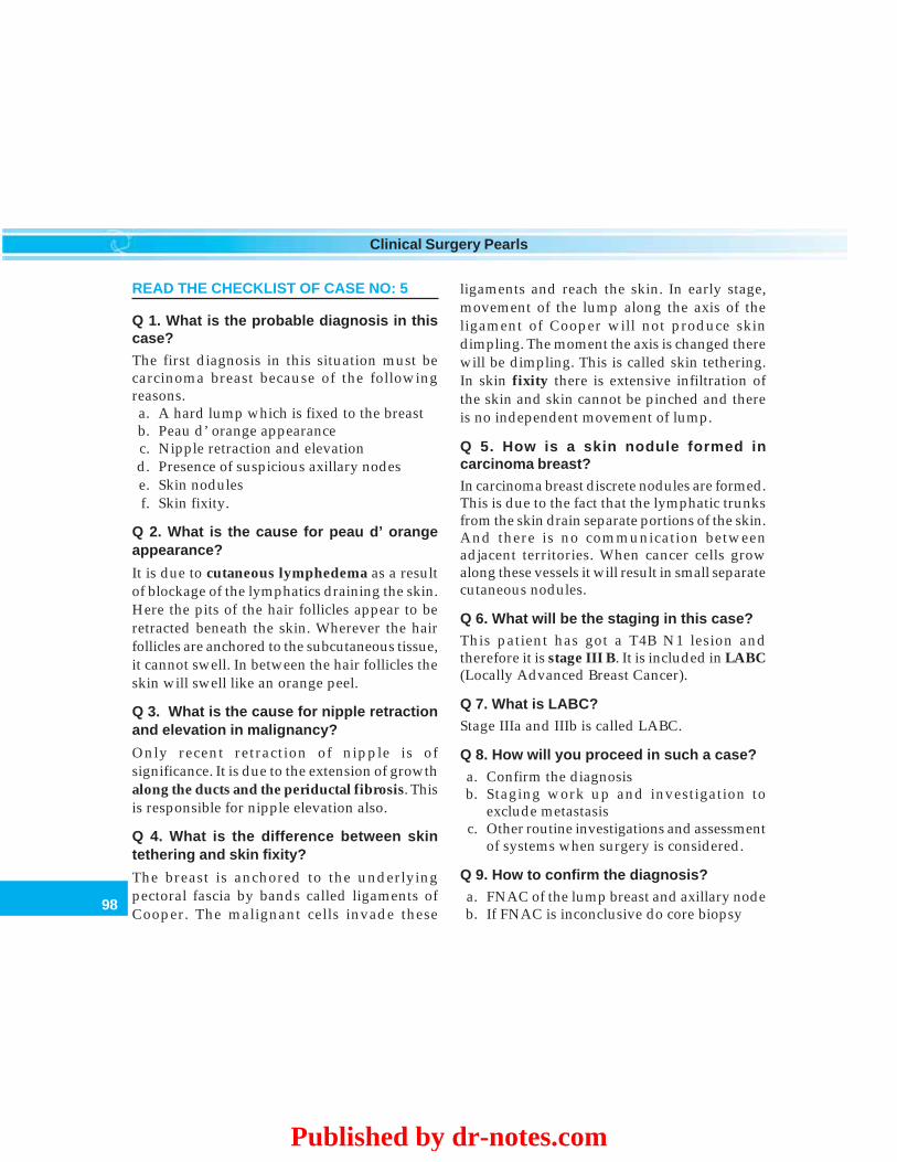
98
Clinical Surgery Pearls
READ THE CHECKLIST OF CASE NO: 5
Q 1. What is the probable diagnosis in thiscase?
The first diagnosis in this situation must becarcinoma breast because of the followingreasons.a. A hard lump which is fixed to the breastb. Peau d’ orange appearancec. Nipple retraction and elevationd. Presence of suspicious axillary nodese. Skin nodulesf. Skin fixity.
Q 2. What is the cause for peau d’ orangeappearance?
It is due to cutaneous lymphedema as a resultof blockage of the lymphatics draining the skin.Here the pits of the hair follicles appear to beretracted beneath the skin. Wherever the hairfollicles are anchored to the subcutaneous tissue,it cannot swell. In between the hair follicles theskin will swell like an orange peel.
Q 3. What is the cause for nipple retractionand elevation in malignancy?
Only recent retraction of nipple is ofsignificance. It is due to the extension of growthalong the ducts and the periductal fibrosis. Thisis responsible for nipple elevation also.
Q 4. What is the difference between skintethering and skin fixity?
The breast is anchored to the underlyingpectoral fascia by bands called ligaments ofCooper. The malignant cells invade these
ligaments and reach the skin. In early stage,movement of the lump along the axis of theligament of Cooper will not produce skindimpling. The moment the axis is changed therewill be dimpling. This is called skin tethering.In skin fixity there is extensive infiltration ofthe skin and skin cannot be pinched and thereis no independent movement of lump.
Q 5. How is a skin nodule formed incarcinoma breast?
In carcinoma breast discrete nodules are formed.This is due to the fact that the lymphatic trunksfrom the skin drain separate portions of the skin.And there is no communication betweenadjacent territories. When cancer cells growalong these vessels it will result in small separatecutaneous nodules.
Q 6. What will be the staging in this case?
This patient has got a T4B N1 lesion andtherefore it is stage III B. It is included in LABC(Locally Advanced Breast Cancer).
Q 7. What is LABC?
Stage IIIa and IIIb is called LABC.
Q 8. How will you proceed in such a case?
a. Confirm the diagnosisb. Staging work up and investigation to
exclude metastasisc. Other routine investigations and assessment
of systems when surgery is considered.
Q 9. How to confirm the diagnosis?
a. FNAC of the lump breast and axillary nodeb. If FNAC is inconclusive do core biopsy
Published by dr-notes.com
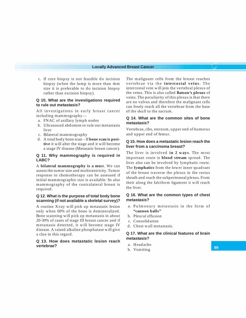
99
Locally Advanced Breast Cancer
c. If core biopsy is not feasible do incisionbiopsy (when the lump is more than 4cmsize it is preferable to do incision biopsyrather than excision biopsy).
Q 10. What are the investigations requiredto rule out metastasis?
All investigations in early breast cancerincluding mammography—a. FNAC of axillary lymph nodesb. Ultrasound abdomen to rule out metastasis
liverc. Bilateral mammographyd. A total body bone scan – If bone scan is posi-
tive it will alter the stage and it will becomea stage IV disease (Metasatic breast cancer).
Q 11. Why mammography is required inLABC?
A bilateral mammography is a must. We canassess the tumor size and multicentricity. Tumorresponse to chemotherapy can be assessed ifinitial mammographic size is available. So alsomammography of the contralateral breast isrequired.
Q 12. What is the purpose of total body bonescanning (if not available a skeletal survey)?
A routine X-ray will pick up metastatic lesiononly when 60% of the bone is demineralized.Bone scanning will pick up metastasis in about20-30% of cases of stage III breast cancer and ifmetastasis detected, it will become stage IVdisease. A raised alkaline phosphatase will givea clue in this regard.
Q 13. How does metastatic lesion reachvertebrae?
The malignant cells from the breast reachesvertebrae via the intercostal veins. Theintercostal vein will join the vertebral plexus ofthe veins. This is also called Batson’s plexus ofveins. The peculiarity of this plexus is that thereare no valves and therefore the malignant cellscan freely reach all the vertebrae from the baseof the skull to the sacrum.
Q 14. What are the common sites of bonemetastasis?
Vertebrae, ribs, sternum, upper end of humerusand upper end of femur.
Q 15. How does a metastatic lesion reach theliver from a carcinoma breast?
The liver is involved in 2 ways. The mostimportant route is blood stream spread. Theliver also can be involved by lymphatic route.The lymphatics from the lower inner quadrantof the breast traverse the plexus in the rectussheath and reach the subperitoneal plexus. Fromtheir along the falciform ligament it will reachthe liver.
Q 16. What are the common types of chestmetastasis?
a. Pulmonary metastasis in the form of“cannon balls”
b. Pleural effusionc. Consolidationd. Chest wall metastasis.
Q 17. What are the clinical features of brainmetastasis?
a. Headacheb. Vomiting
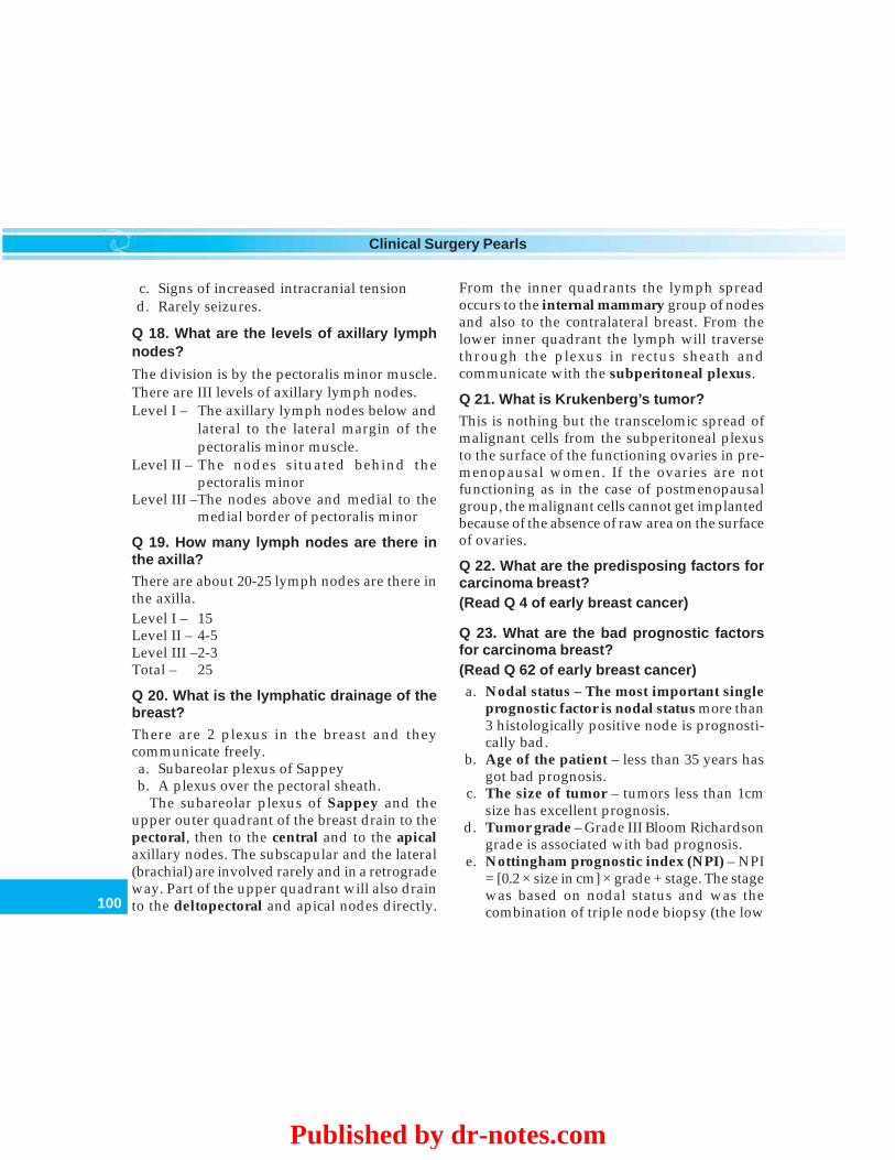
100
Clinical Surgery Pearls
c. Signs of increased intracranial tensiond. Rarely seizures.
Q 18. What are the levels of axillary lymphnodes?
The division is by the pectoralis minor muscle.There are III levels of axillary lymph nodes.Level I – The axillary lymph nodes below and
lateral to the lateral margin of thepectoralis minor muscle.
Level II – The nodes situated behind thepectoralis minor
Level III –The nodes above and medial to themedial border of pectoralis minor
Q 19. How many lymph nodes are there inthe axilla?
There are about 20-25 lymph nodes are there inthe axilla.Level I – 15Level II – 4-5Level III –2-3Total – 25
Q 20. What is the lymphatic drainage of thebreast?
There are 2 plexus in the breast and theycommunicate freely.a. Subareolar plexus of Sappeyb. A plexus over the pectoral sheath.
The subareolar plexus of Sappey and theupper outer quadrant of the breast drain to thepectoral, then to the central and to the apicalaxillary nodes. The subscapular and the lateral(brachial) are involved rarely and in a retrogradeway. Part of the upper quadrant will also drainto the deltopectoral and apical nodes directly.
From the inner quadrants the lymph spreadoccurs to the internal mammary group of nodesand also to the contralateral breast. From thelower inner quadrant the lymph will traversethrough the plexus in rectus sheath andcommunicate with the subperitoneal plexus.
Q 21. What is Krukenberg’s tumor?
This is nothing but the transcelomic spread ofmalignant cells from the subperitoneal plexusto the surface of the functioning ovaries in pre-menopausal women. If the ovaries are notfunctioning as in the case of postmenopausalgroup, the malignant cells cannot get implantedbecause of the absence of raw area on the surfaceof ovaries.
Q 22. What are the predisposing factors forcarcinoma breast?(Read Q 4 of early breast cancer)
Q 23. What are the bad prognostic factorsfor carcinoma breast?(Read Q 62 of early breast cancer)
a. Nodal status – The most important singleprognostic factor is nodal status more than3 histologically positive node is prognosti-cally bad.
b. Age of the patient – less than 35 years hasgot bad prognosis.
c. The size of tumor – tumors less than 1cmsize has excellent prognosis.
d. Tumor grade – Grade III Bloom Richardsongrade is associated with bad prognosis.
e. Nottingham prognostic index (NPI) – NPI= [0.2 × size in cm] × grade + stage. The stagewas based on nodal status and was thecombination of triple node biopsy (the low
Published by dr-notes.com
101
Locally Advanced Breast Cancer
axilla, high axilla and internal mammarychain).
f. Histology – Certain special types likeclassical lobular, tubular, cribriform,medullary, mucinous and papillary arehaving better prognosis than NST.
g. ER and PR status – Approximately 60% ofthe tumors contain detectable estrogenreceptor (ER). ER and progesterone receptor(PR) positive patients having better progno-sis. It is a relatively weak prognostic factor.
h. Ploidy – Ploidy is a measurement of therelative proportion of DNA in each cell.Anuploidy is associated with bad prognosis.Diploid tumors have a lower risk of relapse.The cells in active cell division (S - phase) canbe determined by floor cytometry. S phase isbetter predictor of relapse and survival thanploidy. Low S phase tumor have a morefavorable prognosis. The use of antibody Ki-67 allows an easier estimation of proliferation.
i. erb B2 (HER 2/neu) – This is a cellmembrane receptor protein and is theproduct of neu oncogene. Even though itshould be done routinely to predict thelikelihood of their response to Herceptin itsprognostic significance is doubtful.
j. p53 - the abnormality of p53 expression is seenin breast, ovarian and bowel cancer. Theabnormality is associated with bad prognosis.
k. Epidermal growth factor receptor (EGF) –has got bad prognosis.
Q 24. If there is no evidence of metastasisthis case will be LABC and what will be theline of management?
Multimodal treatment is the therapeuticoption. LABC is an indication for anteriorchemotherapy, i.e. chemotherapy given prior tosurgery (neoadjuvant). Start the patient withCAF/CMF regimen. 2 cycles of chemotherapyare given first (5FU-600mg/m2, Adriamycin50mg/m2, Cyclophosphamide 600mg/m2).After 2 cycles the patient is evaluated forresponse (Flow chart 6.1).
Q 25. How to assess response?
The response is assessed by mammography andclinical assessment. The patient is categorizedinto the following:a. Complete responder – no palpable tumor
after chemotherapy.b. Partial responder – more than 50% decrease
in size.c. Nonresponder – less than 50% decrease in size.d. Progressive disease – more than 25%
increase in size.
Q 26. Why chemotherapy is given first?
a. Downstaging is possible with chemob. Surgery is possible after cyto reductionc. Local control is not the issue in LABCd. LABC patients are likely to harbor micro-
metastasise. The policy of hitting the micrometastasis firstf. Systemic therapy is a must in all patientsg. Allows assessment of chemosensitivity.
Q 27. What are the options for nonrespon-ders/progressive disease?
a. If operable, surgery is done
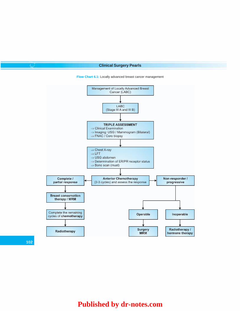
102
Clinical Surgery Pearls
Flow Chart 6.1: Locally advanced breast cancer management
Published by dr-notes.com
103
Locally Advanced Breast Cancer
b. Otherwise, XRT (radiotherapy), hormonetherapy are the options.• Small percentage will respond to the drug
Taxol (only 15% of the chemoresistant willrespond.
• Radiotherapy is given to the chest, axillaand supraclavicular region5000cGy in fractions – 200cGy over 5weeks.
Q 28. If the patient is responding whatsurgery is done?
The surgeons who prefer breast conservationtherapy will do breast conservation surgery.Some surgeons perform MRM (modifiedradical mastectomy) in this setting.
Q 29. After the breast conservation/mastec-tomy what is the sequencing of therapy?
For all those who undergo MRM the remaining4 cycles of chemo is given first. Those whoundergo breast conservation and those withpositive resection margin, radiotherapy is givenfirst, followed by subsequent chemotherapy.
Q 30. What are the side effects of chemo-therapy?
a. Bone marrow depressionb. Gastrointestinal (nausea and vomiting)c. Alopecia.
The patient is monitored by Hb, TLC beforetherapy and after the cycle on day 21. If abnormal,do platelet count also. Serum albumin, SGOT,SGPT are done before and after therapy.
Q 31. Is there any use of cytoprotective agentsfor chemoinduced hematological toxicity? (PG)
GMCF (granulocyte macrophage colonystimulating factor), GCSF (Granulocyte colonystimulating factor) are useful for reducing theseverity and duration of neutropenia. They aregiven either before or upto 24 hours after chemo.Dose: 30million units. But it is costly.
Q 32. What is the role of hormone therapy inLABC?
It should be started in all patients from day 1irrespective of menopausal status and carriedon for 5 years.
Q 33. What is the cut off value of the ER forconsidering it as positive?
There is no cut off value but more than 10fmol/mg is considered positive. About 2/3rd of post-menopausal and half of premenopausal arepositive. ER value more than 30fmol/mgresponse rate for hormone therapy is 75%. ERvalue 3 to 10fmol/mg the response rate is 20%.
Q 34. What type of MRM is done in carcinomabreast?
Three types of MRM are available.a. The classical Patey’s mastectomy where the
pectoralis minor is removed and level IIIaxillary dissection is done (after putting atransverse elliptical incision enclosing thenipple areolar complex and the skin over-lying the tumor, the entire breast is removedinitially). The pectoralis major is preserved.
b. The second modification is by ‘Scanlon’where the pectoralis minor muscle isdivided, not removed and thereby a levelIII dissection is done.
104
Clinical Surgery Pearls
c. Auchincloss modification. This differs fromthe Patey procedure by not removing ordividing the pectoralis minor muscle but bysimply retracting the pectoralis minor. Thismodification limits a level III dissection. Butonly 2% of the patients will potentiallybenefit by removal of the highest level ofnodes and therefore it is justified.
Q 35. What is the difference between axillaryclearance and axillary sampling?
The axillary sampling is usually performedthrough a separate axillary incision and shouldideally be undertaken immediately prior to thewide local excision. Starting from the loweraxilla at least 4 palpable nodes are excised andsent separately for histological examination. If4 nodes are not palpable in level I then higherpalpable nodes from level II, level III,interpectoral region nodes are excised.Therefore axillary sampling is not merely a levelI dissection but samples of palpable nodes fromany level of the axilla. This will allow detectionof the so-called skip metastasis, i.e. level II andIII involvement without involvement of level I.
In axillary clearance the contents of axillabelow the axillary vein are cleared upto the apexof the axilla preserving the long thoracic nerve,thoracodorsal nerve and vessels and if possiblethe intercostobrachial nerve.
Q 36. What is the important complication oflevel III dissection?
Lymphedema. The incidence of lymphedemarises with amount of axillary surgery. The
combination of lymphnode dissection andradiotherapy gives higher incidence.
Q 37. What are the two most importantcommon myocutaneous flaps available for thereconstruction of breast after mastectomy?
a. TRAM FLAP (transverse rectus abdominismyocutaneous flap)
b. Latissimus dorsi myocutaneous flapThe Tram flap may be carried out as a
rotational flap based on the superior epigastricartery or as free flap using microvascularanastomosis of the inferior epigastric vessel tothe subscapular or thoracodorsal vessels. Boththese flap procedures can be carried out as animmediate procedure or delayed procedure.Immediate reconstruction is less timeconsuming for the patient.
Q 38. What is the other option for reconstruc-tion after mastectomy?
Immediate placement of a prosthesis in thesubpectoral position with the help of a tissueexpander and later replacement of the expanderwith a prosthesis.
Q 39. What are the organs/tissues involved instage IV disease (metastatic breast cancer)?
SITES OF METASTASIS IN BREAST CANCER
a. Boneb. Lung with or without pleural effusionc. Pericardial effusiond. Brain
Published by dr-notes.com
105
Locally Advanced Breast Cancer
e. Lymph nodesf. Skin, soft tissues of chest wall, and axillag. Metabolic complications like hypercalcemia.
Q 40. What is the median survival of MBC(metastatic breast cancer)?
24 months.
Q 41. What are the treatment options in MBC?
Treatment options in MBC
H - Hormone therapyE – Endocrine manipulationR – RadiotherapyO – Oestrogen blockersI – ImmunotherapyC – Chemotherapy
Mnemonic by Dr Selvakumar
Q 42. Which metastasis is prognostically better?
Soft tissue metastasis has better prognosis.Visceral metastasis has worst prognosis.Response to treatment decreases when thenumber of organs involved increases. Generallyreceptor negative tumors are aggressive andreceptor positive are indolent.
Q 43. What is the basis for the selection ofchemo/endocrine therapy?
Endocrine therapy is preferred for the followingsituations.a. Slow growing soft tissue/bone metastasisb. Disease-free survival of more than two
yearsc. Age more than 35 yearsd. Objective response to first line hormone
therapy
Chemotherapy is preferred in followingsituations.a. Rapidly growing visceral, skin metastasis
and lymphangitisb. Disease free survival less than two yearsc. Negative response to first line hormonesd. Receptor negative.
Q 44. What is the choice of drugs in MBC?
CAF regime provides better response than CMF.Taxanes are used for anthracycline resistantcases. Combination of taxanes and anthracyclineregimes are becoming gold standard.
Q 45. What are the indications for surgery inMBC?Indications for surgery in MBC:a. Locoregional control (Toilet mastectomy)b. Spinal cord compression (Laminectomy)c. CNS or choroidal metastasisd. Bone fractures (Internal fixation + radio-
therapy)e. Oophorectomyf. Localized chest wall lesions.
Q 46. What are the indications for radio-therapy in MBC?
a. Pain relief in skeletal metastasisb. Spinal cord compressionc. CNS diseasesd. Tumor recurrence in chest walle. Ovarian ablation.
Q 47. What is biological therapy?
HER 2/neu oncogene amplification and overexpression is found in about 25 – 30% of patientswith CA breast. Recombinant humanized
106
Clinical Surgery Pearls
monoclonal antibody against this oncogene isnow available as HERCEPTIN. The responserate is around 15%. The drug is very costly. Thecombination of herceptin and chemotherapymay improve the results. Breast cancer vaccines,inhibitors of protein kinases and antiangio-genesis factors are also included in this category.
Q 48. What is the treatment of pleural effusionin CA breast?
After draining pleural fluid through intercostaldrain, pleurodesis with tetracycline 1.5gm will
give symptomatic relief. Pleurodesis withBleomycin 30-60 units is also being tried.
Q 49. What is the treatment of hypercalcemiadue to metastasis?
This may sometimes be fatal. The treatment ishydration and steroids. Biphosphonates(palmidronate, clodronate) in the dose of 90 mgIV once a month may be useful which will arrestdemineralization and decrease the incidence ofpathological fracture and vertebral collapse. Itwill also decrease the pain from bone metastasis.
Published by dr-notes.com
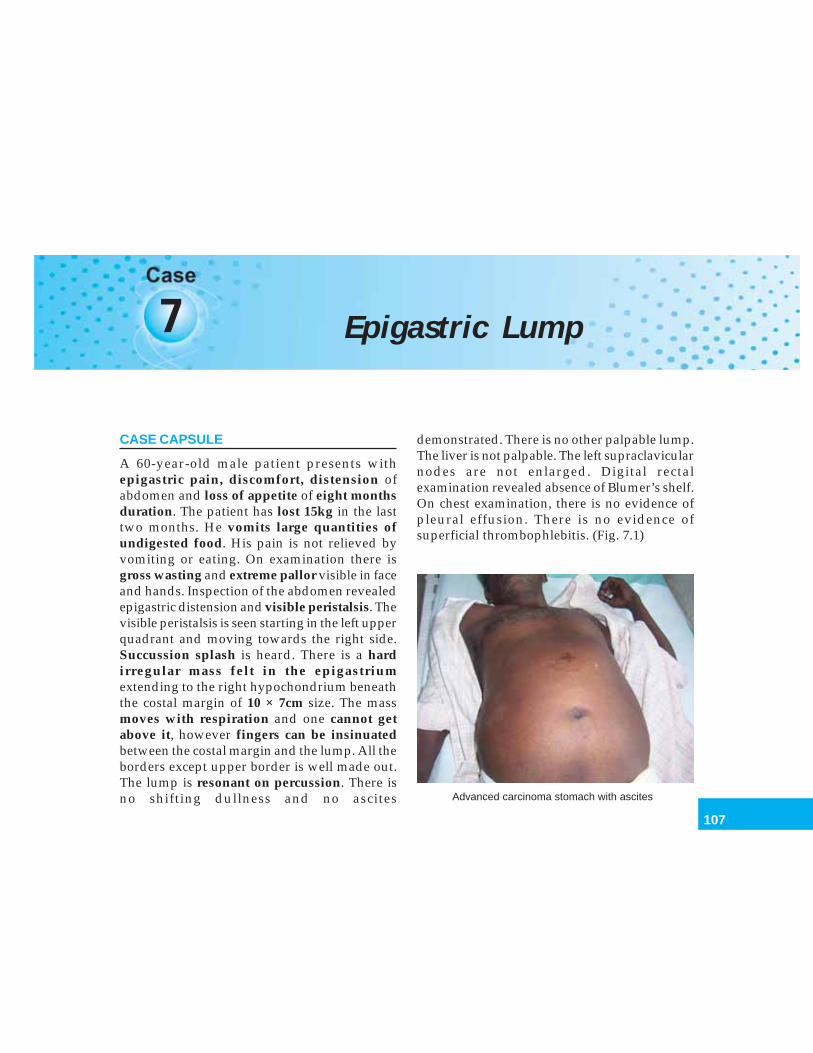
107
Epigastric Lump
7 Epigastric Lump
CASE CAPSULE
A 60-year-old male patient presents withepigastric pain, discomfort, distension ofabdomen and loss of appetite of eight monthsduration. The patient has lost 15kg in the lasttwo months. He vomits large quantities ofundigested food. His pain is not relieved byvomiting or eating. On examination there isgross wasting and extreme pallor visible in faceand hands. Inspection of the abdomen revealedepigastric distension and visible peristalsis. Thevisible peristalsis is seen starting in the left upperquadrant and moving towards the right side.Succussion splash is heard. There is a hardirregular mass felt in the epigastriumextending to the right hypochondrium beneaththe costal margin of 10 × 7cm size. The massmoves with respiration and one cannot getabove it, however fingers can be insinuatedbetween the costal margin and the lump. All theborders except upper border is well made out.The lump is resonant on percussion. There isno shifting dullness and no ascites
demonstrated. There is no other palpable lump.The liver is not palpable. The left supraclavicularnodes are not enlarged. Digital rectalexamination revealed absence of Blumer’s shelf.On chest examination, there is no evidence ofpleural effusion. There is no evidence ofsuperficial thrombophlebitis. (Fig. 7.1)
Advanced carcinoma stomach with ascites
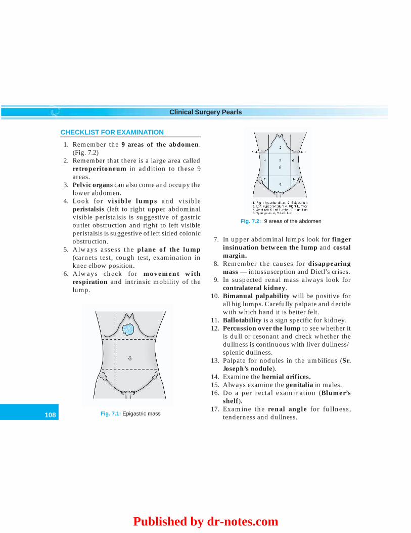
108
Clinical Surgery Pearls
CHECKLIST FOR EXAMINATION
1. Remember the 9 areas of the abdomen.(Fig. 7.2)
2. Remember that there is a large area calledretroperitoneum in addition to these 9areas.
3. Pelvic organs can also come and occupy thelower abdomen.
4. Look for visible lumps and visibleperistalsis (left to right upper abdominalvisible peristalsis is suggestive of gastricoutlet obstruction and right to left visibleperistalsis is suggestive of left sided colonicobstruction.
5. Always assess the plane of the lump(carnets test, cough test, examination inknee elbow position.
6. Always check for movement withrespiration and intrinsic mobility of thelump.
7. In upper abdominal lumps look for fingerinsinuation between the lump and costalmargin.
8. Remember the causes for disappearingmass — intussusception and Dietl’s crises.
9. In suspected renal mass always look forcontralateral kidney.
10. Bimanual palpability will be positive forall big lumps. Carefully palpate and decidewith which hand it is better felt.
11. Ballotability is a sign specific for kidney.12. Percussion over the lump to see whether it
is dull or resonant and check whether thedullness is continuous with liver dullness/splenic dullness.
13. Palpate for nodules in the umbilicus (Sr.Joseph’s nodule).
14. Examine the hernial orifices.15. Always examine the genitalia in males.16. Do a per rectal examination (Blumer’s
shelf).17. Examine the renal angle for fullness,
tenderness and dullness.
Fig. 7.2: 9 areas of the abdomen
Fig. 7.1: Epigastric mass
Published by dr-notes.com
109
Epigastric Lump
18. Look for supraclavicular lymph nodes(especially on left side between the twoheads of sternomastoid) – Troisier’s sign.
Q 1. What is the most probable diagnosis inthis case?
A distal gastric cancer with gastric outletobstruction.
Q 2. What are the reasons for diagnosing astomach lump?
a. Symptoms of loss of appetite, loss of weightand vomiting.
b. The type of visible peristalsis is from left toright which is suggestive of a gastric outletobstruction by a tumor.
c. The lump is occupying the epigastrium andis moving up and down with respiration.
Q 3. What is the significance of family historyin carcinoma stomach?
Gastric cancer can occur in families. A famousgastric cancer patient in history is NapoleonBonaparte. He died of gastric cancer (?). Hisfather and grandfather died of gastric cancer.His 3 sisters and a brother died of gastric cancer.
Q 4. What are the causes for gastric outletobstruction?
Causes for gastric outlet obstruction—
1. Duodenal ulcer with pyloric stenosis2. Distal gastric malignancy with outlet obstruction3. Congenital hypertrophic pyloric stenosis4. Adult type of hypertrophic pyloric stenosis5. Acid poisoning with stenosis
Q 5. What are the signs other than visiblegastric peristalsis (VGP) to diagnose gastricoutlet obstruction?
1. The dilated and distended stomach can bepalpated sometimes.
2. The succusion splash can be elicited.
Q 6. What is the objective evidence for gastricoutlet obstruction?
The objective evidence for gastric outletobstruction is saline load test. This is a simplemeans of assessing the degree of pyloricobstruction and is useful in following thepatient’s progress during the first few days ofnasogastric suction.
Through the nasogastric tube, 700ml ofnormal saline at room temperature is infusedover 3-5 minutes and the tube is clamped. 30minutes later the stomach is aspirated and theresidual volume of saline recorded. Recovery ofmore than 350 ml indicates obstruction.
Q 7. What are the other abdominal clinicalsigns, one should look for ascertaining thatit is stomach?
a. May or may not be able to get above the lumpb. Percussion note will be resonant, may be
some times impaired.
Q 8. What are the clinical signs of inopera-bility in such a patient?
Signs of inoperability in carcinoma stomach
a. The presence of ascitis suggesting peritonealmetastasis
110
Clinical Surgery Pearls
b. The presence of Blumer’s shelf which isnothing but a peritoneal metastasis in therectovesical/ rectouterine pouch
c. The presence of a metastatic liverd. The presence of left supraclavicular lymph-
nodes (Virchow’s, Troisier’s sign)e. The presence of a cutaneous nodule in the
umbilicus (Sr.Mary Joseph’s nodule)f. Presence of jaundiceg. Fixity of massh. Irish nodule (Enlarged left axillary node)i. Pleural effusionj. Krukenberg’s tumor (enlarged ovaries in
pelvic examination)
Q 9. In the absence of these signs can yousay that it is an early gastric cancer?
No. By the time a lump is clinically palpable itwill be advanced gastric cancer.
Q 10. What is the cause for jaundice incarcinoma stomach?
• Infiltration of the growth engulfing the CBD(common bile duct)
• Diffuse involvement of the hepatoduodenalligament.
Q 11. What is the pathway for umbilicalnodule formation (Sister Mary Joseph’sNodule)?
Growth in lesser curvature↓
Involvement of lesser omentum↓
Involvement of ligamentum teres↓
Umbilicus
Note: Sr.Mary Joseph was Nurse to Sir WilliamMayo.
Q 12. What is early gastric cancer (EGC)?
Early gastric cancer is a cancer confined to themucosa and submucosa of the stomachirrespective of the nodal status (T1 any N).
Q 13. What are the clinical manifestations ofcarcinoma stomach?
The pnemonic stomach helps in memorizing theclinical features.
Silent (No symptoms)Tumor (Lump)ObstructionMelenaAchlorhydria
AnemiaAsthenia
CachexiaHemtemesis
Q 14. How will you proceed to investigatesuch a patient?
The most important investigation in thisparticular situation will be an upper GIendoscopy.
Q 15. Why upper GI endoscopy is preferredover a Barium meal examination?
• By Upper GI endoscopy, we can see thegrowth, assess the type and extent of growth,and also take biopsy for confirmation.
• In Barium meal examination we will onlysee the shadow of the lesion in the form ofpersistent irregular filling defect, persistentmucosal irregularity and persistent loss ofperistalsis in a particular segment.
Published by dr-notes.com
111
Epigastric Lump
Q 16. In which situation Barium meal exa-mination is superior to upper GI Endoscopy?
Barium meal examination is superior in thefollowing situations:• Barium meal is useful for the diagnosis
of Linitis Plastica (endoscopic biopsyconfirmation may be difficult because themucosa is normal.
• Lesions in the cardiac.• Lesions causing obstruction.• For early gastric cancer lesions.
Q 17. How many endoscopic biopsies will youtake from a suspected lesion in the stomach?
Minimum Six biopsies from different parts ofthe identified lesion (Diagnostic accuracyreaches 100% if 10 biopsy samples are taken.
Q 18. What are the types of biopsies availableendoscopically?
a. Biopsy with the help of a biopsy forcepsb. Punch biopsyc. Brush biopsyd. Gastric lavage and cytology.
Q 19. If the biopsy is being reported as adeno-carcinoma (commonest carcinoma instomach), how will you proceed?
In the order of priority the following investiga-tions are done to stage the disease and rule outmetastasis.
Staging investigations
a. Ultrasound abdomen to rule out metastasisand assess the extent of the disease
b. X-ray chest to rule pulmonary metastasisc. CT abdomen if required to stage the diseased. Liver function test (LFT) to rule out metastasise. Hemogram to rule out anemia
Q 20. In what way does the ultrasoundexamination help in evaluating this patient?
a. The presence of liver metastasis can beassessed and if required a sonoguidedFNAC can be done
b. The tumor thickness can be assessed withthe help of a high resolution probe
c. The presence or absence of regional lymphnode and adjacent organ involvement.
d. The presence of free fluid can beascertained
Q 21. Is there any role for endoscopic ultra-sound (EUS)?
By ordinary abdominal ultrasound one may notbe able to ascertain the tumor thickness andserosal involvement which is possible with EUS.To some extent the presence of nodes can be alsoassessed by endoscopic ultrasound.
Q 22. Why is CT required in addition to ultra-sound examination?
The gastric carcinoma appear as areas of muralthickening. The measurement of the thicknessof the wall will give an idea of transmuralextension, when the thickness is more than 2cm. In addition, a blurred serosal contour orstrand like densities that extend into theperigastic fat may be seen. Direct invasion ofthe adjacent structures like pancreas,diaphragm, transverse colon, spleen or left lobeof liver can be ascertained by CT scan.
Q 23. Is there any role for laparoscopy in sucha patient?
Yes.
112
Clinical Surgery Pearls
Role of laparoscopy in carcinoma stomach
• Diagnostic laparoscopy is useful for staging thedisease
• Pick up peritoneal metastasis (CT will missperitoneal metastasis
• It can detect occult metastasis in 13-37%• Laparoscopic ultrasound can detect metastasis
in liver• It can identify adjacent organ invasion• Guided biopsies are possible.• Laparoscopy can help in doing a Peritoneal
Lavage and cytology• Laparoscopy can eliminate the need for
laparotomy
Q 24. What are the laparoscopic signs ofinoperability?
Laparoscopic signs of inoperability
1. Positive cytology in peritoneal wash2. Peritoneal deposits3. Posterior fixation4. Fixed celiac nodes5. Paraaortic nodes6. Liver metastasis
Q 25. What is the staging system forcarcinoma stomach?
In the west, the UICC TNM system based on the6th edition is accepted. But the JapaneseInvestigators report their results based onthe JRSGC (Japanese Research Society forGastric Cancer). The N3 and N4 nodes in theJapanese system correspond to extraregionalnodes in UICC system, that is M1 metastatic nodes.
UICC Staging
T1 tumor involving lamina propia T2 involves muscularis or subserosa
T3 tumor involves serosa, T4 invades adjacent organs
N0 No nodes N1 Metastasis in 1-6 regional nodes
N2 Metastasis 7-15 regional nodes N3 Metastasis in > 15 regional nodes
M0 No distant metastasis
M1 Distant metastasis (this includes peritoneum and distant lymph nodes)Stage IA T1 N0 M0Stage IB T1 N1 M0 /T2 N0 M0Stage II Upto T3 N0 /T2 N1Stage IIIA Upto T4 N0 /T3 N1Stage IIIB Upto T3 N2Stage IV Any T, any N, M1
Upto T3 N3T4 N1 – 3
Published by dr-notes.com
113
Epigastric Lump
Q 26. What are the nodal stations accordingto Japanese Research Society?
1. Right cardiac2. Left cardiac3. Lesser curvature4. Greater curvature5. Suprapyloric6. Infrapyloric7. Nodes along left gastric artery8. Common hepatic artery9. Celiac
10. Splenic hilar11. Splenic artery12. Hepatic pedicle13. Retropancreatic14. Mesenteric root15. Middle colic artery16. Para-aortic.
Lymph node stations• 1– 6 are considered N1 nodes (Perigastric
nodes – along the curvatures)• 7 – 11 are considered N2 nodes (Along the
named vessels)• 12 – 14 are considered N3 nodes (Intra-
peritoneal nodes)• 15 – 16 are considered N4 nodes.
Note:• N1 and N2 nodes are regional nodes• N3 nodes are metastatic node stations• Involvement of N3 node station is a
contraindication for radical surgery.
Q 27. What you mean by D1 and D2 resec-tion?
The extent of lymphadenectomy in the Japanesesystem is described using a D descriptor.
• D0 lymphadenectomy means all the JRSGC(Japanese Research Society for GastricCancer) N1 nodes have not been completelyremoved.
• D1 lymphadenectomy means all the N1nodes have been removed but not all the N2nodes.
• D2 lymphadenectomy means that all the N1and N2 nodes have been removed but not allthe N3 nodes.Thus for example a D2 lymphadenectomy for
a tumor involving all 3 gastric areas would involveremoval of lymph nodes at stations 1- 11.
The extent of lymphadenectomy before 1993was classified by the ‘R’ descriptor. Since 1993in order to avoid confusion with the UICC Rdescriptor (which reflects an entirely differentcharacteristics namely, the presence of knownresidual disease after surgical treatment), theextent of lymphadenectomy is denoted by D-descriptor.
Q 28. What are the bad prognostic factorsfor CA stomach?
1. Lymph node involvement is a poorprognostic factor—Four or more lymphnode involvement is a bad prognostic sign.
2. Serosal involvement is the single mostimportant bad prognostic factor.
3. Free carcinoma cells in the peritoneum.4. Intestinal type is having a better prognosis
than diffuse (Lauren’s).
Q 29. What are the poor prognostic variablesthat relate to surgery?
1. Positive resection margin2. Inadequate lymphadenectomy3. Need for splenectomy.
114
Clinical Surgery Pearls
Q 30: What is Lauren’s pathological classi-fication?
DIO• Diffuse• Intestinal• Others.
According to this classification it is dividedinto diffuse and intestinal type. The intestinaltype is having a better prognosis than the diffusetype. In intestinal gastric cancer, the tumorresembles a carcinoma elsewhere in the tubularGI tract and forms polypoidal tumors or ulcers.It probably arises in areas of intestinalmetaplasia.
In contrast diffuse gastric cancer infiltratesdeeply into the stomach without formingobvious mass lesions, but spreads widely in thegastric wall. A small proportion of gastriccancers are of mixed morphology.• The diffuse type may be localized or
generalized.• The generalized type of diffused is called
Linitis plastica (leather bottle stomach).
Q 31. What is Correa cycle?
Helicobacter seems to be principally associatedwith carcinoma of the body and distal stomach.Helicobacter is associated with gastritis leadingon to gastric atrophy. Similarly exposure tonitroso compounds will lead on to nitrosamineleading to gastritis and gastric atrophy whichin turn will produce intestinal metaplasialeading on to dysplasia, carcinoma in situ andfinally carcinoma.
Q 32. What are the other risk factors forcarcinoma stomach?
1. Pernicious anemia2. Gastric atrophy3. Gastric polyps4. Heredity5. Postgastrectomy patients – Post GJ, gastrec-
tomy and postpyloroplasty patientshave approximately 4 times the averagerisk.
6. Gastritis – Duodenogastric reflex and reflexgastritis are related to increased risk ofmalignancy.
7. Diet – Smoked fish, excessive salt intake,chilly etc.
8. Smoking and dust ingestion9. Deficiency of antioxidants
10. Exposure to N nitroso compounds.
Q 33. What is Borrmann classification ofadvanced gastric cancer?
Once there is involvement of the muscularis, itis called advanced gastric cancer. The macro-scopic appearance is classified by Borrmann into4 types: Type III and IV are commonly incurable.
Type I - PolypoidalType II - UlcerativeType III - Ulcerative and polypoidalType IV - Diffuse
Q 34. What is the most important epidemio-logical change in gastric cancer in recentcases?
1. The incidence of gastric cancer continues tofall at about 1% per year. This is speciallyseen in relation to carcinoma of the bodyand distal stomach. This may be due to theuse of refrigerator widely for foodpreservation.
Published by dr-notes.com
115
Epigastric Lump
2. In contrast there appears to be an increasedincidence of carcinoma of the proximalstomach. Particularly the GE junction.
3. The proximal and distal cancers aresupposed to behave differently.
Q 35. What are the differences betweenproximal and distal gastric cancer?
5 modes of spread of carcinoma stomach
1. Horizontal spread along the stomach wall –Submucosal spread
2. Vertical – invasion of the stomach wall and tothe adjacent structures like colon, pancreas,etc.
3. Lymphatic spread4. Peritoneal dissemination5. Blood stream spread – to the liver
Distal gastric cancer Proximal gastric cancer
a) Diet related Not diet relatedb) Environmental Not environmentalc) Epidemic variety Endemic varietyd) Arise in the background of dysplastic mucosa No such backgrounde) Intestinal histology Diffuse typef) Better prognosis Bad prognosis (morbidity and mortality high)g) Resectability is 30% Resectability is 20%h) Needs distal subtotal gastrectomy Needs total gastrectomy
Q 36. What is the classification of esophagogastric junction tumors (OG junctiontumors)?
Sievert classification of OG junction tumor
Type I – esophageal cancer involving OG junctionType II – Primary OG junction growthType III – Stomach lesion involving OG junction
Note: Type I needs esophageal resection with10cm clearance proximally.
Q 37. What are the modes of spread ofcarcinoma stomach?
Two types of horizontal extensions are met with.a. Infiltrative growthb. Expansive growth.
Q 38. What are the preoperative preparationsin a patient with carcinoma stomach?
Preoperative preparations
a. Correction of anemiab. Correction of nutritional statusc. Correction of fluid and electrolyte disturbanced. Assessment of cardiac, respiratory and renal statuse. Arrange adequate bloodf. Preoperative stomach washg. Prophylactic antibiotic
116
Clinical Surgery Pearls
The stomach in normal individual is sterilebecause of the low pH. Patients with gastriccancer however often have an increased pH andcolonization of the stomach. There is higher riskof wound complications in patients with highgastric pH and heavily colonized gastricsecretions. The most common organism isolatedfrom gastric juice are Streptococcus faecalis, E.coli,bacteroides, Staph albus, etc. For this reason aprophylactic antibiotic with a 3rd generationcephalosporin is advocated.
Q 39. What are the controversies regardingthe surgical treatment of gastric adeno-carcinoma?
The points of controversy are:1. The extent of gastric resection2. The extent of lymph node dissection.
Generally radical gastrectomies areperformed for carcinoma stomach. A radicalgastrectomy is one where the stomach alongwith the entire greater omentum, lesseromentum and lymph nodes are removed en -bloc.1. The extent of gastric resection
The distal division line is always placed atthe duodenal bulb at least 1cm from thetumor. As the diagnostic accuracy ininfiltrative type is very low, the macroscopicproximal margin should be greater than5 cm.In the case of scirrhous carcinoma orBorrmann type IV, the horizontal extensionincludes the whole stomach in the majorityof cases and therefore a total gastrectomyis always indicated. In contrast the
diagnostic accuracy is high in expansivegrowth type tumors and also in early gastriccancer. A margin of 2 cm is sufficient forthese types and total or distal radicalgastrectomy can be selected.Total vs subtotal radical gastrectomyThe term radical is vague and lacks properdefinition and may mean differentoperations to different surgeons.Surgical treatment is curative only in stageI and II diseases. A prospective randomizedstudy by the French compared total vssubtotal gastrectomy and found that themorbidity and mortality were similar forboth groups, but the five year survival wasnot improved. The morbidity and mortalityappeared to be associated with distalpancreatectomy and splenectomy.
2. The extent of lymph node dissectionThe controversies regarding the extent oflymphadenectomy relates to D1 vs D2resection. D2 gastrecomy has 2 essentialcomponents.a. Adequate 5 cm clearance in the stomachb. Extensive lymphadenectomy – the
omentum, superior leaf of mesocolon,and the pancreatic capsule are removeden-bloc, along with all N2 nodes (JRSGC)that is upto station number 12.
In order for a patient to have pathologicallyconfirmed D2 lymphadenectomy more than26 nodes has to be identified in thespecimen. Much of the mortality andmorbidity in D2 resection is related to thedistal pancreatectomy and splenectomyresulting in left subphrenic abscess. So now
Published by dr-notes.com

117
Epigastric Lump
a days the D2 gastrectomy will preservespleen and pancreas. Overall theoncological outcome may be betterfollowing D2 gastrectomy. The results ofsurgical treatment stage for stage in Japanare much better than commonly reportedin the West and they attribute this to thestaging and quality of surgery in Japan.In general D1 resection involves the removalof perigastric nodes and D2 resectioninvolves the clearance of the major arterialtrunks.
Q 40. What is the ideal treatment for operablecancers of stomach ?
The ideal treatment consists of D2 Gastrectomy+ Chemoradiation.
Q 41. What is D2 Gastrectomy?
Structures removed in D2 gastrectomy
• Removal of the stomach with the growth• Omental bursa• Entire greater omentum• Lesser omentum• Anterior layer of mesocolon• Anterior pancreatitic capsule• Lymphadenectomy upto D2 station
Q 42. What are the divisions of the stomachfrom therapeutic point of view?
The most appropriate operation for a givenpatient with gastric cancer must take intoaccount the location of the lesion and the knownpattern of spread at that site. For this purposethe stomach can be divided into 3rds.
a. The proximal 3rd – includes GE junctionand the fundus.
b. The middle 3rd – is the body of stomachand extends from the fundus to the incisuraangularis of the lesser curvature.
c. The distal 3rd – is the pyloric antrum andextends from the incisura angularis to thepylorus.
Q 43. What is the surgical treatment formiddle third malignancy?
Lesions in this location are asymptomatic untilthey have become quite bulky with metastasisto regional nodes. Three procedures arecommonly performed in an attempt toencompass all gross and microscopic disease inthe stomach and its lymphatic drainagenetwork.1. High radical subtotal gastrectomy2. Radical total gastrectomy3. Extended total gastrectomy with distal
pancreatectomy and splenectomy.As per the rules of the Japanese Research
Society, tumors encroaching upon or crossing theline extending from the bare area on the greatercurvature (between the portions of the stomachsupplied by the gastroepiploic artery and theareas supplied by the short gastric) to a point 5cmbelow the cardioesophageal junction on thelesser curvature required total gastrectomy.Pathoanatomic studies have demonstrated thatcarcinoma of the mid stomach metastasize to allregional lymph node basins of the stomach. Thereis therefore considerable theoretical basis for aradical total gastrectomy.
118
Clinical Surgery Pearls
Q 44. How much stomach should be removedin a distal radical gastrectomy for a distal 3rdgrowth?
This involves resection of approximately 75%of the stomach, including most of the lessercurvature, where the margins of resection willoften be the closest. At least 1 cm of the 1st partof the duodenum is resected. 5 cm of normalstomach is removed proximal to the tumor toassure adequate margin. Frozen sectionpathological evaluation of the surgical marginis performed before reconstruction. As with allgastric resections for carcinoma, greater andlesser omentectomy and regional lymph noderesection is required in an attempt to removeall microscopic disease.
Q 45. What is the surgical treatment ofproximal gastric cancer?
Esophagogastrectomy followed by reconstruc-tion is the treatment of choice.
Q 46. What is the incision for a surgery forgastric malignancy?
For all types of gastric malignancy a bilateralsubcostal incision (Chevron) is preferred.
Q 47. What is the management of inoperablelesions?
• Without complications and obstructions:Chemotherapy
• With complications: Palliative surgery +chemotherapy.
Q 48. What are the palliative surgicalprocedures?
Palliative procedures for carcinoma stomach
a. Palliative gastrectomy - Is indicated foruncontrolled bleeding, obstruction and perforation.A palliative resection is usually a palliativegastrectomy, which removes the tumor alonefollowed by reconstruction.
b. A palliative gastrojejunostomy – this is a pooroperation which does not allow proper emptyingof the stomach but may produce additionalproblem of bile reflux.
c. Palliative esophagojejunostomyd. Palliative intubation for proximal growthse. Recanalization procedures
Q 49. What is combined resection?
The stomach is surrounded by pancreas,transverse colon, mesocolon, spleen, liver,diaphragm and omental bursa. All thesestructures can be surgically removed togetherwith stomach if necessary. Combined resectionshows survival benefits when there is no distantmetastasis.
Q 50. What is paraaortic dissection?
The fatty tissue is completely removed fromaround the abdominal aorta between the aortichiatus and origin of the inferior mesentericartery. The left adrenal gland is frequentlyremoved to achieve complete dissection aroundthe left renal artery. The overall incidence ofmetastasis in this area is approximately 3%.
Q 51. What is the management of adjacentorgan involvement?
• Liver involvement : Nonanatomicalresection
• Duodenum : 2cm clearance
Published by dr-notes.com
119
Epigastric Lump
• Esophagus : 10cm clearance• Colon : Segmental resection• Pancreas : Distal pancreatectomy
Note: Proximal pancreas is unresectable
Q 52. What is left upper abdominal eviscera-tion?
It is one of the extended combined resectionsdone in association with total gastrectomy. Inaddition to total gastrectomy, pancreatosplenec-tomy and transverse colectomy are minimumrequirements. Some times it includes lefthepatectomy, left nephrectomy, left adrenalec-tomy or resection of the diaphragm. The purposeis to achieve almost complete resection of theomental bursa which consists of the stomach,omentum, mesocolon, transverse colon, spleenand pancreas. The indications for the proceduresare:1. When the tumor is located in the upper or
middle part of the stomach and the serosais penetrated by cancer.
2. When the tumor mainly occupies theposterior wall of the stomach.
3. When there is no remote metastasis4. When there is no severe medical complica-
tion to prevent long aggressive intervention.
Q 53. What is the classification of earlygastric cancer (EGC)?
The EGC is classified as:Type I ProtrudingType II SuperficialType III Excavated.
Q 54. What are the minimally invasiveprocedures and function preservingprocedures?
• These procedures are done for early gastriccancer limited to the mucosa alone.
• EGC affecting the submucosa needs radicalsurgery.
The procedures are:1. Endoscopic mucosal resection2. Laparoscopic wedge resection of the
stomach3. Pylorus preserving gastrectomy.
Q 55. What are the indications for endoscopictreatment of early gastric cancer (EGC)?
Absolute indications.EGC small enough to be completely removedby single endoscopic treatment. They include:1. Protruding cancers smaller than 1cm2. Ulcer-free depressed cancers smaller than
1cm3. Well-demarked cancers.
Relative indications1. Cancers associated with severe medical
illness2. Occurring in elderly patients3. Occurring in patients who refuse surgery.
Q 56. What is pylorus preserving gastrec-tomy?
This is indicated in small cancers located in themiddle third of the stomach. The purpose is toreduce dumping symptoms, gallbladderdysfunction and postoperative gallstonedevelopment. The hepatic and pyloric branchesof vagal nerves are preserved.
Q 57. What is the prognosis of carcinomastomach?
It rarely disseminates widely before it involvesthe lymph nodes and therefore there is an
120
Clinical Surgery Pearls
opportunity to cure the disease prior todissemination. Distant metastasis is uncommonin the absence of lymph node involvement.a. The prognosis of early gastric cancer is very
good. Early gastric cancer associated withlymph node involvement has five yearsurvival rates in the region of 90%.
b. In Japan approximately 75% of the patientswill have a curative resection and theoverall survival will be 50-70%.
c. In the West 25 – 50% of patients willundergo curative resection and the five yearsurvival is 25-30%.
Q 58. Is gastric cancer a different disease inJapan and West?
What is the stage migration phenomenon (WillRoger’s phenomenon)?
This proposition has no basis in evidence. Acombination of differences in the staging andhigher standard of surgery in Japan probablyaccounts for the difference. The more thoroughstaging, the higher the stage is likely to be andtherefore stage for stage, the outcome seemsbetter in patients adequately stagedpathologically. This phenomenon is called stagemigration (Will Roger’s phenomenon).
The pathologist will have considerabledifficulty in orientating a fixed specimen andfinding lymph node groups and therefore thesurgeon should dissect the nodes from thespecimen and send them separately to thepathologist – a practice commonly followed inJapan. Only by this method accurate staging canbe achieved.
Q 59. Is there any role for chemoradiation?
• The standard adjuvant treatment is chemo-radiation.
• Chemotherapy is by 5-FU and Leucovorin.• 3 doses of bolus 5-FU during first and last
weeks of radiotherapy.• Radiotherapy: Dose is 4,500 Rads.
Q 60. Is there any role for neoadjuvantchemotherapy?
This is found to be beneficial to downstagecancer of the lower esophagus and occasionallystomach.
Q 61. What is the commonest site forrecurrence after surgery?
The commonest site of recurrence is stomachbed followed by lymph nodes and anastomoticsite.
Q 62. What is the treatment for recurrence?
• Self-expandable metal stents (SEMS) is usedfor obstruction.
• New chemotherapeutic regimens– Cisplatin based- Taxane based- Irinotecon based
Q 63. What is the follow up after surgery?
Follow up after surgery for carcinoma stomach
• Clinical examination every three months × 2years
• CT scan / chest X-ray every 6 months• Endoscopy every year• Laparoscopy - SOS.
Published by dr-notes.com
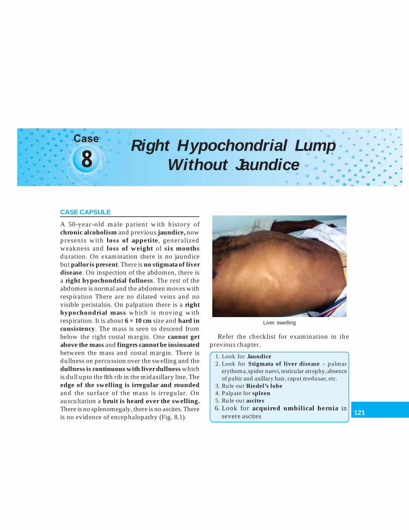
121
Right Hypochondrial Lump Without Jaundice
8Right Hypochondrial Lump
Without Jaundice
CASE CAPSULE
A 50-year-old male patient with history ofchronic alcoholism and previous jaundice, nowpresents with loss of appetite, generalizedweakness and loss of weight of six monthsduration. On examination there is no jaundicebut pallor is present. There is no stigmata of liverdisease. On inspection of the abdomen, there isa right hypochondrial fullness. The rest of theabdomen is normal and the abdomen moves withrespiration There are no dilated veins and novisible peristalsis. On palpation there is a righthypochondrial mass which is moving withrespiration. It is about 6 × 10 cm size and hard inconsistency. The mass is seen to descend frombelow the right costal margin. One cannot getabove the mass and fingers cannot be insinuatedbetween the mass and costal margin. There isdullness on percussion over the swelling and thedullness is continuous with liver dullness whichis dull upto the 8th rib in the midaxillary line. Theedge of the swelling is irregular and roundedand the surface of the mass is irregular. Onauscultation a bruit is heard over the swelling.There is no splenomegaly, there is no ascites. Thereis no evidence of encephalopathy (Fig. 8.1).
Refer the checklist for examination in theprevious chapter.
1. Look for Jaundice2. Look for Stigmata of liver disease – palmar
erythema, spider naevi, testicular atrophy, absenceof pubic and axillary hair, caput medusae, etc.
3. Rule out Riedel’s lobe4. Palpate for spleen5. Rule out ascites6. Look for acquired umbilical hernia in
severe ascites
Liver swelling
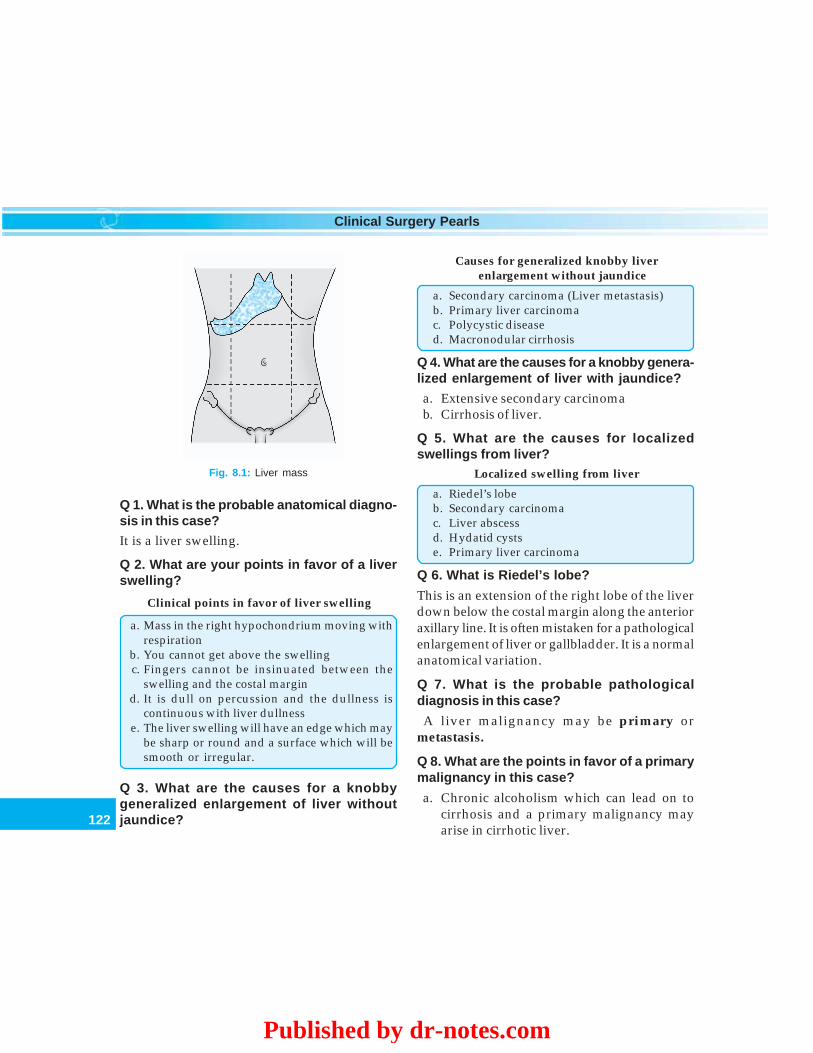
122
Clinical Surgery Pearls
Q 1. What is the probable anatomical diagno-sis in this case?
It is a liver swelling.
Q 2. What are your points in favor of a liverswelling?
Clinical points in favor of liver swelling
a. Mass in the right hypochondrium moving withrespiration
b. You cannot get above the swellingc. Fingers cannot be insinuated between the
swelling and the costal margind. It is dull on percussion and the dullness is
continuous with liver dullnesse. The liver swelling will have an edge which may
be sharp or round and a surface which will besmooth or irregular.
Q 3. What are the causes for a knobbygeneralized enlargement of liver withoutjaundice?
Causes for generalized knobby liverenlargement without jaundice
a. Secondary carcinoma (Liver metastasis)b. Primary liver carcinomac. Polycystic diseased. Macronodular cirrhosis
Q 4. What are the causes for a knobby genera-lized enlargement of liver with jaundice?
a. Extensive secondary carcinomab. Cirrhosis of liver.
Q 5. What are the causes for localizedswellings from liver?
Localized swelling from liver
a. Riedel’s lobeb. Secondary carcinomac. Liver abscessd. Hydatid cystse. Primary liver carcinoma
Q 6. What is Riedel’s lobe?
This is an extension of the right lobe of the liverdown below the costal margin along the anterioraxillary line. It is often mistaken for a pathologicalenlargement of liver or gallbladder. It is a normalanatomical variation.
Q 7. What is the probable pathologicaldiagnosis in this case?
A liver malignancy may be primary ormetastasis.
Q 8. What are the points in favor of a primarymalignancy in this case?
a. Chronic alcoholism which can lead on tocirrhosis and a primary malignancy mayarise in cirrhotic liver.
Fig. 8.1: Liver mass
Published by dr-notes.com
123
Right Hypochondrial Lump Without Jaundice
b. Previous H/o jaundice – hepatitis B andhepatitis C lead on to primary malignancy.
c. Cirrhosis itself will predispose to primarymalignancy.
Q 9. What are the points against a metastasisin the liver?
a. Usually multiple nodules are seen inmetastasis even though multifocalhepatocellular carcinoma is possible
b. The classical sign of umbilication will bepalpable in the liver nodule.
c. Other clinical swelling suggestive of aprimary in another organ may or may not bepalpable in the abdominal cavity.
Q 10. What is the commonest malignantswelling in the liver?
Commonest malignant swelling is metastasis inthe liver. Metastatic cancer is 20 times morecommon than primary tumors in the liver. Thecirrhotic liver, which often gives rise to primaryhepatic tumors, is less susceptible than normalliver to implantation of metastasis.
Q 11. What are the usual presentations ofprimary malignancy of the liver?
Clinical presentation of primarymalignancy of the liver
a. Pain abdomen with or without hepatomegalyb. Sudden deterioration of the condition of
cirrhotic patient owing to the appearance ofhepatic failure, bleeding varices or ascites
c. Sudden massive intraperitoneal hemorrhaged. Acute illness with fever and abdominal paine. Distant metastasisf. No clinical finding
Q 12. Can there be associated metabolic orendocrine abnormalities in hepatocellularcarcinoma (primary)? (PG)
Metabolic and endocrine abnormalities inhepatocellular carcinoma
a. Erythrocytosisb. Hypercalcemiac. Hypoglycemic attackd. Cushing’s syndromee. Virilization
Q 13. What are the complications of hepato-cellular carcinoma? (PG)
a. Sudden intraabdominal hemorrhage due tospontaneous bleeding
b. Obstruction of the portal vein resulting inportal hypertension
c. Obstruction of hepatic veins resulting inBudd-Chiari syndrome
d. Liver failure.
Q 14. What is the classification of livertumors?
The broad classification is into benign andmalignant
Benign Malignant
a. Hepatic adenoma A. Primary(Hepatic adenoma, a. Hepatocellu-Cholangioadenoma lar carcinoma or a combination) b. Cholangiocarci-
b. Focal nodular nomahyperplasia c. Combination of
c. Hemangioma hepato andcholangio-carcinomas
Contd...
124
Clinical Surgery Pearls
d. Adenosquamouscarcinoma
e. Squamouscarcinoma of bileduct
f. Signet ringcarcinoma
g. Undifferentiatedcarcinoma
h. Bile ductcystadeno-carcinoma
B. Metastasis
Q 15. What is the classification of metastasis?
Metastasis may be classified as:a. Carcinomatous – e.g.: Stomach, colon,
pancreas, kidney, prostate, breast, etc.b. Carcinoid metastasis – Carcinoid syndrome
will occur when the liver is metastasized andtherefore 5HT cannot be metabolized to 5hydroxy indoleacetic acid.
c. Melanomatous metastasis – beware of“missing toes and glass eyes”
d. Sarcomatous metastasise. Lymphomatous deposits.
Q 16. What are the salient features of hepaticadenoma?
Salient features of hepatic adenoma
• Occur almost exclusively in women• Associated with the use of oral contraceptives• Well-circumscribed soft yellow tan tumors of 2-
15cm size
• 2/3rd are solitary and remaining are multiple• Transition from adenoma to carcinoma may occur• Pain, intraabdominal hemorrhage and shock may
occur• Bleeding episode with menstruation is seen• Regression is possible when the size is less than
6 cm and when the oral contraceptive pill iswithdrawn
• Tumor should be followed up periodically willultrasound/CT
• When in doubt remove the hepatic adenoma
Q 17: What are the stigmata of liver disease?
Stigmata of liver disease
1. Spider naevi (> 9 new crops indicate progressivedisease)
2. Palmar erythema (Thenar and hypothenareminences)
3. Clubbing, leukonychia4. Dupuytren’s contracture5. Body hair loss (axillary and pubic)6. Fetor hepaticus7. Gynecomastia8. Testicular atrophy9. Liver flap – flapping tremor
10. Encephalopathy – Cog wheel limb rigidity
Q 18. How to manage this patient with livermass?
Management includes investigations andtreatment.The investigations may be classified as –1. Investigations for diagnosis2. Investigations for staging3. Investigations for surgical treatment.
*The most important investigations is imaging.
Benign Malignant
Contd...
Published by dr-notes.com
125
Right Hypochondrial Lump Without Jaundice
Q 19. What is the role of ultrasound forimaging liver?
• It is a screening test• Can identify mass lesions• Can identify bile duct dilatation• Useful for guiding biopsy.Note: But it is operator dependent.
Q 20. What is the role of CT in liver mass?
Triple phase, multislice spiral CT is theinvestigation of choice for liver mass.
Type of lesion Imaging finding Mode of treatment
Hemangioma Hyperechoic in US No need for surgeryHypodense in CT without contrastHalo edge enhancement initially,then fills the center (late, slow venousenhancement)
Hepatic adenoma Well-circumscribed vascular solid tumor. Premalignant lesion. ResectionEnlarged peripheral vessels in arterio- is the treatment of choice. With-graphy – needle biopsy will produce drawal of oral contraceptives willbleeding produce regression
Focal nodular hyper- Central scarring in CT. No need for surgeryplasia (FNH) focal over- Well-vascularized. Sulphur colloid livergrowth of functioning scan is the investigation of choice – Theliver with fibrous lesion includes both hepatocytes andtrauma Kupffer cells
Metastasis Nonenhancing lesion after IV Surgery for colorectal metastasiscontrast in CT
Hepatocellular carcinoma Early arterial phase enhancing lesion Liver resection/liver transplan-with IV contrast tation.
CT finding of mass lesions of liver
• A mass lesion upto 1cm size can be identifiedhepatocellular carcinoma in contrast enhancedCT shows:• Early arterial phase: vascular enhancement.• The venous phase: maps out portal vein and
hepatic veins.
Q 21. What are the CT findings of variousmass lesions?
126
Clinical Surgery Pearls
Q 22. What is the gold standard investigationfor the diagnosis of hepatocellularcarcinoma?
Lipiodol enhanced CT scan is the goldstandard investigation. The practice isan injection of lipiodol into the hepatic arteryduring selective angiogram followed by CTafter 2 weeks. The lipiodol will be taken up bythe tumor.
Q 23. What is the role of magnetic resonanceimaging (MRI)?
1. It is useful for patients with iodine sensitivity,who cannot undergo a lipiodol enhancedCT.
2. Imaging of the biliary tract (MRCP) ispossible.
3. Hepatic artery and portal vein can be well-delineated
Q 24. What is the role of needle biopsy / FNACin mass lesions of liver?
Percutaneous biopsy of lesions in patients withpotentially operable disease is contraindicateddue to the high incidence of needle trackrecurrence and high mortality associated withthis procedure (spontaneous bleeds).
Q 25. If so what is the indication for a needlebiopsy?
1. Unresectable tumors2. Diagnostic dilemma.
Q 26. What are the needles for liver biopsy?
1. Chiba needle (Chiba is the name of aUniversity)
• 20cm long, 22 gauge, malleable needlewith short bevel
2. Vim Silverman needle3. Meghini needle.
Q 27. What are the precautions to be takenbefore biopsy from liver?
• The coagulation profile should be checked andrectified (PT, aPTT and platelet count).
• In cases of jaundice (in coagulation failurealso) give vitamin K injections 10 mg IM/IVfor 3 days.
Note: The vitamin K 1 (phytomenadione) can onlybe given IV. Regular vitamin K is oily and cannotbe given intravenously.
Q 28. What are the macroscopic types ofhepatocellular carcinoma? (PG)
Macroscopic types of hepatocellular carcinoma—
1. Expanding – Sharp demarcation between thetumor mass and compressed surroundingparenchyma.
2. Pedunculated - type 1 – intrahepatic - type2—extrahepatic (nourished by a branch of righthepatic artery) – not a true HCC.
3. Spreading (Engel’s massive form) – lack ofdemarcation between tumor and normal liver -may involve the whole liver as nodules.
4. Multifocal – synchronous swellings5. Indeterminate – different features in different
parts.
Q 29. What are the pathological variants ofHCC? (PG)
1. Fibrolamellar HCC – good prognosis2. Mixed hepatocellular cholangio cellular -
worse prognosis
Published by dr-notes.com
127
Right Hypochondrial Lump Without Jaundice
3. Clear cell variant – better prognosis4. Giant cell variant5. Childhood HCC (Bad prognosis)6. Carcinosarcoma (Sarcomatoid variant).
Q 30. What are the features of fibrolamellarcarcinoma?
1. Younger age group (median 25 years)2. Without history of cirrhosis3. Well-circumscribed4. Better resectability (50 – 75% in contrast to
25% in ordinary HCC)5. Better prognosis6. Does not produce AFP (α fetoprotein)7. Neurotensin B is the tumor marker8. Equal sex incidence
Q 31. What are the etiological factors forHCC?
1. Cirrhosis – Cirrhomimetic (Develops in thebackground of cirrhosis) Noncirrhomimetic
2. HBV (Hepatitis B virus)3. HCV (Hepatitis C virus)4. Alcoholic liver disease5. Hemochromatosis6. Hereditary tyrosinemia7. Mycotoxins (afla toxin B)8. Hepatic helminthiasis – clonorchis sinensis
for cholangiocarcinoma.
Q 32. What is the tumor marker for the HCC?
AFP (α fetoprotein).
Q 33. What is small HCC? (PG)
A tumor which is < 2cm as per Japanese GeneralRules. But a tumor < 3 – 5cm may be considered
small by some authorities. They are nodular andwell-differentiated.
Q 34. What are the investigations for staging?
1. CT abdomen with oral and IV contrast2. C.T. Chest – To rule out metastasis to lung3. Bone scan – To rule out metastasis4. Laparoscopy.
Q 35. What is the role of Child-Pugh grading?
This is very important if one is planning forsurgical resection. Resection should be contemp-lated in Child-Pugh A patients and occasionallyin Child Pugh B patients.
Child-Pugh Classification of functionalstatus of liver
Class:A B CRisk: Low Moderate High
Ascites Absent Slight Tenseto moderate
Encephalopathy None Grades GradesI – II III – IV
Serum albumin > 3.5 2.8 – 3.5 < 2.8(g/dL)Serum bilirubin < 2.0 2.0 – 3.0 > 3.0(mg/dL)Prothrombin time < 4.0 4.0 – 6.0 > 6.0(seconds above control)
Q 36. What are the surgical options availablein HCC?
The two options are –1. Resection2. Liver transplantation.
128
Clinical Surgery Pearls
Q 37. What are the investigations requiredfor surgery?
• Liver function test• Assessment of cardiac status• Pulmonary status• Renal status.
Q 38. What are the indications for resectionin HCC? (PG)
• One or two nodules in a patient with Child-Pugh A(Large tumor per se is not a contraindicationfor resection).
Q 39. What are the contraindications for liverresection in HCC? (PG)
1. Extrahepatic disease2. Lymph nodes3. Peritoneal involvement4. Distant metastasis5. 1cm clear margin is not possible6. Multifocal hepatoma (> 2 nodules) –
Treatment is transplantation.
Q 40. How much liver should be left behindin case of liver resection? (PG)
2 segments of healthy liver must be left behind.
Q 41. What type of anesthesia is preferredfor liver resection? (PG)
Low CVP anesthesia
• The inflow to the liver can be blocked by clampingthe free edge of the lesser omentum
• Bleeding results from hepatic vein duringresection
• If the CVP is decreased to zero, there will be littlebleeding from hepatic veins
• As a result there is no need for blood transfusion
Q 42. What are the indications for transplan-tation? (PG)
Outlook is better for transplantation thanresection provided—• Tumor nodules are 3 cm or less in diameter• Nodules are 3 or less in number.
Q 43. What are the nonsurgical ablativetherapies available? (PG)
1. Radiofrequency ablation – complications areneedle track recurrence, hemorrhage andliver failure.
2. Percutaneous alcohol injection – destructionof tumors up to 3cm size is possible -ineffective in colorectal metastasis.
3. TACE (Transarterial chemoembolization) – forsmall tumors only. A mixture of lipiodol anddoxorubicin is injected directly to the hepaticartery. The complications are post- embolizationsyndrome, liver failure, and liver abscess. 50%reduction in tumor size is seen 50% of cases.
Q 44. What is the role of chemotherapy inHCC? (PG)
The results of chemotherapy are disappointing.Therefore there is no role for systemic chemo-therapy. Subcutaneous Octreotide is being triedin advanced HCC.
Q 45. What is the prognosis of HCC?
• It is a tumor with poor prognosis• Median survival is in the region of one year• Symptomatic patients survive for 6 months• Resected patients (if suitable) the five year
survival is around 30%• Transplantation gives better results• Life expectancy is decided by the liver
disease.
Published by dr-notes.com
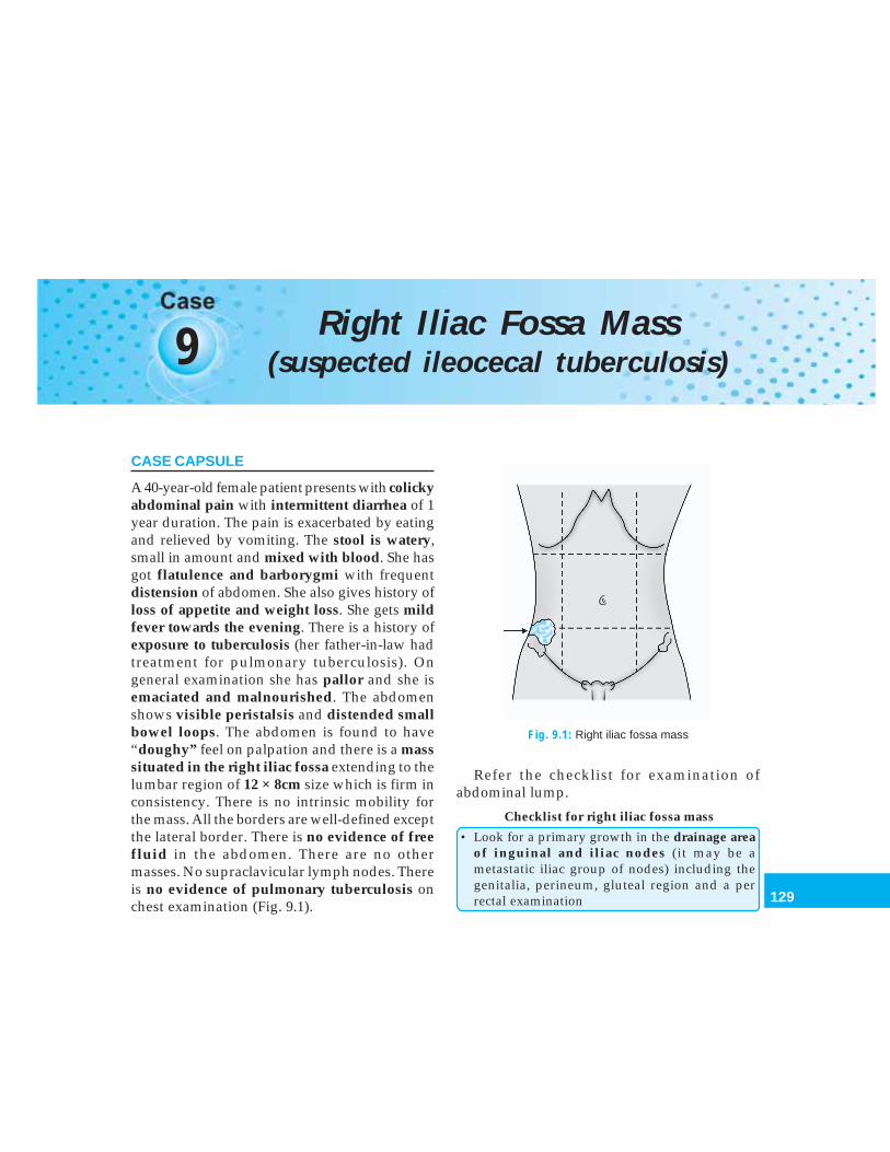
129
Right Iliac Fossa Mass (suspected ileocecal tuberculosis)
9 Right Iliac Fossa Mass(suspected ileocecal tuberculosis)
CASE CAPSULE
A 40-year-old female patient presents with colickyabdominal pain with intermittent diarrhea of 1year duration. The pain is exacerbated by eatingand relieved by vomiting. The stool is watery,small in amount and mixed with blood. She hasgot flatulence and barborygmi with frequentdistension of abdomen. She also gives history ofloss of appetite and weight loss. She gets mildfever towards the evening. There is a history ofexposure to tuberculosis (her father-in-law hadtreatment for pulmonary tuberculosis). Ongeneral examination she has pallor and she isemaciated and malnourished. The abdomenshows visible peristalsis and distended smallbowel loops. The abdomen is found to have“doughy” feel on palpation and there is a masssituated in the right iliac fossa extending to thelumbar region of 12 × 8cm size which is firm inconsistency. There is no intrinsic mobility forthe mass. All the borders are well-defined exceptthe lateral border. There is no evidence of freefluid in the abdomen. There are no othermasses. No supraclavicular lymph nodes. Thereis no evidence of pulmonary tuberculosis onchest examination (Fig. 9.1).
Refer the checklist for examination ofabdominal lump.
Checklist for right iliac fossa mass
• Look for a primary growth in the drainage areaof inguinal and iliac nodes (it may be ametastatic iliac group of nodes) including thegenitalia, perineum, gluteal region and a perrectal examination
Fig. 9.1: Right iliac fossa mass
130
Clinical Surgery Pearls
• Look for edema of the right lower limb – due tocompression of iliac vein by a malignant lymphnode mass
• Examine the spine – to rule out a gibbus as aresult of tuberculosis (the mass may be an ilio-psoas cold abscess)
• Rectal examination to rule out rectal shelf ofBlummer and rectal growth
• Examination of genitalia to rule outundescended testes
• Examine for left supraclavicular nodes• Look for swelling below the inguinal ligament,
if the right iliac fossa mass is soft. Look for crossfluctuation above and below the inguinalligament. The psoas abscess will track down tothe thigh behind the inguinal ligament
• In case of a soft swelling look for a space betweenthe swelling and ilium. The space will be therein appendicular abscess and it will be absent iniliac abscess
• Examine the sacroiliac joint – tuberculousabscess from sacroiliac joint can present as rightiliac fossa mass
There are innumerable causes for mass in theright iliac fossa
The important causes for mass right iliac fossa
• Appendicular mass• Ileocecal tuberculosis• Carcinoma cecum• Right ovarian cyst/tumor• Pelvic inflammatory disease• Iliac lymphadenopathy• Psoas abscess• Tumor in undescended testis• Unascended kidney• Retroperitoneal tumors
• Chondrosarcoma of the ileum• Crohn’s disease• Actinomycosis of the cecum• Tubo-ovarian mass
Q 1. What are the clinical characteristics of aswelling arising from the cecum?
Even though cecum has a complete peritonealcovering, it is relatively fixed and therefore themass arising from the cecum will be fixed. Therewill be no intrinsic mobility. The mass will beintraabdominal and intraperitoneal. One canget above and below the swelling and therewon’t be any movement.
Q 2. What are the characteristic features ofappendicular lump?
There will be typical history – the Murphy’striad – migratory pain (from umbilical regionto the right iliac fossa), vomiting/nausea andfever. The right iliac fossa mass will be tender.The duration of the symptoms and the lump willbe short. In late stages there will be local rise oftemperature and reddening of the overlyingskin. The patient may also have intermittentfever and tachycardia.
Q 3. What are the characteristic features oftuberculosis?
The patient often has central abdominal pain formonths with general ill health, weight loss, andchange in bowel habit. The mass is firm, thesurface and edges are difficult to define. Ascitesmay or may not be there. The typical doughyfeel may be there. The patient may have eveningrise of temperature.
Published by dr-notes.com
131
Right Iliac Fossa Mass (suspected ileocecal tuberculosis)
Q 4. What are the characteristic features ofcarcinoma cecum?
There won’t be any history of acute pain. Somepatients present with anemia, diarrhea, orintestinal obstruction. The right iliac fossa masswill be firm, or hard, distinct and fixed it is nottender, does not resolve with observation. Thepatient’s temperature and pulse are normal.
Q 5. Comparison of the clinical features ofthree most important causes for right iliacfossa mass?
Appendicular lump Ileocecal tuberculosis Carcinoma cecum
Age Younger age group Middle age Above 50 years
Onset Acute – 3 days after the Subacute - recurrent attacks Insidious/asymptomaticinitial symptoms
Symptoms Murphy’s triad Recurrent attacks of abdominal Lump abdomen• Migratory pain pain, diarrhea, loss of weight,• Nausea/vomiting evening rise of temperature• Fever
Nature of mass Irregular, firm and tender Irregular, doughy HardIntrinsic move- Fixed Fixed Fixedment
Response to Become smaller and then No response No responseantibiotic disappears
Radiological No specific radiological Pulled up cecum Fleischner Irregular filling defects insigns signs sign, Sterlin sign , the cecum which is in the
obtuse ileocecal angle normal position
Q 6. What is the most probable diagnosis inthis case?
May be a case of ileocecal tuberculosis, oneshould rule out carcinoma cecum.
Q 7. What are the points in favor of ileocecaltuberculosis?
• The long duration of symptoms• Recurrent attacks of pain abdomen (central
type)• History of diarrhea.• Loss of weight• Evening rise of temperature.
132
Clinical Surgery Pearls
Q 8. How to confirm the diagnosis?
The investigations for confirming the diagnosisare:1. ESR – will be raised in tuberculosis
(complete Hemogram is done)2. Leukocyte count – Leukopenia with
lymphocytosis3. Hemoglobin – anemia may be present4. Mantoux test
• Mantoux is highly nonspecific• Mantoux can be negative in severely
immunocompromised individuals• It can be positive in BCG vaccinated
individuals.5. X-ray chest – May show pulmonary tuber-
culosis6. Plain X-ray abdomen – May show calcified
lymph nodes.Evidence of intestinal dilatation.
7. Ultrasound abdomen – may show masslesion, fluid collection, enlargement oflymph nodes, etc. A tense tender tubularstructure of more than 7mm if identified issuggestive of appendicular mass, alongwith fluid collection
8. Double contrast barium enema – forvisualizing the cecum barium enema is theinvestigation of choice. For visualizing theterminal ileum a barium meal followthrough is the investigation of choice.
9. CT scan if required10. Peritoneal biopsy
a. Needle biopsy (not safe) – Abram’sneedle/Cope’s needle
b. Laparoscopic biopsy (ideal) – Peritoneumloses its glistening and smooth
appearance. Becomes dull, rough andirregular.
c. Open biopsy (under local anesthesia).11. Ascitic fluid study
a. ADA (Adenosine De Aminase) in asciticfluid—indicates the degree of stimulationof lymphocytes (not significant in HIVpatients and cirrhotic patients)
b. Ascitic fluid total protein – more than2.5gm/lt
c. SAAG (Serum ascitic fluid albumingradient) – more than 1.1
d. Ascitic fluid blood glucose ratio of lessthan 0.96.
Q 9. What are the radiological findings infavor of tuberculosis in barium meal followthrough?
1. The cecum will be pulled up (normally it isseen in relation to the bone ilium)
2. Fleischner sign – thickening of ileocecalvalve and it is wide open
3. Sterlin sign – fibrotic terminal ileumopening into a contracted cecum
4. Obtuse ileocecal angle.
Q 10. Which part of the intestine is commonlyaffected by tuberculosis?
It can affect any part of the GI tract. Thecommonly affected areas are ileum andproximal colon (57%).
Q 11. Why ileocecal region is more affectedin tuberculosis?
a. More lymphatic follicles are seen in terminalileum
b. Stasis in terminal ileum
Published by dr-notes.com
133
Right Iliac Fossa Mass (suspected ileocecal tuberculosis)
c. Spread from adjacent fallopian tube (genitalTB will spread via fallopian tube) in females.
Q 12. What are the other forms of abdominaltuberculosis?
Types of abdominal tuberculosis
a. Intestineb. Peritoneum (38%) – 6 types
1. Chronic peritonitis2. Acute peritonitis3. Ascitic type4. Encysted type5. Fibrous type6. Purulent type
c. Mesenteryd. Lymph nodes (6%)e. Omentumf. Liver and spleen
Q 13. What are the pathological types ofintestinal tuberculosis?Four pathological types of intestinaltuberculosis:1. Ulcerative2. Hypertrophic3. Fibrotic4. Ulcerofibrotic.
The first two account for majority of cases.
Q 14. What are the differences betweenulcerative type and hypertrophic type oftuberculosis?
Ulcerative Hypertrophic
1. Secondary to pulmonary tuberculosis by It is primary—due to ingestion of low virulentswallowing of tubercle bacilli organisms by a person with high resistance
2. Patient is very ill Not very ill
3. Multiple transverse ulcers in the ileum* Thickening of intestinal wall and narrowing ofthe lumen
4. Clinical features—Diarrhea and bleeding, Mass right iliac fossa, intestinal obstruction,weight loss, night sweats, anorexia and doughy feel, matted coils of intestine, anemia,evening rise of temperature steatorrhea and weight loss
5. A primary will be there in the chest No primary in the chest.
6. Absence of filling of ileum, cecum and Filling defect in the terminal ileum, pulled upascending colon cecum, obtuse ileocecal angle
7. Presence of gross caseation Absence of gross caseation
8. Obstruction not seen usually Obstruction common
* Crohn’s and typhoid are longitudinal ulcers
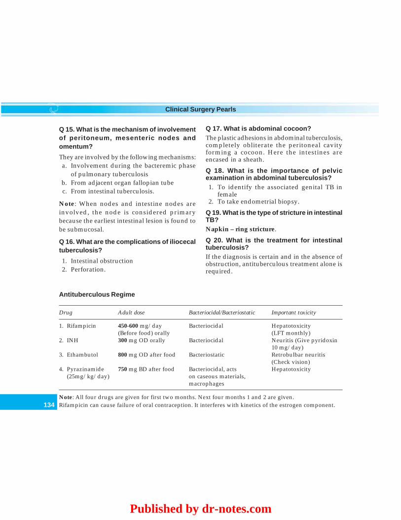
134
Clinical Surgery Pearls
Q 15. What is the mechanism of involvementof peritoneum, mesenteric nodes andomentum?
They are involved by the following mechanisms:a. Involvement during the bacteremic phase
of pulmonary tuberculosisb. From adjacent organ fallopian tubec. From intestinal tuberculosis.
Note: When nodes and intestine nodes areinvolved, the node is considered primarybecause the earliest intestinal lesion is found tobe submucosal.
Q 16. What are the complications of iliocecaltuberculosis?
1. Intestinal obstruction2. Perforation.
Q 17. What is abdominal cocoon?
The plastic adhesions in abdominal tuberculosis,completely obliterate the peritoneal cavityforming a cocoon. Here the intestines areencased in a sheath.
Q 18. What is the importance of pelvicexamination in abdominal tuberculosis?
1. To identify the associated genital TB infemale
2. To take endometrial biopsy.
Q 19. What is the type of stricture in intestinalTB?Napkin – ring stricture.
Q 20. What is the treatment for intestinaltuberculosis?If the diagnosis is certain and in the absence ofobstruction, antituberculous treatment alone isrequired.
Antituberculous Regime
Drug Adult dose Bacteriocidal/Bacteriostatic Important toxicity
1. Rifampicin 450-600 mg/day Bacteriocidal Hepatotoxicity(Before food) orally (LFT monthly)
2. INH 300 mg OD orally Bacteriocidal Neuritis (Give pyridoxin10 mg/day)
3. Ethambutol 800 mg OD after food Bacteriostatic Retrobulbar neuritis(Check vision)
4. Pyrazinamide 750 mg BD after food Bacteriocidal, acts Hepatotoxicity(25mg/kg/day) on caseous materials,
macrophages
Note: All four drugs are given for first two months. Next four months 1 and 2 are given.Rifampicin can cause failure of oral contraception. It interferes with kinetics of the estrogen component.
Published by dr-notes.com
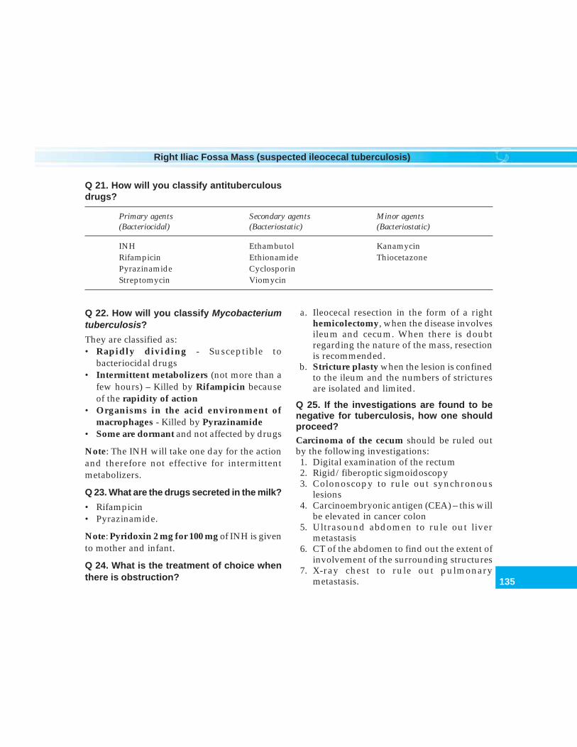
135
Right Iliac Fossa Mass (suspected ileocecal tuberculosis)
Q 21. How will you classify antituberculousdrugs?
Q 22. How will you classify Mycobacteriumtuberculosis?
They are classified as:• Rapidly dividing - Susceptible to
bacteriocidal drugs• Intermittent metabolizers (not more than a
few hours) – Killed by Rifampicin becauseof the rapidity of action
• Organisms in the acid environment ofmacrophages - Killed by Pyrazinamide
• Some are dormant and not affected by drugs
Note: The INH will take one day for the actionand therefore not effective for intermittentmetabolizers.
Q 23. What are the drugs secreted in the milk?
• Rifampicin• Pyrazinamide.
Note: Pyridoxin 2 mg for 100 mg of INH is givento mother and infant.
Q 24. What is the treatment of choice whenthere is obstruction?
Primary agents Secondary agents Minor agents(Bacteriocidal) (Bacteriostatic) (Bacteriostatic)
INH Ethambutol KanamycinRifampicin Ethionamide ThiocetazonePyrazinamide CyclosporinStreptomycin Viomycin
a. Ileocecal resection in the form of a righthemicolectomy, when the disease involvesileum and cecum. When there is doubtregarding the nature of the mass, resectionis recommended.
b. Stricture plasty when the lesion is confinedto the ileum and the numbers of stricturesare isolated and limited.
Q 25. If the investigations are found to benegative for tuberculosis, how one shouldproceed?
Carcinoma of the cecum should be ruled outby the following investigations:1. Digital examination of the rectum2. Rigid/fiberoptic sigmoidoscopy3. Colonoscopy to rule out synchronous
lesions4. Carcinoembryonic antigen (CEA) – this will
be elevated in cancer colon5. Ultrasound abdomen to rule out liver
metastasis6. CT of the abdomen to find out the extent of
involvement of the surrounding structures7. X-ray chest to rule out pulmonary
metastasis.
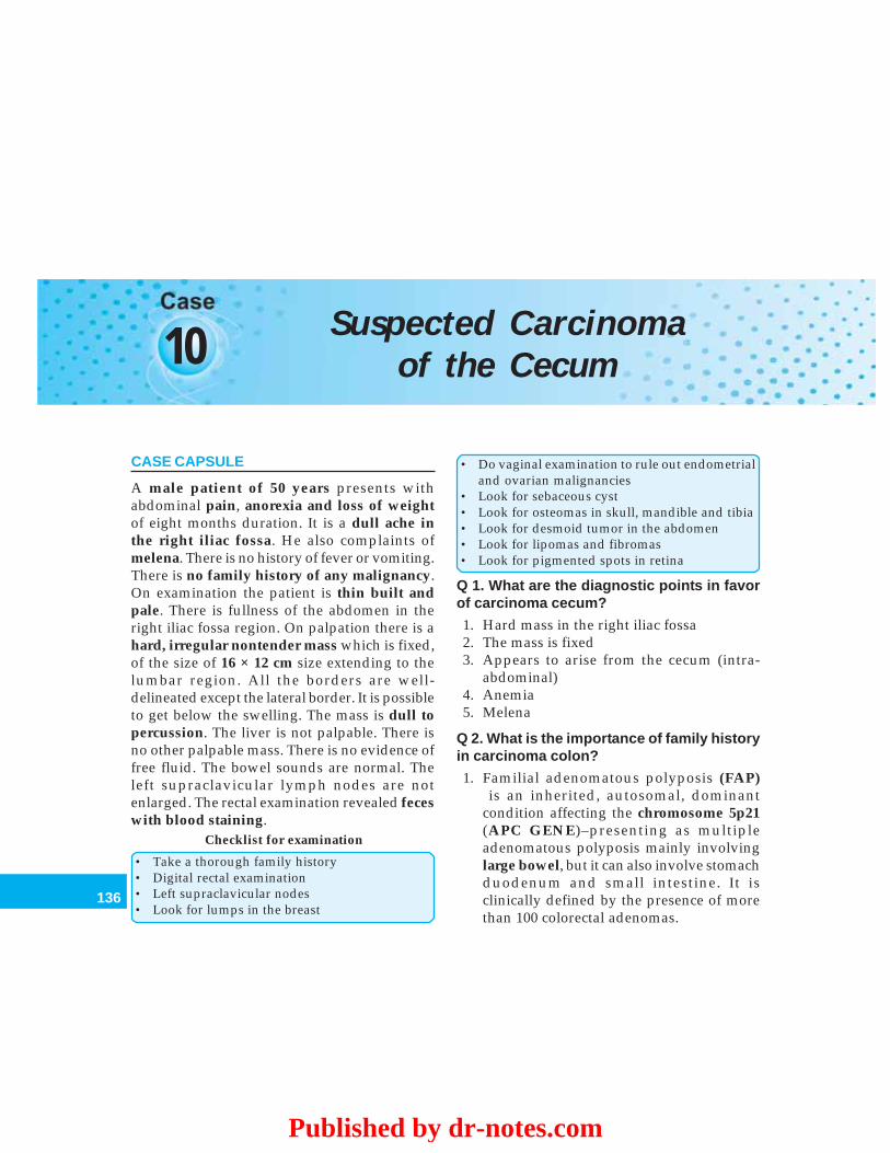
136
Clinical Surgery Pearls
10 Suspected Carcinomaof the Cecum
CASE CAPSULE
A male patient of 50 years presents withabdominal pain, anorexia and loss of weightof eight months duration. It is a dull ache inthe right iliac fossa. He also complaints ofmelena. There is no history of fever or vomiting.There is no family history of any malignancy.On examination the patient is thin built andpale. There is fullness of the abdomen in theright iliac fossa region. On palpation there is ahard, irregular nontender mass which is fixed,of the size of 16 × 12 cm size extending to thelumbar region. All the borders are well-delineated except the lateral border. It is possibleto get below the swelling. The mass is dull topercussion. The liver is not palpable. There isno other palpable mass. There is no evidence offree fluid. The bowel sounds are normal. Theleft supraclavicular lymph nodes are notenlarged. The rectal examination revealed feceswith blood staining.
Checklist for examination
• Take a thorough family history• Digital rectal examination• Left supraclavicular nodes• Look for lumps in the breast
• Do vaginal examination to rule out endometrialand ovarian malignancies
• Look for sebaceous cyst• Look for osteomas in skull, mandible and tibia• Look for desmoid tumor in the abdomen• Look for lipomas and fibromas• Look for pigmented spots in retina
Q 1. What are the diagnostic points in favorof carcinoma cecum?
1. Hard mass in the right iliac fossa2. The mass is fixed3. Appears to arise from the cecum (intra-
abdominal)4. Anemia5. Melena
Q 2. What is the importance of family historyin carcinoma colon?
1. Familial adenomatous polyposis (FAP) is an inherited, autosomal, dominantcondition affecting the chromosome 5p21(APC GENE)–presenting as multipleadenomatous polyposis mainly involvinglarge bowel, but it can also involve stomachduodenum and small intestine. It isclinically defined by the presence of morethan 100 colorectal adenomas.
Published by dr-notes.com
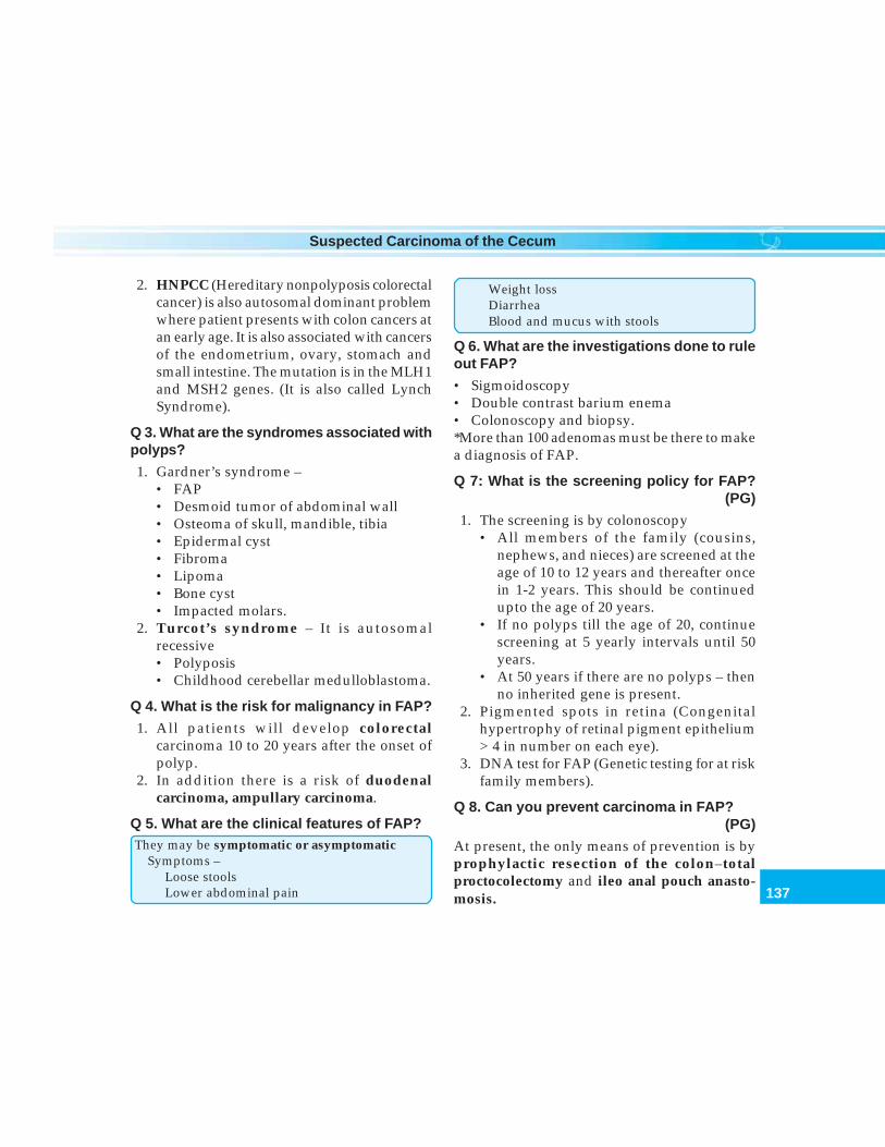
137
Suspected Carcinoma of the Cecum
2. HNPCC (Hereditary nonpolyposis colorectalcancer) is also autosomal dominant problemwhere patient presents with colon cancers atan early age. It is also associated with cancersof the endometrium, ovary, stomach andsmall intestine. The mutation is in the MLH1and MSH2 genes. (It is also called LynchSyndrome).
Q 3. What are the syndromes associated withpolyps?
1. Gardner’s syndrome –• FAP• Desmoid tumor of abdominal wall• Osteoma of skull, mandible, tibia• Epidermal cyst• Fibroma• Lipoma• Bone cyst• Impacted molars.
2. Turcot’s syndrome – It is autosomalrecessive• Polyposis• Childhood cerebellar medulloblastoma.
Q 4. What is the risk for malignancy in FAP?
1. All patients will develop colorectalcarcinoma 10 to 20 years after the onset ofpolyp.
2. In addition there is a risk of duodenalcarcinoma, ampullary carcinoma.
Q 5. What are the clinical features of FAP?
They may be symptomatic or asymptomaticSymptoms –
Loose stoolsLower abdominal pain
Weight lossDiarrheaBlood and mucus with stools
Q 6. What are the investigations done to ruleout FAP?
• Sigmoidoscopy• Double contrast barium enema• Colonoscopy and biopsy.*More than 100 adenomas must be there to makea diagnosis of FAP.
Q 7: What is the screening policy for FAP?(PG)
1. The screening is by colonoscopy• All members of the family (cousins,
nephews, and nieces) are screened at theage of 10 to 12 years and thereafter oncein 1-2 years. This should be continuedupto the age of 20 years.
• If no polyps till the age of 20, continuescreening at 5 yearly intervals until 50years.
• At 50 years if there are no polyps – thenno inherited gene is present.
2. Pigmented spots in retina (Congenitalhypertrophy of retinal pigment epithelium> 4 in number on each eye).
3. DNA test for FAP (Genetic testing for at riskfamily members).
Q 8. Can you prevent carcinoma in FAP?(PG)
At present, the only means of prevention is byprophylactic resection of the colon–totalproctocolectomy and ileo anal pouch anasto-mosis.
138
Clinical Surgery Pearls
Q 9. Is there any role for upper GI endoscopyin FAP? (PG)
Yes. The patient can develop duodenalampullary and stomach malignancies andtherefore upper GI endoscopy is indicated.
Q 10. Is there any medical treatment forpolyp? (PG)
Sulindac and Celecoxib are being tried.
Q 11. What are the features of HNPCC?
It is also an autosomal dominant conditionaffecting the chromosome 2P (short arm of 2)and there are 2 syndromes associated with this–a. Lynch I syndrome - this produces multiple
colonic cancers in the proximal colon at anearly age (hereditary site specific coloncaner)
b. Lynch II syndrome – here in addition tocolon cancer extracolonic adenocarcinomasaffecting (Cancer family syndrome) thefollowing organs are seen:1. Breast, ovary, endometrium, pancreas,
stomach, bile duct, ureter and renalpelvis, etc.
Q 12. What is Amsterdam criteria for HNPCC?(PG)
Amsterdam criteria 1 (Lynch 1) – at least 3relatives must have histologically verifiedcolorectal cancer.1. One must be a 1st degree relative of the
other two2. At least two successive generations must be
affected
3. At least one of the relatives with colorectalcancer must have received the diagnosisbefore the age of 50 years.
Amsterdam criteria 2 (Lynch 2) – at least 3relatives must have a cancer associated withhereditary nonpolyposis colonic cancer namelycolorectal, endometrial, ovarian, stomach,pancreas, hepatobiliary, etc.1. One must be a 1st degree relative of the
other two2. At least two successive generations must be
affected3. At least one of the relatives with HNPCC
associated cancer must have received thediagnosis before the age of 50 years.
Q 13. What is synchronous carcinoma?
In all cases of suspected colorectal cancer lookfor the presence of a second malignancy inanother location in the colon. The developmentof simultaneous cancer is called synchronouscarcinoma. If it is developing after the resectionof one malignancy then it is called metachronouscancer. Therefore even after identification of onemalignancy the rest of the colon must be studiedthoroughly by colonoscopy and double contrastbarium enema to rule out a second malignancy(Synchronous).
Q 14. What are the macroscopical types ofcolorectal cancer?
There are 4 types:Type 1 – AnnularType 2 – TubularType 3 – UlcerativeType 4 – Cauliflower – least malignant.
Published by dr-notes.com
139
Suspected Carcinoma of the Cecum
Q 15. Which type of lesion is seen on the rightside of colon?
Ulcerative and polypoidal lesions are seen onthe right side.
Q 16. What are the investigations for makinga diagnosis in this case?
A. Investigations for diagnosis1. Rigid sigmoidoscopy2. Flexible sigmoidoscopy (60cm length)3. Colonoscopy – can visualize the growth
and take biopsy.- Can demonstrate synchronouscarcinoma (seen in 5%) and polyps
4. Double contrast barium enema–– irregular filling defects– apple core appearance– shouldering– demonstrate polyps
B. Investigations for staging:
1. Chest X-ray2. LFT3. Ultrasound
– for liver metastasis– for free fluid– for knowing the extend of the cecal mass– obstruction of ureter and subsequent
hydronephrosis4. CECT (contrast enhanced)
– for local invasion– for adjacent structure involvement
5. CEA
C. Investigations for surgery:1. Cardiac status2. Renal status3. Hematological investigations.
Q 17. What is virtual colonoscopy?
High resolution 3D images of the colon can bemade with a multislice CT scanner withimproved software. It has a sensitivity andspecificity of 94%. There is no need for sedationas in the case of colonoscopy.
Q 18. What are the methods of spread ofcarcinoma colon?
1. Local spread – may be vertical, horizontaland radial.Vertical is to the adjacent structures andhorizontal is submucosal. Radial iscircumferential.a. It will spread around – it takes one year
for spreading 3/4th circumference andresult in intestinal obstruction
b. Ulcerated type produce fistulization tourinary bladderLocal perforationExternal fecal fistula.
2. Lymphatic spreadRead the lymphatic drainage (below).
3. Blood stream spread– Produce occult hepatic metastasis– Responsible for late death– Metastasis in lung, liver, bone, brain and
skin.4. Transperitoneal spread
– Peritoneal metastasis—ascitis- Carcinomatosis peritoneum- Rectal shelf of Blumer.
5. Intraluminal spread—this is responsible foranastomotic site recurrence.
Q 19. What are the predisposing causes forcolorectal cancer?
140
Clinical Surgery Pearls
Predisposing causes for colorectal cancer
• Ulcerative colitis—Inflammatory bowel diseaseof > 10 yrs
• Crohn's colitis• Schistosomal colitis• Exposure to radiation• Ureterosigmoidostomy• Breast carcinoma (small increased risk).• Gallstone (postcholecystectomy-increased risk
for right colonic malignancy)• Diet—fiber diet protects, cooked meat—
carcinogenic, bile acid—carcinogenic
Q 20. What is the commonest site forcolorectal cancer?
Incidence of colorectal cancer in various sites
Maximum incidence in rectum 38%Sigmoid 21%Cecum 12%Transverse colon 5.5%Ascending colon 5%Descending colon 4%Splenic flexure 3%Hepatic flexure 2%
Q 21. What are the types of adenomatouspolyps of colon?
There are 3 types1. Tubular - 5% chance for malignancy2. Tubulovillous - 22% chance for malig-
nancy3. Villous - 40% chance for malig-
nancy
Q 22. What is the significance of adeno-matous polyp and how will you managepolyp?
• Adenomas are premalignant• They are usually multiple• Synchronous cancer may occur• Adenomas > 5mm size should be removed
because of malignant potential
Q 23. What is the peculiarity of villousadenoma?
They are sessile frond-like spreading lesion.They produce mucus discharge, diarrhea andhypokalemia. Unlike other polyp, removal bycolonoscopic snaring and diathermy oblitera-tion with hot biopsy forceps are not possible invillous adenoma. Proctectomy may be required.
Q 24. What is adenoma - carcinomasequence? (PG)
Fearon-Vogelstein Adenoma-carcinomamultistep model of carcinogenesis
Normal epithelium↓← 5q lossDysplasia↓Early adenoma↓←12p activation K-rasIntermediate adenoma↓←18q lossLate adenoma↓←17p loss, p53Carcinoma↓Metastasis
Q 25. What is the staging for carcinomacolon?
Duke's classification originally described forrectum is used for colon with modification.
Published by dr-notes.com
141
Suspected Carcinoma of the Cecum
Duke's A: Tumor restricted, but not through thebowel wall.Duke's B: Indicating penetration through the bowelwall.Duke's C: Spread to local and regional nodes.
C1: Nodes in the immediate vicinityC2: Along the vessels
Duke's D: Distant metastasis
The staging system by Astler and Coller is amodification of Duke's.
TNM - AJCC 6th edition is the staging systemfollowed now.
Q 26. What is the AJCC - TNM staging?(PG)
Tx Primary tumor cannot be assessedT0 No evidence of primary tumorTis Carcinoma in situ, intraepithelial or
invasion of lamina propriaT1 Tumor invades submucosaT2 Tumor invades muscularis propriaT3 Tumor invades through the muscularis
propria, into the subserosa or into non-peritonialized pericolic or perirectaltissues
T4 Tumor directly invades other organs orstructures and/or perforates visceralperitoneum
Nx Regional lymph nodes cannot be assessedN0 No regional lymph node metastasisN1 Metastasis in 1 to 3 nodesN2 Metastasis in 4 or more nodesMx Distant metastasis cannot be assessedM0 No distant metastasisM1 Distant metastasis
Q 27. Comparative chart showing TNM,Duke's and modified Astler-Coller staging?
(PG)
Stage T N M Dukes MAC
0 Tis N0 M0I T1 N0 M0 A A
T2 N0 M0 A B1IIA T3 N0 M0 B B2IIB T4 N0 M0 B B3IIIA T1-T2 N1 M0 C C1IIIB T3-T4 N1 M0 C C2/C3IIIC Any T N2 M0 C C1/C2/C3IV Any T Any N M1 D D
Q 28. What are the differences between rightand left sided colonic malignancy?
Right sided Left sided
1. Right side colonic diameter is large Narrow2. On right side the content is fluid (fecal) Content is solid3. Growth is ulcerated/cauliflower like Stenosing growth4. Present with pallor Present with intestinal obstruction5. Ill defined pain Colicky pain6. Bowel symptom - Melena Alternating constipation and diarrhea
142
Clinical Surgery Pearls
Q 29. What is the cause for alternatingconstipation and diarrhea in left sided coloncancer?
The left sided cancers are usually tubular orannular producing obstruction. As a result ofobstruction there will be constipation.Subsequent to constipation there will be stasisof fecal matter proximal to the stenosis. This willproduce stercoral enteritis which in turn willproduce diarrhea—the so-called spuriousdiarrhea where the liquid fecal matter willescape through the narrow segment. Again thepatient will go in for constipation. This is avicious cycle.
Q 30. What is the site of perforation of thecolon in cases of obstruction?
The perforation can occur in the followingplaces.a. At the site of the growthb. Just proximal to the growth as a result of
stercoral enteritisc. At the cecum for any distal growth if the
ileocecal valve is competent.
Q 31. What are the acute manifestations ofcolonic cancer?
• Intestinal obstruction• Perforation• Peritonitis• Bleeding
• Fistulization with urinary bladder resulting inpneumaturia
• Gastrocolic fistula• Acute appendicitis/appendicular abscess
(obstructive appendicitis as a result of the growthin the cecum)
• Intussusceptions (always rule out a growth in allcases of adult intussusception. There is no rolefor hydrostatic reduction in adults).
Q 32. Having made the diagnosis ofcarcinoma cecum, how do you manage thepatient?
After doing investigations in the form of X-raychest to rule out metastasis in the lungs,ultrasound abdomen to rule out metastasis liverand free fluid, the patient is prepared forresection.
Q 33. What are the preparations for surgery?
• Make the patient fit for anesthesia (evaluatecardiac, respiratory status)
• Correction of anemia• Correction of hypoproteinemia• Dietary regulation• Bowel preparation• Prophylactic subcutaneous heparin• Prophylactic antibiotics.
Q 34. What is the role of mechanical bowelpreparation?
• Dietary restriction in the form of liquid dietfor 24 hours before surgery
7. Late diagnosis Because of obstruction early diagnosis8. For right colectomy only the branches of Flush ligation of inferior mesenteric artery done
superior mesenteric are ligated for left colectomy9. Bad prognosis because of late diagnosis Good prognosis because of early diagnosis
Published by dr-notes.com
143
Suspected Carcinoma of the Cecum
• The role of mechanical bowel preparation forelective surgery is highly controversial. Thereis enough evidence in the literature for safesurgery without mechanical bowel pre-paration.
• Conventionally– Bowel washes– Laxatives in the form of sodium
picosulphate– Cleansing agents like Polyethyl glycol
(PEG) orally are given the day before
Q 35. What is the role of antibioticprophylaxis in colonic surgery?
The routine practice of Nichol's regime by giving3 doses of antibiotics 18, 17 and 10 hours pre-operatively with Neomycin and Erythromycinis no longer practised.
A single dose of ampicillin - sulbactam orquinolone + Metronidazole or parenteralCefoxitin are recommended for antibioticprophylaxis (1 hour before skin incision).
Q 36. How will you assess the operability?(PG)
a. After laparotomy look for metastases inperitoneum (small white seed-like), liver,pelvic deposits, Krukenberg's tumor (meta-static ovarian tumor) ascites and lymphnodes
b. Look for tumor mobility and adjacentstructure involvement
c. Liver metastasis if found are not biopsied—it will cause tumor dissemination.
d. Hepatic metastasis resection is done as astaged procedure later
e. Tumor dissemination is not a contraindica-tion for resection of the primary.
Q 37. What is the surgical procedure ofchoice when there is a cecal growth?
Right hemicolectomy is the operation of choice,where a radical resection of the involved colonalong with its lymphovascular drainage is done.Any devascularized segment should beremoved in addition. Suspicious lymph nodesare taken for biopsy.
Q 38. How much is the clearance required incolonic resection?
The clearance of a minimum of 5 - 10cm oneither side is recommended.
Q 39. What are the vessels ligated for righthemicolectomy?
The ileocolic, right colic, and right branch of themiddle colic vessels are ligated for righthemicolectomy.
Q 40. What are the technical precautionstaken during surgery and how much ileum isremoved? (PG)
• The cecum is mobilized by incising the lateralperitoneum which is carried around thehepatic flexure.
• Take care not to injure the ureter, spermaticvessels (in males) and duodenum
• The mesentery of last 30 cm of ileum isremoved
• The colon is divided as far as the proximalthird of transverse colon
• The anastomosis is done between the rest ofileum and transverse colon in layers.
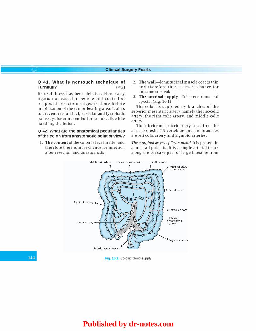
144
Clinical Surgery Pearls
Q 41. What is nontouch technique ofTurnbull? (PG)
Its usefulness has been debated. Here earlyligation of vascular pedicle and control ofproposed resection edges is done beforemobilization of the tumor bearing area. It aimsto prevent the luminal, vascular and lymphaticpathways for tumor emboli or tumor cells whilehandling the lesion.
Q 42. What are the anatomical peculiaritiesof the colon from anastomotic point of view?
1. The content of the colon is fecal matter andtherefore there is more chance for infectionafter resection and anastomosis
2. The wall—longitudinal muscle coat is thinand therefore there is more chance foranastomotic leak
3. The arterixal supply—It is precarious andspecial (Fig. 10.1)
The colon is supplied by branches of thesuperior mesenteric artery namely the ileocolicartery, the right colic artery, and middle colicartery.
The inferior mesenteric artery arises from theaorta opposite L3 vertebrae and the branchesare left colic artery and sigmoid arteries.
The marginal artery of Drummond: It is present inalmost all patients. It is a single arterial trunkalong the concave part of large intestine from
Fig. 10.1: Colonic blood supply
Published by dr-notes.com
145
Suspected Carcinoma of the Cecum
ileocecal junction to rectosigmoid junctionforming a communicating channel between thesuperior and inferior mesenteric artery. It givesoff vasa recta which run perpendicular to thecolonic wall.
Meandering artery (Arc of Riolan): These arecollateral branches connecting proximal middlecolic artery to left colic artery. During colonicmobilization this artery should be protected.
Griffith's point: It is the weak point in thevascular anastomosis between middle and leftcolic arteries. Therefore it is important to avoidanastomosis at the splenic flexure.
Sudek's point: It is at the level of lowest sigmoidand superior rectal artery. There is a higherchance of avascularity if anastomosis isperformed here.
Q 43. What is the lymphatic drainage ofcolon?
Lymph nodes are divided into four groups.a. Epicolic - located on the bowel wallb. Paracolic - located along the marginal arteryc. Intermediate - located along the colic vesselsd. Principal - located along SMA and IMA.
Q 44. What is white line of Toldt?
The ascending and descending colon has noperitoneal covering on their posterior aspectsbut has areolar tissue (fascia of Toldt), whichrepresent the embryonic fusion of mesentery toposterior peritoneum. At the lateral parietal wallit reflects as the white line of Toldt. This line isused as the plane for avascular mobilization ofthe colon.
Q 45. Which part of the colon has the largestdiameter?
Cecum - 7.5 cmNarrowest part is rectosigmoid - 2.5 cm.
Q 46. Which are the mobile portions of thecolon?
Transverse colon and sigmoid colon. (The restof the colon is fixed).
Q 47. What are the ligaments attaching thehepatic flexure of colon (which is fixed)?
Nephrocolic ligamentHepatocolic ligamentDuodenocolic ligament.
Q 48. What is the attachment of splenicflexure?
Phrenicocolic ligament.
Q 49. What is the extend of descendingcolon?
It extends from the splenic flexure to the brimof the true pelvis.
Q 50. What is the attachment of transversemesocolon?
The transverse mesocolon is attached to thedescending part of duodenum, to the head andlower aspect of the body of the pancreas, and tothe anterior surface of the left kidney -horizontally.
Q 51. What is the attachment of meso-sigmoid?
It is inverted 'V' shaped, the apex of thisperitoneal attachment is at the bifurcation of the
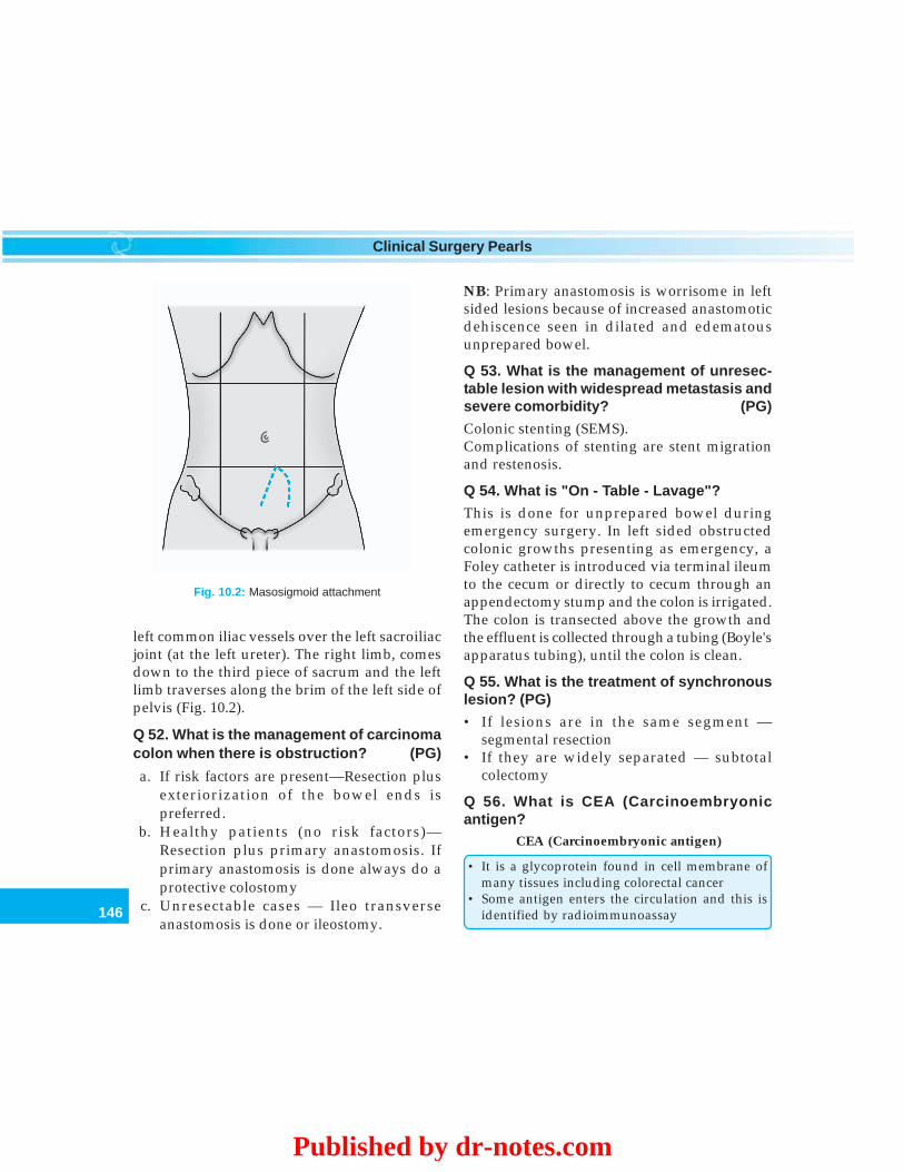
146
Clinical Surgery Pearls
left common iliac vessels over the left sacroiliacjoint (at the left ureter). The right limb, comesdown to the third piece of sacrum and the leftlimb traverses along the brim of the left side ofpelvis (Fig. 10.2).
Q 52. What is the management of carcinomacolon when there is obstruction? (PG)
a. If risk factors are present—Resection plusexteriorization of the bowel ends ispreferred.
b. Healthy patients (no risk factors)—Resection plus primary anastomosis. Ifprimary anastomosis is done always do aprotective colostomy
c. Unresectable cases — Ileo transverseanastomosis is done or ileostomy.
NB: Primary anastomosis is worrisome in leftsided lesions because of increased anastomoticdehiscence seen in dilated and edematousunprepared bowel.
Q 53. What is the management of unresec-table lesion with widespread metastasis andsevere comorbidity? (PG)
Colonic stenting (SEMS).Complications of stenting are stent migrationand restenosis.
Q 54. What is "On - Table - Lavage"?
This is done for unprepared bowel duringemergency surgery. In left sided obstructedcolonic growths presenting as emergency, aFoley catheter is introduced via terminal ileumto the cecum or directly to cecum through anappendectomy stump and the colon is irrigated.The colon is transected above the growth andthe effluent is collected through a tubing (Boyle'sapparatus tubing), until the colon is clean.
Q 55. What is the treatment of synchronouslesion? (PG)
• If lesions are in the same segment —segmental resection
• If they are widely separated — subtotalcolectomy
Q 56. What is CEA (Carcinoembryonicantigen?
CEA (Carcinoembryonic antigen)
• It is a glycoprotein found in cell membrane ofmany tissues including colorectal cancer
• Some antigen enters the circulation and this isidentified by radioimmunoassay
Fig. 10.2: Masosigmoid attachment
Published by dr-notes.com
147
Suspected Carcinoma of the Cecum
• It is present in other body fluids like urine andfeces
• CEA is not specific for carcinoma of colorectum• It is present in other GI cancers, nonalimentary
cancers, and benign diseases• It is high in 70% of carcinoma colon• It is not useful as a screening investigation• It is useful for detecting recurrence after a
curative surgery if the serial CEA values arerising after 6-9 months
• CEA level of > 2.5 ng/ml is significant• CEA level of > 5 ng/ml indicates poor prognosis• If preoperative elevated CEA does not fall to
normal within 2-3 weeks after resection, theresection is incomplete or there is occultmetastasis
• CEA values are highest in lung and livermetastases
Q 57. What is left hemicolectomy?
This is done for tumors of descending colon andsplenic flexure. Here resection of the colon fromsplenic flexure to rectosigmoid is done.
Q 58. What is the treatment for sigmoidcancer?
Sigmoidectomy.
Q 59. What are the indications for totalcolectomy/subtotal colectomy?
• Multiple primary tumors• Completely obstructing sigmoid cancers• HNPCC• Transverse colon carcinoma
Q 60. What is extended right hemicolectomy?
This is done for proximal transverse colongrowth.
Q 61. What is extended left hemicolectomy?
This is done for distal transverse colon growth.
NB: For transverse colon growth subtotalcolectomy is another option.
Q 62. What are the surgical procedures ofchoice for growth in various regions of thecolon?
Surgical procedures of choice for growth invarious regions of the colon
Site Surgical procedureCecum, ascending Right hemicolectomycolon, hepatic flexureProximal part of Extended right hemico-transverse colon lectomyDistal transverse colon Extended left hemico-
lectomyDescending colon and Left hemicolectomysplenic flexureSigmoid colon SigmoidectomyTransverse colon, Subtotal colectomysigmoid colon, multipletumors, HNPCC
Q 63. What are the peculiarities of cancersecondary to ulcerative colitis?
• May occur at many sites• May affect the colon more than rectum• They are high grade tumors• After 20 years the risk is 12%
Q 64. What are the bad prognostic factors incolonic cancer? (PG)
1. Depth of tumor invasion and nodalmetastasis (most important)
2. Bowel obstruction - poor prognosis3. CEA > 5 ng/ml
148
Clinical Surgery Pearls
4. Right sided colon carry worse prognosis5. Pathological variables
– histological grade– signet ring type-poor prognosis– small cell carcinoma– lymphovascular invasion
Q 65. What is the indication for adjuvanttherapy in colorectal cancer? (PG)
In stage 1 lesion no further adjuvant therapyIn stage II and III - adjuvant therapy has mostimportant impactIn Stage II - 5FU + Leucoverin (folenic acid)
– Folenic acid is a potentiator of 5 FU– (immunomodulator Levamisole with 5 FU
is used less nowadays)– Histopathologically positive lymph nodes
is an indication for chemotherapyIn Stage III - FOLFOX 4 regimen
(Oxaliplatin + 5FU - 2 times a month for 12cycles)
OR– FOLFIRI regimen (folic acid, + 5FU +
Irinotecan).
Q 66. What is the most effective singlechemotherapeutic agent for adenocarci-noma?
5 - Fluorouracil
Q 67. What is the follow up for carcinomacolon?
Clinical examination once in six months.Annual examination of the following:• CEA• Chest X-ray• Colonoscopy• LFT• CBC.
Q 68. What is fast track surgery?
It is a multimodal rehabilitation regimen forelective colonic surgery for enhanced recoveryconsisting of-
Fast track surgery
• Preoperative counseling and optimization• Epidural catheter for pain control• General anesthesia using propofol (no nitrous
oxide)• No bowel preparation• Transverse abdominal incision instead of vertical• Continuous epidural analgesia for two days• Oral feeding with protein drink (60-80 gm
protein/day) from the day of surgery• Early mobilization• Laxative and prokinetics for bowel function from
the day of surgery• POP day 1 remove urinary catheter• POP day 2 remove epidural catheter• POP day 3 discharge after lunch
Published by dr-notes.com
149
Appendicular Mass
11 Appendicular Mass
CASE CAPSULE
A 25-year-old female patient presents withhistory of abdominal pain of 4 days duration.Initially the pain started as a vague centralabdominal pain in the early morning hours. Thispain was preceded by loss of appetite. The patientwas constipated for a few days. She felt nauseatedand vomited once on the first day of pain. Laterthe pain shifted to the right iliac fossa. Her painis aggravated by moving and coughing. Thepatient is married and has 2 children. Hermenstrual cycles are regular having normal bloodflow. Her last menstrual period was 6 days back.There is no history of discharge from vagina. Onexamination the patient looks pale. There is mildpyrexia. The tongue is furred and there is foetororis (Halitosis). Her pulse rate is 90 per minute.Abdominal examination revealed a tender massof 16 × 9 cm size in the right iliac fossa withoverlying muscle guard and rigidity confined tothe right iliac fossa. There is no intrinsicmovement for the mass and all the borders arewell made out except the lateral border. The massis intraabdominal and intraperitoneal. There isno extension of the mass below the inguinalregion. The rest of the abdomen is soft and
nontender. There is no free fluid demonstrated.The bowel sounds are normally heard. On digitalrectal examination, the patient complained paindeep in the pelvis and the mass is palpable perrectum. On vaginal examination movement ofthe cervix did not cause pain. Other systems arenormal.
Read the checklist of previous caseChecklist with special reference to this case
• A careful gynecological history is taken in allwomen
• Last menstrual period - to find out whether it isa midmenstrual pain (mittleschmerz) and to ruleout pregnancy
• History of vaginal discharge—pelvicinflammatory disease
• History of dysmenorrhea• Pelvic examination to rule out adnexal and
cervical tenderness• High vaginal swab to rule out Chlamydia
trachomatis• Urine for pregnancy test• Ultrasound abdomen
Q 1. What is the most probable diagnosis inthis case?
Appendicular mass.
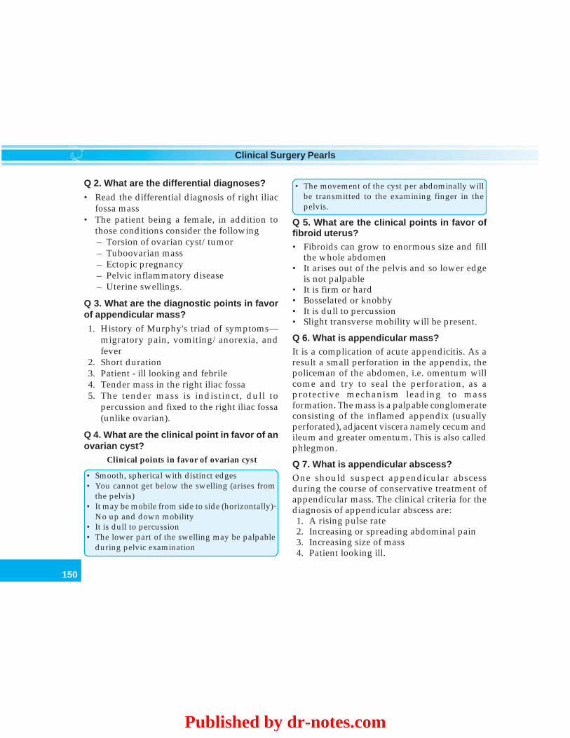
150
Clinical Surgery Pearls
Q 2. What are the differential diagnoses?
• Read the differential diagnosis of right iliacfossa mass
• The patient being a female, in addition tothose conditions consider the following– Torsion of ovarian cyst/tumor– Tuboovarian mass– Ectopic pregnancy– Pelvic inflammatory disease– Uterine swellings.
Q 3. What are the diagnostic points in favorof appendicular mass?
1. History of Murphy's triad of symptoms—migratory pain, vomiting/anorexia, andfever
2. Short duration3. Patient - ill looking and febrile4. Tender mass in the right iliac fossa5. The tender mass is indistinct, dull to
percussion and fixed to the right iliac fossa(unlike ovarian).
Q 4. What are the clinical point in favor of anovarian cyst?
Clinical points in favor of ovarian cyst
• Smooth, spherical with distinct edges• You cannot get below the swelling (arises from
the pelvis)• It may be mobile from side to side (horizontally)·
No up and down mobility• It is dull to percussion• The lower part of the swelling may be palpable
during pelvic examination
• The movement of the cyst per abdominally willbe transmitted to the examining finger in thepelvis.
Q 5. What are the clinical points in favor offibroid uterus?
• Fibroids can grow to enormous size and fillthe whole abdomen
• It arises out of the pelvis and so lower edgeis not palpable
• It is firm or hard• Bosselated or knobby• It is dull to percussion• Slight transverse mobility will be present.
Q 6. What is appendicular mass?
It is a complication of acute appendicitis. As aresult a small perforation in the appendix, thepoliceman of the abdomen, i.e. omentum willcome and try to seal the perforation, as aprotective mechanism leading to massformation. The mass is a palpable conglomerateconsisting of the inflamed appendix (usuallyperforated), adjacent viscera namely cecum andileum and greater omentum. This is also calledphlegmon.
Q 7. What is appendicular abscess?
One should suspect appendicular abscessduring the course of conservative treatment ofappendicular mass. The clinical criteria for thediagnosis of appendicular abscess are:1. A rising pulse rate2. Increasing or spreading abdominal pain3. Increasing size of mass4. Patient looking ill.
Published by dr-notes.com
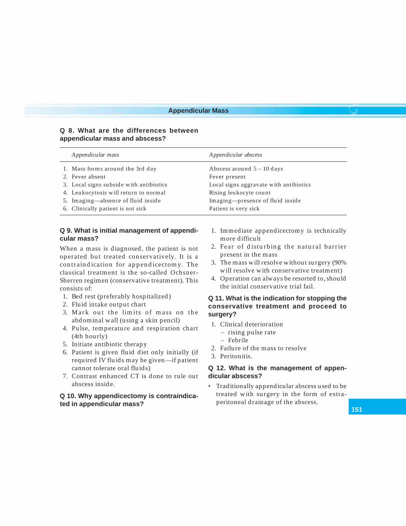
151
Appendicular Mass
Q 8. What are the differences betweenappendicular mass and abscess?
Q 9. What is initial management of appendi-cular mass?
When a mass is diagnosed, the patient is notoperated but treated conservatively. It is acontraindication for appendicectomy. Theclassical treatment is the so-called Ochsner-Sherren regimen (conservative treatment). Thisconsists of:1. Bed rest (preferably hospitalized)2. Fluid intake output chart3. Mark out the limits of mass on the
abdominal wall (using a skin pencil)4. Pulse, temperature and respiration chart
(4th hourly)5. Initiate antibiotic therapy6. Patient is given fluid diet only initially (if
required IV fluids may be given—if patientcannot tolerate oral fluids)
7. Contrast enhanced CT is done to rule outabscess inside.
Q 10. Why appendicectomy is contraindica-ted in appendicular mass?
Appendicular mass Appendicular abscess
1. Mass forms around the 3rd day Abscess around 5 – 10 days2. Fever absent Fever present3. Local signs subside with antibiotics Local signs aggravate with antibiotics4. Leukocytosis will return to normal Rising leukocyte count5. Imaging—absence of fluid inside Imaging—presence of fluid inside6. Clinically patient is not sick Patient is very sick
1. Immediate appendicectomy is technicallymore difficult
2. Fear of disturbing the natural barrierpresent in the mass
3. The mass will resolve without surgery (90%will resolve with conservative treatment)
4. Operation can always be resorted to, shouldthe initial conservative trial fail.
Q 11. What is the indication for stopping theconservative treatment and proceed tosurgery?
1. Clinical deterioration– rising pulse rate– Febrile
2. Failure of the mass to resolve3. Peritonitis.
Q 12. What is the management of appen-dicular abscess?
• Traditionally appendicular abscess used to betreated with surgery in the form of extra-peritoneal drainage of the abscess.
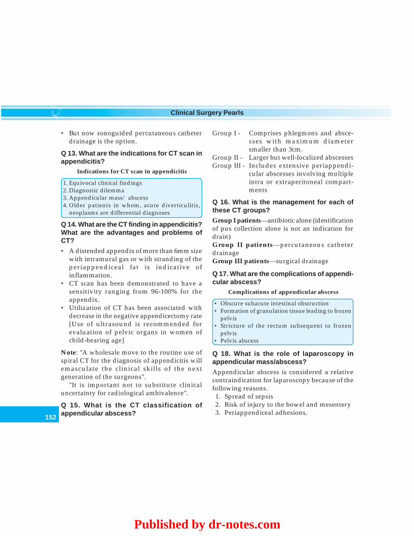
152
Clinical Surgery Pearls
• But now sonoguided percutaneous catheterdrainage is the option.
Q 13. What are the indications for CT scan inappendicitis?
Indications for CT scan in appendicitis
1. Equivocal clinical findings2. Diagnostic dilemma3. Appendicular mass/ abscess4. Older patients in whom, acute diverticulitis,
neoplasms are differential diagnoses
Q 14. What are the CT finding in appendicitis?What are the advantages and problems ofCT?
• A distended appendix of more than 6mm sizewith intramural gas or with stranding of theperiappendiceal fat is indicative ofinflammation.
• CT scan has been demonstrated to have asensitivity ranging from 96-100% for theappendix.
• Utilization of CT has been associated withdecrease in the negative appendicectomy rate[Use of ultrasound is recommended forevaluation of pelvic organs in women ofchild-bearing age]
Note: "A wholesale move to the routine use ofspiral CT for the diagnosis of appendicitis willemasculate the clinical skills of the nextgeneration of the surgeons".
"It is important not to substitute clinicaluncertainty for radiological ambivalence".
Q 15. What is the CT classification ofappendicular abscess?
Group I - Comprises phlegmons and absce-sses with maximum diametersmaller than 3cm.
Group II - Larger but well-localized abscessesGroup III - Includes extensive periappendi-
cular abscesses involving multipleintra or extraperitoneal compart-ments
Q 16. What is the management for each ofthese CT groups?
Group I patients—antibiotic alone (identificationof pus collection alone is not an indication fordrain)Group II patients—percutaneous catheterdrainageGroup III patients—surgical drainage
Q 17. What are the complications of appendi-cular abscess?
Complications of appendicular abscess
• Obscure subacute intestinal obstruction• Formation of granulation tissue leading to frozen
pelvis• Stricture of the rectum subsequent to frozen
pelvis• Pelvis abscess
Q 18. What is the role of laparoscopy inappendicular mass/abscess?
Appendicular abscess is considered a relativecontraindication for laparoscopy because of thefollowing reasons.1. Spread of sepsis2. Risk of injury to the bowel and mesentery3. Periappendiceal adhesions.
Published by dr-notes.com
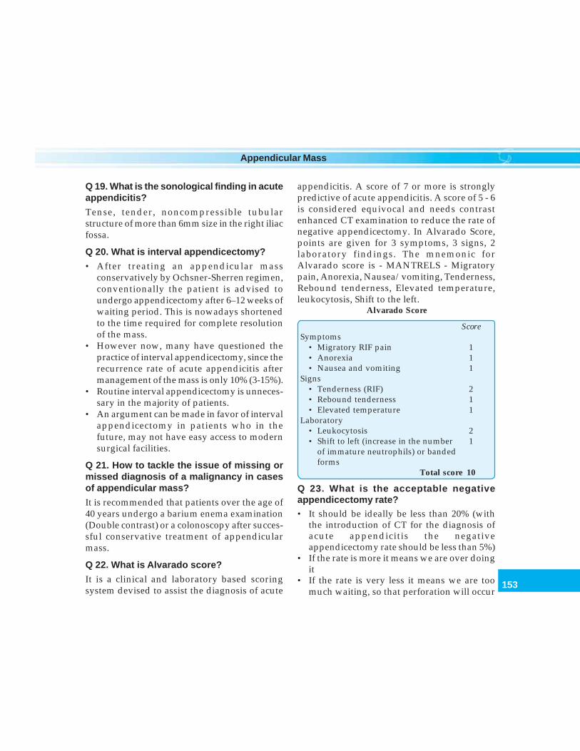
153
Appendicular Mass
Q 19. What is the sonological finding in acuteappendicitis?
Tense, tender, noncompressible tubularstructure of more than 6mm size in the right iliacfossa.
Q 20. What is interval appendicectomy?
• After treating an appendicular massconservatively by Ochsner-Sherren regimen,conventionally the patient is advised toundergo appendicectomy after 6–12 weeks ofwaiting period. This is nowadays shortenedto the time required for complete resolutionof the mass.
• However now, many have questioned thepractice of interval appendicectomy, since therecurrence rate of acute appendicitis aftermanagement of the mass is only 10% (3-15%).
• Routine interval appendicectomy is unneces-sary in the majority of patients.
• An argument can be made in favor of intervalappendicectomy in patients who in thefuture, may not have easy access to modernsurgical facilities.
Q 21. How to tackle the issue of missing ormissed diagnosis of a malignancy in casesof appendicular mass?
It is recommended that patients over the age of40 years undergo a barium enema examination(Double contrast) or a colonoscopy after succes-sful conservative treatment of appendicularmass.
Q 22. What is Alvarado score?
It is a clinical and laboratory based scoringsystem devised to assist the diagnosis of acute
appendicitis. A score of 7 or more is stronglypredictive of acute appendicitis. A score of 5 - 6is considered equivocal and needs contrastenhanced CT examination to reduce the rate ofnegative appendicectomy. In Alvarado Score,points are given for 3 symptoms, 3 signs, 2laboratory findings. The mnemonic forAlvarado score is - MANTRELS - Migratorypain, Anorexia, Nausea/vomiting, Tenderness,Rebound tenderness, Elevated temperature,leukocytosis, Shift to the left.
Alvarado Score
ScoreSymptoms
• Migratory RIF pain 1• Anorexia 1• Nausea and vomiting 1
Signs• Tenderness (RIF) 2• Rebound tenderness 1• Elevated temperature 1
Laboratory• Leukocytosis 2• Shift to left (increase in the number 1
of immature neutrophils) or bandedforms
Total score 10
Q 23. What is the acceptable negativeappendicectomy rate?
• It should be ideally be less than 20% (withthe introduction of CT for the diagnosis ofacute appendicitis the negativeappendicectomy rate should be less than 5%)
• If the rate is more it means we are over doingit
• If the rate is very less it means we are toomuch waiting, so that perforation will occur
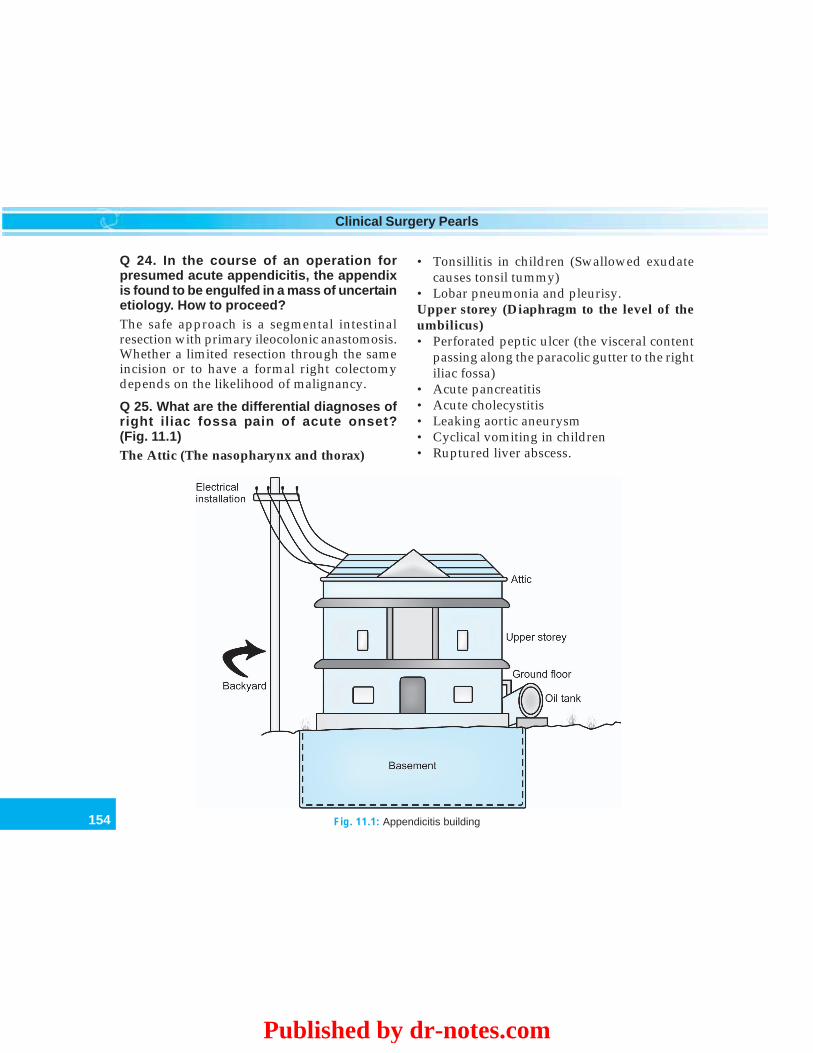
154
Clinical Surgery Pearls
Q 24. In the course of an operation forpresumed acute appendicitis, the appendixis found to be engulfed in a mass of uncertainetiology. How to proceed?
The safe approach is a segmental intestinalresection with primary ileocolonic anastomosis.Whether a limited resection through the sameincision or to have a formal right colectomydepends on the likelihood of malignancy.
Q 25. What are the differential diagnoses ofright iliac fossa pain of acute onset?(Fig. 11.1)
The Attic (The nasopharynx and thorax)
• Tonsillitis in children (Swallowed exudatecauses tonsil tummy)
• Lobar pneumonia and pleurisy.Upper storey (Diaphragm to the level of theumbilicus)• Perforated peptic ulcer (the visceral content
passing along the paracolic gutter to the rightiliac fossa)
• Acute pancreatitis• Acute cholecystitis• Leaking aortic aneurysm• Cyclical vomiting in children• Ruptured liver abscess.
Fig. 11.1: Appendicitis building
Published by dr-notes.com
155
Appendicular Mass
The ground floor (Umbilicus to the brim ofpelvis)• Nonspecific mesenteric lymphadenitis• Meckel's diverticulitis• Gastroenteritis• Enterocolitis• Intestinal obstruction• Intussusception• Carcinoma of the cecum• Diverticulitis• Torsion of appendix epiploicae• Mesenteric infarction• Leaking aortic aneurysm• Regional ileitis• Diverticulum of cecum (Solitary)• Rectus sheath hematomaThe basement (The pelvis)• Pelvic inflammatory disease• Salpingitis• Ectopic gestation• Ruptured ovarian follicle (mittelschmerz)• Twisted right ovarian cyst• Endometriosis
The Backyard (The Retroperitoneal Structures)• Right ureteric colic• Right sided acute pyelonephritisThe electrical installation (Central NervousSystem)• Preherpetic pain of the right tenth and
eleventh dorsal nerves• Tabetic crisis• Spinal conditions
- Pott's disease of the spine,- Secondary carcinomatous deposits- Senile osteoporosis
- Multiple myelomaFuel supply (Blood)• Henoch-Shönlein purpura• Blood dyscrazias• Use of anticoagulantsOther conditions• Abdominal crisis of porphyria• Diabetic abdomen• Leukemic ileocecal syndrome• Clostridial septicemia
156
Clinical Surgery Pearls
12 Obstructive Jaundice
“Jaundice is the disease that your friends diagnose”
Sir William Osler (1849-1919)
CASE CAPSULE
A 68-year-old male patient presents withepigastric discomfort, anorexia and weight lossof six months duration. Initially thesecomplaints were dismissed by his doctor. Laterhe noticed yellow discoloration of urine andconjunctiva for the last two months. Thejaundice is painless and progressive. There isno history of waxing and waning of symptoms.Now he complaints of generalized itching forthe last 1 month. He passes clay-colored stoolsfor the last two months. He is a recently detecteddiabetic. He is a heavy smoker for the last50 years. On examination he is ill built andcachexic. The sclera is yellow orange in color(deeply jaundiced). Scratch marks are seen inthe abdomen and chest. Abdominalexamination revealed a globular mass belowthe costal margin in the midclavicular lineimpinging upon the examining hand oninspiration of about 10 × 4cm size. The mass isvisible and moving up and down with
respiration. This mass is better seen than feltand better palpated by superficial palpationthan deep palpation. The liver is palpable about4cm below the costal margin. It is firm inconsistency, the edges are sharp and the surfaceis smooth. There is no other palpable mass inthe abdomen. There is no free fluid. Digital rectalexamination is normal. There is a hard mobilelymph node in the left supraclavicular areabetween the two heads of sternomastoid muscle.
Read the checklist for abdominal examinationChecklist for history
• Family history of jaundice: Gilbert’s familialnonhemolytic hyper- bilirubinemia, Crigler –Najjar’s familial nonhemolytic jaundice,Dubin- Johnson’s familial conjugated hyper-bilirubinemia.
• History of cigarette smoking – carcinomapancreas
• History of alcoholism – acute alcoholic jaundice• High dietary consumption of meat – carcinoma
pancreas• History of transfusion – hepatitis B• History of omphalitis – infection of umbilicus
incomplete obliteration of umbilical vein jaundice• History of drugs – Chlorpromazine, Methyl
testosterone, etc.
Published by dr-notes.com
157
Obstructive Jaundice
• History of injections, drug abuse, Tattoos(hepatitis B)
• Past history of biliary surgery (postoperativestricture)
• Family history of jaundice with anemia(hemolysis) – Hereditary spherocytosis
• Back pain – 25% of patients with carcinomapancreas (relief of pain in sitting position)
• Whitish clay-colored stools – suggestive ofobstructive jaundice
• Melena –periampullary carcinoma (silver paintstool)
• Waxing and waning of jaundice – suggestive ofCBD stone and periampullary carcinoma
• Diabetes mellitus – early manifestation of 25%of carcinoma pancreas
• Charcot’s triad – intermittent jaundice, pain andintermittent fever
Checklist for examination of a case ofobstructive jaundice
• Exam in day light for yellow discoloration –sclera, skin, nail bed, posterior part of the hardpalate, under surface of the tongue
• Look for presence of scratch mark in the lowerlimbs, chest and abdomen—accumulation of bilesalt.
• Look for migratory thrombophlebitis (Trou-sseau’s sign seen in carcinoma pancreas.
• Look for spleen (hereditary spherocytosis)• Look for stigmata of liver disease – liver palms,
spider angioma, ascitis, collateral veins on theabdominal and splenomegaly.
• Look for a distended gallbladder Courvoisier’s law
• Gallbladder is better seen than felt. Betterpalpated by superficial palpation than by deeppalpation
• Look for ascitis
• Look for leg ulcers – Hereditary spherocytosis,Sickle cell disease
• Look for left supraclavicular nodes• Rectal examination – rectal shelf of Blumer,
presence of primary growth, color of stool, andblood stained finger stall.
Q 1. What are the physical findings of anenlarged gallbladder?
Physical findings of an enlarged gallbladder
• It is better seen than felt• It is better felt by superficial palpation than deep
palpation• It appears from beneath the tip of the ninth rib
on the right side• It is ovoid and smooth and moves with
respiration• It is dull to percussion• You cannot feel a space between the lump and
the edge of the liver
Q 2. What are your points in favor ofobstructive jaundice?
• It is a painless progressive jaundice• Presence of itching and scratch marks• Presence of palpable gallbladder• Loss of weight.
Q 3. What is the definition of jaundice?
Jaundice is yellow staining of body tissuesproduced by an excess of circulating bilirubin.Normal serum concentration is 5-19 mmol/L(0.2-1.2 mg/dL) Jaundice is detected clinicallywhen the level rises above 40 mmol/L (2.5 mg/dL).
Q 4. How is bilirubin formed and how are bilepigments metabolized?
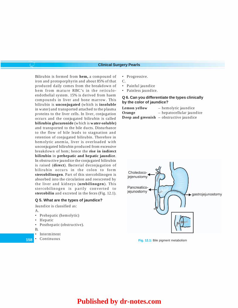
158
Clinical Surgery Pearls
Bilirubin is formed from hem, a compound ofiron and protoporphyrin and about 85% of thatproduced daily comes from the breakdown ofhem from mature RBC’s in the reticulo-endothelial system. 15% is derived from haemcompounds in liver and bone marrow. Thisbilirubin is unconjugated (which is insolublein water) and transported attached to the plasmaproteins to the liver cells. In liver, conjugationoccurs and the conjugated bilirubin is calledbilirubin glucuronide (which is water-soluble)and transported to the bile ducts. Disturbanceto the flow of bile leads to stagnation andretention of conjugated bilirubin. Therefore inhemolytic anemia, liver is overloaded withunconjugated bilirubin produced from excessivebreakdown of hem; hence the rise in indirectbilirubin in prehepatic and hepatic jaundice.In obstructive jaundice the conjugated bilirubinis raised (direct). Bacterial deconjugation ofbilirubin occurs in the colon to formstercobilinogen. Part of this stercobilinogen isabsorbed into the circulation and reexcreted bythe liver and kidneys (urobilinogen). Thisstercobilinogen is partly converted tostercobilin and excreted in the feces (Fig. 12.1).
Q 5. What are the types of jaundice?
Jaundice is classified as:A.• Prehepatic (hemolytic)• Hepatic• Posthepatic (obstructive).B.• Intermittent• Continuous
• Progressive.C.• Painful jaundice• Painless jaundice.
Q 6. Can you differentiate the types clinicallyby the color of jaundice?
Lemon yellow – hemolytic jaundiceOrange – hepatocellular jaundiceDeep and greenish – obstructive jaundice
Fig. 12.1: Bile pigment metabolism
Published by dr-notes.com
159
Obstructive Jaundice
Q 7. Clinically what are the other differen-tiating features of various jaundice?
Q 8. What are the causes for each type ofjaundice?
Clinical features of various types of jaundice
Disease Symptoms Pain Jaundice
Hemolytic General malaise, loss of weight No pain Slow onset and jaundicepersists
Infective hepatitis Loss of appetite, nausea, malaise Dull ache Gradual onset anddisappearance
Gallstone Episodes of flatulent dyspepsia Intermittent Sudden onset, fades slowlysevere pain in days
Carcinoma head Loss of weight, loss of appetite, Backache Progressive jaundiceof pancreas itching
Causes for prehepatic, hepatic and posthepatic jaundice
Prehepatic (hemolytic) Hepatic Posthepatic (obstructive)
• Hereditary spherocytosis Hepatocellular – Acute In the lumen –• Hereditary nonspherocytic viral hepatitis, alcoholic • Gallstone
anemia cirrhosis • Parasites (hydatid, liver fluke• Sickle cell disease Dubin Johnson roundworms• Thalassemia syndrome** • Foreign body – broken T tube• Acquired hemolytic anemia • Hemobilia• Hypersplenism Cholestatic – Toxic drugs, In the wall –• Crigler–Najjar syndrome* Cholestatic jaundice of • Benign stricture• Gilbert’s disease* pregnancy, postoperative • Malignant stricture
cholestatic jaundice, • Congenital atresiaBiliary cirrhosis • Traumatic strictures
• Choledochal cyst• Caroli’s disease• Tumors of bile duct• Klatskin’s tumor• Sclerosing cholangitis
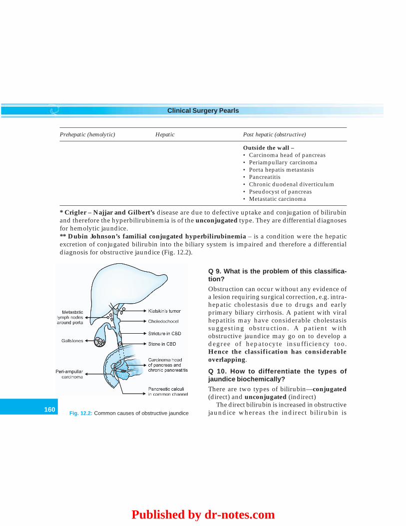
160
Clinical Surgery Pearls
Prehepatic (hemolytic) Hepatic Post hepatic (obstructive)
Outside the wall –• Carcinoma head of pancreas• Periampullary carcinoma• Porta hepatis metastasis• Pancreatitis• Chronic duodenal diverticulum• Pseudocyst of pancreas• Metastatic carcinoma
* Crigler – Najjar and Gilbert’s disease are due to defective uptake and conjugation of bilirubinand therefore the hyperbilirubinemia is of the unconjugated type. They are differential diagnosesfor hemolytic jaundice.** Dubin Johnson’s familial conjugated hyperbilirubinemia – is a condition were the hepaticexcretion of conjugated bilirubin into the biliary system is impaired and therefore a differentialdiagnosis for obstructive jaundice (Fig. 12.2).
Q 9. What is the problem of this classifica-tion?
Obstruction can occur without any evidence ofa lesion requiring surgical correction, e.g. intra-hepatic cholestasis due to drugs and earlyprimary biliary cirrhosis. A patient with viralhepatitis may have considerable cholestasissuggesting obstruction. A patient withobstructive jaundice may go on to develop adegree of hepatocyte insufficiency too.Hence the classification has considerableoverlapping.
Q 10. How to differentiate the types ofjaundice biochemically?
There are two types of bilirubin—conjugated(direct) and unconjugated (indirect)
The direct bilirubin is increased in obstructivejaundice whereas the indirect bilirubin isFig. 12.2: Common causes of obstructive jaundice
Published by dr-notes.com
161
Obstructive Jaundice
increased in hemolytic jaundice. In obstructivejaundice conjugated bilirubin from the hepato-cytes and biliary radicles overflow into thebloodstream, whereas in hemolytic jaundice,unconjugated bilirubin overloads the liver andis detected as elevated in blood.• The serum bilirubin level rarely exceeds 4-
5mg/dl in hemolytic jaundice.• The bilirubin exceed to 10-20mg/dl in
obstruction due to neoplasm.• In obstruction due to stones usually it ranges
upto 5mg, rarely it exceeds 15 mg.
Q 11. What is the role of liver enzymes in thediagnosis of jaundice?
The two most important enzymes are ALT, ASTand alkaline phosphatase (ALP).• ALT above 1000 is suggestive of viral
hepatitis• In alcoholic liver disease AST is raised (AST :
ALT is >2)• Alkaline phosphatase is raised in obstructive
jaundice (and infiltrative liver diseases liketumor or granuloma.
Note: Normal values: ALT: 5 – 35 U/LAST: 5 – 40 U/L
Q 12. What are the sources of alkalinephosphatase and what are the conditions inwhich the enzyme is increased?
There are three sources for alkaline phosphatase1. Liver Equal contribution2. Bone3. Intestine - Small contribution
• In the liver it is a product of epithelialcells of the cholangioles
• It is expressed in Bodansky units(previously KA units)
Note: Normal value - 30 – 115 U/L.
Clinical conditions in which alkalinephosphatase is increased
• Intrahepatic cholestasis• Cholangitis• Extrahepatic obstruction• Focal hepatic lesions without jaundice• Solitary metastasis• Pyogenic abscess• Granulomas• Bone tumors (primary and secondary)
Q 13. How to rule out alkaline phosphataserise due to bone pathology? (PG)
• Serum calcium and phosphorus (which willbe abnormal in bony pathology).
• 5’ nucleotidase and γγγγγ-glutamyl transpep-tidase which are specific for obstructivejaundice.
Q 14. What is acholuric jaundice?
It is nothing but the result of hereditaryspherocytosis. It is called acholuric becausethere is no excretion of bilirubin in the urine. Inthis condition, because of the hemolysis as aresult of spherocytosis, the bilirubin producedis insoluble (unconjugated). Which is not filteredby glomerulus. There will be family history ofjaundice. In addition you get—• Pigmented stones in gallbladder• Enlarged spleen• Leg ulcers.It produces hemolytic crises –• Pyrexia• Abdominal pain
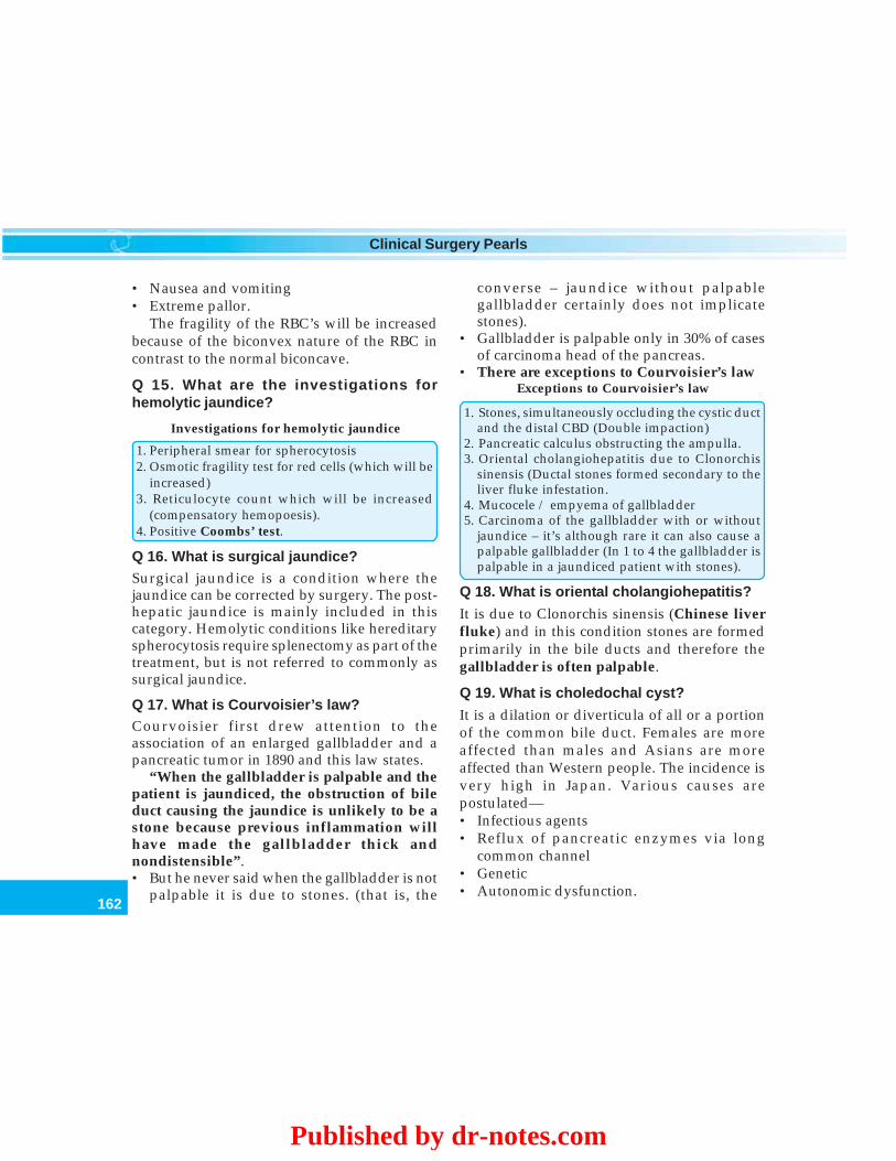
162
Clinical Surgery Pearls
• Nausea and vomiting• Extreme pallor.
The fragility of the RBC’s will be increasedbecause of the biconvex nature of the RBC incontrast to the normal biconcave.
Q 15. What are the investigations forhemolytic jaundice?
Investigations for hemolytic jaundice
1. Peripheral smear for spherocytosis2. Osmotic fragility test for red cells (which will be
increased)3. Reticulocyte count which will be increased
(compensatory hemopoesis).4. Positive Coombs’ test.
Q 16. What is surgical jaundice?
Surgical jaundice is a condition where thejaundice can be corrected by surgery. The post-hepatic jaundice is mainly included in thiscategory. Hemolytic conditions like hereditaryspherocytosis require splenectomy as part of thetreatment, but is not referred to commonly assurgical jaundice.
Q 17. What is Courvoisier’s law?
Courvoisier first drew attention to theassociation of an enlarged gallbladder and apancreatic tumor in 1890 and this law states.
“When the gallbladder is palpable and thepatient is jaundiced, the obstruction of bileduct causing the jaundice is unlikely to be astone because previous inflammation willhave made the gallbladder thick andnondistensible”.• But he never said when the gallbladder is not
palpable it is due to stones. (that is, the
converse – jaundice without palpablegallbladder certainly does not implicatestones).
• Gallbladder is palpable only in 30% of casesof carcinoma head of the pancreas.
• There are exceptions to Courvoisier’s lawExceptions to Courvoisier’s law
1. Stones, simultaneously occluding the cystic ductand the distal CBD (Double impaction)
2. Pancreatic calculus obstructing the ampulla.3. Oriental cholangiohepatitis due to Clonorchis
sinensis (Ductal stones formed secondary to theliver fluke infestation.
4. Mucocele / empyema of gallbladder5. Carcinoma of the gallbladder with or without
jaundice – it’s although rare it can also cause apalpable gallbladder (In 1 to 4 the gallbladder ispalpable in a jaundiced patient with stones).
Q 18. What is oriental cholangiohepatitis?
It is due to Clonorchis sinensis (Chinese liverfluke) and in this condition stones are formedprimarily in the bile ducts and therefore thegallbladder is often palpable.
Q 19. What is choledochal cyst?
It is a dilation or diverticula of all or a portionof the common bile duct. Females are moreaffected than males and Asians are moreaffected than Western people. The incidence isvery high in Japan. Various causes arepostulated—• Infectious agents• Reflux of pancreatic enzymes via long
common channel• Genetic• Autonomic dysfunction.
Published by dr-notes.com
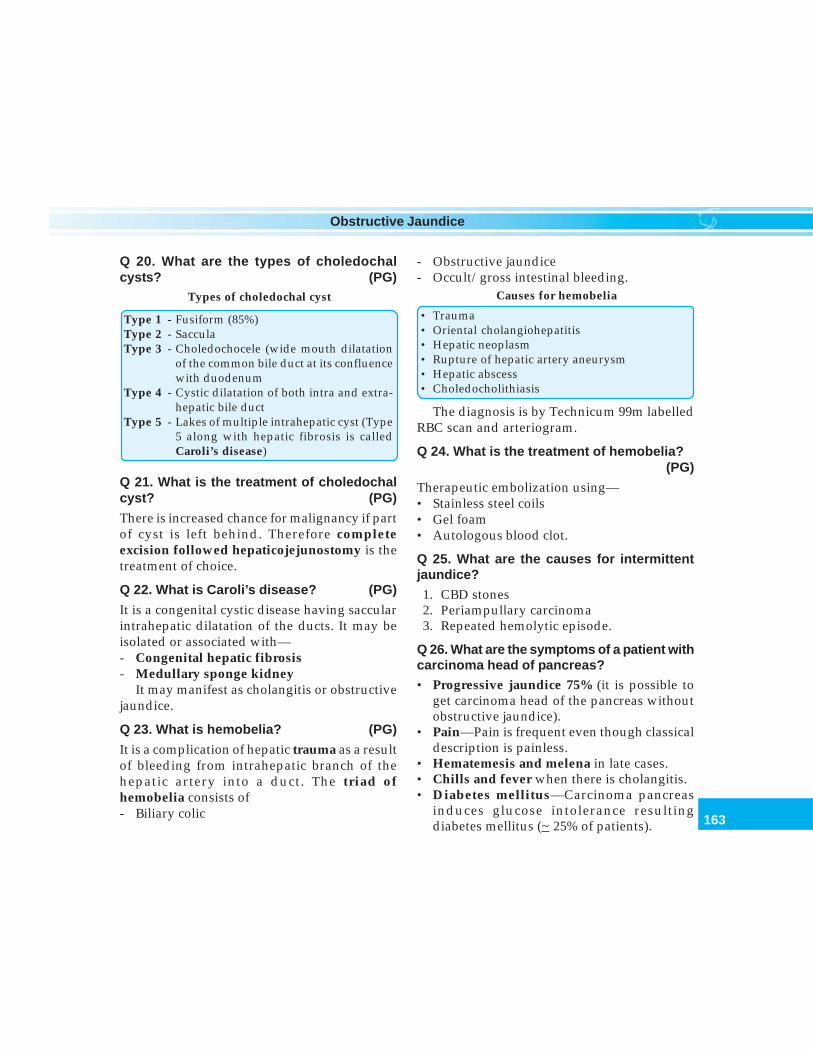
163
Obstructive Jaundice
Q 20. What are the types of choledochalcysts? (PG)
Types of choledochal cyst
Type 1 - Fusiform (85%)Type 2 - SacculaType 3 - Choledochocele (wide mouth dilatation
of the common bile duct at its confluencewith duodenum
Type 4 - Cystic dilatation of both intra and extra-hepatic bile duct
Type 5 - Lakes of multiple intrahepatic cyst (Type5 along with hepatic fibrosis is calledCaroli’s disease)
Q 21. What is the treatment of choledochalcyst? (PG)
There is increased chance for malignancy if partof cyst is left behind. Therefore completeexcision followed hepaticojejunostomy is thetreatment of choice.
Q 22. What is Caroli’s disease? (PG)
It is a congenital cystic disease having saccularintrahepatic dilatation of the ducts. It may beisolated or associated with—- Congenital hepatic fibrosis- Medullary sponge kidney
It may manifest as cholangitis or obstructivejaundice.
Q 23. What is hemobelia? (PG)
It is a complication of hepatic trauma as a resultof bleeding from intrahepatic branch of thehepatic artery into a duct. The triad ofhemobelia consists of- Biliary colic
- Obstructive jaundice- Occult/gross intestinal bleeding.
Causes for hemobelia
• Trauma• Oriental cholangiohepatitis• Hepatic neoplasm• Rupture of hepatic artery aneurysm• Hepatic abscess• Choledocholithiasis
The diagnosis is by Technicum 99m labelledRBC scan and arteriogram.
Q 24. What is the treatment of hemobelia?(PG)
Therapeutic embolization using—• Stainless steel coils• Gel foam• Autologous blood clot.
Q 25. What are the causes for intermittentjaundice?
1. CBD stones2. Periampullary carcinoma3. Repeated hemolytic episode.
Q 26. What are the symptoms of a patient withcarcinoma head of pancreas?
• Progressive jaundice 75% (it is possible toget carcinoma head of the pancreas withoutobstructive jaundice).
• Pain—Pain is frequent even though classicaldescription is painless.
• Hematemesis and melena in late cases.• Chills and fever when there is cholangitis.• Diabetes mellitus—Carcinoma pancreas
induces glucose intolerance resultingdiabetes mellitus (~ 25% of patients).
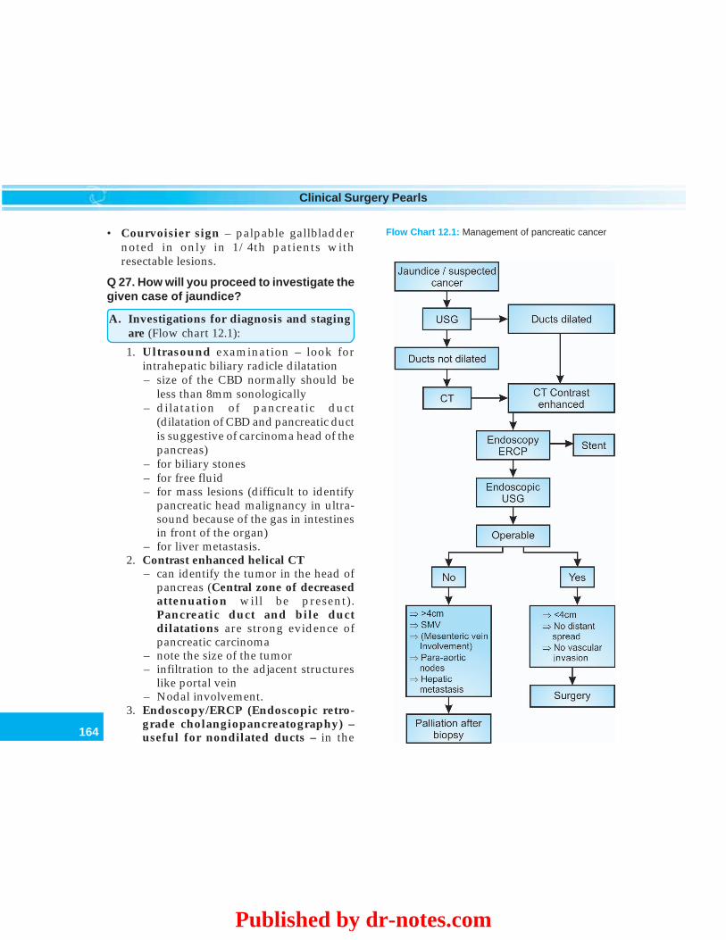
164
Clinical Surgery Pearls
• Courvoisier sign – palpable gallbladdernoted in only in 1/4th patients withresectable lesions.
Q 27. How will you proceed to investigate thegiven case of jaundice?
A. Investigations for diagnosis and stagingare (Flow chart 12.1):
1. Ultrasound examination – look forintrahepatic biliary radicle dilatation– size of the CBD normally should be
less than 8mm sonologically– dilatation of pancreatic duct
(dilatation of CBD and pancreatic ductis suggestive of carcinoma head of thepancreas)
– for biliary stones– for free fluid– for mass lesions (difficult to identify
pancreatic head malignancy in ultra-sound because of the gas in intestinesin front of the organ)
– for liver metastasis.2. Contrast enhanced helical CT
– can identify the tumor in the head ofpancreas (Central zone of decreasedattenuation will be present).Pancreatic duct and bile ductdilatations are strong evidence ofpancreatic carcinoma
– note the size of the tumor– infiltration to the adjacent structures
like portal vein– Nodal involvement.
3. Endoscopy/ERCP (Endoscopic retro-grade cholangiopancreatography) –useful for nondilated ducts – in the
Flow Chart 12.1: Management of pancreatic cancer
Published by dr-notes.com
165
Obstructive Jaundice
absence of mass in the CT, ERCP isindicated. It is the most sensitive test forsuch situations.• The periampullary carcinoma can be
visualized• Biopsy can be taken from the
periampullary lesion• The lower limit of the growth can be
identified by ERCP• Stenting can be done to relieve the
jaundice• Sphincterotomy can be done to
remove the bile duct stone.4. Endoscopic ultrasound
• To assess the operability• Size of the tumor• To know the involvement of
mesenteric lymph nodes, paraaorticnodes and superior mesenteric vein
• Hepatic metastasis.5. Percutaneous transhepatic cholangio-
graphy (PTC) – useful for dilated ductalsystem.• The coagulation profile is checked and
antibiotic coverage is given. In thisprocedure a pliable needle is insertedunder local anesthetic and sonologicalguidance into the dilated biliary duct.Should decompression of the biliarytree be desired pre operatively, then afine bore catheter may be left in theducts to allow continuous drainage ofbile.
• It is also possible to insert an endo-prosthesis to relieve the obstructionfor unresectable lesions.
• The PTC is ideal for demonstrating the
anatomy above an extrahepaticobstruction and the upper limit of theobstructing lesion can be identified byinjecting dye.
6. Upper GI series – to decide whether agastrojejunostomy is indicated or not.The classical findings are (largelyreplaced by Helical CT).• Widening of the C-loop of the
duodenum in carcinoma head of thepancreas.
• Reverse 3 sign in periampullarycarcinoma.
B. Biochemical investigations– Total bilirubin, direct and indirect
(direct is increased obstructive andindirect is increased in hemolyticjaundice (total and direct bilirubin raisedin hepatic).
– Alkaline phosphatase – (over 100international unit is indicative ofcholestasis if bone disease is absent).
– Total protein, albumin and globulin(albumin and globulin levels are reversedin chronic hepatocellular damage).Globulin level will be very high in biliarycirrhosis. In biliary obstruction per sethere won’t be any change in the values.
– Serum transaminase levels are abovenormal in viral hepatitis.
C. Coagulation profile – Prothrombin Time– normal in hemolytic jaundice. Prolongedbut correctable with vitamin K incholestatic jaundice (provided theirremains some functioning liver tissue). Thiswill be prolonged and not correctable in
166
Clinical Surgery Pearls
advanced hepatocellular disease.– Platelet count, bleeding time and
clotting time.D. Urine examination
1. Absent urobilinogen indicates obs-truction to the common bile ducts.
2. Excess urobilinogen occurs in hemolyticjaundice and some times in liver damage.
3. Absent bilirubin indicates hemolyticjaundice (Bilirubin predominantlyunconjugated).
4. Excess bilirubin is present in obstructivejaundice.
E. Stool examination1. Absence of bile pigment indicates biliary
obstruction (Clay-colored stool).2. Positive occult blood indicates ampullary
carcinoma, bleeding esophageal varicesand alimentary carcinoma.
Q 28. What is the dose of vitamin K and howis it given?
The dose of vitamin K is 10 mg and it is givenIM for 3 days.
The vitamin K1 (phytomenodione) is givenintravenously—10 mg daily for 3 days.
Q 29. What are the preoperative preparationsif you are planning for surgery?
1. Hydration2. Nutrition3. Correction of coagulation deficiency—
vitamin K, FFP, platelet transfusion, etc.4. Correction of anemia
Q 30. What are the indications for preoperative biliary decompression? (PG)
Indications for preoperative biliarydecompression
• Bilirubin > 12 mg• Sepsis• Hepatorenal failure• Severe cardiopulmonary disease• Severe malnutrition
NB: Routine drainage is not recommended.
Q 31. What is the other simple option fordrainage?
Simple cholecystomy if other options are notavailable.
Q 32. What are the causes for fever injaundice?
Causes for fever in jaundice
1. Cholangitis 2. Hemolysis3. Septicemia 4. Hepatic abscess
Q 33. What are the 3 important special risksin obstructive jaundice? (PG)
1. Hypocoagulability2. Renal failure – may be due to increased bile
pigment load on the tubules, increased post-operative distal tubular reabsorption ofwater and failure of liver to trap entericnephrotoxins
3. Sepsis of bile with or without calculi.
Q 34. What are the signs of unresectability incarcinoma head of the pancreas?
1. Hepatic metastasis2. Distant metastasis (Distant lymph nodes
from pancreatic head)3. Involvement of superior mesenteric vessels
and portal vein (Minimal invasion is notcontraindication)
Published by dr-notes.com
167
Obstructive Jaundice
4. Ascitis (Peritoneal metastasis)5. Encasement of superior mesenteric, hepatic
or coeliac artery by tumor.
Q 35. Should portal vein involvementpreclude a resection? (PG)
Involvement of a short segment (< 1.5 cm) ofportal vein is not a contraindication to a curativeresection.
Q 36. What is the treatment of itching due toobstructive jaundice? (PG)
Cholestyramine – It is an ion exchange resin,usually provides relief by binding bile salts inthe intestinal lumen, thus inhibiting theirreabsorption into blood.
Q 37. What is the role of percutaneous biopsy(Aspiration) of pancreatic tumor? (PG)
• It is done under ultrasound/CT guidance• It is contraindicated in candidates for
surgery—risk of spreading• Done in patients with radiographic evidence
of unresectability• It is also done per operatively in operable
cases• Biopsy may miss the lesion because a variable
area of pancreatitis there surround the tumor,leading to sampling error.
Q 38. What are the complications of needlebiopsy of pancreatic tumor? (PG)
• Pancreatitis• Fistula• Hemorrhage• Infection.
Q 39. What is the tumor marker for pancreaticmalignancy? (PG)
• CA 19-9 (Carbohydrate antigen) – it is usefulin following the results of the treatment
• CEA.
Q 40. Peroperatively can you differentiatechronic pancreatitis from carcinoma? (PG)
• It is very difficult.• A hard noncystic mass in the head +
obstructive jaundice + dilatation of CBD ismore in favor of carcinoma.
• A hard noncystic mass in the retroampullarypart + no jaundice + no dilatation is in favorof chronic pancreatitis.
Q 41. What are the signs of inoperability incarcinoma head of pancreas?
Signs of inoperability in carcinoma headof pancreas
Clinically• Supraclavicular nodes• Ascitis• Rectal shelf of Blumer• Liver metastasis, etc.
Peroperatively• Ascitis• Liver metastasis• Peritoneal metastasis• Rectal shelf of Blumer• Involvement of the portal vein and superior
mesenteric vessels (peroperatively assess theresectability after Kocherising the duodenum andpassing the surgeons finger between the pancreasand the portal vein).
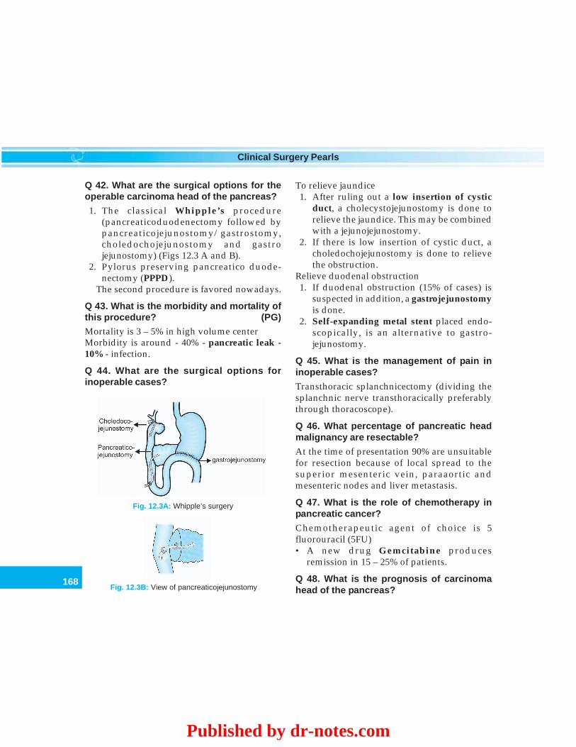
168
Clinical Surgery Pearls
To relieve jaundice1. After ruling out a low insertion of cystic
duct, a cholecystojejunostomy is done torelieve the jaundice. This may be combinedwith a jejunojejunostomy.
2. If there is low insertion of cystic duct, acholedochojejunostomy is done to relievethe obstruction.
Relieve duodenal obstruction1. If duodenal obstruction (15% of cases) is
suspected in addition, a gastrojejunostomyis done.
2. Self-expanding metal stent placed endo-scopically, is an alternative to gastro-jejunostomy.
Q 45. What is the management of pain ininoperable cases?
Transthoracic splanchnicectomy (dividing thesplanchnic nerve transthoracically preferablythrough thoracoscope).
Q 46. What percentage of pancreatic headmalignancy are resectable?
At the time of presentation 90% are unsuitablefor resection because of local spread to thesuperior mesenteric vein, paraaortic andmesenteric nodes and liver metastasis.
Q 47. What is the role of chemotherapy inpancreatic cancer?
Chemotherapeutic agent of choice is 5fluorouracil (5FU)• A new drug Gemcitabine produces
remission in 15 – 25% of patients.
Q 48. What is the prognosis of carcinomahead of the pancreas?
Fig. 12.3A: Whipple’s surgery
Fig. 12.3B: View of pancreaticojejunostomy
Q 42. What are the surgical options for theoperable carcinoma head of the pancreas?
1. The classical Whipple’s procedure(pancreaticoduodenectomy followed bypancreaticojejunostomy/gastrostomy,choledochojejunostomy and gastrojejunostomy) (Figs 12.3 A and B).
2. Pylorus preserving pancreatico duode-nectomy (PPPD).
The second procedure is favored nowadays.
Q 43. What is the morbidity and mortality ofthis procedure? (PG)
Mortality is 3 – 5% in high volume centerMorbidity is around - 40% - pancreatic leak -10% - infection.
Q 44. What are the surgical options forinoperable cases?
Published by dr-notes.com
169
Obstructive Jaundice
• The overall median survival is only 20 weeks• Less than 3% of patients survive for 5years• In carcinoma of the ampulla of Vater, the 5
years survival after resection is 40%.
OBSTRUCTIVE JAUNDICE DUE TO STONES
Q 49. What are the manifestations of CBDstones?
The classical presentation is Charcot’s triadconsisting of—• Intermittent pain• Intermittent fever• Intermittent jaundice.
Q 50. What is cholangitis?
When there is obstruction of bile duct will leadon to bacterial infection of the bile duct.Obstruction is synonymous with infection. Theobstruction may be partial or complete. Thecauses for cholangitis are—• Choledocholithiasis• Biliary stricture• Neoplasms (15%)• Ampullary stenosis (less common)• Chronic pancreatitis• Pseudocyst• Duodenal diverticulum• Parasitic
Q 51. What is the pathology of cholangitis ?(PG)
• The ductal pressure increases when there isobstruction
• Bacteria escape to the systemic circulation viahepatic sinusoids
• Organism will reach the blood and bloodculture will positive.
Q 52. What are the common organisms forcholangitis ? (PG)
Organisms causing cholangitis
• E. Coli• Klebsiella• Pseudomonas• Enterococci• Proteus• Bacteroides
Q 53. What is Reynold’s pentad?
It is a manifestation of acute toxic cholangitisas a result of obstruction of the bile duct (It isalso called suppurative cholangitis). It is a life-threatening condition and should be managedas emergency. The Pentad consists of—• Pain• Fever• Jaundice• Mental confusion• Shock.
Q 54. What is the management of acute toxiccholangitis? (PG)
It is an emergency. The antibiotic of choice isaminoglycoside and clindamycin/metro-nidazole. The ductal system is decompressed byemergency sphincterotomy or by transhepaticdrainage. The associated gallbladder pathologymay be tackled as an elective procedure laterby laparoscopic cholecystectomy.
Q 55. What is the management of obstructivejaundice due to CBD stone?
The investigations, and preparations are thesame as for obstructive jaundice. (which is givenabove). There are 3 surgical options –
170
Clinical Surgery Pearls
1. Endoscopic sphincterotomy initially,followed by laparoscopic cholecystectomypreferably in the same admission. (currentlyfavored where laparoscopic expertise is notavailable).
2. Laparoscopic cholecystectomy andlaparoscopic CBD exploration (if expertiseis available); the CBD exploration may bedone through the cystic duct or if the ductis more than 1.5cm through acholedochotomy.
3. Open—Laparotomy, cholecystectomy +CBD exploration and stone removal.
Q 56. What are the contraindications forsphincterotomy?
• Stone size more than 2 cm.• Stenosis of bile duct proximal to the
sphincter.
Q 57. What is the management of bile ducttumor? (PG)
The bile duct tumors may be benign ormalignant. The benign tumors are rare and farless common. The malignant tumors are calledcholangiocarcinomas. Cholangiocarcinoma is aterm now applied to malignant intrahepatic,perihilar and distal extrahepatic tumors of thebile ducts. The incidence of this condition isrising. The management may be palliativestenting or surgery. Most patients will receiveonly palliative surgery. The consensus ofsurgical opinion is that the surgical treatmentis difficult and should be reserved for thefittest patients getting 5mm clearance, node andliver resection if required. In all cases
reconstruction is made using hepatico-jejunostomy (Roux–en–Y). On the right side anextended right hepatic lobectomy is done dueto anatomical reasons. The length of right hepaticduct is smaller in contrast to the left which hasgot significant length of extrahepatic duct.
Q 58. What are the risk factors forcholangiocarcinoma? (PG)
These include –Risk factors for cholangiocarcinoma
• Sclerosing cholangitis• Chronic intrahepatic gallstones• Caroli’s disease• Choledochal cyst• Ulcerative colitis• Liver flukes
Possible association:• INH• Methyldopa• Oral contraceptives• Asbestos.
Q 59. What is Klatskin tumor? (PG)
The perihilar cholangiocarcinoma is calledKlatskin tumor. About 50% of all cholangiocarcinomas are Klatskin tumor. Of the reminder½ are intrahepatic and ½ are related to extrahepatic bile ducts.
Q 60. Is there any role for fine needleaspiration cytology? (PG)
Percutaneous fine needle aspiration cytology isabsolutely contraindicated in patients who maybe operable.
Published by dr-notes.com
171
Obstructive Jaundice
Q 61. What is the Bismuth classification ofcholangiocarcinoma? (PG)
Type I - involving the common hepatic duct.Type II – the growth involving the proximalcommon hepatic duct which extends to thebifurcation, but both hepatic ducts are free.Type III – growth involving one hepatic duct.Type IV – is the hilar involving both hepatic andproximal common hepatic ducts.
Q 62. What are the contraindications forsurgery? (PG)
• Lymph node involvement• Encasement of hepatic hilar vessels.
Q 63. What is Taj Mahal resection? (PG)
The technique of central liver resection forcholangiocarcinoma is called Taj Mahal
resection owing to the shape of the defect in theliver. This involves removal of segments 1, 4 and5 and the relevant bile ducts.
Q 64. What is the palliative surgicaltreatment? (PG)
Palliative surgical drainage is done byanastomosing a Roux loop to the segment IIIduct.
Q 65. Is there any role for surgery inhereditary spherocytosis?
• Splenectomy is done• Gallbladder stones if present is managed by
cholecystectomy.
Q 66. What type of gallstone you get inhereditary spherocytosis?
Pigment stone.
172
Clinical Surgery Pearls
13 Varicose Veins
CASE CAPSULE
Middle-aged multiparous female patient presentswith dilated tortuous veins on both lower limbs of10years duration. She complains of dull ache feltin the calves and lower leg that get worse throughthe day especially during standing. It is relievedby lying down for 30 minutes. She also complaintsof mild swelling of the ankle by the end of the day.She does not give history of previous accidents,major operations or prolonged illness. Hermother and elder sister had similar complaints.
On inspection, large tortuous visible veins arenoted in the distribution of long saphenous vein.Skin of the lower third of the medial side of the rightleg shows brown pigmentation and there is an ulcersituated just above the medial malleolus of the sizeof 7.5 × 5cm. On palpation there is pitting edemaof the right ankle region. The skin and subcutaneoustissue are thickened over the area. Palpation alongthe course of the long saphenous vein behindthe medial border of the tibia revealed threedefects in the deep fascia 5, 10 and 15cm above themedial malleolus. There is a palpable coughimpulse in the groin at the saphenous opening
region. The veins collapse when the patient liesdown and the limb is elevated. Trendelenburg testI and II are positive on the right side and test Ipositive on left side. Modified Perthes test isnegative on both sides. Multiple tourniquet testshows perforator incompetence in the right legcorresponding to the fascial defects mentionedabove. Schwartz test is positive on right side.There is no bruit heard over the veins.
The ulcer is of the size of 7.5 × 5cm with asloping edge and the surrounding skin is purpleblue in color. The floor of the ulcer is coveredwith pink granulation tissue. The base of theulcer is fixed to the deeper tissues. There is serousdischarge from the ulcer. Equinus deformity ispresent at the ankle joint. Regional lymph nodesare enlarged, firm in consistency and mobile (thevertical group of inguinal nodes on right side). Onabdominal examination there is no vein seencrossing the abdomen and there is no masslesions. Per rectal and per vaginal examinationshows no pelvic cause for the dilated tortuousveins. Examination of the peripheral vascularsystem is normal.
Published by dr-notes.com
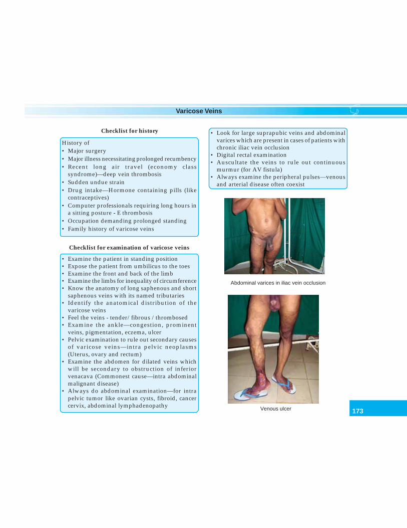
173
Varicose Veins
Checklist for history
History of• Major surgery• Major illness necessitating prolonged recumbency• Recent long air travel (economy class
syndrome)—deep vein thrombosis• Sudden undue strain• Drug intake—Hormone containing pills (like
contraceptives)• Computer professionals requiring long hours in
a sitting posture - E thrombosis• Occupation demanding prolonged standing• Family history of varicose veins
Checklist for examination of varicose veins
• Examine the patient in standing position• Expose the patient from umbilicus to the toes• Examine the front and back of the limb• Examine the limbs for inequality of circumference• Know the anatomy of long saphenous and short
saphenous veins with its named tributaries• Identify the anatomical distribution of the
varicose veins• Feel the veins - tender/fibrous /thrombosed• Examine the ankle—congestion, prominent
veins, pigmentation, eczema, ulcer• Pelvic examination to rule out secondary causes
of varicose veins—intra pelvic neoplasms(Uterus, ovary and rectum)
• Examine the abdomen for dilated veins whichwill be secondary to obstruction of inferiorvenacava (Commonest cause—intra abdominalmalignant disease)
• Always do abdominal examination—for intrapelvic tumor like ovarian cysts, fibroid, cancercervix, abdominal lymphadenopathy
• Look for large suprapubic veins and abdominalvarices which are present in cases of patients withchronic iliac vein occlusion
• Digital rectal examination• Auscultate the veins to rule out continuous
murmur (for AV fistula)• Always examine the peripheral pulses—venous
and arterial disease often coexist
Abdominal varices in iliac vein occlusion
Venous ulcer
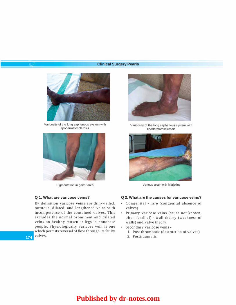
174
Clinical Surgery Pearls
Varicosity of the long saphenous system withlipodermatosclerosis
Venous ulcer with MarjolinsPigmentation in gaiter area
Varicosity of the long saphenous system withlipodermatosclerosis
Q 1. What are varicose veins?
By definition varicose veins are thin-walled,tortuous, dilated, and lengthened veins withincompetence of the contained valves. Thisexcludes the normal prominent and dilatedveins on healthy muscular legs in nonobesepeople. Physiologically varicose vein is onewhich permits reversal of flow through its faultyvalves.
Q 2. What are the causes for varicose veins?
• Congenital - rare (congenital absence ofvalves)
• Primary varicose veins (cause not known,often familial) - wall theory (weakness ofwalls) and valve theory
• Secondary varicose veins -1. Post thrombotic (destruction of valves)2. Posttraumatic
Published by dr-notes.com
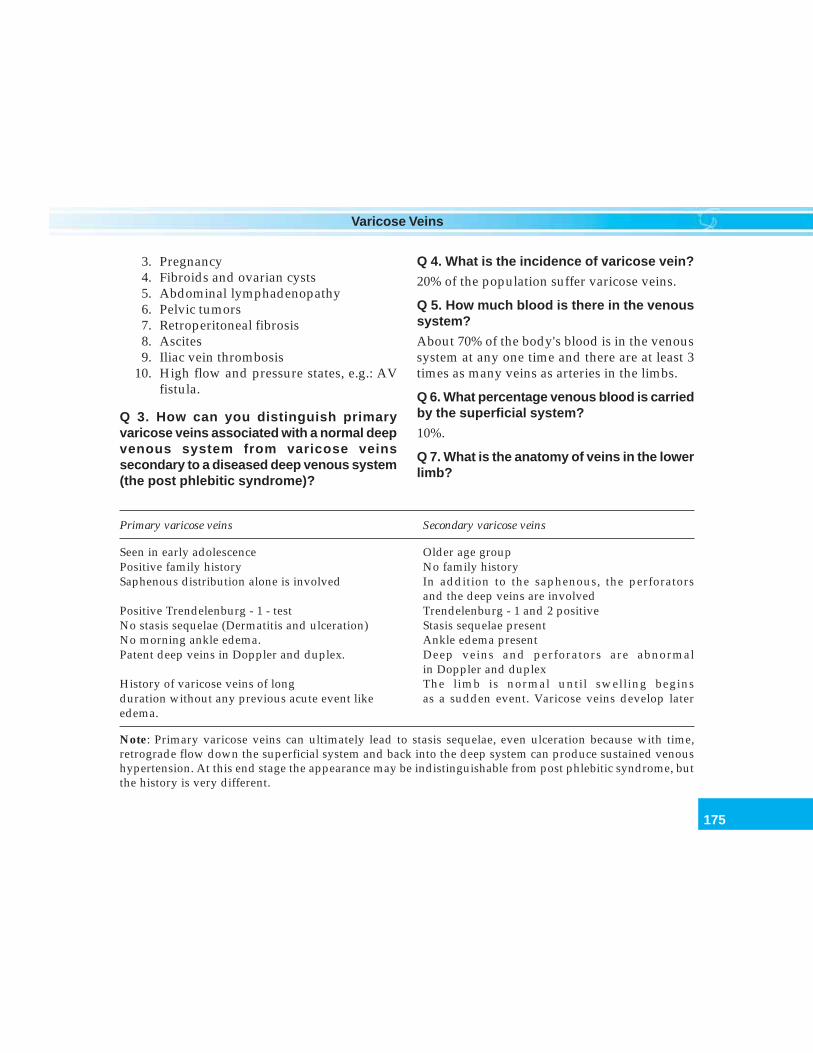
175
Varicose Veins
Primary varicose veins Secondary varicose veins
Seen in early adolescence Older age groupPositive family history No family historySaphenous distribution alone is involved In addition to the saphenous, the perforators
and the deep veins are involvedPositive Trendelenburg - 1 - test Trendelenburg - 1 and 2 positiveNo stasis sequelae (Dermatitis and ulceration) Stasis sequelae presentNo morning ankle edema. Ankle edema presentPatent deep veins in Doppler and duplex. Deep veins and perforators are abnormal
in Doppler and duplexHistory of varicose veins of long The limb is normal until swelling beginsduration without any previous acute event like as a sudden event. Varicose veins develop lateredema.
Note: Primary varicose veins can ultimately lead to stasis sequelae, even ulceration because with time,retrograde flow down the superficial system and back into the deep system can produce sustained venoushypertension. At this end stage the appearance may be indistinguishable from post phlebitic syndrome, butthe history is very different.
Q 4. What is the incidence of varicose vein?
20% of the population suffer varicose veins.
Q 5. How much blood is there in the venoussystem?
About 70% of the body's blood is in the venoussystem at any one time and there are at least 3times as many veins as arteries in the limbs.
Q 6. What percentage venous blood is carriedby the superficial system?
10%.
Q 7. What is the anatomy of veins in the lowerlimb?
3. Pregnancy4. Fibroids and ovarian cysts5. Abdominal lymphadenopathy6. Pelvic tumors7. Retroperitoneal fibrosis8. Ascites9. Iliac vein thrombosis
10. High flow and pressure states, e.g.: AVfistula.
Q 3. How can you distinguish primaryvaricose veins associated with a normal deepvenous system from varicose veinssecondary to a diseased deep venous system(the post phlebitic syndrome)?
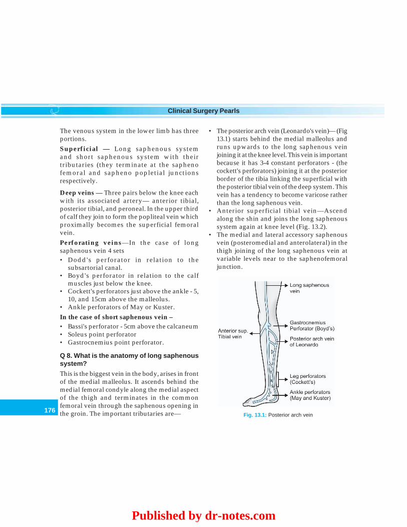
176
Clinical Surgery Pearls
The venous system in the lower limb has threeportions.Superficial — Long saphenous systemand short saphenous system with theirtributaries (they terminate at the saphenofemoral and sapheno popletial junctionsrespectively.
Deep veins — Three pairs below the knee eachwith its associated artery— anterior tibial,posterior tibial, and peroneal. In the upper thirdof calf they join to form the popliteal vein whichproximally becomes the superficial femoralvein.Perforating veins—In the case of longsaphenous vein 4 sets• Dodd's perforator in relation to the
subsartorial canal.• Boyd's perforator in relation to the calf
muscles just below the knee.• Cockett's perforators just above the ankle - 5,
10, and 15cm above the malleolus.• Ankle perforators of May or Kuster.In the case of short saphenous vein –• Bassi's perforator - 5cm above the calcaneum• Soleus point perforator• Gastrocnemius point perforator.
Q 8. What is the anatomy of long saphenoussystem?
This is the biggest vein in the body, arises in frontof the medial malleolus. It ascends behind themedial femoral condyle along the medial aspectof the thigh and terminates in the commonfemoral vein through the saphenous opening inthe groin. The important tributaries are— Fig. 13.1: Posterior arch vein
• The posterior arch vein (Leonardo's vein)— (Fig13.1) starts behind the medial malleolus andruns upwards to the long saphenous veinjoining it at the knee level. This vein is importantbecause it has 3-4 constant perforators - (thecockett's perforators) joining it at the posteriorborder of the tibia linking the superficial withthe posterior tibial vein of the deep system. Thisvein has a tendency to become varicose ratherthan the long saphenous vein.
• Anterior superficial tibial vein—Ascendalong the shin and joins the long saphenoussystem again at knee level (Fig. 13.2).
• The medial and lateral accessory saphenousvein (posteromedial and anterolateral) in thethigh joining of the long saphenous vein atvariable levels near to the saphenofemoraljunction.
Published by dr-notes.com
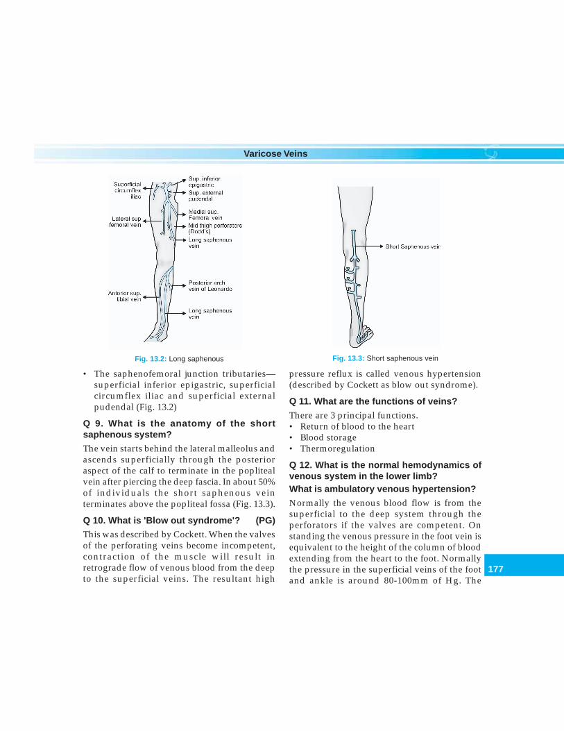
177
Varicose Veins
pressure reflux is called venous hypertension(described by Cockett as blow out syndrome).
Q 11. What are the functions of veins?
There are 3 principal functions.• Return of blood to the heart• Blood storage• Thermoregulation
Q 12. What is the normal hemodynamics ofvenous system in the lower limb?What is ambulatory venous hypertension?
Normally the venous blood flow is from thesuperficial to the deep system through theperforators if the valves are competent. Onstanding the venous pressure in the foot vein isequivalent to the height of the column of bloodextending from the heart to the foot. Normallythe pressure in the superficial veins of the footand ankle is around 80-100mm of Hg. The
Fig. 13.2: Long saphenous
• The saphenofemoral junction tributaries—superficial inferior epigastric, superficialcircumflex iliac and superficial externalpudendal (Fig. 13.2)
Q 9. What is the anatomy of the shortsaphenous system?
The vein starts behind the lateral malleolus andascends superficially through the posterioraspect of the calf to terminate in the poplitealvein after piercing the deep fascia. In about 50%of individuals the short saphenous veinterminates above the popliteal fossa (Fig. 13.3).
Q 10. What is 'Blow out syndrome'? (PG)
This was described by Cockett. When the valvesof the perforating veins become incompetent,contraction of the muscle will result inretrograde flow of venous blood from the deepto the superficial veins. The resultant high
Fig. 13.3: Short saphenous vein
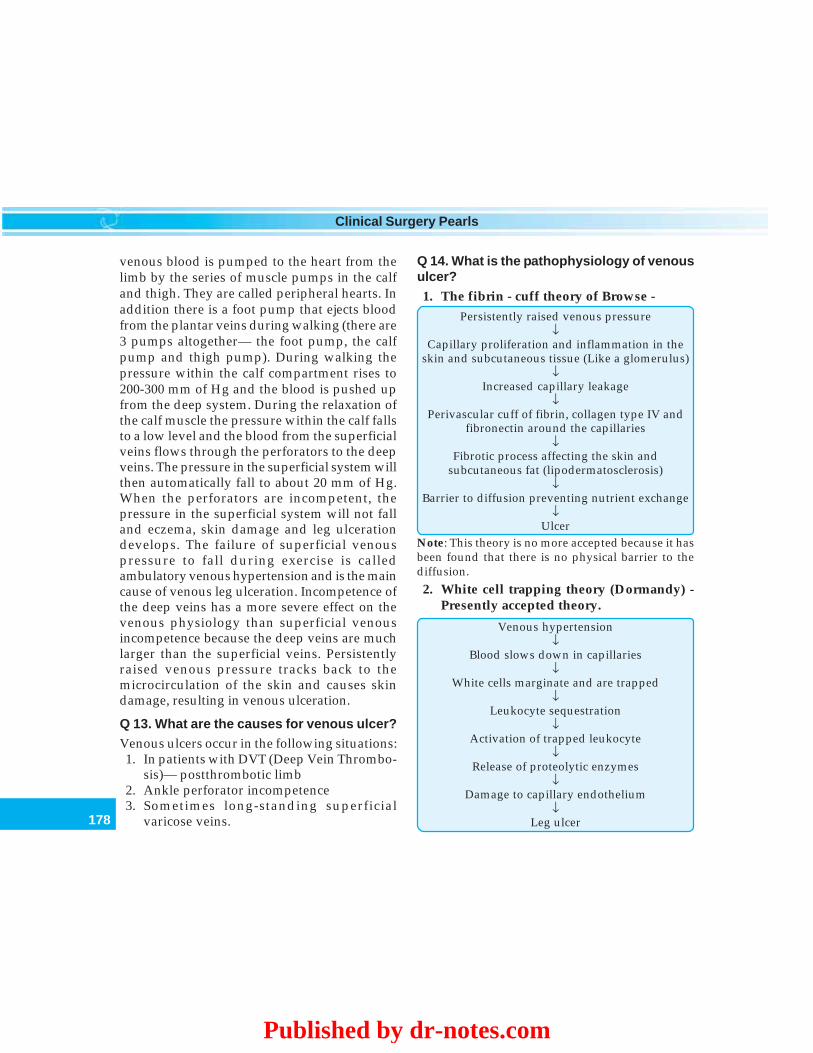
178
Clinical Surgery Pearls
venous blood is pumped to the heart from thelimb by the series of muscle pumps in the calfand thigh. They are called peripheral hearts. Inaddition there is a foot pump that ejects bloodfrom the plantar veins during walking (there are3 pumps altogether— the foot pump, the calfpump and thigh pump). During walking thepressure within the calf compartment rises to200-300 mm of Hg and the blood is pushed upfrom the deep system. During the relaxation ofthe calf muscle the pressure within the calf fallsto a low level and the blood from the superficialveins flows through the perforators to the deepveins. The pressure in the superficial system willthen automatically fall to about 20 mm of Hg.When the perforators are incompetent, thepressure in the superficial system will not falland eczema, skin damage and leg ulcerationdevelops. The failure of superficial venouspressure to fall during exercise is calledambulatory venous hypertension and is the maincause of venous leg ulceration. Incompetence ofthe deep veins has a more severe effect on thevenous physiology than superficial venousincompetence because the deep veins are muchlarger than the superficial veins. Persistentlyraised venous pressure tracks back to themicrocirculation of the skin and causes skindamage, resulting in venous ulceration.
Q 13. What are the causes for venous ulcer?
Venous ulcers occur in the following situations:1. In patients with DVT (Deep Vein Thrombo-
sis)— postthrombotic limb2. Ankle perforator incompetence3. Sometimes long-standing superficial
varicose veins.
Q 14. What is the pathophysiology of venousulcer?
1. The fibrin - cuff theory of Browse -
Persistently raised venous pressure↓
Capillary proliferation and inflammation in theskin and subcutaneous tissue (Like a glomerulus)
↓Increased capillary leakage
↓Perivascular cuff of fibrin, collagen type IV and
fibronectin around the capillaries↓
Fibrotic process affecting the skin andsubcutaneous fat (lipodermatosclerosis)
↓Barrier to diffusion preventing nutrient exchange
↓Ulcer
Note: This theory is no more accepted because it hasbeen found that there is no physical barrier to thediffusion.
2. White cell trapping theory (Dormandy) -Presently accepted theory.
Venous hypertension↓
Blood slows down in capillaries↓
White cells marginate and are trapped↓
Leukocyte sequestration↓
Activation of trapped leukocyte↓
Release of proteolytic enzymes↓
Damage to capillary endothelium↓
Leg ulcer
Published by dr-notes.com
179
Varicose Veins
Q 15. What is venous claudication?
After deep vein thrombosis recanalization willusually occur. When recanalization fails to occurafter ileofemoral venous thrombosis, venouscollaterals develop to bypass the obstruction tothe venous outflow. These collaterals usuallysuffice while the patient is at rest. However, anyleg exercise induces increased arterial inflow tothe lower extremities along with acommensurate increase in venous outflowrequirements. This may exceed the capacity ofthe venous collateral bed resulting in venoushypertension. The pressure built up ismanifested as tight or bursting pain. Relief isobtained with rest but not as prompt as inarterial claudication. This pain is seen afterwalking and towards the evening.
Q 17. What are thread veins (dermal flares)?
They are smaller veins of 0.5mm diameter seenin the skin in varicosity. They are ̀ usually purpleor red in color. These tiny veins are associatedwith superficial venous incompetence in 30%of cases.
Q 18. What are reticular veins?
Reticular veins are small vessels of 1-3 mm in
diameter lying immediately beneath the skin.They may present as small varices. These tinyvarices are associated with superficial venousincompetence in about 30% of cases.
Q 19. What are the symptoms of varicoseveins?
Symptoms of venous insufficiency
1. Asymptomatic2. Cosmetic problems3. Aching4. Heaviness and cramps5. Itching—especially on standing (the whole
lower leg may itch)6. Venous claudication7. Ankle swelling - towards the end of the day8. Pigmentation on the medial aspect of lower leg9. Eczema
10. Ulcer - on the medial aspect of lower leg (gaiterarea)
Note:1. Aching, swelling, cramps, heaviness, itching are
absent in the morning and become evident andaggravates as the day goes by
2. The severity of the symptoms is unrelated tothe size of the veins and is often more severeduring the early stages of the development ofvarices.
Based on symptoms Patients may be classified into 3 groups—
Group I (Cosmetic only) Group II (Symptomatic) Group III (Complications)
• Venous telangiectasia • Ache/cramps/tenderness • Bleeding from trauma• Thread veins (0.5 mm) • Heaviness/swelling • Superficial phlebitis• Reticular varices (1-3 mm) • Champagne bottle leg • Ankle venous flare/edema• Visible varicose veins • Itch/restless legs/paresthesia • Atrophie blanche
• Venous eczema• Lipodermatosclerosis• Venous ulcers
180
Clinical Surgery Pearls
Q 20. What is the cause for ankle edema?
Ankle edema is a feature of persistent venousobstruction. In some patients veins remainpermanently blocked following a deep veinthrombosis. This causes symptoms which areworse than venous valvular incompetence. Thepassage of time will allow recanalization andthe ankle edema may become less. Therecanalized veins are likely to be incompetentand the features of venous hypertension maythen predominate.
Q 21. From the location of varicose veins canyou identify the system affected?
1. Varicosities of the medial calf and thigh →originate from great saphenous vein.
2. Varicosities on the posterior and lateral calfand popliteal fossa→ short saphenoussystem.
3. Varicosities in the groin and along theanterior abdominal wall→ iliac veinobstruction.
Q 22. What is Klippel - Trenaunay syndrome?
In this condition you get prominent veins alongthe lateral aspect of thigh mostly present fromvery young age, with dysplastic deep veins. Theclassical clinical triad consists of —a. Hemangiomasb. Hypertrophy of soft tissue and over growth
of extremityc. Varicose vein.
Q 23. What is postphlebitic syndrome?
It includes a chronically swollen limb withhyperpigmentation, lipodermatosclerosis andnonhealing ulcers with or without varicose
veins around the ankle. One of the commonpresentations is a patient with chronic ulceraround the ankle. A definite history of DVT maynot be present in most of these patients. Almostall of them will have destroyed deep veins dueto the previous DVT.
Q 24. What are the clinical tests to be donein a patient suspected with varicose veins?
Tests for varicose veins
1. Morrissey's cough impulse test2. Brodie - Trendelenburg test - 1 and 23. Modified Perthes' test4. Multiple tourniquet test5. Shwartz's test - (tap sign)6. Pratt's test7. Fegan's test8. Assessment of the short saphenous vein
Q 25. What is Raju's test? (PG)
This is a useful test for venous obstruction. Withpatient lying supine, pressure is measured in theveins of hand and foot. Normally the footpressure is equal to or slightly higher (by 5mmof Hg) than the hand pressure. In venous obstruc-tion this difference is more (10-15mm of Hg).
Note: The venous pressure is measured byinserting a cannula to the dorsal vein of foot andmeasuring the pressure by a saline manometerwith the help of a 3 way.
Q 26. What is Morrissey's cough impulsetest?
In this test the patient is recumbent. The lowerlimb is elevated and the veins are emptied. Thelimb is elevated to about 30° and then the patientis asked to cough. If there is saphenofemoral
Published by dr-notes.com
181
Varicose Veins
incompetence expansile impulse is seen and feltat the saphenous opening. One can also seeretrograde filling of the proximal part of the longsaphenous vein.
Q 27. What is Brodie-Trendelenburg test?
The patient lies down on a couch and the limbis raised to allow the blood to drain out of theveins. The saphenous vein at the upper 3rd ofthe thigh is then compressed with a tourniquet(the saphenofemoral junction may be occludedwith the thumb). The patient is then asked tostand up. The varices are observed for 30 sec.Normally the venous filling occurs from theperiphery slowly when the patient gets up andtakes more than 20 seconds. When thetourniquet is released if the veins fill rapidlyfrom above, it indicates valvular incompetenceof the sapheno femoral junction. This is apositive Trendelenburg - 1 test.
If the veins fill rapidly from below despitethe applied tourniquet this indicatesincompetence of the perforating veins (here thetourniquet is not released). This is a positiveTrendelenburg - 2 test.
Note: Test 1 is done first which is followed bythe 2nd test.
Q 28. How is the assessment for sapheno-popliteal incompetence done? (shortsaphenous system).
This can be done in the same manner applyinga tourniquet around the calf below the poplitealfossa. The long saphenous vein shouldsimultaneously be occluded at the upper thighfor accurate interpretation.
Q 29. What is modified Perthes' test?
This is a test for assessing the patency of the deepveins. In this test, with the patient standing, arubber tourniquet is applied around the upper3rd of thigh tight enough to occlude the longsaphenous vein but not the deep veins (note-here the veins are not emptied before the test).Now the patient is asked to walk quickly for 5minutes. If the patient complains of burstingpain in the lower leg, it is proof that the deepveins are occluded. The additional evidence isthat the superficial varicosities, if present, willbecome more prominent as their exit is blockedby the tourniquet.
In original Perthes' test the affected limb iswrapped with elastic bandage and then allowedto walk.
Q 30. Is there any alternative test forassessing the patency of deep veins ?
Apply a tourniquet at the upper third of the calfwith the patient in standing position. The patientis asked to perform 10 repeated tip toe movements.The patient will experience bursting pain.
Q 31. How is multiple tourniquet test doneand what is the purpose?
This is done for seeking the sites of perforators.The patient is in the supine position. Elevate
the affected lower limb and empty the veins.The first tourniquet is tied at the ankle, secondone below the knee, third one above the kneeand the fourth one below the saphenousopening. The purpose of fourth tourniquet is toprevent retrograde filling from above into thelong saphenous vein.
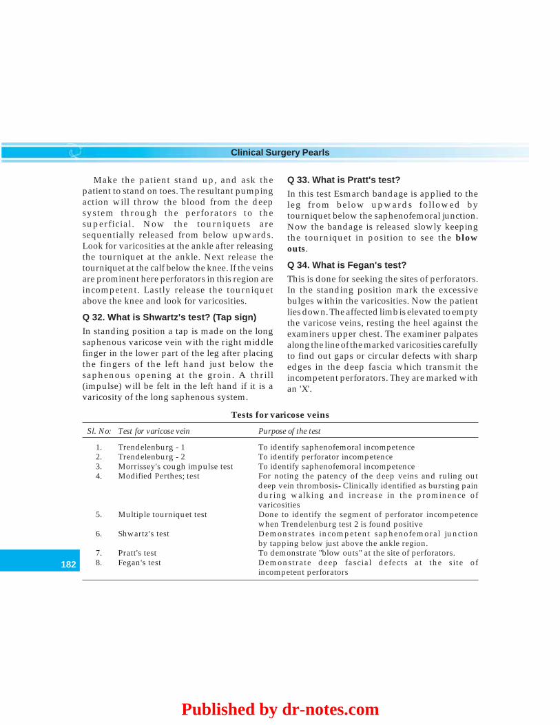
182
Clinical Surgery Pearls
Make the patient stand up, and ask thepatient to stand on toes. The resultant pumpingaction will throw the blood from the deepsystem through the perforators to thesuperficial. Now the tourniquets aresequentially released from below upwards.Look for varicosities at the ankle after releasingthe tourniquet at the ankle. Next release thetourniquet at the calf below the knee. If the veinsare prominent here perforators in this region areincompetent. Lastly release the tourniquetabove the knee and look for varicosities.
Q 32. What is Shwartz's test? (Tap sign)
In standing position a tap is made on the longsaphenous varicose vein with the right middlefinger in the lower part of the leg after placingthe fingers of the left hand just below thesaphenous opening at the groin. A thrill(impulse) will be felt in the left hand if it is avaricosity of the long saphenous system.
Q 33. What is Pratt's test?
In this test Esmarch bandage is applied to theleg from below upwards followed bytourniquet below the saphenofemoral junction.Now the bandage is released slowly keepingthe tourniquet in position to see the blowouts.
Q 34. What is Fegan's test?
This is done for seeking the sites of perforators.In the standing position mark the excessivebulges within the varicosities. Now the patientlies down. The affected limb is elevated to emptythe varicose veins, resting the heel against theexaminers upper chest. The examiner palpatesalong the line of the marked varicosities carefullyto find out gaps or circular defects with sharpedges in the deep fascia which transmit theincompetent perforators. They are marked withan 'X'.
Tests for varicose veins
Sl. No: Test for varicose vein Purpose of the test
1. Trendelenburg - 1 To identify saphenofemoral incompetence2. Trendelenburg - 2 To identify perforator incompetence3. Morrissey's cough impulse test To identify saphenofemoral incompetence4. Modified Perthes; test For noting the patency of the deep veins and ruling out
deep vein thrombosis- Clinically identified as bursting painduring walking and increase in the prominence ofvaricosities
5. Multiple tourniquet test Done to identify the segment of perforator incompetencewhen Trendelenburg test 2 is found positive
6. Shwartz's test Demonstrates incompetent saphenofemoral junctionby tapping below just above the ankle region.
7. Pratt's test To demonstrate "blow outs" at the site of perforators.8. Fegan's test Demonstrate deep fascial defects at the site of
incompetent perforators
Published by dr-notes.com
183
Varicose Veins
Q 35. What is CEAP (American Venous Forum1994) classification? (PG)
This acronym CEAP stands for -• C — Clinical classification• E — Etiological classification• A — Anatomic classification• P — Pathophysiological classification
C — Clinical classification — 7 clinical gradeshave been identified
Class - 0 — No visible or palpable sign ofvenous disease
Class - 1 — Telangiectasis or reticular veinsClass - 2 — Varicose veinsClass - 3 — EdemaClass - 4 — Skin changes (pigmentation,
eczema or lipodermatosclerosis)Class - 5 — Skin changes defined above with
healed ulcerationClass - 6 — Skin changes defined above with
active ulceration
E - Etiological classification—3 etiologiesEc — CongenitalEp — Primary (undetermined cause)Es — Secondary (post thrombotic, post-
traumatic, other causes)
A—Anatomic classification — 18 Anatomicsegments have been described in 3 anatomicregions
As — Superficial veinsAd— Deep veinsAp— Perforating veins
P—Pathophysiological classification—3pathologic mechanisms
Pr - RefluxPo - ObstructionPro - Reflux and obstruction
Q 36. What is gaiter area?
This is an area immediately above the medialmalleolus and less commonly above the lateralmalleolus where the changes of chronic venoushypertension (venous stasis) are seen in the formof lipodermatosclerosis, dermatitis, eczema,pigmentation and ulceration.
Gaiter is a type of protective clothing for aperson's ankles and legs below the knees. Gaiterarea is the area of lower extremity over which agaiter fits roughly from ankle to the proximalcalf.
Q 37. What is Champagne bottle leg (invertedbeer bottle appearance)?
The lipodermatosclerosis will present aspalpable induration in the Gaiter area.Contraction of the skin and subcutaneous tissuein this region will result in narrowing of theankle area. The combination of a narrow ankleand prominent calf is referred to as theChampagne bottle.
Q 38. What is atrophie blanche?
In this condition the superficial veins are lostfrom the skin and white patches develop. Thisis called atrophie blanche. This indicate that theskin has been severely damaged by the venousvalvular incompetence. Venous ulceration maydevelop in these areas.
Q 39. What are the investigations fordiagnosis?
184
Clinical Surgery Pearls
Investigations for varicose veins
• Duplex ultrasound imaging• Doppler ultrasound.• Photo plethysmography• Venography (Phlebography)—invasive• Ambulatory venous pressure studies• Raju's test—Arm foot venous pressure study• Ultrasound of the abdomen
(1) Duplex ultrasound imaging—Theexamination is performed with the patient instanding position. The examiner steadily workhis way from the groin to the ankle.
This is the most important noninvasiveinvestigation for practically all venousdisorders. It produces high quality picturesusing a combination of B mode imaging andDoppler ultrasound. Anatomical, physiologicaland functional details can be obtained. Venouslumen, flow, direction and reflux of blood canbe visualized. The site of perforatorincompetence can be located with accuracy. Ithas replaced the venography. All patients withrecurrent varices, history of previous DVT andpatients with skin changes should be fullyinvestigated by using Duplex ultrasonographyor venography.
(2) Dopplerultra sound—This investigation isalso done in the standing position. Abidirectional flow probe will identify venousreflux. The Doppler probe is first placed overthe sapheno- femoral junction. With one handthe examiner squeezes the calf to produce anacceleration of blood flow in the veins. This isheard as a 'whoosh' from the speaker of theDoppler machine. The calf compression isreleased and look for any reverse flow in the
veins. This procedure is repeated over thesaphenopopliteal junction and over otherareas. The saphenopopliteal junction isinconstant. It can be identified and marked withDoppler. Doppler assessment is the minimuminvestigation required before treating a patientwith venous disease. The arterial disease can alsobe excluded.
• Four characteristics descriptions are Patent,Augmented, Spontaneous and Phasic.
Patent - Flow is heard at the anatomicallevel of the vein
- Rarely does the flow completelydisappear with DVT
- DVT is associated withcontinuous, high pitched signal
Augmented - Firm, gentle compression of thelimb for a few seconds distal tothe vein should result inaugmentation of the flow
- Proximal compression followedby release should also result inaugmentation
- Augmentation produced byproximal compression beforerelease or distal compressionafter release indicates valvularincompetence.
Spontaneous - DVT results in loss ofspontaneous flow
- Anything causing vasoconstric-tion also causes loss of spon-taneity
Phasic - Variation with respiration iscalled phasicity
Published by dr-notes.com
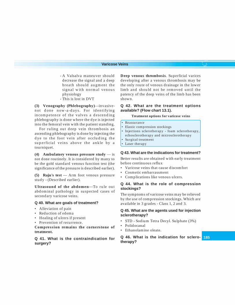
185
Varicose Veins
- A Valsalva maneuver shoulddecrease the signal and a deepbreath should augment thesignal with normal venousphysiology
- This is lost in DVT
(3) Venography (Phlebography)—invasive-not done now-a-days. For identifyingincompetence of the valves a descendingphlebography is done where the dye is injectedinto the femoral vein with the patient standing.
For ruling out deep vein thrombosis anascending phlebography is done by injecting thedye to the foot vein after occluding thesuperficial veins above the ankle by atourniquet.
(4) Ambulatory venous pressure study — isnot done routinely. It is considered by many tobe the gold standard venous function test (thesignificance of the pressure is described earlier).
(5) Raju's test — Arm foot venous pressurestudy - (Described earlier).
Ultrasound of the abdomen—To rule outabdominal pathology in suspected cases ofsecondary varicose veins.
Q 40. What are goals of treatment?
• Alleviation of pain• Reduction of edema• Healing of ulcers if present• Prevention of recurrence.Compression remains the cornerstone oftreatment.
Q 41. What is the contraindication forsurgery?
Deep venous thrombosis. Superficial varicesdeveloping after a venous thrombosis may bethe only route of venous drainage in the lowerlimb and should not be removed until thepatency of the deep veins of the limb has beenshown.
Q 42. What are the treatment optionsavailable? (Flow chart 13.1).
Treatment options for varicose veins
• Reassurance• Elastic compression stockings• Injections sclerotherapy - foam sclerotherapy,
echosclerotherapy and microsclerotherapy• Surgical treatment• Laser therapy
Q 43. What are the indications for treatment?
Better results are obtained with early treatmentbefore continuous reflux• Varicose veins that cause discomfort• Cosmetic embarrassment• Complications like venous ulcers.
Q 44. What is the role of compressionstockings?
The symptoms of varicose veins may be relievedby the use of compression stockings. Which areavailable in 3 grades - Class 1, 2 and 3.
Q 45. What are the agents used for injectionsclerotherapy?
• STD - Sodium Tetra Decyl. Sulphate (3%)• Polidocanal• Ethanolamine oleate.
Q 46. What is the indication for sclero-therapy?
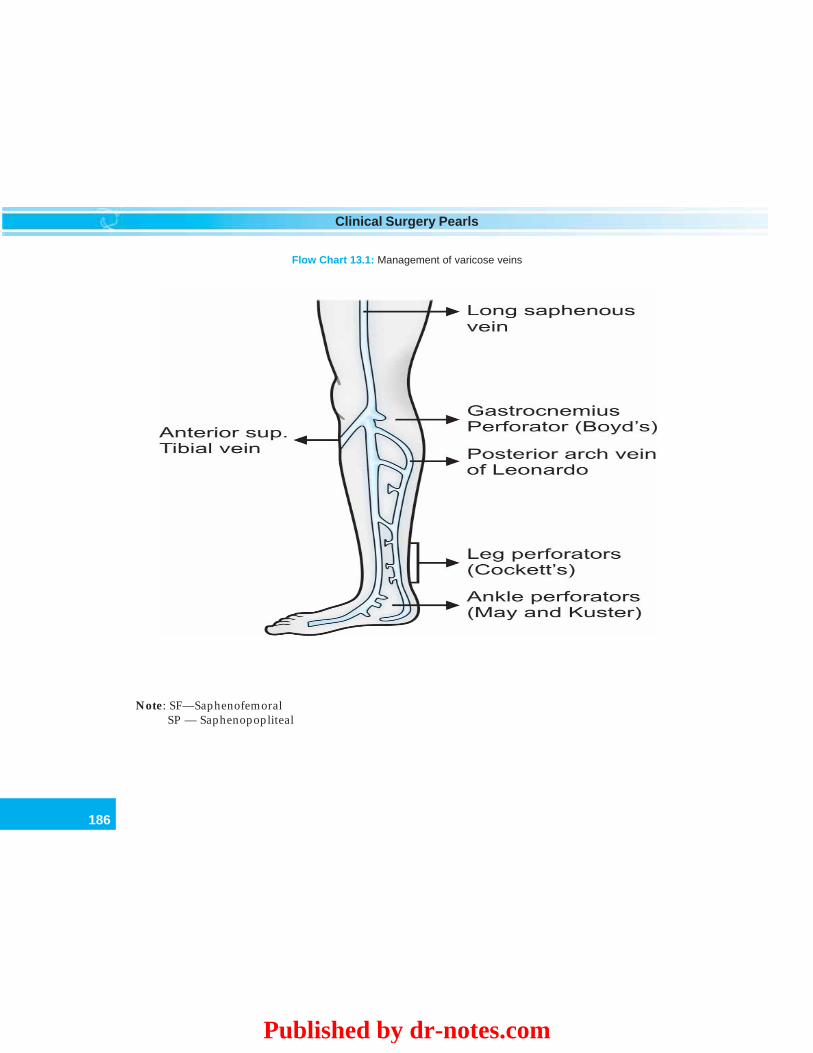
186
Clinical Surgery Pearls
Flow Chart 13.1: Management of varicose veins
Note: SF—SaphenofemoralSP — Saphenopopliteal
Published by dr-notes.com
187
Varicose Veins
1. This is used to treat varicose veins in theabsence of junctional incompetence andmajor perforating veins.
2. Used for smaller veins < 3 mm in size.
Q 47. What is the technique of injectionsclerotherapy?
The sclerosant is injected into an empty vein andthe vein is compressed. The endothelial liningis destroyed. If the vein is not compressed it willbe produce thrombosis which will later getrecanalized producing recurrence.
Q 48. What is foam sclerotherapy (byTessari)?
In this technique the STD is drawn into onesyringe and air is taken in another syringe.Using a 3 way, rapid too and fro movements ofthe piston of the syringe, the foam is produced.This foam is then injected into the longsaphenous vein after cannulation. The air isabsorbed, the vein collapses and the endotheliallining is destroyed. A much larger volume canbe injected into the vein with a small quantityof sclerosant so that it will fill the superficialvaricosities. The patient is lying supine with theleg elevated instead.
Q 49. What is echosclerotherapy? (PG)
When the procedure of foam sclerotherapy isdone under the guidance of duplex ultrasoundimaging it is called echosclerotherapy.
Q 50. What is the advantage of sclero-therapy?
The patients can undergo repeated treatmentsessions to ensure that all veins are removed.
Q 51. What are the complications of sclero-therapy?
Complications of sclerotherapy
• Skin pigmentation• Injury to the skin and ulceration• Allergic reaction• Thrombophlebitis• Deep vein thrombosis
Q 52. What is microsclerotherapy? (PG)
The thread veins and reticular varices areinjected by inserting a 30G needle. The solutionsused are STD and polidocanol. After theinjection compression bandage is given for aperiod of 1-5 days.
Q 53. What is the surgical treatment ofsaphenofemoral incompetence?
Trendelenburg's operation + stripping of thelong saphenous vein.
Q 54. What is the contraindication forTrendelenburg's operation?
When Deep vein thrombosis is destroying themain axial limb vein and the patient will dependon the superficial veins for venous drainage.
Q 55. What is Trendelenburg's operation?
An oblique incision is made in the groin andthe long saphenous vein is exposed. Theprocedure has got two portions—a. Saphenofemoral flush ligationb. Ligation of the proximal 5 tributaries—
1. The superficial external pudendal vein2. The superficial inferior epigastric vein3. The superficial circumflex iliac vein
188
Clinical Surgery Pearls
4. The posteromedial vein (medial acces-sory saphenous vein)
5. The anterolateral (lateral accessorysaphenous vein)
Note: It is important that the femoral vein isinspected carefully at least 1cm above and belowthe SFJ (Saphenofemoral Junction) and alltributaries in this situation ligated and divided.
Q 56. Why it should be combined withstripping of the long saphenous vein?
The main principle of the surgical treatment isto ligate the source of venous reflux and toremove the incompetent saphenous trunk. TheTrendelenburg procedure alone is associated withhigh rate of recurrence. To ensure elimination ofas much reflux as possible it is necessary to removethe long saphenous vein. Similarly in case ofsaphenopopliteal incompetence the part of theshort saphenous vein must be removed.
Q 57. What type of stripping is recommendedfor saphenous vein?
To avoid injury to the saphenous nerve the longsaphenous vein should not be removed belowthe midcalf level. The conventional way ofremoving the long saphenous vein is with astripper (Babcock). Here the stripper wire ispassed down the long saphenous vein. The endis identified in the upper calf and a 2mm incisionis made to retrieve the stripper. An olive of about8mm diameter is attached to the upper end andthe saphenous vein is removed by firm tractionon the wire in the calf.
Q 58. What is invaginating stripping(inverting)? (PG)
The aim of this type of stripping is to reduce thedamage to the tissues around the vein. Therewill be less pain and less bleeding in thisoperation. A rigid metal pin stripper (Oesh) hasbeen developed and this is passed down theinside of the saphenous vein which is recoveredfrom the upper calf. A strong suture is attachedto the stripper and firmly ligated to the proximalend of the vein. During pulling of the stripperthe long saphenous vein will invert and can bedelivered through a 2mm incision in the midcalfregion. Here no olive is used.
Q 59. What are the nerves at risk duringvenous surgery?
1. Saphenous nerve—this is likely to be injuredduring stripping of the long saphenous vein
2. Sural nerve—this is likely to be injuredduring stripping of the short saphenous vein.
Q 60. What is the peculiarity of the saphenopopliteal junction?
The saphenopopliteal junction is not constant.The termination may lie from 2cm below theknee to 15cm above the knee. It is very importantto identify the termination sonologically andmark it on the skin before surgery.
Q 61. What is hook phlebectomy? (PG)
After stripping, the residual veins and tributariesare left behind. These veins conventionally usedto be managed by incisions long enough to insertartery forceps for ligating the veins. Now theseveins can be tackled by means of small hooksthat may be inserted through incisions of 1-2mmsize. The closure of these small incisions isachieved using adhesive strips. This givesexcellent cosmetic outcome.
Published by dr-notes.com
189
Varicose Veins
Q 62. What is the postoperative manage-ment?
1. Compression bandaging is applied to thelimb at the end of the operation to preventbruising.
2. Some surgeons apply compression to thelimb before stripping.
3. After two days the bandage may be replacedwith thigh length high compressionstockings (class 2).
Q 63. What is VNUS closure? (PG)
This is intraluminal destruction of the long andshort saphenous vein using ablation catheterunder ultrasound guidance.
Q 64. What are the complications of varicosevein surgery? (PG)
Complications of varicose vein surgery
1. Pain, discomfort and bruising2. Nerve injury—Saphenous for long saphenous
surgery—Sural for short saphenous surgery3. Venous thrombosis in residual varices4. Deep vein thrombosis—1/1000 operations (give
prophylactic heparin when there is history ofprevious DVT)
Q 65. What are the complications of varicoseveins?
Complications of varicose veins
1. Hemorrhage2. Superficial thrombophlebitis3. Eczema
4. Pigmentation5. Lipodermatosclerosis6. Deep vein thrombosis (rarely)7. Venous ulcer8. Marjolin's ulcer9. Calcification
10. Periosteitis11. Talipes equinus
Q 66. What are the causes for superficialthrombophlebitis ?
Causes for superficial thrombophlebitis
• TAO (Buerger's disease)• Occult carcinoma (Visceral malignancy)• Varicose veins• Polycythemia• Iatrogenic - IV Injection
Q 67. What is the classical location forvenous ulcer?
Just proximal to the medial and lateral malleolus(Gaiter area)
Q 68. What are the differences betweenvenous ulcer and varicose ulcer?
When the ulcer is secondary to deep venouspathology or perforator incompetenceproducing chronic venous hypertension, it iscalled venous ulcer. When the ulcer is due tosuperficial insufficiency alone, it is calledvaricose ulcer. It is better to use the term venousulcer because the ulcer is not caused by thevaricose veins but by the disordered pattern ofthe venous blood flow.
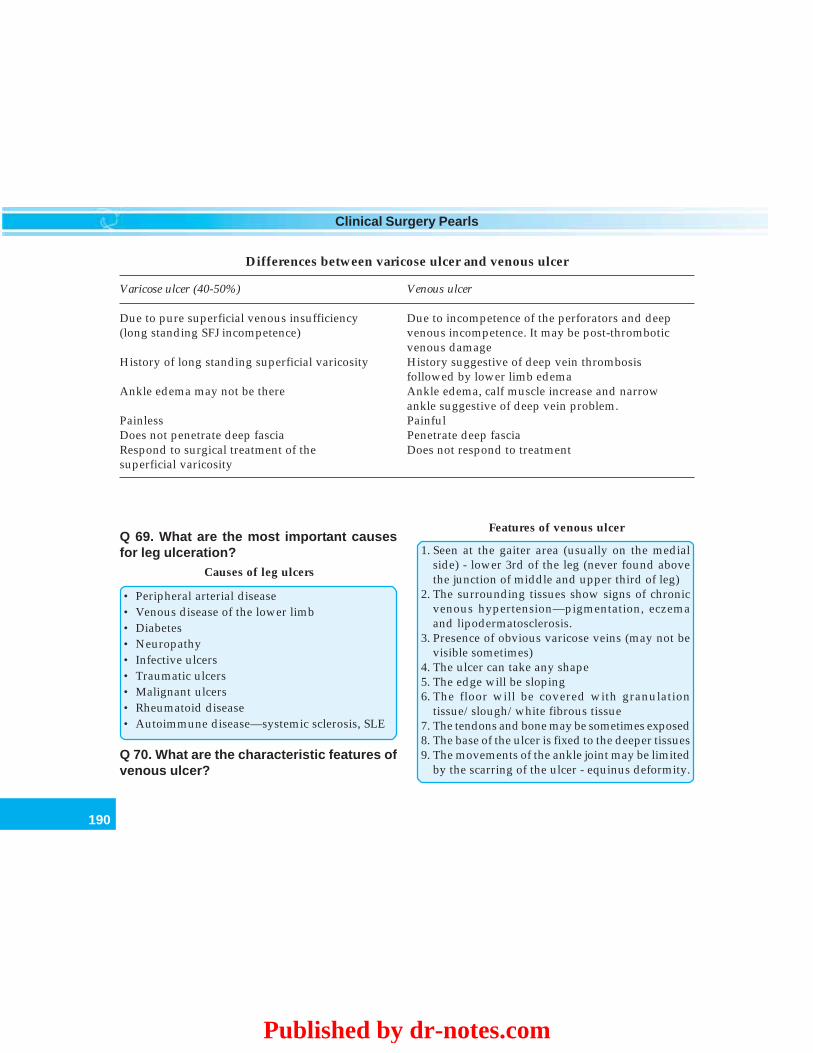
190
Clinical Surgery Pearls
Differences between varicose ulcer and venous ulcer
Varicose ulcer (40-50%) Venous ulcer
Due to pure superficial venous insufficiency Due to incompetence of the perforators and deep(long standing SFJ incompetence) venous incompetence. It may be post-thrombotic
venous damageHistory of long standing superficial varicosity History suggestive of deep vein thrombosis
followed by lower limb edemaAnkle edema may not be there Ankle edema, calf muscle increase and narrow
ankle suggestive of deep vein problem.Painless PainfulDoes not penetrate deep fascia Penetrate deep fasciaRespond to surgical treatment of the Does not respond to treatmentsuperficial varicosity
Q 69. What are the most important causesfor leg ulceration?
Causes of leg ulcers
• Peripheral arterial disease• Venous disease of the lower limb• Diabetes• Neuropathy• Infective ulcers• Traumatic ulcers• Malignant ulcers• Rheumatoid disease• Autoimmune disease—systemic sclerosis, SLE
Q 70. What are the characteristic features ofvenous ulcer?
Features of venous ulcer
1. Seen at the gaiter area (usually on the medialside) - lower 3rd of the leg (never found abovethe junction of middle and upper third of leg)
2. The surrounding tissues show signs of chronicvenous hypertension—pigmentation, eczemaand lipodermatosclerosis.
3. Presence of obvious varicose veins (may not bevisible sometimes)
4. The ulcer can take any shape5. The edge will be sloping6. The floor will be covered with granulation
tissue/slough/white fibrous tissue7. The tendons and bone may be sometimes exposed8. The base of the ulcer is fixed to the deeper tissues9. The movements of the ankle joint may be limited
by the scarring of the ulcer - equinus deformity.
Published by dr-notes.com
191
Varicose Veins
Q 71. What are the investigations in venousulcer?
1. Examine the peripheral pulses to rule outarterial problem
2. Examine the sensation to rule outneuropathy
3. Doppler ankle blood pressure4. Duplex ultrasonography of the veins and
arteries5. Blood examination to rule out SLE,
rheumatoid disease6. Biopsy of the ulcer.
Q 72. What is the management of venous legulcers?
This consists of :1. Management of the ulcer2. Surgery for the superficial venous
incompetence3. Patients with deep venous insufficiency
surgery may not be effective.
Q 73. Is it necessary to delay surgery untilthe ulcer has healed? (PG)
It is not necessary and surgery may be carriedout before the ulcer has healed. It is found thatthe ulcer healing is rapid in this situation.
Q 74. What is the management of ulcer?
The standard old regimen is called Bisgardregime consisting of —
• Elevation of the limb• Elastocrape bandage (compression stocking)• Exercise• Education
Note: Do culture and sensitivity and giveappropriate antibiotics.
Biopsy of the ulcer if nonhealingBlood examination - Blood sugar
ESRRheumatoid factor, etc.
Q 75. What is Marjolin's ulcer?
Malignant change may occur in long standingvenous ulcers. This should be suspected whenthere is evidence of raised or rolled out edge.They are nothing but squamous cell carcinoma.
Q 76. Is there any role for topical applicationsand topical antibiotics?
• No• Topical applications will not speed up healing• It will produce allergy and skin sensitization• Topical antibiotics are ineffective• If eczematous reaction is there around the
ulcer, topical steroid may be useful.
Q 77. What is the role of compression?
• A pressure of 30-45 mm of Hg applied to theulcer is found to be effective for healing ofthe ulcer
• Class III stockings exert about 30mms of Hgcompression
• The compression is applied to the ulcer regionalone (below knee stockings)
• Those who cannot use compression stockingsuse multilayer bandaging regimes (4 layerbandage developed by Charing crosshospital, London). This bandage is changedonce or twice weekly.
Q 78. What is the principle of compression?
The exact mechanism by which compression isof benefit is not known but the followingphysiological alterations occur.
192
Clinical Surgery Pearls
1. Relieves the leg edema2. Controls the chronic venous insufficiency3. Reduction in ambulatory venous pressure4. Improvement in skin microcirculation5. Increase in subcutaneous pressure
preventing transcapillary fluid leakage.
Q 79. What is the drug treatment for venousulcer?
Drugs are inferior to compression bandaging.The following drugs are used —• Aspirin • Pentoxyfylline• Prostaglandin E • Calcium
Analogue Dobesilate• Diosmin • Flavonoids
Q 80. What is the role of systemic antibioticsin venous ulcer?
• Antibiotics have no effect on ulcer healing• Evidence of infection in the form of cellulitis
and fever, are indications for antibiotics.
Q 81. What is the treatment of deep venousinsufficiency? (PG)
The surgical treatment is difficult and being doneonly in a few centers. The surgical options are—a. Direct vein valve repair by valvuloplasty
(Kistner)b. Vein valve transpositionc. Transplantation of a segment of axillary vein
Q 82. What is the indication for deep venoussurgery? (PG)
Those who have persisting swelling of the lowerlimb after a previous venous thrombosis.
Q 83. What is the surgical treatment for deepvenous obstruction? (PG)
1. Palma operation— This involves mobilizingthe long saphenous vein in the opposite leg,tunneling the distal end of the LSV acrosssuprapubically and inserting it into thefemoral vein of the affected side below theobstruction.
2. May-Husni procedure—This is done for theobstruction of the superficial femoral vein.The long saphenous vein is connected to thepopliteal vein in the same limb allowingblood to flow along the superficial vein.
Q 84. What is the treatment of perforatorincompetence?
1. Subfascial endoscopic perforator surgery(SEPS)
2. Sclerotherapy3. Open subfascial ligation.
Poem on Varicose Veins
"Varicose vein disease is sometimes diagnosedwith ease,
But of the best attempts will meet with sad andsorrowful defeat.
So let us give you good advice
Examine once, Examine twice:Examine from head to toes,
Before you dare to diagnose.
More harm is done because you do not look
Than from not knowing what is in the book
Above all do not try to spot
Because you think you know a lot
Spot diagnosis you should hate
Until you are a surgeon great!
By then of course you will have learnt
Some lessons bitter, and you darent't"
Published by dr-notes.com
193
Peripheral Occlusive Vascular Disease
14Peripheral Occlusive
Vascular Disease
CASE CAPSULE
A 60-year-old male patient presents with leftcalf muscle, thigh muscle and gluteal pain of18 months duration. He gets a cramp-like painonly while he walks and the pain disappearswhen he stops walking. At the beginning of thewalk he get numbness, pins and needles andparesthesia in the skin of the foot. Initially theclaudication distance was about 200 meters.The walking distance gradually shortened overa few months. There is no history of chest pain,fainting, blurred vision or weakness orparesthesia of the upper limbs. For the last 3weeks he gets continuous severe pain achingin nature which is present at rest throughoutthe day and night, mostly in the foot and leg.After a few days he noticed black color of theleft big toe and ulcer in the left heel region.The maximum pain he gets is at the junction ofblack area of the big toe and the remainingnormal foot. He prefers to sleep sitting in achair, if at all in the bed, he hangs his leg overthe side of the bed. The patient is requestingfor amputation of the leg.
On examination the patient is found to behypertensive. The patient sits with the leftknee bent, holding the left foot still. He isunwilling to lie flat. When the limb is kepthorizontal the foot is pale. The capillary fillingis retarded. The pulps of the toes showwasting. The veins of the limb are empty andguttered. The entire left big toe is gangrenous.A line of demarcation is seen between thegangrenous area and the normal foot. There isan ulcer of 4 × 3cm size situated in the leftheel region. The floor of the ulcer is coveredwith slough and the edge is punched out. Thesurrounding area of the ulcer is very tender.The skin temperature from midcalfdownwards is cold. The femoral, popliteal,dorsalis pedis and posterior tibial pulses areabsent on the left side. On the right side allthe pulses are present except dorsalis pedis andposterior tibial. There is wasting of left legmuscles. There is an audible bruit over theright femoral artery. Cardiovascular systemand neurological examination are normal.
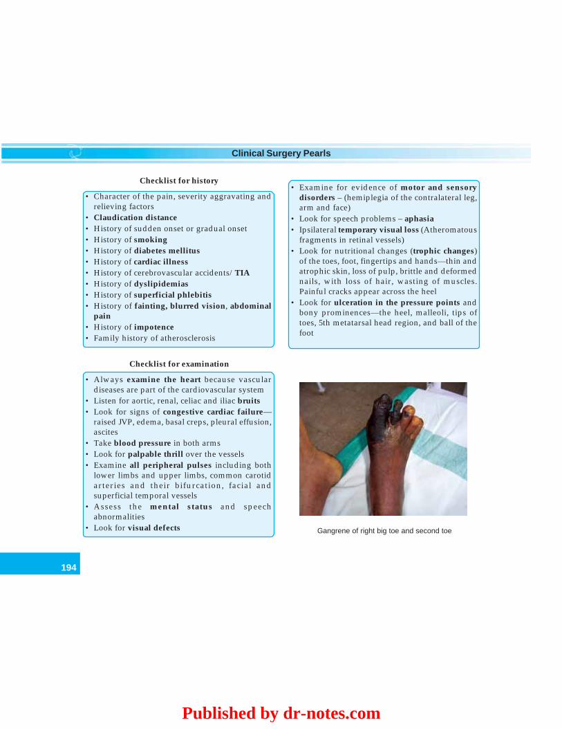
194
Clinical Surgery Pearls
Checklist for history
• Character of the pain, severity aggravating andrelieving factors
• Claudication distance• History of sudden onset or gradual onset• History of smoking• History of diabetes mellitus• History of cardiac illness• History of cerebrovascular accidents/TIA• History of dyslipidemias• History of superficial phlebitis• History of fainting, blurred vision, abdominal
pain• History of impotence• Family history of atherosclerosis
Checklist for examination
• Always examine the heart because vasculardiseases are part of the cardiovascular system
• Listen for aortic, renal, celiac and iliac bruits• Look for signs of congestive cardiac failure—
raised JVP, edema, basal creps, pleural effusion,ascites
• Take blood pressure in both arms• Look for palpable thrill over the vessels• Examine all peripheral pulses including both
lower limbs and upper limbs, common carotidarteries and their bifurcation, facial andsuperficial temporal vessels
• Assess the mental status and speechabnormalities
• Look for visual defects
• Examine for evidence of motor and sensorydisorders – (hemiplegia of the contralateral leg,arm and face)
• Look for speech problems – aphasia• Ipsilateral temporary visual loss (Atheromatous
fragments in retinal vessels)• Look for nutritional changes (trophic changes)
of the toes, foot, fingertips and hands—thin andatrophic skin, loss of pulp, brittle and deformednails, with loss of hair, wasting of muscles.Painful cracks appear across the heel
• Look for ulceration in the pressure points andbony prominences—the heel, malleoli, tips oftoes, 5th metatarsal head region, and ball of thefoot
Gangrene of right big toe and second toe
Published by dr-notes.com
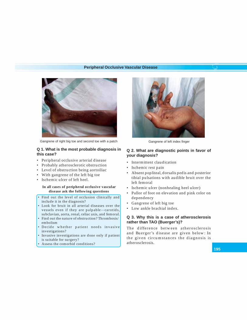
195
Peripheral Occlusive Vascular Disease
Q 1. What is the most probable diagnosis inthis case?
• Peripheral occlusive arterial disease• Probably atherosclerotic obstruction• Level of obstruction being aortoiliac• With gangrene of the left big toe• Ischemic ulcer of left heel.
In all cases of peripheral occlusive vasculardisease ask the following questions
• Find out the level of occlusion clinically andinclude it in the diagnosis?
• Look for bruit in all arterial diseases over thevessels even if they are palpable—carotids,subclavian, aorta, renal, celiac axis, and femoral.
• Find out the nature of obstruction? Thrombosis/embolism
• Decide whether patient needs invasiveinvestigations?
• Invasive investigations are done only if patientis suitable for surgery?
• Assess the comorbid conditions?
Q 2. What are diagnostic points in favor ofyour diagnosis?
• Intermittent claudication• Ischemic rest pain• Absent popliteal, dorsalis pedis and posterior
tibial pulsations with audible bruit over theleft femoral
• Ischemic ulcer (nonhealing heel ulcer)• Pallor of foot on elevation and pink color on
dependency• Gangrene of left big toe• Low ankle brachial index.
Q 3. Why this is a case of atherosclerosisrather than TAO (Buerger’s)?
The difference between atherosclerosisand Buerger’s disease are given below: Inthe given circumstances the diagnosis isatherosclerosis.
Gangrene of left index fingerGangrene of right big toe and second toe with a patch
196
Clinical Surgery Pearls
Differences between ASO and Buerger’s disease
Feature Atherosclerosis Buerger’s
Age of onset Above 40 years 20 – 40 yearsSex Male > female Rare in womenSmoking Can occur without smoking Seen only in smokersProgression of disease Proximal to distal Distal to proximalVenous involvement Not seen CommonThrombophlebitis Not seen CommonInflammatory reaction Not seen CommonLayer of vessel affected Affects the intima Affects all layers – pan arteritis, peri
arteritis, and pan phlebitisSite of lesion Large and medium sized Small arteries
arteries (aorta, iliac, femoraland popliteal)
Arteriography Proximal lesion Distal lesionCollateral in arteriography Extensive collaterals Poor collaterals (cork screw appearance)
(tree root appearance)Distal run off in arteriogram Usually present Usually absentBypass surgery Possible Not possible
Note: ASO-Atherosclerotic obstruction
Q 4. What is claudication?
Claudication means limping (Claudio = I limp).The term is now used to describe the cramp-like muscle pain which appears followingexercise as a result of inadequate arterial bloodflow. The three criteria for claudication are:1. It is a cramp-like muscle pain (usually calf)2. Pain develops only when the muscle is
exercised – (not present on taking the firststep in contrast to osteoarthritis)
3. The pain disappears when the exercise stops(contrast lumbar intervertebral disk nervecompression).
Q 5. What are the types of claudication?
There are three types of claudication1. Arterial claudication2. Venous claudication3. Neurogenic claudication.
Q 6. What is the cause for paresthesia in theskin of the foot?
The patient notices numbness, pins and needlesand other paresthesia in the skin of the foot atthe when muscle pain begins. This is as aresult of shunting of blood from the skin to themuscle.
Published by dr-notes.com
197
Peripheral Occlusive Vascular Disease
Q 7. What is the cause for claudication pain?
The cause of the pain can be explained as aresponse to the accumulation of acidic,anaerobic metabolite (P - substance) due toinadequate blood and oxygen supplies to meetthe requirements of the increased activity. Onstopping the activity the blood supply isreplenished, the anoxia is abolished and thepainful metabolites are washed away.
Q 8. What is claudication distance?
The distance a patient can walk on normalground and at a normal speed. without pain andhaving to stop.
Q 9. What are the sites of claudication pain?
Depending on the level of occlusion, the site ofclaudication pain will differ. The following chartshows the claudication sites for each level ofocclusion.
Q 10. What is Leriche’s syndrome?
It occurs in men as a result of aortoiliac diseaseand consists of:• Claudication of the hip, thigh and buttock
muscles (Gluteal claudication)• Atrophy of the leg muscles• Sexual impotence• Diminished/absent femoral pulses• Transient numbness of the extremity accompa-
nies the pain and fatigue of claudication.
Q 11. What is the cause for impotence inLeriche’s syndrome?
It is due to the lack of blood supply to the penis.The penis is getting blood supply through theinternal iliac vessels which is occluded in thisconditions.
Q 12. What is rest pain? What is the site ofrest pain?
Levels of occlusion with clinical features
Level of occlusion Site of claudication and other clinical findings
1. Absent ankle pulses Claudication of calf and foot2. Femoropopliteal obstruction Unilateral claudication in calf
(femoral pulse present and popliteal pulse absent)3. Iliac vessel obstruction Unilateral claudication in thigh and calf and
(the femoral and distal pulses absent) some times buttock. Bruit over iliac or femoralvessel
4. Aortoiliac obstruction Bilateral claudication of both buttocks, thighs(all pulsations from femoral down and calves. Presence of bruit over both femoral.absent on both sides) Impotence- common
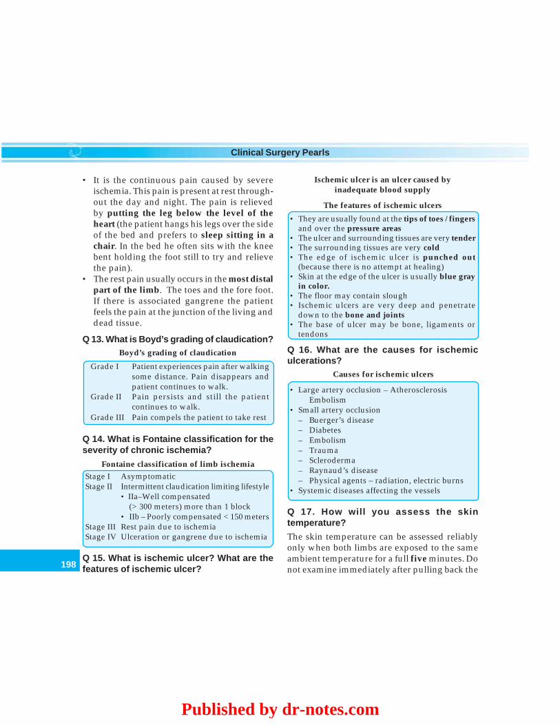
198
Clinical Surgery Pearls
• It is the continuous pain caused by severeischemia. This pain is present at rest through-out the day and night. The pain is relievedby putting the leg below the level of theheart (the patient hangs his legs over the sideof the bed and prefers to sleep sitting in achair. In the bed he often sits with the kneebent holding the foot still to try and relievethe pain).
• The rest pain usually occurs in the most distalpart of the limb. The toes and the fore foot.If there is associated gangrene the patientfeels the pain at the junction of the living anddead tissue.
Q 13. What is Boyd’s grading of claudication?
Boyd’s grading of claudication
Grade I Patient experiences pain after walkingsome distance. Pain disappears andpatient continues to walk.
Grade II Pain persists and still the patientcontinues to walk.
Grade III Pain compels the patient to take rest
Q 14. What is Fontaine classification for theseverity of chronic ischemia?
Fontaine classification of limb ischemiaStage I AsymptomaticStage II Intermittent claudication limiting lifestyle
• IIa–Well compensated(> 300 meters) more than 1 block
• IIb – Poorly compensated < 150 metersStage III Rest pain due to ischemiaStage IV Ulceration or gangrene due to ischemia
Q 15. What is ischemic ulcer? What are thefeatures of ischemic ulcer?
Ischemic ulcer is an ulcer caused byinadequate blood supply
The features of ischemic ulcers
• They are usually found at the tips of toes / fingersand over the pressure areas
• The ulcer and surrounding tissues are very tender• The surrounding tissues are very cold• The edge of ischemic ulcer is punched out
(because there is no attempt at healing)• Skin at the edge of the ulcer is usually blue gray
in color.• The floor may contain slough• Ischemic ulcers are very deep and penetrate
down to the bone and joints• The base of ulcer may be bone, ligaments or
tendons
Q 16. What are the causes for ischemiculcerations?
Causes for ischemic ulcers
• Large artery occlusion – AtherosclerosisEmbolism
• Small artery occlusion– Buerger’s disease– Diabetes– Embolism– Trauma– Scleroderma– Raynaud’s disease– Physical agents – radiation, electric burns
• Systemic diseases affecting the vessels
Q 17. How will you assess the skintemperature?
The skin temperature can be assessed reliablyonly when both limbs are exposed to the sameambient temperature for a full five minutes. Donot examine immediately after pulling back the
Published by dr-notes.com
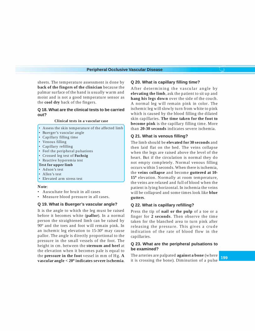
199
Peripheral Occlusive Vascular Disease
sheets. The temperature assessment is done byback of the fingers of the clinician because thepalmar surface of the hand is usually warm andmoist and is not a good temperature sensor asthe cool dry back of the fingers.
Q 18. What are the clinical tests to be carriedout?
Clinical tests in a vascular case
• Assess the skin temperature of the affected limb• Buerger’s vascular angle• Capillary filling time• Venous filling• Capillary refilling• Feel the peripheral pulsations• Crossed leg test of Fuchsig• Reactive hyperemia testTest for upper limb• Adson’s test• Allen’s test• Elevated arm stress test
Note:• Auscultate for bruit in all cases• Measure blood pressure in all cases.
Q 19. What is Buerger’s vascular angle?
It is the angle to which the leg must be raisedbefore it becomes white (pallor). In a normalperson the straightened limb can be raised by90o and the toes and foot will remain pink. Inan ischemic leg elevation to 15-30o may causepallor. The angle is directly proportional to thepressure in the small vessels of the foot. Theheight in cm. between the sternum and heel atthe elevation when it becomes pale is equal tothe pressure in the foot vessel in mm of Hg. Avascular angle < 20o indicates severe ischemia.
Q 20. What is capillary filling time?
After determining the vascular angle byelevating the limb, ask the patient to sit up andhang his legs down over the side of the couch.A normal leg will remain pink in color. Theischemic leg will slowly turn from white to pinkwhich is caused by the blood filling the dilatedskin capillaries. The time taken for the foot tobecome pink is the capillary filling time. Morethan 20-30 seconds indicates severe ischemia.
Q 21. What is venous filling?
The limb should be elevated for 30 seconds andthen laid flat on the bed. The veins collapsewhen the legs are raised above the level of theheart. But if the circulation is normal they donot empty completely. Normal venous fillingoccurs within 5 seconds. When there is ischemia,the veins collapse and become guttered at 10-15o elevation. Normally at room temperature,the veins are relaxed and full of blood when thepatient is lying horizontal. In ischemia the veinswill be collapsed and some times look like bluegutters.
Q 22. What is capillary refilling?
Press the tip of nail or the pulp of a toe or afinger for 2 seconds. Then observe the timetaken for the blanched area to turn pink afterreleasing the pressure. This gives a crudeindication of the rate of blood flow in thecapillaries.
Q 23. What are the peripheral pulsations tobe examined?
The arteries are palpated against a bone (whereit is crossing the bone). Diminution of a pulse
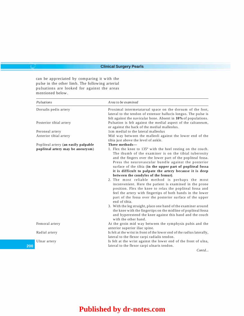
200
Clinical Surgery Pearls
can be appreciated by comparing it with thepulse in the other limb. The following arterialpulsations are looked for against the areasmentioned below.
Contd...
Pulsations Area to be examined
Dorsalis pedis artery Proximal intermetatarsal space on the dorsum of the foot,lateral to the tendon of extensor hallucis longus. The pulse isfelt against the navicular bone. Absent in 10% of populations.
Posterior tibial artery Pulsation is felt against the medial aspect of the calcaneum,or against the back of the medial malleolus.
Peroneal artery 1cm medial to the lateral malleolusAnterior tibial artery Mid way between the malleoli against the lower end of the
tibia just above the level of ankle.Popliteal artery (an easily palpable Three methods—popliteal artery may be aneurysm) 1. Flex the knee to 135o with the heel resting on the couch.
The thumb of the examiner is on the tibial tuberosityand the fingers over the lower part of the popliteal fossa.Press the neurovascular bundle against the posteriorsurface of the tibia (in the upper part of popliteal fossait is difficult to palpate the artery because it is deepbetween the condyles of the femur).
2. The most reliable method is perhaps the mostinconvenient. Here the patient is examined in the proneposition. Flex the knee to relax the popliteal fossa andfeel the artery with fingertips of both hands in the lowerpart of the fossa over the posterior surface of the upperend of tibia.
3. With the leg straight, place one hand of the examiner aroundthe knee with the fingertips on the midline of popliteal fossaand hyperextend the knee against this hand and the couchwith the other hand.
Femoral artery At the groin mid way between the symphysis pubis and theanterior superior iliac spine.
Radial artery Is felt at the wrist in front of the lower end of the radius laterally,lateral to the flexor carpi radialis tendon.
Ulnar artery Is felt at the wrist against the lower end of the front of ulna,lateral to the flexor carpi ulnaris tendon.
Published by dr-notes.com
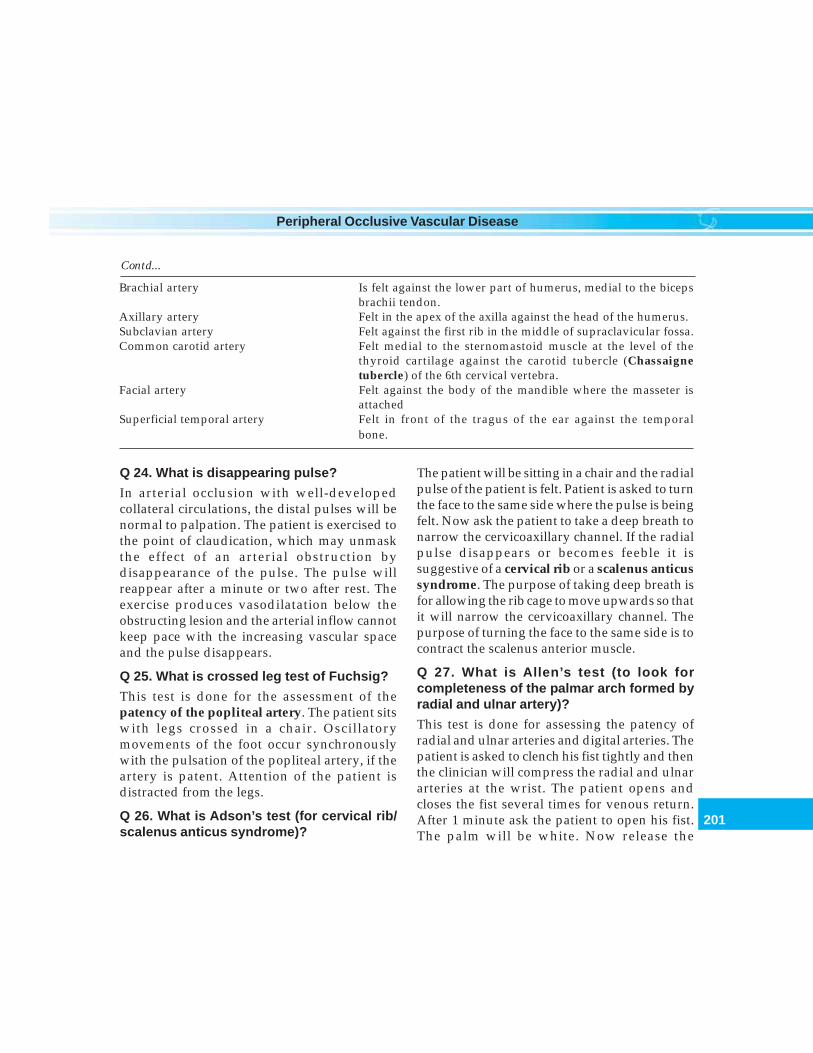
201
Peripheral Occlusive Vascular Disease
Brachial artery Is felt against the lower part of humerus, medial to the bicepsbrachii tendon.
Axillary artery Felt in the apex of the axilla against the head of the humerus.Subclavian artery Felt against the first rib in the middle of supraclavicular fossa.Common carotid artery Felt medial to the sternomastoid muscle at the level of the
thyroid cartilage against the carotid tubercle (Chassaignetubercle) of the 6th cervical vertebra.
Facial artery Felt against the body of the mandible where the masseter isattached
Superficial temporal artery Felt in front of the tragus of the ear against the temporalbone.
The patient will be sitting in a chair and the radialpulse of the patient is felt. Patient is asked to turnthe face to the same side where the pulse is beingfelt. Now ask the patient to take a deep breath tonarrow the cervicoaxillary channel. If the radialpulse disappears or becomes feeble it issuggestive of a cervical rib or a scalenus anticussyndrome. The purpose of taking deep breath isfor allowing the rib cage to move upwards so thatit will narrow the cervicoaxillary channel. Thepurpose of turning the face to the same side is tocontract the scalenus anterior muscle.
Q 27. What is Allen’s test (to look forcompleteness of the palmar arch formed byradial and ulnar artery)?
This test is done for assessing the patency ofradial and ulnar arteries and digital arteries. Thepatient is asked to clench his fist tightly and thenthe clinician will compress the radial and ulnararteries at the wrist. The patient opens andcloses the fist several times for venous return.After 1 minute ask the patient to open his fist.The palm will be white. Now release the
Contd...
Q 24. What is disappearing pulse?
In arterial occlusion with well-developedcollateral circulations, the distal pulses will benormal to palpation. The patient is exercised tothe point of claudication, which may unmaskthe effect of an arterial obstruction bydisappearance of the pulse. The pulse willreappear after a minute or two after rest. Theexercise produces vasodilatation below theobstructing lesion and the arterial inflow cannotkeep pace with the increasing vascular spaceand the pulse disappears.
Q 25. What is crossed leg test of Fuchsig?
This test is done for the assessment of thepatency of the popliteal artery. The patient sitswith legs crossed in a chair. Oscillatorymovements of the foot occur synchronouslywith the pulsation of the popliteal artery, if theartery is patent. Attention of the patient isdistracted from the legs.
Q 26. What is Adson’s test (for cervical rib/scalenus anticus syndrome)?
202
Clinical Surgery Pearls
compression on the radial artery and watch theblood flow to the hand. Normally the hand willbecome pink immediately, whereas if there isobstruction to the artery or one of the digitalarteries the concerned area will remain pale.Slow flow into one finger caused by digitalartery occlusion will be apparent from the rateat which that finger turns pink. Now repeat theprocedure by releasing the pressure on the ulnarartery first.
Q 28. What is elevated arm stress test (forthoracic outlet syndrome)?
This is a test for thoracic outlet syndrome. Inthis test the patient is asked to keep both armsin abducted position at 90o. The patient is thenasked to make fist and release it repeatedly onboth sides for 5 minutes. The patient willcontinue to do the maneuver in the normal side.On the effected side patient will get pain andparesthesia with difficulty to continue themaneuver.
Q 29. What is reactive hyperemia test?
This gives an indication of severity of the arterialischemia. Inflate sphygmomanometer cuffaround the limb to 250mm of Hg for five minutesand then measure the interval between releasingthe cuff and appearance of red flush in the skin.In the normal limb it appears within 1-2seconds.In a severely ischemic leg it may never appear.
Q 30. What is bruit?
It is the sound produced by the turbulent bloodflow through a stenotic arterial segment whichis transmitted distally along the course ofartery. When a bruit is heard over the peripheral
vessel, stenosis is present at or proximal to thatlevel. It is heard loudest during systole and withgreater stenosis it may extend into the diastole.The pitch of the bruit rises as the stenosis becomemore marked. When the vessel becomecompletely occluded, the bruit may disappear.Absence of bruit does not indicate absence ofocclusion. You need a bell on your stethoscopeto listen along the line of an artery. Do not presstoo hard over a superficial artery.
Q 31. What are the common sites to listenfor bruits?
Common sites to listen for bruit
Name of artery Where to look for
Subclavian artery Supraclavicular fossaCarotid Behind the angle of mandibleRenal artery Posteriorly below the 12th ribCeliac axis EpigastriumFemoral artery In the groin– half way between
the anterior superior iliac spineand the symphysis pubis andover the adductor canal (ahand’s breadth above the knee).
Note: The systolic bruits are conducted distally.A bruit at the level of angle of mandible withoutany supraclavicular bruit means a carotid arterystenosis. When a bruit is heard over supraclavicular fossa and angle of mandible, the originmay be more proximal ie – aortic valve, aorticarch, brachiocephalic or subclavian arteries.
Q 32. What are the investigations fordiagnosis?
1. Doppler ultrasound
Published by dr-notes.com
203
Peripheral Occlusive Vascular Disease
2. Ankle brachial pressure index (ABPI) -(Cornerstone of diagnosis)
3. Duplex imaging4. Arteriography – if intervention is planned5. DSA (if required)6. MRA (Magnetic Resonance Angiogram)7. Plethysmography8. Photoplethysmography Not routinely
done9. Transcutaneous oximetry.
Q 33. What is Doppler ultrasound?
The Doppler signal indicates moving blood. Itdoes not indicate that the blood flow detectedis sufficient to prevent limb loss. A continuouswave of ultrasound signal is transmitted fromthe probe at an artery. The reflected beam ispicked up by a receiver within the probe itselfin a case of hand held Doppler. The Dopplerultrasound equipment can be used as a sensitivestethoscope in conjunction with sphygmomanometer to assess the systolic pressure insmall vessels. This is possible even at sites werethe arterial pulse can not be palpated. It can beused to assess the difference in arterial bloodpressure between segments of the limb andhence can identify the site of stenosis. In the legthe cuff is commonly placed above the ankle,mid calf, and mid thigh to provide segmentalpressure.
Note: Christian Johann Doppler enunciatedDoppler principle in 1842.
Q 34. What is ankle brachial pressure index(ABPI)?
ABPI is a quick screening test and is thecornerstone of diagnosis in arterial occlusion.
A blood pressure cuff is applied above the ankleand the systolic pressure is determined using aDoppler probe at the dorsalis pedis or posteriortibial artery region. Similarly systolic pressureis recorded in the brachial artery using Dopplerprobe. The higher systolic BP at the ankle isdivided by the brachial pressure to give theABPI. Normally it is more than 1 or about 1(100%). Vascular disease is confirmed if it is lessthan 0.9. The test may be repeated after exercise.Normally ABPI will rise after exercise. Inocclusive arterial disease it will fall.
Q 35. What is the relationship between ABPIand vascular symptoms?
ABPI Symptoms
> 0.9 None0.5 to 0.8 Claudication0.3 to 0.5 Rest pain< 0.3 Gangrene
Q 36. What is the fallacy of ABPI?
In elderly patients or diabetics the tibial arterymay be calcified. An elevated pressure isrecorded even though the intraluminal pressureis low, since such a vessel cannot be compressedleading to false-negative examination. Wallcalcification should be suspected when ABPIis more than 1.2 or when the value is out ofproportion to the patients clinical status.
Q 37. What are the indications for Dopplerand ABPI? (PG)
1. For confirming the diagnosis.2. Objective estimation of the degree of
ischemia
204
Clinical Surgery Pearls
3. Follow up of patients on conservativetreatment.
4. Follow up of patients after surgery (graftsurveillance). A successful bypass mustdemonstrate a significant increase in ABPI,even if the distal pulse is not palpable.
5. For the diagnosis of diabetic vascular disease.6. Prediction of healing of ischemic ulcers.7. Deciding the level of amputation.8. For intraoperative assessment of the
Doppler signal over the graft.
Q 38. What is Duplex imaging?
The duplex imaging is a combination of B modeultrasound and Doppler. The B modeultrasound will image the vessels. The Doppleris then used to insonate the imaged vessels andthe Doppler shift obtained is analyzed by acomputer which will give knowledge about theblood flow and turbulence. The new machinesare capable of color coding indicating thedirection of flow. High flow in a segmentsuggest stenosis. The duplex imaging is asaccurate as arteriography.
Q 39. Do you recommend arteriography inall cases?
No. Arteriogram is undertaken only afterdeciding intervention. This is because arterio-graphy is invasive and it is associated with risk(especially so in the case of carotid arterieshaving the risk of stroke).
Q 40. What are the techniques available forarteriography?
1. Translumbar method for aortography (notdone now-a-days)
2. The Seldinger technique.
Q 41. What is Seldinger technique?
It is a retrograde percutaneous catheter methodusually done through the femoral artery. It canalso be done through brachial and axillaryvessels. The catheter can be pushed up into theaorta and its various branches depending on therequirement. When vessels like superiormesenteric or inferior mesenteric vessels areselectively cannulated and dye is injected, it iscalled selective arteriography. Through theradial artery or brachial artery the coronaryarteries can be cannulated and angiogramsobtained to search for coronary artery blocks.
Q 42. What are the complications of arterio-gram? (PG)
1. Allergic2. Anaphylactic reactions3. Thrombosis of the vessel4. Hematoma formation5. Arterial dissection6. Neurological dysfunction7. Renal dysfunction
Q 43. What are the dyes used for arterio-graphy?
1. Meglumine diatrozoate2. Ionohexal3. Carbon dioxide4. Gadolinium.
Q 44. What are the angiographic informationsto be looked for?
1. Site of the occlusion2. Extent of the occlusion
Published by dr-notes.com
205
Peripheral Occlusive Vascular Disease
3. Nature of occlusion4. Run in – patency of the vessel proximal to
the occlusion5. Distal run off – patency of the vessel distal
to the occlusion6. State of collateral circulation.
Q 45. What is distal run-off?
It is the patency of the artery distal to theobstruction demonstrated by arteriography.Arterial bypass surgery is feasible only if anamed distal artery is open beyond the blockwhere the anastomosis can be done.
Q 46. What is run - in?
It is the state of arteries proximal to the stenosis(whether it is normal or not). If the proximalartery also is occluded the patient will notbenefit from a bypass. A planned femoralpopliteal bypass femero-popliteal will notfunction if the radialateral iliac artery has got asignificant stenosis.
Q 47. Is it possible to do arterialrevascularization in Buerger’s disease?
It is not usually possible because it is a diseaseof the small arteries and distal run – off is absent.
Q 48. What is DSA (Digital SubstractionAngiography?
A computer system is used to digitize theangiographic images. This allows pre contrastinjection images to be substracted from thecontrast image, removing the extraneousbackground and providing clarity. This can becarried out by injecting the contrast intra-arterially or intravenously. Intravenous injectionof the contrast avoids arterial puncture but the
only problem is that large volumes of contrastagent is required for the investigation.
Q 49. What is magnetic resonanceangiography (MRA)?
It is a multiplanar imaging without arterialpuncture, catheters, or ionizing radiation. Theprinciple is rearrangements of hydrogen atomsin a strong magnetic field. We can image thevascular tree and the soft tissue surrounding thevessel. The dye used is called gadolinium (non-iodine containing). This dye is not nephrotoxicand therefore can be used in patients withcompromised renal function.
Q 50. What is plethysmography? (PG)
It is a device to measure the volume of an organor extremity. The pulse volume recording, i.e.the change in the volume of an extremitybetween systole and diastole is a reflection ofthe pulsatile blood flow. It is not routinely done.
Q 51. What is photoplethysmography? (PG)
It is a measurement of the blood in the cutaneousmicro circulation by detecting the reflection ofinfra red light.
Q 52. What is transcutaneous oximetry?(PG)
It is the measurement of intracutaneous oxygentension (PO2) by placing the probe over the skinsurface. Normally it is 40-70 mm of Hg. Whenthe TCPO2 is less than 30 mm of Hg, the partwill go in for gangrene.
Q 53. What is gangrene? (See definitions)
Macroscopic death of tissue with putrefactionis called gangrene.
206
Clinical Surgery Pearls
Q 54. How will you classify gangreneaccording to etiology?
Causes for gangrene
1. Secondary to arterial obstruction• Atherosclerosis• Buerger’s disease• Embolism• Diabetic arteriopathy• Raynaud’s disease• Iatrogenic – intra arterial injection like
thiopentone2. Infective gangrene
• Carbuncle• Gas gangrene• Fournier’s gangrene (gangrene of scrotum)
3. Traumatic• Direct arterial injury – Crush injury, pressure
sores.• Indirect arterial injury – injury to vessels at
some distance from the site of gangrene, e.g.:Injury to popliteal artery by fractured lowerend of femur
• Physical agents – burns, scalds, frost bite,electrical, irradiation, etc.
• Chemical4. Venous gangrene
Note: Mnemonic for the secondary causes ofgangrene is BREASTED
B – Buerger’s diseaseR – Raynaud’s diseaseE – Ergot intakeA – Arterial injection (Thiopentone)S – Senile (Atherosclerosis)T – ThrombosisE – EmbolismD – Diabetes
Q 55. What is the clinical appearance ofgangrene?
Gangrenous part lacks the following:• Arterial pulsation• Venous return• Capillary response to pressure• Sensation• Warmth• Function.
The color of the part varies from pallor,mottled gray, dark brown, greenish black, andfinally black.
Q 56. What is the cause for black appear-ance?
It is due to the formation of iron sulphide as aresult of disintegration of hemoglobin.
Q 57. What are the clinical types of gangrene?
a. Dry gangrene – slow arterial occlusion alone—the affected part becomes dry and wrinkled.
b. Wet gangrene (moist gangrene) – arterialand venous obstruction is present. Infectionis always present. The affected part isswollen and discolored. The epidermis maybe raised as blebs. Crepitus may bepalpated (gas forming organisms).
Q 58. What is line of demarcation?
It is zone of demarcation between the truly viableand dead or dying tissues indicated on the surfaceby a band of hyperemia and hyperesthesia. Theseparation of the dead and living tissue is achievedby the development of a layer of granulationtissue which will advance into the dead tissueuntil it can get nourishment. This is followed byulceration and final line of the demarcation.
Published by dr-notes.com
207
Peripheral Occlusive Vascular Disease
In dry gangrene the separation is by asepticulceration (final line of demarcation in a fewdays).
In moist gangrene the separation is by septiculceration (here there is more infection andsuppuration reaching the neighboring livingtissues and the separation takes long time andwill be more proximal than dry gangrene. Themoist gangrene should be converted to drygangrene.
Q 59. What is skip lesion?
Appearance of black patches on the other sideof the foot or proximally in the calf is suggestiveof spreading of gangrene due to arterialocclusion and a proximal amputation may berequired.
Q 60. What is pregangrene?
The term pregangrene is used by clinicians todescribe the changes in tissues indicating thatthe blood supply to the part is precarious and itwill soon be inadequate to keep the tissues alive.The principle symptom is rest pain and the signsare:1. Pallor on elevation2. Congestion when dependent3. Coldness4. Tenderness.
Q 61. What is the treatment of gangrene?
• Exposure of gangrenous area for desiccation• Protection of local pressure areas (the skin of
heel and malleoli)• Removal of the hard desiccated skin for
release of pus in other areas• The gangrenous part must be removed
• A limb saving attitude is required• The poor blood supply proximal to the
gangrene in the given case can be improvedby surgery or percutaneous angioplasty.
• General measures – control of diabetes– control of pain– treatment of cardiac failure– correction of anemia– Improve tissue oxygenation.
Q 62. What is the difference between Ray-naud’s disease and Raynaud’s syndrome?
Raynaud’s disease is an idiopathic conditionoccurring in young women due to abnormalsensitivity of the arterioles to cold affectingupper limbs more than the lower limbs.
Raynaud’s syndrome is a secondary pheno-menon, secondary to underlying systemicdisorder often one of the collagen diseases. Theclinical features are much more aggressive. It isseen in diseases like:• Collagen diseasesPeripheral arterial manifestations of the diseaseslike —• Systemic lupus erythematosus (SLE)• Rheumatoid arthritis (RA) manifestations of
the disease• Vibration white finger (seen in those who are
using vibrating tools like pneumatic drills,chain saws, etc.)
Q 63. What are the manifestations ofRaynaud’s disease?
Characteristically it is painful and paroxysmaland the sequences are:• Blanching – constriction of arterioles
208
Clinical Surgery Pearls
• Dusky cyanosis – capillaries then dilate andfill with deoxygenated blood
• Red engorgement – the arterioles relax andoxygenated blood returns to the capillaries
Gangrene is usually uncommon.
Q 64. What is the treatment of Raynaud’sdisease?
1. Protection from cold – Electrically heatedgloves in winter
2. Avoidence of pulp and nail bed infection3. Calcium antagonists – Nifedipine4. Sympathectomy.
Q 65. What is the treatment of Raynaud’ssyndrome?
1. Treatment of the primary condition2. Gangrene will occur in secondary and may
require multiple amputations3. Nifedipine4. Steroids5. Sympathectomy is ineffective.
Q 66. What is acrocynosis?
It is painless and not paroxysmal affecting theyoung females producing cyanosis of fingersand legs accompanied by paresthesia.Sympathectomy may be useful.
Q 67. How will you classify limb ischemia?
Can be classified as –• Acute limb ischemia• Chronic limb ischemia• Acute on chronic limb ischemia.
Q 68. What is the basic difference betweenacute and chronic limb ischemia? (PG)
Acute occlusion leads to gangrene unlessrevascularization is done. Chronic occlusivedisease allows time for collateral arterialformation. In chronic, mild ischemia gives riseto intermittent claudication. But when theresidual vessels are unable to provide enoughblood to support the resting metabolic needs astate of critical ischemia is reached accompaniedby the symptoms of rest pain and gangrene.
Q 69. What is critical limb ischemia? (PG)
Two criteria:1. Recurring rest pain that persists for more
than 2 weeks, requiring regular analgesicswith ankle systolic pressure of < 50 mm ofHg or toe systolic pressure < 30 mm of Hg.
2. Ulceration or gangrene of the foot or toeswith similar hemodynamic parameters.
Q 70. What are the causes for chronic arterialocclusion?
Common causes:1. Atherosclerosis2. Buerger’s disease3. Arteritis – Takayasu’s disease, SLE4. Posttraumatic – direct injury, radiation.
Rare causes:1. External – thoracic outlet syndrome
compression– Popliteal entrapment
2. Developmental – coarctationanomalies
3. Arterial wall – fibromuscular dysplasiadisorders – Cystic medial necrosis
Published by dr-notes.com
209
Peripheral Occlusive Vascular Disease
Q 71. What is the management of the givencase?
General measures• Stop smoking (vascular bypass will get
occluded earlier in smokers)• Keep walking (exercise is to be encouraged,
walking within the limit of disability)• Care of the foot - well fitting footwear
- heel rais –the claudicationdistance can be improvedby 1cm heel raise
• Diet – weight reduction in obese patients• Control of diabetes and hypertension.
Pharmacological therapy• Analgesics – opioid should be avoided• Antiplatelet agents – aspirin – 75 – 300 mg
daily – Clopidogrel• Pentoxyfylline 400-800 mg tablet 3 times daily
alters the blood viscosity (useful inintermittent claudication)
• Naftidrofuryl oxalate (Praxilene) – may alterthe tissue metabolism and increase theclaudication distance
• Prostacyclin – Useful in critical limb ischemia.
Surgical proceduresThe given case needs above knee amputationbecause of the nonhealing ischemic ulcer in theheel with aortoiliac occlusion. His generalcondition and comorbid factors makes himunsuitable for aortofemoral revascularization.If patient’s general condition is good and thereis no ischemic ulcer of the heel the followingsurgical procedures are recommended for him.• Ray amputation of big toe
• Aortofemoral bypass graft or if the contra-lateral femoral is normal a femorofemoralcross over graft or an axillofemoral bypassgraft may be tried for revascularization of thelimb.
Q 72. What is the role of surgery in peripheralvascular disease?
Can be divided into 3 broad groups.1. Direct arterial surgery
• Percutaneous transluminal angioplastyand stenting
• Endarterectomy• Bypass graft.
2. Lumbar sympathectomy3. Amputations.
Q 73. What are the indications for directarterial surgery? (PG)
1. Critical limb ischemia2. Severe intermittent claudication which is
incapacitating3. To lower the level of the amputation.
Q 74. What is percutaneous transluminalangioplasty and where it is used?
It involves a femoral angiogram during whicha guidewire is inserted and a balloon catheter isthen inserted over the guide wire and positionedwithin the stenosis. Now the balloon is inflatedfor 1minute and then deflated. In cases wherethe vessels fail to stay adequately dilated it maybe possible to hold the lumen using metal stent.The catheter is removed. Now self-expandingstents are available. It is used in the followingsituations.
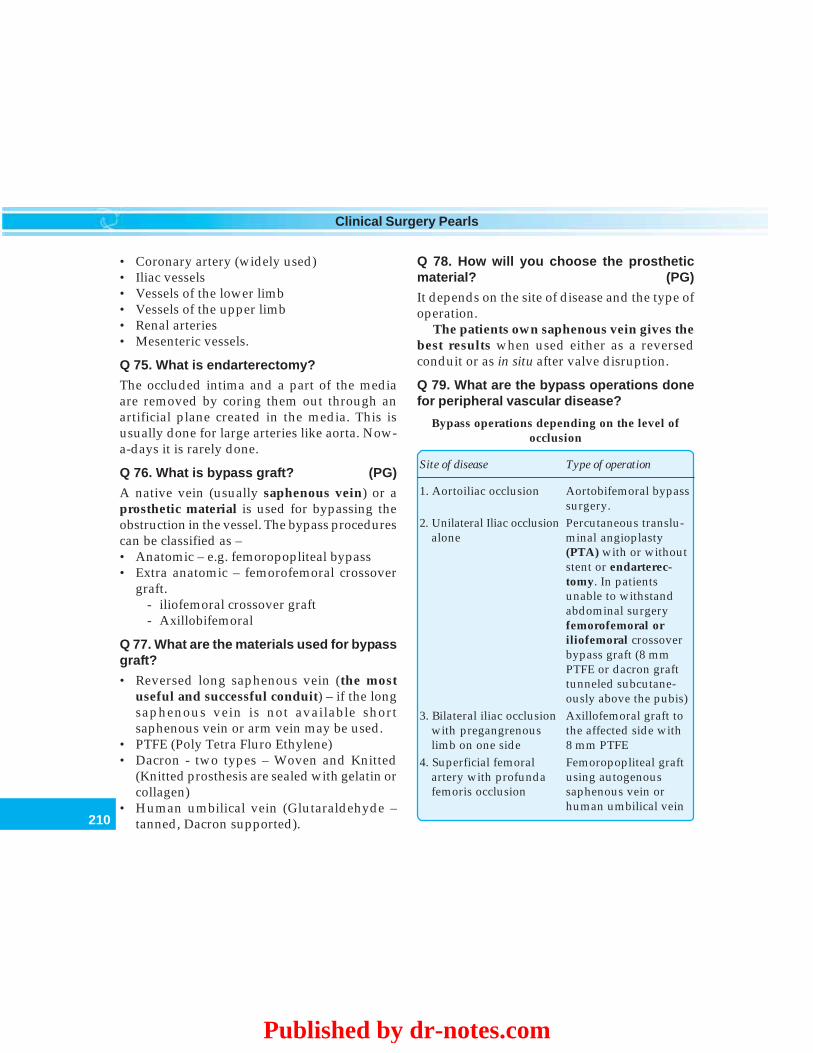
210
Clinical Surgery Pearls
• Coronary artery (widely used)• Iliac vessels• Vessels of the lower limb• Vessels of the upper limb• Renal arteries• Mesenteric vessels.
Q 75. What is endarterectomy?
The occluded intima and a part of the mediaare removed by coring them out through anartificial plane created in the media. This isusually done for large arteries like aorta. Now-a-days it is rarely done.
Q 76. What is bypass graft? (PG)
A native vein (usually saphenous vein) or aprosthetic material is used for bypassing theobstruction in the vessel. The bypass procedurescan be classified as –• Anatomic – e.g. femoropopliteal bypass• Extra anatomic – femorofemoral crossover
graft.- iliofemoral crossover graft- Axillobifemoral
Q 77. What are the materials used for bypassgraft?
• Reversed long saphenous vein (the mostuseful and successful conduit) – if the longsaphenous vein is not available shortsaphenous vein or arm vein may be used.
• PTFE (Poly Tetra Fluro Ethylene)• Dacron - two types – Woven and Knitted
(Knitted prosthesis are sealed with gelatin orcollagen)
• Human umbilical vein (Glutaraldehyde –tanned, Dacron supported).
Q 78. How will you choose the prostheticmaterial? (PG)
It depends on the site of disease and the type ofoperation.
The patients own saphenous vein gives thebest results when used either as a reversedconduit or as in situ after valve disruption.
Q 79. What are the bypass operations donefor peripheral vascular disease?
Bypass operations depending on the level ofocclusion
Site of disease Type of operation
1. Aortoiliac occlusion Aortobifemoral bypasssurgery.
2. Unilateral Iliac occlusion Percutaneous translu-alone minal angioplasty
(PTA) with or withoutstent or endarterec-tomy. In patientsunable to withstandabdominal surgeryfemorofemoral oriliofemoral crossoverbypass graft (8 mmPTFE or dacron grafttunneled subcutane-ously above the pubis)
3. Bilateral iliac occlusion Axillofemoral graft towith pregangrenous the affected side withlimb on one side 8 mm PTFE
4. Superficial femoral Femoropopliteal graftartery with profunda using autogenousfemoris occlusion saphenous vein or
human umbilical vein
Published by dr-notes.com
211
Peripheral Occlusive Vascular Disease
Note:• For long distance claudication conservative
treatment is wise.• Salvage operations should not be performed
for intermittent caudication alone. Gangreneand loss of limb may occur if the operationfails.
• Any vein used for anastomosis requires adiameter of at least 3mm.
• The size of suture material used foranatomosis from groin down is 4/0-5/0polypropylene.
• Femorodistal bypasses are usually carried outusing long saphenous vein preferably in situ.
Q 80. What is in situ bypass graft? (PG)
This is done for femoropopliteal bypass graft.The long saphenous vein is not removed andreversed in this operation. Instead it is left inplace (in situ) and the valves disrupted with avalvulotome. The vein is then anastomosed tothe femoral artery proximally and to thepopliteal vessel distally.
Q 81. What is profunda plasty? (PG)
The common femoral artery and its branches areexposed. After giving heparin IV and clampingthe vessel an incision is made into the commonfemoral artery and carried down into theprofunda dividing the stenotic profunda origin.The arteriotomy is then closed with a patch ofvein to widen the narrowed segment.
Q 82. What is the role of lumbar sympathec-tomy?
This should only be done if there is no chancefor direct arterial surgery or angioplasty. It has
no role in the treatment of intermittentclaudication. The indications are:• Nonhealing ischemic ulcers• Ischemic rest pain• Vasospastic conditions• Hyperhidrosis• Causalgia.
Q 83. What is lumbar sympathectomy?
In lumbar sympathectomy the second, third andfourth lumbar sympathetic ganglia areremoved. The removal of bilateral first lumbarganglia will result in retrograde ejaculation. Theoperation can be done either by open orlaparoscopic method. The open method is doneby extra-peritoneal approach using a loinincision. The sympathetic trunk lies on the sideof the bodies of the lumbar vertebrae on the rightside overlapped by IVC. Care is taken not tomistake and remove small lymph nodes andtendinous strip of psoasminor. It is alsoimportant to avoid the genitofemoral nerve.
Q 84. What is chemical sympathectomy?
Under radiographic fluoroscopic control withthe patient in the lateral position after givinglocal anesthesia a long spinal needle is insertedto seek the side of vertebral body to reach thelumbar sympathetic chain. 5ml of phenol inwater (1:16) is injected after confirming theneedle position by injecting contrast agent. It iscarried out in two sites beside the bodies ofsecond to fourth lumbar vertebrae. It isimportant to avoid aorta, IVC and ureters.It is contraindicated in patients takinganticoagulants.
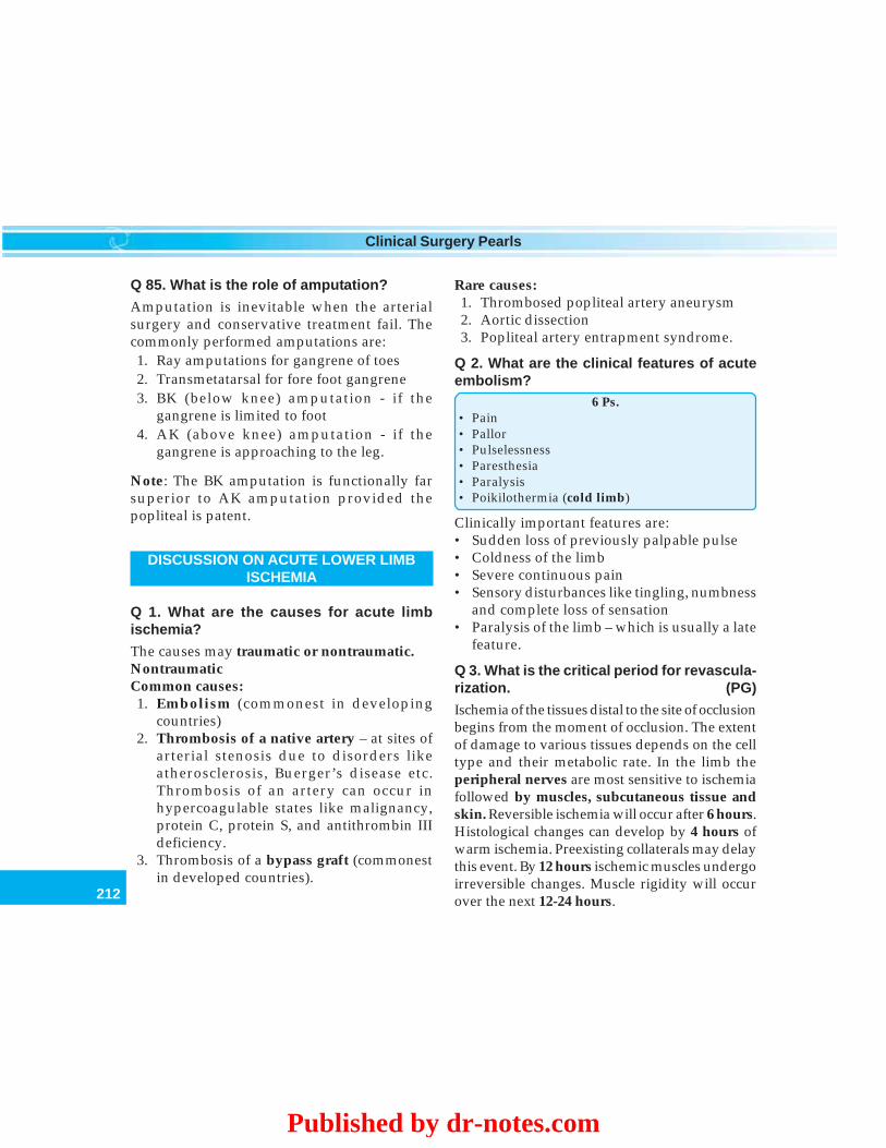
212
Clinical Surgery Pearls
Q 85. What is the role of amputation?
Amputation is inevitable when the arterialsurgery and conservative treatment fail. Thecommonly performed amputations are:1. Ray amputations for gangrene of toes2. Transmetatarsal for fore foot gangrene3. BK (below knee) amputation - if the
gangrene is limited to foot4. AK (above knee) amputation - if the
gangrene is approaching to the leg.
Note: The BK amputation is functionally farsuperior to AK amputation provided thepopliteal is patent.
DISCUSSION ON ACUTE LOWER LIMBISCHEMIA
Q 1. What are the causes for acute limbischemia?
The causes may traumatic or nontraumatic.NontraumaticCommon causes:1. Embolism (commonest in developing
countries)2. Thrombosis of a native artery – at sites of
arterial stenosis due to disorders likeatherosclerosis, Buerger’s disease etc.Thrombosis of an artery can occur inhypercoagulable states like malignancy,protein C, protein S, and antithrombin IIIdeficiency.
3. Thrombosis of a bypass graft (commonestin developed countries).
Rare causes:1. Thrombosed popliteal artery aneurysm2. Aortic dissection3. Popliteal artery entrapment syndrome.
Q 2. What are the clinical features of acuteembolism?
6 Ps.• Pain• Pallor• Pulselessness• Paresthesia• Paralysis• Poikilothermia (cold limb)
Clinically important features are:• Sudden loss of previously palpable pulse• Coldness of the limb• Severe continuous pain• Sensory disturbances like tingling, numbness
and complete loss of sensation• Paralysis of the limb – which is usually a late
feature.
Q 3. What is the critical period for revascula-rization. (PG)
Ischemia of the tissues distal to the site of occlusionbegins from the moment of occlusion. The extentof damage to various tissues depends on the celltype and their metabolic rate. In the limb theperipheral nerves are most sensitive to ischemiafollowed by muscles, subcutaneous tissue andskin. Reversible ischemia will occur after 6 hours.Histological changes can develop by 4 hours ofwarm ischemia. Preexisting collaterals may delaythis event. By 12 hours ischemic muscles undergoirreversible changes. Muscle rigidity will occurover the next 12-24 hours.
Published by dr-notes.com
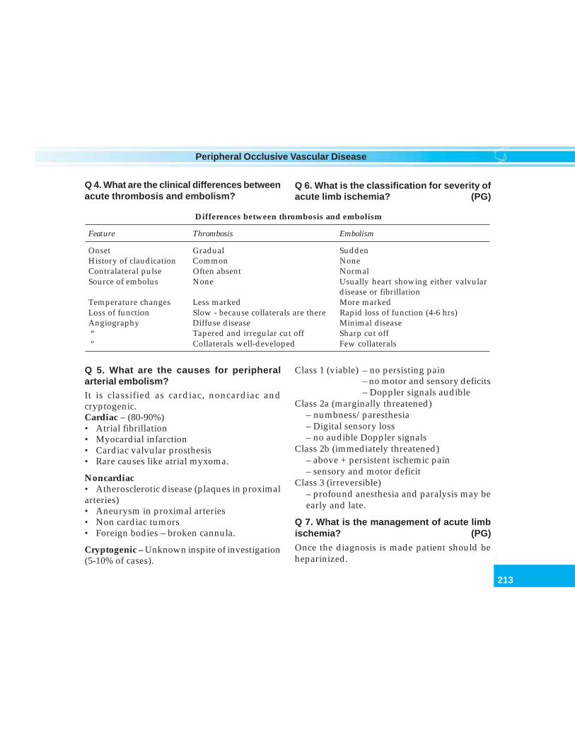
213
Peripheral Occlusive Vascular Disease
Q 4. What are the clinical differences betweenacute thrombosis and embolism?
Differences between thrombosis and embolism
Feature Thrombosis Embolism
Onset Gradual SuddenHistory of claudication Common NoneContralateral pulse Often absent NormalSource of embolus None Usually heart showing either valvular
disease or fibrillationTemperature changes Less marked More markedLoss of function Slow - because collaterals are there Rapid loss of function (4-6 hrs)Angiography Diffuse disease Minimal disease “ Tapered and irregular cut off Sharp cut off “ Collaterals well-developed Few collaterals
Q 5. What are the causes for peripheralarterial embolism?
It is classified as cardiac, noncardiac andcryptogenic.Cardiac – (80-90%)• Atrial fibrillation• Myocardial infarction• Cardiac valvular prosthesis• Rare causes like atrial myxoma.
Noncardiac• Atherosclerotic disease (plaques in proximalarteries)• Aneurysm in proximal arteries• Non cardiac tumors• Foreign bodies – broken cannula.
Cryptogenic – Unknown inspite of investigation(5-10% of cases).
Class 1 (viable) – no persisting pain– no motor and sensory deficits– Doppler signals audible
Class 2a (marginally threatened)– numbness/paresthesia– Digital sensory loss– no audible Doppler signals
Class 2b (immediately threatened)– above + persistent ischemic pain– sensory and motor deficit
Class 3 (irreversible)– profound anesthesia and paralysis may beearly and late.
Q 7. What is the management of acute limbischemia? (PG)
Once the diagnosis is made patient should beheparinized.
Q 6. What is the classification for severity ofacute limb ischemia? (PG)
214
Clinical Surgery Pearls
Contrast angiography is still considered thegold standard investigation of choice.
C-arm facility and peroperative angiogramsare necessary.
The site of entry for angiogram should bepreferably the contralateral limb.
In case of bilateral lower limb problem thebrachial artery should be the entry point.
Immediate revascularization is requiredeither by surgical (embolectomy by Fogarty oropen thrombectomy with or without bypassprocedures) or endovascular proceduresClass 1 and 2 – angiography and
thrombolysisClass 2b and 3 early– emergency surgeryClass 3 late – delayed amputationFor distal embolism thrombolytic therapy ispreferred.
Q 8. What is embolectomy?
• The femoral artery is exposed under generalanesthesia or local anesthesia.
• Longitudinal arteriotomy is made afterplacing vascular clamps.
• A Fogarty catheter of appropriate size ispassed proximally, the bulb inflated, andcatheter gently pulled down to extract theblood clot. This is repeated until a goodantegrade flow is obtained.
• The same procedure is repeated in the distalartery until good back flow is obtained.
• Heparin is given IV. (5000 units) andanticoagulation is continued postoperatively
• Arteriotomy is closed by continuous suturewith 5/0 or 6/0 polypropelene.
• Fasciotomy of the distal muscle is recommen-ded if the ischemic time has exceeded 6 hours.
Q 9. What is thrombolytic therapy? (PG)
This will bring about lysis of fibrin bystimulating the plasmin system. It cannot bedone in a limb threatened with gangrene. Itmay take upto 48 hours to lyse the thrombus.Old clots cannot be lysed by thrombolytictherapy. Duration of the procedure is lengthy.The commonly used agents are:• Streptokinase (A bacterial enzyme) – Cheap
but antigenic – anaphylaxis is a problem.• Urokinase (Extracted from renal
parenchymal cells).• Tissue Plasminogen Activator (TPA-
Synthetic).
Q 10. What are the contraindications forThrombolytic therapy? (PG)
• Recent stroke• Recent surgery• History of GI hemorrhage• Pregnancy• Uncontrolled hypertension• Allergy to the agent.
Published by dr-notes.com
215
Lymphoma
15 Lymphoma
CASE CAPSULE
A 20-year-old male patient presents with weightloss, malaise, pyrexia of unknown origin anddrenching night sweats that require change ofcloths for the last 9 months. There is no historyof pruritus, nor alcohol intolerance. There is noabdominal pain after drinking alcohol. He alsocomplains of bilateral supraclavicular swellingsfor the last 7 months which is painless. There isno history of bone pain.
On examination the patient is febrile andpale. There is a 10 × 6cm size swelling in theright supraclavicular region and 12 × 8cm sizeswelling in the left supraclavicular region. Onpalpation, there is no local rise of temperaturenor tenderness over the swelling. It appears tobe a group of lymph nodes on either side. It ispossible to define individual nodes and theyare ovoid, smooth and discrete. The nodes arehaving rubbery consistency. These nodes canbe moved from side to side. There is no fixityof the nodes. The abdominal examinationrevealed enlargement of the liver, about 4cmbelow the costal margin, firm in consistency, the
surface is smooth and edge is sharp. The spleenis also is enlarged. No palpable abdominalnodes detected. There is no free fluid in theabdominal cavity. Examination of other lymphnode areas like axillary and inguinal are normal.There is no evidence of collaterals veins acrossthe chest wall suggestive of mediastinal mass.There is no venous congestion of the neck.Examination of the genitalia is normal.
Read the checklist for examination ofabdomen.
Checklist for lymph node examination in sectionon short casesMnemonics for lymph node examination –‘PALS’(short case section)
P – Primary (examine the drainage area of thelymph node for a primary lesion)
A – Another node (always look for another nodewhen one group is palpable)
L – Liver (always examine the abdomen forenlargement of liver)
S – Spleen
216
Clinical Surgery Pearls
Remember the 3 lymphatic water sheds of theskin (on each side):A vertical line through the sagittal plane of thebody divides the lymphatic drainage of the skininto 3 areas on each side. There is somecommunications across the midline. The 3 areason each side of the body are demarcated fromeach other by two horizontal lines one at thelevel of the clavicle, the other at the level ofthe umbilicus. These lines are the lymphaticwater sheds and the skin lymph flows in adirection away from them. Each line alsorepresents the meeting place of adjacentterritories, so that the cancer situated in one ofthese lines may spread by two routes along thelymphatics running away from the water shed,E.g. cancer situated the umbilicus may spreadin four directions. Viz: towards both axillaeand both groins, as lymphatics draining to theseglands come into communication at theumbilicus. Similarly a cancer situated anywherein the midline of the surface of the body mayspread in at least two directions because of thelymphatic communication across the midline.Thus we get 3 groups of lymph nodes drainingthe three areas.
1. Cervical lymph nodes2. Axillary lymph nodes3. Inguinal lymph nodes
• The lymphatic above the clavicle will drainto the cervical nodes.
• The lymphatics below the clavicle upto thelevel of umbilicus will drain to the axilla (bothfront and back).
• The lymphatics below the level of umbilicuswill drain to the inguinal nodes.
• If axillary node is enlarged, examine thefollowing drainage areas – breast, front of thechest and upper abdomen upto theumbilicus, back of the chest down upto thelevel of umbilicus, and the entire upper limb.
• If the inguinal lymph node is enlarged,examine the following drainage areas – thelower abdomen from the level of umbilicus,the corresponding area in the back, the glutealregion, natal cleft, perineum, genitalia, thelower part of the anal canal and the entirelower limb. The lower limb lymphatics willdrain to the vertical group of nodes. Theremaining areas will drain to the horizontalgroup of inguinal nodes. A digital rectalexamination must be done in all cases ofhorizontal inguinal lymph node enlargement.
• When the cervical lymph node is enlargedthe drainage areas to be examined will startfrom the scalp down to the level of theclavicle, including examination of the oralcavity, pharynx (oral and nasal) nasal sinusesand the hidden areas.
Checklist for history
• H/o. weight loss (> 10% loss of weight in 6months)
• H/o. Fever occurring in a periodic fashion (Pel– Ebstein fever)
• H/o . Pruritus• Alcohol induced nodal pain• Bone pain (lymphomatous infiltration)• Pallor• Family history of leukemias and similar illness
Published by dr-notes.com
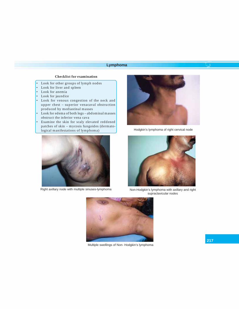
217
Lymphoma
Checklist for examination
• Look for other groups of lymph nodes• Look for liver and spleen• Look for anemia• Look for jaundice• Look for venous congestion of the neck and
upper chest – superior venacaval obstructionproduced by mediastinal masses
• Look for edema of both legs – abdominal massesobstruct the inferior vena cava
• Examine the skin for scaly elevated reddenedpatches of skin – mycosis fungoides (dermato-logical manifestations of lymphoma) Hodgkin’s lymphoma of right cervical node
Multiple swellings of Non- Hodgkin’s lymphoma
Right axillary node with multiple sinuses-lymphoma Non-Hodgkin’s lymphoma with axillary and rightsupraclavicular nodes
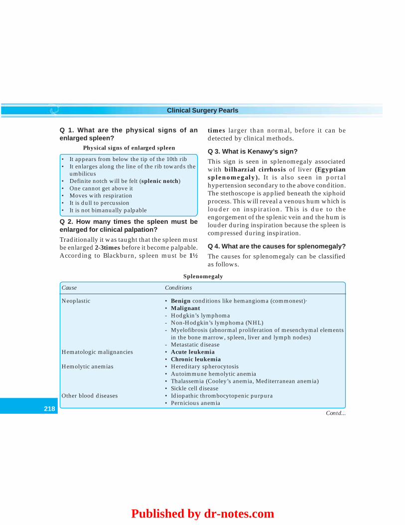
218
Clinical Surgery Pearls
Q 1. What are the physical signs of anenlarged spleen?
Physical signs of enlarged spleen
• It appears from below the tip of the 10th rib• It enlarges along the line of the rib towards the
umbilicus• Definite notch will be felt (splenic notch)• One cannot get above it• Moves with respiration• It is dull to percussion• It is not bimanually palpable
Q 2. How many times the spleen must beenlarged for clinical palpation?
Traditionally it was taught that the spleen mustbe enlarged 2-3times before it become palpable.According to Blackburn, spleen must be 1½
Contd...
times larger than normal, before it can bedetected by clinical methods.
Q 3. What is Kenawy’s sign?
This sign is seen in splenomegaly associatedwith bilharzial cirrhosis of liver (Egyptiansplenomegaly). It is also seen in portalhypertension secondary to the above condition.The stethoscope is applied beneath the xiphoidprocess. This will reveal a venous hum which islouder on inspiration. This is due to theengorgement of the splenic vein and the hum islouder during inspiration because the spleen iscompressed during inspiration.
Q 4. What are the causes for splenomegaly?
The causes for splenomegaly can be classifiedas follows.
Splenomegaly
Cause Conditions
Neoplastic • Benign conditions like hemangioma (commonest)·• Malignant- Hodgkin’s lymphoma- Non-Hodgkin’s lymphoma (NHL)- Myelofibrosis (abnormal proliferation of mesenchymal elements
in the bone marrow, spleen, liver and lymph nodes)- Metastatic disease
Hematologic malignancies • Acute leukemia• Chronic leukemia
Hemolytic anemias • Hereditary spherocytosis• Autoimmune hemolytic anemia• Thalassemia (Cooley’s anemia, Mediterranean anemia)• Sickle cell disease
Other blood diseases • Idiopathic thrombocytopenic purpura• Pernicious anemia
Published by dr-notes.com
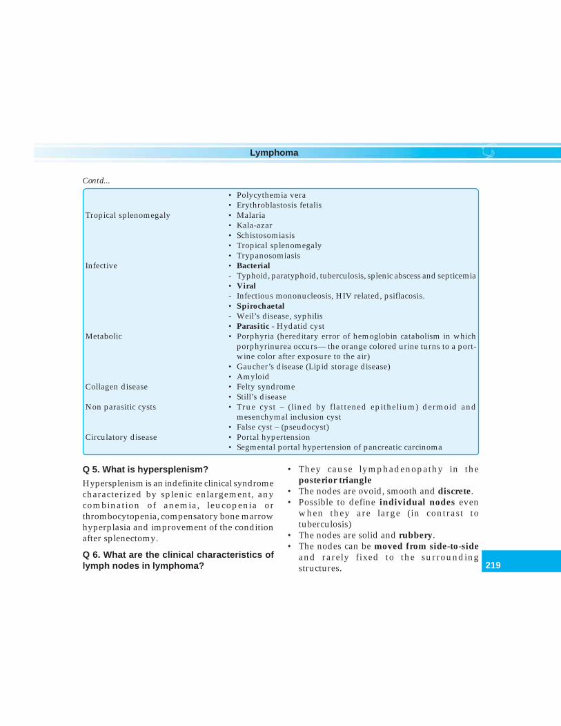
219
Lymphoma
• Polycythemia vera• Erythroblastosis fetalis
Tropical splenomegaly • Malaria• Kala-azar• Schistosomiasis• Tropical splenomegaly• Trypanosomiasis
Infective • Bacterial- Typhoid, paratyphoid, tuberculosis, splenic abscess and septicemia• Viral- Infectious mononucleosis, HIV related, psiflacosis.• Spirochaetal- Weil’s disease, syphilis• Parasitic - Hydatid cyst
Metabolic • Porphyria (hereditary error of hemoglobin catabolism in whichporphyrinurea occurs— the orange colored urine turns to a port-wine color after exposure to the air)
• Gaucher’s disease (Lipid storage disease)• Amyloid
Collagen disease • Felty syndrome• Still’s disease
Non parasitic cysts • True cyst – (lined by flattened epithelium) dermoid andmesenchymal inclusion cyst
• False cyst – (pseudocyst)Circulatory disease • Portal hypertension
• Segmental portal hypertension of pancreatic carcinoma
Contd...
Q 5. What is hypersplenism?
Hypersplenism is an indefinite clinical syndromecharacterized by splenic enlargement, anycombination of anemia, leucopenia orthrombocytopenia, compensatory bone marrowhyperplasia and improvement of the conditionafter splenectomy.
Q 6. What are the clinical characteristics oflymph nodes in lymphoma?
• They cause lymphadenopathy in theposterior triangle
• The nodes are ovoid, smooth and discrete.• Possible to define individual nodes even
when they are large (in contrast totuberculosis)
• The nodes are solid and rubbery.• The nodes can be moved from side-to-side
and rarely fixed to the surroundingstructures.
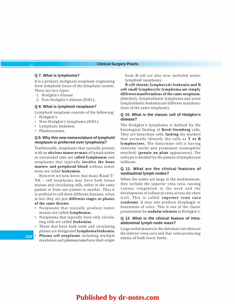
220
Clinical Surgery Pearls
Q 7. What is lymphoma?
It is a primary malignant neoplasm originatingfrom lymphoid tissue of the lymphatic system.There are two types:1. Hodgkin’s disease2. Non-Hodgkin’s disease (NHL).
Q 8. What is lymphoid neoplasm?
Lymphoid neoplasm consists of the following:• Hodgkin’s• Non-Hodgkin’s lymphoma (NHL)• Lymphatic leukemia• Plasmacytoma.
Q 9. Why this new nomenclature of lymphoidneoplasm is preferred over lymphoma?
Traditionally, neoplasms that typically presentwith an obvious tumor or mass of lymph nodesor extranodal sites are called lymphomas andneoplasms that typically involve the bonemarrow and peripheral blood without tumormass are called leukemias.
However we now know that many B and T/NK – cell neoplasms may have both tissuemasses and circulating cells, either in the samepatient or from one patient to another. Thus itis artificial to call them different diseases, whenin fact they are just different stages or phasesof the same disease.• Neoplasms that typically produce tumor
masses are called lymphomas.• Neoplasms that typically have only circula-
ting cells are called leukemias.• Those that have both solid and circulating
phases are designated lymphoma/leukemia.• Plasma cell neoplasms including multiple
myelomas and plasmacytoma have their origin
from B cell are also now included underlymphoid neoplasms.B cell chronic lymphocytic leukemia and B
cell small lymphocytic lymphoma are simplydifferent manifestations of the same neoplasm.(Similarly, lymphoblastic lymphoma and acutelymphoblastic leukemia are different manifesta-tions of the same neoplasm).
Q 10. What is the classic cell of Hodgkin’sdisease?
The Hodgkin’s lymphoma is defined by thehistological finding of Reed–Sternberg cells.They are binucleate cells, lacking the markersthat normally identify the cells as T or Blymphocytes. The binucleate cell is havingvesicular nuclei and prominent eosinophilicneucleoli (pennie on plate appearance). Thesubtype is decided by the pattern of lymphocyteinfiltrate.
Q 11. What are the clinical features ofmediastinal lymph nodes?
When the nodes are large in the mediastinum,they occlude the superior vena cava, causingvenous congestion in the neck and thedevelopment of collateral veins across the chestwall. This is called superior vena cavasyndrome. It may also produce dysphagia orhoarseness of voice. This is one of the classicpresentation for nodular sclerosis in Hodgkin’s.
Q 12. What is the clinical feature of intra-abdominal lymph node mass?
Large nodal masses in the abdomen can obstructthe inferior vena cava and iliac veins producingedema of both lower limbs.
Published by dr-notes.com
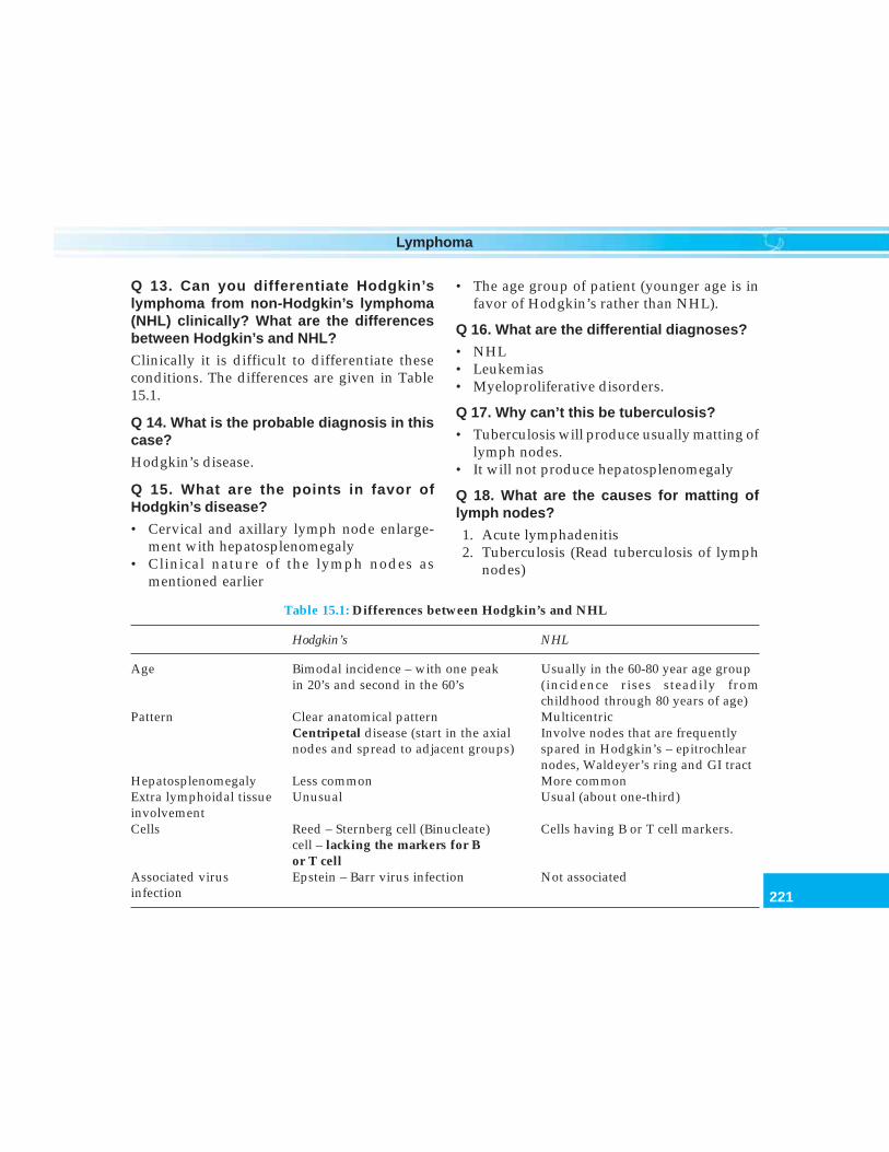
221
Lymphoma
Q 13. Can you differentiate Hodgkin’slymphoma from non-Hodgkin’s lymphoma(NHL) clinically? What are the differencesbetween Hodgkin’s and NHL?
Clinically it is difficult to differentiate theseconditions. The differences are given in Table15.1.
Q 14. What is the probable diagnosis in thiscase?
Hodgkin’s disease.
Q 15. What are the points in favor ofHodgkin’s disease?
• Cervical and axillary lymph node enlarge-ment with hepatosplenomegaly
• Clinical nature of the lymph nodes asmentioned earlier
• The age group of patient (younger age is infavor of Hodgkin’s rather than NHL).
Q 16. What are the differential diagnoses?
• NHL• Leukemias• Myeloproliferative disorders.
Q 17. Why can’t this be tuberculosis?
• Tuberculosis will produce usually matting oflymph nodes.
• It will not produce hepatosplenomegaly
Q 18. What are the causes for matting oflymph nodes?
1. Acute lymphadenitis2. Tuberculosis (Read tuberculosis of lymph
nodes)
Table 15.1: Differences between Hodgkin’s and NHL
Hodgkin’s NHL
Age Bimodal incidence – with one peak Usually in the 60-80 year age groupin 20’s and second in the 60’s (incidence rises steadily from
childhood through 80 years of age)Pattern Clear anatomical pattern Multicentric
Centripetal disease (start in the axial Involve nodes that are frequentlynodes and spread to adjacent groups) spared in Hodgkin’s – epitrochlear
nodes, Waldeyer’s ring and GI tractHepatosplenomegaly Less common More commonExtra lymphoidal tissue Unusual Usual (about one-third)involvementCells Reed – Sternberg cell (Binucleate) Cells having B or T cell markers.
cell – lacking the markers for Bor T cell
Associated virus Epstein – Barr virus infection Not associatedinfection
222
Clinical Surgery Pearls
3. Late stages of lymphoma4. Late stages of metastasis.
Q 19. What is the WHO Classification oflymphoma (REAL classification)?
Revised European – American classification ofLymphoid neoplasms).
It is a list of distinct disease entities, whichare defined by combination of morphology,immuno-phenotype and genetic features andwhich have distinct clinical features. Theclassification includes all lymphoid neoplasms:Hodgkin’s lymphoma, non Hodgkin’slymphoma, lymphoid leukemias and plasmacell neoplasms.
B CELL NEOPLASMS
Precursor B-cell Neoplasm
• Precursor – B – lymphoblastic leukemia/lymphoma (precursor B-cell acutelymphoblastic leukemia).
Mature (peripheral) B-cell neoplasms• B – cell chronic lymphocytic leukemia/small
lymphocytic lymphoma• B – cell prolymphocytic leukemia• Lymphoplasmacytic lymphoma• Splenic marginal zone B-cell lymphoma (with
or without villous lymphocytes)• Hairy cell leukemia• Plasma cell myeloma/plasmacytoma• Extranodal marginal zone B-cell lymphoma
of MALT type
• Nodal marginal zone B-cell lymphoma (withor without monocytoid B cells)
• Follicular lymphoma• Mantle cell lymphoma• Diffuse large B-cell lymphoma• Burkitt lymphoma/Burkitt cell leukemia.
T CELL AND NK CELL NEOPLASMS
Precursor T Cell Neoplasm
• Precursor T-lymphoblastic lymphoma/leukemia (precursor T-cell acute lympho-blastic leukemia).
Mature (peripheral) T/NK-cell neoplasms• T-cell prolymphocytic leukemia• T-cell granular lymphocytic leukemia• Aggressive NK-cell leukemia• Adult T cell lymphoma/leukemia (HTLVI +)• Extranodal NK/T cell lymphoma, nasal type• Enteropathy – type T – cell lymphoma• Hepatosplenic γδ T – cell lymphoma• Subcutaneous panniculitis – like T – cell
lymphoma• Mycosis fungoides / Sezary syndrome• Anaplastic large cell lymphoma, T/null cell,
primary cutaneous type• Peripheral T – cell lymphoma, not otherwise
characterized• Angioimmunoblastic T – cell lymphoma• Anaplastic large cell lymphoma, T/null cell,
primary systemic type.
Q 20. What is the staging of Hodgkin’slymphoma?
Published by dr-notes.com
223
Lymphoma
Ann – Arbor staging
Stage Description
Stage I Involvement of a single lymph noderegion (I), or localized involvement of asingle extralymphatic organ or site in theabsence of any lymph node involvement(1E.) (rare in Hodgkin’s lymphoma)
Stage II Involvement of two or more lymph noderegions on the same side of thediaphragm (II); or localized involvementof a single extralymphatic organ or sitein association with regional lymphnode involvement with or withoutinvolvement of other lymph noderegions on the same side of thediaphgram (IIE). The number of regionsinvolved may be indicated by asubscript, as in, for example, II3
Stage III Involvement of lymph node regionson both sides of the diaphragm (III),which also may be accompanied byextralymphatic extension in associationwith adjacent lymph node involvement(IIIE) or by involvement of the spleen(IIIS) or both (III E,S).
Stage IV Diffuse or disseminated involvement ofone or more extra lymphatic organs, withor without associated lymph nodeinvolvement or isolated extralymphaticorgan involvement in the absence ofadjacent regional lymph nodeinvolvement, but in conjunction withdisease in distant site (s). Anyinvolvement of the liver or bonemarrow or nodular involvement of thelung(s). The location of stage IV diseaseis defined further by specifying the siteaccording to the notation.
Note: All stages are having A/B stagesdepending on the absence or presence of Bsymptoms.
Q 21. What are B symptoms?
The B symptoms are:• Weight loss – unexplained weight loss of >
10% of the usual body weight in 6 monthsprior to diagnosis.
• Fever – unexplained fever with temperature> 38oC
• Drenching night sweats – that require changeof bedclothes.
Note: Pruritus alone does not qualify for Bclassification nor does alcohol intolerance,fatigue or a short febrile illness associated withsuspected infection.
Q 22. What are the pathological types ofHodgkin’s disease?
Major categories of Hodgkin’s lymphoma are:• Nodular lymphocyte predominance
Hodgkin’s lymphoma (NLPHL)• Classic Hodgkin’s Lymphoma (CHL) –
further divisions given below:
Classic Hodgkin’s Lymphoma
Sub type Characteristics1. Lymphocyte rich classic Uncommon-6%. Few
Hodgkin’s lymphoma Reed-Sternberg cells(LRCHL) Diffuse lymphocytes –
excellent prognosis2. Lymphocyte depletion Rare-2%Abundant Reed-
Hodgkin’s lymphoma Sternberg cells(LDHL) Paucity of lymphocyteOlder males affectedAggressive
224
Clinical Surgery Pearls
Note:• Hodgkin’s lymphoma rarely presents in
extra-nodal site• About 25% of NHL are extranodal at
presentation• Mycosis fungoides (a type of NHL affecting
the skin) and MALT lymphomas are virtuallyalways extranodal
Q 25. What is the ‘E Lesion’for Hodgkin’slymphoma?
• The Ann Arbor system defined E as extralymphatic.
• Disease in sites such as Waldeyer’s ring, thethymus, and the spleen although extranodalis not extralymphatic and therefore notconsidered to be ‘E Lesion’.According to the revised AJCC system the E
Lesion is defined as disease that involves extralymphatic sites (s) adjacent to site (s) oflymphatic involvement but in which directextension is not necessarily demonstrable.Examples of E lesions include extension into:• Pulmonary parenchyma from adjacent
pulmonary hilar/mediastinal nodes• Extension into the antierior chest wall and
into the pericardium from mediastinal mass• Involvement of iliac bone from adjacent
lymph node• Involvement of lumbar vertebral body from
paraaortic nodes• Involvement of pleura from internal
mammary nodes• Involvement of thyroid from adjacent
cervical lymph nodes.
3.Mixed cellularity Common 20 – 25%Hodgkin’s lymphoma Often presents with(MCHL) disseminated disease
4.Nodular sclerosis Commonest – 70%Hodgkin’s lymphoma Fibrosis present, Reed-(NSHL) Sternberg cells andlymphocytes seen. Youngwomen affected Cervical+ Mediastinal disease
Q 23. What are the features of the lymphocytepredominant Hodgkin’s lymphoma?
• Considered to be an indolent B cell lymphoma• More common in young men and old ages• Infradiaphragmatic disease common than
Hodgkin’s disease• Bulky disease and splenic involvement
uncommon• Natural history like low grade NHL• Can have delayed relapses• Can progress to large B cell lymphomas.
Q 24. What is nodal/extranodal disease?
For the purpose of coding and staging, lymphnodes, Waldeyer’s ring, and spleen areconsidered nodal or lymphatic sites.
Extranodal or extralymphatic sites includethe following:• Bone marrow• Gastrointestinal tract• Skin• Bone• Central nervous system• Lung• Gonads• Ocular adnexae• Liver• Kidneys and uterus
Published by dr-notes.com
225
Lymphoma
Note: It is proposed that for NHL, the Edesignation should indicate the presentationof lymphoma in extranodal sites and the lackof an E designation should indicate lymphomapresenting in lymph nodes.
Q 26. In which core nodal region, theinfraclavicular nodes are included (Read corenodal regions in NHL section)?
It is considered as part of the axilla.
Q 27. What are the other lymphatic structuresother than lymph nodes?
The lymphatic structures in the body:
• Lymph nodes• Spleen• Appendix• Peyer’s patches• Waldeyer’s ring• Thymus
Q 28. What is splenic involvement inHodgkin’s disease?
• Evidence of one or more nodules in the spleenof any size on imaging evaluation
• Histologic involvement documented bybiopsy or splenectomy.
Note: Splenic enlargement alone is insufficientto support a dignosis of splenic involvement.
Q 29. What is hepatic involvement inHodgkin’s?
• One or more nodules in the liver of any sizeon imaging
• Histological involvement documented bybiopsy.
Note: Hepatic enlargement alone is insufficient.
Q 30. What is bone marrow involvement?
Bone marrow involvement must be documentedby biopsy from a clinically/radiographicallyuninvolved area of bone. Bone marrowinvolvement is designated by the letter M. Bonemarrow involvement is always considered asdiffuse extralymphatic disease (stage IV).
Q 31. What are the unfavorable types ofHodgkin’s disease?
1. The presence of B symptoms2. Unfavorable histology – mixed cellularity
and lymphocyte depleted.
Q 32. What is the most important confir-matory investigation for this case?
Lymph node biopsy.
Q 33. Is there any role for FNAC of the lymphnode?
For the diagnosis of lymphoma a node biopsy isa must in order to study the histologicalbackground and architecture of the lymph node.FNAC can be done as a screening investigationif more than one node is palpable. The needleaspiration will produce anomalies in the nodeand it will be difficult for the histopathologist tointerpret this node if it is taken for biopsy later.
Q 34. Which lymph node is taken for biopsyand what are the precautions for lymph nodebiopsy?
• The largest and most central node in a groupis most likely to be diagnostic.
• The inguinal nodes are avoided because theyoften show changes of chronic infection (ifother enlarged nodes are available).

226
Clinical Surgery Pearls
• A cervical node is preferred to the axillary oringuinal group.
• Always take an intact node for biopsy.• Biopsied node should be handled gently.• Imprint of freshly cut node may be done if
this is available (imprint cytology).• Avoid nodes which are aspirated for biopsy.• For imprint cytology purpose the node
should be send in a bottle containing saline.
Q 35. What is the staging work up?
Clinical staging includes:• Careful recording of medical history• Physical examination• Imaging of chest, abdomen and pelvis• Complete blood count• Blood chemistry• Bone marrow biopsy
Recommendation for the diagnosticevaluation of patients with lymphoma.
A. Mandatory procedure
1. Biopsy, with interpretation by a qualifiedpathologist
2. History, with special attention to the presenceand duration of fever, night sweats, andunexplained loss of 10% or more of body weightin the previous 6 months
3. Physical examination4. Laboratory tests
a. Complete blood cell count and platelet countb. Erythrocyte sedimentation ratec. Liver function tests
5. Radiographic examinationa. Chest X-rayb. CT of chest, abdomen, and pelvisc. Gallium scan
6. Bone marrow biopsy
B. Ancillary procedures
1. Laparotomy and splenectomy if decisionsregarding management are likely to beinfluenced
2. Liver biopsy (needle), if there is a strong clinicalindication of hepatic involvement
3. Radioisotopic bone scans, in selected patientswith bone pain
4. CT of head and neck in extranodal or nodalpresentation to define disease extent
5. Gastroscopy and/ or GI series in patients withGI presentations
6. MRI of spine in patients with suspected spinalinvolvement
7. CSF cytology in patients with Stage IV diseaseand bone marrow involvement, testis involvement,or parameningeal involvement
Q 36. What are the indications for bonemarrow biopsy?
• B symptoms• Anemia• Leukopenia• Thrombocytopenia
Q 37. What is the role of staging laparotomy?(PG)
Staging laparotomy is not done now-a-daysafter CT has become part of the staging workup where by most of the nodal status anddisease process can be ascertained. 40% of thepatients require chemotherapy because of thereasons mentioned below and therefore astaging laparotomy is omitted.• It has got significant morbidity in 10% of
patients.• It is considered when the results are likely to
alter the treatment strategy (chemotherapyvs radiation). If the chemotherapy will be
Published by dr-notes.com
227
Lymphoma
used regardless of the finding on laparotomy,there is no need for staging laparotomy.Hodgkin’s disease which is localized may becurable by radiation therapy if all involvednodes are included in the treatment field.Non Hodgkin’s lymphomas are usuallytreated with chemotherapy because of wide-spread nodal and extra-nodal involvementespecially of the bone barrow. Thereforelaparotomy is unnecessary in NHL.
• Considered only in special situations of stageIIa or less in which mantle radiation isplanned.
• In children splenectomy will produceoverwhelming postsplenectomy sepsis(OPSI) – 0.1%/year
• 20% of the cases the stage is upstaged bystaging laparotomy and therefore will requirechemotherapy
• 20% of the patients will develop relapserequiring chemotherapy.
Q 38. What are the important steps in staginglaparotomy? (PG)
In staging laparotomy the following proceduresare done:• Splenectomy• Wedge biopsy of liver• Biopsy—nodes of the paraaortic, splenic
hilar, hepatic, portal, and iliac nodes—thesuspicious nodes are clipped.
• Oophoropexy in females (childbearing age)which will allow radiation therapy to begiven to the inverted ‘Y’ field which coversthe iliac and inguinal nodes without radiationdamage to the ovaries.
Q 39. Which are the nodal regions affectedby Hodgkin’s disease?
Mediastinal - 60%Pure infradiaphragmatic - 3%Extranodal - 5 – 10%Splenomegaly - 10%
Q 40. What are the etiological factors forHodgkin’s disease? (PG)
1. Siblings with Hodgkin’s disease2. HLA antigens3. Patients who have undergone tonsillectomy4. Immunodeficiency states5. Autoimmune disorders6. Oncogenic viruses7. Epstein – Barr virus (EBV) – Nuclear protein
in 40% of Hodgkin’s8. Lymphotropic viruses.
Q 41. What is the antigen in Hodgkin’sdisease? (PG)
It is CD 30 +ve, which is a surface antigen seenin Reed – Sternberg cells.
Q 42. What is the stage of the disease in thegiven case?
Stage III because of the infradiaphragmaticinvolvement.
Q 43. What is early stage disease?
• Stage IA and IIA• Nonbulky disease.
Q 44. What is bulky disease? (PG)
• 10 cm diameter mass or more• Mediastinal disease with transverse diameter
228
Clinical Surgery Pearls
exceeding 1/3rd of the transverse thoracicdiameter is defined as bulky disease.
Q 45. What is the treatment of Hodgkin’sdisease?
The treatment is classified as –• Radiotherapy – curative radiation therapy is
given to 3 major fields known as-– Mantle– Para-aortic– Pelvic (inverted Y fields)
• The lungs, heart, larynx, kidneys, gonadsand iliac crest are protected by shields
• The dose of radiotherapy is approximately40-45 gray delivered at the rate of 10 Gy/week
• When used with chemotherapy the dose ofradiation is reduced
Chemotherapy -Combined modality – Radiation + Chemo-therapy
Q 46. What is the indication for radiotherapy?
Localized disease - I A and II A.
Q 47. What is the indication for chemo-therapy?
Stages III and IV are generally treated withcombination chemotherapy.
Q 48. What is the management of Bsymptoms of I and II?
Even if the disease seems to be localized, Bsymptoms and unfavorable histology (mixedcellularity and lymphocyte depletion), usuallycalls for the use of chemotherapy with orwithout radiation. The relapse rates are highafter radiation therapy alone.
Q 49. What is Mantle radiation?
Irradiation of -• Cervical• Axillary• Mediastinal• Hilar lymph nodes.
Q 50. What is Subtotal Lymphoid Irradiation(STLI)? (PG)
Irradiation of Mantle area + upper abdominal+ paraaortic and splenic bed nodes.
Q 51. What is Total Lymphoid Irradiation(TLI)? (PG)
Irradiation of the pelvic field in addition to STLIis given.
Q 52. What is the indication for chemotherapyfor early stage Hodgkin’s disease? (PG)
Chemotherapy is given to eradicate subclinicaldisease outside the radiotherapy ports.
ABVD regimen is used because of the bettertoxicity profile and effectiveness.
Q 53. What is combined modality therapy?(PG)
This is done to prevent relapse, where radiationis combined with chemotherapy. Theindications are:• Unfavorable disease (Stage III and IV)• Unfavorable histology (mixed cellularity and
lymphocyte depletion type)• Extensive splenic involvement• Children who cannot tolerate full dose of
radiotherapy.
Published by dr-notes.com
229
Lymphoma
Q 54. What is the chemotherapeutic regimefor Hodgkin’s disease (HD)?
There are two regimes —• ABVD regime• MOPP regime.
Q 54. What is ABVD regime?
(6 – 8 cycles)It is a widely used regimen for Hodgkin’sdisease. The drugs used are:• Adriamycin• Bleomycin• Vinblastin• Dacarbazine.
Q 56. Why ABVD is preferred over MOPP?
• Better disease free survival and over all survival• Better toxicity profile.
Q 57. What is MOPP regime?
The drugs used in this regime for the treatmentof Hodgkin’s disease are:• Nitrogen Mustard• Vincristine (Oncovin)• Procarbazine• Prednisolone.
Q 58. What is high dose chemotherapy?(PG)
Chemotherapeutic drugs are given in high doseswith stem cell transplant (Bone marrowtransplant).
Q 59. What are the problems of chemo-therapy?
• Infertility
• Second cancers – leukemia, solid tumors likebreast and thyroid
• Cardiopulmonary complications.
Q 60. What is radioimmunotherapy? (PG)
• This is usually given for relapse for which131I labelled anti CD30 Monoclonal antibodiesare used
• Dendritic cell vaccines against EB virus• Antibodies against CD3, CD28, CD25 are also
available.
Q 61. What are the bad prognostic factors?
Bulky diseaseAnemiaHigh ESRInflammatory signsInguinal node involvementTissue eosinophiliaHigh serum LDHPathological gradeSex: MaleAge > 45WBC > 15000/cummSerum albumin < 4 gmStage 4 diseaseLymphocyte count < 600/cumm
NON-HODGKIN’S LYMPHOMA
Q 62. What is the definition of NHL?
It is a heterogenous group of B and T cellmalignancies having diversity in — cellularorigin, morphology, cytogenetic abnormalities,response to treatment and prognosis.
230
Clinical Surgery Pearls
Q 63. What are the features of NHL?
These are neoplasms of the immune systemusually presenting as disseminated and extra-nodal disease of the old age. The peculiaritiesare:1. Multicentricity2. Wide spreading3. Showing malignant cell in the blood4. Leukemic transformation may occur in 10-
15% of patients.
Q 64. What are the clinical features of NHL?
1. Asymptomatic2. B symptoms3. Enlarged lymph nodes4. Abdominal mass5. GI symptoms—pain, vomiting, bleeding6. Loss of weight.
Q 65. What are the predisposing factors?(PG)
The following predisposing factors areattributed–1. Immunosuppression2. Autoimmune diseases
– Hashimoto’s disease– Sjogren’s syndrome.
3. Infective agents– EB virus – African Burkitts– AIDS related– Nasopharyngeal lymphoma– Transplant lymphoma– Lymphoma after infectious mono-
nucleosis– HTLV-I infection -
HumanT cell
LymphotropicVirus
– HTLV -II– H. pylori – Gastric lymphoma– Hepatitis C virus – 8 and 6– Human herpes virus
4. Prior chemotherapy5. Prior radiotherapy.
Q 66. What are the congenital disordershaving increased incidence of NHL? (PG)
• Ataxia telangiectasia• Wiskott – Aldrich syndrome• Celiac disease.
Q 67. What are the core nodal regions?
They are as follows:• Right cervical (including cervical, supra-
clavicular, occipital and preauricular lymphnodes).
• Left cervical nodes• Right axillary• Left axillary• Right infraclavicular• Left infraclavicular• Mediastinal lymph nodes• Hilar lymph nodes• Paraaortic lymph nodes• Mesenteric lymph nodes• Right pelvic lymph nodes• Left pelvic lymph nodes• Right inguinofemoral lymph nodes• Left inguinofemoral lymph nodes.
Q 68. Can NHL involve any other nodes otherthan the core nodal regions?
Yes. NHL may involve the following nodes inaddition.
Published by dr-notes.com
231
Lymphoma
• Epitrochlear lymph nodes• Popliteal lymph nodes• Internal mammary lymph nodes• Occipital lymph nodes• Submental lymph nodes• Preauricular lymph nodes• Many other small nodal areas.
Q 69. What is the E designation for NHL?
For NHL the E designation should indicate thepresentation of lymphoma in extranodal siteand the lack of E designation should indicatelymphomas presenting in lymph nodes. Forexample, Lymphoma presenting in thyroidgland with cervical lymph node involvementshould be staged as IIE. Lymphoma presentingonly in cervical node is Stage I.
Q 70. What are the extranodal sites?
Extranodal sites
• Paranasal sinuses• Thyroid• GI tract• Liver• Testicles• Skin• Bone marrow• Bone• CNS• Lung• Gonads• Kidneys• Uterus• Ocular adnexae
Q 71. What are the classifications for NHL?
1. Rappaport2. Working classification
– Low grade – B cell lymphomas– Intermediate grade – B cell or T cell
(lymphoblastic group)– High grade.
3. REAL (Revised European - AmericanLymphoma)/ WHO classification• B cell — precursor B cell cancers
—peripheral B cell neoplasms –Burkitt’s
• T cell and natural killer cells (NK)- Precursor T cell neoplasm- Peripheral T cell and NK cell neoplasm
– Mycosis fungoides- Intestinal T cell lymphoma.
4. Functional classification (for treatmentpurposes).• Indolent – smaller cells, differentiated cells
- low grade, very difficult to cure- progress to more aggressive type
• Aggressive—larger cells, less differen-tiated- Better chance for cure.
• Highly aggressive.
Q 72. Since low grade lymphomas can prog-ress to aggressive type, what are the criterionfor suspecting a disease transformation?
(PG)
1. Increased LDH2. Rapid enlargement of the nodes3. Constitutional symptoms4. Extranodal disease.
Q 73. What is Burkitt’s lymphoma?
It is a B cell tumor.
Q 74. What is Mycosis fungoides?
It has a T cell origin and will manifest as skin lesion.
232
Clinical Surgery Pearls
Q 75. What is the staging of NHL?
The Ann Arbor – classification is used forstaging (see Hodgkin’s disease).
Q 76. What is stage IV disease?
By convention, any involvement of the followingcalls for classification as stage IV disease.
Stage IV disease
• Bone marrow• Liver• Pleura• CSF
Q 77. What are the investigations requiredfor diagnostic evaluation of NHL?
Read the chart for Hodgkin’s lymphoma.
Q 78. What is the treatment of NHL?
Chemotherapy is the primary modality oftreatment in NHL, because NHL spreads widelyand there is early hematogenous spread. Forlocalized disease radiotherapy may be useful.
Indolent lymphoma – treatmenta. Localized – Radiotherapy
Disseminated – rarely curativeb. Palliation – watch and wait until they
progress to more aggressive type.The quality of life is better here.
Aggressive– stage I and II - Radiation alone– stage III and IV – CHOP regimen– high dose chemotherapy – with autologous
marrow transplantation.
Q 79. What is the chemotherapeutic regimefor NHL?
NHL is generally treated with CHOP regimen,
which is a combination chemotherapy of thefollowing drugs.• Cyclophosphamide• Adriamycin• Oncovin• Prednisolone.
Q 80. What is CD - 20? (PG)
It is a cell surface protein involved in thedevelopment and differentiation of natural Bcells.
Q 81. What is Rituximab? (PG)
It is a monoclonal antibody that binds to the Bcell surface antigen CD-20. The dose is 375mg/m2 slow IV infusion once weekly for 4 doses.
Q 82. What is the chemotherapeutic agent forCNS lymphoma? (PG)
The CHOP therapy has poor penetration to theblood brain barrier (BBB) and therefore it isineffective. It can be treated with whole brainirradiation. Methotrexate is effective and it maybe given systemically or intrathecally.
Q 83. What is international prognostic index(IPI) for NHL?
Five pretreatment characteristics were found tobe independent statistically significant factorsfor prognosis.
International Prognostic IndexAge > 60yearsAnn Arbor stage III or IVIncreased LDHReduced performance status (ECOG > 2)Extranodal site-one or more
Published by dr-notes.com
233
Lymphoma
Q 84. What are the extranodal sites which aremore aggressive?
1. GIT2. Nasopharynx3. Testis.
Q 85. What are the lymphomas havingpredilection for CNS metastasis? (PG)
• Testis• Paranasal sinuses• AIDS related.
Note: Prophylactic intrathecal methotrexate isgiven in such situations.
Q 86. What is the role of surgery in NHL?
1. For establishing the diagnosis2. For resection of extranodal GI lesions
Q 87. What is the treatment of NHL ofstomach and small bowel?
It is controversial, whether to treat it withchemotherapy or surgery. Resection is recom-mended because of the following reasons —• Risk of perforation• Risk of fatal bleeding (with chemotherapy)
Q 88. What is paraneoplastic syndrome?(PG)
Many tumors develop the ability to elaboratehormones or cytokines that can havedeleterious consequences for the host. Theeffects may be affecting a single organ systemor systemic. The common cancer sequelae likehypercoagulation, cachexia, fever and anemiaof chronic disease are due to this syndrome.
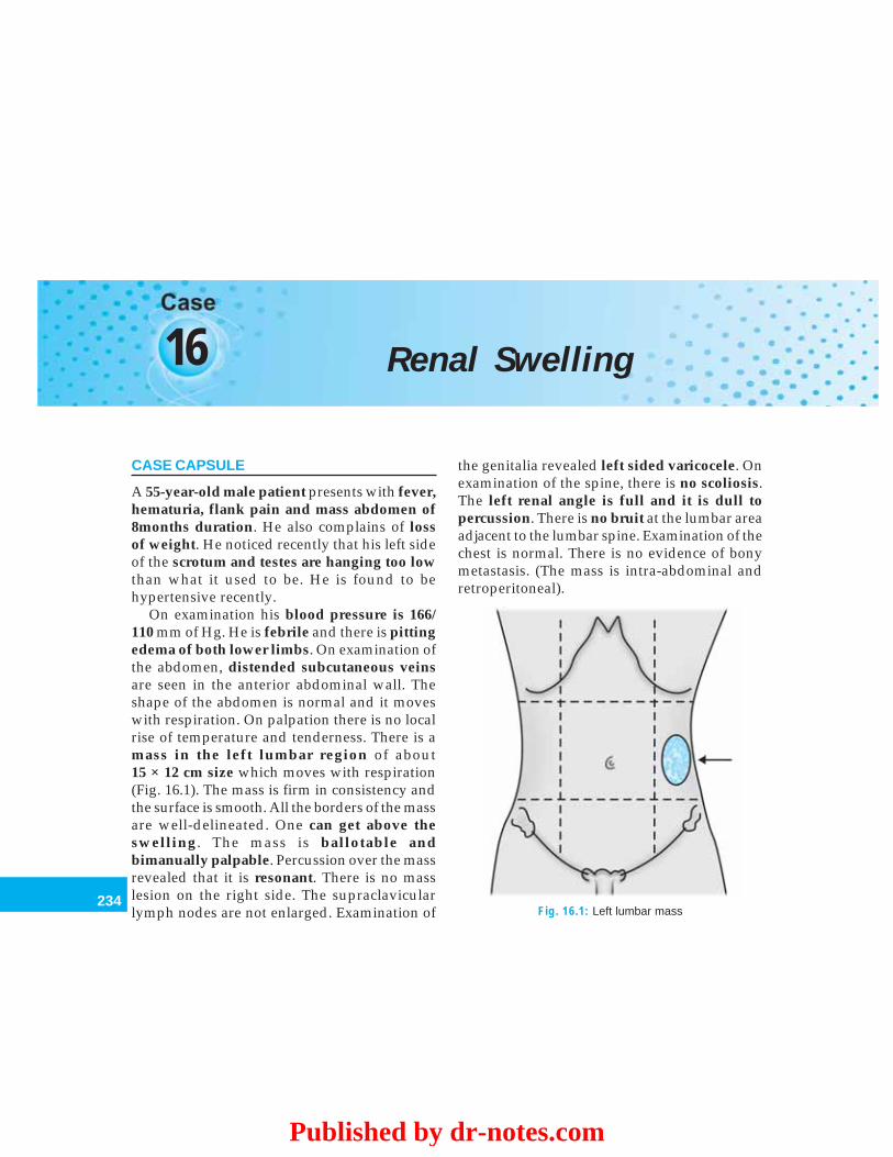
234
Clinical Surgery Pearls
16 Renal Swelling
CASE CAPSULE
A 55-year-old male patient presents with fever,hematuria, flank pain and mass abdomen of8months duration. He also complains of lossof weight. He noticed recently that his left sideof the scrotum and testes are hanging too lowthan what it used to be. He is found to behypertensive recently.
On examination his blood pressure is 166/110 mm of Hg. He is febrile and there is pittingedema of both lower limbs. On examination ofthe abdomen, distended subcutaneous veinsare seen in the anterior abdominal wall. Theshape of the abdomen is normal and it moveswith respiration. On palpation there is no localrise of temperature and tenderness. There is amass in the left lumbar region of about15 × 12 cm size which moves with respiration(Fig. 16.1). The mass is firm in consistency andthe surface is smooth. All the borders of the massare well-delineated. One can get above theswelling. The mass is ballotable andbimanually palpable. Percussion over the massrevealed that it is resonant. There is no masslesion on the right side. The supraclavicularlymph nodes are not enlarged. Examination of
the genitalia revealed left sided varicocele. Onexamination of the spine, there is no scoliosis.The left renal angle is full and it is dull topercussion. There is no bruit at the lumbar areaadjacent to the lumbar spine. Examination of thechest is normal. There is no evidence of bonymetastasis. (The mass is intra-abdominal andretroperitoneal).
Fig. 16.1: Left lumbar mass
Published by dr-notes.com
235
Renal Swelling
Read the checklist for abdomen.Checklist for examination of suspected
renal swelling
1. Always check the blood pressure reading –hypertension can be a cause or consequence ofrenal disease (renal artery stenosis) – patientmay present with headache
2. Remember the triad of renal cell carcinoma –flank pain, hematuria, and flank mass
3. Examine the genitalia for varicocele – tumorthrombus in the renal vein
4. Examine for lower limb edema – tumorthrombus in the inferior vena cava
5. Gross distension of the abdominal veins as aresult of venacaval obstructionExamine the contralateral flank for bilateralrenal swellings
6. Examine the renal angle - for fullness,tenderness and dullness
7. Look for scoliosis – inflammatory conditionswill produce scoliosis with concavity towardsthe side of the lesion
8. Auscultation for a bruit at the lumbar areasadjacent to the lumbar spine at the level of L2.(renal artery stenosis)
9. Always examine the chest for pulmonarymetastasis
10. Always examine the bones for pain andpathological fractures
Q 1. What are the physical signs of anenlarged kidney?
Physical signs of an enlarged kidney
1. It moves with respiration2. It is resonant on percussion (because it is covered
by colon in front)3. Fingers can be insinuated between the mass and
costal margin4. Bimanual palpable5. Ballotable
Q 2. Why the kidney is moving up and downwith respiration?
Upper posterior aspect of the kidney is relatedto the diaphragm (diaphragmatic area) andtherefore it will move up and down withrespiration.
Q 3. What is Ballottement?
This sign is useful for the diagnosis of thefollowing conditions.
a. This is a sign used for the confirmation ofpregnancy of more than 3 months standing.
b. For ballottement of kidneysc. It is also used for mobile intraperitoneal
fluid filled swellings like ovarian cystd. Ballottement of swellings rising out of the
pelvis where one hand is laid over thehypogastrium and the other inserted tovagina or rectum.
Q 4. What is renal ballottement?
The patient is supine and one hand of theexaminer is laid flat upon the abdomen, so thatthe greater part of the flexor surfaces of thefingers overlies the swelling. This hand is calledthe watching hand. The slightly flexed fingersof the other hand are insinuated behind the loinso that they contact the area lateral to thesacrospinalis muscle. This hand is thedisplacing hand. Short, quick, forward thrustsare made by the displacing hand (posterior). Ifthese movements impart a bouncing sensationto the anteriorly placed watching hands, the signis positive.
236
Clinical Surgery Pearls
Q 5. What is the significance of a bimanuallypalpable swelling?
It is a palpation done by both hands right handplaced anteriorly and the left hand placedposteriorly. All the big abdominal masses in theloin are bimanually palpable. Therefore, it isimportant to decide with which hand the massis better felt. If it is better felt with the anteriorhand it is likely to be arising from colon or oneof the viscera. If it is better felt with the posteriorhand, it is likely to be kidney.
Q 6. What is the significance of scoliosis oflumbar spine in a renal mass?
Scoliosis of lumbar spine with concavitytowards the affected side is a constant sign ininflammatory conditions of the kidney likeperinephric abscess. Bending the trunk awayfrom the side of lesion is likely to cause morepain in such conditions.
Q 7. What is renal angle? What is thesignificance of examination of renal angle?
It is the angle formed by the lateral border ofthe sacrospinalis muscle and the last rib. Therenal angle is normally resonant to percussionbecause of the colonic flexure in front of thekidneys. The renal angle is dull, when there isenlargement of the kidney.The renal angle is checked for:
Fullness – when there is mass lesion/enlargement.
Tenderness – in inflammatory pathology.
Dullness – when there is mass lesion/enlargement.
Q 8. What is Murphy’s kidney punch? (Therenal angle test)
The patient sits up and folds his arms in frontof him. The thumb is then placed under the 12thrib to the lateral side of the sacrospinalis muscle.Sharp and jabbing movements are made withthe thumb. Initially the movements are verygentle and if the patient is not experiencing painthe strength is increased. This test is useful fordemonstration of deep-seated tenderness.
Q 9. What is renal pain?
It is a persistent fixed ache situated mainly inthe costovertebral angle. If there is significantrenal enlargement, stretching of the peritoneummay localize the pain more anteriorly to theupper and outer quadrant of the abdomen. Thepain is not strictly lumbar.
Q 10. What is the triad of renal cell carcinoma(RCC)?
Triad of renal cell carcinomaHematuria – 40%Flank pain –30%Palpable flank mass – 20%
Note: Only 10% of patients have all thesesymptoms.
Q 11. What is the most probable diagnosis inthis case?
The first diagnosis will be a renal tumor.
Q 12. What are the differential diagnoses?
Clinically carcinoma of the descending coloncan have a clinical presentation like this.The points against colon are:• Ballottement is positive which is in favor of
kidney
Published by dr-notes.com
237
Renal Swelling
• Renal angle is obliterated and dull• There are no bowel symptoms• In colon the swelling will be better felt with
the anterior hand in bimanual palpation andballottement will be negative.The other differential diagnosis of a renal
mass are:Differential diagnoses of renal mass other than
colon
1. Hydronephrosis2. Adult polycystic kidney disease3. Tuberculosis4. Adrenal tumors5. Retroperitoneal tumors6. Angiomyolipoma7. Benign renal tumors8. Xanthogranulomatous pyelonephrosis9. Solitary cyst of the kidney
10. Abscess (Perinephric)
Q 13. What is the definition of hydronephrosis?
Aseptic dilatation of pelvicalyceal system dueto partial or intermittent obstruction.
Q 14. What are the causes for unilateralhydronephrosis?
Causes for unilateral hydronephrosis
1. Pelviureteric obstruction –a. Congenital pelviureteric junction stenosisb. Stones in the renal pelvisc. Tumors in the renal pelvisd. Pressure from aberrant arteries
2. Ureteric obstructiona. Stonesb. Tumor infiltration of the ureter – carcinoma
colon, rectum, cervix and prostatec. Tumors of the ureterd. Bladder tumorse. Ureterocele
Q 15. What are the causes for bilateral hydro-nephrosis?
Causes for bilateral hydronephrosis
• Prostatic enlargement – benign or malignant• Urethral strictures and valves• Carcinoma of the bladder• Retroperitoneal fibrosis• Phimosis
Q 16. What is the commonest tumor in thisage group?
Renal cell carcinoma (RCC). This the correctterminology. The older names are:-Grawitz’s tumorHypernephromaAdenocarcinomaClear cell tumorThe commonest of the neoplasm of the kidneyis RCC.
Q 17. Why it is called hypernephroma?
Hypernephroma means above the kidney.Grawitz’s initially thought that they arose fromadrenal rests within the kidneys. Later onLubarsch stated that these tumors are definitelyadrenal in origin leading to the term hyper-nephroma.
The tumors usually occupy the poles mostcommonly the upper pole. The tumors of thehilum are less common.
Q 18. Why it is called the ‘golden tumor’ andwhat is the cut section appearance?
The tumor is yellowish due to the abundanceof lipids including cholesterol. It is also rich inglycogen and exhibits areas of necrosis andhemorrhage.
238
Clinical Surgery Pearls
Cut section appearance:The tumors are irregular in shape with centralhemorrhage and necrosis. It is semitransparent.The tumor is often divided into lobules byfibrous septa, some of which are cystic.
Q 19. What is the histology of RCC?
Histologically it is an adenocarcinoma contai-ning clear, granular and spindle cells in varyingproportions.
Q 20. What are the pathological types ofRCC?
Clear cellGranular cellSpindle cellSarcomatoid
Q 21. What are the methods of spread of RCC?
1. Local2. Lymphatic spread – lymph nodes of hilum
and later on to the paraaortic nodes.3. Tumor thrombus into the veins – renal vein,
inferior vena cava and then upto the rightheart—pieces of growth may end up in thelungs where they grow to form cannon ballsecondary deposits.
4. Metastasis to bone – they are highly vascularand may pulsate (pulsatile bony metastasisDD follicular carcinoma thyroid metastasis).
Q 22. What is the cause for hypertension inrenal tumors?
The renal cell carcinoma produce renin and thuscause hypertension.
Q 23. What is the cause for erythrocytosis inrenal cell carcinoma (RCC)?
60% of the tumors will produce raised level oferythropoietin. This will result in erythrocytosis(not polycythemia).
Q 24. What are the ectopic hormonesproduced by renal cell carcinoma (RCC)?
(PG)
Ectopic hormones produced by renal cellcarcinoma
1. Parathyroid hormone2. Parathyroid hormone like substances
hypercalcemia3. Adrenocorticotropic hormone4. Human chorionotropic hormone5. Kinin substances – fever (renal tumors may
present as pyrexia of unknown origin).Persistence of fever after nephrectomy suggeststhe presence of metastasis
Q 25. What is Stauffer’s syndrome? (PG)
It is a nonmetastatic liver dysfunction seen incases of hypernephroma.
Q 26. What is the importance of genitaliaexamination in cases of renal tumors?
Renal carcinoma has a predilection forproducing occlusive thrombi in the renal veinand the inferior vena cava. When the left renalvein is occluded it will produce acute Varicoceleon the left side.
Q 27. Why varicocele is seen only on left sidein case of renal tumors?
The left renal vein is the vein of three pairedorgans namely – the kidneys, the adrenal andthe testis. The right testicular vein joins the
Published by dr-notes.com
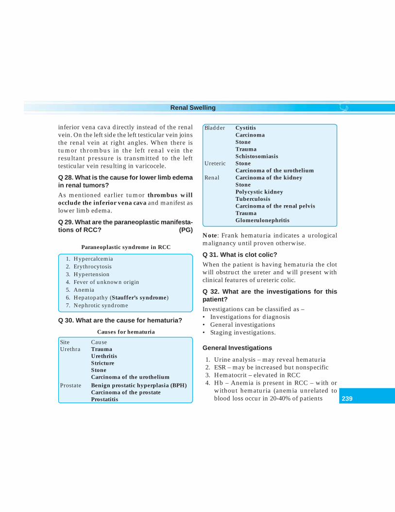
239
Renal Swelling
inferior vena cava directly instead of the renalvein. On the left side the left testicular vein joinsthe renal vein at right angles. When there istumor thrombus in the left renal vein theresultant pressure is transmitted to the lefttesticular vein resulting in varicocele.
Q 28. What is the cause for lower limb edemain renal tumors?
As mentioned earlier tumor thrombus willocclude the inferior vena cava and manifest aslower limb edema.
Q 29. What are the paraneoplastic manifesta-tions of RCC? (PG)
Paraneoplastic syndrome in RCC
1. Hypercalcemia2. Erythrocytosis3. Hypertension4. Fever of unknown origin5. Anemia6. Hepatopathy (Stauffer’s syndrome)7. Nephrotic syndrome
Q 30. What are the cause for hematuria?
Causes for hematuria
Site CauseUrethra Trauma
UrethritisStrictureStoneCarcinoma of the urothelium
Prostate Benign prostatic hyperplasia (BPH)Carcinoma of the prostateProstatitis
Bladder CystitisCarcinomaStoneTraumaSchistosomiasis
Ureteric StoneCarcinoma of the urothelium
Renal Carcinoma of the kidneyStonePolycystic kidneyTuberculosisCarcinoma of the renal pelvisTraumaGlomerulonephritis
Note: Frank hematuria indicates a urologicalmalignancy until proven otherwise.
Q 31. What is clot colic?
When the patient is having hematuria the clotwill obstruct the ureter and will present withclinical features of ureteric colic.
Q 32. What are the investigations for thispatient?
Investigations can be classified as –• Investigations for diagnosis• General investigations• Staging investigations.
General Investigations
1. Urine analysis – may reveal hematuria2. ESR – may be increased but nonspecific3. Hematocrit – elevated in RCC4. Hb – Anemia is present in RCC – with or
without hematuria (anemia unrelated toblood loss occur in 20-40% of patients
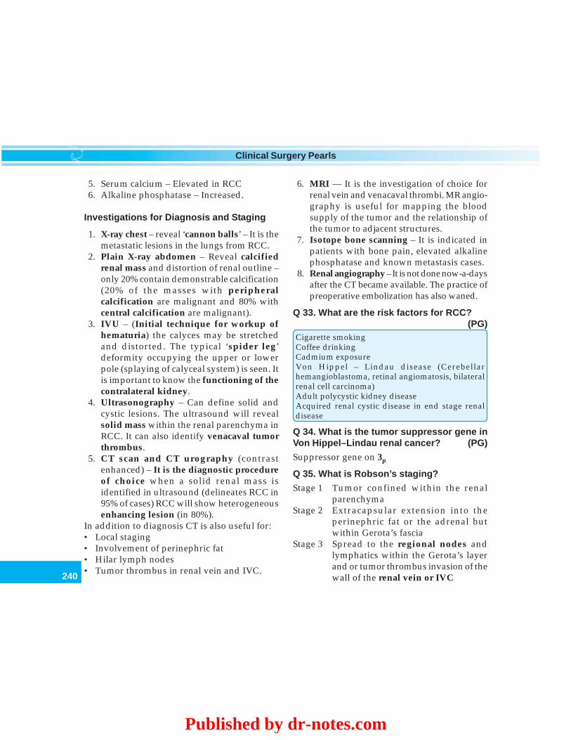
240
Clinical Surgery Pearls
5. Serum calcium – Elevated in RCC6. Alkaline phosphatase – Increased.
Investigations for Diagnosis and Staging
1. X-ray chest – reveal ‘cannon balls’ – It is themetastatic lesions in the lungs from RCC.
2. Plain X-ray abdomen – Reveal calcifiedrenal mass and distortion of renal outline –only 20% contain demonstrable calcification(20% of the masses with peripheralcalcification are malignant and 80% withcentral calcification are malignant).
3. IVU – (Initial technique for workup ofhematuria) the calyces may be stretchedand distorted. The typical ‘spider leg’deformity occupying the upper or lowerpole (splaying of calyceal system) is seen. Itis important to know the functioning of thecontralateral kidney.
4. Ultrasonography – Can define solid andcystic lesions. The ultrasound will revealsolid mass within the renal parenchyma inRCC. It can also identify venacaval tumorthrombus.
5. CT scan and CT urography (contrastenhanced) – It is the diagnostic procedureof choice when a solid renal mass isidentified in ultrasound (delineates RCC in95% of cases) RCC will show heterogeneousenhancing lesion (in 80%).
In addition to diagnosis CT is also useful for:• Local staging• Involvement of perinephric fat• Hilar lymph nodes• Tumor thrombus in renal vein and IVC.
6. MRI — It is the investigation of choice forrenal vein and venacaval thrombi. MR angio-graphy is useful for mapping the bloodsupply of the tumor and the relationship ofthe tumor to adjacent structures.
7. Isotope bone scanning – It is indicated inpatients with bone pain, elevated alkalinephosphatase and known metastasis cases.
8. Renal angiography – It is not done now-a-daysafter the CT became available. The practice ofpreoperative embolization has also waned.
Q 33. What are the risk factors for RCC?(PG)
Cigarette smokingCoffee drinkingCadmium exposureVon Hippel – Lindau disease (Cerebellarhemangioblastoma, retinal angiomatosis, bilateralrenal cell carcinoma)Adult polycystic kidney diseaseAcquired renal cystic disease in end stage renaldisease
Q 34. What is the tumor suppressor gene inVon Hippel–Lindau renal cancer? (PG)
Suppressor gene on 3p
Q 35. What is Robson’s staging?
Stage 1 Tumor confined within the renalparenchyma
Stage 2 Extracapsular extension into theperinephric fat or the adrenal butwithin Gerota’s fascia
Stage 3 Spread to the regional nodes andlymphatics within the Gerota’s layerand or tumor thrombus invasion of thewall of the renal vein or IVC
Published by dr-notes.com
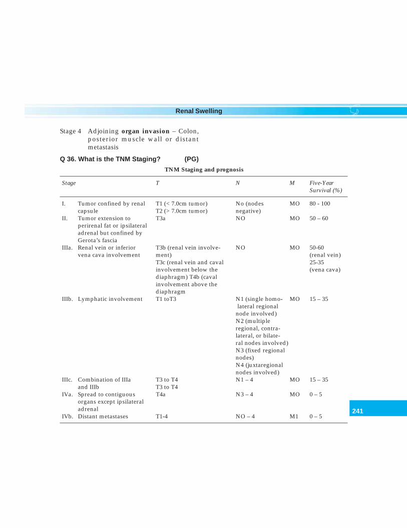
241
Renal Swelling
Stage 4 Adjoining organ invasion – Colon,posterior muscle wall or distantmetastasis
Q 36. What is the TNM Staging? (PG)
TNM Staging and prognosis
Stage T N M Five-YearSurvival (%)
I. Tumor confined by renal T1 (< 7.0cm tumor) No (nodes MO 80 - 100capsule T2 (> 7.0cm tumor) negative)
II. Tumor extension to T3a NO MO 50 – 60perirenal fat or ipsilateraladrenal but confined byGerota’s fascia
IIIa. Renal vein or inferior T3b (renal vein involve- NO MO 50-60vena cava involvement ment) (renal vein)
T3c (renal vein and caval 25-35involvement below the (vena cava)diaphragm) T4b (cavalinvolvement above thediaphragm
IIIb. Lymphatic involvement T1 toT3 N1 (single homo- MO 15 – 35 lateral regionalnode involved)N2 (multipleregional, contra-lateral, or bilate-ral nodes involved)N3 (fixed regionalnodes)N4 (juxtaregionalnodes involved)
IIIc. Combination of IIIa T3 to T4 N1 – 4 MO 15 – 35and IIIb T3 to T4
IVa. Spread to contiguous T4a N3 – 4 MO 0 – 5organs except ipsilateraladrenal
IVb. Distant metastases T1-4 NO – 4 M1 0 – 5
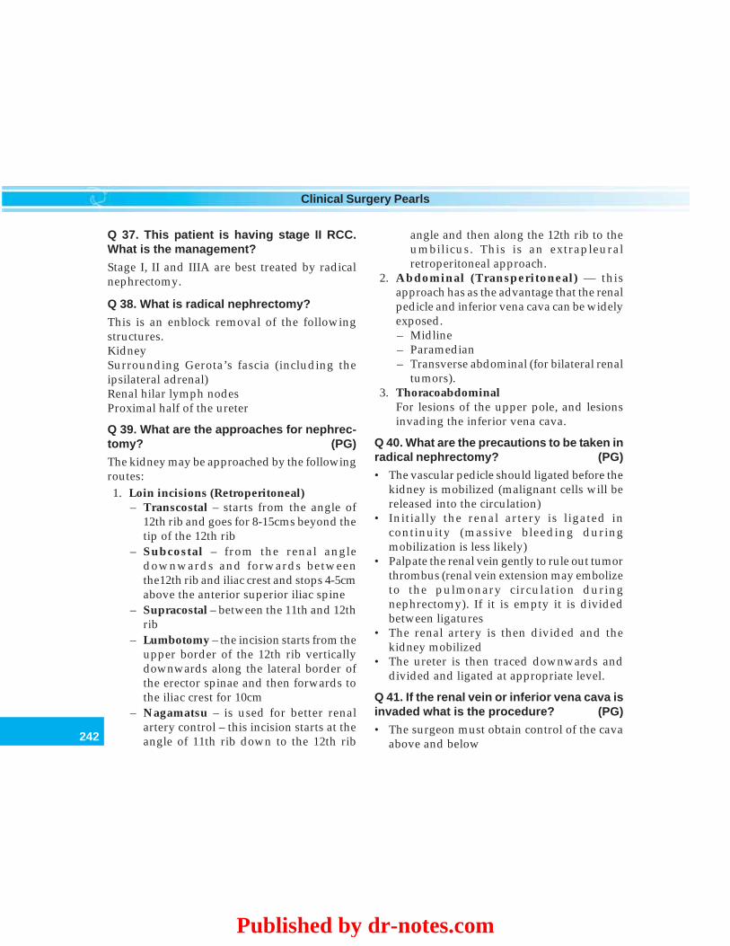
242
Clinical Surgery Pearls
Q 37. This patient is having stage II RCC.What is the management?
Stage I, II and IIIA are best treated by radicalnephrectomy.
Q 38. What is radical nephrectomy?
This is an enblock removal of the followingstructures.KidneySurrounding Gerota’s fascia (including theipsilateral adrenal)Renal hilar lymph nodesProximal half of the ureter
Q 39. What are the approaches for nephrec-tomy? (PG)
The kidney may be approached by the followingroutes:1. Loin incisions (Retroperitoneal)
– Transcostal – starts from the angle of12th rib and goes for 8-15cms beyond thetip of the 12th rib
– Subcostal – from the renal angledownwards and forwards betweenthe12th rib and iliac crest and stops 4-5cmabove the anterior superior iliac spine
– Supracostal – between the 11th and 12thrib
– Lumbotomy – the incision starts from theupper border of the 12th rib verticallydownwards along the lateral border ofthe erector spinae and then forwards tothe iliac crest for 10cm
– Nagamatsu – is used for better renalartery control – this incision starts at theangle of 11th rib down to the 12th rib
angle and then along the 12th rib to theumbilicus. This is an extrapleuralretroperitoneal approach.
2. Abdominal (Transperitoneal) — thisapproach has as the advantage that the renalpedicle and inferior vena cava can be widelyexposed.– Midline– Paramedian– Transverse abdominal (for bilateral renal
tumors).3. Thoracoabdominal
For lesions of the upper pole, and lesionsinvading the inferior vena cava.
Q 40. What are the precautions to be taken inradical nephrectomy? (PG)
• The vascular pedicle should ligated before thekidney is mobilized (malignant cells will bereleased into the circulation)
• Initially the renal artery is ligated incontinuity (massive bleeding duringmobilization is less likely)
• Palpate the renal vein gently to rule out tumorthrombus (renal vein extension may embolizeto the pulmonary circulation duringnephrectomy). If it is empty it is dividedbetween ligatures
• The renal artery is then divided and thekidney mobilized
• The ureter is then traced downwards anddivided and ligated at appropriate level.
Q 41. If the renal vein or inferior vena cava isinvaded what is the procedure? (PG)
• The surgeon must obtain control of the cavaabove and below
Published by dr-notes.com
243
Renal Swelling
• Cardiac bypass may be required if there isextension into the thorax (Get the cardiacteam) and the tumor thrombus may beremoved from the right side of the heart.
Q 42. Is there any role for partial nephrectomy(nephrone sparing surgery)?
Yes. Partial nephrectomy is recommended in thefollowing situations –• Patients with tumors in solitary kidney• Those with diabetes mellitus• Patients with renal insufficiency• Tumors under 4cm size (Even with normal
opposite kidney)(In patients with negative surgical margins
the prognosis will be the same as that of theradical nephrectomy).
Q 43. What is the role of laparoscopicnephrectomy?
It is becoming the gold standard in institutionswith appropriate expertise. Laparoscopic radicalor partial nephrectomy has been advocated asa method equal to the open approach with thefollowing advantages –• Less blood loss• Shorter hospitalization• Less pain• Earlier return to normal activity.
Q 44. What is the treatment for a patient withsolitary pulmonary metastasis? (PG)
Joint surgical removal of both the primary lesionand metastatic lesion is recommended.
Q 45. Is there any role for preoperative arterialembolization? (PG)
This will not improve survival rate. It isrecommended as a single treatment in sympto-matic patients with nonresectable primarylesions.
Q 46. What is the role of radiotherapy?
Useful only for the treatment of symptomaticbone metastatic.
Q 47. What are the chemotherapeutic agentsused in RCC? (PG)
VinblastineMedroxy progesterone – for metastatic RCC.
Q 48. What is the role of immunotherapy?(PG)
• Interferon alpha – 15 – 20% response rate• IL–2 (interleukin)• Combination of interferon alpha and IL-2.
Q 49. What is the prognosis of RCC? (PG)
Removal of even the largest neoplasm may curethe patient. In operable cases, 70% are well after3 years and 60% after 5 years.
Q 50. What are the bad prognostic factors?(PG)
• Tumor invasion beyond the capsule• Macroscopic involvement of renal vein and
IVC• Lymph node involvement.
Q 51. What is congenital polycystic kidneydisease?
It is a hereditary condition transmitted byautosomal dominant trait affecting both kidneys(one kidney may contain larger cysts than the
244
Clinical Surgery Pearls
contralateral). The disease manifests in 2nd or3rd decade of life and usually does not manifestbefore the age of 30 years. The etiology isuncertain. In 18% of cases there is associatedcongenital cystic liver disease. The pancreas andlungs are occasionally affected. The disease ismore common in women. The most importanttheory is failure of fusion between the secretarypart and collecting system during thedevelopment (the secretary part is developedfrom the meta-nephrogenic tissue cap, andcollecting part from the ureteric bud).
Q 52. What is the genetic abnormality inpolycystic kidney disease? (PG)
There are two types–Autosomal recessive polycystic kidney disease(ARPKD) – Previously called infantile. In thiscondition small cysts arise from collecting ductsresulting in bilateral symmetrical enlargement.Occurs in 1/14000 births and may be detectedin utero. Infants usually die of respiratoryfailure.
Autosomal dominant polycystic kidneydisease (ADPKD) — the gene located onchromosome 16p. Another gene responsible isPKD 2gene located in chromosome 4q 21-23.ADPKD occurs in 1/1000 individuals.
Q 53. What is the median age for end stagerenal disease?
In PKD1 - 54 yearsIn PKD2 – 74 years
Q 54. What are the manifestations of poly-cystic disease?
Manifestations of polycystic disease of kidney
• Upper abdominal mass (bilateral knobbyenlargement)
• Hypertension – present in 75% of patients above20 years
• Loin pain – due to the weight of the organ orstretching of the capsule
• Uremia – Large volumes of urine of low specificgravity (10:10 or less) – chronic renal failure(headache, drowsiness, vomiting and anorexia)
• Infection – pyelonephritis• Hematuria – rupture of cyst into the renal pelvis
Q 55. What is the gross appearance ofpolycystic disease?
The gross appearance is that of a collection ofbubbles beneath the renal capsule. The renalparenchyma is riddled with cysts of varyingsizes containing clear fluid, thick brown materialor blood.
Q 56. What are the investigations forpolycystic disease of the kidney?
• IVU – enlarged kidneys with markedelongation of the calyces which arecompressed by the cysts. The spider legdeformity or bell-like appearance
• Ultrasound multiple cysts in both kidneys,some times in liver and other organs
• CT.
Q 57. What are the treatment options forpolycystic disease of the kidney?
• Medical – low protein diet, treatment ofinfection, anemia and renal failure
• Dialysis
Published by dr-notes.com
245
Renal Swelling
• Renal transplantation – is the ideal treatment• Rovsing’s operation – (deroofing the cysts) –
This is a palliative procedure for relieving thepressure on the parenchyma, and forreducing the pain. The open operation is notdone now-a-day. If required it can be donelaparoscopically.
Q 58. What is Dietl’s crisis?
This is seen in intermittent hydronephrosis.After an attack of acute renal pain, swellingappears in the loin. After a few hours followingthe passage of a large volume of urine, the painis relieved and the swelling disappears. This iscalled Dietl’s crisis.
Q 59. What are the clinical features of hydro-nephrosis due to idiopathic pelviuretericjunction obstruction?
Renal colicRenal massIntermittent hydronephrosis.
Q 60. What is the IVU finding in hydro-nephrosis?
The minor calyces lose their normal cuppingThey become flattenedFinally the classical clubbing is seenIn very advanced cases the thin rim of poorlyfunctioning renal parenchyma may give a faintnephrogram around the dilated calyces – thesoap bubble appearance.
Q 61. What is the most helpful test toestablish the dilatation due to obstruction?
Isotope renography using DTPA (DiethyleneTriamine Penta Acetic acid) 99Tc – DTPA is usedfor this investigation.
Q 62. What is the principle behind 99Tc -DTPA?
The DTPA is injected intravenously which isfiltered by the glomeruli and not absorbed.When the DTPA is labeled with 99Tc it will emitgamma rays. The passage of this technetiumlabeled DTPA through the kidneys can betracked using a gamma camera. When the ureteris obstructed the marker is trapped in the renalpelvis and will not be washed out, even if theflow of urine is increased by administeringfrusemide.
Q 63. What is Whitaker test?
A percutaneous puncture of the kidney ismade through the loin and fluid is infusedat a constant rate with monitoring of intrapelvicpressure. An abnormal rise in pressureconfirms obstruction. This test is doneoccasionally.
Q 64. What is RGP (Retrograde pylogram)?
After cystoscopy the affected ureter iscannulated and dye is injected which will revealthe anatomy of obstruction. This is done whenthe kidneys are not functioning in IVU in orderto get an idea about the site of obstruction beforesurgery.
Q 65. What are the indications for surgery inidiopathic pelviureteric junction obstruction(PUJ)?
• Increasing hydronephrosis• Bouts of renal pain• Parenchymal damage• Infection.
246
Clinical Surgery Pearls
Q 66. What are the operations available forPUJ?
Anderson – Hynes – disjunction pyeloplasty isthe most commonly performed operation (it canbe done laparoscopically or by open method).Endoscopic pyelolysis – the disruption of thepelviureteric junction is done by speciallydesigned balloon passed up the ureter underradiographic control.
Q 67. What is the indication for nephrectomyin hydronephrosis?
Conservation of renal parenchyma is the aim.Nephrectomy is considered only when the renalparenchyma has been largely destroyed. If renalfunction is adequate (> 10% of total renalfunction surgical repair of the stenosis isdone.
Published by dr-notes.com
247
Pseudocyst of Pancreas
17 Pseudocyst of Pancreas
CASE CAPSULE
A 45-year-old male patient presents with historyof upper abdominal pain, epigastric fullness,nausea and occasional vomiting of 6weeksduration. He is a chronic alcoholic and giveshistory of hospital admission for acuteabdominal pain two months back. For the lastone week he gives history of severe pain overthe epigastric region with mild fever and rigors.There is no history of trauma.
On general examination there is mild icterusand pallor. On abdominal examination the upperabdomen shows a fullness. The abdomen moveswith respiration and the umbilicus is normal.There are no dilated veins seen in the abdominalwall. On palpation of the abdomen there is a firm,tender mass of 20 × 16 cm size (20cm horizontaland 16cm vertical) in the epigastrium extendingto both hypochondrium. The lower border of theswelling is indistinct. The upper limit is notpalpable. On both sides the mass goes beneaththe costal margin. It does not move withrespiration and there is no intrinsic mobility. Thesurface of the swelling is smooth. The mass isresonant to percussion. The plane of the swellingis intra abdominal and retroperitoneal. The rest
of the abdomen is normal. There is no free fluiddemonstrated and normal bowel sounds areheard. Digital rectal examination revealed noabnormality. The hernial orifices and genitalia arenormal. The left supraclavicular nodes are notenlarged. The renal angles are normal.
Read the checklist for abdominal examination
Checklist for history in the case of pseudocystexamination
• History of acute abdominal pain suggestive ofacute pancreatitis
• History of trauma• History of chronic abdominal pain• History of alcoholism• History of jaundice – due to pressure from
pseudocyst
Q 1. What are the physical features ofpseudocyst of pancreas?
Physical features of pseudocyst of pancreas
1. Epigastric mass with indistinct lower edge2. The upperlimit may or may not be palpable3. It is resonant to percussion (because of the
overlying stomach)4. There is no movement with respiration (minimal
movement may be there)
248
Clinical Surgery Pearls
5. The mass will be firm or soft but it is not possibleto elicit fluctuation or fluid thrill
6. The mass may be tender
Q 2. What is pseudocyst?
This is a collection of pancreatic juice enclosedin a wall of fibrous or granulation tissue thatarises following an attack of acute pancreatitis(old definition is collection of fluid in the lessersac).
Q 3. Why it is called pseudocyst?
It denotes absence of an epithelial lining. Thetrue cyst is lined by epithelium.
Q 4. What is the time gap between the attackof acute pancreatitis and pseudocyst?
Usually it is four weeks.
Q 5. What are the types of pseudocyst?
The Pseudocysts may be-Acute pseudocystChronic pseudocystAnother classification is
• Postnecrotic – following an attack ofnecrotizing pancreatitis (Confined to thelesser sac or into the retroperitoneal spaceor to the mesentery of small or large bowel)
• Peripancreatic – Associated with chronicpancreatitis
• Intrapancreatic cyst – In advanced chronicpancreatitis (majority in the head of thepancreas).
Q 6. What is acute pseudocyst?
It is a fluid collection arising in association withan episode of acute pancreatitis of > 4 weeks
duration and surrounded by well-defined wallof granulation or fibrous tissue.
Q 7. What constitutes the fibrous tissue inthe wall of the cyst?
It is constituted by the inflammatory fibrosis ofthe following:• Peritoneum• Mesentery• Serous membrane.
Q 8. What are the locations of the pseudo-cyst?
• Lesser sac• Neck• Mediastinum• Pelvis
Q 9. What is the incidence of acutepseudocyst?
It develops in about 2% of cases of acutepancreatitis.
Q 10. Can you get multiple pseudocysts?
Yes. The cysts are single in 85% cases andmultiple in the remainder.
Q 11. What is chronic pseudocyst?
Chronic pseudocyst is a collection with a well-defined wall that arises in a patient with chronicpancreatitis, without antecedent episode of acutepancreatitis. It may also occur in trauma victims(after a blow) and alcoholics. The mechanismconsists of ductal obstruction and formation of aretention cyst that loses its epithelial lining as itgoes beyond the confines of the gland. It may alsooccur sometimes as iatrogenic after splenectomy.
Published by dr-notes.com
249
Pseudocyst of Pancreas
Q 12. What is pancreatic abscess?
It is a circumscribed peripancreatic collection ofpus occurring late (more than 4 weeks) after theonset of acute pancreatitis. Infected collectionsidentified earlier are classified as either infectedacute fluid collection or infected pancreaticnecrosis.
Q 13. What is the difference betweenpseudocyst and acute fluid collection?
The acute fluid collection occurs early in thecourse of acute pancreatitis (less than 4 weeks)and is lacking a wall of fibrous or granulationtissue and is located in or near the pancreas.(in pseudocyst there is a wall of fibrous orgranulation tissue).
Q 14. What is the nature of fluid in acutepseudocyst?
It is a collection enzyme rich pancreatic juice andmay contain small amounts of necrotic debris.
Q 15. What are the investigations for thediagnosis of pseudocyst?
• Serum amylase – elevated (if the serumamylase remain elevated for 3weeks, abouthalf will have pseudocyst).
• Leukocytosis – (about 50% of patients).• Bilirubin – elevated (suggestive of biliary
obstruction).• CT scan – is the diagnostic study of
choice (the size, shape of the cyst and itsrelationship to other viscera can be seen).
Acute cysts are irregular in shape, chronic cystsare circular.An enlarged pancreatic duct may be demons-trated in chronic pancreatitis.
A dilated CBD would suggest biliary obstructioneither from the cyst or as a result of pancreatitis.• Ultrasound – to follow changes in size of
acute pseudocyst and to study thegallbladder
• ERCP – not done routinely (If CT showsductal abnormalities and if LFT isabnormal).
• Upper GI series – to look for site of gastricor duodenal obstruction. The cyst will pushthe stomach wall anteriorly (the retrogastricspace will be increased).
Q 16. What is the role of ERCP in pseudocyst?
ERCP will reveal the communication of the cystto the pancreatic duct. Based on this thepseudocysts are classified as—• Communicating–communicating to the
pancreatic duct• Noncommunicating.
Q 17. What are the differential diagnoses inpseudocyst?Differential diagnoses of pseudocyst of pancreas
• Pancreatic abscess• Pancreatic carcinoma (if gallbladder is palpable)• Neoplastic cyst–cystadenoma or cystadeno-
carcinoma (about 5% of all cases of cystic pan-creatic masses)
• Other epigastric masses
Q 18. What are the complications ofpseudocyst?
The complications are –• Pancreatic pseudocyst hemorrhage –
resulting from autodigestion of the pancreasleading to erosion of splenic, gastroduodenal,
250
Clinical Surgery Pearls
or pancreaticoduodenal artery. This willresult in false aneurysm of the artery in thecyst wall. The clinical presentation ofhemorrhage in the cyst are:• Increasing epigastric pain• Increase in the size of abdominal mass• Shock
1. Hematemesis and melena – cyst erodinginto the stomach
2. Rupture of the cyst into the peritoneal cavity– peritonitis and shock – emergency surgerywith irrigation of the peritoneal cavity anddrainage for the pseudocyst.
3. Infection – High fever, chills and leukocytosis(percutaneous drainage or internal drainageis the treatment).
Q 19. What are the indications forintervention in pseudocyst?
Indications for intervention in pseudocyst
Persisting painPressure effects caused by increasing sizeCysts of > 6 cm sizeCysts of > 6 weeks duration
Q 20. Why the procedure is done after 6weeks?
The cyst wall is mature enough only after 6weeks to hold sutures for anastomosis of the gut.
Q 21. Why 6cm size is taken as cut off fortreatment?
The chance for spontaneous resolution is therein 40% of cases if the cysts are smaller andtherefore expectant management isrecommended in smaller cyst.
Q 22. What is the cause for jaundice in apatient with pseudocyst?
It is usually caused by pressure from the cyston the bile duct.
Q 23. What are the therapeutic options forthe management of pseudocyst?
See the algorithm for the management ofpseudocyst ( Flow Chart 17.1).
Q 24. What are the internal drainage proce-dures available for pseudocyst?
The internal drainage may be• Endoscopic – endoscopic cystogastrostomy
or cystojejunostomy• Laparoscopic• Open
Q 25. What are the viscera to which internaldrainage procedures are carried out?
• Cystogastrostomy – technically simpler(Recurrence and GI hemorrhage occur afterthis procedure)
• Cystojejunostomy (Roux-en-Y) – is the mostversatile
• Cystoduodenostomy – risk of biliary injury.
Q 26. What is the prerequisite for the internaldrainage?
Cyst wall should be sufficiently mature foranastomosis (usually 4-6 weeks).
Q 27. What is external drainage?
The external drainage is done when the wallsurrounding the fluid collection is too fragile forinternal drainage. It can be done under sono-
Published by dr-notes.com
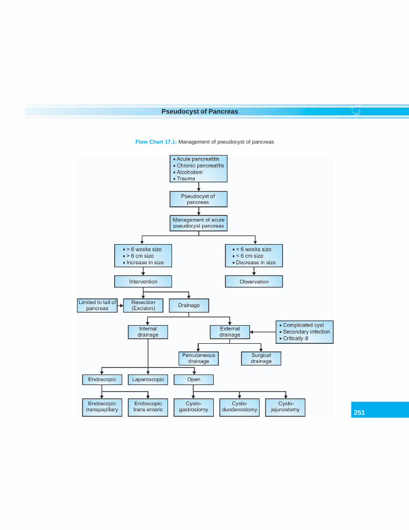
251
Pseudocyst of Pancreas
Flow Chart 17.1: Management of pseudocyst of pancreas
box
252
Clinical Surgery Pearls
logical guidance. This is also calledpercutaneous catheter drainage.
Q 28. What is the incidence of recurrence ofpseudocyst after catheter drainage? (PG)
The recurrence is 4 times greater after externaldrainage than after internal drainage.
Q 29. What are indications for externaldrainage (nonsurgical drainage)? (PG)
• Critically ill patients• Infected pseudocyst• Uncomplicated pseudocyst• To shrink a huge pseudocyst occupying half
of the abdominal cavity.
Q 30. What is the complication of externaldrainage? (PG)
Pancreaticocutaneous fistula in 20–30% ofpatients.
Q 31. Is there any other method of externaldrainage? (PG)
A percutaneous drainage where a catheter ispassed through the anterior abdominal wall, theanterior wall of the stomach and through theposterior stomach into the cyst. After severalweeks the catheter is removed.
Q 32. What are the essential steps ofcystogastrostomy?
• Upper midline incision• Anterior gastrotomy (longitudinal incision
in the anterior wall of stomach)• Identify an area where the cyst wall is
maximum bulging to the lumen of thestomach
• A 4 cm longitudinal window is created inthe posterior wall along with the cyst wall(evacuate the contents of the cyst)
• A full thickness mattress suture is used tosuture the edge of the posterior gastrotomyand cyst wall together
• The anterior gastrotomy is closed• The abdomen is closed.
Q 33. What are the important things to be donewith the pseudocyst during sugery? (PG)Aspirate the cyst fluid and send for culture andsensitivity.Send the cyst fluid for amylase estimation.Take biopsy from the cyst wall (this will ruleout a cystic tumor).
Q 34. What will happen to the passage of foodafter cystogastrostomy? (PG)Surprisingly the food will not go to the cystcavity and the cystogastrostomy will getobliterated within a few weeks and heal in duecourse of time.
Q 35. What is the recurrence rate forpancreatic pseudocyst? (PG)10% (Recurrence is more frequent after externaldrainage).
Q 36. What is the postoperative complicationof cystogastrostomy? (PG)Hemorrhage occurs rarely.
Q 37. What is excision for pseudocyst?(PG)This is the most definitive treatment. Usuallythis is done for chronic pseudocysts in the tailof gland. This is usually recommended for cystsfollowing trauma.
Published by dr-notes.com
253
Pseudocyst of Pancreas
Q 38. What is the treatment of pseudocysthemorrhage? (PG)
• Angiographic embolization• Subsequent elective treatment of the
pseudocysts (after several weeks).
• Emergency laparotomy and ligation of thebleeding vessel after opening the cyst, anddrainage of pseudocyst
• For lesions in the pancreatic headpancreaticoduodenectomy may berequired.
254
Clinical Surgery Pearls
18 Retroperitoneal Tumor
CASE CAPSULE
A 55-year-old male patient presents with back-ache and pain radiating down the left lowerlimb of 6 months duration. He in additioncomplains of anorexia and loss of weight. Forthe last 3 months he complains of distension ofabdomen. There is no history of therapeuticirradiation. There is no family history ofswelling all over the abdomen.
On examination the patient is afebrile, theblood pressure is 140/86 mm of Hg. There ispitting edema of the left lower limb. Onexamination of the abdomen, there is distensionof abdomen which is more towards the lefthalf. A big bosselated mass of 25 × 15cm (25cmvertically and 15 cm horizontally) is felt in theleft half of the abdomen occupying the leftlumbar region, left hypochondrium and leftiliac fossa. The mass is firm in consistency anddoes not move with respiration. All the bordersexcept the lateral border is well-defined.Medially it is not crossing the midline. It isbimanually palpable, but not ballotable. It isintraabdominal as demonstrated by the coughtest. The mass is not falling forwards asdemonstrated by examination in the knee elbow
position and therefore retroperitoneal. There isno intrinsic mobility for the mass. Onpercussion there is resonance over the mass.The rest of the abdomen is normal. There is nofree fluid in the abdomen and there is noorganomegaly. Normal bowel sounds are heardon auscultation. There is no evidence ofvaricocele. The hernial orifices are normal. Theleft lower limb pulsations are normal and thereis no neurological deficits in the left lower limb.Chest examination is normal.
Read the checklist for abdominal examination.Checklist for history
• History of therapeutic radiation• History of exposure to vinyl chloride family• History of Gardner’s syndrome• Family history of Gardner’s• Familial retinoblastoma• Risk factors like – Neurofibromatosis,
Li-Fraumeni syndrome• History of hypertension – due to catecholamine
production• History of back pain and leg pain• History of hypoglycemia – liposarcoma produce
insulin• History of precocious puberty• History of myoclonus in children
Published by dr-notes.com
255
Retroperitoneal Tumor
Checklist for examination
• Take the temperature - RP tumors produce fever• Take the blood pressure – tumors produce
catecholamines• Check the plane and remember the clinical points
for retroperitoneal tumors given below:• Look for dilated abdominal veins – due to the
pressure on vena cava• Look for lower limb edema• Look for varicocele• Look for ascitis (nonmalignant ascitis of RP
tumor)• Look for features of Costello syndrome
Q 1. What is the most probable diagnosis inthis case?
Retroperitoneal tumor.
Q 2. What are the clinical points in favour ofretroperitoneal tumor?
Clinical points in favor of retroperitonealtumor
• The mass is intraabdominal as demonstrated byhead raising test (Carnet’s test)
• The mass is not falling forwards in the kneeelbow position (suggesting the plane to beretroperitoneal)• There is no movement with respiration (retro
peritoneal swelling will not move)• The mass is fixed (no intrinsic movement)• The mass is resonant on percussion because of
the intestinal loops in front• The mass is not crossing the midline (retroperito-
neal swellings usually will not cross the midline)
Q 3. What is the most probable diagnosis inthis case?
Malignant retroperitoneal tumor most probablyliposarcoma.
Q 4. Why liposarcoma?It is the commonest retroperitoneal sarcoma inadults.
Q 5. What is the age group affected inliposarcoma?Usually 40-70years and they are asymptomaticin early stages.
Q 6. What is the cause for left lower limbedema and pain in this patient?These are symptoms of compression on the iliacveins. The backache and lower limb pain in thiscase is due to stretching of the lumbar andpelvic nerves.
Q 7. What are the symptoms and signs ofliposarcoma?
Symptoms and signs of liposarcoma
• Mass abdomen (91%)• Mass pelvis (21%) – DD ovarian malignancy• Abdominal pain and discomfort (71%)• Anorexia, vomiting, fatigue• Weight loss• Caval compression – limb edema, varicocele,
ascites (nonmalignant), dilated abdominal veins·• Back pain and leg pain – compression and
stretching of lumbar and pelvic nerves• Fever
Q 8. What is retroperitoneum?
According to Ackerman retroperitoneum isdefined as –A region of the trunk covered –• Anteriorly by the parietal peritoneum• Superiorly by the 12th rib and diaphragm• Inferiorly by the pelvic diaphragm and fascia
of the levator ani and coccygeus
256
Clinical Surgery Pearls
• Posteriorly by the fascia of the muscles of theanterior abdominal wall.
Q 9. Is it a real space?
It is an actual or potential space.
Q 10. What are the contents?
The contents are structures of mesodermal andendodermal origin with their embryonicremnants. This consists of loose connectivetissue, fat, nerves, lymphatics and lymphnodes.
Q 11. What about the retroperitoneal organs?
Tumors of the retroperitoneal organs are notincluded in the retroperitoneal tumorstraditionally (like kidneys, adrenals, pancreas,and ureters).
Q 12. What is the incidence of retroperitonealtumors?
They are uncommon heterogenous group oftumors arising either primarily in theretroperitoneum or metastasis from else where.The incidence is 0.3 to 3%.
Q 13. What is the classification?Classification
Retroperitoneal Retroperitonealtumors tumors
Cystic Solid Benign Malignant
• Cystic swelling are usually benign• Majority are accidentally discovered• Majority of the solid tumors are malignant
(75-95%)
Q 14. What are the benign swellings?Classification of benign tumors
• Lipomas• Neurofibromas, neurilemmomas• Leiomyomas• Extraadrenal chromaffinomas (pheo may be
malignant)• Mucinous cystadenomas• Hemangiopericytomas• Paragangliomas.
Q 15. What is Costello syndrome? (PG)
It is a syndrome consisting of mental retardationand benign tumors usually papillomata.Children with Costello syndrome developembryonal rhabdomyosarcoma.
Q 16: What is Paraganglioma? (PG)Features of paraganglioma
• They are tumors of embryonal origin from theneural crest
• May arise from any location along the aorta orthe sympathetic chain
• They may be functioning or nonfunctioning(secrete norepinephrine)
• 20% are catecholamine secreting• May be multiple• Malignant paragangliomas can occur• 0.1 – 0.5% cases produce hypertension• May produce syndrome similar to pheo• May be familial (hereditary paragangliomas)• Associated with von Hippel-Lindau disease and
RET protooncogene.
Q 17. What is the pathology in paragang-lioma? (PG)
The paraganglioma may arise from type I ortype II cells.
Published by dr-notes.com
257
Retroperitoneal Tumor
Type I cells are chromogranin A positive (NSE+ ve).Type II cells are - S 100 + ve (with goodprognosis).
Q 18. What are the features of retroperitonealneurilemmomas? (PG)
They are well-demarcated round or oval massin CT with heterogenous contrast enhancement.Nonenhancement of the lesion may be due tocyst formation or tumor calcification.
Q 19. What is mucinous cyst adenoma?(PG)
Mucinous cyst adenoma and cyst adeno-carcinoma are similar to ovarian mucinoustumors.
Q 20. What is hemangiopericytoma? (PG)
They are vascular benign tumors derived frompericytes. They are very bulky and silent tumors.Treatment is excision.
Q 21. What are the malignant retroperitonealtumors?
They may be Primary or Secondary.
Primary Secondary (metastatic)
Sarcomas AdenocarcinomaLymphomas EndometriumMalignant variety of Prostategerm cell tumors (com-mon in children andare benign)Teratocarcinoma PancreasEndodermal sinus tumor Lung
Contd...
Contd...
Tumors of embryonal GIToriginUrogenital ridge tumors Squamous cell
carcinoma (SCC)Cervix, vagina andlung
Chordoma
Q 22. What is the origin of malignantretroperitoneal tumors?
They may originate from mesodermal,neurectodermal or embryonic remnant• 75% mesodermal in origin• 24% neurectodermal• 1% embryonic remnantThe most common malignancy is sarcoma.
Q 23. What is the commonest retroperitonealmalignancy?
Sarcoma.
Q 24. What is the commonest sarcoma?
Liposarcoma is the commonest sarcoma in adults.• Liposarcoma – 50%• Leiomyosarcoma – 29%• Malignant fibrous histiocytoma (MFH).
[In children the commonest sarcoma is -Rhabdomyosarcoma:]• 18% present with neurological deficit• 37% with distant metastasis.
Q 25. What are the etiological factors forretroperitoneal sarcoma? (PG)
Etiological factors for retroperitoneal sarcoma
258
Clinical Surgery Pearls
• Idiopathic• History of therapeutic radiation• Exposure to vinyl chloride• Thorium dioxide• Familial disorders• Gardner’s syndrome• Familial retinoblastoma• Neurofibromatosis• Li-Fraumeni syndrome• Germline mutations of p53 (chr.17)
Q 26. What is the origin of liposarcoma?
1/3rd of the liposarcoma originate fromperinephric fat.
Q 27. What is the cause of death inliposarcoma?
• Local treatment failure is the cause of death• Distant metastasis is rare (30% of cases) – liver
and lungs.
Q 28. What is the pathology of liposarcoma?
They may be well-differentiated or highlyaggressive and undifferentiated. The tumorgrows along the fascial planes and envelopsrather than directly invading the nearby organs.Histologically they exhibit meningothelial likewhorls. Metaplastic bone is seen in thesewhorls. Myofibroblastic or osteoblasticdifferentiation may occur.
Q 29. What are the sarcomas which willspread to the lymph nodes?
Lymph node spread is rare (< 5%). Thefollowing sarcomas spread to the lymphnodes.
Sarcoma with lymph node metastasis
• Rhabdomyosarcoma• Lymphangiosarcoma• Epitheliod sarcoma.
Q 30. What are the paraneoplastic syndromesof the retroperitoneal tumors? (PG)1. Liposarcoma/Lipoma – Intermittent hypogly-
cemia (Insulin like substance production or rapiduse of glucose by metabolically active sarcoma)
2. Paragangliomas – Catecholamine excess3. Germ cell tumors – Precocious puberty4. Neuroblastoma – Myoclonus in
children
Q 31. How will you proceed to investigate?Investigations in a case of
retroperitoneal tumor
• Helical CT with contrast enhancement• MRI• CT scan of the chest/X-ray chest – to rule out
metastasis• PET scan• Retroperitoneal core needle biopsy under CT/
USG guidance• IVU• Retroperitoneoscopy• Laboratory investigations –
Alpha - fetoproteinHCGLiver function testsRenal function tests
Q 32. What are the advantages of MRI overCT? (PG)
Greater accuracy than CT (100% vs 80%)
Published by dr-notes.com
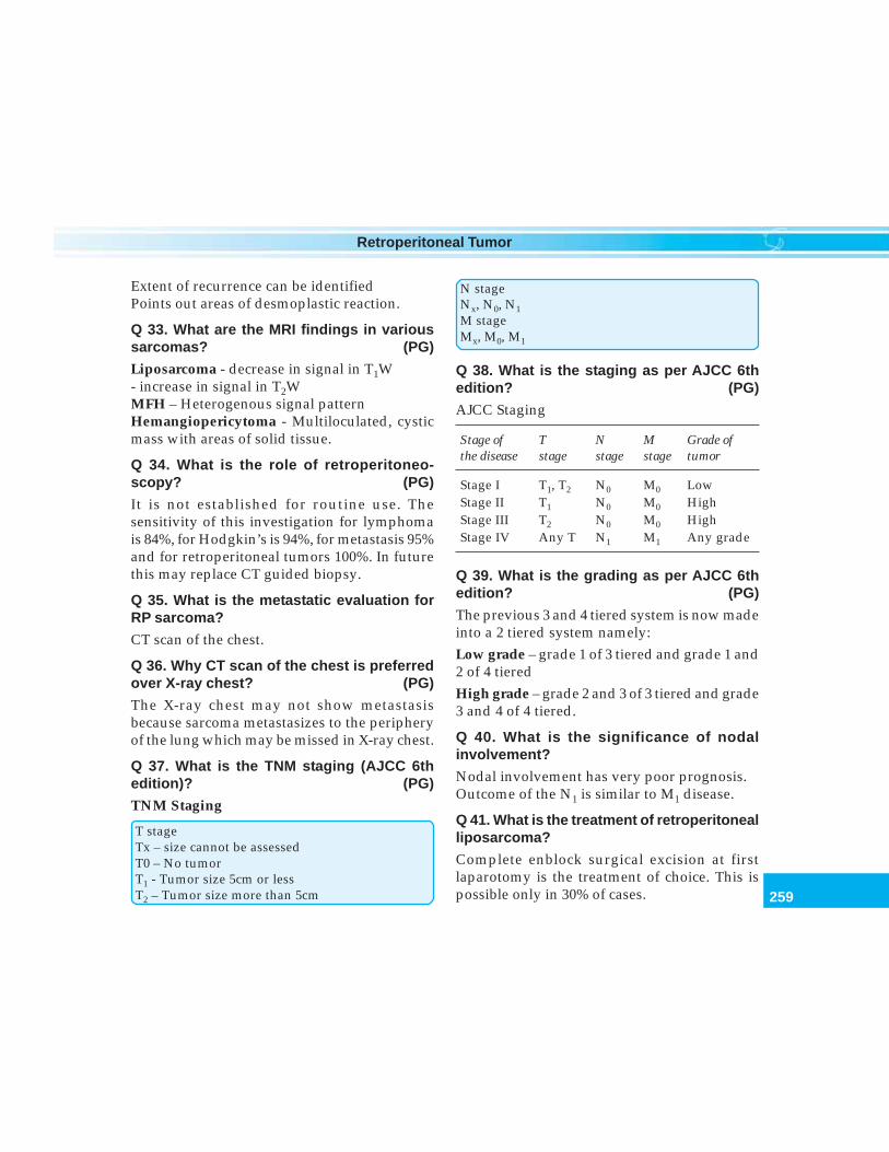
259
Retroperitoneal Tumor
Extent of recurrence can be identifiedPoints out areas of desmoplastic reaction.
Q 33. What are the MRI findings in varioussarcomas? (PG)
Liposarcoma - decrease in signal in T1W- increase in signal in T2WMFH – Heterogenous signal patternHemangiopericytoma - Multiloculated, cysticmass with areas of solid tissue.
Q 34. What is the role of retroperitoneo-scopy? (PG)
It is not established for routine use. Thesensitivity of this investigation for lymphomais 84%, for Hodgkin’s is 94%, for metastasis 95%and for retroperitoneal tumors 100%. In futurethis may replace CT guided biopsy.
Q 35. What is the metastatic evaluation forRP sarcoma?
CT scan of the chest.
Q 36. Why CT scan of the chest is preferredover X-ray chest? (PG)
The X-ray chest may not show metastasisbecause sarcoma metastasizes to the peripheryof the lung which may be missed in X-ray chest.
Q 37. What is the TNM staging (AJCC 6thedition)? (PG)
TNM Staging
T stageTx – size cannot be assessedT0 – No tumorT1 - Tumor size 5cm or lessT2 – Tumor size more than 5cm
N stageNx, N0, N1M stageMx, M0, M1
Q 38. What is the staging as per AJCC 6thedition? (PG)
AJCC Staging
Stage of T N M Grade ofthe disease stage stage stage tumor
Stage I T1, T2 N0 M0 LowStage II T1 N0 M0 HighStage III T2 N0 M0 HighStage IV Any T N1 M1 Any grade
Q 39. What is the grading as per AJCC 6thedition? (PG)
The previous 3 and 4 tiered system is now madeinto a 2 tiered system namely:Low grade – grade 1 of 3 tiered and grade 1 and2 of 4 tieredHigh grade – grade 2 and 3 of 3 tiered and grade3 and 4 of 4 tiered.
Q 40. What is the significance of nodalinvolvement?
Nodal involvement has very poor prognosis.Outcome of the N1 is similar to M1 disease.
Q 41. What is the treatment of retroperitonealliposarcoma?
Complete enblock surgical excision at firstlaparotomy is the treatment of choice. This ispossible only in 30% of cases.
260
Clinical Surgery Pearls
Q 42. What are the preoperative preparationsand precautions?
• Bowel preparation• Consent for removal of whole or part of the
adjacent organs• Consent for fecal or urinary diversion• Arrange adequate blood• Operation is undertaken at a tertiary referral
centre.
Q 43. What anesthesia is used for surgery?(PG)
Hypotensive anesthesia.
Q 44. What are the incisions recommended?
Midline incision for pelvic level tumorsThoracoabdominal for upper level tumors.
Q 45. What is the clearance required forenblock excision?
Best results are seen in which a surgical marginof 3cm or more can be obtained beyond thetumor.
Q 46. What is the most difficult area forclearance? (PG)
If a plane of dissection is there posteriorly, theclearance will be adequate. Decide whether thetumor has traversed along a spinal nerve rootinto the spinal canal and its cord.
Q 47. What is the role of radiotherapy?
Radiotherapy has a definite role in lowering theincidence of local recurrence. The doserecommended is 5500cGy. Radiotherapy will
delay the progression of the disease but doesnot improve the survival.
Q 48. What is the role of IORT (Intraoperativeradiotherapy)? (PG)
IORT reduces local recurrence and reducesradiation enteritis.
Q 49. What is the chemotherapeutic agent ofchoice for RP sarcoma? (PG)
Adriamycin – as a single agent - 15-35%response. This may be combined withgranulocyte macrophage colony stimulatingfactor (GM-CSF).
Q 50. What are the important prognosticfactors? (PG)
The two most important factors are –Histological gradeCompleteness of surgical excision (R0).
Q 51. What is the prognosis of RP sarcoma?(PG)
5-year-survival of 62-92% for well-differentiatedtumor after radical excision.16-48% in undifferentiated tumors.
Q 52. What is the local recurrence rate?(PG)
40-82% with median time of 15 months to 44months.
Q 53. What is the follow up? (PG)
Physical examination every 2-3monthsIf symptoms occur do CT/MRIAsymptomatic patients –CT/MRI to be done atevery 6 months for first 3 years.
Published by dr-notes.com
261
Retroperitoneal Tumor
RETROPERITONEAL CYSTIC LESIONS
Q 54. What are the cystic lesions of theretroperitoneum?
Cystic lesions of the retroperitoneum
• Cystic lesions from the developmental remnants(Wolffian) of Urogenital tract situated near thekidney
• Retroperitoneal mesenteric cyst• Teratomatous and dermoid cysts• Abdominal cystic lymphangioma (lymphogenous
cyst)• Parasitic cysts
Q 55. What are the clinical features of theabdominal cystic lymphangioma?
This is usually seen in infants and children(3months to 5years). The commonest presentation
is abdominal pain. 30% of the children presentwith mass abdomen. 90% of the cysts areintraperitoneal. 10% are retroperitoneal. Childrenmay present with complications like intestinalobstruction.
Q 56. What is the diagnostic investigation?
USG abdomen.
Q 57. What is the treatment of the abdominalcystic lymphangioma?
Surgical excision.
Q 58. Can you get abdominal cysticlymphangioma in adults?
Yes. There will be a history of long duration.Acute presentations are rare in adults.
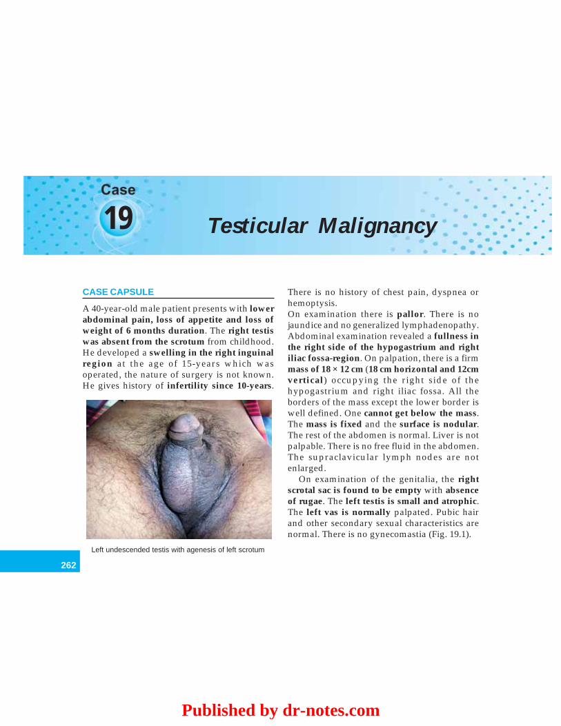
262
Clinical Surgery Pearls
19 Testicular Malignancy
CASE CAPSULE
A 40-year-old male patient presents with lowerabdominal pain, loss of appetite and loss ofweight of 6 months duration. The right testiswas absent from the scrotum from childhood.He developed a swelling in the right inguinalregion at the age of 15-years which wasoperated, the nature of surgery is not known.He gives history of infertility since 10-years.
There is no history of chest pain, dyspnea orhemoptysis.On examination there is pallor. There is nojaundice and no generalized lymphadenopathy.Abdominal examination revealed a fullness inthe right side of the hypogastrium and rightiliac fossa-region. On palpation, there is a firmmass of 18 × 12 cm (18 cm horizontal and 12cmvertical) occupying the right side of thehypogastrium and right iliac fossa. All theborders of the mass except the lower border iswell defined. One cannot get below the mass.The mass is fixed and the surface is nodular.The rest of the abdomen is normal. Liver is notpalpable. There is no free fluid in the abdomen.The supraclavicular lymph nodes are notenlarged.
On examination of the genitalia, the rightscrotal sac is found to be empty with absenceof rugae. The left testis is small and atrophic.The left vas is normally palpated. Pubic hairand other secondary sexual characteristics arenormal. There is no gynecomastia (Fig. 19.1).
Left undescended testis with agenesis of left scrotum
Published by dr-notes.com
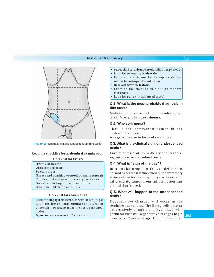
263
Testicular Malignancy
Read the checklist for abdominal examination.
Checklist for history
• History of trauma• Undescended testis• Scrotal surgery• Nausea and vomiting – retroduodenal metastasis• Cough and dyspnea – pulmonary metastasis• Backache – Retroperitoneal metastasis• Bone pain – Skeletal metastasis
Checklist for examination
• Look for empty hemiscrotum with absent rugae• Look for lower limb edema (unilateral or
bilateral) – Pressure from the retroperitonealnodes
• Gynecomastia – seen in 5% of cases
Fig. 19.1: Hypogastric mass (undescended right testis)
• Supraclavicular lymph nodes, iliac lymph nodes• Look for secondary hydrocele• Palpate the abdomen in the supraumbilical
region for retroperitoneal nodes• Rule out liver metastasis• Examine the chest to rule out pulmonary
metastasis• Look for pallor (in advanced cases)
Q 1. What is the most probable diagnosis inthis case?
Malignant tumor arising from the undescendedtestis. Most probably seminoma.
Q 2. Why seminoma?
That is the commonest tumor in theundescended testis.Age group is also in favor of seminoma.
Q 3. What is the clinical sign for undescendedtestis?
Empty hemiscrotum with absent rugae issuggestive of undescedned testis.
Q 4. What is “sign of the vas”?
In testicular neoplasm the vas deferens isnormal, whereas it is thickened in inflammatorylesions of the testis and epididymis. In order todifferentiate tumor from inflammation thisclinical sign is used.
Q 5. What will happen to the undescendedtestis?
Degenerative changes will occur in theseminiferous tubules. The lining cells becomeprogressively atrophic and hyalinized withperitubal fibrosis. Degenerative changes beginto occur at 2 years of age. If not corrected all
264
Clinical Surgery Pearls
bilaterally cryptorchid adult males becomessterile.
Q 6. What is the chronology of descent oftestis?
The testis is formed in front of the kidneys fromthe genital fold medial to the mesonephrons(wolffian body) in early fetal life. They lie in theretroperitoneum below the developing kidneys.The primitive testis is attached to the posteriorabdominal wall by the mesorchium. It’s descentis a complicated process, which depends on thegubernaculum testis, steroid hormones andmaternal gonadotrophins. The testis is intra-abdominal upto 7 months of intrauterine life.Shortly before birth the testis reaches the bottomof the scrotum being invaginated into a tube ofperitoneum – the processus vaginalis. Thechronology of descent is as follows:
Chronology of descent of testis
3rd month of – from loin to iliac fossaintrauterine life4–7 months of – rest at the site of internal ringintrauterine life7th month of – it is travelling through theintrauterine life inguinal canal8th month of – lies at the external ringintrauterine life9th month of – it enters the scrotum, reachingintrauterine life its base at or after birth
Note: The descent may continue after birth, 50%reaching the scrotum during the first month of life.
Q 7. What are the complications ofundescended testis?
Mnemonic – MAT SHOPM – MalignancyA – Atrophy – due to recurrent minor traumaT – TorsionS – Sterility – in bilateral casesH – Hernia – in 95% there is patent processus
vaginalis and 25% develop clinical herniaO – Orchitis (epididymo – orchitis)P – Pain – due to trauma.
Q 8. What are the positions of undescendedtestis?1. Intra-abdominal – (33%) lying extra
peritoneally just above the internal ring2. In the inguinal canal – (50%) it may or may
not be palpable there3. In the superficial inguinal pouch – (50%) it
must be distinguished from retractile testis.
Q 9. What is superficial inguinal pouch?Failure of the testis to descend into the scrotummay be caused by the presence of a dense layerof subcutaneous tissue at the neck of thescrotum. As a result of this the testis turnsupwards to lie in a pouch just above andsuperficial to the external inguinal ring. Thetestis is therefore felt in the subcutaneous tissuejust above and lateral to the pubic tubercle.
Q 10. What is the difference between ectopictestis and incompletely descended testis?An incompletely descended testis will lie alongthe line of the normal descent at the neck ofscrotum, over the external ring to the canal andon the posterior abdominal wall. In this situationthe testis is palpable only when it is at oroutside the external ring. The ectopic testis isalways palpable in contrast to incompletely
Published by dr-notes.com
265
Testicular Malignancy
descended testis and it is not in line of thenormal descent.
Q 11. What are the common positions ofectopic testis?
Common positions of ectopic testis
1. Femoral triangle – confused with lymph node2. Base of the penis3. Perineum4. Superficial inguinal pouch
Q 12. What is the timing of surgery forundescended testis?It is usually done between 12-18 months of age(the degenerative changes will start at the ageof 2 years).
Q 13. What is the fertility after orchidopexy?• Unilateral orchidopexy – fertility rate is 80%• Bilateral orchidopexy – fertility rate is 50%.
Q 14. What is retractile testis?During childhood the testis are mobile. Inchildren < 3years with very active cremastericreflex due to the small size of testis the gonadwill retract into the external ring or within thecanal. It can be manipulated back into the mid orlower scrotum. No treatment is required in thiscondition.
Q 15. What is ‘vanishing’ testis?
It is a condition where the testis develops butdisappears before birth. The most likely causeis prenatal torsion.
Q 16. What is true agenesis?
It is very rare. Laparoscopy is useful in distingui-shing true agenesis from cryptorchidism withintraabdominal testis.
Q 17. What are the anomalies associated withcryptorchidism?
This is seen in 15% of cryptorchidism. Thefollowing are the associated anomalies.
Anomalies associated with cryptorchidism
• Klinefelter’s syndrome• Hypogonadotropic hypogonadism• Prune belly syndrome• Horse shoe kidney• Renal agenesis or hypoplasia• Exstrophy of the bladder• Ureteral reflux• Gastroschisis• Cloacal exstrophy
Q 18. What is the incidence of undescendedtestis?
• 4% of full-term infants• 30% in premature babies.
Q 19. Can the unilateral cryptorchid developmalignancy in the normally descendedcontralateral testis?
Yes. 10% of patients with history of crypto-rchidism develop malignancy in contralateraltestis.
Q 20. What is the risk of malignancyaccording to the position of the testis inundescended testis?The relative risk for malignancy in—Intraabdominal testis – 1 in 20Inguinal testis – 1 in 80.
Q 21. What is the commonest pathologicaltype of malignancy seen in undescendedtestis?SeminomaEmbryonal carcinoma—It is seen nowadays in
266
Clinical Surgery Pearls
patients in whom the testis has been broughtdown.
Q 22. Will orchidopexy change the malignantpotential of the cryptorchid testis?Note: It facilitates examination and tumordetection.
Q 23. What are the factors responsible forthe malignant change in cryptorchidism?
Factors responsible for malignant change incryptorchidism
1. Abnormal germ cell morphology2. Gonadal dysgenesis3. Elevated temperature4. Interference of blood supply5. Endocrine dysfunction
Q 24. What are the differential diagnoses ofa solid swelling in the testis?
Differential diagnoses of solidswelling in the testis
1. Testicular tumor2. Acute and chronic epididymo – orchitis3. Hematocele4. Gumma5. Orchitis (mumps)
Q 25. What is ‘billiard ball’ testis?
Syphilis in adults will cause interstitial inflam-mation turning the testis into a round, hard, andinsensitive mass, which is called ‘billiard balltestis’.
Q 26. What is Gumma of the testis?
Gumma of the testis is due to syphilis. The testisis painless and therefore presents as a lump on
the surface of the testis or enlargement of thewhole organ. The testicular sensation is lostearly. H/o exposure will be there. A search ismade for stigmata of syphilis like Clutton’sjoint, interstitial keratitis, etc. It isindistinguishable from tumor. If untreated itwill breakdown to form Gummatous ulcer.
Q 27. What is the relationship of testiculartumor and trauma?
Many patients attribute the symptoms of theswelling to trauma. But this is to be ignored. Wedon’t know whether the lump is due to thehump or the hump is due to the lump!!
Q 28. What is the commonest starting pointof the tumor in the testis?
Lower pole of the body of the testis, later theyoccupy the whole of the testis.
Q 29. What is the classification for testiculartumors?
Various classifications are there –• Dixon and Moore• WHO• British tumor penal• Histopathological classification.
The testis is composed of actively dividinggerm cells, the supporting cells and connectivetissue stroma. The tumors of the testis arise fromthe actively dividing germ cells. Comprising 90– 95% and the rest contributed by nongerminalneoplasms. The histopathological classificationis given below:
Published by dr-notes.com
267
Testicular Malignancy
Histopathological Classification predominance of one or more cells. The usualvariety is yellowish in color with cystic spacescontaining gelatinous fluid. Cartilaginous areasare often present. The tumor may be as small asa peanut or as large as a coconut.
TESTICULAR PANEL CLASSIFICATION OFTERATOMA
• Teratoma Differentiated (TD) – Uncommon.• Eventhough there is no histologically recog-
nizable malignant component it cannot beconsidered benign. It may contain cartilage,muscle and glandular tissue.
• Malignant teratoma Intermediate orTeratocarcinoma (MTI) (most common)(A and B Type). Contains definitelymalignant and incompletely differentiatedcomponents.
• Malignant Teratoma Anaplastic orEmbryonal Carcinoma (MTA) The cells arepresumed to be derived from yolk sac andare responsible for elevation of AFP.
• Malignant Teratoma Trophoblastic (MTT)—Uncommon
It contains syncytial cell mass with malignantvillous or papillary cytotrophoblast (chorio-carcinoma). It produces chorionicgonadotrophin (hCG). It is one of the mostmalignant tumors having spread bybloodstream and lymphatics early.
Q 31. What are the differences betweenSeminoma and Teratoma?
Q 30. What is teratoma? What is theclassification of teratoma?
Teratoma arises from totipotent cells in the retetestis and contains a variety of cell types with
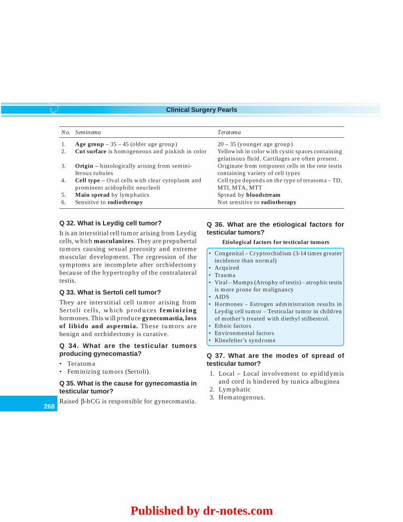
268
Clinical Surgery Pearls
No. Seminoma Teratoma
1. Age group – 35 – 45 (older age group) 20 – 35 (younger age group)2. Cut surface is homogeneous and pinkish in color Yellowish in color with cystic spaces containing
gelatinous fluid. Cartilages are often present.3. Origin – histologically arising from semini- Originate from totipotent cells in the rete testis
ferous tubules containing variety of cell types4. Cell type – Oval cells with clear cytoplasm and Cell type depends on the type of teratoma – TD,
prominent acidophilic neucleoli MTI, MTA, MTT5. Main spread by lymphatics Spread by bloodstream6. Sensitive to radiotherapy Not sensitive to radiotherapy
Q 36. What are the etiological factors fortesticular tumors?
Etiological factors for testicular tumors
• Congenital – Cryptorchidism (3-14 times greaterincidence than normal)
• Acquired• Trauma• Viral – Mumps (Atrophy of testis) – atrophic testis
is more prone for malignancy• AIDS• Hormones – Estrogen administration results in
Leydig cell tumor – Testicular tumor in childrenof mother’s treated with diethyl stilbestrol.
• Ethnic factors• Environmental factors• Klinefelter’s syndrome
Q 37. What are the modes of spread oftesticular tumor?
1. Local – Local involvement to epididymisand cord is hindered by tunica albuginea
2. Lymphatic3. Hematogenous.
Q 32. What is Leydig cell tumor?
It is an interstitial cell tumor arising from Leydigcells, which masculanizes. They are prepubertaltumors causing sexual precosity and extrememuscular development. The regression of thesymptoms are incomplete after orchidectomybecause of the hypertrophy of the contralateraltestis.
Q 33. What is Sertoli cell tumor?
They are interstitial cell tumor arising fromSertoli cells, which produces feminizinghormones. This will produce gynecomastia, lossof libido and aspermia. These tumors arebenign and orchidectomy is curative.
Q 34. What are the testicular tumorsproducing gynecomastia?
• Teratoma• Feminizing tumors (Sertoli).
Q 35. What is the cause for gynecomastia intesticular tumor?
Raised β-hCG is responsible for gynecomastia.
Published by dr-notes.com
269
Testicular Malignancy
Q 38. What is the lymphatic drainage of testis?
From the testis several lymphatic vessels emergefrom the mediastinum testis and accompany thegonadal vessels in the spermatic cord. Wherethe spermatic vessels cross ventral to the uretersome of these lymphatics diverge medially anddrain into the retroperitoneal lymph nodes,while others follow the spermatic vessels to theirorigin and reach the paraaortic nodes.
Lymphatics from the medial side of the testismay run with the artery to the vas and draininto a node at the bifurcation of the commoniliac artery (common iliac nodes).
The inguinal lymph nodes are affected onlyif the scrotal skin is involved.
Q 39. What you mean by landing zones forlymphatics?
The primary landing zones for right testiculartumor lies in the interaortocaval nodesimmediately below the renal vessels. (At thelevel of L2 lumbar vertebrae). The ipsilaterallymp-hatics will also go to paracaval, preaorticand right common iliac and external iliac nodes.
For the left testicular tumor it lies in the trueparaaortic nodes just below the left renal vessels.(Left renal hilum). The ipsilateral distributionalso includes paraaortic, preaortic and leftcommon iliac nodes.
The metastasis reach the common iliac,external iliac and inguinal nodes usuallysecondary to retrograde spread due to largevolume disease.
Q 40. Clinically what is the level of retro-peritoneal nodes?
Above the umbilicus in the abdomen.
Q 41. What is the spread beyond the regionalnodes?
The retroperitoneal lymphatics empty into thecisterna chyle via the right and left lumbartrunks. From here it will reach the leftsupraclavicular nodes.
From the paraaortic nodes the lymphatic willreach the retrocrural nodes and from their it willreach the mediastinal nodes (posterior medias-tinum). Involvement of the anterior mediastinumis rare.
Q 42. What is the method of contralateralspread? (PG)
The contralateral spread is usually seen with rightsided tumors especially in large volume tumors(rare with left sided tumors). If the patient hasundergone herniorrhaphy, vasectomy or othertransscrotal procedures unrelated to the tumor,pelvic and inguinal nodes may get enlarged.
Q 43. What is the mechanism of inguinal nodemetastasis?
This will occur only if the tunica albuginea hasbeen invaded by the tumor or as a result ofprevious surgical procedures like orchidopexyor herniorrhaphy.
Q 44. What are the sites of extranodalmetastasis?
Sites of extranodal metastasis in order ofdecreasing frequency
LungLiverBrainBoneKidney
270
Clinical Surgery Pearls
AdrenalGI tractSpleen
Q 45. Can testicular tumor occur bilaterally?(PG)
Yes. 2-3% of testicular tumors are bilateral. Theymay occur simultaneously or successively.Primary bilateral tumors occur synchronouslyor asynchronously.
Q 46. What is the most common cause forbilateral testicular tumor? (PG)
• Testicular lymphoma• Seminoma is the commonest bilateral
primary tumor.
Q 47. How will you size the testis? (PG)
They can be sized according to Prader’sorchidometer.
Q 48. When examining the testes, which sideis examined first?
The normal side is examined first.
Q 49. What is the incidence of testiculartumor? (PG)
• They make up 1-2% of the male cancers.• The life time probability of developing a
tumor of the testis is 0.2%• The highest incidence is seen in Scandinavia• Lowest incidence is in Africa and Asia• Higher socioeconomic class has higher
incidence• Incidence is 4 times in whites compared to
black.
Q 50. Why testicular tumor is more commonon the right side?
There is increased incidence of cryptorchidismon the right side and therefore tumor is morecommon on the right side.
Q 51. What are the clinical features oftesticular tumor?
Painless enlargement of the testis 90% (Dullache, sensation of heaviness in the lowerabdomen, anal area or scrotum) acute testicularpain due to infarction or intratesticularhemorrhage is seen in about 10% of cases.
Extratesticular manifestations• Cough and dyspnea – Pulmonary metastasis• Anorexia, nausea, vomiting (retroduodenal
metastasis)• Supraclavicular lymph node metastasis• Back pain – retroperitoneal metastasis• Lower extremities swelling – venacaval obstruction
(unilateral or bilateral limb)• Metastatic paraaortic lymph nodes (present
behind and above the umbilicus)• Gynecomastia (5% of cases)• Bone pain (skeletal metastasis)• CNS involvement (spinal cord/root involvement)• Asymptomatic (10%) – identified incidentally
Q 52. What is the incidence of secondaryhydrocele in testicular tumors?
10%. The secondary hydrocele is usually lax.
Q 53. What is the cause for thickening of thespermatic cord? (PG)
This is a late future and it is due to thecremasteric hypertrophy and the enlargementof the testicular vessels.
Published by dr-notes.com

271
Testicular Malignancy
Q 54. What is the importance of testicularsensation in tumors?
The testicular sensation is lost completely in theearly stage of the tumor. This sign is better notdemonstrated due to the fear of dissemination.Palpation of the tumor also should be avoidedto the minimum.
Q 55. What is “hurricane tumor”?
It is a ferocious type of malignancy that killsthe patients in a matter of weeks due to the rapidspread of the tumor.
Q 56. Can you feel the epididymis separatelyin testicular tumor?
Epididymis is normal during initial stages. Butit becomes more difficult to feel as it is flattenedand incorporated in the growth in later stages.
Q 57. What are the tumor markers?
Tumor markers for testicular tumors1. α - Fetoprotein (AFP)2. β - Human chorionic gonadotrophin (β-
hCG)3. Lactate dehydrogenase (LDH)
Q 58. What is the significance of serum tumormarkers? (PG)
In 85% of patients at least 1 marker is raised.Markers are used for assessment of tumorregression and recurrence.Remember !! Tumor is possible withoutelevation of the tumor marker.
Q 59. What is the significance of LDH? (PG)
It is increased in 60% of nonseminomatous GCTand 80% of patients with advanced seminoma.
It has independent prognostic significance inpatients with advanced germ cell tumor.
Increase in the serum concentration is areflection of tumor burden, growth rate andproliferation.
Q 60. What is the significance of AFP? (PG)
• AFP is elevated in 70% of nonseminomas(not in pure seminomas)—restricted toembryonal cell carcinoma and endodermalsinus tumor (yolk sac tumor).
• If AFP is elevated in seminoma, suspectnon- seminoma elements in either theprimary or metastasis.
• The normal adult concentration is usually< 15% ng/ml.
• The physiological half life is 5-7days.
Q 61: What is the significance of βββββ-hCG?(PG)
• It is a glycoprotein produced by 65% of non-seminomas and 10% of seminomas (allpatients with choriocarcinoma and 40%patients with embryonal carcinoma).
• It is produced by syncytiotrophoblasts.• Half life of β-hCG is 24-48 hours
Q 62. What are the investigations in a patientwith suspected testicular tumor?
Investigations in testicular tumor
1. Blood – radioimmunoassay of tumor markers2. Chest – radiograph – for pulmonary metastasis3. Ultrasound – of the affected testis and abdomen—
abdomen for lymph nodes and liver metastasis4. CT/MRI – for intraabdominal and intrathoracic
secondaries
272
Clinical Surgery Pearls
5. IVU – deviated or obstructed ureters as a resultof the retroperitoneal nodes will be demons-trated
6. High orchidectomy
Q 63. What is the sonological finding intesticular tumor? (PG)
Sonologically the tumor will appear as hypo-echoic lesion. It may also reveal secondaryhydrocele.
Q 64. Why high orchidectomy is includedalong with investigations?
Orchidectomy is considered as the basicinvestigation in testicular tumors. It is essentialto remove the primary tumor to obtain thehistological type of malignancy. Thepathological knowledge is important for theAJCC staging because the tumor status in thisstaging is always PT0, PT1, etc.
Q 65. What is the incision for highorchidectomy?
Transinguinal incision.
Q 66. Why not transscrotal incision?
If a transscrotal incision is made in testiculartumor, the tumor will spread to the lymphaticterritory of the inguinal nodes (testis willnormally drain to paraaortic nodes). Thereforeif transscrotal incision is made, the inguinalnodes also must be tackled by radiotherapy. Thismust be avoided. There are three things to beavoided in testicular tumors –
Three No’s in testicular tumors
• No transscrotal incision• No transscrotal aspiration• No transscrotal biopsy
Q 67. What is high orchidectomy (inguinalorchidectomy)? (PG)
Through a transinguinal incision the spermaticcord is displayed by dividing the externaloblique aponeurosis. A soft clamp is placedacross the cord as the initial step to preventdissemination of malignant cells as the testis ismobilized into the wound. The cord is thendoubly ligated at the level of the deep inguinalring and divided, so that the testis can beremoved.
Q 68. What is Chevassu maneuver? (PG)
During the above procedure when the testis isbrought to the inguinal wound, if there is doubtregarding the diagnosis, the testis should bebisected along its anterior convexity so that itsinternal structure can be examined by frozensection. If a tumor is ruled out the bisectedtestis can be closed and returned to the normalposition.
Q 69. If the patient is explored by transscrotalroute already what is the course of action?
(PG)
Hemiscrotectomy and radiation of inguinalnodes is recommended.
Q 70. What is the clinical staging?
Clinical staging of testicular tumors
Published by dr-notes.com
273
Testicular Malignancy
Stage 1 – Tumor confined to the testisStage 2 – Nodes below the level of the diaphragm– Spread to regional lymph nodes – retroperitoneal,paraaorticStage 3 – Nodes above the level of the diaphragm– Spread beyond the retroperitoneal nodes, to themediastinum or extralymphatic metastasisStage 4 – Pulmonary or hepatic metastasis
Q 71. What is the TNM staging? (PG)
T stage* The extent of primary tumor is usuallyclassified after radical orchiectomy and for thisreason, a Pathological Stage is assigned usingthe prefix P*pTXPrimary tumor cannot be assessedpTO No evidence of primary tumor (e.g.
histologic scar in testis)pTis Intratubular germ cell neoplasia
(carcinoma in situ)pT1 Tumor limited to the testis and
epididymis without vascular/lymphaticinvasion, tumor may invade into thetunica albuginea but not the tunicavaginalis
pT2 Tumor limited to the testis andepididymis with vascular/lymphaticinvasion, or tumor extending throughthe tunica albuginea with involvementof the tunica vaginalis
PT3 Tumor invades the spermatic cord withor without vascular/lymphatic invasion
pT4 Tumor invades the scrotum with orwithout vascular/lymphatic invasion.
N Stage (Clinical)
NX Regional lymph nodes cannot beassessed
N0 No regional lymph node metastasisN1 Metastasis with a lymph node mass 2cm
or less in greatest dimension; or multiplelymph nodes, none more than 2cm ingreatest dimension
N2 Metastasis with a lymph node massmore than 2cm but not more than 5cmin greatest dimension; or multiplelymph nodes, any one mass greater than2cm but not more than 5cm in greatestdimension
N3 Metastasis with a lymph node massmore than 5cm in greatest dimension.
Pathological N Stage (pN)
pNX Regional lymph nodes cannot beassessed
pN0 No regional lymph node metastasispN1 Metastasis with a lymph node mass 2 cm
or less in greatest dimension and lessthan or equal to 5 nodes positive, nonemore than 2 cm in greatest dimension
pN2 Metastasis with a lymph node massmore than 2 cm but not more than 5 cmin greatest dimension; or more than5nodes positive, none more than 5 cm;or evidence of extranodal extension oftumor
pN3 Metastasis with a lymph node massmore than 5 cm in greatest dimension
274
Clinical Surgery Pearls
M stage
MX Distant metastasis cannot be assessedM0 No distant metastasisM1 Distant metastasisM1a Nonregional nodal or pulmonary
metastasisM1b Distant metastasis other than to non-
regional lymph nodes and lungs.
Serum Tumor Markers (S)
SX Marker studies not available or notperformed
S0 Marker study levels within normal limitsS1 LDH < 1.5 X N* and hCG (mIu/ml) <
5000 andAFP (ng/ml) < 1000S2 LDH 1.5 – 10 X N orhCG (mIu/ml) 5000
– 50,000 orAFP (ng/ml) 1000 – 10,000S3 LDH > 10 X N orhCG (mIu/ml) > 50,000
orAFP (ng/ml) > 10,000*N indicates upper limit of normal for the LDHassay.
Q 72. What is the management of testiculartumors?
The management of seminomas and non-seminomatous germinal cell tumors aredifferent.
Q 73. What is the management of seminoma?
After high orchidectomy seminoma is treated withradiotherapy (because seminoma is a radiosensitive tumor) for stage 1 and nonbulky stage 2.• Over a 3 week period administer 2500cGy
hockey stick field including the followingnodes–
• Paraaortic, paracaval, bilateral commoniliac and external iliac nodal regions.
• Recently the radiation is confined to thepara- aortic area alone.
• With history of herniorrhaphy andorchidopexy include the contralateralinguinal region shielding the contralateraltestis.
• Mediastinal radiation is currently avoided(chemotherapy is more effective).
Q 74. Is there any role for semen banking?(PG)
If the function of the contralateral testis is inquestion, this should be considered. The semenof the patient may be collected and preserved ifthe patient is desirous of having children.
Q 75. What are the indications forchemotherapy in seminoma? (PG)Bulky abdominal diseaseMetastasis.
Q 76. What is the chemotherapeutic regimen?(PG)It is treated with four cycles of chemotherapywith BEP regimenBleomycinEtoposideCisplatin.
Q 77. What is the treatment of residualabdominal mass after chemotherapy? (PG)
Residual tumor of more than 3 cm is treated bysurgery.
Published by dr-notes.com
275
Testicular Malignancy
Q 78. What is the management of non-seminomatous germ cell tumor?
After high orchidectomy this patients are treatedwith Retroperitoneal Lymph Node Dissection(RPLND) except bulky disease or distantmetastasis.
Q 79. What are the approaches for RPLND?(PG)
TransabdominalThoracoabdominal.
Q 80. What is bulky disease? (PG)All patients with retroperitoneal mass > 5 cm(high burden disease).
Q 81. What is the most important problem ofRPLND? (PG)
Retrograde ejaculation.
Q 82. What is the extent of RPLND? (PG)The standard RPLND is a bilateral dissectionfrom renal vessel down to aortic bifurcation,along the external iliac artery to the deepinguinal ring on the affected side. The followinglymph nodes are removed on both sidesLymph nodes removed in RPLND
ParacavalInteraortocavalParaaorticPreaorticCommon iliac nodes
Q 83. What is the contraindication for RPLND?(PG)
Choriocarcinoma. Here the disease is systemicand the treatment is multiagent chemotherapy.
Q 84. What are the indications for withholding RPLND? (PG)
Normal serum markersNo nodes in abdominal CTNo distant metastasis.
The rationale behind this policy is that only20% of such patients will develop disease andin such a situation patient should be ready forregular follow up.
Q 85. What is the follow up of such a patient?(PG)
Physical examinationChest X-rayTumor markers.
They are done monthly for the first year,every 2 monthly for the second year and 3-6months there after.
Q 86. What is modified RPLND? (PG)
This involves bilateral dissection above theorigin of the inferior mesenteric artery andunilateral dissection confined to the side of thetumor below the origin of the inferior mesentericartery. This preserves the importantsympathetic fibers from the contralateral sidethus maintaining ejaculation.
276
Clinical Surgery Pearls
Q 87. What is the treatment of extensiveretroperitoneal nodes with chest metastasis?
(PG)
High orchidectomy + multiagent chemofollowed by excision of the persistent mass inthe retroperitoneum.
Q 88. What are the alternative drugs forchemotherapy? (PG)Ifosamide/Doxorubicin.
Q 89. What is the prognosis of nonseminomatous germinal cell tumor? (PG)
Stage II - 90% cure rate at 5 yearsStage III - 70% cure rate at 5 yearsChoriocarcinoma - 35% at 5 years
Q 90. What is the management of the givencase?In this patient the tumor is arising from intra-abdominal testis. It should be removed trans-abdominally and the further managementdepends on the histopathology. Considering hisage, the pathology is likely to be seminoma andtherefore he requires radiotherapy.
Published by dr-notes.com
277
Portal Hypertension
20 Portal Hypertension
CASE CAPSULE
A 55-year-old male patient presents withmelena of one week duration. He is a chronicalcoholic and gives history of three bouts ofhematemesis in the last two months. Hecomplains of distension of the abdomen,abdominal discomfort, and lethargy for the last6 months. He gives history of jaundice in thechildhood. His sleep pattern is not altered.There is no history of treatment with NSAIDS,paracetamol and antituberculous drugs. Thereis no history of omphalitis in the newbornperiod. There is no history of chronicpancreatitis or history of convulsions.
On general examination, the patient is illlooking, poorly nourished and there is obviousmuscle wasting. He is fully oriented. There isno memory impairment. The speech is normal.There is pallor and mild icterus. Both lowerlimbs show pitting type of edema. Clubbingof the fingers and toes are seen. About 10 spidernaevi spots are present in the front of the chest,face and arms (1mm size bright red spots withradiating small branches spreading to 2mmdiameter. They fade completely whencompressed with finger and refill as soon as thepressure is released). Patient has got bilateral
gynecomastia. There is loss of pubic andaxillary hair. There is no evidence of palmarerythema. Skin bruising is noted in the rightforearm of about 5 × 4 cm size. There is noflapping tremor and there is no fetor hepaticus.
On examination of the abdomen there isgeneralized distension of the abdomen and theumbilicus is everted. There is no caput medusae.There is shifting dullness and fluid thrilldemonstrated. Dipping palpation revealedsplenomegaly. On auscultation a venous humis heard in the epigastrium which is louder oninspiration. On examination of the genitalia,both testis are found to be small and softsuggestive of testicular atrophy. Centralnervous system examination showed noevidence of encephalopathy. The cardio-vascular system is normal and there are nofeatures of suggestive of constrictive pericarditisor tricuspid valve incompetence.
Read the checklist for abdominal examination.Checklist for history
• History of alcoholism• Previous jaundice• Schistosomiasis• Valvular heart disease, constrictive pericarditis
278
Clinical Surgery Pearls
• Abdominal tumors—extrinsic compression• Hematemesis and melena
Check list for examination
• Stigmata of liver disease (Refer chapter on Liver)• Triad of portal hypertension namely esophageal
varices, ascites and splenomegaly• Look for jaundice• Features of encephalopathy• Look for anemia• Look for pitting edema of lower limbs• Look for ascites• Look for hepatosplenomegaly• Look for Caput medusae• Look for Testicular atrophy• Look for gynecomastia• Look for Kenawy's sign—Venous hum, heard
louder on inspiration in the epigastrium in portalhypertension
• Look for Cruveilhier-Baumgarten syndrome—Loud venous hum at umbilicus in case of portalhypertension
Q 1. What is the probable diagnosis in thiscase?Cirrhosis of liver with portal hypertension.
Q 2. What are the points in favor of portalhypertension?1. History of alcoholism2. History of upper GI bleed3. Melena4. Hepatosplenomegaly on examination5. Mild icterus6. Presence of ascites.
Q 3. What are the causes for ascites?Causes for ascites
Hepatic causes- Cirrhosis with portal hypertension- Hypoalbuminemia of any cause
Malignant ascites- Secondary to any primary or metastatic
malignancyInfective- Tuberculous peritonitis- Bacterial peritonitis- Primary peritonitisCongestive- Right sided heart failure- Budd-chiari syndrome- Constrictive pericarditisMiscellaneous causes- Pancreatic ascites- Chylous ascites- Meig's syndrome- Pseudomyxoma peritonei
Q 4. What are the causes for general fullnessof the abdomen?
It may be due to—• Fat• Fluid• Flatus• Feces• Fetus.
Q 5. What is the minimum amount of fluidrequired to demonstrate free fluid in theabdominal cavity?
The minimum amount of fluid to demonstrateclinical ascite is 1500 ml.
Q 6. What is the minimum amount requiredradiologically to demonstrate fluid within theperitoneal cavity?
The minimum amount required radiologicallyto demonstrate ascites is 150 ml.
Q 7. What are the types of ascitic fluid?
May be transudate or exudates.
Published by dr-notes.com
279
Portal Hypertension
Q 8. What are the differences betweentransudate and exudates?
Differences between transudate and exudate
Transudate Exudates
Protein Low - most of which is albumin < 1gm/dL High—(2.5 - 3.5 gm/dL)High content of fibrinogen
Specific gravity < 1.012 > 1.020Glucose content Same as plasma Low < 60 mg/dLpH > 7.3 < 7.3LDH Low HighCellular debris Few cells More cellsCause Obstruction to the venous outflow from liver Inflammatory extravascular fluid
(ultrafiltrate of blood plasma and results formed by the escape of fluid,from imbalance across the vascular endo- proteins and blood cells fromthelium) permeability of endothelium— the vascular system into thenormal interstitial tissue or body cavities
due to increased vascular permea-bility
SAAG (Serum > 1.1 (Ascitic fluid has less albumin than < 1.1 (Ascitic fluid has more albuminascites albumin serum) than serum)Gradient)
Q 9. What are the signs of ascites?Signs of ascites
• Puddle sign—mild ascites• Shifting dullness—moderate ascites• Fluid thrill—severe ascites• Tanyon sign—the umbilicus is pushed upwards
in ovarian cyst and downwards in ascites
Q 10. What is puddle sign?
This is a clinical method to demonstrateminimum amount of fluid in the peritonealcavity. The patient is positioned in the knee
elbow position and the abdomen is percussedaround the umbilicus which will revealdullness.
Q 11. How will you demonstrate shiftingdullness?
Ask the patient to turn on to his left side, waitfor a minute so that the fluid gravitates. Startpercussion from the right side to the left notingwhere the resonant area becomes dull and thenmark the spot on the abdominal wall. Then thepatient is asked to turn slightly on to his right
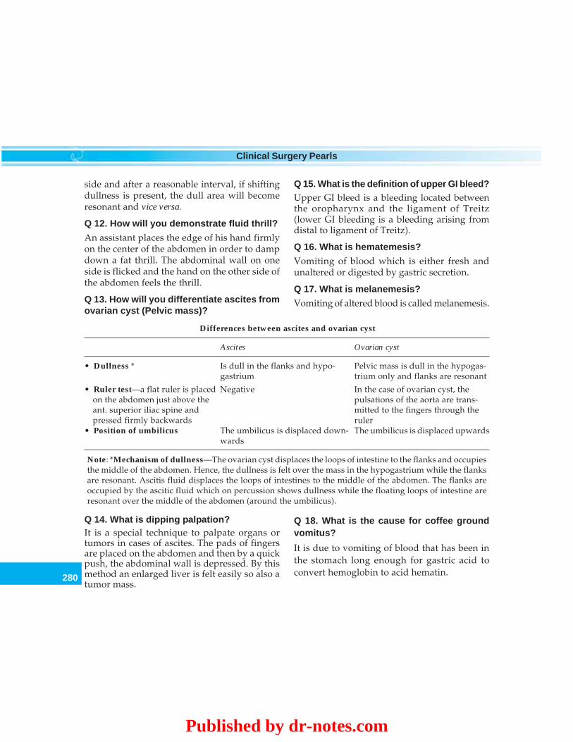
280
Clinical Surgery Pearls
side and after a reasonable interval, if shiftingdullness is present, the dull area will becomeresonant and vice versa.
Q 12. How will you demonstrate fluid thrill?
An assistant places the edge of his hand firmlyon the center of the abdomen in order to dampdown a fat thrill. The abdominal wall on oneside is flicked and the hand on the other side ofthe abdomen feels the thrill.
Q 13. How will you differentiate ascites fromovarian cyst (Pelvic mass)?
Differences between ascites and ovarian cyst
Ascites Ovarian cyst
• Dullness * Is dull in the flanks and hypo- Pelvic mass is dull in the hypogas-gastrium trium only and flanks are resonant
• Ruler test—a flat ruler is placed Negative In the case of ovarian cyst, theon the abdomen just above the pulsations of the aorta are trans-ant. superior iliac spine and mitted to the fingers through thepressed firmly backwards ruler
• Position of umbilicus The umbilicus is displaced down- The umbilicus is displaced upwardswards
Note: *Mechanism of dullness—The ovarian cyst displaces the loops of intestine to the flanks and occupiesthe middle of the abdomen. Hence, the dullness is felt over the mass in the hypogastrium while the flanksare resonant. Ascitis fluid displaces the loops of intestines to the middle of the abdomen. The flanks areoccupied by the ascitic fluid which on percussion shows dullness while the floating loops of intestine areresonant over the middle of the abdomen (around the umbilicus).
Q 14. What is dipping palpation?It is a special technique to palpate organs ortumors in cases of ascites. The pads of fingersare placed on the abdomen and then by a quickpush, the abdominal wall is depressed. By thismethod an enlarged liver is felt easily so also atumor mass.
Q 15. What is the definition of upper GI bleed?Upper GI bleed is a bleeding located betweenthe oropharynx and the ligament of Treitz(lower GI bleeding is a bleeding arising fromdistal to ligament of Treitz).
Q 16. What is hematemesis?
Vomiting of blood which is either fresh andunaltered or digested by gastric secretion.
Q 17. What is melanemesis?
Vomiting of altered blood is called melanemesis.
Q 18. What is the cause for coffee groundvomitus?
It is due to vomiting of blood that has been inthe stomach long enough for gastric acid toconvert hemoglobin to acid hematin.
Published by dr-notes.com
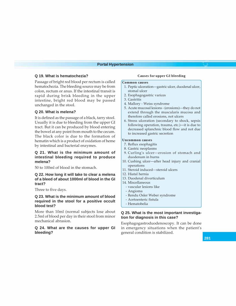
281
Portal Hypertension
Q 19. What is hematochezia?
Passage of bright red blood per rectum is calledhematochezia. The bleeding source may be fromcolon, rectum or anus. If the intestinal transit israpid during brisk bleeding in the upperintestine, bright red blood may be passedunchanged in the stool.
Q 20. What is melena?
It is defined as the passage of a black, tarry stool.Usually it is due to bleeding from the upper GItract. But it can be produced by blood enteringthe bowel at any point from mouth to the cecum.The black color is due to the formation ofhematin which is a product of oxidation of hemeby intestinal and bacterial enzymes.
Q 21. What is the minimum amount ofintestinal bleeding required to producemelena?
50 to 100ml of blood in the stomach.
Q 22. How long it will take to clear a melenaof a bleed of about 1000ml of blood in the GItract?
Three to five days.
Q 23. What is the minimum amount of bloodrequired in the stool for a positive occultblood test?
More than 10ml (normal subjects lose about2.5ml of blood per day in their stool from minormechanical abrasion.
Q 24. What are the causes for upper GIbleeding?
Causes for upper GI bleeding
Common causes1. Peptic ulceration—gastric ulcer, duodenal ulcer,
stomal ulcer2. Esophagogastric varices3. Gastritis4. Mallory - Weiss syndrome5. Acute mucosal lesions - (erosions)—they do not
extend through the muscularis mucosa andtherefore called erosions, not ulcers
6. Stress ulceration (secondary to shock, sepsisfollowing operation, trauma, etc.)—it is due todecreased splanchnic blood flow and not dueto increased gastric secretion
Uncommon causes7. Reflux esophagitis8. Gastric neoplasms9. Curling's ulcer—erosion of stomach and
duodenum in burns10. Cushing ulcer—after head injury and cranial
operations11. Steroid induced—steroid ulcers12. Hiatal hernia13. Duodenal diverticulum14. Miscellaneous
- vascular lesions like- Angioma- Rendu Osler Weber syndrome- Aortoenteric fistula- Hematobelia
Q 25. What is the most important investiga-tion for diagnosis in this case?
Esophagogastroduodenoscopy. It can be donein emergency situations when the patient'sgeneral condition is stabilized.
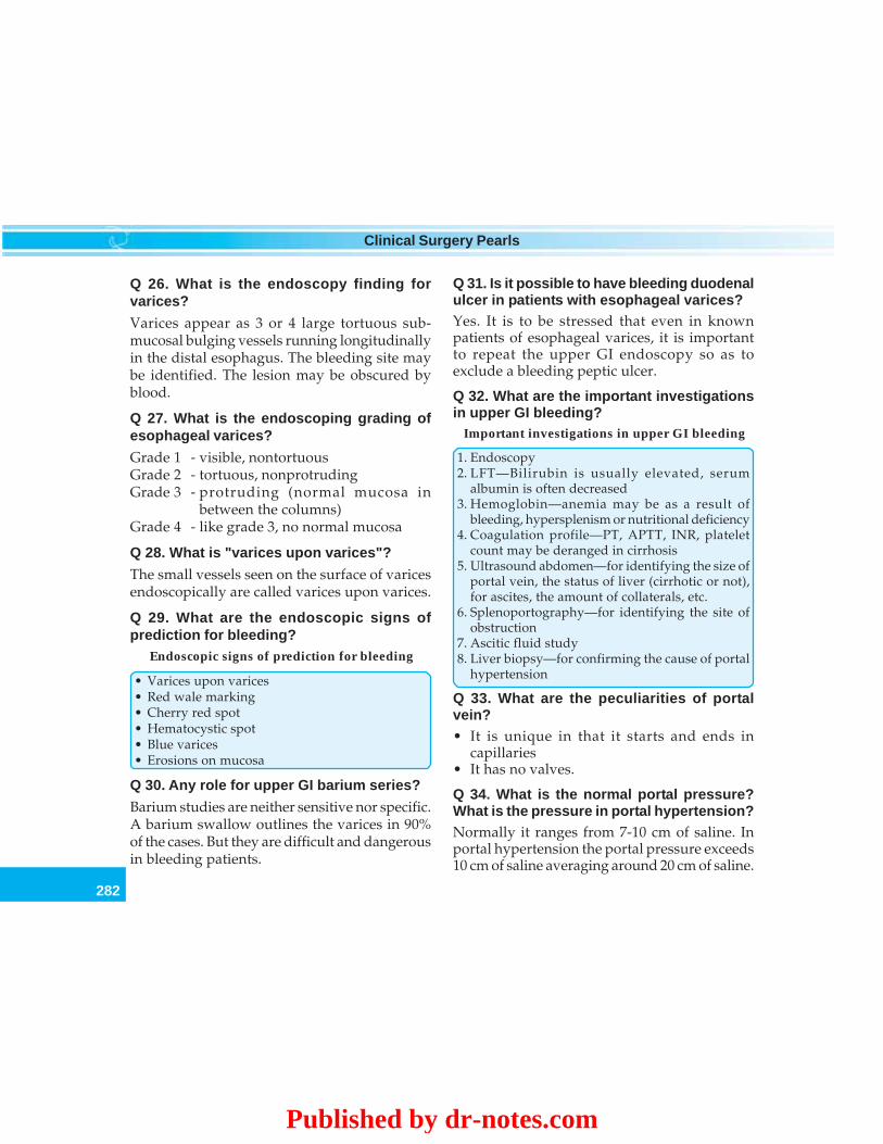
282
Clinical Surgery Pearls
Q 26. What is the endoscopy finding forvarices?
Varices appear as 3 or 4 large tortuous sub-mucosal bulging vessels running longitudinallyin the distal esophagus. The bleeding site maybe identified. The lesion may be obscured byblood.
Q 27. What is the endoscoping grading ofesophageal varices?
Grade 1 - visible, nontortuousGrade 2 - tortuous, nonprotrudingGrade 3 - protruding (normal mucosa in
between the columns)Grade 4 - like grade 3, no normal mucosa
Q 28. What is "varices upon varices"?
The small vessels seen on the surface of varicesendoscopically are called varices upon varices.
Q 29. What are the endoscopic signs ofprediction for bleeding?
Endoscopic signs of prediction for bleeding
• Varices upon varices• Red wale marking• Cherry red spot• Hematocystic spot• Blue varices• Erosions on mucosa
Q 30. Any role for upper GI barium series?
Barium studies are neither sensitive nor specific.A barium swallow outlines the varices in 90%of the cases. But they are difficult and dangerousin bleeding patients.
Q 31. Is it possible to have bleeding duodenalulcer in patients with esophageal varices?Yes. It is to be stressed that even in knownpatients of esophageal varices, it is importantto repeat the upper GI endoscopy so as toexclude a bleeding peptic ulcer.
Q 32. What are the important investigationsin upper GI bleeding?
Important investigations in upper GI bleeding
1. Endoscopy2. LFT—Bilirubin is usually elevated, serum
albumin is often decreased3. Hemoglobin—anemia may be as a result of
bleeding, hypersplenism or nutritional deficiency4. Coagulation profile—PT, APTT, INR, platelet
count may be deranged in cirrhosis5. Ultrasound abdomen—for identifying the size of
portal vein, the status of liver (cirrhotic or not),for ascites, the amount of collaterals, etc.
6. Splenoportography—for identifying the site ofobstruction
7. Ascitic fluid study8. Liver biopsy—for confirming the cause of portal
hypertension
Q 33. What are the peculiarities of portalvein?• It is unique in that it starts and ends in
capillaries• It has no valves.
Q 34. What is the normal portal pressure?What is the pressure in portal hypertension?Normally it ranges from 7-10 cm of saline. Inportal hypertension the portal pressure exceeds10 cm of saline averaging around 20 cm of saline.
Published by dr-notes.com
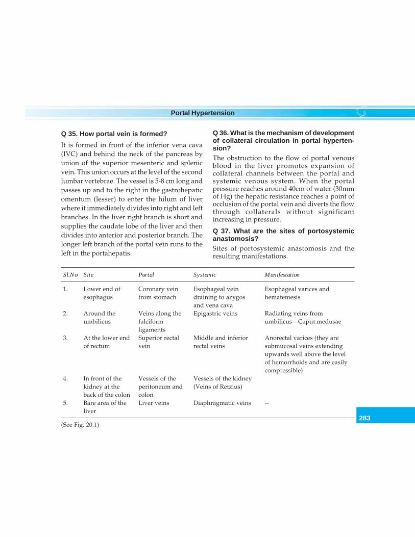
283
Portal Hypertension
Q 35. How portal vein is formed?
It is formed in front of the inferior vena cava(IVC) and behind the neck of the pancreas byunion of the superior mesenteric and splenicvein. This union occurs at the level of the secondlumbar vertebrae. The vessel is 5-8 cm long andpasses up and to the right in the gastrohepaticomentum (lesser) to enter the hilum of liverwhere it immediately divides into right and leftbranches. In the liver right branch is short andsupplies the caudate lobe of the liver and thendivides into anterior and posterior branch. Thelonger left branch of the portal vein runs to theleft in the portahepatis.
Q 36. What is the mechanism of developmentof collateral circulation in portal hyperten-sion?The obstruction to the flow of portal venousblood in the liver promotes expansion ofcollateral channels between the portal andsystemic venous system. When the portalpressure reaches around 40cm of water (30mmof Hg) the hepatic resistance reaches a point ofocclusion of the portal vein and diverts the flowthrough collaterals without significantincreasing in pressure.
Q 37. What are the sites of portosystemicanastomosis?Sites of portosystemic anastomosis and theresulting manifestations.
Sl.No Site Portal Systemic Manifestation
1. Lower end of Coronary vein Esophageal vein Esophageal varices andesophagus from stomach draining to azygos hematemesis
and vena cava2. Around the Veins along the Epigastric veins Radiating veins from
umbilicus falciform umbilicus—Caput medusaeligaments
3. At the lower end Superior rectal Middle and inferior Anorectal varices (they areof rectum vein rectal veins submucosal veins extending
upwards well above the levelof hemorrhoids and are easilycompressible)
4. In front of the Vessels of the Vessels of the kidneykidney at the peritoneum and (Veins of Retzius)back of the colon colon
5. Bare area of the Liver veins Diaphragmatic veins --liver
(See Fig. 20.1)
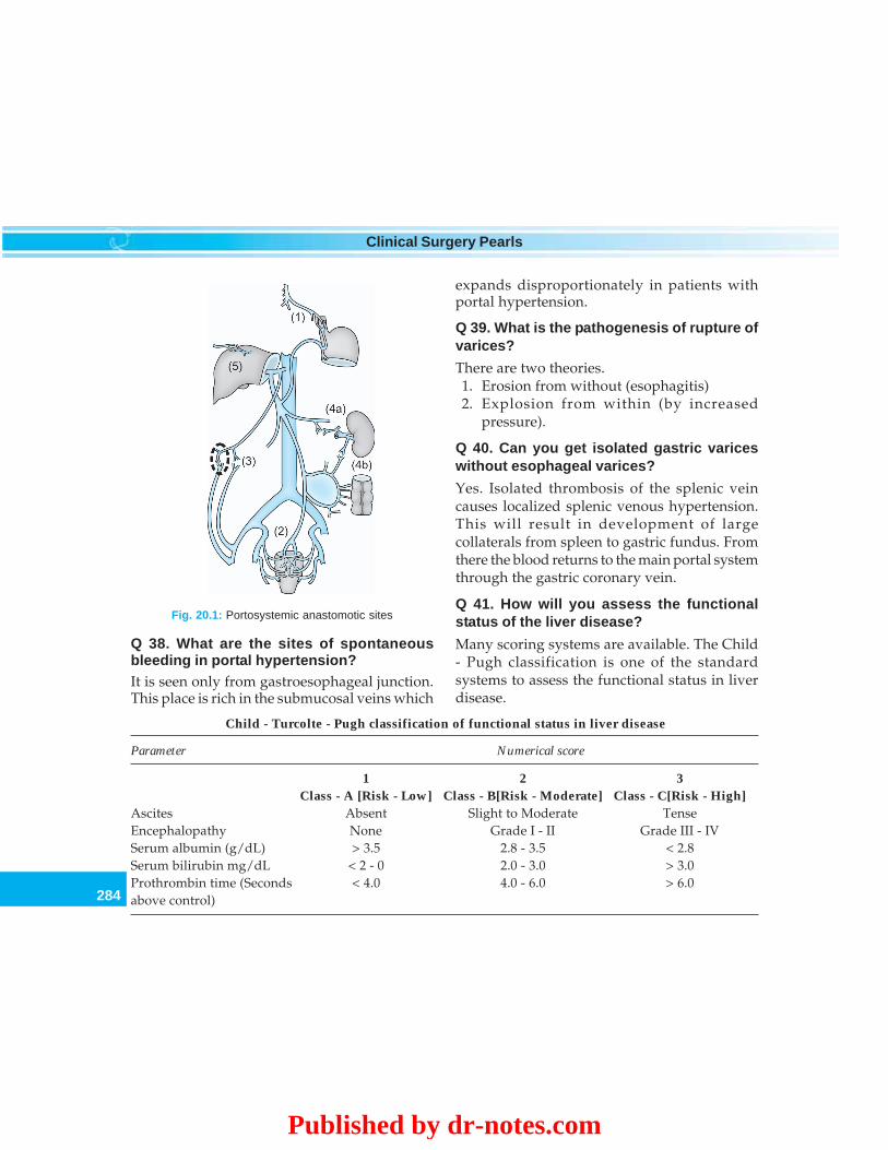
284
Clinical Surgery Pearls
expands disproportionately in patients withportal hypertension.
Q 39. What is the pathogenesis of rupture ofvarices?
There are two theories.1. Erosion from without (esophagitis)2. Explosion from within (by increased
pressure).
Q 40. Can you get isolated gastric variceswithout esophageal varices?
Yes. Isolated thrombosis of the splenic veincauses localized splenic venous hypertension.This will result in development of largecollaterals from spleen to gastric fundus. Fromthere the blood returns to the main portal systemthrough the gastric coronary vein.
Q 41. How will you assess the functionalstatus of the liver disease?
Many scoring systems are available. The Child- Pugh classification is one of the standardsystems to assess the functional status in liverdisease.
Fig. 20.1: Portosystemic anastomotic sites
Q 38. What are the sites of spontaneousbleeding in portal hypertension?It is seen only from gastroesophageal junction.This place is rich in the submucosal veins which
Child - Turcolte - Pugh classification of functional status in liver disease
Parameter Numerical score
1 2 3Class - A [Risk - Low] Class - B[Risk - Moderate] Class - C[Risk - High]
Ascites Absent Slight to Moderate TenseEncephalopathy None Grade I - II Grade III - IVSerum albumin (g/dL) > 3.5 2.8 - 3.5 < 2.8Serum bilirubin mg/dL < 2 - 0 2.0 - 3.0 > 3.0Prothrombin time (Seconds < 4.0 4.0 - 6.0 > 6.0above control)
Published by dr-notes.com
285
Portal Hypertension
Total Child - Turcolte - Pugh Riskscore classification
5 - 6 A Low risk
7 - 9 B Moderate risk
10 - 15 C High risk
Q 42. What is Child's classification?
Child's classification of hepatocellular functionin cirrhosis.
Q 43. What are the causes for portal hyper-tension?
A. May be due to increased resistance to flow—• Prehepatic• Hepatic• Posthepatic
B. Increased portal blood flow - (because of thetremendous reserve capacity of the liver toaccommodate increased blood flow, this isuncommon).• Banti syndrome• Tropical splenomegaly• Myeloid metaplasia.
Prehepatic causes
• Thrombosis of portal vein• Thrombosis of splenic vein• Congenital stenosis or atresia• Extrinsic compression by tumors
Hepatic causes
• Cirrhosis—portal, postnecrotic, biliary, Wilson'sdisease, hemochromatosis
• Congenital hepatic fibrosis• Idiopathic portal hypertension• Schistosomiasis• Chronic active hepatitis• Acute alcoholic liver diseases
Posthepatic causes
• Budd-Chiari syndrome (hepatic vein thrombosis)• Venoocclusive disease• Cardiac diseases—constrictive pericarditis,
valvular heart disease, right heart failure, etc.
Q 44. What are the important common causesfor portal hypertension?
Common causes for portal hypertension
1. Cirrhosisa. Cirrhosis (Alcoholic) - 85%b. Postnecrotic cirrhosisc. Biliary cirrhosis
Group designation A B C
Serum albumin (g/dL) > 3.5 2.8–3.5 < 2.8Serum bilirubin mg/dL < 2 - 0 2.0–3.0 > 3.0Ascites None Easily controlled Poorly controlledNeurological disorder None Minimal AdvancedNutrition Excellent Good Wasting
286
Clinical Surgery Pearls
2. Extrahepatic portal venous thrombosis(Children and Younger patients)
3. Idiopathic portal hypertension (Southern Asia)4. Schistosomiasis (Egypt)
Q 45. What is the cause of abnormalresistance in the portal system in cirrhosis?The resistance is predominantly postsinusoidalthought to be due to—• Distortion of the hepatic veins by
degenerating nodules• Fibrosis of perivascular tissue around the
sinusoids and hepatic veins• Centrilobular swelling and fibrosis produce
portal hypertension in acute alcoholichepatitis in the absence of cirrhosis
Q 46. What is wedged hepatic vein pressure?A catheter is wedged in a tributary of the hepaticveins which will permit estimation of thepressure in the afferent veins to sinusoids.
Q 47. What is the site of obstruction inschistosomiasis?Schistosomiasis produce presinusoidal obs-truction due to deposition of parasite ova insmall portal venules. This produces inflamma-tion followed by fibrosis.
Q 48. What is Budd-Chiari syndrome?
This is a condition affecting mainly youngfemales. The obstruction is in the hepatic veinsas a result of hepatic vein thrombosis orcongenital webs (venous web). The liverbecomes congested as a result of obstruction,resulting in impaired liver function and portalhypertension. The patient also develops ascites
and esophageal varices. If chronic, the liverprogresses to cirrhosis. It is important to ruleout the cause for venous thrombosis like—• Procoagulant states like protein C, protein S
and antithrombin III, deficiencies• Myeloproliferative disorders.
Q 49. What is the CT finding in Budd-Chiarisyndrome? (PG)
• Ascites• Large congested liver (early)• Small cirrhotic liver (late) with gross
enlargement of segment I (caudate lobe)
Q 50. What is the cause for enlargement ofsegment I? (PG)
Segment I is having direct venous drainage tothe IVC (inferior vena cava). When there isatrophy of the rest of the liver due to hepaticvein obstruction, there will be gross enlargementof this segment.
Q 51. How will you confirm the diagnosis ofBudd-Chiari syndrome? (PG)
This is done by hepatic venography through atransjugular approach. This will revealocclusion of the hepatic veins.
Q 52. What is the treatment of Budd-Chiarisyndrome? (PG)
• If cirrhosis is not established - TIPSS or shuntsurgery
• If cirrhosis is established—Liver transplan-tation
• Life-long anticoagulation with warfarin maybe required.
Published by dr-notes.com
287
Portal Hypertension
Q 53. What is TIPSS?Transjugular Intrahepatic Portosystemic StentShunt. This was introduced 1988 as anemergency treatment for variceal hemorrhagenot responding to medical management andendoscopic sclerotherapy. The procedure isdone under local anesthesia and sedation usingfluoroscopic guidance and ultrasonography. Aguide wire is inserted through the internaljugular vein and then via the superior vena cava,hepatic veins and hepatic parenchyma to abranch of the portal vein. The track in theparenchyma is then dilated with a ballooncatheter and a metallic stent is inserted. This willproduce satisfactory drop in portal venouspressure, by shunting blood from portal systemto the inferior vena cava directly.
Q 54. What are the complications of TIPSS?(PG)• Perforation of liver capsule and fatal intra-
peritoneal hemorrhage• TIPSS occlusion—further variceal
hemorrhage• Postshunt encephalopathy (40%)• Stenosis of the shunt (50% at one year).
Q 55. What is the cause for postshuntencephalopathy after TIPSS?What is the incidence? (PG)The portal blood bypassing the detoxificationeffects of the liver will enter the systemiccirculation resulting in a confusional state. Itoccurs in 40% of patients similar to the surgicalshunts. The early manifestations are subtle andmental and personality changes may beoverlooked in the postoperative period.
Restlessness, irritability and insomnia arecommon. Neuromuscular signs of unsustainedclonus, increased tendon reflexes and extensorplantar are seen. Blood ammonia determinationis unreliable. Finally more obvious confusion,drowsiness, stupor and coma develops.
Q 56. What is Banti's syndrome? (PG)
It is defined as a liver disease secondary toprimary splenic disease. It was erroneouslyconsidered as the cause of portal hypertensionbut now it is known that it is as a result ofcirrhosis.
Q 57. What are the criteria by which youcategorize a patient to low-risk category inupper GI bleed? (PG)
Low-risk—upper GI bleed
1. Age < 75years2. No unstable comorbid illness3. No ascites evident on physical examination4. Normal PT (prothrombin time)5. Systolic BP above 100 within 1 hour after
admission (with or without fluid resuscitation)6. Nasogastric aspirate free of fresh blood
Q 58. What is the management of varicealbleeding? (Flow Chart 20.1)
Emergency admission and following manage-ment is carried out.
1. Monitor the patient for BP, pulse, CVP,hematocrit, hourly urine output
2. Peripheral and central venous access3. Arrange adequate blood (initially 10 units)4. Serial hematocrit determination—is best for
monitoring continued blood loss andappropriate replacement of the lost blood
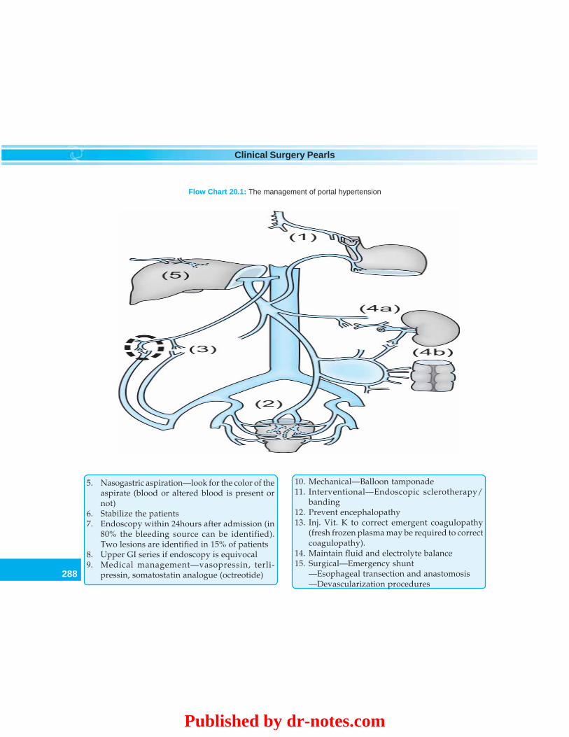
288
Clinical Surgery Pearls
Flow Chart 20.1: The management of portal hypertension
5. Nasogastric aspiration—look for the color of theaspirate (blood or altered blood is present ornot)
6. Stabilize the patients7. Endoscopy within 24hours after admission (in
80% the bleeding source can be identified).Two lesions are identified in 15% of patients
8. Upper GI series if endoscopy is equivocal9. Medical management—vasopressin, terli-
pressin, somatostatin analogue (octreotide)
10. Mechanical—Balloon tamponade11. Interventional—Endoscopic sclerotherapy/
banding12. Prevent encephalopathy13. Inj. Vit. K to correct emergent coagulopathy
(fresh frozen plasma may be required to correctcoagulopathy).
14. Maintain fluid and electrolyte balance15. Surgical—Emergency shunt
—Esophageal transection and anastomosis—Devascularization procedures
Published by dr-notes.com
289
Portal Hypertension
Q 59. What is the initial therapy of choice?
• Endoscopic sclerotherapy or banding• Vasopressin or propranolol may or may not
be included• Balloon tamponade (no longer used
routinely)—reserved for special situations.
Q 60. What is sclerotherapy?
Through the fiberoptic endoscope 1-2 ml of thesclerosant solution is injected into the varixeither intravaricially or paravaricially in acircumferential pattern at the gastroesophagealjunction and subsequently also injected 3-4 cmproximal to the junction. Following the injection,the endoscope is advanced into the stomach andis kept there for 4-5 minutes in order to compressthe varices. It may be done as emergencyprocedure or elective procedure.
Methods of sclerotherapya. Intravariceal—Obliteration of varicesb. Paravariceal—Perivascular cicatrizationc. Combined method of injection.
Q 61. What are the sclerosants used? (PG)The commonly used sclerosants
• Fatty acid derivatives- Sodium morrhuate (5%)- Ethanolamine oleate (5%)
• Synthetic products- STD (Sodium Tetra Decyle Sulphate) - 1-3%- Polidocanol (Hydroxy Polyethoxydodecan
(HPD) also called Aethoxy sklerol - 1%• Other agents - Butyl cyanoacrylate• Mixture of sclerosants
Q 62. What is the mechanism of sclero-therapy? (PG)
It will cause-• Intimal injury• Thrombosis of submucosal vessels (in 24
hours)• Superficial and deep ulceration (5-7 days)• Submucosal fibrosis (1month).
Q 63. How often sclerotherapy is repeated?(PG)
During active bleeding—• It is repeated on 3rd day and 7th day• After resolving edema repeat the procedure
3-4 weeks after the last injection• 5 to 6 procedures may be required for total
obliteration• Endoscopic evaluation is done 3 to 6 months
after this.
Q 64. What is a successful sclerotherapy?(PG)
• If the aspirate is clear within 12hours it isconsidered successful
• If unsuccessful repeat sclerotherapy 3 timesat an interval of 12hours.
Q 65. What is the success rate of sclero-therapy? (PG)
Sclerotherapy controls bleeding in 80-95% ofpatients.
Q 66. What is prophylactic sclerotherapy?(PG)
• When sclerotherapy is given for a patientwith esophageal varices without history ofbleeding it is called prophylacticsclerotherapy
290
Clinical Surgery Pearls
• When sclerotherapy is given during bleedfree intervals, it is called chronicsclerotherapy
• When it is given during hemorrhage it iscalled acute sclerotherapy.
Q 67. What are the complications of sclero-therapy? (PG)
Complications may be classified as:1. Minor
- Substernal pain- Esophagitis (48 hrs)- Fever - 25%
2. MajorEsophageal
- Ulceration - (7 days)- Perforation (5 - 19 days)- Stricture (4 months)Pulmonary and mediastinal- Pleural effusion right side- Atelactasis- Chylothorax right side- ARDS3. Rare - Superior mesenteric vein thrombosis
Q 68. What is the treatment of gastric fundalvarices? (PG)
• Injection sclerotherapy with Butyl Cyano-acrylateor
• Endoscopic banding.
Q 69. What is endoscopic banding?
The varix is lifted with suction tip and a smallrubber band is slipped around the base. Thevarix necroses leaving behind a superficial ulcer.The rebleeding episodes are less in banding, soalso the morbidity compared to sclerotherapy
Q 70. What is the role of vasopressin?
The vasopressin will lower the portal blood flowand portal pressure by directly constrictingsplanchnic arterioles. This will reduce the portalflow. Vasopressin alone controls acute bleedingin 80% of patients and this is increased to 95%when used in combination with balloontamponade.
Q 71. What are the complications ofvasopressin?
Complications of vasopressin
1. Myocardial infarction2. Cardiac arrhythmias3. Intestinal gangrene4. Reduction in cardiac output5. Reduces hepatic blood flow6. Reduces renal blood flow
Q 72. How can one prevent the complicationsof vasopressin?
Simultaneous administration of nitroglycerineor isoproterenol along with vasopressin.
Q 73. What is the dose of vasopressin?
It is given as a IV infusion in the dose of 0.4units/mt. The infusion is better than bolusinjection. The nitroglycerine can be given IV orsublingually (20 units in 10ml of 5% dextroseIV over 10 mts initially).
Q 74. What is Telipressin?
It is a synthetic vasopressin analogue. Itundergoes gradual conversion to vasopressin inthe body. It causes fewer cardiac side effectsthan vasopressin.
Dose 2mg bolus injection IV 6th hourly.
Published by dr-notes.com
291
Portal Hypertension
Q 75. What is the role of octreotide acetate?
It is a long-acting synthetic somatostatinanalogue. The action is similar to vasopressinbut without significant side effects. This is thedrug of choice for the pharmacological controlof acute bleeding varices.Dose: 100μg initial bolus, followed bycontinuous infusion of 25μg/hr for 24hrs.
Q 76. What is the effectiveness of pharma-cological control?
• Vasopressin alone is superior to placebo• Vasopressin and nitroglycerine is superior to
vasopressin alone.
Q 77. What is the role of Sengstaken-Blakemore tube?
It is used for temporary hemostasis initially(Balloon tamponade). It has got 3 lumens with2 balloons which can be inflated (esophagealballoon and gastric balloon). The balloons areinflated in the lumen of gut (gastric balloon isinflated with 250 ml of air, and esophagealballoon to a pressure of 40 mm of Hg). The thirdlumen is for aspiration of the gastric contents.The tube is introduced transorally. Afterinflating the two balloons, traction is applied tothe tube so that gastric balloon compresses thecollateral veins at the cardia of the stomach. Thecontribution of esophageal balloon compressionis negligible. The balloon should be temporarilydeflated after 12hours to prevent pressurenecrosis of the esophagus.
Q 78. What are the complications of this tube?• Aspiration of pharyngeal secretions and
pneumonia
• Esophageal rupture (this balloon is thereforeinfrequently used)
• Pressure necrosis of the esophagus.
Q 79. What is the success rate of balloontamponade?
Above 75% of the actively bleeding patients canbe controlled by balloon tamponade. Whenbleeding has stopped, it should be left in placefor another 24 hours.
Q 80. What is Minnesota tube?
This tube has got a 4th lumen to aspirate theesophagus proximal to the balloon.
Q 81. What is the role of TIPSS in themanagement of portal hypertension (read theanswer in the initial part of this chapter)?
(PG)
• It is useful for the control of acute bleedingand to prevent rebleeding (the portalhypertension also is controlled)
• Patients with advanced liver disease areconsidered for TIPSS
• TIPSS is used as a bridge to transplantation• It should not be regarded for definitive
therapy (the shunt remains open for upto ayear)
• Less severe cirrhosis patients—consider forshunt surgery/devascularization.
Q 82. What are the surgical options? (FlowChart 20.1)
• Surgical procedures may be classified as:- Emergency procedures- Elective surgical procedures.
292
Clinical Surgery Pearls
Q 83. What are the emergency surgicalprocedures? (PG)• Surgical procedures are rarely considered for
the acute management of variceal hemor-rhage, since the morbidity and mortality arehigh
• The increasing availability of TIPSS and livertransplantation is another factor
• Initial bleeding is usually controlled withsclerotherapy/banding
• The main indication for surgery is a Child'sgrade—A cirrhotic patient in whom thebleeding has been controlled by sclerotherapy1. Emergency shunt operations2. Emergency esophageal transection
(stapled transection)3. Emergency devascularization.
Note: All these procedures are usually done aselective procedures.
Q 84. What is the principle of shunt surgeryand what are the types of shunt surgery?
They reduce the pressure in the portalcirculation by diverting the blood into the low-pressure systemic circulation. The differenttypes of shunts are—
Nonselective (portocaval)1. Side-to-side—portacaval, mesocaval,
renosplenic, etc.2. End-to-side—portacaval (total)3. Mesocaval (H)
Selective1. Distal splenorenal (leinorenal) (Warren shunt)
- it is time consuming in emergency situations
2. Left gastric venacaval (Inokuchi).
Q 85. What are the advantages of selectiveshunt? (PG)
• They preserve blood flow to the liver whiledecompressing the left side of the portalcirculation, which is responsible foresophageal and gastric varices.
• Selective shunts are associated with lowerincidence of Portosystemic Encephalopathy(PSE).
Q 86. What is total shunt? (PG)
This end - to - side shunt completely disconnectthe liver from the portal system. The portal veinis transected near its bifurcation in the liverhilum and anastomosed to the side of theinferior vena cava. The hepatic stump of the veinis over sewn. This type of shunt gives immediateand permanent protection from varicealbleeding and some what easier to perform thanside-to-side portacaval shunt. Encephalopathyis less compared to side-to-side shunt.
Q 87. What is mesocaval shunt?
A segment of prosthetic graft or internal jugularvein is anastomosed between the inferior venacava and the superior mesenteric vein. It isuseful in cases of portal vein thrombosis. Theportal flow to the liver is lost in this procedure.The portal flow to the liver can be preserved byreducing the diameter of the graft to 8mm (from12-20 mm). This will decrease the incidence ofencephalopathy, still preventing varicealhemorrhage.
Published by dr-notes.com
293
Portal Hypertension
Q 88. What is central splenorenal shunt?(PG)
Here splenectomy is performed and the splenicvein is anastomosed to the renal vein.
Q 89. What are the emergency shunt surgicalprocedures commonly performed? (PG)
1. End-to-side portacaval2. H - mesocaval shunt.
Q 90. What is the contraindication for shuntprocedure? (PG)
A good transplantation candidate should not besubjected to portosystemic shunt or other shunt.Generally, Child's class—A patients arecandidates for portal decompression and classC patients are candidates for transplantation.
Q 91. How will you select a shunt procedure?(PG)
• For elective portal decompression—Distalsplenorenal shunt
• If ascites is present—End-to-side portacavalshunt
• Severe ascites - Side-to-side shunt
• Budd-Chiari syndrome• Emergency decompression - End-to-side or
H mesocaval.
Q 92. What are the results of portasystemicshunts? (PG)
• Incidence of recurrent variceal bleeding—10%
• 5-year-survival for alcoholic liver disease—45%
• Encephalopathy 15 - 25% (Severe encephalo-pathy in 20% of alcoholic after total shunt)
• Patency of shunt—90%.
Q 93. What is esophageal transection(stapled transection)?
In many surgical units it is the first choice whennonsurgical methods fail. It must be done assoon as a second attempt at sclerotherapy hasfailed. It must be viewed as an emergencyprocedure and not a definitive treatment.
Circular stapling device is used for resectingand reanastomosing a ring of the loweresophagus. This procedure is being replaced byTIPSS in centers where it is available.
Q 94. What are the devascularization proce-dures? (PG)The devascularization procedures reduceproximal gastric blood flow.These procedures are not done nowadays.1. Gastroesophageal decongestion and
splenectomy of Hassab—All the vesselsexcept the left gastric vessels are ligated.
2. Sugiura procedure—It is done in twostages. The first stage is transthoracic andthe dilated venous collaterals betweenesophagus and adjacent structures aredivided. The esophagus is then transectedand reanastomosed. The second stage isdone by laparotomy after the first stage ifthe patient is bleeding (but deferred 4-6weeks in the elective cases). The upper 2/3rds of the stomach is devascularized,selective vagotomy, pyloroplasty andsplenectomy are performed.
Q 95. What is the definitive treatment forportal hypertension due to cirrhosis?In cirrhosis the basic pathology is failing livercells and none of these procedures will rectifythe problem. Therefore the definitive treatment
294
Clinical Surgery Pearls
is liver transplantation. Any young patient withcirrhosis who has survived an episode ofvariceal hemorrhage should be considered acandidate for liver transplantation. Any otherform of treatment carries a much highermortality rate within the subsequent 1-2years.
Q 96. What are the contraindications for livertransplantation? (PG)
Contraindications for liver transplantation
1. Age > 65 years2. Ischemic heart disease3. Cardiac failure4. Chronic respiratory disease5. Continued alcohol use6. Previous surgical shunt (relative)
Q 97. What is the procedure of choice duringthe preparation period for transplantation?
(PG)
TIPSS.
Q 98. What is the treatment of nonbleedingvarices (prophylactic therapy)?
There is a 30% chance for bleeding at some pointfor this group of patients. Of those who bleed50% die. Patients who have bled once fromvarices have a 70% chance of bleeding again.The treatment of patients with varices that havenever bled is referred to as prophylactic therapy.Thus we have prophylactic sclerotherapy, pro-phylactic propranolol, etc. The therapy ofpatients who have bled before is referred to astherapeutic. The prophylactic therapy consistsof the following:a. Prophylactic sclerotherapyb. Prophylactic propranolol.
Q 99. What is the action of propranolol?
This beta-adrenergic blocking agent decreasescardiac output and splanchnic blood flow andtherefore reduces the portal blood pressure.Chronic propranolol therapy in a dose of 20-160mg twice daily decreases the frequency ofre-bleeding by 40%. It also reduces the overallmortality.
Q 100. What is left sided portal hypertension?
It is caused by isolated splenic vein thrombosis.
Q 101. What are the causes of ascites in portalhypertension?
• Decreased colloid osmotic pressure(decreased proteins)
• Increased hydrostatic pressure• Lymphatic blockage.
Q 102. What are the complications of ascites?Complications of ascites
• Umbilical hernia• Spontaneous bacterial peritonitis• Respiratory embarrassment
Q 103. What is the treatment of ascites inportal hypertension?
1. Medical- Spironolactone 25mg bd orally- Limit sodium intake.
2. Ascitic tap—slow gradual tapping is done3. Shunt operations
- LeVeen shunt (peritoneovenous shunt)- Denver shunt
Q 104. What is the management ofencephalopathy?
Published by dr-notes.com
295
Portal Hypertension
1. Protein-free diet2. Oral Lactulose—30 ml tid3. Oral Neomycin—1-2 gm 6th hourly (not
given in renal impairment)4. Parenteral nutrition—(Calories from Carbo-
hydrate Sucrose is preferred over glucose)5. Administration of 50% glucose in central
vein
Q 105. What is hepatorenal syndrome?
Acute renal failure without apparent cause mayoccur in patients with liver disease at any timeduring hospitalization. This is due to impairedrenal perfusion. Other possible causes includeinfection, drug toxicity, etc. They usuallysuccumb to hepatic failure.
296
Clinical Surgery Pearls
21 Mesenteric Cyst
CASE CAPSULE
A 15-year-old girl presents with abdominaldistension and recurrent colicky type ofabdominal pain of two months duration. Thereis no associated vomiting and no othercomplaints. There is no history of respiratoryinfection preceding the attack. There is noalteration in bowel habits.
On general examination, she is afebrile, notill looking. There is no generalized lymphadeno-pathy and no icterus. There is no circumoralpallor. On examination of the abdomen, acentral abdominal fullness is noticed oninspection. A spherical swelling of 15 cmdiameter is situated in the umbilical region, allthe borders of which are well-defined. Thesurface of the swelling is smooth. The swellingmoves freely at right angles to the line ofattachment of the mesentery. It is dull topercussion, however the surrounding areas areresonant. On fixing the swelling with the helpof the patient's hand it appears to be fluctuant.The swelling cannot be pushed to the pelvis.There is neither shifting dullness nor fluidthrill. There is no organomegaly. The hernialorifices are normal. On digital rectal
examination, it is not possible to feel the swellingfrom below. The supraclavicular nodes are notenlarged.
Read the checklist for abdominal examination.Checklist for history
Checklist for examination
Look for• Circumoral pallor—mesenteric adenitis• Cervical lymph nodes—seen in mesenteric
lymphadenitis• Shifting tenderness which is a sign of mesenteric
adenitis• Free movement in a plane at right angles to the
attachment of mesentery (from left of theumbilicus to the right iliac fossa)
• A zone of resonance around the swelling
Q 1. What is the probable diagnosis in thiscase?
Mesenteric cyst.
Q 2. What are the differential diagnosis?Differential diagnosis of central abdominal
cystic swelling
• Ovarian cyst• Omental cyst
Published by dr-notes.com
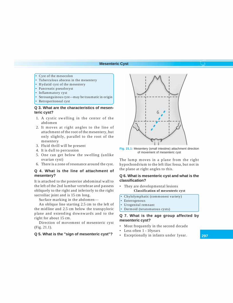
297
Mesenteric Cyst
• Cyst of the mesocolon• Tuberculous abscess in the mesentery• Hydatid cyst of the mesentery• Pancreatic pseudocyst• Inflammatory cyst• Serosanguinous cyst—may be traumatic in origin• Retroperitoneal cyst
Q 3. What are the characteristics of mesen-teric cyst?
1. A cystic swelling in the center of theabdomen
2. It moves at right angles to the line ofattachment of the root of the mesentery, butonly slightly, parallel to the root of themesentery
3. Fluid thrill will be present4. It is dull to percussion5. One can get below the swelling (unlike
ovarian cyst)6. There is a zone of resonance around the cyst.
Q 4. What is the line of attachment ofmesentery?
It is attached to the posterior abdominal wall tothe left of the 2nd lumbar vertebrae and passessobliquely to the right and inferiorly to the rightsacroiliac joint and is 15 cm long.
Surface marking in the abdomen—An oblique line starting 2.5 cm to the left of
the midline and 2.5 cm below the transpyloricplane and extending downwards and to theright for about 15 cm.
Direction of movement of mesenteric cyst(Fig. 21.1).
Q 5. What is the "sign of mesenteric cyst"?
The lump moves in a plane from the righthypochondrium to the left iliac fossa, but not inthe plane at right angles to this.
Q 6. What is mesenteric cyst and what is theclassification?
• They are developmental lesionsClassification of mesenteric cyst
• Chylolymphatic (commonest variety)• Enterogenous• Urogenital remnant• Dermoid (teratomatous cysts)
Q 7. What is the age group affected bymesenteric cyst?
• Most frequently in the second decade• Less often 1 - 10years• Exceptionally in infants under 1year.
Fig. 21.1: Mesentery (small intestine) attachment directionof movement of mesenteric cyst
298
Clinical Surgery Pearls
Q 8. What are the differences betweenchylolymphatic cyst and enterogenouscysts?
Q 9. What is the cause for recurrent attacksof abdominal pain?
It may be due to—1. Torsion of the mesentery2. Temporary impaction of food bolus in the
intestine narrowed by the cyst.
Q 10. What are the complications of the cyst?
Complication of mesenteric cyst
1. Torsion of the cyst2. Rupture of the cyst3. Hemorrhage into the cyst4. Infection5. Peptic ulceration (when it contains ectopic
gastric mucosa)6. Perforation
will show the cyst in front of the intestines, if itis an omental cyst.
Q 12. What are the investigations in asuspected case of mesenteric cyst?1. Plain X-ray will show calcified lymph nodes
in tuberculous mesenteric lymph nodes2. Barium meal and follow through hollow
viscera will be found to be displaced aroundthe cyst or lumen of the intestine may beseen narrowed
3. Ultrasound abdomen will reveal the cysticnature and the origin of the cyst
4. IVU to rule out hydronephrosis if ultrasoundis not done
5. CT scan6. Needle aspiration and instillation of radio
opaque water-soluble contrast media.
Differences between chylolymphatic cyst and enterogenous cysts
Chylolymphatic cyst Enterogenous cysts
• Arises from congenitally misplaced lymphatic • Arises from a diverticulum of the mesenterictissue with no efferent communication with border of the intestine which has becomelymphatic system sequestrated from the intestinal canal or
duplication of the intestine• Wall is thin—connective tissue lined by • Thicker wall—lined by mucous membrane
endothelium (sometimes ciliated)• Filled with clear lymph or chyle (milky) • Content is mucinous (colorless or yellowish
brown)• Blood supply is independent of that of the • Common blood supply with intestine
adjacent intestines• Usually enucleation is possible without • Removal always entails resection of the
resection of the gut related portion of intestine
Q 11. How will you differentiate it fromomental cyst?A lateral radiograph or ultrasound or CT scan
Published by dr-notes.com
299
Mesenteric Cyst
Q 13. What is the treatment of chylolymphaticcyst?• Enucleation in toto• After major portion of the cyst has been
dissected free, a portion abutting on theintestine or a major blood vessel can be leftattached after destroying its lining.
Q 14. What is the treatment of enterogenouscyst?• Enucleation is contraindicated• Resection of the cyst with adherent portion
of the intestine followed by intestinal anasto-mosis
• If a very large segment of small intestine isimplicated, an anastomosis should be madebetween the apex of the coil of small intestineand the cyst wall (the cyst wall will holdsutures well).
Q 15. What is the role of marsupialization?This is an old form of treatment notrecommended nowadays because of the fear offistula and recurrence.
Q 16. What is the surgical treatment if it isomental cysts?Omentectomy.
Q 17. What are the neoplasms of themesentery?They are classified as benign and malignant.
Benign Malignant
• Lipoma • Lymphoma• Fibroma • Secondary carcinoma• Fibromyxoma
Q 18. What is the management of benignneoplasms?Benign tumors are excised in the same way asmesenteric cyst, along with resection of theadjacent intestine.
Q 19. What is the management of malignantneoplasms?• Biopsy confirmation• Chemotherapy for lymphoma• Chemotherapy for secondary carcinoma.
Q 20. How does tuberculous lymphadenitisoccur?The tubercle bacilli are usually ingested andthey enter the mesenteric lymph node by wayof Peyer's patches. The organism may be humanor bovine. It can occur after ingestion of rawmilk. It may affect a single lymph node ormultiple lymph nodes presenting as massiveabdominal swelling.
Q 21. What is pseudomesenteric cyst?When tuberculous mesenteric lymph nodesbreak-down, the tuberculous pus may remainbetween the leaves of the mesentery and cysticswelling similar to mesenteric cyst is formed.When such a situation is found the pus isevacuated without soiling the peritoneal cavityand antituberculous treatment is instituted.
Q 22. What is the cause for yellow-coloredlymph nodes in the ileocecal region?Metastasis from carcinoid of the appendix willgive rise to yellow color for the lymph nodes.
Q 23. What are the causes for calcifiedshadows in the plain radiograph of theabdomen?
300
Clinical Surgery Pearls
Causes for radiopaque shadow in plain X-rayabdomen
1. Renal or ureteric stone (renal stones are uniformin density, take the shape of pelvicaliceal systemand lies superimposed on the shadows of thevertebral column in the lateral view)
2. Gallstones are (less dense in the center andinfront of the vertebral bodies on the lateralview)
3. Pancreatic calculi4. Calcified tuberculous lymph node—usually in
the ileocecal region and line of attachment ofthe mesentery—outline is irregular and thenodes are mottled like black berry
5. Phlebolith6. Calcified costal cartilage7. Fecolith8. Stone in the appendix9. Calcified renal artery
10. Calcified aneurysm of the abdominal aorta11. Chip fracture of the transverse process of the
vertebrae
Q 24. How long it will take for calcification tooccur in tuberculous lymph nodes?
18 months.
Q 25. Will the nodes be noninfective in sucha situation?
No. The node need not be defunct. So infectionis still possible.
Q 26. What is the cause for acute nonspecificileocecal mesenteric adenitis?
The etiology of this condition is unknown, itaffects children and unusual after puberty. Some
cases are associated with Yersinia infection ofthe ileum. In other situations unidentified virusis blamed. Respiratory infection precedes anattack of mesenteric adenitis. This is a self-limiting disease. It is called nonspecific in orderto distinguish it from tuberculous mesentericadenitis.
Q 27. What are the clinical manifestations ofnonspecific mesenteric adenitis?• Central abdominal pain lasting for 10-30 minutes• Associated circumoral pallor• Vomiting is common• No alteration in bowel habits• Intervals of complete freedom from pain• The patient seldom looks ill• Temperature may be elevated but never exceeds
38.5• Tenderness along the line of mesentery• Shifting tenderness (after the patient lies on the
left side for a few minutes the tenderness shift tothe left side)
• Pelvic peritoneum is tender to palpation
Q 28. What will be the total leukocyte countlike?
There is often leukocytosis in contrast totuberculosis.
Q 29. What is the treatment of nonspecificmesenteric adenitis?• Bed rest for a few days (if the diagnosis can
be made with certainty)• If appendicitis cannot be excluded do
laparoscopy followed by appendectomy ifrequired.
Published by dr-notes.com

303
Non-thyroid Neck Swelling
22 Non-thyroid Neck Swelling
Examination of non-thyroid neck swellingsDiagnostic algorithm for a neck swelling
1. Identify the anatomical situation of theswelling (in relation to the triangle in the neck)
↓2. Decide the plane of the swelling
↓3. Recollect your anatomy (what are the normal
anatomical structures situated in the region ofthe swelling in that plane)
↓4. Check for mobility/fixity of the swelling5. Find out the external (size, shape, surface,
edge, temperature, tenderness, etc.) andinternal features of the lump (solid or cystic,compressible/ reducible, pulsation, trans-illumination) and auscultation of the swelling.
↓6. Find out its effect on the surrounding tissue
(feel the superficial temporal artery, examinethe relevant cranial nerves and look forHorner’s syndrome)
↓7. Look for regional nodes (if the swelling is a
node look for another group and contralateralside of the neck)
↓8. In the case of paired organs like salivary gland,
look for contralateral pathology also↓
9. Look for a primary lesion (scalp, oral cavity,pharynx, hidden areas etc).
↓10. Come to an anatomical diagnosis
↓11. Come to a pathological diagnosis. (Decide
whether it is congenital /traumatic/inflam-matory / neoplastic—primary or secondary)
↓12. If it is an organ concerned with function decide
whether it is hyperfunctioning, normallyfunctioning or hypo functioning (functionaldiagnosis). The final diagnosis = Anatomical+ Pathological + Functional diagnosis.
Q 1. What are the causes for nonthyroid neckmasses?All regions• Skin and subcutaneous tissue
1. Sebaceous cyst2. Lipoma3. Neurofibroma
• Lymphadenopathy1. Acute infection2. Chronic infection – Tuberculosis3. Primary malignant – Lymphoma4. Secondary malignant - Metastasis
Midline1. Sublingual dermoid2. Thyroglossal cyst / fistula
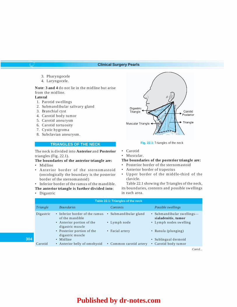
304
Clinical Surgery Pearls
3. Pharyngocele4. Laryngocele.
Note: 3 and 4 do not lie in the midline but arisefrom the midline.Lateral1. Parotid swellings2. Submandibular salivary gland3. Branchial cyst4. Carotid body tumor5. Carotid aneurysm6. Carotid tortuosity7. Cystic hygroma8. Subclavian aneurysm.
TRIANGLES OF THE NECK
The neck is divided into Anterior and Posteriortriangles (Fig. 22.1).The boundaries of the anterior triangle are:• Midline• Anterior border of the sternomastoid
(oncologically the boundary is the posteriorborder of the sternomastoid)
• Inferior border of the ramus of the mandible.The anterior triangle is further divided into:• Digastric
• Carotid• Muscular.The boundaries of the posterior triangle are:• Posterior border of the sternomastoid• Anterior border of trapezius• Upper border of the middle-third of the
clavicle.Table 22.1 showing the Triangles of the neck,
its boundaries, contents and possible swellingsin each area.
Fig. 22.1: Triangles of the neck
Table 22.1: Triangles of the neck
Triangle Boundaries Contents Possible swellings
Digastric • Inferior border of the ramus • Submandibular gland • Submandibular swellings—of the mandible sialadenitis, tumor
• Anterior portion of the • Lymph node • Lymph nodes swellingdigastric muscle
• Posterior portion of the • Facial artery • Ranula (plunging)digastric muscle
• Midline • Sublingual dermoidCarotid • Anterior belly of omohyoid • Common carotid artery • Carotid body tumor
Contd...
Published by dr-notes.com
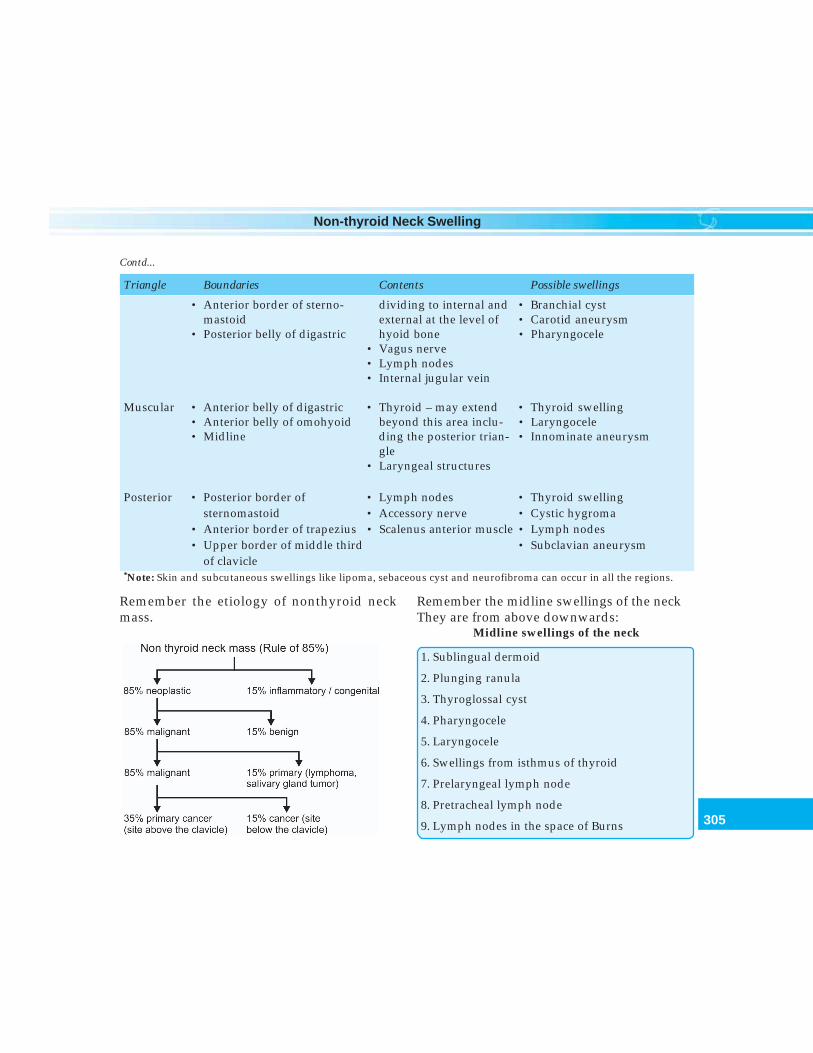
305
Non-thyroid Neck Swelling
Remember the etiology of nonthyroid neckmass.
• Anterior border of sterno- dividing to internal and • Branchial cystmastoid external at the level of • Carotid aneurysm
• Posterior belly of digastric hyoid bone • Pharyngocele• Vagus nerve• Lymph nodes• Internal jugular vein
Muscular • Anterior belly of digastric • Thyroid – may extend • Thyroid swelling• Anterior belly of omohyoid beyond this area inclu- • Laryngocele• Midline ding the posterior trian- • Innominate aneurysm
gle• Laryngeal structures
Posterior • Posterior border of • Lymph nodes • Thyroid swellingsternomastoid • Accessory nerve • Cystic hygroma
• Anterior border of trapezius • Scalenus anterior muscle • Lymph nodes• Upper border of middle third • Subclavian aneurysm
of clavicle*Note: Skin and subcutaneous swellings like lipoma, sebaceous cyst and neurofibroma can occur in all the regions.
Contd...
Triangle Boundaries Contents Possible swellings
Remember the midline swellings of the neckThey are from above downwards:
Midline swellings of the neck
1. Sublingual dermoid
2. Plunging ranula
3. Thyroglossal cyst
4. Pharyngocele
5. Laryngocele
6. Swellings from isthmus of thyroid
7. Prelaryngeal lymph node
8. Pretracheal lymph node
9. Lymph nodes in the space of Burns
306
Clinical Surgery Pearls
Remember the hidden areas of primary for ametastasis in the neck
Hidden areas for primary
• Pyriform sinus
• Base of tongue• Vallecula• Fossa of Rosenmüller• Tonsillar fossa
Published by dr-notes.com
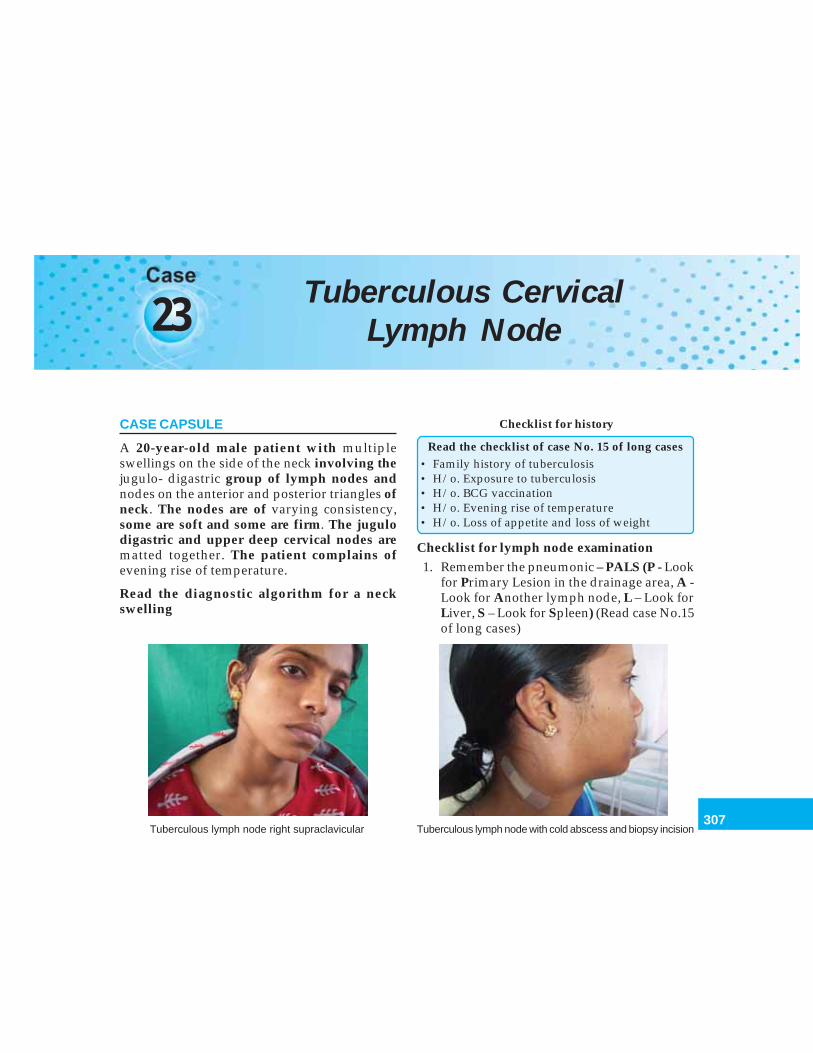
307
Tuberculous Cervical Lymph Node
23 Tuberculous CervicalLymph Node
CASE CAPSULE
A 20-year-old male patient with multipleswellings on the side of the neck involving thejugulo- digastric group of lymph nodes andnodes on the anterior and posterior triangles ofneck. The nodes are of varying consistency,some are soft and some are firm. The jugulodigastric and upper deep cervical nodes arematted together. The patient complains ofevening rise of temperature.
Read the diagnostic algorithm for a neckswelling
Checklist for history
Read the checklist of case No. 15 of long cases• Family history of tuberculosis• H/o. Exposure to tuberculosis• H/o. BCG vaccination• H/o. Evening rise of temperature• H/o. Loss of appetite and loss of weight
Checklist for lymph node examination1. Remember the pneumonic – PALS (P - Look
for Primary Lesion in the drainage area, A -Look for Another lymph node, L – Look forLiver, S – Look for Spleen) (Read case No.15of long cases)
Tuberculous lymph node right supraclavicular Tuberculous lymph node with cold abscess and biopsy incision
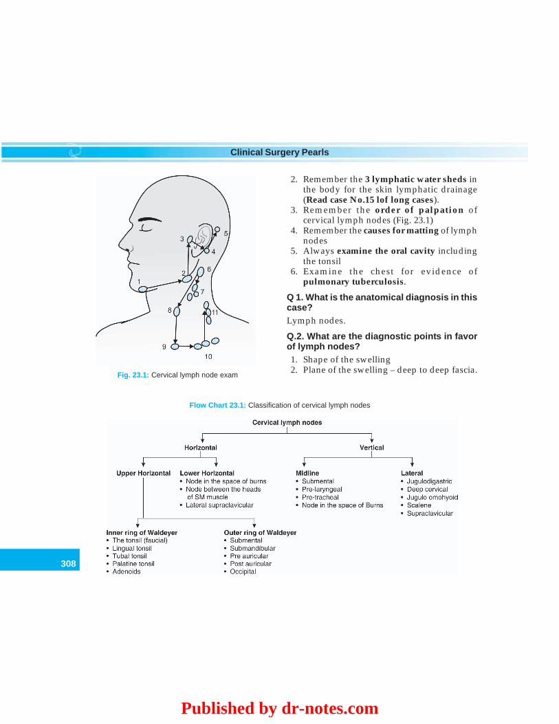
308
Clinical Surgery Pearls
2. Remember the 3 lymphatic water sheds inthe body for the skin lymphatic drainage(Read case No.15 lof long cases).
3. Remember the order of palpation ofcervical lymph nodes (Fig. 23.1)
4. Remember the causes for matting of lymphnodes
5. Always examine the oral cavity includingthe tonsil
6. Examine the chest for evidence ofpulmonary tuberculosis.
Q 1. What is the anatomical diagnosis in thiscase?Lymph nodes.
Q.2. What are the diagnostic points in favorof lymph nodes?1. Shape of the swelling2. Plane of the swelling – deep to deep fascia.
Fig. 23.1: Cervical lymph node exam
Flow Chart 23.1: Classification of cervical lymph nodes
Published by dr-notes.com
309
Tuberculous Cervical Lymph Node
Q 3. What is the plane of the cervical lymphnodes?For all practical purposes majority of the cervicallymph nodes are deep to deep fascia.
Q 4. What are the groups of lymph nodeswhich are superficial to the deep fascia inthe neck?1. The external jugular group of lymph nodes2. The submental nodes3. The occipital nodes4. The facial nodes.
Q 5. How many lymph nodes are there in theneck?• Roughly 300 nodes (out of total 800 nodes in
the human body)• About 150 are in the mesentery.
Q 6. What are the functions of lymph nodes?1. Filtration of effete cells, bacteria and
antigens2. Presentation of antigens to the lymphocytes3. Regulation of protein content of efferent
lymph.
Q 7. How will you classify cervical lymphnodes?They may be classified as shown in Flow Chart23.1Note: SM muscle - Sternomastoid muscle.
Q 8. What is the sequence of palpation ofcervical lymph nodes?
The following sequence is recommended. Startpalpitating from above from the submentalnode and proceed backwards (Fig. 23.1)1. Submental2. Submandibular
3. Preauricular4. Postauricular5. Occipital.
Then proceed downwards laterally from thejugulodigastric6. Jugulodigastric7. Deep cervical8. Juguloomohyoid9. Scalene
10. SupraclavicularNow proceed upwards to the external jugular
nodes (superficial).
Q 9. What are the causes for lymphadeno-pathy?
The causes may be classified as shown inFlow Chart 23.2.
Q 10. What is the plan of action for findingout the source of the enlarged lymph nodes?
Search for primary lesion in cervicallymphadenopathy.
Start from above and work downwards
1. Examine the skin of the scalp, face, ears, andneck.
2. Examine the nose3. Transilluminate the air sinuses4. Examine the oral cavity5. Examine the nasopharynx and larynx (ENT
examination)6. Palpate the salivary glands (parotid and sub-
mandibular)7. Examine the thyroid gland8. Examine the breast9. Examine the chest
10. Examine the abdomen and genitalia
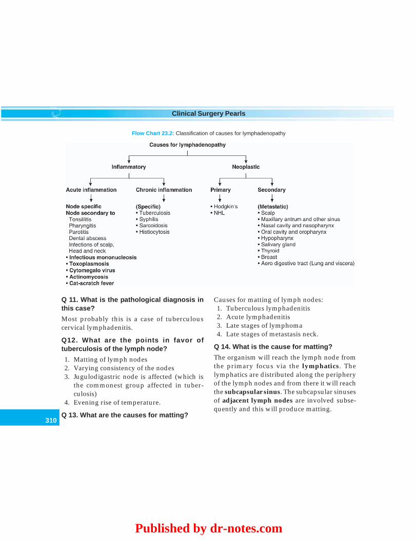
310
Clinical Surgery Pearls
Flow Chart 23.2: Classification of causes for lymphadenopathy
Q 11. What is the pathological diagnosis inthis case?
Most probably this is a case of tuberculouscervical lymphadenitis.
Q12. What are the points in favor oftuberculosis of the lymph node?
1. Matting of lymph nodes2. Varying consistency of the nodes3. Jugulodigastric node is affected (which is
the commonest group affected in tuber-culosis)
4. Evening rise of temperature.
Q 13. What are the causes for matting?
Causes for matting of lymph nodes:1. Tuberculous lymphadenitis2. Acute lymphadenitis3. Late stages of lymphoma4. Late stages of metastasis neck.
Q 14. What is the cause for matting?
The organism will reach the lymph node fromthe primary focus via the lymphatics. Thelymphatics are distributed along the peripheryof the lymph nodes and from there it will reachthe subcapsular sinus. The subcapsular sinusesof adjacent lymph nodes are involved subse-quently and this will produce matting.
Published by dr-notes.com
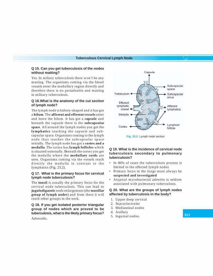
311
Tuberculous Cervical Lymph Node
Q 15. Can you get tuberculosis of the nodeswithout matting?
Yes. In miliary tuberculosis there won’t be anymatting. The organisms coming via the bloodvessels enter the medullary region directly andtherefore there is no periadenitis and mattingin military tuberculosis.
Q 16.What is the anatomy of the cut sectionof lymph node?
The lymph node is kidney-shaped and it has gota hilum. The afferent and efferent vessels enterand leave the hilum. It has got a capsule andbeneath the capsule there is the subcapsularspace. All around the lymph nodes you get thelymphatics reaching the capsule and sub-capsular space. Organisms coming to the lymphnode thus reaches the subcapsular spaceinitially. The lymph node has got a cortex and amedulla. The cortex has lymph follicles whichis situated externally. Beneath the cortex you getthe medulla where the medullary cords areseen. Organisms coming via the vessels reachdirectly the medulla in contrast to thelymphatics (Fig. 23.2).
Q 17. What is the primary focus for cervicallymph node tuberculosis?The tonsil is usually the primary focus for thecervical node tuberculosis. This can lead tojugulodigastric node enlargement (the tonsillargroup of lymph nodes) and from there it willreach other groups in the neck.
Q 18. If you get isolated posterior triangulargroup of nodes which are proved to betuberculosis, what is the likely primary focus?
Adenoids.
Q 19. What is the incidence of cervical nodetuberculosis secondary to pulmonarytuberculosis?
• In 80% of cases the tuberculosis process islimited to the affected lymph nodes
• Primary focus in the lungs must always besuspected and investigated
• Atypical mycobacterial adenitis is seldomassociated with pulmonary tuberculosis.
Q 20. What are the groups of lymph nodesaffected by tuberculosis in the body?
1. Upper deep cervical2. Supraclavicular3. Mediastinal nodes4. Axillary5. Inguinal nodes.
Fig. 23.2: Lymph node section
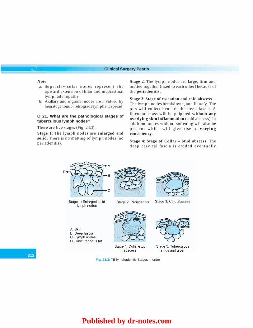
312
Clinical Surgery Pearls
Note:a. Supraclavicular nodes represent the
upward extension of hilar and mediastinallymphadenopathy
b. Axillary and inguinal nodes are involved byhematogenous or retrograde lymphatic spread.
Q 21. What are the pathological stages oftuberculous lymph nodes?
There are five stages (Fig. 23.3):Stage 1: The lymph nodes are enlarged andsolid. There is no matting of lymph nodes (noperiadenitis).
Stage 2: The lymph nodes are large, firm andmatted together (fixed to each other) because ofthe periadenitis.
Stage 3: Stage of caseation and cold abscess—The lymph nodes breakdown, and liquefy. Thepus will collect beneath the deep fascia. Afluctuant mass will be palpated without anyoverlying skin inflammation (cold abscess). Inaddition, nodes without softening will also bepresent which will give rise to varyingconsistency.
Stage 4: Stage of Collar – Stud abscess. Thedeep cervical fascia is eroded eventually
Fig. 23.3: TB lymphadenitis Stages in order
Published by dr-notes.com
313
Tuberculous Cervical Lymph Node
resulting in the escape of pus beneath thesuperficial fascia which is a laborious space.
Stage 5: Stage of sinuses and ulcers. The puswill eventually burst through the skin resultingin a discharging sinus or ulcer.
Q 22. Will the cold abscess contain tuberclebacilli?Yes. The abscess is lined by granulation tissueand caseous material.
Q 23. How a tuberculous ulcer is formed?Once a sinus is formed the discharge will infectthe surrounding skin and cause ulceration.
Q 24. What are the characteristics oftuberculous ulcer?The tuberculous ulcer has got underminededges, seropurulent discharge and palegranulation tissue.
Q 25. What is collar–stud abscess?The pus is formed as a result of breakdown of alymph node which will subsequently erode thedeep fascia and come to the space beneath thesuperficial fascia. There is a superficial softswelling, which is nothing but pus, and iscommunicating with the offending lymph nodesituated deep to the deep fascia. The node, theconnection and the superficial soft swellingtogether will take the shape of a collar–stud.
Q 26. What are the clinical types of tuber-culous lymphadenitis?There are four types:1. Acute type
– Seen in infants and children below fiveyears
– Inflammatory signs may be there andmay resemble acute lymphadenitis.
2. Caseating type – commonest type seen inyoung adults with typical matted nodeswith caseation, cold abscess and sinuses.
3. Hyperplastic type – this is seen in patientswith good resistance. The lymphoidhyperplasia is more predominant thancaseation. The nodes are usually firm anddiscrete.
4. Atrophic type – seen in elderly where thelymph nodes undergo natural involution.The nodes are small and burst resulting incaseation.
Q 27. What is scrofuloderma?The skin involvement as a result of tuberculosisis called scrofuloderma. The skin will bediscolored bluish or hyperpigmented.
Q 28. How will you investigate and confirmyour diagnosis?1. ESR—will be raised2. X-ray chest—evidence of pulmonary
tuberculosis need not be there becausemajority of the cervical lymph nodetuberculosis are primary (and not secondaryto pulmonary tuberculosis)
3. Tuberculin test (Mantoux)—a positive testhas no diagnostic value (because of BCGvaccination) a negative test is useful inexcluding tuberculosis
4. FNAC of the lymph node5. Biopsy of the lymph node for pathology and
microbiology6. Aspiration of the cold abscess for AFB
staining.
314
Clinical Surgery Pearls
Q 29. How Mantoux test is done?
1 unit of PPD (Purified Protein Derivative) isinjected intradermally in the volar surface ofthe forearm. After 48hrs, look for area ofinduration surrounding the injection site. Apositive test is one where the indurationexceeds 12mm. A positive test is suggestive ofprior or present infection with M. tuberculosis.Negative results do not always rule outtuberculosis (immunosuppression, malnutritionand diseases like lymphomas suppress the test).
Q 30. Is FNAC of the node reliable?
For lymph node pathology biopsy of the intactlymph node is always superior because apathologist can study the architecture of thelymph node (this is not possible with FNAC). Ifthe needle is striking the granuloma in thelymph node, the pathologist will give a positivereport. Therefore, biopsy is always a superiorinvestigation for lymph node.
Q 31. What are the important points to beremembered while taking cervical lymphnodes for biopsy?
1. Always try to take an intact node for biopsy2. If possible take 2 nodes (1 node for the
pathologist and 1 node for the microbiologist)3. Send the lymph node for the pathologist in
formalin4. Send the lymph node for the microbiologist
in saline solution (this is for AFB culture).
Q 32. What is the media used for culture andhow long it will take to get a positive culture?
• Lowenstein – Jensen media• Takes 6 weeks for positive culture
• Selinite medium – shortens the time ofgrowth to 5 days.
Q 33. Suppose there is only one lymph nodewould you attempt FNAC?
No. FNAC will induce changes in the lymphnode, which are likely to alter the pathologicalpicture if it is subsequently taken for biopsystudy. This is called WARF (WorrisomeAnomaly Related to FNAC).
Q 34. What are the pathological changes inthe lymph node?
Pathologically we get the tubercle, which consistsof an area of caseation surrounded by giant cells,epithelioid cells, lymphocytes and plasma cells.
Q 35. What is the difference between caseousmaterial and tuberculous pus?
Caseous material—Dry, granular and cheeselike material is called caseous material (granularstructure less material).
Tuberculous pus – softening and liquefactionof the caseous material results in the formationof a thick creamy fluid called tuberculous pus.It is highly infective because liquefaction isassociated with multiplication of the organism.In addition, it contains fatty debris in serousfluid with few necrotic cells.
Q 36. Can you start antituberculous treatmentempirically without pathological or micro-biological report?
No. Pathological or microbiological support isnecessary for starting the antituberculoustreatment.
Published by dr-notes.com
315
Tuberculous Cervical Lymph Node
Q 37. What are the organisms responsiblefor lymph node tuberculosis?
• Human tuberculosis• Bovine tuberculosis
Q 38. What are the organisms responsiblefor atypical Mycobacterium tuberculosis?
• Mycobacterium avium intracellulare• Mycobacterium scrofulaceum.
Q 39. Is there any role for urine examination?
Yes. The renal and pulmonary tuberculosisoccasionally coexist. Therefore the urine shouldbe carefully examined.
Q 40. Is it possible to get calcified cervicallymph node without prior symptoms ofcervical lymph node tuberculosis?
Yes. In patients with natural resistance to theinfection, the nodes may accidentally detectedat a later date as calcified nodes.
Q 41. What is the treatment of choice in aproved case of cervical lymph node tuber-culosis?
Antituberculous chemotherapy – Triple DrugTherapy – Rifampicin, INH and Ethambutol—(Read the chapter on long cases- right iliac fossamass - abdominal tuberculosis).
Q 42. What are the indications for surgery incervical lymph node tuberculosis?
Indications for surgery in tuberculous lymph-adenitis1. Biopsy (the most important indications)2. Excision of a group of nodes if it is not
responding to antituberculous treatment3. Excision of the offending lymph node in
cases of collar – stud abscess4. Excision of abscess if it is not responding to
aspiration5. Persistent sinus
Q 43. What are the causes for persistentsinus?
Causes for persistent sinus:1. Secondary infection2. Considerable fibrosis3. Necrotic and calcified material replacing the
lymph node.
Q 44. If the cervical nodes are not respondingto the antituberculous drugs what should besuspected?
Atypical mycobacterial adenitis.
Q 45. What is the treatment of cold abscess?
Aspiration. Incision and drainage is notrecommended because it will result in sinusformation.
Q 46. How will you aspirate cold abscess?
Aspiration is done through the nondependentpart.
316
Clinical Surgery Pearls
24Cervical Metastatic Lymph
Node and Neck Dissections
CASE CAPSULE
A 65-year-old male patient presents with a hardlymph node swelling of 3 cm size involvingthe level III group on right side. The swellingis mobile. The superficial temporal artery ispalpable. The cranial nerves are normal. Thereare no abdominal, chest or ENT complaints.The patient is apparently healthy.
Read the diagnostic algorithm for a neckswelling.
Checklist for history
1. Alcohol and tobacco use in history2. Pain around the eyes – referred from the
nasopharynx3. Otalgia–Ca base of tongue, tonsil, and
hypopharynx can cause otalgia4. Odynophagia – as a result of cancers of the base
of the tongue, hypopharynx, cervical nodemetastasis, etc.
5. Bleeding from the nose (epistaxis) – cancers ofthe nasal cavity
6. Hemoptysis7. Alteration of phonation8. Difficulty in breathing
9. Difficulty in swallowing – late symptom of baseof tongue, hypopharynx and cervicalesophagus
10. Difficulty in hearing – from nasopharynx11. Hoarseness of voice–carcinoma glottis and
carcinoma thyroid12. History of prior SCC
Checklist for examination
1. Careful examination of oral cavity afterremoval of dentures
2. Bimanual palpation of the floor of the mouth3. Check for nasal block4. Check for sensory loss in the distribution of
infraorbital nerves – maxillary sinus cancer5. Examine the cranial nerves III – VII and IX –
XII (involvement in nasopharyngeal cancer)6. Look for Horner’s syndrome – involvement of
cervical sympathetic chain, extralaryngealspread of laryngeal cancer and extracapsularinvasion of cervical lymph node
7. Look for trismus8. A thorough ENT examination9. Examination of thyroid10. Examination of salivary glands11. Examination of breast12. Examination of chest13. Examination of abdomen
Published by dr-notes.com
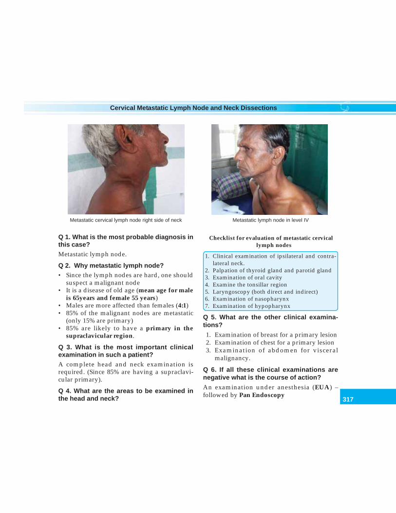
317
Cervical Metastatic Lymph Node and Neck Dissections
Q 1. What is the most probable diagnosis inthis case?Metastatic lymph node.
Q 2. Why metastatic lymph node?• Since the lymph nodes are hard, one should
suspect a malignant node• It is a disease of old age (mean age for male
is 65years and female 55 years)• Males are more affected than females (4:1)• 85% of the malignant nodes are metastatic
(only 15% are primary)• 85% are likely to have a primary in the
supraclavicular region.
Q 3. What is the most important clinicalexamination in such a patient?A complete head and neck examination isrequired. (Since 85% are having a supraclavi-cular primary).
Q 4. What are the areas to be examined inthe head and neck?
Checklist for evaluation of metastatic cervicallymph nodes
1. Clinical examination of ipsilateral and contra-lateral neck.
2. Palpation of thyroid gland and parotid gland3. Examination of oral cavity4. Examine the tonsillar region5. Laryngoscopy (both direct and indirect)6. Examination of nasopharynx7. Examination of hypopharynx
Q 5. What are the other clinical examina-tions?
1. Examination of breast for a primary lesion2. Examination of chest for a primary lesion3. Examination of abdomen for visceral
malignancy.
Q 6. If all these clinical examinations arenegative what is the course of action?
An examination under anesthesia (EUA) –followed by Pan Endoscopy
Metastatic cervical lymph node right side of neck Metastatic lymph node in level IV
318
Clinical Surgery Pearls
Pan endoscopy
• Nasopharyngoscopy• Esophagobronchoscopy• Laryngoscopy (direct)
Q 7. What is the purpose of esophagoscopyand bronchoscopy?
In metastatic squamous cell carcinoma (SCC),10-20% chance for a second primary is there inthe aerodigestive tract.
Q 8. What is the definition of a “new primary”after treatment of previous cancer?
One arising more than 3years after previouscancer is considered a new primary.
Q 9. If nothing is found on pan endoscopy,what next?
Surveillance biopsy — Blind biopsies are takenfrom the following areas.
Areas for Blind biopsy
• Tonsils• Tonsillar beds• Base of tongue• Pyriform sinus• Subglottic region• Fossa of Rosenmüller
Q 10. If surveillance biopsy is negative howto proceed?
Ipsilateral tonsillectomy.
Q 11. What is the purpose of surveillancebiopsy?
In the absence of gross lesion, in 10 – 15% ofcases primary will be revealed by surveillancebiopsy.
Q12. What is the order of frequency ofprimary in a case of metastasis?
Head and neck source of primary—Theprimary sites in order of frequency are:
1. Nasopharynx2. Tonsil3. Base of tongue4. Thyroid5. Supraglottic larynx6. Floor of mouth7. Palate8. Pyriform fossa
• Nonhead and neck source of primary – (inorder of frequency)
1. Bronchus2. Esophagus3. Breast4. Stomach
Q 13. What is the contraindication for a pre-liminary lymph node biopsy in a metastaticlymph node? (PG)
• A biopsy will produce scarring of subcuta-neous tissue and will destroy the tissueplanes. This will affect the neck dissection ifit becomes necessary because the scar tissuecan not be distinguished from the tumor
• Biopsy will destroy nodal or fascial barriersholding the cancer in check and seedling ofthe soft tissues and lymphatics will occur
• Chances for neck recurrence will occur as aresult of biopsy (recurrence is the major causeof death rather than metastasis in SCC)
• Chances for general spread is high.
Published by dr-notes.com
319
Cervical Metastatic Lymph Node and Neck Dissections
Q 14. If nothing is found after pan endoscopyand blind biopsy, what next?
MRI of the neck is done.
Q 15. Why MRI is superior to CT forevaluation of a metastatic node of unknownprimary?
• MRI can identify subtle changes in soft tissues• Guided biopsy of the primary lesion is possible• Extension of the primary to the surrounding
soft tissues can be identified.
Q 16. If MRI is negative, what is the next step?
FNAC.
Q 17. If FNAC is negative, what is the nextstep?
An open biopsy is indicated now. If metastaticSCC is found on frozen section, it is immediatelyfollowed by a neck dissection if it is operable.
Q 18. Why not a delayed neck dissection?
The best chance for cure and time for dissectionis when the normal tissue planes are intact.Thus, the time to carry out a biopsy is whenyou are ready to carry out a dissection.
Q 19. What are the possible FNAC or biopsyreports?
Histological types of metastasis (50% SCC, 25%poorly differentiated and 25% adenocarcinoma).
Histological type of metastasis
1. Squamous cell carcinoma (SCC)2. Nonsquamous cell carcinoma
• Adenocarcinoma• Poorly differentiated carcinoma• Poorly differentiated neoplasm
Q 20. If the report is adenocarcinoma whatare the possibilities?
Primary source for adenocarcinomatousdeposits in the neck nodes:• Salivary neoplasm• Thyroid carcinoma• Breast carcinoma• Occult lung cancer• Prostatic cancer• Renal malignancy• GI malignancy
Q 21. What is the treatment of metastaticadenocarcinoma? (Flow Chart 24.1)
There is no role for surgery because it is adisseminated malignancy. Patient will go in forchemotherapy (Paclitaxel + carboplatin).
Q 22. What is the management of poorlydifferentiated neoplasm? (Flow Chart 24.1)(PG)
Repeat the FNAC. If this too turns out to beinconclusive, do a biopsy. If biopsy too provesto be inconclusive do immunohistochemistry.
Q 23. What is the purpose of immunohisto-chemistry?
Immunohistochemistry and electron microscopyis done to identify the lymphomas and otherchemoresponsive neoplasms (about 60%).
Q 24. What is the management of poorlydifferentiated carcinoma? (Flow Chart 24.1)(PG)
Again immunohistochemistry and electronmicroscopy are recommended in order toidentify the chemoresponsive subgroups –
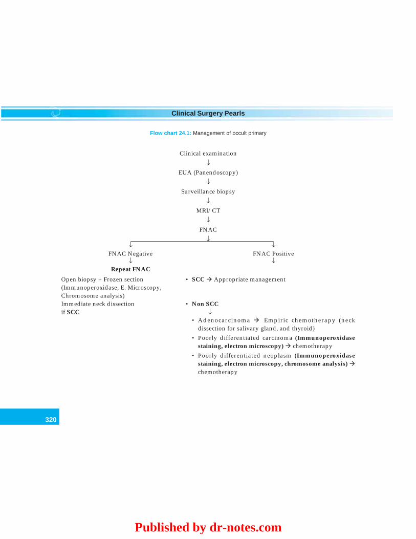
320
Clinical Surgery Pearls
Clinical examination
↓EUA (Panendoscopy)
↓Surveillance biopsy
↓MRI/CT
↓FNAC
↓↓ ↓
FNAC Negative FNAC Positive↓ ↓
Repeat FNAC
Open biopsy + Frozen section • SCC Appropriate management(Immunoperoxidase, E. Microscopy,Chromosome analysis)Immediate neck dissection • Non SCCif SCC ↓
• Adenocarcinoma Empiric chemotherapy (neckdissection for salivary gland, and thyroid)
• Poorly differentiated carcinoma (Immunoperoxidasestaining, electron microscopy) chemotherapy
• Poorly differentiated neoplasm (Immunoperoxidasestaining, electron microscopy, chromosome analysis) chemotherapy
Flow chart 24.1: Management of occult primary
Published by dr-notes.com
321
Cervical Metastatic Lymph Node and Neck Dissections
• Lymphoma• Ewing’s tumor• Neuroendocrine tumors• Primitive sarcomas.
Q 25. What is the commonest pathologicaltype of neck node metastasis?
Squamous cell carcinoma – 80%.
Q 26. What are the squamous cell carcinomaswhich will metastasize bilaterally? (PG)
SCC with bilateral metastasis
1. Lower lip2. Supraglottis3. Soft palate
Q 27. Which group of lymph node is involvedin carcinoma nasopharynx? (PG)
Nodes involved in carcinoma nasopharynx
• Retropharyngeal nodes• Parapharyngeal nodes• Level II – V
Q 28. What are the carcinomas which willmetastasize to retropharyngeal lymphnodes? (PG)
Malignancies involving the retropharyngealnodes
1. Nasopharynx2. Soft palate3. Posterior and lateral oropharynx4. Hypopharynx
Q 29. What are the primary sites below theclavicle?
Sites of the primary below the clavicle (15%)
• Lung (commonest)• Pancreas• Esophagus• Stomach• Breast• Ovary• Testis• Prostate
Q 30. Which group of lymph nodes areinvolved in infraclavicular primary?
The level IV and V (lower jugular chain andsupraclavicular nodes).
Q 31. What are the other investigationsrecommended?• X-ray chest• Sputum cytology• CT scan of the chest and abdomen• Mammography• PET scan (if required)
Q 32. What is the role of PET scan?
18-Fluorodeoxyglucose (18FDG) analogue ispreferentially absorbed by neoplastic cells andcan be detected by Positron Emission Tomo-graphy (PET) scanning. It is more sensitive thanCT in identifying the primary lesion. But in thecase of unknown primary the sensitivity is notmore than 50%. This is because the unknownprimary tumor may have spontaneouslyinvoluted.
Q 33. What is the definition of occult primary?When the lymph node is found to containmetastatic carcinoma but the primary is
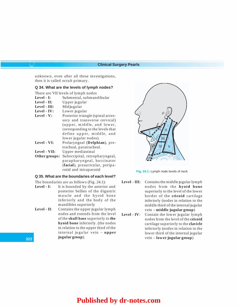
322
Clinical Surgery Pearls
unknown, even after all these investigations,then it is called occult primary.
Q 34. What are the levels of lymph nodes?There are VII levels of lymph nodesLevel - I: Submental, submandibularLevel - II: Upper jugularLevel - III: MidjugularLevel - IV: Lower jugularLevel - V: Posterior triangle (spinal acces-
sory and transverse cervical)(upper, middle, and lower,corresponding to the levels thatdefine upper, middle, andlower jugular nodes).
Level - VI: Prelaryngeal (Delphian), pre-tracheal, paratracheal.
Level - VII: Upper mediastinalOther groups: Suboccipital, retropharyngeal,
parapharyngeal, buccinator(facial), preauricular, peripa-rotid and intraparotid
Q 35. What are the boundaries of each level?The boundaries are as follows (Fig. 24.1):Level - I: It is bounded by the anterior and
posterior bellies of the digastricmuscle and the hyoid boneinferiorly and the body of themandibles superiorly
Level - II: Contains the upper jugular lymphnodes and extends from the levelof the skull base superiorly to thehyoid bone inferiorly. (the nodesin relation to the upper third of theinternal jugular vein – upperjugular group).
Level - III: Contains the middle jugular lymphnodes from the hyoid bonesuperiorly to the level of the lowerborder of the cricoid cartilageinferiorly (nodes in relation to themiddle third of the internal jugularvein – middle jugular group)
Level - IV: Contain the lower jugular lymphnodes from the level of the cricoidcartilage superiorly to the clavicleinferiorly (nodes in relation to thelower third of the internal jugularvein – lower jugular group)
Fig. 24.1: Lymph node levels of neck
Published by dr-notes.com
323
Cervical Metastatic Lymph Node and Neck Dissections
Level - V: Contains the lymph nodes in theposterior triangle bounded by theanterior border of the trapeziusmuscle posteriorly, the posteriorborder of the sternocleidomastoidmuscle anteriorly, and the clavicleinferiorly. For descriptive pur-poses, Level V may be furthersubdivided into upper, middle,and lower levels correspondingto the superior and inferiorplanes that define Levels II, III,and IV.
Level - VI: Contains the lymph nodes of theanterior central compartmentfrom the hyoid bone superiorly tothe suprasternal notch inferiorly.On each side, the lateral boundaryis formed by the medial border ofthe carotid sheath.
Level - VII: Contains the lymph nodesinferior to the suprasternal notchin the superior mediastinum.
Q 36. What are the probable primary sites foreach level? (PG)
Primary sites for each level of cervicallymph nodes
Lymph node Primary cancer siteslevel
Level I Oral cavity, lip, salivary gland, skinLevel II Oral cavity, nasopharynx, oropha-
rynx, larynx, salivary glandLevel III Oral cavity, oropharynx, hypo-
pharynx, larynx, thyroidLevel IV Oropharynx, hypopharynx, larynx,
thyroid, cervical esophagus
Level V Nasopharynx, scalp(Accessory nodes)Level V GI tract, breast, lung(supraclavicular)
Q 37. What is the area of drainage of sub-occipital nodes?
Skin of the scalp.
Q 38. What is the drainage area of parotidnodes?
Parotid gland and skin.
Q 39. What is the N (regional lymph node)staging?
N staging as per AJCC 6th edition
NX Regional lymph nodes cannot be assessedNo No regional lymph node metastasis*N1 Metastasis in a single ipsilateral lymph
node, 3 cm or less in greatest dimension*N2 Metastasis in a single ipsilateral lymph
node, more than 3 cm but not more than6 cm in greatest dimension; or in multipleipsilateral lymph nodes, none more than6 cm in greatest dimension; or in bilateralor contralateral lymph nodes, none morethan 6cm in greatest dimension.
*N2a Metastasis in single ipsilateral lymph nodemore than 3cm but not more than 6 cm ingreatest dimension
*N2b Metastasis in multiple ipsilateral lymphnodes, none more than 6 cm in greatestdimension.
*N2c Metastasis in bilateral or contralaterallymph nodes, none more than 6 cm ingreatest dimension
*N3 Metastasis in a lymph node more than 6cmin greatest dimension
324
Clinical Surgery Pearls
*Note: A designation of “U” or “L” may be usedto indicate metastasis above the Lowerborder of the cricoid (U) or below thelower border of the cricoid (L).
Q 40. What is the importance of the “U” and“L”?
When the lower lymph nodes namely level 4and 5, below the lower border of the cricoidcartilage are involved the prognosis is bad.
Q 41. In how many % of occult metastasis,the primary identification is possible?
Roughly in 1/3rd cases primary can beidentified.
Q 42. Why primary is nonidentifiable in somecases? (PG)
Possibly because of the spontaneous involutionof the unknown primary.
Q 43. If primary is not identified in the givencase would you recommend surgery if thereport is coming as SCC?
• Yes. A neck dissection is recommended if thenodes are resectable
• A neck dissection removes additionalipsilateral cervical nodes.
Q 44. What are the conditions where neckdissections are valuable? (PG)
Conditions in which neck dissections arerecommended
1. Squamous cell carcinoma2. Salivary gland tumors3. Thyroid carcinoma4. Melanoma
Q 45. What type of neck dissection is recom-mended?
Modified neck dissection may be appropriate.
Q 46. What are the indications for radio-therapy after a modified neck dissection?
Indications for radiotherapy after a modifiedneck dissection:• If more than two lymph nodes contain metastasis• Nodes at two or more levels contain metastasis• Extracapsular spread of metastasis
Q 47. What are the types of neck dissection?
The neck dissections may be classified as –• Radical neck dissection (RND) – classical
Crile procedure (Level I – V nodes removed)• Modified radical neck dissection (MRND)
(described by Bocca) preserves one or moreof the following structures—spinal accessorynerve, internal jugular vein and sternomastoidmuscle—Type I, Type II, Type IIIType I—Spinal accessory alone preservedType II—Spinal accessory and sternomastoidpreservedType III—Spinal accessory, sternomastoidand internal jugular vein are preserved.
• Functional Neck Dissection (Level II – V) –preserving sternomastoid, internal jugularvein and spinal accessory nerve
• Selective Neck Dissection – here one or morelymph node groups are preserved –1. Supraomohyoid neck dissection
(removal of Level I – III)2. Posterolateral neck dissection (removal
of Level II, III, IV, V)3. Lateral neck dissection (removal of Level
II, III, IV)
Published by dr-notes.com
325
Cervical Metastatic Lymph Node and Neck Dissections
4. Anterior compartment dissection(Removal of Level VI).
Q 48. What is the difference between modifiedradical neck dissection and functional neckdissection?• Modified neck dissection always preserves
spinal accessory nerve• Functional neck dissection always preserves
sternomastoid muscle, the internal jugularvein and spinal accessory nerve.
Q. 49 What are the structures removed inradical neck dissection?En-bloc removal of fat, fascia, and lymph nodesfrom level I to level V.They include the following –• Two muscles—Sternomastoid and omohyoid• Two veins—Internal jugular vein and
external jugular vein• Two nerves—Spinal accessory nerve and
cervical plexus• Two glands—Submandibular salivary
glands and tail of parotid• Prevertebral fascia
Q 50. What is extended radical neck dissec-tion? (PG)
This refers to the removal of one or moreadditional lymph node groups and/or non-lymphatic structures not encompassed by theradical neck dissection. This may include theparapharyngeal and superior mediastinallymph nodes. The nonlymphatic structures mayinclude the carotid artery, the hypoglossalnerve, the vagus nerve and the paraspinalmuscles. This is not an operation for occultprimary.
Q. 51. What is the prognosis if the primarytumor is never found? (PG)
This won’t influence the prognosis. If theprimary tumor is small or occult, will beprobably included in the field of thepostoperative irradiation and cured by suchtreatment.
Prognosis is determined by whether or notthe tumor recurs or whether it metastasizes(metastasis to lungs, bone or liver).
Q 52. How will you summarize the treatmentfor SCC occult metastasis? [treatment ofadenocarcinoma, poorly differentiatedcarcinoma and poorly differentiated neo-plasms are already given above]Summary of treatment for squamous cellcarcinoma metastasis from occult primaryIt is treated according to the N stage. –
N 1 – MRND (surgery is the treatment of all N1nodes)RT (radiotherapy) if positivemargins, capsular invasion and multiplelevel nodes irradiate neck and all potentialsites of primary
N 2a and – Mobile RND followed by RT, FixedN2b RT followed by RNDN 2c – Bilateral RND followed by bilateral RTN 3 – Resectable RND followed by RT +
Chemo (controversy)Unresectable RT. followed by RNDwhen it becomes resectable
RND: Radical neck dissection
RT: Radiotherapy
Note: Regarding radiotherapy:1. Radiotherapy is given for contralateral neck
nodes if primary is nasopharyngealcarcinoma.
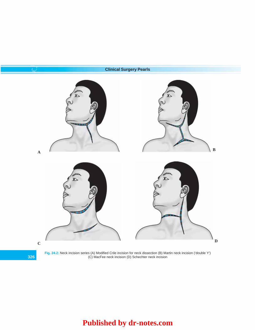
326
Clinical Surgery Pearls
Fig. 24.2: Neck incision series (A) Modified Crile incision for neck dissection (B) Martin neck incision (‘double Y’)(C) MacFee neck incision (D) Schechter neck incision
A
CD
B
Published by dr-notes.com
327
Cervical Metastatic Lymph Node and Neck Dissections
2. Level 2 lymph nodes alone-primary is likelyto be nasopharynx and RT is preferred forsuch cases.
Q 53. What are the incisions used for neckdissection? (PG)1. Mac Fee incision – Consisting of 2
horizontal limbs (the first begins over themastoid curving down to the hyoid bone,and up again to the chin, the secondhorizontal incision lies about 2cm above theclavicle from the anterior border of thetrapezius to the midline)
2. Schechter incision – having a vertical limband horizontal limb (the vertical comes fromthe mastoid process to the point wheretrapezius meets the clavicle along theanterior border of the trapezius. Thehorizontal, starting from the middle of thevertical to the prominence of thyroidcartilage)
3. The classical incision by Crile – it’s a Y-shaped incision with the upper limbs of the“Y” reaching posteriorly to the mastoid andanteriorly to the chin. The stem of the “Y”reaches down to the middle of the posteriortriangle.
4. Martin incision—“double Y” incision.
Q 54. What is the most poorly vascularizedarea of skin in the neck and why? (PG)
• The middle of the neck laterally over thecommon carotid artery
• The blood supply to the skin comes downfrom the face, up from the chest, aroundfrom trapezius and from the external carotidon the other side
• Avoid a vertical incision over this area sothat a carotid artery rupture can be avoided
• Avoid 3 point junctions in the center of the neck
Q 55. What are the complications of neckdissection? (PG)
Complications of neck dissection
1. Bleeding2. Pneumothorax3. Raised intracranial pressure (avoid pressure
dressings, use mannitol if required)4. Wound breakdown5. Infection6. Necrosis of the skin flap7. Seroma (use suction drain)8. Rupture of the carotid artery9. Chylous fistula (thoracic duct injury)
10. Frozen shoulder (due to accessory nervedamage) – difficulty to abduct the arm
Q 56. What precaution is taken to preventrupture of carotid artery? (PG)
• The carotid sheath should be protected eitherby a muscle flap or a free dermal graft
• The commonly used muscle flap is levatorscapulae
• Use horizontal incisions• Avoid three point junctions in incisions.
Q 57. What is the sequencing of bilateral neckdissection and its prognosis? (PG)
• The presence of bilateral neck nodes atpresentation is a bad prognostic sign
• Five year survival rate falls to about 5%• The usual practice of staged neck dissection
is now changing to simultaneous bilateralneck dissection
328
Clinical Surgery Pearls
• The most feared complication after bilateralneck dissection is increased intracranialpressure
• Tying one internal jugular vein produces 3fold increase in the intracranial pressure
• Tying the second side produces 5 foldincrease in intracranial pressure
• However, the pressure tends to fall over a
period of 8 days (the pressure fall is rapidwithin the 1st 12 hours.
Q 58. How will you avoid this complicationof increased intracranial tension? (PG)
1. Lumbar drain (removal of CSF)2. Nursing the patient in the sitting position3. Infusion of mannitol4. Avoiding pressure dressings.
Published by dr-notes.com
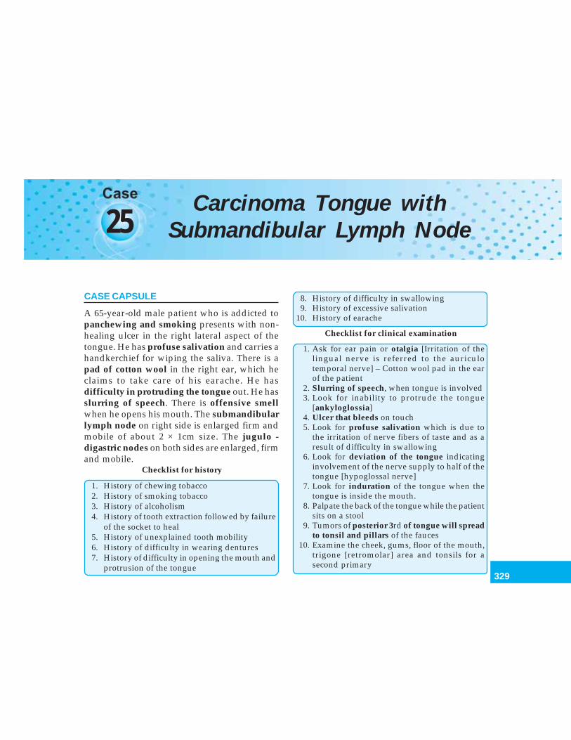
329
Carcinoma Tongue with Submandibular Lymph Node
25Carcinoma Tongue with
Submandibular Lymph Node
CASE CAPSULE
A 65-year-old male patient who is addicted topanchewing and smoking presents with non-healing ulcer in the right lateral aspect of thetongue. He has profuse salivation and carries ahandkerchief for wiping the saliva. There is apad of cotton wool in the right ear, which heclaims to take care of his earache. He hasdifficulty in protruding the tongue out. He hasslurring of speech. There is offensive smellwhen he opens his mouth. The submandibularlymph node on right side is enlarged firm andmobile of about 2 × 1cm size. The jugulo -digastric nodes on both sides are enlarged, firmand mobile.
Checklist for history
1. History of chewing tobacco2. History of smoking tobacco3. History of alcoholism4. History of tooth extraction followed by failure
of the socket to heal5. History of unexplained tooth mobility6. History of difficulty in wearing dentures7. History of difficulty in opening the mouth and
protrusion of the tongue
8. History of difficulty in swallowing9. History of excessive salivation
10. History of earache
Checklist for clinical examination
1. Ask for ear pain or otalgia [Irritation of thelingual nerve is referred to the auriculotemporal nerve] – Cotton wool pad in the earof the patient
2. Slurring of speech, when tongue is involved3. Look for inability to protrude the tongue
[ankyloglossia]4. Ulcer that bleeds on touch5. Look for profuse salivation which is due to
the irritation of nerve fibers of taste and as aresult of difficulty in swallowing
6. Look for deviation of the tongue indicatinginvolvement of the nerve supply to half of thetongue [hypoglossal nerve]
7. Look for induration of the tongue when thetongue is inside the mouth.
8. Palpate the back of the tongue while the patientsits on a stool
9. Tumors of posterior 3rd of tongue will spreadto tonsil and pillars of the fauces
10. Examine the cheek, gums, floor of the mouth,trigone [retromolar] area and tonsils for asecond primary
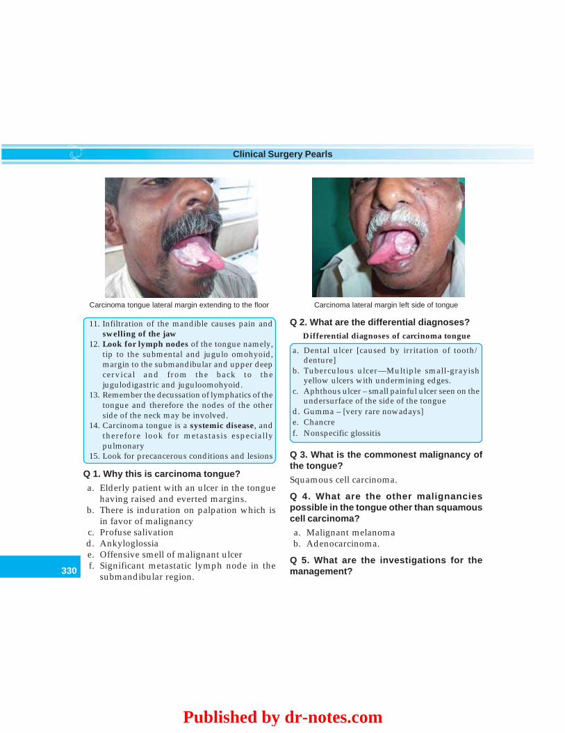
330
Clinical Surgery Pearls
11. Infiltration of the mandible causes pain andswelling of the jaw
12. Look for lymph nodes of the tongue namely,tip to the submental and jugulo omohyoid,margin to the submandibular and upper deepcervical and from the back to thejugulodigastric and juguloomohyoid.
13. Remember the decussation of lymphatics of thetongue and therefore the nodes of the otherside of the neck may be involved.
14. Carcinoma tongue is a systemic disease, andtherefore look for metastasis especiallypulmonary
15. Look for precancerous conditions and lesions
Q 1. Why this is carcinoma tongue?
a. Elderly patient with an ulcer in the tonguehaving raised and everted margins.
b. There is induration on palpation which isin favor of malignancy
c. Profuse salivationd. Ankyloglossiae. Offensive smell of malignant ulcerf. Significant metastatic lymph node in the
submandibular region.
Q 2. What are the differential diagnoses?Differential diagnoses of carcinoma tongue
a. Dental ulcer [caused by irritation of tooth/denture]
b. Tuberculous ulcer—Multiple small-grayishyellow ulcers with undermining edges.
c. Aphthous ulcer – small painful ulcer seen on theundersurface of the side of the tongue
d. Gumma – [very rare nowadays]e. Chancref. Nonspecific glossitis
Q 3. What is the commonest malignancy ofthe tongue?
Squamous cell carcinoma.
Q 4. What are the other malignanciespossible in the tongue other than squamouscell carcinoma?
a. Malignant melanomab. Adenocarcinoma.
Q 5. What are the investigations for themanagement?
Carcinoma lateral margin left side of tongueCarcinoma tongue lateral margin extending to the floor
Published by dr-notes.com
331
Carcinoma Tongue with Submandibular Lymph Node
Investigations for oral carcinoma
1. Incisional biopsy of the ulcer under localanesthesia for confirmation of the diagnosis –biopsy should include the most suspiciousarea along with normal adjacent mucosa. Areasof necrosis and gross infection should beavoided
2. FNAC of the lymph node3. Radiography–Orthopantomogram (OPG)–
provides information regarding the entiremandible, but limited in its ability to evaluatethe symphysis and lingual cortex
4. OPG may be supplemented with dentalocclusal and intraoral X-rays
5. CT• Indicated in patients with trismus• Lesions abutting the mandible• Where marginal mandibulectomy is planned• To evaluate the clinically negative neck• Patients with large nodes to look for carotid
artery involvement.• It is very useful for the assessment of
pterygoid regions6. MRI scan for assessing the soft tissue spread
and perineural involvement. It is very usefulfor tongue for assessing the extent of cancer.It is also useful for other oral andoropharyngeal cancers. Its great advantageover CT is that the image is not degraded bythe presence of metallic dental restoration.
7. Ultrasound of the neck and abdomen –ultrasound guided aspiration of the neck isuseful in surveillance of patients with clinicallyNO neck after treatment.
8. X-ray chest for all patients9. Dental consultation if radiation is planned
10. Assessment of the performance status (Seechart section)
11. Hb, full blood count, nutritional status, LFTand RFT
Q 6. What are the macroscopic types of oralcancers?
Macroscopic types of oral cancers
• Exophytic—less aggressive• Ulcerative• Combination
Q 7. What are the pathological types ofsquamous cell carcinoma?
Types of squamous cell carcinoma
• Verrucous – No lymph nodes• Basaloid SCC – Advanced disease (Metastasis
may be there)• Sarcomatoid – Lethal (Rapidly growing
polypoidal cancers)
Q 8. What are the peculiarities of verrucouscarcinoma?
• It is a controversial subject• Presents as exophytic, whitish warty or
cauliflower - like growth• Radiotherapy in verrucous carcinoma results
in a recurrence with anaplastic pattern thanthe original primary
• Radiotherapy induces anaplastic transfor-mation
• It seems that verrucous carcinoma alreadycontain foci of more malignant cells beforeradiotherapy
• There is minimal invasion and induration.• The lesions is densely keratinized and
presents as soft white velvety area• Lymh node metastasis is late• It is a low grade squamous cell carcinoma• Most verrucous carcinomas are suitable for
excision and that is the treatment of choice.
332
Clinical Surgery Pearls
Q 9. What are the modes of spread of oralcancer?1. Local spread to adjacent structures – soft
tissues, muscles, bone and neurovascularstructures
2. Lymphatic spread – the first echelon lymphnodes of primary SCC of the oral cavity arein the supraomohyoid triangle of the neck(Level I, II, III)
3. Distant metastasis – exceedingly rare (lungsand bones).
Note: Skip metastasis from primary carcinomamay occur in 15% of patients of carcinomatongue without involvement of first echelonlymph nodes.
Q 10. Which oral cancer is having highestincidence of nodal metastasis?• Carcinoma of the tongue, followed in
descending order by:• Tumors of the floor of the mouth• Lower alveolus• Buccal mucosa• Upper alveolus• Hard palate.
Q 11. What is the mechanism of involvementof mandible?
• It is involved by infiltration through its dentalsockets
• Through dental pores on the edentulousalveolar ridge.
Q 12. What are the etiological factors for oralcancer?
Etiological factors for oral cancer
A.: Lifestyle habitsa. Tobacco (smoked or smokeless)
(Synergistic effect of smoking and chewing oftobacco)
b. Betel nutc. Alcohold. Human papilloma virus (HPV)
– Detected in 60-90% cases of oral cancer– Present in 40% of normal oral cavity(direct
link between HPV and oral cancer remainsto be established)
e. Epstein – Barr virusB. Dietary factorsa. Vitamin A (protective role)b. Fresh fruits and vegetablesc. Iron deficiency anemia (Plummer-Vinson
syndrome)(SCC of hypopharynx and oralcavity)
C. Other risk factorsa. Poor dental hygieneb. Ill fitting dentures (chronic irritation)
The six Ss—Spices, Sprit, Sepsis, Sharptooth, Syphilis and Smoking.
Q 13. What is the risk of tobacco chewing fororal cancer?• Tobacco chewing, the risk is 8 times for
buccal cancer.• With quid it increases to 10 times• If the quid is kept overnight, the risk
increases to 30 times• Alcohol has synergetic effect with tobacco.
Q 14. What are the ingredients of tobaccochewing?It contains the following:• Betel leaf
Published by dr-notes.com
333
Carcinoma Tongue with Submandibular Lymph Node
• Areca nut• Smoked lime• Catechu• Condiments.
Note: It is commercially available as Pan masala.
Q 15. What is quid (Night quid)?The above ingredients are kept in the gingivo-labial suclus during night gives kick throughoutnight. This is called a night quid.
Q 16. Which component of the chewing isresponsible for the premalignant lesions?The chewing habits vary from place-to-place.The usual ingredients are: betel leaf, lime, betelnut and tobacco. The most importantcarcinogen is tobacco. The betel nut has got twoalkaloids namely, arecoline and tannins. Thearecoline stimulate collagen synthesis andproliferation of fibroblasts. The tanninsstabilizes collagen fibrils.
Q 17. What is the action of alcohol?
The following actions are there for thecarcinogenesis:• Promoter• Irritant• Solvent – increases the solubility of carcinogen• Alcohol supresses the efficiency of the DNA
repair after exposure to nitrosamine com-pounds.
Q 18. What are the premalignant lesions ofthe oral cavity?
Lesions of the oral cavity associated with anincreased risk of malignancy.
Precancerous lesionsa. Leukoplakiab. Erythroplakiac. Chronic hyperplastic candidiasis
Precancerous conditionsa. Oral submucous fibrosisb. Syphilitic glossitisc. Sideropenic dysphagia
Doubtful associationa. Oral lichen planusb. Discoid lupus erythematosusc. Dyskeratosis congenita
Note:• Precancerous lesions – There is morpho-
logically altered tissue in which cancer ismore likely to occur than in its apparentlynormal counter part.
• Precancerous conditions – These are genera-lized states associated with significantlyincreased risk of cancer.
Q 19. What is the WHO definition of leuko-plakia?Any white patch or plaque that cannot becharacterized clinically or pathologically as anyother disease.
Clinically present as white or gray/soft orcrusty lesion.
Q 20. What is the natural course of leuko-plakia?
It may:• Persist• Regress• Progress• Recur.
334
Clinical Surgery Pearls
Q 21. Which type of leukoplakia is dange-rous?
There are two types of leukoplakia:• Nodular• Homogenous.
Speckled or nodular leukoplakia, which are themost likely ones which will turn malignant.
Q 22. What are the pathological changes inleukoplakia?
Pathological changes in leukoplakia
• Hyperkeratosis• Parakeratosis• Acanthosis
Q 23. What is the incidence of malignantchange in leukoplakia?
Incidence of malignancy in leukoplakia
• Overall 5% risk of malignant transformation• More than 10 years duration – 2.4%• More than 20 years duration – 4%• Less than 50 years of patients age – 1%• Between 70 and 89 years – 7.5%
Note: Leukoplakia of the floor of the mouth andventral surface of the tongue has high incidenceof malignant change due to the pooling ofcarcinogens in the floor of the mouth.
Q 24. What are the early clinical features ofmalignancy in leukoplakia?
Clinical features of malignancy in leukoplakia
• Nodularity and thickness• Ulceration• Rolled margins• Growths• Indurated areas
Q 25. What is the management of leuko-plakia?
• Most of cases of leukoplakia will disappearif alcohol and tobacco consumption ceases –ask the patient to stop tobacco
• 1 year after the patient stops smoking anddrinking alcohol, leukoplakia will disappearin 60% of cases
• All lesions are biopsied (Biopsy from suspiciousarea – ulceration, induration and hyperemia)
• If required surgical excision/CO2 laser maybe used and the small defects are closed andthe larger defects are left to epithelialize
• Regular follow up at 4 monthly intervals
Q 26. What is hairy leukoplakia?
White friable lesions of the tongue seen in AIDSis called hairy leukoplakia.
Q 27. What is the WHO definition oferythroplakia ?
Any lesion of the oral mucosa that presents asbright red velvety plaques which cannot becharacterized clinically or pathologically as anyother recognizable condition.
Q 28. What is the management of erythro-plakia?All lesions are excised because of the highincidence of malignancy.
Q 29. What is chronic hyperplastic candi-diasis?Dense chalky plaques of keratin which are moreopaque than noncandidial leukoplakia. Theselesions are seen commonly in commissures.Here there is invasive candidial infection withan immunological defect. There is highincidence of malignant change.
Published by dr-notes.com
335
Carcinoma Tongue with Submandibular Lymph Node
Q 30. What is oral submucous fibrosis?In this condition, fibrous bands form beneaththe oral mucosa and these bands progressivelycontract ultimately resulting in restriction ofopening of the mouth and tongue movements.This entity is confined to Asians. The etiologyis obscure. Hypersensitivity to chilli, betel nut,tobacco and vitamin deficiencies areimplicated. Slowly growing squamous cellcarcinoma is seen in 1/3rd of patients.
Q 31. What are the features of oral sub-mucous fibrosis (SMF)?
Features of oral submucous fibrosis
• SMF is a high-risk precancerous condition• There is strong association between SMF and
chewing areca nut• It can affect any part of the oral mucosa• Palpable fibrous bands over the buccal mucosa,
retromolar area and rima oris• Restriction of mouth opening – Trismus (in
severe case impossible to open the mouth)• It will not regress with cessation of areca nut
chewing• It may spread to involve wider areas
Q 32. What is the histology of oralsubmucous fibrosis?
• Juxta epithelial fibrosis with atrophy orhyperplasia of the overlying epithelium(fibroelastic transformation initially)
• Areas of epithelial dysplasia are seen.
Q 33. What is the treatment of oralsubmucous fibrosis?
• Intralesional injection of steroids• Surgical excision + grafting (Note: this will
not prevent squamous cell carcinoma).
Q 34. What is syphilitic glossitis?
Syphilitic glossitis will produce the followingchanges:
Syphilitic glossitis↓
Endarteritis↓
Atrophy of overlying epithelium↓
More vulnerable to irritants↓
Squamous cell carcinoma (even in the absence ofleukoplakia)
Note:• These changes are irreversible• There is no specific treatment for syphilitic
glossitis• The syphilis must be treated
Q 35. What are the causes for glossitis?Causes for glossitis
• Median rhomboid glossitis• Geographic tongue• Hairy tongue – It is only the appearance and not
the presence of hair• Pernicious anemia – Hunter’s glossitis• Agranulocytosis• Pellagra (deficiency of B2)
Q 36. What are the causes for hairy tongue?• Black hairy tongue occurs in response to some
antibiotics and antiseptics• There is overgrowth of filiform papillae
which become stained black by bacteria,medication or tobacco.
336
Clinical Surgery Pearls
Q 37. What is median rhomboid glossitis?It is characterized by the appearance of arhomboid or oval mass in the midline of thetongue, immediately in front of the foramencecum. The mass is slightly raised, smooth anddevoid of papillae. It is probably as a result ofcandidal infection.
Q 38. What is geographic tongue?• It is a condition of unknown etiology• Red patches with yellow borders form a
pattern on the dorsum of the tongue• The pattern will change from day-to-day• The condition starts in childhood and
continues throughout life• Some cases remit spontaneously.
Q 39. What is sideropenic dysphagia(Plummer–Vinson syndrome/Paterson - Kellysyndrome)?
• Common in Swedish women• Higher incidence of cancer of the upper
alimentary tract in this group• It is the cause for higher incidence of oral
cancer in women in Sweden• Of women with oral cancer 25% are
sideropenic• The pathogenesis may be similar to syphilitic
glossitis (as a result of epitheial atrophy)• The iron deficiency anemia seen will
respond to treatment with iron supplements(the risk of subsequent malignant changemay not be altered).
Q 40. In which type of oral lichen planus, thereis more risk for malignant transformation?
Atrophic and erosive lichen planus.
Q 41. What is dyskeratosis congenita?
This syndrome is characterized bya. Reticular atrophyb. Nail dystrophyc. Oral leukoplakia.
Q 42. What is the commonest site ofsquamous cell carcinoma in the tongue?
Middle third of the lateral margin of the tongue.The incidence at various sites in the tongue
are given below:• 25% - Anterior 1/3rd (lateral margin) × 2 (on
each side = 50%)• 10% - Tip of the tongue• 10% - Under surface of the tongue• 5% - Dorsum of the tongue• 25% - Posterior 3rd of the tongue (posterior
3rd is not oral tongue)
Q 43. What are the clinical features ofcarcinoma of the tongue?
Clinical features of carcinoma of the tongue
• Exophytic lesion with areas of ulceration• Ulcer in the depth of fissure• Superficial ulceration with infiltration• Ankyloglossia (inability to protrude the tongue)• Hypoglossal nerve palsy• Regional node enlargement• Ear ache• Profuse salivation (inability to swallow and
increased salivation due to irritation of the nervesof taste)
• Difficulty in speech• Dysphagia• Offensive smell (fetor)
Q 44. What is the lymphatic drainage oftongue?
Published by dr-notes.com
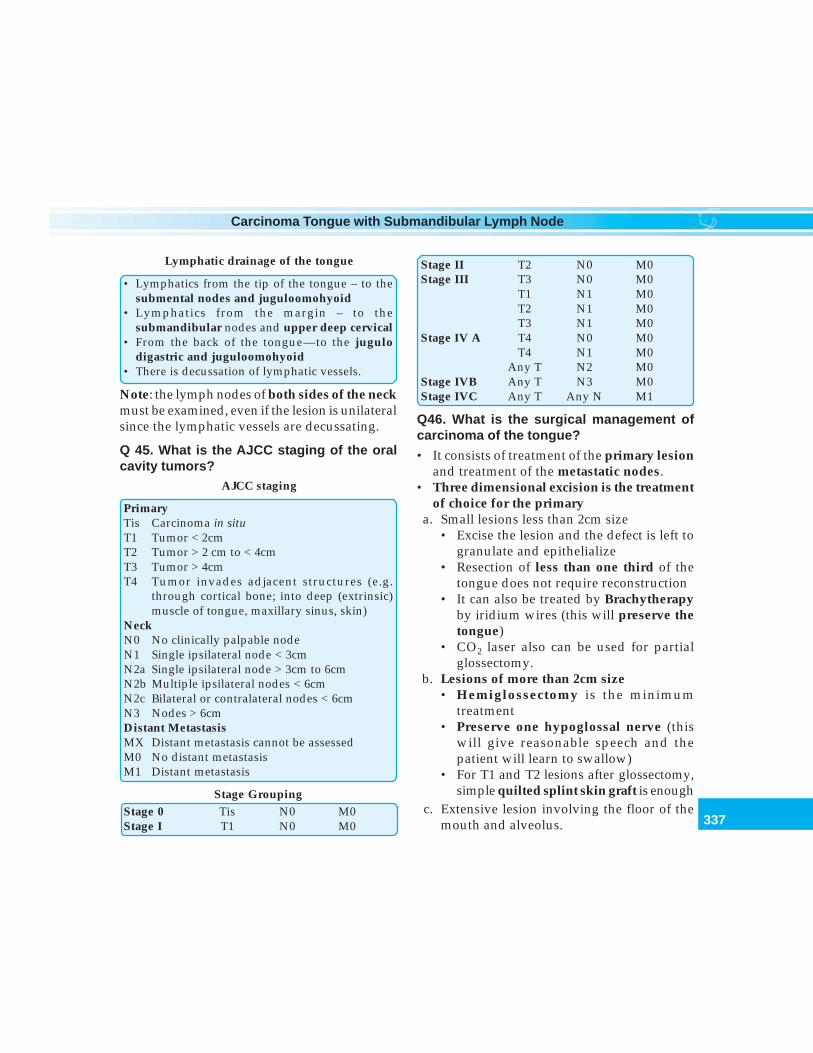
337
Carcinoma Tongue with Submandibular Lymph Node
Lymphatic drainage of the tongue
• Lymphatics from the tip of the tongue – to thesubmental nodes and juguloomohyoid
• Lymphatics from the margin – to thesubmandibular nodes and upper deep cervical
• From the back of the tongue—to the jugulodigastric and juguloomohyoid
• There is decussation of lymphatic vessels.
Note: the lymph nodes of both sides of the neckmust be examined, even if the lesion is unilateralsince the lymphatic vessels are decussating.
Q 45. What is the AJCC staging of the oralcavity tumors?
AJCC staging
PrimaryTis Carcinoma in situT1 Tumor < 2cmT2 Tumor > 2 cm to < 4cmT3 Tumor > 4cmT4 Tumor invades adjacent structures (e.g.
through cortical bone; into deep (extrinsic)muscle of tongue, maxillary sinus, skin)
NeckN0 No clinically palpable nodeN1 Single ipsilateral node < 3cmN2a Single ipsilateral node > 3cm to 6cmN2b Multiple ipsilateral nodes < 6cmN2c Bilateral or contralateral nodes < 6cmN3 Nodes > 6cmDistant MetastasisMX Distant metastasis cannot be assessedM0 No distant metastasisM1 Distant metastasis
Stage GroupingStage 0 Tis N0 M0Stage I T1 N0 M0
Stage II T2 N0 M0Stage III T3 N0 M0
T1 N1 M0T2 N1 M0T3 N1 M0
Stage IV A T4 N0 M0T4 N1 M0
Any T N2 M0Stage IVB Any T N3 M0Stage IVC Any T Any N M1
Q46. What is the surgical management ofcarcinoma of the tongue?
• It consists of treatment of the primary lesionand treatment of the metastatic nodes.
• Three dimensional excision is the treatmentof choice for the primary
a. Small lesions less than 2cm size• Excise the lesion and the defect is left to
granulate and epithelialize• Resection of less than one third of the
tongue does not require reconstruction• It can also be treated by Brachytherapy
by iridium wires (this will preserve thetongue)
• CO2 laser also can be used for partialglossectomy.
b. Lesions of more than 2cm size• Hemiglossectomy is the minimum
treatment• Preserve one hypoglossal nerve (this
will give reasonable speech and thepatient will learn to swallow)
• For T1 and T2 lesions after glossectomy,simple quilted splint skin graft is enough
c. Extensive lesion involving the floor of themouth and alveolus.
338
Clinical Surgery Pearls
• Major 3 dimensional resection by lip splitand mandibulotomy is required
• Marginal mandibular resection may berequired
• Dissection of the neck on the same sideis also carried out
• This is followed by reconstruction with aRadial forearm flap with microvascularanastomosis (radial forearm flap is thework horse of oral reconstruction). Thisflap is useful if the volume defect is lessthan 2/3rd of the original tongue.
• A bulky flap may be required after totalglossectomy for a very large defect
Q 47. What is marginal mandibular resection?
Marginal mandibulectomy involves anincontinuity excision of tumor with a marginof mandible and overlying gingiva. Mandibularcontinuity is maintained and a much bettercosmetic and functional end result is achieved.A segment of bone at least 1cm thick must beleft inferiorly. Marginal mandibular resection isdone if the tumor reaches but does not invadethe alveolus.
This is because of the peculiarity of the modeof involvement of the mandible. It is involvedby infiltration through its dental sockets ordental pores on the edentulous alveolar ridge.These cells procede along the root of the toothinto the cancellous part of the mandible andthen along the mandibular canal.
Q 48. What are the contraindications formarginal mandibulectomy?
• Radiological involvement of the bone
• Previous radiotherapy—cause osteoradionecrosis and fracture
• Retromolar primary lesion• Deeply infiltrating gingivobuccal lesion with
paramandibular infiltration.
Q 49. What is commando operation?
It is an old operation where combined(composite) excision of the primary tumor, blockdissection of the cervical lymph nodes andremoval of the intervening body of the mandibleis done (it was presumed previously that thespread to the mandible is by lymphatics on itsway to the regional nodes. But now we know themethod of spread to the mandible and hence theintroduction of marginal mandibulectomy).
Q 50. What is the management of necknodes?
(Read the block dissection part in short caseNo:2)• A modified radical neck dissection (MRND)
is recommended for N1 and N2 nodes.• A supraomohyoid neck dissection (SOHND)
(clearance of level I, II, III nodes withpreservation of sternocleidomastoid, internaljugular vein and spinal accessory) + post-operative radiotherapy has been advocatedby some authors for N1, Level I disease.
Q 51. Is there any role for elective lymph nodedissection (ELND) in N0 neck (no necknodes)?
Yes.• Occult nodal metastatic disease is present in
5-40% of oral cancers depending on T statusand grade of primary
Published by dr-notes.com
339
Carcinoma Tongue with Submandibular Lymph Node
• Clinical N0 neck should be treated by supraomohyoid neck dissection (SOHND) if therisk of occult nodal metastasis is greater than15-20% in patients with T3/T4 primary
• Patient with T1/T2 tongue tumors andcancers of the floor of the mouth more than2mm thick.
• It is also indicated if it is necessary to enterthe neck for resectng the primary
• In short neck individuals requiring bulky flapfor oral reconstruction (to create space)
• If the patients are unreliable for follow up.• In patients undergoing elective SOHND 24
to 31% will have histological evidence oflymph node metastasis.
Q 52. If the neck nodes are pathologicallypositive after SOHND, what next?• If detected positive on the operating table,
then SOHND should be converted to RND/MRND
• If positive following surgery – subsequentRND or postoperative radiotherapy.
Q 53. How to tackle the skip metastasis tolevel IV which is seen in 15% patients withtongue cancer?Extended SOHND is recommended by somegroup to tackle this problem where the level IVnodes are also removed.
Q 54. What is the management of bilateralnodal metastasis?Bilateral neck dissection with preservation ofinternal jugular vein on one side.
Q 55. What are the indications for radio-therapy for primary?
Indications for radiotherapy
• For early lesions of the tongue• Early lesions of the buccal mucosa• Patient is medically unfit• Patient is unwilling for surgery
Q 56. How is radiotherapy given?
• External beam radiotherapy• Interstitial radiotherapy• Combination of both .
Q 57. What is the dose of radiotherapy?
The total dose is 65-75 Gy to the primary andneck.
Q 58. What are the complications ofradiotherapy?
Complications of radiotherapy
• Xerostomia• Tissue edema• Erythema• Skin sloughing• Ulceration• Dental caries• Osteoradio necrosis
Q 59. What is the cause of death in carcinomatongue?
Causes of death in carcinoma tongue
• Inhalation and aspiration pneumonia• Cachexia and starvation• Hemorrhage from growth• Hemorrhage from carotid artery when eroded by
metastatic lymph nodes• Asphyxia secondary to pressure from lymph
nodes• Edema glottis
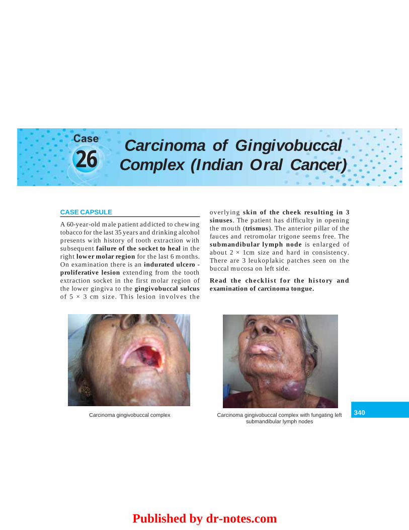
340
Carcinoma of Gingivobuccal Complex (Indian Oral Cancer)
26Carcinoma of Gingivobuccal
Complex (Indian Oral Cancer)
CASE CAPSULE
A 60-year-old male patient addicted to chewingtobacco for the last 35 years and drinking alcoholpresents with history of tooth extraction withsubsequent failure of the socket to heal in theright lower molar region for the last 6 months.On examination there is an indurated ulcero -proliferative lesion extending from the toothextraction socket in the first molar region ofthe lower gingiva to the gingivobuccal sulcusof 5 × 3 cm size. This lesion involves the
overlying skin of the cheek resulting in 3sinuses. The patient has difficulty in openingthe mouth (trismus). The anterior pillar of thefauces and retromolar trigone seems free. Thesubmandibular lymph node is enlarged ofabout 2 × 1cm size and hard in consistency.There are 3 leukoplakic patches seen on thebuccal mucosa on left side.
Read the checklist for the history andexamination of carcinoma tongue.
Carcinoma gingivobuccal complex Carcinoma gingivobuccal complex with fungating leftsubmandibular lymph nodes
Published by dr-notes.com
341
Carcinoma of Gingivobuccal Complex (Indian Oral Cancer)
Q 1. What is the most probable diagnosis inthis case?
Carcinoma of the gingivobuccal complex.
Q 2. What are the clinical points in favor ofcarcinoma?
• History of tooth extraction followed by failureof the socket to heal
• The indurated ulceroproliferative lesion witheverted margins
• Involvement of the overlying skin with sinuses• Presence of hard submandibular lymph node• Trismus• Presence of leukoplakia• History of pan chewing and smoking.
Q 3. What is the definition of oral cavity?
The term oral cavity refers to the following:Oral cavity
• Lips• Buccal mucosa• Alveolar ridges (upper and lower gingiva)• Retromolar trigone• Hard palate• Floor of the mouth• Anterior two–thirds of the tongue (oral or mobile
tongue)
Note: Cancer of the lip behaves clinically likeskin cancer, and therefore not discussed withoral cavity lesion.
Q 4. What is the incidence of oral cancers inIndia?
16 to 28 per 100,000 population [ICMR].
Q 5. What is the commonest oral cancer inIndia?
In India, carcinoma of the buccal mucosa is thecommonest oral cancer constituting about 50 to83% of oral cancers. In the West, tongue andfloor of the mouth are the commonest sites[30%].
Q 6. What are the areas involved by buccalcancers?
• The gingivobuccal sulcus• Retromolar trigone• Lower and upper alveolus• Buccal mucosa.
Q 7. What is Indian oral cancer?
The buccal mucosa and gingiva are more oftenaffected by cancer as a result of placement ofthe tobacco quid in the oral cavity. This cancerof the gingivobuccal complex is described asthe Indian oral cancer.
Q 8. What is the commonest age groupaffected?
5th to 7th decade.
Q 9. What is the extent of buccal mucosa?
The buccal mucosa extends from the upperalveolar ridge down to the lower alveolar ridge,from the commissure anteriorly to themandibular ramus and retromolar regionposteriorly.
Q 10. What is the cause for trismus in thiscase?
Infiltration of the muscles by carcinoma isresponsible for trismus in this case. Thefollowing muscles may be involved incarcinoma of the buccal mucosa:
342
Clinical Surgery Pearls
• Buccinator• Pterygoid• Masseter• Temporalis.
Q 11. What are the causes for trismus?Causes for trismus
• Submucous fibrosis• Invasion of muscles by carcinoma (mentioned
earlier)• Tetanus – Risus sardonicus (painful smiling)• Parotitis• Dental abscess• Erupting wisdom tooth• Peritonsillar abscess
Q 12. What is the grading of trismus? (PG)
Depending on the degree of mouth openingpossible it is graded into 4 groups.
Grading of trismus
• Grade I - more than 35 mm• Grade II - 26 to 35 mm• Grade III - 16 to 25mm• Grade IV - < 15mm
Q 13. What is the cause for sinus in this case?
Orocutaneous fistula secondary to malignantinfiltration.
Q 14. What is retromolar trigone?
• The retromolar trigone is defined as theanterior surface of the ascending ramus ofthe mandible
• It is triangular in shape• The base is superior behind the 3rd upper
molar tooth
• The apex is inferior behind the 3rd lowermolar tooth.
Q 15. What are the special problems ofcarcinoma of the retromolar trigone? (PG)
• Tumors at this site may invade the ascendingramus of the mandible
• It may spread upwards to involve thepterygomandibular space
• A lip split and mandibulotomy are neededto gain access to this region
• Mandibulectomy is required for clearance ofthe pterygoid region
• The resultant defect is managed by masseterand or temporalis muscle flap.
Q 16. What is the staging in this case?
Read the staging of oral cancers given inCarcinoma Tongue Chapter.
It can summarized as:Stage I - T1, N0, M0Stage II - T2, N0, M0Stage III - T3, N1, M0Stage IVa - T4, N1, N2, M0Stage IVb - T4, N3, M0Stage IVc - Any T with M1.
In this case the staging is –T4, N1, M0 → stage IV A.
Q 17. What are the investigations required?
Read the investigation chart for carcinomatongue. (Confirm the diagnosis by biopsy fromthe most suspicious area avoiding the areas ofinfection and necrosis).
Published by dr-notes.com
343
Carcinoma of Gingivobuccal Complex (Indian Oral Cancer)
Q 18. What is the management of stage IVdisease?
• Generally stage I and II (Early) diseases aremanaged by Surgery/Radiotherapy (Eithersurgery or radiotherapy) – No radiotherapyin gingivobuccal complex due to closeproximity of the tumor to bone and risk ofradio necrosis.
• Stage III and IV (Advanced) are managedby Radical Surgery + Reconstruction +Radiotherapy (Surgery and Radiotherapy arecombined).
• Surgery is the treatment of choice for allalveolar carcinomas except for patients unfitfor surgery.
Q 19. What is the surgical treatment if thecarcinoma is confined to the buccal mucosa?
• It is excised widely including the underlyingBuccinator muscle.
• This is followed by split - skin graft.
Q 20. What is the surgical management ofmore extensive lesion?
• Three dimensional excision + reconstruction.
Q 21. What are the flaps available for recons-truction after 3dimensional excision of oralcancer? (PG)
Flaps available for reconstruction
• Free radial forearm flap – is the work - horse oforal reconstruction
• Buccal fat pad – For small intraoral defects ofupto 3 × 5cm.– Used for reconstruction ofmaxillary defects, hard and soft palate defects,cheek and retromolar defects
• Temporalis muscle flap (for larger defects alongwith buccal fat pad)
• Forehead flap - now rarely used because of thepoor cosmetic outcome
Q 22. What is the management if the mandibleis radiologically not involved?
Marginal mandibulectomy (Read carcinomatongue).
Q 23. What are the contraindications formarginal mandibulectomy? (PG)Contraindications for marginal mandibulectomy
• Gross clinical involvement of mandible• Radiological involvement of the mandible• Deeply infiltrating lesions of the gingivobuccal
sulcus with paramandibular infiltration ·• Previous radiotherapy (osteoradio necrosis) ·• Retromolar lesions (clearance of the pterygoid
region is not possible)
Q 24. In the present case there is grossclinical involvement of the mandible andparamandibular infiltration. What is thesurgical management?
Hemimandibulectomy or segmental mandi-bulectomy is required, along with the 3dimensional excision + a Modified RadicalNeck Dissection (MRND).
Q 25. What is the deformity produced by theresection of the anterior arch of mandible?
(PG)Andy Gump deformity.
Q 26. What are the bony substitutes availablefor reconstruction after mandibulectomy?
(PG)
344
Clinical Surgery Pearls
Reconstruction with osteomyocutaneous flapor free microvascular bone graft immediately.• Radial forearm flap with a section of radius• Compound groin flap based on the deep
circumflex iliac vessels• Free fibula flap• Corticocancellous grafts harvested from iliac
crest (packed into mesh trays–Titaniumtrays)
• Rib grafts.
Q 27. What is the soft tissue cover for thereconstructed bone? (PG)• For the microvascular free flaps the
associated skin is used (compound groin flapbased on the deep circumflex iliac vessels)Pectoralis major muscle flap (this is wrappedaround the bone graft and sutured on thelabial aspect).
Q 28. What are the indications for surgery ingeneral for oral cancer?
Indications for surgery in oral carcinoma1. Tumors on alveolar process2. Very large mass when there is invasion of bone3. Nodal involvement – primary and nodes are
treated surgically4. Multiple primary tumors – surgery is preferred5. Verrucous carcinoma
Q 29. Is there any role for radiotherapy as aprimary modality in Early gingivobuccalcomplex?
No. (Because of the proximity to mandible)
Q 30. What is the role for preoperativeradiotherapy in Advanced gingivobuccalcomplex?
Indications for preoperative radiotherapy inadvanced gingivobuccal complex
• Inoperable diseases• Patient is unfit for surgery• Patient is unwilling for surgery• Down staging is possible
Q 31. What are the indications for postoperative radiation therapy to the primary?
Indications for post operative radiation therapyto the primary
• T3/T4 primary• Residual microscopic tumor• Positive surgical margins• Gross residual tumor after resection
Q 32. What are the indications for adjuvantradiation to the neck after radical neckdissection?
Indications for adjuvant radiation to the neckafter radical neck dissection
• More than 2 positive nodes• 2 or more levels of nodes involved• Extracapsular spread
Q 33. What is the indications for elective neckirradiation?
Elective neck irradiation is used for N0 or N1neck if the treatment of primary with radiationtherapy is effective.
Q 34. What is the survival for stage III and IVdisease after treatment? (PG)
• With radiation or surgery alone the survivalfor stage III is 41% and Stage IV is 15%.
Published by dr-notes.com
345
Carcinoma of Gingivobuccal Complex (Indian Oral Cancer)
• When surgery is combined withpostoperative radiation therapy these ratesincrease to 60% and 35%.
Q 35. What is the management of inoperablecases?
Inoperable cases are managed by radiationtherapy with or without chemotherapy.
Q 36. What are the indications for chemo-therapy? (PG)
Indications for chemotherapy
• Palliation for advanced oral cancer• Recurrent oral cancers• Verrucous carcinoma• Adjuvant• Neoadjuvant• Concurrently with radiation: chemoradiation
Q 37. What is the advantage of chemo-radiation?
• Improve the locoregional control• Prevent metastasis.
Q 38. What is the disadvantage of chemo-radiation?
• Significant treatment related morbidity.
Q 39. What is the contraindication forchemotherapy?
Poor performance status (Read the Chart andTable section).
Q 40. What are the chemotherapeutic agentsused?
Cisplatin – based combination chemotherapy ismore effective than single agent chemotherapy(Cisplatin and 5-FU).
The commonly used agents either alone orin combination are:• Methotrexate• 5-fluorouracil (5-FU)• Cisplatin• Bleomycin• Ifosfamide.
Q 41. What are the poor prognostic factors?Poor prognostic factors
• Stage at presentation (single most important)• Lymph node metastasis• Number of lymph nodes involved• Extracapsular spread in the node• Tongue cancer has poor prognosis compared to
other subsites
Q 42. What is the survival figure for early andadvanced stages? (PG)
• Stage I and II (early) – 5 year survival of 31-100%.
• Stage III and IV (advanced stages) – 5 yearsurvival of 7 – 41% depending on the site ofthe disease.
346
Clinical Surgery Pearls
27 Parotid Swelling
CASE CAPSULE
A 45-year-old male patient presents withpainless enlargement of the right parotidgland. On examination, there is a swelling ofabout 4 × 3cm size irregular in shape andoccupying the hollow between the mandibleand mastoid. It is firm in consistency, deep tothe parotid fascia, and superficial to the massetermuscle. The swelling raises the right ear lobule.The facial nerve is intact. The superficialtemporal artery is palpable above the swelling.There are no palpable ipsilateral nodes. Thepatient is apparently healthy.
(Read the diagnostic algorithm for a neckswelling)
Checklist for history
1. History of systemic diseases responsible forsialadenosis like – DM, drugs (antiasthmatic,Guanethidine) endocrine disorders, alcoholism,pregnancy, bulimia (eating disorders)
2. History of exposure to mumps3. History of collagen diseases4. History of salivary colic
5. History of increase in size during salivation6. History of similar swelling on the contralateral side7. History of recent illness and major surgery (acute
parotitis)8. History of exposure to HIV (HIV associated
sialadenitis)
Checklist for examination
1. Look for obliteration of the hollow below the earlobule
2. Look for fixity to masseter3. Bimanual palpation of the deep lobe with one
finger inside at the tonsillar region and otherhand externally
4. Bidigital palpation of the Stensen’s duct (thumbexternally and index finger internally)
5. Look for lymph nodes—Preauricular, parotidand submandibular nodes
6. Look for movements of the jaw7. Look for facial nerve palsy8. Examine the oral cavity – orifice of Stensen’s duct,
Tonsil (whether pushed medially or not)9. Always examine other ipsilateral salivary glands
and other contralateral salivary glands
Published by dr-notes.com
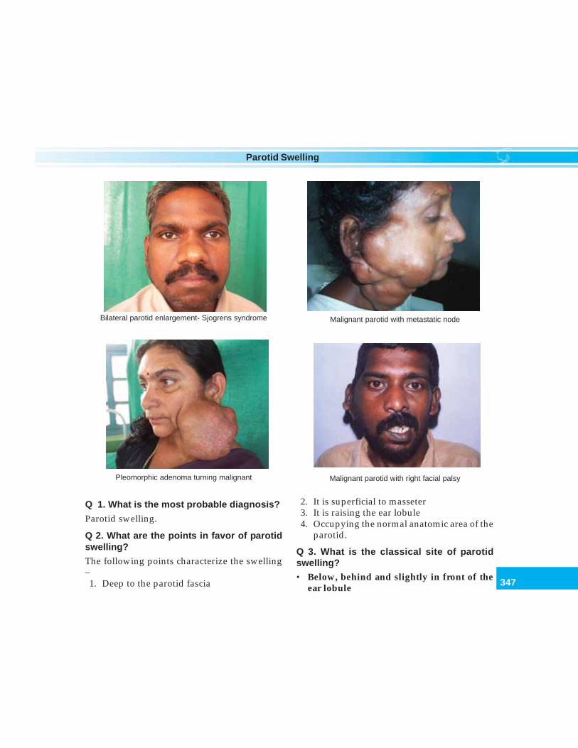
347
Parotid Swelling
Q 1. What is the most probable diagnosis?
Parotid swelling.
Q 2. What are the points in favor of parotidswelling?
The following points characterize the swelling–1. Deep to the parotid fascia
2. It is superficial to masseter3. It is raising the ear lobule4. Occupying the normal anatomic area of the
parotid.
Q 3. What is the classical site of parotidswelling?• Below, behind and slightly in front of the
ear lobule
Bilateral parotid enlargement- Sjogrens syndrome Malignant parotid with metastatic node
Pleomorphic adenoma turning malignant Malignant parotid with right facial palsy
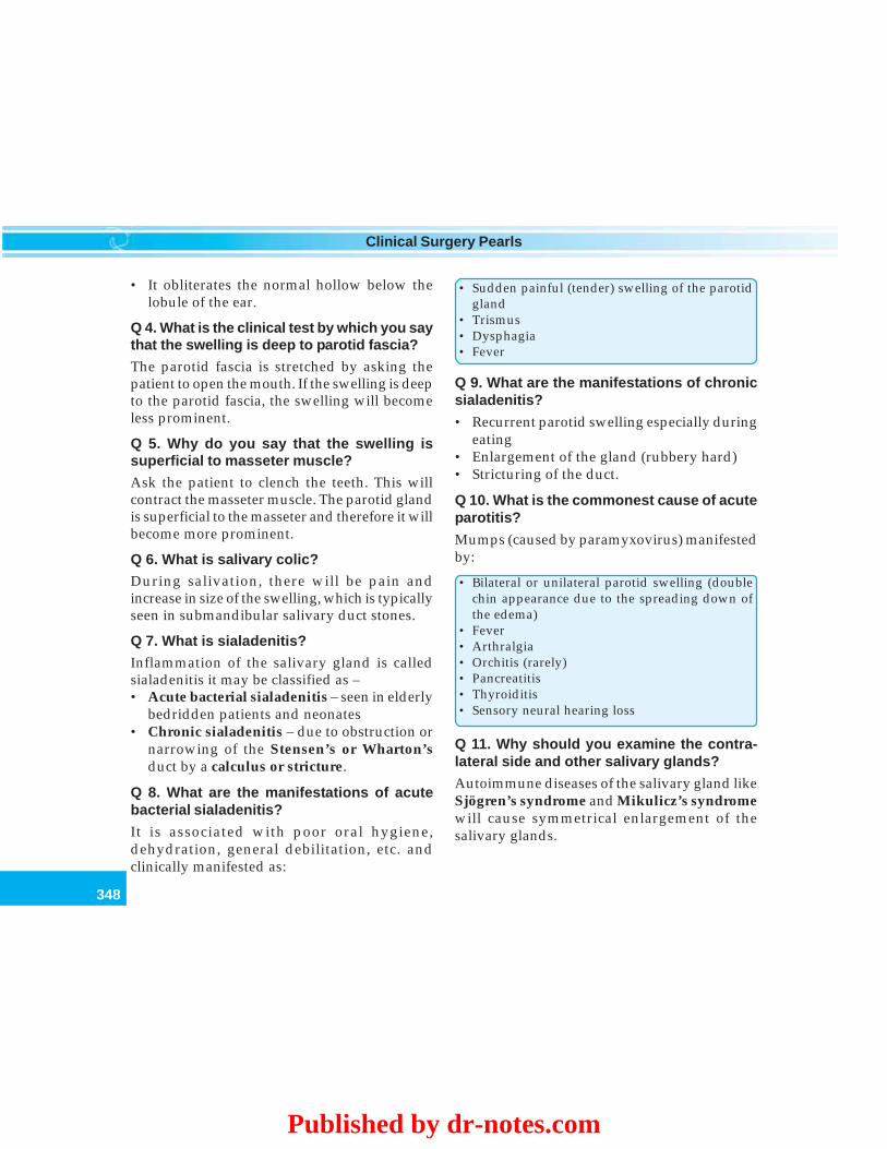
348
Clinical Surgery Pearls
• It obliterates the normal hollow below thelobule of the ear.
Q 4. What is the clinical test by which you saythat the swelling is deep to parotid fascia?
The parotid fascia is stretched by asking thepatient to open the mouth. If the swelling is deepto the parotid fascia, the swelling will becomeless prominent.
Q 5. Why do you say that the swelling issuperficial to masseter muscle?
Ask the patient to clench the teeth. This willcontract the masseter muscle. The parotid glandis superficial to the masseter and therefore it willbecome more prominent.
Q 6. What is salivary colic?During salivation, there will be pain andincrease in size of the swelling, which is typicallyseen in submandibular salivary duct stones.
Q 7. What is sialadenitis?
Inflammation of the salivary gland is calledsialadenitis it may be classified as –• Acute bacterial sialadenitis – seen in elderly
bedridden patients and neonates• Chronic sialadenitis – due to obstruction or
narrowing of the Stensen’s or Wharton’sduct by a calculus or stricture.
Q 8. What are the manifestations of acutebacterial sialadenitis?
It is associated with poor oral hygiene,dehydration, general debilitation, etc. andclinically manifested as:
• Sudden painful (tender) swelling of the parotidgland
• Trismus• Dysphagia• Fever
Q 9. What are the manifestations of chronicsialadenitis?
• Recurrent parotid swelling especially duringeating
• Enlargement of the gland (rubbery hard)• Stricturing of the duct.
Q 10. What is the commonest cause of acuteparotitis?
Mumps (caused by paramyxovirus) manifestedby:
• Bilateral or unilateral parotid swelling (doublechin appearance due to the spreading down ofthe edema)
• Fever• Arthralgia• Orchitis (rarely)• Pancreatitis• Thyroiditis• Sensory neural hearing loss
Q 11. Why should you examine the contra-lateral side and other salivary glands?
Autoimmune diseases of the salivary gland likeSjögren’s syndrome and Mikulicz’s syndromewill cause symmetrical enlargement of thesalivary glands.
Published by dr-notes.com
349
Parotid Swelling
Q 12. What is Mikulicz’s syndrome?
It is a combination of bilateral salivary andlacrimal gland enlargement• Symmetrical enlargement of salivary glands
(one gland alone is involved initially for aquite long time)
• Enlargement of lacrimal glands (bulge belowthe outer end of the eyelids and narrowingof the palpebral fissure)
• Dry mouth.
Q 13. What are the causes for Mikulicz’ssyndrome?
• Sarcoidosis• Leukemia• Lymphoma• Sjögren’s syndrome
Q 14. What is Sjögren’s syndrome?
It is a rare autoimmune condition affecting thesalivary glands and it can occur in combinationwith other autoimmune connective tissuedisorders.
The manifestations and associated conditionsseen in Sjögren’s syndrome.
Manifestation Condition
Deficient tear film Keratoconjunctivitissicca
Deficient salivation and Xerostomiagland enlargementDeficient tear and saliva Primary glandular
sicca syndrome
Deficient tear and saliva Primary extra-along with hyperglobu- glandular siccalinemic purpura, vascu- syndromelitis, or Raynaud’s pheno-menon or B cell lymphoma
Any of the above occur- Secondary Sjögren’sring together with rheu- syndromematoid arthritis, systemiclupus erythematosus orother recognizableconnective tissue disorders
Q 15. What is the importance of oral cavityexamination in parotid swelling?
• To look for the orifice of the Stensen’s ductswhich is situated opposite the crown of thesecond upper molar tooth—look for bloodand pus.
• To palpate the mouth of the duct for anylumps and induration.
• Gentle pressure on the gland externally maybring out purulent discharge.
• The tonsil may be pushed medially whenthe deep lobe of the parotid gland is enlarged.
• Bimanual palpation of the parotid gland— Onefinger externally behind the ramus of themandible and one finger inside the mouth justin front of the tonsil and behind 3rd molar toothinternally.
Q 16. What is the anatomical position of theparotid duct?
It is deep to the anterior border of the gland andruns superficial to the masseter muscle. It thencurves inwards by piercing the buccinator toopen on the mucous membrane of the mouthContd...
Contd...
350
Clinical Surgery Pearls
opposite the crown of the upper second molartooth.
Q 17. How would you palpate the Stensen’sduct?
It is best done by a bidigital palpation by indexfinger inside the mouth and thumb over thecheek.
Q 18. What is the surface marking forStensen’s duct?
It lies about one fingerbreadth below theinferior border of the zygomatic bone.
Q 19. What is the most important differentialdiagnosis for a small parotid swelling?
Preauricular lymph nodes (enlargementsecondary to infection or metastasis).
Q 20. What are the primary foci for enlargedpreauricular lymph nodes?
Primary sites for preauricular node metastasis—Drainage area for pre auricular node
• Forehead• Scalp• Eyelids• Cheek• External auditory meatus
Q 21. What is the distinguishing clinicalfeature of the lymph node?
It is the mobility of the lymph node—the preauricular lymph node is outside the capsule ofthe gland and usually very mobile, unlike thetumor in the parotid which has got restrictedmobility.
Q 22. What is parotid sandwich?
The facial nerve is passing through thesubstance of the parotid gland, dividing thegland into a superficial lobe and deep lobe.Therefore the gland is called parotid sandwich.
Q 23. What is faciovenous plane of Patey?The facial nerve is seen, superficial to theposterior facial vein in the substance of thegland. This plane is called faciovenous plane ofPatey.
Q 24. What is Pes anserinus?Pes anserinus means goose foot.In the parotid gland the facial nerve divides into–1. The temporofacial (runs sharply upwards)
– two divisions (temporal, zygomatic)2. The cervicofacial – continues the course of
the parent trunk downwards, forwards andoutwards – three divisions (buccal,mandibular and cervical).
These divisions in turn divide to form thegoose’s foot (Pes anserinus).
Q 25. What is socia parotidis?It is nothing but accessory lobe of the parotidseen just above the Stensen’s duct.
Q 26. Which type of facial palsy is seen inparotid tumors?
Lower motor neuron type of facial palsy is seen(involvement of both lower and upper half ofthe face).
Published by dr-notes.com
351
Parotid Swelling
Q 27. What are the tests for facial palsy?
The tests for facial palsy are:
Test Manifestations
1. Ask the patient to show his teeth Angle of the mouth drawn to the healthy side2. Ask the patient to puff out the cheeks The paralyzed side bellows out more than
the normal side3. Ask the patient to shut his eyes Will not be able to close the eyes on the
affected side and on attempting to do so to theeyeball will be seen to roll upwards
4. Ask the patient to move his eyebrows upwards The paralyzed side remains immobile
Note: The nasolabial fold and furrows of the eyebrow are less marked on the affected side. The angle of themouth is drawn to the sound side.
Q 28. Will all the malignant tumors producefacial palsy?
No.
Q 29. What are the clinical features ofmalignancy in the parotid?
Clinical features of malignancy
a. Painb. Rapid increase in sizec. Very hard consistencyd. Facial palsye. Enlarged metastatic regional nodef. Skin involvement (skin tethering)g. Fixityh. Trismus – involvement of pterygoid muscle by
deep parotid lobe malignancy
Q 30. What is the commonest parotidswelling?Pleomorphic adenoma (mixed parotid tumor).
Q 31. What percentage of tumors are benignin parotid?
Site % of % ofbenign malignant
Parotid 80% 20%
Submandibular 50% 50%
Minor salivary gland 10% 90%
Sublingual 5% 95%
Q 32. What are the other benign tumors ofthe parotid gland?
• Warthin’s tumors (papillary cyst adenomalymphomatosum)
• Oxyphylic adenoma (Oncocytoma)
Q 33. What are the features of Warthin’stumor?
352
Clinical Surgery Pearls
Clinical features of Warthin’s tumor
• 2nd most common benign tumor• It is soft and sometimes fluctuant (cystic)• Seen usually in males• Seen after 40 years• May be bilateral (10% bilateral)• This tumor has no malignant potential
Q 34. What is the origin of Warthin’s tumor?
The tumor probably arises from parotid tissueincluded in the lymph nodes which are usuallypresent within the parotid sheath. Micro-scopically it is lined by columnar epithelial cellssupported by lymphoid stroma.
Q 35. Is there any method of confirmingWarthin’s other than FNAC? (PG)Yes. Tc 99m scintigraphy will reveal a hot spot.This is due to the high mitochondrial contentwithin the cell.
Q 36. What is oncocytoma? (PG)They arise from oncocytes which are derivedfrom intralobular ducts or acini. They areusually seen in minor salivary glands,nasopharynx and larynx in the elderly males.
Q 37. What is the investigation of choice inparotid tumors?FNAC.
Q 38. Why biopsy is contraindicated inparotid tumors?Biopsy is contraindicated because of thefollowing reasons:1. Seedling of the tumor will occur2. Chance for parotid fistula is there3. Chance for facial nerve injury.
Q 39 What are the other investigations?
1. CT/MRI is taken to rule out deep lobeinvolvement
2. Chest X-ray to rule out metastasis.
Indications for CT1. If deep lobe tumor is suspected2. If extension to deep lobe is suspected3. Trismus.
Indication for MRIWhen facial nerve is involved.
Q 40. What is the WHO classification ofparotid neoplasms?
1. Adenomas – PleomorphicMonomorphic – Warthin’s tumor
2. Carcinoma—Low gradei. Acinic cell carcinoma
ii. Adenoid cystic carcinomaiii. Low grade mucoepidermoid carcinoma
High gradei. Adenocarcinoma
ii. Squamous cell carcinomaiii. High grade mucoepidermoid carcinoma
3. Nonepithelial tumors—• Hemangioma• Lymphangioma• Neurofibroma.
4. Lymphomas—• Primary - NHL• Lymphoma in Sjögren’s syndrome.
5. Secondary—• Local – tumors of head and neck• Distant – skin and bronchus.
6. Unclassified tumors
Published by dr-notes.com
353
Parotid Swelling
7. Tumor-like lesions—– Adenomatoid hyperplasia– Salivary gland cysts.
Q 41. Why pleomorphic adenoma is calledmixed parotid tumor?
It is called mixed parotid tumor because it hasgot both epithelial and mesodermal elements.
Q 42. Can pleomorphic adenoma occurbilaterally?
Yes.
Q 43. What are the peculiarities of pleo-morphic adenoma?
• It is considered a benign tumor with longquiescent periods and short periods of rapidgrowth
• Potential for recurrence• Potential for malignant change.
Features of pleomorphic adenoma
1. The capsule is incomplete and the tumor willhave extensions beyond the capsule
2. Recurrence can occur if tumor excision is notcomplete
3. 10% of the tumors are highly cellular and moreliable to recur
4. Tumor contains both epithelial andmesodermal elements (myoepithelial cellssurrounding the tubules)
5. After surgery for recurrence, radiotherapy isindicated even though it is benign.
6. Benign pleomorphic adenomas metastasizeinexplicably
7. It is a tumor readily implanted during removalin the residual parotid
Q 44. What are the malignant parotid tumorsin order of frequency?• Mucoepidermoid carcinoma (most common)• Malignant mixed tumor• Acinic cell carcinoma• Adenocarcinoma• Adenoid cystic carcinoma – 10% (2/3 rd in minor
salivary gland)• Epidermoid carcinoma (squamous cell
carcinoma)
Q 45. Which is the carcinoma parotid withworst prognosis? (PG)
• Carcinoma arising in pleomorphic adenoma• There is accelerated recurrence rate and high
incidence of metastasis• Five year survival is less than 40%.
Q 46. What are the types of malignancies inpleomorphic adenoma? (PG)
1. Carcinoma originating from pleomorphicadenoma (carcinoma ex – pleomorphicadenoma) 15years after the originalswelling - 9.5% chance for carcinoma
2. Benign pleomorphic adenoma inexplicablymetastasize (de novo malignant mixedtumors).
Q 47. What are the peculiarities of adenoidcystic carcinoma?
• Propensity for perineural invasion• Regional lymph node involvement
uncommon• Distant metastasis occur within 5 years
(however they remain asymptomatic foryears).
354
Clinical Surgery Pearls
Q 48. What is the difference between lowgrade and high grade mucoepidermoidcarcinoma?
High grade lesions have propensity for bothregional and distant metastasis.
Q 49. What is the staging of parotid tumors?(PG)
TNM staging as per AJCC 6th edition isrecommended.TX - Primary tumor cannot be assessedT0 - No evidence of primary tumorT1 - Tumor 2cm or less in greatest dimension
without extraparenchymal extensionT2 - Tumor more than 2cm but not more than
4cm in greatest dimension without extra-parenchymal extension
T3 - Tumor more than 4cm and/or tumorhaving extraparenchymal extension
T4a - Tumor invades skin, mandible, ear canal,and/or facial nerve
T4b - Tumor invades skull base and/or pterygoidplates and/or encases carotid artery
Note: N stage is same for all head and neckmalignancies.
Staging
Stage 1 - T1 N0 M0Stage 2 - T2 N0 M0Stage 3 - T3 N1/N0 M0Stage 4A - T4a N2/N1N0 M0Stage 4B - T4b any N M0
Any T N3 M0Stage 4C - Any T Any N M1
Q 50. What is the difference between stagingfor major salivary gland tumors and minorsalivary gland tumors? (PG)
The minor salivary gland tumors are located inthe lining of upper aerodigestive tract and theyare staged according to the anatomic site oforigin (e.g.: oral cavity, sinuses, etc.)
Q 51. What are the major salivary glands?
They include parotid, submandibular and sub-lingual glands.
Q 52. What is the regional node spread inparotid tumor?
Intraglandular node → Periparotid node →Submandibular node → Upper and Midjugularnodes (occasionally to retropharyngealnodes).
Q 53. What is the treatment of pleomorphicadenoma?
• Superficial parotidectomy is the minimumsurgical procedure
• There is no role for enucleation and excision(because of the reasons mentioned above)
• Facial nerve should be spared if a plane existsand when it is not involved
• Facial nerve is scarified only if it is involvedor it is totally encased as in cases ofcarcinomas.
Q 54. If the facial nerve is involved what isthe treatment option?
The nerve is excised and a nerve graft is donewith Great auricular nerve.
Q 55. Is nerve grafting a contraindication forradiotherapy?
No.
Published by dr-notes.com
355
Parotid Swelling
Q 56. What is the timing of radiotherapy?(PG)
3 – 6 weeks after surgery.
Q 57. What are the indications for radio-therapy? (PG)
Indications for radiotherapy
(50-70 Gy given in 1.8-2.0 Gy in 5-8 weeks time)
1. T3 and T4 tumors2. High grade tumors3. Deep lobe involvement4. Perineural spread5. Vascular invasion6. Multiple lymph node involvement7. Close margins
Q 58. Is there any indication for chemo-therapy? (PG)
No.
Q 59. What is the surgical treatment ofnodes?
Comprehensive neck dissection in the form ofRadical neck dissection is done.
Q 60. What are the important anatomicalpoints to be remembered in parotid surgery?
1. The gland is situated in the space behindthe ramus of the mandible, below the baseof the skull and in front of mastoid process.
2. Deeply it is applied to the styloid processand its muscles.
3. The upper pole lies just below the zygomaticarch and wedged between the meatus andmandibular joint.
4. Upper pole—The superficial temporalvessels, the temporal branches of the facialnerve and the auriculotemporal nerve arefound entering or leaving the gland near theupper pole.
5. Lower pole—The cervical branch of thefacial nerve and the two divisions of theposterior facial vein emerge from its lowerpole.
6. Anterior border—overlies the masseter. Theparotid ducts, the zygomatic, buccal andmandibular branches of facial nerve emergefrom the anterior border.
7. The external carotid artery, the facial nerveand the retromandibular vein pass throughthe substance of the gland (the externalcarotid artery terminates behind the neckof the mandible by dividing into maxillaryand superficial temporal arteries).Intraparotid lymph nodes are also seen inthe substance of gland.
8. The facial nerve is seen in the faciovenousplane of Patey (the nerve is seen superficialto the posterior facial vein which is formedwithin the substance of gland by thecontinuation of the superficial temporalvein and emerges usually into two branchesat the lower pole of the gland).
9. The nerve is dividing the gland into asuperficial lobe and deep lobe. 80% of thegland lies superficial to the nerve and 20%deep to the nerve.
10. An accessory lobe is present in less than50% of the population.
356
Clinical Surgery Pearls
Q 61. What is the incision used for superficialparotidectomy?
A lazy ‘S’ incision is used – Preauricular –Mastoid - Cervical incision.
Q 62. What are the essential steps ofsuperficial parotidectomy?
1. Surgery is done under general anesthesiawith endotracheal intubation.
2. Lazy ‘s’ incision is used as mentioned above.3. Infiltration with local anaesthetic and
adrenalin for better delineation of the plane.4. Reflect the skin flaps anteriorly just
superficial to the parotid fascia upto theanterior border of the gland.
5. Back of the parotid gland is identified, anddissection is carried out to expose the facialnerve.
6. The sternomastoid is retracted and greatauricular nerve divided in the avascularplane along the anterior border of themuscle.
7. Identify the posterior belly of digastric.8. Identify the avascular plane along the
anterior border of cartilaginous and bonyexternal auditory meatus immediatelyanterior to the tragus.
9. Landmarks for identification of facialnerve—(always identify the trunk of thenerve first rather than tracing the branchesfrom the periphery).a. Conley’s pointer – The inferior portion
of the cartilaginous canal. The facialnerve lies 1cm deep and inferior to its tip.
b. The upper border of the posterior belly
of digastric muscle – The facial nerve isusually located immediately superiorto it.
c. The stylomastoid artery lies immediatelylateral to the nerve.
10. Identify the two main divisions.11. Dissect the gland off branches of the facial
nerve.12. With the exception of buccal branch, all
transected nerves are repaired with cablegraft from great auricular nerve.
13. The desired amount of gland is removed.14. A suction drain is applied and wound is closed.
Q 63. What is radical parotidectomy?
Radical parotidectomy involves removal of allparotid gland tissue and elective sectioning ofthe facial nerve usually through the main trunk.The surgery removes ipsilateral masseter musclein addition. If there is clinical, radiological, andcytological evidence of lymph node metastasisa simultaneous radical neck dissection is carriedout. It is done for –• High grade malignant tumors• Squamous cell carcinoma.
Q 64. What are the complications of parotidsurgery?
Complications are:
Complications of parotidectomy
1. Seroma2. Wound infection3. Permanent facial palsy (transection of the nerve)4. Temporary facial nerve weakness5. Facial numbness
Published by dr-notes.com
357
Parotid Swelling
6. Permanent numbness of the ear lobe (due togreat auricular nerve transection)
7. Sialocele8. Frey’s syndrome (Gustatory sweating)9. Parotid fistula
Q 65. What is Frey’s syndrome?
This is due to inappropriate regeneration of thedamaged parasympathetic autonomic nervefibers to the overlying skin. Salivation resultingfrom smell or taste of food, will stimulate thesweat glands of the over lying skin instead ofthe parotid. The clinical features are:1. Sweating over the region of parotid gland2. Erythema over the region of parotid gland.
Q 66. What is the clinical test to demonstrateFrey’s syndrome? (PG)
Starch iodine test - Paint the affected area withiodine and allow it to dry. Apply dry starch over
it. The starch turns blue on exposure to iodinein the presence of sweat. The sweating isstimulated after painting starch.
Q 67. What is the management of Frey’ssyndrome? (PG)
PreventionIt can be prevented by placing a barrier betweenthe skin and parotid bed to preventinappropriate regeneration of autonomic nervefibers. The following methods are useful –1. Temporalis fascial flap2. Sternomastoid muscle flap3. Artificial membrane between the skin and
parotid bed.
Management of established syndrome:1. Tympanic neurectomy2. Injection of botulinum toxin into the affected
skin. (simple and effective method)3. Antiperspirants—aluminum chloride.
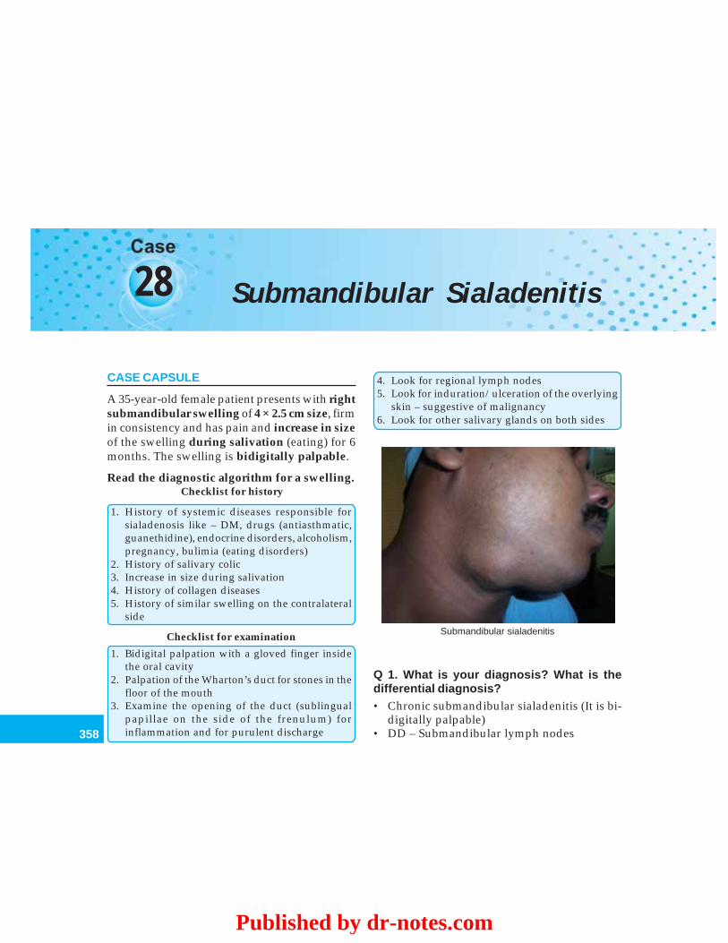
358
Clinical Surgery Pearls
28 Submandibular Sialadenitis
CASE CAPSULE
A 35-year-old female patient presents with rightsubmandibular swelling of 4 × 2.5 cm size, firmin consistency and has pain and increase in sizeof the swelling during salivation (eating) for 6months. The swelling is bidigitally palpable.
Read the diagnostic algorithm for a swelling.Checklist for history
1. History of systemic diseases responsible forsialadenosis like – DM, drugs (antiasthmatic,guanethidine), endocrine disorders, alcoholism,pregnancy, bulimia (eating disorders)
2. History of salivary colic3. Increase in size during salivation4. History of collagen diseases5. History of similar swelling on the contralateral
side
Checklist for examination
1. Bidigital palpation with a gloved finger insidethe oral cavity
2. Palpation of the Wharton’s duct for stones in thefloor of the mouth
3. Examine the opening of the duct (sublingualpapillae on the side of the frenulum) forinflammation and for purulent discharge
4. Look for regional lymph nodes5. Look for induration/ulceration of the overlying
skin – suggestive of malignancy6. Look for other salivary glands on both sides
Submandibular sialadenitis
Q 1. What is your diagnosis? What is thedifferential diagnosis?• Chronic submandibular sialadenitis (It is bi-
digitally palpable)• DD – Submandibular lymph nodes
Published by dr-notes.com
359
Submandibular Sialadenitis
Q 2. How to differentiate them?• If the swelling is bidigitally palpable, it is sub-
mandibular salivary gland• Lymph nodes are not bidigitally palpable.
Q 3. Why salivary gland is palpable bi-digitally?The submandibular salivary gland has a portionabove the myelohyoid muscle (deep lobe) in thefloor of mouth. Therefore, the gland is bidigitallypalpable.
Q 4. What are your points in favor of submandibular salivary gland?1. Salivary colic – pain induced by salivation
as a result of obstruction to the outflow fromthe gland (may be a stone in the duct).
2. Increase in size during salivation (abovereason)
3. Decrease in size of the swelling ordisappearance 1 to 2 hrs after the meal iscompleted
4. Positive bi digital palpation5. Solid nature of the swelling
Q 5. If the swelling is cystic what are thepossibilities?
If it is cystic, one has to rule out a sublingualdermoid.
Q 6. What is the importance of palpating theWharton’s duct and its opening (sublingualpapillae)?
• Palpation in the gingivolingual sulcus willreveal stones in the Wharton’s duct.
• Inspection of the sublingual papillae revealsinflammation and discharge (purulent).
Q 7. Why stones are more common in thesubmandibular salivary gland compared tothe parotid?
• The gland and duct system has a shapesimilar to the retort.
• The duct is above and the gland is below. Thegland has antigravity drainage.
• The secretion is thicker in the submandibularsalivary gland.
Q 8. What are the types of inflammation inthe submandibular gland?
The inflammation of the gland is calledsialadenitis. The types of sialadenitis are:• Acute• Chronic• Acute on chronic.
Q 9. What are the causes for acute sub-mandibular sialadenitis?
• Viral – mumps• Bacterial – secondary to obstruction.
Q 10. What are the causes for chronicsialadenitis?
• Obstruction by stone formation – which maybe within the gland (Sialolithiasis)– within the duct system
• Trauma to the floor of mouth by denture –subsequent inflammation and stricture of theduct.
Q 11. How many percentage of the sub-mandibular are radio opaque?
• 80% are radio opaque• It can be identified in plain radiograph.
360
Clinical Surgery Pearls
Q 12. Which is the commonest site of the sub-mandibular stone—Gland or duct?
80% of the stones are situated in the submandibular gland.
Q 13. Which type of sialadenitis is morecommon—Bacterial or viral?
Bacterial.
Q 14. How will you manage this case?
• Investigations for diagnosis• Investigations for surgery.
Investigations for diagnosis1. Plain X-ray—Occlusive view using dental
film. This will demonstrate radioopaquecalculus in the duct and salivary gland.
2. USG—Ultrasound is a very useful tool forthe demonstration of stones
3. FNAC—To rule out a tumor of the salivarygland and to rule out lymph node.
Investigations for surgery1. X-ray chest2. ECG3. Hemogram4. Blood sugar5. Renal status.
Q 15. If no stone is demonstrated and it isfound to be sialadenitis, what is the manage-ment?
Bacterial sialadenitis has a poor capacity forrecovery following infection and the glandbecomes chronically inflamed. Therefore thegland has to be removed—Sialadenectomy.
Q 16. If stone is demonstrated radiologically,what is it called?
Sialolithiasis.
Q 17. If the stone is clinically andradiologically demonstrated in the Wharton’sduct, what is the management?
If the stone is lying anterior to a point at whichthe duct crosses the lingual nerve (secondmolar region), the stone can be removed byincising longitudinally over the duct. This isdone under the local anesthesia.
Q 18. Will you close the duct after removal ofthe stone?
• No• The duct should be left open for free drainage
of saliva.• Suturing will lead to stricture formation and
recurrence of obstructive symptoms.
Q19. When the stone is proximal to thecrossing of lingual nerve, what is themanagement? (PG)
• Intraoral approach is avoided because it leadsto injury to the lingual nerve
• The gland is removed by external approach– Sialadenectomy along with removal of thestone from the duct and ligation of the duct.
Q 20. What are the indications for excisionof submandibular salivary gland? (PG)
1. Sialadenitis2. Stones in the gland3. Stones in the duct proximal to the lingual nerve4. Salivary tumors.
Published by dr-notes.com
361
Submandibular Sialadenitis
Q 21. What is the difference betweensialadenectomy for inflammatory conditionand tumor of the submandibular salivarygland? (PG)
• Intracapsular dissection is done forinflammatory condition
• Extracapsular dissection with a cuff of normaltissue around is done for tumor. This may becombined with suprahyoid neck dissection.
Q 22. What is the incision for sialadenectomyand what precautions are taken?
It is important to avoid injury to the marginalmandibular branch of the facial nerve andtherefore the incision is placed 3 to 4cm belowthe lower border of the mandible. A 6cm longincision is cited within the skin crease.
Q 23. In which plane you get marginalmandibular branch of the facial nerve? (PG)
Subplatysmal plane. The skin flaps are raisedat subplatysmal level.
Q 24. Is it necessary to close the platysma atthis situation? (PG)
• Yes. It is sutured with continuous absorbablesuture.
• Platysma muscle has direct contribution to thedepressor activity of the corner of the mouth.
Q 25. What are the important anatomicalrelationships of the submandibular gland?
• 3 cranial nerves are at risk:
1. Marginal mandibular branch of the facialnerve
2. The lingual nerve3. The hypoglossal nerve
• 2 Vessels need ligation1. Anterior facial vein running over the
surface of the gland2. Facial artery.
• 2 Muscles are related to the gland1. Mylohyoid muscle—around its posterior
border the large superficial lobebecomes the small deeper lobe
2. The deep part of the gland lies on thehyoglossus muscle closely related to thelingual nerve and hypoglossal nerve.
Q 26. What is the anesthesia of choice forsialadenectomy?
General anesthesia—with endotrachealintubation.
Q 27. What are the peculiarities of the facialartery in this situation? (PG)
• The course of the artery is variable here• The artery lies in the groove on the deeper
aspect of the gland• Some times the artery will penetrate the
substance of the gland• Passes around the gland sometimes• The artery has to be ligated doubly,
superiorly and inferiorly.
Q 28. What is the landmark for identificationof the deep lobe?
• Posterior border of the myelohyoid• Once the muscle is retracted forwards the
deep lobe can be identified.
Q 29. Is there any attachment of the gland tothe Lingual Nerve? (PG)
The gland is attached to the lingual nervethrough parasympathetic secretomotor fibers.

362
Clinical Surgery Pearls
These parasympathetic nerve fibers are divided,protecting the lingual nerve.
Q 30. How do you tackle the submandibularduct? (PG)
It is identified and ligated as anteriorly aspossible.
Q 31. Is there a need for a wound drain aftersurgery?
It is better to put a continuous suction drain for24hrs. There are numerous veins encounteredon the deeper aspect of gland, which arecoagulated or ligated.
Q 32. What are the complications of sialade-nectomy?
Complications of submandibular sialade-nectomy
1. Injury to the marginal mandibular nerve2. Hematoma3. Wound infection4. Injury to the nerve to mylohyoid producing
submental anesthesia5. Lingual nerve injury6. Hypoglossal nerve injury
Q 33. What are the clinical features ofmalignancy in the gland?
Signs of malignancy in submandibularsalivary gland
• Rapid increase in size• Induration• Ulceration of the overlying skin• Cervical node enlargement
Q 34. What is the incidence of malignancy insubmandibular salivary gland?50% are malignant in contrast to the parotidwhere only 20% are malignant. The chances ofmalignancy increase from parotid to sub-mandibular to sublingual and minor salivaryglands.
Q 35. What are the investigations fordiagnosis in suspected tumors of the sub-mandibular salivary gland?
• FNAC – It is the investigation of choice.• CT/MRI – If required to know the nature of
surrounding invasion.
Q 36. Is there any contraindication for openbiopsy?
Open biopsy is contraindicated because of thetumor seedling.
Q 37. What is “Stafne Bone Cyst”? (PG)
It is an ectopic lobe of the submandibularsalivary gland presenting as asymptomaticradiolucency of the angle of the mandible,below the inferior dental neurovascular bundle.Edward C.Stafne a dental surgeon from Mayoclinic described this condition. No treatment isrequired for this condition.
Published by dr-notes.com
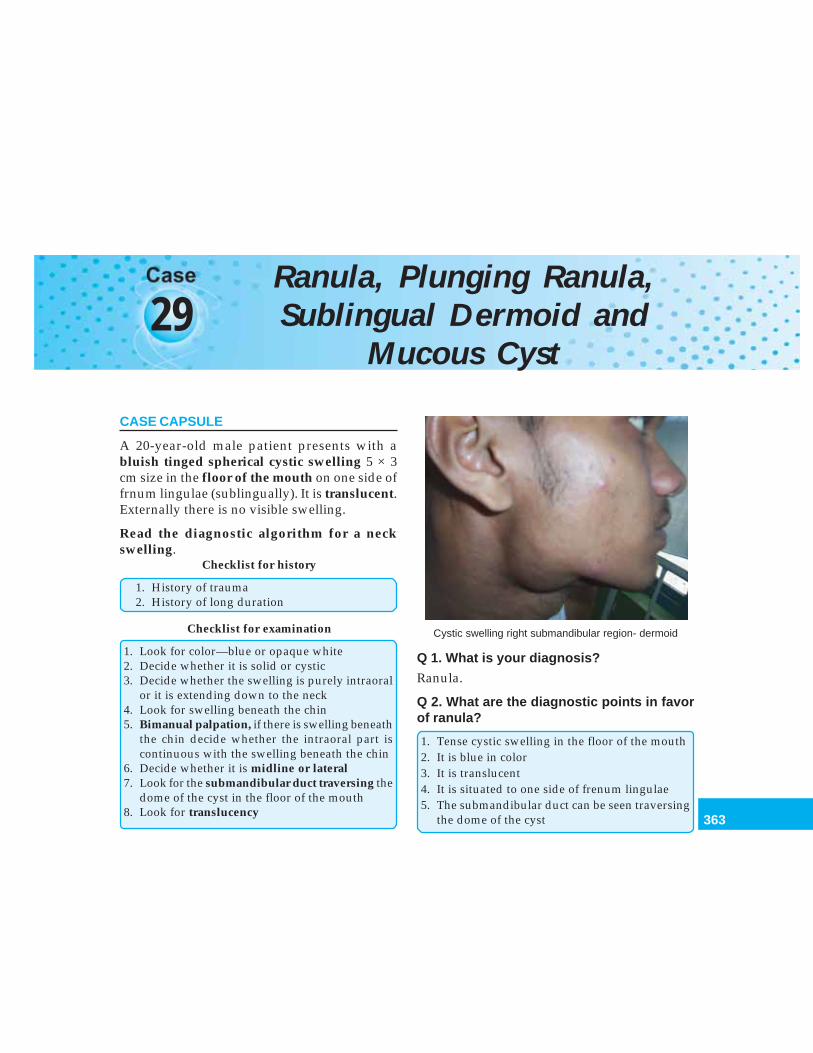
363
Ranula, Plunging Ranula, Sublingual Dermoid and Mucous Cyst
29Ranula, Plunging Ranula,Sublingual Dermoid and
Mucous Cyst
CASE CAPSULE
A 20-year-old male patient presents with abluish tinged spherical cystic swelling 5 × 3cm size in the floor of the mouth on one side offrnum lingulae (sublingually). It is translucent.Externally there is no visible swelling.
Read the diagnostic algorithm for a neckswelling.
Checklist for history
1. History of trauma2. History of long duration
Checklist for examination
1. Look for color—blue or opaque white2. Decide whether it is solid or cystic3. Decide whether the swelling is purely intraoral
or it is extending down to the neck4. Look for swelling beneath the chin5. Bimanual palpation, if there is swelling beneath
the chin decide whether the intraoral part iscontinuous with the swelling beneath the chin
6. Decide whether it is midline or lateral7. Look for the submandibular duct traversing the
dome of the cyst in the floor of the mouth8. Look for translucency
Q 1. What is your diagnosis?
Ranula.
Q 2. What are the diagnostic points in favorof ranula?
1. Tense cystic swelling in the floor of the mouth2. It is blue in color3. It is translucent4. It is situated to one side of frenum lingulae5. The submandibular duct can be seen traversing
the dome of the cyst
Cystic swelling right submandibular region- dermoid
364
Clinical Surgery Pearls
Q 3. What is the most important differentialdiagnosis?
Sublingual dermoid cyst.
Q 4. What are the clinical points againstsublingual dermoid in this case?
• Translucency is a point against sublingualdermoid
• Sublingual dermoid may be placed in themid- line unlike ranula which is to one sideof the frenum
• Sublingual dermoid is not blue in color. It iswhite and opaque.
Q 5. What is ranula?
It is a mucous extravasation cyst arising fromthe sublingual salivary gland.
Q 6. Why it is called ranula?
It is called ranula because of its resemblance tofrog’s belly.
Q 7. Who gave this name?
Hippocrates.
Q 8. What is plunging ranula?
Here the cyst penetrates the mylohyoiddiaphragm to enter the neck. Therefore therewill be a swelling in the floor of the mouth alongwith a swelling in the neck, resembling a dumb-bell. This retention cyst may be arising fromsublingual or submandibular salivary gland.The neck swelling may be in the submental orsubmandibular region of the neck.
Q 9. What is the clinical test for plungingranula?
Here one should elicit the Bidigital palpation –with index finger of one hand in the mouth andfingers of the other hand exerting upwardpressure externally from the below the lowerjaw over the neck swelling. In case of plungingranula it will be positive.
Q 10. How to confirm the diagnosis of ranula?
1. Ultrasound examination2. MRI (for plunging ranula)3. Plain X-ray to rule out stones.
Q 11. What are the complications of ranula?
1. Infection2. Mechanical interference with speech3. Difficulty in eating.
Q 12. What is the surgical treatment ofranula?
Excision of the cyst and the affected sublingualgland.
Q 13. What is the approach for surgery?
Intraoral approach is preferred.
Q 14. Why not incision and drainage of thecyst?
It is not recommended, because it will result inrecurrence of the cyst.
Q 15. What is the surgical approach forplunging ranula?
Excision is performed via cervical approach.
Q 16. What is the incision for cervicalapproach?
• The incision is similar to submandibularsialadenectomy
Published by dr-notes.com
365
Ranula, Plunging Ranula, Sublingual Dermoid and Mucous Cyst
• The cyst together with submandibular andsublingual salivary glands are excised.
Q 17. What is mucous cyst?
It is an extravasation/retention cyst in relationto minor salivary gland.
Q 18. What is the commonest site for mucouscyst?
Lower lip.
Q 19. What is the cause for mucous cyst?
It is as a result of trauma to the overlyingmucosa.
Q 20. What is the transillumination finding inmucous cyst?
It may or may not be translucent.
Q 21. What is the surgical treatment?
• Formal surgical excision is required alongwith the affected minor salivary gland underlocal anesthesia.
• Some may resolve spontaneously.
Q 22. How many minor salivary glands arethere in the oral cavity?
Around 450 (They contribute 10% of the salivaryvolume).
Q 23. What is the distribution of minorsalivary gland?
Sites of minor salivary glands
• Cheek• Palate• Floor of the mouth• Lips• Retromolar area
• Upper aerodigestive tract:OropharynxLarynxTracheaSinuses
Q 24. Is it possible to get a tumor in the sub-lingual salivary gland?
Yes. They are extremely rare. They present asfirm or hard painless swelling in the floor ofmouth. 95% are malignant.
Q 25. What is the surgical management ofthis malignancy?
Wide excision along with overlying mucosacombined with suprahyoid neck dissection.
Q 26. What are the types of sublingualdermoid?
There are two types –1. Median - in the midline2. Lateral—
• It may be situated above the mylohyoid– supramylohyoid variety (floor of themouth)
• It may be below the mylohyoid – infra-mylohyoid variety (swelling beneath thechin) – double chin appearance.
Q 27. What are the diagnostic points in favorof sublingual dermoid?
Diagnostic points for sublingual dermoid1. Color-opaque white cyst in contrast to
ranula2. Content – Sebaceous material3. May present as swelling in the floor of the
mouth or swelling beneath the chin
366
Clinical Surgery Pearls
4. No attachment to the covering mucosa orskin
5. Do not transilluminate
Q 28. What are the two most importantexaminations in suspected sublingualdermoid cyst?
1. Examination of the neck to rule out anextension beneath the chin or laterally in thesubmandibular region in the case of thelateral variety
2. Bimanual palpation—positive bimanualpalpation suggests extension beneath themylohyoid.
Q 29. What are the other differentialdiagnoses of a swelling beneath the chin?
• Thyroglossal cyst• Subhyoid bursa.
Q 30. What is the incision for excision of amedian sublingual dermoid?
1. If there is extension beneath the chin,consider external incision in the submental
region. Division of mylohyoid may berequired. The cyst is then enucleated.
2. If there is no extension beneath the chin,intraoral approach will suffice for theenucleation.
Q 31. How a sublingual dermoid cyst isformed?During the process of fusion of the facialprocesses, a piece of the skin may get trappeddeep in the midline just behind the jaw and laterform the dermoid cyst. The cyst may be midlineor lateral.
Q 32. What is the age group affected?10 – 25 years (Both sexes equally affected).
Q 33. What are the complications of sub-lingual dermoid?1. Infection (painful)2. Interfere with eating (when it is big)3. Interfere with speech.
Published by dr-notes.com
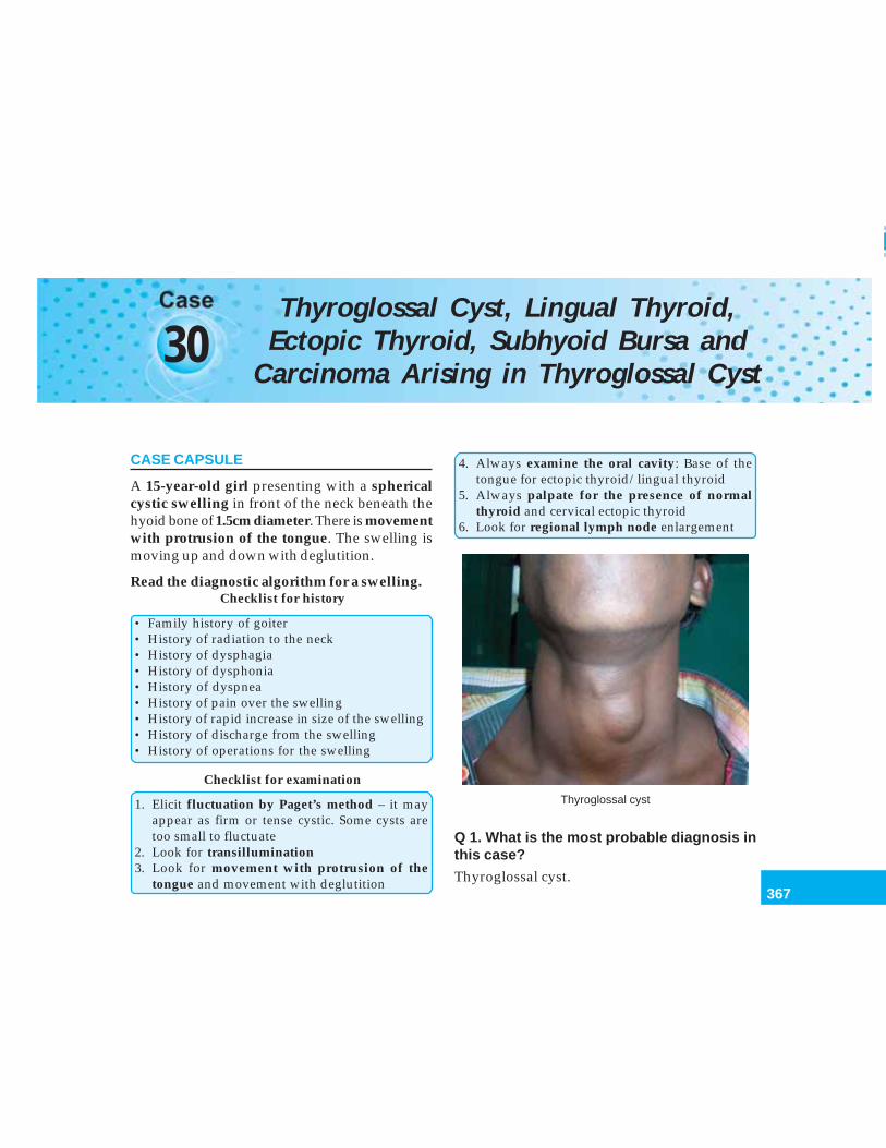
367
Thyroglossal Cyst, Lingual Thyroid, Ectopic Thyroid, Subhyoid Bursa
30Thyroglossal Cyst, Lingual Thyroid,
Ectopic Thyroid, Subhyoid Bursa andCarcinoma Arising in Thyroglossal Cyst
CASE CAPSULE
A 15-year-old girl presenting with a sphericalcystic swelling in front of the neck beneath thehyoid bone of 1.5cm diameter. There is movementwith protrusion of the tongue. The swelling ismoving up and down with deglutition.
Read the diagnostic algorithm for a swelling.Checklist for history
• Family history of goiter• History of radiation to the neck• History of dysphagia• History of dysphonia• History of dyspnea• History of pain over the swelling• History of rapid increase in size of the swelling• History of discharge from the swelling• History of operations for the swelling
Checklist for examination
1. Elicit fluctuation by Paget’s method – it mayappear as firm or tense cystic. Some cysts aretoo small to fluctuate
2. Look for transillumination3. Look for movement with protrusion of the
tongue and movement with deglutition
4. Always examine the oral cavity: Base of thetongue for ectopic thyroid/lingual thyroid
5. Always palpate for the presence of normalthyroid and cervical ectopic thyroid
6. Look for regional lymph node enlargement
Thyroglossal cyst
Q 1. What is the most probable diagnosis inthis case?
Thyroglossal cyst.
368
Clinical Surgery Pearls
Q 2. What are the diagnostic points in favorof thyroglossal cyst?
1. The most important sign of thyroglossal cystis the upward movement of the swellingwhen the tongue is protruded. This isbecause of its connection via the thyroglossalduct with the base of the tongue. It will alsomove up and down with deglutition. Inaddition there will be horizontal movementbut no vertical movement.
2. The presence of a small cystic swelling inthe midline of the neck (seldom large enoughto exhibit fluctuation).
3. May or may not be translucent (when thecontent is thick as a result of past infection,the cysts do not transilluminate.)
Q 3. How will you demonstrate the upwardmovement during protrusion of the tongue?
a. Request the patient to open the mouthb. Grasp the swelling between the finger and
thumbc. Instruct the patient to put out the tongued. The positive test will give an unmistakable
upward tug (certain amount of movementwill be there in this region for all swellings).The mouth must be open when the tug isappreciated.
e. The patient may be instructed to put thetongue in and out again if required.
Q 4. What are the differential diagnoses?Differential diagnoses of thyroglossal cyst
1. Median sublingual dermoid2. Enlarged submental lymph node3. Subhyoid bursa
4. Enlarged Delphian node5. Solitary thyroid nodule6. Thyroid cyst7. Dermoid cyst8. Collar – Stud abscess in connection with a lymph
node9. Ectopic thyroid10. Sebaceous cyst11. Lipoma
Q 5. What are the other midline swellings infront of the neck?
Midline swellings in front of the neck (in additionto the differential diagnoses given above).
• Ludwig’s angina in the upper part• Retrosternal and plunging goiter in the lower
part• Swellings from the Space of Burns in the lower
part (lymph node and lipoma)• Dermoid cyst in the lower part• Thymic swellings• Aneurysm of innominate artery
Q 6. What are the causes for enlargedsubmental lymph node?
1. Inflammatory—Specific like tuberculosis—Nonspecific
2. Neoplastic—Primary – Hodgkin’s and NHL—Secondary from carcinoma of
lower lip, tip of tongue, floor ofmouth.
Q 7. What is sublingual dermoid?
It is a sequestration dermoid cyst secondary tosequestration of surface ectoderm at the site offusion of mandibular arches. The swelling issituated in the midline in the floor of the mouth.The lateral variety of sublingual dermoid arises
Published by dr-notes.com
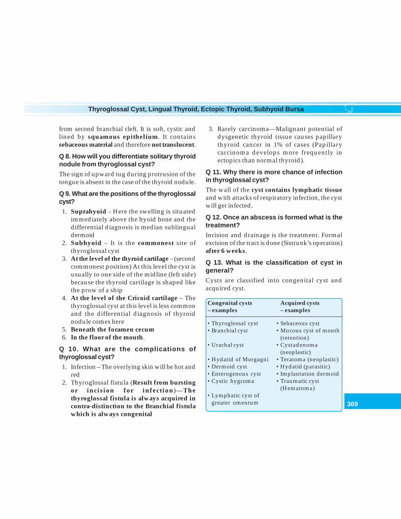
369
Thyroglossal Cyst, Lingual Thyroid, Ectopic Thyroid, Subhyoid Bursa
from second branchial cleft. It is soft, cystic andlined by squamous epithelium. It containssebaceous material and therefore not translucent.
Q 8. How will you differentiate solitary thyroidnodule from thyroglossal cyst?
The sign of upward tug during protrusion of thetongue is absent in the case of the thyroid nodule.
Q 9. What are the positions of the thyroglossalcyst?
1. Suprahyoid – Here the swelling is situatedimmediately above the hyoid bone and thedifferential diagnosis is median sublingualdermoid
2. Subhyoid – It is the commonest site ofthyroglossal cyst
3. At the level of the thyroid cartilage – (secondcommonest position) At this level the cyst isusually to one side of the midline (left side)because the thyroid cartilage is shaped likethe prow of a ship
4. At the level of the Cricoid cartilage – Thethyroglossal cyst at this level is less commonand the differential diagnosis of thyroidnodule comes here
5. Beneath the foramen cecum6. In the floor of the mouth.
Q 10. What are the complications ofthyroglossal cyst?
1. Infection – The overlying skin will be hot andred
2. Thyroglossal fistula (Result from burstingor incision for infection)—Thethyroglossal fistula is always acquired incontra-distinction to the Branchial fistulawhich is always congenital
3. Rarely carcinoma—Malignant potential ofdysgenetic thyroid tissue causes papillarythyroid cancer in 1% of cases (Papillarycarcinoma develops more frequently inectopics than normal thyroid).
Q 11. Why there is more chance of infectionin thyroglossal cyst?
The wall of the cyst contains lymphatic tissueand with attacks of respiratory infection, the cystwill get infected.
Q 12. Once an abscess is formed what is thetreatment?
Incision and drainage is the treatment. Formalexcision of the tract is done (Sistrunk’s operation)after 6 weeks.
Q 13. What is the classification of cyst ingeneral?
Cysts are classified into congenital cyst andacquired cyst.
Congenital cysts Acquired cysts– examples – examples
•Thyroglossal cyst •Sebaceous cyst•Branchial cyst •Mucous cyst of mouth
(retention)•Urachal cyst •Cystadenoma
(neoplastic)•Hydatid of Morgagni •Teratoma (neoplastic)•Dermoid cyst •Hydatid (parasitic)•Enterogenous cyst •Implantation dermoid•Cystic hygroma •Traumatic cyst
(Hematoma)•Lymphatic cyst of
greater omentum
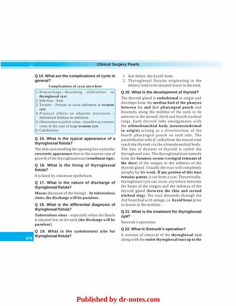
370
Clinical Surgery Pearls
Q 14. What are the complications of cysts ingeneral?
Complications of cysts anywhere
1. Hemorrhage—Breathing difficulties inthyroglossal cyst
2. Infection – Pain3. Torsion – Present as acute abdomen in ovarian
cyst4. Pressure effects on adjacent structures –
abdominal fullness in abdomen5. Obstruction to pelvic veins – manifest as varicose
veins in the case of large ovarian cyst6. Calcification
Q 15. What is the typical appearance of athyroglossal fistula?
The skin surrounding the opening has a peculiarcrescentic appearance due to the uneven rate ofgrowth of the thyroglossal tract (semilunar sign).
Q 16. What is the lining of thyroglossalfistula?
It is lined by columnar epithelium.
Q 17. What is the nature of discharge ofthyroglossal fistula?
Mucus (because of the lining) – In tuberculoussinus, the discharge will be purulent.
Q 18. What is the differential diagnosis ofthyroglossal fistula?
Tuberculous sinus – especially when the fistulais situated low in the neck (the discharge will bepurulent).
Q 19. What is the commonest site forthyroglossal fistula?
1. Just below the hyoid bone2. Thyroglossal fistulas originating in the
infancy tend to be situated lower in the neck.
Q 20. What is the development of thyroid?
The thyroid gland is endodermal in origin anddevelops from the median bud of the pharynxbetween 1st and 2nd pharyngeal pouch anddescends along the midline of the neck to lieanterior to the second, third and fourth trachealrings. Each thyroid lobe amalgamates withthe ultimobranchial body (neuroectodermalin origin) arising as a diverticulum of thefourth pharyngeal pouch on each side. Theparafollicular cells (C cells) from the neural crestreach the thyroid via the ultimobranchial body.The line of descent of thyroid is called thethyroglossal tract. The thyroglossal tract extendsfrom the foramen cecum (vestigial remnant ofthe duct) of the tongue to the isthmus of thethyroid gland. Usually the tract will completelyatrophy by 5th week. If any portion of this tractremains patent, it can form a cyst. Theoretically,thyroglossal cyst can occur anywhere betweenthe bases of the tongue and the isthmus of thethyroid gland (between the chin and secondtracheal ring). The tract descends through the2nd branchial arch anlage, i.e. hyoid bone priorto fusion in the midline. .
Q 21. What is the treatment for thyroglossalcyst?
Sistrunk’s operation.
Q 22. What is Sistrunk’s operation?
It consists of removal of the thyroglossal cystalong with the entire thyroglossal tract up to the
Published by dr-notes.com
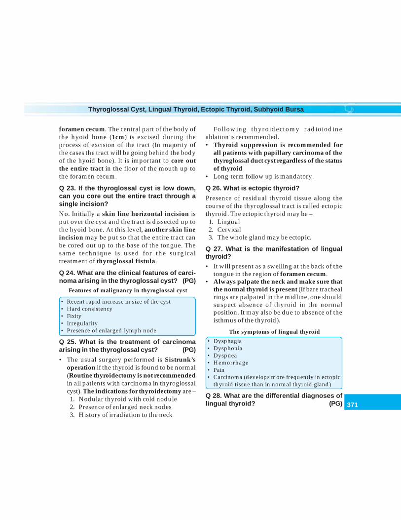
371
Thyroglossal Cyst, Lingual Thyroid, Ectopic Thyroid, Subhyoid Bursa
foramen cecum. The central part of the body ofthe hyoid bone (1cm) is excised during theprocess of excision of the tract (In majority ofthe cases the tract will be going behind the bodyof the hyoid bone). It is important to core outthe entire tract in the floor of the mouth up tothe foramen cecum.
Q 23. If the thyroglossal cyst is low down,can you core out the entire tract through asingle incision?
No. Initially a skin line horizontal incision isput over the cyst and the tract is dissected up tothe hyoid bone. At this level, another skin lineincision may be put so that the entire tract canbe cored out up to the base of the tongue. Thesame technique is used for the surgicaltreatment of thyroglossal fistula.
Q 24. What are the clinical features of carci-noma arising in the thyroglossal cyst? (PG)
Features of malignancy in thyroglossal cyst
• Recent rapid increase in size of the cyst• Hard consistency• Fixity• Irregularity• Presence of enlarged lymph node
Q 25. What is the treatment of carcinomaarising in the thyroglossal cyst? (PG)
• The usual surgery performed is Sistrunk’soperation if the thyroid is found to be normal(Routine thyroidectomy is not recommendedin all patients with carcinoma in thyroglossalcyst). The indications for thyroidectomy are –1. Nodular thyroid with cold nodule2. Presence of enlarged neck nodes3. History of irradiation to the neck
Following thyroidectomy radioiodineablation is recommended.• Thyroid suppression is recommended for
all patients with papillary carcinoma of thethyroglossal duct cyst regardless of the statusof thyroid
• Long-term follow up is mandatory.
Q 26. What is ectopic thyroid?
Presence of residual thyroid tissue along thecourse of the thyroglossal tract is called ectopicthyroid. The ectopic thyroid may be –1. Lingual2. Cervical3. The whole gland may be ectopic.
Q 27. What is the manifestation of lingualthyroid?• It will present as a swelling at the back of the
tongue in the region of foramen cecum.• Always palpate the neck and make sure that
the normal thyroid is present (If bare trachealrings are palpated in the midline, one shouldsuspect absence of thyroid in the normalposition. It may also be due to absence of theisthmus of the thyroid).
The symptoms of lingual thyroid
• Dysphagia• Dysphonia• Dyspnea• Hemorrhage• Pain• Carcinoma (develops more frequently in ectopic
thyroid tissue than in normal thyroid gland)
Q 28. What are the differential diagnoses oflingual thyroid? (PG)
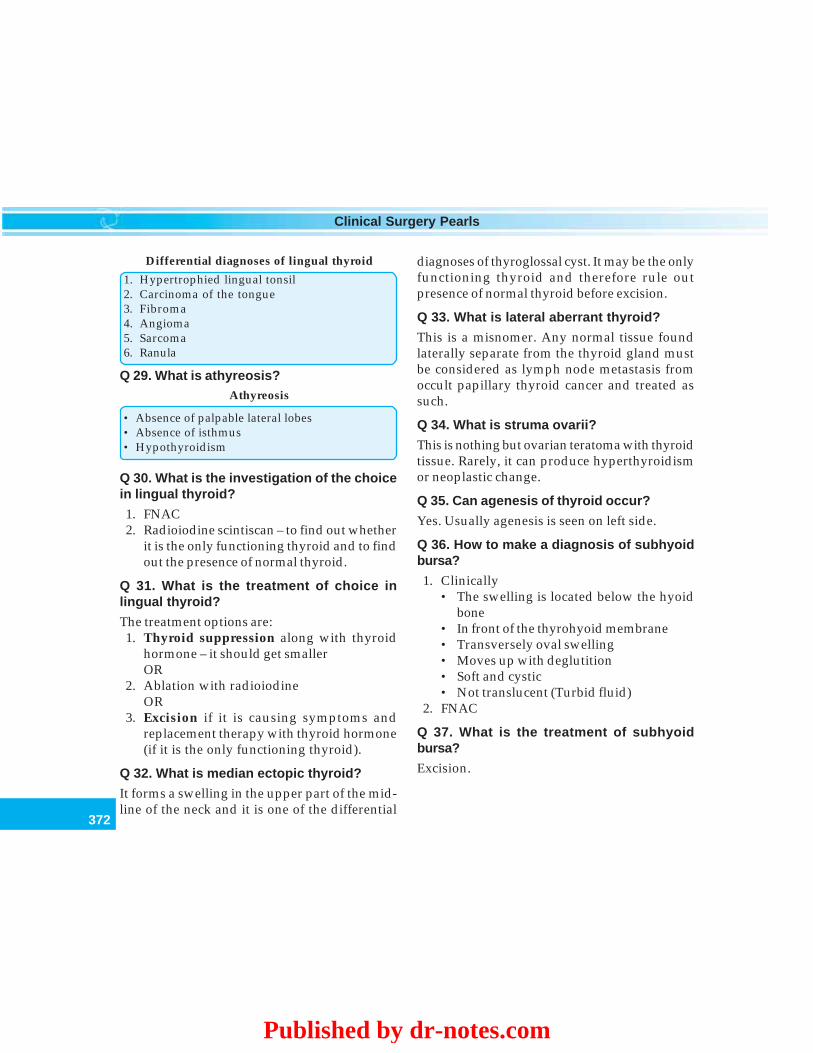
372
Clinical Surgery Pearls
Differential diagnoses of lingual thyroid
1. Hypertrophied lingual tonsil2. Carcinoma of the tongue3. Fibroma4. Angioma5. Sarcoma6. Ranula
Q 29. What is athyreosis?Athyreosis
• Absence of palpable lateral lobes• Absence of isthmus• Hypothyroidism
Q 30. What is the investigation of the choicein lingual thyroid?
1. FNAC2. Radioiodine scintiscan – to find out whether
it is the only functioning thyroid and to findout the presence of normal thyroid.
Q 31. What is the treatment of choice inlingual thyroid?
The treatment options are:1. Thyroid suppression along with thyroid
hormone – it should get smallerOR
2. Ablation with radioiodineOR
3. Excision if it is causing symptoms andreplacement therapy with thyroid hormone(if it is the only functioning thyroid).
Q 32. What is median ectopic thyroid?
It forms a swelling in the upper part of the mid-line of the neck and it is one of the differential
diagnoses of thyroglossal cyst. It may be the onlyfunctioning thyroid and therefore rule outpresence of normal thyroid before excision.
Q 33. What is lateral aberrant thyroid?
This is a misnomer. Any normal tissue foundlaterally separate from the thyroid gland mustbe considered as lymph node metastasis fromoccult papillary thyroid cancer and treated assuch.
Q 34. What is struma ovarii?
This is nothing but ovarian teratoma with thyroidtissue. Rarely, it can produce hyperthyroidismor neoplastic change.
Q 35. Can agenesis of thyroid occur?
Yes. Usually agenesis is seen on left side.
Q 36. How to make a diagnosis of subhyoidbursa?
1. Clinically• The swelling is located below the hyoid
bone• In front of the thyrohyoid membrane• Transversely oval swelling• Moves up with deglutition• Soft and cystic• Not translucent (Turbid fluid)
2. FNAC
Q 37. What is the treatment of subhyoidbursa?
Excision.
Published by dr-notes.com
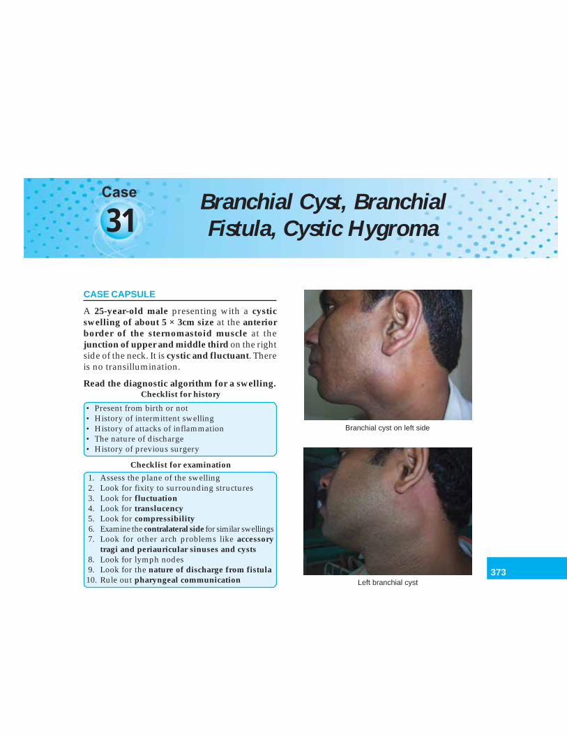
373
Branchial Cyst, Branchial Fistula, Cystic Hygroma
31Branchial Cyst, BranchialFistula, Cystic Hygroma
CASE CAPSULE
A 25-year-old male presenting with a cysticswelling of about 5 × 3cm size at the anteriorborder of the sternomastoid muscle at thejunction of upper and middle third on the rightside of the neck. It is cystic and fluctuant. Thereis no transillumination.
Read the diagnostic algorithm for a swelling.Checklist for history
• Present from birth or not• History of intermittent swelling• History of attacks of inflammation• The nature of discharge• History of previous surgery
Checklist for examination
1. Assess the plane of the swelling2. Look for fixity to surrounding structures3. Look for fluctuation4. Look for translucency5. Look for compressibility6. Examine the contralateral side for similar swellings7. Look for other arch problems like accessory
tragi and periauricular sinuses and cysts8. Look for lymph nodes9. Look for the nature of discharge from fistula10. Rule out pharyngeal communication
Branchial cyst on left side
Left branchial cyst
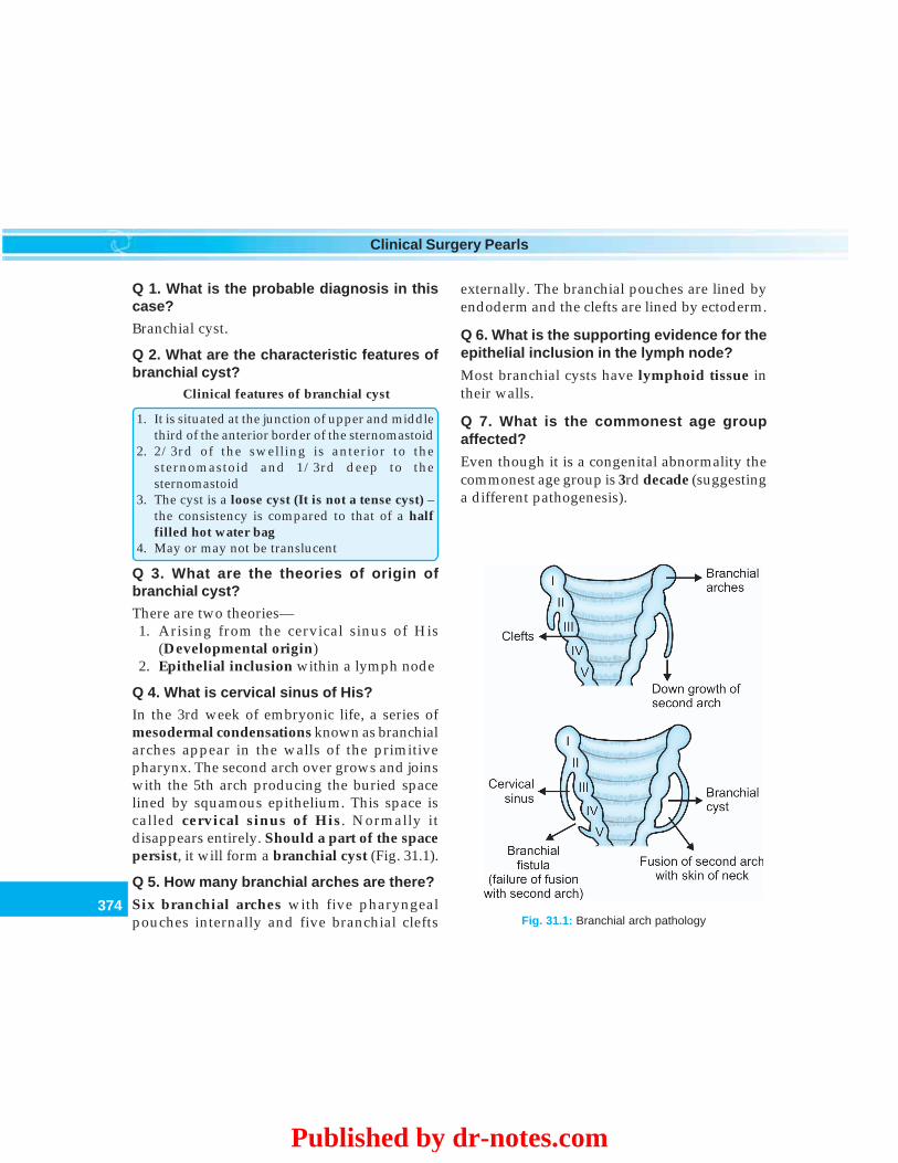
374
Clinical Surgery Pearls
Q 1. What is the probable diagnosis in thiscase?
Branchial cyst.
Q 2. What are the characteristic features ofbranchial cyst?
Clinical features of branchial cyst
1. It is situated at the junction of upper and middlethird of the anterior border of the sternomastoid
2. 2/3rd of the swelling is anterior to thesternomastoid and 1/3rd deep to thesternomastoid
3. The cyst is a loose cyst (It is not a tense cyst) –the consistency is compared to that of a halffilled hot water bag
4. May or may not be translucent
Q 3. What are the theories of origin ofbranchial cyst?
There are two theories—1. Arising from the cervical sinus of His
(Developmental origin)2. Epithelial inclusion within a lymph node
Q 4. What is cervical sinus of His?
In the 3rd week of embryonic life, a series ofmesodermal condensations known as branchialarches appear in the walls of the primitivepharynx. The second arch over grows and joinswith the 5th arch producing the buried spacelined by squamous epithelium. This space iscalled cervical sinus of His. Normally itdisappears entirely. Should a part of the spacepersist, it will form a branchial cyst (Fig. 31.1).
Q 5. How many branchial arches are there?
Six branchial arches with five pharyngealpouches internally and five branchial clefts
externally. The branchial pouches are lined byendoderm and the clefts are lined by ectoderm.
Q 6. What is the supporting evidence for theepithelial inclusion in the lymph node?
Most branchial cysts have lymphoid tissue intheir walls.
Q 7. What is the commonest age groupaffected?
Even though it is a congenital abnormality thecommonest age group is 3rd decade (suggestinga different pathogenesis).
Fig. 31.1: Branchial arch pathology
Published by dr-notes.com
375
Branchial Cyst, Branchial Fistula, Cystic Hygroma
Q 8. How to explain the late appearance ofthe cyst if it is congenital?
Initially the cyst is an empty sac of embryologicaltissue. The epithelial debris accumulates fornumber of years with super added infectionmaking the appearance of the cyst.
Q 9. Why branchial cysts do not transil-luminate?
The cyst contains yellow fluid with cholesterolcrystals and epithelial debris therefore it will nottransilluminate.
Q 10. How a branchial fistula is formed?
Should the second arch fail to fuse with the 5tharch, an opening will be found on the neck atbirth along the anterior border of the sterno-mastoid muscle at the junction of the middle andlower third.
Q 11. Is there any communication of thefistula to the pharynx?
Usually they are separated from the pharynx bya septum, which represents the remains of thecleft membrane (ending blindly on the lateralpharyngeal wall like a sinus). If the fistuloustract is complete, it will open just behind thetonsil on the affected side (On the anterioraspect of the posterior faucial pillar). There maybe small amount of mucus or mucopurulentdischarge coming out through the fistulousopening. If the tract is communicating into theoral cavity the liquids taken during meals willcome out through the fistulous opening.
Q 12. What is the lining of the branchial cyst?
Two types of lining are seen:1. Stratified squamous epithelium2. Nonciliated columnar epithelium.
Q 13. What is the lining of branchial fistula?
The fistula is lined with squamous epitheliumup to the partition (cleft membrane). Internal tothe partition it is lined with ciliated columnarepithelium.
Q 14. What is the course of the branchialfistula?
It is below the 2nd arch structures and abovethe 3rd arch structures.
Q 15. What is the nerve and artery of 2nd and3rd arches?
• The nerve of 2nd arch is facial nerve• The artery of the 2nd arch is external carotid
artery• The nerve of 3rd arch is 9th• The artery of 3rd arch is internal carotid.
Q 16. What is the time of clinical presentationof branchial fistula?
The opening is present at birth (It is alwayscongenital).
Q 17. What is the clinical feature other thanthe opening?
Mucoid discharge is noticed in children after ahot bath.
Q 18. What is the exact course of thebranchial fistula?
The external opening is usually seen in relationto the lower 3rd of the anterior border of
376
Clinical Surgery Pearls
sternomastoid muscle. From its opening on theskin it passes subcutaneously to the level of theupper border of the thyroid cartilage where itpierces the deep fascia. The fistula then passesbeneath the posterior belly of the digastricmuscle and the stylohyoid muscle, crosses thehypoglossal nerve and internal jugular vein,to traverse the fork of the carotid bifurcation.Here the external carotid artery is superficialand the internal carotid deep to the tract. It thencrosses the glossopharyngeal nerve and thestylopharyngeus muscle to pierce the superiorconstrictor and to open on the posterior pillarof the fauces behind the tonsil.
Q 19. Can the fistula be bilateral?
Yes. In 30% of cases.
Q 20. Can the branchial cyst be bilateral?
Yes. In 2% of cases it is bilateral.
Q 21. On which side the branchial cyst ismore commonly seen?
60% it is seen on left side.
Q 22. Which sex is more affected?
Males are more affected – 60%.
Q 23. Can the cyst be intermittent?
Yes. In 20% of the patients, the cyst will beintermittent.
Q 24. If the cyst is disappearing at the timeof surgery what is the course of action?
There is no place for exploration in this situation.The cyst may not be found at exploration. Partialexcision will lead on to sinus formation.
Q 25. What are the other associated con-genital anomalies?
Associated anomalies in branchial cyst
• Preauricular sinuses• Periauricular cysts• Accessory tragi• Subcutaneous cartilaginous nodules
Q 26. The branchial fistula is congenital oracquired?
• The branchial fistula is always congenital(the position of the cyst is in the upper partand the fistula is in the lower part due to thedevelopmental reasons already mentioned)
• The thyroglossal fistula is always acquired.
Q 27. Can similar swellings occur in relationto other cleft apparatus?
Yes. It can occur in relation to first branchialcleft.
Q 28. What is branchogenic carcinoma?
It is controversial whether this entity is amalignancy in branchial cyst or it is a cysticdegeneration in a lymph node containing depositfrom squamous cell carcinoma. The latter is morepossible. The primary growth may not be apparentand it may be situated in the nasopharynx, tonsil,base of tongue, pyriform fossa or supraglotticlarynx. Therefore it is important to rule out anoccult primary in such conditions.
Q 29. What are the other cysts havingcholesterol crystals?
• Hydrocele• Dental cyst• Dentigerous cyst.
Published by dr-notes.com
377
Branchial Cyst, Branchial Fistula, Cystic Hygroma
Q 30. What are the most important differentialdiagnoses for a branchial cyst?
Other cystic swellings on the lateral side of theneck like.
Cystic swellings on the side of the neck
• Branchial cyst• Cystic hygroma (Lymphangioma)• Hemangioma• Cold abscess• Dermoid cyst• Laryngocele• Pharyngocele• Sebaceous cyst
In addition the following solid swellings formdifferential diagnoses for branchial cyst.
Solid swellings on the side of the neck
1. Reactive lymphadenitis2. Lipoma3. Neurofibroma4. Chemodectoma (potato tumor)5. Paraganglioma6. Lymphoma7. Metastatic carcinoma in node from thyroid8. Tuberculous lymph node
Q 31. How to differentiate clinically cystichygroma from branchial cyst?
• Cystic hygroma is brilliantly translucent• Cystic hygroma is partially compressible• Usually seen in the neonate and early infancy• Increase in size when the child cries or coughs.
Q 32. Why cystic hygroma is brilliantly trans-lucent?
• The cysts are filled with clear fluid• Lined by single layer of epithelium (with a
mosaic appearance).
Q 33. How a cystic hygroma is formed?
Cystic hygroma develops from the primitive lymphsac called jugular lymph sac (they are situated inthe neck between the jugular and subclavian veins).Sequestration of a portion of jugular lymph sac willresult in the formation of cystic hygroma.
Q 34. What are the other situations where youget cystic hygroma?
Sites for cystic hygroma
• Axilla• Groin• Mediastinum• Cheek• May involve salivary glands• Tongue• Floor of the mouth
Q 35. What are the complications of cystichygroma?
• Infection• Increase in size and respiratory embarras-
sment• Obstructed labor during delivery.
Q 36. What are the treatment options forcystic hygroma?
1. Meticulous conservative neck dissectionwith excision of all lymphatic tissue
2. Injection of sclerosing agents (Picibanil orOK432 is a recent sclerosant).
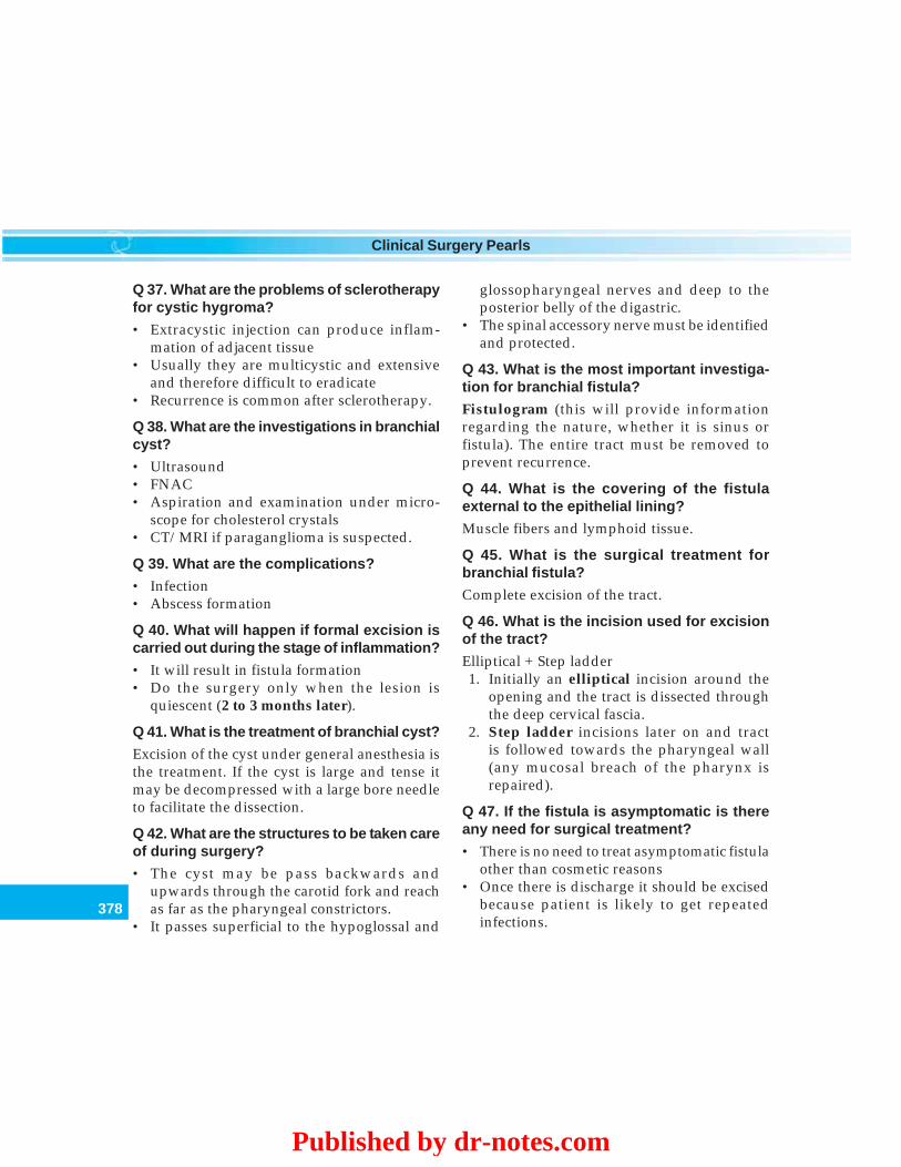
378
Clinical Surgery Pearls
Q 37. What are the problems of sclerotherapyfor cystic hygroma?
• Extracystic injection can produce inflam-mation of adjacent tissue
• Usually they are multicystic and extensiveand therefore difficult to eradicate
• Recurrence is common after sclerotherapy.
Q 38. What are the investigations in branchialcyst?
• Ultrasound• FNAC• Aspiration and examination under micro-
scope for cholesterol crystals• CT/MRI if paraganglioma is suspected.
Q 39. What are the complications?
• Infection• Abscess formation
Q 40. What will happen if formal excision iscarried out during the stage of inflammation?
• It will result in fistula formation• Do the surgery only when the lesion is
quiescent (2 to 3 months later).
Q 41. What is the treatment of branchial cyst?
Excision of the cyst under general anesthesia isthe treatment. If the cyst is large and tense itmay be decompressed with a large bore needleto facilitate the dissection.
Q 42. What are the structures to be taken careof during surgery?
• The cyst may be pass backwards andupwards through the carotid fork and reachas far as the pharyngeal constrictors.
• It passes superficial to the hypoglossal and
glossopharyngeal nerves and deep to theposterior belly of the digastric.
• The spinal accessory nerve must be identifiedand protected.
Q 43. What is the most important investiga-tion for branchial fistula?
Fistulogram (this will provide informationregarding the nature, whether it is sinus orfistula). The entire tract must be removed toprevent recurrence.
Q 44. What is the covering of the fistulaexternal to the epithelial lining?
Muscle fibers and lymphoid tissue.
Q 45. What is the surgical treatment forbranchial fistula?
Complete excision of the tract.
Q 46. What is the incision used for excisionof the tract?
Elliptical + Step ladder1. Initially an elliptical incision around the
opening and the tract is dissected throughthe deep cervical fascia.
2. Step ladder incisions later on and tractis followed towards the pharyngeal wall(any mucosal breach of the pharynx isrepaired).
Q 47. If the fistula is asymptomatic is thereany need for surgical treatment?
• There is no need to treat asymptomatic fistulaother than cosmetic reasons
• Once there is discharge it should be excisedbecause patient is likely to get repeatedinfections.
Published by dr-notes.com
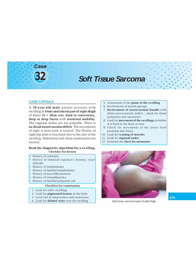
379
Soft Tissue Sarcoma
32 Soft Tissue Sarcoma
CASE CAPSULE
A 50-year-old male patient presents withswelling in front and lateral part of right thighof about 15 × 10cm size, hard in consistency,deep to deep fascia with restricted mobility.The regional nodes are not palpable. There isno distal neurovascular deficit. The movementsof right is knee joint is normal. The flexion ofright hip joint is restricted due to the size of theswelling. Abdominal and chest examination arenormal.
Read the diagnostic algorithm for a swelling.Checklist for history
• History of radiation• History of chemical exposure—Arsenic, vinyl
chloride• History of lymphedema• History of familial lymphedema• History of neurofibromatosis• History of retinoblastoma• History of familial polyposis coli
Checklist for examination
1. Look for other swellings2. Look for pigmented lesions in the body3. Local rise of temperature and tenderness4. Look for dilated veins over the swelling
5. Assessment of the plane of the swelling6. Involvement of muscle groups7. Involvement of neurovascular bundle with
distal neurovascular deficit – check for distalpulsations and sensations
8. Look for movement of the swellings (whetherit is fixed to the bone or not)
9. Check for movement of the joints bothproximal and distal
10. Look for wasting of muscles11. Look for regional nodes12. Examine the chest for metastasis
Soft tissue sarcoma back of right thigh
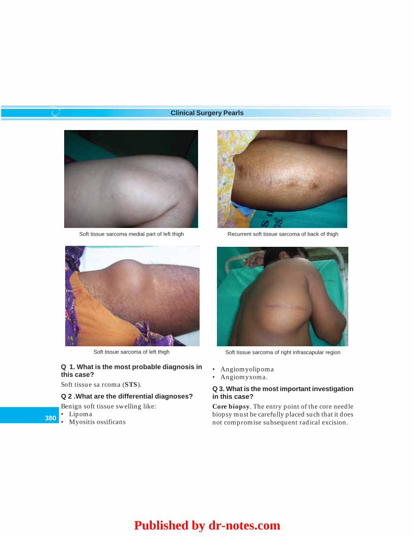
380
Clinical Surgery Pearls
Q 1. What is the most probable diagnosis inthis case?Soft tissue sa rcoma (STS).
Q 2 .What are the differential diagnoses?Benign soft tissue swelling like:• Lipoma• Myositis ossificans
Recurrent soft tissue sarcoma of back of thighSoft tissue sarcoma medial part of left thigh
Soft tissue sarcoma of left thigh Soft tissue sarcoma of right infrascapular region
• Angiomyolipoma• Angiomyxoma.
Q 3. What is the most important investigationin this case?Core biopsy. The entry point of the core needlebiopsy must be carefully placed such that it doesnot compromise subsequent radical excision.
Published by dr-notes.com
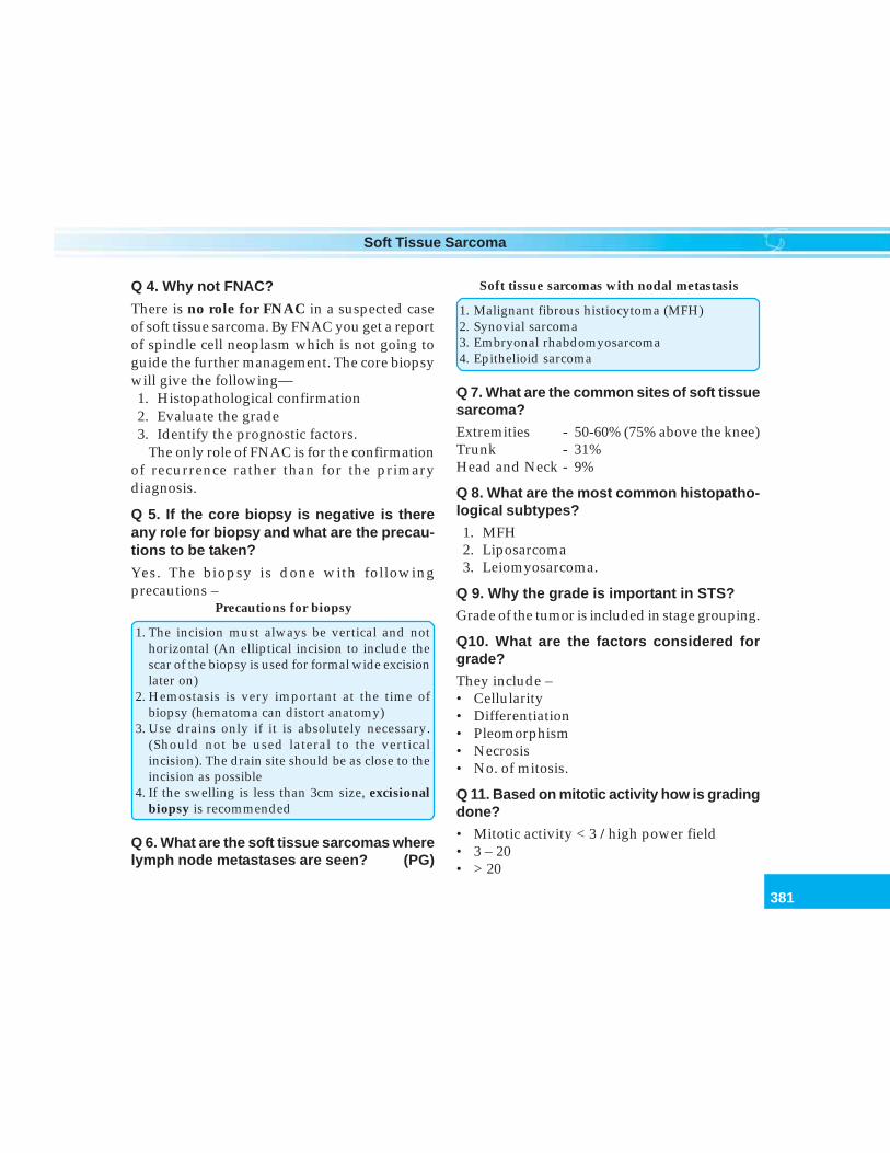
381
Soft Tissue Sarcoma
Q 4. Why not FNAC?
There is no role for FNAC in a suspected caseof soft tissue sarcoma. By FNAC you get a reportof spindle cell neoplasm which is not going toguide the further management. The core biopsywill give the following—1. Histopathological confirmation2. Evaluate the grade3. Identify the prognostic factors.
The only role of FNAC is for the confirmationof recurrence rather than for the primarydiagnosis.
Q 5. If the core biopsy is negative is thereany role for biopsy and what are the precau-tions to be taken?
Yes. The biopsy is done with followingprecautions –
Precautions for biopsy
1. The incision must always be vertical and nothorizontal (An elliptical incision to include thescar of the biopsy is used for formal wide excisionlater on)
2. Hemostasis is very important at the time ofbiopsy (hematoma can distort anatomy)
3. Use drains only if it is absolutely necessary.(Should not be used lateral to the verticalincision). The drain site should be as close to theincision as possible
4. If the swelling is less than 3cm size, excisionalbiopsy is recommended
Q 6. What are the soft tissue sarcomas wherelymph node metastases are seen? (PG)
Soft tissue sarcomas with nodal metastasis
1. Malignant fibrous histiocytoma (MFH)2. Synovial sarcoma3. Embryonal rhabdomyosarcoma4. Epithelioid sarcoma
Q 7. What are the common sites of soft tissuesarcoma?
Extremities - 50-60% (75% above the knee)Trunk - 31%Head and Neck - 9%
Q 8. What are the most common histopatho-logical subtypes?
1. MFH2. Liposarcoma3. Leiomyosarcoma.
Q 9. Why the grade is important in STS?
Grade of the tumor is included in stage grouping.
Q10. What are the factors considered forgrade?
They include –• Cellularity• Differentiation• Pleomorphism• Necrosis• No. of mitosis.
Q 11. Based on mitotic activity how is gradingdone?
• Mitotic activity < 3 / high power field• 3 – 20• > 20
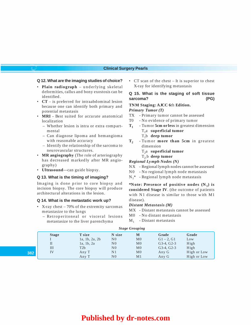
382
Clinical Surgery Pearls
Q 12. What are the imaging studies of choice?
• Plain radiograph – underlying skeletaldeformities, callus and bony exostosis can beidentified.
• CT – is preferred for intraabdominal lesionbecause one can identify both primary andpotential metastasis
• MRI - Best suited for accurate anatomicallocalization– Whether lesion is intra or extra compart-
mental– Can diagnose lipoma and hemangioma
with reasonable accuracy– Identify the relationship of the sarcoma to
neurovascular structures.• MR angiography (The role of arteriography
has decreased markedly after MR angio-graphy)
• Ultrasound—can guide biopsy.
Q 13. What is the timing of imaging?
Imaging is done prior to core biopsy andincision biopsy. The core biopsy will producearchitectural alterations in the lesion.
Q 14. What is the metastatic work up?
• X-ray chest – 70% of the extremity sarcomasmetastasize to the lungs– Retroperitoneal or visceral lesions
metastasize to the liver parenchyma
• CT scan of the chest – It is superior to chestX-ray for identifying metastasis
Q 15. What is the staging of soft tissuesarcoma? (PG)
TNM Staging: AJCC 6th Edition.Primary Tumor (T)TX - Primary tumor cannot be assessedT0 - No evidence of primary tumorT1 - Tumor 5cm or less in greatest dimension
T1a superficial tumorT1b deep tumor
T2 - Tumor more than 5cm in greatestdimensionT2a superficial tumorT2-b deep tumor
Regional Lymph Nodes (N)NX - Regional lymph nodes cannot be assessedN0 - No regional lymph node metastasisN1* - Regional lymph node metastasis
*Note: Presence of positive nodes (N1) isconsidered Stage IV. (the outcome of patientswith N1 disease is similar to those with M1disease).Distant Metastasis (M)MX - Distant metastasis cannot be assessedM0 - No distant metastasisM1 - Distant metastasis
Stage Grouping
Stage T size N size M Grade GradeI 1a, 1b, 2a, 2b N0 M0 G1 – 2, G1 LowII 1a, 1b, 2a N0 M0 G3-4, G2-3 HighIII T2b N0 M0 G3-4, G2-3 HighIV Any T N1 M0 Any G High or Low
Any T N0 M1 Any G High or Low
Published by dr-notes.com
383
Soft Tissue Sarcoma
Q 16. What you mean by superficial and deepin staging? (PG)
Superficial lesions – lesions not involving thesuperficial fascia.
Deep –a. lesions deep to, or involves the superficial
fasciab. All intraperitoneal visceral lesionsc. Retroperitoneal lesionsd. Mediastinal (Intrathoracic lesions)e. Pelvic sarcomasf. Head and neck tumors
Q 17. What type of grading is used in newAJCC 6th edition? (PG)
The previously existing 3 and 4 tiered system ofgrading is converted into 2 tiered system withlow and high grade designation. The mostcommonly employed 3 tiered system will thusbecome:Grade 1 - considered as low gradeGrade 2 and 3 - considered as high gradeThe 4 - tiered system will thus become:Grade 1 and 2 - considered as low gradeGrade 3 and 4 : considered as high grade
Q 18. What are the sarcomas excluded fromthis staging system? (PG)
• Kaposi’s sarcoma• Dermatofibrosarcoma protuberans• Infantile fibrosarcoma• Angiosarcoma• Sarcomas arising from the dura mater
including brain
• Sarcoma arising in the parenchymatousorgans
• Hollow viscera• Inflammatory myofibroblastic tumor• Fibromatosis (Desmoid tumor)• Mesothelioma.
Q 19. What is fibromatosis?
Original fibrosarcoma grade I is now mentionedas fibromatosis (Desmoid) and they are not insoft tissue sarcomas.
Q 20. What are the etiological factors for softtissue sarcoma?
1. Ionizing radiation2. Chemical exposure – arsenic, vinyl chloride3. Genetic
– Neurofibromatosis– NF 1 gene– Retinoblastoma– Familial polyposis coli– Mutation of p53 (nuclear over expression of
p53)– High ki 67 proliferation index
Q 21. What is the staging in this case?
T2b, N0, M0 (Stage 3).
Q 22. What is the management of soft tissuesarcoma?
Multidisciplinary approach is recommendedcomprising expertise in the following specialties:• Radiology• Clinical oncology• Surgical oncology.
Note: Limb sparing surgery is preferred.Amputation is performed in < 5-10% of cases.
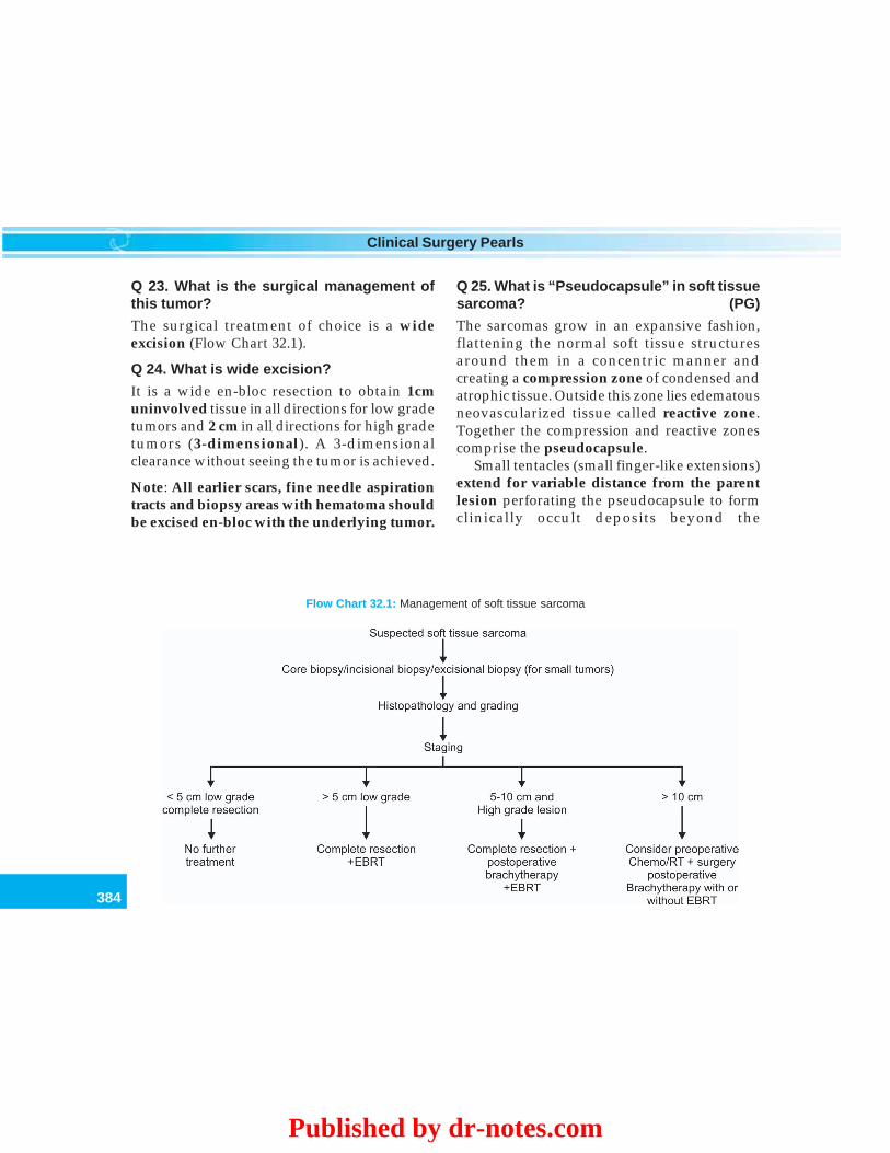
384
Clinical Surgery Pearls
Q 23. What is the surgical management ofthis tumor?
The surgical treatment of choice is a wideexcision (Flow Chart 32.1).
Q 24. What is wide excision?
It is a wide en-bloc resection to obtain 1cmuninvolved tissue in all directions for low gradetumors and 2 cm in all directions for high gradetumors (3-dimensional). A 3-dimensionalclearance without seeing the tumor is achieved.
Note: All earlier scars, fine needle aspirationtracts and biopsy areas with hematoma shouldbe excised en-bloc with the underlying tumor.
Q 25. What is “Pseudocapsule” in soft tissuesarcoma? (PG)
The sarcomas grow in an expansive fashion,flattening the normal soft tissue structuresaround them in a concentric manner andcreating a compression zone of condensed andatrophic tissue. Outside this zone lies edematousneovascularized tissue called reactive zone.Together the compression and reactive zonescomprise the pseudocapsule.
Small tentacles (small finger-like extensions)extend for variable distance from the parentlesion perforating the pseudocapsule to formclinically occult deposits beyond the
Flow Chart 32.1: Management of soft tissue sarcoma
Published by dr-notes.com
385
Soft Tissue Sarcoma
pseudocapsule. Therefore tumor masses willbe seen outside the pseudocapsule.
Q 26. What are the barriers for the infiltrativegrowth of the sarcomas? (PG)
The expansive growth stops at the followingboundaries:1. Fascial boundaries2. Periosteal structures3. Adventitia of the vessels4. Nerve sheaths
Note: Gross involvement of these structures isseen early (may become infiltrated).
Q 27. What is intralesional excision? (PG)
When you leave behind the pseudocapsule andremove the lesion it is called intra lesionalexcision. It is not recommended.
Q 28. What is marginal excision? (PG)
Removal of tumor along with its pseudocapsuleis called marginal excision (Not recommended).
Q 29. What is compartment excision? (PG)
Compartment excisions are done for soft tissuesarcomas of the extremities. Removal of musclebundles from origin to insertion and all otherstructures in the compartment is calledcompartment excision. This is also given up infavor of wide excision.
Q 30. What is the role of amputation in softtissue sarcoma?
The indications for amputations are:
1. Sarcomas involving bone or joint2. Where conservative surgery is inadequate3. Distal extremity lesions where wide excision may
not be feasible.
Note: Amputation should be performed onejoint above the tumor. Most distal lowerextremity and foot lesions require below kneeamputation.
Q 31. When complete encirclement of majorneurovascular bundle occurs what should bethe surgical approach? (PG)
1. Nerve resection: It may be necessary tosacrifice these structures and give braces forfoot – drop after sciatic nerve resection,knee brace for joint stability after loss ofquadriceps function secondary to femoralnerve resection.
2. Resection of a major artery followed bysaphenous vein graft or prosthetic graft forrestoration of arterial flow.
3. If 1 and 2 are not feasible do amputation.
Q 32. In the given patient what will be thetreatment option?
In the given case the wide excision will involveremoval of part of the quadriceps muscle,resection of femoral nerve followed by kneebrace for stability of the knee. The femoralvessels are unlikely to be encircled by the tumor.
Q 33. What are the other surgical proceduresrecommended? (PG)
• Above knee amputations
386
Clinical Surgery Pearls
• Hip disarticulation (for proximal thighlesion)
• Hemipelvectomy (for anterior/medialproximal thigh/groin sarcomas)
• Extended hemi pelvectomy• Internal hemipelvectomy (selected patients
with small tumors confined to the bony pelvison the acetabular or internal iliac bone surface).
• Forequarter amputation (for managingsarcomas located in the proximal arm andshoulder region where brachial plexus isinvolved with tumor).
Q 34. What is the role of adjuvant radiationtherapy (Flow Chart 32.1)?
For tumors more than 5 cm, after wide excisionadjuvant RT is given.
Q 35. What is the role of neoadjuvant chemo-therapy? (PG)
Tumor size of 10cm or more and high gradetumors are indication for neoadjuvantchemotherapy.
Q 36. What is the role of brachytherapy(Flow Chart 32.1)? (PG)
Adjuvant brachytherapy is being usedincreasingly nowadays. Brachytherapy willtreat the tumor bed, within 2cm of the margin.Radio- active wires are placed into the operativebed to improve local control. It will not treat
large margins, overlying skin, and scar or drainsite. It has got a short duration of treatment (4-6days) compared to the external beam therapyconsisting of 6-8weeks duration.
Q 37. What are the recommendations forradiotherapy? (PG)
• Completely resected high grade tumor –adjuvant brachytherapy
• Low grade lesions – external beam RT• High grade with positive margin – external
beam RT + brachytherapy as a boost.
Q 38. What is rationale for preoperativechemotherapy? (PG)
• To tackle occult micrometastasis• To do less radical surgery later on• To limit the spread of tumor at the time of
surgery• In patients with unresectable tumors for limb
sparing surgery later on
Q 39. What are the chemotherapeutic regimens?
The recommended regimens for metastasisare:• CyVADIC (Cyclophosphamide, vincristine,
adriamycin, dacarbazine)• CAM (Cyclophosphamide, doxorubicin,
methotrexate)• VACAR (Vincristine, doxorubicin, cyclo-
phosphamide, actinomycin D.
Published by dr-notes.com
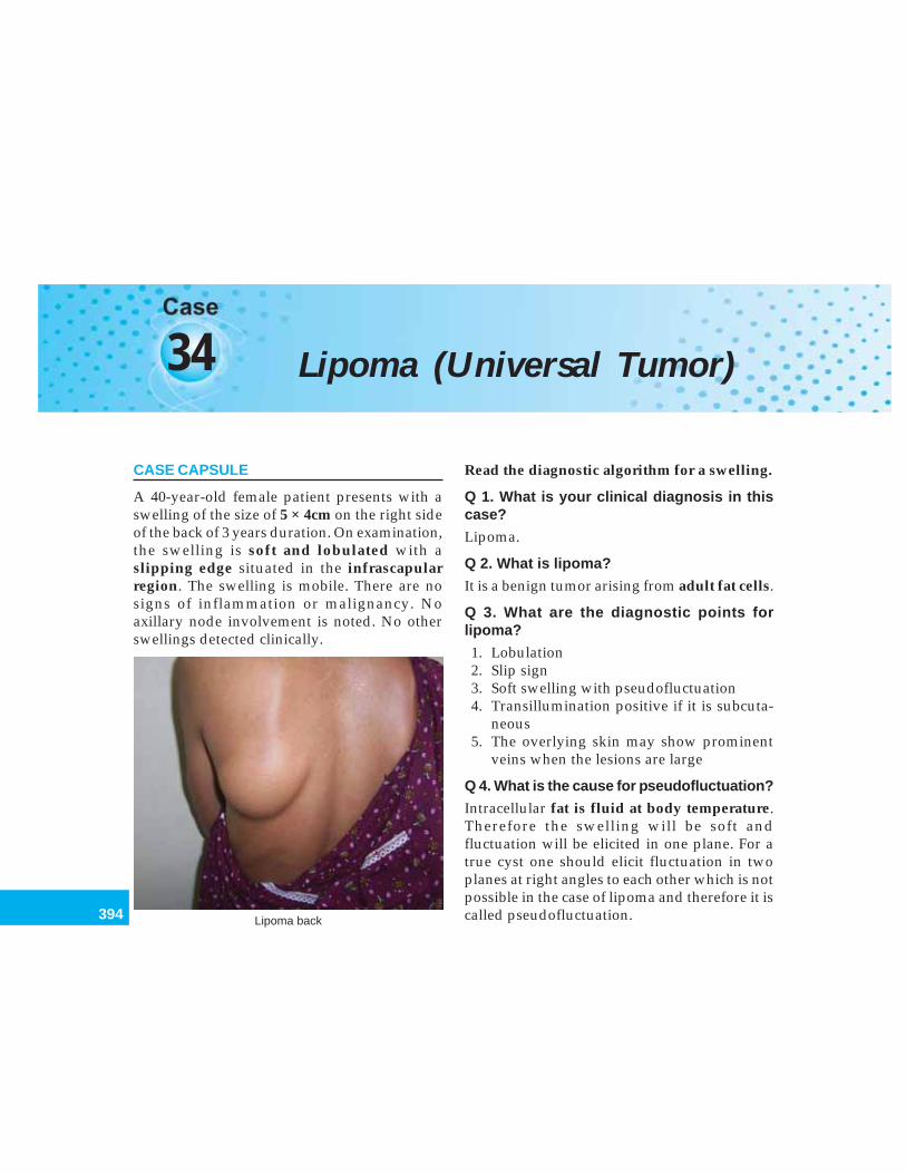
394
Clinical Surgery Pearls
34 Lipoma (Universal Tumor)
CASE CAPSULE
A 40-year-old female patient presents with aswelling of the size of 5 × 4cm on the right sideof the back of 3 years duration. On examination,the swelling is soft and lobulated with aslipping edge situated in the infrascapularregion. The swelling is mobile. There are nosigns of inflammation or malignancy. Noaxillary node involvement is noted. No otherswellings detected clinically.
Read the diagnostic algorithm for a swelling.
Q 1. What is your clinical diagnosis in thiscase?
Lipoma.
Q 2. What is lipoma?
It is a benign tumor arising from adult fat cells.
Q 3. What are the diagnostic points forlipoma?
1. Lobulation2. Slip sign3. Soft swelling with pseudofluctuation4. Transillumination positive if it is subcuta-
neous5. The overlying skin may show prominent
veins when the lesions are large
Q 4. What is the cause for pseudofluctuation?
Intracellular fat is fluid at body temperature.Therefore the swelling will be soft andfluctuation will be elicited in one plane. For atrue cyst one should elicit fluctuation in twoplanes at right angles to each other which is notpossible in the case of lipoma and therefore it iscalled pseudofluctuation.Lipoma back
395
Lipoma (Universal Tumor)
Q 5. What is slip sign?
If the edge of the lump is pressed, the swellingslips from beneath the finger. This can be easilydemonstrated in the case of a subcutaneouslipoma and it is said to be pathognomonic.
Q 6. How will you demonstrate lobulation?
The swelling is compressed between the fingerand thumb of one hand, while its surface whichis now made more prominent, is stroked firmlyby the fingers of the other hand.
Q 7. What is the cause for lobulation?
The lobulation is due to the intervening finestrands of fibrous septa seen between the fatlobules (collection of overgrown fat cells). Thelobules bulge out between the fibrous strandswhen pressure is applied.
Q 8. Why it is called universal tumor orubiquitous tumor?
Lipomas can occur anywhere in the body wherefat is present and therefore it is called universaltumor. The aphorism “When in doubt hedgeon fat” is still true. Whenever you don’t have adiagnosis for a given swelling, lipoma is one ofthe differential diagnoses.
Q 9. Depending on the plane, how will youclassify lipomas?
Lipomas can be classified as:• Subcutaneous • Intraarticular• Subfascial • Subserous• Intermuscular • Subperitoneal• Intramuscular • Subpleural• Paraosteal • Subpericardial
• Subperiosteal • Extradural (type of spinaltumor)
• Subsynovial • Submucous• Intraglandular – seen in breast, pancreas,
under renal capsule, etc.
Q 10. What is the commonest plane oflipoma?
Subcutaneous (between the deep fascia andskin).
Q 11. Is it possible to demonstrate all theclassical signs of lipoma when the swellingis deep?
No. The classical signs of lipoma like slip sign,lobulation and pseudofluctuation, etc. aredemonstrated in subcutaneous lipoma. It is notpossible to demonstrate, slip sign and lobulationin deep lipomas.
Q 12. What will be the finding in transillumina-tion test?
Subcutaneous lipomas may be transilluminant.Big lipomas and deep lipomas may not betransilluminant.
Q 13. What are the diagnostic points forintermuscular lipoma?
On contraction of the concerned muscle thelipoma becomes more prominent, if it isintermuscular. If it is deep to the muscle, thelipoma will become less prominent.
Q 14. What is Naevolipoma?
When a lipoma contains dilated capillaries, it iscalled Naevolipoma. These swellings will bepartially compressible.
Published by dr-notes.com
396
Clinical Surgery Pearls
Q 15. What is Dercum’s disease (Adiposisdolorosa)?
It is a condition where multiple painful or tendersubcutaneous lipomas are seen. It is calledadiposis dolorosa. It is also called neurolipo-matosis.
Q 16. What is fibrolipoma?
When lipoma contains much fibrous tissue it iscalled fibrolipoma.
Q 17. What are the symptoms of lipoma?
1. Swelling (lump)2. Cosmetic (unsightly swelling)3. Interfere with movement4. Pain (due to trauma and fat necrosis)
Q18. What are the complications of lipoma?
1. Myxomatous degeneration2. Calcification3. Pressure effects4. Fat necrosis (if they are situated over
prominent areas and subjected to trauma)5. Ulceration (repetitive friction cause
ulceration)
6. Intussusception in submucous lipoma.
Note: Lipomas never turn malignant.Liposarcomas arise de novo and not in a benignlesion.
Q 19. Which are the areas where liposar-comas are commonly seen?
1. Proximal thigh2. Retroperitoneum3. Mediastinum.
Q 20. What are the investigations required?
1. FNAC from the swelling2. Radiology of the part3. If the swelling is big and sarcoma is
suspected, core biopsy and MRI are done.
Q 21. What is the surgical treatment recom-mended in the given case?
Excision of the swelling under generalanesthesia (if the swelling is small excision isdone under local anesthesia using 1% lignocainewith or without adrenaline).
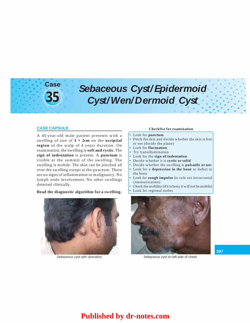
397
Sebaceous Cyst/Epidermoid Cyst/Wen/Dermoid Cyst
35Sebaceous Cyst/Epidermoid
Cyst/Wen/Dermoid Cyst
CASE CAPSULE
A 45-year-old male patient presents with aswelling of size of 3 × 2cm on the occipitalregion of the scalp of 4 years duration. Onexamination, the swelling is soft and cystic. Thesign of indentation is present. A punctum isvisible at the summit of the swelling. Theswelling is mobile. The skin can be pinched allover the swelling except at the punctum. Thereare no signs of inflammation or malignancy. Nolymph node involvement. No other swellingsdetected clinically.
Read the diagnostic algorithm for a swelling.
Checklist for examination
• Look for punctum• Pinch the skin and decide whether the skin is free
or not (decide the plane)• Look for fluctuation• Try transillumination• Look for the sign of indentation• Decide whether it is cystic or solid• Decide whether the swelling is pulsatile or not• Look for a depression in the bone or defect in
the bone• Look for cough impulse (to rule out intracranial
communication)• Check the mobility (if it is bony it will not be mobile)• Look for regional nodes
Sebaceous cyst with ulceration Sebaceous cyst on left side of cheek
Published by dr-notes.com
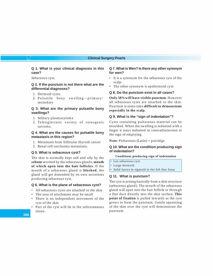
398
Clinical Surgery Pearls
Q 1. What is your clinical diagnosis in thiscase?
Sebaceous cyst.
Q 2. If the punctum is not there what are thedifferential diagnoses?
1. Dermoid cysts.2. Pulsatile bony swelling—primary/
secondary
Q 3. What are the primary pulsatile bonyswellings?
1. Solitary plasmacytoma2. Telengiectatic variety of osteogenic
sarcoma.
Q 4. What are the causes for pulsatile bonymetastasis in this region?
1. Metastasis from follicular thyroid cancer2. Renal cell carcinoma metastasis.
Q 5. What is sebaceous cyst?
The skin is normally kept soft and oily by thesebum secreted by the sebaceous glands, mouthof which open into the hair follicles. If themouth of a sebaceous gland is blocked, thegland will get distended by its own secretionsproducing sebaceous cyst.
Q 6. What is the plane of sebaceous cyst?
• All sebaceous cysts are attached to the skin• The area of attachment may be small• There is no independent movement of the
cyst of the skin• Part of the cyst will lie in the subcutaneous
tissue.
Q 7. What is Wen? Is there any other synonymfor wen?
• It is a synonym for the sebaceous cyst of thescalp.
• The other synonym is epidermoid cyst.
Q 8. Do the punctum exist in all cases?
Only 50% will have visible punctum. Howeverall sebaceous cysts are attached to the skin.Punctum is some time difficult to demonstrateespecially in the scalp.
Q 9. What is the “sign of indentation”?
Cysts containing pultaceous material can bemoulded. When the swelling is indented with afinger it stays indented in contradistinction tothe sign of emptying.
Note: Pultaceous (Latin) = porridge
Q 10. What are the condition producing signof indentation?
Conditions producing sign of indentation
• Lax sebaceous cyst• Large dermoid• Solid faeces in sigmoid in the left iliac fossa
Q 11. What is punctum?
The cyst is arising basically from a skin structure(sebaceous gland). The mouth of the sebaceousgland will open into the hair follicle or througha fine duct directly into the skin surface. Thispoint of fixation is pulled inwards as the cystgrows to form the punctum. Gentle squeezingof the skin over the cyst will demonstrate thepunctum.
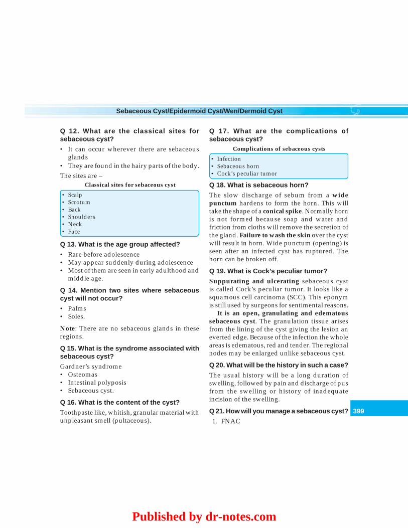
399
Sebaceous Cyst/Epidermoid Cyst/Wen/Dermoid Cyst
Q 12. What are the classical sites forsebaceous cyst?
• It can occur wherever there are sebaceousglands
• They are found in the hairy parts of the body.The sites are –
Classical sites for sebaceous cyst
• Scalp• Scrotum• Back• Shoulders• Neck• Face
Q 13. What is the age group affected?
• Rare before adolescence• May appear suddenly during adolescence• Most of them are seen in early adulthood and
middle age.
Q 14. Mention two sites where sebaceouscyst will not occur?
• Palms• Soles.
Note: There are no sebaceous glands in theseregions.
Q 15. What is the syndrome associated withsebaceous cyst?
Gardner’s syndrome• Osteomas• Intestinal polyposis• Sebaceous cyst.
Q 16. What is the content of the cyst?
Toothpaste like, whitish, granular material withunpleasant smell (pultaceous).
Q 17. What are the complications ofsebaceous cyst?
Complications of sebaceous cysts
• Infection• Sebaceous horn• Cock’s peculiar tumor
Q 18. What is sebaceous horn?
The slow discharge of sebum from a widepunctum hardens to form the horn. This willtake the shape of a conical spike. Normally hornis not formed because soap and water andfriction from cloths will remove the secretion ofthe gland. Failure to wash the skin over the cystwill result in horn. Wide punctum (opening) isseen after an infected cyst has ruptured. Thehorn can be broken off.
Q 19. What is Cock’s peculiar tumor?
Suppurating and ulcerating sebaceous cystis called Cock’s peculiar tumor. It looks like asquamous cell carcinoma (SCC). This eponymis still used by surgeons for sentimental reasons.
It is an open, granulating and edematoussebaceous cyst. The granulation tissue arisesfrom the lining of the cyst giving the lesion aneverted edge. Because of the infection the wholeareas is edematous, red and tender. The regionalnodes may be enlarged unlike sebaceous cyst.
Q 20. What will be the history in such a case?
The usual history will be a long duration ofswelling, followed by pain and discharge of pusfrom the swelling or history of inadequateincision of the swelling.
Q 21. How will you manage a sebaceous cyst?
1. FNAC
Published by dr-notes.com
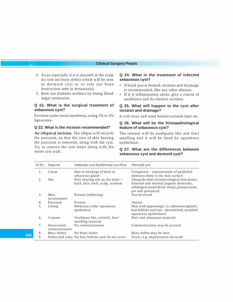
400
Clinical Surgery Pearls
2. X-ray especially if it is situated in the scalp(to rule out bony defect which will be seenin dermoid cyst or to rule out bonedestruction seen in metastasis).
3. Rule out diabetes mellitus by doing bloodsugar estimation.
Q 22. What is the surgical treatment ofsebaceous cyst?
Excision under local anesthesia, using 1% or 2%lignocaine.
Q 23. What is the incision recommended?
An elliptical incision. The ellipse will encirclethe punctum, so that the area of skin bearingthe punctum is removed, along with the cyst.Try to remove the cyst intact along with theentire cyst wall.
Q 24. What is the treatment of infectedsebaceous cyst?
• If frank pus is formed, incision and drainageis recommended, like any other abscess.
• If it is inflammation alone, give a course ofantibiotics and do elective excision.
Q 25. What will happen to the cyst afterincision and drainage?
It will recur and need formal excision later on.
Q 26. What will be the histopathologicalfeature of sebaceous cyst?
The content will be toothpaste like and fowlsmelling and it will be lined by squamousepithelium.
Q 27. What are the differences betweensebaceous cyst and dermoid cyst?
Sl.No. Features Sebaceous cyst/Epidermoid cyst/Wen Dermoid cyst
1. Cause Due to blockage of duct of Congenital – sequestration of epithelialsebaceous gland elements deep to the skin surface
2. Site Hair bearing site on the body— Along the lines of embryological skin fusion.back, face, neck, scalp, scrotum External and internal angular dermoids,
sublingual (superficial/deep), postauricular,pre and postsacral
3. Skin- Present (tethering) Not involvedinvolvement
4. Punctum Present Absent5. Lining Sebaceous cells/squamous Skin with appendage, i.e. sebaceous glands,
epithelium hair follicles and hair – (keratinized, stratifiedsquamous epithelium)
6. Content Toothpast like, whitish, fowl Hair and sebaceous materialsmelling material
7. Intracranial No communication Communication may be presentcommunication
8. Bony defect No bony defect Bony defect may be seen9. Palms and soles No hair follicles and do not occur Occur, e.g. implantation dermoid
401
Sebaceous Cyst/Epidermoid Cyst/Wen/Dermoid Cyst
Q 28. What are the classical sites of dermoidcysts?
• Dermoid cysts may be congenital oracquired.
• The example of acquired dermoid cyst isImplantation dermoid, seen in the handsand feet as a result of trauma
• Congenital dermoid cysts are seen along thelines of embryological fusion:-
Sites where congenital dermoid cysts are seen:
• Midline of the body• Sites where two embryonic processes meet –
a. Outer angle of the orbit (frontonasal processand maxillary process fuse in this region) –External angular dermoid
b. Behind pinna (Postauricular)c. Below the tongue (sublingual dermoid)d. Pre and postsacral dermoid
Q 29. Why is the dermoid cyst at the outerangle of the orbit called external angulardermoid?
That’s because it lies behind the outer end ofthe eyebrow, over the external angular pro-tuberance of the skull. This is a congenitaldermoid cyst.
Q 30. Can dermoid cyst occur at the innerend of the eyebrow (medial end of the eye-brow)?
Yes. It is called internal angular dermoid.
Q 31. What are the complications of externalangular dermoid?
1. Bony depression – by pressure2. Dumb-bell extension into the orbit3. Erosion of the orbital plate of frontal bone
and getting attached to the dura.
Q 32. What is the peculiarity of postsacraldermoid?
It may expand within the spinal canal causingcompression of the cauda eqina. The lesion maybe present at birth but usually noted in the firsttwo years and occasionally may be seen inadulthood.
Q 33. What is the cause for implantationdermoid?
They are usually traumatic. The surfaceectoderm is being driven inside as a result oftrauma (small cut or stab injury) and this willresult in implantation dermoid. They are usuallyseen in fingers and toes. They may be sometimestense and hard. History of old injury orpresence of a scar will give the clue.
Q 34. What are the classical features ofdermoid cyst?
Diagnostic features of dermoid cyst
1. Cystic swelling2. Not transilluminant3. The skin can be pinched (no punctum and
therefore no tethering)—They are seen deep tothe skin in the subcutaneous tissue
4. A bony depression or defect may be felt5. Intracranial communication may be there
Published by dr-notes.com
402
Clinical Surgery Pearls
36 Ulcer
CASE CAPSULE
A 20-year-old male athlete presents with anulcer of size of 6 × 3cm over the shin of the legof 6 months duration. He gives history oftrauma to the shin region while playingfollowed by the development of ulcer. Onexamination, the floor of the ulcer is coveredwith red granulation tissue surrounded by ablue line outer to the granulation and whiteline further outside. There is minimal serousdischarge. The ulcer is mobile and there is nofixity to the deeper structures. The verticalgroup of inguinal nodes are enlarged andtender. On examination, there is no evidence ofvaricose veins. The peripheral pulsations arenormal. There are no sensory deficits and noother neurological problems. Systemicexamination is normal.
Checklist for examination of ulcer
1. Examine the site, edge floor, base andsurrounding tissue (SEFBS)
2. Look for fixity to underlying structures (checkthe movements of the ulcer)
3. Look for peripheral pulsations (arterial ulcer)
4. Look for varicose veins (venous ulcer)5. Check for sensations (Neuropathic ulcers)6. Look for nerve thickening, hypopigmented
patches and other stigmata of Hansen’s disease(to rule out Hansen’s disease)
7. Look for movements of the joints anddeformities (deformity of the foot in DMequinus deformity in venous ulcer anddeformity of Paget’s disease of the bone)
8. Examine the regional nodes (vertical group ofinguinal nodes are involved in lower limbulcers)
9. Look for stigmata of syphilis10. Examine for lymph nodes of neck, axilla, and
inguinal region11. Examination of the chest, to rule out
tuberculosis12. Rule out diabetes mellitus13. Exclude anemia (including sickle cell anemia)
and leukemia14. Look for features of rheumatoid arthritis15. Exclude Paget’s disease of the bone16. General neurological examination to rule out
nervous diseases, spinal injuries, sciatic nerveinjuries, etc.
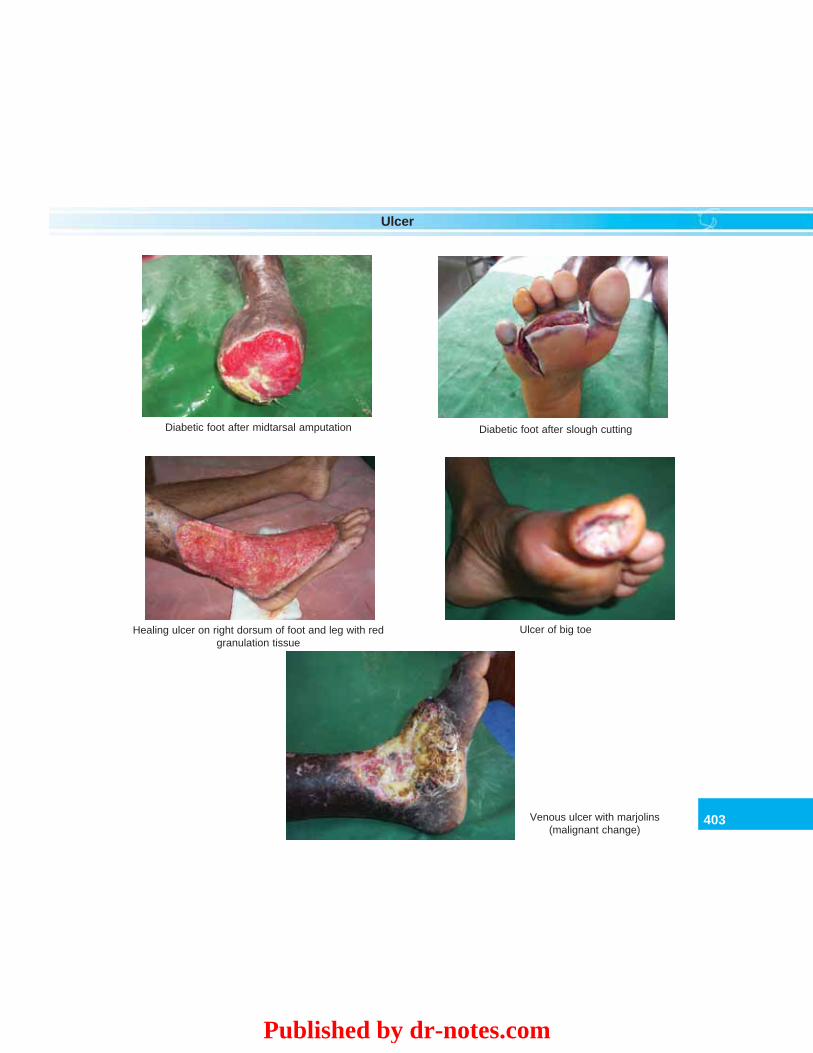
403
Ulcer
Diabetic foot after midtarsal amputation Diabetic foot after slough cutting
Healing ulcer on right dorsum of foot and leg with redgranulation tissue
Ulcer of big toe
Venous ulcer with marjolins(malignant change)
Published by dr-notes.com
404
Clinical Surgery Pearls
Q 1. What is your diagnosis?
A healing traumatic ulcer.
Q 2. Why healing ulcer? Why traumatic?Healing ulcer because
• The floor is covered with red granulation• The blue line outside the granulation is
suggestive of growing epithelium• The white line is suggestive of fibrous tissue• There is no evidence of infection.
Traumatic because• No evidence of arterial, venous, neurological
and other systemic problems• Young athlete• History of trauma is present.
Q 3. What is ulcer?The ulcer is a break in the continuity of anepithelial surface that can occur in the skin ormucosa of the alimentary or respiratory passages.
Q 4. What is slough?Slough is a piece of dead tissue.
Q 5. What is the classification of ulcer?Ulcer may be classified as:1. Acute or chronic2. Painful or painless (malignant ulcers are
painless in the early stages)3. Spreading ulcer or healing ulcer or callous
ulcer4. Nonspecific ulcer or specific ulcer or
malignant ulcer.
Q 6. What are the characteristics of healingulcer?
Characteristics of healing ulcer
• The surrounding skin is not inflamed• The edge shows blue zone (bluish outline of
growing epithelium) and a white zone (fibrosisof the scar).
• Floor will have reddish granulation tissue• Minimal serous discharge may be there
Q 7. What are the characteristics of spreadingulcer?
Characteristics of spreading ulcer
• The surrounding skin of the ulcer is inflamed andedematous
• The floor is covered with slough• No evidence of granulation tissue• Discharge of pus will be there
Q 8. What are the characteristics of callousulcer?
Characteristics of callous ulcer
• Induration of the edge and surrounding tissue• The floor will have pale granulation tissue• No tendency towards healing• Copious serous discharge
Q 9. What are the causes for nonspecificulcers?
The causes for nonspecific ulcers are:1. Traumatic – (Footballers ulcer)
• Physical – Electrical• Chemical – Caustics• Mechanical – Dental ulcer, pressure from
splint, etc.2. Arterial – Atherosclerosis, TAO, Raynaud’s
disease
405
Ulcer
3. Venous – Venous ulcer, Postthromboticulcer, gravitational ulcer.
4. Neuropathic – (Neurotrophic or Perfo-rating) Diabetes, leprosy, tabes dorsalis,spina bifida, paraplegia, syringomyelia
5. Tropical ulcers—Tropical countries6. Metabolic – Diabetes – Gout7. Secondary to diseases like
– Rheumatoid arthritis– Erythrocyanosis frigida– Osteitis Deformans (Paget’s disease of the
bone)– Avitaminosis
8. Ulcers complicating blood diseases– Sickle cell anemia– Mediterranean anemia– Felty’s syndrome (look for spleen)
9. Ulcer occurring on paralysed leg – usuallyseen in anterior poliomyelitis
10. Factitious ulcer (Artefact ulcer)11. Diphtheritic desert sore12. Yaws13. Decubitus ulcer14. Iatrogenic ulcer – extravasation of IV fluid15. Ulcers in congenital arteriovenous fistula16. Miscellaneous - Martorell’s ulcer.
Q 10. What are the specific types of ulcers?Examples of specific ulcers
• Tuberculosis• Syphilitic• Actinomycosis (bacterial)• Soft sore• Herpes simplex• Fungal
Q 11. What are the types of ulcers seen insyphilis?• Hunterian chancre (hard chancre) – Seen in
primary syphilis– 3 – 4weeks after exposure in the external
genitalia– Ulcer is painless– Lymph nodes are enlarged
• Gumma (lymph nodes are not involved) –Seen in tertiary syphilis– Seen in subcutaneous bones like tibia,
sternum, skull, and ulna– In relation to the testis– In the leg.
Q 12. What are the lesions in the secondarysyphilis?• Mucous patches—Sodden thickened
epithelium• Condylomas—Raised flat, white hyper-
trophied epithelium at the mucocutaneousjunction (angles of mouth, anus, and vulva)
• Enlarged lymph nodes——Epitrochlear, andsub occipital nodes.
Q 13. What is soft sore or soft chancre?It is also called Ducrey’s ulcer. They are multipleacute ulcers with yellowish slough seen in theexternal genitalia appearing 3 days afterinfection. They have copious purulentdischarge. The regional lymph nodes areenlarged.
Q 14. What are the types of edges for theulcer?Edges may be—
Published by dr-notes.com
406
Clinical Surgery Pearls
Q15. What is the cause for undermined edgein tuberculosis?
The tuberculosis destroys the subcutaneoustissue faster than it destroys the skin. Theoverhanging skin in this case is blue andunhealthy.
Q 16. What is the difference between floorand base?
• The floor is what you see and the base is whatyou feel
• The floor is the exposed surface of the ulcer.• The base is on which the ulcer rests• Pick up the ulcer between the thumb and
index finger for feeling the base• Slight induration is seen normally for any
ulcer• Marked induration is a feature of squamous
cell carcinoma.
Q 17. What are the classical sites for thecommonly seen ulcers?
Various types of edges of ulcer with examples
Sl no Type of edge Examples
1. Sloping edge Healing ulcers, venous ulcers2. Punched out edge Syphilitic, trophic, ischemic and leprosy3. Undermined edge Tuberculosis, bedsore, carbuncle4. Raised edge Rodent ulcer (basal cell carcinoma)5. Everted edge Squamous cell carcinoma (old name–epithelioma)
Classical sites for commonly seen ulcers
Type of ulcer Site
Venous ulcer Just above the medial malleolus (Gaiter area)Arterial ulcer Tips of toes and between the toes (where the pressure is lowest),
over the malleoli and heel (pressure areas)Neuropathic ulcer Over the heads of the first and second metatarsal.Traumatic ulcer (footballer’s ulcer) Shin (the tibia is subcutaneous and there is lack of underlying
muscle with resultant reduced blood supply)Rodent ulcer (BCC) Above a line joining the angle of the mouth to the lobule of the
earTuberculous ulcer Seen in neck, axilla and groin (where tuberculous lymph nodes
are seen)Gummatous ulcer Over the subcutaneous bones such as sternum, skull, tibia, etc.Trophic ulcers On the heel and ball of the foot
407
Ulcer
Q18. What are the stigmata of Hansen’sdisease?
Stigmata of Hansen’s disease
There are three types of leprosy – the lepromatous,tuberculoid and mixedLepromatous leprosy• Leonine facies (subcutaneous tissue becomes
infiltrated with granulomatous masses)• Loss of outer half of the eyebrow hair• Destruction of nasal cartilages – saddle nose
deformity• Testicular atrophy• GynaecomastiaTuberculoid• Nerve paralysis• Look for tender thickening of the following
nerves– Ulnar nerve at the elbow– Great auricular nerve in the posterior triangle
below the ear– Lateral popliteal nerve around the neck of
fibula• Ulnar claw hand• Claw foot• Trophic ulcers
Q 19. What are the stigmata of syphilis?
Syphilitic stigmata
• Alopecia (loss of hair)• Bossing of the skull• Depression of the bridge of the nose• Interstitial keratitis• Otitis interna• Perforation of the nasal septum• Perforation of the hard palate• Chronic superficial glossitis
• Hutchinson’s teeth• Mucous patches• Condylomas• Enlarged occipital lymph nodes• Enlarged epitrochlear lymph nodes• Gummatous orchitis• Clutton’s joints• Sabre tibia.
Q 20.What is the peculiarity of venous ulcerin relation to the depth of penetration?
The venous ulcers usually do not extend beyondthe deep fascia, unlike ischemic ulcers, whichdestroys the deep fascia and exposes thetendons, bones and joints.
Q 21. What are the types of discharges fromthe ulcer?
Types of discharges and the probable causes
Discharge Cause• Serous discharge In healing ulcer• Serosanguineous Tuberculous ulcer
discharge• Greenish/bluish Ulcer infected with
pseudomonas• Yellow discharge and Staphylococci
creamy pus• Watery and opalescent Streptococcus• Yellow granules Actinomycosis
Q 22. What is the significance of granulationtissue?
Granulation tissue signifies healing. It is usuallypink with red dots. The red dots are seen at thesites of capillary loops.
Published by dr-notes.com
408
Clinical Surgery Pearls
Q 23. What is the significance of bluishgranulation tissue in the floor?
It is suggestive of tuberculosis (Apple - jellygranulation).
Q 24. What is Wash-leather appearance (wetchamois leather)?
It is suggestive of syphilitic ulcer—gummatousulcer.
Q 25. If the floor is covered with black masswhat is the inference?
It is suggestive of malignant melanoma.
Q26. What is neurotrophic ulcer (perfora-ting)?
Neurotrophic ulcers are seen in the followingconditions.
Causes for neurotrophic ulcer
1. Diabetic neuropathy (peripheral neuritis)2. Transverse myelitis3. Syringomyelia4. Tabes dorsalis5. Spina bifida6. Injury to spinal cord7. Sciatic nerve injury8. Hansen’s disease (Leprosy)
Q 27. What are the characteristic features oftuberculous ulcer?
The tuberculous ulcer results as a result ofbursting of caseous lymph node. It is seen inplaces where tuberculous lymph nodes are seen-neck, axilla and groin.
The characteristic features are as follows:
Features of tuberculous ulcer
1. Undermined edge2. Sites—axilla, neck and groin (where tuberculous
nodes are seen)3. Floor is covered with blue granulation tissue
(Apple - jelly granulation)4. Serosanguinous discharge from the ulcer
Q 28. What is lupus vulgaris?
It is nothing but cutaneous tuberculosis seen inthe face and hand. The ulcer heels at the centerand spreads at the periphery (like a wolf) andhence the name lupus means wolf.
Q 29. What are the characteristic features ofvenous ulcer (Named by John Gay in 1867)?
• They are seen in the Gaiter area of the leg(between the two malleoli and the tibialtuberosity) – usually above and behind themedial malleolus
• The ulcer is painless• Ulcer never penetrates the deep fascia• Surrounding skin will be pigmented and
thickened (lipodermatosclerosis)• One or more large feeding veins can be seen
proceeding towards the edge of the ulcer• Varicose veins affecting the long saphenous,
short saphenous and perforating veins willbe visible.
Q 30. What are the precursors of venousulcer?
• Dermatitis (eczema)• Pigmentation (sign of venous stasis)• Splay of venules from the medial malleolus
(flare sign).
409
Ulcer
Q 31. What is postthrombotic ulcer?
Venous ulcer may be secondary to deep veinthrombosis and subsequent valvular incompe-tence resulting in chronic venous hypertensionor it may be secondary to the superficial systemincompetence. When it is secondary to deepvein thrombosis it is called postthrombotic ulcer.The peculiarity of this ulcer is it will be painful.In such cases, varicose veins are lacking in spiteof careful search. Extensive induration is aremarkable feature in this case. The skin appearsto be tethered to the underlined structures,which may extend half way up of the calf.
Q 32. What is gravitational ulcer?
It is another name for venous ulcer.
Q 33. What are the characteristic features ofarterial ulcer?
• They are usually seen at the tips of toes andbetween the toes (heel and malleoli).
• Seen in usually older age group• Destroys the deep fascia and expose the
tendons• Ulcers are punched out• Peripheral pulsations will be absent.
Q 34. What is Meleney’s ulcer?
It is also called synergistic ulcer (symbiotic). Itis due to symbiotic action of microaerophilicnon hemolytic streptococci and hemolyticStaphylococcus aureus. This was originallydescribed in relation to the infected abdominaland thoracic operation wounds. However it canoccur in the leg and hands, arising either de novoare as a complication of preexisting ulcers. Thecharacteristic feature of the ulcer is burrowing
with a resultant undermined edge, which mayextend for 2cm. The ulcer is painful and tenderand shows a tendency to spread. There will bea central purplish zone surrounded by redinflammation initially. This purplish zonebecomes gangrenous producing an ulcer.
Q 35. What is factitious ulcer (Artefact ulcer)?It is also called automutilation ulcer. This is aself-induced ulcer of the leg seen in highlyneurotic individuals or in a litigant desirous ofobtaining compensation. The mode ofproducing the ulcer varies. The ulcer is situatedalways in accessible places like anterior andlateral surface of the leg. Usually the ulcer willhave clean pink healthy look with unusualshapes. If the ulcer is covered with, plaster castso that the wound cannot be tampered thewound will heal.
Q 36. What is the feature of ulcer associatedwith Erythrocyanosis frigida (Bazin’sDisease)?This is usually seen in young women withplump legs and thick ankles living in coldclimates. The patients are troubled bychilblains. Small superficial painful noduleswill be felt in the legs, which are areas of fatnecrosis, which will breakdown to form theulcers. In addition the blood supply to the lowerthird of the leg will be diminished producingischemia of the leg. The skin is abnormallysensitive to temperature changes.
Q 37. What is the cause for ulcer in rheuma-toid arthritis?This is seen in 20% of the patients. It is due tobreakdown of a nodule. The ulcers may be more
Published by dr-notes.com
410
Clinical Surgery Pearls
than one having punched out edge seen in thelateral surface of the lower third of the leg.
Q 38. What is the situation of the ulcersecondary to osteitis deformans (Paget’sdisease?
These small deep ulcers are situated right overthe convexity of the anteriorly bowed tibia. Thebase of the ulcer is bone and the edges aredensely adherent to the bone.
Q 39. What is tropical ulcer?This ulcer is due to infection by Vincent’sorganism (Bacteroides fusiformis) togetherwith many pyogenic bacteria, secondary totrauma or insect bite. It commences as apapulopustule which in a matter of hoursbecomes surrounded by a zone of inflammationand induration. This is accompanied by tenderlymphadenitis. In two or three days the pustulebursts and ulcer forms. The edges areundermined. There is copious sero-sanguineousdischarge. The ulcer will remain indolent for along time. Pain is a constant feature.
The classical features are:• Profuse serosanguineous discharge• Over powering vile odor• Unremitting pain• Minimal constitutional symptoms• Extreme tenacity of the slough.
On healing it leaves a permanent scar whichis circular, parchment like and faintlypigmented.
Q 40. What is the feature of ulcer due toYaws?The causative organism is Treponema pertenue.The primary sore may be found on the legs or
foot or buttocks (of children before they walk).They are painless and in the course of healingform tissue paper like scars. In the tertiary stagemultiple deep ulcers are seen.
Q 41. What is diphtheritic desert sore?
As the name implies it is seen in the desert. Theorganism responsible is Corynebacteriumdiphtheriae. It begins as a papulopustule.Within a few days it will form an ulcer reachingthe size of 1-2cm. The floor will be covered bydiphtheritic membrane, which will be difficultto remove. Rarely the patient shows signs ofperipheral neuritis due to toxins produce bydiphtheria.
Q 42. What is Martorell’s ulcer (hypertensiveulcer)?
This is seen in old age group with athero-sclerosis. A patch of skin on the outer side ofthe calf suddenly becomes gangrenous andsloughs away producing a punched out ulcer.Even though it is an ischemic ulcer all theperipheral pulses will be normal.
Q 43. What is decubitus ulcer?
It is a synonym for bedsore. It is a type of directtraumatic gangrene. The bedsores arepredisposed by factors like—• Pressure• Injury• Moisture• Anemia• Malnutrition.
They typically appear over areas subjectedto pressure like sacrum, gluteal region and heelin bedridden patients.
411
Ulcer
Q 44. What is the warning signal for bedsore?
The bedsore is to be expected over an erythema,which does not change color on pressure.
Q 45. How will you prevent bed sore?Prevention of bedsore
• Skilled nursing• Frequent change of position (2 hourly change of
posture)• Use of adhesive films such as Opsite• Water bed – ripple bed• Keep the area dry
Q 46. Is there an entity called diabetic ulcer?No. Diabetic ulcers are of multiple etiologies andtherefore it is important to identify the cause ofthe ulcer in diabetes. The various causes for ulcerformation in diabetes mellitus are –1. Arterial
– Atherosclerosis (10 yrs earlier than thenormal population)
– Microvascular.2. Neuropathy
– Sensory – glove and stocking type ofperipheral neuropathy
– Motor (Intrinsic muscle paralysis withresultant unopposed action of long flexortendons → shortening of thelongitudinal arches → heads ofmetatarsals are subjected to additionalload during walking).
– Autonomic – Dry skin due to the absenceof sweating
3. Deformity in diabetic foot.Therefore in a given case the ulcer may be
purely neuropathic or arterial or it may be a
combination of factors, which the student hasto identify.
Q 47. What is the commonest site forneuropathic ulcer in Diabetes?
Over the heads of the first and secondmetatarsals.
Q 48. What is Marjolin’s ulcer?
It’s a malignant ulcer developing in burns scaror venous ulcer or edge of any chronic ulcer orchronic discharging sinuses such as osteo-myelitis is called Marjolin’s ulcer. Malignantchange can occur in the tuberculous skinscarring of lupus vulgaris. It is nothing but asquamous cell carcinoma of skin with evertededges or rolled out edges. These changes canbe easily missed and the clinician must alwaysbe on the look out for any changes in the edge.The peculiarity of this malignancy is the absenceof involvement of regional lymph nodes. Thescarring process in this condition destroys thelymphatics in the ulcer area.
Q 49. What is the management of the ulcer?
The principles of management of the ulcer areas follows:1. Identify and correct the comorbid factors
like DM, anemia, leukemia, etc. by doing:• FBS - If diabetic, control the diabetes• PPBS• Peripheral smear• Hemoglobin• Total leukocyte count• X-ray chest – to rule out tuberculosis.
Published by dr-notes.com
412
Clinical Surgery Pearls
2. Determine the etiology of the ulcer:• Biopsy from the edge of ulcer• If pus is present, send for culture
and sensitivity and give appropriateantibiotic
• X-ray of the part to rule out osteomyelitis.3. Adequate drainage and desloughing4. Care of the ulcer:
• Daily dressing• Avoid antiseptic solutions – impair
capillary circulation• Use normal saline for cleaning
• Use nonadherent and nonallergicdressing
• Use microporous poly urethane filmwhich are permeable to gases and waterand impermeable to microorganisms.
5. Treat the underlying cause:• E.g. Antituberculous drugs in the case of
tuberculous ulcer• Treat the varicose veins in the case of
venous ulcer• Revascularization in the case of arterial
ulcers6. Excision of the ulcer and skin grafting: If
the ulcer is not healing.
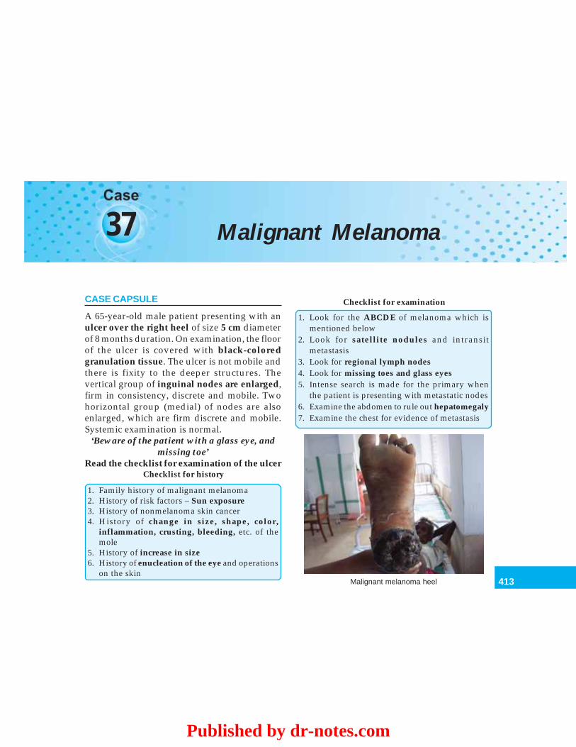
413
Malignant Melanoma
37 Malignant Melanoma
CASE CAPSULE
A 65-year-old male patient presenting with anulcer over the right heel of size 5 cm diameterof 8 months duration. On examination, the floorof the ulcer is covered with black-coloredgranulation tissue. The ulcer is not mobile andthere is fixity to the deeper structures. Thevertical group of inguinal nodes are enlarged,firm in consistency, discrete and mobile. Twohorizontal group (medial) of nodes are alsoenlarged, which are firm discrete and mobile.Systemic examination is normal.
‘Beware of the patient with a glass eye, andmissing toe’
Read the checklist for examination of the ulcerChecklist for history
1. Family history of malignant melanoma2. History of risk factors – Sun exposure3. History of nonmelanoma skin cancer4. History of change in size, shape, color,
inflammation, crusting, bleeding, etc. of themole
5. History of increase in size6. History of enucleation of the eye and operations
on the skin
Checklist for examination
1. Look for the ABCDE of melanoma which ismentioned below
2. Look for satellite nodules and intransitmetastasis
3. Look for regional lymph nodes4. Look for missing toes and glass eyes5. Intense search is made for the primary when
the patient is presenting with metastatic nodes6. Examine the abdomen to rule out hepatomegaly7. Examine the chest for evidence of metastasis
Malignant melanoma heel
Published by dr-notes.com
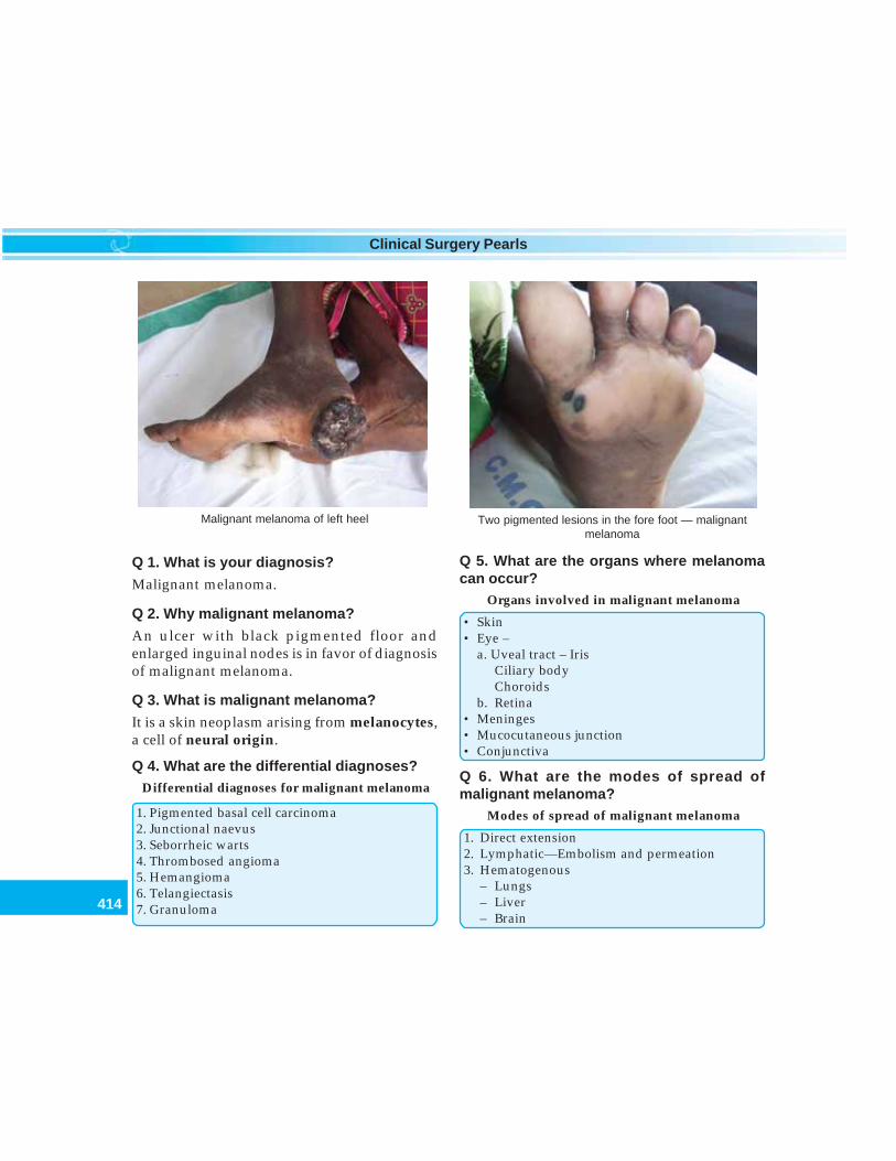
414
Clinical Surgery Pearls
Q 1. What is your diagnosis?
Malignant melanoma.
Q 2. Why malignant melanoma?
An ulcer with black pigmented floor andenlarged inguinal nodes is in favor of diagnosisof malignant melanoma.
Q 3. What is malignant melanoma?
It is a skin neoplasm arising from melanocytes,a cell of neural origin.
Q 4. What are the differential diagnoses?Differential diagnoses for malignant melanoma
1. Pigmented basal cell carcinoma2. Junctional naevus3. Seborrheic warts4. Thrombosed angioma5. Hemangioma6. Telangiectasis7. Granuloma
Q 5. What are the organs where melanomacan occur?
Organs involved in malignant melanoma
• Skin• Eye –
a. Uveal tract – IrisCiliary bodyChoroids
b. Retina• Meninges• Mucocutaneous junction• Conjunctiva
Q 6. What are the modes of spread ofmalignant melanoma?
Modes of spread of malignant melanoma
1. Direct extension2. Lymphatic—Embolism and permeation3. Hematogenous
– Lungs– Liver– Brain
Malignant melanoma of left heel Two pigmented lesions in the fore foot — malignantmelanoma
415
Malignant Melanoma
– Bones– Intestines– Breast
Q 7. What is the spread of malignantmelanoma of the uveal tract?
There are no lymphatics in the uveal tract. Theyrarely metastasize to lymph nodes.
Q 8. What is a naevus?
The word naevus means a lesion which ispresent since birth.
Q 9. What is a mole?
Benign melanin producing lesions are calledmoles.
Q 10. What is the average number of moles aperson will have?
80-100 moles in most Caucasians.
Q 11. In which area of the body moles arecommonly seen?
They are most common in limbs, face andmucocutaneous junctions (The mouth andanus).
Q 12. What are the layers of the skin?
The skin has got epidermis and dermis.The epidermis has got five layers.The five layers of the epidermis are:• Stratum cornium• Stratum lucidum• Stratum granulosum• Stratum spinosum (prickle cell layer)• Stratum basale (Basal layer).
Q 13. In which layer of the skin themelanocytes are seen?
They are normally found in small numbersamong the cells of the basal layer of epidermis.
Q 14. What is melanoblast? (PG)
They are believed to originate in the neural crest.They have the power of forming the pigmentand they are seen in the basal layer of epidermis.The cells are Dopa positive and Fontanapositive.
Q 15. What are melanophores? (PG)They are dermal macrophages carrying thepigment. They are Dopa negative and Fontanapositive.
Q 16. What is the pathology of a mature adultmole? (Intradermal mole)They consist of clusters of melanocytes inthe dermis and therefore they are calledintradermal mole. Macroscopically the mole canbe flat or raised, smooth or warty, hairy ornonhairy. They hardly ever turn malignant.
Q 17. What is a junctional mole?If the movement of the melanocytes stop beforethey have all migrated into the dermis, therewill be clusters of cells at various stages ofmaturity in the epidermis and dermis. Thislesion is called junctional mole. Junctionalmoles are immature and unstable and can turnmalignant. The majority of the malignantmelanomas begin in junctional moles.
Q 18. In which part of the skin you getjunctional moles?• Palms of the hands• Soles of the feet• External genitalia.
Published by dr-notes.com
416
Clinical Surgery Pearls
Note: Higher incidence of malignant melanomais seen in these sites.
Q 19. What is a compound mole?When intradermal and junctional features are bothpresent in one mole it is called compound mole.
Q 20. What is juvenile mole (spitz naevus)?
A mole showing junctional activity before pubertyis called juvenile mole (pigmented spitz naevus).They finally become mature intradermal moles.
Q 21. What is a blue naevus?
When the melanocytes migrate to the bottom ofthe dermis and into the subcutaneous tissue, thelesion will get a blue appearance and it is calleda blue naevus.
Q 22. In which ethnic group malignantmelanoma is more common?
It is more common in Caucasians living in hotcountries such as Australia (because of thegreater quantity of UV light exposure). It is lesscommon in Negroes.
Q 23. What is the incidence of malignantmelanoma?
• The incidence of malignant melanoma is rising• It forms 5% of the cutaneous malignancy• Seen 20 times more in whites• Lowest incidence in Asia• The incidence has doubled in Britain,
Norway, Canada and America• The incidence has quadrupled in Australia.
Q 24. What are the types of malignantmelanoma?
Types of malignant melanoma
• Superficial spreading• Nodular• Lentigo malignant• Acrolentigenous• Amelanotic
Q 25. What are the growth phases ofmalignant melanomatous lesions?
It has two types of growth –1. Radial growth phase – e.g.: The superficial
spreading type (all melanomas show radialgrowth phase except nodular melanoma.Radial growth is an intraepidermalgrowth).
2. Vertical growth phase – e.g.: The nodularmelanoma (The vertical growth is the tumorgrowth in dermis leading to noduleformation).
Q 26. What are the features of superficialspreading type of malignant melanoma?
• It forms 70% of the lesions• Peak incidence in the 5th decade• Long radial growth phase• Wood’s lamp can identify radial growth
phase• Dark brown, blue, black, pink or gray, white
in color.
Q 27. What are the features of nodularmelanoma?
• It is the most malignant type of melanoma• Forms 12% of the lesions• Predominant growth phase is vertical• Twice common in men
417
Malignant Melanoma
• Ulceration of the lesion is seen• Crust formation also seen
Q 28. What are the features of lentigomalignant melanoma?
• It forms 10-15% of the lesions.• It arises in Hutchinson’s melanotic freckle• Seen in old age• Seen in sun exposed skin• Lesions are mainly seen in head and neck.
Q 29. What is Hutchinson’s lentigo?
1. This is a term used to describe a large areaof dark pigmentation, seen commonly onthe face and neck in late adult life. It hasgot two special features –
2. Late development3. High incidence of malignant change.
Some pathologists consider this as pre-cancerous. The surface is smooth with someraised areas suggesting the sites of junctionalactivity.
Q 30. What is Hutchinson’s halo?
• This is a halo of brown pigment in the skinaround the melanoma and satellite nodules.The halo is suggestive of malignant changein a preexisting mole.
Q 31. What are the features of acro-Lentigenous?
• It affects the palms, soles, and beneath thenails (Subungual)
• More seen in dark skinned group – 70% ofmelanomas in black (It will affect any ethnicgroup)
• No history of sun exposure• It forms 2-8% of the melanomas in white• 46% of melanomas in Asians• Usually > 3 cm in size• Late presentation is a common feature• The melanoma is more aggressive.
Q 32. Why the amelanotic melanoma is calledso?
Here the malignant melanomatous lesion is notblack and they appear as white. Therefore it iscalled amelanotic melanoma.
Q 33. What are the features of amelanoticmelanoma?
• The prognosis of this type is worse thannodular melanoma
• The lesions appear pink• There will be delay in the diagnosis.
Q 34. What is the ABCDE system for evalua-tion of pigmented skin lesion?
Look for the following things in a preexistingpigmented lesion. If they are present it issuggestive of malignancy.
ABCDE system for evaluation of malignantchange in a pigmented lesion
A - AsymmetryB – Border irregularityC – Color – variegationD – Diameter > 6 mmE – Elevation
Q 35. What is the Glasgow seven pointchecklist?
Published by dr-notes.com

418
Clinical Surgery Pearls
Glasgow seven point checklist
1. Change in size2. Change in shape3. Change in color4. Inflammation5. Crusting and bleeding6. Sensory change7. Diameter > 6 mm
Q 36. What is Satellite lesion?
A skin or subcutaneous lesion within 2cm of theprimary tumor is called satellite lesion. This isconsidered as an extension of the primary andis categorized as N2C (stage III C) in the newAJCC 6th edition.
Q 37. What is intransit metastasis?
Skin or subcutaneous metastasis more than 2cmfrom the primary lesion but not beyond regionallymph node basin is called intransit metastasis.
Q 38. What are the risk factors incriminatedin malignant melanoma?
The various risk factors are—1. Sun exposure – most common (50%)—sun
exposure before the age of 10 years andsignificant adult exposure
2. Multiple atypical naevi3. Multiple benign naevi > 1004. Giant pigmented hairy naevus (20 cm
diameter or > 5% of the body surface)5. Dysplastic naevus syndrome6. Xeroderma pigmentosum7. Nonmelanoma skin cancer (NMSC)8. Previous skin cancer9. Immunosuppression
10. Use of tanning lamps/photochemotherapy11. Family history (8-12%).
Q 39. What is the role of heredity in malignantmelanoma?
• When three first degree relatives are affectedit should be suspected.
• There will be mutation in CDKN2A gene• Genetic predisposition• 8 – 12% of all melanomas are hereditary• Second primary will be seen in 3-5% of cases.
Q 40. What is Breslow’s thickness?
The thickness of the malenomatous lesion ismeasured from the top of the basal layer, to thebase of the tumor with the help of a ocularmicrometer. This is called Breslow’s thickness,which is the most important prognostic factorfor malignant melanoma. The originaldimensions described by Breslow are notconsidered now for classification.
Q 41. What is a thin melanoma?
A melanoma which is less than 1 mm is calledthin melanoma. The survival of patients withthis lesion is 95%.
Q 42. What is intermediate thickness lesion?
Lesions of thickness between 1 – 4 mm are calledintermediate thick lesions.
Q 43. What is thick melanoma?
Lesion with thickness more than 4 mm is calledthick melanoma.
Q 44. What are the bad prognostic factorsfor melanoma? (PG)
419
Malignant Melanoma
The bad prognostic factors are:
Bad prognostic factors
1. Tumor thickness in mm2. Older age3. Male sex4. Site – head and neck, trunk and acral5. Ulceration – negative impact on survival6. Angiogenesis7. Vascular invasion
Q 45. What is the new AJCC staging systemfor malignant melanoma (6th edition)? (PG)
The summary of changes in 6th edition are –• The number of lymph nodes are considered
rather than the gross dimension• The nodes are further divided into micro-
scopic and macroscopic• The serum LDH is included for M catego-
rization• Level of invasion of the lesion is used only
for T1• Ulceration of the lesion is included in the
staging and the lesion is upstaged when themelanoma is ulcerated
• Satellite lesion and intransit metastasis willform a single stage, i.e. IIIc disease.
T - classification
T0 No evidence of primary tumorTis Melanoma in situT1 Melanoma < 1 mm with or without
ulcerationT1a Melanoma < 1 mm Clark’s level II or III,
no ulcerationT1b Melanoma < 1 mm Clark’s level IV or V or
with ulceration
T2 Melanoma 1.01 to 2 mm in thickness withor without ulceration
T2a Melanoma 1.01 to 2 mm in thickness, noulceration
T2b Melanoma 1.01 to 2 mm in thickness, withulceration
T3 Melanoma 2.01 to 4 mm in thickness, withor without ulceration
T3a Melanoma 2.01 to 4 mm in thickness, noulceration
T3b Melanoma 2.01 to 4 mm in thickness, withulceration
T4 Melanoma greater than 4 mm in thicknesswith or without ulceration
T4a Melanoma > 4 mm in thickness noulceration
T4b Melanoma > 4 mm in thickness withulceration
Regional lymph nodesNX Regional lymph nodes cannot be assessedN0 No regional lymph node metastasisN1 Metastasis in one lymph nodeN1a Clinically occult (microscopic) metastasisN1b Clinically apparent (macroscopic) metastasisN2 Metastasis in two to three regional nodes
or intralymphatic regional metastasiswithout nodal metastases
N2a Clinically occult (microscopic) metastasisN2b Clinically apparent (macroscopic) metastasisN2c Satellite or intransit metastasis without
nodal metastasis.N3 Metastasis in four or more regional nodes,
or matted metastatic nodes, or intransitmetastasis or satellite (s) with metastasisin regional node
Distal metastasis (M)MX Distant metastasis cannot be assessedM0 No distant metastasis
Published by dr-notes.com
420
Clinical Surgery Pearls
M1 Distant metastasisM1a Metastasis to skin, subcutaneous tissues or
distant lymph nodesM1b Metastasis to lungM1c Metastasis to all other visceral sites or
distant metastasis at any site associatedwith an elevated serum lactic dehydrogenase(LDH)
Clinical stage grouping (used after completeexcision of the primary melanoma withclinical assessment for regional and distantmetastasis).
Stage Tumor size N M0 Tis N0 M0IA T1a N0 M0IB T1b N0 M0
T2a N0 M0IIA T2b N0 M0
T3a N0 M0IIB T3b N0 M0
T4a N0 M0IIC T4b N0 M0III Any T N1 M0
Any T N2 M0Any T N3 M0
IV Any T Any N M1
Note:• Melanoma with lymph node or satellite
nodule or intransit metastasis is stage III• M1 disease is stage IV• Without lymph node is stage I or II depending
on the thickness of the lesion and ulceration.
Q 46. What is Clark’s level?
This is used for the pathological assessment ofthe depth of invasion of the malignant melanoma.
Because of the discrepancy in assessment of thepathological depth amongst pathologists Clark’slevel is used only for T1 lesions.
Clark’s levels for depth of invasion
Level I Lesion confined to the epidermisLevel II Lesion extending to the papillary dermisLevel III Lesion filling the papillary dermisLevel IV Lesion involving the reticular dermisLevel V Lesion extending to the subcutaneous
tissue
Q 47. What is the confirmatory investigationfor the diagnosis of this lesion?
Biopsy.
Q 48. What type of biopsy is recommendedand what are the precautions taken?
• Excision biopsy is the procedure of choice.• There is no role for shave biopsies• Incision biopsy is done only for large lesions
– The incision biopsy may be taken from thethickest part as the last resort. It will interferewith the accurate Breslow’s thicknessdetermination which decides the ultimateexcision margin (some areas may be thickerthan others and therefore full histologicalexamination is not possible)
• Clark’s level of invasion and Breslow’smaximal thickness should be reported
• The deep fascia is not excised, unlessinvolved
• In small lesions primary closure may beattempted
• Margins may have to be altered for cosmetic,or functional reasons
• In other places it is better to consider a splitskin graft or fasciocutaneous flap.
421
Malignant Melanoma
Note: Original excision margin recommendedby Handley in 1907 was 5 cm, which is havingno clinical evidence support.
Q 49. What are the current guideline for themargin of excision?
• In situ – 0.5 cm• Less than 1mm – 1 cm margin is accep-
thickness lesions table• 1 – 2 mm thickness – 2 cm margin is recom-
lesion mended• More than 2 mm – 2 or more cm margin
Q 50. What is the margin of excisionrecommended for subungual melanoma?
1cm margin is not possible in this situationwithout excision of the terminal phalanx.In general amputation of the digit isrecommended.
Q 51. What are the diagnostic immunohisto-chemical markers for malignant melanoma?
(PG)
Immunohistochemical markers for malignant melanoma
Antigen Results Specificity
S-100 + Most melanoma, neural tumors, some of breast Ca.Cytokeratin + Epithelial tumors, rarely + in melanoma. Occasionally
positive in recurrent and metastatic melanomaHMB – 45 (Premelanosomal protein) + Specific for melanomaNerve growth factor receptor + Cells of neural origin, melanomaMel – 5 (tyrosine related protein) + Radial growth phase and most vertical growth phase of
melanomas
Q 52. What is the management of lymphnode?
• If clinically enlarged FNAC is preferable toexcision biopsy for confirmation
• Open biopsy may increase the risk of tumorspillage
• If open biopsy is required the incision shouldbe placed in such a way that it can be excisedin continuity with lymph node field.
Q 53. What is the management of positivenode?
• Therapeutic radical lymph node dissection isrecommended
• For the lower limb it is radical ilioinguinalblock dissection
• Limited dissections like superficial femoraldissection and node picking are not recom-mended.
Q 54. What is the role of Elective Lymph NodeDissection – ELND (means managing thenodes, which are not involved)? (PG)
Published by dr-notes.com
422
Clinical Surgery Pearls
• Only 25% of the patients will show histologicaloccult metastasis in such dissection
• It is unnecessary therefore in 75% ofpatients
• There is no survival advantage for ELND• Thus it is not recommended routinely.
The arguments in favor of ELND are—• Incidence of positive nodes is 25-65%• Survival rate are lower in patients who
develop nodes• The recurrence rate is higher when there is
extra capsular spread or multiple nodes• Patient may fail to attend regular follow up• The node dissection carries negligible morbidity.
Q 55. What is sentinel lymph node biopsy?(PG)
The anatomical concept is that lymphatic fromdefined regions of skin drain specifically to aninitial node or nodes (Sentinel nodes) prior todisseminating to other nodes in the same or nearby basins.
Sentinel lymph node biopsy is a techniqueinitially described by Cabanas in 1974 forpenile carcinoma. This was popularized byMorton in 1994 for melanoma. The aim of thistechnique is to identify those patients in whomit may be appropriate to carry out elective lymphnode dissection by identifying the micro-metastasis in the sentinel node. The techniqueis to identify the first echelon lymph node(sentinel node) by lymphoscintigraphyfollowing intradermal injection of radioactivetechnetium99 sulphur colloid around theprimary site. The position of this node is markedon the skin surface. The lymph node is identified
intra- operatively by injecting patent blue dyearound the site of the primary lesion. A handheld gamma probe may also help to locate thenode. The node is removed and subjected forhistopathological examination. If the node isfound to be, pathologically positive radicallymph node dissection is carried out.
Q 56. What are the indications for sentinellymph node biopsy? (PG)
• It should be performed at the time of wideexcision for primary melanomas thicker than1mm in size.
• Less than 0.75 mm lesions are very unlikelyto have regional metastasis and do notrequire SLNB.
• Lesions between 0.75 and 1mm SLNB maybe done by a selective policy.
Q 57. What are the advantages of sentinellymph node biopsy? (PG)
The node is send for frozen section, routine Hand E staining and immunohistochemicalstaining.• The pathologist can study 1 to 2 nodes fully
rather than 10 to 30 nodes.• Routine H and E staining can identify 1 tumor
cell/10,000 normal size• Reverse transcriptase PCR will identify
tumor protein production and one tumor cellper 1 million is identifiable.
• Histology negative but PCR positive tumorsare having worse prognosis than histologynegative.
• PCR status of the lymph node is the mostsignificant predictor of survival even overtumor thickness.
423
Malignant Melanoma
Q 58. What are the investigations in the givencase capsule?
1. Since wide excision is not possible in thiscase because of the fixity, take incisionbiopsy from the thickest part of the lesion.
2. X-ray chest to rule out metastasis.3. Ultrasound abdomen to rule out liver
metastasis4. Estimation of LDH to rule out visceral
metastasis.
Q 59. What is the staging in the given case?
Since there is inguinal lymph node enlargementit is Stage III.
Q 60. What is the recommended surgicaltreatment in the given case?
Since it is a big ulcer with fixity to the calcaneumhe needs a below knee amputation for theprimary along with ilioinguinal block dissectionfor the nodes.
Q 61. What is desmoplastic melanoma?(PG)
In desmoplastic melanoma the followingfeatures are seen—• Histologically spindle cells are seen in fibrotic
stroma with lymphocytic infiltration• There is high rate of recurrence• Wider margins are desirable• Minimum of additional 1 cm margin is
recommended• They are rare and can arise de novo.
Q 62. What is neurotropic melanoma?(PG)
Neurotropic melanoma is characterized by –
• Peri/Intraneural infiltration• It forms > 30% of desmoplastic melanoma• High rate of recurrence• Additional 1cm margin is recommended.
Q 63. Any special form of treatment recom-mended for childhood melanoma? (PG)
• Melanoma in childhood is rare• The differential diagnosis is pigmented spitz
naevus (juvenile melanoma) which is acompound naevus in childhood
• Same treatment is recommended as in adults.
Q 64. What is the management of melanomain pregnancy? (PG)
• Same treatment as in nonpregnant patient• Early termination of pregnancy when the
diagnosis is made• Delay pregnancy for 2 years after treatment
for melanoma.
Q 65. What is the incidence of mucosalmelanomas? (PG)
• It forms < 1% of all melanomas• Commonest site is oral cavity.
Q 66. What are the locations where you getmucosal melanomas?
Mucosal melanomas are seen in the followingsituations –
Sites of mucosal melanoma
• Oral cavity• Rectum• Anal canal• Female genital tract• Larynx• Oropharynx
Published by dr-notes.com
424
Clinical Surgery Pearls
It originates from the pigment cells of thechoroid, ciliary body or iris.
Q 71. What are the clinical presentations ofmelanoma of eye?It can present as—• Reduction in vision• Vitreous hemorrhage• Elevated pigmented lesion in the eye.
Note: The more posterior the lesion in the eyethe more malignant it is likely to be.
Q 72. What is the spread of melanoma of theeye?
The spread is often delayed for many years andoften goes to the liver. Therefore the aphorism –
‘Beware of the patient with a glass eye, andan enlarged liver’—There will be history ofenucleation of the eyeball years back formelanomatous lesion followed by use ofglass eyes. The enlarged liver may be due tometastasis.
Q 73. What is the treatment of melanoma ofthe eye?
The following options are available.Treatment options in melanoma of the eye
• Laser coagulation• Radioactive plaques• Radiotherapy• Local excision using hypotensive anesthesia• Enucleation
Q 74. What is the investigations of choice forthe diagnosis of melanoma of choroid? (PG)
Ultrasound examination which will show asolid tumor.
• Hypopharynx• Nasopharynx• Paranasal sinuses• Esophagus (less common)
Q 67. What are the features of oral mela-nomas? (PG)
The characteristic features of oralmelanoma
• Age incidence is between 40 and 60 years• 50% are on hard palate• 25% are on the upper gingiva• 30% are preceded by an area of hyperpigmen-
tation• Pigmentation varies black to brown• Red color indicates nonpigmented melanoma• It may be flat or raised or nodular, later become
ulcerated and may bleed• It is notorious for rapid growth and poor
prognosis• Destruction of the underlying bone is a feature• 50% will have metastasis at the time of
presentation
Q 68. What is the prognosis of mucosalmelanomas? (PG)They are having poor prognosis. The five-yearsurvival rate appears to be about 5%.
Q 69. What is the treatment of mucosalmelanomas? (PG)• Wide excision and reconstruction followed
by radical dissection of the nodes andradical radiotherapy
• In the case of anal canal an abdominoperinealresection is recommended.
Q 70. What is the origin of melanoma in theeye?
425
Malignant Melanoma
Q 75. What is the role of adjuvant chemo-therapy after surgery in melanoma of theskin? (PG)
• At present chemotherapy as an adjuvantmodality is being given for high-risk patientswith no evidence of systemic metastasis onclinical trial basis.
• The only approved agent for therapy isinterferon alpha – 2b.
Q 76. What you mean by high-risk group?
High-risk group consists of stages IIb, IIc andIII. (node positive and thick node negative).
Q 77. What are the other chemotherapeuticagents used in melanoma?
• Dacarbazine (DTIC) is the single mostimportant drug with a response rate of 15-30%
• CVD Regimen is used usually – Cisplatin,Vinblastine and Dacarbazine.
Q 78. What is the treatment of local recur-rence? (PG)
Wide excision with 2cm margin is recommended.
Q 79. What is the treatment of intransitmetastasis? (PG)
a. If they are few in number surgical excisionwith a margin of surrounding normalcutaneous and subcutaneous tissues(excised en block with primary if possibleand closure of the defect with flap or graft).
b. Intralesional therapy with GranulocyteMacrophage Colony Stimulating Factor(GM – CSF)
c. Laser therapy
d. Radiotherapye. Hyperthermic isolated limb perfusion.
Q 80. What is hyperthermic isolated limbperfusion? (PG)
It is introduced by Sydney Melanoma Unit in1993.
Isolating blood circuit to the extremity andadministering chemotherapeutic agent regio-nally at a concentration of 15-25 times higherwithout side effects is called hyperthermicisolated limb perfusion. A pneumatic tourniquetabove the limb is applied and a small borevascular catheter is used for introducing theagent.• Gives significant palliation of locoregional
symptoms.• No improval in survival is noted.
Q 81. What is the agent used for isolated limbperfusion? (PG)
• Melphalan is the agent of choice and this isgiven with mild hyperthermia
• Melphalan + TNF also tried• Melphalan + Actinomycin D also given.
Q 82. Is there any role for radiotherapy inmalignant melanoma?
Even though melanoma was considered to beresistant to radiotherapy it is indicated in certainspecial situations with the dose mentioned below.• More than 4Gy dose-high response rate• Large facial lentigomalignant melanoma• Medically inoperable, those refusing surgery,
thick melanoma, desmoplastic melanoma• Incomplete excision of regional nodes
especially head and neck
Published by dr-notes.com
426
Clinical Surgery Pearls
• For metastasis in lung, bone, brain, LN andsubcutaneous nodule.
Q 83. What are the advantages of radio-therapy? (PG)
The advantages are—• Lower recurrence rate following adjuvant
XRT (10%-25%)• 50% response rate for skin lesions and 30%
for brain• Effect on survival is doubtful.
Q 84. What is the management of metastaticdisease?
• <5 % of patients with metastasis survive 5years (6-10 months)
• Resection of solitary mets can increase 5 yrsurvival to 30 %
• Lung → lobectomy• Liver → lobar resection
Q 85. What is melanuria? (PG)
• Presence of melanin in urine is calledmelanuria
• It is seen in cases of extensive visceralinvolvement
• It is a terminal feature of malignantmelanoma.
Q 86. What is the chemotherapeutic agentrecommended for metastatic disease? (PG)
• Interferon α α α α α 2b is being tried with a responserate of 15-20% for skin, subcutaneous nodulesand lymph nodes
• High dose bolus interleukin - 2 is also tried.However it is having severe side effects with
capillary leak syndrome, hypotension andrenal insufficiency.
Q 87. What is targeted therapy for malignantmelanoma? (PG)
2/3rd of melanomas show activating mutationin BRAF. There is elevated Raf kinase activity.Blocking the Raf pathway using BAY 43 – 9006is being tried as targeted therapy.
Q 88. What is spontaneous regression?
Spontaneous regression is an immunologicalphenomenon. Melanoma is considered animmunogenic human solid tumor. There will beno physical evidence of primary in 3 to 15% ofpatients and the patients may present withlymph node metastasis. In such cases, intensesearch should be made for the primary. Ifprimary is not found, treat the metastasis. Theregression may be—• Partial / complete regression• Presenting as variation in color and irregular
borders• It is a host antitumor phenomenon• Antimelanoma antibodies are found in blood• Regression is associated with risk of
metastasis.
Q 89. What are the pathological types ofregression? (PG)
Pathologically the regression may be early,intermediate or late• Early – Lymphocytes are seen disrupting
nests of melanoma cells• Intermediate – There is loss of continuity of
the lesion with mild fibrosis• Late – There is extensive horizontal fibrosis.
427
Malignant Melanoma
Q 90. Why there is increased chance formetastasis in cases of spontaneousregression? (PG)
A subpopulation of melanoma cells escapingimmune recognition is responsible for meta-stasis.• Look for primary and excise• Surgery for the metastatic lymph node.
Q 91. What is melanoma vaccine? (PG)
Unlike other vaccines the melanoma vaccine isused for treatment and not for prevention.• Irradiated, allogenic cultured melanoma cells
with or without BCG is used for treatment(polyvalent melanoma vaccine– 3 melanomacell line - Cancer vax)
• The vaccine is prepared from the melanomacells of the patients (autologous tumorvaccine) large amount of tumor is necessaryfor the production of vaccine.
• Vaccines prepared from viruses are also used- Vaccinia Melanoma Oncolysates - Melacin
• GM2 ganglioside-based vaccine• DNA immunization.
Q 92. What is the recommended follow upfor malignant melanoma? (PG)• Every 3 months for 3 years• Every 6 months for 2 more years• Then yearly• CXR annually.
Q 93. What are the preventive measures formelanoma?• Genetic – autosomal dominant mode of
transmission• Sun exposure - intermittent intense exposure
to UV is responsible for melanoma• Monthly skin self examination is
recommended• Clinical skin examination once or twice a year
in high-risk areas• Low threshold for simple excision of
pigmented lesions• Use of sun screen.
Published by dr-notes.com
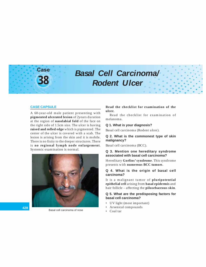
428
Clinical Surgery Pearls
38Basal Cell Carcinoma/
Rodent Ulcer
CASE CAPSULE
A 60-year-old male patient presenting withpigmented ulcerated lesion of 2years durationat the region of nasolabial fold of the face onthe right side of 1.5cm size. The ulcer is havingraised and rolled edge which is pigmented. Thecenter of the ulcer is covered with a scab. Thelesion is arising from the skin and it is mobile.There is no fixity to the deeper structures. Thereis no regional lymph node enlargement.Systemic examination is normal.
Read the checklist for examination of theulcer.
Read the checklist for examination ofmelanoma.
Q 1. What is your diagnosis?
Basal cell carcinoma (Rodent ulcer).
Q 2. What is the commonest type of skinmalignancy?
Basal cell carcinoma (BCC).
Q 3. Mention one hereditary syndromeassociated with basal cell carcinoma?
Hereditary Gorlins’ syndrome. This syndromepresents with numerous BCC tumors.
Q 4. What is the origin of basal cellcarcinoma?
It is a malignant tumor of pluripotentialepithelial cell arising from basal epidermis andhair follicle – affecting the pilosebaceous skin.
Q 5. What are the predisposing factors forbasal cell carcinoma?
• UV light (most important)• Arsenical compounds• Coal tarBasal cell carcinoma of nose
429
Basal Cell Carcinoma/Rodent Ulcer
• Aromatic hydrocarbons• Infrared rays• Genetic skin cancer syndromes.
Q 6. What is the commonest age groupaffected?
40-80 years (95% are seen in this age group).
Q 7. What are the diagnostic points for basalcell carcinoma?
1. Ulcerated lesion in the right side of the facewith raised and rolled edge
2. Central part of the ulcer is covered with ascab
3. No regional lymph node enlargement.
Q 8. What are the characteristics of BCC?Characteristics of BCC
1. It grows very slowly (over the course of manyyears)
2. No lymph node involvement3. Local destruction by infiltration is the hallmark4. The edge will be raised and rolled5. The lesion may be pigmented or nonpigmented6. It is seen in the classical area above a line joining
the angle of the mouth to the ear lobule
Q 9. What is the relationship of the BCC toarsenic?
Multiple basal cell carcinomas are seen inpersons who have arsenical dermatitisfollowing the administration of arsenic.
Q 10. Which sex is affected more by BCC
• Men more than women• Fair skinned more than dark skinned.
Q 11. What are the differential diagnoses inthis situation?
1. Malignant melanoma (regional lymph nodewill be enlarged)
2. Keratoacanthoma just beginning to slough3. Squamous cell carcinoma4. Seborrheic keratosis.
Q 12. What is seborrheic keratosis?
This also called senile wart, senile keratosis,or basal cell papilloma or verruca senilis orseborrheic wart.
It is a benign over growth of epidermiscontaining swollen abnormal epithelial cellswhich raise it above the level of the normalepidermis giving a semitransparent oilyappearance. It is seen on any part of the skinexcept those areas subjected to regular abrasionssuch as palms and sole. The majority are foundon the back of the trunk. They have a rough andpapilliferous surface.
Q 13. What is keratoacanthoma?
• This is a cup-shaped growth• The central crater is filled with a plug of
keratin• Seen in the face of 50-70-year-old group
(more common in men)• Papilloma virus infection, smoking and
chemical carcinogen exposure are etiologicalfactors
• Lesions can grow upto 1 to 3cm over 6 monthsand then, they spontaneously resolve
• Excision is recommended• It is also called adenoma sebaceum or
molluscum pseudocarcinomatosum.
Published by dr-notes.com
430
Clinical Surgery Pearls
Q 14. What are the types of basal cellcarcinoma?
There are about 26 varieties of basal cellcarcinoma. The important types are:
Important types of basal cell carcinoma
1. Nodular2. Nodulocystic3. Ulcerative4. Morpheic (Sclerosing)5. Pigmented6. Superficial7. Field - fire8. Cystic9. Naevoid
10. Infiltrative
Note: Nodular and nodulocystic variantsaccount for 90% of BCC.
Q 15. What are the sites for basal cellcarcinoma?
They are usually seen on the face above a linedrawn from the angle of the mouth to the earlobule.• Medial canthus of the eye• Lateral canthus of the eye• Nasolabial fold• Nose• Eyelid• Cheek• Ear.
It can also occur in other sites like scalp, neck,arms and hands.
Q16. Can you get multiple basal cellcarcinomas?
Yes.
Q 17. What is the usual evolution of a basalcell carcinoma (BCC)?
• Usually it will start as a small nodule.• The center of the nodule will die resulting in
ulcer formation with rolled edge• If the center does not necrose and ulcerate
the nodule will look cystic (but actually it willbe solid).
Q 18. Why it is called rodent ulcer?
The main mode of spread is local infiltration.The long-standing ulcers erode deep into theface destroying the bone and exposing the nasalcavity, nasal sinuses, the eye and even the brain.Because the ulcer is eroding into deeperstructures like a rodent, it is called a rodent ulcer.The BCC will kill the patient by local infiltration.
Q 19. What is geographical type of BCC?
The central area of the lesion is healing andthe peripheral area is spreading andadvancing. This is called the geographical typeof BCC or a Forest Fire type. The edge will beirregular and rolled out with a white scar in thecenter.
Q 20. What is pearly edge?
When the nodule is getting ulcerated, the edgewill appear as pearly white nodules just belowthe epidermis and therefore it is called pearlyedge.
Q 21. What are the bad prognostic types?(PG)
• Infiltrative• Morpheic.
431
Basal Cell Carcinoma/Rodent Ulcer
Q 22. What is high risk BCC?
• Lesions larger than 2cm situated near the eye,nose and ear
• Recurrent tumors• Presence of immunosuppression.
Q 23. What is the macroscopic classification?
It is divided into:• Localized (Nodular, nodulocystic, cystic,
pigmented and naevoid )• Generalized: Superficial (multifocal or
superficial spreading).Infiltrative (morpheic, ice pick and cicatrizing).
Q 24. What is the microscopic pathology ofbasal cell carcinoma? (PG)
• The characteristic finding is ovoid cells in nestswith an outer palisading layer perpendicularto the surrounding connective tissue. Only theouter layer of cells will actively divide. This isthe reason for the slower growth rate.
• The morpheic BCC synthesize type IVcollagenase and spread rapidly.
• Mitotic figures are absent.
Q 25. What are the modes of spread of BCC?
• Local infiltration – it is the predominantmode of spread
• Perineural invasion is seen in morphea formof BCC – it is associated with high rate ofrecurrence and incomplete excision.
Q 26. What are the options for treatment?
The treatment options are –1. Surgical excision2. Destructive Treatments -
a. Electrodesiccation and curettage (EDC)b. Cryosurgeryc. Carbon dioxide laserd. Radiotherapy.
Q 27. What is the margins of excision recom-mended?
Most authors currently recommend a margin of5mm when excising BCC’s of small nodularsubtype.
Q 28. What is Moh’s micrographic surgery?(PG)
This involves –• Excision of all visible tumors in horizontal
slices while mapping the exact size and shapeof the lesion
• Horizontal frozen sections are taken from theunder surface of the excised lesion
• They are examined microscopically• Incompletely excised areas of tumor are
mapped and marked for further excision• This process is repeated until the entire tumor
is removed.
Q 29. What are the advantages of thistechnique? (PG)
• High cure rate• Decreased morbidity• Tissue conservation.
Q 30. What are the disadvantages of thistechnique? (PG)
The only disadvantages is the need for twoseparate procedures when reconstruction isrequired.
Published by dr-notes.com
432
Clinical Surgery Pearls
Q 31. What is electrodesiccation andCurettage? (PG)
• This form of treatment is effective only forvery small and superficial tumors
• Lesions less than 2mm are completelyremoved in 100%
• Lesions 2-5mm are removed in 85% of cases• Tumors more than 3cm recur in 50% of
cases.
Q 32. What is the role of cryosurgery?
This is also a form of treatment for small tumors.
Q 33. What are the complications ofcryosurgery? (PG)
• Marked edema• Permanent hypopigmentation• Increased morbidity.
Q 34. What is the role of carbon dioxidelaser?
• Useful for treatment of multiple tumors• Useful in patients with Hereditary Gorlin's
syndrome• Useful only for superficial BCCs confined to
the epidermis and papillary dermis• Treatment of deeper lesions will produce
scarring• Laser produces superficial vaporization of
tissue• Usually requires 3 passes on clinically visible
tumor.
Q 35. What is the role of radiotherapy?
BCC is radiosensitive with overall cure rate of92%. The advantages of radiotherapy are –• Can be used in areas which are difficult to
reconstruct. e.g. Eyelids, tear ducts, nasal tip,etc.
• Less traumatic than surgical excision• No need for hospitalization• Wide margin of tissue can be treated.
Q 36. What are the disadvantages of radio-therapy? (PG)
• Expensive facilities are necessary• Radiation dermatitis• Osteitis and chondritis• Scarring• Ulceration• Ectropion• Epilation• Repeated is treatments are required over a
period of 4-6weeks• Usually not used in age group less than 40• Cannot be used if not responding to
radiotherapy.Radiation is therefore used only for elderlypatients who are not suitable candidates forsurgery.
Q 37. What is the cause of death in BCC?
• Direct intracranial extension by infiltration• Erosion of major blood vessels.
Q 38. What is the treatment of recurrence?
Immediate reexcision of all lesions.
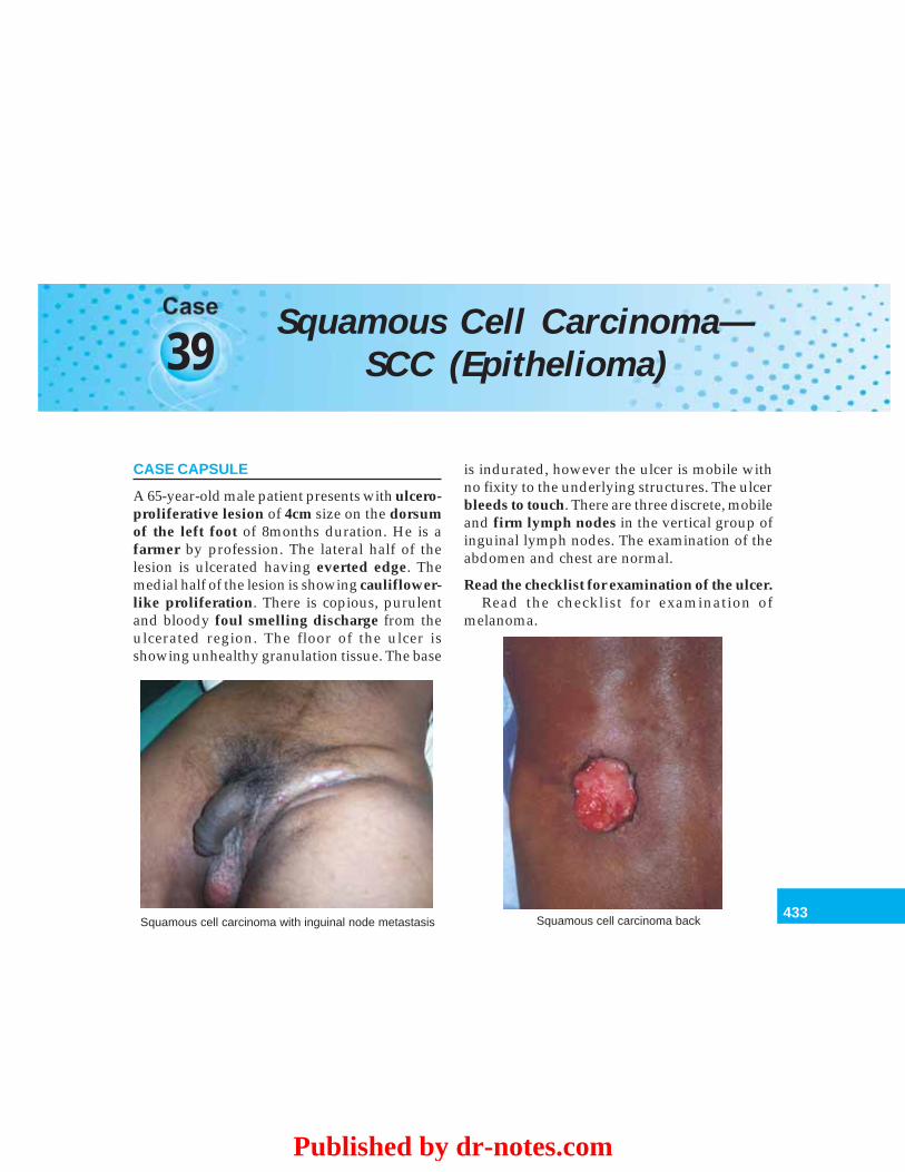
433
Squamous Cell Carcinoma—SCC (Epithelioma)
39Squamous Cell Carcinoma—
SCC (Epithelioma)
CASE CAPSULE
A 65-year-old male patient presents with ulcero-proliferative lesion of 4cm size on the dorsumof the left foot of 8months duration. He is afarmer by profession. The lateral half of thelesion is ulcerated having everted edge. Themedial half of the lesion is showing cauliflower-like proliferation. There is copious, purulentand bloody foul smelling discharge from theulcerated region. The floor of the ulcer isshowing unhealthy granulation tissue. The base
is indurated, however the ulcer is mobile withno fixity to the underlying structures. The ulcerbleeds to touch. There are three discrete, mobileand firm lymph nodes in the vertical group ofinguinal lymph nodes. The examination of theabdomen and chest are normal.
Read the checklist for examination of the ulcer.Read the checklist for examination of
melanoma.
Squamous cell carcinoma with inguinal node metastasis Squamous cell carcinoma back
Published by dr-notes.com
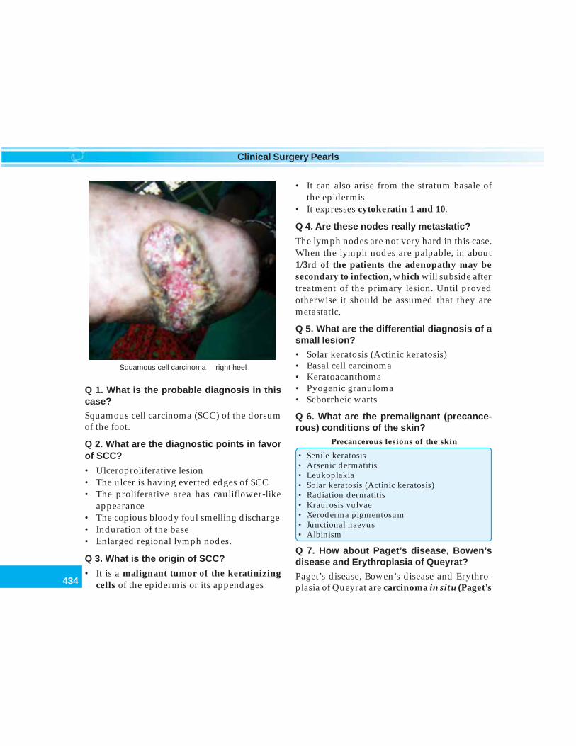
434
Clinical Surgery Pearls
Q 1. What is the probable diagnosis in thiscase?
Squamous cell carcinoma (SCC) of the dorsumof the foot.
Q 2. What are the diagnostic points in favorof SCC?
• Ulceroproliferative lesion• The ulcer is having everted edges of SCC• The proliferative area has cauliflower-like
appearance• The copious bloody foul smelling discharge• Induration of the base• Enlarged regional lymph nodes.
Q 3. What is the origin of SCC?
• It is a malignant tumor of the keratinizingcells of the epidermis or its appendages
• It can also arise from the stratum basale ofthe epidermis
• It expresses cytokeratin 1 and 10.
Q 4. Are these nodes really metastatic?
The lymph nodes are not very hard in this case.When the lymph nodes are palpable, in about1/3rd of the patients the adenopathy may besecondary to infection, which will subside aftertreatment of the primary lesion. Until provedotherwise it should be assumed that they aremetastatic.
Q 5. What are the differential diagnosis of asmall lesion?
• Solar keratosis (Actinic keratosis)• Basal cell carcinoma• Keratoacanthoma• Pyogenic granuloma• Seborrheic warts
Q 6. What are the premalignant (precance-rous) conditions of the skin?
Precancerous lesions of the skin
• Senile keratosis• Arsenic dermatitis• Leukoplakia• Solar keratosis (Actinic keratosis)• Radiation dermatitis• Kraurosis vulvae• Xeroderma pigmentosum• Junctional naevus• Albinism
Q 7. How about Paget’s disease, Bowen’sdisease and Erythroplasia of Queyrat?
Paget’s disease, Bowen’s disease and Erythro-plasia of Queyrat are carcinoma in situ (Paget’s
Squamous cell carcinoma— right heel
435
Squamous Cell Carcinoma—SCC (Epithelioma)
disease is seen in nipple, erythroplasia ofQueyrat is seen in penis.)
Q 8. What is the difference between pre-cancerous lesion and carcinoma in situ?
The precancerous lesions will lead on to in-situcancer and finally cancer.
Precancerous lesion↓
In situ carcinoma (preinvasive)↓
Carcinoma
Q 9. What are the features of solar keratosis(Actinic keratoses)? (PG)They are discrete, scaly, irregular patches,which may project occasionally.
Q 10. What is the treatment of actinickeratoses? (PG)They are treated with –• Cryotherapy• Topical 5. Fu – (5 fluorouracil).• Electro desiccation and curettage• CO2 Laser• Dermabrasion.
Q 11. What are the features of Bowen’sdisease? (PG)They are irregularly shaped well-definedplaques resembling a patch of eczema havingbeefy red, erythematous raised scaly crust onthe surface.
Q 12. What are the features of erythroplasia?(PG)
They are having the same features as Bowen’sdisease, except for the fact that they are situatedon the penis.
Q 13. Which part of the ultraviolet ray isresponsible for skin damage?UVB is thought to be the form of radiationresponsible for the damage by sunlight.
Q 14. What are the mechanisms by which theUV radiation affects the skin? (PG)It affects the skin in two ways –1. Direct carcinogenic effect on dividing
keratinocytes in the basal layer of theepidermis.
2. Depression of the cutaneous immunesurveillance response.
Q 15. What are the sites of SCC other thanskin?
SCC is seen in the following sites other than skin.• Lips• Mouth• Pharynx• Esophagus• Anal canal• Glans penis• Uterine cervix• Metaplastic areas of respiratory epithelium.
Q 16. What are the other predisposingfactors?
Predisposing factors for SCC
1. Sunlight2. Susceptible phenotype3. Compromised immunity – (after 10years of
immunosuppression 10% will develop malig-nancies – may be secondary to HPV virus).
4. Chemicals – E.g. Hydrocarbons (soot),arsenical, tar, etc.
5. Infection – HPV 5 and HPV 16
Published by dr-notes.com
436
Clinical Surgery Pearls
6. Mineral oils7. Arsenic8. Chrome compounds9. Chronic inflammation – Chronic sinus tract,
pre-existing scars (Marjolin’s ulcer), osteo-myelitis, burns and vaccination points
10. Immunosuppression (organ transplant re-cipients)
Q 17. What is “Chimney sweeps” cancer?
The soot is one of the most important hydro-carbons responsible for squamous cellcarcinoma of the scrotum in chimney sweeps.It is also seen in people who work in tar.
Q 18. What is “Khangri” cancer?
Khangri is an earthenware filled with burningcharcoal. The Kashmiris keep the Khangri ontheir abdomen to keep themselves warm. Thepatient will develop squamous cell carcinomaof the abdominal wall.
Q 19. What is Kang cancer?
This is a squamous cell carcinoma developingin the buttocks, back, heels, and elbows. This isseen in Tibetans who sleep on the oven bed.
Q 20. What is countryman’s lip?
This is nothing but SCC of the lower lip seen infarmers as a result of sun exposure.
Q 21. What are the macroscopic types ofSCC?
Three types are seen:1. Ulcerative2. Proliferative (Cauliflower-like)3. Ulceroproliferative.
Q 22. What is the microscopic pathology ofSCC?
• Epithelial pearls (Cell nests) – Squamouscells arranged in concentric manner likeonion skin
• Plasma cell infiltration.
Q 23. What is the spread of SCC?
• Local spread – to the subcutaneous tissue,tendons, muscles, bone and vasculature(Involvement of the local blood vessel cancause thrombosis and ischemia. The subcuta-neous spread may involve the near by nervescausing neuritis – perineural involvement)
• The main spread is lymphatic reaching thelymph node
• Blood stream spread occurs very rarely – (tothe lungs).
Q 24. Can you get SCC without lymph nodemetastasis?
Yes. Lymph node spread is absent in thefollowing situations.• When the lesion is developing in scar and
chronic ulcer (Marjolin’s ulcer)• SCC in old age.
Q 25. What are the characteristics ofMarjolin’s ulcer?
Characteristics of Marjolin’s ulcer
• It arises from ulcer or scar• The edge is not always raised and everted• It is not very invasive• The growth is very slow• More aggressive than spontaneous SCC
437
Squamous Cell Carcinoma—SCC (Epithelioma)
Q 26. What are the aggressive type ofsquamous cell carcinoma? (PG)
1. Squamous cell carcinomas seen intransplant patients
2. Squamous cell carcinomas developing inscars and ulcers
3. Anaplastic squamous cell carcinoma.
Note: Metastasis are more likely to arise fromSCC in scars and ulcers even though the lymphnodes are not involved.
Q 27. What are the types of lesions prone formalignant change resulting in Marjolin’s ulcer?
Lesions prone for Marjolin’s
1. Venous ulcers2. Chronic ulcers3. Scar tissues – Postburn scarring4. Scarring secondary to lupus vulgaris (tuber-
culosis)5. Chronic discharging sinuses – e.g. osteomyelitis
Q 28. How aggressive is SCC?
SCC is more aggressive than BCC and thereforewider excision margins are required for localcontrol.
Q 29. What is the type of biopsy recommen-ded for SCC?• Incision biopsy from the edge of the ulcer if
the lesion is large• If the lesion is small excision is recommended.
Q 30. Why the edge of the ulcer is preferredfor incision biopsy?The edge of ulcer is the growing part which willshow the malignant cells.
Q 31. What are the other investigationsrequired?• X-ray of the affected part to rule out bone
involvement• X-ray of the chest to rule out metastasis (very
rare event)• Other investigations required for anesthesia
clearance.
Q 32. What is the staging of SCC? (PG)The TNM staging is used (this is different fromthe TNM of the melanoma).Primary Tumor (T)TX Primary tumor cannot be assessedT0 No evidence of primary tumorTis Carcinoma in situT1 Tumor 2 cm or less in greatest dimensionT2 Tumor more than 2 cm, but not more than
5cm, in greatest dimensionT3 Tumor more than 5 cm in greatest dimensionT4 Tumor invades deep extradermal structures
(i.e. cartilage, skeletal muscle, or bone).
Note: In case of multiple simultaneous tumors,the tumor with highest T category will beclassified.
Regional lymph node (N)NX Regional lymph nodes cannot be assessedN0 No regional lymph node metastasisN1 Regional lymph node metastasis
Distant metastasis (M)MX Distant metastasis cannot be assessedM0 No distant metastasisM1 Distant metastasis
Published by dr-notes.com
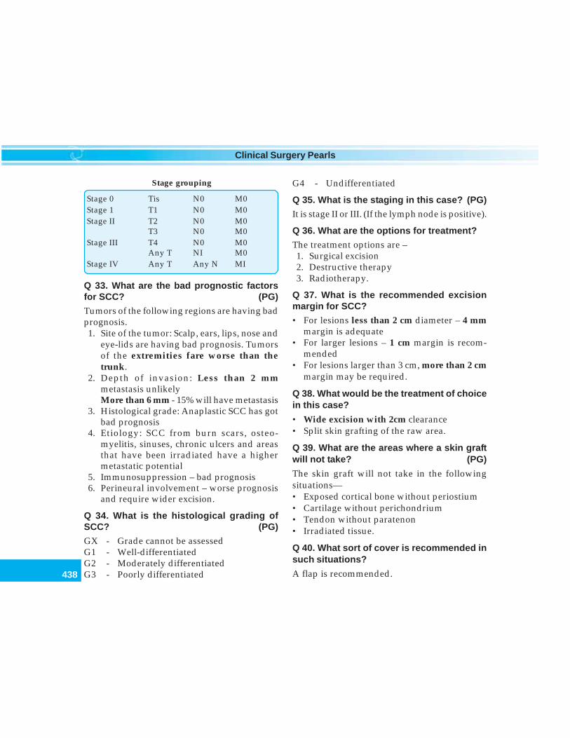
438
Clinical Surgery Pearls
Stage grouping
Stage 0 Tis N0 M0Stage 1 T1 N0 M0Stage II T2 N0 M0
T3 N0 M0Stage III T4 N0 M0
Any T NI M0Stage IV Any T Any N MI
Q 33. What are the bad prognostic factorsfor SCC? (PG)
Tumors of the following regions are having badprognosis.1. Site of the tumor: Scalp, ears, lips, nose and
eye-lids are having bad prognosis. Tumorsof the extremities fare worse than thetrunk.
2. Depth of invasion: Less than 2 mmmetastasis unlikelyMore than 6 mm - 15% will have metastasis
3. Histological grade: Anaplastic SCC has gotbad prognosis
4. Etiology: SCC from burn scars, osteo-myelitis, sinuses, chronic ulcers and areasthat have been irradiated have a highermetastatic potential
5. Immunosuppression – bad prognosis6. Perineural involvement – worse prognosis
and require wider excision.
Q 34. What is the histological grading ofSCC? (PG)
GX - Grade cannot be assessedG1 - Well-differentiatedG2 - Moderately differentiatedG3 - Poorly differentiated
G4 - Undifferentiated
Q 35. What is the staging in this case? (PG)
It is stage II or III. (If the lymph node is positive).
Q 36. What are the options for treatment?
The treatment options are –1. Surgical excision2. Destructive therapy3. Radiotherapy.
Q 37. What is the recommended excisionmargin for SCC?
• For lesions less than 2 cm diameter – 4 mmmargin is adequate
• For larger lesions – 1 cm margin is recom-mended
• For lesions larger than 3 cm, more than 2 cmmargin may be required.
Q 38. What would be the treatment of choicein this case?
• Wide excision with 2cm clearance• Split skin grafting of the raw area.
Q 39. What are the areas where a skin graftwill not take? (PG)
The skin graft will not take in the followingsituations—• Exposed cortical bone without periostium• Cartilage without perichondrium• Tendon without paratenon• Irradiated tissue.
Q 40. What sort of cover is recommended insuch situations?
A flap is recommended.
439
Squamous Cell Carcinoma—SCC (Epithelioma)
Q 41. What are the indications of the flap?(PG)
Flaps are indicated in the following situations—1. Areas where graft will not take2. Where there is risk of scar contracture
especially across a joint3. Where esthetic result is important4. Where bulk or structural support is needed.
Q 42. What are the types of flaps? (PG)
It may be Local flapIsland flapFree flap
Q 43. How do you manage the enlargedinguinal nodes?
• Give a course of antibiotics and see whetherthe nodes are subsiding or not
• If the nodes are remaining the same after 3weeks, FNAC of the node is done
• If the FNAC of the node is positive formetastasis, an inguinal block dissection iscarried out.
Q 44. If the lesion is involving the bone, whatwould be the treatment option? (In givencase)
The primary needs below knee amputation andthe inguinal nodes are managed as mentionedabove.
Q 45. What are the indications for radio-therapy?
Radiotherapy has been shown to cure 90% casesof SCC—
It is recommended for the followingsituations-• Debilitated patients• Poor surgical risk candidates• Those who refuse surgery.
Q 46. What are the problems of radiotherapy?(PG)
• Unpleasant side effects• Protracted treatment.
Q 47. Is there any role for radiotherapy as anadjuvant treatment? (PG)
Yes. The indications are—• High stage large tumors• Recurrent tumors.
Q 48. Is there any role for topical 5–fluoro-uracil (5-FU)? (PG)
No, it is not recommended for the primarytreatment of SCC. 5 -FU is an excellent methodof treating, premalignant lesions associated withSCC such as actinic keratoses.
Q 49.What is the role of Moh’s micrographictechnique in SCC? (PG)
This technique is also used for the treatment ofSCC.The advantages are—• Tissue preservation• Lower recurrence rate.
The disadvantages are—• Patient inconvenience• Expensive.
Published by dr-notes.com
440
Clinical Surgery Pearls
Q 50. What is the role of destructive techni-ques in the treatments of SCC (Cryosurgeryand electrodesiccation and curettage)?
(PG)
• They are best reserved for very smallsuperficial lesions in noncritical areas
• The local failure rate is high
• They do not produce surgical specimen forhistological examination and margin analysis
• The healing is by secondary intention• It results in poor scars.
Q 51. Is there any role for prophylactic lymphnode dissection? (PG)
No.
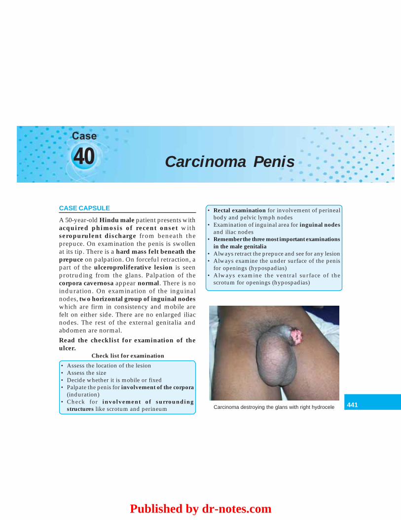
441
Carcinoma Penis
40 Carcinoma Penis
CASE CAPSULE
A 50-year-old Hindu male patient presents withacquired phimosis of recent onset withseropurulent discharge from beneath theprepuce. On examination the penis is swollenat its tip. There is a hard mass felt beneath theprepuce on palpation. On forceful retraction, apart of the ulceroproliferative lesion is seenprotruding from the glans. Palpation of thecorpora cavernosa appear normal. There is noinduration. On examination of the inguinalnodes, two horizontal group of inguinal nodeswhich are firm in consistency and mobile arefelt on either side. There are no enlarged iliacnodes. The rest of the external genitalia andabdomen are normal.Read the checklist for examination of theulcer.
Check list for examination
• Assess the location of the lesion• Assess the size• Decide whether it is mobile or fixed• Palpate the penis for involvement of the corpora
(induration)• Check for involvement of surrounding
structures like scrotum and perineum
• Rectal examination for involvement of perinealbody and pelvic lymph nodes
• Examination of inguinal area for inguinal nodesand iliac nodes
• Remember the three most important examinationsin the male genitalia
• Always retract the prepuce and see for any lesion• Always examine the under surface of the penis
for openings (hypospadias)• Always examine the ventral surface of the
scrotum for openings (hypospadias)
Carcinoma destroying the glans with right hydrocele
Published by dr-notes.com
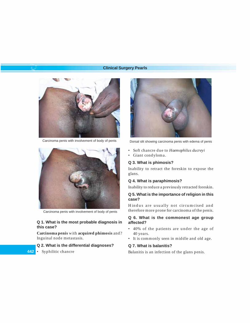
442
Clinical Surgery Pearls
Q 1. What is the most probable diagnosis inthis case?
Carcinoma penis with acquired phimosis and?Inguinal node metastasis.
Q 2. What is the differential diagnoses?
• Syphilitic chancre
• Soft chancre due to Haemophilus ducreyi• Giant condyloma.
Q 3. What is phimosis?
Inability to retract the foreskin to expose theglans.
Q 4. What is paraphimosis?
Inability to reduce a previously retracted foreskin.
Q 5. What is the importance of religion in thiscase?
Hindus are usually not circumcised andtherefore more prone for carcinoma of the penis.
Q 6. What is the commonest age groupaffected?
• 40% of the patients are under the age of40 years.
• It is commonly seen in middle and old age.
Q 7. What is balanitis?
Balanitis is an infection of the glans penis.
Carcinoma penis with involvement of body of penis
Dorsal slit showing carcinoma penis with edema of penisCarcinoma penis with involvement of body of penis
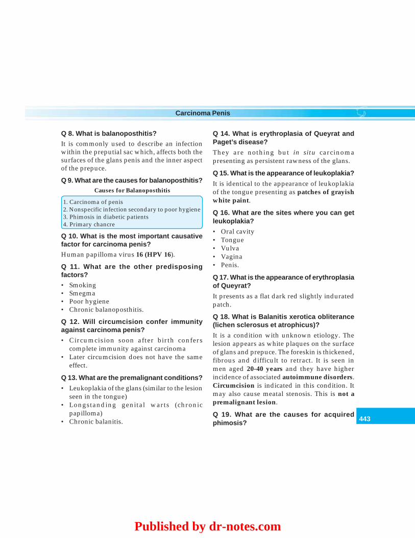
443
Carcinoma Penis
Q 8. What is balanoposthitis?
It is commonly used to describe an infectionwithin the preputial sac which, affects both thesurfaces of the glans penis and the inner aspectof the prepuce.
Q 9. What are the causes for balanoposthitis?Causes for Balanoposthitis
1. Carcinoma of penis2. Nonspecific infection secondary to poor hygiene3. Phimosis in diabetic patients4. Primary chancre
Q 10. What is the most important causativefactor for carcinoma penis?
Human papilloma virus 16 (HPV 16).
Q 11. What are the other predisposingfactors?
• Smoking• Smegma• Poor hygiene• Chronic balanoposthitis.
Q 12. Will circumcision confer immunityagainst carcinoma penis?
• Circumcision soon after birth conferscomplete immunity against carcinoma
• Later circumcision does not have the sameeffect.
Q 13. What are the premalignant conditions?
• Leukoplakia of the glans (similar to the lesionseen in the tongue)
• Longstanding genital warts (chronicpapilloma)
• Chronic balanitis.
Q 14. What is erythroplasia of Queyrat andPaget’s disease?
They are nothing but in situ carcinomapresenting as persistent rawness of the glans.
Q 15. What is the appearance of leukoplakia?
It is identical to the appearance of leukoplakiaof the tongue presenting as patches of grayishwhite paint.
Q 16. What are the sites where you can getleukoplakia?
• Oral cavity• Tongue• Vulva• Vagina• Penis.
Q 17. What is the appearance of erythroplasiaof Queyrat?
It presents as a flat dark red slightly induratedpatch.
Q 18. What is Balanitis xerotica obliterance(lichen sclerosus et atrophicus)?
It is a condition with unknown etiology. Thelesion appears as white plaques on the surfaceof glans and prepuce. The foreskin is thickened,fibrous and difficult to retract. It is seen inmen aged 20-40 years and they have higherincidence of associated autoimmune disorders.Circumcision is indicated in this condition. Itmay also cause meatal stenosis. This is not apremalignant lesion.
Q 19. What are the causes for acquiredphimosis?
Published by dr-notes.com
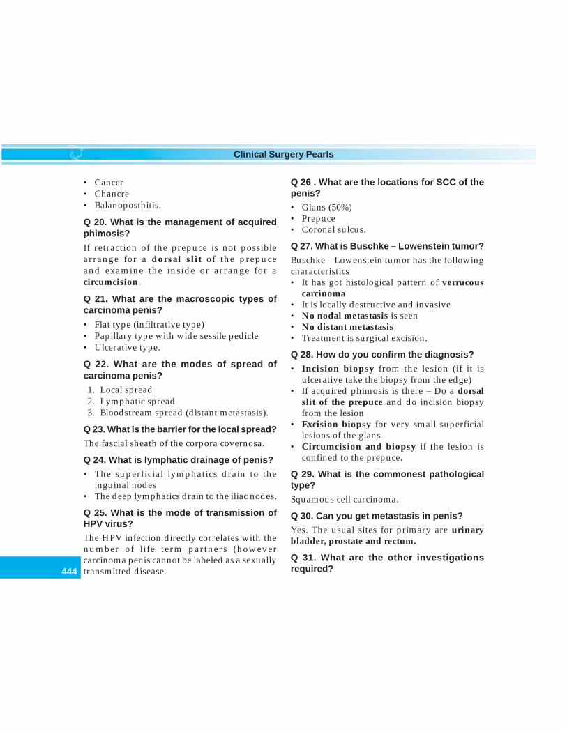
444
Clinical Surgery Pearls
• Cancer• Chancre• Balanoposthitis.
Q 20. What is the management of acquiredphimosis?
If retraction of the prepuce is not possiblearrange for a dorsal slit of the prepuceand examine the inside or arrange for acircumcision.
Q 21. What are the macroscopic types ofcarcinoma penis?
• Flat type (infiltrative type)• Papillary type with wide sessile pedicle• Ulcerative type.
Q 22. What are the modes of spread ofcarcinoma penis?
1. Local spread2. Lymphatic spread3. Bloodstream spread (distant metastasis).
Q 23. What is the barrier for the local spread?
The fascial sheath of the corpora covernosa.
Q 24. What is lymphatic drainage of penis?
• The superficial lymphatics drain to theinguinal nodes
• The deep lymphatics drain to the iliac nodes.
Q 25. What is the mode of transmission ofHPV virus?
The HPV infection directly correlates with thenumber of life term partners (howevercarcinoma penis cannot be labeled as a sexuallytransmitted disease.
Q 26 . What are the locations for SCC of thepenis?
• Glans (50%)• Prepuce• Coronal sulcus.
Q 27. What is Buschke – Lowenstein tumor?
Buschke – Lowenstein tumor has the followingcharacteristics• It has got histological pattern of verrucous
carcinoma• It is locally destructive and invasive• No nodal metastasis is seen• No distant metastasis• Treatment is surgical excision.
Q 28. How do you confirm the diagnosis?
• Incision biopsy from the lesion (if it isulcerative take the biopsy from the edge)
• If acquired phimosis is there – Do a dorsalslit of the prepuce and do incision biopsyfrom the lesion
• Excision biopsy for very small superficiallesions of the glans
• Circumcision and biopsy if the lesion isconfined to the prepuce.
Q 29. What is the commonest pathologicaltype?
Squamous cell carcinoma.
Q 30. Can you get metastasis in penis?
Yes. The usual sites for primary are urinarybladder, prostate and rectum.
Q 31. What are the other investigationsrequired?

445
Carcinoma Penis
1. Ultrasound of the penis• To evaluate the depth of invasion• Detecting involvement of the corpus
cavernosum2. Contrast enhanced MRI—For lesions
suspected to invade corpora3. CT—For evaluation of the inguinal and
pelvic nodes (physical examination isenough in the ordinary circumstances)
4. X-ray chest.
Q 32. What is the staging of carcinoma penis?(PG)
The TNM classification is used.• Tis - Preinvasive carcinoma• T0 - No evidence of primary tumors• Ta - Noninvasive verrucous carcinoma• T1 - Tumor invades subepithelial connec-
tive tissue• T2 - Tumor invades corpus spongiosum or
cavernosum• T3 - Tumor invades urethra or prostate• T4 - Tumor invades other adjacent structures
Regional Lymph Nodes (N)• NX - Regional lymph nodes cannot be
assessed• N0 - No regional lymph node metastasis• N1 - Metastasis in a single superficial,
inguinal lymph node• N2 - Metastasis in multiple or bilateral
superficial inguinal lymph node• N3 - Metastasis in deep inguinal or pelvic
lymph node (s) unilateral or bilateral
Distant Metastasis (M)• MX - Distant metastasis cannot be assessed• M0 - No distant metastasis• M1 - Distant metastasis
Stage Grouping
Stage 0 Tis N0 M0Ta N0 M0
Stage I T1 N0 M0Stage II T1 N1 M0
T2 N0 M0T2 N1 M0
Stage III T1 N2 M0T2 N2 M0T3 N0 M0T3 N1 M0T3 N2 M0
Stage IV T4 Any N M0Any T N3 M0Any T Any N M1
Q 33. What is the most important prognosticfactor for survival in carcinoma penis?
Presence of metastasis to the inguinal nodes.
Q 34. What are the predictors for nodalinvolvement? (PG)
Predictors for nodal involvement
• Grade of the tumor (higher the grade morechance for involvement of nodes)
• Corporal involvement• Vascular involvement• Lymphatic embolization
Published by dr-notes.com
446
Clinical Surgery Pearls
Q 35. What are the treatment options?Treatment options for carcinoma penis
• Surgery (Amputation of penis – partial or total)• Primary radiation therapy for the primary• Laser therapy – CO2 laser/Nd:YAG laser (for
very small noninfiltrating lesions)• 5-FU cream (for early lesions)• Treatment of inguinal nodes – unilateral or
bilateral lymph node dissection (inguinal) orbilateral inguinal irradiation
• Adjuvant chemotherapy• Reconstruction of the penis if suitable
Note: Stage 1 and 2 lesions can be treated withradiotherapy with a good cosmetic andfunctional result.
Q 36. What are the indications for radiationtherapy for the primary?
Indications for radiotherapy
• Young patients with small lesions (2-4 cm)• Superficial lesions• Exophytic lesions• Noninvasive lesions on glans or coronal sulcus• Patients refusing surgery• Patients with inoperable tumors
Q 37. What are the types of radiotherapy?
• External beam radiation• Interstitial brachytherapy – Iridium 192 or
Tantalum wire or Cesium 137• Radioactive mould application (applied
externally to the penis).
Q 38. What are the indications for thesurgery?
• Infiltration of the shaft of the penis• Large anaplastic growth
• Failure of radiotherapy.
Q 39. What are the indications for partialamputation of penis?
• T1 and T2 - lesions• If preservation of 2 cm penile stump is possible
after 2 cm clearance from the gross tumor.
Q 40. What are the advantages of partialamputation of penis?
• Less psychological trauma• Ability to pass urine in standing position• Preservation of sexual function.
Q 41. What is the minimum clearancerequired in Ca penis?
Well and moderately differentiated tumors needonly 1cm margin.
Q 42. What is glansectomy? (PG)
• This is reserved for verrucous carcinoma andminimally invading T1 lesion
• It preserves more erectile tissue.
Q 43. What is total amputation of penis?
• This is done for advanced lesions, and ana-plastic lesions
• Perineal urethrostomy is done after totalamputation.
Q 44. Is there any need for bilateral orchi-dectomy along with total amputation of thepenis?• The traditional arguments in favor of bilateral
orchidectomy will not hold today (soiling ofscrotum during urination and edemascrotum after inguinal block dissection)
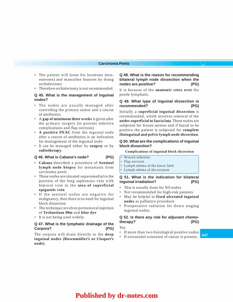
447
Carcinoma Penis
• The patient will loose his hormone (test-osterone) and masculine features by doingorchidectomy
• Therefore orchidectomy is not recommended.
Q 45. What is the management of inguinalnodes?• The nodes are usually managed after
controlling the primary tumor and a courseof antibiotics
• A gap of minimum three weeks is given afterthe primary surgery (to prevent infectivecomplications and flap necrosis)
• A positive FNAC from the inguinal nodeafter a course of antibiotics is an indicationfor management of the inguinal node
• It can be managed either by surgery or byradiotherapy.
Q 46. What is Cabana’s node? (PG)• Cabana described a procedure of Sentinel
lymph node biopsy for metastasis fromcarcinoma penis
• These nodes are situated superomedial to thejunction of the long saphenous vein withfemoral vein in the area of superficialepigastric vein
• If the sentinel nodes are negative formalignancy, then there is no need for inguinalblock dissection
• The technique involves peritumoral injectionof Technetium 99m and blue dye
• It is not being used widely.
Q 47. What is the lymphatic drainage of theCorpora? (PG)
The corpora will drain directly to the deepinguinal nodes (Rosenmüller’s or Cloquet’snode).
Q 48. What is the reason for recommendingbilateral lymph node dissection when thenodes are positive? (PG)
It is because of the anatomic cross over thepenile lymphatic.
Q 49. What type of inguinal dissection isrecommended? (PG)
Initially a superficial inguinal dissection isrecommended, which involves removal of thenodes superficial to fascia lata. These nodes aresubjected for frozen section and if found to bepositive the patient is subjected for completeilioinguinal and pelvic lymph node dissection.
Q 50. What are the complications of inguinalblock dissection?
Complications of inguinal block dissection
• Wound infection• Flap necrosis• Lymph edema of the lower limb• Lymph edema of the scrotum
Q 51. What is the indication for bilateralinguinal irradiation? (PG)
• This is usually done for N0 nodes• Not recommended for high-risk patients• May be helpful in fixed ulcerated inguinal
nodes as palliative procedure• Preoperative radiation for down staging
inguinal nodes.
Q 52. Is there any role for adjuvant chemo-therapy? (PG)
Yes• If more than two histological positive nodes• If extranodal extension of cancer is present.
Published by dr-notes.com
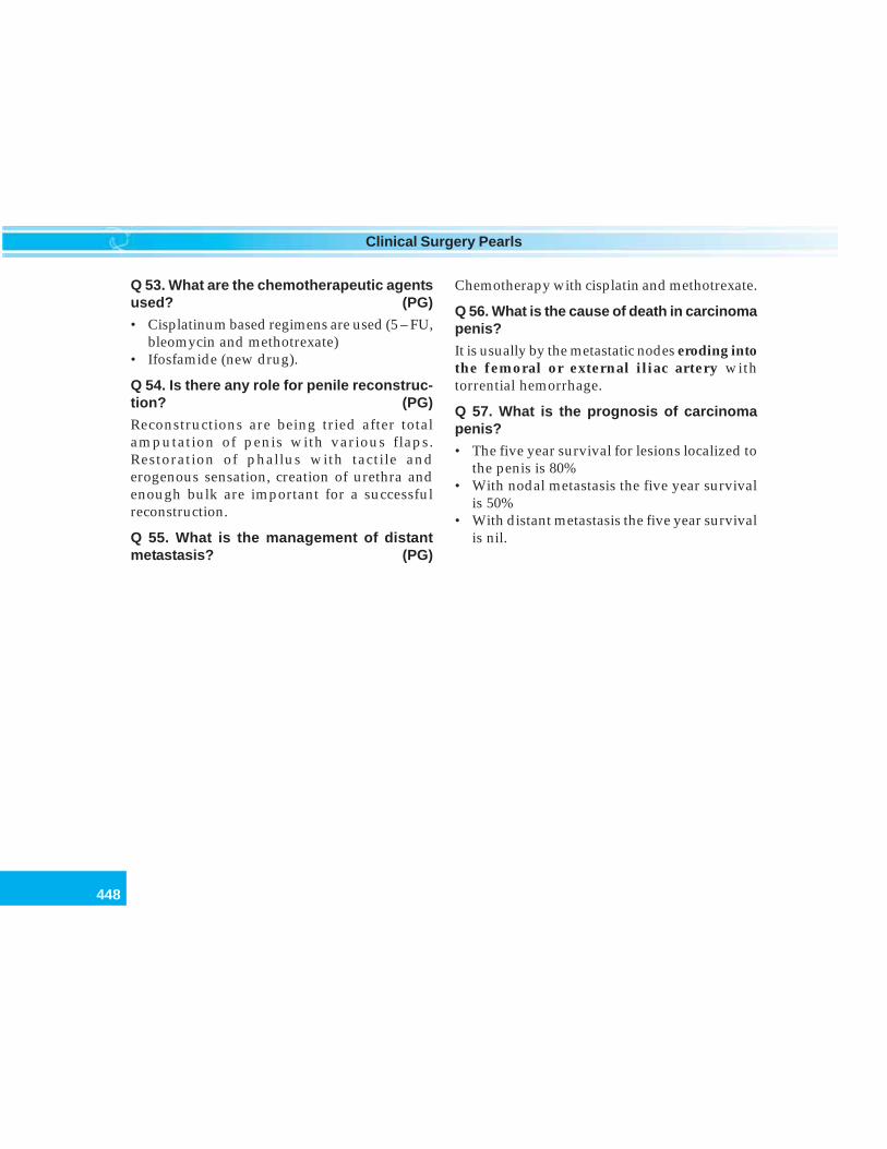
448
Clinical Surgery Pearls
Q 53. What are the chemotherapeutic agentsused? (PG)
• Cisplatinum based regimens are used (5 – FU,bleomycin and methotrexate)
• Ifosfamide (new drug).
Q 54. Is there any role for penile reconstruc-tion? (PG)
Reconstructions are being tried after totalamputation of penis with various flaps.Restoration of phallus with tactile anderogenous sensation, creation of urethra andenough bulk are important for a successfulreconstruction.
Q 55. What is the management of distantmetastasis? (PG)
Chemotherapy with cisplatin and methotrexate.
Q 56. What is the cause of death in carcinomapenis?
It is usually by the metastatic nodes eroding intothe femoral or external iliac artery withtorrential hemorrhage.
Q 57. What is the prognosis of carcinomapenis?
• The five year survival for lesions localized tothe penis is 80%
• With nodal metastasis the five year survivalis 50%
• With distant metastasis the five year survivalis nil.
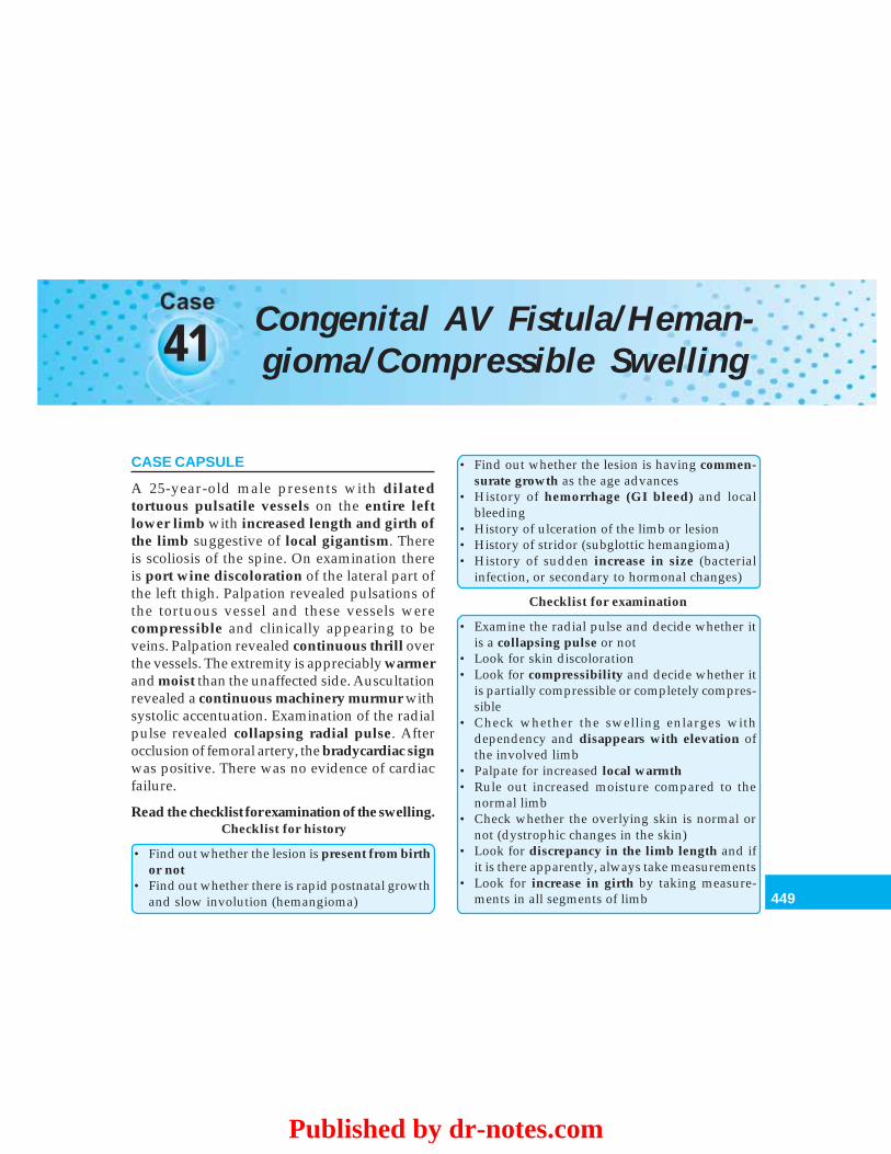
449
Congenital AV Fistula/Hemangioma/Compressible Swelling
41 Congenital AV Fistula/Heman-gioma/Compressible Swelling
CASE CAPSULE
A 25-year-old male presents with dilatedtortuous pulsatile vessels on the entire leftlower limb with increased length and girth ofthe limb suggestive of local gigantism. Thereis scoliosis of the spine. On examination thereis port wine discoloration of the lateral part ofthe left thigh. Palpation revealed pulsations ofthe tortuous vessel and these vessels werecompressible and clinically appearing to beveins. Palpation revealed continuous thrill overthe vessels. The extremity is appreciably warmerand moist than the unaffected side. Auscultationrevealed a continuous machinery murmur withsystolic accentuation. Examination of the radialpulse revealed collapsing radial pulse. Afterocclusion of femoral artery, the bradycardiac signwas positive. There was no evidence of cardiacfailure.
Read the checklist for examination of the swelling.Checklist for history
• Find out whether the lesion is present from birthor not
• Find out whether there is rapid postnatal growthand slow involution (hemangioma)
• Find out whether the lesion is having commen-surate growth as the age advances
• History of hemorrhage (GI bleed) and localbleeding
• History of ulceration of the limb or lesion• History of stridor (subglottic hemangioma)• History of sudden increase in size (bacterial
infection, or secondary to hormonal changes)
Checklist for examination
• Examine the radial pulse and decide whether itis a collapsing pulse or not
• Look for skin discoloration• Look for compressibility and decide whether it
is partially compressible or completely compres-sible
• Check whether the swelling enlarges withdependency and disappears with elevation ofthe involved limb
• Palpate for increased local warmth• Rule out increased moisture compared to the
normal limb• Check whether the overlying skin is normal or
not (dystrophic changes in the skin)• Look for discrepancy in the limb length and if
it is there apparently, always take measurements• Look for increase in girth by taking measure-
ments in all segments of limb
Published by dr-notes.com
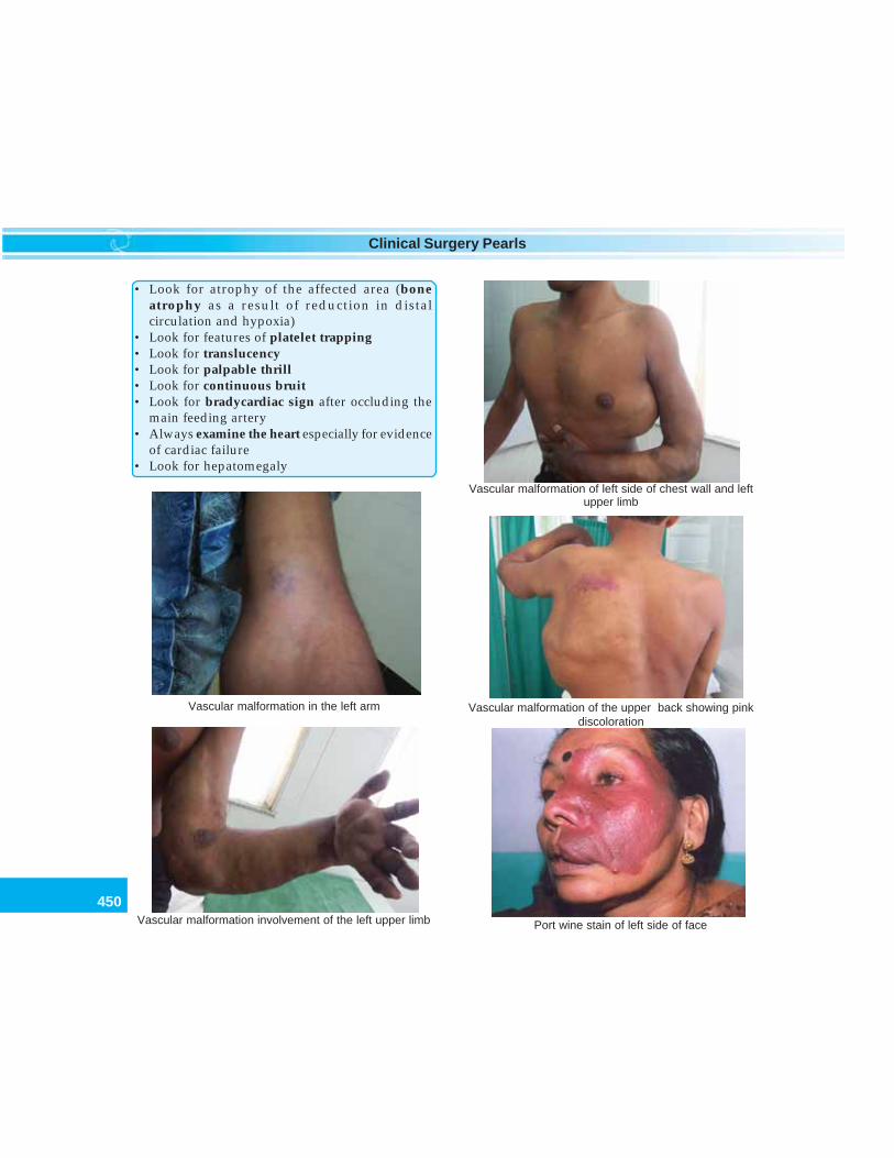
450
Clinical Surgery Pearls
• Look for atrophy of the affected area (boneatrophy as a result of reduction in distalcirculation and hypoxia)
• Look for features of platelet trapping• Look for translucency• Look for palpable thrill• Look for continuous bruit• Look for bradycardiac sign after occluding the
main feeding artery• Always examine the heart especially for evidence
of cardiac failure• Look for hepatomegaly
Port wine stain of left side of face
Vascular malformation in the left arm
Vascular malformation involvement of the left upper limb
Vascular malformation of left side of chest wall and leftupper limb
Vascular malformation of the upper back showing pinkdiscoloration
451
Congenital AV Fistula/Hemangioma/Compressible Swelling
Q 1. What are the differential diagnoses for acompressible swelling?
1. Lymphatic cyst/lymphangioma2. Hemangioma3. Aneurysm4. AV fistula.
Q 2. What are the clinical points to differen-tiate these four conditions?
• The lymph cyst will be brilliantly transillu-minant
• Hemangioma is not transilluminant, butpartially compressible
• Aneurysm will show expansile pulsationsand systolic bruit
• Arteriovenous fistula will show continuousthrill on palpation and continuous machinerymurmur on auscultation.
Q 3. What is the difference between caver-nous hemangioma and capillary heman-gioma?
These are old terms and they are best avoided.Cavernous hemangioma is used to describe adeep lesion involving the deeper dermis or thesubcutaneous tissue and capillary hemangiomais one which proliferates in the superficial dermis.
Q 4. What is the old classification ofhemangioma?
They are classified into capillary and cavernous(not used now).
• Capillary hemangioma - Strawberry- Port wine stain- Spider naevi
• Cavernous hemangioma
NB: The term cavernous hemangioma is notthere in the current terminology. Most of thelesions are actually venous malformations.
Q 5. What is the new classification?
The new classification distinguishes lesions asfollows:• Lesions that regress spontaneously—
Hemangiomas• Those that do not regress spontaneously—
Congenital vascular malformations (Ham-burg classification—predominantly arterial,venous, lymphatic, AV shunting and mixed).
Another classification is• Slow flow lesions• Fast flow lesions—Arteriovenous fistula.
Q 6. Which channel is more affected incongenital vascular malformations?
• Venous defects are the most common• Arteriovenous malformations makeup 1/3rd
of the lesions• 90% of the arteriovenous malformations
occur in the extremities, pelvis, trunk andshoulder girdle.
Q 7. What are the difference between heman-gioma and vascular malformation?
• Hemangiomas result from cellular prolifera-tion (It is a fibrofatty structure and thecontained blood cannot be evacuated comple-tely). The hemangiomas grow over the firstsix to eight months of life. After 1 year signsof involution appear until about 5-10 years old.
• Vascular malformations are embryonic anddevelopmental abnormalities (error in
Published by dr-notes.com
452
Clinical Surgery Pearls
vascular morphogenesis). These lesions growparallel with the age, they may expandsuddenly at times with associated infections.There is no involution. Limb hypertrophy oratrophy may occur.
radiating from the tumor may be seen beneaththe skin. During proliferative phase the tumorenlarges and becomes brighter in color.Involution is heralded by softening and fadingof color.
Differences between hemangioma and vascular malformations
Hemangioma Vascular malformation
• Usually present at birth • Present at birth, may not be apparent• Result from cellular proliferation (Benign tumor) • Embryonic and developmental abnormalities• Fibrofatty structure and contained blood cannot • Easily evacuated by compression
be evacuated completely by compression• No enlargement with dependency and • Enlarges with dependency and disappears
disappearance with elevation of the limb with elevation of the limb• Endothelial hyperplasia • Flat endothelium• Female to male ratio 5:1 • Female to male ratio 1:1• Mast cells are increased • Normal mast cells• Multilaminated basement membrane • Unilaminated basement membrane• Platelet trapping present (Kasabach - Merritt's • No platelet trapping
syndrome)• Rapid postnatal growth with slow involution • Commensurate growth
No spontaneous involution• No limb hypertrophy/atrophy • Limb hypertrophy or atrophy may occur• No treatment required in majority • Treatment may be required
Q 8. What are the examples of vascularmalformations?
• Port wine stain (capillary malformation)• Naevi• Venous malformations• Lymphatic malformation• Arteriovenous fistula.
Q 9. What is the classical appearance of astrawberry hemangioma?
It looks like a raised, bright red patch with atextured surface like a strawberry. Veins
Q 10. What are the classical sites of heman-gioma?
Sites of hemangioma
• Head and neck (60%)– Facial hemangioma– Eyelid hemangioma can cause astigmatism
and amblyopia– Subglottic hemangioma causing biphasic
stridor• Trunk (25%)• Limbs (15%)• Liver
453
Congenital AV Fistula/Hemangioma/Compressible Swelling
Q 12. What is the Kasabach-Merrittsyndrome?
This was described by Kasabach and Merritt in1940. It is characterized by -
Kasabach-Merritt Syndrome
• Hemangioma > 5cm• Thrombocytopenia• Bleeding diathesis
The thrombocytopenia is by platelet trapping.There is 37% mortality for this condition whichis largely due to bleeding. The treatment isdifficult and interferon, steroids and irradiationhave all been tried with variable results.
Q 13. What is Hamburg classification?
Humburg classification is for vascularmalformation based on the predominant natureof the malformation. They are classified intofive types—predominantly arterial defects,predominantly venous defects, predominantlylymphatic defects, predominantly AV shuntingdefects and combined or mixed vasculardefects.
– Multiple intrahepatic hemangiomas– Hepatomegaly– Heart failure– Anemia
• Intestines• Lungs• Brain
Note: 80% are single tumors and 20% aremultiple tumors.
Q 11. What are the complications of heman-giomas?
Complications of hemangiomas
• Platelet trapping– Kasabach – Merritt syndrome
• Thrombocytopenia and hemorrhage– Gastrointestinal– Pleural– Intracranial– Intraperitoneal
• Ulceration (5%)• Local bleeding• Infection• Visual problems by obstructing the vision• Stridor
Hamburg classification of congenital vascular defects
Type Forms
Truncular ExtratruncularPredominantly arterial defects Aplasia or Obstructive dilation Infiltrating or limitedPredominantly venous defects Aplasia or Obstructive dilation Infiltrating or limitedPredominantly lymphatic defects Aplasia or Obstructive Infiltrating or limitedPredominantly AV shunting defects Deep or superficial Infiltrating or limitedCombined/mixed vascular defects Arterial and venous, no AV Infiltrating hemolympha-
shunt Hemolymphatic, with or tic or limited hemoly-without AV shunt mphatic
Published by dr-notes.com
454
Clinical Surgery Pearls
Q 14. What are the associations of congenitalvascular malformations?
• Naevi• Port wine stain• Varicosities• Arteriovenous fistula• Hypertrophy and atrophy of the extremities• Edema of the limbs.
Q 15. What is port wine stain (in old classifica-tion it was included with hemangioma)?
This is an extensive intradermal capillarymalformations giving a deep purple color to theoverlying skin. They are present at birth,commonly seen on the face, at the junctionbetween limbs and trunk (shoulders, neck andbuttocks). Sometimes they are seen distributedalong sensory branches of the fifth cranial nerve.Microscopically they are formed by thin-walledcapillaries in the dermis. They are non-involuting lesions like any other arteriovenousmalformation (unlike hemangiomas).
Q 16. What are the treatment options of portwine stain?
• Camouflaging and Tattooing are the optionsfor the management
• Flash lamp pulsed—dye laser (multiplesessions are necessary)
• Selective photothermolysis• If laser is unsuccessful, surgical excision and
skin grafting.
Q 17. What is the age group for lymphaticmalformations and what is the presentation?
They are seen in the first year of life andchildhood. They present as lymphatic vesicles.
Q 18. What is lymphangioma/cystic hygroma?It is a localized cluster of dilated lymph sac inthe skin and subcutaneous tissues which do notconnect into the normal lymph system (they areclusters of lymph sacs that fail to join into thelymphatic system during development). Whenthey are large, cystic and translucent andconfined to the subcutaneous tissue, they arecalled cystic hygroma.
Q 19. What are the manifestations oflymphangioma?
• Skin vesicles noticed by parent (may be clearor brown or black) as a result of the containedclotted blood - 0.5 to 3 to 4 mm in diameter
• Some times the vesicles leak clear fluid• May present with infected vesicles and pain• The number and extent of the vesicles
increases with age• Multiple small lesions may not fluctuate and
feel soft and spongy (one or two large cystsshow fluctuation, fluid thrill andtranslucency)
• It may or may not be compressible• When there is infection the regional nodes
may be enlarged.
Q 20. What are the classical sites oflymphangioma?
Classical sites of lymphangioma
• Junction of the limb and neck• Junction of the limb and trunk• Around the shoulder• Axilla• Buttock• Groin
455
Congenital AV Fistula/Hemangioma/Compressible Swelling
Q 21. What is lymphangioma circums-criptum?
When the lesion is localized to a region likebuttocks or side of the thigh it is called lymphan-gioma circumscriptum.
Q 22. What is Vin rose patch?
It is a congenital intradermal vascularabnormality in which mild dilatation of thevessels in the subpapillary dermal plexus givesthe skin a pale pink color. It is associated withother vascular abnormalities like extensivehemangioma, arteriovenous fistulae and lymph-edema. It can occur any where and causes nosymptoms.
Q 23. What is Campbell de Morgan spot?
It is a bright red well-defined spot caused bythe collection of dilated capillaries fed by a singleor cluster of arterioles. They are usually seen inolder age group above 45. One or more spotsand some times a cluster may be seen. The usualsite is upper half of the trunk and rarely in thelimbs and face. They look like drops of dark redpaint.
Q 24. What is spider naevus?
It is a solitary dilated skin arteriole, with visibleradiating branches. They are usually associatedwith chronic liver disease and tumors producingestrogens. They appear on the upper half of thetrunk, the face and the arms. They fadecompletely when compressed and refill as soonas the pressure is released.
Q 25. What is the age group for venousmalformation?
The venous malformation may present at birth,childhood or adolescence.
Q 26. What is the clinical presentation ofarteriovenous fistula?
• In early stages a pink stain and increased localtemperature of the surrounding skin are theonly signs
• Gradually distended veins occur and a thrillcan be felt (followed by audible bruit). Whenvaricose vein and skin discoloration over thelateral aspect of the thigh are present suspectcongenital AV fistula.
• Extensive dilated tortuous veins which arepulsatile (arterialized veins) are seen.
• Limb hypertrophy and local gigantism willoccur.
• Blood is diverted from the arterial side to thevenous side resulting in distal hypoxia.
Q 27. What is the commonest site forcongenital AV fistula?
• The lower extremity is the most common site• Upper extremity• Congenital AV fistula in relation to the
superficial temporal artery• Congenital AV fistula in relation to the
occipital artery.
Q 28. What are the signs of congenital AVfistula?
The following are the signs of congenital AVfistula• Port wine discoloration and varicose veins—
suspect AV fistula• Local gigantism—Congenital arteriovenous
fistula in the young patient will produce
Published by dr-notes.com
456
Clinical Surgery Pearls
increase length and girth of the limb (the localgigantism may be associated with scoliosis).
• The lower limb is apparently warmer andmoist than the unaffected side (in the case ofhead and neck the affected side will bewarmer).
• Palpable continuous thrill in the dilatedvessels.
• Continuous machinery murmur withsystolic accentuation is obtained when thestethoscope is applied to the arterializedveins.
• Collapsing arterial pulse.• Bradycardiac sign of Branham and
Nicolodona — Digital occlusion of the mainartery to the limb is followed by bradycardiaindicating that considerable volume of bloodis being short circuited.
• Leg ulceration due to hypoxia—These ulcersare known as hot ulcers (because thesurrounding skin feels warmer than normal).These ulcers are very painful.
Q 29. What is the cause for acquired AV fistula?
• Trauma to the vessels• Iatrogenic AV fistula (for dialysis).
Q 30. What are the differences betweencongenital and acquired AV fistula?
• The arteriovenous communications areinnumerable in congenital
• The communications are one or two inacquired
• Congenital is difficult to treat• Acquired is easily treated.
Q 31. What is Cirsoid aneurysm?
They are nothing but congenital arteriovenousmalformations in relation to the superficialtemporal artery or occipital artery. They mayhave intracranial extensions. It is very difficultto treat such lesions.
Q 32. What are the investigations in a caseof vascular malformation?
Color Doppler and MR angiography are the twomost useful investigations in the case of arterio-venous fistula.• Laboratory investigations—No lab investiga-
tion can differentiate hemangioma fromvascular malformation. However estimationof fibroblastic growth factor secreted in theurine of patients with hemangioma may beuseful.
• Duplex scanning—It is a useful noninvasiveinvestigation.
• Tc-99 labeled human albumin is injectedintraarterially proximal to the AV fistula andmeasuring the radioactivity in the lungs witha gamma camera is a useful investigation forarteriovenous fistula (< 3% pass to the lungsnormally) .
• CT scanning with contrast enhancement• MRI—Superior to CT (high and low flow
lesions can be identified).• Magnetic resonance angiogram—This will
accurately distinguish hemangioma fromvascular malformations. This is the mostuseful imaging study of choice.
• Angiography—It is not routinely done unlessembolization is required prior to surgery
457
Congenital AV Fistula/Hemangioma/Compressible Swelling
Q 33. What is the treatment of hemangioma?Treatment of hemangioma
• Most hemangiomas do not require any treatmentbecause they resolve spontaneously
• Avoid local trauma• Parents are direct to give local pressure in the
event of bleeding• Corticosteroids (both systemic and intralesional)• Interferon α2a - used for life-threatening
complications (interferon is an inhibitor ofangiogenesis)
• Lasers—Used to treat residual telangiectaticspots in after involution
• Surgical excision—It is indicated whencomplications occur and also in the case of eyelids
Q 34. What are the indications for corticoste-roids?
• Life-threatening complications—airwayobstruction, bleeding
• Large facial hemangiomas• Platelet trapping syndrome.
Q 35. What is the dose and duration oftherapy?
Prednisolone 2mg/kg/day are used orally. Thetreatment is continued for several months untilthe tumor is in its involuting phase.
Q 36. What is intralesional steroid therapy?
• This is used for localized facial hemangiomas• Triamcinolone—3 to 5mg/kg/procedure is
injected directly into the lesion with a 26gauge needle. The volume should not exceed1ml per injection.
Q 37. What are the management options invenous malformation?
Management options in venous malformation
• Elastic support• Low dose aspirin for preventing episodic
thrombosis• Sclerotherapy - sodium tetradecyl sulphate
injection locally followed by pressure• Laser therapy• Surgical treatment
Q 38. What are the management options forarteriovenous fistula?
Management options for arteriovenous fistula
Emergency management of bleeding• Emergency ligation of feeding vessel or surgical
packing - this will give temporary control (alwaysrecurrence should be anticipated and therecurrence is too difficult to treat owing to therecruitment of multiple new channels)
• Trans catheter embolisation• Management of cardiac failureElective treatment• Preoperative embolization followed by surgical
excision (The resectability rate is <20%).• The embolization should be done as near to the
time of surgery as is feasible
Q 39. What are the materials used forembolization?
Materials used for embolization
• Plastic particles• Foam pledgets• Stainless steel coils• Polymerizing adhesives• Ethanol - polyvinyl alcohol foam particles are
available in graded sizes from 50 - 1000micrometers diameter
• Sclerosing agents• Acrylic adhesives
Published by dr-notes.com
458
Clinical Surgery Pearls
Q 40. What is Klippel-Trenaunay syndrome?
They are combined vascular malformations(combined in the sense that they involve morethan one type of channel). In this case it is acombined capillary lymphovenous malforma-tion. It is a slow flow lesion characterized bythe following lesions -
Klippel-Trenaunay syndrome
• Geographic capillary stain• Venous anomaly of the superficial and deep system• Lymphatic abnormality• Over growth of soft tissues and bones
Q 41. What is Parkes-Weber syndrome?
This is a combined capillary and arterio-venous malformation. It is a flat blue lesioncharacterized by:• Flat pink, (warm) stain• Underlying multiple AV connections• Venous malformation• Tissue overgrowth.
Q 42. What is Maffucci syndrome?
The association of following constitutesMaffucci syndrome
• Multiple enchondromas• Venous malformations• Bony deformities
459
Unilateral Lower Limb Edema
42 Unilateral Lower Limb Edema
CASE CAPSULE
A 45-year-old male patient presents withswelling of the left lower limb of 3 yearsduration. The inguinal lymph nodes areenlarged, firm, discrete and mobile. There ishistory of recurrent attacks of fever, chills andpain in the affected limb. The first episode ofsuch attack started in early adult life. In theinitial stages the swelling used to disappear inbetween the attacks of fever and chills. Thefrequency of such attack is increasing and theedema is persistent nowadays. The swellingdoes not reverse on elevation of the limb. Theskin is thickened and some warty nodules areseen in the lower part of the leg anteriorly. Thelimb has attained enormous size in the last fewmonths and it is now interfering with mobilityof the limb and routine activities of the patient.The edema is seen involving the dorsum of footand toes. On interrogation it was found that heis coming from a filarial belt. There is no historyof surgery and irradiation. Examination of thegenitalia revealed hydrocele of the TV sac onleft side. Abdominal examination is normal soalso the digital rectal examination. There is no
evidence of edema of the contralateral lowerlimb and upper limbs.
Checklist for history
• History of geographical location from where thepatient is coming (endemic place for filariasis)
• Onset and duration of edema (congenital,adolescence, or adulthood)
• History of injuries, wounds, abrasions,fissuring of the skin, eczema and paronychia
• History of recurrent attacks of fever, chills andpainful swellings
• Whether the swelling is initially reversible or not• History of tumors of the pelvic floor, prostate
cancer, etc.• History of surgical dissection of lymph nodes• History of radiation therapy for malignant
tumors• History of podoconiosis (cutaneous absorption
of mineral particles)• History of venous diseases, venous surgery and
venous thrombosis• History of Hansen’s disease• History of Leishmaniasis• History of cardiac diseases• History of hepatic and nutritional disorders• History of amyloidosis
Published by dr-notes.com
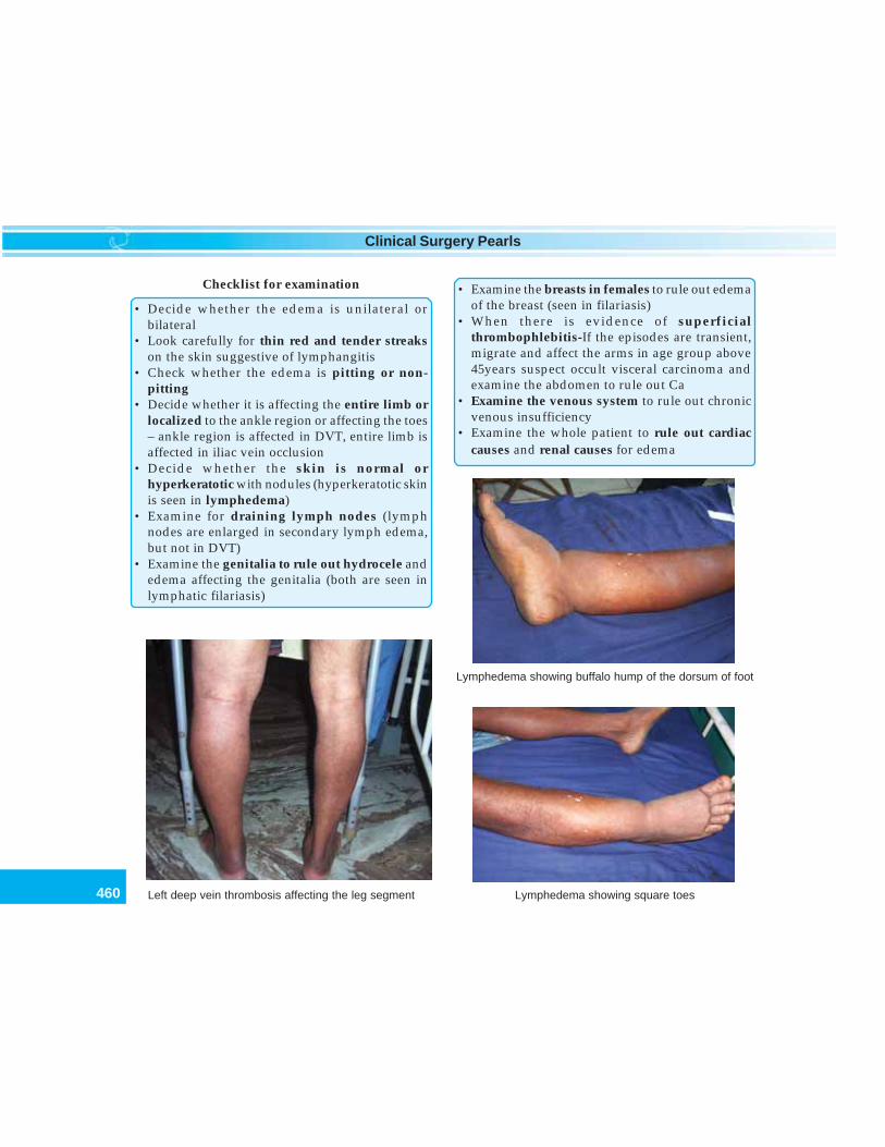
460
Clinical Surgery Pearls
Checklist for examination
• Decide whether the edema is unilateral orbilateral
• Look carefully for thin red and tender streakson the skin suggestive of lymphangitis
• Check whether the edema is pitting or non-pitting
• Decide whether it is affecting the entire limb orlocalized to the ankle region or affecting the toes– ankle region is affected in DVT, entire limb isaffected in iliac vein occlusion
• Decide whether the skin is normal orhyperkeratotic with nodules (hyperkeratotic skinis seen in lymphedema)
• Examine for draining lymph nodes (lymphnodes are enlarged in secondary lymph edema,but not in DVT)
• Examine the genitalia to rule out hydrocele andedema affecting the genitalia (both are seen inlymphatic filariasis)
• Examine the breasts in females to rule out edemaof the breast (seen in filariasis)
• When there is evidence of superficialthrombophlebitis-If the episodes are transient,migrate and affect the arms in age group above45years suspect occult visceral carcinoma andexamine the abdomen to rule out Ca
• Examine the venous system to rule out chronicvenous insufficiency
• Examine the whole patient to rule out cardiaccauses and renal causes for edema
Left deep vein thrombosis affecting the leg segment
Lymphedema showing buffalo hump of the dorsum of foot
Lymphedema showing square toes
461
Unilateral Lower Limb Edema
Q 1. What is your diagnosis?
Lymphatic filariasis with lymphedema.
Q 2. What are your points in favor of yourdiagnosis?• Patient gives history of recurrent fever, chills
and pain in the affected limb.• The edema is affecting the toes (a point in
favor of lymphedema)• The edema is nonpitting• The swelling does not reverse on elevation
of the limb.• The skin is thickened and warty nodules are
seen.• The regional nodes are enlarged.• Patient is coming from an endemic area for
filariasis.
Q 3. What are the differential diagnoses?1. Lower limb edema from lymphatic causes
• Primary lymphedema- lymphedema congenita- lymphedema praecox – 2 to 35 years
(sporadic or familial). The familial iscalled Meige’s disease.
- lymphedema tarda – after 35 years(associated with obesity – the nodesreplaced with fibrofatty tissue).
• Familial lymphedema (Milroy’sdisease) — familial form of congenitallymphedema
• Secondary lymphedema—Lymphaticfilariasis- Other infections – Tuberculosis,
lymphogranuloma inguinale- Tumors of the pelvic floor (prostate
cancer)
- Surgical dissection of lymph nodes(block dissection)
- Radiation therapy for malignanttumors
- Podoconiosis (cutaneous absorption ofmineral particles)
• Lymphedema secondary to congenitalvascular anomalies- Lymphatic angiodysplasia syndrome- Klippel – Trenaunay syndrome- Hyperstomy syndrome.
2. Lower limb edema due to venous causes:• Chronic venous insufficiency• Deep vein thrombosis• Phlegmasia Alba Dolens (white leg or
milk leg)• Phlegmasia Cerulea Dolens
3. Cellulitis4. Hansen’s disease5. Dermal leishmaniasis6. Mycetoma7. Systemic causes.
Q 4. What is edema?
Edema represents an imbalance betweencapillary filtration and lymphatic drainage (therole of lymphatics in the development of edemais substantial. This does not mean that alledemas are lymphedemas).
Q 5. How the lymphatic system is formed andwhat is the function of lymphatic system?
It forms part of the microcirculation that helpsto return macromolecules like proteins, celldebris and other particulate matter and excessfluid from the interstitial spaces to the venous
Published by dr-notes.com
462
Clinical Surgery Pearls
system via large lymph vessels. The initiallymphatic capillaries in the skin originate asblind ended tubes formed by a single layer ofendothelial cells with spaces in between for theentry of fluid and large molecules. They join toform larger vessel which have smooth musclesin their walls. The flow of lymph isunidirectional because of the presence of valvessituated every 2 to 12 mm distance. The largervessels are having smooth vessels in their walls.Gentle massage of the skin stimulatescontractions. Lymphatics are responsible forresorption of 10 to 20% of the tissue fluid. Twoto four liters of lymph with 70 to 200gm ofprotein pass daily into the systemic circulation.
Q 6. What are the factors responsible forlymph flow?
• Contraction of muscles• Pressure exerted by arterial pulsations• Increase in fluid volume• Elevation of the limb.
Q 7. What are the possible mechanisms forlymphedema?
1. When there is increased load of interstitialfluid – high output failure.
2. When the lymph vessels are absent, orabnormal or damaged due to acquiredcauses – low output failure.
Q 8. In which plane you get the lymphatics?
In the limbs they form –• Superficial and deep plexus in the dermis –
they drain to the subcutaneous lymph vesselsfollowing the course of superficial veins
• Subfascial plexus in the muscularcompartments
• The lymphatics finally drain to the lymphnodes.
Q 9. What is the WHO grading of lymph-edema?WHO grading of lymphedema of the limbs (1992)
Grade I – Pitting edema reversible on elevationof the affected limb
Grade II – Pitting or nonpitting edema whichdoes not reverse on elevation of theaffected limb, and there are no skinchanges
Grade III – Nonpitting edema that is notreversible, with thickening of skin
Grade IV – Nonpitting edema that is notreversible, with thickening of skinalong with nodular or wartyexcrescences – the stage of elephan-tiasis
Q 10. What are the disadvantages of thisgrading?
• It does not differentiate the severity of skinchanges
• Does not denote the magnitude of disability.
Q 11. What is the recent modification by theWHO?
Recent modification of WHO staging oflymphedema
Stage 1 – Same features as in grade I aboveStage 2 – Same features as in grade II aboveStage 3 – Presence of shallow skin foldsStage 4 – Presence of knobs or nodules
463
Unilateral Lower Limb Edema
Stage 5 – Presence of deep skin foldsStage 6 – Warty changes in the skinStage 7 – Patient unable to move around due to
enormous size of the swelling.
Q 12. What is the cause for primary lymph-edema?
It is a congenital pathology affecting thelymphatic channels in the form of –• Agenesis of lymphatics (Aplasia) –
lymphedema congenita• Hypoplasia – the commonest variety –
lymphedema praecox and tarda• Hyperplasia – lymphatics are enlarged,
increased in number and tortuous• Lymphangiectasia.
Q 13. What are the clinical subtypes ofprimary lymphedema?
The clinical subtypes are –1. Congenital lymphedema
– which appears shortly after birth2. Lymphedema praecox
– which starts during puberty3. Lymphedema tarda
– which usually starts in the 3rd decade.
Q 14. What are the clinical syndromes asso-ciated with primary lymphedema? (PG)
Syndromes associated with primarylymphedema
• Turner syndrome• Klinefelter syndrome• Noonan syndrome• Yellow nail syndrome• Intestinal lymphangiectasia
Q 15. What is the commonest cause forlymphedema?
The commonest cause is always secondary. Thecommonest cause for secondary lymphedemaworldwide is lymphatic filariasis. For the othercauses for secondary lymphedema see answerof Q 3.
Q 16. What is acute dermatolymphangio-adenitis (ADLA)? (PG)
All cases of lymphedema whether primary orsecondary are prone for acute dermato-lymphangioadenitis and the manifestations are–• The draining lymph nodes are enlarged• Fever and chills• Painful swelling of the affected limb (initial
stages the swelling of the limb will subsideafter an ADLA episode).
Q 17. What is the pathogenesis of filariallymphedema?
• The adult parasites living in the lymphvessels initiate the damage
• Earliest pathology is the dilatation of thelymph vessel (Lymphangiectasia)
• This lymphangiectasia is irreversible evenafter treatment
• This will result in stagnation of the lymphand incompetence of the unidirectionalvalves
• Lymph stasis encourages the growth ofinvading bacteria as a result of trivial trauma
• The entry of organisms are through the socalled entry lesions namely –
Published by dr-notes.com
464
Clinical Surgery Pearls
– Fissuring of skin– Paronychia– Eczema
• Secondary infection by microorganismsespecially streptococci resulting in ADLA
• Each attack worsens the lymphedema as aresult of obstructive changes in the lympha-tics
• Finally dermatosclerosis with nodular andwarty excrescences develops.
Q 18. What are the clinical features of filariallymphedema?• In the early stages the swelling is reversible
on elevation of the limb and will pit onpressure
• The skin will be smooth without thickening• Repeated attacks of ADLA (Fever, chills and
pain)• Later the swelling will become persistent,
does not reverse on elevation of the limb• Skin becomes thickened and it is no more
pitting
Bancrofti Malayi
Involve the entire affected limb Swelling is confined to the legs below theknee or upper limb below the elbow
Genitalia and breast are involved No involvement of genitalia and breast
Hydrocele is common Hydrocele is rare
• Formation of skin folds, nodules and wartychanges
• The limb will attain enormous size interferingwith mobility – elephantiasis
Q 19. What are the organisms responsiblefor filariasis?1. Wuchereria bancrofti2. Brugia malayi.
Q 20. Which organism will cause hydrocelemore often?The Wuchereria bancrofti.
Q 21. What are the sites of lymphodema?Sites of lymphedema
• Lower limbs (commonest) – Unilateral or bilateral• Upper limbs• Male genitalia• Breast in females
Q 22. What is the difference betweenbancrofti and Malayi lymphoedema?
465
Unilateral Lower Limb Edema
Q 23.What is the clinical difference betweenlymphedema and venous edema?
The lymphedema will affect the toes much morethan other forms of edema. Later the toes getsquashed together and become squared – off.
Q 24. What is Milroy’s disease?
It is a hereditary, autosomal dominantcondition. The patient will present with brawnylymphedema of both legs (sometimes genitalia,arms and face). The edema develops from birthand it is associated with wide range of lymphaticabnormalities. The congenital lymphedemas aremore common in males.
Q 25. What is the dangerous complication oflymphedema?
Lymphangiosarcoma (very rare)—This conditionis rapidly fatal.
Q 26. What is Phlegmasia Alba Dolens?(PG)
Venous edema Lymphedema
• The edema is around the ankle if • The lymphedema will affect the toes andthrombosis is confined to the calf. It may dorsum of foot more than other forms ofextent to the groin, if the iliac vein is thrombosed edema
• Edema will pit • Early stages pits, later on nonpitting
• The skin – lipodermatosclerosis if long standing • Skin is hyperkeratotic later on nodules(early stage skin is normal) and folds of skin
• The muscles are thick and woody • Muscles are normal
• Regional lymph nodes—not enlarged • Lymph nodes are enlarged in secondarylymphoedema
This results from venous thrombosis of the ilio-femoral segment. The patients present withswelling which commences below the knee andspreads to the thigh reaching upto the inguinalfold. The edema pits on pressure. The limb is
pale. There is tenderness along the course of thefemoral vein. The foot feels colder. The acutephase will last for 2 to 4 weeks.
Q 27. What is Phlegmasia Cerulea Dolens?(PG)
This is due to deep vein thrombosis affectingthe iliofemoral vein and it blocks all the mainveins in the skin. The skin is deeply cyanoticand blue. The limb is greatly swollen and it feelstense. It is difficult to feel the arterialpulsations in the affected limb because thetissues overlying the vessels are bloated andstiff. Finally venous gangrene will appear.
Published by dr-notes.com
466
Clinical Surgery Pearls
Q 28. What is cellulitis?
Cellulitis is a spreading inflammation of thecellular tissue caused by Streptococcus pyogenes.This may be superficial or deep. The superficialis again classified into cutaneous andsubcutaneous. The clinical features are –• The affected parts are swollen, tense and
tender.• Later it becomes red, boggy and shiny.
Note: The classical description is:“Swelling with no edge, no limit, no
fluctuation and no pus”
Q 29. What are the investigations?
1. Night examination of the peripheral smear formicrofilaria
2. ELISA test for Circulating Filarial Antigen -(CFA) – In the early stage CFA may be positive
3. Lymphoscintigraphy – is useful to differentiateprimary and secondary type of lymphedema(it is performed by injecting radio labeledalbumin or dextran in the web space of the toesand scanning the lymphatics using a gammacamera) – not routinely done
4. USG – will demonstrate thickening of thesubcutaneous tissue in lymphatic filariasis incontrast to increase in size of the musclecompartment seen in varicose veins
5. Doppler - helps to confirm venous problems.6. CT/MRI – rarely required – reveal thickening
of skin and subcutaneous tissue and honeycomb pattern in cases of lymphedema
7. MR Angiogram – useful to establish thediagnosis of arteriovenous malformation
Q 30. What are the surgical complications offilariasis?
Surgical complications of filariasis
1. Chronic lymphadenitis2. Chronic epididymo orchitis3. Hydrocele – the most common feature of
filariasis (40% of the hydroceles are filarial)4. Chyluria5. Elephantiasis
– Scrotum– Upper and lower limbs– Breast– Vulva
6. Chylous ascites7. Chylothorax8. Chylous diarrhea
Q 31. What is the management of lymph-edema?
The management consists of—• Conservative management of the edema• Drug therapy• Surgery.
Q 32. What is the conservative managementof lymphedema?
1. Prevention of infection -General cleanliness
- Avoiding bare foot walking2. Massage3. Limb elevation4. Exercise5. Compression garments6. Intermittent pneumatic external compression
– the limb is enclosed in an inflatable encasingwhich is inflated upto 150mm of Hg
7. Use of custom built stocking.
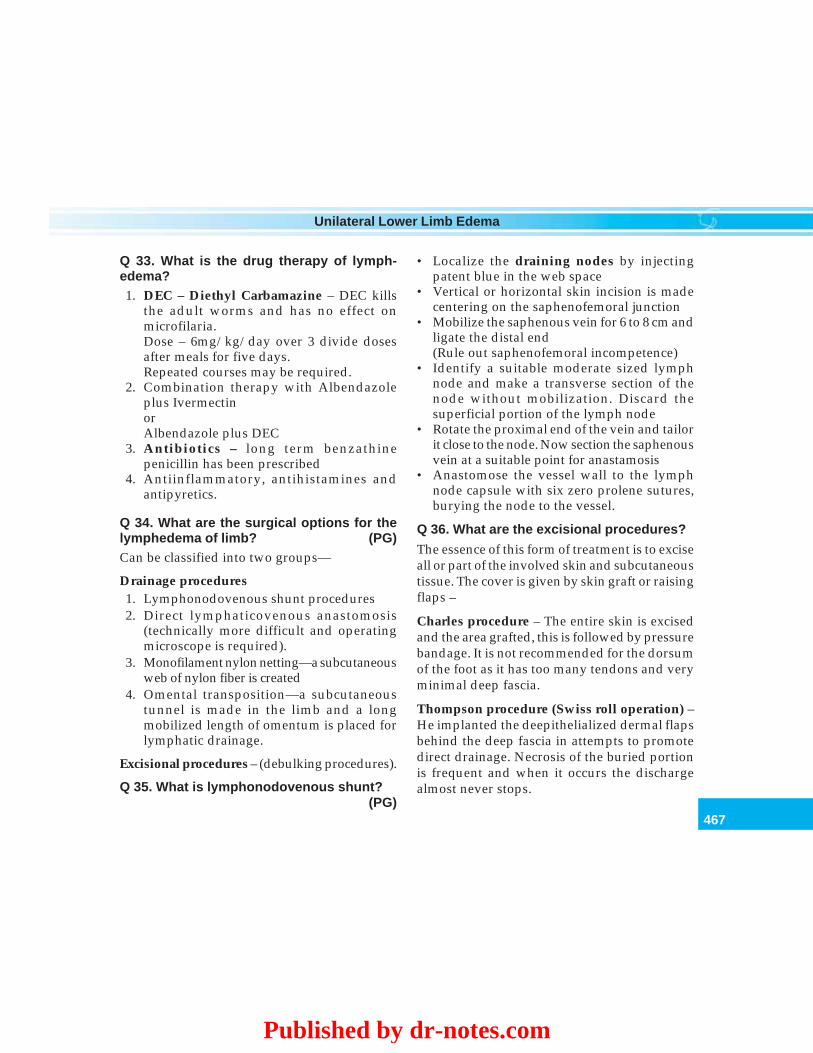
467
Unilateral Lower Limb Edema
Q 33. What is the drug therapy of lymph-edema?1. DEC – Diethyl Carbamazine – DEC kills
the adult worms and has no effect onmicrofilaria.Dose – 6mg/kg/day over 3 divide dosesafter meals for five days.Repeated courses may be required.
2. Combination therapy with Albendazoleplus IvermectinorAlbendazole plus DEC
3. Antibiotics – long term benzathinepenicillin has been prescribed
4. Antiinflammatory, antihistamines andantipyretics.
Q 34. What are the surgical options for thelymphedema of limb? (PG)Can be classified into two groups—
Drainage procedures1. Lymphonodovenous shunt procedures2. Direct lymphaticovenous anastomosis
(technically more difficult and operatingmicroscope is required).
3. Monofilament nylon netting—a subcutaneousweb of nylon fiber is created
4. Omental transposition—a subcutaneoustunnel is made in the limb and a longmobilized length of omentum is placed forlymphatic drainage.
Excisional procedures – (debulking procedures).
Q 35. What is lymphonodovenous shunt?(PG)
• Localize the draining nodes by injectingpatent blue in the web space
• Vertical or horizontal skin incision is madecentering on the saphenofemoral junction
• Mobilize the saphenous vein for 6 to 8 cm andligate the distal end(Rule out saphenofemoral incompetence)
• Identify a suitable moderate sized lymphnode and make a transverse section of thenode without mobilization. Discard thesuperficial portion of the lymph node
• Rotate the proximal end of the vein and tailorit close to the node. Now section the saphenousvein at a suitable point for anastamosis
• Anastomose the vessel wall to the lymphnode capsule with six zero prolene sutures,burying the node to the vessel.
Q 36. What are the excisional procedures?
The essence of this form of treatment is to exciseall or part of the involved skin and subcutaneoustissue. The cover is given by skin graft or raisingflaps –
Charles procedure – The entire skin is excisedand the area grafted, this is followed by pressurebandage. It is not recommended for the dorsumof the foot as it has too many tendons and veryminimal deep fascia.
Thompson procedure (Swiss roll operation) –He implanted the deepithelialized dermal flapsbehind the deep fascia in attempts to promotedirect drainage. Necrosis of the buried portionis frequent and when it occurs the dischargealmost never stops.
Published by dr-notes.com
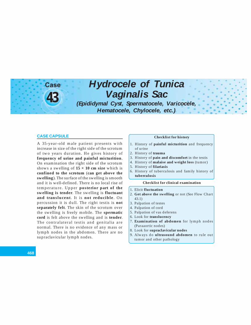
468
Clinical Surgery Pearls
43Hydrocele of Tunica
Vaginalis Sac
CASE CAPSULE
A 35-year-old male patient presents withincrease in size of the right side of the scrotumof two years duration. He gives history offrequency of urine and painful micturition.On examination the right side of the scrotumshows a swelling of 15 × 10 cm size which isconfined to the scrotum (can get above theswelling). The surface of the swelling is smoothand it is well-defined. There is no local rise oftemperature. Upper posterior part of theswelling is tender. The swelling is fluctuantand translucent. It is not reducible. Onpercussion it is dull. The right testis is notseparately felt. The skin of the scrotum overthe swelling is freely mobile. The spermaticcord is felt above the swelling and is tender.The contralateral testis and genitalia arenormal. There is no evidence of any mass orlymph nodes in the abdomen. There are nosupraclavicular lymph nodes.
Checklist for history
1. History of painful micturition and frequencyof urine
2. History of trauma3. History of pain and discomfort in the testis4. History of malaise and weight loss (tumor)5. History of filariasis6. History of tuberculosis and family history of
tuberculosis
Checklist for clinical examination
1. Elicit fluctuation2. Get above the swelling or not (See Flow Chart
43.1)3. Palpation of testes4. Palpation of cord5. Palpation of vas deferens6. Look for translucency7. Examination of abdomen for lymph nodes
(Paraaortic nodes)8. Look for supraclavicular nodes9. Always do ultrasound abdomen to rule out
tumor and other pathology
(Epididymal Cyst, Spermatocele, Varicocele,Hematocele, Chylocele, etc.)
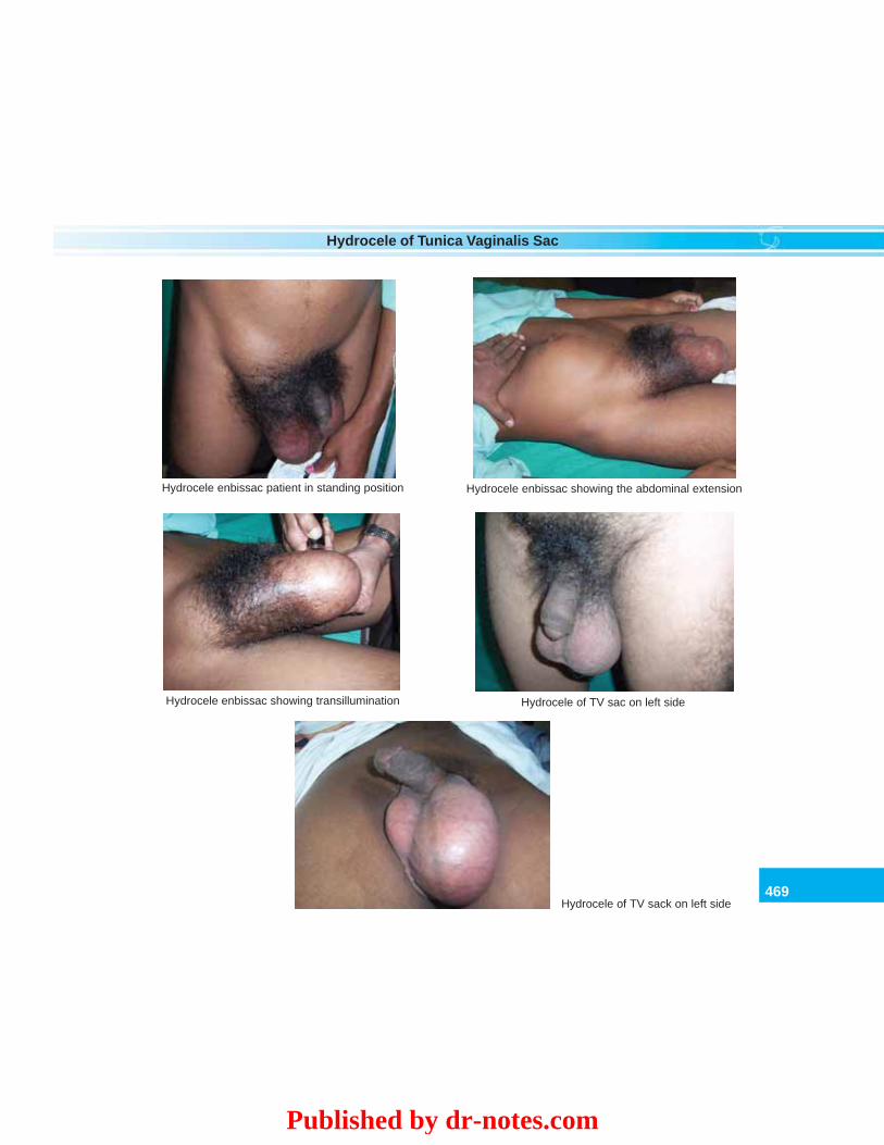
469
Hydrocele of Tunica Vaginalis Sac
Hydrocele enbissac patient in standing position Hydrocele enbissac showing the abdominal extension
Hydrocele enbissac showing transillumination Hydrocele of TV sac on left side
Hydrocele of TV sack on left side
Published by dr-notes.com
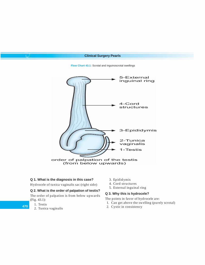
470
Clinical Surgery Pearls
Flow Chart 43.1: Scrotal and inguinoscrotal swellings
Q 1. What is the diagnosis in this case?
Hydrocele of tunica vaginalis sac (right side)
Q 2. What is the order of palpation of testis?
The order of palpation is from below upwards(Fig. 43.1):
1. Testis2. Tunica vaginalis
3. Epididymis4. Cord structures5. External inguinal ring
Q 3. Why this is hydrocele?
The points in favor of hydrocele are:1. Can get above the swelling (purely scrotal)2. Cystic in consistency
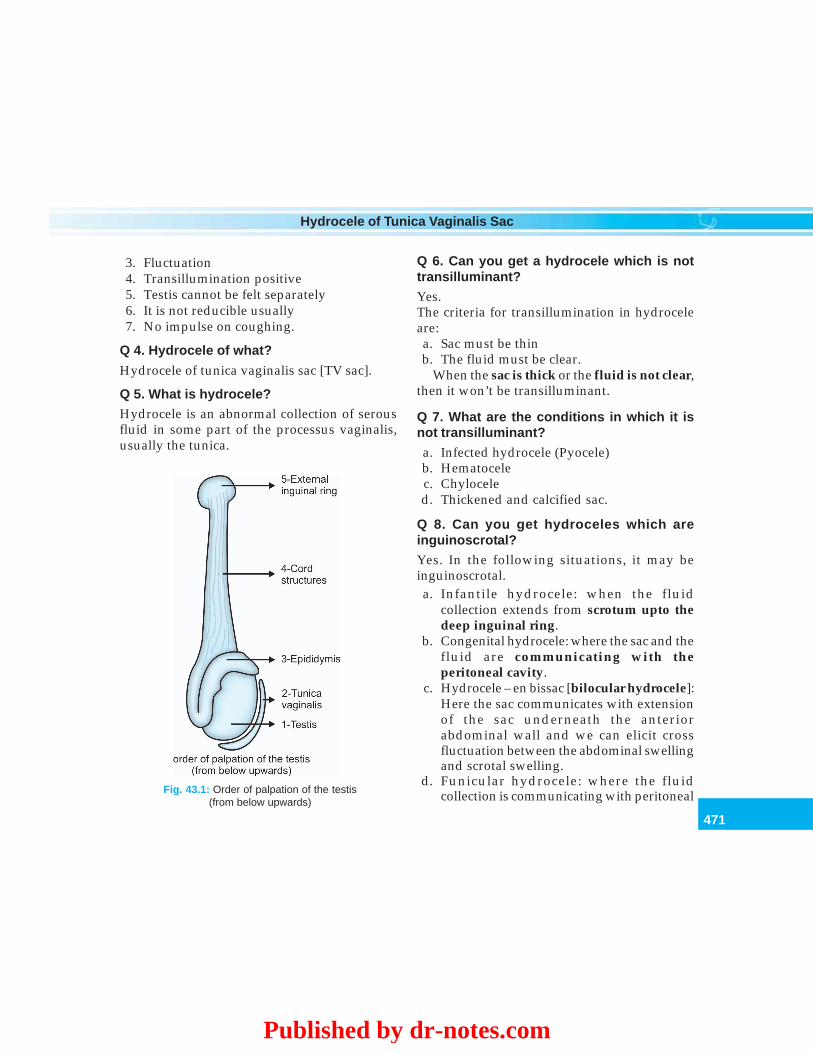
471
Hydrocele of Tunica Vaginalis Sac
Fig. 43.1: Order of palpation of the testis(from below upwards)
3. Fluctuation4. Transillumination positive5. Testis cannot be felt separately6. It is not reducible usually7. No impulse on coughing.
Q 4. Hydrocele of what?
Hydrocele of tunica vaginalis sac [TV sac].
Q 5. What is hydrocele?Hydrocele is an abnormal collection of serousfluid in some part of the processus vaginalis,usually the tunica.
Q 6. Can you get a hydrocele which is nottransilluminant?Yes.The criteria for transillumination in hydroceleare:a. Sac must be thinb. The fluid must be clear.
When the sac is thick or the fluid is not clear,then it won’t be transilluminant.
Q 7. What are the conditions in which it isnot transilluminant?
a. Infected hydrocele (Pyocele)b. Hematocelec. Chyloceled. Thickened and calcified sac.
Q 8. Can you get hydroceles which areinguinoscrotal?Yes. In the following situations, it may beinguinoscrotal.a. Infantile hydrocele: when the fluid
collection extends from scrotum upto thedeep inguinal ring.
b. Congenital hydrocele: where the sac and thefluid are communicating with theperitoneal cavity.
c. Hydrocele – en bissac [bilocular hydrocele]:Here the sac communicates with extensionof the sac underneath the anteriorabdominal wall and we can elicit crossfluctuation between the abdominal swellingand scrotal swelling.
d. Funicular hydrocele: where the fluidcollection is communicating with peritoneal
Published by dr-notes.com
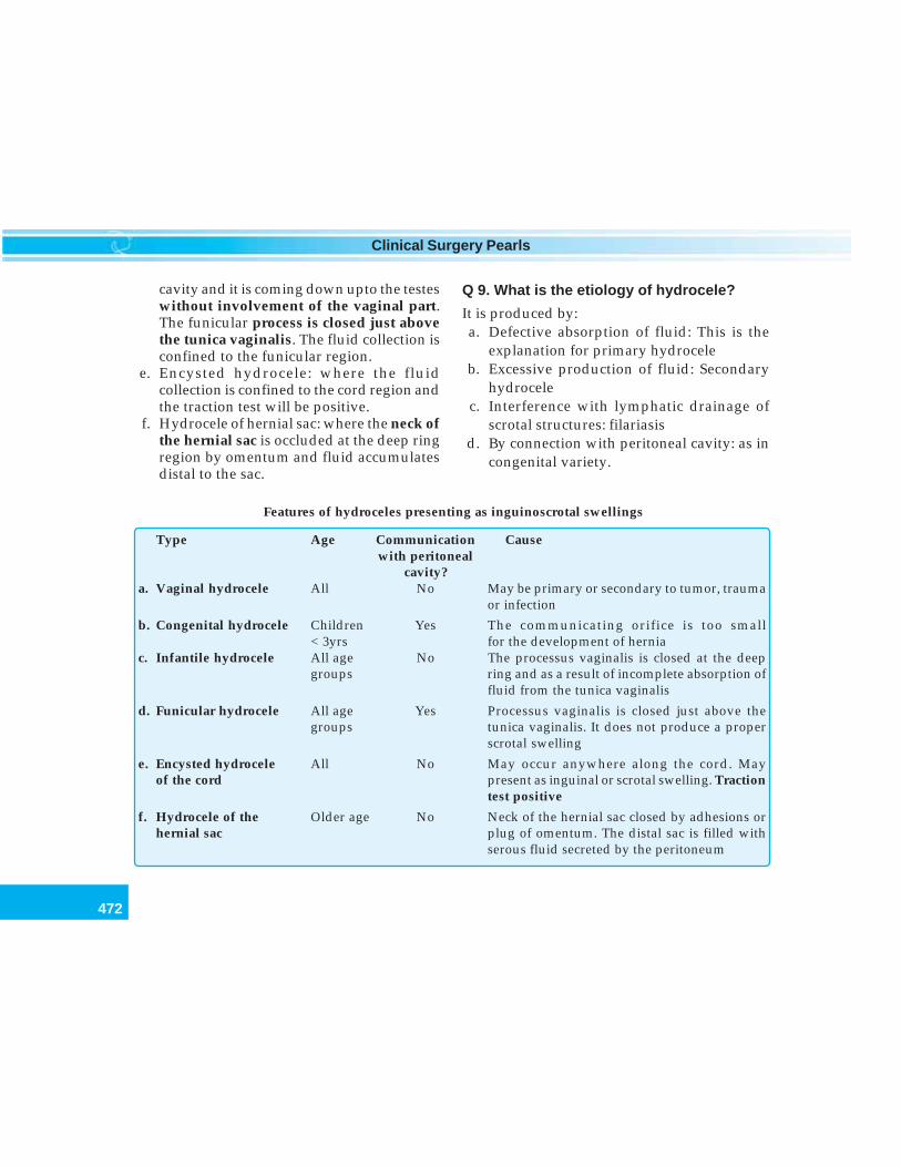
472
Clinical Surgery Pearls
cavity and it is coming down upto the testeswithout involvement of the vaginal part.The funicular process is closed just abovethe tunica vaginalis. The fluid collection isconfined to the funicular region.
e. Encysted hydrocele: where the fluidcollection is confined to the cord region andthe traction test will be positive.
f. Hydrocele of hernial sac: where the neck ofthe hernial sac is occluded at the deep ringregion by omentum and fluid accumulatesdistal to the sac.
Q 9. What is the etiology of hydrocele?
It is produced by:a. Defective absorption of fluid: This is the
explanation for primary hydroceleb. Excessive production of fluid: Secondary
hydrocelec. Interference with lymphatic drainage of
scrotal structures: filariasisd. By connection with peritoneal cavity: as in
congenital variety.
Features of hydroceles presenting as inguinoscrotal swellings
Type Age Communication Causewith peritoneal
cavity?a. Vaginal hydrocele All No May be primary or secondary to tumor, trauma
or infection
b. Congenital hydrocele Children Yes The communicating orifice is too small< 3yrs for the development of hernia
c. Infantile hydrocele All age No The processus vaginalis is closed at the deepgroups ring and as a result of incomplete absorption of
fluid from the tunica vaginalis
d. Funicular hydrocele All age Yes Processus vaginalis is closed just above thegroups tunica vaginalis. It does not produce a proper
scrotal swelling
e. Encysted hydrocele All No May occur anywhere along the cord. Mayof the cord present as inguinal or scrotal swelling. Traction
test positive
f. Hydrocele of the Older age No Neck of the hernial sac closed by adhesions orhernial sac plug of omentum. The distal sac is filled with
serous fluid secreted by the peritoneum
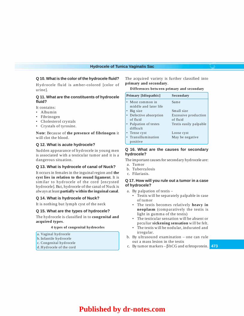
473
Hydrocele of Tunica Vaginalis Sac
Q 10. What is the color of the hydrocele fluid?
Hydrocele fluid is amber-colored [color ofurine].
Q 11. What are the constituents of hydrocelefluid?It contains:• Albumin• Fibrinogen• Cholesterol crystals• Crystals of tyrosine.
Note: Because of the presence of fibrinogen itwill clot the blood.
Q 12. What is acute hydrocele?Sudden appearance of hydrocele in young menis associated with a testicular tumor and it is adangerous situation.
Q 13. What is hydrocele of canal of Nuck?It occurs in females in the inguinal region and thecyst lies in relation to the round ligament. It issimilar to hydrocele of the cord [encystedhydrocele]. But, hydrocele of the canal of Nuck isalways at least partially within the inguinal canal.
Q 14. What is hydrocele of Nuck?It is nothing but lymph cyst of the neck
Q 15. What are the types of hydrocele?The hydrocele is classified in to congenital andacquired types.
4 types of congenital hydroceles
a. Vaginal hydroceleb. Infantile hydrocelec. Congenital hydroceled. Hydrocele of the cord
The acquired variety is further classified intoprimary and secondary.
Differences between primary and secondary
Primary [Idiopathic] Secondary
• Most common in Samemiddle and later life
• Big size Small size• Defective absorption Excessive production
of fluid of fluid• Palpation of testes Testis easily palpable
difficult• Tense cyst Loose cyst• Transillumination May be negative
positive
Q 16. What are the causes for secondaryhydrocele?The important causes for secondary hydrocele are:a. Tumorb. Tuberculosisc. Filariasis.
Q 17. How will you rule out a tumor in a caseof hydrocele?a. By palpation of testis –
• Testis will be separately palpable in caseof tumor
• The testis becomes relatively heavy inneoplasm (comparatively the testis islight in gumma of the testis)
• The testicular sensation will be absent orpeculiar sickening sensation will be felt.
• The testis will be nodular, indurated andirregular.
b. By ultrasound examination – one can ruleout a mass lesion in the testis
c. By tumor markers – β hCG and α fetoprotein.
Published by dr-notes.com
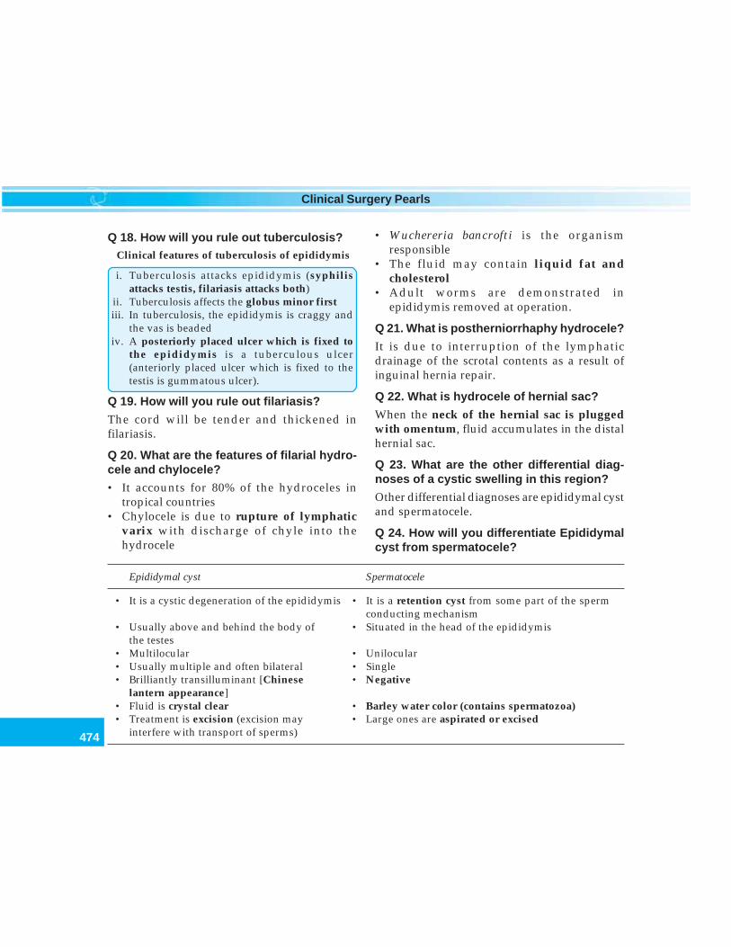
474
Clinical Surgery Pearls
Q 18. How will you rule out tuberculosis?Clinical features of tuberculosis of epididymis
i. Tuberculosis attacks epididymis (syphilisattacks testis, filariasis attacks both)
ii. Tuberculosis affects the globus minor firstiii. In tuberculosis, the epididymis is craggy and
the vas is beadediv. A posteriorly placed ulcer which is fixed to
the epididymis is a tuberculous ulcer(anteriorly placed ulcer which is fixed to thetestis is gummatous ulcer).
Q 19. How will you rule out filariasis?
The cord will be tender and thickened infilariasis.
Q 20. What are the features of filarial hydro-cele and chylocele?
• It accounts for 80% of the hydroceles intropical countries
• Chylocele is due to rupture of lymphaticvarix with discharge of chyle into thehydrocele
• Wuchereria bancrofti is the organismresponsible
• The fluid may contain liquid fat andcholesterol
• Adult worms are demonstrated inepididymis removed at operation.
Q 21. What is postherniorrhaphy hydrocele?
It is due to interruption of the lymphaticdrainage of the scrotal contents as a result ofinguinal hernia repair.
Q 22. What is hydrocele of hernial sac?
When the neck of the hernial sac is pluggedwith omentum, fluid accumulates in the distalhernial sac.
Q 23. What are the other differential diag-noses of a cystic swelling in this region?
Other differential diagnoses are epididymal cystand spermatocele.
Q 24. How will you differentiate Epididymalcyst from spermatocele?
Epididymal cyst Spermatocele
• It is a cystic degeneration of the epididymis • It is a retention cyst from some part of the spermconducting mechanism
• Usually above and behind the body of • Situated in the head of the epididymisthe testes
• Multilocular • Unilocular• Usually multiple and often bilateral • Single• Brilliantly transilluminant [Chinese • Negative
lantern appearance]• Fluid is crystal clear • Barley water color (contains spermatozoa)• Treatment is excision (excision may • Large ones are aspirated or excised
interfere with transport of sperms)
475
Hydrocele of Tunica Vaginalis Sac
Q 25. What is the treatment of hydrocele ofTV sac (Tunica vaginalis sac)?It is mainly treated by surgery.
Q 26. What are the surgical options?1. Jaboulay’s procedure (Eversion of the TV
sac)2. Lord’s operation (when the sac is thin-
walled) the sac is opened and it is plicatedaround the testis. In this operation, there isminimal dissection, the chance forhematoma is reduced and there is no needfor a drain.
Q 27. Can you drain the hydrocele fluid?Drainage of hydrocele fluid is usually not donebecause of the fear of complications likeinfection and hematocele.
Q 28. What are the complications ofhydrocele?
Complications of hydrocele
a. Hematoceleb. Infection and pyocelec. Hernia of hydroceled. Calcification of the sace. Rupture as a result of trauma or spontaneousf. Atrophy of the testis
Q 29. What are the causes for hematocele?Causes for hematocele
• Tapping of hydrocele (damage to small vesselsduring tapping)
• Testicular trauma• Postoperative• Tumor
Q 30. What is clotted hematocele?
• It is due to a spontaneous slow ooze of bloodinto the tunica vaginalis
• Usually it is painless• A tumor may present as clotted hematocele.
Q 31. What is the treatment of clottedhematocele?
• Orchidectomy (It is very difficult todifferentiate tumor from a benign conditionlike this).
Q 32. What is the most important complica-tion of hydrocele operation?
Hematocele
Q 33. What is varicocele?
It is a varicose dilatation of the veins drainingthe testis (pampiniform plexus).
Q 34. What are the signs demonstrable invaricocele?
Signs of varicocele
1. It feels like a ‘bag of worms’2. It can only be felt with the patient standing. If
the patient is asked to lie down, the veins of theplexus will empty
3. A thrill is felt while the patient is coughing4. The ‘bow’ sign: lightly hold the varicocele
between the fingers and thumb. Now the patientis instructed to bow. Tension within the veinsbecome appreciably less by this procedure.
5. The scrotum on the affected side hangs lowerthan normal
6. The testis below a large varicocele may be smallerand softer than the normal side
Published by dr-notes.com
476
Clinical Surgery Pearls
7. After emptying the varicocele in supine position,obstruct the external ring and make the patientstand up. The varicocele will slowly fill up frombelow upwards
Q 35. What is the incidence of varicocele?
It is seen in upto 15% of the male population.
Q 36. Which age group is affected byvaricocele?
• Seen in adolescence and early adulthood• Seen in tall thin men with pendulous
scrotum (short, fat individuals are seldomaffected).
Q 37. What are the symptoms of varicocele?
• Chronic dull ache in the scrotum, worse onstanding for prolonged periods
• May be associated with oligospermia (theassociation remains unclear).
Q 38. On which side varicocele is morecommon and why?
• Varicocele is more common on left sidebecause the left testicular vein enters the leftrenal vein at right angles (95%)
• At times the left testicular artery arches overthe left renal vein to compress it
• The loaded left colon may press on the lefttesticular vein.
Q 39. What is the significance of recent onsetof left sided varicocele?
It is associated with left renal cell carcinomadue to obstruction of the renal vein by a tumorthrombus growing through the left renal vein.
Q 40. What is the venous drainage of testisand epididymis?The draining veins form a plexus calledpampiniform plexus in the scrotal region. Theveins become fewer as they traverse the inguinalcanal. At or near the deep inguinal ring theyjoin to form one or two testicular veins. Theypass upwards behind the peritoneum and joinsthe renal vein on left side and inferior venacava on right side.
Q 41. What is the peculiarity of the testicularvein?They are having valves only at the terminationwhich may be absent (valve less system).
Q 42. Is there an alternative venous drainagefor the testis?Yes. The collateral venous return from the testisis through the cremasteric veins which draininto the inferior epigastric vein.
Q 43. What is the relationship betweenvaricocele and spermatogenesis?• Normally the scrotal temperature is 2.5oC
less than the rectal temperature• The presence of unilateral varicocele
interferes with the normal temperaturecontrol of the scrotum which reduces thetemperature differential to (scrotum/rectum) about 0.1oC below the rectaltemperature.
• Since varicocele is relatively common, thosewho are having oligospermia with varicoceleit is tempting to blame the varicocele as thecause for infertility (not yet clinically proved).
477
Hydrocele of Tunica Vaginalis Sac
Q 44. What are the muscles responsible forsupporting the testis?
Dartos and cremaster.
Q 45. What are the actions of Dartos andCremaster?
• Dartos bears the weight of the testis and actsas a kind of thermostat
• The cremaster is responsible for reflexretraction of the testis during threat of traumaand during fight.
Q 46. What is the grading of varicocele?
• Grade I: Impulse felt in the scrotum onValsalva maneuver
• Grade II: Tortuous and dilated veins palpatedwithout Valsalva maneuver
• Grade III: Varicocele is visible through thescrotal skin.
Note: Sonologically: More than two to threeveins of 3mm or greater in size are found withenlargement on standing and reflux on valsalvamaneuver.
Q 47. What is subclinical varicocele?
Those which are impalpable on physicalexamination is called subclinical varicocele.
Q 48. What are the indications for surgery?
3 ‘S’• Symptoms• Subfertility• Service recruitment.
Q 49. What are the procedures available?
• Palomos operation• Laparoscopic ligation of testicular veins• Embolization of the testicular vein under
radiographic control.
Q 50. What is Palomos operation?
• It is a ligation of the testicular veins abovethe inguinal ligament where thepampiniform plexus coalesce to form one ortwo veins.
• An incision is made 3cm above the deepinguinal ring. The external oblique, internaloblique and transversus muscles are split andthe testicular veins are ligated extraperitoneally (the alternative venous pathwayfor the testis is described above).
• This operation can be done laparoscopicallyalso.
Q 51. What is “triangle of doom”?
It is a laparoscopic finding seen in herniasurgery and varicocele surgery. The triangle isformed medially by the vas deferens, laterallyby the testicular vessel and an imaginary linejoining these two structures. Inside this trianglethe iliac artery and vein are seen which arelikely to be injured and hence called triangle ofdoom.
Q 52. Will varicocele recur after surgery?
Yes. Recurrence is common after all types ofvaricocele surgery.
Published by dr-notes.com
478
Clinical Surgery Pearls
44 Inguinal Hernia/Femoral Hernia
CASE CAPSULE
A 60-year-old male patient presents with aswelling in the right inguinoscrotal region of 2years duration. He also complains of draggingand aching sensation in the groin. He is a chronicsmoker with bronchitis. For the last 2 years hehas difficulty in passing water. He has to getup 3-4 times every night for this purpose. Thereis no history of chronic constipation, abdominalpain or vomiting. He says the swelling is presentonly during standing position and it willdisappear as soon as he lies down. On examina-tion there is a large pear-shaped swelling seenabove the crease of the groin and medial to thepubic tubercle of 8 × 4 cm size. The swelling isnot extending to the scrotum, but confined tothe inguinal region. There is a visible expansilecough impulse, which is demonstrated inpalpation also. One "cannot get above theswelling": The swelling is reducible. Byapplying pressure over the internal ring (Deepring occlusion test), the swelling cannot be heldreduced. A defect is felt in the abdominal wallabove the pubic tubercle. The swelling is dullto percussion. The contralateral side is normal,so also the external genitalia. Malgaigne's
bulging is noted on the left sided whileperforming head raising test. The abdominalexamination revealed no scars and no masslesions. Perrectal examination is normal.
Checklist for history
• History of chronic cough, asthma, bronchitis• History of heavy weight lifting• History of constipation (straining to pass motion)• History of urinary complaints: night frequency,
hesitancy-difficulty to initiate the act ofmicturition and urgency, etc.
• History of pain in the groin• History of epigastric pain (dragging on the
mesentery)• History of appendicectomy (damage to ilio-
inguinal nerve)• History of abdominal pain and vomiting
Checklist for clinical examination
1. Always examine the patient in standingposition, while the examiner sits.
2. Always examine both inguinal regions (20%hernias are bilateral)
3. Look for visible expansile impulse on coughing4. Look for palpable expansile impulse on
coughing5. Assess whether you can get above the swelling
or not
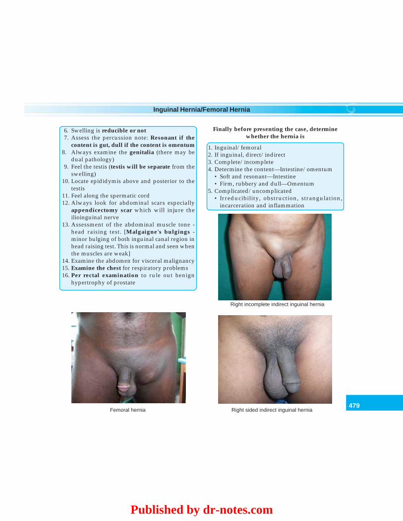
479
Inguinal Hernia/Femoral Hernia
6. Swelling is reducible or not7. Assess the percussion note: Resonant if the
content is gut, dull if the content is omentum8. Always examine the genitalia (there may be
dual pathology)9. Feel the testis (testis will be separate from the
swelling)10. Locate epididymis above and posterior to the
testis11. Feel along the spermatic cord12. Always look for abdominal scars especially
appendicectomy scar which will injure theilioinguinal nerve
13. Assessment of the abdominal muscle tone -head raising test. [Malgaigne's bulgings -minor bulging of both inguinal canal region inhead raising test. This is normal and seen whenthe muscles are weak]
14. Examine the abdomen for visceral malignancy15. Examine the chest for respiratory problems16. Per rectal examination to rule out benign
hypertrophy of prostate
Finally before presenting the case, determinewhether the hernia is
1. Inguinal/femoral2. If inguinal, direct/indirect3. Complete/incomplete4. Determine the content—Intestine/omentum
• Soft and resonant—Intestine• Firm, rubbery and dull—Omentum
5. Complicated/uncomplicated• Irreducibility, obstruction, strangulation,
incarceration and inflammation
Femoral hernia
Right incomplete indirect inguinal hernia
Right sided indirect inguinal hernia
Published by dr-notes.com
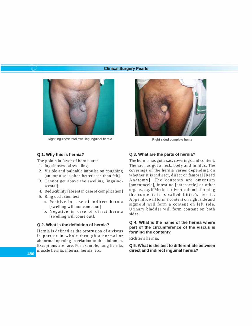
480
Clinical Surgery Pearls
Right inguinoscrotal swelling-inguinal hernia Right sided complete henia
Q 1. Why this is hernia?
The points in favor of hernia are:1. Inguinoscrotal swelling2. Visible and palpable impulse on coughing
[an impulse is often better seen than felt].3. Cannot get above the swelling [inguino-
scrotal]4. Reducibility [absent in case of complication]5. Ring occlusion test
a. Positive in case of indirect hernia[swelling will not come out]
b. Negative in case of direct hernia[swelling will come out].
Q 2. What is the definition of hernia?Hernia is defined as the protrusion of a viscusin part or in whole through a normal orabnormal opening in relation to the abdomen.Exceptions are rare. For example, lung hernia,muscle hernia, internal hernia, etc.
Q 3. What are the parts of hernia?The hernia has got a sac, coverings and content.The sac has got a neck, body and fundus. Thecoverings of the hernia varies depending onwhether it is indirect, direct or femoral [ReadAnatomy]. The contents are omentum[omentocele], intestine [enterocele] or otherorgans, e.g. if Meckel's diverticulum is formingthe content, it is called Littre’s hernia.Appendix will form a content on right side andsigmoid will form a content on left side.Urinary bladder will form content on bothsides.
Q 4. What is the name of the hernia wherepart of the circumference of the viscus isforming the content?Richter's hernia.
Q 5. What is the test to differentiate betweendirect and indirect inguinal hernia?
481
Inguinal Hernia/Femoral Hernia
Deep ring occlusion test is the test of choice todifferentiate these two. Before doing this, thepatient should be in supine position and thehernia should be reduced. Reducibility is testedin the recumbent position. If this is not possible,flex the thigh in order to keep the pillars of theexternal ring relaxed.
Q 6. What is Zieman's test?This is done in the standing position. Theclinician stands behind and somewhat to theright for the right side and behind andsomewhat to the left for the left side. The hand
of the corresponding side is used forexamination [Right hand for right side and lefthand for left side]. He places his index fingerover the indirect hernial site, the middle fingerover the direct hernial site, and ring finger overthe femoral hernial site. The patient is requestedto hold the nose and blow. A peculiar glidingmotion of the walls of the sac will be felt beneaththe relevant finger corresponding to the type ofhernia.
Q 7. What are the clinical differences betweendirect and indirect inguinal hernia?
Clinical differences between direct and indirect inguinal hernia
No Direct Indirect1. Extend to scrotum Does not go down to the Can descend into the scrotum
scrotum2. Direction of reduction Reduce upwards and then Reduce upwards, then laterally and
straight backwards backwards3. Controlled by pressure Not controlled after reduction, Controlled, after reduction by
over the internal ring by pressure over the internal pressure over the internal inguinalinguinal ring ring
4. Direction of reappearance The bulge reappears outwards The bulge reappears in the middleafter reduction to original position of the inguinal region and then
flows medially before turning downto the neck of the scrotum
5. Palpable defect Defect may be felt in the No palpable defect as it is behindabdominal wall above the the fibers of the external obliquepubic tubercle muscle
6. Relationship of cord Sac appears medial to the The sac is inside the spermatic cordto sac inferior epigastric artery and is
outside the spermatic cord(posterior to the cord)
Published by dr-notes.com
482
Clinical Surgery Pearls
Q 8. What are the peculiarities of directinguinal hernia?
Peculiarities of direct inguinal hernia
• Appear later in life• Do not occur in children• Rare in women• Rarely strangulate• Direct hernia is always acquired• Usually seen in males• They do not often attain large size or descend
into the scrotum• The protruding mass mainly consists of extra
peritoneal fat• The neck of the sac is wide
Q 9. How will you assess the content of thesac?
If it is omentum:a. It will give a doughy feelb. The first part of the hernial sac is reduced
easily and the last part is difficult to reduce.If it is intestinea. The first part is difficult to reduce and the
last part is reduced easilyb. It has got a characteristic gurgling sound
during reduction.
Q 10. What is sliding hernia [Hernia-en-glissade]?Sliding hernia is a condition where portion ofcecum and appendix on right side, sigmoid onleft side and urinary bladder on both sides willslide down behind the sac. Even though it is notinside the sac, it forms the posterior wall of thesac. If the wall of the sac is unusually thick per-
operatively, one should carefully rule out asliding hernia.
Q 11. What is Maydl's Hernia?
This is the so-called W loop hernia where thesmall intestine forms a W loop within the hernialsac. The importance of this type of hernia is incase of obstruction, even if the visible intestineinside the sac is viable if one is not pulling outthe rest of the intestine, you are likely to missgangrene for the rest of the bowel.
Q 12. How will you differentiate inguinalhernia from femoral hernia?
Inguinal Femoral
• Above and medial to • Below and lateral tothe pubic tubercle the pubic tubercle
• Above the crease of • Below the crease ofthe groin the groin
• Can be reduced • Cannot be reducedcompletely completely
• Cough impulse • Many do not haveusually present cough impulse
Q 13. Why femoral hernia is usuallyirreducible?
The femoral hernias are having.a. Narrow neck of the sacb. The contents are adherent to the peritoneal
sac.
Q 14. What is the direction of enlargement offemoral hernia?
It is usually downwards, forwards andupwards.
483
Inguinal Hernia/Femoral Hernia
Q 15. What is the name of the triangle inwhich you get the direct hernia?
The direct hernia comes out through Hessel-bach's triangle. It is bounded medially by thelateral border of rectus abdominis, laterally bythe inferior epigastric artery and below by theinguinal ligament.
Q16. What are the differential diagnoses ofinguinal hernia?
Differential diagnoses of inguinal hernia
a. Femoral herniab. Vaginal hydrocelec. Undescended testis in superficial inguinal pouchd. Hydrocele of the corde. Lipoma of the cordf. Infantile hydroceleg. Ectopic testish. Lipoma of the cordi. Hydrocele of canal of Nuckj. Psoas abscessk. Psoas bursael. Sapheno - varixm. Enlarged lymph nodesn. Femoral aneurysm
Note: There are two lumps which occur in theline of the spermatic cord which can pop in andout of the external ring, viz. undescended testesand hydrocele of the cord.
Q 17. What are the 3 types of inguinal hernia?a. Bubonocele - when the hernia is limited to
the inguinal canal.b. Funicular - when the processus vaginalis is
closed just above the epididymis. Here thecontents of the sac can be felt separatelyfrom the testis.
c. Complete - [Scrotal ] In complete hernia, thetestis appears to lie within the lower part ofthe hernia.
Q 18. What is the situation of testis in acomplete (scrotal) hernia?
It is sited posteroinferior to the hernia.
Q 19. What are the etiological factors forhernia?
Etiology of hernia
a. Congenital - Preformed sac where the processusis patent
b. Acquired -i. Increased intraabdominal pressure [chronic
cough, straining, whooping cough, etc.]ii. Smokers [collagen deficiency due to smoking]
iii. Intraabdominal malignancy [acute onset ofhernia]
iv. Obesity [Muscles are weak, fat separate musclebundles and weakens aponeurosis]
v. Multiparity [for femoral hernia - stretching ofpelvic ligaments]
vi. TA/TF deficiency - transverses abdominis,transversalis fascia deficiency
c. Hereditary:d. The evolutionary factors are
i. Absence of posterior rectus sheath below thearcuate line
ii. Adoption of upright positioniii. Change from quadrapedal to bipedal
locomotion [In animals, the weight of theabdominal content is directed away from theinguinal region].
Q 20. What is the definition of inguinal canal?Inguinal canal is an intermuscular slit situatedbetween the superficial and deep inguinal rings(Fig. 44.1)
Published by dr-notes.com
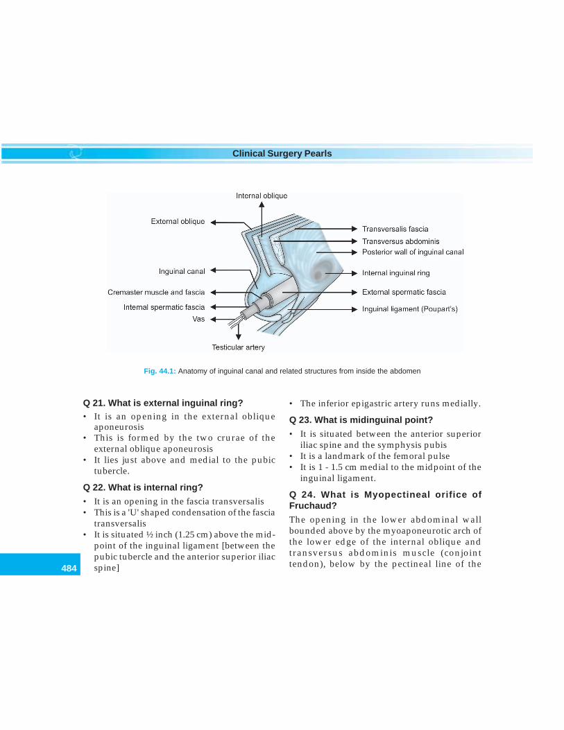
484
Clinical Surgery Pearls
Q 21. What is external inguinal ring?• It is an opening in the external oblique
aponeurosis• This is formed by the two crurae of the
external oblique aponeurosis• It lies just above and medial to the pubic
tubercle.
Q 22. What is internal ring?
• It is an opening in the fascia transversalis• This is a 'U' shaped condensation of the fascia
transversalis• It is situated ½ inch (1.25 cm) above the mid-
point of the inguinal ligament [between thepubic tubercle and the anterior superior iliacspine]
• The inferior epigastric artery runs medially.
Q 23. What is midinguinal point?
• It is situated between the anterior superioriliac spine and the symphysis pubis
• It is a landmark of the femoral pulse• It is 1 - 1.5 cm medial to the midpoint of the
inguinal ligament.
Q 24. What is Myopectineal orifice ofFruchaud?
The opening in the lower abdominal wallbounded above by the myoaponeurotic arch ofthe lower edge of the internal oblique andtransversus abdominis muscle (conjointtendon), below by the pectineal line of the
Fig. 44.1: Anatomy of inguinal canal and related structures from inside the abdomen
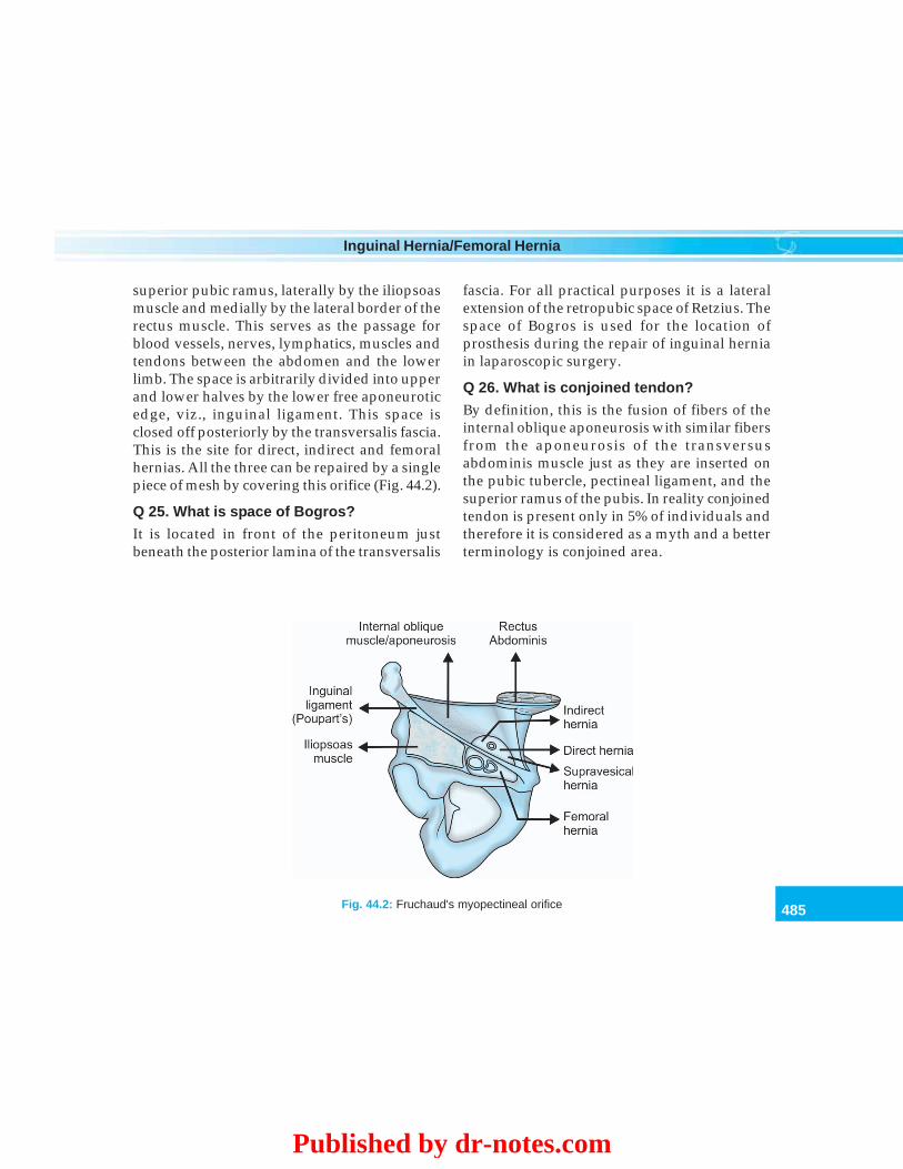
485
Inguinal Hernia/Femoral Hernia
superior pubic ramus, laterally by the iliopsoasmuscle and medially by the lateral border of therectus muscle. This serves as the passage forblood vessels, nerves, lymphatics, muscles andtendons between the abdomen and the lowerlimb. The space is arbitrarily divided into upperand lower halves by the lower free aponeuroticedge, viz., inguinal ligament. This space isclosed off posteriorly by the transversalis fascia.This is the site for direct, indirect and femoralhernias. All the three can be repaired by a singlepiece of mesh by covering this orifice (Fig. 44.2).
Q 25. What is space of Bogros?
It is located in front of the peritoneum justbeneath the posterior lamina of the transversalis
fascia. For all practical purposes it is a lateralextension of the retropubic space of Retzius. Thespace of Bogros is used for the location ofprosthesis during the repair of inguinal herniain laparoscopic surgery.
Q 26. What is conjoined tendon?
By definition, this is the fusion of fibers of theinternal oblique aponeurosis with similar fibersfrom the aponeurosis of the transversusabdominis muscle just as they are inserted onthe pubic tubercle, pectineal ligament, and thesuperior ramus of the pubis. In reality conjoinedtendon is present only in 5% of individuals andtherefore it is considered as a myth and a betterterminology is conjoined area.
Fig. 44.2: Fruchaud's myopectineal orifice
Published by dr-notes.com
486
Clinical Surgery Pearls
Q 27. What is transversalis fascia?
The term may be restricted to the internal fascialining the transversus abdominis muscle. In theinguinal area the transversalis fascia is bilaminarenveloping the inferior epigastric vessels.
Q 28. What is the frequency of various typesof hernia?
Frequency of types of Hernia
• Inguinal - 75%• Femoral - 8.5%• Umbilical - 15%• Rarer forms 1.5%
Q 29. What is the frequency of direct hernia?
• About 35% of inguinal hernias are direct inmales (65% indirect)
• 12% will have contralateral hernia• About four-fold increased risk for future
development on contralateral side.
Q 30. Inguinal hernia is more commonly seenon which side?
55% seen on right side.
Q 31. What is the frequency of direct herniain females?
Women practically never develop a directinguinal hernia.
Q 32. What is the sex-wise incidence ofhernia?
The male to female ratio for inguinal hernia is20:1.
Q 33. What is funicular direct inguinal hernia[Prevesical hernia]?
This is a narrow necked hernia with prevesicalfat and sometimes a portion of the bladder thatprotrudes through a small oval defect in themedial part of conjoint muscle, just above thepubic tubercle. It occurs in elderly males andoccasionally becomes strangulated.
Q 34. What is dual hernia [Pantaloon, "saddle- bag"]?
This is a type of hernia where two sacs straddlethe inferior epigastric artery, one sac beingmedial and other lateral to the artery. This isone of the causes for recurrence, if one isoverlooked during surgery.
Q 35. What will be the course of action, if apatient develops sudden asymptomatichernia?
One must search for occult intraabdominalmalignancy and ascites. Do the following tests.• Digital rectal examination• Fecal study for occult blood• Sigmoidoscopy/colonoscopy• Double contrast barium enema.
Q 36. Why must a hernia be repaired?
Because of the potential dangerous complica-tions.
Q 37. What are the complications of hernia?Complications of hernia
• Irreducibility• Obstruction• Strangulation• Incarceration• Inflammation
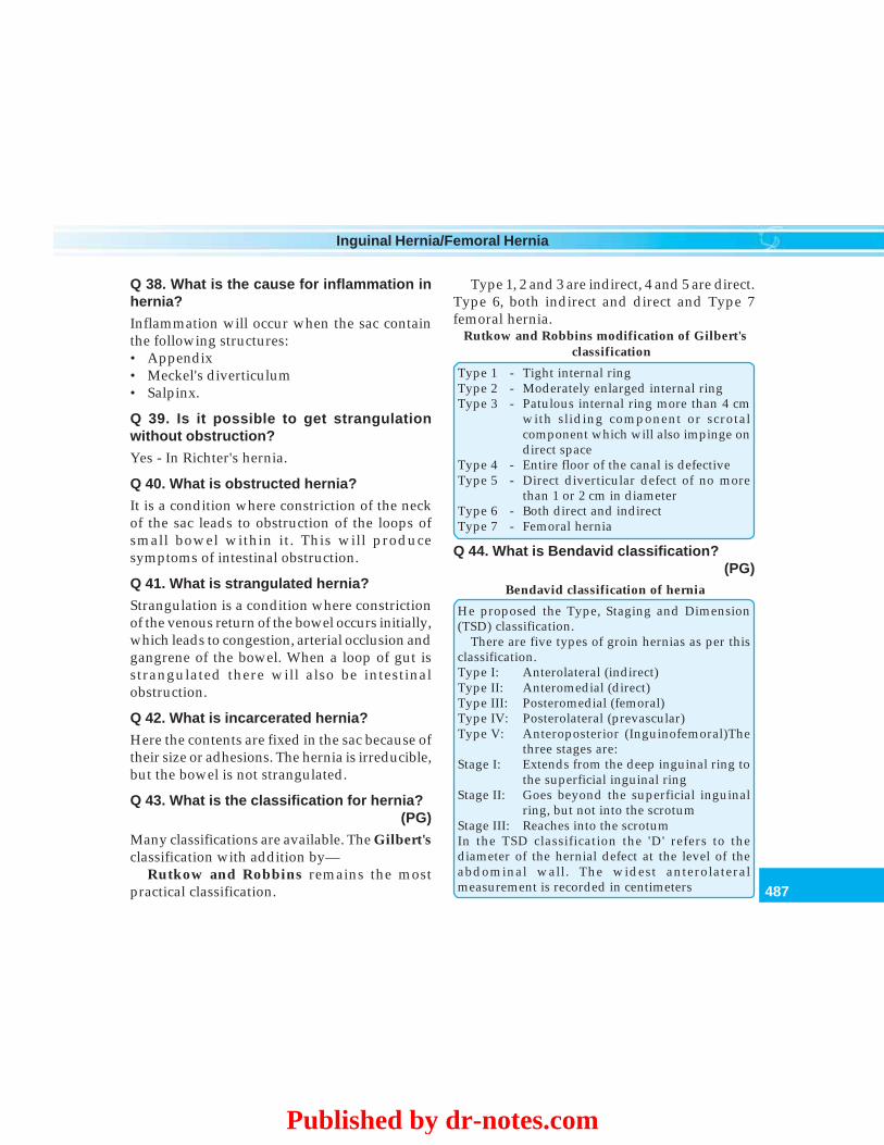
487
Inguinal Hernia/Femoral Hernia
Q 38. What is the cause for inflammation inhernia?
Inflammation will occur when the sac containthe following structures:• Appendix• Meckel's diverticulum• Salpinx.
Q 39. Is it possible to get strangulationwithout obstruction?
Yes - In Richter's hernia.
Q 40. What is obstructed hernia?
It is a condition where constriction of the neckof the sac leads to obstruction of the loops ofsmall bowel within it. This will producesymptoms of intestinal obstruction.
Q 41. What is strangulated hernia?
Strangulation is a condition where constrictionof the venous return of the bowel occurs initially,which leads to congestion, arterial occlusion andgangrene of the bowel. When a loop of gut isstrangulated there will also be intestinalobstruction.
Q 42. What is incarcerated hernia?
Here the contents are fixed in the sac because oftheir size or adhesions. The hernia is irreducible,but the bowel is not strangulated.
Q 43. What is the classification for hernia?(PG)
Many classifications are available. The Gilbert'sclassification with addition by—
Rutkow and Robbins remains the mostpractical classification.
Type 1, 2 and 3 are indirect, 4 and 5 are direct.Type 6, both indirect and direct and Type 7femoral hernia.
Rutkow and Robbins modification of Gilbert'sclassification
Type 1 - Tight internal ringType 2 - Moderately enlarged internal ringType 3 - Patulous internal ring more than 4 cm
with sliding component or scrotalcomponent which will also impinge ondirect space
Type 4 - Entire floor of the canal is defectiveType 5 - Direct diverticular defect of no more
than 1 or 2 cm in diameterType 6 - Both direct and indirectType 7 - Femoral hernia
Q 44. What is Bendavid classification?(PG)
Bendavid classification of hernia
He proposed the Type, Staging and Dimension(TSD) classification.
There are five types of groin hernias as per thisclassification.Type I: Anterolateral (indirect)Type II: Anteromedial (direct)Type III: Posteromedial (femoral)Type IV: Posterolateral (prevascular)Type V: Anteroposterior (Inguinofemoral)The
three stages are:Stage I: Extends from the deep inguinal ring to
the superficial inguinal ringStage II: Goes beyond the superficial inguinal
ring, but not into the scrotumStage III: Reaches into the scrotumIn the TSD classification the 'D' refers to thediameter of the hernial defect at the level of theabdominal wall. The widest anterolateralmeasurement is recorded in centimeters
Published by dr-notes.com
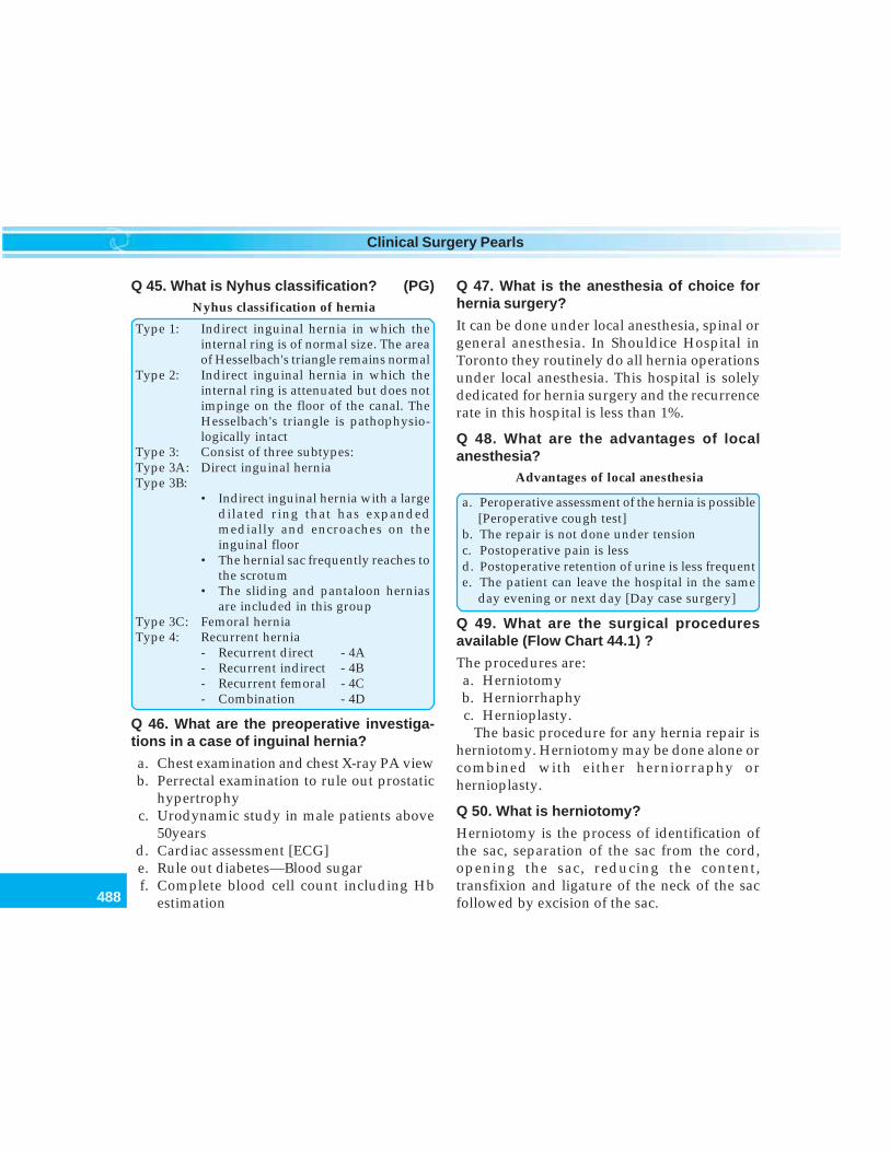
488
Clinical Surgery Pearls
Q 45. What is Nyhus classification? (PG)Nyhus classification of hernia
Type 1: Indirect inguinal hernia in which theinternal ring is of normal size. The areaof Hesselbach's triangle remains normal
Type 2: Indirect inguinal hernia in which theinternal ring is attenuated but does notimpinge on the floor of the canal. TheHesselbach's triangle is pathophysio-logically intact
Type 3: Consist of three subtypes:Type 3A: Direct inguinal herniaType 3B:
• Indirect inguinal hernia with a largedilated ring that has expandedmedially and encroaches on theinguinal floor
• The hernial sac frequently reaches tothe scrotum
• The sliding and pantaloon herniasare included in this group
Type 3C: Femoral herniaType 4: Recurrent hernia
- Recurrent direct - 4A- Recurrent indirect - 4B- Recurrent femoral - 4C- Combination - 4D
Q 46. What are the preoperative investiga-tions in a case of inguinal hernia?
a. Chest examination and chest X-ray PA viewb. Perrectal examination to rule out prostatic
hypertrophyc. Urodynamic study in male patients above
50yearsd. Cardiac assessment [ECG]e. Rule out diabetes—Blood sugarf. Complete blood cell count including Hb
estimation
Q 47. What is the anesthesia of choice forhernia surgery?
It can be done under local anesthesia, spinal orgeneral anesthesia. In Shouldice Hospital inToronto they routinely do all hernia operationsunder local anesthesia. This hospital is solelydedicated for hernia surgery and the recurrencerate in this hospital is less than 1%.
Q 48. What are the advantages of localanesthesia?
Advantages of local anesthesia
a. Peroperative assessment of the hernia is possible[Peroperative cough test]
b. The repair is not done under tensionc. Postoperative pain is lessd. Postoperative retention of urine is less frequente. The patient can leave the hospital in the same
day evening or next day [Day case surgery]
Q 49. What are the surgical proceduresavailable (Flow Chart 44.1) ?
The procedures are:a. Herniotomyb. Herniorrhaphyc. Hernioplasty.
The basic procedure for any hernia repair isherniotomy. Herniotomy may be done alone orcombined with either herniorraphy orhernioplasty.
Q 50. What is herniotomy?
Herniotomy is the process of identification ofthe sac, separation of the sac from the cord,opening the sac, reducing the content,transfixion and ligature of the neck of the sacfollowed by excision of the sac.
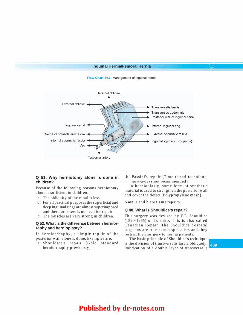
489
Inguinal Hernia/Femoral Hernia
Q 51. Why herniotomy alone is done inchildren?Because of the following reasons herniotomyalone is sufficient in children:a. The obliquity of the canal is lessb. For all practical purposes the superficial and
deep inguinal rings are almost superimposedand therefore there is no need for repair
c. The muscles are very strong in children.
Q 52. What is the difference between hernior-raphy and hernioplasty?In herniorrhaphy, a simple repair of theposterior wall alone is done. Examples are:a. Shouldice's repair [Gold standard
herniorrhaphy previously]
b. Bassini's repair [Time tested technique,now-a-days not recommended].
In hernioplasty, some form of syntheticmaterial is used to strengthen the posterior walland cover the defect [Polypropylene mesh].
Note: a and b are tissue repairs.
Q 48. What is Shouldice's repair?This surgery was devised by E.E. Shouldice(1890-1965) of Toronto. This is also calledCanadian Repair. The Shouldice hospitalsurgeons are true hernia specialists and theyrestrict their surgery to hernia patients.
The basic principle of Shouldice's techniqueis the division of transversalis fascia obliquely,imbrication of a double layer of transversalis
Flow Chart 44.1: Management of inguinal hernia
Published by dr-notes.com
490
Clinical Surgery Pearls
fascia to the inguinal ligament followed by adouble layer of conjoint tendon—internaloblique muscle suturing to the undersurface ofthe external oblique.• In short, it is a 4 layer repair. In first layer,
the lower leaf of the divided transversalisfascia is sutured first to the undersurface ofthe upper leaf by continuous 30 polypropylenesuture.
• The first layer is continued as the second layerwhere the lower border of the upper leaf issutured to the inguinal ligament by the samesuture material continuously. The first layeris started medially from the pubic tubercleregion and goes laterally and then comesmedially as the second layer.
• The third layer is started laterally at the deepinguinal ring region and comes medially andthen it is continued again laterally as thefourth layer.
• The third and fourth layers are suturing ofthe conjoint tendon and undersurface of theexternal oblique/ inguinal ligament.
Q 54. What is Bassini's herniorrhaphy?
This is the oldest technique of hernia repairwhere after herniotomy the conjoint tendon isapproximated to the inguinal ligament usingNo.1 size interrupted polypropylene sutures(synthetic nonabsorbable).
Q 55. What is the current Gold Standardsurgery for hernia repair [hernioplasty]?
The Gold Standard current hernia surgery is theLichtenstein Tension-free Hernioplasty. Hereapproximately 16 × 8cm size mesh (polypro-
pylene) is placed anterior to the posterior wallafter herniotomy and overlapping it generouslyin all directions including medially over thepubic tubercle.
Q 56. What are the essential steps of inguinalherniorrhaphy?
• Local anesthesia/regional anesthesia/general anesthesia.
• An oblique inguinal skin crease incisionapproximately 1.5-2 cm above the medialtwo-thirds of the inguinal ligament.
• Incise the fatty and membranous layers of thesuperficial fascia (fascia of Camper and fasciaof Scarpa).
• Ligate the superficial circumflex iliac,superficial epigastric and superficial externalpudendal vessels (external pudendal may beretracted to avoid edema scrotum).
• Identify the external oblique aponeurosis andexternal ring. Divide the external obliqueaponeurosis along the line of its fibers andturn back the edges.
• Identify the internal oblique and conjoinedtendon above and the inguinal ligamentbelow. The cord is in between.
• In indirect inguinal hernia the sac is insidethe spermatic cord and the cremaster musclehas to be divided in the line of the spermaticcord for identifying the sac. In direct herniathe sac is posterior to the cord.
• Safeguard the ilioinguinal nerve, vas deferensand spermatic artery.
• Identification of the sac and isolation the sacfrom the cord (the sac is seen as pearly whitestructure).
491
Inguinal Hernia/Femoral Hernia
Indirect inguinal hernial sac:• If the sac is small it can be freed in toto. If it is
of long and scrotal type, the fundus of thesac must not be sought (the blood supply tothe testis may be compromised by such aprocedure). The sac is isolated upto the neckof the sac until the parietal peritoneum canbe seen on all sides and extraperitoneal fat isvisualized. The inferior epigastric vessels areseen on the medial side.
• Open the sac, reduce the contents (omentumand intestines are returned to the peritonealcavity).
• Herniotomy: The neck of the sac is transfixedas high as possible and the rest of the sac isremoved.
Direct hernial sac:• In this situation there is no need to incise the
cremaster muscle to identify the sac (thehernial sac is seen posterior to the cord).
• Push the direct hernial sac inwards (no needto open the sac and reduce the contents.Transfixion and ligation and herniotomy arenot required).
• Interrupted polypropylene sutures are put sothat the sac will remain invaginated.
Repair of both direct and indirect hernia:• The best tissue repair is the four layer repair
of Shouldice (read the steps above).• For Lichtenstein tension-free repair, the
polypropylene mesh is used to reinforce theposterior wall. The mesh is sutured below tothe inguinal ligament, above to the internaloblique and conjoined tendon. The mesh
should overlap beyond the pubic tuberclemedially. Laterally the mesh is incised toaccommodate for the spermatic cord. Finallythe spermatic cord will be lying over themesh.
Closure• Approximate the external oblique aponeurosis• Subcutaneous sutures are put with interrupted
absorbable sutures• Skin is closed, ideally by skin tapes.
Q 57. Can you do hernia repair by laparo-scopy?
Yes.
Q 58. What are the methods of laparoscopichernia repair? (PG)
TEP and TAPP.TAPP: Transabdominal preperitoneal repair—Here after entering the peritoneal cavity, theperitoneum is incised cephalad to the inguinalfloor and the hernia defects are dissected.Moderate sized indirect sacs are dissected andreduced. Large sacs are occasionally transectedand the distal sac left in situ leading to a possiblehydrocele formation avoiding hematoma. Meshis secured to Cooper's ligament and theunderside to the conjoined tendon with nostaples being placed lateral to epigastric vessels.
TEP: Total extraperitoneal repair—Here theperitoneal cavity is not entered. With the helpof an infraumbilical trocar, the pre-peritonealspace is entered using a combination of carbondioxide and blunt dissection to expose the entiremyopectineal orifice. Polypropylene mesh is
Published by dr-notes.com
492
Clinical Surgery Pearls
then placed between the underside of theabdominal wall and the peritoneum fixing themesh to Cooper's ligament and the aponeuroticsling.
Q 59. What are the indications for laparo-scopic hernia repair? (PG)
Indications for laparoscopic hernia repair
• Bilateral inguinal hernia• Recurrent hernia• Femoral hernia
Q 60. What is Stoppa groin hernia repair?(PG)
It is also called giant prosthetic reinforcementof visceral sac [GPRVS].• This is a revolutionary and innovative
bilateral properitoneal prosthetic hernio-plasty. The procedure is although useful forrepair of all hernias of the groin, it is mainlyused to manage complex hernias at high riskfor recurrence and for recurrent groinhernias.
• The essential feature of GPRVS is thereplacement of the transversalis fascia in thegroin by a large prosthesis that extendsbeyond the myopectineal orifice (MPO).
• The prosthesis envelops the visceral sac heldin place by intraabdominal pressure and laterby connective tissue ingrowth.
• The mesh adheres to the peritoneum andrenders it inextensible so that it cannotprotrude through the parietal defect. Here theparietal defects are not closed and should notbe closed. GPRVS is a sutureless and tension-free repair.
Bilateral GPRVS• Bilateral GPRVS may be achieved through a
subumbilical midline or Pfannenstielincision.
• The preperitoneal space is cleaved in alldirections, exposing the space of Bogros andthe space of Retzius, the superior ramus ofthe pubis, the obturator foramen, iliac vesselsand the iliopsoas muscle.
• The elements of the spermatic cord areparietalized.
• The chevron- shaped mesh is tailored to thepatient and should measure transversely 2cm less than the distance between the anteriorsuperior iliac spines and vertically shouldmeasure the distance between the umbilicusand the symphysis pubis.
• In obese patients the mesh should be severalcentimeters wider than the interspinousdimensions.
• The repair also can be done unilaterally andthe mesh implanted through a lowerquadrant transverse abdominal incision orthrough an anterior groin incision.
Q 61. What is the management of dualhernia?
The sac should be delivered to the lateral sideof the deep epigastric vessel and dealt with.
Q 62. What are the vessels likely to be injuredin hernia surgery?
Vessels likely to be injured in hernia surgery
1. Pubic branch of the obturator artery2. Aberrant obturator artery originating from the
deep inferior epigastric artery - 'Artery of death'
493
Inguinal Hernia/Femoral Hernia
3. Inferior deep epigastric artery4. Deep circumflex iliac vessel5. Cremasteric artery6. External iliac vessel
Q 63. What is the course of action if a deepbite is taken through external iliac vessel?• Remove the suture (Don't tie the suture). If
not the artery will blow out within 2-3 days• Apply pressure.
Q 64. What are the nerves inside the inguinalcanal?1. Ilioinguinal nerve (T12 - L1)
• It is in close relationship to spermaticcord
• It is seen in the usual position only in 60%of cases
• It may be absent• Innervation - base of the penis at the
scrotum and adjacent side.2. Iliohypogastric nerve (T12 - L1)
• Seen 1 to 2cm above the inguinal canal• Supplies suprapubic area• Injured while a relaxing incision is put
over the rectus sheath.3. Genitofemoral nerve (L1,2,3)
Genital branch.• Prone to injury at the internal ring• Penetrate the internal oblique at the
origin of the cremaster• Motor innervation - Cremaster muscle• Sensory innervation - to the penis and
scrotum.Femoral branch• Innervation - Upper thigh (less likely to
be injured).
Q 65. If a nerve is cut what is the course ofaction? (PG)• It may be ligated or clipped to allow closure
of the neurilemmal sheath• Repair is impossible.
Q 66. What is the blood supply to the testis?
Blood supply to the testis
a. Testicular artery (major supply) - from aortab. External spermatic artery - from inferior
epigastric artery - also supply cremaster musclec. Artery to vas deferens - from superior vesical
artery
Note: There is a rich collateral existing betweenthese three vessels. In addition the vesical andprostatic branches communicate with the above.The scrotal vessels from internal and externalpudendal vessels, freely communicate withvessels in the spermatic cord external to thesuperficial inguinal ring.
Q 67. What will happen if testicular artery isligated? (PG)
Ligation of testicular artery alone at the deepring may not lead to testicular atrophy providedcollateral circulation is undisturbed.
Q 68. What are the steps in surgery whichwill jeopardize the collateral supply to thetestis? (PG)
• Removal of testis from scrotum• Dissection of sac distal to the external ring.
Q 69. What are the complications of herniasurgery?
Published by dr-notes.com
494
Clinical Surgery Pearls
Complications of hernia surgery
i. Hematomaii. Seroma
iii. Wound infection- Superficial incisional surgical site infection- Deep incisional surgical site infection
iv. Infection of meshv. Scrotal edema
vi. Postherniorrhaphy hydrocelevii. Recurrent herniaviii.Ischemic orchitisix. Testicular atrophyx. Chronic residual neuralgia (sensory nerve):
Ilioinguinal neuralgiaGenitofemoral neuralgia
xi. Obstruction of vas deferensxii. Dysejaculation
Q 70. What is the incidence of recurrenthernia?1. For inguinal hernia:2 - 3 to 20%2. Femoral hernia: 11.8 - 75%
Q 71. What is the most common site ofrecurrence? (PG)• Medially - the transversus abdominis tendon
is inserted to the rectus sheath as much as2cm above the pubic tubercle. If the mesh isnot reaching beyond the pubic tubercle for1cm, there is chance for recurrence
• The second most common site is at theinternal ring
Q 72. What are the causes for hematoma afterherniorrhaphy? (PG)• Bleeding from superficial vessels (External
pudendal, circumflex iliac and superficialepigastric vessels).
• On a deeper plane, during resection of thecremaster, careless ligature of the externalspermatic artery can result in tensehematoma and ecchymosis that extend to thescrotum.
• Injury to the deep inferior epigastric vessel(one artery and two veins) during divisionof the transversalis fascia.
• Bleeding from venous circulation within thespace of Bogros.
• Bleeding from iliopubic artery.• Injury to aberrant obturator artery from the
deep inferior epigastric artery when suturesare inserted to Cooper's ligament ("Artery ofdeath").
• Injury to femoral vein and artery.
Q 73. How to avoid scrotal edema? (PG)
• Do not divide external pudendal vessels atthe medial limit during incision
• Bilateral simultaneous herniorrhaphy willcause edema of penis and scrotum.
Q 74. What is the causes for posthernior-rhaphy hydrocele? (PG)
• Overzealous skeletonization of the spermaticcord
• Severance of the lymphatic drainage.
Q 75. What is ischemic orchitis? (PG)
• Ischemic orchitis will produce swollen,painful, hard and tender spermatic cord,epididymis and testicles
• It is seen within 24-72 hours of hernia surgery• May be associated with fever• The pain and tenderness will last for 6 weeks• Swelling will last for upto 5 months
495
Inguinal Hernia/Femoral Hernia
• The process is sterile and there is nosuppuration
• It is as a result of intense venous congestionsecondary to massive thrombosis of the veinsof the cord.
Q 76. What is the natural course of ischemicorchitis? (PG)• Subsides completely
or• Testicular atrophy as late as 12 months (no
pain or tenderness).
Q 77. What are the causes for chronicresidual neuralgia? (PG)It may be due to the following reasons:• Primary damage to the nerve—stretching,
contusion, crushing and suture— compression• Secondary damage—cicatrical compression
and suture granuloma.
Q 78. What are the manifestations of chronicresidual neuralgia? (PG)• It may be a neuroma pain, referred pain or
projected pain• The pain will be out of proportion to the
pathology• Patient may be unable to return to work.
Q 79. What is the treatment of chronicresidual neuralgia? (PG)• Nerve block• Neurectomy (division of 3 nerves)• TENS• Drugs.
Q 80. What is the cause for dysejaculation?(PG)
• It may be due to adhesion of the vas deferens
• Painful and burning sensation in the groinpreceding, during and after ejaculation.
Q 81. What is the management of infectedmesh? (PG)• Infection does not necessarily imply removal
of a mesh unless the mesh is sequestrated andbathing in purulent exudates.
• Usually will subsided with culture andsensitivity and appropriate antibiotics andirrigation. Partial resection may be required.
Q 82. What is "metastatic emphysema ofREAD"? (PG)
Acquired herniation is considered as the endresult of a collagen deficiency which ismentioned as metastatic emphysema of Read.
FEMORAL HERNIA
Q 83. What are the diagnostic points in favorof femoral hernia?
• The femoral hernia is below and lateral to thepubic tubercle (inguinal hernia is above andmedial to the pubic tubercle)
• The swelling is placed more laterally than theinguinal hernia
• Irreducibility is encountered ten times morefrequently with a femoral hernia
• The visible impulse on coughing may beabsent in the supine position.
Q 84. What are the three stages of femoralhernia?Stage I: There is a rounded reducible swelling
below the medial end of the inguinalligament
Published by dr-notes.com
496
Clinical Surgery Pearls
Stage II: The hernia after passing through thefemoral canal bulges into the femoralTriangle (Scarpa's triangle). Usuallythis variety is irreducible
Stage III: Further expansion downward isprevented by blending of fascia, thefundus mounts upwards in front ofthe inguinal ligament and overlies theinguinal canal. By this time it isalways irreducible. Finally thefemoral hernia takes the shape of aretort.
Q 85. Femoral hernia is more common inwhich sex?
• In female patients (commonest hernia infemale is inguinal hernia)
• Female to male ratio is 2:1• Elderly group is involved in female (after
repeated pregnancies)• The commonest age group in male is 30-45.
Q 86. What are the boundaries of femoralcanal?
It is bounded medially by the lacunar ligament,laterally by the femoral vein, superoanteriorlyby the inguinal ligament and inferoposteriorlyby the Astley Cooper's ligament (pectineal).
Q 87. What are the most important problemsof femoral hernia?
• It has got a very narrow neck and thereforethe hernia is more prone for strangulation
• It is more likely to obstruct and strangulatethan inguinal hernia
• Strangulation without obstruction is possiblein femoral hernia (Richter's hernia).
Q 88. How to differentiate femoral hernia fromsaphena varix?
• Saphena varix is softer than the femoralhernia.
• Cruveilhier's sign - In the erect position whenthe patient coughs or blows his nose, there isa tremor imparted to the palpating fingers asthough a jet of water entering and filling thepouch.
• Saphena varix is usually associated withvaricosity of the long saphenous vein.
• There will be blue discoloration of the skinover the saphena varix
Q 89. How to differentiate irreducible Femoralhernia from enlarged lymph node (Cloquet'snode)?
It is a perplexing problem. When you suspectCloquet's node, search for a focus of infectionin the following areas:• Lower limb• Buttocks• Perineum• Anus• Genitals.
Note: Pressure by the hernial sac on thesuperficial epigastric or circumflex iliac veincauses distension of the superficial epigastricvein on the anterior abdominal wall (Gaur'ssign).
Q 90. How to differentiate reducible femoralhernia and psoas abscess pointing beneaththe inguinal ligament?
Cold abscess arising from tuberculous diseaseof the body of the lumbar vertebrae will track
497
Inguinal Hernia/Femoral Hernia
along the psoas sheath to the insertion of psoasmajor muscle• A psoas abscess will point lateral to the
femoral artery (it will be reducible andpainless)
• Examination of the back will reveal evidenceof tuberculosis of the spine
• There will be a swelling in the iliac fossa andcross fluctuation can be elicited between theswelling in the femoral region and iliac fossa.
Q 91. What is Laugier's femoral hernia?This is a hernia through a gap in the lacunar(Gimbernat's) ligament. When there is unusualmedial position of the femoral hernial sac, oneshould suspect Laugier's femoral hernia. Thehernia is nearly always strangulated at the timeof diagnosis.
Q 92. What is Narath's femoral hernia?
This occurs only in patients with congenitaldislocation of the hip and is due to the lateraldisplacement of the psoas muscle. The hernialies behind the femoral vessels.
Q 93. What is Cloquet's hernia?This is one in which the sac lies under the fasciacovering the pectineus muscle. It may co-existwith usual type of femoral hernia.
Q 94. What is the ideal timing for femoralhernia surgery?As early as possible (Early surgery).
Q 95. What is the principle of repair of femoralhernia?Suture of the inguinal ligament to the pectinealligament after dealing the sac.
Q 96. What are the operations available forfemoral hernia?
a. "Low" operation of Lockwood: here anincision is made 1cm below and parallel tothe inguinal ligament. After dealing withthe sac and contents (freeing the omentum,etc.), the neck of the sac is pulled down andligated as high as possible. The femoralcanal is closed by three nonabsorbableinterrupted sutures by suturing the inguinalligament to the iliopectineal line.Alternatively a role of polypropylene meshmay be kept and anchored withnonabsorbable sutures placed medially,superiorly and inferiorly.
b. The Lotheissen’s operation: (Inguinalapproach) This is an inguinal approach. Thetransversalis fascia is divided in the line ofincision avoiding injury to the inferiorepigastric vessel. The sac is withdrawnupwards after releasing the adhesions andits contents are dealt with. The femoral ringis now obliterated by suturing the conjoinedtendon to the iliopectineal line to form ashutter by protecting the femoral vein.Alternatively a polypropylene mesh isinserted into the preperitoneal space andanchored inferiorly to the iliopectineal line,superiorly to the Cooper's and supero-medially to the rectus sheath.
c. McEvedy's approach: This operation maybe carried out by three incisionsi. Vertical incision (an incision extending
from the femoral canal region in the thighupwards to above the inguinal ligament)
ii. Oblique incision
Published by dr-notes.com
498
Clinical Surgery Pearls
iii. Unilateral Pfannenstiel incision—It canbe made a complete Pfannenstiel iflaparotomy is required.
Through the lower part of the incision the sacis dissected out and through the upper part ofthe incision which is going above the inguinalligament, the upper part of the procedure iscarried out.• An vertical incision is put parallel to the outer
border of the rectus muscle 2.5cm above thesuperficial inguinal ring until the extra-peritoneal space is identified.
• By Guaze dissection the hernial sac enteringthe femoral canal is identified
• If the sac is small and empty it may be drawnupwards, if large the fundus of the sac isopened below and dealt with beforedelivering the sac upwards.
• After freeing the sac, the neck is ligated.• The conjoined tendon is sutured to the
iliopectineal ligament with nonabsorbablesutures.
• Alternatively a polypropylene mesh maybe kept and sutured inferiorly to theiliopectineal ligament and medially to therectus sheath.
• Advantage of this technique is that ifresection of the intestine is required, it canbe easily carried out.
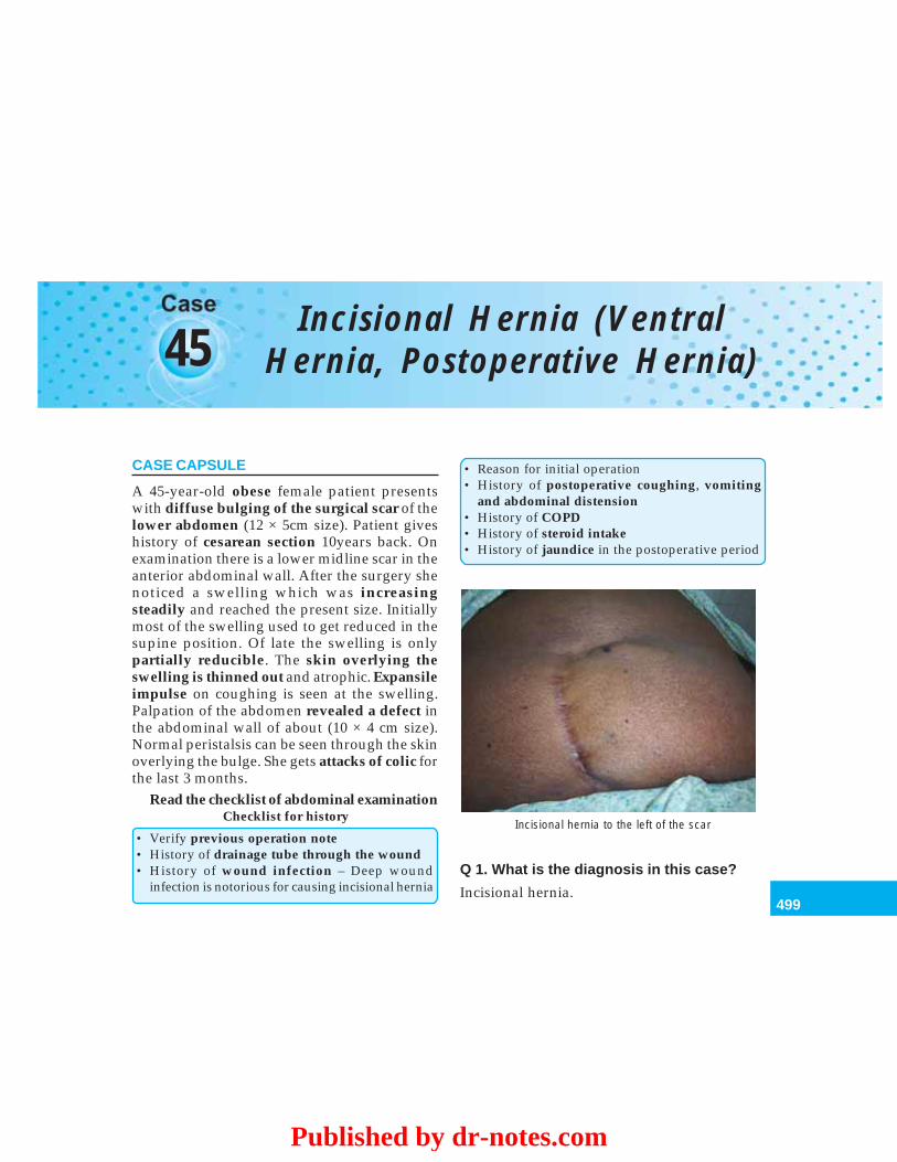
499
Incisional Hernia (Ventral Hernia, Postoperative Hernia)
45Incisional Hernia (Ventral
Hernia, Postoperative Hernia)
CASE CAPSULE
A 45-year-old obese female patient presentswith diffuse bulging of the surgical scar of thelower abdomen (12 × 5cm size). Patient giveshistory of cesarean section 10years back. Onexamination there is a lower midline scar in theanterior abdominal wall. After the surgery shenoticed a swelling which was increasingsteadily and reached the present size. Initiallymost of the swelling used to get reduced in thesupine position. Of late the swelling is onlypartially reducible. The skin overlying theswelling is thinned out and atrophic. Expansileimpulse on coughing is seen at the swelling.Palpation of the abdomen revealed a defect inthe abdominal wall of about (10 × 4 cm size).Normal peristalsis can be seen through the skinoverlying the bulge. She gets attacks of colic forthe last 3 months.
Read the checklist of abdominal examinationChecklist for history
• Verify previous operation note• History of drainage tube through the wound• History of wound infection – Deep wound
infection is notorious for causing incisional hernia
• Reason for initial operation• History of postoperative coughing, vomiting
and abdominal distension• History of COPD• History of steroid intake• History of jaundice in the postoperative period
Incisional hernia to the left of the scar
Q 1. What is the diagnosis in this case?
Incisional hernia.
Published by dr-notes.com
500
Clinical Surgery Pearls
Q 2. What are the diagnostic points in favorof incisional hernia?
• History of lower abdominal surgery with alower midline scar
• The swelling is seen at the site of the scar inthe abdominal wall
• The swelling is partially reducible in thesupine position
• The swelling is more in the standing position• Expansile impulse on coughing present• Palpable defect in the lower abdomen.
Q 3. What is the most important clinical signfor incisional hernia?
• The swelling will become more prominentin head raising test
• Reducibility and cough impulse
Q 4. If the lump does not reduce and doesnot have a cough impulse, what are thedifferential diagnoses?
Then it may not be a hernia. The followingdifferential diagnoses are to be considered:
Differential diagnoses if it is not reducible
• Deposit of tumor – Desmoid tumor• Hematoma• Foreign - body granuloma• Old abscess• Lipoma
Q 5. What is incisional hernia?
• It is a hernia through an acquired scar in theabdominal wall.
• An incisional hernia usually starts as asymptomless partial disruption of thedeeper layers of a laparotomy wound during
the immediate or early postoperative period,the event being unnoticed if the skin woundremains intact.
Q 6. What are the causes for incisionalhernia?
Causes for incisional hernia
1. Technique of wound closure (technical failureis the most important cause for incisional hernia):• Selection of suture material for closure of the
fascial layer (nonabsorbable synthetic,monofilament, No.1 size suture materialis preferred for closing the fascia. Eg:Polypropylene)
• Method of closure: Single layer mass closurewith interrupted ‘far and near’ sutures
• Length of suture material must be at least 4times the length of incision but < 5 times ifcontinuous suture is used
• Use Jenkin’s formula for closure of the fascia:The bites must be taken at least 1cm away fromthe fascial edge and 1cm apart (the reason forthis is the fact that the collagenolysis willextend upto 1cm from the fascial edge duringhealing).
2. Drainage tube• Keeping drainage tubes through the wound –
higher tendency to burst and later produceincisional hernia
3. Type of incision• Midline and vertical incisions have greater
tendency than horizontal wounds – moretendency to produce burst abdomen
4. Wound infection• Following operations for peritonitis – more
chance for wound infection and wounddehiscence
• Operations on pancreas and subsequentleakage
501
Incisional Hernia (Ventral Hernia, Postoperative Hernia)
5. Postoperative events – leading to wounddisruption• Postoperative vomiting• Persistent postoperative cough• Postoperative abdominal distension – due to
ileus• Postoperative ventilation
6. Creation of stoma (colostomy, ileostomy, etc.).7. Collagen deficiency – Decreased ratio of collagen
I/III (increase in collagen III).8. Failure to close the fascia of laparoscopic trocar
sites over 10mm size9. General condition of the patient
• Obesity – There is increased intraabdominalpressure in obesity – Increased incidence ofseroma and hematoma in wounds
• Jaundice• Cirrhosis• Malignant disease• Hypoproteinemia• Anemia• Malnutrition• COPD• Abdominal wounds in pregnancy• Steroids
Note: These are the causes for wound dehiscence(Burst abdomen) also
Q 7. Since incisional hernia is a symptomlesspartial disruption of the deeper layers of thelaparotomy wound, it is better to knowsomething about wound dehiscence. Whatis the timing of wound dehiscence and whatare the manifestations?
• Between 6th and 8th day of surgery• The initial manifestations is serosanguinous
(pink) discharge from the wound which is thepathognomonic sign of wound disruption.
It signifies intraperitoneal contents lyingextraperitoneally
• Patient may volunteer the information thatthey ‘felt something give way’
• In revealed dehiscence the omentum andcoils of intestines may be found lying outside.
Q 8. What are the three most importantetiological factors?
Three most important causes for incisionalhernia
• Technical failure in wound closure• Wound infection• Subclinical wound dehiscence in the post-
operative period
Q 9. What is ventral hernia?The term ventral hernia should be restricted toincisional hernia arising in abdominal midlineoperative wounds.
Q 10. What is the management? The most important investigations are:• X-ray chest – To rule out COPD and
other pulmonary dis-eases
• Ultrasound – To demonstrate theabdomen size of the defect
– To rule out other intra-abdominal pathology
• Hematological – To rule out anemiaexamination
• Biochemical – To rule out hypopro-investigation teinemia
• Nutritional assessment• Evaluation of cardiac status and renal statusAfter proper evaluation surgery is recom-mended.
Published by dr-notes.com
502
Clinical Surgery Pearls
Q 11. Is there any conservative management?If so what is the indication?• Abdominal belt is sometimes recommended
especially in upper abdominal incisionalhernias
• Belt is also recommended until the patient isfit for surgery.
Q 12. What are the preoperative preparationsrequired?• Weight reduction by dieting and exercise• Patient is asked to stop smoking• Treat the respiratory problems.
Q 13. What are the complications of incisionalhernia?
Complications of incisional hernia
• Irreducibility• Obstruction• Strangulation (Rarely)• Ulceration of the overlying skin
Q 14. What is large hernia?
A hernia can be considered large when thefascial edges cannot be approximated withouttension (more than 4 cm).
Q 15. Which hernia is more dangerous, smallor large?
Small hernias may cause bowel obstruction earlyand therefore it is more dangerous.
Q 16. A very large hernia of long duration inwhich most of the intestines are in the sac,what additional precaution is taken to preventpostoperative embarrassment?
Here there are three important problems:• Increased risk of paralytic ileus from visceral
compression
• Pulmonary complications as a result ofelevation of the diaphragm when thecontents are reduced
• Risk of failure of hernioplastyThis may be managed by enlarging the
abdominal cavity preoperatively by prolongedpneumoperitoneum. This is achieved byincreasing the intraabdominal pressure to15-18cm of water with the help of a pneum-operitoneum apparatus for several weekspreoperatively.
Q 17. What are the surgical proceduresavailable for repair of incisional hernia?1. Anatomical repair (primary fascial repair)
– Useful only for small hernias < 4cm sizebecause of the high incidence of recurrence
2. Mesh repair (tension free repair) – The Goldstandard treatment
3. Autogenous repair by vascularizedinnervated muscle flaps – Reserved for largeand recurrent hernias
4. Complex apposition (considered obsoleteand is of historical importance only.Examples are: Keel’s operation, Cattel’soperation).
Q 18. What are the most important steps offascial repair?• The hernial sac is dissected• The adherent omentum and bowel are
released from the sac• The contents are reduced• The mouth of the sac is defined• The layers are repaired with nonabsorbable
sutures – No.1 polypropylene, first theperitoneum and then the fascial layer.
503
Incisional Hernia (Ventral Hernia, Postoperative Hernia)
Q 19. What is mesh repair and what is theideal plane for keeping the mesh? (PG)• The mesh repair can be done either by Open
method or Laparoscopic method• The initial steps are same as the fascial repair• For incisional hernias above the umbilicus, a
sheet of polypropylene mesh is insertedbetween the posterior rectus sheath andmuscle fibers and anchored in place (Inlayrepair)
• For hernias below the umbilicus the mesh isplaced in the preperitoneal space (Inlay repair)
• The mesh may also be placed as on lay infront of the fascia (Inlay is superior)
(Here the deficiency can be bridged bysewing the mesh to the fascia on either sideof the defect ensuring at least a 4 cm overlapof the fascial edges)
• Tension-free mesh repair can be undertakenlaparoscopically apparently with good results.
Q 20. What is autogenous repair byvascularized innervated muscle flaps? (PG)
• This method is reserved for massive midlinedefects and recurrent hernias
• It is also done after removal of infectedsynthetic mesh
• The autogenous tissue reconstruction wasintroduced by Ramirez et al
• The technique utilizes bilateral, innervated,bi-pedicle, rectus abdominis – transverseabdominis – internal oblique muscle flaps,that are transposed medially to reconstructthe central defect.
Q 21. What are the complications of incisionalhernia repair?
Complications of incisional hernia repair
• Wound infection• Seroma formation• Wound sinus• Enterocutaneous fistula• Recurrence• Infection of the mesh
Published by dr-notes.com
504
Clinical Surgery Pearls
46 Epigastric Hernia (Fatty Herniaof the Linea Alba)
CASE CAPSULE
A 40-year-old strong muscular laborer malepatient presents with a mid line firm swellingin the epigastrium of 2.5 cm size, which isassociated with epigastric pain after a large meal,(postprandial epigastric discomfort) which isrelieved by lying down. He has in additionabdominal bloating and occasional nausea andvomiting. There is no impulse on coughing. Theswelling is not reducible. The swelling clinicallyappears to be parietal in nature. The hernialorifices are normal. There is no organomegalyand no other masses felt per abdomen.
Read the check list for history and examinationof abdomen
Q 1. What is the most probable diagnosis inthis case?
Epigastric hernia.
Q 2. What is the cause for postprandialepigastric discomfort in this case?
It may be because of the epigastric distensionafter full meal
Q 3. What are the differential diagnoses?a. Parietal swellings b. Intraabdominal con-
ditions
• Subcutaneous lipoma • Peptic ulcer• Neurofibroma • Gallbladder diseases• Fibroma • Hiatal hernia• Divarication of rectus • Pancreatitis
abdominis (Diastasis) • Small intestinal obs-truction
Q 4. What is epigastric hernia?• Hernia through the linea alba between the
umbilicus and the xiphisternum• They develop through the opening of para
midline nerves and vessels• 80% occur just off the midline.
Q 5. Which sex is affected more?More common in men than women.
Q. 6. What is the incidence and age groupaffected?• 3 to 5% of the population• Age group is 20-50 years.
505
Epigastric Hernia (Fatty Hernia of the Linea Alba)
Q 7. What are the peculiarities of epigastrichernia?
• They are better felt than seen• Impulse on coughing may not be there• Reducibility is not there even in uncomp-
licated hernias• Usually no peritoneal sac and no bowel
content.
Q 8. What is the usual content in epigastrichernia?
Preperitoneal fat which will come out throughthe openings in the linea alba for vessels. Usuallythere is no peritoneal sac.
Q 9. What are the stages of the herniaformation?
• First stage it is sac less (Only preperitonealfat protrusion)
• Second stage—A small pouch of peritoneumis drawn after it
• Last stage—Small tag of omentum gets intothe sac and adherent to it.
Q 10. What are the usual symptoms andsigns?
Symptoms and signs
• May be identified by routine clinical examination• Present as painless swellings• Mass in the epigastrium (difficult to feel in obese
patients)• Mild epigastric pain/Burning epigastric pain• Abdominal bloating, nausea or vomiting (these
symptoms may be also due to concomitant othervisceral pathology)
• Symptoms may occur after a large meal followedby relief in lying down position
Q 11. What are the complications ofepigastric hernia?
• Incarceration• Strangulation.
Q 12. What are the investigations forconfirming the diagnosis?
• Ultrasound abdomen/CT to demonstrate thedefect in the linea alba especially in obesepatients (ultrasound will also rule out biliarypathology and other visceral causes for pain)
• Upper GI endoscopy to rule out peptic ulcerand other gastroesophageal diseases
• Assessment of the cardiac status, renal statusand respiratory status before surgery
• Hemogram and biochemical investigations.
Q 13. What is the surgical management ofepigastric hernia?
It is managed by epigastric hernia repair.
Q 14. What are the essential steps ofepigastric hernia repair?
• Anesthesia – GA/Local (for small cases)• Position of the patient – supine.• Drapes are arranged so that the whole of
epigastric area from costal margin to justbelow the umbilicus is exposed for surgery.
• Incision – Vertical/horizontal. Verticalincision has the advantage for ruling outmore than one defects in the linea alba so that,hernia is not missed. Another advantage isthat the abdomen can be opened if required.
• The herniated fat is dissected out from thesurrounding abdominal fat.
• The opening in the linea alba which is usuallytiny is identified and enlarged transversally.
Published by dr-notes.com
506
Clinical Surgery Pearls
• The hernia is incised at the neck to determinewhether peritoneal sac is present or not.
• If sac is there, open the sac and reducethe contents, transfix the neck. If sac is notthere, reduce the protruding extraperitonealfat.
• The opening in the linea alba is closed byoverlapping its edges (double breasting)with two rows of interrupted polypro-pylene/nylon sutures. (First row mattresssutures, and second row simple sutures).
• Subcutaneous sutures are put with inter-rupted cat gut.
• Skin is closed preferably with skin tapes.
Summary of epigastric hernia
• Usually no impulse on coughing• Usually not reducible• Smaller the hernia, greater the symptoms• Symptoms mimic peptic ulcers• Rule out gastrointestinal pathology like peptic
ulcer by investigations before surgery• Rule out epigastric hernia in all cases of dyspeptic
symptoms• Rule out multiple hernias which is there in 20%
of patients
507
Paraumbilical Hernia, Umbilical Hernia in Adults and Children
47Paraumbilical Hernia,Umbilical Hernia inAdults and Children
CASE CAPSULE
A 50-year-old obese multiparous femalepatient presents with a swelling on one side ofthe umbilicus with pain and discomfort of oneyear duration. She also complains of recurrentattacks of colicky abdominal pain. Onexamination there is a swelling of about 3 cmsize which is firm in consistency havingexpansile impulse on coughing. The swellingis reducible and according to the patient at timesit is not going in. Once the hernia is reduced, afirm fibrous edge of the defect of about 2 cmsize is felt at the periphery of the umbilicalcicatrix. The umbilicus is stretched into acrescent shape. There is no other hernia.
Read the check list for history and exami-nation of abdomen.
Q 1. What is the most probable diagnosis inthis case?
Paraumbilical hernia.
Q 2. What is paraumbilical hernia?
It is a herniation through the linea alba justabove or sometimes just below the umbilicus,sometimes on the sides (supraumbilical hernia/infraumbilical hernia).
Q 3. What is the difference between umbilicalhernia and paraumbilical hernia?
In umbilical hernia the herniation occursthrough the umbilical scar and the umbilical scaris weak. The abdominal contents bulge throughthe weak spot everting the umbilicus.
Difference between umbilical hernia and paraumbilical hernia
Umbilical hernia Paraumbilical hernia
1. The abdominal contents bulge through the Herniation through the linea alba just above or belowweak umbilical scar the umbilicus
2. Umbilicus is everted The hernia is beside the umbilicus and push it to oneside taking a crescent shape
Contd...
Published by dr-notes.com
508
Clinical Surgery Pearls
Umbilical hernia Paraumbilical hernia
3. The entire fundus of the sac is covered by the Half of the fundus of the sac is covered by umbilicusumbilicus and the remainder by the adjacent abdominal skin
4. More common in girls than boys 5 times more seen in females5. May be congenital or acquired Always acquired6. Ascitis (cirrhosis) and malignant peritoneal Obesity, repeated pregnancies and flabby abdominal
effusion are predisposing factors for muscles are responsible for paraumbilical herniaacquired umbilical hernia. The congenitalvariety is due to incomplete closure ofumbilical ring at birth
7. Neck of the sac is wide Neck of the sac is narrow and complications can occurearly
8. Congenital type can wait upto 4 years Always needs surgerybecause spontaneous closure can occur
Q 4. What are the etiological factors forparaumbilical hernia?• Obesity• Flabby abdominal muscles• Repeated pregnancy.
Q. 5 What is the content in paraumbilicalhernia?Greater omentum, small intestine and transversecolon.
Q 6. What are the clinical manifestations ofparaumbilical hernia?• Swelling/lump• Dragging pain• Gastrointestinal symptoms – traction on
stomach and transverse colon• Intestinal Colic and signs of intestinal
obstruction.
Q 7. What are the complications of para-umbilical hernia?
• Dragging pain• Irreducibility (omental adhesions)• Obstruction – colic• Strangulation – due to the narrow neck and
the fibrous edge of the linea alba- Presence of loculi may also result instrangulation of the bowel
• Ulceration• Intertrigo in long-standing cases.
Q 8. What are the differential diagnoses ofparaumbilical hernia?
• Lipoma• Neurofibroma• Desmoid tumor• Hematoma• Sister Joseph nodule (umbilical metastasis)• Caput medusa
Contd...
509
Paraumbilical Hernia, Umbilical Hernia in Adults and Children
Q 9. What are the indications for surgery inparaumbilical hernia?
Surgery is indicated in all cases.
Q 10. What are the investigations required?
• Ultrasound abdomen– To identify the defect– To rule out other pathologies
• Plain X-ray abdomen – when obstruction issuspected
• X-ray chest• Assessment of the cardiac status• Assessment of the renal status• Complete hemogram.
Q 11. Any preparation required beforesurgery?
• If the hernia is symptomless and the patientis obese—weight reduction
• In symptomatic patients surgery is donewithout weight reduction.
Q 12. What is the surgical management ofparaumbilical hernia?
• Paraumbilical hernioplasty, without excisionof the umbilicus (modified Mayo’s repair)
• In Mayo’s repair the umbilicus is excised.
Q 13. What are the indications for prostheticmesh in paraumbilical hernia surgery?
• Very large paraumbilical hernia (fascialdefect more than 4cm size)
• Recurrent paraumbilical hernia.
Q 14. What are the essential steps ofparaumbilical hernioplasty?
• A curved incision is put over the herniadepending on the situation of the hernia inrelation to the umbilicus
• The hernial sac is identified and dissected allaround
• After opening the neck of the sac, the contentsare released and reduced
• The sac is excised• The defect in the linea alba is closed by non-
absorbable polypropylene sutures• For larger hernias prosthetic mesh repair may
be required if the surgery is elective.
Q 15. What is the cause for congenitalumbilical hernia?
• It is due to incomplete closure of the umbilicalring at birth
• Failure of complete obliteration at the sitewhere the fetal umbilical vessels (umbilicalvein and two umbilical arteries) are joined tothe placenta during gestation.
Q 16. What is the incidence of Congenitalumbilical hernia?
• 20% of the full-term neonates may haveincomplete closure and umbilical hernia
• 75-85% of the premature infants (weightbetween 1 and 1.5kg) show evidence ofumbilical hernia at birth.
Q 17. Is there any sex/race predilection forcongenital umbilical hernia?
• It is more common in girls than boys• The incidence is higher in Black children than
White children.
Published by dr-notes.com
510
Clinical Surgery Pearls
Q 18. What is the natural course of congenitalumbilical hernia?
• 80% will decrease in size and close spon-taneously by five years of age
• Congenital umbilical hernia of more than2 cm size rarely close spontaneously.
Q 19. What are the complications ofcongenital umbilical hernia?• Incarceration• Small bowel obstruction (1 in 1500 umbilicus
hernias)• Spontaneous rupture (very rare in first year
of life due to excessive crying) results inpartial evisceration requiring urgentintervention.
Q 20. What are the indications for surgery incongenital umbilical hernia?• Ring size of more than 2cm• Failure of closure of the umbilical ring above
4 years.
Q 21. What are the steps for umbilical herniarepair?• A curved (‘smile’) incision in the natural skin
crease immediately below the umbilicus. Thecurved incision should not extend beyond180o . It is considered important to preservethe umbilical cicatrix after excising the sac.
• The skin cicatrix is dissected upwards andthe neck of the sac is isolated
• After ensuring that the sac is empty, thedefect is repaired with polypropylenesutures.
Q 22. What are the causes for adult umbilicalhernia?Adult umbilical hernia may be a manifestation of:
• Cirrhosis• Malignant peritoneal effusion.
Q 23. What is the most importantcomplication of adult umbilical hernia?• Incarceration – It is a well-documented
complication of effective relief of ascitesfollowing diuresis, paracentesis, peritoneo-venous shunt and TIPS (Transjugular Intrahepatic Portosystemic Shunt).
Q 24. What are the important problems in thesurgical management of adult umbilicalhernia?• Cirrhotic patient may have caput medusae
which may interfere with repair.• Repair may be difficult requiring a prosthetic
mesh• A subcutaneous suction drain should be
avoided because of the increased risk ofinfection of the ascitic fluid
Q 25. What is omphalocele (exomphalos)?• It is due to failure of all or part of the mid
gut to return to the celom during early fetallife
• There is a midline abdominal defect• Liver and bowel are seen contained within a
sac composed of inner layer of peritoneumand outer layer of amnion from which theumbilical cord arises at the apex and center.
Q 26. What are the types of omphalocele?• Omphalocele major – more than 4cm size• Omphalocele minor – less than 4cm size
(herniation of the umbilical cord).Note: A single loop of intestine in omphaloceleminor may not be obvious and ligation of the
511
Paraumbilical Hernia, Umbilical Hernia in Adults and Children
umbilical cord without recognizing this fact willresult in transection of the intestine. This willleave an umbilicoenteric fistula.
Q 27. What are the structures seen inomphalocele major?
Structures seen in omphalocele
• Liver – Evidence of adhesions to the sac• Spleen• Stomach• Pancreas• Colon• Bladder• Intestine – Lies freely mobile
Q 28. What is the incidence of omphalocele?
Occurs once in every 6000 births.
Q 29. What are the associated anomalies inomphalocele?
Associated anomalies are seen in 30-70% ofinfants and the following anomalies are seen:• Trisomy 13, 18, 21• Tetralogy of Fallot• Atrial Septal Defect• Beckwith – Wiedemann syndrome (large - for
- gestational - age baby)HyperinsulinismVisceromegalyHepatorenal tumorsCloacal extrophy.
Q 30. What is the management of om-phalocele?
• Work up for associated anomalies• Orogastric tube to prevent distension of
abdomen
• Since there is a sac covering the viscera,emergency operation is not necessary
• Various surgical options are there for themanagement.
Q 31. What are the surgical options?
1. Non operative therapy—This is re-commended for premature infants with agigantic intact sac with associatedanomalies where survival of a majoroperation is questionable.
The intact sac is painted daily withdesiccated antiseptic solutions (Mercuro-chrome) and this will allow to form aneschar over the sac. Granulation tissuegrows from the periphery over the eschar.This will ultimately form a ventral hernia,which can be repaired later.
2. Skin flap closure –• The sac is gently trimmed• The skin is freed from the fascial edges
and undermined laterally• The umbilical vessels are ligated• The skin flaps are approximated in the
midline with simple sutures• The ventral hernia is repaired at a later
date (months to years later).3. Staged closure –
• The sac is trimmed• The skin is further freed from the fascial
attachment• Prosthetic material like PTFE (Polytetra
fluoroethylene) is sutured with inter-rupted nonabsorbable sutures circum-ferentially to the full thickness of themusculofascial abdominal wall to forma silo
Published by dr-notes.com
512
Clinical Surgery Pearls
• The top of the silo is gathered and tiedwith umbilical tape
• Daily the silo is opened under strictaseptic conditions and the contentsexamined for infection
• The viscera are pushed gently back intothe abdominal cavity and the child isobserved for signs of raised intra-abdominal pressure
• The silo is tied at reduced level daily untilthe sac is flush with abdominal wall
• The fascia may be closed with inter-rupted sutures at this stage
• Skin is closed over the top.4. Primary closure –
• The sac is gently dissected away from theskin edge and underlying fascia
• The intestine is evacuated completely ofmeconium and fluid distally and proxi-mally with the help of a nasogastrictube
• The abdominal wall is stretchedgradually and repeatedly in all quadrantsachieving a doubling of volume
• The viscera are then replaced• The fascial layer is closed primarily under
moderate tension.
Q 32. What is gastroschisis?• It is a defect in the abdominal wall to the
right of the normal insertion of theumbilical cord, without any investing sac(compare with omphalocele)
• It is seen at the site of involution of the rightumbilical vein.
Q 33. What is the incidence of gastroschisis?Twice as common as omphalocele.
Q 34. What is the cause for gastroschisis?There is a controversy as to whether gastro-schisis represents a ruptured omphalocele sacin utero or simply a separate entity.
Q 35. What are the differences between omphalocele and gastroschisis?
Sl. No Omphalocele Gastroschisis
1. The midline abdominal defect as a result Defect to the right of the normal insertion of theof failure of return of the mid gut to the umbilical cord (at the site of involution of the rightcelom umbilical vein)
2. The bowels are contained within a sac There is no sac covering the intestinecomposed of peritoneum and amnion
3. The umbilical cord arises at the apex of There is normal insertion of the umbilical cordthe sac
4. Liver may be seen as a content inside Liver is not seenthe sac
5. The bowel is not edematous Bowel is edematous and intestines are mattedtogether and appears to be short
6. Urgent repair is not required because of Urgent repair is requiredthe covering sac
513
Paraumbilical Hernia, Umbilical Hernia in Adults and Children
Q 36. What are the associated anomalies ingastroschisis?
• Nonrotation of the mid gut• Intestinal atresia.
Q 37. What is the surgical management?
Urgent primary closure.
Published by dr-notes.com
514
Clinical Surgery Pearls
48Desmoid Tumor, InterparietalHernia (Interstitial), Spigelian
Hernia
CASE CAPSULE
A 30-year-old female patient presents with apainless parietal swelling below the level ofumbilicus of 1 year duration. There is historyof cesarean operation during her firstpregnancy. She noticed the swelling within oneyear of the child birth. She also gives history ofusing oral contraceptives. There is no historyof trauma. On examination the swelling is hardin consistency of about 6 × 4 cm size overlyingthe upper part of the cesarean scar. The inguinallymph node are not enlarged. The rest of theabdomen is normal. There is no organomegaly.The X-ray chest is normal.
Read the checklist for history and examinationof abdomen.
Q 1. What is the most probable diagnosis?
Desmoid tumor (aggressive fibromatosis).
Q 2. What are the points in favor of adiagnosis of desmoid tumor?
• Onset of the swelling within a year ofchildbirth
• Use of oral contraceptives
• History of cesarean operations• Presence of painless hard parietal swelling
below the umbilicus.
Q 3. What are the types of desmoid tumor?Classification of desmoid tumors
• Sporadic type• Part of inherited syndrome
– FAP (Mesenteric Desmoids)– Risk increased 1000 fold in patients with FAP
It may also be classified as:1. Superficial (fascial) – Dupuytren’s fibromatosis
(they are having slow growth)2. Deep (musculoaponeurotic)
- Relatively rapid growth and attains large size- It has high rate of local recurrence- Involves musculature of trunk and extremities
Depending on the location it is classified as:• Extraabdominal – shoulder girdle• Abdominal wall• Intraabdominal (mesenteric and pelvic desmoid)
Q 4. What is the significance of oral contra-ceptives and cesarean?
• There is association between the developmentof this neoplasm and abdominal trauma likeoperations
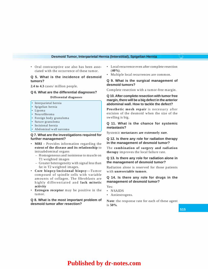
515
Desmoid Tumor, Interparietal Hernia (Interstitial), Spigelian Hernia
• Oral contraceptive use also has been asso-ciated with the occurrence of these tumor.
Q 5. What is the incidence of desmoidtumors?
2.4 to 4.3 cases/million people.
Q 6. What are the differential diagnoses?Differential diagnoses
• Interparietal hernia• Spigelian hernia• Lipoma• Neurofibroma• Foreign body granuloma• Suture granuloma• Incisional hernia• Abdominal wall sarcoma
Q 7. What are the investigations required forfurther management?• MRI – Provides information regarding the
extent of the disease and its relationship tointraabdominal organs– Homogeneous and isointense to muscle on
T1 weighted images– Greater heterogeneity with signal less than
fat in T2 weighted images.• Core biopsy/incisional biopsy—Tumor
composed of spindle cells with variableamounts of collagen. The fibroblasts arehighly differentiated and lack mitoticactivity
• Estrogen receptor may be positive in thetumor.
Q 8. What is the most important problem ofdesmoid tumor after resection?
• Local recurrence even after complete resection(40%).
• Multiple local recurrences are common.
Q 9. What is the surgical management ofdesmoid tumors?
Complete resection with a tumor-free margin.
Q 10. After complete resection with tumor freemargin, there will be a big defect in the anteriorabdominal wall. How to tackle the defect?
Prosthetic mesh repair is necessary afterexcision of the desmoid when the size of theswelling is big.
Q 11. What is the chance for systemicmetastasis?
Systemic metastases are extremely rare.
Q 12. Is there any role for radiation therapyin the management of desmoid tumor?
The combination of surgery and radiationtherapy improves the local failure rate.
Q 13. Is there any role for radiation alone inthe management of desmoid tumor?
Radiation alone is reserved for those patientswith unresectable tumors.
Q 14. Is there any role for drugs in themanagement of desmoid tumor?
Yes:• NSAIDS• Antiestrogens.
Note: the response rate for each of these agentis 50%.
Published by dr-notes.com
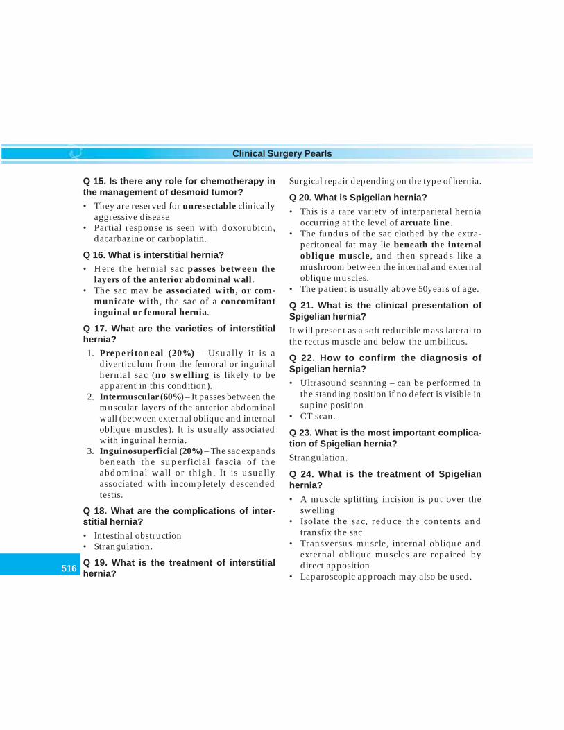
516
Clinical Surgery Pearls
Q 15. Is there any role for chemotherapy inthe management of desmoid tumor?• They are reserved for unresectable clinically
aggressive disease• Partial response is seen with doxorubicin,
dacarbazine or carboplatin.
Q 16. What is interstitial hernia?• Here the hernial sac passes between the
layers of the anterior abdominal wall.• The sac may be associated with, or com-
municate with, the sac of a concomitantinguinal or femoral hernia.
Q 17. What are the varieties of interstitialhernia?1. Preperitoneal (20%) – Usually it is a
diverticulum from the femoral or inguinalhernial sac (no swelling is likely to beapparent in this condition).
2. Intermuscular (60%) – It passes between themuscular layers of the anterior abdominalwall (between external oblique and internaloblique muscles). It is usually associatedwith inguinal hernia.
3. Inguinosuperficial (20%) – The sac expandsbeneath the superficial fascia of theabdominal wall or thigh. It is usuallyassociated with incompletely descendedtestis.
Q 18. What are the complications of inter-stitial hernia?
• Intestinal obstruction• Strangulation.
Q 19. What is the treatment of interstitialhernia?
Surgical repair depending on the type of hernia.
Q 20. What is Spigelian hernia?
• This is a rare variety of interparietal herniaoccurring at the level of arcuate line.
• The fundus of the sac clothed by the extra-peritoneal fat may lie beneath the internaloblique muscle, and then spreads like amushroom between the internal and externaloblique muscles.
• The patient is usually above 50years of age.
Q 21. What is the clinical presentation ofSpigelian hernia?
It will present as a soft reducible mass lateral tothe rectus muscle and below the umbilicus.
Q 22. How to confirm the diagnosis ofSpigelian hernia?
• Ultrasound scanning – can be performed inthe standing position if no defect is visible insupine position
• CT scan.
Q 23. What is the most important complica-tion of Spigelian hernia?
Strangulation.
Q 24. What is the treatment of Spigelianhernia?
• A muscle splitting incision is put over theswelling
• Isolate the sac, reduce the contents andtransfix the sac
• Transversus muscle, internal oblique andexternal oblique muscles are repaired bydirect apposition
• Laparoscopic approach may also be used.
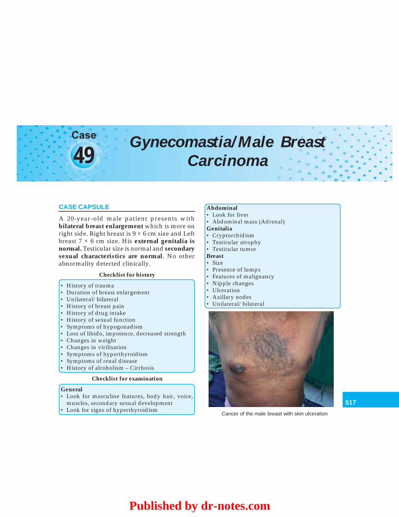
517
Gynecomastia/Male Breast Carcinoma
49Gynecomastia/Male Breast
Carcinoma
CASE CAPSULE
A 20-year-old male patient presents withbilateral breast enlargement which is more onright side. Right breast is 9 × 6 cm size and Leftbreast 7 × 6 cm size. His external genitalia isnormal. Testicular size is normal and secondarysexual characteristics are normal. No otherabnormality detected clinically.
Checklist for history
• History of trauma• Duration of breast enlargement• Unilateral/bilateral• History of breast pain• History of drug intake• History of sexual function• Symptoms of hypogonadism• Loss of libido, impotence, decreased strength• Changes in weight• Changes in virilisation• Symptoms of hyperthyroidism• Symptoms of renal disease• History of alcoholism – Cirrhosis
Checklist for examination
General• Look for masculine features, body hair, voice,
muscles, secondary sexual development• Look for signs of hyperthyroidism
Abdominal• Look for liver• Abdominal mass (Adrenal)Genitalia• Cryptorchidism• Testicular atrophy• Testicular tumorBreast• Size• Presence of lumps• Features of malignancy• Nipple changes• Ulceration• Axillary nodes• Unilateral/bilateral
Cancer of the male breast with skin ulceration
Published by dr-notes.com
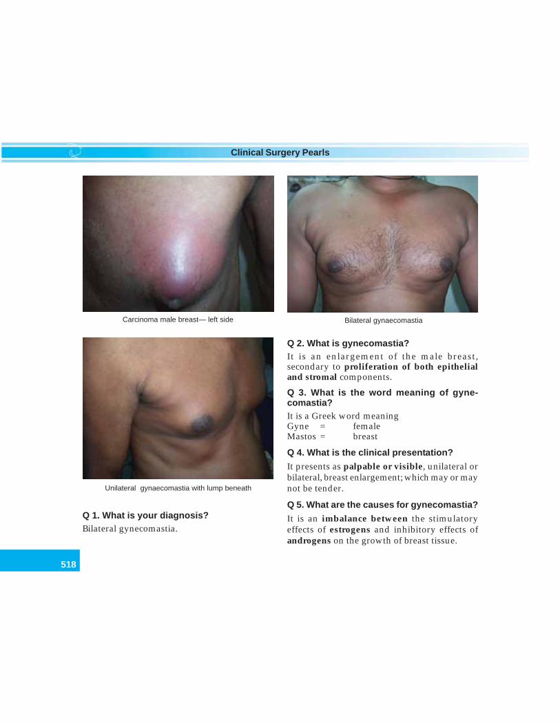
518
Clinical Surgery Pearls
Bilateral gynaecomastiaCarcinoma male breast— left side
Unilateral gynaecomastia with lump beneath
Q 1. What is your diagnosis?Bilateral gynecomastia.
Q 2. What is gynecomastia?It is an enlargement of the male breast,secondary to proliferation of both epithelialand stromal components.
Q 3. What is the word meaning of gyne-comastia?It is a Greek word meaningGyne = femaleMastos = breast
Q 4. What is the clinical presentation?
It presents as palpable or visible, unilateral orbilateral, breast enlargement; which may or maynot be tender.
Q 5. What are the causes for gynecomastia?
It is an imbalance between the stimulatoryeffects of estrogens and inhibitory effects ofandrogens on the growth of breast tissue.
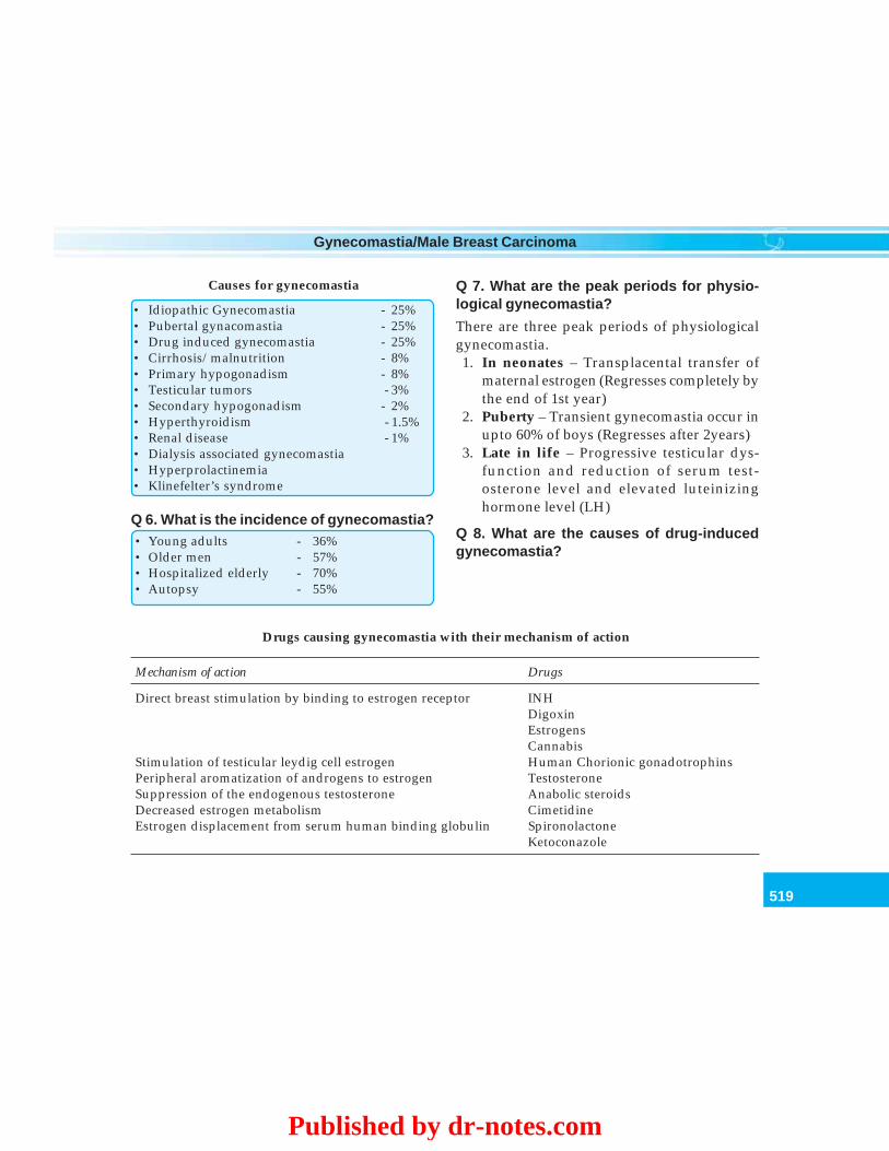
519
Gynecomastia/Male Breast Carcinoma
Causes for gynecomastia
• Idiopathic Gynecomastia - 25%• Pubertal gynacomastia - 25%• Drug induced gynecomastia - 25%• Cirrhosis/malnutrition - 8%• Primary hypogonadism - 8%• Testicular tumors - 3%• Secondary hypogonadism - 2%• Hyperthyroidism - 1.5%• Renal disease - 1%• Dialysis associated gynecomastia• Hyperprolactinemia• Klinefelter’s syndrome
Q 6. What is the incidence of gynecomastia?• Young adults - 36%• Older men - 57%• Hospitalized elderly - 70%• Autopsy - 55%
Q 7. What are the peak periods for physio-logical gynecomastia?
There are three peak periods of physiologicalgynecomastia.1. In neonates – Transplacental transfer of
maternal estrogen (Regresses completely bythe end of 1st year)
2. Puberty – Transient gynecomastia occur inupto 60% of boys (Regresses after 2years)
3. Late in life – Progressive testicular dys-function and reduction of serum test-osterone level and elevated luteinizinghormone level (LH)
Q 8. What are the causes of drug-inducedgynecomastia?
Drugs causing gynecomastia with their mechanism of action
Mechanism of action Drugs
Direct breast stimulation by binding to estrogen receptor INHDigoxinEstrogensCannabis
Stimulation of testicular leydig cell estrogen Human Chorionic gonadotrophinsPeripheral aromatization of androgens to estrogen TestosteroneSuppression of the endogenous testosterone Anabolic steroidsDecreased estrogen metabolism CimetidineEstrogen displacement from serum human binding globulin Spironolactone
Ketoconazole
Published by dr-notes.com
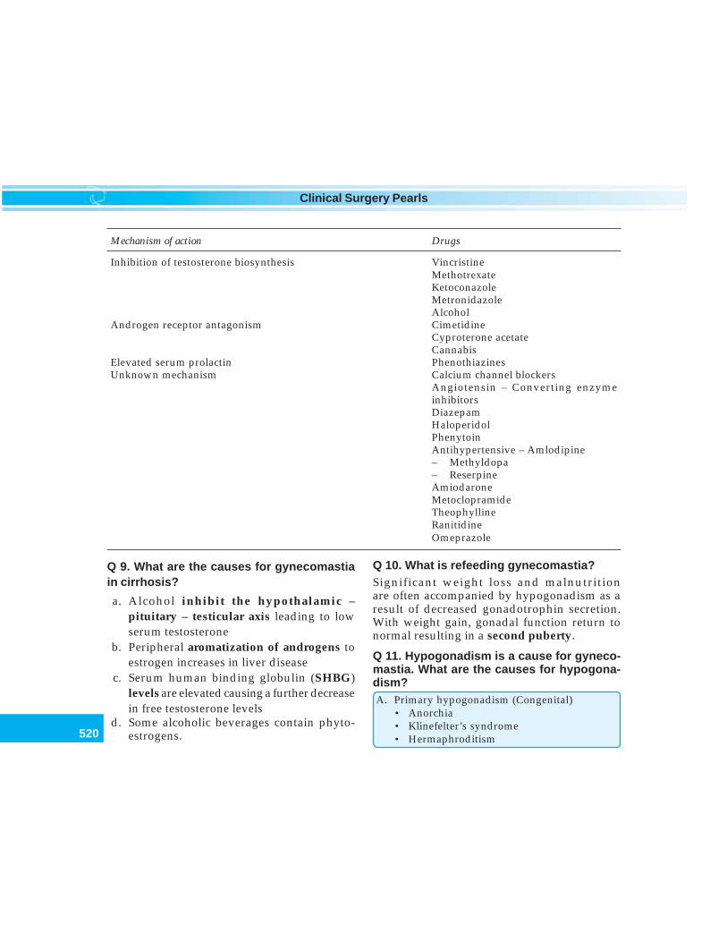
520
Clinical Surgery Pearls
Mechanism of action Drugs
Inhibition of testosterone biosynthesis VincristineMethotrexateKetoconazoleMetronidazoleAlcohol
Androgen receptor antagonism CimetidineCyproterone acetateCannabis
Elevated serum prolactin PhenothiazinesUnknown mechanism Calcium channel blockers
Angiotensin – Converting enzymeinhibitorsDiazepamHaloperidolPhenytoinAntihypertensive – Amlodipine– Methyldopa– ReserpineAmiodaroneMetoclopramideTheophyllineRanitidineOmeprazole
Q 9. What are the causes for gynecomastiain cirrhosis?
a. Alcohol inhibit the hypothalamic –pituitary – testicular axis leading to lowserum testosterone
b. Peripheral aromatization of androgens toestrogen increases in liver disease
c. Serum human binding globulin (SHBG)levels are elevated causing a further decreasein free testosterone levels
d. Some alcoholic beverages contain phyto-estrogens.
Q 10. What is refeeding gynecomastia?Significant weight loss and malnutritionare often accompanied by hypogonadism as aresult of decreased gonadotrophin secretion.With weight gain, gonadal function return tonormal resulting in a second puberty.
Q 11. Hypogonadism is a cause for gyneco-mastia. What are the causes for hypogona-dism?A. Primary hypogonadism (Congenital)
• Anorchia• Klinefelter’s syndrome• Hermaphroditism
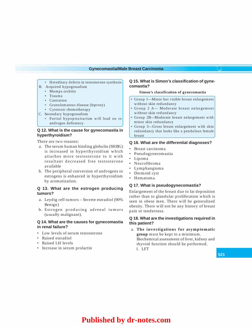
521
Gynecomastia/Male Breast Carcinoma
• Hereditary defects in testosterone synthesisB. Acquired hypogonadism
• Mumps orchitis• Trauma• Castration• Granulomatous disease (leprosy)• Cytotoxic chemotherapy
C. Secondary hypogonadism• Partial hypopituitarism will lead on to
androgen deficiency.
Q 12. What is the cause for gynecomastia inhyperthyroidism?
There are two reasons:a. The serum human binding globulin (SHBG)
is increased in hyperthyroidism whichattaches more testosterone to it withresultant decreased free testosteroneavailable
b. The peripheral conversion of androgens toestrogens is enhanced in hyperthyroidismby aromatization.
Q 13. What are the estrogen producingtumors?
a. Leydig cell tumors – Secrete estradiol (90%Benign)
b. Estrogen producing adrenal tumors(usually malignant).
Q 14. What are the causes for gynecomastiain renal failure?
• Low levels of serum testosterone• Raised estradiol• Raised LH levels• Increase in serum prolactin
Q 15. What is Simon’s classification of gyne-comastia?
Simon’s classification of gynecomastia
• Group 1—Minor but visible breast enlargementwithout skin redundancy
• Group 2 A— Moderate breast enlargementwithout skin redundancy
• Group 2B—Moderate breast enlargement withminor skin redundancy
• Group 3—Gross breast enlargement with skinredundancy that looks like a pendulous femalebreast
Q 16. What are the differential diagnoses?• Breast carcinoma• Pseudogynecomastia• Lipoma• Neurofibroma• Lymphangioma• Dermoid cyst• Hematoma.
Q 17. What is pseudogynecomastia?Enlargement of the breast due to fat depositionrather than to glandular proliferation which isseen in obese men. There will be generalizedobesity. There will not be any history of breastpain or tenderness.
Q 18. What are the investigations required inthis patient?a. The investigations for asymptomatic
group must be kept to a minimum.Biochemical assessment of liver, kidney andthyroid function should be performed.1. LFT
Published by dr-notes.com
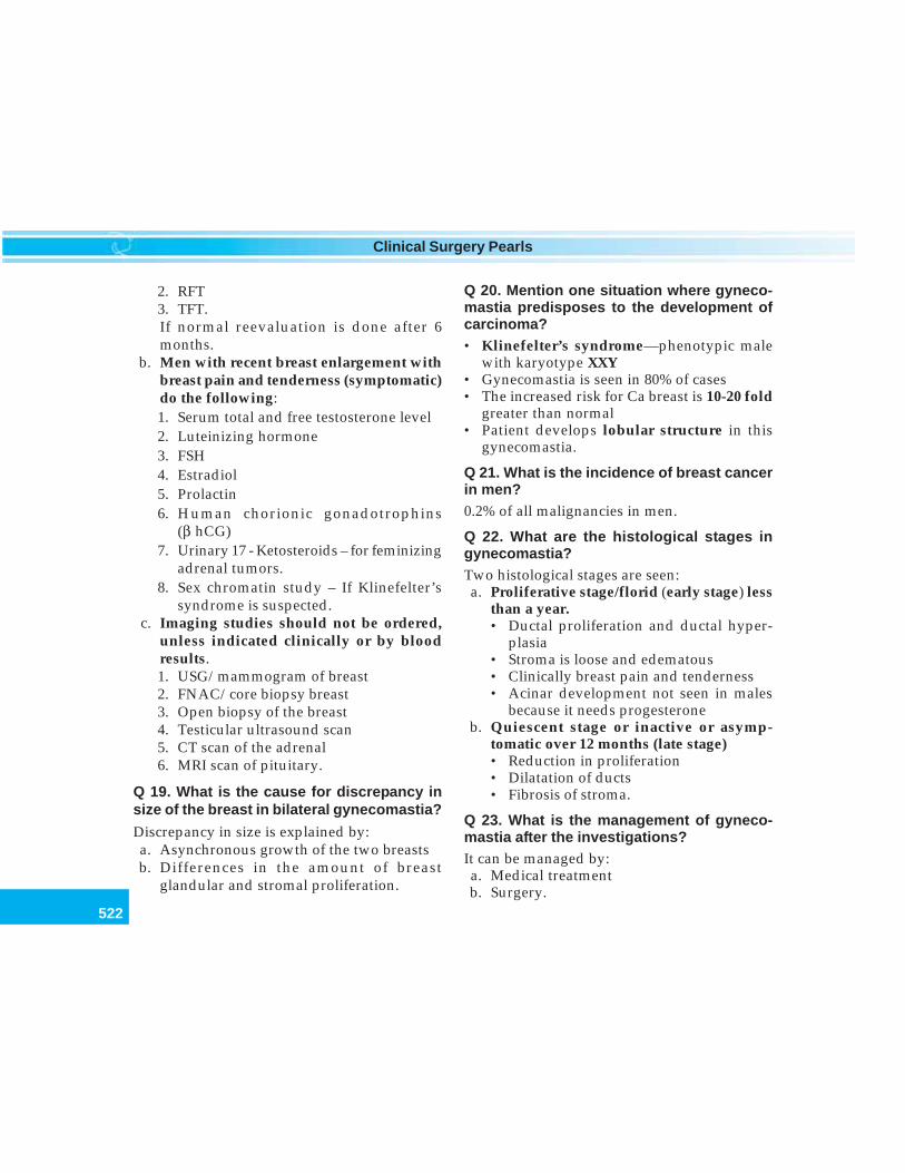
522
Clinical Surgery Pearls
2. RFT3. TFT.If normal reevaluation is done after 6months.
b. Men with recent breast enlargement withbreast pain and tenderness (symptomatic)do the following:1. Serum total and free testosterone level2. Luteinizing hormone3. FSH4. Estradiol5. Prolactin6. Human chorionic gonadotrophins
(β hCG)7. Urinary 17 - Ketosteroids – for feminizing
adrenal tumors.8. Sex chromatin study – If Klinefelter’s
syndrome is suspected.c. Imaging studies should not be ordered,
unless indicated clinically or by bloodresults.1. USG/mammogram of breast2. FNAC/core biopsy breast3. Open biopsy of the breast4. Testicular ultrasound scan5. CT scan of the adrenal6. MRI scan of pituitary.
Q 19. What is the cause for discrepancy insize of the breast in bilateral gynecomastia?
Discrepancy in size is explained by:a. Asynchronous growth of the two breastsb. Differences in the amount of breast
glandular and stromal proliferation.
Q 20. Mention one situation where gyneco-mastia predisposes to the development ofcarcinoma?• Klinefelter’s syndrome—phenotypic male
with karyotype XXY• Gynecomastia is seen in 80% of cases• The increased risk for Ca breast is 10-20 fold
greater than normal• Patient develops lobular structure in this
gynecomastia.
Q 21. What is the incidence of breast cancerin men?0.2% of all malignancies in men.
Q 22. What are the histological stages ingynecomastia?Two histological stages are seen:a. Proliferative stage/florid (early stage) less
than a year.• Ductal proliferation and ductal hyper-
plasia• Stroma is loose and edematous• Clinically breast pain and tenderness• Acinar development not seen in males
because it needs progesteroneb. Quiescent stage or inactive or asymp-
tomatic over 12 months (late stage)• Reduction in proliferation• Dilatation of ducts• Fibrosis of stroma.
Q 23. What is the management of gyneco-mastia after the investigations?It can be managed by:a. Medical treatmentb. Surgery.
523
Gynecomastia/Male Breast Carcinoma
Principles of management of gynecomastia
1. Spontaneous improvement is seen in 85%without treatment
2. When gynecomastia has been present for >2years, medical therapy is unlikely to be effective
3. Medical therapy should be limited to only 6months
4. Stop the drugs causing gynecomastia5. Gynecomastia following chemotherapy will
resolve spontaneously6. Treat the hyperthyroidism7. Surgical removal of testicular/adrenal tumors8. Hypogonadism is treated with testosterone
Q 24. What are the drugs used for medicalmanagement?
Drugs used for medical management ofgynecomastia
I. Androgensi. Dihydrotestosterone—Injection or
percutaneous administration (Testosterone isaromatized to estradiol which will exacerbatethe gynecomastia and therefore dihydro-testosterone is used which is nonaromatizableandrogen)• ↓ in Breast volume in 75%• Complete resolution in 25%
ii. Danazol (weak androgen) – 400mg daily• The only licensed drug for the treatment
of gynecomastia in UK• Complete resolution in 23%• Inhibits pituitary secretion of LH and FSH• Course of therapy is for 6 months
II.Antiestrogensi. Clomiphen citrate—Response rate 36 – 95%
ii. Tamoxifen 10mg twice daily• Complete regression in 78%
• 10 mg/day for 3 months – safe for painfulidiopathic or physiological gynecomastia
iii.Aromatase inhibitorsTestolactone—an aromatase inhibitor is tried withgood result in pubertal gynecomastia
Q 25. What are the indications for surgery?Indications for surgery in gynecomastia
• Social embarrassment• Psychological trauma• When there is no underlying treatable
condition• When trial of hormone treatment have failed.
Q 26. What are the surgical options?1. Open subcutaneous mastectomy by a
circum areolar incision extending from 3 to9’O clock position
2. Endoscopic – assisted subcutaneousmastectomy (very small distant incision)
3. Liposuction – assisted mastectomy (forpseudogynecomastia)
4. Ultrasound – assisted liposuction.
Note: In Simon’s grade I, II A and II B thenipple is left in its normal position. Grade IIIcases require skin resection and repositioningof the nipple areolar complex. Otherwise therewill be redundant skin folds and the nipple/areolar complex will be at a lower position thannormal.
Q 27. What are the complications of surgery?Complications of surgery for gynecomastia
1. Nipple/areolar ischemia2. Nipple distortion3. Risk of saucer deformity
Published by dr-notes.com
524
Clinical Surgery Pearls
4. Hematoma5. Seroma6. Infection7. Skin redundancy8. Skin necrosis9. Breast asymmetry
Q 28. Can you prevent iatrogenic gynco-mastia?Yes.1. Select a drug with lowest association for
gynecomastiaCalcium channel blocker• Nifedipine – Highest frequency• Verapamil – Lower• Diltiazem – Lowest associationH2 Receptor / Proton pump inhibitor• Cimetidine – highest• Ranitidine – Lower• Omeprazole - Lowest
2. Prophylactic breast irradiation (low dose of900rads) is effective in preventinggynecomastia and mastodynia in patientsreceiving estrogen therapy for prostatecancer.
CARCINOMA MALE BREAST
(Read female breast cancer).
Q 29. What are the suspicious clinicalfindings for carcinoma?
Suspicious clinical findings for carcinoma ofmale breast
• Unilateral hard mass• Eccentric mass rather than subareolar• Nipple retraction• Skin dimpling
• Nipple discharge• Axillary lymphadenopathy
Q 30. Is there any increased incidence of malebreast cancer in patients with benigngynecomastia?
No.
Q 31. What is the incidence of male breastcancer and the mean age at diagnosis?
• < 1% (0.7%)• Mean age at diagnosis – 65years (5 – 10 years
older than in females).
Q 32. What are risk factors for male breastcancer?
Risk factors for male breast cancer
• Radiation exposure• Estrogen administration• Cirrhosis of liver• Klinefelter’s syndrome• Hepatic schistosomiasis• Positive family history• BRCA 2 gene
Q 33. Why male breast cancers present asadvanced malignancies?
• There is only small amount of soft tissue inthe male breast and therefore the carcinomawill infiltrate the skin and nipple early
• Early involvement of pectoral fascia andmuscle because of the above reason
• Lack of awareness of male breast cancer.
Q 34. What is the prognosis of male breastcancer?
Similar to that of the female breast cancer whencompared stage for stage.
525
Gynecomastia/Male Breast Carcinoma
Q 35. What are the prognostic factors in malebreast cancer?
• Nodal status• Tumor size• Receptor status.
Q 36. What is the surgical management ofmale breast cancer?
• Modified radical mastectomy (MRM)• Breast conservation may not be possible in
most of the instances• Radiotherapy is given because of the
narrower margin of excision and locallyadvanced nature of the disease.
Q 37. Is there any role for tamoxifen in malebreast cancer?
• Yes. It is given for ER positive patients forfive years (20 mg daily)
• Many male breast cancers are ER positive• It is used for the first line hormonal
manipulation.
Q 38. What is the role of adjuvant systemicchemotherapy in male breast cancer?• Same indication as in female breast cancer.
Q 39. Is there any role for orchiectomy in malebreast cancer?• No. orchiectomy is obsolete• LHRH (Luteinizing hormone releasing
hormone) analogue is a better option• Orchiectomy is used as a 2nd line hormonal
manipulation in metastatic male breastcancer occasionally in some centers.
Published by dr-notes.com
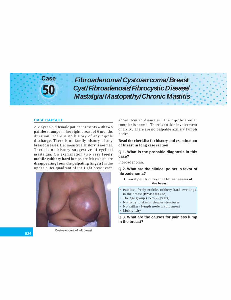
526
Clinical Surgery Pearls
50Fibroadenoma/Cystosarcoma/BreastCyst/Fibroadenosis/Fibrocystic Disease/Mastalgia/Mastopathy/Chronic Mastitis
CASE CAPSULE
A 20-year-old female patient presents with twopainless lumps in her right breast of 6 monthsduration. There is no history of any nippledischarge. There is no family history of anybreast diseases. Her menstrual history is normal.There is no history suggestive of cyclicalmastalgia. On examination two very freelymobile rubbery hard lumps are felt (which aredisappearing from the palpating fingers) in theupper outer quadrant of the right breast each
about 2cm in diameter. The nipple areolarcomplex is normal. There is no skin involvementor fixity. There are no palpable axillary lymphnodes.
Read the checklist for history and examinationof breast in long case section.
Q 1. What is the probable diagnosis in thiscase?
Fibroadenoma.
Q 2. What are the clinical points in favor offibroadenoma?
Clinical points in favor of fibroadenoma ofthe breast
• Painless, freely mobile, rubbery hard swellingsin the breast (Breast mouse)
• The age group (15 to 25 years)• No fixity to skin or deeper structures• No axillary lymph node involvement• Multiplicity
Q 3. What are the causes for painless lumpin the breast?
Cystosarcoma of left breast
527
Fibroadenoma/Cystosarcoma/Breast Cyst/Fibroadenosis/Fibrocystic Disease/Mastalgia
Causes for painless lump in the breast
• Fibroadenoma• Carcinoma• Fibroadenosis• Traumatic fat necrosis
Q 4. What are the causes for painful lump inthe breast?
Causes for painful lump in the breast
• Mastitis• Breast abscess• Fibroadenosis• Fat necrosis
Q 5. What are the causes for massiveenlargement of the breast?
Causes for massive enlargement of the breast
• Giant fibroadenoma• Cystosarcoma phyllodes• Sarcoma of the breast• Benign hypertrophy of the breast (Diffuse hyper-
trophy)• Filarial elephantiasis of the breast• Colloid carcinoma of the breast
Q 6. What are the causes for cystic swellingsin the breast?
Causes for cystic swellings in the breast
• Fibroadenosis• Galactocele• Abscess• Lymph cyst• Hematoma• Parasitic cyst
Q 7. What is fibroadenoma?
• It is hyperplasia of a single lobule where asneoplasms arise from a single cell
• It is not a benign neoplasm, but are bestconsidered as aberrations of normal develop-ment and involution (ANDI)
• They arise in the fully developed breastduring the 15 to 25 year period (after 40 yearsthey are less common)
• They show the same hormonal dependenceas the remainder of the breast, e.g. Theylactate during pregnancy and involute duringthe perimenopausal period.
Q 8. What is the incidence of fibroadenoma?
• They account for 12% of all palpablesymptomatic breast masses
• They are more frequently seen in Negropopulation (black races).
Q 9. How will you classify fibroadenoma?
There are four separate entities of fibro-adenomas. They are:
Four types of fibroadenoma
1. Common fibroadenoma2. Giant fibroadenoma3. Juvenile fibroadenoma4. Phyllodes tumor
Q 10. What is intracanalicular and pericanali-cular fibroadenoma?
• This is a conventional classification by thepathologists
• This histological distinction has no clinicalrelevance
• This terminology can therefore beabandoned.
Q 11. What is giant fibroadenoma?
• A fibroadenoma must measure over 5cms insize to qualify for this definition
Published by dr-notes.com
528
Clinical Surgery Pearls
• They may or may not have different behaviorto an ordinary fibroadenoma
• Treatment is enucleation through a sub-mammary incision
Q 12. What is phyllodes tumor (serocysticdisease of Brodie/Cystosarcoma phyllodes)?
• It is a distinct pathological entity and betterclassified separately from fibroadenoma
• They occur in women more than 40 years• Clinically they present as large massive
tumor with an unevenly bosselated surface• Pressure necrosis of the overlying skin will
result in ulceration• They are mobile despite the size• Some of the tumors are purely benign, some
are malignant with higher mitotic index• Cystosarcoma phyllodes is a misnomer:
usually neither cystic nor sarcomatous(Sarcomatous change can occur)
• The malignant variety recur locally• Malignant tumors may metastasize via the
bloodstream• Treatment for benign variety-wide
local excision, malignant need simplemastectomy.
Note: Phyllus means - "leaf-like" - branchingprojections of tumor tissue into the cysticcavities of this neoplasm histologically.
Q 13. What are the indications for mastectomyin phyllodes tumor?
• Malignant tumor• Massive tumour• Recurrent tumor
Q 14. What are the differences betweenPhyllodes tumor and carcinoma breast?
No. Phyllodes tumor Carcinoma
1. No nipple retraction Nipple retraction may be there2. Absence of skin involvement, tethering and Tethering, fixity and skin involvement may be
skin fixity there3. Bosselated surface Not bosselated4. Warm to touch Need not be warm5. No axillary node involvement Axillary nodes present6. Ulceration of the overlying skin may occur Skin ulceration is due to direct skin involvement
due to pressure necrosis7. There will be a gap between the skin and the The ulcer is fixed to the tumor
tumor when there is ulceration8. For malignant phyllodes, simple mastectomy Breast conservation + axillary dissection +
alone without axillary dissection radiotherapy OR modified radical mastectomy
529
Fibroadenoma/Cystosarcoma/Breast Cyst/Fibroadenosis/Fibrocystic Disease/Mastalgia
Q 15. What is the natural course of fibroa-denoma?
• One-third get smaller or disappear over twoyear period
• Less than 5% increase in size• The reminder stay the same size, but becomes
clinically less distinct with time• There is no need for excision in below 30year
age group.
Q 16. Can fibroadenoma turn malignant?
No. Breast cancer is no more likely to developin fibroadenoma than in any other part of thebreast.
Q 17. Can fibroadenoma occur after themenopause?
No. Does not normally occur after menopause,but may occasionally develop after adminis-tration of hormone.
Q 18. What are the investigations forsuspected fibroadenoma?
The triple assessment consisting of:• Clinical examination• Imaging—USG• FNAC/core biopsy.
Q 19. What are the indications for surgery infibroadenoma?
Indications for surgery in fibroadenoma
• Lump more than 3 to 4 cm in size• Above 30years• Suspicious cytology• Patient desire
Note: If access to good quality cytology and USGis not available, it is wise to excise all fibro-adenomas to be certain in that no malignancy ismissed.
Q 20. What is the incision for excision offibroadenoma?• The lines of tension in the skin of breast (the
Langer's lines) are generally concentric andparallel with the nipple areolar complex.Therefore a curved incision that parallels theareola is cosmetically acceptable.
• Radial incisions are not recommended in thebreast (except in 3 and 9O’ clock position).
Q 21. What is complex fibroadenoma?This is a relatively uncommon pathologicallesion and appear to be associated with a slightlyincreased risk of breast cancer (1.5 to 2 times).
Q 22. What is juvenile fibroadenoma?They occur in adolescent girls and is rare.
Q 23. What is diffuse hypertrophy of thebreast (benign hypertrophy)?• It is due to alteration in the normal
sensitivity of the breast to estrogenichormones
• They occur in healthy girls at puberty• At times they also develop during pregnancy• The breasts attain enormous dimensions• The treatment is reduction mammoplasty/
antiestrogen.
Q 24. What is Breast cyst?• They are due to nonintegrated involution of
stroma and epithelium.
Published by dr-notes.com
530
Clinical Surgery Pearls
• Most commonly occur in the last decade ofreproductive life
• Often multiple, may be bilateral• Confirmed by USG and aspiration• Can mimic malignancy
Q 25. What is the management of breast cyst?Management of breast cyst
• More than 35years do mammogram prior toneedle aspiration (1 to 3 % of patients with cystshave an incidental carcinoma)
• Aspirate the cyst to dryness with 21gauge needle• No need for fluid cytology unless evenly blood
stained• After aspiration examine the patient for residual
mass• If there is a residual lump do FNAC from that• 30% of the cysts will recur and require
reaspiration• Review the patient 3 to 6 weeks after cyst
aspiration to check for refilling
Q 26. What are the indications for excisionof the breast cyst?
Indications for excision of the breast cyst
• If the cysts refill more than twice• If the fluid is blood stained• If there is residual lump
Q 27. What is the risk of carcinoma in breastcyst?
• The relative risk for carcinoma is 1.5 to 4times• The risk is greatest in young patients less than
45years (risk may be as high as 6 times thatof the general population).
Q 28.What is the management of multipleasymptomatic cyst?
• Generally no treatment is indicated formultiple cysts
• Regular USG and mammogram every 1 to 2years
• Only symptomatic cysts are aspirated.
Q 29. What is galactocele?
• Galactocele presents as a solitary subareolarcyst and always dates from lactation.
• It contains milk and in long-standing casesthe walls tend to calcify
• It may reach enormous sizes.
Q 30. What are the benign diseases associa-ted with increased risk for invasive breastcancer?• Gross cysts• Moderate and florid hyperplasia• Papilloma• Sclerosing adenosis• Complex fibroadenoma
Q 31. What is fibroadenosis and what is thepathogenesis (fibrocystic disease/chronicmastitis/mastopathy)?• Seen in women of reproductive age group (35
to 45years)• The nomenclature of benign breast disease is
confusing and various synonyms are used asmentioned above
• A new system has been developed anddescribed by the Cardiff Breast Clinic termingit as ANDI (Aberration in Normal Develop-ment and Involution)
• The breast is a dynamic structure thatundergoes changes throughout reproductivelife in addition to the cyclical changesthroughout the menstrual cycles
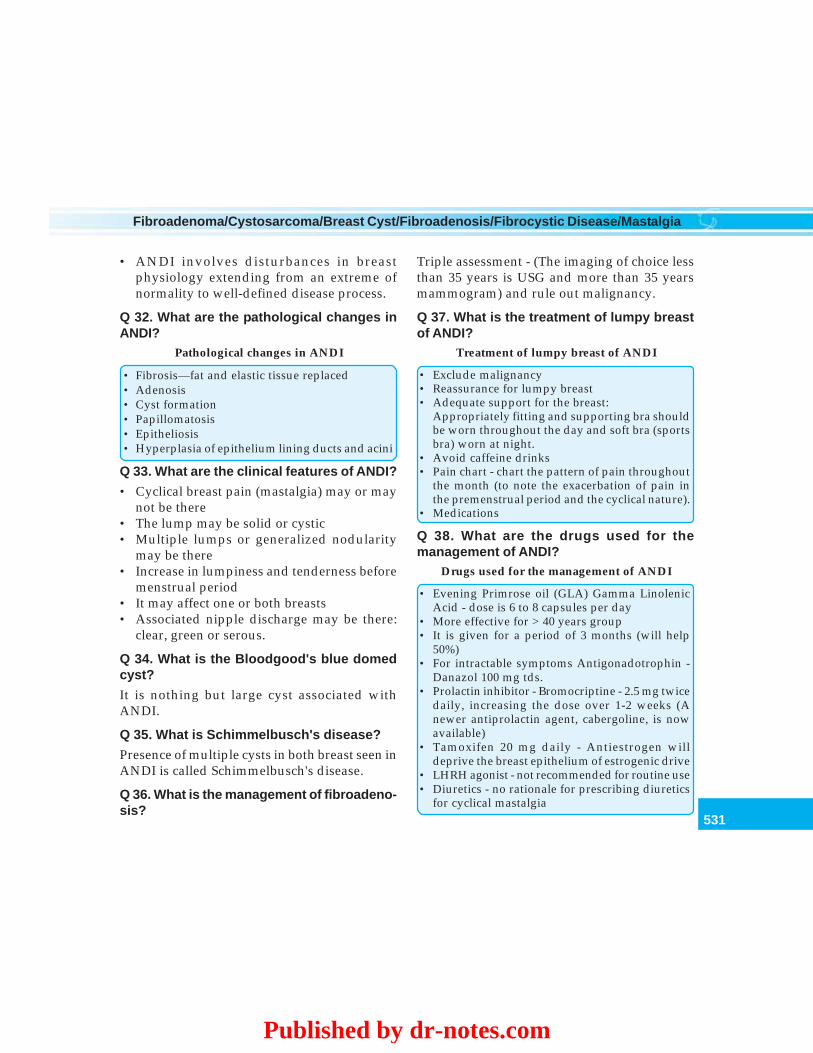
531
Fibroadenoma/Cystosarcoma/Breast Cyst/Fibroadenosis/Fibrocystic Disease/Mastalgia
• ANDI involves disturbances in breastphysiology extending from an extreme ofnormality to well-defined disease process.
Q 32. What are the pathological changes inANDI?
Pathological changes in ANDI
• Fibrosis—fat and elastic tissue replaced• Adenosis• Cyst formation• Papillomatosis• Epitheliosis• Hyperplasia of epithelium lining ducts and acini
Q 33. What are the clinical features of ANDI?
• Cyclical breast pain (mastalgia) may or maynot be there
• The lump may be solid or cystic• Multiple lumps or generalized nodularity
may be there• Increase in lumpiness and tenderness before
menstrual period• It may affect one or both breasts• Associated nipple discharge may be there:
clear, green or serous.
Q 34. What is the Bloodgood's blue domedcyst?
It is nothing but large cyst associated withANDI.
Q 35. What is Schimmelbusch's disease?
Presence of multiple cysts in both breast seen inANDI is called Schimmelbusch's disease.
Q 36. What is the management of fibroadeno-sis?
Triple assessment - (The imaging of choice lessthan 35 years is USG and more than 35 yearsmammogram) and rule out malignancy.
Q 37. What is the treatment of lumpy breastof ANDI?
Treatment of lumpy breast of ANDI
• Exclude malignancy• Reassurance for lumpy breast• Adequate support for the breast:
Appropriately fitting and supporting bra shouldbe worn throughout the day and soft bra (sportsbra) worn at night.
• Avoid caffeine drinks• Pain chart - chart the pattern of pain throughout
the month (to note the exacerbation of pain inthe premenstrual period and the cyclical nature).
• Medications
Q 38. What are the drugs used for themanagement of ANDI?
Drugs used for the management of ANDI
• Evening Primrose oil (GLA) Gamma LinolenicAcid - dose is 6 to 8 capsules per day
• More effective for > 40 years group• It is given for a period of 3 months (will help
50%)• For intractable symptoms Antigonadotrophin -
Danazol 100 mg tds.• Prolactin inhibitor - Bromocriptine - 2.5 mg twice
daily, increasing the dose over 1-2 weeks (Anewer antiprolactin agent, cabergoline, is nowavailable)
• Tamoxifen 20 mg daily - Antiestrogen willdeprive the breast epithelium of estrogenic drive
• LHRH agonist - not recommended for routine use• Diuretics - no rationale for prescribing diuretics
for cyclical mastalgia
Published by dr-notes.com
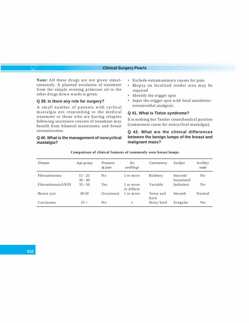
532
Clinical Surgery Pearls
Note: All these drugs are not given simul-taneously. A planned escalation of treatmentfrom the simple evening primrose oil to theother drugs down wards is given.
Q 39. Is there any role for surgery?A small number of patients with cyclicalmastalgia not responding to the medicaltreatment or those who are having relapsesfollowing successive courses of treatment maybenefit from bilateral mastectomy and breastreconstruction.
Q 40. What is the management of noncyclicalmastalgia?
• Exclude extramammary causes for pain• Biopsy on localized tender area may be
required• Identify the trigger spot• Inject the trigger spot with local anesthetic/
nonsteroidal analgesic.
Q 41. What is Tietze syndrome?
It is nothing but Tender costochondral junction(commonest cause for noncyclical mastalgia).
Q 42. What are the clinical differencesbetween the benign lumps of the breast andmalignant mass?
Comparison of clinical features of commonly seen breast lumps
Disease Age group Presence No Consistency Surface Axillaryof pain swellings node
Fibroadenoma 15 - 25 No 1 or more Rubbery Smooth/ No30 - 40 bosselated
FibroadenosisANDI 35 - 50 Yes 1 or more Variable Indistinct Noor diffuse
Breast cyst 30-50 Occasional 1 or more Tense and Smooth Normalhard
Carcinoma 25 + No 1 Stony hard Irregular Yes

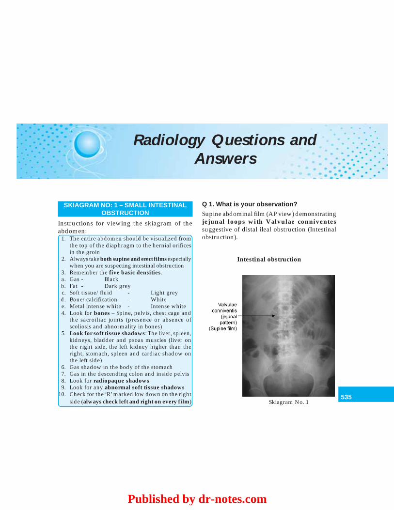
535
Radiology Questions and Answers
Radiology Questions andAnswers
SKIAGRAM NO: 1 – SMALL INTESTINALOBSTRUCTION
Instructions for viewing the skiagram of theabdomen:
1. The entire abdomen should be visualized fromthe top of the diaphragm to the hernial orificesin the groin
2. Always take both supine and erect films especiallywhen you are suspecting intestinal obstruction
3. Remember the five basic densities.a. Gas - Blackb. Fat - Dark greyc. Soft tissue/fluid - Light greyd. Bone/calcification - Whitee. Metal intense white - Intense white4. Look for bones – Spine, pelvis, chest cage and
the sacroiliac joints (presence or absence ofscoliosis and abnormality in bones)
5. Look for soft tissue shadows: The liver, spleen,kidneys, bladder and psoas muscles (liver onthe right side, the left kidney higher than theright, stomach, spleen and cardiac shadow onthe left side)
6. Gas shadow in the body of the stomach7. Gas in the descending colon and inside pelvis8. Look for radiopaque shadows9. Look for any abnormal soft tissue shadows
10. Check for the ‘R’ marked low down on the rightside (always check left and right on every film)
Q 1. What is your observation?
Supine abdominal film (AP view) demonstratingjejunal loops with Valvulae conniventessuggestive of distal ileal obstruction (Intestinalobstruction).
Intestinal obstruction
Skiagram No. 1
Published by dr-notes.com
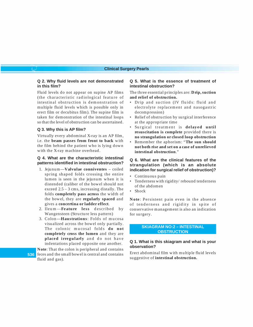
536
Clinical Surgery Pearls
Q 2. Why fluid levels are not demonstratedin this film?
Fluid levels do not appear on supine AP films(the characteristic radiological feature ofintestinal obstruction is demonstration ofmultiple fluid levels which is possible only inerect film or decubitus film). The supine film istaken for demonstration of the intestinal loopsso that the level of obstruction can be ascertained.
Q 3. Why this is AP film?
Virtually every abdominal X-ray is an AP film,i.e. the beam passes from front to back withthe film behind the patient who is lying downwith the X-ray machine overhead.
Q 4. What are the characteristic intestinalpatterns identified in intestinal obstruction?
1. Jejunum—Valvulae conniventes – coiledspring shaped folds crossing the entirelumen is seen in the jejunum when it isdistended (caliber of the bowel should notexceed 2.5 – 3 cms, increasing distally. Thefolds completely pass across the width ofthe bowel, they are regularly spaced andgives a concertina or ladder effect.
2. Ileum—Feature less described byWangensteen (Structure less pattern)
3. Colon—Haustrations: Folds of mucosavisualized across the bowel only partially.The colonic mucosal folds do notcompletely cross the lumen and they areplaced irregularly and do not haveindentations placed opposite one another.
Note: That the colon is peripheral and containsfeces and the small bowel is central and containsfluid and gas).
Q 5. What is the essence of treatment ofintestinal obstruction?
The three essential principles are: Drip, suctionand relief of obstruction.• Drip and suction (IV fluids: fluid and
electrolyte replacement and nasogastricdecompression)
• Relief of obstruction by surgical interferenceat the appropriate time
• Surgical treatment is delayed untilresuscitation is complete provided there isno strangulation or closed loop obstruction
• Remember the aphorism: “The sun shouldnot both rise and set on a case of unrelievedintestinal obstruction.”
Q 6. What are the clinical features of thestrangulation (which is an absoluteindication for surgical relief of obstruction)?
• Continuous pain• Tenderness with rigidity/rebound tenderness
of the abdomen• Shock
Note: Persistent pain even in the absenceof tenderness and rigidity in spite ofconservative management is also an indicationfor surgery.
SKIAGRAM NO:2 – INTESTINALOBSTRUCTION
Q 1. What is this skiagram and what is yourobservation?
Erect abdominal film with multiple fluid levelssuggestive of intestinal obstruction.
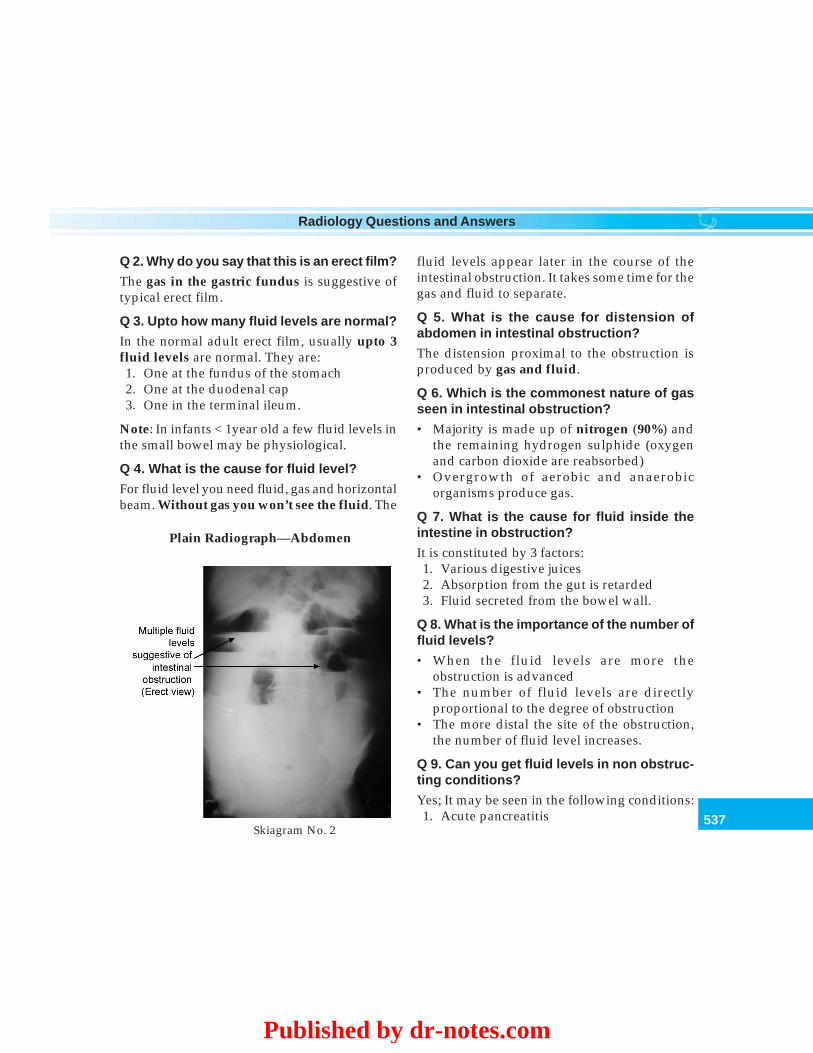
537
Radiology Questions and Answers
Q 2. Why do you say that this is an erect film?
The gas in the gastric fundus is suggestive oftypical erect film.
Q 3. Upto how many fluid levels are normal?
In the normal adult erect film, usually upto 3fluid levels are normal. They are:1. One at the fundus of the stomach2. One at the duodenal cap3. One in the terminal ileum.
Note: In infants < 1year old a few fluid levels inthe small bowel may be physiological.
Q 4. What is the cause for fluid level?
For fluid level you need fluid, gas and horizontalbeam. Without gas you won’t see the fluid. The
fluid levels appear later in the course of theintestinal obstruction. It takes some time for thegas and fluid to separate.
Q 5. What is the cause for distension ofabdomen in intestinal obstruction?
The distension proximal to the obstruction isproduced by gas and fluid.
Q 6. Which is the commonest nature of gasseen in intestinal obstruction?
• Majority is made up of nitrogen (90%) andthe remaining hydrogen sulphide (oxygenand carbon dioxide are reabsorbed)
• Overgrowth of aerobic and anaerobicorganisms produce gas.
Q 7. What is the cause for fluid inside theintestine in obstruction?
It is constituted by 3 factors:1. Various digestive juices2. Absorption from the gut is retarded3. Fluid secreted from the bowel wall.
Q 8. What is the importance of the number offluid levels?
• When the fluid levels are more theobstruction is advanced
• The number of fluid levels are directlyproportional to the degree of obstruction
• The more distal the site of the obstruction,the number of fluid level increases.
Q 9. Can you get fluid levels in non obstruc-ting conditions?
Yes; It may be seen in the following conditions:1. Acute pancreatitis
Plain Radiograph—Abdomen
Skiagram No. 2
Published by dr-notes.com
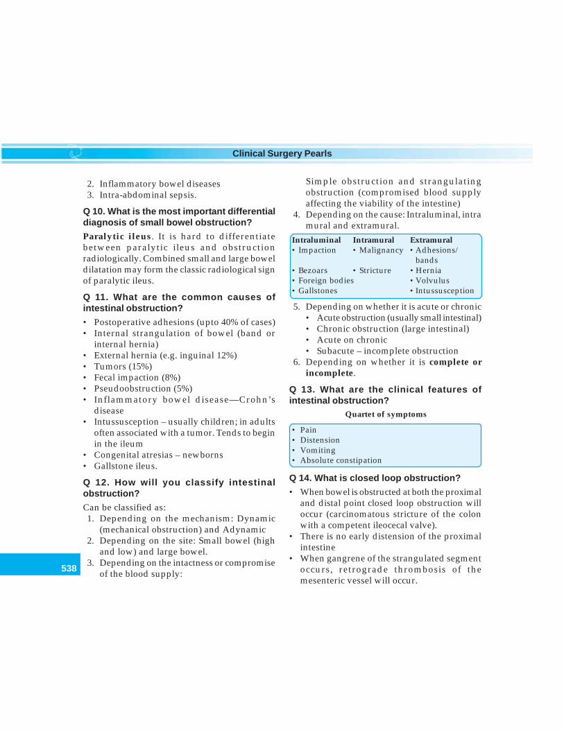
538
Clinical Surgery Pearls
2. Inflammatory bowel diseases3. Intra-abdominal sepsis.
Q 10. What is the most important differentialdiagnosis of small bowel obstruction?
Paralytic ileus. It is hard to differentiatebetween paralytic ileus and obstructionradiologically. Combined small and large boweldilatation may form the classic radiological signof paralytic ileus.
Q 11. What are the common causes ofintestinal obstruction?
• Postoperative adhesions (upto 40% of cases)• Internal strangulation of bowel (band or
internal hernia)• External hernia (e.g. inguinal 12%)• Tumors (15%)• Fecal impaction (8%)• Pseudoobstruction (5%)• Inflammatory bowel disease—Crohn’s
disease• Intussusception – usually children; in adults
often associated with a tumor. Tends to beginin the ileum
• Congenital atresias – newborns• Gallstone ileus.
Q 12. How will you classify intestinalobstruction?
Can be classified as:1. Depending on the mechanism: Dynamic
(mechanical obstruction) and Adynamic2. Depending on the site: Small bowel (high
and low) and large bowel.3. Depending on the intactness or compromise
of the blood supply:
Simple obstruction and strangulatingobstruction (compromised blood supplyaffecting the viability of the intestine)
4. Depending on the cause: Intraluminal, intramural and extramural.
Intraluminal Intramural Extramural• Impaction • Malignancy •Adhesions/
bands• Bezoars • Stricture •Hernia• Foreign bodies •Volvulus• Gallstones •Intussusception
5. Depending on whether it is acute or chronic• Acute obstruction (usually small intestinal)• Chronic obstruction (large intestinal)• Acute on chronic• Subacute – incomplete obstruction
6. Depending on whether it is complete orincomplete.
Q 13. What are the clinical features ofintestinal obstruction?
Quartet of symptoms
• Pain• Distension• Vomiting• Absolute constipation
Q 14. What is closed loop obstruction?
• When bowel is obstructed at both the proximaland distal point closed loop obstruction willoccur (carcinomatous stricture of the colonwith a competent ileocecal valve).
• There is no early distension of the proximalintestine
• When gangrene of the strangulated segmentoccurs, retrograde thrombosis of themesenteric vessel will occur.
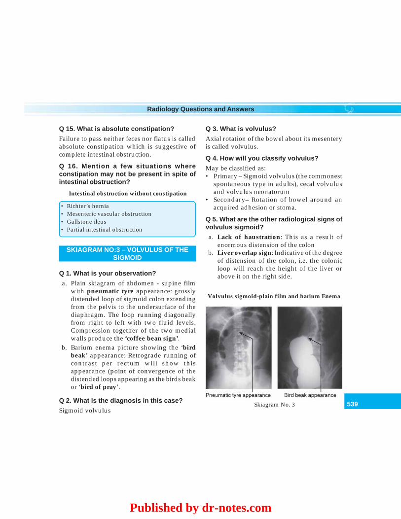
539
Radiology Questions and Answers
Q 15. What is absolute constipation?
Failure to pass neither feces nor flatus is calledabsolute constipation which is suggestive ofcomplete intestinal obstruction.
Q 16. Mention a few situations whereconstipation may not be present in spite ofintestinal obstruction?
Intestinal obstruction without constipation
• Richter’s hernia• Mesenteric vascular obstruction• Gallstone ileus• Partial intestinal obstruction
SKIAGRAM NO:3 – VOLVULUS OF THESIGMOID
Q 1. What is your observation?
a. Plain skiagram of abdomen - supine filmwith pneumatic tyre appearance: grosslydistended loop of sigmoid colon extendingfrom the pelvis to the undersurface of thediaphragm. The loop running diagonallyfrom right to left with two fluid levels.Compression together of the two medialwalls produce the ‘coffee bean sign’.
b. Barium enema picture showing the ‘birdbeak’ appearance: Retrograde running ofcontrast per rectum will show thisappearance (point of convergence of thedistended loops appearing as the birds beakor ‘bird of pray’.
Q 2. What is the diagnosis in this case?
Sigmoid volvulus
Q 3. What is volvulus?
Axial rotation of the bowel about its mesenteryis called volvulus.
Q 4. How will you classify volvulus?
May be classified as:• Primary – Sigmoid volvulus (the commonest
spontaneous type in adults), cecal volvulusand volvulus neonatorum
• Secondary– Rotation of bowel around anacquired adhesion or stoma.
Q 5. What are the other radiological signs ofvolvulus sigmoid?
a. Lack of haustration: This as a result ofenormous distension of the colon
b. Liver overlap sign: Indicative of the degreeof distension of the colon, i.e. the colonicloop will reach the height of the liver orabove it on the right side.
Skiagram No. 3
Volvulus sigmoid-plain film and barium Enema
Published by dr-notes.com
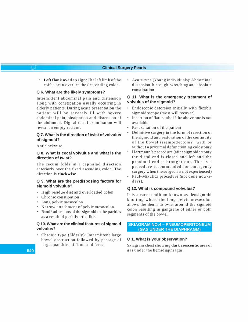
540
Clinical Surgery Pearls
c. Left flank overlap sign: The left limb of thecoffee bean overlies the descending colon.
Q 6. What are the likely symptoms?
Intermittent abdominal pain and distensionalong with constipation usually occurring inelderly patients. During acute presentation thepatient will be severely ill with severeabdominal pain, obstipation and distension ofthe abdomen. Digital rectal examination willreveal an empty rectum.
Q 7. What is the direction of twist of volvulusof sigmoid?
Anticlockwise.
Q 8. What is cecal volvulus and what is thedirection of twist?
The cecum folds in a cephalad directionanteriorly over the fixed ascending colon. Thedirection is clockwise.
Q 9. What are the predisposing factors forsigmoid volvulus?
• High residue diet and overloaded colon• Chronic constipation• Long pelvic mesocolon• Narrow attachment of pelvic mesocolon• Band/adhesions of the sigmoid to the parities
as a result of peridiverticulitis
Q 10. What are the clinical features of sigmoidvolvulus?
• Chronic type (Elderly): Intermittent largebowel obstruction followed by passage oflarge quantities of flatus and feces
• Acute type (Young individuals): Abdominaldistension, hiccough, wretching and absoluteconstipation.
Q 11. What is the emergency treatment ofvolvulus of the sigmoid?
• Endoscopic detorsion initially with flexiblesigmoidoscope (most will recover)
• Insertion of flatus tube if the above one is notavailable
• Resuscitation of the patient• Definitive surgery in the form of resection of
the sigmoid and restoration of the continuityof the bowel (sigmoidectomy) with orwithout a proximal defunctioning colostomy
• Hartmann’s procedure (after sigmoidectomythe distal end is closed and left and theproximal end is brought out. This is aprocedure recommended for emergencysurgery when the surgeon is not experienced)
• Paul–Mikulicz procedure (not done now-a-days).
Q 12. What is compound volvulus?
It is a rare condition known as ileosigmoidknotting where the long pelvic mesocolonallows the ileum to twist around the sigmoidcolon resulting in gangrene of either or bothsegments of the bowel.
SKIAGRAM NO:4 – PNEUMOPERITONEUM(GAS UNDER THE DIAPHRAGM)
Q 1. What is your observation?
Skiagram chest showing dark crescentic area ofgas under the hemidiaphragm.
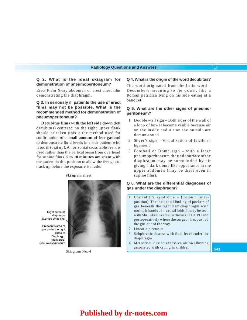
541
Radiology Questions and Answers
Q 2. What is the ideal skiagram fordemonstration of pneumoperitoneum?
Erect Plain X-ray abdomen or erect chest filmdemonstrating the diaphragm.
Q 3. In seriously ill patients the use of erectfilms may not be possible. What is therecommended method for demonstration ofpneumoperitoneum?
Decubitus films with the left side down (leftdecubitus) centered on the right upper flankshould be taken (this is the method used forconfirmation of a small amount of free gas andto demonstrate fluid levels in a sick patient whois too ill to sit up). A horizontal cross table beam isused rather than the vertical beam from overheadfor supine films. 5 to 10 minutes are spent withthe patient in this position to allow the free gas totrack up before the exposure is made.
Q 4. What is the origin of the word decubitus?
The word originated from the Latin word –Decumbere meaning to lie down, like aRoman patrician lying on his side eating at abanquet.
Q 5. What are the other signs of pneumo-peritoneum?
1. Double wall sign – Both sides of the wall ofa loop of bowel become visible because airon the inside and air on the outside aredemonstrated
2. Silver’s sign – Visualization of falciformligament
3. Football or Dome sign – with a largepneumoperitoneum the unde surface of thediaphragm may be surrounded by airgiving a dark dome-like appearance in theupper abdomen (may be there even insupine film).
Q 6. What are the differential diagnoses ofgas under the diaphragm?
1. Chilaiditi’s syndrome – (Colonic inter-position): The incidental finding of pockets ofgas beneath the right hemidiaphragm withmultiple bands of mucosal folds. It may be seenwith Shrunken livers (Cirrhosis), in COPD andpostoperatively where the surgeon has pushedthe gut out of the way.
2. Linear atelectasis:3. Subphrenic abscess with fluid level under the
diaphragm4. Meteorism due to excessive air swallowing
associated with crying in children
Skiagram chest
Skiagram No. 4
Published by dr-notes.com
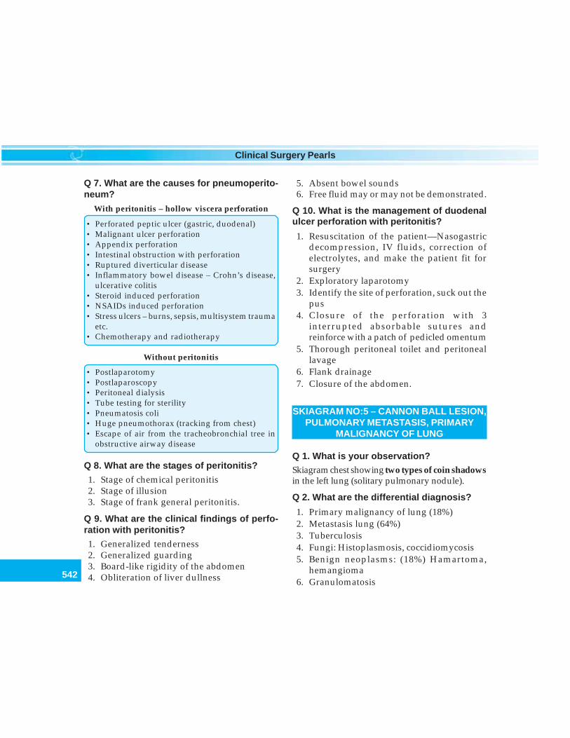
542
Clinical Surgery Pearls
Q 7. What are the causes for pneumoperito-neum?
With peritonitis – hollow viscera perforation
• Perforated peptic ulcer (gastric, duodenal)• Malignant ulcer perforation• Appendix perforation• Intestinal obstruction with perforation• Ruptured diverticular disease• Inflammatory bowel disease – Crohn’s disease,
ulcerative colitis• Steroid induced perforation• NSAIDs induced perforation• Stress ulcers – burns, sepsis, multisystem trauma
etc.• Chemotherapy and radiotherapy
Without peritonitis
• Postlaparotomy• Postlaparoscopy• Peritoneal dialysis• Tube testing for sterility• Pneumatosis coli• Huge pneumothorax (tracking from chest)• Escape of air from the tracheobronchial tree in
obstructive airway disease
Q 8. What are the stages of peritonitis?
1. Stage of chemical peritonitis2. Stage of illusion3. Stage of frank general peritonitis.
Q 9. What are the clinical findings of perfo-ration with peritonitis?
1. Generalized tenderness2. Generalized guarding3. Board-like rigidity of the abdomen4. Obliteration of liver dullness
5. Absent bowel sounds6. Free fluid may or may not be demonstrated.
Q 10. What is the management of duodenalulcer perforation with peritonitis?
1. Resuscitation of the patient—Nasogastricdecompression, IV fluids, correction ofelectrolytes, and make the patient fit forsurgery
2. Exploratory laparotomy3. Identify the site of perforation, suck out the
pus4. Closure of the perforation with 3
interrupted absorbable sutures andreinforce with a patch of pedicled omentum
5. Thorough peritoneal toilet and peritoneallavage
6. Flank drainage7. Closure of the abdomen.
SKIAGRAM NO:5 – CANNON BALL LESION,PULMONARY METASTASIS, PRIMARY
MALIGNANCY OF LUNG
Q 1. What is your observation?Skiagram chest showing two types of coin shadowsin the left lung (solitary pulmonary nodule).
Q 2. What are the differential diagnosis?
1. Primary malignancy of lung (18%)2. Metastasis lung (64%)3. Tuberculosis4. Fungi: Histoplasmosis, coccidiomycosis5. Benign neoplasms: (18%) Hamartoma,
hemangioma6. Granulomatosis
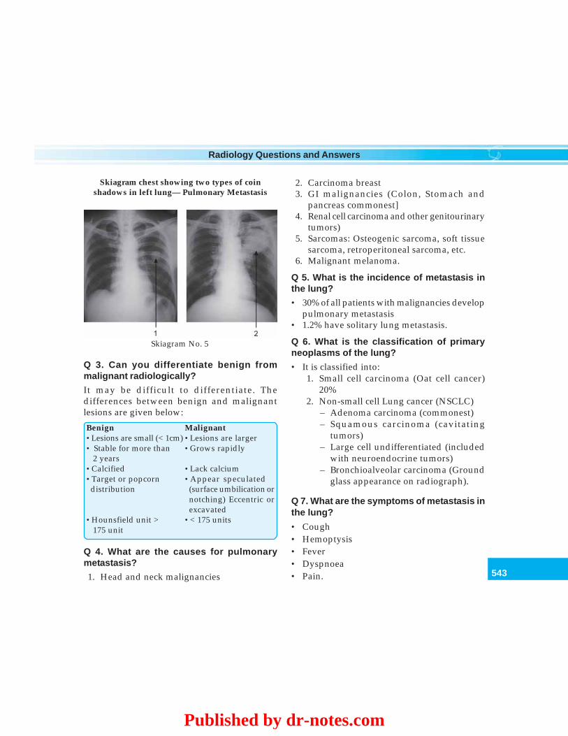
543
Radiology Questions and Answers
Q 3. Can you differentiate benign frommalignant radiologically?
It may be difficult to differentiate. Thedifferences between benign and malignantlesions are given below:
Benign Malignant•Lesions are small (< 1cm) •Lesions are larger• Stable for more than •Grows rapidly
2 years•Calcified •Lack calcium•Target or popcorn •Appear speculated
distribution (surface umbilication ornotching) Eccentric orexcavated
•Hounsfield unit > •< 175 units 175 unit
Q 4. What are the causes for pulmonarymetastasis?
1. Head and neck malignancies
2. Carcinoma breast3. GI malignancies (Colon, Stomach and
pancreas commonest]4. Renal cell carcinoma and other genitourinary
tumors)5. Sarcomas: Osteogenic sarcoma, soft tissue
sarcoma, retroperitoneal sarcoma, etc.6. Malignant melanoma.
Q 5. What is the incidence of metastasis inthe lung?
• 30% of all patients with malignancies developpulmonary metastasis
• 1.2% have solitary lung metastasis.
Q 6. What is the classification of primaryneoplasms of the lung?
• It is classified into:1. Small cell carcinoma (Oat cell cancer)
20%2. Non-small cell Lung cancer (NSCLC)
– Adenoma carcinoma (commonest)– Squamous carcinoma (cavitating
tumors)– Large cell undifferentiated (included
with neuroendocrine tumors)– Bronchioalveolar carcinoma (Ground
glass appearance on radiograph).
Q 7. What are the symptoms of metastasis inthe lung?
• Cough• Hemoptysis• Fever• Dyspnoea• Pain.
Skiagram No. 5
Skiagram chest showing two types of coinshadows in left lung— Pulmonary Metastasis
Published by dr-notes.com
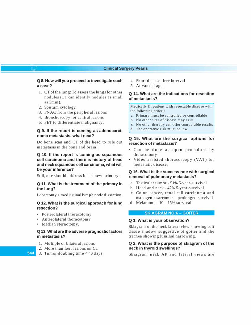
544
Clinical Surgery Pearls
Q 8. How will you proceed to investigate sucha case?
1. CT of the lung: To assess the lungs for othernodules (CT can identify nodules as smallas 3mm).
2. Sputum cytology3. FNAC from the peripheral lesions4. Bronchoscopy for central lesions5. PET to differentiate malignancy.
Q 9. If the report is coming as adenocarci-noma metastasis, what next?
Do bone scan and CT of the head to rule outmetastasis in the bone and brain.
Q 10. If the report is coming as squamouscell carcinoma and there is history of headand neck squamous cell carcinoma, what willbe your inference?
Still, one should address it as a new primary.
Q 11. What is the treatment of the primary inthe lung?
Lobectomy + mediastinal lymph node dissection.
Q 12. What is the surgical approach for lungresection?
• Posterolateral thoracotomy• Anterolateral thoracotomy• Median sternotomy.
Q 13. What are the adverse prognostic factorsin metastasis?
1. Multiple or bilateral lesions2. More than four lesions on CT3. Tumor doubling time < 40 days
4. Short disease- free interval5. Advanced age.
Q 14. What are the indications for resectionof metastasis?
Medically fit patient with resectable disease withthe following criteriaa. Primary must be controlled or controllableb. No other sites of disease may existc. No other therapy can offer comparable resultsd. The operative risk must be low
Q 15. What are the surgical options forresection of metastasis?
• Can be done as open procedure bythoracotomy
• Video assisted thoracoscopy (VAT) formetastatic disease.
Q 16. What is the success rate with surgicalremoval of pulmonary metastasis?
a. Testicular tumor - 51% 5-year-survivalb. Head and neck - 47% 5-year-survivalc. Colon cancer, renal cell carcinoma and
osteogenic sarcomas – prolonged survivald. Melanoma - 10 – 15% survival.
SKIAGRAM NO:6 – GOITER
Q 1. What is your observation?Skiagram of the neck lateral view showing softtissue shadow suggestive of goiter and thetrachea showing luminal narrowing.
Q 2. What is the purpose of skiagram of theneck in thyroid swellings?Skiagram neck AP and lateral views are
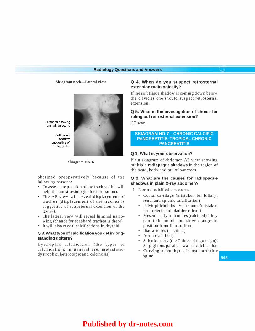
545
Radiology Questions and Answers
obtained preoperatively because of thefollowing reasons:• To assess the position of the trachea (this will
help the anesthesiologist for intubation).• The AP view will reveal displacement of
trachea (displacement of the trachea issuggestive of retrosternal extension of thegoiter).
• The lateral view will reveal luminal narro-wing (chance for scabbard trachea is there)
• It will also reveal calcifications in thyroid.
Q 3. What type of calcification you get in long-standing goiters?Dystrophic calcification (the types ofcalcifications in general are: metastatic,dystrophic, heterotopic and calcinosis).
Q 4. When do you suspect retrosternalextension radiologically?
If the soft tissue shadow is coming down belowthe clavicles one should suspect retrosternalextension.
Q 5. What is the investigation of choice forruling out retrosternal extension?
CT scan.
SKIAGRAM NO:7 – CHRONIC CALCIFICPANCREATITIS, TROPICAL CHRONIC
PANCREATITIS
Q 1. What is your observation?
Plain skiagram of abdomen AP view showingmultiple radiopaque shadows in the region ofthe head, body and tail of pancreas.
Q 2. What are the causes for radiopaqueshadows in plain X-ray abdomen?
1. Normal calcified structures• Costal cartilage (mistaken for biliary,
renal and splenic calcification)• Pelvic phleboliths – Vein stones (mistaken
for ureteric and bladder calculi)• Mesenteric lymph nodes (calcified): They
tend to be mobile and show changes inposition from film-to-film.
• Iliac arteries (calcified)• Aorta (calcified)• Splenic artery (the Chinese dragon sign):
Serpiginous parallel - walled calcification• Curving osteophytes in osteoarthritic
spine
Skiagram No. 6
Skiagram neck—Lateral view
Published by dr-notes.com
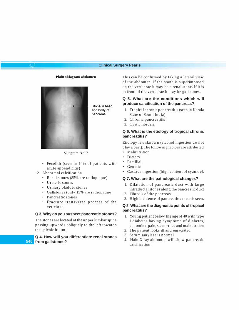
546
Clinical Surgery Pearls
Plain skiagram abdomen
Skiagram No. 7
• Fecolith (seen in 14% of patients withacute appendicitis)
2. Abnormal calcification• Renal stones (85% are radiopaque)• Ureteric stones• Urinary bladder stones• Gallstones (only 15% are radiopaque)• Pancreatic stones• Fracture transverse process of the
vertebrae.
Q 3. Why do you suspect pancreatic stones?
The stones are located at the upper lumbar spinepassing upwards obliquely to the left towardsthe splenic hilum.
Q 4. How will you differentiate renal stonesfrom gallstones?
This can be confirmed by taking a lateral viewof the abdomen. If the stone is superimposedon the vertebrae it may be a renal stone. If it isin front of the vertebrae it may be gallstones.
Q 5. What are the conditions which willproduce calcification of the pancreas?
1. Tropical chronic pancreatitis (seen in KeralaState of South India)
2. Chronic pancreatitis3. Cystic fibrosis.
Q 6. What is the etiology of tropical chronicpancreatitis?
Etiology is unknown (alcohol ingestion do notplay a part): The following factors are attributed• Malnutrition• Dietary• Familial• Genetic• Cassava ingestion (high content of cyanide).
Q 7. What are the pathological changes?
1. Dilatation of pancreatic duct with largeintraductal stones along the pancreatic duct
2. Fibrosis of the pancreas3. High incidence of pancreatic cancer is seen.
Q 8. What are the diagnostic points of tropicalpancreatitis?
1. Young patient below the age of 40 with typeI diabetes having symptoms of diabetes,abdominal pain, steatorrhea and malnutrition
2. The patient looks ill and emaciated3. Serum amylase is normal4. Plain X-ray abdomen will show pancreatic
calcification.

547
Radiology Questions and Answers
Q 9. What is the treatment of tropical chronicpancreatitis?
Medical treatment:• Pancreatic enzymes• Insulin therapy for diabetes• Management of pain as per analgesic ladder.
Endoscopic management:1. Small head stones can be managed by
endoscopic extraction at ERCP2. If pancreatic duct stricture is predominant
with upstream dilatation, a stent can beinserted.
Surgical treatment: Done only for intractablepain• The operations are:
1. Extraction of pancreatic duct stonesfollowed by longitudinal pancreat-ojejunostomy (Frey procedure)
2. Beger procedure (Head coring):Duodenum preserving resection of thepancreatic head
3. If mass lesion is there at the head ofpancreas, a pancreaticoduodenectomy isdone
4. If the disease is limited to the tail of thepancreas, a distal pancreatectomy is done
5. Intractable pain with diffuse disease:Total pancreatectomy.
SKIAGRAM NO:8 – GALLSTONE
Q 1. What is your observation?
Plain radiograph of abdomen showing aradiopaque stone in the region of the gallbladder, suggestive of gallstone.
The second picture is a barium enema takento rule out diverticulosis of the colon because ofthe patient’s left lower abdominal complaints.There is no evidence of diverticulosis in thepicture (Normal study).
Q 2. What is Saint’s triad?
The association of gallstones, hiatus hernia anddiverticulosis.
Q 3. How it is differentiated from renalstones?
Answer given (Skiagram No:7).
Q 4. How many percentage of the gallstonesare radiopaque?
10% (90% are nonradiopaque in contrast to renalstones (85% radiopaque).
Q 5. What are the types of gallstones?
1. Cholesterol stones2. Pigment stones (brown or black): contain
< 30% cholesterol.
Gallstone in plain radiograph and barium enema
Skiagram No. 8A and B
Published by dr-notes.com
548
Clinical Surgery Pearls
• Black stones are composed of insolublebilirubin pigment mixed with calciumphosphate and calcium bicarbonate. Theincidence rises with age. They are alsoseen in hemolytic conditions likehereditary spherocytosis and sickle celldisease. Therefore it is important to do aperipheral smear examination andfragility test if pigment stone is suspected.In hereditary spherocytosis in addition tocholecystectomy a splenectomy has to bedone.
• Brown pigment stones: Containcholesterol and calcium bilirubinate,calcium palmitate and calcium stearate.They are usually seen in bile duct andrelated to infected bile as a result ofdeconjugation of bilirubin diglucuronideby bacterial beta glucuronidase. It is alsoassociated with presence of foreignbodies and parasites.
3. Mixed stones (pure cholesterol + a mixtureof calcium salts, bile acids, bile pigmentsand phospholipids
Note: In the West 80% are cholesterol and mixedstones.
In Asia 80% are pigment stones.
Q 6. What is Mercedes – Benz or Seagullsign?
The center of a stone may contain radiolucentgas in a triradiate or biradiate fissure givingcharacteristic dark shapes in radiograph. Thisis called Mercedes Benz sign.
Q 7. What is “porcelain” gallbladder, what isthe importance of it?
Calcification of the gallbladder in plain X-ray iscalled porcelain gallbladder. The importance ofthis appearance is an association of carcinomain up to 25% of patients.
Q 8. What are the complications of gall-stones?
Complications of gallstones
• Biliary colic• Acute cholecystitis:
Empyema of gallbladderGangrene of gallbladderPerforation of gallbladder with peritonitisMucocele of gallbladder
• Chronic cholecystitis• Bile duct stone – obstructive jaundice• Cholangitis – secondary to bile duct obstruction
(Charcot’s triad consisting of intermittent fever,intermittent pain and intermittent jaundice).
• Acute toxic cholangitis (Reynold’s pentadconsisting of Charcot’s triad + mentalobtundation + hypotension)
• Acute pancreatitis (Gallstone pancreatitis)• Gallstone ileus (stone obstructing the bowel
usually the terminal ileum)• Gallbladder carcinoma (0.08% of symptomatic
patients)
Q 9. What are the etiological factors for gallstone formation?
1. Supersaturation of bile by cholesterol ordecrease in bile acid concentration andunstable unilamelar phospholipid vesicles
2. Nucleating factors: Infection (klebsiella,E-coli, Enterococci, Bacteroides and typhoidorganism), mucus and glycoprotein
3. Stasis (Impaired gallbladder function):Repeated pregnancy
549
Radiology Questions and Answers
4. Enterohepatic circulation is not taking place.For example, Ileal resection, Cholestyramine.
Q 10. What is Moynihan’s aphorism?
“Gallstone is the tomb stone erected to thememory of the organism within it” Thisstatement was given with respect to gallstoneshaving salmonella organism inside leading totyphoid gallbladder. Salmonella typhimuriumcan infect the gallbladder and produce chroniccholecystitis or acute cholecystitis and thepatient will remain a typhoid carrier byexcreting bacteria in the bile (“Typhoid Mary”,a cook general who passed salmonella typhi inher feces and urine and was responsible fornearly twenty epidemics of typhoid in andaround New York city in USA). Surgeons shouldnot give patients their stones after surgery ifthere is any suspicion of typhoid.
Q 11. What is the incidence of gallstone?
10-15% of the adult population.
Q 12. What is the incidence of asymptomaticgallstone developing future symptoms?
1-2% will develop symptoms per year.
Q 13. What is the treatment of symptomaticgallstones?
Laparoscopic cholecystectomy.
Q 14. Why stone dissolution is not recom-mended in gallstones?
It is not recommended because of two reasons:1. Dissolution by ESWL will result in
fragmentation of the stones. They comedown and produce obstructive jaundice.
2. Removing gallstones without removal of thegallbladder will lead to gallstone recurrence.
Q 15. What is the treatment of asymptomaticgallstones?
• Observation is enough• Cholecystectomy is indicated in the following
situations:1. Diabetic patients – chance for infection
and complication2. Hemolytic anemias3. Patients undergoing Bariatric surgery for
morbid obesity4. Calcified gallbladder wall (Porcelain
gallbladder).
Q 16. What is the timing of surgery for acutecholecystitis?1. Early laparoscopic cholecystectomy
(preferred) during the golden period(72hours): The open conversion rate oflaparoscopic cholecystectomy is five timeshigher than in the elective setting
2. Elective surgery after a period ofconservative treatment for 6 weeks.
SKIAGRAM NO:9 – FRACTURE OF RIBS,FLAIL CHEST
Q 1. What is your observation?Skiagram chest showing multiple fracture ribsshown with arrows.
Q 2. What are the clinical signs of fracture rib?• Crepitus• Deformity• Limitation of chest wall movement.
Published by dr-notes.com
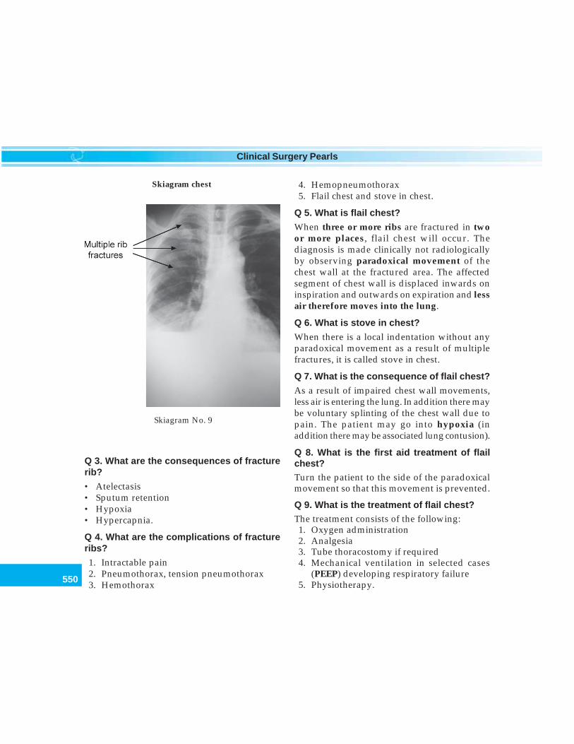
550
Clinical Surgery Pearls
Q 3. What are the consequences of fracturerib?
• Atelectasis• Sputum retention• Hypoxia• Hypercapnia.
Q 4. What are the complications of fractureribs?
1. Intractable pain2. Pneumothorax, tension pneumothorax3. Hemothorax
4. Hemopneumothorax5. Flail chest and stove in chest.
Q 5. What is flail chest?
When three or more ribs are fractured in twoor more places, flail chest will occur. Thediagnosis is made clinically not radiologicallyby observing paradoxical movement of thechest wall at the fractured area. The affectedsegment of chest wall is displaced inwards oninspiration and outwards on expiration and lessair therefore moves into the lung.
Q 6. What is stove in chest?
When there is a local indentation without anyparadoxical movement as a result of multiplefractures, it is called stove in chest.
Q 7. What is the consequence of flail chest?
As a result of impaired chest wall movements,less air is entering the lung. In addition there maybe voluntary splinting of the chest wall due topain. The patient may go into hypoxia (inaddition there may be associated lung contusion).
Q 8. What is the first aid treatment of flailchest?Turn the patient to the side of the paradoxicalmovement so that this movement is prevented.
Q 9. What is the treatment of flail chest?The treatment consists of the following:1. Oxygen administration2. Analgesia3. Tube thoracostomy if required4. Mechanical ventilation in selected cases
(PEEP) developing respiratory failure5. Physiotherapy.
Skiagram No. 9
Skiagram chest
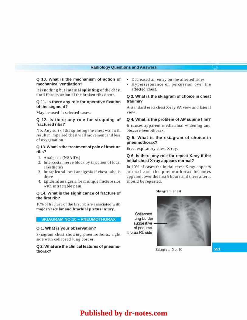
551
Radiology Questions and Answers
• Decreased air entry on the affected sides• Hyperresonance on percussion over the
affected chest.
Q 3. What is the skiagram of choice in chesttrauma?
A standard erect chest X-ray PA view and lateralview.
Q 4. What is the problem of AP supine film?
It causes apparent mediastinal widening andobscure hemothorax.
Q 5. What is the skiagram of choice inpneumothorax?
Erect expiratory chest X-ray.
Q 6. Is there any role for repeat X-ray if theinitial chest X-ray appears normal?
In 10% of cases the initial chest X-ray appearsnormal and the pneumothorax becomesapparent over the first 8 hours and there after itshould be repeated.
Q 10. What is the mechanism of action ofmechanical ventilation?It is nothing but internal splinting of the chestuntil fibrous union of the broken ribs occur.
Q 11. Is there any role for operative fixationof the segment?May be used in selected cases.
Q 12. Is there any role for strapping offractured ribs?No. Any sort of the splinting the chest wall willresult in impaired chest wall movement and lessof oxygenation.
Q 13. What is the treatment of pain of fractureribs?1. Analgesic (NSAIDs)2. Intercostal nerve block by injection of local
anesthetics3. Intrapleural local analgesia if chest tube is
there4. Epidural analgesia for multiple fracture ribs
with intractable pain.
Q 14. What is the significance of fracture ofthe first rib?
10% of fracture of the first rib are associated withmajor vascular and brachial plexus injury.
SKIAGRAM NO:10 – PNEUMOTHORAX
Q 1. What is your observation?
Skiagram chest showing pneumothorax rightside with collapsed lung border.
Q 2. What are the clinical features of pneumo-thorax? Skiagram No. 10
Skiagram chest
Published by dr-notes.com
552
Clinical Surgery Pearls
Q 7. What is the difference between simplepneumothorax and tension pneumothorax?
A pneumothorax with mediastinaldisplacement to the opposite side is calledtension pneumothorax. This is manifested bytracheal shift to the opposite side and shift ofthe apex beat. The air is forced into the thoraciccavity without any means of escape by a one wayvalve either from the lung or through the chestwall. As a result of tension, the lung will becompletely collapsed and the opposite lung willbe compressed because of the mediastinal shift.This will result in decrease in the venous return.
Simple pneumothorax Tension pneumothorax
Tracheal position Normal DisplacedPercussion note Normal Increased (hyperresonant)Jugular pressure Normal Elevated (unless hypovolemic)Breath sounds Normal (unless large) DecreasedRespiratory distress Variable Severe
Q 8. What are the causes for tension pneumo-thorax?
1. Penetrating chest injury2. Blunt chest injury with lung injury3. Primary spontaneous pneumothorax4. Secondary pneumothorax – diseases of lung
(tuberculosis, cavitating lung disease)– necrosing tumors– diseases of plurae
5. Iatrogenic lung injury due to central venouscannulation
6. Mechanical ventilation.
Q 9. What are the clinical features of tensionpneumothorax?
Clinical features of tension pneumothorax
• Acute dyspnea• Distended neck veins• Weak pulse• Low blood pressure• Tracheal shift• Shifting of the apex beat• Hyperresonance on the affected hemithorax• Absent breath sounds on the same side
Q 10. Mention another condition where thereis elevated JVP as a result of chest trauma?
Cardiac tamponade: Cardiac injury resulting inhemopericardium which may seal temporarilythe bleeding from the cardiac injury.
Q 11. What is the diagnostic triad for cardiactamponade?
Beck’s triad: Elevated JVP, diminished heartsounds (Muffled) and pulse paradox (15mm ofHg fall on inspiration).
Q 12. How to confirm tension pneumothorax?
It is a clinical diagnosis and treatment must beinstituted urgently without wasting time forradiology.
553
Radiology Questions and Answers
Q 13. What are the radiological findings oftension pneumothorax?
Radiological findings of tension pneumothorax
• The lung is completely collapsed (the collapsedlung margin can be seen, the rest of the chestcavity showing only air without any lungmarkings)
• The mediastinum is pushed to the opposite side• The diaphragm is flattened and pushed down• The spaces between the ribs are widened on the
affected side (spreading of the ribs)
Q 14. What is the treatment of tensionpneumothorax?
• Urgent needle thoracostomy by inserting alarge bore needle into the second intercostalspace in the midclavicular line
• This is followed by tube thoracostomy in thesafe triangle which is connected to anunderwater seal.
Q 15. Is there any role for conservativemanagement of small traumatic pneumothorax?
• All traumatic pneumothoraces are drained toprevent tension pneumothorax
• This will encourage early lung expansion andevacuate any hemothorax
• Without drainage half of all pneumothoraceswill increase over the first 24 hours.
Q 16. What is sucking wound?
It is nothing but open pneumothorax as a resultof open defect in the chest of more than 3cm.This will lead on to hypoventilation or hypoxia.
Q 17. What is the treatment of suckingwound?
1. Closing the defect with sterile occlusivedressing sealed on three sides so that theair will escape but preventing the air entry
2. Tube thoracostomy3. Formal closure of the defect may be required.
Q 18. If there is failure to expand the lungwith effective drainage, how one shouldproceed?
This is an indication for bronchoscopy (andoften thoracotomy).
SKIAGRAM NO:11 – SURGICALEMPHYSEMA
Q 1. What is your observation?
Skiagram chest showing air spaces in thesubcutaneous tissue suggestive of surgicalemphysema (Subcutaneous emphysema).
Q 2. What is surgical emphysema?
• It is the presence of air in the tissues• It requires a breach of air containing viscus
which is in communication with soft tissuesand the generation of positive pressure topush the air along tissue planes.
Q 3. What is the physical sign of surgicalemphysema?
Crepitus (a peculiar crackling sensationimparted to the examining fingers when oneplaces the fingers fan wise on the affected areaand exerts light pressure).
Published by dr-notes.com
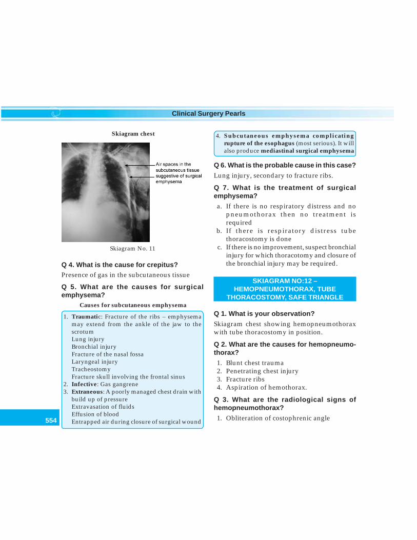
554
Clinical Surgery Pearls
Q 4. What is the cause for crepitus?Presence of gas in the subcutaneous tissue
Q 5. What are the causes for surgicalemphysema?
Causes for subcutaneous emphysema
1. Traumatic: Fracture of the ribs – emphysemamay extend from the ankle of the jaw to thescrotumLung injuryBronchial injuryFracture of the nasal fossaLaryngeal injuryTracheostomyFracture skull involving the frontal sinus
2. Infective: Gas gangrene3. Extraneous: A poorly managed chest drain with
build up of pressureExtravasation of fluidsEffusion of bloodEntrapped air during closure of surgical wound
4. Subcutaneous emphysema complicatingrupture of the esophagus (most serious). It willalso produce mediastinal surgical emphysema
Q 6. What is the probable cause in this case?
Lung injury, secondary to fracture ribs.
Q 7. What is the treatment of surgicalemphysema?
a. If there is no respiratory distress and nopneumothorax then no treatment isrequired
b. If there is respiratory distress tubethoracostomy is done
c. If there is no improvement, suspect bronchialinjury for which thoracotomy and closure ofthe bronchial injury may be required.
SKIAGRAM NO:12 –HEMOPNEUMOTHORAX, TUBE
THORACOSTOMY, SAFE TRIANGLE
Q 1. What is your observation?
Skiagram chest showing hemopneumothoraxwith tube thoracostomy in position.
Q 2. What are the causes for hemopneumo-thorax?
1. Blunt chest trauma2. Penetrating chest injury3. Fracture ribs4. Aspiration of hemothorax.
Q 3. What are the radiological signs ofhemopneumothorax?
1. Obliteration of costophrenic angle
Skiagram No. 11
Skiagram chest
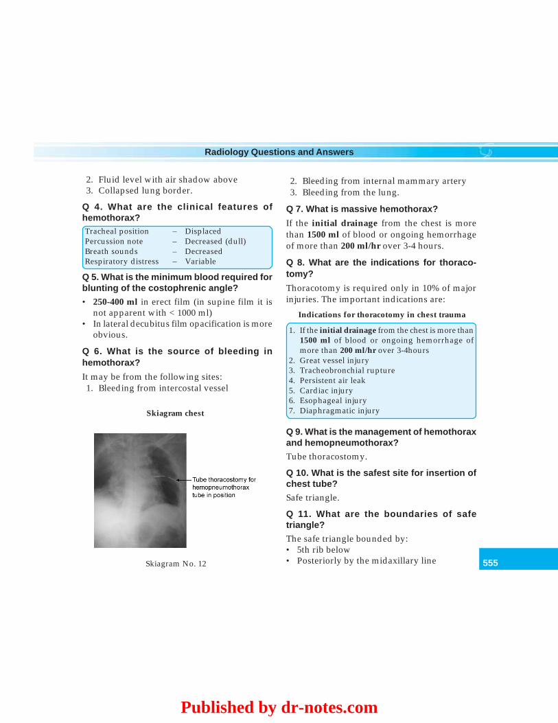
555
Radiology Questions and Answers
2. Fluid level with air shadow above3. Collapsed lung border.
Q 4. What are the clinical features ofhemothorax?Tracheal position – DisplacedPercussion note – Decreased (dull)Breath sounds – DecreasedRespiratory distress – Variable
Q 5. What is the minimum blood required forblunting of the costophrenic angle?
• 250-400 ml in erect film (in supine film it isnot apparent with < 1000 ml)
• In lateral decubitus film opacification is moreobvious.
Q 6. What is the source of bleeding inhemothorax?
It may be from the following sites:1. Bleeding from intercostal vessel
2. Bleeding from internal mammary artery3. Bleeding from the lung.
Q 7. What is massive hemothorax?
If the initial drainage from the chest is morethan 1500 ml of blood or ongoing hemorrhageof more than 200 ml/hr over 3-4 hours.
Q 8. What are the indications for thoraco-tomy?
Thoracotomy is required only in 10% of majorinjuries. The important indications are:
Indications for thoracotomy in chest trauma
1. If the initial drainage from the chest is more than1500 ml of blood or ongoing hemorrhage ofmore than 200 ml/hr over 3-4hours
2. Great vessel injury3. Tracheobronchial rupture4. Persistent air leak5. Cardiac injury6. Esophageal injury7. Diaphragmatic injury
Q 9. What is the management of hemothoraxand hemopneumothorax?
Tube thoracostomy.
Q 10. What is the safest site for insertion ofchest tube?
Safe triangle.
Q 11. What are the boundaries of safetriangle?
The safe triangle bounded by:• 5th rib below• Posteriorly by the midaxillary lineSkiagram No. 12
Skiagram chest
Published by dr-notes.com
556
Clinical Surgery Pearls
• Anterior axillary line anteriorly (lateral to thepectoralis major muscle).
Q 12. What is the importance of this triangle?Why safe triangle is selected
• It is important to remember that the abdominalcavity is extending upto the nipple level
• Any insertion of tube below the 5th rib is likelyto enter the abdominal cavity and consequentinjury to the viscera
• The thickness of the chest wall in this triangle isthin and constituted only by the intercostalmuscles (inner and outer) and no other muscleis coming in the triangle. Therefore it is easier toinsert the chest tube here
• The interspace is large here• No impairment of accessory respiratory muscles• Away from mediastinal structures and internal
mammary artery• Since the position of the tube is anterior, in the
supine position the tube will not kink.
Q 13. What is the ideal direction of the chesttube for the purpose of draining hemo-pneumothorax?
• The tube should go posteriorly and upwardstowards the apex of the lung (apex for air,posteriorly for blood)
• A drain for pleural effusion and empyemashould be nearer the base.
Q 14. How will you prevent neurovascularinjury during chest tube insertion?
The tube should pass over the upper edge ofthe rib to avoid neurovascular bundle.
Q 15. What are the important steps of tubethoracostomy?
1. Take sterile precautions and paint theselected area of the chest wall withantiseptics (centering the safe triangle).
2. Infiltrate local anesthesia at the siteincluding the pleura
3. Make and skin incision in the intercostalspace for about 2.5 cms
4. Blunt dissection is carried out through theintercostal muscles
5. An oblique tract is made (the skin incisionis made one interspace lower) so as to enterthe pleural cavity
6. The gloved finger is introduced into thepleural cavity and the pleural adhesions areseparated
7. Sterile chest tube is introduced with the helpof an artery forces in an upward and medialdirection
8. See that all the side holes are inside the chestcavity
9. The tube is fixed in position with a retainingstitch and see that the retaining stitch is notobliterating the tube
10. The tube is connected to under water sealof the chest drain bottle
11. The wound is sealed12. Take chest radiograph and see that the chest
tube is in position.
Q 16. What is the daily postoperative care ofthe tube?• See that the air column in the tube is moving
(means, the tube is in the chest cavity. If thecolumn is not moving, the tube is occludedor it is not in the chest cavity).
• Look for air bubbling-suggestive of airescape from the pleural cavity if there ispneumothorax.
557
Radiology Questions and Answers
• See that the lower end of the chest tube isbelow the under water seal of the drainingbottle.
• Measure the total drainage of blood in thedraining bottle (whenever the bottle is full, itshould be emptied after clamping the chesttube).
• See that all the side holes of the chest tubeare inside the chest cavity- (there should notbe any air leak at the tube chest wall junction).
• Check for air entry on the side by auscultation• Take check X-ray of the chest to look for
expansion of the lungs and clearance of thecostophrenic angle.
Q 17. Is there any role for applying suctionto the chest drain?
No:
Q 18. When to remove the chest tube?Indications for removal of the chest tube
• Remove the drain when it is no longer draining• The drain should be removed when there has
been no air leak for 24 hours with a fullyexpanded lung
• If the patient is ventilated the drain should beleft until after extubation or there has been noair leak for five days
Q 19. What are the causes for failure toexpand the lung?
1. Inadequate drain size2. Position of the drain not reaching the apex3. Kinking of the tube4. Excessive air leak
Q 20. What are the complications of tubethoracostomy?
1. Infection – empyema2. Danger of disconnection of the tube and
siphoning of air.
Q 21. What are the radiological findings ofrupture of the diaphragm?
Traumatic rupture is commonly seen on leftside. The radiological findings are:1. Stomach containing fluid level within the
left hemithorax (the fluid level in stomachwill not be completely across the hemithoraxunlike hemopneumothirax)
2. Compressed lung lying above and medialto the top of the intrathoracic stomach
3. Gas shadows of intrathoracic colon if theescaping viscera is colon
4. Lateral view showing double fluid level ifthere is intrathoracic gastric volvulus.
SKIAGRAM NO:13 – CERVICAL RIB,THORACIC OUTLET SYNDROME
Q 1. What is your observation?
Skiagram neck showing incomplete cervical ribon right side.
Q 2. What is cervical rib?
It is an extension of costal element of transverseprocess of C7 vertebra.
Q 3. What are the types of cervical rib?
1. Complete fibrous band (commonest type)from the transverse process and reachinganteriorly over the first rib or manubrium.It is not demonstrated radiologically
2. Complete bony type (radiopaque)
Published by dr-notes.com
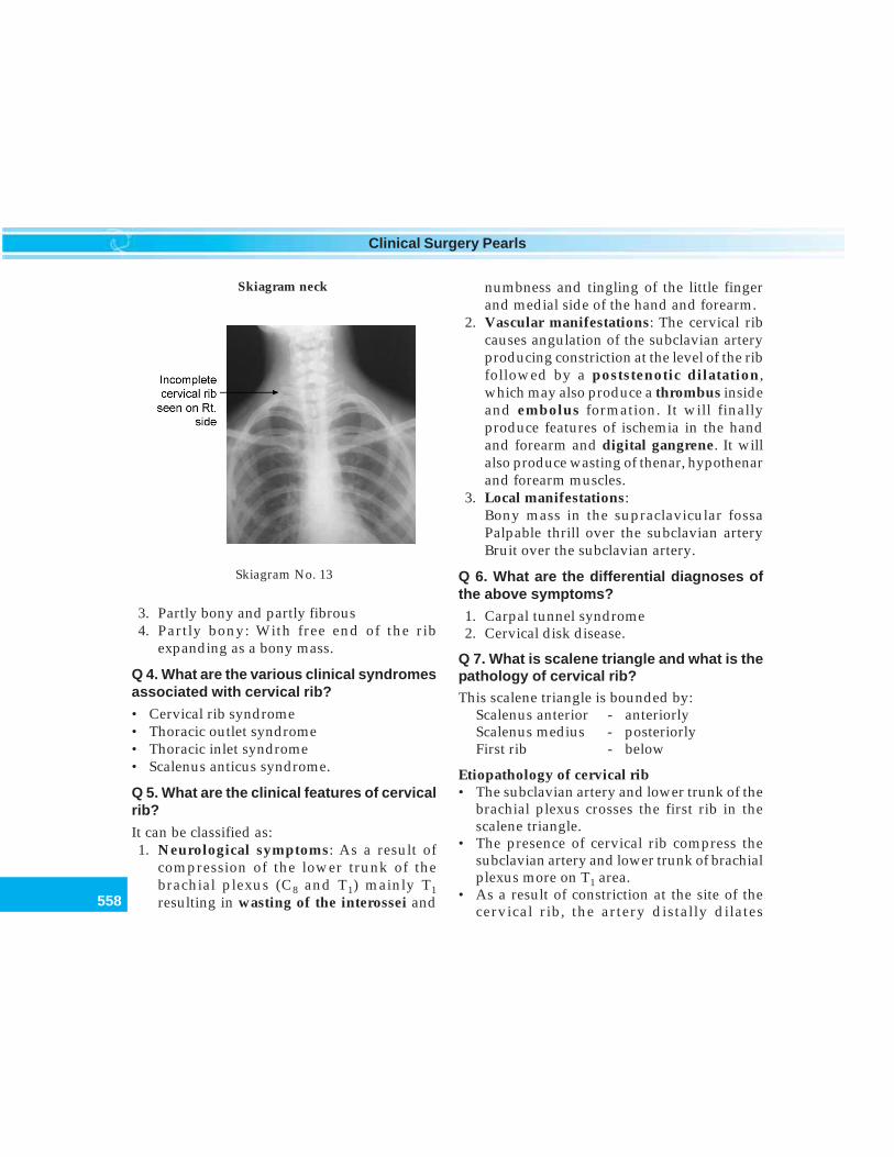
558
Clinical Surgery Pearls
3. Partly bony and partly fibrous4. Partly bony: With free end of the rib
expanding as a bony mass.
Q 4. What are the various clinical syndromesassociated with cervical rib?
• Cervical rib syndrome• Thoracic outlet syndrome• Thoracic inlet syndrome• Scalenus anticus syndrome.
Q 5. What are the clinical features of cervicalrib?
It can be classified as:1. Neurological symptoms: As a result of
compression of the lower trunk of thebrachial plexus (C8 and T1) mainly T1resulting in wasting of the interossei and
numbness and tingling of the little fingerand medial side of the hand and forearm.
2. Vascular manifestations: The cervical ribcauses angulation of the subclavian arteryproducing constriction at the level of the ribfollowed by a poststenotic dilatation,which may also produce a thrombus insideand embolus formation. It will finallyproduce features of ischemia in the handand forearm and digital gangrene. It willalso produce wasting of thenar, hypothenarand forearm muscles.
3. Local manifestations:Bony mass in the supraclavicular fossaPalpable thrill over the subclavian arteryBruit over the subclavian artery.
Q 6. What are the differential diagnoses ofthe above symptoms?
1. Carpal tunnel syndrome2. Cervical disk disease.
Q 7. What is scalene triangle and what is thepathology of cervical rib?This scalene triangle is bounded by:
Scalenus anterior - anteriorlyScalenus medius - posteriorlyFirst rib - below
Etiopathology of cervical rib• The subclavian artery and lower trunk of the
brachial plexus crosses the first rib in thescalene triangle.
• The presence of cervical rib compress thesubclavian artery and lower trunk of brachialplexus more on T1 area.
• As a result of constriction at the site of thecervical rib, the artery distally dilates
Skiagram No. 13
Skiagram neck
559
Radiology Questions and Answers
(poststenotic dilatation) which may containthrombus which leads to embolus. Thevascular manifestations are as a result of thepressure from the cervical rib.
• The neurological manifestations are as aresult of pressure on the lower trunk.
Q 8. What are the upper limb positions whichwill precipitate the symptoms of cervical rib?1. Prolonged hyperabduction of the upper
limb. For example Painters, hair dressersand truck drivers
2. Carrying heavy weight in the shoulder canalso precipitate the symptoms.
Q 9. How to radiologically differentiate firstrib from cervical rib?The transverse process of the first thoracicvertebra has obliquity upwards whereas thetransverse process of C7 vertebra has obliquitydownwards. If a radiologically demonstrable ribarises from the latter (transverse process withobliquity downwards), then it is cervical rib.
Q 10. What are the other investigationsrequired other than skiagram of neck andchest?
• Nerve conduction studies• Color Doppler for vascular assessment• Arteriogram if required (If there is a post-
stenotic dilatation it is suggestive of cervicalrib).
Q 11. What are the clinical tests for cervicalrib?
1. Adson’s test2. Elevated Arm Stress Test (EAST)
3. Roos test4. Tinel test.
Q 12. How Adson’s test is carried out?
The patient sits and the examiner feels the radialpulse of the patient. Now the patient isinstructed to take a deep breath. He holds it andturns his chin up and to the affected side. Adiminution or obliteration of the radial pulseindicates the presence of scalenus anticussyndrome.
Q 13. What is Elevated Arm Stress Test?
Arm is elevated above the shoulder with theelbow stretched fully. Now ask the patient tomove the fingers rapidly. Patient will feel fatigueon the affected side if cervical rib is present.
Q 14. What is Roos test?
Ask the patient to raise the arm above theshoulder. On the affected side the patient cannothold the arm in that position and drops the handdown.
Q 15. What is Tinel test?
Light percussion over the brachial plexus in thesupraclavicular fossa produces peripheralsensations and reproduces the symptoms ofneurological impingement.
Q 16. What is the treatment of cervical rib?
The cervical rib may be symptomatic orasymptomatic (may be unilateral or bilateral).Asymptomatic cases are left alone. Symptomaticcases can be managed conservatively orsurgically.
Published by dr-notes.com
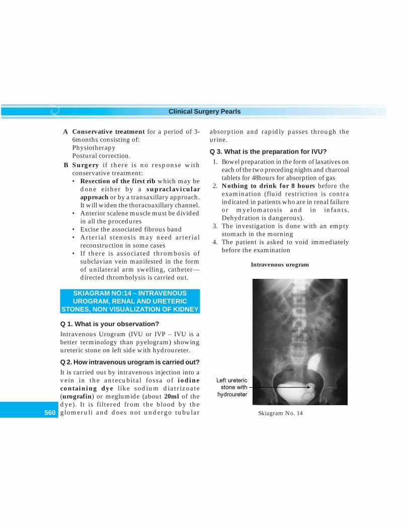
560
Clinical Surgery Pearls
A Conservative treatment for a period of 3-6months consisting of:PhysiotherapyPostural correction.
B Surgery if there is no response withconservative treatment:• Resection of the first rib which may be
done either by a supraclavicularapproach or by a transaxillary approach.It will widen the thoracoaxillary channel.
• Anterior scalene muscle must be dividedin all the procedures
• Excise the associated fibrous band• Arterial stenosis may need arterial
reconstruction in some cases• If there is associated thrombosis of
subclavian vein manifested in the formof unilateral arm swelling, catheter—directed thrombolysis is carried out.
SKIAGRAM NO:14 – INTRAVENOUSUROGRAM, RENAL AND URETERIC
STONES, NON VISUALIZATION OF KIDNEY
Q 1. What is your observation?
Intravenous Urogram (IVU or IVP – IVU is abetter terminology than pyelogram) showingureteric stone on left side with hydroureter.
Q 2. How intravenous urogram is carried out?
It is carried out by intravenous injection into avein in the antecubital fossa of iodinecontaining dye like sodium diatrizoate(urografin) or meglumide (about 20ml of thedye). It is filtered from the blood by theglomeruli and does not undergo tubular
absorption and rapidly passes through theurine.
Q 3. What is the preparation for IVU?
1. Bowel preparation in the form of laxatives oneach of the two preceding nights and charcoaltablets for 48hours for absorption of gas
2. Nothing to drink for 8 hours before theexamination (fluid restriction is contraindicated in patients who are in renal failureor myelomatosis and in infants.Dehydration is dangerous).
3. The investigation is done with an emptystomach in the morning
4. The patient is asked to void immediatelybefore the examination
Skiagram No. 14
Intravenous urogram
561
Radiology Questions and Answers
5. Diuretics are not given prior to theexamination (the dye as such has gotdiuretic effect).
Q 4. What are the contraindications for IVU?
The contraindications are:1. Those who are allergic to iodine, patients
with atopy and eczema2. If the blood urea is more than 60ml/100ml3. Impaired renal function4. Anuria5. Infants6. Pregnant women.
Q 5. What are the types of urogram?
1. Intravenous urogram2. Retrograde pyelography (the contrast is
injected through ureteric catheter aftercystoscopy
3. Antegrade pyelography (the contrast isinjected to the pelvis).
Q 6. How many films are taken?
1. Scout film (the plain X-ray abdomen takenbefore injection of the dye).
2. Nephrogram picture at 2minutes, when thedye is being filtered by the glomerulus –shows the renal parenchyma opacified bycontrast medium. A delayed nephrogramon one side indicates functionalimpairment.
3. Serial films thereafter at 5, 10, 15, 20 and30minutes after the injection of the dye (thecontrast is excreted into the collecting systemopacifying the renal pelvis and calyces).
4. Another film at 1 hour followed by anotherat 2 hours.
5. The patient is asked to void urine and a postmicturition film is taken to show the detailsof the bladder area.
6. If there is poor functioning of kidney, filmsare taken after 8-hours.
Q 7. What are the indications for IVU?
With the introduction of CT scan the indicationsare becoming less and less.
The indications are:1. To demonstrate stones within the urinary tract
which are not visualized in ultrasonography.2. To demonstrate tumors which will distort
the pelvicalyceal system.3. Hydronephrosis.4. To show details of abnormal anatomy
(horse - shoe kidney, bifid pelvis, bifidureter and ectopic kidney).
5. In cases for trauma for extravasation of thedye and nonvisualization of the kidney.
6. To demonstrate tumor infiltration fromelsewhere of the kidney, ureter and bladderareas.
Q 8. If pelvicalyceal system on one side isnot visualized what is the inference?
• That side the kidney is not functioning.• If there is delay of appearance of the dye on
one side the inference is there is poorfunctioning on that side.
Q 9. What are the causes for nonvisualizationof the kidney?
When 90-95% of the parenchymal function islost the kidneys are nonvisualized. In such casesradioisotope study using DTPA labelled withtechnetium 99m can be used.
Published by dr-notes.com
562
Clinical Surgery Pearls
1. Gross hydronephrosis with no function2. Tumors3. Trauma4. Tuberculosis5. Congenital absence of one kidney.
Q 10. Can you visualize the entire length ofureter in a single film?
No. Because of the ureteric peristalsis, the entireureter may not be visualized (The entire ureteris visualized only when the ureter is diseasedas in the case of tuberculosis).
Q 11. What are the changes of hydronephrosisin IVU?
Calyceal changes: Normally the calyces are cup-shaped. In hydronephrosis the followingchanges are seen.• Loss of cupping• Flattening and• Clubbing of the calyces.
Q 12. What is the appearance of the pelvi-calyceal system in tumor?
Spider leg deformity of the pelvicalyceal system(distortion).
Q 13. What is double dose IVU?
When 2 ampoules (40ml) of the dye is used fordelineation of the nonfunctioning side, it iscalled double dose IVU.
Q 14. What is infusion pyelogram?
When six ampoules (120ml) are put in a bottleand given as infusion, it is called infusionpyelogram.
Q 15. How many percentage of the renalstones are radiopaque?
85-90%. Owing to the calcium content.
Q 16. What is staghorn calculus?
It is a calculus filling the entire pelvicalycealsystem consisting of triple phosphate or struvite(calcium, ammonium and magnesiumphosphate). It is smooth and dirty white in color,seen to grow in alkaline urine in the presenceof urea splitting proteus organism. They areradiopaque.
Q 17. What are the types of renal stones?
1. Oxalate calculus2. Phosphate (triple phosphate)3. Uric acid and urate calculi4. Cystine calculus5. Xanthine calculus.
Q 18. What is the treatment of staghorncalculus?
1. PCNL (Percutaneous Nephrolithotomy)2. ESWL (Extracorporeal Shock Wave
Lithotripsy)3. Silent Staghorn Calculus in the elderly is
treated conservatively.
Q 19. Which side is operated first in cases ofbilateral renal stones?
Kidney with better function is treated firstunless the other kidney is more painful orinfected.
Q 20. What is the treatment of uretericstones?
1. Conservative: Calculi smaller than 0.5 cmpass spontaneously (larger stones, impactedstones and infection in the upper urinarytract are indications for intervention)
563
Radiology Questions and Answers
2. Ureteroscopic stone removal (URS):Ureteroscope is a long, thin endoscopepassed transurethrally across the bladderinto the ureter for removal of the impactedstones.
3. Push bang: Stone in the middle or upperpart of the ureter is flushed back into thekidney using a ureteric catheter and treatedwith ESWL later.
4. Open surgery: Ureterolithotomy.
SKIAGRAM NO:15 – HYDRONEPHROSIS
Q 1. What is your observation?
Intravenous urogram showing dilatation of thecalyces and delayed excretion of dye on rightside suggestive of hydronephrosis.
Q 2. What are the changes suggestive ofhydronephrosis?
See Question 11 of Skiagram No:14.
Q 3. What is hydronephrosis?
Aseptic dilatation of pelvicalyceal system dueto partial or intermittent obstruction. Whenthere is infection it is called pyonephrosis.Complete and continuous obstruction may notproduce hydronephrosis.
Q 4. What is Dietl’s crisis?
It is nothing but intermittent hydronephrosis.The patient will present with loin mass and renalpain. After a few hours the pain is relieved andthe swelling disappears following the passageof large volume of urine.
Q 5. What are the causes for hydronephrosis?
Hydronephrosis may be unilateral or bilateral.The most common cause for unilateralhydronephrosis are idiopathic pelvi – uretericjunction obstruction and calculus.
Causes for hydronephrosis
Causes for unilateral hydronephrosis Causes for bilateral hydronephrosisCauses inside the lumen Congenital• Stones • Posterior urethral valve• Sloughed papillae • Urethral atresiaCauses in the wall Physiological• Idiopathic pelviureteric junction obstruction • Pregnancy• Ureterocele Acquired• Stricture of the ureter • Benign prostatic enlargement• Tumors of the ureter • Carcinoma of the prostate• Bladder cancer involving ureteric orifice • Bladder neck obstructionCauses outside the wall • Urethral stricture• Carcinoma of the colon, cecum, rectum, uterus, • Phimosis
prostate, etc.• Retroperitoneal sarcomas• Retroperitoneal fibrosis
Published by dr-notes.com
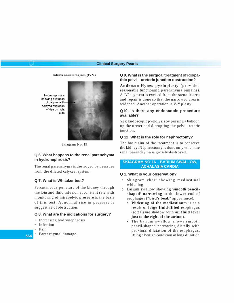
564
Clinical Surgery Pearls
Q 6. What happens to the renal parenchymain hydronephrosis?
The renal parenchyma is destroyed by pressurefrom the dilated calyceal system.
Q 7. What is Whitaker test?
Percutaneous puncture of the kidney throughthe loin and fluid infusion at constant rate withmonitoring of intrapelvic pressure is the basisof this test. Abnormal rise in pressure issuggestive of obstruction.
Q 8. What are the indications for surgery?
• Increasing hydronephrosis• Infection• Pain• Parenchymal damage.
Q 9. What is the surgical treatment of idiopa-thic pelvi – ureteric junction obstruction?
Anderson-Hynes pyeloplasty (providedreasonable functioning parenchyma remains).A ‘V’ segment is excised from the stenotic areaand repair is done so that the narrowed area iswidened. Another operation is V-Y plasty.
Q10. Is there any endoscopic procedureavailable?
Yes: Endoscopic pyelolysis by passing a balloonup the ureter and disrupting the pelvi uretericjunction.
Q 12. What is the role for nephrectomy?
The basic aim of the treatment is to conservethe kidney. Nephrectomy is done only when therenal parenchyma is grossly destroyed.
SKIAGRAM NO:16 – BARIUM SWALLOW,ACHALASIA CARDIA
Q 1. What is your observation?
a. Skiagram chest showing mediastinalwidening
b. Barium swallow showing ‘smooth pencil-shaped’ narrowing at the lower end ofesophagus (“bird’s beak” appearance).• Widening of the mediastinum is as a
result of large fluid-filled esophagus(soft tissue shadow with air fluid leveljust to the right of the atrium).
• The barium swallow shows smoothpencil-shaped narrowing distally withproximal dilatation of the esophagus.Being a benign condition of long duration
Skiagram No. 15
Intravenous urogram (IVV)
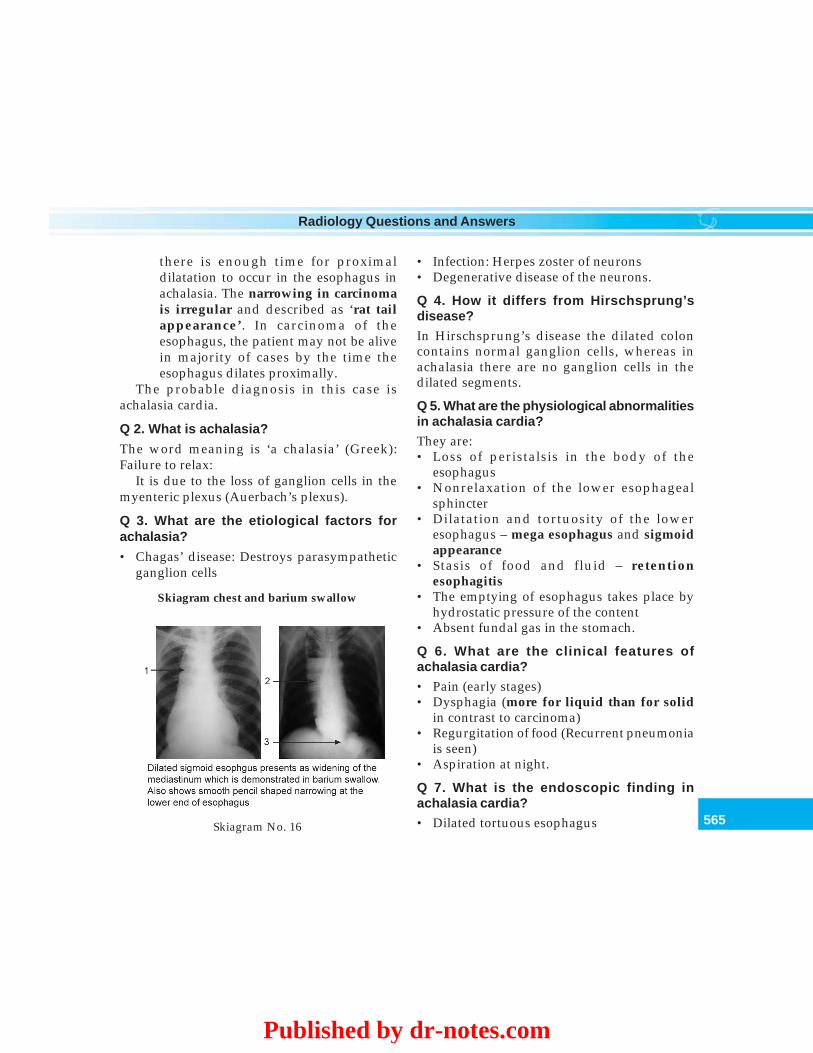
565
Radiology Questions and Answers
there is enough time for proximaldilatation to occur in the esophagus inachalasia. The narrowing in carcinomais irregular and described as ‘rat tailappearance’. In carcinoma of theesophagus, the patient may not be alivein majority of cases by the time theesophagus dilates proximally.
The probable diagnosis in this case isachalasia cardia.
Q 2. What is achalasia?
The word meaning is ‘a chalasia’ (Greek):Failure to relax:
It is due to the loss of ganglion cells in themyenteric plexus (Auerbach’s plexus).
Q 3. What are the etiological factors forachalasia?
• Chagas’ disease: Destroys parasympatheticganglion cells
• Infection: Herpes zoster of neurons• Degenerative disease of the neurons.
Q 4. How it differs from Hirschsprung’sdisease?In Hirschsprung’s disease the dilated coloncontains normal ganglion cells, whereas inachalasia there are no ganglion cells in thedilated segments.
Q 5. What are the physiological abnormalitiesin achalasia cardia?They are:• Loss of peristalsis in the body of the
esophagus• Nonrelaxation of the lower esophageal
sphincter• Dilatation and tortuosity of the lower
esophagus – mega esophagus and sigmoidappearance
• Stasis of food and fluid – retentionesophagitis
• The emptying of esophagus takes place byhydrostatic pressure of the content
• Absent fundal gas in the stomach.
Q 6. What are the clinical features ofachalasia cardia?• Pain (early stages)• Dysphagia (more for liquid than for solid
in contrast to carcinoma)• Regurgitation of food (Recurrent pneumonia
is seen)• Aspiration at night.
Q 7. What is the endoscopic finding inachalasia cardia?• Dilated tortuous esophagusSkiagram No. 16
Skiagram chest and barium swallow
Published by dr-notes.com
566
Clinical Surgery Pearls
• Food residue and fluid in the esophagus• Tight cardia• Evidence of esophagitis.
Q 8. What is the confirmatory test forachalasia cardia?
Esophageal manometry: Raised resting pressurein the esophagus.
Q 9. What is pseudoachalasia?
• This is produced by adenocarcinoma andbenign tumors of the esophagus
• Cancers outside of the esophagus, e.g.bronchus can also produce pseudoachalasia.
Q 10. What are the treatment options inachalasia cardia?
• Pneumatic dilatation• Heller’s myotomy• Injection of botulinum toxin into the lower
esophageal sphincter with temporary effectfor a few months (has to be repeated)
• Drugs will give only transient reliefSublingual NifedipineCalcium Channel Antagonists.
Q 11. What is pneumatic dilatation?
• The dilatation of the cardia was originallydescribed by Plummer using Plummer’shydrostatic bag.
• This is now replaced by pneumatic dilatationusing plastic balloons of sizes 30-40 mmdiameter which are introduced over a guidewire.
• Progressive dilatations are carried out over aperiod of weeks.
• Perforation is the most important complica-tions.
Q 12. What is Heller’s myotomy?
This can be carried out either by open methodor laparoscopic method. In this procedure alongitudinal incision is made in the anterioraspect of the lower esophagus and cardia todivide the muscles of the narrow segmentwithout injuring the mucosa so that the mucosawill pout.
Q 13. What is the most important complica-tion of Heller’s myotomy?
Gastroesophageal Reflux.Most surgeons therefore perform a partial
anterior fundoplication. This is called Heller –Dor’s operation .
SKIAGRAM NO:17 – BARIUM SWALLOW,CARCINOMA OF THE ESOPHAGUS
Q 1. What is your observation?
Barium swallow picture showing narrowingat the lower end of the esophagus havingthe typical ‘rat tail appearance’ suggestiveof ‘carcinoma of the lower end ofesophagus’.
Q 2. What are the pathological types ofcarcinoma of the esophagus?
• Squamous cell carcinoma – affects the uppertwo – thirds (most common type)
• Adenocarcinoma – affects the lower third.
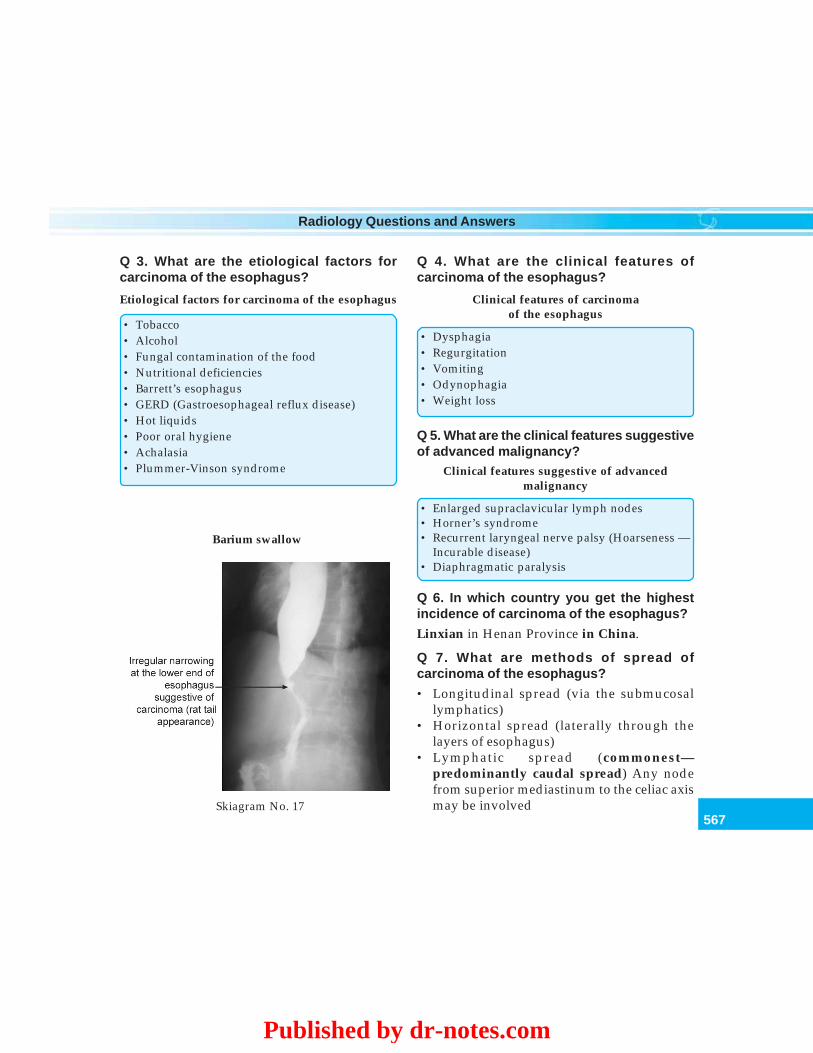
567
Radiology Questions and Answers
Q 3. What are the etiological factors forcarcinoma of the esophagus?
Etiological factors for carcinoma of the esophagus
• Tobacco• Alcohol• Fungal contamination of the food• Nutritional deficiencies• Barrett’s esophagus• GERD (Gastroesophageal reflux disease)• Hot liquids• Poor oral hygiene• Achalasia• Plummer-Vinson syndrome
Q 4. What are the clinical features ofcarcinoma of the esophagus?
Clinical features of carcinomaof the esophagus
• Dysphagia• Regurgitation• Vomiting• Odynophagia• Weight loss
Q 5. What are the clinical features suggestiveof advanced malignancy?
Clinical features suggestive of advancedmalignancy
• Enlarged supraclavicular lymph nodes• Horner’s syndrome• Recurrent laryngeal nerve palsy (Hoarseness —
Incurable disease)• Diaphragmatic paralysis
Q 6. In which country you get the highestincidence of carcinoma of the esophagus?
Linxian in Henan Province in China.
Q 7. What are methods of spread ofcarcinoma of the esophagus?
• Longitudinal spread (via the submucosallymphatics)
• Horizontal spread (laterally through thelayers of esophagus)
• Lymphatic spread (commonest—predominantly caudal spread) Any nodefrom superior mediastinum to the celiac axismay be involvedSkiagram No. 17
Barium swallow
Published by dr-notes.com

568
Clinical Surgery Pearls
• Blood stream spread (liver, lungs, brain, bones)• Transperitoneal – for the intraabdominal part
of the esophagus.
Q 8. What is the significance of supra-clavicular lymph nodes in carcinoma of theesophagus?
Supraclavicular lymph nodes are suggestive ofmetastasis (M1). So also celiac node involvementin distal esophageal tumor.
Q 9. What are the most important investiga-tions in carcinoma of the esophagus?
1. Endoscopy and biopsy for confirmation ofthe diagnosis.
Staging investigations2. Endoscopic ultrasound is the most
important staging investigations—for depthof tumor, invasion of the adjacent organsand for metastasis to lymph nodes
3. Chest radiography for pulmonary metastasis4. Liver function tests (LFTs)—Abnormal
LFTs are suggestive of liver metastasis.Normal LFT does not rule out livermetastasis.
5. Ultrasound abdomen—to rule out livermetastasis.
6. Bronchoscopy (for middle and upperthirds).
7. Laparoscopy—for carcinoma arising fromthe abdominal esophagus-abdominal andhepatic metastasis
8. Spiral CT—for local spread and lymphnode assessment (also for assessment ofmetastasis).
9. MRI – Magnetic Resonance Imaging10. PET (Positron Emission Tomography) –
combined with CT.
Q 10. What are the most important badprognostic factors in carcinoma of theesophagus?
• Depth of tumor penetration• Regional lymph node involvement.
Q 11. What is the management of carcinomaof the esophagus?
1. If the patient is unfit for surgery or metastasisor adjacent organ invasion or peritonealspread—palliation for dysphagia, e.g.chemo radiotherapy and endoscopicpalliation.
2. If fit for surgery and there is no lymph nodemetastasis – surgery alone (radicaloesophagectomy).
3. If lymph node metastasis is presentmultimodal therapy – neoadjuvanttreatment, chemo radiotherapy.• Radical esophagectomy for curative
treatment with 10 cm clearance abovethe macroscopic tumor and 5 cm distally.
• Adenocarcinoma involving gastric cardianeed some degree of gastric resection.
Q 12. How many percentage of the carcinomaof the esophagus are operable?
Above two-thirds of the tumors at the time ofdiagnosis are inoperable and only one-third isoperable.
569
Radiology Questions and Answers
Q 13. What are the most important factors tobe considered before deciding surgicaloption if it is operable?
1. General condition of the patient2. Tumor location3. Endoscopic appearance4. Nodal status.
Q 14. What is the incision of choice for radicalsurgery?
• For lower third growth: Left thoracoabdominal incision for carcinoma of thelower end of the esophagus below the aorticarch (esophagogastrectomy – part of theupper stomach is removed followed byesophago gastric anastomosis).
• For middle third growth: If the tumour isabove the level of aortic arch, the two phaseIvor Lewis operation along with two fieldlymphadenectomy (abdominal andmediastinal nodes) by an initial laparotomyfor construction of a gastric tube followed bya right thoracotomy for resection of the tumorand esophagogastric anastomosis.
• For upper third growth: McKeown operationfor carcinoma of the upper thoracic eso-phagus: It is a three phase operation withthree field lymphadenectomy: Here a thirdincision is put in the neck for removal of thenodes there and creating the cervicalanastomosis.
Q 15. What are the viscera used for inter-position as a substitute for esophagus?
They are placed in the substernal space and thefollowing viscera are used.
• Stomach (commonly used)• Jejunum• Colon.
Q 16. What is the blood supply of thetransposed stomach into the chest cavity?
Right gastroepiploic and right gastric vessels.
Q 17. What are the complications of surgicaltreatment?
Complications of surgical treatment
• Anastomotic leakage• Respiratory complications• Recurrent laryngeal nerve injury• Chylothorax• Reflux: It can be avoided by subtotal eso-
phagectomy and making the esophagogastricanastomosis high up in the chest
Q 18. What is transhiatal esophagectomy?
• This was devised by Orringer for the removalof Chagasic mega esophagus
• This is a useful procedure for the lesions ofthe lower esophagus, but dangerous for themiddle third lesion
• The stomach is mobilized through anabdominal incision
• The cervical esophagus is mobilized througha cervical incision
• The diaphragm is opened from the abdomenand the posterior mediastinum entered
• The tumor and lower esophagus aremobilized under vision
• The upper esophagus is mobilized by bluntdissection
• Total esophagectomy is done and esophago-jejunostomy is carried out in the neck.
Published by dr-notes.com
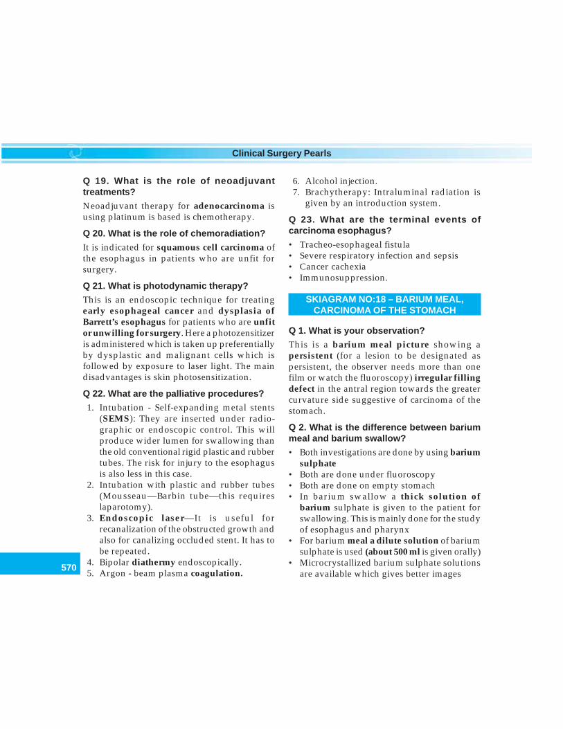
570
Clinical Surgery Pearls
Q 19. What is the role of neoadjuvanttreatments?
Neoadjuvant therapy for adenocarcinoma isusing platinum is based is chemotherapy.
Q 20. What is the role of chemoradiation?
It is indicated for squamous cell carcinoma ofthe esophagus in patients who are unfit forsurgery.
Q 21. What is photodynamic therapy?
This is an endoscopic technique for treatingearly esophageal cancer and dysplasia ofBarrett’s esophagus for patients who are unfitor unwilling for surgery. Here a photozensitizeris administered which is taken up preferentiallyby dysplastic and malignant cells which isfollowed by exposure to laser light. The maindisadvantages is skin photosensitization.
Q 22. What are the palliative procedures?
1. Intubation - Self-expanding metal stents(SEMS): They are inserted under radio-graphic or endoscopic control. This willproduce wider lumen for swallowing thanthe old conventional rigid plastic and rubbertubes. The risk for injury to the esophagusis also less in this case.
2. Intubation with plastic and rubber tubes(Mousseau—Barbin tube—this requireslaparotomy).
3. Endoscopic laser—It is useful forrecanalization of the obstructed growth andalso for canalizing occluded stent. It has tobe repeated.
4. Bipolar diathermy endoscopically.5. Argon - beam plasma coagulation.
6. Alcohol injection.7. Brachytherapy: Intraluminal radiation is
given by an introduction system.
Q 23. What are the terminal events ofcarcinoma esophagus?
• Tracheo-esophageal fistula• Severe respiratory infection and sepsis• Cancer cachexia• Immunosuppression.
SKIAGRAM NO:18 – BARIUM MEAL,CARCINOMA OF THE STOMACH
Q 1. What is your observation?
This is a barium meal picture showing apersistent (for a lesion to be designated aspersistent, the observer needs more than onefilm or watch the fluoroscopy) irregular fillingdefect in the antral region towards the greatercurvature side suggestive of carcinoma of thestomach.
Q 2. What is the difference between bariummeal and barium swallow?
• Both investigations are done by using bariumsulphate
• Both are done under fluoroscopy• Both are done on empty stomach• In barium swallow a thick solution of
barium sulphate is given to the patient forswallowing. This is mainly done for the studyof esophagus and pharynx
• For barium meal a dilute solution of bariumsulphate is used (about 500 ml is given orally)
• Microcrystallized barium sulphate solutionsare available which gives better images
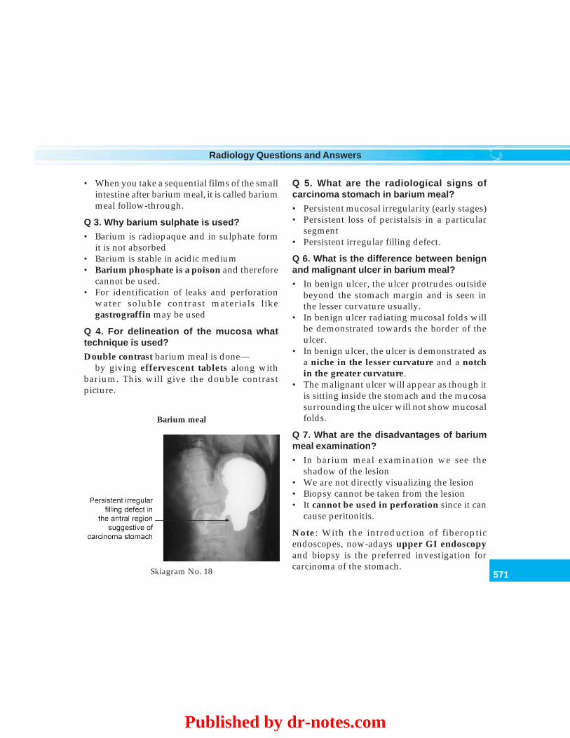
571
Radiology Questions and Answers
• When you take a sequential films of the smallintestine after barium meal, it is called bariummeal follow-through.
Q 3. Why barium sulphate is used?
• Barium is radiopaque and in sulphate formit is not absorbed
• Barium is stable in acidic medium• Barium phosphate is a poison and therefore
cannot be used.• For identification of leaks and perforation
water soluble contrast materials likegastrograffin may be used
Q 4. For delineation of the mucosa whattechnique is used?
Double contrast barium meal is done—by giving effervescent tablets along with
barium. This will give the double contrastpicture.
Q 5. What are the radiological signs ofcarcinoma stomach in barium meal?
• Persistent mucosal irregularity (early stages)• Persistent loss of peristalsis in a particular
segment• Persistent irregular filling defect.
Q 6. What is the difference between benignand malignant ulcer in barium meal?
• In benign ulcer, the ulcer protrudes outsidebeyond the stomach margin and is seen inthe lesser curvature usually.
• In benign ulcer radiating mucosal folds willbe demonstrated towards the border of theulcer.
• In benign ulcer, the ulcer is demonstrated asa niche in the lesser curvature and a notchin the greater curvature.
• The malignant ulcer will appear as though itis sitting inside the stomach and the mucosasurrounding the ulcer will not show mucosalfolds.
Q 7. What are the disadvantages of bariummeal examination?
• In barium meal examination we see theshadow of the lesion
• We are not directly visualizing the lesion• Biopsy cannot be taken from the lesion• It cannot be used in perforation since it can
cause peritonitis.
Note: With the introduction of fiberopticendoscopes, now-adays upper GI endoscopyand biopsy is the preferred investigation forcarcinoma of the stomach.Skiagram No. 18
Barium meal
Published by dr-notes.com
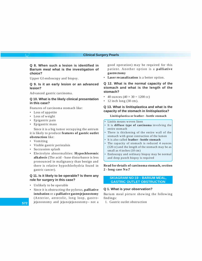
572
Clinical Surgery Pearls
Q 8. When such a lesion is identified inBarium meal what is the investigation ofchoice?
Upper GI endoscopy and biopsy.
Q 9. Is it an early lesion or an advancedlesion?
Advanced gastric carcinoma.
Q 10. What is the likely clinical presentationin this case?
Features of carcinoma stomach like:• Loss of appetite• Loss of weight• Epigastric pain• Epigastric mass
Since it is a big tumor occupying the antrumit is likely to produce features of gastric outletobstruction like:• Vomiting• Visible gastric peristalsis• Succussion splash• Electrolyte abnormalities: Hypochloremic
alkalosis (The acid - base disturbance is lesspronounced in malignancy than benign andthere is relative hypochlorhydria found ingastric cancer).
Q 11. Is it likely to be operable? Is there anyrole for surgery in this case?
• Unlikely to be operable• Since it is obstructing the pylorus, palliative
intubation or a palliative gastrojejunostomy(Anterior, antecolic, long loop, gastro-jejunostomy and jejunojejunostomy– not a
good operation) may be required for thispatient. Another option is a palliativegastrectomy
• Laser recanalization is a better option.
Q 12. What is the normal capacity of thestomach and what is the length of thestomach?
• 40 ounces (40 × 30 = 1200 cc)• 12 inch long (30 cm).
Q 13. What is linitisplastica and what is thecapacity of the stomach in linitisplastica?
Linitisplastica or leather - bottle stomach
• Linitis means woven linen• It is diffuse type of carcinoma involving the
entire stomach• There is thickening of the entire wall of the
stomach with great contraction of the lumen• It is also called leather - bottle stomach• The capacity of stomach is reduced 4 ounces
(120 cc) and the length of the stomach may be assmall as 4 inches (10 cm)
• Endoscopy and ordinary biopsy may be normaland deep punch biopsy is required
Read for details of carcinoma stomach, section2 - long case No:7
SKIAGRAM NO:19 – BARIUM MEAL,GASTRIC OUTLET OBSTRUCTION
Q 1. What is your observation?
Barium meal picture showing the followingfindings:1. Gastric outlet obstruction
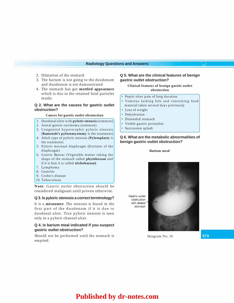
573
Radiology Questions and Answers
2. Dilatation of the stomach3. The barium is not going to the duodenum
and duodenum is not demonstrated4. The stomach has got mottled appearance
which is due to the retained food particlesinside.
Q 2. What are the causes for gastric outletobstruction?
Causes for gastric outlet obstruction
1. Duodenal ulcer with pyloric stenosis (common)2. Antral gastric carcinoma (common)3. Congenital hypertrophic pyloric stenosis
(Ramstedt’s pyloromyotomy is the treatment)4. Adult type of pyloric stenosis (Pyloroplasty is
the treatment)5. Pyloric mucosal diaphragm (Excision of the
diaphragm)6. Gastric Bezoar (Vegetable matter taking the
shape of the stomach called phytobezoar andif it is hair it is called trichobazoar)
7. Lymphoma8. Gastritis9. Crohn’s disease10. Tuberculosis
Note: Gastric outlet obstruction should beconsidered malignant until proven otherwise.
Q 3. Is pyloric stenosis a correct terminology?
It is a misnomer. The stenosis is found in thefirst part of the duodenum if it is due toduodenal ulcer. True pyloric stenosis is seenonly in a pyloric channel ulcer.
Q 4. Is barium meal indicated if you suspectgastric outlet obstruction?
Should not be performed until the stomach isemptied.
Q 5. What are the clinical features of benigngastric outlet obstruction?
Clinical features of benign gastric outletobstruction
• Peptic ulcer pain of long duration• Vomitus lacking bile and containing food
material taken several days previously• Loss of weight• Dehydration• Distended stomach• Visible gastric peristalsis• Succussion splash
Q 6. What are the metabolic abnormalities ofbenign gastric outlet obstruction?
Skiagram No. 19
Barium meal
Published by dr-notes.com
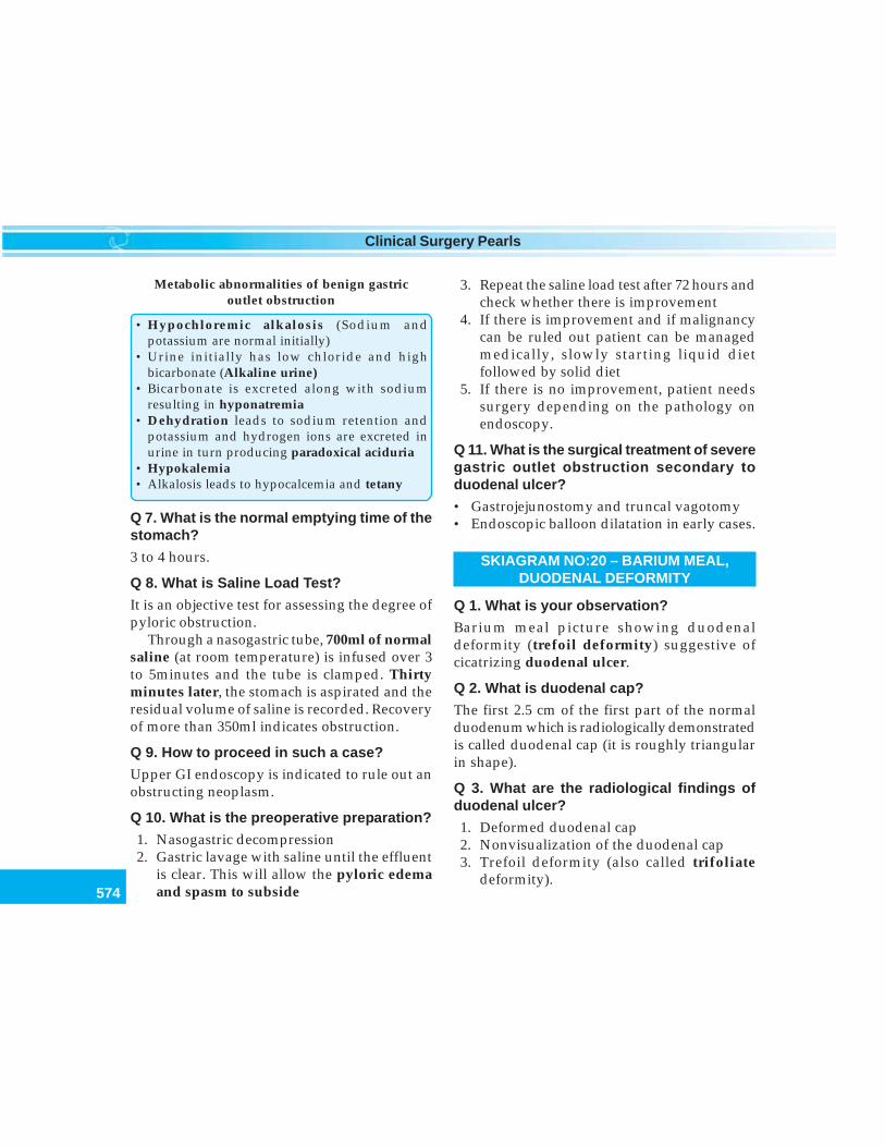
574
Clinical Surgery Pearls
Metabolic abnormalities of benign gastricoutlet obstruction
• Hypochloremic alkalosis (Sodium andpotassium are normal initially)
• Urine initially has low chloride and highbicarbonate (Alkaline urine)
• Bicarbonate is excreted along with sodiumresulting in hyponatremia
• Dehydration leads to sodium retention andpotassium and hydrogen ions are excreted inurine in turn producing paradoxical aciduria
• Hypokalemia• Alkalosis leads to hypocalcemia and tetany
Q 7. What is the normal emptying time of thestomach?
3 to 4 hours.
Q 8. What is Saline Load Test?
It is an objective test for assessing the degree ofpyloric obstruction.
Through a nasogastric tube, 700ml of normalsaline (at room temperature) is infused over 3to 5minutes and the tube is clamped. Thirtyminutes later, the stomach is aspirated and theresidual volume of saline is recorded. Recoveryof more than 350ml indicates obstruction.
Q 9. How to proceed in such a case?
Upper GI endoscopy is indicated to rule out anobstructing neoplasm.
Q 10. What is the preoperative preparation?
1. Nasogastric decompression2. Gastric lavage with saline until the effluent
is clear. This will allow the pyloric edemaand spasm to subside
3. Repeat the saline load test after 72 hours andcheck whether there is improvement
4. If there is improvement and if malignancycan be ruled out patient can be managedmedically, slowly starting liquid dietfollowed by solid diet
5. If there is no improvement, patient needssurgery depending on the pathology onendoscopy.
Q 11. What is the surgical treatment of severegastric outlet obstruction secondary toduodenal ulcer?
• Gastrojejunostomy and truncal vagotomy• Endoscopic balloon dilatation in early cases.
SKIAGRAM NO:20 – BARIUM MEAL,DUODENAL DEFORMITY
Q 1. What is your observation?
Barium meal picture showing duodenaldeformity (trefoil deformity) suggestive ofcicatrizing duodenal ulcer.
Q 2. What is duodenal cap?
The first 2.5 cm of the first part of the normalduodenum which is radiologically demonstratedis called duodenal cap (it is roughly triangularin shape).
Q 3. What are the radiological findings ofduodenal ulcer?
1. Deformed duodenal cap2. Nonvisualization of the duodenal cap3. Trefoil deformity (also called trifoliate
deformity).
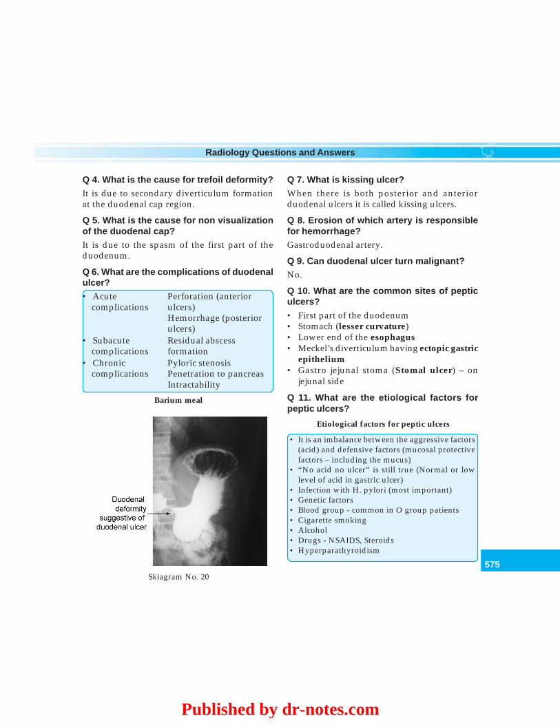
575
Radiology Questions and Answers
Q 4. What is the cause for trefoil deformity?
It is due to secondary diverticulum formationat the duodenal cap region.
Q 5. What is the cause for non visualizationof the duodenal cap?It is due to the spasm of the first part of theduodenum.
Q 6. What are the complications of duodenalulcer?• Acute Perforation (anterior
complications ulcers)Hemorrhage (posteriorulcers)
• Subacute Residual abscesscomplications formation
• Chronic Pyloric stenosiscomplications Penetration to pancreas
Intractability
Q 7. What is kissing ulcer?
When there is both posterior and anteriorduodenal ulcers it is called kissing ulcers.
Q 8. Erosion of which artery is responsiblefor hemorrhage?Gastroduodenal artery.
Q 9. Can duodenal ulcer turn malignant?
No.
Q 10. What are the common sites of pepticulcers?
• First part of the duodenum• Stomach (lesser curvature)• Lower end of the esophagus• Meckel’s diverticulum having ectopic gastric
epithelium• Gastro jejunal stoma (Stomal ulcer) – on
jejunal side
Q 11. What are the etiological factors forpeptic ulcers?
Etiological factors for peptic ulcers
• It is an imbalance between the aggressive factors(acid) and defensive factors (mucosal protectivefactors – including the mucus)
• “No acid no ulcer” is still true (Normal or lowlevel of acid in gastric ulcer)
• Infection with H. pylori (most important)• Genetic factors• Blood group - common in O group patients• Cigarette smoking• Alcohol• Drugs - NSAIDS, Steroids• Hyperparathyroidism
Skiagram No. 20
Barium meal
Published by dr-notes.com
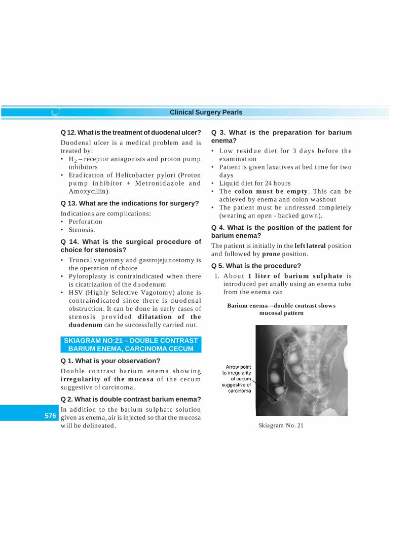
576
Clinical Surgery Pearls
Q 12. What is the treatment of duodenal ulcer?
Duodenal ulcer is a medical problem and istreated by:• H2 – receptor antagonists and proton pump
inhibitors• Eradication of Helicobacter pylori (Proton
pump inhibitor + Metronidazole andAmoxycillin).
Q 13. What are the indications for surgery?
Indications are complications:• Perforation• Stenosis.
Q 14. What is the surgical procedure ofchoice for stenosis?
• Truncal vagotomy and gastrojejunostomy isthe operation of choice
• Pyloroplasty is contraindicated when thereis cicatrization of the duodenum
• HSV (Highly Selective Vagotomy) alone iscontraindicated since there is duodenalobstruction. It can be done in early cases ofstenosis provided dilatation of theduodenum can be successfully carried out.
SKIAGRAM NO:21 – DOUBLE CONTRASTBARIUM ENEMA, CARCINOMA CECUM
Q 1. What is your observation?
Double contrast barium enema showingirregularity of the mucosa of the cecumsuggestive of carcinoma.
Q 2. What is double contrast barium enema?
In addition to the barium sulphate solutiongiven as enema, air is injected so that the mucosawill be delineated.
Q 3. What is the preparation for bariumenema?
• Low residue diet for 3 days before theexamination
• Patient is given laxatives at bed time for twodays
• Liquid diet for 24 hours• The colon must be empty. This can be
achieved by enema and colon washout• The patient must be undressed completely
(wearing an open - backed gown).
Q 4. What is the position of the patient forbarium enema?
The patient is initially in the left lateral positionand followed by prone position.
Q 5. What is the procedure?
1. About 1 liter of barium sulphate isintroduced per anally using an enema tubefrom the enema can
Skiagram No. 21
Barium enema—double contrast showsmucosal pattern
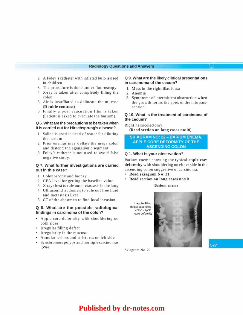
577
Radiology Questions and Answers
2. A Foley’s catheter with inflated bulb is usedin children
3. The procedure is done under fluoroscopy4. X-ray is taken after completely filling the
colon5. Air is insufflated to delineate the mucosa
(Double contrast)6. Finally a post evacuation film is taken
(Patient is asked to evacuate the barium).
Q 6. What are the precautions to be taken whenit is carried out for Hirschsprung’s disease?
1. Saline is used instead of water for dilutingthe barium
2. Prior enemas may deflate the mega colonand distend the aganglionic segment
3. Foley’s catheter is not used to avoid falsenegative study.
Q 7. What further investigations are carriedout in this case?
1. Colonoscopy and biopsy2. CEA level for getting the baseline value3. X-ray chest to rule out metastasis in the lung4. Ultrasound abdomen to rule out free fluid
and metastasis liver5. CT of the abdomen to find local invasion.
Q 8. What are the possible radiologicalfindings in carcinoma of the colon?
• Apple core deformity with shouldering onboth sides
• Irregular filling defect• Irregularity in the mucosa• Annular lesions and strictures on left side• Synchronous polyps and multiple carcinomas
(5%).
Q 9. What are the likely clinical presentationsin carcinoma of the cecum?1. Mass in the right iliac fossa2. Anemia3. Symptoms of intermittent obstruction when
the growth forms the apex of the intussus-ception.
Q 10. What is the treatment of carcinoma ofthe cecum?Right hemicolectomy.
(Read section on long cases no:10).
SKIAGRAM NO: 22 – BARIUM ENEMA,APPLE CORE DEFORMITY OF THE
ASCENDING COLON
Q 1. What is your observation?
Barium enema showing the typical apple coredeformity with shouldering on either side in theascending colon suggestive of carcinoma.• Read skiagram No: 21• Read section on long cases no:10.
Skiagram No. 22
Barium enema
Published by dr-notes.com
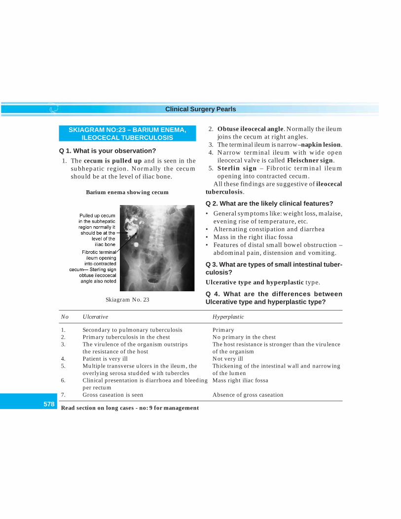
578
Clinical Surgery Pearls
SKIAGRAM NO:23 – BARIUM ENEMA,ILEOCECAL TUBERCULOSIS
Q 1. What is your observation?
1. The cecum is pulled up and is seen in thesubhepatic region. Normally the cecumshould be at the level of iliac bone.
2. Obtuse ileocecal angle. Normally the ileumjoins the cecum at right angles.
3. The terminal ileum is narrow–napkin lesion.4. Narrow terminal ileum with wide open
ileocecal valve is called Fleischner sign.5. Sterlin sign – Fibrotic terminal ileum
opening into contracted cecum.All these findings are suggestive of ileocecal
tuberculosis.
Q 2. What are the likely clinical features?
• General symptoms like: weight loss, malaise,evening rise of temperature, etc.
• Alternating constipation and diarrhea• Mass in the right iliac fossa• Features of distal small bowel obstruction –
abdominal pain, distension and vomiting.
Q 3. What are types of small intestinal tuber-culosis?
Ulcerative type and hyperplastic type.
Q 4. What are the differences betweenUlcerative type and hyperplastic type?
No Ulcerative Hyperplastic
1. Secondary to pulmonary tuberculosis Primary2. Primary tuberculosis in the chest No primary in the chest3. The virulence of the organism outstrips The host resistance is stronger than the virulence
the resistance of the host of the organism4. Patient is very ill Not very ill5. Multiple transverse ulcers in the ileum, the Thickening of the intestinal wall and narrowing
overlying serosa studded with tubercles of the lumen6. Clinical presentation is diarrhoea and bleeding Mass right iliac fossa
per rectum7. Gross caseation is seen Absence of gross caseation
Read section on long cases - no: 9 for management
Skiagram No. 23
Barium enema showing cecum
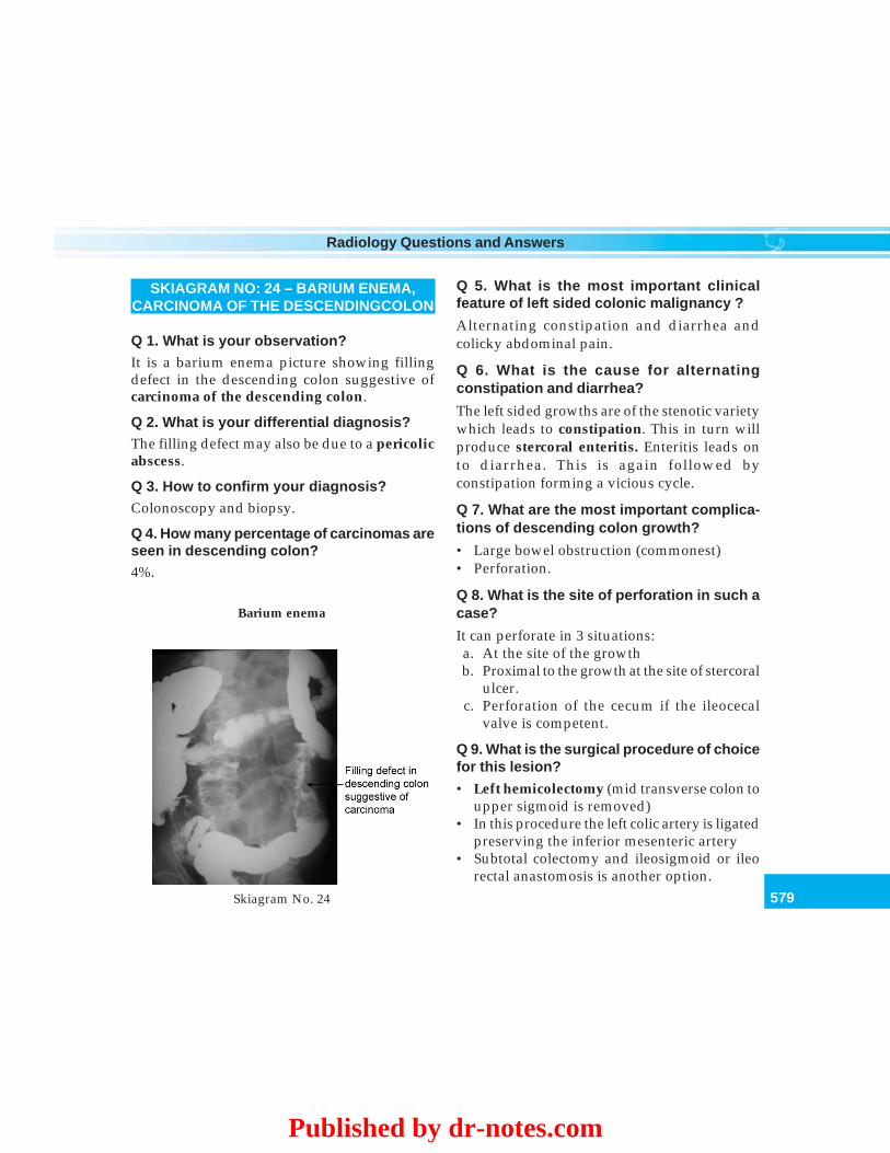
579
Radiology Questions and Answers
SKIAGRAM NO: 24 – BARIUM ENEMA,CARCINOMA OF THE DESCENDINGCOLON
Q 1. What is your observation?It is a barium enema picture showing fillingdefect in the descending colon suggestive ofcarcinoma of the descending colon.
Q 2. What is your differential diagnosis?The filling defect may also be due to a pericolicabscess.
Q 3. How to confirm your diagnosis?Colonoscopy and biopsy.
Q 4. How many percentage of carcinomas areseen in descending colon?4%.
Q 5. What is the most important clinicalfeature of left sided colonic malignancy ?
Alternating constipation and diarrhea andcolicky abdominal pain.
Q 6. What is the cause for alternatingconstipation and diarrhea?
The left sided growths are of the stenotic varietywhich leads to constipation. This in turn willproduce stercoral enteritis. Enteritis leads onto diarrhea. This is again followed byconstipation forming a vicious cycle.
Q 7. What are the most important complica-tions of descending colon growth?
• Large bowel obstruction (commonest)• Perforation.
Q 8. What is the site of perforation in such acase?
It can perforate in 3 situations:a. At the site of the growthb. Proximal to the growth at the site of stercoral
ulcer.c. Perforation of the cecum if the ileocecal
valve is competent.
Q 9. What is the surgical procedure of choicefor this lesion?
• Left hemicolectomy (mid transverse colon toupper sigmoid is removed)
• In this procedure the left colic artery is ligatedpreserving the inferior mesenteric artery
• Subtotal colectomy and ileosigmoid or ileorectal anastomosis is another option.
Skiagram No. 24
Barium enema
Published by dr-notes.com
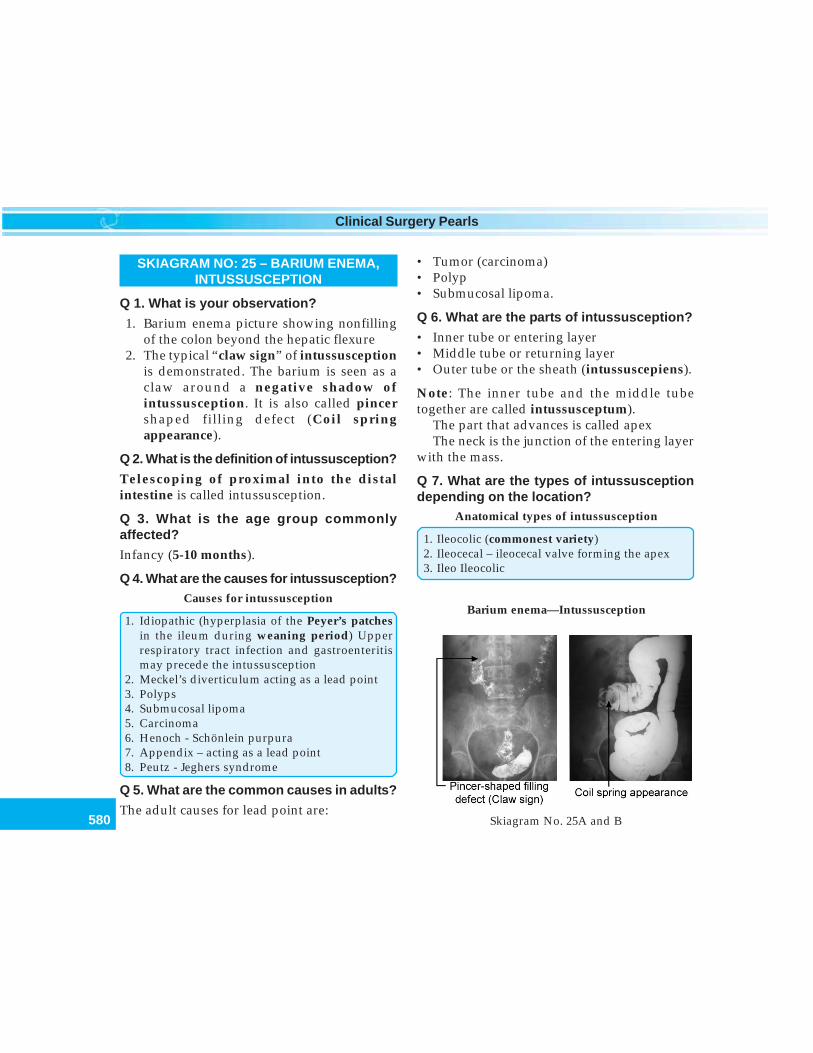
580
Clinical Surgery Pearls
SKIAGRAM NO: 25 – BARIUM ENEMA,INTUSSUSCEPTION
Q 1. What is your observation?
1. Barium enema picture showing nonfillingof the colon beyond the hepatic flexure
2. The typical “claw sign” of intussusceptionis demonstrated. The barium is seen as aclaw around a negative shadow ofintussusception. It is also called pincershaped filling defect (Coil springappearance).
Q 2. What is the definition of intussusception?
Telescoping of proximal into the distalintestine is called intussusception.
Q 3. What is the age group commonlyaffected?
Infancy (5-10 months).
Q 4. What are the causes for intussusception?Causes for intussusception
1. Idiopathic (hyperplasia of the Peyer’s patchesin the ileum during weaning period) Upperrespiratory tract infection and gastroenteritismay precede the intussusception
2. Meckel’s diverticulum acting as a lead point3. Polyps4. Submucosal lipoma5. Carcinoma6. Henoch - Schönlein purpura7. Appendix – acting as a lead point8. Peutz - Jeghers syndrome
Q 5. What are the common causes in adults?
The adult causes for lead point are:
• Tumor (carcinoma)• Polyp• Submucosal lipoma.
Q 6. What are the parts of intussusception?
• Inner tube or entering layer• Middle tube or returning layer• Outer tube or the sheath (intussuscepiens).
Note: The inner tube and the middle tubetogether are called intussusceptum).
The part that advances is called apexThe neck is the junction of the entering layer
with the mass.
Q 7. What are the types of intussusceptiondepending on the location?
Anatomical types of intussusception
1. Ileocolic (commonest variety)2. Ileocecal – ileocecal valve forming the apex3. Ileo Ileocolic
Skiagram No. 25A and B
Barium enema—Intussusception
581
Radiology Questions and Answers
4. Ileoileal5. Colocolic6. Multiple7. Jejunogastric
Q 8. What is retrograde intussusception?
Telescoping of the distal intestine into theproximal is called retrograde intussusception.The typical example is a jejunogastric intussus-ception seen after a gastrojejunostomy.
Q 9. What type of intestinal obstruction isproduced in intussusception?
Strangulating obstruction (the blood supply ofthe inner layer is usually affected).
Q 10. What are the diagnostic clinical featuresand important diagnostic signs?
Clinical features of intussusception
• Colicky abdominal pain (screaming and drawingup of the legs in infants)
• “Redcurrant jelly” stool (initially the stool maybe normal)
• Vomiting is a later event (Milk followed by bile)• Dehydration• Abdominal distension• Sausage – shaped mass with concavity towards
the umbilicus which harden on palpation(Disappearing mass)
• Sign de-dance (Emptiness of right iliac fossa)• Blood - stained mucus on rectal examination• Palpable mass per rectum (apex of the
intussusception)• The finger can be introduced between the mass
and anal verge in intussusception which is notpossible in prolapse rectum
Q 11. What is the ultrasound finding inintussusception?
Doughnut appearance of concentric rings intransverse section.
Q 12. What is the nonoperative management?
Nonoperative management consists of:• Resuscitation – IV fluids (fluid and electrolyte
correction)• Nasogastric decompression• Hydrostatic reduction – Using barium enema
or air (pneumatic reduction).
Q 13. What is successful hydrostaticreduction?
• Free reflux of barium or air into the smallbowel is suggestive of successful reduction
• Resolution of symptoms and signs.
Q 14. What are the contraindications for nonoperative management?Contraindications for nonoperative management
• Signs of peritonitis• Perforation• Shock
Q 15. What is the recurrence rate of nonoperative management?
10%.
Q 16. What are the indications for surgery?Indications for surgery
• Contraindications for nonoperative reduction• Failure of pneumatic or hydrostatic reduction• Suspected pathological lead point• Gangrene
Published by dr-notes.com
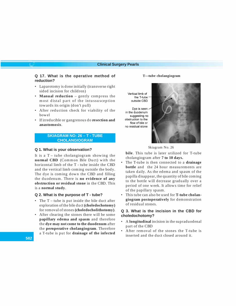
582
Clinical Surgery Pearls
Q 17. What is the operative method ofreduction?
• Laparotomy is done initially (transverse rightsided incision for children)
• Manual reduction – gently compress themost distal part of the intussusceptiontowards its origin (don’t pull)
• After reduction check for viability of thebowel
• If irreducible or gangrenous do resection andanastomosis.
SKIAGRAM NO: 26 – T - TUBECHOLANGIOGRAM
Q 1. What is your observation?
It is a T – tube cholangiogram showing thenormal CBD (Common Bile Duct) with thehorizontal limb of the T - tube inside the CBDand the vertical limb coming outside the body.The dye is coming down the CBD and fillingthe duodenum. There is no evidence of anyobstruction or residual stone in the CBD. Thisis a normal study.
Q 2. What is the purpose of T - tube?
• The T – tube is put inside the bile duct afterexploration of the bile duct (choledochotomy)for removal of stones (choledocholithotomy).
• After clearing the stones there will be somepapillary edema and spasm and thereforethe dye may not come to the duodenum afterthe peroperative cholangiogram. Thereforea T-tube is put for drainage of the infected
Skiagram No. 26
T—tube cholangiogram
bile. This tube is later utilized for T-tubecholangiogram after 7 to 10 days.
• The T-tube is then connected to a drainagebottle and the 24 hour measurements aretaken daily. As the edema and spasm of thepapilla disappear, the quantity of bile comingto the bottle will decrease gradually over aperiod of one week. It allows time for reliefof the papillary spasm.
• This tube can also be used for T-tube cholan-giogram postoperatively for demonstrationof residual stones.
Q 3. What is the incision in the CBD forcholedochotomy?
• A longitudinal incision in the supraduodenalpart of the CBD
• After removal of the stones the T-tube isinserted and the duct closed around it.
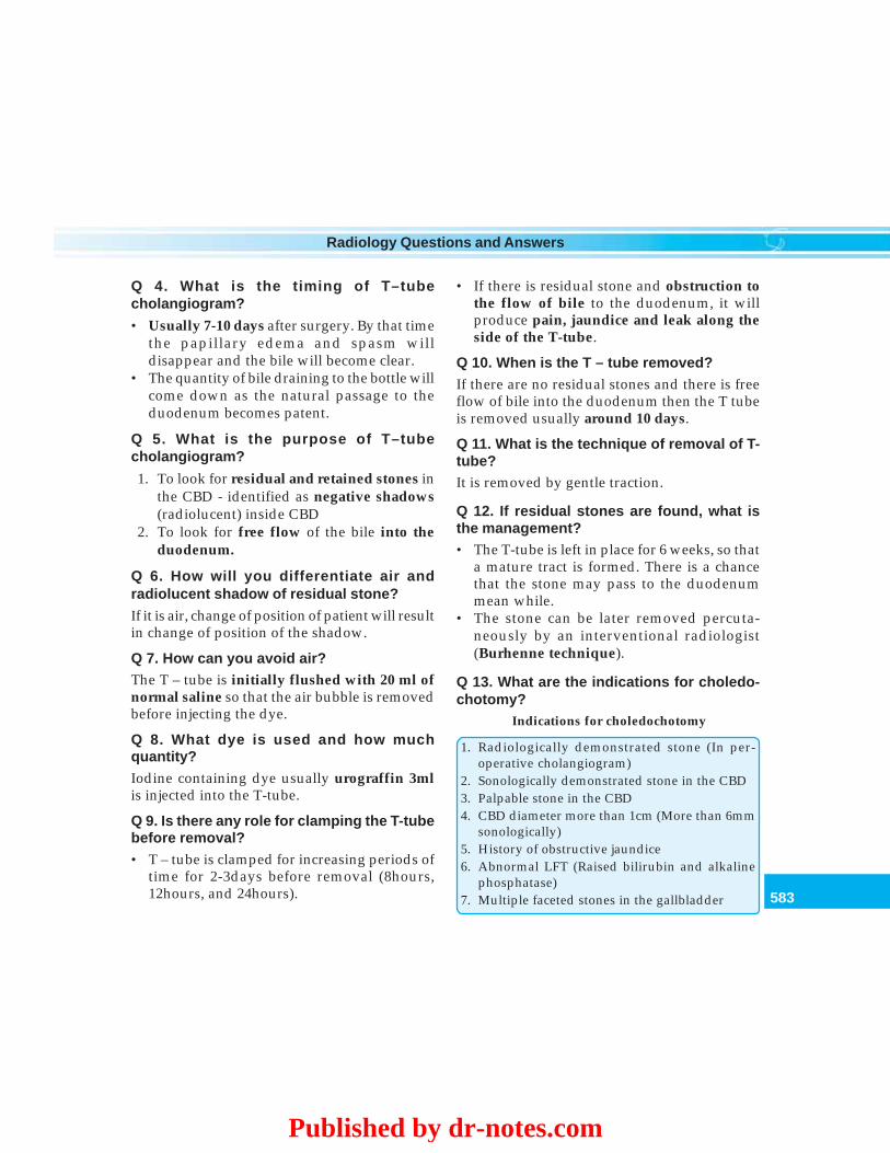
583
Radiology Questions and Answers
Q 4. What is the timing of T–tubecholangiogram?
• Usually 7-10 days after surgery. By that timethe papillary edema and spasm willdisappear and the bile will become clear.
• The quantity of bile draining to the bottle willcome down as the natural passage to theduodenum becomes patent.
Q 5. What is the purpose of T–tubecholangiogram?
1. To look for residual and retained stones inthe CBD - identified as negative shadows(radiolucent) inside CBD
2. To look for free flow of the bile into theduodenum.
Q 6. How will you differentiate air andradiolucent shadow of residual stone?
If it is air, change of position of patient will resultin change of position of the shadow.
Q 7. How can you avoid air?The T – tube is initially flushed with 20 ml ofnormal saline so that the air bubble is removedbefore injecting the dye.
Q 8. What dye is used and how muchquantity?Iodine containing dye usually urograffin 3mlis injected into the T-tube.
Q 9. Is there any role for clamping the T-tubebefore removal?• T – tube is clamped for increasing periods of
time for 2-3days before removal (8hours,12hours, and 24hours).
• If there is residual stone and obstruction tothe flow of bile to the duodenum, it willproduce pain, jaundice and leak along theside of the T-tube.
Q 10. When is the T – tube removed?If there are no residual stones and there is freeflow of bile into the duodenum then the T tubeis removed usually around 10 days.
Q 11. What is the technique of removal of T-tube?It is removed by gentle traction.
Q 12. If residual stones are found, what isthe management?• The T-tube is left in place for 6 weeks, so that
a mature tract is formed. There is a chancethat the stone may pass to the duodenummean while.
• The stone can be later removed percuta-neously by an interventional radiologist(Burhenne technique).
Q 13. What are the indications for choledo-chotomy?
Indications for choledochotomy
1. Radiologically demonstrated stone (In per-operative cholangiogram)
2. Sonologically demonstrated stone in the CBD3. Palpable stone in the CBD4. CBD diameter more than 1cm (More than 6mm
sonologically)5. History of obstructive jaundice6. Abnormal LFT (Raised bilirubin and alkaline
phosphatase)7. Multiple faceted stones in the gallbladder
Published by dr-notes.com
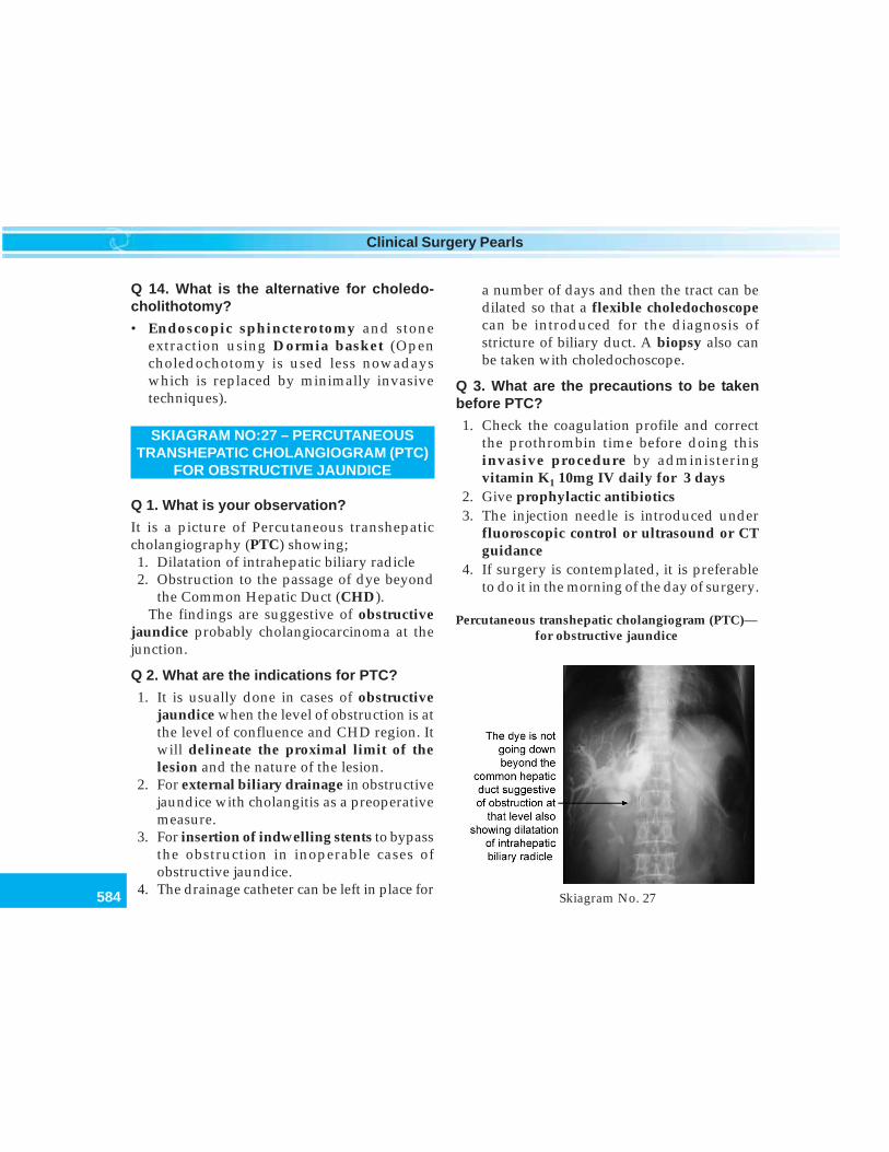
584
Clinical Surgery Pearls
Q 14. What is the alternative for choledo-cholithotomy?
• Endoscopic sphincterotomy and stoneextraction using Dormia basket (Opencholedochotomy is used less nowadayswhich is replaced by minimally invasivetechniques).
SKIAGRAM NO:27 – PERCUTANEOUSTRANSHEPATIC CHOLANGIOGRAM (PTC)
FOR OBSTRUCTIVE JAUNDICE
Q 1. What is your observation?
It is a picture of Percutaneous transhepaticcholangiography (PTC) showing;1. Dilatation of intrahepatic biliary radicle2. Obstruction to the passage of dye beyond
the Common Hepatic Duct (CHD).The findings are suggestive of obstructive
jaundice probably cholangiocarcinoma at thejunction.
Q 2. What are the indications for PTC?
1. It is usually done in cases of obstructivejaundice when the level of obstruction is atthe level of confluence and CHD region. Itwill delineate the proximal limit of thelesion and the nature of the lesion.
2. For external biliary drainage in obstructivejaundice with cholangitis as a preoperativemeasure.
3. For insertion of indwelling stents to bypassthe obstruction in inoperable cases ofobstructive jaundice.
4. The drainage catheter can be left in place for
a number of days and then the tract can bedilated so that a flexible choledochoscopecan be introduced for the diagnosis ofstricture of biliary duct. A biopsy also canbe taken with choledochoscope.
Q 3. What are the precautions to be takenbefore PTC?
1. Check the coagulation profile and correctthe prothrombin time before doing thisinvasive procedure by administeringvitamin K1 10mg IV daily for 3 days
2. Give prophylactic antibiotics3. The injection needle is introduced under
fluoroscopic control or ultrasound or CTguidance
4. If surgery is contemplated, it is preferableto do it in the morning of the day of surgery.
Skiagram No. 27
Percutaneous transhepatic cholangiogram (PTC)—for obstructive jaundice
585
Radiology Questions and Answers
Q 4. What is the procedure?
1. After giving local anesthesia the Chiba orOkuda needle is introduced into the liverthrough the 8th intercostal space in mid-axillary line.
2. Needle is placed in a dilated biliary radicleunder sonological guidance or fluoroscopy.
3. The bile is under tension and it is aspiratedand sent for culture and cytology.
4. Water-soluble contrast medium is theninjected into the biliary system and multipleimages are taken to visualize the area andnature of obstruction.
5. A catheter is introduced into the biliary systemif external biliary drainage is required.
Q 5. What are the complications of PTC?
1. The bile is under tension and it will leakinto the peritoneal cavity producing biliaryperitonitis
2. Bleeding3. Infection4. Septicemia.
NO: 28 - ULTRASOUND ABDOMEN –INTUSSUSCEPTION, B-MODE AND REAL
TIME ULTRASONOGRAPHY
Q 1. What is your observation in theultrasound picture of the abdomen?
The ultrasound showing the typical doughnutappearance of concentric rings is suggestive ofintussusception.
Q 2. What is ultrasonography?
• Ultrasound is energy in the form ofmechanical vibrations, the frequency ofwhich is in excess of that to which the humanear is sensitive, i.e. greater than 20,000Hz.
• Ultrasound makes use of high frequencysound waves generated by a transducercontaining the piezoelectric material.
• The sound waves are reflected by tissueinterfaces (as the sound energy passesthrough various tissues of the body, itinteracts by reflection, refraction, defraction,scattering and absorption.
• The echoes vary depending on the tissuetype.
• The frequencies in the range of 3 to 20 MHzare used (1 MHz being equal to 1000,000 Hz).
• For abdomen 3 to 7 MHz transducer is used
Skiagram No. 28- Ultrasound Abdomen
USG—Abdomen
Published by dr-notes.com
586
Clinical Surgery Pearls
• Low frequency waves have greaterpenetrating powers than high frequencywaves, but produce less definition. (Forsuperficial structures higher frequencytransducers are used).
Q 3. What is piezoelectric effect?
It is the process of conversion of electrical tomechanical energy.
Q 4. What are the examples of piezoelectricmaterials?• Quartz• Lithium sulphate• Synthetic ceramic lead zurconate tinanate (PTZ):
most commonly used
Q 5. What is B- mode ultrasound?
This is the primary display mode introduced in1972. It is called B-mode or brightness modedisplay which gives two dimensional cross-sectional representation of the tissues underexamination on horizontal and vertical axeswhile encoding echo amplitude information ingray levels of 1 and 14.
Q 6. What is real time ultrasonography?
This is the universally accepted method whichproduces about 40 B - scan images per second.This is above the flicker rate of the eye (16-18images/second) and the examiner perceives acontinuum of motion like the systolic dilatationand diastolic diminution of the diameter of theaorta.
Q 7. What are the uses of ultrasonography?
Use of ultrasound in surgery
1. It can differentiate solid and cystic lesions2. Vascular system can be assessed (by Doppler
study)3. Guided biopsies are possible4. Can differentiate benign and malignant lesions5. First - line investigation of choice of the liver:
For hepatic tumors, metastasis, abscessformation, cystic diseases, etc.
6. First-line investigation of choice of the biliarysystem: Intrahepatic biliary radicle dilatation,assessment of the CBD size, identification of thestones, assessment of the wall thickness of gallbladder, etc.
7. First-line investigation of choice of the renaltract.
8. Fluid collection in the peritoneal cavity, pleuralcavity and pericardial cavity can be identified
9. Can identify lesions in other solid organs like spleen10. Pancreatic diseases: Difficult to visualize distal
body and tail. Head and proximal body can bevisualized by making use of the acousticwindow of the liver
11. Lesions of thyroid can be assessed12. Breast lesions can be ascertained in young
patients < 35 years13. Testicular lesions.14. Imaging method of choice in obstetrics and
gynecology15. Endocavitary ultrasound is possible.
Q 8. What are the advantages of ultrasound?
1. It is noninvasive2. No radiation hazard3. Inexpensive4. Interaction with patient is possible.
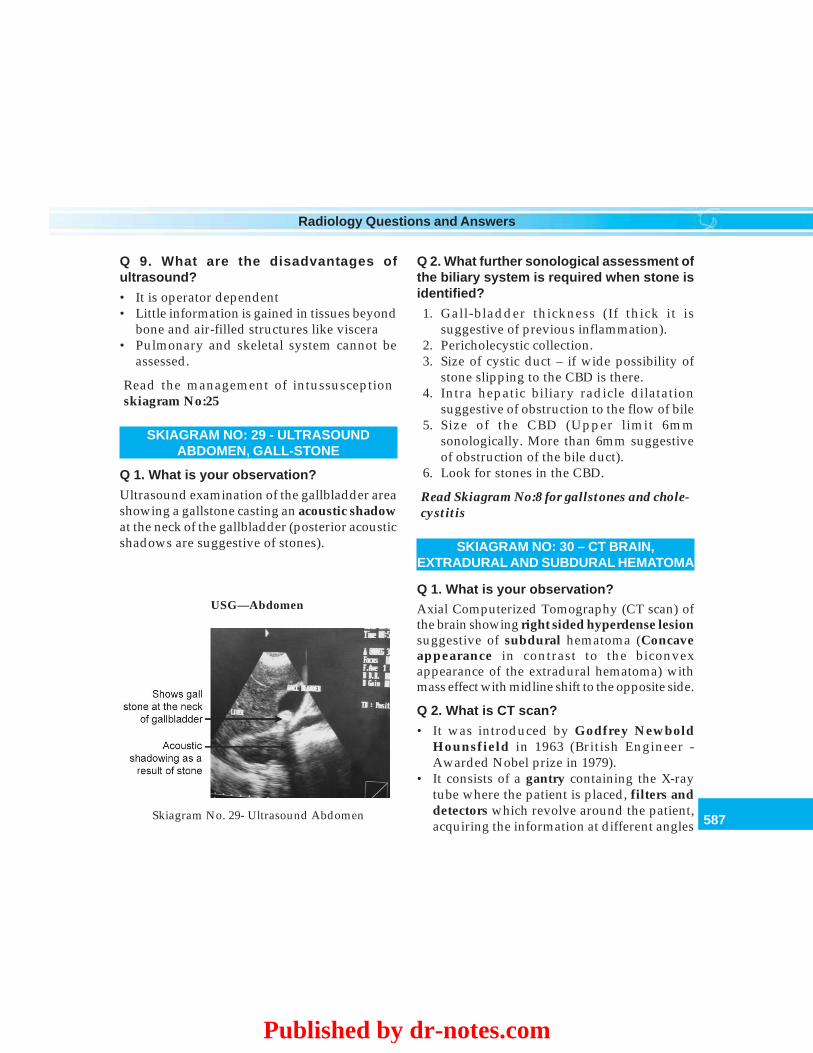
587
Radiology Questions and Answers
Q 9. What are the disadvantages ofultrasound?
• It is operator dependent• Little information is gained in tissues beyond
bone and air-filled structures like viscera• Pulmonary and skeletal system cannot be
assessed.
Read the management of intussusceptionskiagram No:25
SKIAGRAM NO: 29 - ULTRASOUNDABDOMEN, GALL-STONE
Q 1. What is your observation?
Ultrasound examination of the gallbladder areashowing a gallstone casting an acoustic shadowat the neck of the gallbladder (posterior acousticshadows are suggestive of stones).
Q 2. What further sonological assessment ofthe biliary system is required when stone isidentified?
1. Gall-bladder thickness (If thick it issuggestive of previous inflammation).
2. Pericholecystic collection.3. Size of cystic duct – if wide possibility of
stone slipping to the CBD is there.4. Intra hepatic biliary radicle dilatation
suggestive of obstruction to the flow of bile5. Size of the CBD (Upper limit 6mm
sonologically. More than 6mm suggestiveof obstruction of the bile duct).
6. Look for stones in the CBD.
Read Skiagram No:8 for gallstones and chole-cystitis
SKIAGRAM NO: 30 – CT BRAIN,EXTRADURAL AND SUBDURAL HEMATOMA
Q 1. What is your observation?Axial Computerized Tomography (CT scan) ofthe brain showing right sided hyperdense lesionsuggestive of subdural hematoma (Concaveappearance in contrast to the biconvexappearance of the extradural hematoma) withmass effect with midline shift to the opposite side.
Q 2. What is CT scan?
• It was introduced by Godfrey NewboldHounsfield in 1963 (British Engineer -Awarded Nobel prize in 1979).
• It consists of a gantry containing the X-raytube where the patient is placed, filters anddetectors which revolve around the patient,acquiring the information at different angles
Skiagram No. 29- Ultrasound Abdomen
USG—Abdomen
Published by dr-notes.com
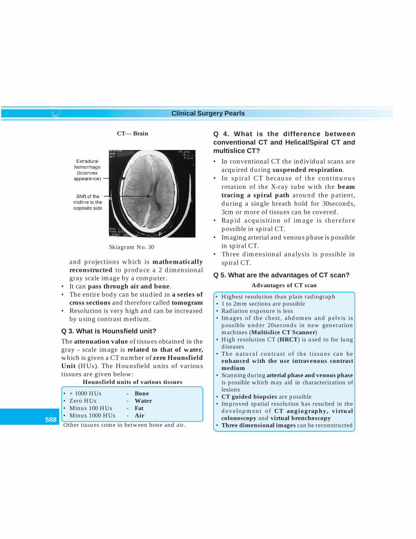
588
Clinical Surgery Pearls
and projections which is mathematicallyreconstructed to produce a 2 dimensionalgray scale image by a computer.
• It can pass through air and bone.• The entire body can be studied in a series of
cross sections and therefore called tomogram• Resolution is very high and can be increased
by using contrast medium.
Q 3. What is Hounsfield unit?
The attenuation value of tissues obtained in thegray - scale image is related to that of water,which is given a CT number of zero HounsfieldUnit (HUs). The Hounsfield units of varioustissues are given below:
Hounsfield units of various tissues
• + 1000 HUs - Bone• Zero HUs - Water• Minus 100 HUs - Fat• Minus 1000 HUs - Air
Other tissues come in between bone and air.
Q 4. What is the difference betweenconventional CT and Helical/Spiral CT andmultislice CT?
• In conventional CT the individual scans areacquired during suspended respiration.
• In spiral CT because of the continuousrotation of the X-ray tube with the beamtracing a spiral path around the patient,during a single breath hold for 30seconds,3cm or more of tissues can be covered.
• Rapid acquisition of image is thereforepossible in spiral CT.
• Imaging arterial and venous phase is possiblein spiral CT.
• Three dimensional analysis is possible inspiral CT.
Q 5. What are the advantages of CT scan?Advantages of CT scan
• Highest resolution than plain radiograph• 1 to 2mm sections are possible• Radiation exposure is less• Images of the chest, abdomen and pelvis is
possible under 20seconds in new generationmachines (Multislice CT Scanner)
• High resolution CT (HRCT) is used to for lungdiseases
• The natural contrast of the tissues can beenhanced with the use intravenous contrastmedium
• Scanning during arterial phase and venous phaseis possible which may aid in characterization oflesions
• CT guided biopsies are possible• Improved spatial resolution has resulted in the
development of CT angiography, virtualcolonoscopy and virtual bronchoscopy
• Three dimensional images can be reconstructed
Skiagram No. 30
CT— Brain
589
Radiology Questions and Answers
Q 6. What are the disadvantages of CT scan?
• High dose of ionizing radiation• Increased cost• Availability of the equipment• Radiological expertise required for
interpretation.
Q 7. What is the reason for concaveappearance in the given CT?
In subdural hematoma the blood spreads acrossthe surface of the brain. Since there is lessresistance to blood moving through thesubdural space than through the extraduralspace, it takes the concave shape (In extraduralhematoma a lentiform or lens-shaped orbiconvex hyperdense lesion is seen).
Q 8. What are the types of hemorrhages inrelation to the brain?
1. Extradural (outside dura, associated withfracture skull and more common in the young).
2. Subdural – acute subdural and chronicsubdural.
3. Subarachnoid hemorrhage.4. Intracerebral hemorrhage.5. Intraventricular hemorrhage.
Q 9. Which type of hematoma is morecommon — extradural or subdural?
Subdural is more common (5:1).
Q 10. What are the differences betweenextradural and acute subdural hematoma?
Differences between extradural and acute subdural hematoma
Acute subdural Extradural
Cause Need not be associated with fracture Always associated with skull fractureof the skull
Cause Injury to the cerebral veins Arterial injury (major dural venous sinuspossible)
Age group Any age Younger ageLocation Space between dura and arachnoid. Space between bone and dura Usually
Depends on the area of bleeding temporal region.Mass effect and Less common and late since it is More common and early since it is arterialmidline shift venousConscious level Impaired conscious level from the Conscious level normal initially
time of injuryLucid interval Not present PresentBrain damage Extensive damage (laceration) may No primary damage or minimal
be seenMarch of paralysis May occur on both sides Typical paralysis on the opposite sideCT appearance Hyperdense diffuse and concave Lentiform or biconvex hyperdense lesion
appearance with or without mass effect or midline shiftTreatment Evacuation via craniotomy. Immediate surgical evacuation via
Small hematomas may be managed craniotomyconservatively
Mortality rate 40% 2 to 18%
Published by dr-notes.com
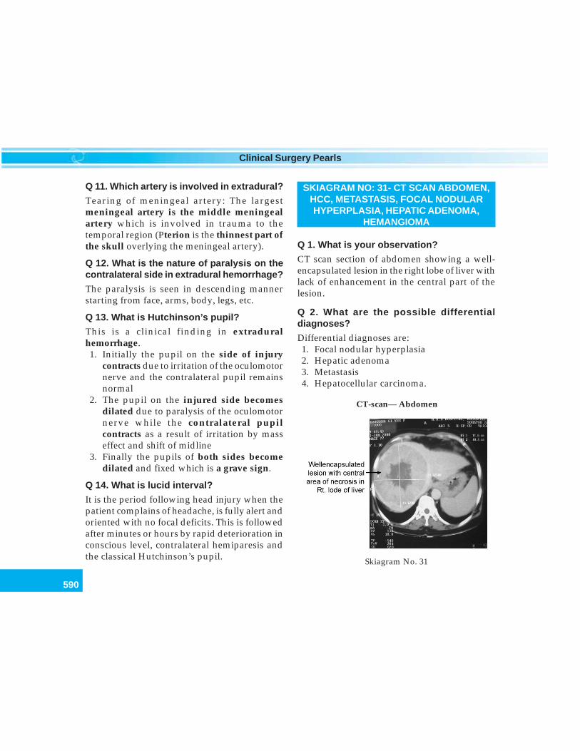
590
Clinical Surgery Pearls
Q 11. Which artery is involved in extradural?
Tearing of meningeal artery: The largestmeningeal artery is the middle meningealartery which is involved in trauma to thetemporal region (Pterion is the thinnest part ofthe skull overlying the meningeal artery).
Q 12. What is the nature of paralysis on thecontralateral side in extradural hemorrhage?
The paralysis is seen in descending mannerstarting from face, arms, body, legs, etc.
Q 13. What is Hutchinson’s pupil?
This is a clinical finding in extraduralhemorrhage.1. Initially the pupil on the side of injury
contracts due to irritation of the oculomotornerve and the contralateral pupil remainsnormal
2. The pupil on the injured side becomesdilated due to paralysis of the oculomotornerve while the contralateral pupilcontracts as a result of irritation by masseffect and shift of midline
3. Finally the pupils of both sides becomedilated and fixed which is a grave sign.
Q 14. What is lucid interval?
It is the period following head injury when thepatient complains of headache, is fully alert andoriented with no focal deficits. This is followedafter minutes or hours by rapid deterioration inconscious level, contralateral hemiparesis andthe classical Hutchinson’s pupil.
SKIAGRAM NO: 31- CT SCAN ABDOMEN,HCC, METASTASIS, FOCAL NODULARHYPERPLASIA, HEPATIC ADENOMA,
HEMANGIOMA
Q 1. What is your observation?
CT scan section of abdomen showing a well-encapsulated lesion in the right lobe of liver withlack of enhancement in the central part of thelesion.
Q 2. What are the possible differentialdiagnoses?
Differential diagnoses are:1. Focal nodular hyperplasia2. Hepatic adenoma3. Metastasis4. Hepatocellular carcinoma.
Skiagram No. 31
CT-scan— Abdomen
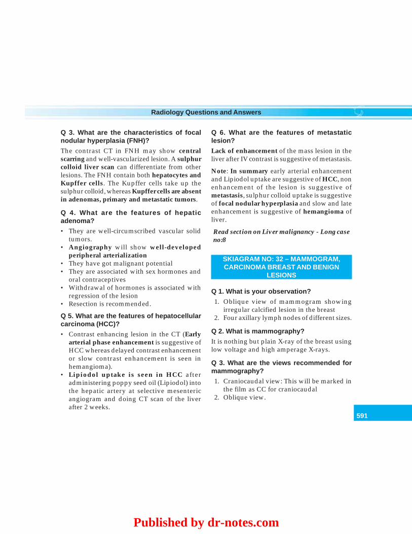
591
Radiology Questions and Answers
Q 3. What are the characteristics of focalnodular hyperplasia (FNH)?
The contrast CT in FNH may show centralscarring and well-vascularized lesion. A sulphurcolloid liver scan can differentiate from otherlesions. The FNH contain both hepatocytes andKupffer cells. The Kupffer cells take up thesulphur colloid, whereas Kupffer cells are absentin adenomas, primary and metastatic tumors.
Q 4. What are the features of hepaticadenoma?
• They are well-circumscribed vascular solidtumors.
• Angiography will show well-developedperipheral arterialization
• They have got malignant potential• They are associated with sex hormones and
oral contraceptives• Withdrawal of hormones is associated with
regression of the lesion• Resection is recommended.
Q 5. What are the features of hepatocellularcarcinoma (HCC)?
• Contrast enhancing lesion in the CT (Earlyarterial phase enhancement is suggestive ofHCC whereas delayed contrast enhancementor slow contrast enhancement is seen inhemangioma).
• Lipiodol uptake is seen in HCC afteradministering poppy seed oil (Lipiodol) intothe hepatic artery at selective mesentericangiogram and doing CT scan of the liverafter 2 weeks.
Q 6. What are the features of metastaticlesion?
Lack of enhancement of the mass lesion in theliver after IV contrast is suggestive of metastasis.
Note: In summary early arterial enhancementand Lipiodol uptake are suggestive of HCC, nonenhancement of the lesion is suggestive ofmetastasis, sulphur colloid uptake is suggestiveof focal nodular hyperplasia and slow and lateenhancement is suggestive of hemangioma ofliver.
Read section on Liver malignancy - Long caseno:8
SKIAGRAM NO: 32 – MAMMOGRAM,CARCINOMA BREAST AND BENIGN
LESIONS
Q 1. What is your observation?
1. Oblique view of mammogram showingirregular calcified lesion in the breast
2. Four axillary lymph nodes of different sizes.
Q 2. What is mammography?
It is nothing but plain X-ray of the breast usinglow voltage and high amperage X-rays.
Q 3. What are the views recommended formammography?
1. Craniocaudal view: This will be marked inthe film as CC for craniocaudal
2. Oblique view.
Published by dr-notes.com
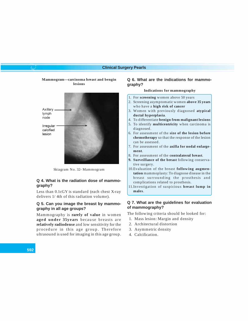
592
Clinical Surgery Pearls
Q 4. What is the radiation dose of mammo-graphy?
Less than 0.1cGY is standard (each chest X-raydelivers 1/4th of this radiation volume).
Q 5. Can you image the breast by mammo-graphy in all age groups?
Mammography is rarely of value in womenaged under 35years because breasts arerelatively radiodense and low sensitivity for theprocedure in this age group. Thereforeultrasound is used for imaging in this age group.
Q 6. What are the indications for mammo-graphy?
Indications for mammography
1. For screening women above 50 years2. Screening asymptomatic women above 35 years
who have a high risk of cancer3. Women with previously diagnosed atypical
ductal hyperplasia.4. To differentiate benign from malignant lesions5. To identify multicentricity when carcinoma is
diagnosed.6. For assessment of the size of the lesion before
chemotherapy so that the response of the lesioncan be assessed.
7. For assessment of the axilla for nodal enlarge-ment.
8. For assessment of the contralateral breast.9. Surveillance of the breast following conserva-
tive surgery.10.Evaluation of the breast following augmen-
tation mammoplasty: To diagnose disease in thebreast surrounding the prosthesis andcomplications related to prosthesis.
11.Investigation of suspicious breast lump inmales.
Q 7. What are the guidelines for evaluationof mammography?
The following criteria should be looked for:1. Mass lesion: Margin and density2. Architectural distortion3. Asymmetric density4. Calcification.
Skiagram No. 32- Mammogram
Mammogram—carcinoma breast and benginlesions
593
Radiology Questions and Answers
Malignant Benign
Margin of mass Spiculated mass lesion with ill - Well circumscribed mass lesionlesion defined margin (commonest finding of
invasive carcinoma)Density High density increases the probability Relatively low density
of malignancyArchitectural Stellate lesion: Numerous straight lines No architectural distortiondistortion radiating towards the central areaAsymmetric With associated microcalcification, Can occur in normal involution ofdensity spiculation and architectural distortion glandular tissue, hormone replacement
therapy and traumaCalcification Clustered microcalcification with Scattered and rounded calcification
variable shape and size: Granular, with relatively uniform size andbranching, rod-shaped, bizarre micro- densitycalcifications are suspicious (tight • Popcorn calcification –clusters >1cm2 area is highly suggestive) fibroadenoma
• Teacup calcification – fibrocysticdisease
• Needle-like calcification – ductectasia
3. Requires high technology machinery.4. Requires special film and processing.5. Requires highly trained radiologists.
Q 10. What is the reason for recommendingscreening the breast between the age groupof 50 to 70 years by mammography?
• Current available data indicate that thereduction in mortality is maximal betweenthe age of 50 to 70years (25 to 30% reductionin mortality).
• In age group of 40 to 49years the reductionin mortality is only 7 to 8%.
Q 8. What are the secondary signs of malig-nancy?
• Skin changes – thickening, retraction anddimpling
• Nipple retraction, flattening of the nipple• Focally dilated duct• Increased vascularity• Axillary lymphadenopathy.
Q 9. What are the disadvantages of mammo-graphy?
1. It is painful.2. It is expensive.
Published by dr-notes.com
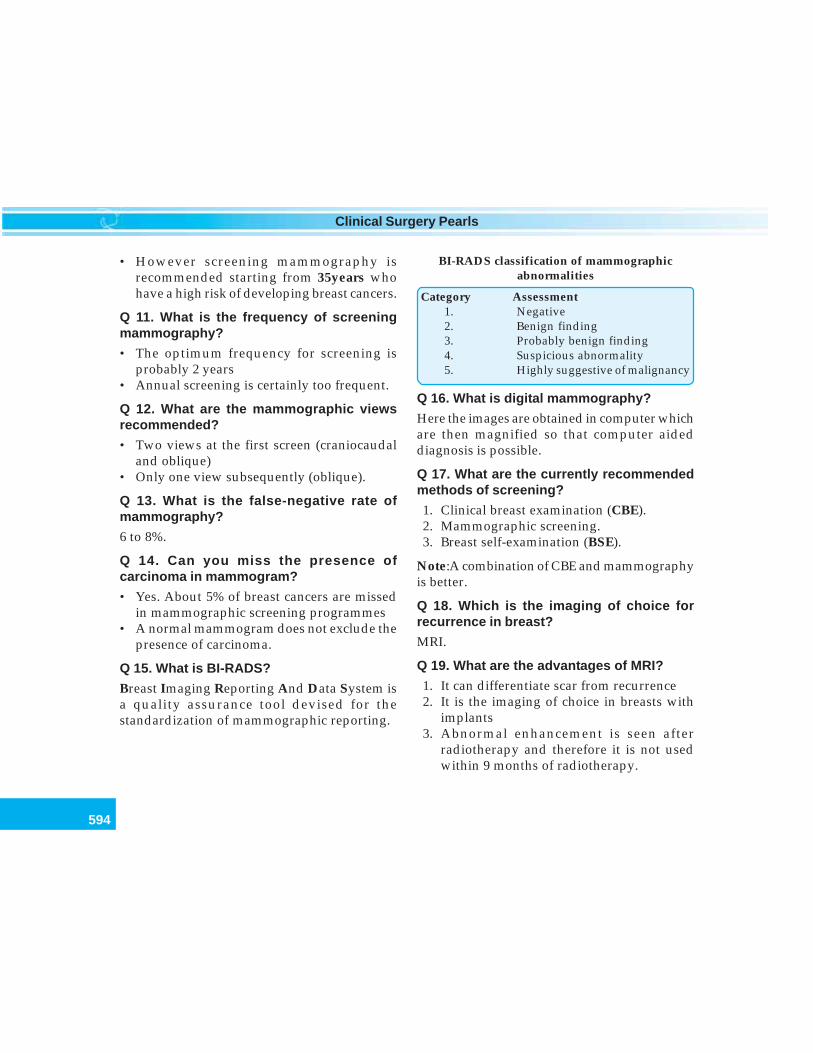
594
Clinical Surgery Pearls
• However screening mammography isrecommended starting from 35years whohave a high risk of developing breast cancers.
Q 11. What is the frequency of screeningmammography?
• The optimum frequency for screening isprobably 2 years
• Annual screening is certainly too frequent.
Q 12. What are the mammographic viewsrecommended?
• Two views at the first screen (craniocaudaland oblique)
• Only one view subsequently (oblique).
Q 13. What is the false-negative rate ofmammography?
6 to 8%.
Q 14. Can you miss the presence ofcarcinoma in mammogram?
• Yes. About 5% of breast cancers are missedin mammographic screening programmes
• A normal mammogram does not exclude thepresence of carcinoma.
Q 15. What is BI-RADS?
Breast Imaging Reporting And Data System isa quality assurance tool devised for thestandardization of mammographic reporting.
BI-RADS classification of mammographicabnormalities
Category Assessment1. Negative2. Benign finding3. Probably benign finding4. Suspicious abnormality5. Highly suggestive of malignancy
Q 16. What is digital mammography?
Here the images are obtained in computer whichare then magnified so that computer aideddiagnosis is possible.
Q 17. What are the currently recommendedmethods of screening?
1. Clinical breast examination (CBE).2. Mammographic screening.3. Breast self-examination (BSE).
Note:A combination of CBE and mammographyis better.
Q 18. Which is the imaging of choice forrecurrence in breast?
MRI.
Q 19. What are the advantages of MRI?
1. It can differentiate scar from recurrence2. It is the imaging of choice in breasts with
implants3. Abnormal enhancement is seen after
radiotherapy and therefore it is not usedwithin 9 months of radiotherapy.

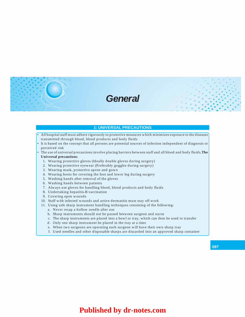
597
General
General
1: UNIVERSAL PRECAUTIONS
• All hospital staff must adhere rigorously to protective measures which minimizes exposure to the diseasestransmitted through blood, blood products and body fluids
• It is based on the concept that all persons are potential sources of infection independent of diagnosis orperceived risk
• The use of universal precautions involve placing barriers between staff and all blood and body fluids.TheUniversal precautions
1. Wearing protective gloves (Ideally double gloves during surgery)2. Wearing protective eyewear (Preferably goggles during surgery)3. Wearing mask, protective apron and gown4. Wearing boots for covering the foot and lower leg during surgery5. Washing hands after removal of the gloves6. Washing hands between patients7. Always use gloves for handling blood, blood products and body fluids8. Undertaking hepatitis-B vaccination9. Covering open wounds
10. Staff with infected wounds and active dermatitis must stay off work11. Using safe sharp instrument handling techniques consisting of the following:
a. Never recap a hollow needle after useb. Sharp instruments should not be passed between surgeon and nursec. The sharp instruments are placed into a bowl or tray, which can then be used to transferd. Only one sharp instrument be placed in the tray at a timee. When two surgeons are operating each surgeon will have their own sharp trayf. Used needles and other disposable sharps are discarded into an approved sharp container
Published by dr-notes.com
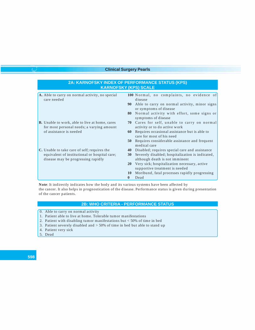
598
Clinical Surgery Pearls
2A: KARNOFSKY INDEX OF PERFORMANCE STATUS (KPS)KARNOFSKY (KPS) SCALE
A. Able to carry on normal activity, no special 100 Normal, no complaints, no evidence ofcare needed disease
90 Able to carry on normal activity, minor signsor symptoms of disease
80 Normal activity with effort, some signs orsymptoms of disease
B. Unable to work, able to live at home, cares 70 Cares for self, unable to carry on normalfor most personal needs; a varying amount activity or to do active workof assistance is needed 60 Requires occasional assistance but is able to
care for most of his need50 Requires considerable assistance and frequent
medical careC. Unable to take care of self; requires the 40 Disabled; requires special care and assistance
equivalent of institutional or hospital care; 30 Severely disabled; hospitalization is indicated,disease may be progressing rapidly although death is not imminent
20 Very sick; hospitalization necessary, activesupportive treatment is needed
10 Moribund, fatal processes rapidly progressing0 Dead
Note: It indirectly indicates how the body and its various systems have been affected bythe cancer. It also helps in prognostication of the disease. Performance status is given during presentationof the cancer patients.
2B: WHO CRITERIA - PERFORMANCE STATUS
0. Able to carry on normal activity1. Patient able to live at home. Tolerable tumor manifestations2. Patient with disabling tumor manifestations but < 50% of time in bed3. Patient severely disabled and > 50% of time in bed but able to stand up4. Patient very sick5. Dead
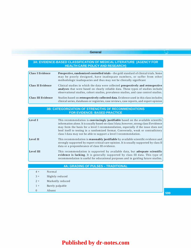
599
General
3A: EVIDENCE-BASED CLASSIFICATION OF MEDICAL LITERATURE (AGENCY FORHEALTH CARE POLICY AND RESEARCH)
Class I Evidence Prospective, randomized controlled trials – the gold standard of clinical trials. Somemay be poorly designed, have inadequate numbers, or suffer from othermethodologic inadequacies and thus may not be clinically significant
Class II Evidence Clinical studies in which the data were collected prospectively and retrospectiveanalyses that were based on clearly reliable data. These types of studies includeobservational studies, cohort studies, prevalence studies, and case control studies.
Class III Evidence Studies based on retrospectively collected data. Evidence used in this class includesclinical series, databases or registries, case reviews, case reports, and expert opinion
3B: CATEGORIZATION OF STRENGTHS OF RECOMMENDATIONSFOR EVIDENCE- BASED PRACTICE
Level I This recommendation is convincingly justifiable based on the available scientificinformation alone. It is usually based on class I data; however, strong class II evidencemay form the basis for a level I recommendation, especially if the issue does notlend itself to testing in a randomized format. Conversely, weak or contradictoryclass I data may not be able to support a level I recommendation.
Level II This recommendation is reasonably justifiable by available scientific evidence andstrongly supported by expert critical care opinion. It is usually supported by class IIdata or a preponderance of class III evidence.
Level III This recommendation is supported by available data, but adequate scientificevidence is lacking. It is generally supported by class III data. This type ofrecommendation is useful for educational purposes and in guiding future studies.
4A: GRADING OF PULSES – TRADITIONAL
4 + Normal
3 + Slightly reduced
2 + Markedly reduced
1 + Barely palpable
0 Absent
Published by dr-notes.com
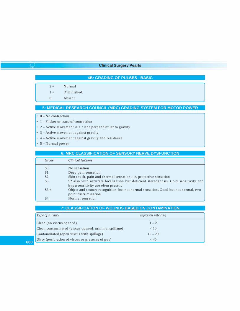
600
Clinical Surgery Pearls
4B: GRADING OF PULSES - BASIC
2 + Normal
1 + Diminished
0 Absent
5: MEDICAL RESEARCH COUNCIL (MRC) GRADING SYSTEM FOR MOTOR POWER
• 0 – No contraction
• 1 – Flicker or trace of contraction
• 2 – Active movement in a plane perpendicular to gravity
• 3 – Active movement against gravity
• 4 – Active movement against gravity and resistance
• 5 – Normal power
6: MRC CLASSIFICATION OF SENSORY NERVE DYSFUNCTION
Grade Clinical features
S0 No sensationS1 Deep pain sensationS2 Skin touch, pain and thermal sensation, i.e. protective sensationS3 S2 also with accurate localization but deficient stereognosis. Cold sensitivity and
hypersensitivity are often presentS3 + Object and texture recognition, but not normal sensation. Good but not normal, two –
point discriminationS4 Normal sensation
7: CLASSIFICATION OF WOUNDS BASED ON CONTAMINATION
Type of surgery Infection rate (%)
Clean (no viscus opened) 1 – 2
Clean contaminated (viscus opened, minimal spillage) < 10
Contaminated (open viscus with spillage) 15 – 20
Dirty (perforation of viscus or presence of pus) < 40
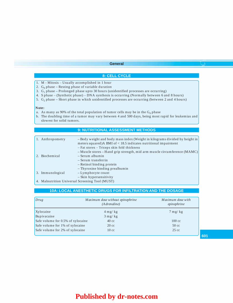
601
General
8: CELL CYCLE
1. M - Mitosis – Usually accomplished in 1 hour2. G0 phase – Resting phase of variable duration3. G1 phase – Prolonged phase upto 30 hours (unidentified processes are occurring)4. S phase – (Synthetic phase) – DNA synthesis is occurring (Normally between 6 and 8 hours)5. G2 phase – Short phase in which unidentified processes are occurring (between 2 and 4 hours)
Note:a. As many as 90% of the total population of tumor cells may be in the G0 phaseb. The doubling time of a tumor may vary between 4 and 500 days, being most rapid for leukemias and
slowest for solid tumors.
9: NUTRITIONAL ASSESSMENT METHODS
1. Anthropometry – Body weight and body mass index (Weight in kilograms divided by height inmeters squared)A BMI of < 18.5 indicates nutritional impairment– Fat stores – Triceps skin fold thickness– Muscle stores – Hand grip strength, mid arm muscle circumference (MAMC)
2. Biochemical – Serum albumin– Serum transferrin– Retinol binding protein– Thyroxine binding prealbumin
3. Immunological – Lymphocyte count– Skin hypersensitivity
4. Malnutrition Universal Screening Tool (MUST)
10A: LOCAL ANESTHETIC DRUGS FOR INFILTRATION AND THE DOSAGE
Drug Maximum dose without epinephrine Maximum dose with(Adrenaline) epinephrine
Xylocaine 4 mg/kg 7 mg/kgBupivacaine 3 mg/kgSafe volume for 0.5% of xylocaine 40 cc 100 ccSafe volume for 1% of xylocaine 20 cc 50 ccSafe volume for 2% of xylocaine 10 cc 25 cc
Published by dr-notes.com
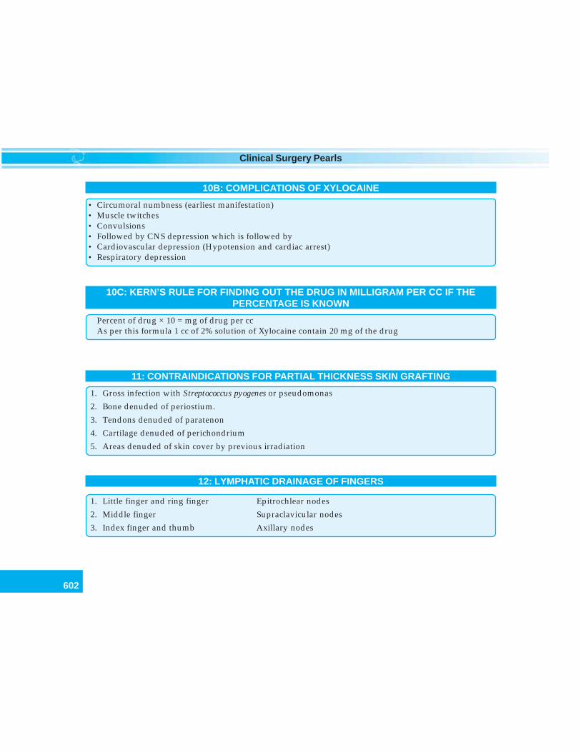
602
Clinical Surgery Pearls
10B: COMPLICATIONS OF XYLOCAINE
• Circumoral numbness (earliest manifestation)• Muscle twitches• Convulsions• Followed by CNS depression which is followed by• Cardiovascular depression (Hypotension and cardiac arrest)• Respiratory depression
10C: KERN’S RULE FOR FINDING OUT THE DRUG IN MILLIGRAM PER CC IF THEPERCENTAGE IS KNOWN
Percent of drug × 10 = mg of drug per ccAs per this formula 1 cc of 2% solution of Xylocaine contain 20 mg of the drug
11: CONTRAINDICATIONS FOR PARTIAL THICKNESS SKIN GRAFTING
1. Gross infection with Streptococcus pyogenes or pseudomonas
2. Bone denuded of periostium.
3. Tendons denuded of paratenon
4. Cartilage denuded of perichondrium
5. Areas denuded of skin cover by previous irradiation
12: LYMPHATIC DRAINAGE OF FINGERS
1. Little finger and ring finger Epitrochlear nodes
2. Middle finger Supraclavicular nodes
3. Index finger and thumb Axillary nodes

603
General
13: CLASSIFICATION OF DERMOIDS
Type Origin Examples
Congenital Arises in the line of embryonic fusion 1. External angular: Line of fusion betweenThey lie deep to the skin and not frontonasal process and maxillaryattached to the skin process
2. Postauricular: Line of fusion of the meso-dermal hillocks which form the pinna
3. Sublingual: Below the tongue in themidline
Implantation Develops after a puncture injury in Found on the fingers or hand and toesdermoid which epithelial cells are implanted in-
to the subcutaneous tissue
Sequestration Formed by the inclusion of epithelial Branchial cystsdermoid nests beneath the surface
Teratomatous Malignant change can occur which may Found in the ovary, testis,dermoids be carcinomatous/and sarcomatous retroperitoneum, superior mediastinum
and presacral area
14: DIFFERENCES BETWEEN TRANSUDATE AND EXUDATES
Transudate Exudates
Protein Low—most of which is albumin High— > 25g/L High content of< 25g/L fibrinogen
Specific gravity < 1.012 > 1.020Glucose content Same as plasma Low < 60 mg/dLpH > 7.3 < 7.3LDH Low HighCellular debris Few cells More cellsCause Obstruction to the venous outflow Inflammatory extravascular fluid formed
from liver (ultrafiltrate of blood plasma by the escape of fluid, proteins and bloodand results from imbalance across cells from the vascular system into thethe vascular endothelium). Permeability interstitial tissue or body cavities due toof endothelium—normal increased vascular permeability
SAAG—(Serum > 1.1 (Ascitic fluid has less albumin < 1.1 (Ascitic fluid has more albuminAscites albumin than serum) than serum)gradient)
Published by dr-notes.com
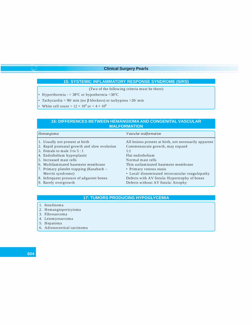
604
Clinical Surgery Pearls
15: SYSTEMIC INFLAMMATORY RESPONSE SYNDROME (SIRS)
(Two of the following criteria must be there)
• Hyperthermia - > 38oC or hypothermia <36oC
• Tachycardia > 90/min (no β blockers) or tachypnea >20/min
• White cell count > 12 × 109 or < 4 × 10⁹
16: DIFFERENCES BETWEEN HEMANGIOMA AND CONGENITAL VASCULARMALFORMATION
Hemangioma Vascular malformation
1. Usually not present at birth All lesions present at birth, not necessarily apparent2. Rapid postnatal growth and slow evolution Commensurate growth, may expand3. Female to male 3 to 5 : 1 1:14. Endothelium hyperplastic Flat endothelium5. Increased mast cells Normal mast cells6. Multilaminated basement membrane Thin unilaminated basement membrane7. Primary platelet trapping (Kasabach – • Primary venous stasis
Merritt syndrome) • Local/disseminated intravascular coagulopathy8. Infrequent pressure of adgacent bones Defects with AV fistula: Hypertrophy of bones9. Rarely overgrowth Defects without AV fistula: Atrophy
17: TUMORS PRODUCING HYPOGLYCEMIA
1. Insulinoma2. Hemangiopericytoma3. Fibrosarcoma4. Leiomyosarcoma5. Hepatoma6. Adrenocortical carcinoma
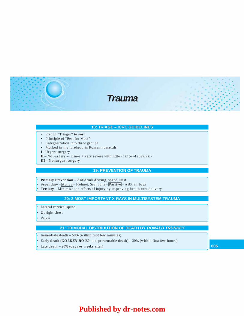
605
Trauma
Trauma
18: TRIAGE – ICRC GUIDELINES
• French “Triager” to sort• Principle of “Best for Most”• Categorization into three groups• Marked in the forehead in Roman numeralsI - Urgent surgeryII – No surgery – (minor + very severe with little chance of survival)III – Nonurgent surgery
19: PREVENTION OF TRAUMA
• Primary Prevention – Antidrink driving, speed limit• Secondary – Active – Helmet, Seat belts – Passive – ABS, air bags• Tertiary – Minimize the effects of injury by improving health care delivery
20: 3 MOST IMPORTANT X-RAYS IN MULTISYSTEM TRAUMA
• Lateral cervical spine
• Upright chest
• Pelvis
21: TRIMODAL DISTRIBUTION OF DEATH BY DONALD TRUNKEY
• Immediate death – 50% (within first few minutes)
• Early death (GOLDEN HOUR and preventable death) – 30% (within first few hours)
• Late death – 20% (days or weeks after)
Published by dr-notes.com
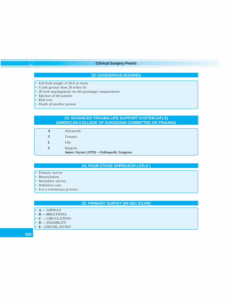
606
Clinical Surgery Pearls
22: DANGEROUS INJURIES
• Fall from height of 20 ft or more• Crash greater than 20 miles/hr• 20 inch impingement on the passenger compartment• Ejection of the patient• Roll over• Death of another person
23: ADVANCED TRAUMA LIFE SUPPORT SYSTEM (ATLS)(AMERICAN COLLEGE OF SURGEONS COMMITTEE ON TRAUMA)
A Advanced
T Trauma
L Life
S SupportJames Styner (1970) – Orthopedic Surgeon
24: FOUR STAGE APPROACH ( ATLS )
• Primary survey• Resuscitation• Secondary survey• Definitive care• It is a continuous process
25: PRIMARY SURVEY (60 SEC EXAM)
• A — AIRWAY• B — BREATHING• C — CIRCULATION• D — DISABILITY• E – EXPOSE, ENTRY
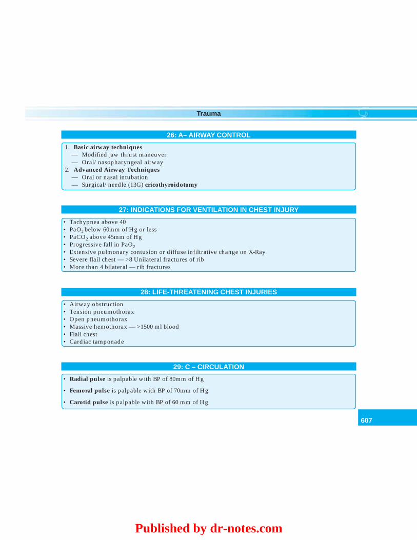
607
Trauma
26: A– AIRWAY CONTROL
1. Basic airway techniques— Modified jaw thrust maneuver— Oral/nasopharyngeal airway
2. Advanced Airway Techniques— Oral or nasal intubation— Surgical/needle (13G) cricothyroidotomy
27: INDICATIONS FOR VENTILATION IN CHEST INJURY
• Tachypnea above 40• PaO2 below 60mm of Hg or less• PaCO2 above 45mm of Hg• Progressive fall in PaO2• Extensive pulmonary contusion or diffuse infiltrative change on X-Ray• Severe flail chest — >8 Unilateral fractures of rib• More than 4 bilateral — rib fractures
28: LIFE-THREATENING CHEST INJURIES
• Airway obstruction• Tension pneumothorax• Open pneumothorax• Massive hemothorax — >1500 ml blood• Flail chest• Cardiac tamponade
29: C – CIRCULATION
• Radial pulse is palpable with BP of 80mm of Hg
• Femoral pulse is palpable with BP of 70mm of Hg
• Carotid pulse is palpable with BP of 60 mm of Hg
Published by dr-notes.com
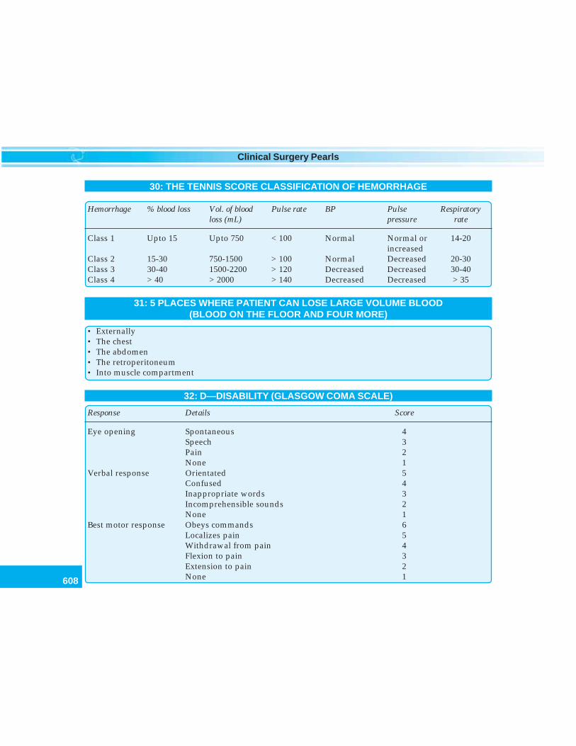
608
Clinical Surgery Pearls
30: THE TENNIS SCORE CLASSIFICATION OF HEMORRHAGE
Hemorrhage % blood loss Vol. of blood Pulse rate BP Pulse Respiratoryloss (mL) pressure rate
Class 1 Upto 15 Upto 750 < 100 Normal Normal or 14-20increased
Class 2 15-30 750-1500 > 100 Normal Decreased 20-30Class 3 30-40 1500-2200 > 120 Decreased Decreased 30-40Class 4 > 40 > 2000 > 140 Decreased Decreased > 35
31: 5 PLACES WHERE PATIENT CAN LOSE LARGE VOLUME BLOOD(BLOOD ON THE FLOOR AND FOUR MORE)
• Externally• The chest• The abdomen• The retroperitoneum• Into muscle compartment
32: D—DISABILITY (GLASGOW COMA SCALE)
Response Details Score
Eye opening Spontaneous 4Speech 3Pain 2None 1
Verbal response Orientated 5Confused 4Inappropriate words 3Incomprehensible sounds 2None 1
Best motor response Obeys commands 6Localizes pain 5Withdrawal from pain 4Flexion to pain 3Extension to pain 2None 1
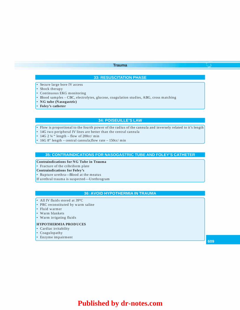
609
Trauma
33: RESUSCITATION PHASE
• Secure large bore IV access• Shock therapy• Continuous EKG monitoring• Blood samples – CBC, electrolytes, glucose, coagulation studies, ABG, cross matching• NG tube (Nasogastric)• Foley’s catheter
34: POISEUILLE’S LAW
• Flow is proportional to the fourth power of the radius of the cannula and inversely related to it’s length• 14G two peripheral IV lines are better than the central cannula• 14G 2 ¼ “ length – flow of 200cc/min• 16G 8” length – central cannula;flow rate – 150cc/min
35: CONTRAINDICATIONS FOR NASOGASTRIC TUBE AND FOLEY’S CATHETER
Contraindications for NG Tube in Trauma• Fracture of the cribriform plateContraindications for Foley’s• Rupture urethra—Blood at the meatusIf urethral trauma is suspected—Urethrogram
36: AVOID HYPOTHERMIA IN TRAUMA
• All IV fluids stored at 39°C• PRC reconstituted by warm saline• Fluid warmer• Warm blankets• Warm irrigating fluids
HYPOTHERMIA PRODUCES• Cardiac irritability• Coagulopathy• Enzyme impairment
Published by dr-notes.com

610
Clinical Surgery Pearls
37: BLOODY VICIOUS CYCLE IN TRAUMA
• Metabolic acidosis• Profound hypothermia• Coagulopathy
38: AUTOTRANSFUSION—NOT RECOMMENDED
• Consumptive coagulopathy• Platelet dysfunction• These risks may outweigh the benefits of auto transfusion.
39: SECONDARY SURVEY
• Head to toe examination• Look, listen and feel• High yield X-rays
– Cervical lateral– Upright chest
– Pelvis• Rule out intraabdominal bleeding in all cases of multisystem trauma
40: SECONDARY SURVEY AMPLE HISTORY
• A Allergy• M Medication• P Past medical history• L Last meal• E Events of the incident
41: RULE OUT INTRAABDOMINAL BLEEDING IN EVERY PATIENT
• CT scan/USG• Physical examination• Serial hematocrit• FAST – Focused assessment with sonography for trauma• DPL (diagnostic peritoneal lavage)
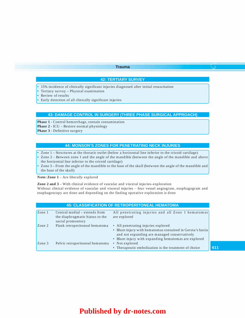
611
Trauma
42: TERTIARY SURVEY
• 15% incidence of clinically significant injuries diagnosed after initial resuscitation• Tertiary survey – Physical examination• Review of results• Early detection of all clinically significant injuries
43: DAMAGE CONTROL IN SURGERY (THREE PHASE SURGICAL APPROACH)
Phase 1 - Control hemorrhage, contain contaminationPhase 2 - ICU – Restore normal physiologyPhase 3 - Definitive surgery
44: MONSON’S ZONES FOR PENETRATING NECK INJURIES
• Zone 1 – Structures at the thoracic outlet (below a horizontal line inferior to the cricoid cartilage)• Zone 2 – Between zone 1 and the angle of the mandible (between the angle of the mandible and above
the horizontal line inferior to the cricoid cartilage)• Zone 3 – From the angle of the mandible to the base of the skull (between the angle of the mandible and
the base of the skull)
Note: Zone 1 – Are liberally explored
Zone 2 and 3 – With clinical evidence of vascular and visceral injuries–explorationWithout clinical evidence of vascular and visceral injuries – four vessel angiogram, esophagogram andesophagoscopy are done and depending on the finding operative exploration is done
45: CLASSIFICATION OF RETROPERITONEAL HEMATOMA
Zone 1 Central medial – extends from All penetrating injuries and all Zone 1 hematomasthe diaphragmatic hiatus to the are exploredsacral promontory
Zone 2 Flank retroperitoneal hematoma • All penetrating injuries explored• Blunt injury with hematomas contained in Gerota’s fascia
and not expanding are managed conservatively• Blunt injury with expanding hematomas are explored
Zone 3 Pelvic retroperitoneal hematoma • Not explored• Therapeutic embolization is the treatment of choice
Published by dr-notes.com
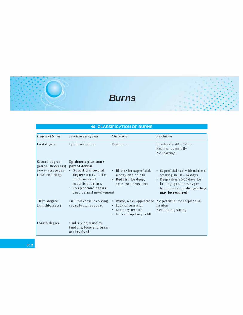
612
Clinical Surgery Pearls
Burns
46: CLASSIFICATION OF BURNS
Degree of burns Involvement of skin Characters Resolution
First degree Epidermis alone Erythema Resolves in 48 – 72hrsHeals uneventfullyNo scarring
Second degree Epidermis plus some(partial thickness) part of dermistwo types: super- • Superficial secondficial and deep degree: injury to the
epidermis andsuperficial dermis
• Deep second degree:deep dermal involvement
Third degree Full thickness involving • White, waxy appearance No potential for reepithelia-(full thickness) the subcutaneous fat • Lack of sensation lization
• Leathery texture Need skin grafting• Lack of capillary refill
Fourth degree Underlying muscles,tendons, bone and brainare involved
• Blister for superficial, • Superficial heal with minimalweepy and painful scarring in 10 – 14 days
• Reddish for deep, • Deep takes 25-35 days fordecreased sensation healing, produces hyper-
trophic scar and skin graftingmay be required
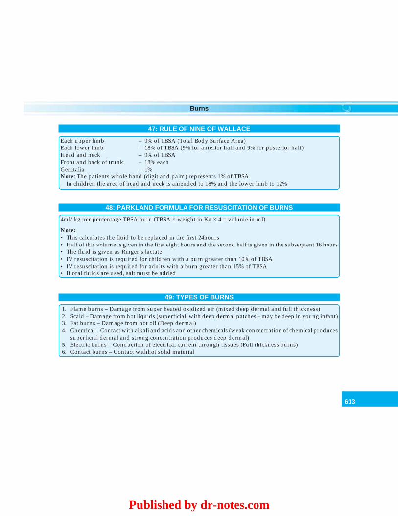
613
Burns
47: RULE OF NINE OF WALLACE
Each upper limb – 9% of TBSA (Total Body Surface Area)Each lower limb – 18% of TBSA (9% for anterior half and 9% for posterior half)Head and neck – 9% of TBSAFront and back of trunk – 18% eachGenitalia – 1%Note: The patients whole hand (digit and palm) represents 1% of TBSA
In children the area of head and neck is amended to 18% and the lower limb to 12%
48: PARKLAND FORMULA FOR RESUSCITATION OF BURNS
4ml/kg per percentage TBSA burn (TBSA × weight in Kg × 4 = volume in ml).
Note:• This calculates the fluid to be replaced in the first 24hours• Half of this volume is given in the first eight hours and the second half is given in the subsequent 16 hours• The fluid is given as Ringer’s lactate• IV resuscitation is required for children with a burn greater than 10% of TBSA• IV resuscitation is required for adults with a burn greater than 15% of TBSA• If oral fluids are used, salt must be added
49: TYPES OF BURNS
1. Flame burns – Damage from super heated oxidized air (mixed deep dermal and full thickness)2. Scald – Damage from hot liquids (superficial, with deep dermal patches – may be deep in young infant)3. Fat burns – Damage from hot oil (Deep dermal)4. Chemical – Contact with alkali and acids and other chemicals (weak concentration of chemical produces
superficial dermal and strong concentration produces deep dermal)5. Electric burns – Conduction of electrical current through tissues (Full thickness burns)6. Contact burns – Contact withhot solid material
Published by dr-notes.com
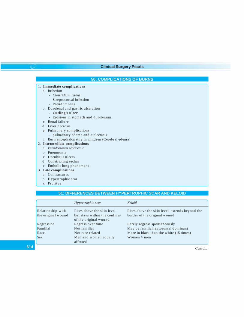
614
Clinical Surgery Pearls
50: COMPLICATIONS OF BURNS
1. Immediate complicationsa. Infection
- Clostridium tetani- Streptococcal infection- Pseudomonas
b. Duodenal and gastric ulceration- Curling’s ulcer- Erosions in stomach and duodenum
c. Renal failured. Liver necrosise. Pulmonary complications
– pulmonary edema and atelectasisf. Burn encephalopathy in children (Cerebral edema)
2. Intermediate complicationsa. Pseudomonas septicemiab. Pneumoniac. Decubitus ulcersd. Constricting eschare. Embolic lung phenomena
3. Late complicationsa. Contracturesb. Hypertrophic scarc. Pruritus
51: DIFFERENCES BETWEEN HYPERTROPHIC SCAR AND KELOID
Hypertrophic scar Keloid
Relationship with Rises above the skin level Rises above the skin level, extends beyond thethe original wound but stays within the confines border of the original wound
of the original woundRegression Regress over time Rarely regress spontaneouslyFamilial Not familial May be familial, autosomal dominantRace Not race related More in black than the white (15 times)Sex Men and women equally Women > men
affected
Contd...
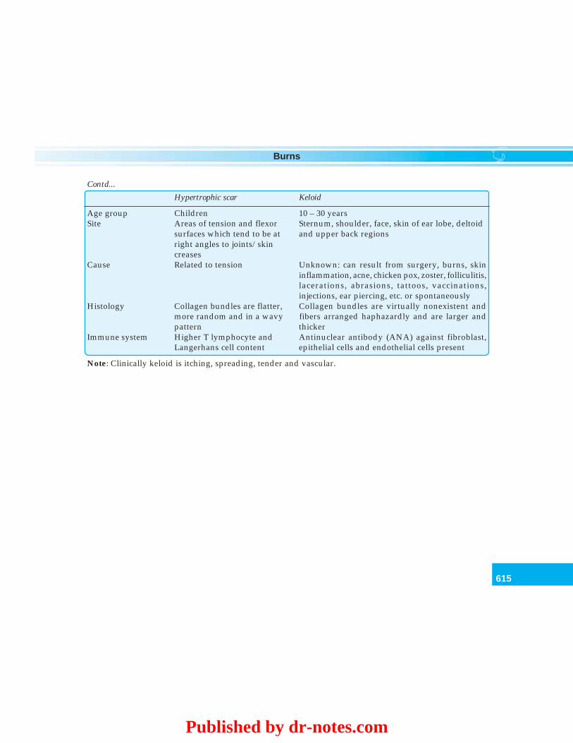
615
Burns
Age group Children 10 – 30 yearsSite Areas of tension and flexor Sternum, shoulder, face, skin of ear lobe, deltoid
surfaces which tend to be at and upper back regionsright angles to joints/skincreases
Cause Related to tension Unknown: can result from surgery, burns, skininflammation, acne, chicken pox, zoster, folliculitis,lacerations, abrasions, tattoos, vaccinations,injections, ear piercing, etc. or spontaneously
Histology Collagen bundles are flatter, Collagen bundles are virtually nonexistent andmore random and in a wavy fibers arranged haphazardly and are larger andpattern thicker
Immune system Higher T lymphocyte and Antinuclear antibody (ANA) against fibroblast,Langerhans cell content epithelial cells and endothelial cells present
Note: Clinically keloid is itching, spreading, tender and vascular.
Contd...
Hypertrophic scar Keloid
Published by dr-notes.com
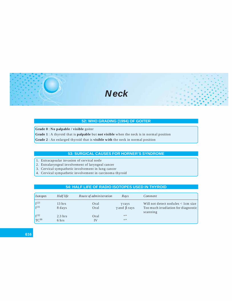
616
Clinical Surgery Pearls
Neck
52: WHO GRADING (1994) OF GOITER
Grade 0 : No palpable / visible goiter
Grade 1 : A thyroid that is palpable but not visible when the neck is in normal position
Grade 2 : An enlarged thyroid that is visible with the neck in normal position
53: SURGICAL CAUSES FOR HORNER’S SYNDROME
1. Extracapsular invasion of cervical node2. Extralaryngeal involvement of laryngeal cancer3. Cervical sympathetic involvement in lung cancer4. Cervical sympathetic involvement in carcinoma thyroid
54: HALF LIFE OF RADIO ISOTOPES USED IN THYROID
Isotopes Half life Route of administration Rays Comment
I123 13 hrs Oral γ rays Will not detect nodules < 1cm sizeI131 8 days Oral γ and β rays Too much irradiation for diagnostic
scanningI132 2.3 hrs Oral “”TC99 6 hrs IV “”
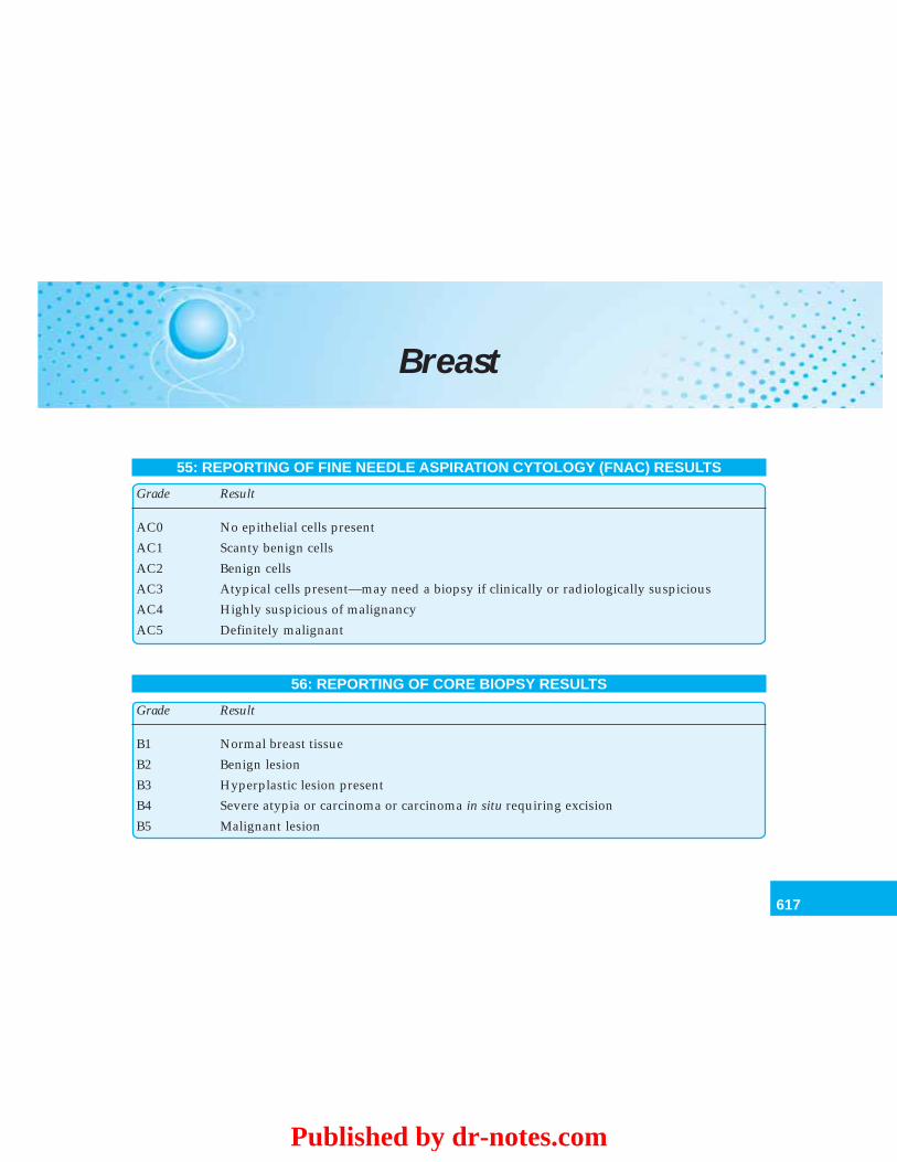
617
Breast
Breast
55: REPORTING OF FINE NEEDLE ASPIRATION CYTOLOGY (FNAC) RESULTS
Grade Result
AC0 No epithelial cells present
AC1 Scanty benign cells
AC2 Benign cells
AC3 Atypical cells present—may need a biopsy if clinically or radiologically suspicious
AC4 Highly suspicious of malignancy
AC5 Definitely malignant
56: REPORTING OF CORE BIOPSY RESULTS
Grade Result
B1 Normal breast tissue
B2 Benign lesion
B3 Hyperplastic lesion present
B4 Severe atypia or carcinoma or carcinoma in situ requiring excision
B5 Malignant lesion
Published by dr-notes.com
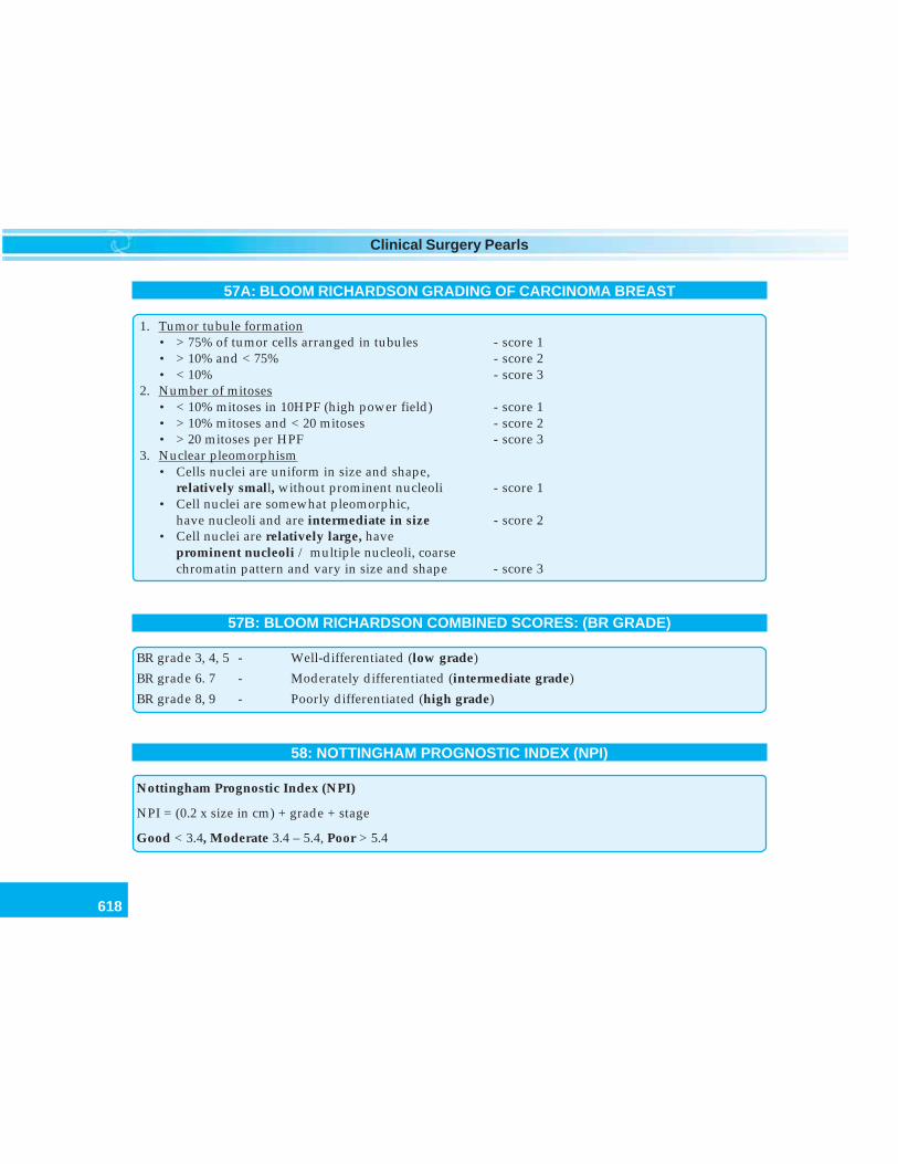
618
Clinical Surgery Pearls
57A: BLOOM RICHARDSON GRADING OF CARCINOMA BREAST
1. Tumor tubule formation• > 75% of tumor cells arranged in tubules - score 1• > 10% and < 75% - score 2• < 10% - score 3
2. Number of mitoses• < 10% mitoses in 10HPF (high power field) - score 1• > 10% mitoses and < 20 mitoses - score 2• > 20 mitoses per HPF - score 3
3. Nuclear pleomorphism• Cells nuclei are uniform in size and shape,
relatively small, without prominent nucleoli - score 1• Cell nuclei are somewhat pleomorphic,
have nucleoli and are intermediate in size - score 2• Cell nuclei are relatively large, have
prominent nucleoli / multiple nucleoli, coarsechromatin pattern and vary in size and shape - score 3
57B: BLOOM RICHARDSON COMBINED SCORES: (BR GRADE)
BR grade 3, 4, 5 - Well-differentiated (low grade)
BR grade 6. 7 - Moderately differentiated (intermediate grade)
BR grade 8, 9 - Poorly differentiated (high grade)
58: NOTTINGHAM PROGNOSTIC INDEX (NPI)
Nottingham Prognostic Index (NPI)
NPI = (0.2 x size in cm) + grade + stage
Good < 3.4, Moderate 3.4 – 5.4, Poor > 5.4
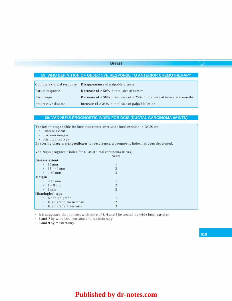
619
Breast
59: WHO DEFINITION OF OBJECTIVE RESPONSE TO ANTERIOR CHEMOTHERAPY
Complete clinical response Disappearance of palpable disease
Partial response Decrease of > 50% in total size of tumor
No change Decrease of < 50% or increase of < 25% in total size of tumor at 6 months
Progressive disease Increase of > 25% in total size of palpable lesion
60: VAN NUYS PROGNOSTIC INDEX FOR DCIS (DUCTAL CARCINOMA IN SITU)
The factors responsible for local recurrence after wide local excision in DCIS are:• Disease extent• Excision margin• Histological type
By scoring three major predictors for recurrence, a prognostic index has been developed.
Van Nuys prognostic index for DCIS (Ductal carcinoma in situ)Score
Disease extent• 15 mm 1• 15 – 40 mm 2• > 40 mm 3
Margin• < 10 mm 1• 1 – 9 mm 2• 1 mm 3
Histological type• Nonhigh grade 1• High grade, no necrosis 2• High grade + necrosis 3
• It is suggested that patients with score of 3, 4 and 5 be treated by wide local excision• 6 and 7 by wide local excision and radiotherapy• 8 and 9 by mastectomy.
Published by dr-notes.com
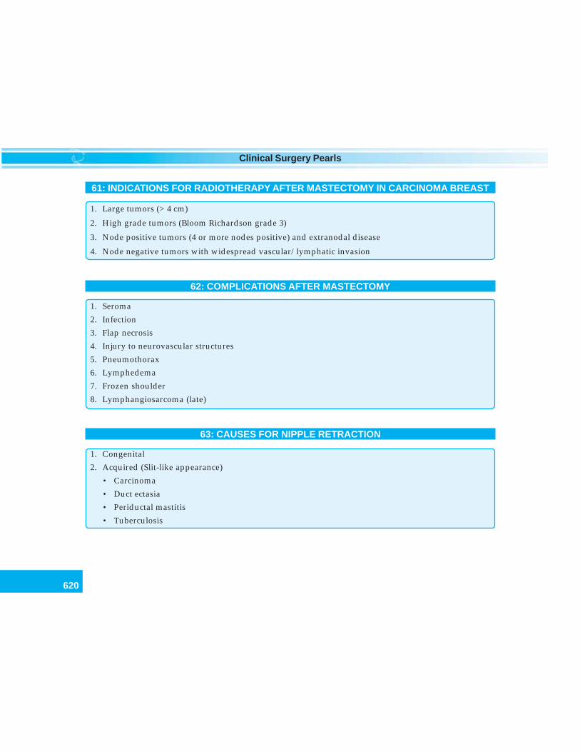
620
Clinical Surgery Pearls
61: INDICATIONS FOR RADIOTHERAPY AFTER MASTECTOMY IN CARCINOMA BREAST
1. Large tumors (> 4 cm)
2. High grade tumors (Bloom Richardson grade 3)
3. Node positive tumors (4 or more nodes positive) and extranodal disease
4. Node negative tumors with widespread vascular/lymphatic invasion
62: COMPLICATIONS AFTER MASTECTOMY
1. Seroma
2. Infection
3. Flap necrosis
4. Injury to neurovascular structures
5. Pneumothorax
6. Lymphedema
7. Frozen shoulder
8. Lymphangiosarcoma (late)
63: CAUSES FOR NIPPLE RETRACTION
1. Congenital
2. Acquired (Slit-like appearance)
• Carcinoma
• Duct ectasia
• Periductal mastitis
• Tuberculosis
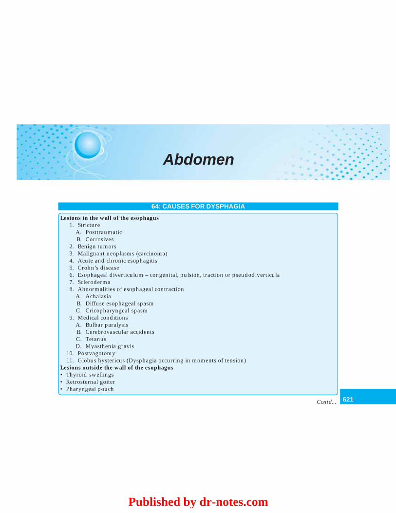
621
Abdomen
Abdomen
64: CAUSES FOR DYSPHAGIA
Lesions in the wall of the esophagus1. Stricture
A. PosttraumaticB. Corrosives
2. Benign tumors3. Malignant neoplasms (carcinoma)4. Acute and chronic esophagitis5. Crohn’s disease6. Esophageal diverticulum – congenital, pulsion, traction or pseudodiverticula7. Scleroderma8. Abnormalities of esophageal contraction
A. AchalasiaB. Diffuse esophageal spasmC. Cricopharyngeal spasm
9. Medical conditionsA. Bulbar paralysisB. Cerebrovascular accidentsC. TetanusD. Myasthenia gravis
10. Postvagotomy11. Globus hystericus (Dysphagia occurring in moments of tension)
Lesions outside the wall of the esophagus• Thyroid swellings• Retrosternal goiter• Pharyngeal pouch
Contd...
Published by dr-notes.com
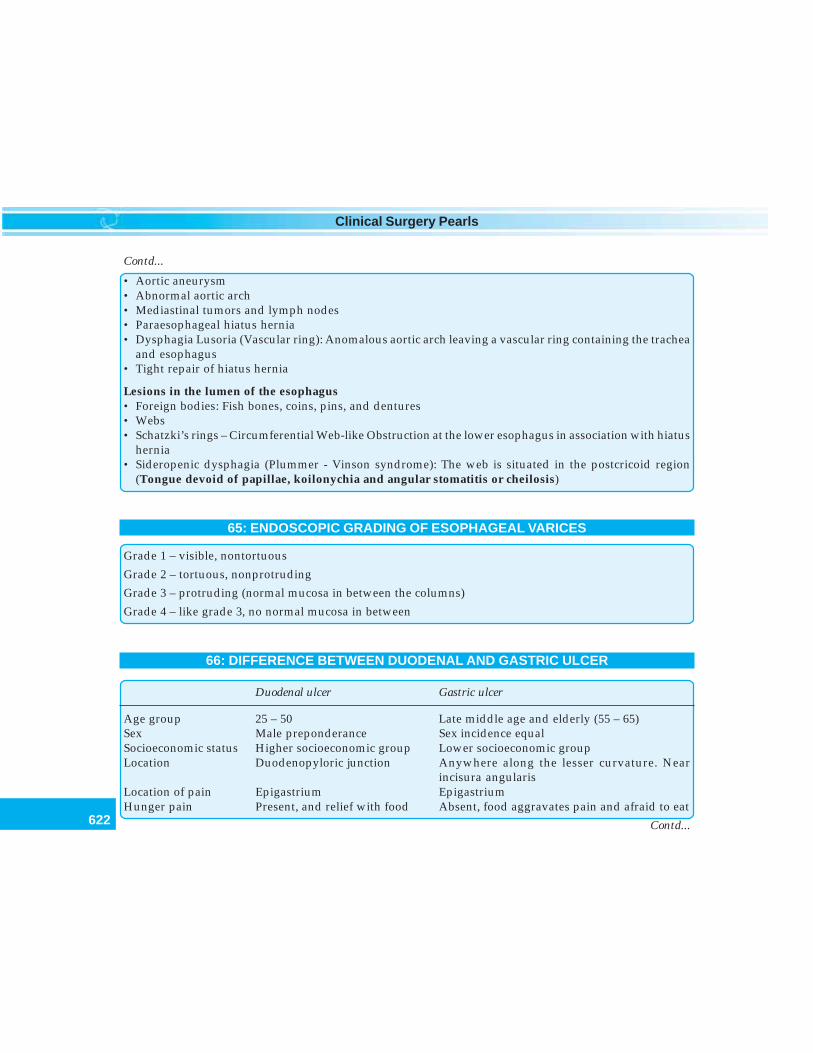
622
Clinical Surgery Pearls
• Aortic aneurysm• Abnormal aortic arch• Mediastinal tumors and lymph nodes• Paraesophageal hiatus hernia• Dysphagia Lusoria (Vascular ring): Anomalous aortic arch leaving a vascular ring containing the trachea
and esophagus• Tight repair of hiatus hernia
Lesions in the lumen of the esophagus• Foreign bodies: Fish bones, coins, pins, and dentures• Webs• Schatzki’s rings – Circumferential Web-like Obstruction at the lower esophagus in association with hiatus
hernia• Sideropenic dysphagia (Plummer - Vinson syndrome): The web is situated in the postcricoid region
(Tongue devoid of papillae, koilonychia and angular stomatitis or cheilosis)
65: ENDOSCOPIC GRADING OF ESOPHAGEAL VARICES
Grade 1 – visible, nontortuous
Grade 2 – tortuous, nonprotruding
Grade 3 – protruding (normal mucosa in between the columns)
Grade 4 – like grade 3, no normal mucosa in between
66: DIFFERENCE BETWEEN DUODENAL AND GASTRIC ULCER
Duodenal ulcer Gastric ulcer
Age group 25 – 50 Late middle age and elderly (55 – 65)Sex Male preponderance Sex incidence equalSocioeconomic status Higher socioeconomic group Lower socioeconomic groupLocation Duodenopyloric junction Anywhere along the lesser curvature. Near
incisura angularisLocation of pain Epigastrium EpigastriumHunger pain Present, and relief with food Absent, food aggravates pain and afraid to eat
Contd...
Contd...
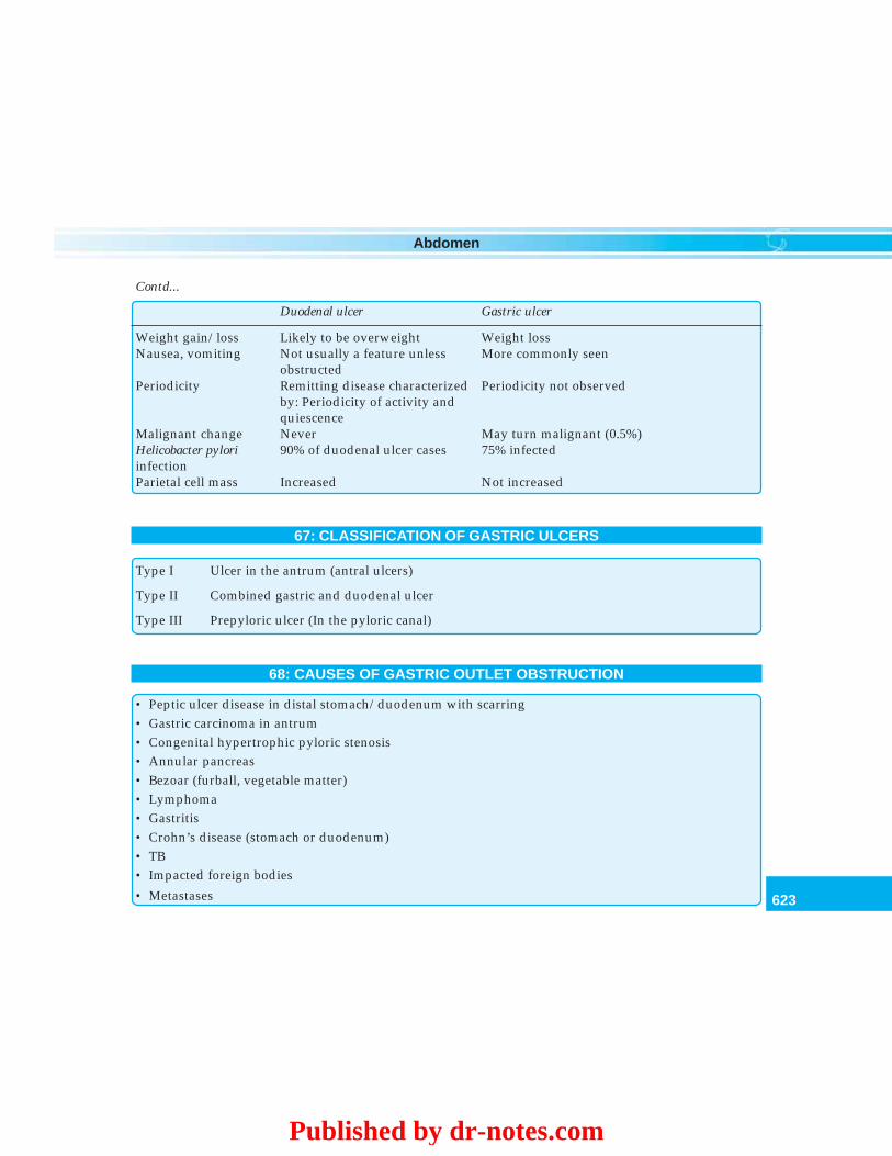
623
Abdomen
Weight gain/loss Likely to be overweight Weight lossNausea, vomiting Not usually a feature unless More commonly seen
obstructedPeriodicity Remitting disease characterized Periodicity not observed
by: Periodicity of activity andquiescence
Malignant change Never May turn malignant (0.5%)Helicobacter pylori 90% of duodenal ulcer cases 75% infectedinfectionParietal cell mass Increased Not increased
67: CLASSIFICATION OF GASTRIC ULCERS
Type I Ulcer in the antrum (antral ulcers)
Type II Combined gastric and duodenal ulcer
Type III Prepyloric ulcer (In the pyloric canal)
68: CAUSES OF GASTRIC OUTLET OBSTRUCTION
• Peptic ulcer disease in distal stomach/duodenum with scarring• Gastric carcinoma in antrum• Congenital hypertrophic pyloric stenosis• Annular pancreas• Bezoar (furball, vegetable matter)• Lymphoma• Gastritis• Crohn’s disease (stomach or duodenum)• TB• Impacted foreign bodies
• Metastases
Contd...
Duodenal ulcer Gastric ulcer
Published by dr-notes.com

624
Clinical Surgery Pearls
69: FORREST’S CLASSIFICATION FOR ACTIVITY OF BLEED IN UPPER GI ENDOSCOPY
Grade Ia: Spurting vessel at base of lesionGrade Ib: Ooze of blood from lesionGrade 2a: Nonbleeding visible vessel at base of lesionGrade 2b: Stigmata of Recent Hemorrhage (SRH): adherent clot, black spotGrade 3: Lesion seen but no evidence of recent bleed
70A: CHILD - TURCOTE - PUGH CLASSIFICATION OF FUNCTIONAL STATUS INLIVER DISEASE
Parameter Numerical score1 2 3
Class - A Class - B Class - CRisk – Low Risk – Moderate Risk – High
Ascites Absent Slight to Moderate TenseEncephalopathy None Grade I – II Grade III – IVSerum Albumin (g/dL) > 3.5 2.8 to 3.5 < 2.8Serum Bilirubin mg/dL < 2 2.0 – 3.0 > 3.0Prothrombin time (Seconds above control) < 4.0 4.0 – 6.0 > 6.0
70B:
Total score Child – Turcote – RiskPugh classification
5 – 6 A Low risk7 – 9 B Moderate risk10 – 15 C High risk
71: UICC STAGING OF GALLBLADDER CANCER
Stage I Confined to the mucosa/submucosaStage II Involvement of the muscle layerStage III Serosal involvementStage IV Spread to the cystic nodeStage V (advanced carcinoma) Invasion of the liver and adjacent organs
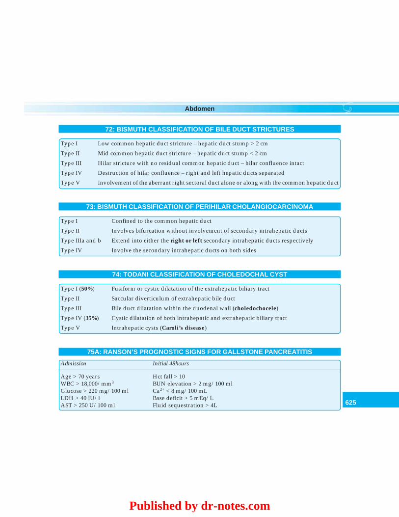
625
Abdomen
72: BISMUTH CLASSIFICATION OF BILE DUCT STRICTURES
Type I Low common hepatic duct stricture – hepatic duct stump > 2 cm
Type II Mid common hepatic duct stricture – hepatic duct stump < 2 cm
Type III Hilar stricture with no residual common hepatic duct – hilar confluence intact
Type IV Destruction of hilar confluence – right and left hepatic ducts separated
Type V Involvement of the aberrant right sectoral duct alone or along with the common hepatic duct
73: BISMUTH CLASSIFICATION OF PERIHILAR CHOLANGIOCARCINOMA
Type I Confined to the common hepatic duct
Type II Involves bifurcation without involvement of secondary intrahepatic ducts
Type IIIa and b Extend into either the right or left secondary intrahepatic ducts respectively
Type IV Involve the secondary intrahepatic ducts on both sides
74: TODANI CLASSIFICATION OF CHOLEDOCHAL CYST
Type I (50%) Fusiform or cystic dilatation of the extrahepatic biliary tract
Type II Saccular diverticulum of extrahepatic bile duct
Type III Bile duct dilatation within the duodenal wall (choledochocele)
Type IV (35%) Cystic dilatation of both intrahepatic and extrahepatic biliary tract
Type V Intrahepatic cysts (Caroli’s disease)
75A: RANSON’S PROGNOSTIC SIGNS FOR GALLSTONE PANCREATITIS
Admission Initial 48hours
Age > 70 years Hct fall > 10WBC > 18,000/mm3 BUN elevation > 2 mg/100 mlGlucose > 220 mg/100 ml Ca2+ < 8 mg/100 mLLDH > 40 IU/l Base deficit > 5 mEq/LAST > 250 U/100 ml Fluid sequestration > 4L
Published by dr-notes.com
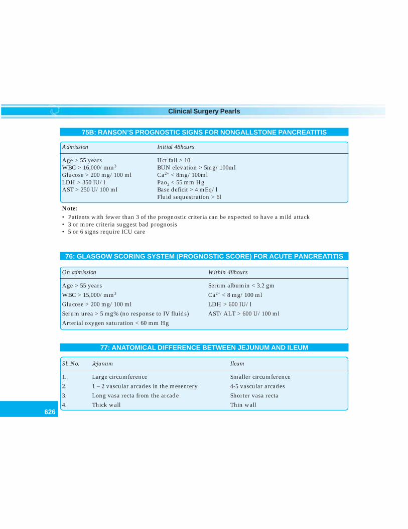
626
Clinical Surgery Pearls
75B: RANSON’S PROGNOSTIC SIGNS FOR NONGALLSTONE PANCREATITIS
Admission Initial 48hours
Age > 55 years Hct fall > 10WBC > 16,000/mm3 BUN elevation > 5mg/100mlGlucose > 200 mg/100 ml Ca2+ < 8mg/100mlLDH > 350 IU/l Pao2 < 55 mm HgAST > 250 U/100 ml Base deficit > 4 mEq/l
Fluid sequestration > 6l
Note:• Patients with fewer than 3 of the prognostic criteria can be expected to have a mild attack• 3 or more criteria suggest bad prognosis• 5 or 6 signs require ICU care
76: GLASGOW SCORING SYSTEM (PROGNOSTIC SCORE) FOR ACUTE PANCREATITIS
On admission Within 48hours
Age > 55 years Serum albumin < 3.2 gm
WBC > 15,000/mm3 Ca2+ < 8 mg/100 ml
Glucose > 200 mg/100 ml LDH > 600 IU/l
Serum urea > 5 mg% (no response to IV fluids) AST/ALT > 600 U/100 ml
Arterial oxygen saturation < 60 mm Hg
77: ANATOMICAL DIFFERENCE BETWEEN JEJUNUM AND ILEUM
Sl. No: Jejunum Ileum
1. Large circumference Smaller circumference
2. 1 – 2 vascular arcades in the mesentery 4-5 vascular arcades
3. Long vasa recta from the arcade Shorter vasa recta
4. Thick wall Thin wall
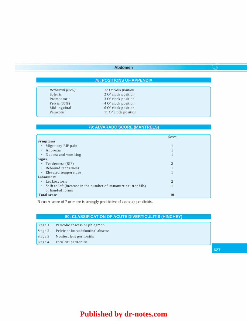
627
Abdomen
78: POSITIONS OF APPENDIX
Retrocecal (65%) 12 O’ clock positionSplenic 2 O’ clock positionPromontoric 3 O’ clock positionPelvic (30%) 4 O’ clock positionMid inguinal 6 O’ clock positionParacolic 11 O’ clock position
79: ALVARADO SCORE (MANTRELS)
ScoreSymptoms
• Migratory RIF pain 1• Anorexia 1• Nausea and vomiting 1
Signs• Tenderness (RIF) 2• Rebound tenderness 1• Elevated temperature 1
Laboratory• Leukocytosis 2• Shift to left (increase in the number of immature neutrophils) 1
or banded forms Total score 10
Note: A score of 7 or more is strongly predictive of acute appendicitis.
80: CLASSIFICATION OF ACUTE DIVERTICULITIS (HINCHEY)
Stage 1 Pericolic abscess or phlegmon
Stage 2 Pelvic or intraabdominal abscess
Stage 3 Nonfeculent peritonitis
Stage 4 Feculent peritonitis
Published by dr-notes.com
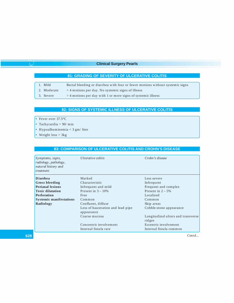
628
Clinical Surgery Pearls
81: GRADING OF SEVERITY OF ULCERATIVE COLITIS
1. Mild Rectal bleeding or diarrhea with four or fewer motions without systemic signs
2. Moderate > 4 motions per day. No systemic signs of illness
3. Severe > 4 motions per day with 1 or more signs of systemic illness
82: SIGNS OF SYSTEMIC ILLNESS OF ULCERATIVE COLITIS
• Fever over 37.5oC
• Tachycardia > 90/min
• Hypoalbuminemia < 3 gm/liter
• Weight loss > 3kg
83: COMPARISON OF ULCERATIVE COLITIS AND CROHN’S DISEASE
Symptoms, signs, Ulcerative colitis Crohn’s diseaseradiology, pathology,natural history andtreatment
Diarrhea Marked Less severeGross bleeding Characteristic InfrequentPerianal lesions Infrequent and mild Frequent and complexToxic dilatation Present in 3 – 10% Present in 2 – 5%Perforation Free LocalizedSystemic manifestations Common CommonRadiology Confluent, diffuse Skip areas
Loss of haustration and lead pipe Cobble-stone appearanceappearanceCoarse mucosa Longitudinal ulcers and transverse
ridgesConcentric involvement Eccentric involvementInternal fistula rare Internal fistula common
Contd...
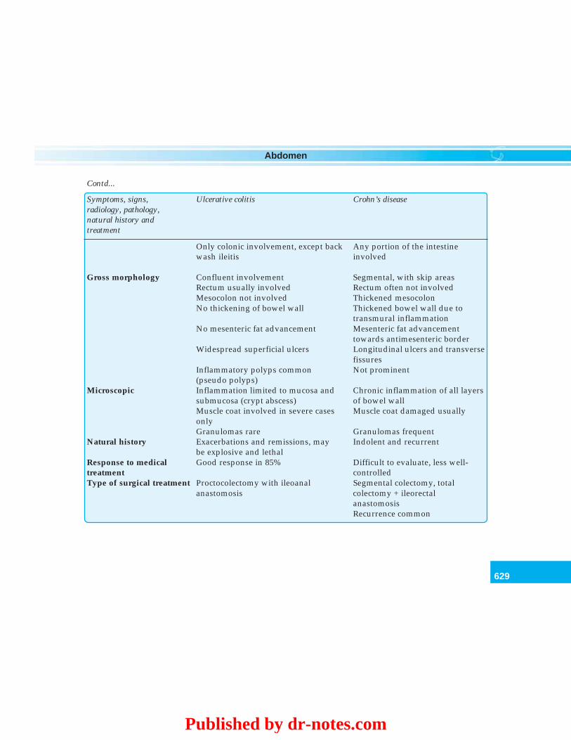
629
Abdomen
Only colonic involvement, except back Any portion of the intestinewash ileitis involved
Gross morphology Confluent involvement Segmental, with skip areasRectum usually involved Rectum often not involvedMesocolon not involved Thickened mesocolonNo thickening of bowel wall Thickened bowel wall due to
transmural inflammationNo mesenteric fat advancement Mesenteric fat advancement
towards antimesenteric borderWidespread superficial ulcers Longitudinal ulcers and transverse
fissuresInflammatory polyps common Not prominent(pseudo polyps)
Microscopic Inflammation limited to mucosa and Chronic inflammation of all layerssubmucosa (crypt abscess) of bowel wallMuscle coat involved in severe cases Muscle coat damaged usuallyonlyGranulomas rare Granulomas frequent
Natural history Exacerbations and remissions, may Indolent and recurrentbe explosive and lethal
Response to medical Good response in 85% Difficult to evaluate, less well-treatment controlledType of surgical treatment Proctocolectomy with ileoanal Segmental colectomy, total
anastomosis colectomy + ileorectalanastomosisRecurrence common
Contd...
Symptoms, signs, Ulcerative colitis Crohn’s diseaseradiology, pathology,natural history andtreatment
Published by dr-notes.com
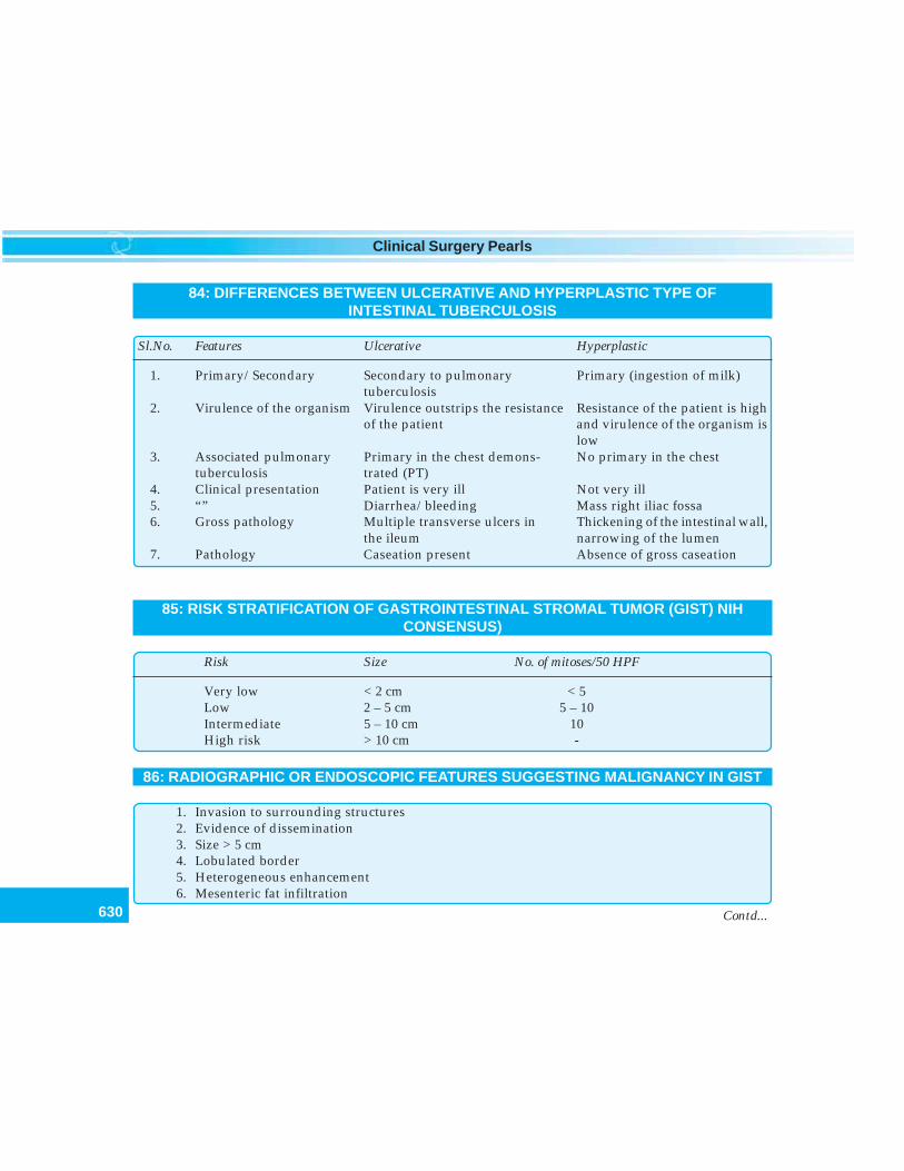
630
Clinical Surgery Pearls
84: DIFFERENCES BETWEEN ULCERATIVE AND HYPERPLASTIC TYPE OFINTESTINAL TUBERCULOSIS
Sl.No. Features Ulcerative Hyperplastic
1. Primary/Secondary Secondary to pulmonary Primary (ingestion of milk)tuberculosis
2. Virulence of the organism Virulence outstrips the resistance Resistance of the patient is highof the patient and virulence of the organism is
low3. Associated pulmonary Primary in the chest demons- No primary in the chest
tuberculosis trated (PT)4. Clinical presentation Patient is very ill Not very ill5. “” Diarrhea/bleeding Mass right iliac fossa6. Gross pathology Multiple transverse ulcers in Thickening of the intestinal wall,
the ileum narrowing of the lumen7. Pathology Caseation present Absence of gross caseation
85: RISK STRATIFICATION OF GASTROINTESTINAL STROMAL TUMOR (GIST) NIHCONSENSUS)
Risk Size No. of mitoses/50 HPF
Very low < 2 cm < 5Low 2 – 5 cm 5 – 10Intermediate 5 – 10 cm 10High risk > 10 cm -
86: RADIOGRAPHIC OR ENDOSCOPIC FEATURES SUGGESTING MALIGNANCY IN GIST
1. Invasion to surrounding structures2. Evidence of dissemination3. Size > 5 cm4. Lobulated border5. Heterogeneous enhancement6. Mesenteric fat infiltration
Contd...
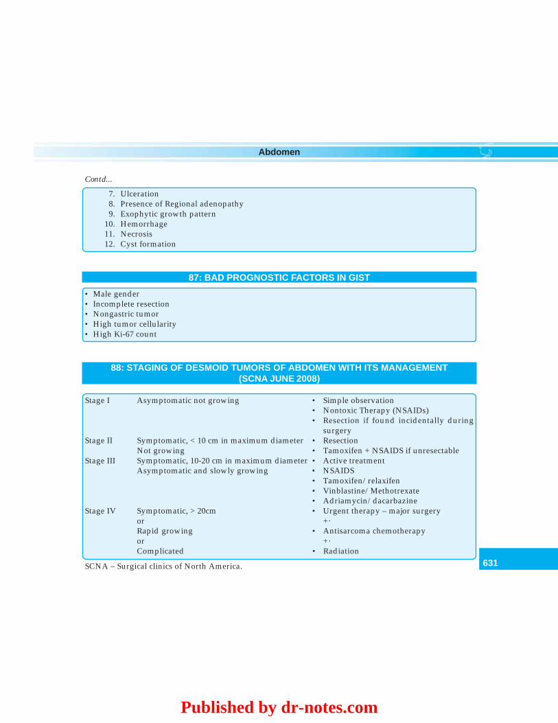
631
Abdomen
7. Ulceration8. Presence of Regional adenopathy9. Exophytic growth pattern
10. Hemorrhage11. Necrosis12. Cyst formation
87: BAD PROGNOSTIC FACTORS IN GIST
• Male gender• Incomplete resection• Nongastric tumor• High tumor cellularity• High Ki-67 count
88: STAGING OF DESMOID TUMORS OF ABDOMEN WITH ITS MANAGEMENT(SCNA JUNE 2008)
Stage I Asymptomatic not growing • Simple observation• Nontoxic Therapy (NSAIDs)• Resection if found incidentally during
surgeryStage II Symptomatic, < 10 cm in maximum diameter • Resection
Not growing • Tamoxifen + NSAIDS if unresectableStage III Symptomatic, 10-20 cm in maximum diameter • Active treatment
Asymptomatic and slowly growing • NSAIDS• Tamoxifen/relaxifen• Vinblastine/Methotrexate• Adriamycin/dacarbazine
Stage IV Symptomatic, > 20cm • Urgent therapy – major surgeryor +·Rapid growing • Antisarcoma chemotherapyor +·Complicated • Radiation
Contd...
SCNA – Surgical clinics of North America.
Published by dr-notes.com
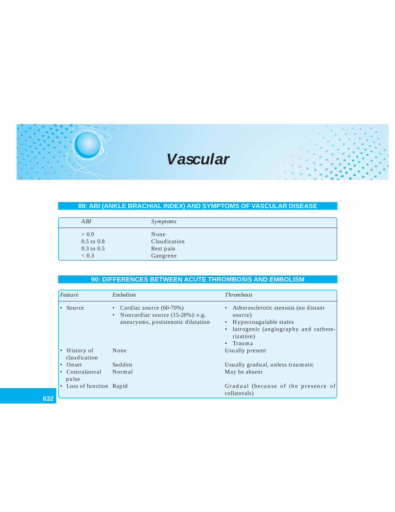
632
Clinical Surgery Pearls
Vascular
89: ABI (ANKLE BRACHIAL INDEX) AND SYMPTOMS OF VASCULAR DISEASE
ABI Symptoms
> 0.9 None0.5 to 0.8 Claudication0.3 to 0.5 Rest pain< 0.3 Gangrene
90: DIFFERENCES BETWEEN ACUTE THROMBOSIS AND EMBOLISM
Feature Embolism Thrombosis
• Source • Cardiac source (60-70%) • Atherosclerotic stenosis (no distant• Noncardiac source (15-20%): e.g. source)
aneurysms, poststenotic dilatation • Hypercoagulable states• Iatrogenic (angiography and cathete-
rization)• Trauma
• History of None Usually presentclaudication
• Onset Sudden Usually gradual, unless traumatic• Contralateral Normal May be absent
pulse• Loss of function Rapid Gradual (because of the presence of
collaterals)
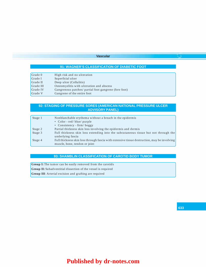
633
Vascular
91: WAGNER’S CLASSIFICATION OF DIABETIC FOOT
Grade 0 High risk and no ulcerationGrade I Superficial ulcerGrade II Deep ulcer (Cellulitis)Grade III Osteomyelitis with ulceration and abscessGrade IV Gangrenous patches/partial foot gangrene (fore foot)Grade V Gangrene of the entire foot
92: STAGING OF PRESSURE SORES (AMERICAN NATIONAL PRESSURE ULCERADVISORY PANEL)
Stage 1 Nonblanchable erythema without a breach in the epidermis• Color - red/blue/purple• Consistency - firm/boggy
Stage 2 Partial thickness skin loss involving the epidermis and dermisStage 3 Full thickness skin loss extending into the subcutaneous tissue but not through the
underlying fasciaStage 4 Full thickness skin loss through fascia with extensive tissue destruction, may be involving
muscle, bone, tendon or joint
93: SHAMBLIN CLASSIFICATION OF CAROTID BODY TUMOR
Group I: The tumor can be easily removed from the carotids
Group II: Subadventitial dissection of the vessel is required
Group III: Arterial excision and grafting are required
Published by dr-notes.com
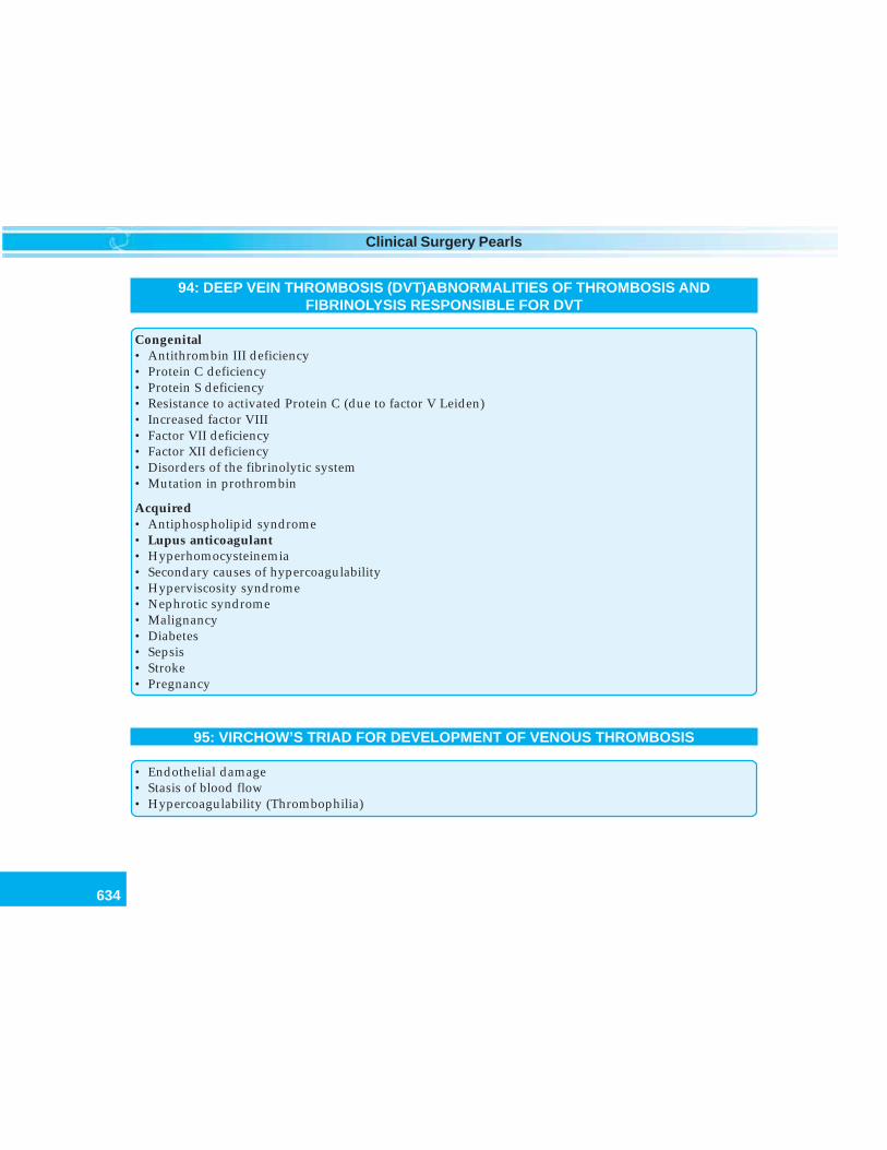
634
Clinical Surgery Pearls
94: DEEP VEIN THROMBOSIS (DVT)ABNORMALITIES OF THROMBOSIS ANDFIBRINOLYSIS RESPONSIBLE FOR DVT
Congenital• Antithrombin III deficiency• Protein C deficiency• Protein S deficiency• Resistance to activated Protein C (due to factor V Leiden)• Increased factor VIII• Factor VII deficiency• Factor XII deficiency• Disorders of the fibrinolytic system• Mutation in prothrombin
Acquired• Antiphospholipid syndrome• Lupus anticoagulant• Hyperhomocysteinemia• Secondary causes of hypercoagulability• Hyperviscosity syndrome• Nephrotic syndrome• Malignancy• Diabetes• Sepsis• Stroke• Pregnancy
95: VIRCHOW’S TRIAD FOR DEVELOPMENT OF VENOUS THROMBOSIS
• Endothelial damage• Stasis of blood flow• Hypercoagulability (Thrombophilia)
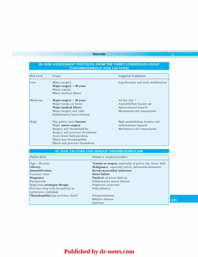
635
Vascular
96: RISK ASSESSMENT PROTOCOL FROM THE THRIFT CONSENSUS GROUP(THROMBOEMBOLIC RISK FACTORS)
Risk Level Group Suggested Prophylaxis
Low Minor surgery Leg elevation and early mobilizationMajor surgery < 40 yearsMinor traumaMinor medical illness
Moderate Major surgery > 40 years As low risk +Major trauma or burns Antiembolism hosiery orMajor medical illness Subcutaneous heparinMinor surgery and risks Mechanical calf compressionInflammatory bowel disease
High Hip, pelvis, knee fracture Both antiembolism hosiery andMajor cancer surgery subcutaneous heparinSurgery and thrombophilia Mechanical calf compressionSurgery and previous thrombosisAcute lower limb paralysisIllness and thrombophiliaIllness and previous thrombosis
97: RISK FACTORS FOR VENOUS THROMBOEMBOLISM
Patient factor Disease or surgical procedure
Age > 60 years Trauma or surgery, especially of pelvis, hip, lower limbObesity Malignancy, especially pelvis, abdominal metastaticImmobilization Recent myocardial infarctionVaricose veins Heart failurePregnancy Paralysis of lower limb (s)Puerperium Inflammatory bowel diseaseHigh dose oestrogen therapy Nephrotic syndromePrevious deep vein thrombosis or Polycythemiapulmonary embolismThrombophilia [see previous chart] Paraproteinemia
Behçet’s diseaseInfection
Published by dr-notes.com
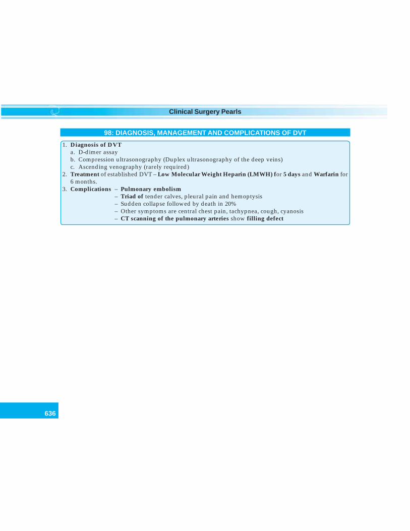
636
Clinical Surgery Pearls
98: DIAGNOSIS, MANAGEMENT AND COMPLICATIONS OF DVT
1. Diagnosis of DVTa. D-dimer assayb. Compression ultrasonography (Duplex ultrasonography of the deep veins)c. Ascending venography (rarely required)
2. Treatment of established DVT – Low Molecular Weight Heparin (LMWH) for 5 days and Warfarin for6 months.
3. Complications – Pulmonary embolism– Triad of tender calves, pleural pain and hemoptysis– Sudden collapse followed by death in 20%– Other symptoms are central chest pain, tachypnea, cough, cyanosis– CT scanning of the pulmonary arteries show filling defect
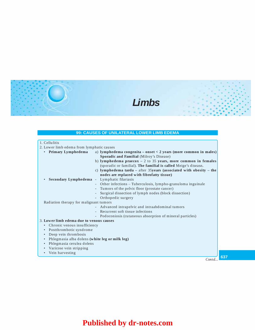
637
Limbs
Limbs
99: CAUSES OF UNILATERAL LOWER LIMB EDEMA
1. Cellulitis2. Lower limb edema from lymphatic causes
• Primary Lymphedema a) lymphedema congenita – onset < 2 years (more common in males)Sporadic and Familial (Milroy’s Disease)
b) lymphedema praecox – 2 to 35 years, more common in females(sporadic or familial). The familial is called Meige’s disease.
c) lymphedema tarda – after 35years (associated with obesity – thenodes are replaced with fibrofatty tissue)
• Secondary Lymphedema - Lymphatic filariasis- Other infections – Tuberculosis, lympho-granuloma inguinale- Tumors of the pelvic floor (prostate cancer)- Surgical dissection of lymph nodes (block dissection)- Orthopedic surgery
Radiation therapy for malignant tumors- Advanced intrapelvic and intraabdominal tumors- Recurrent soft tissue infections- Podoconiosis (cutaneous absorption of mineral particles)
3. Lower limb edema due to venous causes• Chronic venous insufficiency• Postthrombotic syndrome• Deep vein thrombosis• Phlegmasia alba dolens (white leg or milk leg)• Phlegmasia cerulea dolens• Varicose vein stripping• Vein harvesting
Contd...
Published by dr-notes.com

638
Clinical Surgery Pearls
4. Arterial causes: Arteriovenous malformationAneurysmIschemia - reperfusion (following lower limb revascularization)
5. Edema secondary to congenital vascular anomalies- Lymphatic angiodysplasia syndrome- Klippel – Trenaunay’s syndrome- Hyperstomy syndrome
6. Posttraumatic: Sympathetic dystrophy
7. Obesity: Lipodystrophy. Lipoidosis
8. Gigantism
9. Retroperitoneal fibrosis: Causes arterial, venous and lymphatic abnormalities
10. Hansen’s disease
11. Dermal leishmaniasis
12. Mycetoma
13. Allergic disorders – Angioedema
100A: GRADING OF LYMPHOEDEMA (BRUNNER)
Subclinical Excess interstitial fluid is present with histological abnormalities in lymphatics and lymphnodes. No clinically apparent lymphedema
Grade I Edema pits on pressure-Swelling largely/completely disappears on elevation/bed rest
Grade II Nonpitting edema. Does not significantly reduce upon elevation
Grade III Nonpitting edema associated with irreversible skin changes, i.e. fibrosis, papillae, etc.
100B: ANOTHER GRADING OF LYMPHOEDEMA
Mild < 20% excess limb volume
Moderate 20 – 40 % excess limb volume
Severe > 40% excess limb volume
Contd...
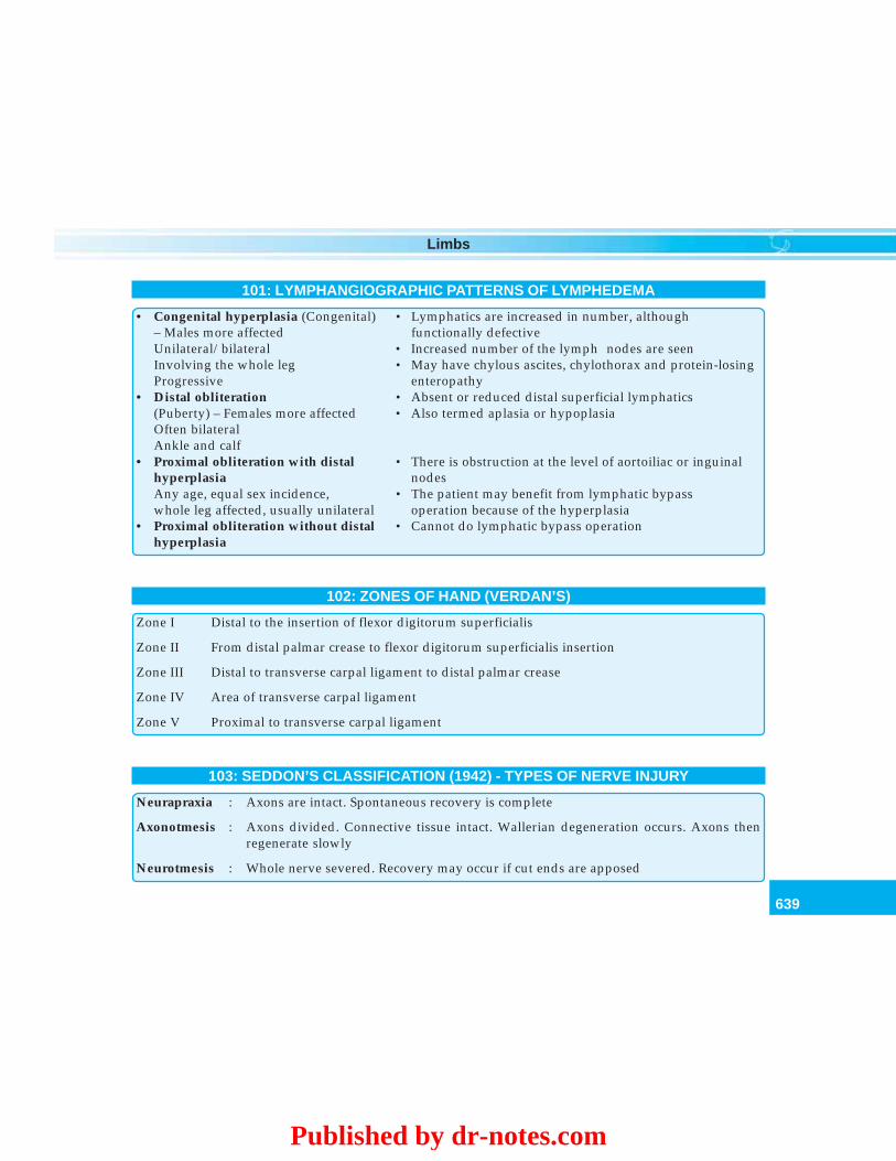
639
Limbs
101: LYMPHANGIOGRAPHIC PATTERNS OF LYMPHEDEMA
• Congenital hyperplasia (Congenital) • Lymphatics are increased in number, although– Males more affected functionally defectiveUnilateral/bilateral • Increased number of the lymph nodes are seenInvolving the whole leg • May have chylous ascites, chylothorax and protein-losingProgressive enteropathy
• Distal obliteration • Absent or reduced distal superficial lymphatics(Puberty) – Females more affected • Also termed aplasia or hypoplasiaOften bilateralAnkle and calf
• Proximal obliteration with distal • There is obstruction at the level of aortoiliac or inguinalhyperplasia nodesAny age, equal sex incidence, • The patient may benefit from lymphatic bypasswhole leg affected, usually unilateral operation because of the hyperplasia
• Proximal obliteration without distal • Cannot do lymphatic bypass operationhyperplasia
102: ZONES OF HAND (VERDAN’S)
Zone I Distal to the insertion of flexor digitorum superficialis
Zone II From distal palmar crease to flexor digitorum superficialis insertion
Zone III Distal to transverse carpal ligament to distal palmar crease
Zone IV Area of transverse carpal ligament
Zone V Proximal to transverse carpal ligament
103: SEDDON’S CLASSIFICATION (1942) - TYPES OF NERVE INJURY
Neurapraxia : Axons are intact. Spontaneous recovery is complete
Axonotmesis : Axons divided. Connective tissue intact. Wallerian degeneration occurs. Axons thenregenerate slowly
Neurotmesis : Whole nerve severed. Recovery may occur if cut ends are apposed
Published by dr-notes.com
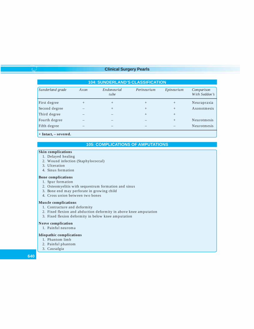
640
Clinical Surgery Pearls
104: SUNDERLAND’S CLASSIFICATION
Sunderland grade Axon Endoneurial Perineurium Epineurium Comparisontube With Seddon’s
First degree + + + + Neurapraxia
Second degree – + + + Axonotmesis
Third degree – – + +
Fourth degree – – – + Neurotmesis
Fifth degree – – – – Neurotmesis
+ Intact, – severed.
105: COMPLICATIONS OF AMPUTATIONS
Skin complications1. Delayed healing2. Wound infection (Staphylococcal)3. Ulceration4. Sinus formation
Bone complications1. Spur formation2. Osteomyelitis with sequestrum formation and sinus3. Bone end may perforate in growing child4. Cross union between two bones
Muscle complications1. Contracture and deformity2. Fixed flexion and abduction deformity in above knee amputation3. Fixed flexion deformity in below knee amputation
Nerve complication1. Painful neuroma
Idiopathic complications1. Phantom limb2. Painful phantom3. Causalgia

641
Limbs
106: SITE OF ELECTION FOR ABOVE KNEE AND BELOW KNEE AMPUTATION
Above knee – 10 – 12 inches (25-30cm) below the greater trochanter
Below knee – 5½ inch (14cm) below the tibial plateau
Published by dr-notes.com
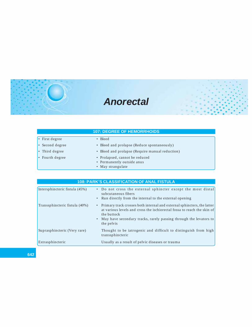
642
Clinical Surgery Pearls
Anorectal
107: DEGREE OF HEMORRHOIDS
• First degree • Bleed
• Second degree • Bleed and prolapse (Reduce spontaneously)
• Third degree • Bleed and prolapse (Require manual reduction)
• Fourth degree • Prolapsed, cannot be reduced• Permanently outside anus• May strangulate
108: PARK’S CLASSIFICATION OF ANAL FISTULA
Intersphincteric fistula (45%) • Do not cross the external sphincter except the most distalsubcutaneous fibers
• Run directly from the internal to the external opening
Transsphincteric fistula (40%) • Primary track crosses both internal and external sphincters, the latterat various levels and cross the ischiorectal fossa to reach the skin ofthe buttock
• May have secondary tracks, rarely passing through the levators tothe pelvis
Suprasphincteric (Very rare) Thought to be iatrogenic and difficult to distinguish from hightranssphincteric
Extrasphincteric Usually as a result of pelvic diseases or trauma
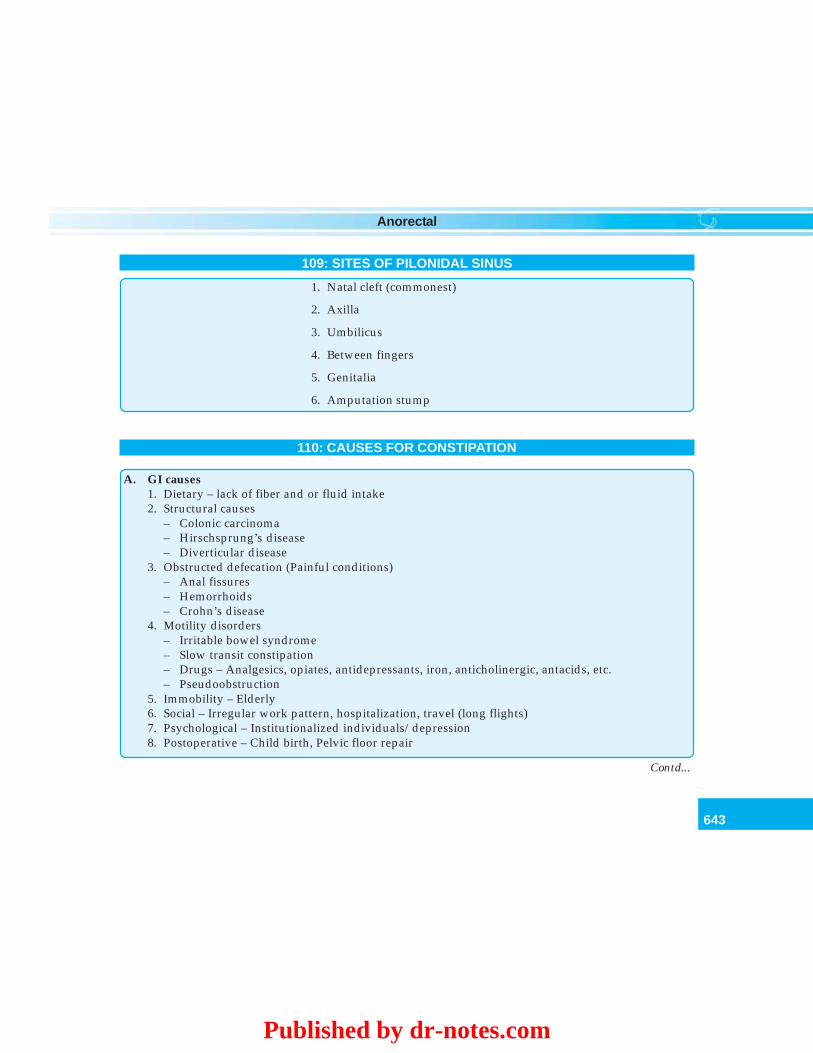
643
Anorectal
109: SITES OF PILONIDAL SINUS
1. Natal cleft (commonest)
2. Axilla
3. Umbilicus
4. Between fingers
5. Genitalia
6. Amputation stump
110: CAUSES FOR CONSTIPATION
A. GI causes1. Dietary – lack of fiber and or fluid intake2. Structural causes
– Colonic carcinoma– Hirschsprung’s disease– Diverticular disease
3. Obstructed defecation (Painful conditions)– Anal fissures– Hemorrhoids– Crohn’s disease
4. Motility disorders– Irritable bowel syndrome– Slow transit constipation– Drugs – Analgesics, opiates, antidepressants, iron, anticholinergic, antacids, etc.– Pseudoobstruction
5. Immobility – Elderly6. Social – Irregular work pattern, hospitalization, travel (long flights)7. Psychological – Institutionalized individuals/depression8. Postoperative – Child birth, Pelvic floor repair
Contd...
Published by dr-notes.com
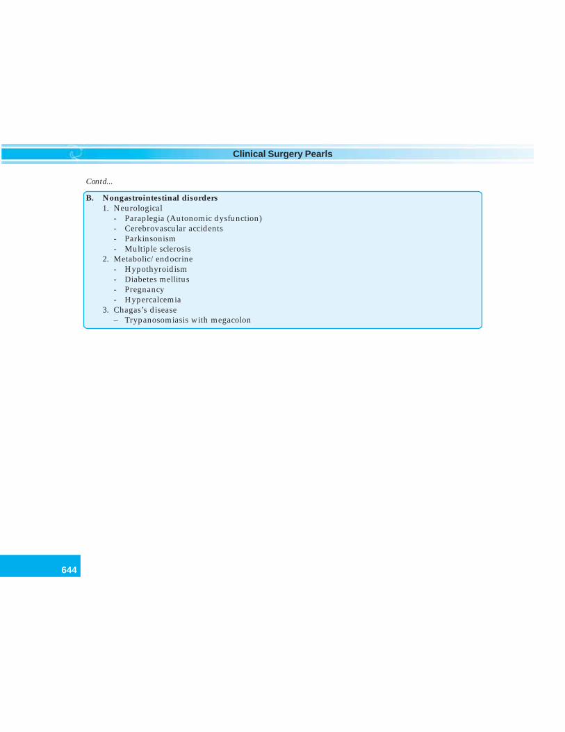
644
Clinical Surgery Pearls
B. Nongastrointestinal disorders1. Neurological
- Paraplegia (Autonomic dysfunction)- Cerebrovascular accidents- Parkinsonism- Multiple sclerosis
2. Metabolic/endocrine- Hypothyroidism- Diabetes mellitus- Pregnancy- Hypercalcemia
3. Chagas’s disease– Trypanosomiasis with megacolon
Contd...

645
Index
Index
AAbdominal
apoplexy 3cocoon 134cystic lymphangioma
in adults 261tuberculosis 133, 134
Aberrant thyroid 372Abscess 3Absent
bilirubin 166urobilinogen 166
Absolute constipation 539ABVD regime 229Achalasia 565Acholuric jaundice 161Acrocynosis 208Actinic keratoses 435Actions of Dartos and
Cremaster 477Activities of daily living (ADL) 3Acute
abdomen 3bacterial sialadenitis 348fluid collection 249leukemia 218limb ischemia 213pancreatitis 15parotitis 348
pseudocyst 248toxic cholangitis 169
Adenocarcinoma 237Adenomatous polyps of colon 140Adiposis dolorosa 396Adson’s test 201, 559Advanced trauma life support
system (ATLS) 606Agenesis 3Alcohol 333Alkaline
phosphatase 165urine 576
Alvarado score 153, 627Ambulatory venous
hypertension 177venous pressure study 185
American College of SurgeonsCommittee on trauma 606Ampullary carcinoma 137Amputation neuroma 390Amsterdam criteria 138Amylase 3Amyloid stroma 68Ancillary procedures 226Ankle edema 180Ankyloglossia 3, 15Ann-Arbor staging 223Anterior chemotherapy 101
Antibiotic prophylaxis in colonicsurgery 143Antiestrogenic action 89Antituberculous
drugs 135regime 134
Apathetic hyperthyroidism 3Appendicular
abscess 150mass 150
Apple-Jelly granulation 408Arc of Riolan 4Aromatase inhibitors 90Artefact ulcer 409Arterial injection 206Arteriography 204Arteriovenous fistula 405, 455Artery of death 492Ascending vessels
lateral 59medial 59
Ascites 278Ascitic fluid study 132Astler-Coller staging 141Atherosclerosis 206Atresia 3Atrophie blanche 183Axillary
clearance 104
Published by dr-notes.com
646
Clinical Surgery Pearls
dissection 88lymph nodes 80, 88, 100node 86, 216sampling 104
BBacteremia 4Bacteroides fusiformis 410Bad prognostic factors 229Balanitis 442Balanoposthitis 443Ballottement 235Banti’s syndrome 287Barium
enema 580meal examination 110
Barrett’s esophagus 4Basal
cell carcinoma 428epidermis 428
Bashful bladder 18Bassini’s herniorrhaphy 490Bazin’s disease 409Beger procedure 547Bendavid classification 487Benign
papilloma 16swellings 256
Bilateralbreast cancer 94hydronephrosis 237
Bilharzial cirrhosis 218Biliary colic 4Billiard ball testis 266Bilocular hydrocele 471Bimanual palpation 363Biopsy
thyroid 52ulcer 191
Bird beak 539Bisgard regime 191
Bismuth classification 171Black stones 548Blanching 207Bleeding time 166Blind biopsy 318Blood pressure 194Bloom - Richardson grading 86Blumer’s shelf 108, 110Bone marrow biopsy 226Borrmann type IV 116Bow’ sign 475Bowen’s disease 434Boyd’s grading of claudication 198Brachytherapy 337Brain metastasis 99Branchial cyst 375, 378Breast 517
biopsy 82cancer surgery 88carcinoma 4conservation 86
Breslow’s thickness 418Brilliantly translucent 377Brim of pelvis 155Brodie-Trendelenburg test 181Bronchoscopy 318Brown pigment stones 548Bruit 5,202Buccal
cancers 341mucosa 15
Budd-Chiari syndrome 286Buerger’s
disease 196, 205, 206vascular angle 199
Bunion 6Burkitt’s lymphoma 231Burns 6Bursae 6Buschke-Lowenstein tumor 444Bypass graft 212
CCabana’s node 447Café-au-lait spot 391Calcium channel blocker 524Campbell De Morgan spot 455Candidal infection 336Cannon balls 99, 542Carbohydrate antigen 167Carbon dioxide laser 432Carbuncle 4,6Carcinoembryonic antigen 146Carcinoma
breast 79cecum 131cecum 135colon 136, 139head of pancreas 167male breast 524penis 443tongue 330,332
Caroli’s disease 163, 625Catecholamine excess 258Cecal growth 143Cell nests 436Cellulitis 9,466Cellulitis of scrotum 6Central nervous system 155Cervical lymph node 216, 309Cervical rib 201, 557Charcot’s triad 157, 169, 548Charles procedure 467Chassaigne tubercle 201Chemical sympathectomy 211Chemoradiation 117,345Chemotherapeutic regimen 274, 386Chemotherapy 89, 101, 105, 118, 229
HCC 128pancreatic cancer 168seminoma 274
Chernobyl nuclear disaster 62

647
Index
Chestmetastasis 99wall 84
Chevassu maneuver 272Child-Turcolte-Pugh classification284, 624Child-Pugh grading 127Child’s classification 285Chimney Sweeps cancer 436Cholangiocarcinoma 171Cholecystitis 4Choledochal cyst 162, 163, 625Choudary Millis criteria 94Christian Johann Doppler 203Chronic
arterial occlusion 208leukemia 218occlusive disease 208pancreatitis from carcinoma 167pseudocyst 248sialadenitis 348, 359
Chronology of descent of testis 264Chylolymphatic cyst 298Cigarette smoking 156Circum areolar 83Circumcision and biopsy 444Cirsoid aneurysm 456Clark’s level 420Claudication 196
distance 197pain 197rest pain 6
Clay-colored stool 166Clear cell tumor 237Clergyman’s knee 6, 7Cloquet’s hernia 497Clot colic 239Clotting time 166CNS lymphoma 232Cock’s peculiar tumor 399
Crossed leg test of Fuchsig 201CT
liver mass 125scan 240urography 240
Curling’s ulcer 614Cyst 8Cystic hygroma 377Cystogastrostomy 252Cystosarcoma phyllodes 528Cytology 112
DD2 gastrectomy 117Decubitus
films 541ulcer 410
Deepring occlusion test 478vein thrombosis 187veins 176venous thrombosis 185
Dercum’s disease 396Dermal flares 179Dermoid cysts 401Descending
lateral 59medial 59
Diabetes mellitus 157, 163, 402Diabetic ulcer 411Diagnostic algorithm for neck
swelling 303Diarrhea 8Dietl’s crisis 8, 245, 563Differentiated thyroid cancer
(DTC) 62, 65Diffuse
gastric cancer 114sclerosing variant 63
Digital substractionangiography 205
Coffee ground vomitus 280Coil spring appearance 582Cold
abscess 3, 7nodules 52
Collar-Stud abscess 313Colonoscopy 139Color of jaundice 158Colorectal cancer 138, 139Columnar cell variety of papillary
cancer 66Commando operation 338Compartment excision 385Completion thyroidectomy 54Compound Palmar ganglion 7Concepts in breast cancer 79Congenital hepatic fibrosis 163Congenital pelviureteric junction
stenosis 237Conjugated hyperbilirubinemia 160Constipation 7Consumption of meat 156Contralateral breast 89Contrast enhanced helical CT 164Coombs’ test 162Core biopsy 61, 81Cornerstone of diagnosis 203Correa cycle 114Costello syndrome 256Cough impulse 7Countryman’s lip 436Courvoisier’s
law 162sign 164
Cowden syndrome 62Crepitus 7Cricoid cartilage 369Crigler-Najjar and Gilbert’s
disease 160Crohn’s disease 628
Published by dr-notes.com

648
Clinical Surgery Pearls
Dipping palpation 280Direct arterial surgery 209Disappearing pulse 201Disease of reclus 93Distal run off 205Distant metastasis 84, 337Doppler ultrasound 184, 203Double contrast barium enema 132Dubin Johnson’s familial 160Ducrey’s ulcer 405Ductal carcinoma in situ 90Duke’s classification 140Dumbbell tumor 390Duodenal carcinoma 137Duplex imaging 204Duplex ultrasound imaging 184Dusky cyanosis 208Dyskeratosis congenita 336
EEarly
gastric cancer (EGC) 11, 110, 119menarche 80
Echosclerotherapy 187Ectopic
testis 265thyroid 371, 372
Eczema of nipple 93Edema 8Egyptian splenomegaly 218Elephantiasis neuromatosis 390Elevated arm stress test 559Elston-Ellis modification 86Embolectomy 214Embolism 206Empty hemiscrotum 263Empyema 8Enblock excision 260Encapsulated variant 63Endarterectomy 210Endocrine therapy 105
Endoscopicendoscopic sphincterotomy 170laser 570pyelolysis 246retrograde cholangiopancreato-
graphy 164ultrasound 111, 165
Endotoxin 9Enlarged
kidney 235left axillary node 110
ENT examination 309Enterogenous cysts 298Epigastric hernia 504Epithelial pearls 436Ergot intake 206Erysipelas 9Erythroplasia of Queyrat 434Esophageal varices 282ESR 132Estrogen 81, 90Excess
bilirubin 166urobilinogen 166
Excision biopsy 82Exotoxin 9Expansive growth type tumors 116Extensive in situ component 5Extent of gastric resection 116Extrathyroidal spread 63
FFactitious ulcer 409Fascial barriers 318Fat burn 6Fearon-Vogelstein adenoma-
carcinoma multistepmode 140
Fegan’s test 182Fibroid uterus 150Filarial lymphedema 463, 464
Fistula, sinus 10Flail chest 10, 549Fleischner sign 581Fluid in acute pseudocyst 249Foley’s catheter 609Follicular
adenoma 53carcinoma 53neoplasia 54
Folliculitis 10Fontaine classification 198Forequarter amputation 386Forrest’s classification 624Fracture of ribs 549Frey’s syndrome 357Fuel supply 155Full-term pregnancy 80Functional neck dissection 66, 324
GGalactocele 530Gallbladder 157Gallstone 547Ganglion 10Gangrene 10, 205Gastrectomy 117Gastrinoma 11Gastrointestinal symptoms 69Gastroschisis 512Genitalia 108, 517Geographic tongue 336Gilbert’s classification 487Gingivobuccal sulcus 340Glasgow coma scale 608Glasgow scoring system 626Glossitis 335Goiter 11, 544Goiterogen 74Golfer’s elbow 6Granuloma 11Gravitational ulcer 409

649
Index
Grawitz’s tumor 237Guardian of genome 92Gumma of testis 266Gummatous ulcer 266Gynecomastia 263, 518
cirrhosis 520testicular tumor 268
HHairy tongue 335Hamartoma 11Hamburg classification 453Hansen’s disease 407, 459Hard thyroid nodule 54Heller’s myotomy 566Hematemesis 12, 163, 280Hematochezia 12, 281Hemiglossectomy 337Hemipelvectomy 386Hemobelia 163Hemoglobin 132Hemolytic
crises 161jaundice 162
Hepatic duct 171Hepatocellular carcinoma 123, 126Hepatorenal syndrome 295Hereditary spherocytosis 161Hernia 12, 480Hernia-En-Glissade 482Heterogeneous cell cohorts 73Hip disarticulation 386Hirschsprung’s disease 565, 580Hodgkin’s
disease 81, 220, 225lymphoma 221
Hook phlebectomy 188Hormone therapy 91Horner’s syndrome 616Hot nodules 52Hounsfield unit 590
Housemaid’s knee 6Hunter’s glossitis 335Hunterian chancre 405Hurricane tumor 271Hurthle cell
neoplasm 63cell tumor 12
Hutchinson’shalo 417lentigo 417pupil 592
Hydrocele 263, 471Hydronephrosis 237, 563Hydronephrosis, Dietl’s crisis 12Hydrostatic reduction 583Hypernephroma 237Hyperparathyroidism 12Hypersplenism 219Hypertensive ulcer 410Hypertrophic type of
tuberculosis 133Hypertrophy of nodule 75Hypochloremic alkalosis 574
IIleocecal tuberculosis 131, 134, 580Immunotherapy 243In situ bypass graft 211Incidentaloma 71Incision biopsy 82Incisional hernia 502Incontinence of
stool 12urine 12
Indirect laryngoscopy 74Indolent lymphoma 232Infarction 10,12Inflammatory carcinoma 5,88Inguinal
canal 13hernia 483
lymph node 216orchidectomy 272
Inguinoscrotal 471Intermittent jaundice 163Internal
fixation 105hemipelvectomy 386mammary nodes 88
Internodular tissue 75Interval appendicectomy 153Intestinal
obstruction 536tuberculosis 133, 134
Intra- abdominal lymph nodemass 220
Intradermal mole 415Intralesional excision 385Intraoperative radiotherapy 260Intussusception 13Ionizing radiation 81Ipsilateral internal mammary 84Isolated tumor cells 5Isotope bone scanning 240
JJapanese Research Society 113Jaundice 13,157
KKang cancer 436Karnofsky performance status
(KPS) 13Kasabach-Merritt syndrome 453Kenawy’s sign 218Keratoacanthoma 429Kern’s rule 602Khangri cancer 436Kidney 244Kissing ulcer 578Klatskin tumor 170Klinefelter’s syndrome 265
Published by dr-notes.com
650
Clinical Surgery Pearls
Klippel-Trenaunaysyndrome 180, 458
Krukenberg’s tumor 100, 110Kupffer cells 593
LLaparoscopic
CBD exploration 170cholecystectomy 170nephrectomy 243
Laparoscopy 265carcinoma stomach 112
Large goiter 11Late menopause 80Lateral aberrant thyroid 61Latissimus dorsi myocutaneous
flap 104Laugier’s femoral hernia 497Lauren’s pathological
classification 114Leather bottle stomach 114Leg ulcers 157Lepromatous leprosy 407Leriche’s syndrome 197Leukocyte count 132Leukoplakia 9,333Levothyroxine therapy 75Leydig cell tumor 268Lichen sclerosus et atrophicus 443Limb ischemia 208Lindsay tumor 63Line of demarcation 13Lingual nerve 361Lipodermatosclerosis 408Lipoma 14Liver metastasis 263Lobular carcinoma in situ 91Lord’s operation 475Lotheissen’s operation 497Lower GI bleed 14
Lowerjugular group 322limb edema 263
Ludwig’s angina 368Lumbar sympathectomy 209, 211Lumpectomy 91Lymph node
dissection 116stations 113
Lymphadenopathy 303, 309Lymphedema 464Lymphoid neoplasm 220Lymphoscintigraphy 466
MMac Fee incision 327Maffucci syndrome 458Magnetic resonance angiography
(MRA) 205Malayi lymphoedema 464Malignancy of breast 83Malignant
ascites 278neoplasms 299tumors of thyroid gland 59
Mantleradiation 228radiotherapy 81
Mantoux test 132, 314Marfanoid habitus 69Marginal
excision 385mandibular resection 338
Marjolin’s ulcer 191, 411, 436Martorell’s ulcer 405, 410Massive
blood transfusion 14hemothorax 14
Mastectomy 104Maydl’s hernia 482Mayo’s repair 509
McEvedy’s approach 497Meandering mesenteric artery 4Median rhomboid glossitis 336Mediastinal lymph nodes 220Medullary
sponge kidney 163thyroid carcinoma 68, 69
Melanemesis 280Melanoma vaccine 427Melanuria 426Melemesis 12,14Melena 12, 14, 157, 163, 281Meleney’s ulcer 409Menarche and first child birth 80Menarche—early 14Menopause —late 14Mental status 194Mesenteric
cyst 297desmoids 514
Mesentery ofsigmoid 14small intestine 14transverse colon 14
Metastasis 99Metastasis in liver 112Metastatic
breast cancer 104liver 110lymph node 317
Micrometastasis 5Microsclerotherapy 187Middle
jugular group 322third of stomach 119
Midline swellings of neck 305Migratory thrombophlebitis 157Mikulicz’s syndrome 349Milian’s ear sign 6Milk leg 461Milroy’s disease 461, 465Minnesota tube 291

651
Index
Mobile tongue 341Moh’s micrographic surgery 431Mopp regime 229Morison’s aphorism 6Morrissey’s cough impulse test 180Moynihan’s aphorism 549Mucosal neuromas 69Multinodular goiter 74Multiple
endocrine neoplasia 68, 69pseudocysts 248
Murphy’s triad 130Mycobacterium
avium intracellulare 315scrofulaceum 315tuberculosis 135
Mycosis fungoides 231Mylohyoid muscle 361Myoclonus in children 258
NNapoleon Bonaparte 109Narath’s femoral hernia 497Nasopharynx 154Neck
nodes 338swelling 316
Necrosis 10,15Needle aspiration cytology 170Neonate and early infancy 377Neoplasms of thyroid 59Neurofibroma 389Neurofibromatosis 390Night quid 333Nipple discharge 93Non- seminomatous germ cell
tumor 275Non-Hodgkin’s lymphoma 229Nonsquamous cell carcinoma 319Nottingham prognostic index
(NPI) 100Nyhus classification 488
OObstipation 7Occult
carcinoma 61metastasis 112
OG junction tumors 115Oral cavity 341, 337Orchidopexy 265Organ invasion 112, 241Oriental cholangiohepatitis 162Orphan Annie 61Ovarian cyst 150
PPachydermatocele 390Paget’s
disease of 92, 410, 434bone 402breast 92
method 367Pale empty nuclei 61Palliative
esophagojejunostomy 118gastrectomy 118,574gastrojejunostomy 118, 574intubation 118, 574surgery 118surgical treatment 171
Palomos operation 477Palpable thrill 194Pan
endoscopy 317masala 333
Pancreaticabscess 249malignancy 167tumor 167
Papillary micro carcinoma 61Papillary thyroid
cancer 65, 66carcinoma 55
Papilloma 16Paraaortic dissection 118Paraganglioma 256Paralytic ileus 16Paraneoplastic syndrome 70, 233, 239Paraphimosis 16, 442Paraumbilical hernia 508Park’s classification 642Parkes-Weber syndrome 458Parkland formula 613Parotid
surgery 356swelling 347tumors 354
Parotidectomy 356Partial amputation of penis 446Paterson-Kelly syndrome 336Peau d’ orange 5, 17, 78, 98Pelvic organs 108Pelvis 155Penile reconstruction 448Per rectal examination 479Percutaneous transhepatic
cholangiogram 586Perfusion 17Perihilar cholangiocarcinoma 625Peripheral vascular disease 209Perithyroid soft tissue 65Peritoneal
biopsy 132lavage 112metastasis 112
Perthes’ test 181PET scan 321Peyer’s patches 582Pheochromocytoma 69, 70Phimosis 16,442Phlebography 185Phlegmasia
Alba Dolens 465Cerulea Dolens 465
Published by dr-notes.com
652
Clinical Surgery Pearls
Photoplethysmography 205Piezoelectric effect 588Pilosebaceous skin 428Plain X-ray abdomen 132, 240, 300Plasma cell neoplasms 220Platelet count, 166Pleomorphic adenoma 353Plethysmography 205Plexiform neurofibromas 390Plexus in dermis 462Ploidy 101Plummer’s 75
hydrostatic bag 566Vinson syndrome 336
Plunging ranula 364Pneumatic reduction 584Pneumoperitoneum 540Pneumothorax 551Polycystic
disease 244kidney disease 243
Polyp 16Polyposis 16Portal
hypertension 278, 285system in cirrhosis 286vein 282
Position of umbilicus 280Post phlebitic syndrome 175Posterior triangle 219Postherniorrhaphy hydrocele 474Postmenopausal obese female 96Postphlebitic syndrome 180Postthrombotic ulcer 409Pott’s disease 155Pratt’s test 182Precancerous
conditions 333lesions 333
Precocious puberty 258
Pregnancy and carcinoma breast 93Prevertebral fascia 64, 325Primary
malignancy of lung 542malignant tumors of thyroid 61
Profunda plasty 211Progesterone 81Progressive jaundice 163Prophylactic therapy 294Prosthetic material 210Prothrombin time 165Proton pump inhibitor 524Proximal common hepatic 171Pseudothyrotoxicosis 17Pseudocyst 248
hemorrhage 253pancreas 247
Pseudofluctuation 394Puberty 519Puddle sign 279Pulmonary
function test 75metastasis 542
Punctum 397Pus 17Pyemia 4Pyridoxin 135
QQuartet of symptoms 538Queyrat and Paget’s disease 443
RRadial
forearm flap 338growth phase 416
Radicalneck dissection 324, 425parotidectomy 356
Radiographic fluoroscopiccontrol 211
Radioimmunotherapy 229Radioiodine therapy 75Radiotherapy 243Raju’s test 180, 185Raloxifen 92Ramstedt’s pyloromyotomy 575Ranson’s prognostic signs 626Ranula 364Raynaud’s
disease 206, 207syndrome 207
Reactive hyperemia test 202Red engorgement 208Redcurrant jelly 583Regional lymph node 111, 382Renal
angiography 240angle 17,108,236cell carcinoma 236pain 236swelling 235
Rest pain 7Retention of urine 17Reticular veins 179Retractile testis 265Retrograde pylogram 245Retromolar trigone 18, 342Retroperitoneal
cystic lesions 261neurilemmomas 257nodes 263organs 256structures 155tumors 256, 257
Retroperitoneoscopy 259Retroperitoneum 255Retrosternal extension 74Reynold’s pentad 169, 548Riedel’s lobe 122Right hemicolectomy 143Rituximab 232

653
Index
Robson’s staging 240Roos test 559Rule of nine of Wallace 613Ruler test 280
SSaint’s triad 547Saline load test 109, 576Salivary glands 348Saphenofemoral incompetence 187Saphenous
system 176vein 176, 210
Satellite lesion 418skin nodules 84
Scald 6Scalenus anticus syndrome 201Scarff Bloom-Richardson grading
system 86Schechter incision 327Scirrhous carcinoma 116Sclerotherapy 289Scoliosis 19Scrofuloderma 313Seagull sign 548Secondary hydrocele in testiculartumors 270Seldinger technique 204Selective neck dissection 324Semen banking 274Seminoma 263, 267Sengstaken-Blakemore tube 291Septicemia 4Serocystic disease 528Sertoli cell tumor 268Serum
calcium 74transaminase 165tumor markers 271, 274
Short saphenous system 181
Shouldice’s repair 489Shwartz’s test 182Sialadenitis 348Sialolithiasis 359Sign of
ascites 279cyst 297vas 263
Sipple’s syndrome 69Sister Mary Joseph’s nodule
110, 111Sistrunk’s operation 370Sjögren’s syndrome 349Skeletal
metastasis 67survey 99
Skinfixity 4malignancy 428tethering 4
Sliding hernia 482Slough 10Slurring of speech 329Small intestinal obstruction 535Socia parotidis 350Soft tissue sarcoma 381, 383Solitary
pulmonary metastasis 243thyroid nodule 50
Space of Bogros 485Spherical cystic swelling 367Sphincterotomy 170Spider naevus 455Spitz naevus 416Splenic notch 218Splenomegaly 218Squamous cell carcinoma 319, 331,
437Stafne bone cyst 362Staining FNAC smears 81Stapled transection 293
Stauffer’s syndrome 238, 239Sterlin sign 581Sternothyroid muscle 65Stigmata of
liver disease 124, 157syphilis 402
Stomach lump 109Stool examination 166Stoppa groin hernia repair 492Struma ovarii 372Students elbow 6Stylomastoid artery 356Subhyoid bursa 372Sublingual dermoid 368Submandibular
duct traversing 363lymph node 340
Submucosal spread 115Subperitoneal plexus 100Subtotal lymphoid irradiation
(STLI) 228Subtotal radical gastrectomy 116Subtotal thyroidectomy 76Sugiura procedure 293Sunderland’s classification 640Superficial inguinal pouch 264Suppurative cholangitis 169Supraclavicular
lymph nodes 263nodes 5, 157
Surgery for gastric malignancy 118Surgical jaundice 162Swelling 303Swiss roll operation 467Synchronous
carcinoma 138lesion 146
Syphiliticglossitis 335stigmata 407
Systemic disease 89
Published by dr-notes.com

654
Clinical Surgery Pearls
TTaj Mahal resection 171Tamoxifen 89Technique of Turnbull 144Telipressin 290Temporary visual loss 194Tennis elbow 6Tension pneumothorax 19Teratoma 11,267Terminal duct lobular unit
(TDLU) 5Therapeutic abortion in carcinomabreast 94Thoracic outlet syndrome 202, 557Thorax 154Thread veins 179Thrombolytic therapy 214Thrombosis 206Thymic swellings 368Thyroglobin (TG) 67Thyroglossal
cyst 369fistula 370
Thyroidcancer 62occur 372scintigraphy 75
Tietze syndrome 532Tinel test 559TNM staging 84Tobacco chewing 332Toilet mastectomy 105Total lymphoid irradiation
(TLI) 228TRAM flap 104Transcutaneous oximetry 205Transfusion 17Transversalis fascia 486Treatment of gangrene 207
Tree root appearance 196Trendelenburg’s operation 187Triangle of
doom 477neck 304
Troisier’s sign 110Trophic changes 194Tropical ulcer 410Trucut
biopsy 61biopsy in thyroid 61
Tuberculosis 130,310Tuberculous
lymph nodes 300lymphadenitis 313ulcer 313
Tumor 86marker 68, 271
Tumoricidal 89Turcot’s syndrome 137Typhoid mary 549
UUbiquitous tumor 395Ulcer 20, 190Ulcerative colitis 628Ultrasonography 74, 240
thyroid 52Ultrasound 164
abdomen 132, 185neck and abdomen 331
Umbilicus 154Universal tumor 14, 395Upper abdominal pain 247Upper GI
bleed 14endoscopy 110series 165
Upper jugular group 322USG guided FNAC 74
VValvulae conniventes 536Vanishing’ testis 265Varices upon varices 282Varicose vein 20, 173Venography 185Venous
claudication 179gangrene 206ulcer 178, 190
Verrucous carcinoma 331, 444Vertical growth phase 416Villous adenoma 140Vincent’s organism 410Visible gastric peristalsis (VGP) 109Visual defects 194Volvulus 20, 539von Hippel-Lindau
disease 256renal cancer 240
von Recklinghausen’s disease 391
WWarm nodules 52Warthin’s tumor 351Weakness of tracheal rings 75Wedged hepatic vein pressure 286Weight loss 20Whitaker test 245, 564White leg 461Will Roger’s phenomenon 120
XX-ray of
chest 74, 132, 259neck 74
Related Documents