Clinical Study Cementless Hydroxyapatite Coated Hip Prostheses Antonio Herrera, 1,2 Jesús Mateo, 1,2,3 Jorge Gil-Albarova, 1,2,3 Antonio Lobo-Escolar, 1,2,4 Elena Ibarz, 5 Sergio Gabarre, 5 Yolanda Más, 5 and Luis Gracia 5 1 Department of Surgery, Medicine School, University of Zaragoza, Domingo Miral s/n, 50009 Zaragoza, Spain 2 Arag´ on Health Sciences Institute, Avenida San Juan Bosco 13, 50009 Zaragoza, Spain 3 Department of Orthopaedic Surgery and Traumatology, Miguel Servet University Hospital, Avenida Isabel la Cat´ olica 3, 50009 Zaragoza, Spain 4 Department of Orthopaedic and Trauma Surgery, San Jorge Hospital, Avenida Mart´ ınez de Velasco 36, 22004 Huesca, Spain 5 Department of Mechanical Engineering, Engineering and Architecture School, University of Zaragoza, Mar´ ıa de Luna 3, 50018 Zaragoza, Spain Correspondence should be addressed to Antonio Herrera; [email protected] Received 28 July 2014; Revised 16 December 2014; Accepted 30 December 2014 Academic Editor: Joseph F. Fetto Copyright © 2015 Antonio Herrera et al. is is an open access article distributed under the Creative Commons Attribution License, which permits unrestricted use, distribution, and reproduction in any medium, provided the original work is properly cited. More than twenty years ago, hydroxyapatite (HA), calcium phosphate ceramics, was introduced as a coating for cementless hip prostheses. e choice of this ceramic is due to its composition being similar to organic apatite bone crystals. is ceramic is biocompatible, bioactive, and osteoconductive. ese qualities facilitate the primary stability and osseointegration of implants. Our surgical experience includes the implantation of more than 4,000 cementless hydroxyapatite coated hip prostheses since 1990. e models implanted are coated with HA in the acetabulum and in the metaphyseal area of the stem. e results corresponding to survival and stability of implants were very satisfactory in the long-term. From our experience, HA-coated hip implants are a reliable alternative which can achieve long term survival, provided that certain requirements are met: good design selection, sound choice of bearing surfaces based on patient life expectancy, meticulous surgical technique, and indications based on adequate bone quality. 1. Introduction Since Charnley’s first design of low friction arthroplasty which emerged in the 1960s [1] a number of improvements have gradually arisen, affecting design, materials, primary and secondary implant fixation systems, and biomechanical and biological adaptations which occur in the bone aſter joint replacement. e main objective in all instances has been to improve the survival of the implant in the long term [2], a goal shared by all surgeons. Implant to bone fixation was initially achieved by means of acrylic cement, which was also introduced by Charnley. Although long-term outcomes of cemented hip arthroplasty have been good and modern cementing techniques can improve implant survival even further, cemented hip implants have always been a concern in young and more active patients. Cementless hip arthroplasty emerged at the end of the 1970s, as an alternative to cemented systems. Primary fixation of cementless designs is based on a tight press-fit of the implant into the bone, and secondary or definitive fixation depends on a biological anchoring in bone, needed to ensure long-term survival of the implant [3]. In early cementless designs biological fixation was poor, and loosening was common. Primary bone-implant stability is critical because although 50 to 150 m wide micromovements still allow new bone formation [4–7], the greater the mag- nitude of micromovements, the lower the amount of bone formation. us, bone-implant anchorage could turn into a fibrous tissue which leads to loosening. In the late 1970s, porous coatings were added to implant surfaces in order to improve osseointegration [8–12]; how- ever, a high incidence of thigh pain, bead shedding, and loos- ening was found. Implant osseointegration of these designs took a long time, and surgical technique was demanding. Fur- ther research on porous coatings [13] brought improvements Hindawi Publishing Corporation BioMed Research International Volume 2015, Article ID 386461, 13 pages http://dx.doi.org/10.1155/2015/386461

Welcome message from author
This document is posted to help you gain knowledge. Please leave a comment to let me know what you think about it! Share it to your friends and learn new things together.
Transcript
Clinical StudyCementless Hydroxyapatite Coated Hip Prostheses
Antonio Herrera,1,2 Jesús Mateo,1,2,3 Jorge Gil-Albarova,1,2,3 Antonio Lobo-Escolar,1,2,4
Elena Ibarz,5 Sergio Gabarre,5 Yolanda Más,5 and Luis Gracia5
1Department of Surgery, Medicine School, University of Zaragoza, Domingo Miral s/n, 50009 Zaragoza, Spain2Aragon Health Sciences Institute, Avenida San Juan Bosco 13, 50009 Zaragoza, Spain3Department of Orthopaedic Surgery and Traumatology, Miguel Servet University Hospital, Avenida Isabel la Catolica 3,50009 Zaragoza, Spain4Department of Orthopaedic and Trauma Surgery, San Jorge Hospital, Avenida Martınez de Velasco 36, 22004 Huesca, Spain5Department of Mechanical Engineering, Engineering and Architecture School, University of Zaragoza, Marıa de Luna 3,50018 Zaragoza, Spain
Correspondence should be addressed to Antonio Herrera; [email protected]
Received 28 July 2014; Revised 16 December 2014; Accepted 30 December 2014
Academic Editor: Joseph F. Fetto
Copyright © 2015 Antonio Herrera et al. This is an open access article distributed under the Creative Commons AttributionLicense, which permits unrestricted use, distribution, and reproduction in any medium, provided the original work is properlycited.
More than twenty years ago, hydroxyapatite (HA), calcium phosphate ceramics, was introduced as a coating for cementless hipprostheses. The choice of this ceramic is due to its composition being similar to organic apatite bone crystals. This ceramic isbiocompatible, bioactive, and osteoconductive. These qualities facilitate the primary stability and osseointegration of implants.Our surgical experience includes the implantation of more than 4,000 cementless hydroxyapatite coated hip prostheses since 1990.The models implanted are coated with HA in the acetabulum and in the metaphyseal area of the stem. The results correspondingto survival and stability of implants were very satisfactory in the long-term. From our experience, HA-coated hip implants are areliable alternative which can achieve long term survival, provided that certain requirements are met: good design selection, soundchoice of bearing surfaces based on patient life expectancy, meticulous surgical technique, and indications based on adequate bonequality.
1. Introduction
Since Charnley’s first design of low friction arthroplastywhich emerged in the 1960s [1] a number of improvementshave gradually arisen, affecting design, materials, primaryand secondary implant fixation systems, and biomechanicaland biological adaptations which occur in the bone after jointreplacement. The main objective in all instances has beento improve the survival of the implant in the long term [2],a goal shared by all surgeons. Implant to bone fixation wasinitially achieved by means of acrylic cement, which wasalso introduced by Charnley. Although long-term outcomesof cemented hip arthroplasty have been good and moderncementing techniques can improve implant survival evenfurther, cemented hip implants have always been a concern inyoung and more active patients. Cementless hip arthroplastyemerged at the end of the 1970s, as an alternative to cemented
systems. Primary fixation of cementless designs is based ona tight press-fit of the implant into the bone, and secondaryor definitive fixation depends on a biological anchoring inbone, needed to ensure long-term survival of the implant [3].In early cementless designs biological fixation was poor, andloosening was common. Primary bone-implant stability iscritical because although 50 to 150𝜇mwidemicromovementsstill allow new bone formation [4–7], the greater the mag-nitude of micromovements, the lower the amount of boneformation. Thus, bone-implant anchorage could turn into afibrous tissue which leads to loosening.
In the late 1970s, porous coatings were added to implantsurfaces in order to improve osseointegration [8–12]; how-ever, a high incidence of thigh pain, bead shedding, and loos-ening was found. Implant osseointegration of these designstook a long time, and surgical techniquewas demanding. Fur-ther research on porous coatings [13] brought improvements
Hindawi Publishing CorporationBioMed Research InternationalVolume 2015, Article ID 386461, 13 pageshttp://dx.doi.org/10.1155/2015/386461

2 BioMed Research International
in manufacturing which immediately improved clinical out-comes.
The search for a type of coating capable of enhancingimplant osseointegration led to growing interest in calciumphosphate ceramics, which were first used for coating dentalimplants and then were brought into the orthopaedics field.Hydroxyapatite (HA) was chosen from calcium phosphateceramics for its chemical characteristics and for being amajor component of bone mineral content. The LeidenBiomaterials Research Group, Gloot and Geesing [14, 15],Furlong [16], Manley [17], and Epinette [18] pioneered theorthopaedic use of HA, as an osteoconductivematerial whichpromotes osseointegration of implants improving their long-term survival. Currently, HA-coated implants have been inuse for nearly 30 years with excellent results.
Hydroxyapatite: Structure and Properties. Synthetic calciumphosphate ceramics have similar chemical and crystallineproperties to biological apatite crystals. Among them, theHA (Ca
10(PO4)6(OH)2) is the most similar to biological
apatite crystals, but its atomic ratio is 1.67 times greater thanthat of bone or dentine, and it is the least soluble of allcalcium phosphate ceramics [19]. HA is biocompatible (itdoes not cause toxic or inflammatory responses in vivo) [20],bioactive, and osteoconductive, but HA is not osteoinductive[19]. Its mechanical characteristics include high compressivestrength (700MPa) but low tensile (250MPa) and fatiguestrength [21]. HA is used for creating a coating on prostheticcup or stem surfaces, which are usually made of a titaniumalloy (TiAlV). HA deposition is often achieved throughthe plasmaspray technique, which is performed at hightemperature (15000∘) and under vacuum, by projecting HAparticles onto the metallic material at a speed of 300m/s.Themetallic substrate has a rough surface to promote adhesion.The other manufacturing method achieves HA depositionby electrochemical means, although it appears that theplasma spray technique is associated with improved boneongrowth [22]. Advisable HA coating thickness is 50 𝜇mbecause coats 15–20𝜇m thick are quickly dissolved, and coats100–150𝜇m thick may suffer delamination as a result ofthe tensile forces produced in uploading [21], even thoughgood results have been reported with thicker (200 microns)coatings [16]. HA coating should have pores of about 100–200 microns and an acceptable porosity index to promoteosteoconductivity, although coating strength decreases withporosity [19]. Another key characteristic of HA coating iscrystallinity, which is associated with increased bioactiv-ity and bone growth and with decreased bone resorption[23].
Other important matters are the HA coating resorptionafter implantation, or the coating delamination process, mostlikely in thicker coatings (150–200 microns) and unstableimplants [24].
No strong evidence can be found in literature about theHA resorption process and loss of HA coating. However, twophases in HA loss have been suggested [25]. The first one,in the immediate postoperative period, is when micromove-ments in the HA-bone interface lead to the formation of afibrous membrane with high metabolic activity, containing
fibroblasts andmacrophages which are able to remove theHAcoating. This inflammatory response, described as transientby Geesink et al. [21], along with an increased fluid contentand a low pH, helps to dissolve the less crystalline HA, releas-ing calcium ions which may have biological activity [19].These calcium ions can be incorporated into the remainingHA coating, increasing its crystallinity and thus reducing thesubsequent coating loss, which takes years to disappear [26].The bone around the implant is also undergoing a remodelingprocess, adaptive remodeling, regulated by biomechanicalchanges, among other factors. Osteoclastic activity linked toadaptative remodeling contributes to HA resorption [19, 26–31], which is related to the thickness of the coating layer [28].Later the lost HA coating is replaced by new bone [26–31]leading to implant osseointegration. New bone apposition onthe HA coating surface begins at third postoperative week[21, 32–34], and initially it has a lamellar structure which isgradually replaced by a Haversian structure as remodelingprogresses [21].
2. Materials and Methods
In 1990 our department started using cementless HA-coatedhip implants, in particular the ABG I prosthesis (Stryker).It consists of an anatomical HA-coated stem with press-fitmetaphyseal fixation and an HA-coated hemispherical cup.TheABG I implant ismade of a titaniumalloy (Ti6Al4V)withYoung’s modulus of 110GPa (Figure 1(a)). The HA coating,applied through a plasma spraying process, was 50 𝜇m thickand of about 80% crystallinity after the manufacturingprocess. Until September 1999, this design was implanted in1637 patients (bilateral in 277 of them), with a total of 1914hip arthroplasties. The bearing surfaces were conventionalpolyethylenewithmetal or zirconia heads in all cases. In 1999,the ABG I system was replaced by a new design, the ABG IImodel (Figure 1(b)). The new cup has only five holes throughwhich spikes or screws can be inserted for proper primaryfixation, and hole plugs are supplied for sealing unused holes.The “shoulder” of the stem is higher and its metaphysealregion has a decreased volume.The diaphyseal portion of thestem also has a smaller diameter and length and is highlypolished. Titanium alloy was improved with the additionof molybdenum, zirconium, and ferrous (iron), reducingYoung’smodulus to 74–85GPa.HA coating,manufactured bya Stryker patented process, keeps the same coating thicknessbut crystallinity has been improved to 98%. Highly cross-linked polyethylene (Duration) liners with metallic or zir-conium heads and ceramic on ceramic heads were used asbearing couplings. From September 1999 to December 2013,1694 patients were operated on; 428 of them were bilateral,bringing the number to 2122 total hip arthroplasties.
A similar surgical technique was used in both ABGmodels, and only the surgical instruments for acetabular andfemoral preparation varied somewhat between them. Thesame posterolateral approach, intravenous antibiotic prophy-laxis (2nd generation cephalosporins), and antithromboticprophylaxis protocol (low molecular weight heparin) wereused in all cases. Over the years, the only significant changes
BioMed Research International 3
(a)
(b)
Figure 1: (a) ABG-I stem and acetabular cups; (b) ABG-II stem andacetabular cup.
in postoperative management have been a shorter postoper-ative immobilization and a reduced length in hospital stay.
Regardless of our participation in an international mul-ticenter follow-up study on ABG I outcomes [35], severallong-term follow-up studies have been carried out in ourdepartment.
A ten-year follow-up study, on 630ABG I prosthesesimplanted in 579 patients, was reported [36]. Clinical out-comes were assessed with the Merle D’Aubigne-Postel score[37], and bone was quality scored, on plain preoperativeradiographs, according to the modified Singh scale [38].Different radiographic items were evaluated at the first, fifth,and tenth postoperative years. Description of radiologicalfindings was done according to the Gruen zones [39] inproximal femur and De Lee and Charnley zones [40] inperiacetabular bone. Broker scale for heterotopic ossificationswas used [41]. Polyethylene wear was assessed with theLivermore method [42], and granulomatous and osteolyticlesions, secondary to wear debris particles, were also exam-ined. Position of the cup in relation to the anatomic hiprotation centre, cup inclination (opening) angle, and size ofthe stem in relation to the diameter of the femur were studiedtoo.
On the other hand, 196ABG II arthroplasties, implantedin 168 patients, were followed up for a mean of 11.3 years [43].In this case, clinical outcomes were assessed with Harris hipscore [44] and subjective outcomes with the EuroQolGroupEQ-5D questionnaire [45]. The Livermore method [42]was used again to evaluate polyethylene wear, even thoughevaluation was done by means of a computer program sincedigital radiology had become available. Granulomatous andosteolytic lesions were measured, in this case, according tothe scale proposed by Goetz et al. [46].
Both in the ABG I and in the ABG II studies, a statistical𝜒2 analysis for categorical data and percentages comparison
and a Student’s 𝑡-test for means comparison of isolated dataor between pairs of related data with Pearson correlationwereused. The level of significance was set at 𝑃 < .05.
To assess the femoral remodeling changes which occurafter stem implantation, a group of patients with ABGI implants had DXA exams which were recorded in thepreoperative and throughout the follow-up period (15thday, 3rd, 6th, and 12th month, and annually until the 10thpostoperative year) [47]. Similarly, another group of patientswith ABG II implants hadDXA exams throughout a five-yearfollow-up period [48].
Finally, simulations were made with the ABG I and ABGII stems, by means of the finite element method, to assess thebiomechanical changes which occur in the femur after stemimplantation. Simulation results were compared with theirrespective DXA studies in each model [49–51].
3. Results
The gender distribution in the ABG I group was 55.39%male,with a mean age of 58.10 years, and 44.61% female, with amean age of 61.32 years. In the ABG II group, 70.83% weremen and 29.17% were women, and the mean age was 11.26 ±58.84 years (SD) with a range of 23–77 years.
Clinical outcomes in each implant group are specifiedbelow: in the ABG I group the mean preoperative MerleD’Aubigne-Postel score was 7.91, and it increased to 16.21(range 9–18) at the 10th year; subjective assessment wasexcellent or good in 82.1% of cases. In the ABG II group,the mean preoperative Harris hip score was 32.55, and theaverage postoperative score rose to 85.80 (range 26.05–95.82);subjective assessment was excellent or good in 90.32% ofcases.
ABG I implants survival at 10-year follow-up was 97.1%.Although all acetabular components were stable, 1.35% ofthese patients needed revision surgery because of an excessivepolyethylene wear. In these cases, the liner was replacedby a highly cross-linked polyethylene, and femoral and/orperiacetabular osteolytic lesions were cleaned and grafted.But the prosthetic cup remained stable in all cases and wasnot replaced.
At 17-year follow-up with ABG I, 18 patients needed revi-sion surgery for major acetabular and/or femoral osteolyticlesions. In such cases both the stem and the cup remainedstable; therefore only polyethylene liner was replaced andosteolytic lesions were curetted and grafted. At 20-year
4 BioMed Research International
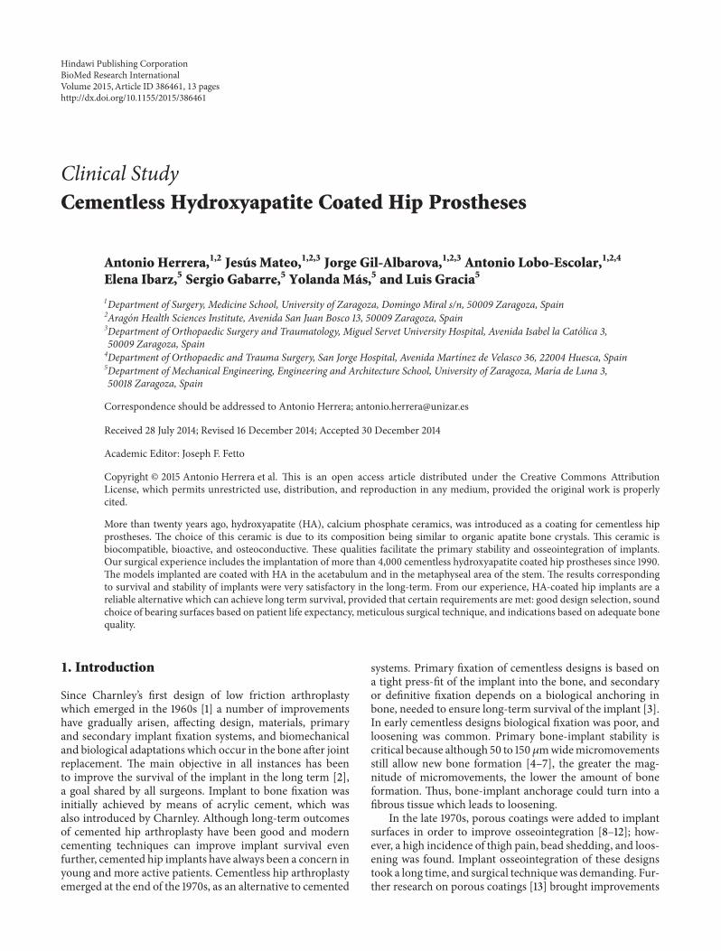
Figure 2: X-ray image of patient with follow-up at 12 y. Osteolysisin acetabulum produced by excessive polyethylene wear.
Figure 3: Computed tomography of the same case in Figure 2.Osteolysis in acetabulum.
follow-up, 21 patients needed revision surgery for majoracetabular and/or femoral osteolytic lesions. Only in threepatients a replacement of the acetabular cup and femoralstem was performed, implanting a cemented prosthesis inall of them. In the remaining 18 the implants were stabledespite osteolytic lesions; therefore only polyethylene linerwas replaced and osteolytic lesions were curetted and grafted(Figures 2 to 5).
ABG II prosthesis survival at a mean of 11.3 years offollow-up was 98.30%, with all acetabular components beingstable and with no signs of migration.
The key difference between the two model outcomesis polyethylene wear. Duration polyethylene, used in theABG II model, has shown a 54.55% less wear rate thanconventional polyethylene used in the ABG I model. TheABG I polyethylene wear has been greatest in acetabular cupsplaced in a low position (𝑃 = .038), with opening anglesgreater than 46∘, and in patients under the age of 65.At ten years of follow-up, the incidence of periacetabular
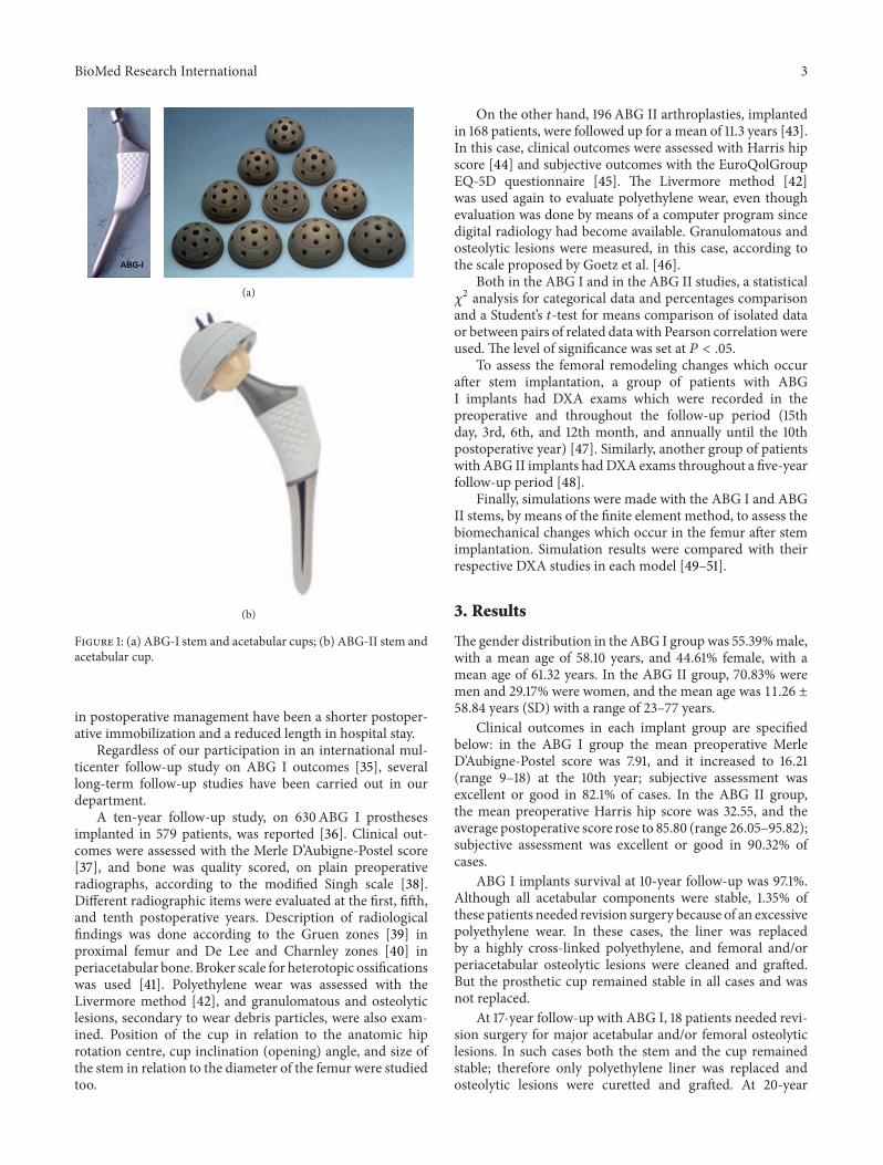
Figure 4: X-ray control of patient in Figures 2 and 3, after 4 y follow-up, after changing polyethylene and fulfilling with bone graft theacetabulum osteolysis. No change of original implant.
Figure 5: Same case as in Figures 2, 3, and 4. X-ray control imagein 2013, after 8 y. follow-up of second surgery with original implantsince 1993.
granulomatous lesions in the ABG I group was 44.23% inzone I, 37.11% in zone II, and 15% in zone III; and it was78% in Gruen zone I and 91.73% in Gruen zone 7. Despitethis, all the acetabular and femoral components were stable,even though some stems showed some subsidence. In theABG II group, granulomatous lesions were very small andonly occurred in metal on polyethylene or zirconium onpolyethylene couplings. In this group, 9.52% of cases showedosteolytic lesions in acetabular zone I and 12.7% in Gruenzones 1 and 7. Decreased incidence of osteolytic lesions inthe ABG II model is due to the lesser wear of the newpolyethylene but, in our opinion, the sealing of unused cupholes may also have played a role.
Stem subsidence has been evaluated in both models andsignificant differences have been found. In the ABG I group,of the cases in which the size of the stem was deemed
BioMed Research International 5
appropriate, mean subsidence was 1.51mm at the first yearand increased to 3mm at the 10th year. However, whenthe size of the stem was deemed large, mean subsidencewas 2.29mm at the first year and reaches 4.17mm at the10th year. In the ABG II group, lesser subsidence has beenfound among the cases in which the size of the stem wasdeemed appropriate; mean subsidence at the first year was0.514mm and rose to 0.638 ± 0.180 (SD) at the end of follow-up, but oversized stems showed 2.435mm and 2.830mm,respectively. In both studies oversized stems were associatedwith a significantly greater subsidence (𝑃 = .0001).
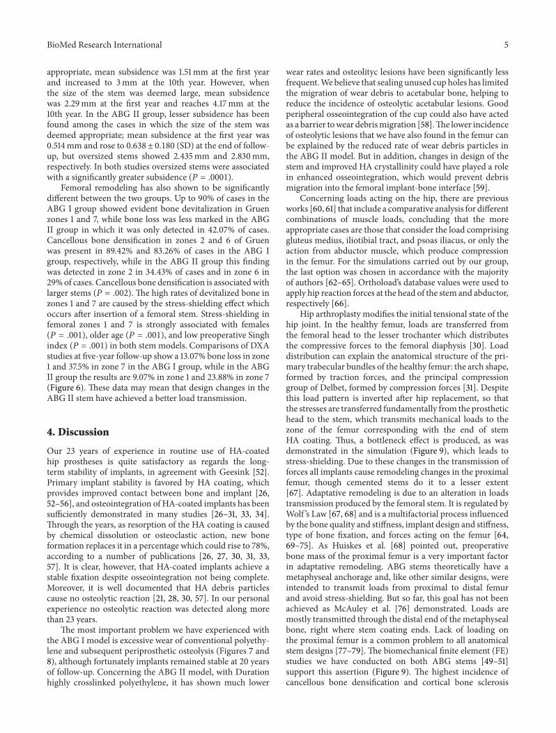
Femoral remodeling has also shown to be significantlydifferent between the two groups. Up to 90% of cases in theABG I group showed evident bone devitalization in Gruenzones 1 and 7, while bone loss was less marked in the ABGII group in which it was only detected in 42.07% of cases.Cancellous bone densification in zones 2 and 6 of Gruenwas present in 89.42% and 83.26% of cases in the ABG Igroup, respectively, while in the ABG II group this findingwas detected in zone 2 in 34.43% of cases and in zone 6 in29%of cases. Cancellous bone densification is associatedwithlarger stems (𝑃 = .002). The high rates of devitalized bone inzones 1 and 7 are caused by the stress-shielding effect whichoccurs after insertion of a femoral stem. Stress-shielding infemoral zones 1 and 7 is strongly associated with females(𝑃 = .001), older age (𝑃 = .001), and low preoperative Singhindex (𝑃 = .001) in both stem models. Comparisons of DXAstudies at five-year follow-up show a 13.07% bone loss in zone1 and 37.5% in zone 7 in the ABG I group, while in the ABGII group the results are 9.07% in zone 1 and 23.88% in zone 7(Figure 6). These data may mean that design changes in theABG II stem have achieved a better load transmission.
4. Discussion
Our 23 years of experience in routine use of HA-coatedhip prostheses is quite satisfactory as regards the long-term stability of implants, in agreement with Geesink [52].Primary implant stability is favored by HA coating, whichprovides improved contact between bone and implant [26,52–56], and osteointegration of HA-coated implants has beensufficiently demonstrated in many studies [26–31, 33, 34].Through the years, as resorption of the HA coating is causedby chemical dissolution or osteoclastic action, new boneformation replaces it in a percentage which could rise to 78%,according to a number of publications [26, 27, 30, 31, 33,57]. It is clear, however, that HA-coated implants achieve astable fixation despite osseointegration not being complete.Moreover, it is well documented that HA debris particlescause no osteolytic reaction [21, 28, 30, 57]. In our personalexperience no osteolytic reaction was detected along morethan 23 years.
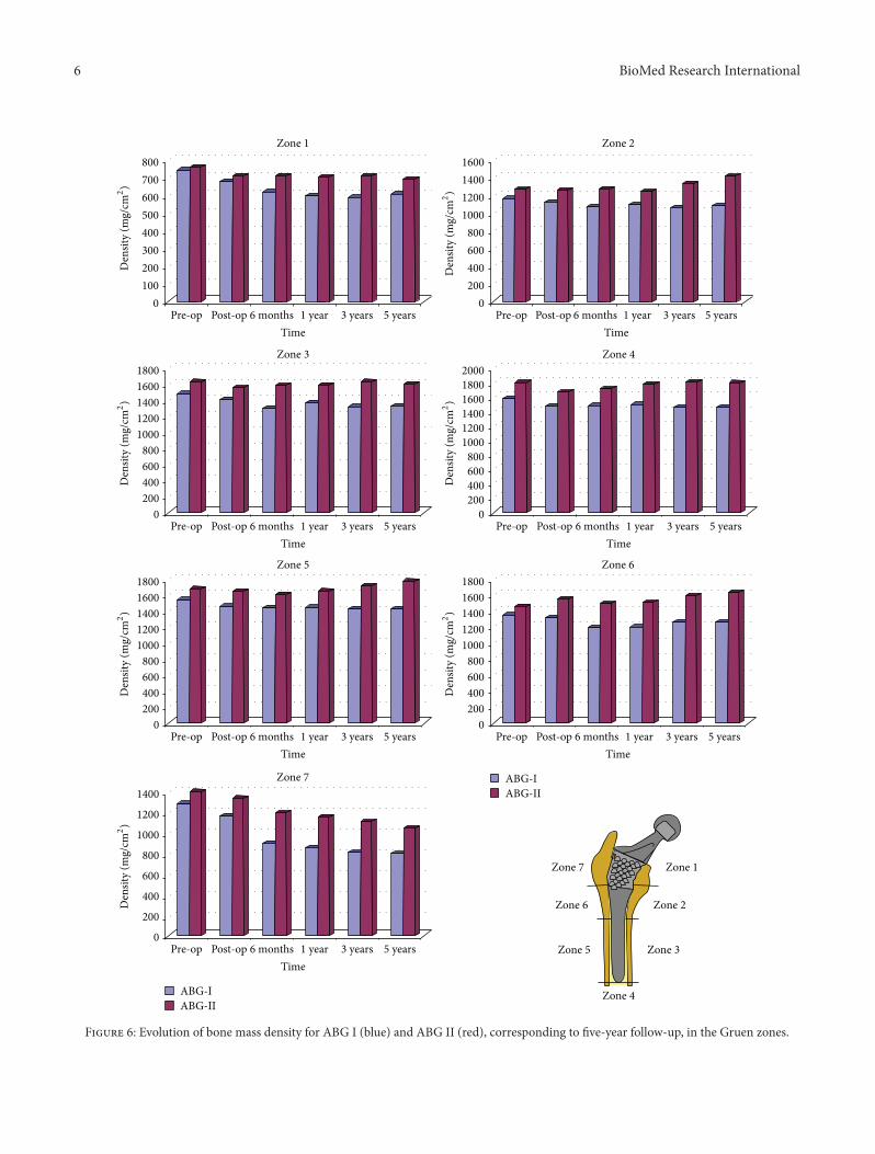
The most important problem we have experienced withthe ABG I model is excessive wear of conventional polyethy-lene and subsequent periprosthetic osteolysis (Figures 7 and8), although fortunately implants remained stable at 20 yearsof follow-up. Concerning the ABG II model, with Durationhighly crosslinked polyethylene, it has shown much lower
wear rates and osteolityc lesions have been significantly lessfrequent.We believe that sealing unused cup holes has limitedthe migration of wear debris to acetabular bone, helping toreduce the incidence of osteolytic acetabular lesions. Goodperipheral osseointegration of the cup could also have actedas a barrier towear debrismigration [58].The lower incidenceof osteolytic lesions that we have also found in the femur canbe explained by the reduced rate of wear debris particles inthe ABG II model. But in addition, changes in design of thestem and improved HA crystallinity could have played a rolein enhanced osseointegration, which would prevent debrismigration into the femoral implant-bone interface [59].
Concerning loads acting on the hip, there are previousworks [60, 61] that include a comparative analysis for differentcombinations of muscle loads, concluding that the moreappropriate cases are those that consider the load comprisinggluteus medius, iliotibial tract, and psoas iliacus, or only theaction from abductor muscle, which produce compressionin the femur. For the simulations carried out by our group,the last option was chosen in accordance with the majorityof authors [62–65]. Orthoload’s database values were used toapply hip reaction forces at the head of the stem and abductor,respectively [66].
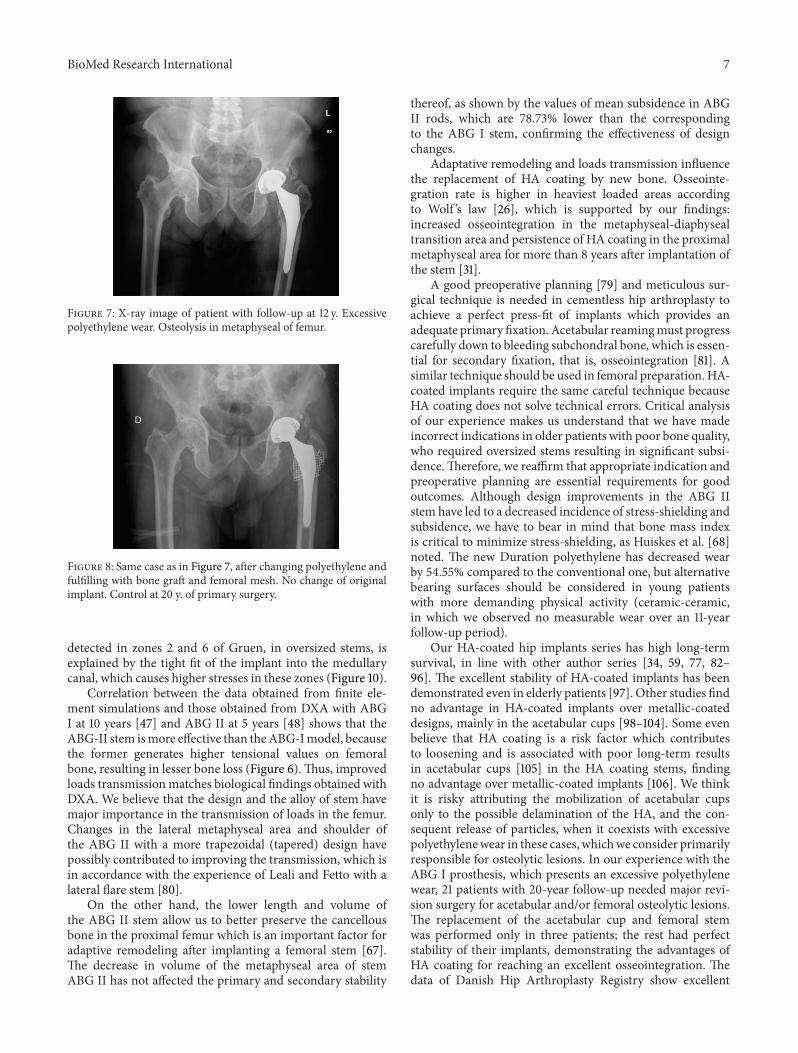
Hip arthroplasty modifies the initial tensional state of thehip joint. In the healthy femur, loads are transferred fromthe femoral head to the lesser trochanter which distributesthe compressive forces to the femoral diaphysis [30]. Loaddistribution can explain the anatomical structure of the pri-mary trabecular bundles of the healthy femur: the arch shape,formed by traction forces, and the principal compressiongroup of Delbet, formed by compression forces [31]. Despitethis load pattern is inverted after hip replacement, so thatthe stresses are transferred fundamentally from the prosthetichead to the stem, which transmits mechanical loads to thezone of the femur corresponding with the end of stemHA coating. Thus, a bottleneck effect is produced, as wasdemonstrated in the simulation (Figure 9), which leads tostress-shielding. Due to these changes in the transmission offorces all implants cause remodeling changes in the proximalfemur, though cemented stems do it to a lesser extent[67]. Adaptative remodeling is due to an alteration in loadstransmission produced by the femoral stem. It is regulated byWolf ’s Law [67, 68] and is a multifactorial process influencedby the bone quality and stiffness, implant design and stiffness,type of bone fixation, and forces acting on the femur [64,69–75]. As Huiskes et al. [68] pointed out, preoperativebone mass of the proximal femur is a very important factorin adaptative remodeling. ABG stems theoretically have ametaphyseal anchorage and, like other similar designs, wereintended to transmit loads from proximal to distal femurand avoid stress-shielding. But so far, this goal has not beenachieved as McAuley et al. [76] demonstrated. Loads aremostly transmitted through the distal end of the metaphysealbone, right where stem coating ends. Lack of loading onthe proximal femur is a common problem to all anatomicalstem designs [77–79]. The biomechanical finite element (FE)studies we have conducted on both ABG stems [49–51]support this assertion (Figure 9). The highest incidence ofcancellous bone densification and cortical bone sclerosis
6 BioMed Research International
0100200300400500600700800
Pre-op Post-op 6 months 1 year 3 years 5 yearsTime
Zone 1D
ensit
y (m
g/cm
2)
0200400600800
1000120014001600
Pre-op Post-op 6 months 1 year 3 years 5 yearsTime
Zone 2
Den
sity
(mg/
cm2)
0200400600800
1000120014001600
Pre-op Post-op 6 months 1 year 3 years 5 yearsTime
Zone 3
Den
sity
(mg/
cm2)
1800
0200400600800
1000120014001600
Pre-op Post-op 6 months 1 year 3 years 5 yearsTime
Zone 4
Den
sity
(mg/
cm2)
20001800
0200400600800
1000120014001600
Pre-op Post-op 6 months 1 year 3 years 5 yearsTime
Zone 5
Den
sity
(mg/
cm2)
1800
0200400600800
1000120014001600
Pre-op Post-op 6 months 1 year 3 years 5 yearsTime
Zone 6
Den
sity
(mg/
cm2)
1800
ABG-IABG-II
0
200
400
600
800
1000
1200
1400
Pre-op Post-op 6 months 1 year 3 years 5 yearsTime
Zone 7
Den
sity
(mg/
cm2)
ABG-IABG-II
Zone 1
Zone 2
Zone 3
Zone 4
Zone 5
Zone 6
Zone 7
Figure 6: Evolution of bone mass density for ABG I (blue) and ABG II (red), corresponding to five-year follow-up, in the Gruen zones.
BioMed Research International 7
Figure 7: X-ray image of patient with follow-up at 12 y. Excessivepolyethylene wear. Osteolysis in metaphyseal of femur.
Figure 8: Same case as in Figure 7, after changing polyethylene andfulfilling with bone graft and femoral mesh. No change of originalimplant. Control at 20 y. of primary surgery.
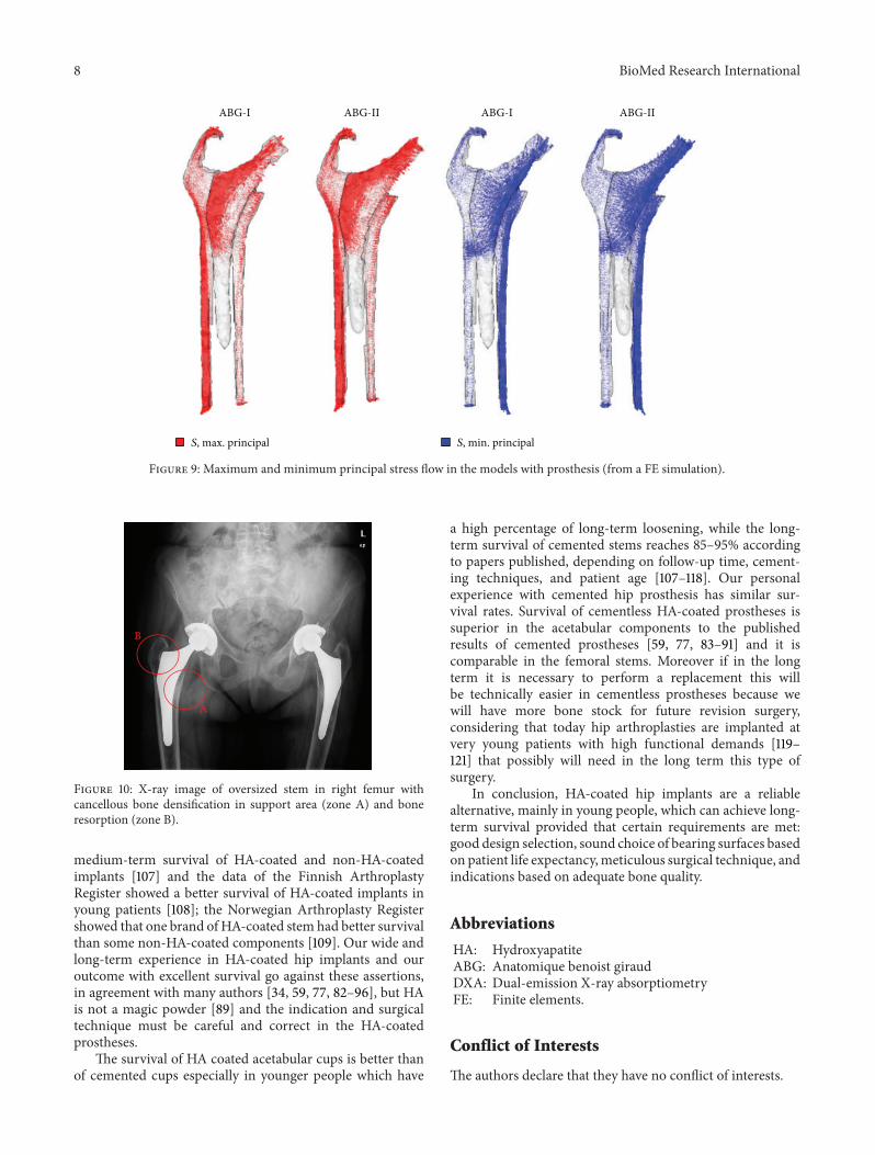
detected in zones 2 and 6 of Gruen, in oversized stems, isexplained by the tight fit of the implant into the medullarycanal, which causes higher stresses in these zones (Figure 10).
Correlation between the data obtained from finite ele-ment simulations and those obtained from DXA with ABGI at 10 years [47] and ABG II at 5 years [48] shows that theABG-II stem ismore effective than theABG-Imodel, becausethe former generates higher tensional values on femoralbone, resulting in lesser bone loss (Figure 6).Thus, improvedloads transmissionmatches biological findings obtained withDXA. We believe that the design and the alloy of stem havemajor importance in the transmission of loads in the femur.Changes in the lateral metaphyseal area and shoulder ofthe ABG II with a more trapezoidal (tapered) design havepossibly contributed to improving the transmission, which isin accordance with the experience of Leali and Fetto with alateral flare stem [80].
On the other hand, the lower length and volume ofthe ABG II stem allow us to better preserve the cancellousbone in the proximal femur which is an important factor foradaptive remodeling after implanting a femoral stem [67].The decrease in volume of the metaphyseal area of stemABG II has not affected the primary and secondary stability
thereof, as shown by the values of mean subsidence in ABGII rods, which are 78.73% lower than the correspondingto the ABG I stem, confirming the effectiveness of designchanges.
Adaptative remodeling and loads transmission influencethe replacement of HA coating by new bone. Osseointe-gration rate is higher in heaviest loaded areas accordingto Wolf ’s law [26], which is supported by our findings:increased osseointegration in the metaphyseal-diaphysealtransition area and persistence of HA coating in the proximalmetaphyseal area for more than 8 years after implantation ofthe stem [31].
A good preoperative planning [79] and meticulous sur-gical technique is needed in cementless hip arthroplasty toachieve a perfect press-fit of implants which provides anadequate primary fixation. Acetabular reamingmust progresscarefully down to bleeding subchondral bone, which is essen-tial for secondary fixation, that is, osseointegration [81]. Asimilar technique should be used in femoral preparation.HA-coated implants require the same careful technique becauseHA coating does not solve technical errors. Critical analysisof our experience makes us understand that we have madeincorrect indications in older patients with poor bone quality,who required oversized stems resulting in significant subsi-dence.Therefore, we reaffirm that appropriate indication andpreoperative planning are essential requirements for goodoutcomes. Although design improvements in the ABG IIstem have led to a decreased incidence of stress-shielding andsubsidence, we have to bear in mind that bone mass indexis critical to minimize stress-shielding, as Huiskes et al. [68]noted. The new Duration polyethylene has decreased wearby 54.55% compared to the conventional one, but alternativebearing surfaces should be considered in young patientswith more demanding physical activity (ceramic-ceramic,in which we observed no measurable wear over an 11-yearfollow-up period).
Our HA-coated hip implants series has high long-termsurvival, in line with other author series [34, 59, 77, 82–96]. The excellent stability of HA-coated implants has beendemonstrated even in elderly patients [97]. Other studies findno advantage in HA-coated implants over metallic-coateddesigns, mainly in the acetabular cups [98–104]. Some evenbelieve that HA coating is a risk factor which contributesto loosening and is associated with poor long-term resultsin acetabular cups [105] in the HA coating stems, findingno advantage over metallic-coated implants [106]. We thinkit is risky attributing the mobilization of acetabular cupsonly to the possible delamination of the HA, and the con-sequent release of particles, when it coexists with excessivepolyethylenewear in these cases, whichwe consider primarilyresponsible for osteolytic lesions. In our experience with theABG I prosthesis, which presents an excessive polyethylenewear, 21 patients with 20-year follow-up needed major revi-sion surgery for acetabular and/or femoral osteolytic lesions.The replacement of the acetabular cup and femoral stemwas performed only in three patients; the rest had perfectstability of their implants, demonstrating the advantages ofHA coating for reaching an excellent osseointegration. Thedata of Danish Hip Arthroplasty Registry show excellent
8 BioMed Research International
ABG-I ABG-II ABG-I ABG-II
S, max. principal S, min. principal
Figure 9: Maximum and minimum principal stress flow in the models with prosthesis (from a FE simulation).
B
A
Figure 10: X-ray image of oversized stem in right femur withcancellous bone densification in support area (zone A) and boneresorption (zone B).
medium-term survival of HA-coated and non-HA-coatedimplants [107] and the data of the Finnish ArthroplastyRegister showed a better survival of HA-coated implants inyoung patients [108]; the Norwegian Arthroplasty Registershowed that one brand of HA-coated stem had better survivalthan some non-HA-coated components [109]. Our wide andlong-term experience in HA-coated hip implants and ouroutcome with excellent survival go against these assertions,in agreement with many authors [34, 59, 77, 82–96], but HAis not a magic powder [89] and the indication and surgicaltechnique must be careful and correct in the HA-coatedprostheses.
The survival of HA coated acetabular cups is better thanof cemented cups especially in younger people which have
a high percentage of long-term loosening, while the long-term survival of cemented stems reaches 85–95% accordingto papers published, depending on follow-up time, cement-ing techniques, and patient age [107–118]. Our personalexperience with cemented hip prosthesis has similar sur-vival rates. Survival of cementless HA-coated prostheses issuperior in the acetabular components to the publishedresults of cemented prostheses [59, 77, 83–91] and it iscomparable in the femoral stems. Moreover if in the longterm it is necessary to perform a replacement this willbe technically easier in cementless prostheses because wewill have more bone stock for future revision surgery,considering that today hip arthroplasties are implanted atvery young patients with high functional demands [119–121] that possibly will need in the long term this type ofsurgery.
In conclusion, HA-coated hip implants are a reliablealternative, mainly in young people, which can achieve long-term survival provided that certain requirements are met:good design selection, sound choice of bearing surfaces basedon patient life expectancy,meticulous surgical technique, andindications based on adequate bone quality.
Abbreviations
HA: HydroxyapatiteABG: Anatomique benoist giraudDXA: Dual-emission X-ray absorptiometryFE: Finite elements.
Conflict of Interests
The authors declare that they have no conflict of interests.
BioMed Research International 9
Authors’ Contribution
Antonio Herrera and Luis Gracia conceived the design ofthe study and coordinated the work between surgeons andengineers. Jesus Mateo, Jorge Gil-Albarova, and AntonioLobo-Escolar performed the surgery and supervised the post-operative follow-up. Elena Ibarz, SergioGabarre, andYolandaMas conceived and developed the finite element models andcarried out all the simulations and results processing. Allauthors participated in the drawing up of the paper and readand approved the final paper.
References
[1] J. Charnley, “Arthroplasty of the hip. A new operation,” TheLancet, vol. 277, no. 7187, pp. 1129–1132, 1961.
[2] W.H.Harris, “The first 50 years of total hip arthroplasty: lessonslearned,” Clinical Orthopaedics and Related Research, vol. 467,no. 1, pp. 28–31, 2009.
[3] C. A. Engh, J. D. Bobyn, and A. H. Glassman, “Porous-coated hip replacement. The factors governing bone ingrowth,stress shielding, and clinical results,” Journal of Bone and JointSurgery—Series B, vol. 69, no. 1, pp. 45–55, 1987.
[4] R.M. Pilliar, J. M. Lee, and C.Maniatopoulos, “Observations onthe effect of movement on bone ingrowth into porous-surfacedimplants,” Clinical Orthopaedics and Related Research, no. 208,pp. 108–113, 1986.
[5] H. Kienapfel, C. Sprey, A. Wilke, and P. Griss, “Implant fixationby bone ingrowth,”The Journal of Arthroplasty, vol. 14, no. 3, pp.355–368, 1999.
[6] M. Jasty, C. Bragdon, D. Burke, D. O’Connor, J. Lowenstein, andW. H. Harris, “In vivo skeletal responses to porous-surfacedimplants subjected to small induced motions,” The Journal ofBone and Joint Surgery—Series A, vol. 79, no. 5, pp. 707–714, 1997.
[7] I. J. Udofia, F. Liu, Z. Jin, P. Roberts, and P. Grigoris, “Theinitial stability and contact mechanics of a press-fit resurfacingarthroplasty of the hip,”The Journal of Bone and Joint Surgery—Series B, vol. 89, no. 4, pp. 549–556, 2007.
[8] H. U. Cameron, I. Macnab, and R. M. Pilliar, “A porous metalsystem for joint replacement surgery,”The International Journalof Artificial Organs, vol. 1, no. 2, pp. 104–109, 1978.
[9] R. Judet, M. Siguier, B. Brumpt, and T. Judet, “Prothese totalede hanche en poro-metal sans ciment,” Revue de ChirurgieOrthopedique et Reparatrice de l’Appareil Moteur, vol. 64,supplement 2, pp. 14–21, 1978.
[10] G. Lord, J. H. Marotte, J. P. Blanchard, J. L. Guillamon, andM. Gory, “The fixation of madreporic total hip prosthesis;an experimental study (author’s transl),” Revue de ChirurgieOrthopedique et Reparatrice de l’Appareil Moteur, vol. 64, no. 6,pp. 459–470, 1978.
[11] R. M. Pilliar, H. U. Cameron, R. P. Welsh, and A. G. Binning-ton, “Radiographic and morphologic studies of load-bearingporous-surfaced structured implants,” Clinical Orthopaedicsand Related Research, vol. 156, pp. 249–257, 1981.
[12] C. A. Engh, “Hip arthroplasty with a Moore prosthesis withporous coating. A five-year study,” Clinical Orthopaedics andRelated Research, no. 176, pp. 52–66, 1983.
[13] R.M. Pilliar, “Porous-surfacedmetallic implants for orthopedicapplications,” Journal of Biomedical Materials Research, vol. 21,supplement A1, pp. 1–33, 1987.
[14] K. de Groot, R. Geesink, C. P. A. T. Klein, and P. Serekian,“Plasma sprayed coatings of hydroxylapatite,” Journal ofBiomedicalMaterials Research, vol. 21, no. 12, pp. 1375–1381, 1987.
[15] R. G. T. Geesink, K. de Groot, and C. P. A. T. Klein, “Chemicalimplant fixation using hydroxyl-apatite coatings. The develop-ment of a human total hip prosthesis for chemical fixation tobone using hydroxyl-apatite coatings on titanium substrates,”Clinical Orthopaedics and Related Research, no. 225, pp. 147–170,1987.
[16] R. J. Furlong and J. F. Osborn, “Fixation of hip prostheses byhydroxyapatite ceramic coatings,”The Journal of Bone and JointSurgery—Series B, vol. 73, no. 5, pp. 741–745, 1991.
[17] M. T. Manley and R. Koch, “Clinical results with thehydroxyapatite-coated Omnifit hip stem,” Dental Clinics ofNorth America, vol. 36, no. 1, pp. 257–262, 1992.
[18] J.-A. Epinette, M. T. Manley, J. A. D’Antonio, A. A. Edidin,and W. N. Capello, “A 10-year minimum follow-up of hy-droxyapatite-coated threaded cups: clinical, radiographic andsurvivorship analyses with comparison to the literature,” TheJournal of Arthroplasty, vol. 18, no. 2, pp. 140–148, 2003.
[19] N. Passuti, “New concepts in bone substitution,” in EuropeanInstructinal Course Lectures, P. Fulford, F.Horan, andR. P. Jakob,Eds., vol. 4, The British Editorial Society of Bobe and JointSurgery, London, UK, 1999.
[20] H. A. Hoogendoorn, W. Renooij, and L. M. A. Akkermans,“Long-term study of large ceramic implants (porous hydrox-yapatite) in dog femora,” Clinical Orthopaedics and RelatedResearch, vol. 187, pp. 281–288, 1984.
[21] R. G. T. Geesink, K. De Groot, and C. P. A. T. Klein, “Bonding ofbone to apatite-coated implants,”The Journal of Bone and JointSurgery—Series B, vol. 70, no. 1, pp. 17–22, 1988.
[22] H. Daugaard, B. Elmengaard, J. E. Bechtold, T. Jensen, andK. Soballe, “The effect on bone growth enhancement ofimplant coatings with hydroxyapatite and collagen depositedelectrochemically and by plasma spray,” Journal of BiomedicalMaterials Research Part A, vol. 92, no. 3, pp. 913–921, 2010.
[23] S. Overgaard, U. Bromose, M. Lind, C. Bunger, and K. Søballe,“The influence of crystallinity of the hydroxyapatite coating onthe fixation of implants. Mechanical and histomorphometricresults,” The Journal of Bone and Joint Surgery Series B, vol. 81,no. 4, pp. 725–731, 1999.
[24] M.Røkkum,A.Reigstad, andC. B. Johansson, “HAparticles canbe released from well-fixed HA-coated stems: histopathologyof biopsies from 20 hips 2–8 years after implantation,” ActaOrthopaedica Scandinavica, vol. 73, no. 3, pp. 298–306, 2002.
[25] O. O. S. Rahbet and K. Soballe, “Calcium phosphate coatingfor implants fixation,” in Fifteen Years of Clinical Experiencewith Hydroxyapatite Coating in Joint Arthroplasty, pp. 35–52,Springer, Paris, France, 2004.
[26] T. W. Bauer, R. C. T. Geesink, R. Zimmerman, and J. T.McMahon, “Hydroxyapatite-coated femoral stems.Histologicalanalysis of components retrieved at autopsy,” The Journal ofBone and Joint Surgery—Series A, vol. 73, no. 10, pp. 1439–1452,1991.
[27] S. Overgaard, K. Søballe, K. Josephsen, E. S. Hansen, and C.Bunger, “Role of different loading conditions on resorption ofhydroxyapatite coating evaluated by histomorphometric andstereological methods,” Journal of Orthopaedic Research, vol. 14,no. 6, pp. 888–894, 1996.
10 BioMed Research International
[28] S. Overgaard, M. Lind, O. Rahbek, C. Bunger, and K. Søballe,“Improved fixation of porous-coated versus grit-blasted sur-face texture of hydroxyapatite-coated implants in dogs,” ActaOrthopaedica Scandinavica, vol. 68, no. 4, pp. 337–343, 1997.
[29] D. C. R. Hardy, P. Frayssinet, P. Krallis et al., “Histopathologyof a well-functioning hydroxyapatite-coated femoral prosthesisafter 52 months,” Acta Orthopaedica Belgica, vol. 65, no. 1, pp.72–82, 1999.
[30] A. J. Tonino, M. Therin, and C. Doyle, “Hydroxyapatite-coatedfemoral stems. Histology and histomorphometry around fivecomponents retrieved at postmortem,” Journal of Bone and JointSurgery, vol. 81, no. 1, pp. 148–154, 1999.
[31] A. J. Tonino, B. C. H. van der Wal, I. C. Heyligers, andB. Grimm, “Bone remodeling and hydroxyapatite resorptionin coated primary hip prostheses,” Clinical Orthopaedics andRelated Research, vol. 467, no. 2, pp. 478–484, 2009.
[32] C. L. Tisdel, V. M. Goldberg, J. A. Parr, J. S. Bensusan, L. S.Staikoff, and S. Stevenson, “The influence of a hydroxyapatiteand tricalcium-phosphate coating on bone growth into tita-nium fiber-metal implants,” Journal of Bone and Joint Surgery—Series A, vol. 76, no. 2, pp. 159–171, 1994.
[33] A. Tonino, C. Oosterbos, A. Rahmy, M. Therin, and C. Doyle,“Hydroxyapatite-coated acetabular components. Histologicaland histomorphometric analysis of six cups retrieved at autopsybetween three and seven years after successful implantation,”The Journal of Bone and Joint Surgery—Series A, vol. 83, no. 6,pp. 817–825, 2001.
[34] B. Chambers, S. F. St. Clair, and M. I. Froimson, “Hydroxyapa-tite-coated tapered cementless femoral components in total hiparthroplasty,” Journal of Arthroplasty, vol. 22, no. 4, supplement1, pp. 71–74, 2007.
[35] A. Herrera, V. Canales, J. Anderson, C. Garcıa-Araujo, A.Murcia-Mazon, and A. J. Tonino, “Seven to 10 years followupof an anatomic hip prosthesis: an international study,” ClinicalOrthopaedics and Related Research, no. 423, pp. 129–137, 2004.
[36] V. Canales, J. J. Panisello, A. Herrera, A. Sola, J. J. Mateo, andM. J. Caballero, “Extensive osteolysis caused by polyethyleneparticle migration in an anatomical hydroxyapatite-coated hipprosthesis: 10 years’ follow-up,” Journal of Arthroplasty, vol. 25,no. 7, pp. 1115.e1–1124.e1, 2010.
[37] R. M. D’Aubigne and M. Postel, “Function al results of hiparthroplasty with acrylic prosthesis,” The Journal of Bone andJoint Surgery. American volume, vol. 36, no. 3, pp. 451–475, 1954.
[38] M. Singh, A. R. Nagrath, and P. S.Maini, “Changes in trabecularpattern of the upper end of the femur as an index of osteoporo-sis,” The Journal of Bone and Joint Surgery Series A, vol. 52, no.3, pp. 457–467, 1970.
[39] T. A. Gruen, G. M. McNeice, and H. C. Amstutz, “’Modesof failure’ of cemented stem-type femoral components. Aradiographic analysis of loosening,” Clinical Orthopaedics andRelated Research, vol. 141, pp. 17–27, 1979.
[40] J. G. DeLee and J. Charnley, “Radiological demarcationof cemented sockets in total hip replacement,” ClinicalOrthopaedics and Related Research, vol. 121, pp. 20–32, 1976.
[41] A. F. Brooker, J. W. Bowerman, R. A. Robinson, and L. H.Riley Jr., “Ectopic ossification following total hip replacement.Incidence and a method of classification,” The Journal of Boneand Joint Surgery. American Volume, vol. 55, no. 8, pp. 1629–1632, 1973.
[42] J. Livermore, D. Ilstrup, and B. Morrey, “Effect of femoralhead size on wear of the polyethylene acetabular component,”
The Journal of Bone and Joint Surgery—Series A, vol. 72, no. 4,pp. 518–528, 1990.
[43] A. Herrera, J. Mateo, A. Lobo-Escolar, J. J. Panisello, E. Ibarz,and L. Gracia, “Long-term outcomes of a new model ofanatomical hydroxyapatite-coated hip prosthesis,” The Journalof Arthroplasty, vol. 28, no. 7, pp. 1160–1166, 2013.
[44] W. H. Harris, “Traumatic arthritis of the hip after dislocationand acetabular fractures: treatment by mold arthroplasty. Anend-result study using a new method of result evaluation,” TheJournal of Bone and Joint Surgery American Volume, vol. 51, no.4, pp. 737–755, 1969.
[45] A. Szende and A. Williams, Eds., Measuring Self-ReportedPopulation Health: An International Perspective based on EQ-5D, The EuroQol Group’s International Task Force on Self-Reported Health, 2004.
[46] D. D. Goetz, E. J. Smith, and W. H. Harris, “The prevalenceof femoral osteolysis associated with components inserted withor without cement in total hip replacements. A retrospectivematched- pair series,” The Journal of Bone and Joint Surgery—Series A, vol. 76, no. 8, pp. 1121–1129, 1994.
[47] J. J. Panisello, L. Herrero, V. Canales, A. Herrera, A. A.Martınez,and J.Mateo, “Long-term remodeling in proximal femur arounda hydroxyapatite-coated anatomic stem: ten years densitometricfollow-up,”The Journal of Arthroplasty, vol. 24, no. 1, pp. 56–64,2009.
[48] J. J. Panisello, V. Canales, L. Herrero, A. Herrera, J. Mateo, andM. J. Caballero, “Changes in periprosthetic bone remodellingafter redesigning an anatomic cementless stem,” InternationalOrthopaedics, vol. 33, no. 2, pp. 373–379, 2009.
[49] A. Herrera, J. J. Panisello, E. Ibarz, J. Cegonino, J. A. Puertolas,and L. Gracia, “Long-term study of bone remodelling afterfemoral stem: a comparison between dexa and finite elementsimulation,” Journal of Biomechanics, vol. 40, no. 16, pp. 3615–3625, 2007.
[50] A. Herrera, J. J. Panisello, E. Ibarz, J. Cegonino, J. A. Puertolas,and L. Gracia, “Comparison between DEXA and finite elementstudies in the long-term bone remodeling of an anatomicalfemoral stem,” Journal of Biomechanical Engineering, vol. 131, no.4, Article ID 041013, 2009.
[51] L. Gracia, E. Ibarz, S. Puertolas et al., “Study of bone remodelingof two models of femoral cementless stems by means of DEXAand finite elements,” BioMedical Engineering Online, vol. 9,article 22, 2010.
[52] R. D. Geesink, “Fixation strategies in total hip arthroplasty,”Surgical Technology International, vol. 21, pp. 240–247, 2011.
[53] K. Soballe, “Hydroxyapatite ceramic coating for bone implantfixation: mechanical anf histological studies in dogs,” ActaOrthopaedica Scandinavica, Supplementum, vol. 64, no. 255, pp.1–58, 1993.
[54] J. Karrholm,H.Malchau, F. Snorrason, and P. Herberts, “Micro-motion of femoral stems in total hip arthroplasty. A randomizedstudy of cemented, hydroxyapatite-coated and porous-coatedstems with roentgen stereophotogrammetric analysis,” TheJournal of Bone and Joint Surgery—Series A, vol. 76, no. 11, pp.1692–1705, 1994.
[55] M. J. Coathup, G. W. Blunn, N. Flynn, C. Williams, andN. P. Thomas, “A comparison of bone remodelling aroundhydroxyapatite-coated, porous-coated and grit-blasted hipreplacements retrieved at post-mortem,” The Journal of Boneand Joint Surgery Series B, vol. 83, no. 1, pp. 118–123, 2001.
BioMed Research International 11
[56] R. G. T. Geesink, “Osteoconductive coatings for total jointarthroplasty,” Clinical Orthopaedics and Related Research, no.395, pp. 53–65, 2002.
[57] N. Aebli, J. Krebs, D. Schwenke, H. Stich, P. Schwalder, andJ.-C. Theis, “Degradation of hydroxyapatite coating on a well-functioning femoral component,”The Journal of Bone and JointSurgery British Volume, vol. 85, no. 4, pp. 499–503, 2003.
[58] M. J. Coathup, J. Blackburn, A. E. Goodship, J. L. Cunningham,T. Smith, and G. W. Blunn, “Role of hydroxyapatite coatingin resisting wear particle migration and osteolysis aroundacetabular components,” Biomaterials, vol. 26, no. 19, pp. 4161–4169, 2005.
[59] P. J. Emans, R. H. Broeke, J. M. vanMulken, R. Kuijer, L. W. vanRhijn, and R. G. Geesink, “Results of total hip arthroplasties inthe young patient; further evidence for a barrier against artic-ular wear debris by hydroxyapatite coatings,”Hip International,vol. 19, no. 4, pp. 343–351, 2009.
[60] E. F. Rybicki, F. A. Simonen, and E. B. Weis Jr., “On themathematical analysis of stress in the human femur,” Journal ofBiomechanics, vol. 5, no. 2, pp. 203–215, 1972.
[61] V. M. Domınguez-Hernandez, M. F. Carbajal, G. Urriolagoitiaet al., “Biomecanica de un femur sometido a carga. Desarrollode un modelo tridimensional por medio del metodo del ele-mento finito,” Revista Mexicana de Ortopaedica Traumatologie,vol. 13, no. 6, pp. 633–638, 1999.
[62] H. Weinans, R. Huiskes, and H. J. Grootenboer, “Effects of fitand bonding characteristics of femoral stems on adaptive boneremodeling,” Journal of Biomechanical Engineering, vol. 116, no.4, pp. 393–400, 1994.
[63] B. van Rietbergen and R. Huiskes, “Load transfer and stressshielding of the hydroxyapatite-ABG hip: a study of stemlength and proximal fixation,” Journal of Arthroplasty, vol. 16,supplement 1, no. 8, pp. 55–63, 2001.
[64] J. Kerner, R. Huiskes, G. H. van Lenthe et al., “Correlationbetween pre-operative periprosthetic bone density and post-operative bone loss in THA can be explained by strain-adaptiveremodelling,” Journal of Biomechanics, vol. 32, no. 7, pp. 695–703, 1999.
[65] V. M. Domınguez-Hernandez, V. H. Ramos, C. V. Feria, G.Urriolagoitia, and L. H. Hernandez, “Efecto del espesor de lacapa de cemento en el componente femoral de una protesis deCharnley. Anaılisis biomecanico mediante el metodo del ele-mento finito,” Revista Mexicana de Ortopaedica Traumatologie,vol. 14, no. 6, pp. 443–448, 2000.
[66] G. E. A. Bergmann, Orthoload data base. Loading of Orthopae-dic Implants, 2013, http://www.orthoload.com/.
[67] A. Herrera, S. Rebollo, E. Ibarz, J. Mateo, S. Gabarre, and L.Gracia, “Mid-Term study of bone remodeling after femoralcemented stem implantation: comparison between DXA andfinite element simulation,” The Journal of Arthroplasty, vol. 29,no. 1, pp. 90–100, 2014.
[68] R. Huiskes, H. Weinans, and M. Dalstra, “Adaptive boneremodeling and biomechanical design considerations for non-cemented total hip arthroplasty,” Orthopedics, vol. 12, no. 9, pp.1255–1267, 1989.
[69] J. D. Bobyn, E. S. Mortimer, A. H. Glassman, C. A. Engh, J. E.Miller, and C. E. Brooks, “Producing and avoiding stress shield-ing: laboratory and clinical observations of noncemented totalhip arthroplasty,” Clinical Orthopaedics and Related Research,no. 274, pp. 79–96, 1992.
[70] R. Huiskes and B. van Rietbergen, “Preclinical testing oftotal hip stems. The effects of coating placement,” ClinicalOrthopaedics and Related Research, no. 319, pp. 64–76, 1995.
[71] J. A. D’Antonio, W. N. Capello, and M. T. Manley, “Remodelingof bone around hydroxyapatite-coated femoral stems,” Journalof Bone and Joint Surgery—Series A, vol. 78, no. 8, pp. 1226–1234,1996.
[72] L. Rosenthall, J. D. Bobyn, and M. Tanzer, “Bone densit-ometry: influence of prosthetic design and hydroxyapatitecoating on regional adaptive bone remodelling,” InternationalOrthopaedics, vol. 23, no. 6, pp. 325–329, 1999.
[73] C. J. Sychterz, A. M. Claus, C. A. Engh, and R. Wolf, “What wehave learned about long-term cementless fixation from autopsyretrievals,” Clinical Orthopaedics and Related Research, no. 405,pp. 79–91, 2002.
[74] A. I. A. Rahmy, T. Gosens, G. M. Blake, A. Tonino, and I.Fogelman, “Periprosthetic bone remodelling of two types ofuncemented femoral implant with proximal hydroxyapatitecoating: a 3-year follow-up study addressing the influence ofprosthesis design and preoperative bone density on peripros-thetic bone loss,” Osteoporosis International, vol. 15, no. 4, pp.281–289, 2004.
[75] P. Chandran, M. Azzabi, M. Andrews, and J. G. Bradley,“Periprosthetic bone remodeling after 12 years differs incemented and uncemented hip arthroplasties,” ClinicalOrthopaedics and Related Research, vol. 470, no. 5, pp. 1431–1435, 2012.
[76] J. P. McAuley, C. J. Sychterz, and C. A. Engh Sr., “Influence ofporous coating level on proximal femoral remodeling: a post-mortem analysis,” Clinical Orthopaedics and Related Research,no. 371, pp. 146–153, 2000.
[77] C. J. M. Oosterbos, A. I. A. Rahmy, A. J. Tonino, and W.Witpeerd, “High survival rate of hydroxyapatite-coated hipprostheses: 100 consecutive hips followed for 10 years,” ActaOrthopaedica Scandinavica, vol. 75, no. 2, pp. 127–133, 2004.
[78] W.N. Capello, J. A. D’Antonio, R. G. Geesink, J. R. Feinberg, andM. Naughton, “Late remodeling around a proximally ha-coatedtapered titanium femoral component,” Clinical Orthopaedicsand Related Research, vol. 467, no. 1, pp. 155–165, 2009.
[79] A. G. Della Valle, D. E. Padgett, and E. A. Salvati, “Preoperativeplanning for primary total hip arthroplasty,” The Journal of theAmerican Academy of Orthopaedic Surgeons., vol. 13, no. 7, pp.455–462, 2005.
[80] A. Leali and J. F. Fetto, “Preservation of femoral bone mass aftertotal hip replacements with a lateral flare stem,” InternationalOrthopaedics, vol. 28, no. 3, pp. 151–154, 2004.
[81] E. W. Morscher, “Zementfrei pfannenverankerung nach dem‘Press-Fit-Konzept’ bei der Totalprothesen-Arthroplastik derHufte,” Acta Chirurgiae Orthopaedicae et TraumatologiaeCechoslovaca, vol. 69, no. 1, pp. 8–15, 2002.
[82] O. Reikeras and R. B. Gunderson, “Excellent results of HAcoating on a grit-blasted stem: 245 patients followed for 8–12years,” Acta Orthopaedica Scandinavica, vol. 74, no. 2, pp. 140–145, 2003.
[83] W. N. Capello, J. A. D’Antonio, W. L. Jaffe, R. G. Geesink, M.T. Manley, and J. R. Feinberg, “Hydroxyapatite-coated femoralcomponents: 15-yearminimum followup,”ClinicalOrthopaedicsand Related Research, no. 453, pp. 75–80, 2006.
[84] S. S. Rajaratnam, C. Jack, A. Tavakkolizadeh et al., “Long-termresults of a hydroxyapatite-coated femoral component in totalhip replacement: a 15- to 21-year follow-up study,”The Journal ofBone and Joint Surgery— Series B, vol. 90, no. 1, pp. 27–30, 2008.
12 BioMed Research International
[85] J.-A. Epinette and M. T. Manley, “Uncemented stems in hipreplacement—hydroxyapatite or plain porous: does it matter?Based on a prospective study of HA Omnifit stems at 15-yearsminimum follow-up,” HIP International, vol. 18, no. 2, pp. 69–74, 2008.
[86] D. Camazzola, T.Hammond, R. Gandhi, and J. R. Davey, “A ran-domized trial of hydroxyapatite-coated femoral stems in totalhip arthroplasty: a 13-year follow-up,” Journal of Arthroplasty,vol. 24, no. 1, pp. 33–37, 2009.
[87] R. Bidar, P. Kouyoumdjian, E. Munini, and G. Asencio, “Long-term results of the ABG-1 hydroxyapatite coated total hiparthroplasty: analysis of 111 cases with a minimum follow-up of 10 years,” Orthopaedics and Traumatology: Surgery andResearch, vol. 95, no. 8, pp. 579–587, 2009.
[88] P. N. Baker, I. A.McMurtry, G. Chuter, A. Port, and J. Anderson,“Thawith the abg i prosthesis at 15 years: excellent survival withminimal osteolysis,”Clinical Orthopaedics and Related Research,vol. 468, no. 7, pp. 1855–1861, 2010.
[89] J.-P. Vidalain, “Twenty-year results of the cementless Corailstem,” International Orthopaedics, vol. 35, no. 2, pp. 189–194,2011.
[90] E. Sariali, A. Mouttet, P. Mordasini, and Y. Catonne, “High 10-year survival rate with an anatomic cementless stem (SPS),”Clinical Orthopaedics and Related Research, vol. 470, no. 7, pp.1941–1949, 2012.
[91] J. A. Epinette, G. Asencio, J. Essig, B. Llagonne, and C.Nourissat, “Clinical results, radiological findings and survivalof a proximally hydroxyapatite-coated hip ABG II stem ata minimum of ten years’ follow-up: results of a consecutivemulticentre study of 1148 hips in 1053 patients,”TheBone& JointJournal B, vol. 95, no. 12, pp. 1610–1616, 2013.
[92] O. A. Gabbar, R. A. Rajan, S. Londhe, and I. D. Hyde, “Ten-to twelve-year follow-up of the furlong hydroxyapatite-coatedfemoral stem and threaded acetabular cup in patients youngerthan 65 years,”The Journal of Arthroplasty, vol. 23, no. 3, pp. 413–417, 2008.
[93] H.Wangen, P. Lereim, I. Holm, R. Gunderson, and O. Reikeras,“Hip arthroplasty in patients younger than 30 years: excellentten to 16-year follow-up results with a HA-coated stem,”International Orthopaedics, vol. 32, no. 2, pp. 203–208, 2008.
[94] X. Flecher, O. Pearce, S. Parratte, J. M. Aubaniac, and J. N.Argenson, “Custom cementless stem improves hip function inyoung patients at 15-year followup,” Clinical Orthopaedics andRelated Research, vol. 468, no. 3, pp. 747–755, 2010.
[95] M. Gottliebsen, O. Rahbek, P. F. Ottosen, K. Søballe, and M.Stilling, “Superior 11-year survival but higher polyethylene wearof hydroxyapatite-coated Mallory-Head cups,” HIP Interna-tional, vol. 22, no. 1, pp. 35–40, 2012.
[96] Y. Nakashima, T. Sato, T. Yamamoto et al., “Results at aminimum of 10 years of follow-up for AMS and PerFix HA-coated cementless total hip arthroplasty: impact of cross-linkedpolyethylene on implant longevity,” Journal of OrthopaedicScience, vol. 18, no. 6, pp. 962–968, 2013.
[97] M. Ulivi, V. Meroni, L. C. Orlandini, P. Berjano, and V. C.Sansone, “Minimum 10 year survivorship analysis of a partiallycoated hydroxyapatite tapered femoral stem in elderly patientswith an average age over 75,”The Journal of Arthroplasty, vol. 28,no. 8, pp. 1372–1377, 2013.
[98] Y.-S. Park, J.-Y. Lee, S.-H. Yun, M.-W. Jung, and I. Oh, “Com-parison of hydroxyapatite- and porous-coated stems in total hipreplacement,”ActaOrthopaedica Scandinavica, vol. 74, no. 3, pp.259–263, 2003.
[99] A. V. Lombardi Jr., K. R. Berend, and T. H. Mallory,“Hydroxyapatite-coated titanium porous plasma spray taperedstem: experience at 15 to 18 years,” Clinical Orthopaedics andRelated Research, no. 453, pp. 81–85, 2006.
[100] K. S. Yoon, H. J. Kim, J. H. Lee, S. B. Kang, N. H. Seong,and K.-H. Koo, “A randomized clinical trial of cementlessfemoral stems with and without hydroxyapatite/tricalcium-phosphate coating: an 8- to 12-year follow-up study,”The Journalof Arthroplasty, vol. 22, no. 4, pp. 504–508, 2007.
[101] J.-M. Lee and C.-W. Lee, “Comparison of hydroxyapatite-coated and non-hydroxyapatite-coated noncemented total hiparthroplasty in same patients,”The Journal of Arthroplasty, vol.22, no. 7, pp. 1019–1023, 2007.
[102] K. Valancius, K. Soballe, P. T. Nielsen, and M. B. Laursen, “Nosuperior performance of hydroxyapatite-coated acetabular cupsover porous-coated cups,” Acta Orthopaedica, vol. 84, no. 6, pp.544–548, 2013.
[103] R. Gandhi, J. R. Davey, and N. N. Mahomed, “Hydroxyapatitecoated femoral stems in primary total hip arthroplasty: a meta-analysis,” The Journal of Arthroplasty, vol. 24, no. 1, pp. 38–42,2009.
[104] S. Li, B. Huang, Y. Chen et al., “Hydroxyapatite-coated femoralstems in primary total hip arthroplasty: a meta-analysis ofrandomized controlled trials,” International Journal of Surgery,vol. 11, no. 6, pp. 477–482, 2013.
[105] S. Lazarinis, J. Krarholm, and N. P. Hailer, “Increased riskof revision of acetabular cups coated with hydroxyapatite: aSwedish Hip Arthroplasty Register study involving 8,043 totalhip replacements,” Acta Orthopaedica, vol. 81, no. 1, pp. 53–59,2010.
[106] S. Lazarinis, J. Krrholm, and N. P. Hailer, “Effects of hydroxya-patite coating on survival of an uncemented femoral stem. ASwedish Hip Arthroplasty Register study on 4,772 hips,” ActaOrthopaedica, vol. 82, no. 4, pp. 399–404, 2011.
[107] A. Paulsen, A. B. Pedersen, S. P. Johnsen, A. Riis, U. Lucht, and S.Overgaard, “Effect of hydroxyapatite coating on risk of revisionafter primary total hip arthroplasty in younger patients: findingsfrom theDanishHipArthroplasty Registry,”ActaOrthopaedica,vol. 78, no. 5, pp. 622–628, 2007.
[108] A. Eskelinen, V. Remes, I. Helenius, P. Pulkkinen, J. Nevalainen,and P. Paavolainen, “Uncemented total hip arthroplasty forprimary osteoarthritis in young patients: a mid- to long-termfollow-up study from the Finnish Arthroplasty Register,” ActaOrthopaedica, vol. 77, no. 1, pp. 57–70, 2006.
[109] L. I. Havelin, L. B. Engesæter, B. Espehaug, O. Furnes, S. A. Lie,and S. E. Vollset, “The Norwegian arthroplasty register: 11 yearsand 73,000 arthroplasties,”ActaOrthopaedica Scandinavica, vol.71, no. 4, pp. 337–353, 2000.
[110] S. M. Madey, J. J. Callaghan, J. P. Olejniczak, D. D. Goetz, andR. C. Johnston, “Charnley total hip arthroplasty with use ofimproved techniques of cementing.The results after aminimumof fifteen years of follow-up,” Journal of Bone and Joint Surgery.American, vol. 79, no. 1, pp. 53–64, 1997.
[111] J. J. Callaghan, J. C. Albright, D. D. Goetz, J. P. Olejniczak, andR. C. Johnston, “Charnley total hip arthroplasty with cement:minimum twenty-five-year follow-up,” The Journal of Bone &Joint Surgery A, vol. 82, no. 4, pp. 487–497, 2000.
[112] D. J. Berry, W. S. Harmsen, M. E. Cabanela, and B. F. Morrey,“Twenty-five-year survivorship of two thousand consecutiveprimary Charnley total hip replacements: Factors affectingsurvivorship of acetabular and femoral components,” Journal ofBone and Joint Surgery - Series A, vol. 84, no. 2, pp. 171–177, 2002.
BioMed Research International 13
[113] M. Skutek, R. B. Bourne, C. H. Rorabeck, A. Burns, S. Kearns,and G. Krishna, “The twenty to twenty-five-year outcomes ofthe Harris design-2 matte-finished cemented total hip replace-ment: a concise follow-up of a previous report,” Journal of Boneand Joint Surgery—Series A, vol. 89, no. 4, pp. 814–818, 2007.
[114] J. J. Callaghan, P. Bracha, S. S. Liu, S. Piyaworakhun, D. D.Goetz, and R. C. Johnston, “Survivorship of a Charnley total hiparthroplasty: a concise follow-up, at a minimum of thirty-fiveyears, of previous reports,” Journal of Bone and Joint Surgery—Series A, vol. 91, no. 11, pp. 2617–2621, 2009.
[115] D. C. J. de Kam, J. W. M. Gardeniers, R. P. H. Veth, and B. W.Schreurs, “Good results with cemented total hip arthroplasty inpatients between 40 and 50 years of age,”ActaOrthopaedica, vol.81, no. 2, pp. 165–170, 2010.
[116] J. Caton and J. L. Prudhon, “Over 25 years survival afterCharnley’s total hip arthroplasty,” International Orthopaedics,vol. 35, no. 2, pp. 185–188, 2011.
[117] D. Pakvis, G. van Hellemondt, E. de Visser, W. Jacobs, andM. Spruit, “Is there evidence for a superior method of socketfixation in hip arthroplasty? A systematic review,” InternationalOrthopaedics, vol. 35, no. 8, pp. 1109–1118, 2011.
[118] M.W. J. L. Schmitz, V. J. Busch, J. W. Gardeniers, J. C. Hendriks,R. P. Veth, and B. W. Schreurs, “Long-term results of cementedtotal hip arthroplasty in patients younger than 30 years andthe outcome of subsequent revisions,” BMC MusculoskeletalDisorders, vol. 14, article 37, 2013.
[119] J. R. Lieberman, “Two alternative bearings for total hip arthro-plasty: more data are needed,” Journal of the American Academyof Orthopaedic Surgeons, vol. 17, no. 2, pp. 61–62, 2009.
[120] K. Huch, K. A. C. Muller, T. Sturmer, H. Brenner, W. Puhl,and K.-P. Gunther, “Sports activities 5 years after total knee orhip arthroplasty: the Ulm osteoarthritis study,” Annals of theRheumatic Diseases, vol. 64, no. 12, pp. 1715–1720, 2005.
[121] U.Chatterji,M. J. Ashworth, P. L. Lewis, andP. J.Dobson, “Effectof total hip arthroplasty on recreational and sporting activity,”ANZ Journal of Surgery, vol. 74, no. 6, pp. 446–449, 2004.
Related Documents