Hindawi Publishing Corporation ISRN Surgery Volume 2013, Article ID 248126, 5 pages http://dx.doi.org/10.1155/2013/248126 Clinical Study A Modified Method in Laparoscopic Peritoneal Catheter Implantation: The Combination of Preperitoneal Tunneling and Pelvic Fixation Mehmet Emin Gunes, 1 Gungor Uzum, 1 Oguz Koc, 1 YiLit Duzkoylu, 1 Meltem Kucukyilmaz, 1 Yavuz Selim Sari, 1 Vahit TunalJ, 1 and Sennur Kose 2 1 General Surgery Clinic, Istanbul Training and Research Hospital, Istanbul, Turkey 2 Nephrology Department, Istanbul Training and Research Hospital, Istanbul, Turkey Correspondence should be addressed to Yi˘ git Duzkoylu; [email protected] Received 22 March 2013; Accepted 4 May 2013 Academic Editors: B. Ali-El-Dein and J. E. Losanoff Copyright © 2013 Mehmet Emin Gunes et al. is is an open access article distributed under the Creative Commons Attribution License, which permits unrestricted use, distribution, and reproduction in any medium, provided the original work is properly cited. Introduction. Continuous ambulatory peritoneal dialysis (CAPD) is widely accepted for the management of end-stage renal disease (ESRD). Although not as widely used as hemodialysis, CAPD has clear advantages, especially those related to patient satisfaction and simplicity. Peritoneal dialysis (PD) catheter insertion can be accomplished by several different techniques. In this study, we aimed to evaluate our results obtained with peritoneal dialysis catheter placement by combination of pelvic fixation plus preperitoneal tunneling. Material and Methods. Laparoscopic peritoneal catheter implantation by combining preperitoneal tunneling and pelvic fixation methods was performed in 82 consecutive patients with end-stage renal disease. Sex, age, primary disease etiology, complications, mean duration of surgery, mean duration of hospital stay, morbidity, mortality, and catheter survival rates and surgical technique used were assessed. Analysis of catheter survival was performed using the Kaplan-Meier method. Results. Mean follow-up period was 28.35 ± 14.5 months (range of 13–44 months). Mean operative time was 28 ± 6 minutes, and mean duration of hospital stay was 3 ± 1 days. ere were no conversions from laparoscopy to other insertion methods. None of the patients developed serious complications during surgery or the postoperative period. No infections of the exit site or subcutaneous tunnel, hemorrhagic complications, abdominal wall hernias, or extrusion of the superficial catheter cuff was detected. No mortality occurred in this series of patients. Catheter survival was found to be 92% at 3 years followup. Conclusions. During one-year followup, we had seven patients of migrated catheters due to separation of pelvic fixation suture from peritoneal surface, but they were reimplanted and fixated again laparoscopically with success. Over a three-year followup period, catheter survival was found to be 92%. In the literature, similar catheter survival rates without combination of the two techniques are reported. As a conclusion, although laparoscopic placement of PD catheters avoids many perioperative and early complications, as well as increasing catheter free survival period and quality of life, our results comparing to other studies in the literature indicate that different laparoscopic placement methods are still in debate, and further studies are necessary to make a more accurate decision. 1. Introduction Continuous ambulatory peritoneal dialysis (CAPD) is widely accepted for the management of end-stage renal disease (ESRD) [1]. Although not as widely used as hemodialysis, CAPD has clear advantages, especially those related to patient satisfaction and simplicity. Peritoneal dialysis (PD) catheter insertion can be accomplished by any 1 of 4 techniques. ese generally include open surgery, percutaneous blind insertion, peritoneoscopic (Y-Tec; Medigroup, Oswego, IL, USA), and laparoscopic techniques [2]. Lately, placement of PD catheters under flouroscopic or ultrasonographic guid- ance are under investigation [3, 4], but, with the evolution of laparoscopic surgery, different laparoscopic techniques have also been presented, suggesting that the technique is prefer- able to the open and percutaneous methods [5–8]. Open surgery and especially the percutaneous technique are asso- ciated with poor outcomes and sometimes life-threatening

Welcome message from author
This document is posted to help you gain knowledge. Please leave a comment to let me know what you think about it! Share it to your friends and learn new things together.
Transcript
Hindawi Publishing CorporationISRN SurgeryVolume 2013, Article ID 248126, 5 pageshttp://dx.doi.org/10.1155/2013/248126
Clinical StudyA Modified Method in Laparoscopic Peritoneal CatheterImplantation: The Combination of Preperitoneal Tunneling andPelvic Fixation
Mehmet Emin Gunes,1 Gungor Uzum,1 Oguz Koc,1 YiLit Duzkoylu,1 Meltem Kucukyilmaz,1
Yavuz Selim Sari,1 Vahit TunalJ,1 and Sennur Kose2
1 General Surgery Clinic, Istanbul Training and Research Hospital, Istanbul, Turkey2Nephrology Department, Istanbul Training and Research Hospital, Istanbul, Turkey
Correspondence should be addressed to Yigit Duzkoylu; [email protected]
Received 22 March 2013; Accepted 4 May 2013
Academic Editors: B. Ali-El-Dein and J. E. Losanoff
Copyright © 2013 Mehmet Emin Gunes et al. This is an open access article distributed under the Creative Commons AttributionLicense, which permits unrestricted use, distribution, and reproduction in any medium, provided the original work is properlycited.
Introduction. Continuous ambulatory peritoneal dialysis (CAPD) is widely accepted for the management of end-stage renal disease(ESRD).Althoughnot aswidely used as hemodialysis, CAPDhas clear advantages, especially those related to patient satisfaction andsimplicity. Peritoneal dialysis (PD) catheter insertion can be accomplished by several different techniques. In this study, we aimedto evaluate our results obtained with peritoneal dialysis catheter placement by combination of pelvic fixation plus preperitonealtunneling.Material and Methods. Laparoscopic peritoneal catheter implantation by combining preperitoneal tunneling and pelvicfixation methods was performed in 82 consecutive patients with end-stage renal disease. Sex, age, primary disease etiology,complications, mean duration of surgery, mean duration of hospital stay, morbidity, mortality, and catheter survival rates andsurgical technique used were assessed. Analysis of catheter survival was performed using the Kaplan-Meier method. Results. Meanfollow-up period was 28.35 ± 14.5 months (range of 13–44months). Mean operative time was 28 ± 6minutes, andmean duration ofhospital stay was 3± 1 days.Therewere no conversions from laparoscopy to other insertionmethods. None of the patients developedserious complications during surgery or the postoperative period.No infections of the exit site or subcutaneous tunnel, hemorrhagiccomplications, abdominal wall hernias, or extrusion of the superficial catheter cuffwas detected.Nomortality occurred in this seriesof patients. Catheter survival was found to be 92% at 3 years followup.Conclusions. During one-year followup, we had seven patientsofmigrated catheters due to separation of pelvic fixation suture fromperitoneal surface, but theywere reimplanted and fixated againlaparoscopically with success. Over a three-year followup period, catheter survival was found to be 92%. In the literature, similarcatheter survival rates without combination of the two techniques are reported. As a conclusion, although laparoscopic placementof PD catheters avoids many perioperative and early complications, as well as increasing catheter free survival period and qualityof life, our results comparing to other studies in the literature indicate that different laparoscopic placement methods are still indebate, and further studies are necessary to make a more accurate decision.
1. Introduction
Continuous ambulatory peritoneal dialysis (CAPD) is widelyaccepted for the management of end-stage renal disease(ESRD) [1]. Although not as widely used as hemodialysis,CAPDhas clear advantages, especially those related to patientsatisfaction and simplicity. Peritoneal dialysis (PD) catheterinsertion can be accomplished by any 1 of 4 techniques.These generally include open surgery, percutaneous blind
insertion, peritoneoscopic (Y-Tec; Medigroup, Oswego, IL,USA), and laparoscopic techniques [2]. Lately, placement ofPD catheters under flouroscopic or ultrasonographic guid-ance are under investigation [3, 4], but, with the evolution oflaparoscopic surgery, different laparoscopic techniques havealso been presented, suggesting that the technique is prefer-able to the open and percutaneous methods [5–8]. Opensurgery and especially the percutaneous technique are asso-ciated with poor outcomes and sometimes life-threatening
2 ISRN Surgery
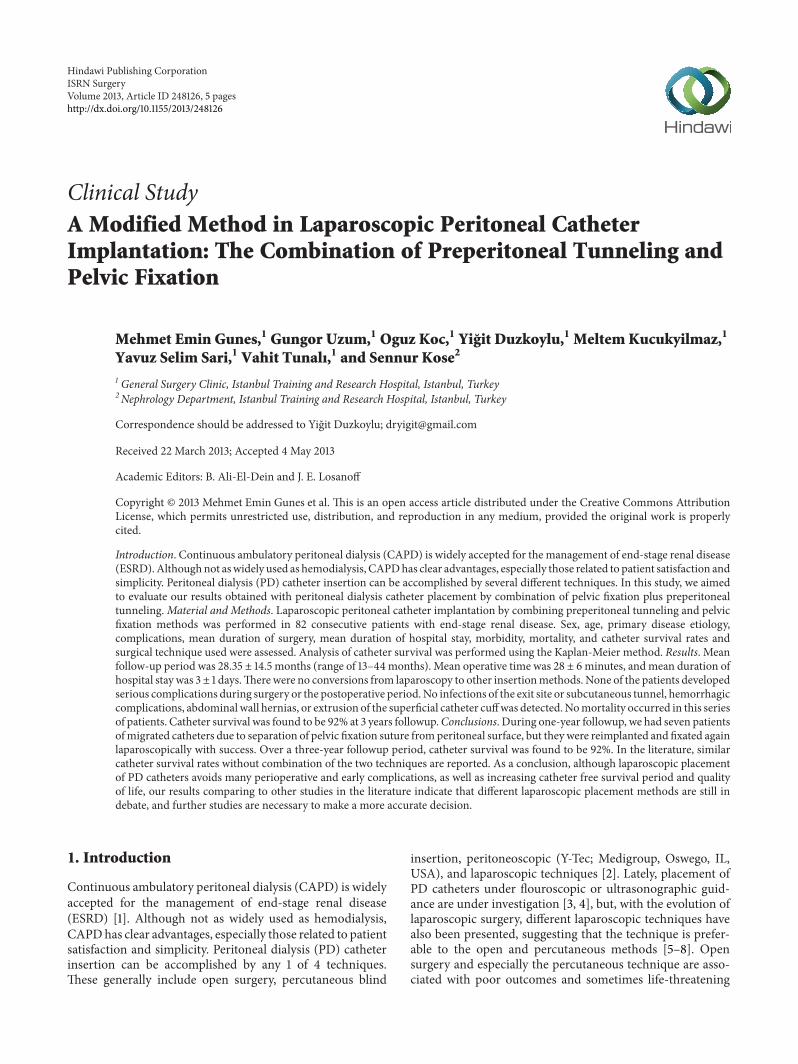
Figure 1: Entering sites of two ports.
complications. In fact, the incidence of omental wrapping,catheter displacement, and intraabdominal complications,specifically bowel and bladder perforation, is higher withthese two methods [5, 9]. In particular in patients suspectedof having intra-abdominal adhesions, the application oflaparoscopic surgical techniques has significantly changedour surgical approach to dialysis catheter placement, but thedebate on open or laparoscopic placement of PD cathetersis still ongoing [5, 8, 10, 11]. Malfunction of the peritonealcatheter is a frequent complication in peritoneal dialysis andresults in catheter loss. Therefore, it is vital that the dialysiscatheter tip is sited and secured accurately in the pelvisif long-term catheter function is to be achieved. This hasled to improvements in laparoscopic techniques, includingthe preperitoneal tunneling, pelvic fixation, omentopexy, orother minimally invasive one port insertions, which aims tominimize omental wrapping and catheter dislocation [11–20].
Although each technique alone has been shown to pro-long catheter survey, only Soontrapornchai’s study has evalu-ated the combination of these two techniques in the literature[6]. We, therefore, aimed to report the results obtained withperitoneal dialysis catheter placement by combination ofpelvic fixation plus preperitoneal tunneling.
2. Materials and Methods
Between January 2008 and June 2011, patients have under-gone laparoscopic peritoneal catheter placement by combin-ing preperitoneal tunneling and pelvic fixation (𝑛 = 82).A prophylactic antibiotic, cefazolin, was administered priorto the surgery. Catheter irrigation was commenced 3 daysafter surgery and consisted of a daily in-and-out flush withdialysate solution. Complete PD was generally commencedat the 14th day of surgery (14 ± 2).
The Peritoneal Catheter kit (Quinton Instrument Com-pany, Seattle,WA) formodified Seldinger (Littleford-Spector)technique and a 16 French Pull-Apart Sheath Introducer(SherwoodMedical Company, St. Louis,MO) are used for thecatheter placement. All surgical procedures were performedunder general anesthesia and by the same team experiencedin laparoscopic surgery. Sex, age, primary disease etiology,complications, mean duration of surgery, mean length of
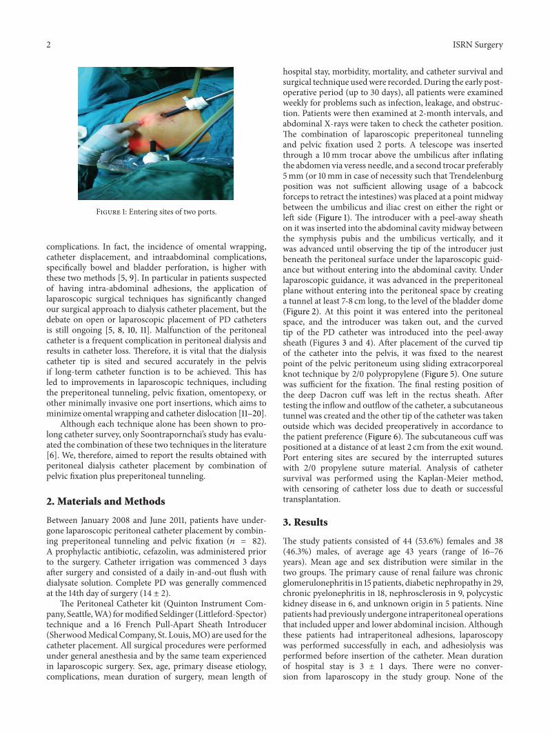
hospital stay, morbidity, mortality, and catheter survival andsurgical technique usedwere recorded. During the early post-operative period (up to 30 days), all patients were examinedweekly for problems such as infection, leakage, and obstruc-tion. Patients were then examined at 2-month intervals, andabdominal X-rays were taken to check the catheter position.The combination of laparoscopic preperitoneal tunnelingand pelvic fixation used 2 ports. A telescope was insertedthrough a 10mm trocar above the umbilicus after inflatingthe abdomen via veress needle, and a second trocar preferably5mm (or 10mm in case of necessity such that Trendelenburgposition was not sufficient allowing usage of a babcockforceps to retract the intestines) was placed at a pointmidwaybetween the umbilicus and iliac crest on either the right orleft side (Figure 1). The introducer with a peel-away sheathon it was inserted into the abdominal cavity midway betweenthe symphysis pubis and the umbilicus vertically, and itwas advanced until observing the tip of the introducer justbeneath the peritoneal surface under the laparoscopic guid-ance but without entering into the abdominal cavity. Underlaparoscopic guidance, it was advanced in the preperitonealplane without entering into the peritoneal space by creatinga tunnel at least 7-8 cm long, to the level of the bladder dome(Figure 2). At this point it was entered into the peritonealspace, and the introducer was taken out, and the curvedtip of the PD catheter was introduced into the peel-awaysheath (Figures 3 and 4). After placement of the curved tipof the catheter into the pelvis, it was fixed to the nearestpoint of the pelvic peritoneum using sliding extracorporealknot technique by 2/0 polypropylene (Figure 5). One suturewas sufficient for the fixation. The final resting position ofthe deep Dacron cuff was left in the rectus sheath. Aftertesting the inflow and outflow of the catheter, a subcutaneoustunnel was created and the other tip of the catheter was takenoutside which was decided preoperatively in accordance tothe patient preference (Figure 6). The subcutaneous cuff waspositioned at a distance of at least 2 cm from the exit wound.Port entering sites are secured by the interrupted sutureswith 2/0 propylene suture material. Analysis of cathetersurvival was performed using the Kaplan-Meier method,with censoring of catheter loss due to death or successfultransplantation.
3. Results
The study patients consisted of 44 (53.6%) females and 38(46.3%) males, of average age 43 years (range of 16–76years). Mean age and sex distribution were similar in thetwo groups. The primary cause of renal failure was chronicglomerulonephritis in 15 patients, diabetic nephropathy in 29,chronic pyelonephritis in 18, nephrosclerosis in 9, polycystickidney disease in 6, and unknown origin in 5 patients. Ninepatients had previously undergone intraperitoneal operationsthat included upper and lower abdominal incision. Althoughthese patients had intraperitoneal adhesions, laparoscopywas performed successfully in each, and adhesiolysis wasperformed before insertion of the catheter. Mean durationof hospital stay is 3 ± 1 days. There were no conver-sion from laparoscopy in the study group. None of the
ISRN Surgery 3
Figure 2: The introducer with a peel-away sheath on it which was oriented downward preperitoneally.
Figure 3: Peel-away sheet is taken out.
Figure 4: Tenckhoff catheter is introduced in the minor pelvis.
patients developed serious complications during surgery orthe postoperative period. Nomortality occurred in this seriesof patients. Mean follow-up period was 28.35 ± 14.5 months(range of 13–44 months). Two patients developed an earlyleakage from the 5mmport site, whichwas treated by applica-tion of a primary suture to the fascia of entering site. Bacterialperitonitis were seen in one patient at follow-up periodand responded to antibiotic therapy. No infections of theexit site or subcutaneous tunnel, hemorrhagic complications,abdominal wall hernias, port site hernias, or extrusion of thesuperficial catheter cuff was detected. Eight patients (8.53%)developed catheter function disorders during three-yearfollow-up period, and each underwent laparoscopic revision,which restored catheter function. The cause of disfunction
Figure 5: Fixation suture is performed.
Figure 6: Exit site is created to the preferential of patient before theoperation.
was catheter tip dislocation and/or omental wrapping in thesepatients. Catheter survival was found to be 92% at 3 years dueto disfunction, death, or successful transplantation. One- andtwo-year catheter survival was 96% and 95%, respectively.
4. Discussion
Although CAPD is used more frequently than hemodialy-sis in European countries as a renal replacement method,the rate of CAPD is quite low in other countries. Oneof the primary reasons for this low rate of CAPD use isthe incidence of catheter dysfunction, combined with the
4 ISRN Surgery
difficulty in performing remedial operations [18, 21]. Increas-ing the primary survival rate of laparoscopically implantedPD catheters will, therefore, enhance its acceptance as areplacement treatment. Among the methods used to insertPD (Tenckhoff) catheters are open surgery, blind insertionwith or without radiological assistance, and laparoscopy-assisted insertion. Due to the growing worldwide acceptanceof minimally invasive procedures, laparoscopic placement ofperitoneal catheters is preferred over open surgery with localanesthesia, increasing catheter survival rates significantly.However, catheter function disorders can occur, generally asa result of catheter migration or omental wrapping [7, 13–21].
New techniques have been developed to avoid thesecomplications and have led to increased survival rates [11–24]. These techniques include fixation of the omentum to thelateral abdominal wall (omentopexy) or omental excision andpreperitoneal tunneling and pelvic fixation [13–27]. Duringthe placement of PD catheters, efforts should be made toemploy minimally invasive methods, using fewer ports. If thecurved tip of the catheter with holes is kept in the pelviccavity, omental wrapping and migration can be avoided.Although the results of omentopexy or omentum excisionare positive, this technique necessitates greater experiencein laparoscopy. Use of the preperitoneal tunneling method,while advancing the catheter in the downward position andplacing it in the pelvic fossa, would eliminate the possibilityof extreme migration. However, the tip of the catheter canstill migrate to the right or left fossa iliaca and lead tofunctional disorders. Therefore, a combination of catheterfixing to the pelvic cavity at the nearest point to its tiptogether with preperitoneal tunnelingmay bringmuch betterlong-term results. The combination of omentopexy and thepreperitoneal tunneling method resulted in a longer cathetersurvival than each method alone [13–21].
In addition, fixation of the tip of the catheter in thepelvic region has also been found to increase survival [6,17]. To our knowledge, however, there have been one studyof the combination of preperitoneal tunneling and pelvicfixation [6]. We, therefore, planned a study combining thesetechniques, omental wrapping and catheter tip migration,which cause disturbances in fluid inflow or outflow whichcan be minimized, to make a more clear statement about thismethod. We observed no significance with having comparedto the literature as to the patient mortality and morbidity[6, 7, 11–17, 20–23]. Laparoscopy also permits to remedialoperations such as repositioning of the catheter or dissolvingthe omental wrapping. During three-year followup, we hadeight patients of malfunctioned catheters due to probablyseparation of pelvic fixation suture from peritoneal surfacebecause they were found to be dislocated from the minorpelvis on abdominal X-rays, but they were repositioned andfixed again laparoscopically with success. Over a three-yearfollow-up period, catheter survival rate was found to be92%. Maio et al. reported 91% of catheter survival ratesat 3 years with their musculofascial downward incline ofplacement technique without pelvic fixation as a differencefrom our technique but in very similar fashion [24]. As aconclusion, although laparoscopic placement of PD cathetersavoids many perioperative and early complications, as well as
increasing catheter free survival period and quality of life, ourresults comparing to other studies in the literature indicatethat different laparoscopic placement methods are still indebate, and further studies are necessary to make a moreaccurate decision [15, 18, 20, 25–27].
References
[1] R. Gokal, “CAPD overview,” Peritoneal Dialysis International,vol. 16, no. 1, pp. S13–S18, 1996.
[2] C. Cruz, “Implantation techniques for peritoneal dialysiscatheters,” Peritoneal Dialysis International, vol. 16, no. 1, pp.S319–S321, 1996.
[3] A. K. Abdel-Aal, S. Gaddikeri, and S. Saddekni, “Techniqueof peritoneal catheter placement under fluroscopic guidance,”Radiology Research and Practice, vol. 2011, Article ID 141707, 4pages, 2011.
[4] S. J. Savader, “Percutaneous radiologic placement of peritonealdialysis catheters,” Journal of Vascular and Interventional Radi-ology, vol. 10, no. 3, pp. 249–256, 1999.
[5] L. Lund andM. Jønler, “Peritoneal dialysis catheter placement—is laparoscopy an option?” International Urology and Nephrol-ogy, vol. 39, no. 2, pp. 625–628, 2007.
[6] P. Soontrapornchai and T. Simapatanapong, “Comparison ofopen and laparoscopic secure placement of peritoneal dialysiscatheters,” Surgical Endoscopy and Other Interventional Tech-niques, vol. 19, no. 1, pp. 137–139, 2005.
[7] A. S. Adamson, J. P. Kelleher, M. E. Snell, and B. Hulme,“Endoscopic placement of CAPD catheters: a review of onehundred procedures,” Nephrology Dialysis Transplantation, vol.7, no. 8, pp. 855–857, 1992.
[8] S. M. Hagen, A. M. van Alphen, J. N. Ijzermans, and F. J.Dor, “Laparoscopic versus open peritoneal dialysis catheterinsertion,” BMC Surgery, vol. 11, p. 35, 2011.
[9] A. Asif, “Peritoneal dialysis catheter insertion,”Minerva Chirur-gica, vol. 60, no. 5, pp. 417–428, 2005.
[10] P. J. Yang, C. Y. Lee, C. C. Yeh, H. C. Nien, T. J. Tsai,and M. K. Tsai, “Mini-laparotomy implantation of peritonealdialysis catheters: outcome and rescue,” Peritoneal DialysisInternational, vol. 30, no. 5, pp. 513–518, 2010.
[11] S. H. Kang, J. Y. Do, K. H. Cho, J. W. Park, and K. W. Yoon,“Blind peritoneal catheter placement with a Tenckhoff trocar bynephrologists: a single-center experience,” Nephrology, vol. 17,no. 2, pp. 141–147, 2012.
[12] X. Yan, W. Zhu, C. M. Jiang, H. F. Huang, M. Zhang, and H. Q.Guo, “Clinical application of one-port laparoscopic placementof peritoneal dialysis cathetetrs,” Scandinavian Journal of Urol-ogy and Nephrology, vol. 44, no. 5, pp. 341–344, 2010.
[13] J. H. Crabtree, “Selected best demonstrated practices in peri-toneal dialysis access,” Kidney International, vol. 70, no. 103, pp.S27–S37, 2006.
[14] C. T. Lu, D. I. Watson, T. J. Elias, R. J. Faull, A. R. Clarkson, andK.M. Bannister, “Laparoscopic placement of peritoneal dialysiscatheters: 7 years experience,” ANZ Journal of Surgery, vol. 73,no. 3, pp. 109–111, 2003.
[15] G. Ogunc, “Minilaparoscopic extraperitoneal tunneling withomentopexy: a new technique for CAPD catheter placement,”Peritoneal Dialysis International, vol. 25, no. 6, pp. 551–555,2005.
ISRN Surgery 5
[16] M. Comert, A. Borazan, E. Kulah, and B. H. Ucan, “A newlaparoscopic technique for the placement of a permanent peri-toneal dialysis catheter: the preperitoneal tunneling method,”Surgical Endoscopy and Other Interventional Techniques, vol. 19,no. 2, pp. 245–248, 2005.
[17] H. V. Harissis, C. S. Katsios, E. L. Koliousi et al., “A newsimplified one port laparoscopic technique of peritoneal dialysiscatheter placement with intra-abdominal fixation,” AmericanJournal of Surgery, vol. 192, no. 1, pp. 125–129, 2006.
[18] J. H. Crabtree and R. J. Burchette, “Effective use of laparoscopyfor long-term peritoneal dialysis access,” American Journal ofSurgery, vol. 198, no. 1, pp. 135–141, 2009.
[19] A. Keshvari, I. Najafi, M. Jafari-Javid, M. Yunesian, R. Chaman,andM. N. Taromlou, “Laparoscopic peritoneal dialysis catheterimplantation using a Tenckhoff trocar under local anesthe-sia with nitrous oxide gas insufflation,” American Journal ofSurgery, vol. 197, no. 1, pp. 8–13, 2009.
[20] V. Attaluri, C. Lebeis, S. Brethauer, and S. Rosenblatt, “Advancedlaparoscopic techniques significantly improve function of peri-toneal dialysis catheters,” Journal of the American College ofSurgeons, vol. 211, no. 6, pp. 699–704, 2010.
[21] D. Neufeld, Z. Korzets, J. Bernheim, and B. Shpitz, “Laparo-scopic management of peritoneal dialysis catheters,” Harefuah,vol. 150, no. 2, pp. 84–86, 209, 208, 2011.
[22] A. Borazan, M. Comert, B. H. Ucan et al., “The comparisonin terms of early complications of a new technique and percu-taneous method for the placement of CAPD catheters,” RenalFailure, vol. 28, no. 1, pp. 37–42, 2006.
[23] S. A. Carrillo, M. M. Ghersi, and S. W. Unger, “Laparoscopic-assisted peritoneal dialysis catheter placement: a microinvasivetechnique,” Surgical Endoscopy and Other Interventional Tech-niques, vol. 21, no. 5, pp. 825–829, 2007.
[24] R. Maio, N. Figueiredo, and P. Costa, “Laparoscopic placementof Tenckhoff catheters for peritoneal dialysis: a safe, effective,and reproducible procedure,” Peritoneal Dialysis International,vol. 28, no. 2, pp. 170–173, 2008.
[25] D. I. Watson, D. Paterson, and K. Bannister, “Secure placementof peritoneal dialysis catheters using a laparoscopic technique,”Surgical Laparoscopy, Endoscopy and Percutaneous Techniques,vol. 6, no. 1, pp. 35–37, 1996.
[26] C. D. Gerhart, “Needleoscopic placement of Tenckhoffcatheters,” Journal of the Society of Laparoendoscopic Surgeons ,vol. 3, no. 2, pp. 155–158, 1999.
[27] D. Bar-Zohar, B. Sagie, N. Lubezky, M. Blum, J. Klausner, andS. Abu-Abeid, “Laparoscopic implantation of the Tenckhoffcatheter for the treatment of end-stage renal failure and conges-tive heart failure: experience with the pelvic fixation technique,”Israel Medical Association Journal, vol. 8, no. 3, pp. 174–178,2006.

Submit your manuscripts athttp://www.hindawi.com
Stem CellsInternational
Hindawi Publishing Corporationhttp://www.hindawi.com Volume 2014
Hindawi Publishing Corporationhttp://www.hindawi.com Volume 2014
MEDIATORSINFLAMMATION
of
Hindawi Publishing Corporationhttp://www.hindawi.com Volume 2014
Behavioural Neurology
EndocrinologyInternational Journal of
Hindawi Publishing Corporationhttp://www.hindawi.com Volume 2014
Hindawi Publishing Corporationhttp://www.hindawi.com Volume 2014
Disease Markers
Hindawi Publishing Corporationhttp://www.hindawi.com Volume 2014
BioMed Research International
OncologyJournal of
Hindawi Publishing Corporationhttp://www.hindawi.com Volume 2014
Hindawi Publishing Corporationhttp://www.hindawi.com Volume 2014
Oxidative Medicine and Cellular Longevity
Hindawi Publishing Corporationhttp://www.hindawi.com Volume 2014
PPAR Research
The Scientific World JournalHindawi Publishing Corporation http://www.hindawi.com Volume 2014
Immunology ResearchHindawi Publishing Corporationhttp://www.hindawi.com Volume 2014
Journal of
ObesityJournal of
Hindawi Publishing Corporationhttp://www.hindawi.com Volume 2014
Hindawi Publishing Corporationhttp://www.hindawi.com Volume 2014
Computational and Mathematical Methods in Medicine
OphthalmologyJournal of
Hindawi Publishing Corporationhttp://www.hindawi.com Volume 2014
Diabetes ResearchJournal of
Hindawi Publishing Corporationhttp://www.hindawi.com Volume 2014
Hindawi Publishing Corporationhttp://www.hindawi.com Volume 2014
Research and TreatmentAIDS
Hindawi Publishing Corporationhttp://www.hindawi.com Volume 2014
Gastroenterology Research and Practice
Hindawi Publishing Corporationhttp://www.hindawi.com Volume 2014
Parkinson’s Disease
Evidence-Based Complementary and Alternative Medicine
Volume 2014Hindawi Publishing Corporationhttp://www.hindawi.com
Related Documents


![Research Article - Hindawi Publishing Corporationdownloads.hindawi.com/archive/2012/591450.pdf2 ISRN Civil Engineering probe methods [11, 12]. However, these methods are time consuming,](https://static.cupdf.com/doc/110x72/5e8959b0c343ca456238d0eb/research-article-hindawi-publishing-2-isrn-civil-engineering-probe-methods-11.jpg)