pathogens Review Clinical Parasitology and Parasitome Maps as Old and New Tools to Improve Clinical Microbiomics Stefania Pane 1,† , Maria Vittoria Ristori 1,2,† , Simone Gardini 3 , Alessandra Russo 1 , Federica Del Chierico 2 and Lorenza Putignani 1,2, * Citation: Pane, S.; Ristori, M.V.; Gardini, S.; Russo, A.; Del Chierico, F.; Putignani, L. Clinical Parasitology and Parasitome Maps as Old and New Tools to Improve Clinical Microbiomics. Pathogens 2021, 10, 1550. https://doi.org/10.3390/ pathogens10121550 Academic Editors: Donato Traversa, Angela Di Cesare and Simone Morelli Received: 23 September 2021 Accepted: 25 November 2021 Published: 28 November 2021 Publisher’s Note: MDPI stays neutral with regard to jurisdictional claims in published maps and institutional affil- iations. Copyright: © 2021 by the authors. Licensee MDPI, Basel, Switzerland. This article is an open access article distributed under the terms and conditions of the Creative Commons Attribution (CC BY) license (https:// creativecommons.org/licenses/by/ 4.0/). 1 Microbiology and Diagnostic Immunology Unit, Unit of Microbiomics, Bambino Gesù Children’s Hospital, IRCCS, 00146 Rome, Italy; [email protected] (S.P.); [email protected] (M.V.R.); [email protected] (A.R.) 2 Multimodal Laboratory Medicine Research Area, Unit of Human Microbiome, Bambino Gesù Children’s Hospital, IRCCS, 00146 Rome, Italy; [email protected] 3 GenomeUp, Via Nemorense 91, 00199 Rome, Italy; [email protected] * Correspondence: [email protected]; Tel.: +39-0668592598 (ext. 4076) † These authors contributed equally to this work. Abstract: A growing body of evidence shows that dysbiotic gut microbiota may correlate with a wide range of disorders; hence, the clinical use of microbiota maps and fecal microbiota transplantation (FMT) can be exploited in the clinic of some infectious diseases. Through direct or indirect ecologi- cal and functional competition, FMT may stimulate decolonization of pathogens or opportunistic pathogens, modulating immune response and colonic inflammation, and restoring intestinal home- ostasis, which reduces host damage. Herein, we discuss how diagnostic parasitology may contribute to designing clinical metagenomic pipelines and FMT programs, especially in pediatric subjects. The consequences of more specialized diagnostics in the context of gut microbiota communities may improve the clinical parasitology and extend its applications to the prevention and treatment of several communicable and even noncommunicable disorders. Keywords: parasites; microbiota; Faecal Microbiota Transplantation (FMT) 1. Introduction The intestinal environment is an ecosystem where biological and chemical interactions occur at various organizational levels between host, parasites, and microbial communities, greatly affecting human health and physiology. Considering the gut microbiota at the taxonomic level, we can observe a significant variation among individuals, each harboring a unique collection of bacterial species, which may change over time and could be considered a fingerprint [1–3]. The microbiota at the gut microenvironment level provides important protective, immune regulatory and metabolic functions. The defensive mechanism against pathogenic bacteria is exerted by the barrier effect of the intestinal epithelium, playing a major role in protecting the host and representing an important obstacle to pathogenic invasion [4,5]. Indeed, gut microbiota has an important role in immunological activation and development, as demonstrated in many metabolic and autoimmune diseases [6], by influencing host immune response [7–11]. The balance of the gut microbial ecosystem, eubiosis, is an important concept. Indeed, the eubiotic gut microbiota is characterized by a preponderance of potentially beneficial species, belonging mainly to the two bacterial phylum Firmicutes and Bacteroidetes, and, potentially, pathogenic species such as Proteobacteria. In the dysbiosis state, “bad bacteria” predominate “good bacteria” [12–14]. Moreover, environmental factors, such as age, diet, stress, drugs, and infections in- cluding parasitosis, strongly influence the composition of the human microbiota [15–18]. Nowadays, it is evident that the intestinal microbiota has an important impact on human Pathogens 2021, 10, 1550. https://doi.org/10.3390/pathogens10121550 https://www.mdpi.com/journal/pathogens

Welcome message from author
This document is posted to help you gain knowledge. Please leave a comment to let me know what you think about it! Share it to your friends and learn new things together.
Transcript

pathogens
Review
Clinical Parasitology and Parasitome Maps as Old and NewTools to Improve Clinical Microbiomics
Stefania Pane 1,† , Maria Vittoria Ristori 1,2,†, Simone Gardini 3, Alessandra Russo 1, Federica Del Chierico 2
and Lorenza Putignani 1,2,*
�����������������
Citation: Pane, S.; Ristori, M.V.;
Gardini, S.; Russo, A.; Del Chierico, F.;
Putignani, L. Clinical Parasitology
and Parasitome Maps as Old and
New Tools to Improve Clinical
Microbiomics. Pathogens 2021, 10,
1550. https://doi.org/10.3390/
pathogens10121550
Academic Editors: Donato Traversa,
Angela Di Cesare and Simone Morelli
Received: 23 September 2021
Accepted: 25 November 2021
Published: 28 November 2021
Publisher’s Note: MDPI stays neutral
with regard to jurisdictional claims in
published maps and institutional affil-
iations.
Copyright: © 2021 by the authors.
Licensee MDPI, Basel, Switzerland.
This article is an open access article
distributed under the terms and
conditions of the Creative Commons
Attribution (CC BY) license (https://
creativecommons.org/licenses/by/
4.0/).
1 Microbiology and Diagnostic Immunology Unit, Unit of Microbiomics, Bambino Gesù Children’s Hospital,IRCCS, 00146 Rome, Italy; [email protected] (S.P.); [email protected] (M.V.R.);[email protected] (A.R.)
2 Multimodal Laboratory Medicine Research Area, Unit of Human Microbiome, Bambino Gesù Children’sHospital, IRCCS, 00146 Rome, Italy; [email protected]
3 GenomeUp, Via Nemorense 91, 00199 Rome, Italy; [email protected]* Correspondence: [email protected]; Tel.: +39-0668592598 (ext. 4076)† These authors contributed equally to this work.
Abstract: A growing body of evidence shows that dysbiotic gut microbiota may correlate with a widerange of disorders; hence, the clinical use of microbiota maps and fecal microbiota transplantation(FMT) can be exploited in the clinic of some infectious diseases. Through direct or indirect ecologi-cal and functional competition, FMT may stimulate decolonization of pathogens or opportunisticpathogens, modulating immune response and colonic inflammation, and restoring intestinal home-ostasis, which reduces host damage. Herein, we discuss how diagnostic parasitology may contributeto designing clinical metagenomic pipelines and FMT programs, especially in pediatric subjects. Theconsequences of more specialized diagnostics in the context of gut microbiota communities mayimprove the clinical parasitology and extend its applications to the prevention and treatment ofseveral communicable and even noncommunicable disorders.
Keywords: parasites; microbiota; Faecal Microbiota Transplantation (FMT)
1. Introduction
The intestinal environment is an ecosystem where biological and chemical interactionsoccur at various organizational levels between host, parasites, and microbial communities,greatly affecting human health and physiology.
Considering the gut microbiota at the taxonomic level, we can observe a significantvariation among individuals, each harboring a unique collection of bacterial species, whichmay change over time and could be considered a fingerprint [1–3].
The microbiota at the gut microenvironment level provides important protective,immune regulatory and metabolic functions. The defensive mechanism against pathogenicbacteria is exerted by the barrier effect of the intestinal epithelium, playing a major role inprotecting the host and representing an important obstacle to pathogenic invasion [4,5].Indeed, gut microbiota has an important role in immunological activation and development,as demonstrated in many metabolic and autoimmune diseases [6], by influencing hostimmune response [7–11]. The balance of the gut microbial ecosystem, eubiosis, is animportant concept. Indeed, the eubiotic gut microbiota is characterized by a preponderanceof potentially beneficial species, belonging mainly to the two bacterial phylum Firmicutesand Bacteroidetes, and, potentially, pathogenic species such as Proteobacteria. In thedysbiosis state, “bad bacteria” predominate “good bacteria” [12–14].
Moreover, environmental factors, such as age, diet, stress, drugs, and infections in-cluding parasitosis, strongly influence the composition of the human microbiota [15–18].Nowadays, it is evident that the intestinal microbiota has an important impact on human
Pathogens 2021, 10, 1550. https://doi.org/10.3390/pathogens10121550 https://www.mdpi.com/journal/pathogens
Pathogens 2021, 10, 1550 2 of 15
pathophysiology, also regulating pathogenic burden [19]. Intestinal helminths may directlyinfluence the immune system through their effects on both gut luminal and mucosal micro-biota [20]. Indeed, Th2 cells have an important role in combatting parasitic infections [21].However, in existing literature, there is conflicting evidence on the effect of parasites onmicrobiota ecology and function and their impact on health and disease balance.
Within this complex scenario, intestinal citizens (e.g., viruses, mycetes, and parasites)interact with the microbial community, modifying the balance between host and gut micro-biota [4,22]. The intestinal microenvironment, considered as a whole community, providesan important protective mucosal defense mechanism, but there is evidence that changein the composition of the commensal microbiota alters the gut environment, making thiscomposition vulnerable to pathogenic organisms [23–25]. Many factors such as antibiotics,psychological stress, physical stress, modern diet, and hygiene can affect microbial stability,and thus contribute to intestinal dysbiosis [13].
2. Role and Relationship amongst Gut Microbiota Citizens
In addition to bacteria, other key microorganisms, such as yeasts and filamentousfungi, viruses, and phages, are present in the gut [26,27]. Moreover, for ~25% of the world’spopulation, the gut microbiota also comprises intestinal protozoan and worms, namelymeiofauna [28,29].
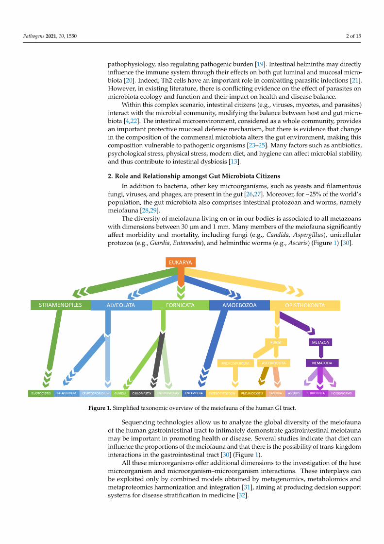
The diversity of meiofauna living on or in our bodies is associated to all metazoanswith dimensions between 30 µm and 1 mm. Many members of the meiofauna significantlyaffect morbidity and mortality, including fungi (e.g., Candida, Aspergillus), unicellularprotozoa (e.g., Giardia, Entamoeba), and helminthic worms (e.g., Ascaris) (Figure 1) [30].
Pathogens 2021, 10, x
usual event in patients who suffer from IBD. Although the main reason for this alteration
is not clear, the interaction between gut bacteria and gut fungi seems to be an important
subject in IBD patients [43].
So, intestinal parasites, both protozoans and metazoan (nematodes and platyhel-
minths), interact with the microbial community, modifying the balance between host and
gut microbiota [44].
Figure 1. Simplified taxonomic overview of the meiofauna of the human GI tract.
3. Parasites and Gut Microbiota Profiling
Parasitic infections represent a significant health problem, particularly in underde-
veloped and developing countries. Soil-transmitted helminths (STHs), e.g., Strongyloides
stercoralis and Trichuris trichiura, are common intestinal parasites, followed by blood
flukes (i.e., Schistosoma spp.) and filarial worms (e.g., Wuchereria and Brugia). Generally
speaking, intestinal parasitic infections may have a low impact in immunocompetent sub-
jects; however, they can potentially become a major issue in vulnerable groups. In fact,
helminth infections produce malnutrition, physical damage, and cognitive development
complications in children [45]. Furthermore, a wide range of protozoans are common par-
asites of the human gastro-intestinal tract (e.g., Cryptosporidium spp., Entamoeba histolytica,
Giardia duodenalis). The spectrum of clinical manifestations of protozoan infections varies
from chronic diarrhea or weight loss, or mild self-limiting illness to acute disease, until
malabsorption [29,46,47], as well as the modality of transmission, zoonotic or anthropon-
otic, particularly investigated as reported for Apicomplexan parasites [48]. A study con-
ducted on packaged salads highlighted the presence of protozoan contamination, such as
Giardia duodenalis, Cryptosporidium spp., Toxoplasma gondii, and Cyclospora cayetanensis,
through microscopy and molecular analyses, concluding that 4.2% of the samples were
contaminated by at least one protozoan species, and 0.6% of samples with at least two
protozoa [49].
Together with trillions of microorganisms, i.e., archaea, viruses, bacteria, and eukar-
yotes residing in the GI tract, parasitic worms establish the “macrobiota” [50]. The micro-
biota of a host may strongly interfere with the survival and physiology of many parasites
Figure 1. Simplified taxonomic overview of the meiofauna of the human GI tract.
Sequencing technologies allow us to analyze the global diversity of the meiofaunaof the human gastrointestinal tract to intimately demonstrate gastrointestinal meiofaunamay be important in promoting health or disease. Several studies indicate that diet caninfluence the proportions of the meiofauna and that there is the possibility of trans-kingdominteractions in the gastrointestinal tract [30] (Figure 1).
All these microorganisms offer additional dimensions to the investigation of the hostmicroorganism and microorganism–microorganism interactions. These interplays canbe exploited only by combined models obtained by metagenomics, metabolomics andmetaproteomics harmonization and integration [31], aiming at producing decision supportsystems for disease stratification in medicine [32].
Pathogens 2021, 10, 1550 3 of 15
All microbial inhabitants may have both beneficial and detrimental roles in the humanhealth, including improvement of microbial resilience, immune evasion, maintenance ofphysiologic processes, but even alteration of microbial communities [33]. Bacteriophagesin the gut are largely unexplored, despite their potential to regulate bacterial communitiesand thus human health [34,35]. Indeed, experimental limitations still successfully affectthe isolation of phages and genome annotation as well as the full characterization ofvirus–human populations [36]. Some studies have highlighted the uniqueness of phagecommunities in individuals and their capacity to be stable in a healthy gut [37]. Remarkably,the stability of the viral genome is probably responsible for the stableness of bacteria andmicrobiota metagenomes [38]. However, there are still only a few studies on the role of thevirome in the host’s intestinal microbiota ecosystem [39].
Fungi are normal inhabitants of the mammalian gastrointestinal tract. In fact, thehuman gut is colonized by more than 50 genera of fungi [40]. In particular, the gut ischaracterized by the presence of Candida, Saccharomyces and Cladosporium species. Nutri-tional modification may have an effect on the fungal microbiota; in particular, plant-baseddiets increase the levels of Candida, whereas animal-based diets improve the presence ofPenicillium species [41,42]. Indeed, fungi compose a very small portion of gut microbiotabut play determinative roles in the homeostasis of the gut bacterial composition and themucosal immune responses. An interkingdom correlation between bacteria and fungi hasbeen suggested. Alterations in the composition and function of the gut microbiota are ausual event in patients who suffer from IBD. Although the main reason for this alterationis not clear, the interaction between gut bacteria and gut fungi seems to be an importantsubject in IBD patients [43].
So, intestinal parasites, both protozoans and metazoan (nematodes and platyhelminths),interact with the microbial community, modifying the balance between host and gut micro-biota [44].
3. Parasites and Gut Microbiota Profiling
Parasitic infections represent a significant health problem, particularly in underde-veloped and developing countries. Soil-transmitted helminths (STHs), e.g., Strongyloidesstercoralis and Trichuris trichiura, are common intestinal parasites, followed by blood flukes(i.e., Schistosoma spp.) and filarial worms (e.g., Wuchereria and Brugia). Generally speak-ing, intestinal parasitic infections may have a low impact in immunocompetent subjects;however, they can potentially become a major issue in vulnerable groups. In fact, helminthinfections produce malnutrition, physical damage, and cognitive development complica-tions in children [45]. Furthermore, a wide range of protozoans are common parasites ofthe human gastro-intestinal tract (e.g., Cryptosporidium spp., Entamoeba histolytica, Giardiaduodenalis). The spectrum of clinical manifestations of protozoan infections varies fromchronic diarrhea or weight loss, or mild self-limiting illness to acute disease, until mal-absorption [29,46,47], as well as the modality of transmission, zoonotic or anthroponotic,particularly investigated as reported for Apicomplexan parasites [48]. A study conductedon packaged salads highlighted the presence of protozoan contamination, such as Giardiaduodenalis, Cryptosporidium spp., Toxoplasma gondii, and Cyclospora cayetanensis, through mi-croscopy and molecular analyses, concluding that 4.2% of the samples were contaminatedby at least one protozoan species, and 0.6% of samples with at least two protozoa [49].
Together with trillions of microorganisms, i.e., archaea, viruses, bacteria, and eu-karyotes residing in the GI tract, parasitic worms establish the “macrobiota” [50]. Themicrobiota of a host may strongly interfere with the survival and physiology of manyparasites and, consequently, with the outcome of many parasitic infections. During parasiteinfections, the interaction between the gut microbiota and the helminths has been associ-ated with the establishment of the infection, the clinical manifestations, and even immunemodulation [51,52]. However, both increased and decreased gut microbiota diversity hasbeen observed in subjects affected by intestinal parasites [53–57].
Pathogens 2021, 10, 1550 4 of 15
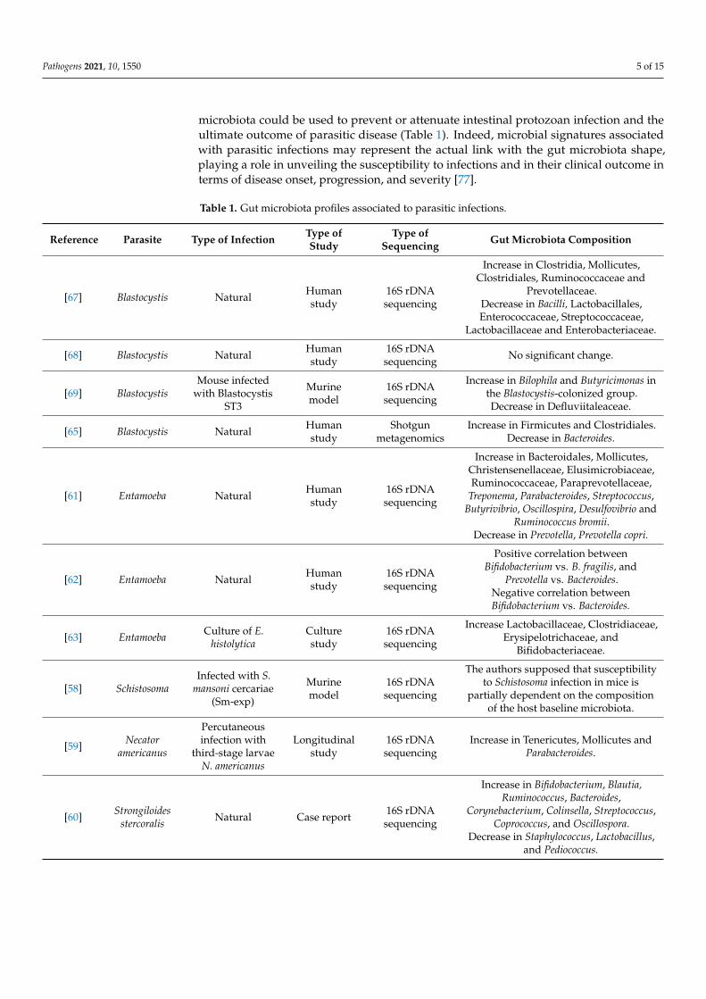
Experiments performed in murine models of intestinal schistosomiasis have suggestedinteractions between Schistosoma parasites and the host gut microbiota with a direct impacton the intestinal microbial communities [58]. In a study by Alba Cortés et al., the gutmicrobiome composition of the host actually influenced the host’s susceptibility to S.mansoni infection, as well as infection-associated changes in gut microbiota profiles [58].Some studies have evidenced that Necator americanus infection could alleviate chronicinflammation in celiac disease and improve prokaryotic species richness, reestablishingthe eubiosis and immune homeostasis [59]. In a case report on Strongiloides stercoralisinfection, gut microbiota composition was associated with enrichment in Bifidobacterium,Blautia, Ruminococcus, Bacteroides, Corynebacterium, Colinsella, Streptococcus, Coprococcus,and Oscillospora genera, and a decrease in Staphylococcus, Lactobacillus, and Pediococcus.The authors suggested a putative direct or immune-mediated ability of S. stercoralis topromote the increase in bacterial diversity [60]. In some studies conducted in a ruralAfrican population, the authors showed a characteristic gut microbiota ecology in patientscarrying Entamoeba colonization [61–63] (Table 1).
A review of the literature tried to study in depth the mutual influences of intestinalnematodes and host-gut microbiota, highlighting the potential beneficial effects (i.e., promo-tion of eubiosis) through the production of useful metabolites (i.e., short-chain fatty acids,SCFA) [60]. Conversely, nematode infection may promote dysbiosis due to promotion ofpathogenic bacterial species and decrease in mutualistic commensal. Authors exemplifiedthe nematode–microbiota interactions and their impact on the host immune response [64].
A shotgun metagenomics study on samples of patients with the Blastocystis spp.infection showed a very strong association between the presence of Blastocystis spp. and theabundance of archaeal organisms (Methanobrevibacter smithii) [65]. Additionally, anotherstudy showed a decrease in Blastocystis in individuals with the Bacteroides enterotypecompared to subjects with the Ruminococcus or Prevotella enterotypes [66]. Audebertet al. highlighted a high microbiota diversity, increased abundance of Clostridia, andlow abundance of Enterobacteriaceae in Blastocystis-colonized patients, suggesting thatBlastocystis infection may be associated with a healthy gut microbiota [67]. Conversely,another study did not highlight significant modifications in the gut microbiota of Blastocystispositive subjects affected by irritable bowel syndrome [68]. In a rat model of Blastocystis ST3infection, the colonization altered gut microbiota composition, but not richness, inducingonly mild gut inflammation but no clinical symptoms. In addition, the long-term Blastocystisexposure appeared to promote faster recovery from colitis, suggesting that Blastocystis mayalter the gut ecosystem in a protective way and promote faster recovery [69]. Conversely,in healthy subjects, the fermentation by anaerobic bacteria and Blastocystis induced anincreased SCFA production [70]. A further study has developed a bioinformatic pipeline todetect Blastocystis subtypes (STs) from shotgun metagenomics data, identifying Blastocystisas a common component of the healthy gut microbiome [65]. The authors showed howmetagenomics could play an important role in advancing evidence on population genomicsof human parasites. Therefore, through DNA sequencing-based methods, it might bepossible to address the role of Blastocystis and Dientamoeba as commensal components ofhealthy gut microbiota rather than pathogenic microorganisms.
In other papers, B. hominis and D. fragilis appeared to be more or less obligatedeukaryotic members of the gut microbiota [71–73] and less frequent in subjects withintestinal diseases and metabolic disorders [65,74–76].
Conversely, other studies have highlighted the possible role of D. fragilis and B.hominis as infectious agents, providing a relationship with gut microbiota profiles andhost phenotypic features, including microbiota dysbiosis or eubiosis [70]. Indeed, in thisarticle, Stensvold C.R. et al. discussed the possible role of Blastocystis as an indicator ofdisease-related gut microbiota profiles in subjects affected by gut dysbiosis [70] or as anindicator of eubiosis in healthy subjects.
Since the composition of the intestinal bacterial population affects the impact of theinfection of protozoans, or vice versa, the modulation of different components of the
Pathogens 2021, 10, 1550 5 of 15
microbiota could be used to prevent or attenuate intestinal protozoan infection and theultimate outcome of parasitic disease (Table 1). Indeed, microbial signatures associatedwith parasitic infections may represent the actual link with the gut microbiota shape,playing a role in unveiling the susceptibility to infections and in their clinical outcome interms of disease onset, progression, and severity [77].
Table 1. Gut microbiota profiles associated to parasitic infections.
Reference Parasite Type of Infection Type ofStudy
Type ofSequencing Gut Microbiota Composition
[67] Blastocystis Natural Humanstudy
16S rDNAsequencing
Increase in Clostridia, Mollicutes,Clostridiales, Ruminococcaceae and
Prevotellaceae.Decrease in Bacilli, Lactobacillales,Enterococcaceae, Streptococcaceae,
Lactobacillaceae and Enterobacteriaceae.
[68] Blastocystis Natural Humanstudy
16S rDNAsequencing No significant change.
[69] BlastocystisMouse infected
with BlastocystisST3
Murinemodel
16S rDNAsequencing
Increase in Bilophila and Butyricimonas inthe Blastocystis-colonized group.Decrease in Defluviitaleaceae.
[65] Blastocystis Natural Humanstudy
Shotgunmetagenomics
Increase in Firmicutes and Clostridiales.Decrease in Bacteroides.
[61] Entamoeba Natural Humanstudy
16S rDNAsequencing
Increase in Bacteroidales, Mollicutes,Christensenellaceae, Elusimicrobiaceae,Ruminococcaceae, Paraprevotellaceae,
Treponema, Parabacteroides, Streptococcus,Butyrivibrio, Oscillospira, Desulfovibrio and
Ruminococcus bromii.Decrease in Prevotella, Prevotella copri.
[62] Entamoeba Natural Humanstudy
16S rDNAsequencing
Positive correlation betweenBifidobacterium vs. B. fragilis, and
Prevotella vs. Bacteroides.Negative correlation betweenBifidobacterium vs. Bacteroides.
[63] Entamoeba Culture of E.histolytica
Culturestudy
16S rDNAsequencing
Increase Lactobacillaceae, Clostridiaceae,Erysipelotrichaceae, and
Bifidobacteriaceae.
[58] SchistosomaInfected with S.
mansoni cercariae(Sm-exp)
Murinemodel
16S rDNAsequencing
The authors supposed that susceptibilityto Schistosoma infection in mice is
partially dependent on the compositionof the host baseline microbiota.
[59] Necatoramericanus
Percutaneousinfection with
third-stage larvaeN. americanus
Longitudinalstudy
16S rDNAsequencing
Increase in Tenericutes, Mollicutes andParabacteroides.
[60] Strongiloidesstercoralis Natural Case report 16S rDNA
sequencing
Increase in Bifidobacterium, Blautia,Ruminococcus, Bacteroides,
Corynebacterium, Colinsella, Streptococcus,Coprococcus, and Oscillospora.
Decrease in Staphylococcus, Lactobacillus,and Pediococcus.
Pathogens 2021, 10, 1550 6 of 15
4. New Molecular Approaches in Translational and Clinical Parasitology
In January 2019, the Parasite Microbiome Project (PMP) began to understand the role ofparasite-associated gut microbiota in the pathophysiology of helminthiases [78]. The PMPtried to draw best practices for experimental studies to ensure reliable comparisons betweendata sets and the introduction of appropriate controls to identify possible environmentalmicrobial contaminants [78]. In a recent review, the authors suggested four elements thatmust be considered when the scientist wants to generate reliable and reproducible data [50].As a first step, they propose generating appropriate negative controls (“blanks”) in each stepof the experiment, followed by the microscopy-based visualization of helminth-associatedbacteria to identify and characterize worm microbiomes across different helminth tissuesand developmental stages. Best practices are becoming more and more important becauseclinical metagenomic next-generation sequencing (mNGS) is rapidly moving from researchto clinical laboratories. Chiu et al. focused on the challenges of implementing mNGS in theclinical laboratory and addressing potential solutions for maximizing its impact on patientcare and public health [79]. In fact, metagenomic methods (NGS) are the new approach thatmicrobiologists and parasitologists should undertake to improve accuracy and sensitivity,with respect to the classical approach based on the microscopical method, which maybe affected by ambiguities and subjective interpretations [80]. Indeed, major usage ofdifferent genomic approaches, such as metataxonomics and metagenomes [81], may assistin reducing ambiguities and subjective interpretation in parasite description and assessingtheir relationship with microbial communities. Marchesi et al. suggested metataxonomicsto assess fine metataxonomic tree descriptions of the entire microbiota and metagenomicsto assemble microbial genomes and characterize new genes after annotation throughshotgun sequencing [81]. Particularly, metataxonomics refers to targeted sequencing of16S rRNA gene hypervariable regions [82] andallows representative bacterial taxonomicdescription [83].
In this context, remarkable is the choice of primers able to reduce potential bias in therepresentation of entire taxonomic units [84–86]. The sequencing output represents a set ofclusters of close related sequences, called operational taxonomic units (OTUs) [87]. Throughbioinformatic OTUs analysis, it is possible to assess the level of microbial communitydiversity, both in terms of evenness and richness [88] and the degree of divergence betweendifferent ecosystems or sample types [89].
On the contrary, shotgun metagenomics is based on DNA molecules, which arerandomly broken into fragments that are then sequenced [90]. Hence, the shotgun metage-nomic approach provides major information on the taxonomic composition and inferredfunctional genes, thus fully characterizing the sample, despite requested high coverage [91].
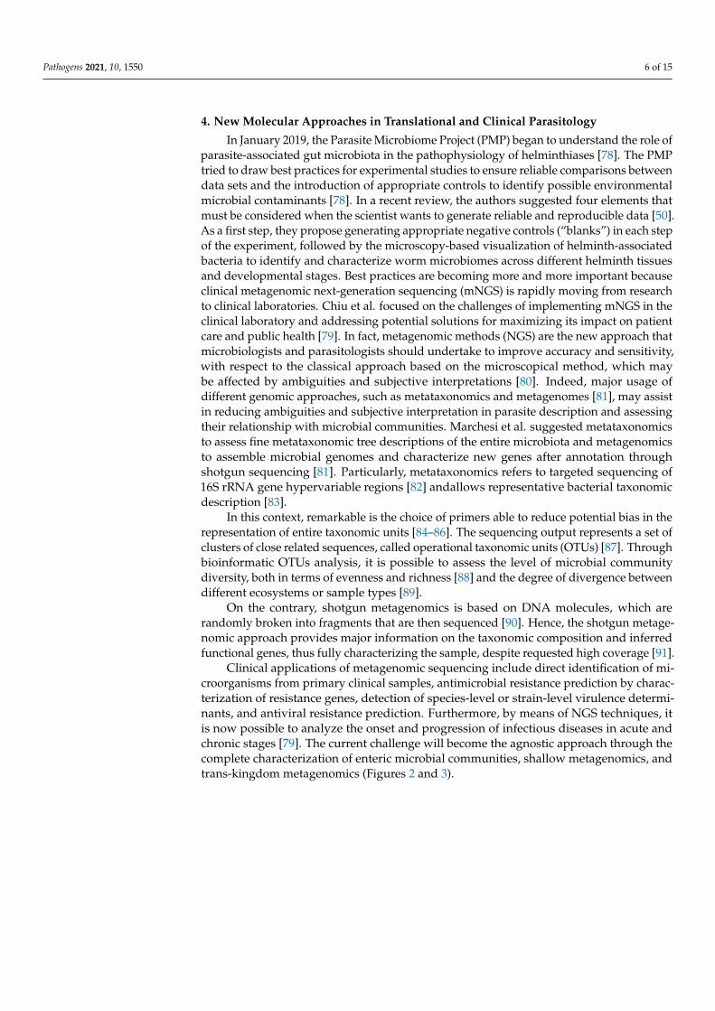
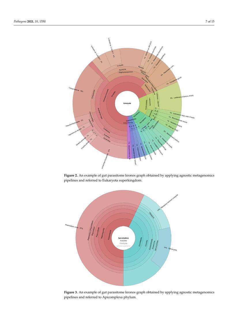
Clinical applications of metagenomic sequencing include direct identification of mi-croorganisms from primary clinical samples, antimicrobial resistance prediction by charac-terization of resistance genes, detection of species-level or strain-level virulence determi-nants, and antiviral resistance prediction. Furthermore, by means of NGS techniques, itis now possible to analyze the onset and progression of infectious diseases in acute andchronic stages [79]. The current challenge will become the agnostic approach through thecomplete characterization of enteric microbial communities, shallow metagenomics, andtrans-kingdom metagenomics (Figures 2 and 3).
Pathogens 2021, 10, 1550 7 of 15Pathogens 2021, 10, x
Figure 2. An example of gut parasitome kronos graph obtained by applying agnostic metagenomics
pipelines and referred to Eukaryota superkingdom.
Figure 2. An example of gut parasitome kronos graph obtained by applying agnostic metagenomicspipelines and referred to Eukaryota superkingdom.
Pathogens 2021, 10, x
Figure 3. An example of gut parasitome kronos graph obtained by applying agnostic metagenomics
pipelines and referred to Apicomplexa phylum.
5. Gut Microbiota Profiling as Tools to Restore and Modulate Gut Microbiota
Gut dysbiosis may be harmful to human beings, leading to organ-localized inflam-
mation, delocalized low inflammation, neuro-inflammation, and mucosal tissue damage
predisposed to pathological conditions, such as in the case of obesity, liver steatosis, au-
tism, and PANDAs [92–95]. Regarding inflammatory mechanisms, amongst infectious
diseases, the infection by C. difficile, exerted by the toxin causing gastrointestinal illness,
is associated with a wide spectrum of severity, ranging from mild diarrhea to pseudo-
membranous colitis, toxic megacolon, sepsis, and death 17 [96,97]. C. difficile, a Gram-pos-
itive spore-forming bacillus, is considered a member of the normal gut microbiota. Re-
gardless, its abnormal growth is suppressed by other more dominant anaerobes [98]. The
gut colonization of C. difficile is reversely related with host age, growing in early infancy
and senescence, and decreasing in adulthood. Furthermore, gut colonization of C. difficile
depends on the loss of the commensal microbiota barrier, loss potentiated by antimicro-
bial therapies.
In this context, C. difficile infection is responsible for the initiation of the cascade of
inflammatory processes, which may play an important and destructive role in the initia-
tion and perpetuation of intestinal inflammation [98]. Patients with recurrent C. difficile
infections are characterized by almost monomicrobial bacterial distributions of the fecal
microbiota [99]. A decrease in bacterial diversity and a strong variation in global distribu-
tion of OTUs are, indeed, registered in the fecal microbiota profiling of adult patients who
present C. difficile infections [99]. In particular, a statistically significant increase in Firmic-
utes and a decrease in Bacteroidetes phyla, compared to healthy subjects are observed in
Figure 3. An example of gut parasitome kronos graph obtained by applying agnostic metagenomicspipelines and referred to Apicomplexa phylum.
Pathogens 2021, 10, 1550 8 of 15
5. Gut Microbiota Profiling as Tools to Restore and Modulate Gut Microbiota
Gut dysbiosis may be harmful to human beings, leading to organ-localized inflam-mation, delocalized low inflammation, neuro-inflammation, and mucosal tissue damagepredisposed to pathological conditions, such as in the case of obesity, liver steatosis, autism,and PANDAs [92–95]. Regarding inflammatory mechanisms, amongst infectious diseases,the infection by C. difficile, exerted by the toxin causing gastrointestinal illness, is associatedwith a wide spectrum of severity, ranging from mild diarrhea to pseudomembranous colitis,toxic megacolon, sepsis, and death 17 [96,97]. C. difficile, a Gram-positive spore-formingbacillus, is considered a member of the normal gut microbiota. Regardless, its abnormalgrowth is suppressed by other more dominant anaerobes [98]. The gut colonization of C.difficile is reversely related with host age, growing in early infancy and senescence, anddecreasing in adulthood. Furthermore, gut colonization of C. difficile depends on the loss ofthe commensal microbiota barrier, loss potentiated by antimicrobial therapies.
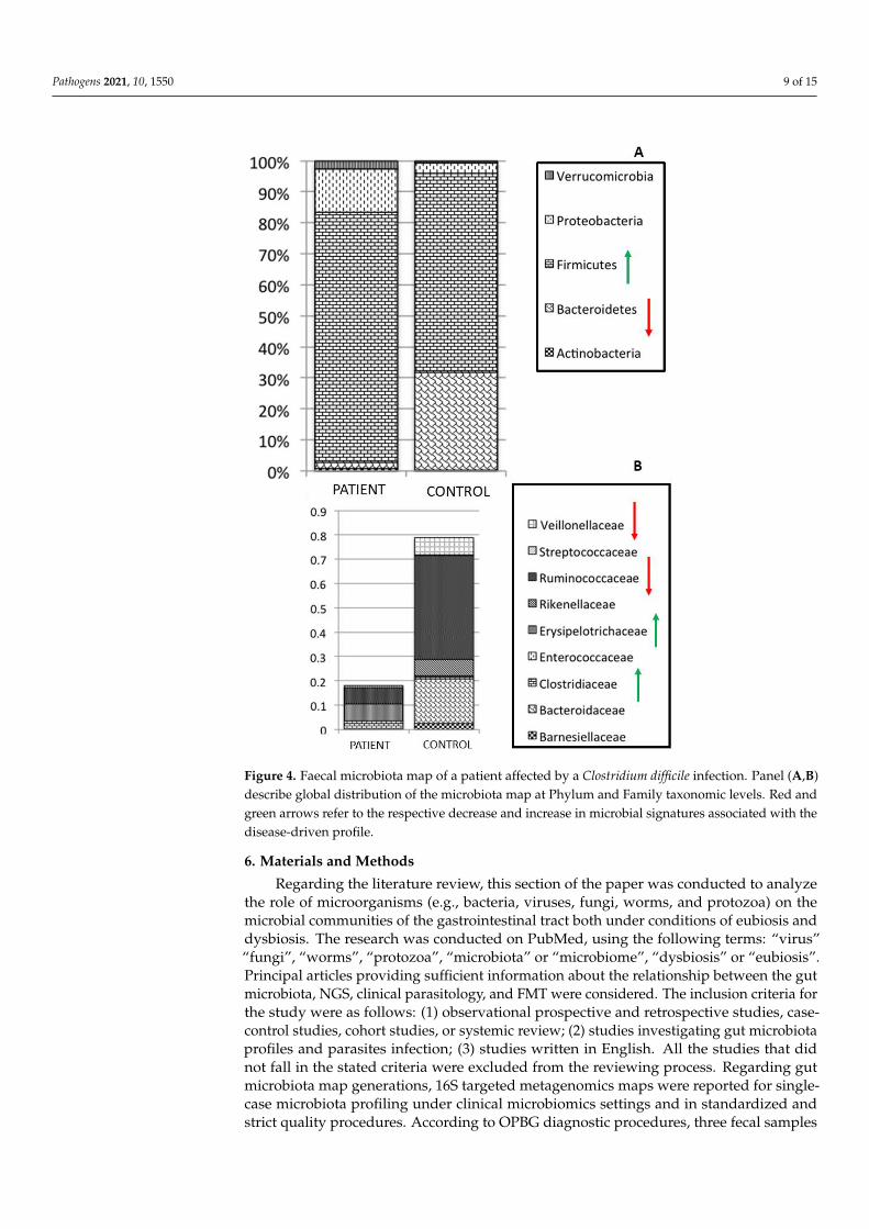
In this context, C. difficile infection is responsible for the initiation of the cascadeof inflammatory processes, which may play an important and destructive role in theinitiation and perpetuation of intestinal inflammation [98]. Patients with recurrent C.difficile infections are characterized by almost monomicrobial bacterial distributions of thefecal microbiota [99]. A decrease in bacterial diversity and a strong variation in globaldistribution of OTUs are, indeed, registered in the fecal microbiota profiling of adultpatients who present C. difficile infections [99]. In particular, a statistically significantincrease in Firmicutes and a decrease in Bacteroidetes phyla, compared to healthy subjectsare observed in the patients, as well as an increase in Clostridiaceae and Erysipelotrichaceaefamilies (Figure 4). Remarkably, in microbiota profiles associated with infectious agents,the dysbiosis index (DI) is usually very high, based on the prevalence of few microbialtaxa, as reported by diagnostic maps obtained for C. difficile infection, for which observeddysbiosis values are actually very high (DI > 35%) (https://www.ospedalebambinogesu.it/parassitologia-98785/, (accessed on 20 September 2021)) (Lorenza Putignani and AntonioGasbarrini, oral communication, 24◦Congresso Nazionale delle Malattie Digestive, Rome,FISMAD 23 March 2018) (Patent IT: PCT40659, EU: PCT/IT2017/000119, MetagenomicMethod for in vitro diagnosis of intestinal disbiosis).
Fecal microbiota transplantation (FMT) has achieved a major role in the clinicalmanagement of C. difficile infections. In these cases, the transplantation is followed bya re-establishment of diversity, and, in many cases, the percentage of efficacy is greaterthan 90% [100]. FMT is considered as the “ultimate probiotics” because it directly changesthe intestinal microbial composition of the host, thus restoring eubiosis and intestinalhomeostasis. FMT donor screening is a key factor in the safety of the procedure in order toprevent iatrogenic infectious diseases that are potentially transmittable to the recipient [101].In fact, the international consensus on stool banking for FMT has recently established thatdonor stool must be tested for protozoa and helminths, including B. hominis, D. fragilis,G. duodenalis, Cryptosporidium spp., Isospora, and Microsporidia [102]. In addition, thedonor must be negative in blood nematodes testing (i.e., S. stercoralis) [102], and particularattention is necessary for the parasitological screening of donors when recipients arechildren [103]. Remarkably, the fecal material (i.e., emulsion) can be stored frozen in a stoolbank for use when needed [103]. Indeed, FMT requires a combination of expertise andappropriate methods to identify the best donors, including advanced clinical parasitology.
Pathogens 2021, 10, 1550 9 of 15
Pathogens 2021, 10, x
the patients, as well as an increase in Clostridiaceae and Erysipelotrichaceae families (Fig-
ure 4). Remarkably, in microbiota profiles associated with infectious agents, the dysbiosis
index (DI) is usually very high, based on the prevalence of few microbial taxa, as reported
by diagnostic maps obtained for C. difficile infection, for which observed dysbiosis values
are actually very high (DI > 35%) (https://www.ospedalebambinogesu.it/parassitologia-
98785/) (Lorenza Putignani and Antonio Gasbarrini, oral communication, 24°Congresso
Nazionale delle Malattie Digestive, Rome, FISMAD 23 March, 2018) (Patent IT: PCT40659,
EU: PCT/IT2017/000119, Metagenomic Method for in vitro diagnosis of intestinal disbiosis)
Figure 4. Faecal microbiota map of a patient affected by a Clostridium difficile infection. Panel (A,B)
describe global distribution of the microbiota map at Phylum and Family taxonomic levels. Red and
green arrows refer to the respective decrease and increase in microbial signatures associated with
the disease-driven profile.
Fecal microbiota transplantation (FMT) has achieved a major role in the clinical man-
agement of C. difficile infections. In these cases, the transplantation is followed by a re-
establishment of diversity, and, in many cases, the percentage of efficacy is greater than
Figure 4. Faecal microbiota map of a patient affected by a Clostridium difficile infection. Panel (A,B)describe global distribution of the microbiota map at Phylum and Family taxonomic levels. Red andgreen arrows refer to the respective decrease and increase in microbial signatures associated with thedisease-driven profile.
6. Materials and Methods
Regarding the literature review, this section of the paper was conducted to analyzethe role of microorganisms (e.g., bacteria, viruses, fungi, worms, and protozoa) on themicrobial communities of the gastrointestinal tract both under conditions of eubiosis anddysbiosis. The research was conducted on PubMed, using the following terms: “virus”“fungi”, “worms”, “protozoa”, “microbiota” or “microbiome”, “dysbiosis” or “eubiosis”.Principal articles providing sufficient information about the relationship between the gutmicrobiota, NGS, clinical parasitology, and FMT were considered. The inclusion criteria forthe study were as follows: (1) observational prospective and retrospective studies, case-control studies, cohort studies, or systemic review; (2) studies investigating gut microbiotaprofiles and parasites infection; (3) studies written in English. All the studies that didnot fall in the stated criteria were excluded from the reviewing process. Regarding gutmicrobiota map generations, 16S targeted metagenomics maps were reported for single-case microbiota profiling under clinical microbiomics settings and in standardized andstrict quality procedures. According to OPBG diagnostic procedures, three fecal samples
Pathogens 2021, 10, 1550 10 of 15
for each subject were collected, processed to optimize microbial DNA extraction, amplifiedto generate 16S rRNA libraries, pooled, quantified, and sequenced [104] by a MiSeq DXplatform, in accordance with the manufacturer (Illumina, CA, USA). In addition to thewet part of the map generation pipeline, the dry bioinformatics module was assessedby an automated process designed by OPBG-GenomeUP in accordance with the OPBGpatent. (Patent IT: PCT40659, EU: PCT/IT2017/000119, Metagenomic Method for in vitrodiagnosis of intestinal disbiosis). Regarding shotgun procedures, before DNA extraction,differential sample homogenization (Precellys 24 original tissue homogenizer, (BertinInstruments, Montigny-le-Bretonneux, France) was performed to optimize parasite andbacteria DNA extraction. Hence, AllPrep Power DNA/RNA Kit was exploited to provideshotgun DNA library templates (bacteriome, parasitome). Library preparation was setup by using Illumina® DNA Prep, (M) Tagmentation (96 Samples), IDT® for Illumina®
DNA UD Indexes Set A, NextSeq 550 High Output Kit v2.5 (300 Cycles) according toIllumina specific instructions. Sequencing was performed on the NextSeq550 platform,ensuring at least a sequencing depth of 50–100 M reads/sample. For the dry protocol, a pre-processing procedure was assured by the removal of the input sequences containing regionsof low quality/complexity and readings of less than 50 nucleotides. Only reads passingQC filters were directed to the following steps, in which data were mapped against thehuman genome. In the third step, a comparison with reference databases was undertaken,and taxonomic annotation was provided by processing GenBank and RefSeq databases.Alignments were filtered based on identity percentage and query coverage. Reports weregenerated as CSV files, HTML interactive tables, and Krona graphs [105]. Taxonomic andfunctional annotation were processed by Python statistics modules.
7. Conclusions
Over the last two decades, helminths and protozoans, previously considered onlyas pathogens, have been being increasingly suggested as also commensal, protective, oreven curative microorganisms. These findings essentially point out that most of theseprimarily considered “parasites” could have an evolutionary history, tipping the balancetowards commensalism, where they have adapted to live off the host without causing anyharm. However, it is important to keep in mind that for most of these data, there are otherstudies that have found opposing results. A likely explanation for these differences canbe attributed to various parameters affecting the study design and protocols. The methodof analysis, which includes the type of sample used for sequencing, the workflow stan-dardization, and lab-to-lab or operator variability, can create important biases in the results.Moreover, the majority of the studies rely on fecal samples, which does not give enoughinformation on the localization of the parasite within the GI tract. The characterizationof the human gut parasitome is now urgent, but this still requires time and new tools.The “-omics” analyses, i.e., (meta)genomics, (meta)transcriptomics, (meta)proteomics,(meta)metabolomics), represent a robust tool for such types of studies [106]. In this context,the application of “big data” methodologies to GM may boost this intervention. The intro-duction of sequencing technologies has revolutionized the field, enabling investigators tocharacterize microbial communities. By utilizing larger datasets, researchers are able todesign large-scale studies to ask (and answer) complex questions. Metadata associated withsamples are becoming an increasingly large contributor to microbiome big data and thechallenges associated with streamlining data analysis. The successful application of big mi-crobiome dataset analysis has already provided relevant insights for other areas of research,such as epidemiology, agriculture, and healthcare. Since gut microbiota composition differswidely according to host genetics, diet, lifestyle, geographical location, and disease burden,the application of big data methodologies to gut microbiota and parasitome profilingcould be of utmost importance in developing a wide understanding of various infectiousdiseases. The reach of a new type of clinical parasitology, encountering the approachesand needs of the clinical microbiomics [107], may contribute to the new data-driven era in
Pathogens 2021, 10, 1550 11 of 15
medicine, establishing new connections with other big data and genome-wide associationlinks, opening the way to a novel holobiont perspective in infectious diseases.
Author Contributions: Conceptualization, S.P., L.P.; resources, L.P.; data curation, A.R., F.D.C., S.G.,S.P., M.V.R.; writing-original draft preparation, S.P., M.V.R. and L.P.; writing-review and editing, S.P.,M.V.R., F.D.C. and L.P. All authors have read and agreed to the published version of the manuscript.
Funding: Italian Ministry of Health: Ricerca Corrente 2020 and 2021, assigned to L.P.
Data Availability Statement: Not applicable.
Acknowledgments: We wish to thank all the staff of the Microbiomics and Human Microbiome Units.
Conflicts of Interest: The authors declare no conflict of interest.
References1. Bäckhed, F.; Ley, R.E.; Sonnenburg, J.L.; Peterson, D.A.; Gordon, J.I. Host-Bacterial Mutualism in the Human Intestine. Science
2005, 307, 1915–1920. [CrossRef]2. Eckburg, P.B.; Bik, E.M.; Bernstein, C.N.; Purdom, E.; Dethlefsen, L.; Sargent, M.; Gill, S.R.; Nelson, K.E.; Relman, D.A. Diversity
of the Human Intestinal Microbial Flora. Science 2005, 308, 1635–1638. [CrossRef]3. Qin, J.; Li, R.; Raes, J.; Arumugam, M.; Burgdorf, K.S.; Manichanh, C.; Nielsen, T.; Pons, N.; Levenez, F.; Yamada, T.; et al. A
Human Gut Microbial Gene Catalogue Established by Metagenomic Sequencing. Nature 2010, 464, 59–65. [CrossRef]4. Bancroft, A.J.; Hayes, K.S.; Grencis, R.K. Life on the Edge: The Balance between Macrofauna, Microflora and Host Immunity.
Trends Parasitol. 2012, 28, 93–98. [CrossRef] [PubMed]5. Mazmanian, S.K.; Liu, C.H.; Tzianabos, A.O.; Kasper, D.L. An Immunomodulatory Molecule of Symbiotic Bacteria Directs
Maturation of the Host Immune System. Cell 2005, 122, 107–118. [CrossRef]6. Becattini, S.; Taur, Y.; Pamer, E.G. Antibiotic-Induced Changes in the Intestinal Microbiota and Disease. Trends Mol. Med. 2016, 22,
458–478. [CrossRef] [PubMed]7. Becattini, S.; Littmann, E.R.; Carter, R.A.; Kim, S.G.; Morjaria, S.M.; Ling, L.; Gyaltshen, Y.; Fontana, E.; Taur, Y.; Leiner, I.M.; et al.
Commensal Microbes Provide First Line Defense against Listeria Monocytogenes Infection. J. Exp. Med. 2017, 214, 1973–1989.[CrossRef]
8. Sekirov, I.; Tam, N.M.; Jogova, M.; Robertson, M.L.; Li, Y.; Lupp, C.; Finlay, B.B. Antibiotic-Induced Perturbations of the IntestinalMicrobiota Alter Host Susceptibility to Enteric Infection. Infect. Immun. 2008, 76, 4726–4736. [CrossRef] [PubMed]
9. Abt, M.C.; Osborne, L.C.; Monticelli, L.A.; Doering, T.A.; Alenghat, T.; Sonnenberg, G.F.; Paley, M.A.; Antenus, M.; Williams,K.L.; Erikson, J.; et al. Commensal Bacteria Calibrate the Activation Threshold of Innate Antiviral Immunity. Immunity 2012, 37,158–170. [CrossRef] [PubMed]
10. Molloy, M.J.; Bouladoux, N.; Belkaid, Y. Intestinal Microbiota: Shaping Local and Systemic Immune Responses. Semin. Immunol.2012, 24, 58–66. [CrossRef] [PubMed]
11. Alexander, K.L.; Targan, S.R.; Elson, C.O. Microbiota Activation and Regulation of Innate and Adaptive Immunity. Immunol. Rev.2014, 260, 206–220. [CrossRef] [PubMed]
12. Zhang, Y.-J.; Li, S.; Gan, R.-Y.; Zhou, T.; Xu, D.-P.; Li, H.-B. Impacts of Gut Bacteria on Human Health and Diseases. Int. J. Mol. Sci.2015, 16, 7493–7519. [CrossRef]
13. Putignani, L.; Del Chierico, F.; Vernocchi, P.; Cicala, M.; Cucchiara, S.; Dallapiccola, B. Dysbiotrack Study Group Gut MicrobiotaDysbiosis as Risk and Premorbid Factors of IBD and IBS Along the Childhood-Adulthood Transition. Inflamm. Bowel Dis. 2016,22, 487–504. [CrossRef]
14. Mariat, D.; Firmesse, O.; Levenez, F.; Guimarăes, V.; Sokol, H.; Doré, J.; Corthier, G.; Furet, J.-P. The Firmicutes/BacteroidetesRatio of the Human Microbiota Changes with Age. BMC Microbiol. 2009, 9, 123. [CrossRef]
15. Putignani, L.; Del Chierico, F.; Petrucca, A.; Vernocchi, P.; Dallapiccola, B. The Human Gut Microbiota: A Dynamic Interplay withthe Host from Birth to Senescence Settled during Childhood. Pediatr. Res. 2014, 76, 2–10. [CrossRef]
16. Del Chierico, F.; Vernocchi, P.; Bonizzi, L.; Carsetti, R.; Castellazzi, A.M.; Dallapiccola, B.; de Vos, W.; Guerzoni, M.E.; Manco, M.;Marseglia, G.L.; et al. Early-Life Gut Microbiota under Physiological and Pathological Conditions: The Central Role of CombinedMeta-Omics-Based Approaches. J. Proteom. 2012, 75, 4580–4587. [CrossRef]
17. Rawls, J.F.; Mahowald, M.A.; Ley, R.E.; Gordon, J.I. Reciprocal Gut Microbiota Transplants from Zebrafish and Mice to Germ-FreeRecipients Reveal Host Habitat Selection. Cell 2006, 127, 423–433. [CrossRef] [PubMed]
18. Putignani, L.; Dallapiccola, B. Foodomics as Part of the Host-Microbiota-Exposome Interplay. J. Proteom. 2016, 147, 3–20.[CrossRef] [PubMed]
19. Lynch, S.V.; Pedersen, O. The Human Intestinal Microbiome in Health and Disease. N. Engl. J. Med. 2016, 375, 2369–2379.[CrossRef]
20. Bobardt, S.D.; Dillman, A.R.; Nair, M.G. The Two Faces of Nematode Infection: Virulence and Immunomodulatory MoleculesFrom Nematode Parasites of Mammals, Insects and Plants. Front. Microbiol. 2020, 11, 577846. [CrossRef]
Pathogens 2021, 10, 1550 12 of 15
21. Kaiko, G.E.; Horvat, J.C.; Beagley, K.W.; Hansbro, P.M. Immunological Decision-Making: How Does the Immune System Decideto Mount a Helper T-Cell Response? Immunology 2008, 123, 326–338. [CrossRef] [PubMed]
22. Nicholson, J.K.; Holmes, E.; Lindon, J.C.; Wilson, I.D. The Challenges of Modeling Mammalian Biocomplexity. Nat. Biotechnol.2004, 22, 1268–1274. [CrossRef]
23. Manichanh, C.; Rigottier-Gois, L.; Bonnaud, E.; Gloux, K.; Pelletier, E.; Frangeul, L.; Nalin, R.; Jarrin, C.; Chardon, P.; Marteau, P.;et al. Reduced Diversity of Faecal Microbiota in Crohn’s Disease Revealed by a Metagenomic Approach. Gut 2006, 55, 205–211.[CrossRef] [PubMed]
24. Darfeuille-Michaud, A.; Boudeau, J.; Bulois, P.; Neut, C.; Glasser, A.-L.; Barnich, N.; Bringer, M.-A.; Swidsinski, A.; Beaugerie,L.; Colombel, J.-F. High Prevalence of Adherent-Invasive Escherichia Coli Associated with Ileal Mucosa in Crohn’s Disease.Gastroenterology 2004, 127, 412–421. [CrossRef] [PubMed]
25. Swidsinski, A.; Ladhoff, A.; Pernthaler, A.; Swidsinski, S.; Loening-Baucke, V.; Ortner, M.; Weber, J.; Hoffmann, U.; Schreiber, S.;Dietel, M.; et al. Mucosal Flora in Inflammatory Bowel Disease. Gastroenterology 2002, 122, 44–54. [CrossRef]
26. Parfrey, L.W.; Walters, W.A.; Knight, R. Microbial Eukaryotes in the Human Microbiome: Ecology, Evolution, and FutureDirections. Front. Microbiol. 2011, 2, 153. [CrossRef]
27. Human Microbiome Project Consortium. Structure, Function and Diversity of the Healthy Human Microbiome. Nature 2012, 486,207–214. [CrossRef] [PubMed]
28. Hewitson, J.P.; Maizels, R.M. Vaccination against Helminth Parasite Infections. Expert Rev. Vaccines 2014, 13, 473–487. [CrossRef][PubMed]
29. Cacciò, S.M.; Putignani, L. Epidemiology of Human Cryptosporidiosis. Cryptosporidium Parasite Dis. 2014, 43–79. [CrossRef]30. Norman, J.M.; Handley, S.A.; Virgin, H.W. Kingdom-Agnostic Metagenomics and the Importance of Complete Characterization
of Enteric Microbial Communities. Gastroenterology 2014, 146, 1459–1469. [CrossRef] [PubMed]31. Nyholm, L.; Koziol, A.; Marcos, S.; Botnen, A.B.; Aizpurua, O.; Gopalakrishnan, S.; Limborg, M.T.; Gilbert, M.T.P.; Alberdi,
A. Holo-Omics: Integrated Host-Microbiota Multi-Omics for Basic and Applied Biological Research. iScience 2020, 23, 101414.[CrossRef]
32. Putignani, L.; Gasbarrini, A.; Dallapiccola, B. Potential of Multiomics Technology in Precision Medicine. Curr. Opin. Gastroenterol.2019, 35, 491–498. [CrossRef] [PubMed]
33. Matijašic, M.; Meštrovic, T.; Paljetak, H.C.; Peric, M.; Barešic, A.; Verbanac, D. Gut Microbiota beyond Bacteria-Mycobiome,Virome, Archaeome, and Eukaryotic Parasites in IBD. Int. J. Mol. Sci. 2020, 21, 2668. [CrossRef]
34. Zuo, T.; Wong, S.H.; Lam, K.; Lui, R.; Cheung, K.; Tang, W.; Ching, J.Y.L.; Chan, P.K.S.; Chan, M.C.W.; Wu, J.C.Y.; et al.Bacteriophage Transfer during Faecal Microbiota Transplantation in Clostridium Difficile Infection Is Associated with TreatmentOutcome. Gut 2018, 67, 634–643. [CrossRef]
35. Dalmasso, M.; Hill, C.; Ross, R.P. Exploiting Gut Bacteriophages for Human Health. Trends Microbiol. 2014, 22, 399–405. [CrossRef]36. Jonas, O.; Seifman, R. Do We Need a Global Virome Project? Lancet Glob. Health 2019, 7, e1314–e1316. [CrossRef]37. Minot, S.; Bryson, A.; Chehoud, C.; Wu, G.D.; Lewis, J.D.; Bushman, F.D. Rapid Evolution of the Human Gut Virome. Proc. Natl.
Acad. Sci. USA 2013, 110, 12450–12455. [CrossRef]38. Townsend, E.M.; Kelly, L.; Muscatt, G.; Box, J.D.; Hargraves, N.; Lilley, D.; Jameson, E. The Human Gut Phageome: Origins and
Roles in the Human Gut Microbiome. Front. Cell. Infect. Microbiol. 2021, 11, 643214. [CrossRef] [PubMed]39. Mirzaei, M.K.; Maurice, C.F. Ménage à Trois in the Human Gut: Interactions between Host, Bacteria and Phages. Nat. Rev.
Microbiol. 2017, 15, 397–408. [CrossRef]40. Huffnagle, G.B.; Noverr, M.C. The Emerging World of the Fungal Microbiome. Trends Microbiol. 2013, 21, 334–341. [CrossRef]41. David, L.A.; Maurice, C.F.; Carmody, R.N.; Gootenberg, D.B.; Button, J.E.; Wolfe, B.E.; Ling, A.V.; Devlin, A.S.; Varma, Y.;
Fischbach, M.A.; et al. Diet Rapidly and Reproducibly Alters the Human Gut Microbiome. Nature 2014, 505, 559–563. [CrossRef]42. Hoffmann, C.; Dollive, S.; Grunberg, S.; Chen, J.; Li, H.; Wu, G.D.; Lewis, J.D.; Bushman, F.D. Archaea and Fungi of the Human
Gut Microbiome: Correlations with Diet and Bacterial Residents. PLoS ONE 2013, 8, e66019. [CrossRef]43. Beheshti-Maal, A.; Shahrokh, S.; Ansari, S.; Mirsamadi, E.S.; Yadegar, A.; Mirjalali, H.; Zali, M.R. Gut Mycobiome: The Probable
Determinative Role of Fungi in IBD Patients. Mycoses 2021, 64, 468–476. [CrossRef]44. Ianiro, G.; Iorio, A.; Porcari, S.; Masucci, L.; Perno, C.F.; Gasbarrini, A.; Putignani, L.; Cam-marota, G. How the Gut Parasitome
Affects Human Health.45. Prommi, A.; Prombutara, P.; Watthanakulpanich, D.; Adisakwattana, P.; Kusolsuk, T.; Yoonuan, T.; Poodeepiyasawat, A.;
Homsuwan, N.; Prummongkol, S.; Tanita, M.; et al. Intestinal Parasites in Rural Communities in Nan Province, Thailand:Changes in Bacterial Gut Microbiota Associated with Minute Intestinal Fluke Infection. Parasitology 2020, 147, 972–984. [CrossRef]
46. Farthing, M.J. Giardiasis. Gastroenterol. Clin. N. Am. 1996, 25, 493–515. [CrossRef]47. Cryptosporidium–ScienceDirect. Available online: https://www.sciencedirect.com/science/article/pii/B97801281873190008
0X?via%3Dihub (accessed on 21 September 2021).48. Putignani, L.; Menichella, D. Global Distribution, Public Health and Clinical Impact of the Protozoan Pathogen Cryptosporidium.
Interdiscip. Perspect. Infect. Dis. 2010, 2010, 753512. [CrossRef] [PubMed]49. Caradonna, T.; Marangi, M.; Del Chierico, F.; Ferrari, N.; Reddel, S.; Bracaglia, G.; Normanno, G.; Putignani, L.; Giangaspero, A.
Detection and Prevalence of Protozoan Parasites in Ready-to-Eat Packaged Salads on Sale in Italy. Food Microbiol. 2017, 67, 67–75.[CrossRef] [PubMed]
Pathogens 2021, 10, 1550 13 of 15
50. Formenti, F.; Cortés, A.; Brindley, P.J.; Cantacessi, C.; Rinaldi, G. A Bug’s Life: Delving into the Challenges of HelminthMicrobiome Studies. PLoS Negl. Trop. Dis. 2020, 14, e0008446. [CrossRef]
51. White, E.C.; Houlden, A.; Bancroft, A.J.; Hayes, K.S.; Goldrick, M.; Grencis, R.K.; Roberts, I.S. Manipulation of Host and ParasiteMicrobiotas: Survival Strategies during Chronic Nematode Infection. Sci. Adv. 2018, 4, eaap7399. [CrossRef] [PubMed]
52. Holzscheiter, M.; Layland, L.E.; Loffredo-Verde, E.; Mair, K.; Vogelmann, R.; Langer, R.; Wagner, H.; Prazeres da Costa, C. Lack ofHost Gut Microbiota Alters Immune Responses and Intestinal Granuloma Formation during Schistosomiasis. Clin. Exp. Immunol.2014, 175, 246–257. [CrossRef] [PubMed]
53. Yason, J.A.; Liang, Y.R.; Png, C.W.; Zhang, Y.; Tan, K.S.W. Interactions between a Pathogenic Blastocystis Subtype and GutMicrobiota: In Vitro and in Vivo Studies. Microbiome 2019, 7, 30. [CrossRef] [PubMed]
54. Toro-Londono, M.A.; Bedoya-Urrego, K.; Garcia-Montoya, G.M.; Galvan-Diaz, A.L.; Alzate, J.F. Intestinal Parasitic InfectionAlters Bacterial Gut Microbiota in Children. PeerJ 2019, 7, e6200. [CrossRef]
55. Alzate, J.F.; Toro-Londoño, M.; Cabarcas, F.; Garcia-Montoya, G.; Galvan-Diaz, A. Contrasting Microbiota Profiles Observed inChildren Carrying Either Blastocystis Spp. or the Commensal Amoebas Entamoeba Coli or Endolimax Nana. Sci. Rep. 2020, 10,15354. [CrossRef] [PubMed]
56. Rosa, B.A.; Supali, T.; Gankpala, L.; Djuardi, Y.; Sartono, E.; Zhou, Y.; Fischer, K.; Martin, J.; Tyagi, R.; Bolay, F.K.; et al. DifferentialHuman Gut Microbiome Assemblages during Soil-Transmitted Helminth Infections in Indonesia and Liberia. Microbiome 2018, 6,33. [CrossRef]
57. Giacomin, P.; Zakrzewski, M.; Jenkins, T.P.; Su, X.; Al-Hallaf, R.; Croese, J.; de Vries, S.; Grant, A.; Mitreva, M.; Loukas, A.; et al.Changes in Duodenal Tissue-Associated Microbiota Following Hookworm Infection and Consecutive Gluten Challenges inHumans with Coeliac Disease. Sci. Rep. 2016, 6, 36797. [CrossRef]
58. Cortés, A.; Clare, S.; Costain, A.; Almeida, A.; McCarthy, C.; Harcourt, K.; Brandt, C.; Tolley, C.; Rooney, J.; Berriman, M.; et al.Baseline Gut Microbiota Composition Is Associated With Schistosoma Mansoni Infection Burden in Rodent Models. Front.Immunol. 2020, 11, 593838. [CrossRef]
59. Jenkins, T.P.; Pritchard, D.I.; Tanasescu, R.; Telford, G.; Papaiakovou, M.; Scotti, R.; Cortés, A.; Constantinescu, C.S.; Cantacessi, C.Experimental Infection with the Hookworm, Necator Americanus, Is Associated with Stable Gut Microbial Diversity in HumanVolunteers with Relapsing Multiple Sclerosis. BMC Biol. 2021, 19, 74. [CrossRef]
60. Pane, S.; Sacco, A.; Iorio, A.; Romani, L.; Putignani, L. Strongyloides Stercoralis Infestation in a Child: How a Nematode CanAffect Gut Microbiota. Int. J. Mol. Sci. 2021, 22, 2131. [CrossRef]
61. Morton, E.R.; Lynch, J.; Froment, A.; Lafosse, S.; Heyer, E.; Przeworski, M.; Blekhman, R.; Ségurel, L. Variation in Rural AfricanGut Microbiota Is Strongly Correlated with Colonization by Entamoeba and Subsistence. PLoS Genet. 2015, 11, e1005658.[CrossRef]
62. Iebba, V.; Santangelo, F.; Totino, V.; Pantanella, F.; Monsia, A.; Di Cristanziano, V.; Di Cave, D.; Schippa, S.; Berrilli, F.; D’Alfonso,R. Gut Microbiota Related to Giardia Duodenalis, Entamoeba Spp. and Blastocystis Hominis Infections in Humans from Côted’Ivoire. J. Infect. Dev. Ctries 2016, 10, 1035–1041. [CrossRef] [PubMed]
63. Iyer, L.R.; Verma, A.K.; Paul, J.; Bhattacharya, A. Phagocytosis of Gut Bacteria by Entamoeba Histolytica. Front. Cell. Infect.Microbiol. 2019, 9, 34. [CrossRef]
64. Midha, A.; Schlosser, J.; Hartmann, S. Reciprocal Interactions between Nematodes and Their Microbial Environments. Front. Cell.Infect. Microbiol. 2017, 7, 144. [CrossRef] [PubMed]
65. Beghini, F.; Pasolli, E.; Truong, T.D.; Putignani, L.; Cacciò, S.M.; Segata, N. Large-Scale Comparative Metagenomics of Blastocystis,a Common Member of the Human Gut Microbiome. ISME J. 2017, 11, 2848–2863. [CrossRef]
66. Andersen, L.O.; Stensvold, C.R. Blastocystis in Health and Disease: Are We Moving from a Clinical to a Public Health Perspective?J. Clin. Microbiol. 2016, 54, 524–528. [CrossRef] [PubMed]
67. Audebert, C.; Even, G.; Cian, A.; Blastocystis Investigation Group; Loywick, A.; Merlin, S.; Viscogliosi, E.; Chabé, M. Colonizationwith the Enteric Protozoa Blastocystis Is Associated with Increased Diversity of Human Gut Bacterial Microbiota. Sci. Rep. 2016,6, 25255. [CrossRef]
68. Nagel, R.; Traub, R.J.; Allcock, R.J.N.; Kwan, M.M.S.; Bielefeldt-Ohmann, H. Comparison of Faecal Microbiota in Blastocystis-Positive and Blastocystis-Negative Irritable Bowel Syndrome Patients. Microbiome 2016, 4, 47. [CrossRef] [PubMed]
69. Billy, V.; Lhotská, Z.; Jirku, M.; Kadlecová, O.; Frgelecová, L.; Parfrey, L.W.; Pomajbíková, K.J. Blastocystis Colonization Altersthe Gut Microbiome and, in Some Cases, Promotes Faster Recovery From Induced Colitis. Front. Microbiol. 2021, 12, 641483.[CrossRef]
70. Stensvold, C.R.; van der Giezen, M. Associations between Gut Microbiota and Common Luminal Intestinal Parasites. TrendsParasitol. 2018, 34, 369–377. [CrossRef]
71. Scanlan, P.D.; Stensvold, C.R.; Rajilic-Stojanovic, M.; Heilig, H.G.H.J.; De Vos, W.M.; O’Toole, P.W.; Cotter, P.D. The MicrobialEukaryote Blastocystis Is a Prevalent and Diverse Member of the Healthy Human Gut Microbiota. FEMS Microbiol. Ecol. 2014, 90,326–330. [CrossRef] [PubMed]
72. Scanlan, P.D.; Stensvold, C.R.; Cotter, P.D. Development and Application of a Blastocystis Subtype-Specific PCR Assay RevealsThat Mixed-Subtype Infections Are Common in a Healthy Human Population. Appl. Environ. Microbiol. 2015, 81, 4071–4076.[CrossRef]
Pathogens 2021, 10, 1550 14 of 15
73. Holtman, G.A.; Kranenberg, J.J.; Blanker, M.H.; Ott, A.; Lisman-van Leeuwen, Y.; Berger, M.Y. Dientamoeba Fragilis ColonizationIs Not Associated with Gastrointestinal Symptoms in Children at Primary Care Level. Fam. Pract. 2017, 34, 25–29. [CrossRef][PubMed]
74. Petersen, A.M.; Stensvold, C.R.; Mirsepasi, H.; Engberg, J.; Friis-Møller, A.; Porsbo, L.J.; Hammerum, A.M.; Nordgaard-Lassen, I.;Nielsen, H.V.; Krogfelt, K.A. Active Ulcerative Colitis Associated with Low Prevalence of Blastocystis and Dientamoeba FragilisInfection. Scand. J. Gastroenterol. 2013, 48, 638–639. [CrossRef]
75. Coskun, A.; Malatyali, E.; Ertabaklar, H.; Yasar, M.B.; Karaoglu, A.O.; Ertug, S. Blastocystis in Ulcerative Colitis Patients: GeneticDiversity and Analysis of Laboratory Findings. Asian Pac. J. Trop. Med. 2016, 9, 916–919. [CrossRef] [PubMed]
76. Krogsgaard, L.R.; Lee, O.; Johannesen, T.B.; Engsbro, A.L.; Stensvold, C.R.; Nielsen, H.V.; Bytzer, P. Characteristics of the BacterialMicrobiome in Association with Common Intestinal Parasites in Irritable Bowel Syndrome. Clin. Transl. Gastroenterol. 2018, 9, 161.[CrossRef]
77. Berrilli, F.; Di Cave, D.; Cavallero, S.; D’Amelio, S. Interactions between Parasites and Microbial Communities in the Human Gut.Front. Cell. Infect. Microbiol. 2012, 2, 141. [CrossRef]
78. Dheilly, N.M.; Martínez Martínez, J.; Rosario, K.; Brindley, P.J.; Fichorova, R.N.; Kaye, J.Z.; Kohl, K.D.; Knoll, L.J.; Lukeš, J.;Perkins, S.L.; et al. Parasite Microbiome Project: Grand Challenges. PLoS Pathog. 2019, 15, e1008028. [CrossRef]
79. Chiu, C.Y.; Miller, S.A. Clinical Metagenomics. Nat. Rev. Genet. 2019, 20, 341–355. [CrossRef] [PubMed]80. Hugenholtz, P.; Chuvochina, M.; Oren, A.; Parks, D.H.; Soo, R.M. Prokaryotic Taxonomy and Nomenclature in the Age of Big
Sequence Data. ISME J. 2021, 15, 1879–1892. [CrossRef]81. Marchesi, J.R.; Ravel, J. The Vocabulary of Microbiome Research: A Proposal. Microbiome 2015, 3, 31. [CrossRef]82. Chistoserdovai, L. Functional Metagenomics: Recent Advances and Future Challenges. Biotechnol. Genet. Eng. Rev. 2010, 26,
335–352. [CrossRef]83. Choi, K.Y.; Lee, T.K.; Sul, W.J. Metagenomic Analysis of Chicken Gut Microbiota for Improving Metabolism and Health of
Chickens—A Review. Asian-Australas J. Anim. Sci. 2015, 28, 1217–1225. [CrossRef]84. Breitwieser, F.P.; Lu, J.; Salzberg, S.L. A Review of Methods and Databases for Metagenomic Classification and Assembly. Brief.
Bioinform. 2019, 20, 1125–1136. [CrossRef]85. Campanaro, S.; Treu, L.; Kougias, P.G.; Zhu, X.; Angelidaki, I. Taxonomy of Anaerobic Digestion Microbiome Reveals Biases
Associated with the Applied High Throughput Sequencing Strategies. Sci. Rep. 2018, 8, 1926. [CrossRef]86. Fouhy, F.; Clooney, A.G.; Stanton, C.; Claesson, M.J.; Cotter, P.D. 16S RRNA Gene Sequencing of Mock Microbial Populations-
Impact of DNA Extraction Method, Primer Choice and Sequencing Platform. BMC Microbiol. 2016, 16, 123. [CrossRef] [PubMed]87. Cole, J.R.; Wang, Q.; Fish, J.A.; Chai, B.; McGarrell, D.M.; Sun, Y.; Brown, C.T.; Porras-Alfaro, A.; Kuske, C.R.; Tiedje, J.M.
Ribosomal Database Project: Data and Tools for High Throughput RRNA Analysis. Nucleic Acids Res. 2014, 42, D633–D642.[CrossRef]
88. Colwell, R.K. III.1 Biodiversity: Concepts, Patterns, and Measurement; Princeton University Press: Princeton, NJ, USA, 2009; pp.257–263. ISBN 978-1-4008-3302-3.
89. Sala, C.; Vitali, S.; Giampieri, E.; do Valle, Ì.F.; Remondini, D.; Garagnani, P.; Bersanelli, M.; Mosca, E.; Milanesi, L.; Castellani, G.Stochastic Neutral Modelling of the Gut Microbiota’s Relative Species Abundance from next Generation Sequencing Data. BMCBioinform. 2016, 17, 16. [CrossRef] [PubMed]
90. Weinstock, G.M. Genomic Approaches to Studying the Human Microbiota. Nature 2012, 489, 250–256. [CrossRef] [PubMed]91. Durazzi, F.; Sala, C.; Castellani, G.; Manfreda, G.; Remondini, D.; De Cesare, A. Comparison between 16S RRNA and Shotgun
Sequencing Data for the Taxonomic Characterization of the Gut Microbiota. Sci. Rep. 2021, 11, 3030. [CrossRef]92. Del Chierico, F.; Abbatini, F.; Russo, A.; Quagliariello, A.; Reddel, S.; Capoccia, D.; Caccamo, R.; Ginanni Corradini, S.; Nobili, V.;
De Peppo, F.; et al. Gut Microbiota Markers in Obese Adolescent and Adult Patients: Age-Dependent Differential Patterns. Front.Microbiol. 2018, 9, 1210. [CrossRef]
93. Del Chierico, F.; Nobili, V.; Vernocchi, P.; Russo, A.; De Stefanis, C.; Gnani, D.; Furlanello, C.; Zandonà, A.; Paci, P.; Capuani,G.; et al. Gut Microbiota Profiling of Pediatric Nonalcoholic Fatty Liver Disease and Obese Patients Unveiled by an IntegratedMeta-Omics-Based Approach. Hepatology 2017, 65, 451–464. [CrossRef]
94. Mortera, S.L.; Vernocchi, P.; Basadonne, I.; Zandonà, A.; Chierici, M.; Durighello, M.; Marzano, V.; Gardini, S.; Gasbarrini, A.;Urbani, A.; et al. A Metaproteomic-Based Gut Microbiota Profiling in Children Affected by Autism Spectrum Disorders. J.Proteom. 2021, 251, 104407. [CrossRef] [PubMed]
95. Quagliariello, A.; Del Chierico, F.; Russo, A.; Reddel, S.; Conte, G.; Lopetuso, L.R.; Ianiro, G.; Dallapiccola, B.; Cardona,F.; Gasbarrini, A.; et al. Gut Microbiota Profiling and Gut-Brain Crosstalk in Children Affected by Pediatric Acute-OnsetNeuropsychiatric Syndrome and Pediatric Autoimmune Neuropsychiatric Disorders Associated With Streptococcal Infections.Front. Microbiol. 2018, 9, 675. [CrossRef] [PubMed]
96. Dobson, G.; Hickey, C.; Trinder, J. Clostridium Difficile Colitis Causing Toxic Megacolon, Severe Sepsis and Multiple OrganDysfunction Syndrome. Intensive Care Med. 2003, 29, 1030. [CrossRef]
97. Mylonakis, E.; Ryan, E.T.; Calderwood, S.B. Clostridium Difficile—Associated Diarrhea: A Review. Arch. Intern. Med. 2001, 161,525–533. [CrossRef] [PubMed]
98. Smits, W.K.; Lyras, D.; Lacy, D.B.; Wilcox, M.H.; Kuijper, E.J. Clostridium Difficile Infection. Nat. Rev. Dis. Primers 2016, 2, 16020.[CrossRef]
Pathogens 2021, 10, 1550 15 of 15
99. Chang, J.Y.; Antonopoulos, D.A.; Kalra, A.; Tonelli, A.; Khalife, W.T.; Schmidt, T.M.; Young, V.B. Decreased Diversity of the FecalMicrobiome in Recurrent Clostridium Difficile-Associated Diarrhea. J. Infect. Dis. 2008, 197, 435–438. [CrossRef]
100. Guo, B.; Harstall, C.; Louie, T.; Veldhuyzen van Zanten, S.; Dieleman, L.A. Systematic Review: Faecal Transplantation for theTreatment of Clostridium Difficile-Associated Disease. Aliment. Pharmacol. Ther. 2012, 35, 865–875. [CrossRef]
101. Cammarota, G.; Ianiro, G.; Tilg, H.; Rajilic-Stojanovic, M.; Kump, P.; Satokari, R.; Sokol, H.; Arkkila, P.; Pintus, C.; Hart, A.; et al.European Consensus Conference on Faecal Microbiota Transplantation in Clinical Practice. Gut 2017, 66, 569–580. [CrossRef]
102. Cammarota, G.; Ianiro, G.; Kelly, C.R.; Mullish, B.H.; Allegretti, J.R.; Kassam, Z.; Putignani, L.; Fischer, M.; Keller, J.J.; Costello,S.P.; et al. International Consensus Conference on Stool Banking for Faecal Microbiota Transplantation in Clinical Practice. Gut2019, 68, 2111–2121. [CrossRef]
103. Keller, J.J.; Ooijevaar, R.E.; Hvas, C.L.; Terveer, E.M.; Lieberknecht, S.C.; Högenauer, C.; Arkkila, P.; Sokol, H.; Gridnyev, O.;Mégraud, F.; et al. A Standardised Model for Stool Banking for Faecal Microbiota Transplantation: A Consensus Report from aMultidisciplinary UEG Working Group. United Eur. Gastroenterol. J. 2021, 9, 229–247. [CrossRef] [PubMed]
104. Klindworth, A.; Pruesse, E.; Schweer, T.; Peplies, J.; Quast, C.; Horn, M.; Glöckner, F.O. Evaluation of General 16S RibosomalRNA Gene PCR Primers for Classical and Next-Generation Sequencing-Based Diversity Studies. Nucleic Acids Res. 2013, 41, e1.[CrossRef] [PubMed]
105. Ondov, B.D.; Bergman, N.H.; Phillippy, A.M. Interactive Metagenomic Visualization in a Web Browser. BMC Bioinform. 2011, 12,385. [CrossRef]
106. Marzano, V.; Mancinelli, L.; Bracaglia, G.; Del Chierico, F.; Vernocchi, P.; Di Girolamo, F.; Garrone, S.; Tchidjou Kuekou, H.;D’Argenio, P.; Dallapiccola, B.; et al. “Omic” Investigations of Protozoa and Worms for a Deeper Understanding of the HumanGut “Parasitome”. PLoS Negl. Trop. Dis. 2017, 11, e0005916. [CrossRef] [PubMed]
107. Scherz, V.; Greub, G.; Bertelli, C. Building up a Clinical Microbiota Profiling: A Quality Framework Proposal. Crit. Rev. Microbiol.2021, 1–20. [CrossRef]
Related Documents