Clinical Genetics Services for Haemophilia This report on Clinical Genetic Services for Haemophilia has been compiled by the Genetics Working Party on behalf of the United Kingdom Haemophilia Centre Doctors’ Organisation. Review Date: May 2018

Welcome message from author
This document is posted to help you gain knowledge. Please leave a comment to let me know what you think about it! Share it to your friends and learn new things together.
Transcript
Clinical Genetics Services for Haemophilia
This report on Clinical Genetic Services for Haemophilia has been compiled by
the Genetics Working Party on behalf of the United Kingdom Haemophilia
Centre Doctors’ Organisation.
Review Date: May 2018
Page 2
Contents
Preface…………………………………………………………………………………………5
Executive Summary .................................................................................................................... 6
Membership of Working Party ................................................................................................... 8
Section 1. Introduction .............................................................................................................. 9
1.1 Genetic counselling ...................................................................................................... 9
1.2 Confidentiality and clinical records ........................................................................... 10
1.3 Information and Informed Consent ............................................................................ 10
1.4 Carriers of haemophilia .............................................................................................. 10
1.5 Genetic testing of children ......................................................................................... 10
1.6 UKHCDO Genetic Laboratory Network.................................................................... 10
1.7 Required resources ..................................................................................................... 11
1.8 Levels of evidence ...................................................................................................... 11
1.9 Future ......................................................................................................................... 11
Section 2. Provision of services .............................................................................................. 12 2.1 Setting the scene ......................................................................................................... 12
2.2 Ensuring quality of genetic healthcare services ......................................................... 12
2.3 Recommendations ...................................................................................................... 14
2.4 Practical issues ........................................................................................................... 14
Section 3. Consent and written information............................................................................ 16 3.1 Introduction ................................................................................................................ 16
3.2 Process ........................................................................................................................ 16
3.3 Information ................................................................................................................. 17
Section 4. Data management ................................................................................................... 18 4.1 Family genetic records ............................................................................................... 18
4.2 Genetic Family Recall systems .................................................................................. 19
4.3 Contacting relatives to offer genetic counselling ....................................................... 19
4.4 Storage of data and sharing of that data. .................................................................... 20
4.5 Access to records of a relative.................................................................................... 20
4.6 Disclosure of information about a relative without consent ...................................... 21
4.7 Genetic testing in persons unable to give consent...................................................... 22

Page 3
Section 5. Pregnancy and prenatal diagnosis .......................................................................... 23 5.1 Confirmation of diagnosis .......................................................................................... 23
5.2 Counselling................................................................................................................. 23
5.3 Prenatal diagnosis ....................................................................................................... 24
5.4 Invasive prenatal diagnostic tests ............................................................................... 25
5.4.1 Chorionic villus sampling ................................................................................... 25
5.4.2 Amniocentesis ..................................................................................................... 27
5.4.3 Third trimester amniocentesis ............................................................................. 28
5.4.4 Cord blood sampling ........................................................................................... 28
5.5 Non-invasive prenatal diagnostic tests ....................................................................... 30
5.5.1 Ultrasound Assessment ....................................................................................... 30
5.5.2 Free fetal DNA in the maternal circulation ......................................................... 30
5.5.3 Preimplantation genetic diagnosis ...................................................................... 31
5.6 Termination of pregnancy .......................................................................................... 32
Section 6. Genetic testing in children ..................................................................................... 34 6.1 Males with haemophilia ............................................................................................. 34
6.2 Females who are potential carriers ............................................................................. 34
Section 7. The Clinical - Laboratory Interface ....................................................................... 37 7.1 Liaison and communication ....................................................................................... 37
7.2 Requests for Genetic Testing ..................................................................................... 37
7.2.1 Sample requirements and patient identification: ................................................. 38
7.3 Mutation Data ............................................................................................................. 38
7.4 Laboratory Database .................................................................................................. 39
7.5 Laboratory Reports ..................................................................................................... 39
7.6 Mutation databases on the Internet............................................................................. 40
Section 8. Genetic Diagnosis and Management of complex cases ......................................... 41 8.1 No mutation detectable using standard techniques .................................................... 41
8.2 Using probability to inform potential carriers ............................................................ 42
8.3 Mosaicism .................................................................................................................. 42
8.4 Females with haemophilia .......................................................................................... 43
8.5 Novel mutations ......................................................................................................... 44
8.6 Testing of carriers in the absence of an affected individual ....................................... 44

Page 4
Appendix I…. ........................................................................................................................... 46
Appendix II ............................................................................................................................... 49
References……………………………………………………………………………………50

Page 5
Preface
During the intervening period since the previous second edition of these guidelines (Ludlam
et al, 2005), there have been both significant developments in laboratory genetic techniques
and also an increasing awareness of the importance of demonstrating and enhancing the
quality of the clinical service. These revised guidelines describe the value of new scientific
techniques, such as the use of free fetal DNA in the maternal circulation to determine the sex
of the developing embryo, and the potential place for pre-implantation genetic diagnosis. The
importance of the quality of genetic counselling, although considered in the previous edition,
is given further space here particularly in relation to the training of those who provide the
service and the arrangements for helping consultees. An enhanced section on issues around
the genetic testing of children offers guidance to those who may be faced with requests from
parents requesting genetic testing of girls who are at risk of being carriers.
Perhaps the principal message of this guideline is that there must be not only high quality of
care by each member of the healthcare team but that the members of the extended team must
work seamlessly together. This collaboration is not only between those providing clinical
care and counselling for haemophilia and other clinical services, e.g. obstetric, but also there
must be a close and defined working relationship with those with responsibility for the
genetic laboratory.
Both the clinical and laboratory service, provided within the UK Haemophilia Genetic
Network, must be of demonstrable high quality. The importance of external audit, as part of
the UKHCDO audit of Comprehensive Care centres and Clinical Pathology Accreditation, of
both these aspects of the service is emphasised and arrangements for these are described.
This edition, as for the previous ones, has been produced by a UKHCDO Working Party
whose composition reflects the multidisciplinary professional skill mix which is needed for
those providing haemophilia genetic services. I should like to acknowledge the enthusiasm
with which all members of the Working Party contributed to the development of these
guidelines but also to thank particularly those who primarily work in non-haemophilia
specialties. Although this edition of the guidelines was designed to inform service provision
within the NHS administrative structure these will inevitably change and in responding to this
evolution it is of paramount importance to maintain and if possible enhance the quality of
service which can be provided to individuals and families. Whilst many UKHCDO guidelines
are compiled for use in the context of the UK healthcare system many are read and used by
those providing services in many different countries around the world; in this context it is
important to note that some aspects of the guidance is informed by laws pertaining to clinical
practise in the UK.
K John Pasi
Chairman
Ludlam CA, Pasi KJ, Bolton-Maggs P, Collins PW, Cumming AM, Dolan G, Fryer A,
Harrington C, Hill FG, Peake IR, Perry DJ, Skirton H, Smith M; UK Haemophilia Centre
Doctors' Organisation; A framework for genetic service provision for haemophilia and other
inherited bleeding disorders. Haemophilia. 2005 Mar;11(2):145-63.
Page 6
Executive Summary
The commissioning of Haemophilia Genetic Services in England is subject to guidance in the
NHS Standard Contract for Haemophilia (All Ages) – B05/S/a 2013-14. This requires access
to genetic services, commissioned and funded as part of the overall Haemophilia Service.
Arrangements in Scotland, Wales and Northern Ireland need to be compatible with those in
England so that a UK co-ordinated, seamless service is provided to family members who may
be dispersed throughout the country.
This report is to inform and offer guidance to commissioners, providers of services, patients,
physicians, nurses, genetic counsellors and laboratory scientists on the provision of clinical
and laboratory haemophilia genetic services.
The scope of services which should be provided by Haemophilia Centres and the larger
Comprehensive Care Haemophilia Centres has been set out historically by the Departments
of Health (HSG(93)30, MEL(1994 29 and DGM(93) 100) and in greater detail in the
National Service Specification for Haemophilia and Related Conditions (The Haemophilia
Alliance) [2006]. These describe the wide range of clinical and laboratory services that
should be offered directly to patients with congenital bleeding disorders and their families.
Haemophilia Centres provide the diagnostic laboratory service for bleeding disorders and are
responsible for the prevention and treatment of acute bleeds. They also co-ordinate a broad
range of other specialist services, e.g. for dental and orthopaedic surgery, HIV and chronic
liver disease, necessary for patients. In addition the families of these individuals are likely to
include adults and children who may be carriers of haemophilia who themselves may have a
haemorrhagic diathesis and require treatment.
This document provides guidance on the range and the standards for clinical and
laboratory genetic services which should be offered to patients and their families. In
summary these are:
Laboratory diagnostic genetic service. This is provided by a co-ordinated network
of laboratories at the larger Comprehensive Care Haemophilia Centres which together
form the UK Haemophilia Genetic Laboratory Network. This Network has close links
with the broader clinical genetics UK Genetic Testing Network.
Genetic counselling for patients and families. Before any laboratory genetic testing
can be undertaken, counselling should be offered by professionals with appropriate
training in counselling and specialist experience in heritable bleeding disorders. These
counsellors should also work in close association with local clinical genetic services.
Counselling and appropriate antenatal investigations and care should be offered to
carriers and potential carriers of haemophilia.
Genetic data storage, retrieval and disclosure. Genetic data is potentially very
sensitive, personal medical information. It is therefore particularly important that its
handling conforms to recent legislation which includes the Data Protection Act and
Children’s Act. It is likely that in future further national guidance about confidential
genetic data will be forthcoming and it will be essential that this can be incorporated
into existing arrangements.
The commissioners and providers of the services should ensure that appropriate staff and
other resources are available to provide these services

Page 7

Page 8
Membership of Working Party
Prof John Pasi Professor of Haematology (Chairman)
Dr Keith Gomez Consultant Haematologist (Secretary)
Dr Tony Cumming Consultant Clinical Scientist in Haematology
Dr Alan Fryer Joint Committee on Genomics in Medicine of the
Royal Colleges of Physicians and Pathologists and
the British Society for Genetic Medicine
Prof Christopher Ludlam Professor of Haematology and Coagulation
Medicine
Dr Mike Mitchell Chairman of Haemophilia Genetics Laboratory
Network
Ms Dianne Marshall Advanced Nurse Practitioner in Haemophilia
Prof Heather Skirton Plymouth University and Chair of European Board
of Medical Genetics
Member of UK Genetic Counsellor Registration
Board (2007-2012)
Ms Christine Harrington Nurse Consultant
(2009-10)
Prof Edward Tuddenham Professor of Haemophilia
(2009-11)
Co-opted members
Prof Mike Laffan British Committee for Standards in Haematology
representative
Professor of Haematology
Ms Rezan Abdul-Kadir Consultant Obstetrician
Acknowledgements
Dr Pamela Renwick Consultant Clinical Scientist

Page 9
Section 1. Introduction
This guidance document is accented towards haemophilia, however many of the key
principles are applicable to other heritable bleeding disorders.
The provision of a clinical and laboratory service for haemophilia and allied disorders has
always required a high degree of coordination between those with an expertise in blood
coagulation and colleagues in a range of other clinical services. This is particularly true in
relation to the genetic aspects of haemophilia and other heritable bleeding disorders. The
UKHCDO Genetics Working Party has drawn on the experience of Regional Genetic Centres
and the report emphasises the continuing desirability of further developing and maintaining
close links with them. The guidelines describe a framework of arrangements for clinical and
laboratory haemophilia genetic services which depend upon close collaboration with other
specialists to provide a cross-disciplinary, seamless service for patients and their families.
This document is accented towards haemophilia, however many of the key principles are
applicable to other heritable bleeding disorders.
The arrangements and standards for services have been described in circulars from the
Department of Health, UK Haemophilia Alliance Service Specification (The Haemophilia
Alliance, 2006) and previously the National Specialist Services definition set no.3 (National
Specialised Commissioning Group, 2010) and currently the NHS Standard Contract for
Haemophilia (All Ages) – B05/S/a 2013-14. The former was devised by representatives of
the Haemophilia Society (representing patients), UKHCDO, specialist haemophilia nurses,
Chartered Physiotherapists in haemophilia and social workers, as well as clinical and
biomedical scientists working in Haemophilia Centres. The 2010 Service Specification,
which has been widely acknowledged as being the standard which patients and families
should reasonably expect, forms the basis of the NHS Standard Contract for Haemophilia
(All Ages). In this report the Working Party sets out a series of recommendations based on
these documents, which aim to promote good clinical practice and provide standards against
which services can be audited.
With recent advances in molecular laboratory techniques it is now possible to give the vast
majority of individual patients and family members very reliable genetic information. To
enable these genetic data to be used to ensure both the optimal treatment of the patient with a
bleeding disorder and for reproductive choice in those who may be carriers, there needs to be
established a clear and robust framework for systematically acquiring the necessary clinical,
personal, family and laboratory information upon which decisions can be made. In this report
guidance is offered as to how this information can be collected and recorded as a basis for
genetic counselling.
1.1 Genetic counselling
For individuals within a family to make decisions in relation to a heritable disorder skilled
non-directive counselling must be available. It is important to distinguish between
information giving, education, and counselling; the latter enables each individual to reach
their own decisions based on all the appropriate information. The way in which such
counselling services may be developed and the necessary training and skills of the
counsellors are set out in the guideline. This is merely a starting point for the service and
further discussions, in the light of experience gained, will inform its future direction.
Page 10
1.2 Confidentiality and clinical records
Genetic testing raises many issues of confidentiality and consent. Some of these are generic
to all clinical records and are covered by legislation, e.g. Data Protection Act, whereas others
are more specific to genetic testing and relate to an individual’s understanding of how their
genetic information may be used within the family. We have tried to offer guidance for the
more common situations based on our understanding of current legislation and good clinical
practice. We have recommended the establishment of family genetic files as well as formal
genetic family registers in Haemophilia Centres.
1.3 Information and Informed Consent
Even before a blood sample is taken for genetic testing it is essential that the individual
understands what investigations are proposed and the potential use to which the result may be
put. To help inform patients and family members a Patient Information Leaflet has been
developed which sets out some of the background to genetic testing. This can be used as one
of the starting points for counselling. A small audit we have undertaken suggests that many
patients have found it helpful. It is accompanied by a Consent Form that can act as a record
of the individual’s agreement as to who should receive the result and where data can be held.
These are generic forms and for good Clinical Governance it is appropriate to ensure that
they conform to local arrangements, in some instances it may also be necessary for an
individual to sign a local hospital consent form.
1.4 Carriers of haemophilia
The arrangements that should be available for haemophilia carriers are described in detail in
the guideline. The counselling of potential carriers should take place at an appropriate time
and preferably before pregnancy. The management of early pregnancy requires close
collaboration between haemophilia physician and obstetrician and in the case of antenatal
diagnosis may involve a clinical geneticist. The overall arrangements for antenatal diagnosis
require a coordinated input from many members of the haemophilia team including the
genetic counsellor. The management of the mother and fetus in later pregnancy in relation to
any potential haemorrhagic disorder, e.g. carrier of haemophilia or potentially affected fetus,
is not covered in this report.
1.5 Genetic testing of children
Children have particular rights in relation to genetic testing as reviewed in the specific
guideline on the value of genetic testing in children (British Society for Human Genetics,
2010). In the present document we seek to inform healthcare staff about some of the
important issues related to a child’s rights.
1.6 UKHCDO Genetic Laboratory Network
An essential cornerstone of a clinical genetic service is a high quality laboratory service. The
Working Party has established the UKHCDO Genetics Laboratory Network (GLN) which is
a consortium of laboratories, mostly within Comprehensive Care Haemophilia Centres that
work to agreed standards of quality and turn round times. A Network national co-ordinating
committee oversees collaboration, adherence to quality standards and the development of the
service. The GLN is represented on the Clinical & Scientific Advisory Group of the UK
Genetic Testing Network. This ensures appropriate links and close collaboration between
haemophilia genetic services and the wider world of clinical genetics. For the laboratory
service to be used appropriately and optimally there needs to be effective collaboration

Page 11
between clinical scientists and clinicians and the guideline offers suggestions on how this can
be achieved. A directory of laboratories in the GLN is available on the UKHCDO website.
This directory also lists those laboratories with particular expertise in some of the less
common heritable bleeding disorders.
1.7 Required resources
Implementation of the recommendations of this report will require additional resources to be
invested in haemophilia services. The family files and genetic registers will need to be
established to record both factual genetic information and details of clinical consultations
with patients and family members. Much of this could be developed with the expertise of
genetic counsellors who will bring experience of arrangements from clinical genetics centres.
The financing of the laboratory genetic service needs to be from NHS sources and not
dependent on research funding for the core staff, equipment and consumables.
1.8 Levels of evidence
Most current guidelines support recommendations with levels of evidence. As there is a
paucity of randomised trials for the topics covered in this report, the recommendations are
considered to represent, by common consent, good clinical practice. Some aspects, however,
of the guidance have statutory authority, e.g. the handling of data.
1.9 Future
Nothing in medicine moves faster than developments in genetics! Although the report offers
current guidance on how services should develop for people with haemophilia and their
families, arrangements will need to evolve in response to advances in laboratory techniques,
new statutes, changes in the way health services are commissioned, and above all by changes
within society and its expectations.

Page 12
Section 2. Provision of services
2.1 Setting the scene
The National Service Specification for Haemophilia and Related Conditions states that all
individuals with haemophilia (or a related bleeding disorder) and their families should have
access to specialised genetic services (The Haemophilia Alliance, 2006). Genetic counselling
should be available for all people potentially affected by or at risk of being a carrier of one of
these conditions before, during and after the process of genetic analysis. This document sets
out proposals for the future direction of genetic counselling provision in haemophilia
services.
The provision of genetic counselling varies between haemophilia centres. The involvement of
different members of staff in the provision of genetic counselling depends upon their role
within the multidisciplinary team, and the skills, knowledge, experience and qualifications
held and used by individual members of the team. Centre teams vary in terms of their
membership of professionals from social and psychological services, and in the extent to
which these practitioners are explicitly involved in genetic counselling. Haemophilia centres
also vary in the extent of their engagement with local clinical genetics centres.
All specialist medical and nursing staff within haemophilia centres are expected to have the
requisite skills, knowledge and attitudes to enable them to provide information to patients and
families on the following:
Inheritance patterns
The nature and implications of heritable bleeding conditions
Treatment and complications
The options open to family members who may wish to have genetic testing.
The regular audit of Comprehensive Care Centres undertaken by the UKHCDO does
incorporate the genetic service but does not formally examine the quality of counselling or
the competency of involved personnel. In this document the multidisciplinary UKHCDO
Genetics Working Group addresses the governance issues relating to genetic counselling
provision within haemophilia centres. This requires the development of a clear, structured
and more formalised approach to the audit of genetic counselling conducted within
haemophilia centres.
2.2 Ensuring quality of genetic healthcare services
Due to advances in molecular genetics, families have options for diagnostic, carrier and
prenatal testing, as well as pre-implantation genetic diagnosis. The service provided by
Haemophilia Centres should enable families to consider all of the options available to them
and to access those services they feel are appropriate. While staff concerned principally with
offering treatment and care will have many genetic healthcare skills there may be issues that
need to be considered away from the day to day treatment setting. It may therefore be helpful
for specialist genetic practitioners to be involved with genetic counselling. This enables the
family to consider genetic testing and reproductive issues without disclosing those decisions
to professionals offering treatment or care.
Page 13
Precedents have been set in services for many other types of genetic condition, where
specialist health care and specialist genetic counselling are offered in different settings or by
different professionals. For example, patients affected by cystic fibrosis are cared for in
specialist centres, but genetic counselling is usually offered to such families by separate
specialist genetic staff. Similarly, pregnant women seeking genetic information about a
potential or actual fetal abnormality are referred to genetic services by the obstetric team
responsible for management of the pregnancy. In many centres, joint clinics are held between
genetic services and other specialists to serve the needs of families concerned about
conditions affecting a particular body system (e.g. joint genetics/ophthalmic clinic,
genetic/skeletal dysplasia clinic, genetic/neuromuscular clinic). This enables families to
discuss therapeutic options, prognosis for the condition and current reproductive options.
It is essential that those seeking genetic counselling feel free to make decisions that are not
constrained by their commitment to existing family members. This professional issue has
been identified recently in other areas of healthcare, such as midwifery and is of particular
importance when considering the ethical principles of autonomy and justice (Cignacco,
2002). The criteria for offering prenatal genetic testing to individuals include confidentiality
and obtaining prior informed consent (Maddox, 1992). These criteria are more effectively
fulfilled when a distinction is made between professionals providing clinical management for
a condition and those who offer genetic testing. The results of genetic tests should only be
disclosed to others with the consent of the consultand. Haemophilia specialists are highly
committed to the success of treatment and may, or may not, be conscious of the potential
impact of this in the genetic counselling situation. It is also important to consider whether
particularly sensitive issues such as paternity can be addressed in a setting where staff and
families know each other well. For these reasons families should have the option to access a
genetic counsellor who is not directly involved in provision of care for the affected family,
and a choice of venue for receiving genetic counselling, either within or outside their
haemophilia centre. Reports by the Genetic Alliance UK (Genetic Alliance UK, 2000) have
indicated that families should be offered the option of prenatal testing so that they can make
decisions relevant to their own situations.
Information on genetic testing and interpretation of results is primarily imparted to patients
by haemophilia doctors. However, specialist nurses and psychosocial professionals know
patients and families well and therefore have an important role in identifying and reaching
individuals who should be offered genetic testing. While these haemophilia specialists would
not be expected to have a working knowledge of all genetic conditions, it is appropriate that
they have the requisite genetic competences related to bleeding disorders. Guidance on the
specific genetics knowledge and skills expected of nurse specialists has been developed for
use in European countries and these are directly relevant to haemophilia nurses (Skirton et al,
2010). A detailed list of competences and learning outcomes for this group can be found at
the website of the European Society of Human Genetics.
While many health professionals use counselling skills in their work, genetic counselling in
the United Kingdom is a specialised area of practice with a professional registration system
operated by the Genetic Counsellor Registration Board (GCRB). It is anticipated that genetic
counsellors will be subject to statutory regulation by The Health and Care Professions
Council (HCPC) in the future but a voluntary regulation system is at present still
administered by the GCRB. The essential competencies for genetic counsellors have been
defined by the Association of Genetic Nurses and Counsellors (AGNC). These are consistent
Page 14
with the European core competences for genetic counsellors and would apply to genetic
counsellors working within a haemophilia centre.
It is not feasible, or necessarily desirable, for all haemophilia specialists to attain the level of
practice required for registration as a genetic counsellor, as their work with one group of
conditions would not confer generic skills. However, there may be some haemophilia nurses
who have a special interest in this area who wish to develop competency to the registration
level. This would entail accepting a greater proportion of genetic counselling work and
therefore impact on the skill-mix and responsibilities within the haemophilia team. Becoming
a registered genetic counsellor also requires knowledge of a wider range of genetic conditions
and therefore a period of time working in the regional genetics centre. Strong links with the
Regional Genetics Centre would be necessary along with clinical supervision by a registered
genetic counsellor.
2.3 Recommendations
Genetic counselling is provided by a multidisciplinary team.
Maintenance of strong links between haemophilia centres and regional genetics
services.
Education and competency development for haemophilia specialists involved in
provision of genetic counselling.
Identification of a lead professional for genetic counselling within each haemophilia
comprehensive care centre to play a key role in service provision, clinical governance,
audit, education and liaison with regional genetics services.
Development of a more structured examination of the genetic counselling service
provided by a comprehensive care centre in the UKHCDO triennial audit.
Genetic counsellors working jointly with haemophilia centres and regional genetic centres
may be employed to undertake:
Genetic counselling for members of families with haemophilia and related heritable
bleeding conditions.
Maintenance of a register of families affected by or at risk of heritable bleeding
disorders to enable the haemophilia centre to offer a full service to family members
Clinical supervision for haemophilia centre staff on genetic counselling issues.
Development of education and training for haemophilia centre staff on genetic
counselling issues.
2.4 Practical issues
Genetic counselling is appropriately provided by many healthcare professionals involved in
the care of patients. Named healthcare professionals should be identified to take a lead on the
co-ordination of genetic counselling aspects of care. This may be achieved through a variety
of approaches.
One option is employment of a specialist genetic counsellor who works within the
haemophilia centre on a sessional basis. The specialist genetic counsellor would be mainly
based within the Regional Genetic Centre, to enable him or her to access support, supervision
and maintain current genetics knowledge. It is essential that a genetic counsellor in this role
has the relevant knowledge of heritable bleeding disorders to undertake this co-ordinating

Page 15
role. Regular education and supervision from haemophilia centre staff is key. An appropriate
portion of the salary would be funded by the haemophilia centre.
This approach may not be possible due to practical local or funding issues. A practical
solution is to identify a haemophilia nurse specialist with a special interest in genetics who
develops their role within the Centre. This person would have strong links with staff of the
regional genetics centre, for education, supervision and support, and would be expected to
spend some time regularly in the genetics centre. Initially a period of training in the Regional
Genetics Centre would be required. Again it is important that clients are offered the choice of
seeing the genetic counsellor in the genetics centre rather than in the haemophilia centre.
In both of the above cases, the emphasis is on providing strong links between genetics centre
and haemophilia centre, and enabling the genetic counsellor to access education and
supervision in both areas. The system would also enable registers to be kept up to date and
enable new developments to be available for families rapidly.
It is suggested that a named clinical geneticist be asked to act as the key link person from a
clinical genetics perspective in each region.
Page 16
Section 3. Consent and written information
3.1 Introduction
Seeking informed consent for genetic testing requires careful and considered explanation.
The recommendations of the European Society of Human Genetics state that genetic testing
should be based on respect for the principle of self-determination of the persons concerned
and therefore subject to their express, free and informed consent. No condition should be
attached to the acceptance or the undertaking of genetic tests. Informed consent is also
required for all types of DNA banking. These recommendations advocate careful
consideration of the psychological complexities of testing and a multidisciplinary approach.
The guideline on “Consent and Confidentiality in Genetic Practice” from the Joint Committee
on Medical Genetics (JCMG) (Joint Committee on Medical Genetics, 2011) noted the
General Medical Council (GMC) guideline that patients may indicate their informed consent
either orally or in writing. General Medical Council advice states that written consent is
important if there are significant consequences for the patient's employment, social or
personal life or where providing clinical care is not the primary purpose of the test, but the
judgement of what constitutes significant has to be made on a case by case basis. The JCMG
did not wish to prescribe in which situations formal consent forms are used but in the case of
haemophilia we recommend that written consent is obtained. The issue of testing children is
considered in section 6.
For patients and family members it is recommended that written information is made
available and that signed informed consent is obtained for genetic testing.
Within the context of testing for a bleeding disorder a model patient information sheet and
consent form is given in Appendix I. This can be adapted to local circumstances and
arrangements within each haemophilia centre. A photocopy of the completed and signed form
should be given to the patient, a copy of the information sheet and the original of the consent
form should be filed in the patient’s case notes and a copy of the consent form filed in the
family genetic file. Some key elements related to testing should be considered as part of the
content of pre-test discussion and follow-up.
3.2 Process
The clinical practitioner should:
Establish that a bleeding disorder is present in the family and determine its type and
severity
Establish a pedigree/family tree
Assess understanding, expectations, beliefs and wishes
Acknowledge the implications of individual and family experiences, values and
culture
Address personal and relationship concerns related to testing
Provide the opportunity for questions to be asked prior to obtaining consent
Provide the opportunity for the consultand to present their understanding of the
information that has been discussed and its implications for themselves and others.
Ensure information and its significance is understood and accepted
Offer a follow-up appointment
Where the need for ongoing support is identified in the course of the consultation,
make appropriate referrals

Page 17
Make clear arrangements for imparting the results of testing.
3.3 Information
Information provided should include:
The potential clinical effects of being a carrier or affected person
Current treatment and implications of the condition
The mode of inheritance and the individual's genetic risk (for haemophilia A and B
leaflets describing X-linked inheritance may be used in addition to the information
specific to haemophilia testing)
The rationale for identifying the genetic defect
The means by which carrier status is assessed
What is involved in genetic testing: sample collection; transfer/storage of data;
research projects on stored material; insurance issues; risk of error
Information on the procedures for prenatal and preimplantation testing.
The NHS consent policy and generic forms point to the importance of making written
information available to patients to back up the content of face to face discussion. NHS
organisations remain responsible for satisfying themselves as to the quality and accuracy of
the information they provide to patients (see HSC 2001/023 Good Practice in Consent). A
range of patient information leaflets on relevant inheritance patterns and forms of genetic
testing are available in many languages from the Genetic Alliance UK website. These have
been rigorously assessed by both professionals and patients as appropriate for use and may
supplement other individualised written information provided to patients.
Many laboratories seek confirmation that consent has been obtained prior to carrying out
genetic analysis and storage of DNA samples. Use of a consent form (such as that in
Appendix I) would provide documentary evidence and comply with such a requirement. The
responsibility to obtain consent lies with the requesting healthcare professional.

Page 18
Section 4. Data management
This section provides a detailed background to data collection and storage and consent / right
of access to medical records.
Genetic counselling is an essential part of the comprehensive service offered to patients and
their families with haemophilia and other heritable coagulation disorders. It is recognised that
this should include the offer of counselling and risk assessment to female relatives at an
appropriate age and time in those families with X-linked disorders such as haemophilia.
Indeed, the Genetic Alliance (UK) (formerly the Genetic Interest Group, GIG), an umbrella
organisation of genetic disorder support groups states in its guidelines that, ‘systems are
needed to facilitate efficient, effective, long-term follow up of service users and their families
and contact of at-risk relatives’ (Genetic Interest Group, 1998). In terms of specific
recommendations, the same GIG document states that ‘the service should enable children and
young people in a family to be offered the opportunity of referral for genetic information and
counselling when appropriate’ and that ‘services should make direct contact with young
adults in affected families when they reach the age of 16, and invite them to use the service’.
In order to enable this process, clinical genetic services have established computerised family
recall systems and a system of family genetic records that may be paper based or electronic.
4.1 Family genetic records
In many Haemophilia Centres pedigrees may be compiled but are filed either in the index
patient’s case notes or manually elsewhere in the Haemophilia Centre. There are advantages
in linking members of the same family within the same file and clinical genetics departments
keep family-based records for this purpose.
It is recommended that Haemophilia Centres develop family genetic records of patients
with haemophilia and other heritable bleeding disorders.
It is recommended that these notes should:
Be organised in a separate ‘genetic’ file
Be kept within the Haemophilia Centre
Contain a family pedigree compiled using standard conventions
Contain the results of all relevant genetic and phenotypic tests
Contain informed written consent for genetic studies, sharing of appropriate family
information and inclusion on a register
Contain copies of all pedigree related correspondence
Be kept confidential and only accessed by authorised staff of the Haemophilia Centre.
The maintenance of pedigrees will require continued commitment. The pedigree should be
updated at least annually, taking advantage of one of the regular clinic visits of the index
patient where possible. Note should be taken of any family members who should be offered
genetic advice or are reaching an age where it would be appropriate to do so.
At these updates it is important to try and confirm the family relationships that have
previously been documented and to add new family members that have been born in the
intervening period. Reminders should be put in place to ensure this happens.
Page 19
4.2 Genetic Family Recall systems
A system needs to be in place to offer follow-up of possibly affected relatives, in particular
for the recall and counselling of potential carriers within affected families. As indicated
above, this could be achieved by discussing genetic issues at least annually at routine clinic
appointments. Clinical genetic departments that do not routinely review families in clinic
have generally adopted a recall system, usually computerised, whereby family files are
brought up for review at an appropriate time. This acts as a trigger to contact the family and
advise that a referral for genetic advice would be appropriate. It would be appropriate for
Haemophilia Centres to consider adopting a similar approach.
In conjunction with the development of the family genetic records, it is recommended that
a haemophilia genetic recall system is also established in each centre.
This recall system could take the form of a genetic register. In simple terms, a genetic register
comprises a list of people affected by, or at risk of genetic disease, linked as families, and
linked to a diagnostic index (Dean et al, 2000). It is usual for such databases to be
computerised. Such a confidential database of families can serve several functions as it
allows:
Regular contact with families
Planned follow-up in order to offer counselling to at-risk family members at
appropriate ages
Recall of families in the light of genetic research developments.
Such a database could be a genetic add-on to the Haemophilia Centre’s general patient
management system.
4.3 Contacting relatives to offer genetic counselling
When a pedigree is taken for the first time or when it is updated, the genetic counsellor will
seek to identify those other family members to whom the offer of genetic counselling would
be appropriate, such as the close female relatives of a male with haemophilia. It is the usual
practice in clinical genetic departments to indicate to the patient (or their parents in the case
of a child) those relatives to whom this offer would be appropriate. It is usually regarded as
the family’s responsibility to contact these relatives and alert them to this offer. The Nuffield
Council on Bioethics report in 1993 stated that the primary responsibility for communicating
genetic information to a family member lies with the individual and not with the doctor
(Nuffield Council on Bioethics, 1993). The Medical Ethics Committee of the British Medical
Association suggested that in those cases where the individual is unwilling to transmit the
information but gives consent for the information to be shared, the genetic centre should
approach the relatives through their General Practitioner (GP).
Clinical genetic departments often provide the family with an explanatory letter or
information sheet that could be sent to relatives. Certainly it is considered good practice to
write to all families following a genetic counselling appointment to provide written
confirmation of the risk assessment given during counselling and a summary of the options
open to the family, including the possibility of antenatal diagnosis where appropriate.
Page 20
It is recommended that a post consultation letter is sent to all families indicating the
genetic risks, options available and the offer of genetic counselling to other at-risk
relatives. The letter should include a recommendation to contact the haemophilia/genetic
centre preferably prior to any pregnancy but in the event of a pregnancy, as soon as a
pregnancy is confirmed. This should be offered whether or not prenatal diagnosis is a
consideration for the family, as it may be necessary to make arrangements for safe delivery
of the fetus.
4.4 Storage of data and sharing of that data.
With regard to the storage of data, the Human Genetics Commission (HGC) in its report
“Inside Information” (section 4.2, page 69) notes that the storage of information about other
persons raises potential data protection issues. The report states, “There is potentially a
considerable amount of information about family members on most medical records.
However there is potentially far more significant information on records held by clinical
genetic centres. This is especially true when family pedigrees are stored in combined files or
where genetic registers are held”.
When families are seen in clinic they can be asked to consent for their data, including DNA
results, to be stored on a local register and also on the National Register and this is covered in
the information sheet and consent form for molecular genetic analysis and the leaflet
explaining the National Haemophilia Database.
Consent to information processing is governed by the Data Protection Act. Information
processing includes the collection, storage, disclosure, retrieval, destruction and alteration of
data. The Joint Committee on Medical Genetics in their recent updated report on “Consent
and Confidentiality in Genetic Practice” recommended that family history and clinical
information can be shared “with other health professionals (regardless of their geographical
location) provided that the sharing of confidential information is necessary for the purposes
of health care, and disclosure is between health care professionals who share in their duty of
confidence (pursuant to Schedule 3 of the Data Protection Act 1998)”.
4.5 Access to records of a relative
The sharing of stored information discussed above could involve access to medical records or
gaining information from colleagues without access to specific records. Whilst referral of a
patient implicitly includes consent to review their medical records, there may be occasions
when in the genetic counselling of a family, it is important to have access to the records or
test results from relatives.
An example would be if a woman was referred for carrier testing because she has a male
relative with haemophilia: that male relative’s records and test results may need to be
accessed to confirm the diagnosis and familial mutation or alternatively to see whether there
was evidence of another bleeding disorder, and, if so, which one. Sometimes the relatives
may be deceased.
Under these circumstances, information could be obtained from the patient’s case notes. In
terms of seeking information from case notes the legal position for living relatives is broadly
that consent can be obtained for access to information from that person.
Page 21
If the person is alive it is recommended that consent is from them or the person with
parental responsibility to access the required information
Access to the health records of the deceased is governed by the Access to Health Records Act
1990. This Act applies only to records compiled on or after 1 November 1991, although the
record holder (usually an NHS Trust) does have discretion to permit access to earlier records.
Some hospitals exercise this discretion by choosing to allow access to such records only with
consent from the spouse, but the information contained in the records may be relevant to the
medical management of a blood relative, a possibility not considered in the Act. The Human
Tissue Act (HTA) acknowledges this by establishing the concept that those satisfying 'any
qualifying relationship' may provide consent for the release of bodily material posthumously
for genetic analysis rather than a hierarchy of relatives being applied. Although the HTA
rules apply only to cellular material, the recent report on Consent and Confidentiality by the
Joint Committee on Medical Genetics (2011) recommends that a similar concept of consent
from qualifying relatives be accepted by healthcare facilities to allow access to medical
records of deceased patients. Verbal consent from qualifying relatives to staff should be
sufficient and this can be documented in the “request for information” letter sent by genetics
or haematology departments.
4.6 Disclosure of information about a relative without consent
The above discussion was centred on obtaining information about relatives from medical
notes. It is also possible to gain information from colleagues. Although there are many trusted
links between departments and laboratories, the established links do not remove the need for
consent for both information and sample sharing. However, in exceptional circumstances it
should be acceptable under current GMC guidelines to proceed without consent if necessary.
An example of such exceptional circumstances would be the case of a pregnant woman
presenting at an antenatal clinic and stating that her sister who lives elsewhere is a carrier for
haemophilia. If the sister cannot be contacted then, in such circumstances, it should be
professionally acceptable for the laboratory that established the diagnosis to share
information/samples with those involved in the care of the pregnant woman. The reasons for
doing so should be carefully documented.
The recent report from the Joint Committee also highlights the situation where the pregnant
lady does not want anyone to know about the pregnancy until test results are available.
Seeking consent from a relative may reveal information about the pregnancy and breach the
lady’s confidentiality. It may be judged that more harm would result by not using the
information about the relative than would occur by using their information/sample without
confirmation that consent had been obtained.
Disclosure without consent should be carefully considered and documented including the
reasons for disclosure and the absence of consent.
Consent for sharing of information with relatives could be achieved prospectively if such
information sharing was discussed at the outset of a genetic consultation and consented to. If
this is not the case then it is good practice to try to obtain consent retrospectively if this
becomes possible e.g. in this example above, if the carrier sister had been abroad.
In order to avoid disclosure of information difficulties the working party recommends the
use of an information sheet with written consent for genetic testing. The consent obtained

Page 22
includes the agreement for sharing the results of genetic tests for the benefit of other
family members.
It is good practice to obtain consent for this disclosure whether the other family members
are being seen in the same department or another one.
It is good practice to ensure that the proband understands the benefit of keeping the primary
Health Care Team informed and also the potential implications of a genetic diagnosis. As
mentioned earlier it is recommended that the proband gives consent to information sharing
with other Health Professionals.
Another situation is where a relative refuses to consent to the release of important
information. The report of the Joint Committee noted that guidance from the Human Genetics
Commission, the Nuffield Council on Bioethics and the General Medical Council reaffirms
that the rule of confidentiality is not absolute. In special circumstances it may be justifiable to
break confidence where the avoidance of harm by the disclosure substantially outweighs the
patient’s claim to confidentiality
4.7 Genetic testing in persons unable to give consent
The Joint Committee recommend that where genetic testing involves a person who is unable
to consent, a consent form should not usually be signed, but it is good practice to document in
the medical records why the action was believed to be in the patient's best interests. The
report considers the issues in relation to the Mental Capacity Act (2005) and the Adults with
Incapacity (Scotland) Act 2000.
Page 23
Section 5. Pregnancy and prenatal diagnosis
It is good practice to address issues related to the genetics of heritable bleeding disorders
before the first pregnancy so that individuals and families are not faced with large amounts of
information and potentially difficult decisions in a short period of time during early
pregnancy. In addition, laboratories should not be asked to provide results under time
pressure if this can be avoided. It is the case, however, that some known or potential carriers
of bleeding disorders unavoidably present during pregnancy and in these cases the relevant
issues must be addressed urgently.
Good communication between all interested parties is essential to a successful process. This
is best co-ordinated by the Haemophilia Centre. Communication should include the pregnant
woman, obstetric/fetal medicine unit, laboratories and GP. There may be more than one
laboratory involved in providing phenotypic testing, analysis of free fetal DNA (ffDNA) in
the maternal circulation for fetal gender determination, molecular diagnosis and karyotype
analysis (when prenatal diagnosis for chromosomal abnormalities is also performed).
5.1 Confirmation of diagnosis
The family diagnosis should be confirmed unequivocally and if necessary affected family
members should be reinvestigated. This may be particularly relevant if a diagnosis was made
some years ago as reinvestigation with modern techniques and assays may yield important
information relevant to genetic counselling and management. The coagulation factor level
and clinical severity of affected individuals in the family should be reviewed. A definitive
confirmation of the family diagnosis and coagulation factor level may not be possible if an
affected family member is not available for investigation. The available phenotypic and
genetic laboratory data should be critically reviewed. The quality of results should be
reviewed with regard to the techniques and controls used. A family tree should be drawn up
or the accuracy of an existing family tree confirmed. The status of the pregnant woman can
then be confirmed. If the family is affected by a recessive disorder testing of the partner may
be helpful.
5.2 Counselling
The purpose of genetic counselling is to provide the mother and the family with adequate
information to reach a decision regarding prenatal diagnosis and to provide support
throughout the process. Genetic counselling should be conducted before pregnancy. For the
woman who presents in pregnancy, genetic counselling should be available as early as
possible. The environment should offer privacy and comfort. Staff involved should be
competent in genetic counselling and knowledgeable about heritable bleeding disorders as
described in detail in section 2. In practice, this entails access to more than one professional.
If prenatal diagnosis is being considered, appropriate staff from the fetal medicine unit should
be involved at an early stage.
Genetic counselling should cover the following topics:
The diagnosis, coagulation factor level and mutation within the family should be
definitively confirmed, if possible, and the family tree updated to ensure that carrier
assignment is accurate.
Page 24
Clinical phenotype: The bleeding disorder in the family should be described along with
likely bleeding phenotype and its severity and potential complications including the potential
risk of inhibitor development. The expected quality of life of affected children and the impact
on the family should be discussed. The efficacy, safety and side effects of current treatment
should be covered. It may be necessary to explore the individual’s previous experiences of
the disorder within the family, particularly in relation to infective complications and severe
disability. Partners and individuals who have limited firsthand experience of the disorder will
require extensive counselling about the condition and its current treatment and management.
Inheritance: The mode of inheritance of the disorder should be described and the situation of
the individual seeking counselling established.
Reproductive Options: The available reproductive options should be discussed.
The options for women with heritable bleeding disorders, in general, include:
1.) Not having children
2.) Adoption or fostering
3.) Conceiving naturally and accepting the outcome of the pregnancy. In this case the
majority will not have prenatal diagnosis. However, some may opt for prenatal diagnosis for
other reasons such as psychological preparation and planning for place and type of delivery in
case of an affected foetus
4.) Conceiving naturally and having prenatal diagnosis with the option of termination of
affected pregnancy
5.) Assisted conception with donor gametes
6.) Conceived using IVF and either not having prenatal diagnosis and accepting the
outcome or having prenatal diagnosis (including pre-implantation genetic diagnosis) with
option of TOP
The advantages and disadvantages of each option should be explored including the
psychological effects on other family members and the family as whole.
These discussions will be affected by the individual’s and the family’s previous experiences
of the condition and its complications. Each option should be fully explained including their
availability and the procedures involved (how and where they would be performed, accuracy
and success rates and potential risks to the fetus and the mother).
5.3 Prenatal diagnosis
Counselling: All options available for antenatal diagnosis should be discussed with the
pregnant woman and, if appropriate, her partner. The risks and benefits of each approach
should be discussed and compared. Options may include fetal gender determination by
analysis of ffDNA in the maternal circulation with or without ultrasound examination and/or
invasive tests for specific diagnosis with chorionic villous biopsy (CVS) or amniocentesis or
cord blood sampling (if genetic analysis was not informative). Pre-test counselling is
undertaken jointly by appropriate Haemophilia Centre and fetal medicine staff. The
mother/couple should be informed about the procedures; how they will be performed, the
possibility of not obtaining an adequate sample, non-diagnostic results and potential side
effects for both mother and fetus. Processes for any long-term sample storage and quality
control should be discussed. It should also be agreed with them what tests will be performed
Pre-pregnancy counselling should be offered to discuss suitable reproductive
options and methods of prenatal diagnosis.
Pre-pregnancy counselling should be offered to discuss suitable reproductive
options and methods of prenatal diagnosis.

Page 25
and in what order. In particular it should be agreed whether tests unrelated to the bleeding
disorder will be performed. The latter includes screening for fetal chromosomal and structural
abnormalities (e.g. fetal nuchal translucency) during ultrasound examination and of genetic
testing for chromosomal abnormalities in the event of invasive testing.
An indication should be given about how long the tests will take to be performed. A crucial
part of pre-test counselling is a discussion of what options would be taken by the woman with
each possible test outcome and the potential effects of these decisions should be explored.
When planning for invasive testing, the standard precautions for the prevention of rhesus
isoimmunisation and haemostatic cover for the procedure, if required, should be discussed
along with issues related to maternal and fetal exposure to blood products if relevant.
Communication of results: It should be decided in advance how and where the results of the
diagnostic test should be given and who will be responsible for this. Once the results are
known the options available to the woman should be discussed. It may be necessary to allow
time for the results to be considered before a decision is reached.
5.4 Invasive prenatal diagnostic tests
At present, the definitive (specific) prenatal diagnosis of heritable bleeding disorders can only
be achieved through invasive procedures. These include chorionic villus sampling (CVS) or
amniocentesis for obtaining fetal materials for genetic analysis or cord blood sampling
(cordocentesis) for clotting factor assay. A detailed guideline for amniocentesis and CVS has
been published by the Royal College of Obstetricians and Gynaecologists (RCOG) (Royal
College of Obstetricians and Gynaecologists, 2010).
Some women may need haemostatic cover, such as desmopressin or coagulation factor
concentrates, for these procedures depending on their diagnosis and level of coagulation
factor. This should be assessed and organised in advance and administered prior to the
procedures appropriately.
5.4.1 Chorionic villus sampling
Chorionic villus sampling (CVS) is currently the method of choice for obtaining fetal
materials for the prenatal diagnosis of heritable bleeding disorders. It involves taking a
sample of chorionic villi for analysis. The main advantage is that it allows first trimester
diagnosis, thus a shorter period of uncertainty and hence avoids late termination of pregnancy
if opted for.
Procedure: Written informed consent for the procedure must be taken. The counselling
process and the consent should be clearly documented in the patient’s notes. Before the
procedure, an ultrasound assessment is performed to confirm the viability of the pregnancy,
the gestational age, the number of fetuses and the position of the fetus and the placenta. CVS
Counselling for antenatal diagnosis should be performed by a combination of
haemophilia centre and fetal medicine staff.
Procedures and communication between Haemophilia Centre, fetal medicine
department, laboratories and GP should be formalised in a written protocol.

Page 26
is usually performed between 11+0
and 13+6
weeks of gestation. CVS must be performed
under continuous ultrasound guidance. The procedure can be performed using either trans-
abdominal or trans-cervical approach. However, most centres use a trans-abdominal
approach. This approach takes about 10-15 minutes and is often performed under local
anaesthetic. The sample is usually taken either by single needle or double needle aspiration
by negative pressure using a syringe or a vacuum aspirator or biopsy forceps. The trans-
cervical route involves the use of a speculum and passing a fine forceps or aspiration cannula
through the cervix to obtain the sample. Clinicians carrying out this procedure should be
trained to the competencies laid down by the RCOG and should use the technique with which
they are familiar (Royal College of Obstetricians and Gynaecologists, 2010). Competency
should be maintained by carrying out at least 30 ultrasound-guided invasive procedures per
annum.
In case of multiple pregnancies, the chorionicity should be established by ultrasound
examination early in the first trimester. For dichorionic twins, the role of CVS remains
controversial and most authorities suggest that amniocentesis is preferable to CVS to
minimise the risk of DNA contamination. For monochorionic twins a single CVS on a
definitively monochorionic placenta may be acceptable. CVS or amniocentesis in multiple
pregnancy should only be performed by specialist with the expertise to perform these
procedures in multiple pregnancies. The principal of sampling in multiple pregnancy involves
carefully mapping the pregnancy such that each fetus is clearly sampled separately and that
each individual fetus can later be identified if a selective termination of pregnancy is
required. The selective termination should be performed by the same specialist who mapped
the pregnancy and performed the CVS or amniocentesis.
The material obtained is examined visually to confirm that it is adequate and labelled clearly,
especially in a multiple pregnancy The sample is placed in transport medium. The fetal heart
is checked after the procedure and Anti-D given if appropriate. Before leaving, arrangements
should be made regarding the method of communication of the result.
Adverse events: The main adverse event related to CVS is miscarriage which is estimated at
about 1-2% with an experienced operator (Mujezinovic & Alfirevic, 2007). There are no data
comparing miscarriage in pregnancies exposed to CVS to women with no invasive prenatal
testing. Meta-analysis of randomised trials comparing CVS (trans-abdominal and trans-
cervical) to second trimester amniocentesis showed an excess pregnancy loss after CVS
(Alfirevic & von Dadelszen, 2003). However, randomised comparison of trans-abdominal
CVS with second trimester amniocentesis in one study showed similar pregnancy loss rates
with both procedures (Smidt-Jensen et al, 1992).
Fetal limb abnormality has been associated with CVS taken before 10+0
weeks gestation
(Firth et al, 1991). Thus, it is recommended that CVS should not be carried out before 10+0
completed weeks of gestation.
Sampling failure can occur due to technical difficulties. There is a small (<1%) chance of
failing to obtain a result from the laboratory test. There is also a very small (less than 1 in
1000) risk of serious infection from inadvertent puncture of the bowel or from contaminates
on the skin or the ultrasound probe/gel. Standard procedures for infection control are
recommended to avoid this complication. The risk of injury to the fetus is minimal and is
decreased by the use of real-time ultrasound guidance.
Page 27
Each unit should audit the outcomes of invasive procedures performed and advise patients of
the respective complication rates.
Laboratory testing: In the case of X-linked disorders the fetal sex should be established
initially. If the fetus is female no further tests are done apart from exclusion of maternal
contamination. If the fetus is male, tests are performed to establish whether the mutation has
been inherited. This may be done by direct mutation analysis, gene tracking techniques or a
combination. Results are usually available within 48-72 hours of receipt of samples.
Laboratories should be CPA accredited and part of the UK Haemophilia Genetics Laboratory
Network.
5.4.2 Amniocentesis
Amniocentesis can also be used for prenatal diagnosis of heritable bleeding disorders.
Amniotic fluid contains fetal cells (amniocytes) from which rapid detection of some specific
chromosome trisomies can be achieved. Such rapid methods can also identify the
chromosomal sex in cases of haemophilia. DNA can be extracted directly and used for PCR-
based testing direct mutation detection or linkage analysis. The main disadvantage of
amniocentesis compared to CVS is that a termination, if opted for, will occur later in
pregnancy and surgical option for termination of pregnancy would not be an option in most
NHS hospitals.
Procedure: Written informed consent for the procedure must be taken. This technique is
generally performed between 15+0
to 18+0
gestational weeks. Before the procedure, an
ultrasound assessment is performed to confirm the viability of the pregnancy, the gestational
age, the number of fetuses, placental site and the umbilical cord insertion. A fine 20- or 22-
gauge needle is inserted through the maternal abdominal wall into the amniotic cavity to
obtain a sample (15-20 ml) of the amniotic fluid through needle aspiration. It is
recommended that amniocentesis be performed under direct ultrasound control with
continuous needle tip visualisation to reduce the chance of obtaining a ‘bloody tap’ as the
presence of blood can interfere with cell culture. This also helps to minimise the risk of fetal
trauma, which is rare. Local anaesthetic is usually not required for this procedure. Clinicians
carrying out this procedure should be trained to the competencies laid down by the RCOG
(Royal College of Obstetricians and Gynaecologists, 2010). The fetal heart is checked after
the procedure and Anti-D given if appropriate. Before leaving, arrangements should be made
regarding the method of communication of the result.
Amniocentesis should NOT be performed before 15+0 weeks because of the increased risk of
miscarriage and fetal talipes (CEMAT Investigators, 1998). Furthermore, early amniocentesis
is technically more difficult with higher rates of multiple needle insertions and cytogenetic-
culture failure. This may be due to the presence of two separate membranes (amnion and
chorion) until the 15+0
gestational week. For these reasons, early amniocentesis is not
recommended and CVS is preferable for achieving early prenatal diagnosis.
Adverse events: The miscarriage risk associated with amniocentesis is around 1% (Tabor et
al, 1986). Other complications include a small chance (<1%) of not obtaining a definitive
diagnosis due to inconclusive results or culture failure and an even smaller risk (<0.1%) of
serious infection caused by skin or ultrasound probe/gel contaminants or by inadvertent
puncturing of the bowel.
Laboratory testing:
Page 28
Rapid detection of the sex chromosomes is achieved by fluorescence in situ hybridization
(FISH) or increasingly by QF-PCR. This information is usually available within 24-48 hours.
DNA is also extracted and used for molecular analysis. However, there is sometimes
insufficient DNA present in the sample for analysis. Therefore, the testing may be delayed
until cultured cells are available which takes approximately two weeks.
5.4.3 Third trimester amniocentesis
Three to four percent of infants with haemophilia experience a cranial bleed that occurs
during labour and delivery (Kulkarni et al, 2009;Kulkarni & Lusher, 1999). The best mode of
delivery for affected fetuses remains controversial. The traditional recommendation suggests
a vaginal delivery, while avoiding a prolonged labour and difficult instrumental deliveries
(Lee et al, 2006). However, it is not possible to predict which women will have an abnormal
labour and subsequently a difficult and/or operative vaginal delivery, all of which increase
the risk of cranial bleeding. Therefore, a planned Caesarean section has recently been
recommended and increasingly used for delivery of affected or potentially affected fetuses
(James & Hoots, 2010;Huq & Kadir, 2011). A planned Caesarean section allows for a
controlled delivery and reduces the risk of intracerebral haemorrhage by an estimated 85%
compared to vaginal delivery. On the other hand advocates for vaginal delivery argue that
section is associated with an increased maternal risk due to haemorrhage and abnormal
placentation in subsequent pregnancies (Clark et al, 1985).
Recently, prenatal diagnosis by third trimester amniocentesis has been utilised as a possible
option that enables appropriate planning of mode and place of delivery for parents who are
unwilling to accept the risk of fetal loss associated with earlier prenatal testing (Cutler et al,
2013). If the fetus is unaffected, labour and delivery can be managed without any restrictions
in the local maternity unit. However, third trimester amniocentesis is an invasive procedure
and there is an approximate 1% risk of procedure-related complications such as preterm
delivery, premature rupture of membrane and placental abruption (Stark et al, 2000;Gordon
et al, 2002). Complications such as multiple attempts (>5%) and blood-stained fluid (5-10%)
are also more common compared with mid-trimester procedures. Furthermore, there is an
approximate 1% chance of failing to obtain a sample and a higher culture failure rate in
amniotic fluid samples taken in the third compared to the second trimester (10% vs. <1%)
(O'Donoghue et al, 2007;Hodor et al, 2006). There is also the risk of unexpected delivery
before the availability of the test result. In addition, a positive diagnosis could become a
psychological burden in advance of delivery. It may also raise several ethical and moral
dilemmas that are beyond the scope of this guideline. Published data on the use of third
trimester amniocentesis in pregnancies affected with bleeding disorders has demonstrated
safety and acceptability (Cutler et al, 2013)
5.4.4 Cord blood sampling
Fetal cord blood sampling to investigate haemostatic disorders is very rarely used in the UK
and should only be considered if all other possible techniques cannot be used or do not give
conclusive results. In the vast majority of cases molecular techniques will be available, will
give more reliable results with a lower risk of complications and a lower risk of artefact
affecting the sample leading to misinterpretation of the results. Fetal cord sampling is a
technique that should only be used to investigate severe deficiencies of coagulation factors
where an undetectable level would suggest an affected fetus. Fetal cord sampling may be
considered if a woman wishes to ensure that she does not have a child affected with severe
Page 29
haemophilia and a causative mutation cannot identified. Pre-test counselling should cover the
possibility of artefact, incorrect or uninterpretable results.
Procedure: Fetal cord blood sampling should only be considered in tertiary referral fetal
medicine units experienced in this technique and capable of performing coagulation assays on
fetal blood. Written informed consent should be taken.
Cordocentesis is performed after 18+0
weeks of gestation. It involves the insertion of a 20-22
gauge needle under continuous ultrasound guidance through the maternal abdomen and
uterine walls and into the umbilical cord. A sample of fetal blood is taken usually under
continuous ultrasound control from umbilical vein at the placental insertion of the umbilical
cord. The fetal heart is checked after the procedure and Anti-D given if appropriate. Before
leaving, arrangements should be made regarding the method of communication of the result.
There is a risk of the sample being contaminated by maternal blood or amniotic fluid which
will artefactually affect coagulation factor levels. One practice is to take three 1ml samples
and test the first and third for coagulation factor levels to ensure these are consistent. The
middle sample is tested for Hb and MCV and compared to the maternal MCV to confirm that
the blood is of fetal origin (>120 fl for fetal MCV and ~90 fl for maternal MCV). Other
measures can be used to check for maternal blood contamination including the Kleihauer-
Betke test showing resistance of fetal haemoglobin to acid elution and/or analysis of
molecular markers.
Adverse events: The procedure has a 1-2% risk of miscarriage in experienced departments
(Tongsong et al, 2000). There is also a risk of cord haematoma or bleeding from the puncture
site, which is usually transient but could potentially be significant in the presence of an
abnormal coagulation. Fetuses affected with a bleeding disorder are at particular risk of this
haemorrhagic complication and this should be taken into consideration when planning for the
procedure (Ash et al, 1988). Other potential complications include fetal bradycardia (this may
require urgent delivery if the procedure is performed after age of viability), infection,
premature rupture of membrane and premature birth.
Laboratory testing: Contamination by maternal blood or amniotic fluid will invalidate the
assay as coagulation factor levels will be affected. The plasma should be tested for the
coagulation factor under investigation and one other control coagulation factor level.
Fibrinogen should be tested as a markedly reduced level would suggest activation of the
sample and that other measured coagulation factor levels are unreliable. The results should be
interpreted with regard to fetal blood normal ranges derived from the appropriate gestation. If
two samples have been taken interpretation should only be undertaken if the results are
consistent. If the coagulation factor under investigation is undetectable and a control
coagulation factor level, and fibrinogen level are within the expected range then a severe
deficiency of that coagulation factor is confirmed. The test results would normally be
available within 24 hours.
- Chorionic villus sampling is the method of choice for specific prenatal diagnosis of
haemophilia.
- Maternal clotting factor level should be checked prior to any invasive procedures and
prophylactic treatment arranged if the level is <50IU/dL.
- For RhD negative mother, anti-D is given after the procedure.
- Before leaving, arrangements should be made regarding the method of
communication of the result.
Chorionic villus sampling is the method of choice for specific prenatal diagnosis of
haemophilia.
Maternal clotting factor level should be checked prior to any invasive procedures and
prophylactic treatment arranged if the level is <50IU/dL.
For RhD negative mother, anti-D is given after the procedure.
Before leaving, arrangements should be made regarding the method of
communication of the result.

Page 30
5.5 Non-invasive prenatal diagnostic tests
Prenatal determination of fetal gender is valuable in the management of pregnancies at risk of
X-linked genetic disorders such as haemophilia. Knowledge of fetal gender enables carriers
of haemophilia to avoid invasive prenatal tests in pregnancies involving a female fetus, thus,
avoiding the risks of invasive tests in 50% of cases. Knowledge of fetal sex also allows
planning for the management of labour and mode of delivery. Fetal gender determination can
be achieved with non-invasive test through ultrasound assessment of fetal external genitalia
or by the analysis of ffDNA in the maternal blood.
5.5.1 Ultrasound Assessment
Sonographic fetal gender determination in the late second trimester is based on direct
visualization of the external genitalia (Elejalde et al, 1985). This can be achieved with close
to 100% accuracy if there is good visualisation, and enables detection and exclusion of
female gender prior to amniocentesis. A repeat examination may be required if visualisation
is not clear due to fetal lie. However, the development of the external genitalia is similar in
both sexes until 14+0
weeks gestation, thus in the late first and early second trimester,
ultrasound assessment of fetal sex is based mainly on the direction of the genital tubercle (the
"sagittal sign") by measuring, in a mid-sagittal plane, the angle between the genital tubercle
and a line drawn through the lumbosacral vertebrae. The fetal sex is considered to be male if
the phallus is directed cranially with an angle to the lumbosacral vertebrae >30o and female if
the phallus is directed caudally with an angle <30o (Efrat et al, 1999). The accuracy of this
technique is limited around at 11+0
weeks gestation, but it increases with advancing gestation.
After 13+0
weeks, it is accurate in 99% to 100% of cases (Avent & Chitty, 2006) in fetuses
without malformed external genitalia. Errors in diagnosis of gender are more likely to occur
in the assessment of the female fetus.
5.5.2 Free fetal DNA in the maternal circulation
Analysis of ffDNA in the maternal circulation is an alternative non-invasive means of
determining fetal sex. With advances in molecular technology, particularly the development
of quantitative real-time polymerase chain reaction (PCR), determination of fetal gender from
the analysis of ffDNA in maternal circulation has become highly accurate with 97-100%
specificity and sensitivity (Cremonesi et al, 2004). Although ffDNA can be detected as early
as the 4-5th week of pregnancy, due to its low concentration at this stage the test can be
unreliable with a high incidence of false negative results. However, the amount of ffDNA and
consequently the sensitivity of the test increase with gestational age. It is, therefore, possible
to utilise this technique to accurately identify fetal gender before 11+0
weeks gestation and
avoid CVS in pregnancies involving a female fetus (Chi et al, 2006). This test is now
incorporated in many units for prenatal diagnosis of haemophilia and pregnancies at risk of
other X-linked disorders. It is essential that patients are counselled regarding its accuracy,
limitations and the implications of its limitations. Furthermore, it should only be carried out
in accredited and experienced laboratories.
Prenatal diagnosis of fetal gender should be offered to all carriers of haemophilia. Ultrasound
diagnosis can be used in carriers who will have amniocentesis for specific prenatal diagnosis
to exclude female fetuses as well as carriers who decline specific prenatal diagnosis to help
management of labour and delivery. An attempt to determine the fetal sex by ultrasound
should not be made before 12+0
weeks gestation because it is relatively inaccurate. FfDNA in
the maternal circulation can be used prior to CVS to exclude female fetuses.
Page 31
It is expected that further advances in ffDNA techniques will enable mutation detection in
addition to fetal sexing. In a recent study, ffDNA analysis was used successfully for non-
invasive specific prenatal diagnosis of haemophilia in seven carries of haemophilia carrying a
male fetus by identifying the mutant or wild-type allele inherited by the fetus (Tsui et al,
2011). However, this is still a research tool and further work is required before it is
considered for clinical practice.
5.5.3 Preimplantation genetic diagnosis
Preimplantation genetic diagnosis (PGD) is an option for couples who would not consider
termination of an affected pregnancy for religious or personal reasons and for those with
concurrent infertility. The couple undergo in-vitro fertilization (IVF) to identify and transfer
unaffected embryos. This reproductive option has been established for severe monogenic
diseases over the last 20 years and is now offered in a substantial number of specialized
centres. These are listed on the website of the European Society of Human Reproduction and
Embryology (ESHRE). Best practice guidelines for PGD have been drawn up by the ESHRE
PGD Consortium (Harton et al, 2011) and an external quality assurance scheme for molecular
Preimplantation Genetic Diagnosis is run through UK NEQAS.
Haemophilia is the third most common X-linked disorder, after Fragile X and Duchenne
muscular dystrophy, to be tested by PGD (Harper et al, 2010). Initially FISH was used in
PGD of X-linked disorders to provide a diagnosis of fetal sex; with re-implantation of female
embryos only. This leads to unnecessary disposal of healthy male embryos and reduces the
success rate by decreasing the number of embryos suitable for transfer. More recently,
disease-specific DNA amplification based tests have become available and these allow the
identification of unaffected male embryos which can then be considered for transfer. This
also distinguishes female carriers from normal female embryos. While it is not normal
practice to exclude female carrier embryos this should be discussed with the parents.
Depending on the previous experience of carrier phenotype in the family some couples will
wish to exclude these embryos. There is also an extremely small chance of loss or
inactivation of the normal X chromosome with the result that the resulting female child could
manifest symptoms. Since the purpose of PGD is to avoid an affected child, some couples
may not accept that risk however small, and will rank their embryos accordingly. Disease
specific tests have been achieved by direct mutation detection in conjunction with testing
informative linked markers (Laurie et al, 2010; Sanchez-Garcia et al, 2006; Michaelides et al,
2006; Fiorentino et al, 2003) or indirect genetic testing using flanking informative linked
markers only (Renwick et al, 2010; Gigarel et al, 2004). The genetic distance of the linked
markers needs to be considered to minimise the chance of an undetected cross-over event; the
inclusion of flanking markers within 1Mb of the gene are recommended. Ten years of data
collection by the ESHRE PGD consortium has seen the use of >1100 cycles of sex
determination for X-linked disorders whilst the number of haemophilia disease specific tests
undertaken is growing with just under 100 cycles undertaken (Harper et al, 2010).
Preimplantation genetic diagnosis is likely to become a realistic option for more couples at
risk of having a child affected by haemophilia or other severe heritable bleeding disorders in
the near future. However, PGD remains technically challenging and labour intensive with
considerable financial implications, as it entails the use of in-vitro fertilisation (IVF). The
IVF procedure is associated with significant risks mainly ovarian hyper-stimulation
(approximately 1% of cycles started) and multiple pregnancies, with the associated
complications of premature births and low birth weights. A 25% multiple pregnancy rate

Page 32
(MPR) is reported by the ESHRE PGD data collection in 2007. In the UK the HFEA has
introduced a policy to reduce the MPR and IVF clinics are working to bring rates below 15%
by single embryo transfer and freezing of excess embryos for younger women.
In-vitro fertilization is also associated with high levels of stress and anxiety and the success
rate (overall live birth rate) is much lower than spontaneous conception. In PGD cycles, the
number of suitable embryos available for transfer is reduced by excluding affected embryos
or those with inconclusive results). Latest figures available for PGD in the UK show that in
2009 there were 86 live births resulting in 100 babies. The live birth rate (births per cycles
started) in the UK was 30% (data from the Human Fertilisation and Embryology Authority).
The mean live birth rate of the ESHRE PGD data collection IX was 22% per cycle started;
range 0%-50% (Goossens et al, 2009). This overall success rate varies considerably between
PGD centres depending on their local IVF and genetic testing protocols. All PGD should be
carried out in licensed centres and practice guidelines have been drawn up by the ESHRE
PGD Consortium.
5.6 Termination of pregnancy
If a woman decides to have a termination of pregnancy the appropriate documents of the
Abortion Act must be completed. Dependent upon which clause is being enacted,
management would be co-ordinated by either the gynaecology unit or maternity unit.
Termination of pregnancy can be performed surgically before 15+0
weeks of pregnancy and
uterine evacuation can be achieved by vacuum extraction with an appropriate-sized curette
after cervical preparation with misoprostol or gemeprost. However, most units do not offer
surgical option after 13+0
weeks of gestation. Medical termination is performed by
mifepristone followed 36-48 hours later by either misoprostol or gemeprost. It is important
that the mother is made aware of the gestation limit for surgical termination for the unit in
relation to the likely timing of results from antenatal diagnosis. For a termination after 21+6
weeks, feticide is employed using an intracardiac injection of KCl to prevent the possibility
of a live birth. If selective termination of dichorionic twins is necessary, intracardiac injection
of KCl is administered to the affected fetus. This carries a 3% risk of co-twin death (Alvarado
et al, 2012; Hillman et al, 2011). For monochorionic twins, a vaso-occlusive technique such
as bipolar cord occlusion or radiofrequency ablation is performed to prevent trans-placental
passage of the lethal agent as well as agonal twin-to-twin transfusion at the time of fetal
demise and its co-twin sequelae. These procedures should be carried out and co-ordinated by
a fetal medicine unit. Counselling and support should be provided by the haemophilia centre
as well as the gynaecology unit. Anti-D prophylaxis for Rh negative mothers is administered
as appropriate.
Prenatal diagnosis of fetal gender can be achieved accurately by non-invasive methods
and should be offered to carriers of haemophilia considering specific diagnosis to
exclude female fetuses.
Free fetal DNA in the maternal blood can be used prior to CVS and ultrasound
examination prior to amniocentesis.
Knowledge of fetal gender allows appropriate management of labour and delivery.
For carriers who decline prenatal diagnosis, antenatal fetal gender determination should
be offered and its importance should be conveyed to the parents.

Page 33
Women with bleeding disorders are at a higher risk of bleeding complications during and
after termination of pregnancy (Kadir et al, 1997; Kadir et al, 1998). Coagulation factors
should be assessed prior to the procedure and appropriate haemostatic cover should be
provided to minimise the risk of bleeding. This is organised and arranged by the haemophilia
centre.
When termination of pregnancy is opted for, close collaboration between
haemophilia centre, fetal medicine and gynaecology unit is required for the
appropriate choice of method of termination and avoidance of bleeding
complications

Page 34
Section 6. Genetic testing in children
Scientific and technological advances have made it possible to establish the causative
mutation in most families with haemophilia and in many other heritable bleeding disorders.
This has significantly improved the quality of information that can be offered to families by
allowing assessment of the risk of inhibitor formation in affected males, precise carrier
detection and improved prenatal diagnosis. It is recognised that in adults genetic tests are
performed on people who can give informed consent after appropriate counselling but that
some people decide that they would prefer not to know their genetic status for a variety of
reasons. The recent updated guidance on Consent and Confidentiality in Genetic Practice
considers the situation in those persons who cannot give consent for themselves, including
children (Joint Committee on Medical Genetics, 2011). A more detailed discussion of the
issues of genetic testing in children is provided in the recent guidance from the British
Society for Human Genetics (BSHG) which updates the 1994 Clinical Genetics Society
report on this topic (British Society for Human Genetics, 2010).
6.1 Males with haemophilia
The BSHG guideline notes that where genetic testing in childhood leads to better
management of a child’s condition (e.g. surveillance or treatment which can be initiated or
stopped) the decision to test is unlikely to be contentious. It is thus recommended that all
children with haemophilia have their genotype established. This gives useful information
regarding the risk of developing an inhibitor and in the future individual genotype will be
required if gene therapy becomes a proven treatment option. In addition establishing the
genotype allows valuable information to become available to other family members.
6.2 Females who are potential carriers
Genetic testing of female children to see whether they are carriers of an X-linked condition,
such as haemophilia, is contentious. The BSHG has recently reviewed this difficult area and
considered the competing ethical, clinical, parental and individual requirements. The
guidance states that where genetic testing is primarily for the purpose of predicting future
reproductive risks, a cautious approach should be adopted. In such circumstances testing
should normally be delayed until the young person can decide for herself when, or whether,
to be tested. The rationale for this recommendation is that testing in childhood removes the
opportunity of the future young person to make her own choices about such decisions and
that this opportunity should not normally be denied her without good reason. Similar
reasoning has been applied to inadvertent carrier testing in prenatal and preimplantation
diagnosis for X-linked conditions. Here it is argued that an at-risk fetus or embryo could be
sexed and, if female, no further tests need be performed unless the fetus is at risk of being
symptomatic (e.g. Turner’s syndrome).
However, the situation in haemophilia is more complicated than many other X-linked
disorders because carriers may have some symptoms of the condition during childhood. It is
important to establish whether carriers have an increased risk of bleeding to ensure
appropriate treatment at times of surgery or trauma. There is therefore a medical reason to
test baseline coagulation factor levels in carrier girls. This is usually done after one year of
age when peripheral venous samples can be easily obtained but the tests should be performed
earlier if required for a specific clinical reason. Since such tests need to be performed on
more than one occasion to ensure reproducible results, a request for genetic testing to
establish whether the girl is a carrier more definitively may well arise. Low coagulation
Page 35
factors would imply that a girl is a carrier, but normal levels would not exclude it. A genetic
test (if negative) might avoid the need for sequential coagulation factor testing and it can
therefore be argued that such testing alters the immediate medical management of the child.
Thus, an argument can be made in the case of haemophilia A and B that it may be in the
female child’s best interests to have definitive carrier testing performed in early childhood in
order to prevent repeated venepunctures in a girl who is shown not to carry the mutation
(especially as carrier testing can be performed on DNA extracted from a cheek swab/smear).
Indeed, in the BSHG guidance (2010) the example of haemophilia is specifically quoted in
this context. On the other hand, performing such testing removes the child’s future autonomy
to make this decision for herself. Other adverse effects to testing during childhood have been
proposed, such as actual or perceived stigma or discrimination, as well as poorer
understanding of her carrier status as an adult. Some reports suggest that the knowledge of a
girl’s carrier status has not always been transmitted to her when she is older and performing a
test in early childhood passes the onus of informing the girl of her carrier status to her
parents/guardians rather than a health professional. It is therefore important to ensure that if
testing is performed in early childhood that steps are taken to offer her appropriate
information when she reaches adulthood. As the BSHG guidance states, an immediate
decision about testing is unlikely to do justice to the complexity of the issues; ample time for
discussion and consideration of the timing of test with all relevant parties should be allowed.
Another special consideration is the request to perform neonatal testing on unaffected
children born following PGD. The parents may already hold information on the genetic status
of the embryo(s) that were transferred so the test will not be supplying the parents with new
information but will allow confirmation of the PGD result.
Whatever testing is performed (whether coagulation factor levels and/or genotyping),
families should be sent written information of the result, its interpretation and an indication as
to whether further tests should be considered or performed in the future. To avoid confusion
all people tested should have their own individual case notes and record number and GPs
should, where appropriate, be alerted to ensure appropriate follow-up when the child reaches
adulthood.
The following are recommended when considering genetic testing in children:
a) Genetic tests should only be performed after adequate informed consent has been
obtained. This should be documented in the medical notes or on a consent form
b) Boys with haemophilia should have their genotype established, as this has potential
clinical benefit to the boy and his family
c) Phenotypic testing of potential carrier girls should be performed around the age of one
(unless required earlier for a specific clinical reason) with results confirmed on at least two
occasions unless genotyping indicates that the girl is not a carrier
d) The complexities around genotypic testing for females who are potential carriers of
haemophilia should be carefully discussed and the optimal timing of testing should be
discussed with those with parental responsibility

Page 36
e) After imparting the results individuals or families should be sent written, confirmatory
information regarding the result and interpretation of any tests (genetic or phenotypic).
This letter should indicate whether further tests should be considered in the future
f) All individuals tested should have their own set of clinical notes.

Page 37
Section 7. The Clinical - Laboratory Interface
7.1 Liaison and communication
For some Comprehensive Care Haemophilia Centres, the genetics laboratory forms part of
the Centre. For other units, however, such services may be geographically separate and
formal arrangements need to be in place to ensure appropriate and effective liaison and
communication. A close relationship between the coagulation laboratory, the genetics
laboratory and the clinical genetic counselling service is fundamental to the provision of a
successful genetic diagnostic service. The laboratory-clinical interface is best maintained by
regular meetings between the clinical and scientific staff, for example in MDTs, to discuss
genetics related issues including individual cases.
Within the comprehensive care centre regular meetings of clinical and laboratory staff
from the genetics and coagulation laboratories are essential to review the genetics service,
to identify any problems and to ensure the quality of the service.
Such meetings should include audit and review of the results of external quality assurance
schemes. Relevant laboratory reports should be reviewed. It is also important that there are
good communications with other Haemophilia Centres using the genetic diagnostic services
of the Comprehensive Care Centre in order to facilitate the appropriate provision of genetic
services to patients and families attending all UK Haemophilia Centres.
A specification for a Haemophilia Genetics Laboratory is set out in Appendix II.
7.2 Requests for Genetic Testing
There should be specific laboratory requests (either electronic or paper) for genetic studies
in heritable bleeding disorders.
Clinical information: Sufficient information must be made available by the requesting
clinician to enable the laboratory to investigate a family appropriately. A referral letter or
request form identifying the disorder and its severity (clotting factor levels), the individuals
requiring investigation and the investigations required should be provided to the laboratory.
The proband should be identified, and other relevant family details provided.
Pedigree: An accurate and appropriately detailed family tree may be an important
prerequisite for genetic family studies. A copy of the family tree should be provided to the
laboratory, where appropriate, by the clinical team along with the request for investigations.
For further details refer to Section 4.
Consent: Genetic testing can only be performed after appropriate informed consent has been
obtained. It is preferable for the laboratory to have confirmation that consent has been
obtained but receipt by the laboratory of a sample with a request for genetic diagnosis will be
taken by the laboratory to indicate that appropriate informed consent has been obtained.
It is the responsibility of the clinician dealing with the particular case, and not the
laboratory, to ensure that informed consent is obtained for both testing and storage.
Page 38
The laboratory should be made aware by the requesting clinician of any restriction on
consent, e.g. storage of sample, use of an individual’s genetic information in subsequent
family studies, storage of results on databases. The completed consent form should be
retained in the patient’s notes. The laboratory should keep a record of any restrictions,
entering them into the database or the genetics file. Refer to Section 3 with regard to
confidentiality issues and Section 4 for disclosure of results.
7.2.1 Sample requirements and patient identification:
Details of samples required for laboratory services should be available to all staff members
involved in genetic counselling. This information should also be available to outside hospitals
/ units / centres that may refer patients or samples for investigation. The clinician requesting
the investigation should be clearly identified to the laboratory together with the address for
reports. The precise sample requirements and the type of anticoagulant may vary from centre-
to-centre.
Samples and request forms must be clearly and accurately labelled with:
1. The patient’s first name and surname
2. The patient’s date of birth
3. The patient’s NHS or hospital number or other unique identifier.
This is the minimum patient identification data set required for samples to be accepted for
investigation.
Specimens must be clearly and reliably matched with the patient’s details on the request. The
use of the same hospital number for individuals within the same family is not acceptable. In
the case of twins, some unique identifier (i.e. other than date of birth) must be supplied. A
unique identifier will also be generated by the laboratory. The date of sample collection
should be provided. Inadequately or unlabelled samples or request cards will not be accepted
by the laboratory. Samples from each patient or family member should preferably be bagged
separately with a separate request for each individual sample.
7.3 Mutation Data
Up-to-date mutation data on individual patients and families must be made available by the
laboratory to the clinical staff involved in patient management and genetic counselling.
All putative mutations must be assessed for likely pathogenic effect, and validated in
accordance with the Association for Clinical Genetic Science (ACGS) practice guidelines
for the evaluation of pathogenicity and the reporting of sequence variants in clinical
molecular genetics.
In some cases the family mutation may be known even though the patient may not have been
investigated by that centre e.g. as part of studies performed elsewhere. Such data, if known,
should be communicated to the laboratory on the request together with a copy of the original
report from the previous investigating laboratory. In cases where the family mutation has
been identified elsewhere it is recommended practice for the current investigating laboratory
to “confirm the mutation”, where possible. This is especially important if the mutation was
originally identified as part of a research project, rather than as a formal diagnostic report
Page 39
from an accredited laboratory. If the mutation has not been confirmed in the current
investigating laboratory a statement to this effect must be made in the laboratory report.
7.4 Laboratory Database
Accurate and readily accessible records of all stored samples and patient / family studies
must be kept for all families with heritable bleeding disorders. Such records should include
the results of genetic and phenotypic studies. Mutation information should be maintained
on a controlled and confidential database, and appropriately transferred to the patient’s
notes.
Records must be updated regularly to reflect changes in relevant information that may
become available, and effective liaison between clinicians, the genetics laboratory and the
coagulation laboratory is a prerequisite. Regular meetings between laboratory and clinical
staff to discuss the results of laboratory studies are considered to be essential. Liaison with
other centres may be necessary to investigate rare disorders where such expertise does not
exist within a specific centre.
In haemophilia A and B it is envisaged that the mutation will be sought in all families. For
these reasons, regular updates of sample requirements from family members for outstanding
mutation analysis should be made available. This is particularly important for mild cases of
haemophilia A or B, who may be seen infrequently.
7.5 Laboratory Reports
Laboratory reports should be timely, accurate and concise. The clinical question being
asked should always be restated in the text. Reports should include the following:
Patient identification data
Disorder and severity, and diagnostic question asked
Test(s) performed and brief description of techniques used
Result presented clearly and concisely
Full and clear interpretation of result
Authorisation signatures.
Reports should be referenced so that the original mutational data can be readily accessed if
necessary. A key to any nomenclature used should be included. HGVS (Human Genome
Variation Society) numbering and nomenclature should be used to describe DNA, RNA and
protein sequence variants. Any further tests required or information needed to allow further
investigation should be detailed. All reports should be signed and dated by the individual
carrying out the laboratory tests, and appropriately authorised, for example by the scientific
head of the laboratory.
Reporting of genetic investigations should be in accordance with the best practice guideline
on reporting which are currently available on the website of the ACGS.
Page 40
External Quality Assurance
Member laboratories of the UKHCDO Haemophilia Genetics Laboratory Network are
required to participate in the UK NEQAS Blood Coagulation Scheme for the Genetics of
Heritable Bleeding Disorders.
Research & Development
A close and effective laboratory-clinical interface is essential to facilitate research and
development activities in the genetics of heritable bleeding disorders.
7.6 Mutation databases on the Internet
Details of reported mutations in heritable bleeding disorders, together with other important
related information, are available at dedicated websites. Examples include (URLs correct at
time of publication):
FVIII / haemophilia A: www.eahad-db.org
FIX / haemophilia B: www.eahad-db.org
VWF / VWD: www.eahad-db.org
FXI deficiency: www.eahad-db.org
FVII deficiency: www.eahad-db.org
Rare Bleeding Disorders: http://www.rbdd.org/
Page 41
Section 8. Genetic Diagnosis and Management of complex cases
This section discusses some of the situations when standard genetic analysis methods might
be uninformative and how this might be approached in the clinical setting. An integral part of
the process of obtaining consent is discussion of the limitations of the tests being carried out
in order that patient expectations are appropriately managed. Some of the alternative
strategies described below are labour intensive and time consuming. Results may not be
available for several months and realistic details of the timescale and potential outcome
should be discussed prior to sampling.
8.1 No mutation detectable using standard techniques
The techniques used for genetic analysis in the haemophilias are designed to detect the
majority of mutation types that are known to be pathogenic. In this respect haemophilia B is
typical of most monogenic disorders in that 75% of mutations affect a single nucleotide
(Giannelli et al, 1998). The most appropriate method for detecting these abnormalities is
amplification of the target sequence by PCR followed by direct sequencing. Gross genetic
abnormalities such as inversions or large deletions are less easy to define by this method.
Where the deletion affects one or more entire exons then an affected individual can be
detected by failure of amplification in the corresponding PCR reactions. However, detection
of carriers is more problematic because the presence of the normal allele in the female will
support amplification thereby masking the deletion. Large deletions are responsible for 6% of
haemophilia B cases. Multiplex ligation-dependent probe amplification (MLPA) is the
preferred method for detecting deletions and duplications as it uses specific probes to allow
relative quantification of each exon of the gene (Sellner & Taylor, 2004). A deletion is
readily detectable in carriers who will have half the amount of the affected exon compared
with the rest of the coding sequence. MLPA is unable to detect the exact breakpoints of the
deletion but that is not important for clinical management. Complex rearrangements represent
only 1% of cases of haemophilia B and remain difficult to detect by the methods described
above and require the design of pedigree-specific probes or PCRs. This type of analysis will
often take several months and would rarely be available as part of a standard clinical service.
Overall standard genetic analysis techniques uncover the cause of haemophilia B in 99% of
cases. MLPA kits are also available for analysis of deletions and duplications in Haemophilia
A and von Willebrand’s disease.
Haemophilia A has a quite different mutation profile because of some unusual features of the
F8 gene. The standard techniques described above were used en masse following the
characterisation of the gene in 1984. It soon became clear that in nearly half of severe cases a
mutation could not be defined by these methods. In 1993 it was demonstrated that 40% of
severe haemophilia A was caused by a complex rearrangement involving homologous
regions in intron 22 and a region of the X chromosome well away from the gene (Lakich et
al, 1993). The resulting inversion of the gene cannot be detected by conventional sequencing
and requires a specific Southern Blot, or long range PCR, or inverse-shifting PCR technique
(Rossetti et al, 2008). Whilst still an effective technique Southern Blot has become less
popular because of the requirement for radioactivity and the relatively long processing time
of 5-7 days. A similar inversion affecting intron 1 causes about 1% of severe cases (Brinke et
al, 1996). The strategy for genetic analysis in severe haemophilia A therefore includes
specific assays for these two inversions followed by the more standard techniques described
above. With these advances the causative mutation can be found in >99% of cases of severe
haemophilia A. In non-severe disease approximately 5% to 10% of cases remain cryptic

Page 42
suggesting that there are further genetic mechanisms of haemophilia A awaiting discovery
(Klopp et al, 2002;Bogdanova et al, 2007). F8 is an extremely large gene at 186 kbp and
conventional sequencing strategies that screen the coding regions and intron-exon boundaries
cover only about 15 kbp of the gene. This is a limitation of direct sequencing techniques that
can only access relatively short stretches of DNA. Advancing techniques in next generation
sequencing may allow sequencing of the entire gene (excluding repetitive DNA regions in the
introns) and could reveal currently hidden mutations. This technology also has the capacity to
analyse other genes of interest simultaneously allowing the possible introduction of a test for
a “heritable bleeding disorders” panel of genes.
In a pedigree where no mutation is detectable alternative causes of coagulation factor
deficiency should be considered. Acquired defects such as liver disease or vitamin K
deficiency are readily detectable as they are associated with multiple laboratory abnormalities
although they may occasionally present with an isolated factor deficiency in the early stages.
There are two well-described genetic causes of FVIII deficiency other than haemophilia: von
Willebrand’s disease (particularly type 2N) and combined FVIII and FV deficiency. Both of
these are readily detectable by specific laboratory assays and genetic analysis. Similarly FIX
deficiency may be seen as part of a multiple coagulation factor deficiency caused by genetic
abnormalities of the γ-carboxylase pathway. This rare condition is mostly due to mutations in
the VKORC1 or GGCX genes and is characterised by deficiency to a varying extent of all the
vitamin K-dependent factors.
Where other genetic causes or acquired deficiency have been excluded the likelihood is that
the cause is haemophilia but without an easily detectable genetic abnormality. Where there is
an extended pedigree further studies by linkage analysis may be useful in identifying a
genetic disease marker. There are a few well-defined polymorphisms in F8 and F9 which
may be informative if the relevant obligate carriers in the pedigree are heterozygous.
However, it should be remembered that the linkage between the marker and the disease-
causing mutation may become less reliable with increasing separation in a pedigree from the
index case (Keeney et al, 2010; Mitchell et al, 2010).
8.2 Using probability to inform potential carriers
In the absence of a defined mutation or an informative genetic marker counselling may still
be provided to potential carriers using the rules of Mendelian inheritance and Bayesian
methodology to estimate the probability of carriership. More than 50% of cases of
haemophilia A are sporadic and studies tracking the origin of the mutated gene back through
the pedigree have shown that in 80% of these spontaneous cases the mutation occurred in a
germ cell of the maternal grandfather. This results in a pre-test probability of 0.85 that the
mother of a sporadic case is a carrier (Becker et al, 1996). In daughters of obligate carriers
the pre-test probability of 0.5 for carriership can be modified by information from her
descendant pedigree. If a potential carrier produces multiple normal male offspring this
reduces her probability of being a carrier. The probability of a carrier female having n normal
sons is 0.5n. Thus the overall probability of carriership is arrived at by combining the
probabilities from the ancestor and descendant pedigrees (Peake et al, 1993).
8.3 Mosaicism
A mosaic is an individual who has genetically different cell lines derived from a single
zygote (Kasper & Buzin, 2009). Although the chance of a mutation occurring during any
particular cell division is low, the vast number of divisions that take place during a person’s
Page 43
lifetime means that all individuals should be genetic mosaics to some extent. This is generally
not noticeable because it would normally affect a tiny proportion of the total number of cells.
However, if a mutation arises during early embryogenesis in selected progenitor cells the
consequences can be significant. If these cells are then entirely incorporated into one germ
cell layer, or sub-layer, the mutation will manifest in all the tissues derived from that layer
but not in tissues derived from other layers. Thus a haemophilia-causing mutation might
occur in gonadal tissue, while a normal gene is found in other cell types such as those found
in the liver. The haemophilia gene can be transmitted to offspring but there are no clinical
effects in the mosaic individual because liver endothelial cells express the normal gene.
Similarly peripheral blood, which is the normal source of DNA used in testing, will not show
any evidence of the mutation. This is referred to as gonadal or germline mosaicism. A female
with gonadal mosaicism would be recognised by having multiple affected or carrier offspring
while having a normal genotype in their own blood sample. Rarely such a situation might be
difficult to distinguish from chimaerism where an individual has two genotypes arising from
the fusion of two zygotes. This would usually result in some phenotypic abnormality such as
ambiguous genitalia which would not occur in a mosaic. As mosaicism is difficult to detect
by standard tests the incidence is not known for certain. One study found evidence of
mosaicism in 13% of haemophilia A carriers (Leuer et al, 2001).
Somatic mosaicism occurs when different genotypes exist within the different somatic tissues
or organs of a single individual. Thus hepatic endothelial cells might carry a mutation while
circulating peripheral blood cells might not. As the liver is the organ for production of
clotting factors such an individual would have a clotting factor deficiency. However, the
mutation would not be detectable in a peripheral blood sample, nor would it be inheritable as
the gametes would be spared. This condition can only be definitively proven by genotyping
of specific cell types.
Recommendation:
In a sporadic case of haemophilia absence of the mutation in the peripheral blood of the
mother does not exclude carrier status because of the small chance of mosaicism. This
possibility should be explained to potential carriers who appear to have no genetic
abnormality on blood tests.
8.4 Females with haemophilia
Many female carriers have symptoms consistent with mild disease. Severe or moderate
disease in a female generally indicates either inheritance of two abnormal F8 genes or loss or
inactivation of the normal gene (Pavlova et al, 2009). Very rarely a low FVIII level in a
haemophilia carrier might be further decreased by co-inheritance of an autosomal condition
such as type 2N VWD or combined factor V and VIII deficiency. True homozygosity or
compound heterozygosity for haemophilia is extremely rare, although it has been reported in
regions where consanguinity is more common. The co-existence of an abnormal F8 gene with
an unrelated abnormality of the other X chromosome, such as Turner’s syndrome (monosomy
X), is a more frequent observation. Such gross chromosomal abnormalities are often
associated with mosaicism which goes some way to ameliorating the effect (Kasper & Buzin,
2009).
Inactivation of one of the X chromosomes (lyonisation) in a female cell is a normal process
that is necessary to prevent genetic over dosage that would otherwise be caused by over

Page 44
expression of genes on the X chromosome (Puck & Willard, 1998). As the X chromosome
inactivated in each particular cell is determined randomly in humans the ratio of active
paternal and maternal X chromosomes in the body generally follows a normal distribution. If
by chance, or because of some chromosomal abnormality, the ratio is skewed towards one
chromosome this is referred to as skewed lyonisation. Carriership of haemophilia coupled
with extreme skewing resulting in >90% inactivation of the normal chromosome can result in
moderate or even severe haemophilia. As silencing of the X chromosomes is achieved
through DNA methylation, assays that measure the relative amounts of methylation on each
allele are used to indicate the degree of lyonisation. Alternatively measurement of expression
of each XIST gene which directs methylation on the corresponding X chromosome may be
used.
8.5 Novel mutations
The primary clinical benefit from databases of mutations lies in providing evidence of
pathogenicity from previous reports of a mutation. In this situation a database that
accumulates multiple reports of the same mutation is far more useful than those that limit
each mutation to a single occurrence. A clear association with a phenotype, particularly the
association with inhibitor formation, can be a useful predictor of the phenotype for a newly
diagnosed individual. However, if genetic analysis indicates a novel mutation, prediction of
the molecular effects is required. Changes which unequivocally result in a null allele, such as
most nonsense or frameshift mutations, can be assigned to a severe phenotype without further
analysis. Many novel mutations are missense changes and additional techniques detailed in
the CMGS best practice guidelines (Bell et al, 2008) are then required, such as alignment
with homologous sequences to check for evolutionary conservation, or mapping onto known
structures. These results should then be corroborated with biochemical data before a
conclusion as to the pathogenicity of the putative mutation is reached. Ultimately these
conclusions may not be definitive in the absence of gene expression studies which remain
largely research tools and rarely available to a clinical service.
8.6 Testing of carriers in the absence of an affected individual
Whenever possible the initial genetic analysis in a pedigree should be carried out on an
affected individual. This facilitates correlation of the genetic result with biochemical data. In
the absence of biochemical data from an affected individual, it may still be possible to predict
the clinical severity from the history. As the haemophilias are inherited through a classical
Mendelian pattern, an accurate family tree allows calculation of the probability that the
consultand carries an abnormal gene. This probability in conjunction with the known or
predicted phenotype of an affected individual in the pedigree can then be used to guide
genetic analysis. When genetic analysis indicates a previously reported mutation, particularly
in association with severe or moderate disease, causality can be assigned with some
confidence. However, in pedigrees where the phenotype is mild or the putative mutation is
not previously reported it may not be possible to be certain of pathogenicity.
Page 45
Acknowledgements
Many individuals have helped with the discussions during the preparation of this report and
we are indebted to them for their help.
A draft of the report was widely circulated, and put on the web, for consultation and the
following responded with helpful comments and suggestion:
Royal College of Physicians (London)
Royal College of Physicians (Edinburgh)
Royal College of Physicians and Surgeons (Glasgow)
Royal College of Pathologists
Royal College of Paediatrics and Child Health
Royal College of Obstetricians and Gynaecologists
British Society of Haematology
British Society for Haemostasis and Thrombosis
Some members of UKHCDO, clinical geneticists and other interested individuals
Page 46
Appendix I
(This space is for heading for hospital and Haemophilia Centre details.) A Word version of this information
sheet and consent form for use (and if appropriate modification) is available from UKHCDO Secretariat
Information on Genetic Testing and Consent Form for Patients and Families with
Bleeding Disorders
Introduction The purpose of this information sheet is to explain the reasons why you are being offered genetic tests
and the consent form you will be asked to sign before these are performed.
Someone from your haemophilia centre has already explained the nature of your disorder to you, and
the manner in which it can be passed down through your family. If you require further information, or
you are unclear about what you have been told, please ask for clarification or more help.
Genetic testing can tell us which people in your family have the condition and who are ‘carriers’ who
might pass the disorder on to their own children. This can be more helpful than simple tests of the
defective clotting factor (coagulation factor) where sometimes the level is normal in carriers. With
modern genetic techniques it is usually possible to locate the faulty genetic change in each family,
although this can sometimes take time. Although many families may have the disorder, it is common
for each family to have its own unique genetic change.
1. What is the purpose of obtaining a blood sample? It is very useful to know exactly the genetic
change that is causing the disorder in you/your child. Sometimes this helps us to be alerted about
how the disorder may respond to treatment in the future. Measurement of the blood coagulation
factor level does not always clearly indicate if there is a genetic change present or not; genetic
testing is a more accurate way of telling this. For this a special type of blood sample is required
from which the genetic material (DNA) can be extracted. A second sample may be taken from
you on a separate occasion to confirm the result of the initial test.
2. Where will the blood sample be tested? The tests needed to detect a genetic change are
specialised. Some of them are performed locally, but depending upon the nature of you/your
child’s disorder, it may be necessary to send the blood sample away to one of a small number of
specialised laboratories. In all these, there are strict regulations to ensure complete confidentiality
of personal details.
3. How long will the test take? The answers to genetic tests often take some time to obtain. Your
doctor will discuss the likely time course with you, as this varies with the disorder. It may take
many months if you have one of the less common, or more complicated disorders. You will be
informed of progress if it will take a long time to obtain results.
4. How long will my blood sample be stored? Sometimes it may not be possible with existing
methods to find the genetic change in your family. In this case, the DNA will be stored until new
tests are available. It is usual practice to store DNA samples indefinitely. Other new tests relevant
to you/your child’s disorder may arise in the future, which will help us understand more about the
mechanisms of you/your child’s disorder.
5. What are the risks of genetic testing? In addition to information on the inheritance of a bleeding
disorder, the results from these genetic tests may inadvertently provide other information, such as
confirmation of whether a child’s parent is as assumed by the family. Therefore, occasionally

Page 47
unexpected results about family relationships arise from these tests, which, if known, could cause
embarrassment or upset within a family.
The studies performed will be specific for the disorder known to be in your family. They will not
exclude all forms of possible bleeding disorders.
6. What else might be done with my blood sample? We might want to use your/your child’s
sample to help develop or refine tests for bleeding disorders. In such cases the blood samples
would not be linked back to you/your child. The results would therefore be completely
anonymous. It can be very useful to run tests on a series of DNA samples anonymously to
compare how common some changes in the DNA are which are not responsible for the condition.
If the sample is used for such testing, no one will know whose it is, and there will be no comeback
to you and your family.
7. Who gets to know about the results? The results will be told to you personally. Your family
doctor will be sent the result, unless you withhold consent for this.
8. Why might it be useful for other members of my family to know about the results? Information about the genetic change in you/your child is likely to be of benefit to other members
of your family. It may, for example, be used to discover if a woman is a carrier and therefore if
there is a risk of passing on the disorder to her children. With your permission we would like to be
able to make the information about your genetic change available to doctors looking after other
people in your family if they ask.
9. Who should give consent for testing a child? A child may not provide informed consent until he
or she is mature enough to understand the implications of the test being performed. This age
varies with the individual child. Generally genetic carrier testing will not normally be carried out
before a child is at an age to appreciate the issues and give consent. However, there may be
reasons why the results of such a test would be valuable. Information about the genetic change in
a child affected by the bleeding disorder may affect treatment and is likely to be of benefit to
other family members. In both of these cases the parent or legal guardian of the child will be
asked to provide informed consent.
10. Are my genetic results going to be stored anywhere other than in my hospital and GP case
records? There are local, national and international confidential or anonymised databases, which
keep information about genetic disorders of coagulation. We would like to record the information
about your gene change. These databases are secure and protected and comply with the
recommendations of the Caldicott report (1997) to ensure patient confidentiality and the Data
Protection Act.
11. What will happen if I decide to withhold consent? You may withhold consent for any
or all of the above uses for samples and results. This would not jeopardise your treatment
(or that of your child).
Further information on general issues of consent can be found in the Trust’s “Consent to Treatment”
leaflets for patients and parents. Please ask for a copy if these have not been provided to you.
If you would like to have your blood tested please read the attached consent form.
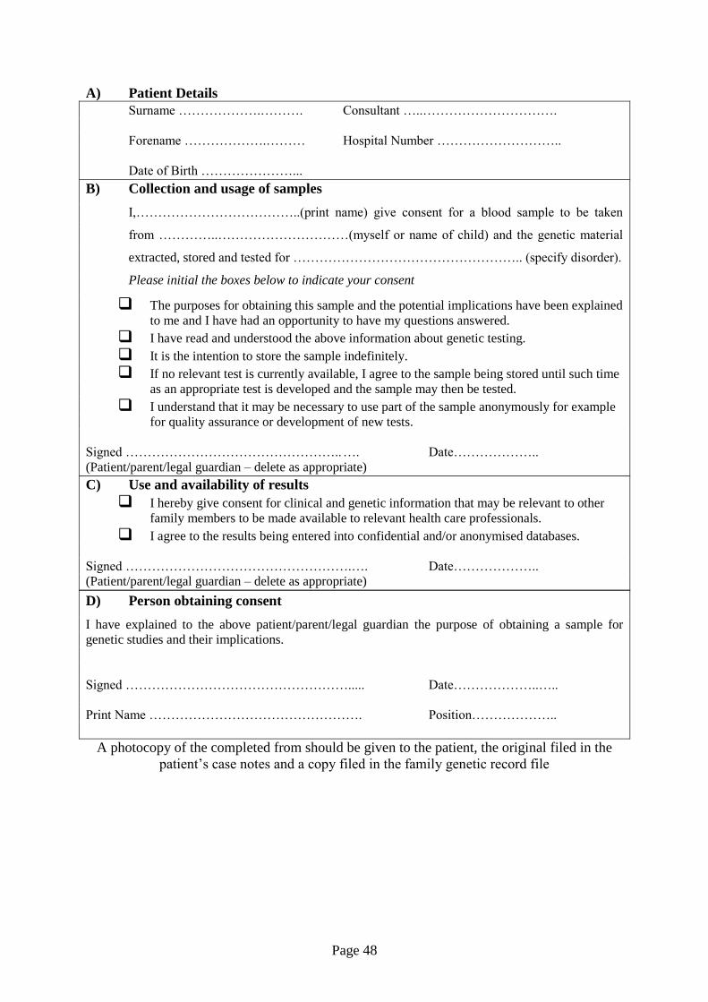
Page 48
A) Patient Details
Surname ……………….………. Consultant …..………………………….
Forename ……………….……… Hospital Number ………………………..
Date of Birth …………………...
B) Collection and usage of samples
I,………………………………..(print name) give consent for a blood sample to be taken
from …………..…………………………(myself or name of child) and the genetic material
extracted, stored and tested for …………………………………………….. (specify disorder).
Please initial the boxes below to indicate your consent
The purposes for obtaining this sample and the potential implications have been explained
to me and I have had an opportunity to have my questions answered.
I have read and understood the above information about genetic testing.
It is the intention to store the sample indefinitely.
If no relevant test is currently available, I agree to the sample being stored until such time
as an appropriate test is developed and the sample may then be tested.
I understand that it may be necessary to use part of the sample anonymously for example
for quality assurance or development of new tests.
Signed ………………………………………….. …. Date………………..
(Patient/parent/legal guardian – delete as appropriate)
C) Use and availability of results
I hereby give consent for clinical and genetic information that may be relevant to other
family members to be made available to relevant health care professionals.
I agree to the results being entered into confidential and/or anonymised databases.
Signed …………………………………………….…. Date………………..
(Patient/parent/legal guardian – delete as appropriate)
D) Person obtaining consent
I have explained to the above patient/parent/legal guardian the purpose of obtaining a sample for
genetic studies and their implications.
Signed ……………………………………………..... Date………………..…..
Print Name …………………………………………. Position………………..
A photocopy of the completed from should be given to the patient, the original filed in the
patient’s case notes and a copy filed in the family genetic record file

Page 49
Appendix II
The Haemophilia Genetics Laboratory Network and availability of Genetic Diagnostic
Services in the UK: Specification for Haemophilia Genetics Laboratory
The Network functions to ensure the provision nationally of a robust and high-quality genetic
diagnostic service for the heritable bleeding disorders. A directory of laboratories affiliated to
the Network, identifying services available and contact details, is available via the UKHCDO
website.
UK laboratories providing a genetic diagnostic service for haemophilia and other heritable
bleeding disorders should be affiliated to the UKHCDO Haemophilia Genetics Laboratory
Network.
UKHCDO Genetics Laboratory Network-affiliated laboratories are required to comply with
the following quality standards:
1. A molecular genetics laboratory, affiliated to a Comprehensive Care Haemophilia
Centre, providing an NHS diagnostic service
2. Facilities and expertise to allow the identification of mutations in haemophilia A and
B and other heritable bleeding disorders.(including handling risk of infection samples)
3. Ability to assign carriership and make antenatal diagnosis
4. Compliance with locally agreed turnaround times, appropriate to the clinical service.
The ability to turnaround urgent samples within two weeks. A three working day
turnaround time for prenatal diagnosis cases.
5. CPA accreditation
6. Participation in appropriate genetics external quality control scheme (currently
NEQAS)
7. Active collaboration between all haemophilia genetic laboratories to provide a robust
service available throughout the year
Assessment of compliance with these standards is an integral part of the external peer-review
audit system operated by the UKHCDO for UK haemophilia centres.
Adequate numbers of appropriately qualified HCPC-registered staff are required to provide a
high-quality and up-to-date genetic diagnostic service in accordance with the quality
standards identified above. Professional direction for the laboratory should be provided by a
consultant haematologist, clinical geneticist or a senior HCPC-registered scientist.
Page 50
References
Alfirevic, Z., & von Dadelszen, P. (2003). Instruments for chorionic villus sampling for
prenatal diagnosis. Cochrane Database Syst Rev, 1.
Alvarado,E.A., Pacheco,R.P., Alderete,F.G., Luis,J.A., de la Cruz,A.A., & Quintana,L.O.
(2012) Selective termination in dichorionic twins discordant for congenital defect.
Eur.J.Obstet.Gynecol.Reprod.Biol., 161, 8-11.
Ash,K.M., Mibashan,R.S., & Nicolaides,K.H. (1988) Diagnosis and treatment of feto-
maternal hemorrhage in a fetus with homozygous von Willebrand's disease. Fetal
Ther., 3, 189-191.
Avent,N.D. & Chitty,L.S. (2006) Non-invasive diagnosis of fetal sex; utilisation of free fetal
DNA in maternal plasma and ultrasound. Prenat.Diagn., 26, 598-603.
Becker,J., Schwaab,R., Moller-Taube,A., Schwaab,U., Schmidt,W., Brackmann,H.H.,
Grimm,T., Olek,K., & Oldenburg,J. (1996) Characterization of the factor VIII defect
in 147 patients with sporadic hemophilia A: family studies indicate a mutation type-
dependent sex ratio of mutation frequencies. Am.J Hum.Genet., 58, 657-670.
Bogdanova,N., Markoff,A., Eisert,R., Wermes,C., Pollmann,H., Todorova,A., Chlystun,M.,
Nowak-Gottl,U., & Horst,J. (2007) Spectrum of molecular defects and mutation
detection rate in patients with mild and moderate hemophilia A. Hum.Mutat., 28, 54-
60.
Brinke,A., Tagliavacca,L., Naylor,J., Green,P., Giangrande,P., & Giannelli,F. (1996) Two
chimaeric transcription units result from an inversion breaking intron 1 of the factor
VIII gene and a region reportedly affected by reciprocal translocations in T-cell
leukaemia. Hum.Mol.Genet., 5, 1945-1951.
British Society for Human Genetics. Genetic Testing of Children. 2010.
CEMAT Investigators (1998) Randomised trial to assess safety and fetal outcome of early
and midtrimester amniocentesis. The Canadian Early and Mid-trimester
Amniocentesis Trial (CEMAT) Group. Lancet, 351, 242-247.
Chi,C., Hyett,J.A., Finning,K.M., LEE,C.A., & Kadir,R.A. (2006) Non-invasive first
trimester determination of fetal gender: a new approach for prenatal diagnosis of
haemophilia. BJOG., 113, 239-242.
Cignacco,E. (2002) Between professional duty and ethical confusion: midwives and selective
termination of pregnancy. Nurs.Ethics, 9, 179-191.
Clark,S.L., Koonings,P.P., & Phelan,J.P. (1985) Placenta previa/accreta and prior cesarean
section. Obstet.Gynecol., 66, 89-92.
Cremonesi,L., Galbiati,S., Foglieni,B., Smid,M., Gambini,D., Ferrari,A., Viora,E.,
Campogrande,M., Pagliano,M., Travi,M., Piga,A., Restagno,G., & Ferrari,M. (2004)
Feasibility study for a microchip-based approach for noninvasive prenatal diagnosis
of genetic diseases. Ann.N.Y.Acad.Sci., 1022, 105-112.
Page 51
Cutler,J., Chappell,L.C., Kyle,P., & Madan,B. (2013) Third trimester amniocentesis for
diagnosis of inherited bleeding disorders prior to delivery. Haemophilia., 19, 904-907.
Dean,J.C., Fitzpatrick,D.R., Farndon,P.A., Kingstn,H., & Cusine,D. (2000) Genetic registers
in clinical practice: a survey of UK clinical genetics. J.Med.Genet., 37, 636-640.
Efrat,Z., Akinfenwa,O.O., & Nicolaides,K.H. (1999) First-trimester determination of fetal
gender by ultrasound. Ultrasound Obstet.Gynecol., 13, 305-307.
Elejalde,B.R., de Elejalde,M.M., & Heitman,T. (1985) Visualization of the fetal genitalia by
ultrasonography: a review of the literature and analysis of its accuracy and ethical
implications. J.Ultrasound Med., 4, 633-639.
Fiorentino,F., Magli,M.C., Podini,D., Ferraretti,A.P., Nuccitelli,A., Vitale,N., Baldi,M., &
Gianaroli,L. (2003) The minisequencing method: an alternative strategy for
preimplantation genetic diagnosis of single gene disorders. Mol.Hum.Reprod., 9, 399-
410.
Firth,H.V., Boyd,P.A., Chamberlain,P., MacKenzie,I.Z., Lindenbaum,R.H., & Huson,S.M.
(1991) Severe limb abnormalities after chorion villus sampling at 56-66 days'
gestation. Lancet, 337, 762-763.
Genetic Alliance UK. ACGT Consultation on Prenatal Genetic Testing: Response from the
Genetic Interest Group. 1-5-2000.
Genetic Interest Group. Guidelines for Genetic Services. 1998.
Giannelli,F., Green,P.M., Sommer,S.S., Poon,M., Ludwig,M., Schwaab,R., Reitsma,P.H.,
Goossens,M., Yoshioka,A., Figueiredo,M.S., & Brownlee,G.G. (1998) Haemophilia
B: database of point mutations and short additions and deletions--eighth edition.
Nucleic Acids Res., 26, 265-268.
Gigarel,N., Frydman,N., Burlet,P., Kerbrat,V., Steffann,J., Frydman,R., Munnich,A., &
Ray,P.F. (2004) Single cell co-amplification of polymorphic markers for the indirect
preimplantation genetic diagnosis of hemophilia A, X-linked adrenoleukodystrophy,
X-linked hydrocephalus and incontinentia pigmenti loci on Xq28. Hum.Genet., 114,
298-305.
Goossens,V., Harton,G., Moutou,C., Traeger-Synodinos,J., Van,R.M., & Harper,J.C. (2009)
ESHRE PGD Consortium data collection IX: cycles from January to December 2006
with pregnancy follow-up to October 2007. Hum.Reprod., 24, 1786-1810.
Gordon,M.C., Narula,K., O'Shaughnessy,R., & Barth,W.H., Jr. (2002) Complications of
third-trimester amniocentesis using continuous ultrasound guidance. Obstet.Gynecol.,
99, 255-259.
Harper,J.C., Coonen,E., De,R.M., Harton,G., Moutou,C., Pehlivan,T., Traeger-Synodinos,J.,
Van Rij,M.C., & Goossens,V. (2010) ESHRE PGD Consortium data collection X:
cycles from January to December 2007 with pregnancy follow-up to October 2008.
Hum.Reprod., 25, 2685-2707.
Page 52
Harton,G.L., De,R.M., Fiorentino,F., Moutou,C., SenGupta,S., Traeger-Synodinos,J., &
Harper,J.C. (2011) ESHRE PGD consortium best practice guidelines for
amplification-based PGD. Hum.Reprod., 26, 33-40.
Hillman,S.C., Morris,R.K., & Kilby,M.D. (2011) Co-twin prognosis after single fetal death: a
systematic review and meta-analysis. Obstet.Gynecol., 118, 928-940.
Hodor,J.G., Poggi,S.H., Spong,C.Y., Goodwin,K.M., Vink,J.S., Pezzullo,J.C., & Ghidini,A.
(2006) Risk of third-trimester amniocentesis: a case-control study. Am.J.Perinatol.,
23, 177-180.
Huq,F.Y. & Kadir,R.A. (2011) Management of pregnancy, labour and delivery in women
with inherited bleeding disorders. Haemophilia., 17 Suppl 1, 20-30.
James,A.H. & Hoots,K. (2010) The optimal mode of delivery for the haemophilia carrier
expecting an affected infant is caesarean delivery. Haemophilia., 16, 420-424.
Joint Committee on Medical Genetics. Consent and confidentiality in clinical genetic
practice: guidance on genetic testing and sharing genetic information, 2nd edn. 1-9-
2011.
Kadir,R.A., Economides,D.L., Braithwaite,J., Goldman,E., & Lee,C.A. (1997) The obstetric
experience of carriers of haemophilia. Br.J.Obstet.Gynaecol., 104, 803-810.
Kadir,R.A., Lee,C.A., Sabin,C.A., Pollard,D., & Economides,D.L. (1998) Pregnancy in
women with von Willebrand's disease or factor XI deficiency. Br.J.Obstet.Gynaecol.,
105, 314-321.
Kasper,C.K. & Buzin,C.H. (2009) Mosaics and haemophilia. Haemophilia., 15, 1181-1186.
Keeney,S., Mitchell,M., & Goodeve,A. Practice Guidelines for the Molecular Diagnosis of
Haemophilia A. 2010. Association for Clinical Genetic Science
(http://www.acgs.uk.com/).
Klopp,N., Oldenburg,J., Uen,C., Schneppenheim,R., & Graw,J. (2002) 11 hemophilia A
patients without mutations in the factor VIII encoding gene. Thromb Haemost, 88,
357-360.
Kulkarni,R. & Lusher,J.M. (1999) Intracranial and extracranial hemorrhages in newborns
with hemophilia: a review of the literature. J.Pediatr.Hematol.Oncol., 21, 289-295.
Kulkarni,R., Soucie,J.M., Lusher,J., Presley,R., Shapiro,A., Gill,J., Manco-Johnson,M.,
Koerper,M., Mathew,P., Abshire,T., DiMichele,D., Hoots,K., Janco,R., Nugent,D.,
Geraghty,S., & Evatt,B. (2009) Sites of initial bleeding episodes, mode of delivery
and age of diagnosis in babies with haemophilia diagnosed before the age of 2 years:
a report from The Centers for Disease Control and Prevention's (CDC) Universal Data
Collection (UDC) project. Haemophilia, 15, 1281-1290.

Page 53
Lakich,D., Kazazian,H.H., Jr., Antonarakis,S.E., & Gitschier,J. (1993) Inversions disrupting
the factor VIII gene are a common cause of severe haemophilia A. Nat.Genet., 5, 236-
241.
Laurie,A.D., Hill,A.M., Harraway,J.R., Fellowes,A.P., Phillipson,G.T., Benny,P.S.,
Smith,M.P., & George,P.M. (2010) Preimplantation genetic diagnosis for hemophilia
A using indirect linkage analysis and direct genotyping approaches.
J.Thromb.Haemost., 8, 783-789.
Lee,C.A., Chi,C., Pavord,S.R., Bolton-Maggs,P.H., Pollard,D., Hinchcliffe-Wood,A., &
Kadir,R.A. (2006) The obstetric and gynaecological management of women with
inherited bleeding disorders--review with guidelines produced by a taskforce of UK
Haemophilia Centre Doctors' Organization. Haemophilia., 12, 301-336.
Leuer,M., Oldenburg,J., Lavergne,J.M., Ludwig,M., Fregin,A., Eigel,A., Ljung,R.,
Goodeve,A., Peake,I., & Olek,K. (2001) Somatic mosaicism in hemophilia A: a fairly
common event. Am.J Hum.Genet., 69, 75-87.
Maddox,J. (1992) Genetics and the public interest. Nature, 356, 365-366.
Michaelides,K., Tuddenham,E.G., Turner,C., Lavender,B., & Lavery,S.A. (2006) Live birth
following the first mutation specific pre-implantation genetic diagnosis for
haemophilia A. Thromb.Haemost., 95, 373-379.
Mitchell,M., Keeney,S., & Goodeve,A. Practice Guidelines for the Molecular Diagnosis of
Haemophilia B. 2010. Association for Clinical Genetic Science
(http://www.acgs.uk.com/).
Mujezinovic,F. & Alfirevic,Z. (2007) Procedure-related complications of amniocentesis and
chorionic villous sampling: a systematic review. Obstet.Gynecol., 110, 687-694.
National Specialised Commissioning Group. Specialised Services for Haemophilia and Other
Related Bleeding Disorders. 2010.
Nuffield Council on Bioethics. Genetic screening: ethical issues. 1-1-1993. Nuffield
Foundation, London.
O'Donoghue,K., Giorgi,L., Pontello,V., Pasquini,L., & Kumar,S. (2007) Amniocentesis in the
third trimester of pregnancy. Prenat.Diagn., 27, 1000-1004.
Pavlova,A., Brondke,H., Musebeck,J., Pollmann,H., SRIVASTAVA,A., & Oldenburg,J.
(2009) Molecular mechanisms underlying hemophilia A phenotype in seven females.
J.Thromb.Haemost., 7, 976-982.
Peake,I.R., Lillicrap,D.P., Boulyjenkov,V., Briet,E., Chan,V., Ginter,E.K., Kraus,E.M.,
Ljung,R., Mannucci,P.M., Nicolaides,K., & . (1993) Haemophilia: strategies for
carrier detection and prenatal diagnosis. Bull.World Health Organ, 71, 429-458.
Page 54
Puck,J.M. & Willard,H.F. (1998) X inactivation in females with X-linked disease.
N.Engl.J.Med., 338, 325-328.
Renwick,P., Trussler,J., Lashwood,A., Braude,P., & Ogilvie,C.M. (2010) Preimplantation
genetic haplotyping: 127 diagnostic cycles demonstrating a robust, efficient
alternative to direct mutation testing on single cells. Reprod.Biomed.Online., 20, 470-
476.
Rossetti,L.C., Radic,C.P., Larripa,I.B., & De Brasi,C.D. (2008) Developing a new generation
of tests for genotyping hemophilia-causative rearrangements involving int22h and
int1h hotspots in the factor VIII gene. J Thromb Haemost, 6, 830-836.
Royal College of Obstetricians and Gynaecologists. Amniocentesis and Chorionic Villus
Sampling. 1-6-2010. Green-top Guideline No. 8.
Sanchez-Garcia,J.F., Gallardo,D., Navarro,J., Marquez,C., Gris,J.M., Sanchez,M.A.,
Altisent,C., & Vidal,F. (2006) A versatile strategy for preimplantation genetic
diagnosis of haemophilia A based on F8-gene sequencing. Thromb.Haemost., 96,
839-845.
Sellner,L.N. & Taylor,G.R. (2004) MLPA and MAPH: new techniques for detection of gene
deletions. Hum.Mutat., 23, 413-419.
Skirton,H., Lewis,C., Kent,A., & Coviello,D.A. (2010) Genetic education and the challenge
of genomic medicine: development of core competences to support preparation of
health professionals in Europe. Eur.J.Hum.Genet., 18, 972-977.
Smidt-Jensen,S., Permin,M., Philip,J., Lundsteen,C., Zachary,J.M., Fowler,S.E., &
Gruning,K. (1992) Randomised comparison of amniocentesis and transabdominal and
transcervical chorionic villus sampling. Lancet, 340, 1237-1244.
Stark,C.M., Smith,R.S., Lagrandeur,R.M., Batton,D.G., & Lorenz,R.P. (2000) Need for
urgent delivery after third-trimester amniocentesis. Obstet.Gynecol., 95, 48-50.
Tabor,A., Philip,J., Madsen,M., Bang,J., Obel,E.B., & Norgaard-Pedersen,B. (1986)
Randomised controlled trial of genetic amniocentesis in 4606 low-risk women.
Lancet, 1, 1287-1293.
The Haemophilia Alliance. A National Service Specification for Haemophilia and Related
Conditions. 1-6-2006.
Tongsong,T., Wanapirak,C., Kunavikatikul,C., Sirirchotiyakul,S., Piyamongkol,W., &
Chanprapaph,P. (2000) Cordocentesis at 16-24 weeks of gestation: experience of
1,320 cases. Prenat.Diagn., 20, 224-228.
Tsui,N.B., Kadir,R.A., Chan,K.C., Chi,C., Mellars,G., Tuddenham,E.G., Leung,T.Y.,
Lau,T.K., Chiu,R.W., & Lo,Y.M. (2011) Noninvasive prenatal diagnosis of
hemophilia by microfluidics digital PCR analysis of maternal plasma DNA. Blood,
117, 3684-3691.

Page 55
Wallis Y, Payne S, McAnulty C, Bodmer D, Sister-mans E, Robertson K, Moore D, Abbs S,
Deans Z, and Devereau A. Practice Guidelines for the Evaluation of Pathogenicity
and the Reporting of Sequence Variants in Clinical Molecular Genetics. Association
for Clinical Genetic Science (http://www.acgs.uk.com/).
Related Documents